269 www.eymj.org INTRODUCTION Ophthalmic emergencies such as corneal perforation or im- pending corneal perforation require immediate detection and prompt intervention. e integrity of the cornea should be re- stored as soon as possible using tissue adhesives, conjunctival flaps, amniotic membrane transplantation, or penetrating ker- atoplasty (PKP). 1 Of those, PKP provides a good corneal tissue substitute. However, in cases of PKP with an uncontrolled infection, the remaining diseased corneal tissue may persistently harbor pathogens, and removal of the recipient’s endothelium may in- crease the risk of causative organisms spreading into the ante- rior chamber and causing endophthalmitis. In addition, a graft-versus-host reaction between the donor tissue and the recipient may result in graft failure and visual loss. 2,3 erefore, it is important to select the right surgical method and corneal substitute to obtain a better visual outcome. Cryopreservation provides preserved stability of the tissues, and therefore, can be used for preserving tissues for urgent clinical use. However, a critical drawback of cryopreserved cor- neal use is impaired endothelial cell function. To allow the ap- plication of cryopreserved cornea, surgical methods that con- serve the endothelium of the recipient are needed. In the following report, we describe the treatment course and surgical outcomes of tectonic lamellar keratoplasty, using cryopreserved cornea to rescue an abrupt stromal melting with a large descemetocele. Tectonic Lamellar Keratoplasty Using Cryopreserved Cornea in a Large Descemetocele Kang Yoon Kim 1 , Ji Won Jung 1 , Eung Kweon Kim 1,2 , Kyoung Yul Seo 1 , and Tae-im Kim 1 1 Department of Ophthalmology, e Institute of Vision Research, Severance Hospital, Yonsei University College of Medicine, Seoul; 2 Corneal Dystrophy Research Institute, Severance Biomedical Science Institute, and Brain Korea 21 PLUS Project for Medical Science, Yonsei University College of Medicine, Seoul, Korea. We describe herein a case of an impending corneal perforation with a large descemetocele in a patient with previous penetrating keratoplasty (PKP) that subsequently was treated with an emergent lamellar keratoplasty using frozen preserved cornea. A 76-year-old male patient, who had a PKP, presented with a completely whitish and edematous graft accompanied by large epi- thelial defects. Although antibiotics and antiviral agents were tried for three days, the corneal stroma abruptly melted, except for the Descemet’s membrane and endothelium. Cryopreserved corneal tissue that was kept at -80°C was thawed and sutured on top of the remaining Descemet’s membrane and endothelium. Pathological and microbiological tests were conducted using the re- maining donor and recipient corneal tissues. After tectonic corneal transplantation on top of a large descemetocele, a healthy graft and relatively clear interfaces between graft-host junctions were maintained without serious adverse reactions throughout 6 month follow-up period. Microbiological evaluations of donor tissue at the time of thawing and tissue preparation were done, and the results were all negative. Tissue that was taken intraoperatively from the recipient cornea also showed negative microbio- logical results. In conclusion, tectonic lamellar keratoplasty, using cryopreserved corneal tissue, only onto the remaining Des- cemet’s membrane and endothelium in an emergent condition, was a safe and effective treatment. Key Words: Tectonic keratoplasty, cryopreserved cornea, descemetocele Yonsei Med J 2016 Jan;57(1):269-271 http://dx.doi.org/10.3349/ymj.2016.57.1.269 Case Report pISSN: 0513-5796 · eISSN: 1976-2437 Received: November 12, 2014 Revised: December 19, 2014 Accepted: December 22, 2014 Corresponding author: Dr. Tae-im Kim, Department of Ophthalmology, The Insti- tute of Vision Research, Yonsei University College of Medicine, 50-1 Yonsei-ro, Seodaemun-gu, Seoul 03722, Korea. Tel: 82-2-2228-3570, Fax: 82-2-312-0541, E-mail: [email protected] •The authors have no financial conflicts of interest. © Copyright: Yonsei University College of Medicine 2016 This is an Open Access article distributed under the terms of the Creative Com- mons Attribution Non-Commercial License (http://creativecommons.org/ licenses/ by-nc/3.0) which permits unrestricted non-commercial use, distribution, and repro- duction in any medium, provided the original work is properly cited.

Welcome message from author
This document is posted to help you gain knowledge. Please leave a comment to let me know what you think about it! Share it to your friends and learn new things together.
Transcript

269www.eymj.org
INTRODUCTION
Ophthalmic emergencies such as corneal perforation or im-pending corneal perforation require immediate detection and prompt intervention. The integrity of the cornea should be re-stored as soon as possible using tissue adhesives, conjunctival flaps, amniotic membrane transplantation, or penetrating ker-atoplasty (PKP).1
Of those, PKP provides a good corneal tissue substitute.
However, in cases of PKP with an uncontrolled infection, the remaining diseased corneal tissue may persistently harbor pathogens, and removal of the recipient’s endothelium may in-crease the risk of causative organisms spreading into the ante-rior chamber and causing endophthalmitis. In addition, a graft-versus-host reaction between the donor tissue and the recipient may result in graft failure and visual loss.2,3 Therefore, it is important to select the right surgical method and corneal substitute to obtain a better visual outcome.
Cryopreservation provides preserved stability of the tissues, and therefore, can be used for preserving tissues for urgent clinical use. However, a critical drawback of cryopreserved cor-neal use is impaired endothelial cell function. To allow the ap-plication of cryopreserved cornea, surgical methods that con-serve the endothelium of the recipient are needed.
In the following report, we describe the treatment course and surgical outcomes of tectonic lamellar keratoplasty, using cryopreserved cornea to rescue an abrupt stromal melting with a large descemetocele.
Tectonic Lamellar Keratoplasty Using Cryopreserved Cornea in a Large Descemetocele
Kang Yoon Kim1, Ji Won Jung1, Eung Kweon Kim1,2, Kyoung Yul Seo1, and Tae-im Kim1
1Department of Ophthalmology, The Institute of Vision Research, Severance Hospital, Yonsei University College of Medicine, Seoul;2 Corneal Dystrophy Research Institute, Severance Biomedical Science Institute, and Brain Korea 21 PLUS Project for Medical Science, Yonsei University College of Medicine, Seoul, Korea.
We describe herein a case of an impending corneal perforation with a large descemetocele in a patient with previous penetrating keratoplasty (PKP) that subsequently was treated with an emergent lamellar keratoplasty using frozen preserved cornea. A 76-year-old male patient, who had a PKP, presented with a completely whitish and edematous graft accompanied by large epi-thelial defects. Although antibiotics and antiviral agents were tried for three days, the corneal stroma abruptly melted, except for the Descemet’s membrane and endothelium. Cryopreserved corneal tissue that was kept at -80°C was thawed and sutured on top of the remaining Descemet’s membrane and endothelium. Pathological and microbiological tests were conducted using the re-maining donor and recipient corneal tissues. After tectonic corneal transplantation on top of a large descemetocele, a healthy graft and relatively clear interfaces between graft-host junctions were maintained without serious adverse reactions throughout 6 month follow-up period. Microbiological evaluations of donor tissue at the time of thawing and tissue preparation were done, and the results were all negative. Tissue that was taken intraoperatively from the recipient cornea also showed negative microbio-logical results. In conclusion, tectonic lamellar keratoplasty, using cryopreserved corneal tissue, only onto the remaining Des-cemet’s membrane and endothelium in an emergent condition, was a safe and effective treatment.
Key Words: Tectonic keratoplasty, cryopreserved cornea, descemetocele
Yonsei Med J 2016 Jan;57(1):269-271http://dx.doi.org/10.3349/ymj.2016.57.1.269
Case Report
pISSN: 0513-5796 · eISSN: 1976-2437
Received: November 12, 2014 Revised: December 19, 2014Accepted: December 22, 2014Corresponding author: Dr. Tae-im Kim, Department of Ophthalmology, The Insti-tute of Vision Research, Yonsei University College of Medicine, 50-1 Yonsei-ro, Seodaemun-gu, Seoul 03722, Korea.Tel: 82-2-2228-3570, Fax: 82-2-312-0541, E-mail: [email protected]
•The authors have no financial conflicts of interest.
© Copyright: Yonsei University College of Medicine 2016This is an Open Access article distributed under the terms of the Creative Com-mons Attribution Non-Commercial License (http://creativecommons.org/ licenses/by-nc/3.0) which permits unrestricted non-commercial use, distribution, and repro-duction in any medium, provided the original work is properly cited.

http://dx.doi.org/10.3349/ymj.2016.57.1.269270
Tectonic Keratoplasty Using Cryopreserved Cornea
CASE REPORT
A 76-year-old male patient presented with 4 days of persistent ocular pain. After traumatic cataract extraction 30 years prior, he underwent PKP 3 years ago due to pseudophakic bullous keratopathy. The patient was taking 200 mg of oral acyclovir, topical levofloxacin, and topical prednisolone acetate for fre-quently recurring herpetic keratitis. Examination revealed that his cornea was edematous and entirely opaque with 7×7 mm sized epithelial defects. His visual acuity was light perception, and ultrasonography confirmed no definite change of vitreous and retina. The patient was given topical moxifloxacin, forti-fied with cefazolin and tobramycin, every 2 hours, and addi-tionally, he was given an oral acyclovir dosage of 600 mg, and a topical acyclovir ointment. Two days after changing medica-tions, the patient visited the clinic and showed improved vision, but microscopic examination revealed only a very thin layer of stroma and Descemet’s membrane, with the endothelium re-maining (Fig. 1A).
Donor cornea was prepared from the whole globe, without any contraindicative features at the Yonsei University Eye Bank, cryopreserved at -80°C for 3 months. The whole globe was se-quentially thawed at 20°C for 4 hours, at 4°C for 4 hours, and at room temperature for 2 hours to minimize thawing-related tis-sue stress. After the whole globe was completely thawed, a cor-neal button was removed, and then preserved in Optisol GS® (Bausch & Lomb, Irvine, CA, USA) for 2 hours until surgery.
Tectonic lamellar keratoplasty was done with prepared cor-nea. Completely thawed donor tissue was trephined by the size of 8.25 mm. The donor cornea, with intact entire layers, was placed on top of the recipient’s descemetocele and was sutured to the host’s eye bed, using interrupted 10-0 nylon, to avoid penetrating the descemetocele. Topical moxifloxacin and 2% voriconazole were initiated every 2 hours, immediately after the operation. At postoperative 2 months, topical moxifloxacin was
discontinued, and levofloxacin and rimexolone were initiated. Oral anti-inflammatory medications, 12 mg of triamcinolone and 300 mg of cyclosporine, were given from 1 week after sur-gery.
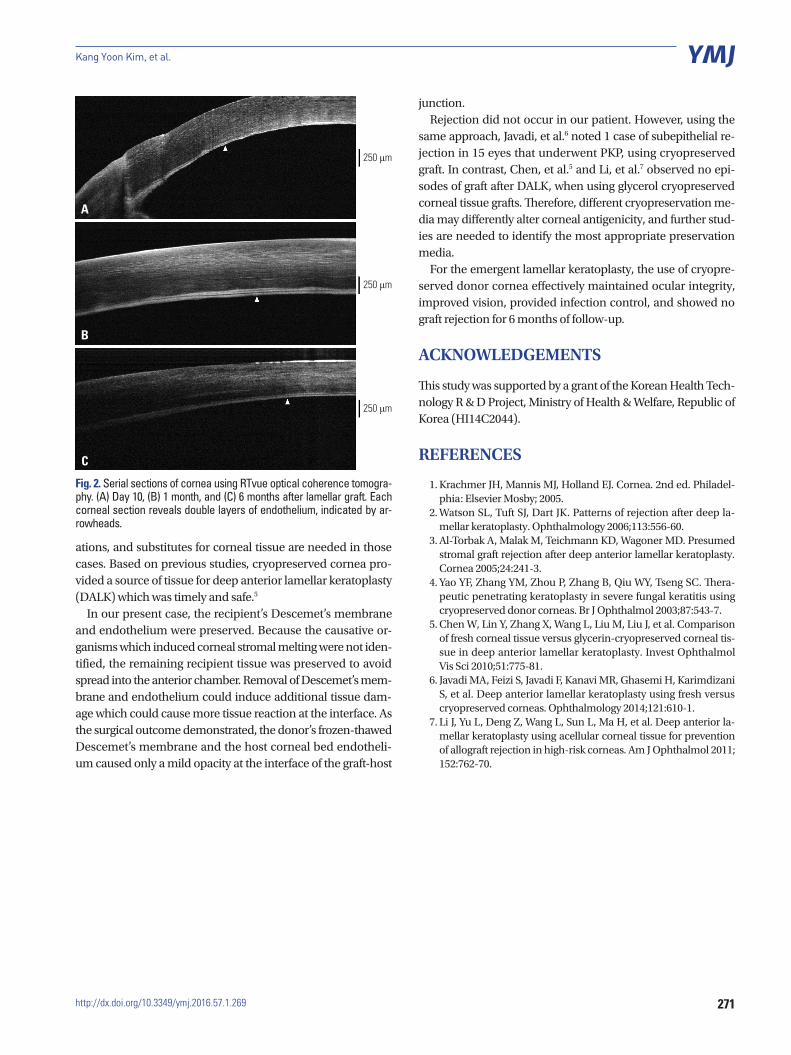
Donor-recipient interfaces were evaluated 10 days, 1 month, and 6 months after the operation, using corneal optical coher-ence tomography (OCT) (RTVue; Optoview, Inc., Fremont, CA, USA).
Microbiological evaluations were done on donor tissues that were biopsied during and after thawing, and on the recipient’s tissue that was cultured before grafting. Bacterial, fungal and viral assessments all showed negative results, even though the assessment for viral infection indicated that the amount of sample was insufficient for viral polymerase chain reaction. Because cryopreserved donor corneas are devoid of immune cells,4 we delayed immune modulating agents for the first week to reduce the chance of recurrent infection. However, the graft survived without complications (Fig. 1B). Serially anterior seg-mented OCT images revealed well attached double layers of Descemet’s membrane and endothelium between the graft and the host (Fig. 2). The interface showed mild opacity which could slightly interfere with visual function. At 6 months post-operatively, the uncorrected and corrected visual acuity was 20/400, the keratometry value was 46.25/50.25 with axis of 163, and intraocular pressure was 11 mm Hg.
DISCUSSION
Infectious corneal ulcer with significant melting of the corneal stromal tissue must be immediately corrected with a corneal substitute. Especially, corneal ulcer with infiltration that pro-gresses deeper into the stroma should be treated with prompt intervention to restore ocular structural integrity. However, fresh corneal tissue is not always available in emergency situ-
Fig. 1. Slit lamp photograph of cornea taken preoperatively and postoperatively. (A) A photograph of the cornea 1 day before the operation. The stroma of the corneal graft melted down with preservation of only the Descemet’s membrane and endothelium. (B) Six months after the operation, an intact corneal graft with mild interface opacity is shown.
A B
271http://dx.doi.org/10.3349/ymj.2016.57.1.269
Kang Yoon Kim, et al.
ations, and substitutes for corneal tissue are needed in those cases. Based on previous studies, cryopreserved cornea pro-vided a source of tissue for deep anterior lamellar keratoplasty (DALK) which was timely and safe.5
In our present case, the recipient’s Descemet’s membrane and endothelium were preserved. Because the causative or-ganisms which induced corneal stromal melting were not iden-tified, the remaining recipient tissue was preserved to avoid spread into the anterior chamber. Removal of Descemet’s mem-brane and endothelium could induce additional tissue dam-age which could cause more tissue reaction at the interface. As the surgical outcome demonstrated, the donor’s frozen-thawed Descemet’s membrane and the host corneal bed endotheli-um caused only a mild opacity at the interface of the graft-host
junction. Rejection did not occur in our patient. However, using the
same approach, Javadi, et al.6 noted 1 case of subepithelial re-jection in 15 eyes that underwent PKP, using cryopreserved graft. In contrast, Chen, et al.5 and Li, et al.7 observed no epi-sodes of graft after DALK, when using glycerol cryopreserved corneal tissue grafts. Therefore, different cryopreservation me-dia may differently alter corneal antigenicity, and further stud-ies are needed to identify the most appropriate preservation media.
For the emergent lamellar keratoplasty, the use of cryopre-served donor cornea effectively maintained ocular integrity, improved vision, provided infection control, and showed no graft rejection for 6 months of follow-up.
ACKNOWLEDGEMENTS
This study was supported by a grant of the Korean Health Tech-nology R & D Project, Ministry of Health & Welfare, Republic of Korea (HI14C2044).
REFERENCES
1. Krachmer JH, Mannis MJ, Holland EJ. Cornea. 2nd ed. Philadel-phia: Elsevier Mosby; 2005.
2. Watson SL, Tuft SJ, Dart JK. Patterns of rejection after deep la-mellar keratoplasty. Ophthalmology 2006;113:556-60.
3. Al-Torbak A, Malak M, Teichmann KD, Wagoner MD. Presumed stromal graft rejection after deep anterior lamellar keratoplasty. Cornea 2005;24:241-3.
4. Yao YF, Zhang YM, Zhou P, Zhang B, Qiu WY, Tseng SC. Thera-peutic penetrating keratoplasty in severe fungal keratitis using cryopreserved donor corneas. Br J Ophthalmol 2003;87:543-7.
5. Chen W, Lin Y, Zhang X, Wang L, Liu M, Liu J, et al. Comparison of fresh corneal tissue versus glycerin-cryopreserved corneal tis-sue in deep anterior lamellar keratoplasty. Invest Ophthalmol Vis Sci 2010;51:775-81.
6. Javadi MA, Feizi S, Javadi F, Kanavi MR, Ghasemi H, Karimdizani S, et al. Deep anterior lamellar keratoplasty using fresh versus cryopreserved corneas. Ophthalmology 2014;121:610-1.
7. Li J, Yu L, Deng Z, Wang L, Sun L, Ma H, et al. Deep anterior la-mellar keratoplasty using acellular corneal tissue for prevention of allograft rejection in high-risk corneas. Am J Ophthalmol 2011; 152:762-70.
A
B
C
Fig. 2. Serial sections of cornea using RTvue optical coherence tomogra-phy. (A) Day 10, (B) 1 month, and (C) 6 months after lamellar graft. Each corneal section reveals double layers of endothelium, indicated by ar-rowheads.
250 μm
250 μm
250 μm
Related Documents


![Review Article Lamellar Keratoplasty: A Literature Reviewdownloads.hindawi.com/journals/joph/2013/894319.pdfJournal of Ophthalmology described by Melles et al. [ ] allowing transplantation](https://static.cupdf.com/doc/110x72/5e39106a1415da08cf09cef9/review-article-lamellar-keratoplasty-a-literature-journal-of-ophthalmology-described.jpg)