J Toxicol Pathol 2012; 25: 183–188 Technical Report Development of a Delayed-Type Hypersensitivity (DTH) Model in the Cynomolgus Monkey Caroline Bouchez 1 , Fréderic Gervais 1 , Renaud Fleurance 1 , Bernard Palate 1 , Jean-Jacques Legrand 1 , and Jacques Descotes 2 1 CiToxLaB France, BP 503, 27005 Evreux, France 2 Poison Center, 69003 Lyon, France Abstract: Although a T-dependent antibody response (TDAR) assay is generally recommended as the first-line immune function assay in nonclinical immunotoxicity evaluation, second-line assays such as delayed-type hypersensitivity (DTH) to measure cell-mediated responses can provide helpful additional information. In this study, male Cynomolgus monkeys were injected intramuscularly either once or twice with 1 mg Keyhole Limpet Hemocyanin (KLH) or twice with a commercially available tetanus vaccine (40 IU tetanus toxoid + 0.06 mg aluminum hydroxide). All animals were subsequently challenged by intradermal injections of the same antigen or aluminum hydroxide after 4, 6 and 8 weeks. Clinical reactions at the injection sites were scored 24, 48 and 72 h post challenge. Skin biopsies were taken on completion of the observation period after each challenge for standard histological examination and immuno- labeling using CD3 (T lymphocytes), CD19 (B lymphocytes) and CD68 (macrophages) antibodies. Tetanus toxoid induced stronger clinical reactions than KLH, whereas aluminum hydroxide induced no clinical reaction. Perivascular mononuclear cell infiltrates, a histopathological finding consistent with a DTH reaction, were seen after all challenges with tetanus toxoid or KLH, but not with alu- minum hydroxide. Immunohistochemistry evidenced the presence of T lymphocytes and macrophages within these infiltrates. These results suggest that tetanus toxoid adjuvanted with aluminum hydroxide can induce a consistent DTH response for use as a model of cell-mediated response in Cynomolgus monkeys. (DOI: 10.1293/tox.25.183; J Toxicol Pathol 2012; 25: 183–188) Key words: immunotoxicity evaluation, delayed-type hypersensitivity, cynomolgus monkeys, tetanus toxoid, KLH Introduction Nowadays a T-dependent antibody response (TDAR) assay is widely considered to be the first-line function assay when the weight of evidence review as defined by the ICH guideline S8 1 recommends that additional immunotoxicity studies should be performed to assess the immunotoxic po- tential of drug candidates. Nevertheless, second-line func- tion assays may be further needed case-by-case depending on histopathological and clinical findings in standard tox- icity studies, the results of the TDAR assay or the drug’s mechanism of action. In addition to lymphocyte subset anal- ysis (immunophenotyping) and assays to measure natural killer (NK) cell activity or neutrophil/macrophage function, it may be helpful to assess cell-mediated immune responses using either in vitro or ex vivo assays (e.g., lymphoprolifera- tion induced by mitogens or mixed lymphocyte reaction) or in vivo animal models. Although assays to measure cell-mediated immunity have long been used, especially in rodents 2–4 , they are rath- er seldom included in current nonclinical immunotoxicity evaluation. One reason may be that only limited efforts have been paid to standardize and validate these assays until re- cently. The situation, however, is evolving, as shown by the recent study in B6C3F1 mice by Smith and White 5 . In com- parison to in vitro assays, in vivo models of cell-mediated immunity offer the advantage of measuring multiple cellu- lar components involving several cell interactions, inflam- matory mediators and complex signaling cascades. Thus, they can be useful for assessing cell-mediated immunity as well as general immune competence. Nonhuman primates (NHP) are often the only relevant species available for the nonclinical safety evaluation of novel biopharmaceuticals because of increasingly species- specific targets 6 . So far, only few studies have been devoted to designing in vivo models of cell-mediated immunity in NHP 7–9 . The aim of the present study was to develop a de- layed-type hypersensitivity (DTH) model in the Cynomol- gus monkey for use in regulatory immunotoxicity evalua- tion. Received: 22 December 2011, Accepted: 20 February 2012 Mailing address: Caroline Bouchez, CiToxLaB France, BP 503, 27005 Evreux Cedex, France TEL: +33-2-32-29-26-94 FAX: +33-2-32-67-87-05 E-mail: [email protected] ©2012 The Japanese Society of Toxicologic Pathology This is an open-access article distributed under the terms of the Cre- ative Commons Attribution Non-Commercial No Derivatives (by-nc- nd) License <http://creativecommons.org/licenses/by-nc-nd/3.0/>.

Welcome message from author
This document is posted to help you gain knowledge. Please leave a comment to let me know what you think about it! Share it to your friends and learn new things together.
Transcript

J Toxicol Pathol 2012; 25: 183–188
Technical Report
Development of a Delayed-Type Hypersensitivity (DTH) Model in the Cynomolgus Monkey
Caroline Bouchez1, Fréderic Gervais1, Renaud Fleurance1, Bernard Palate1, Jean-Jacques Legrand1, and Jacques Descotes2
1 CiToxLaB France, BP 503, 27005 Evreux, France2 Poison Center, 69003 Lyon, France
Abstract: Although a T-dependent antibody response (TDAR) assay is generally recommended as the first-line immune function assay in nonclinical immunotoxicity evaluation, second-line assays such as delayed-type hypersensitivity (DTH) to measure cell-mediated responses can provide helpful additional information. In this study, male Cynomolgus monkeys were injected intramuscularly either once or twice with 1 mg Keyhole Limpet Hemocyanin (KLH) or twice with a commercially available tetanus vaccine (40 IU tetanus toxoid + 0.06 mg aluminum hydroxide). All animals were subsequently challenged by intradermal injections of the same antigen or aluminum hydroxide after 4, 6 and 8 weeks. Clinical reactions at the injection sites were scored 24, 48 and 72 h post challenge. Skin biopsies were taken on completion of the observation period after each challenge for standard histological examination and immuno-labeling using CD3 (T lymphocytes), CD19 (B lymphocytes) and CD68 (macrophages) antibodies. Tetanus toxoid induced stronger clinical reactions than KLH, whereas aluminum hydroxide induced no clinical reaction. Perivascular mononuclear cell infiltrates, a histopathological finding consistent with a DTH reaction, were seen after all challenges with tetanus toxoid or KLH, but not with alu-minum hydroxide. Immunohistochemistry evidenced the presence of T lymphocytes and macrophages within these infiltrates. These results suggest that tetanus toxoid adjuvanted with aluminum hydroxide can induce a consistent DTH response for use as a model of cell-mediated response in Cynomolgus monkeys. (DOI: 10.1293/tox.25.183; J Toxicol Pathol 2012; 25: 183–188)
Key words: immunotoxicity evaluation, delayed-type hypersensitivity, cynomolgus monkeys, tetanus toxoid, KLH
Introduction
Nowadays a T-dependent antibody response (TDAR) assay is widely considered to be the first-line function assay when the weight of evidence review as defined by the ICH guideline S81 recommends that additional immunotoxicity studies should be performed to assess the immunotoxic po-tential of drug candidates. Nevertheless, second-line func-tion assays may be further needed case-by-case depending on histopathological and clinical findings in standard tox-icity studies, the results of the TDAR assay or the drug’s mechanism of action. In addition to lymphocyte subset anal-ysis (immunophenotyping) and assays to measure natural killer (NK) cell activity or neutrophil/macrophage function, it may be helpful to assess cell-mediated immune responses using either in vitro or ex vivo assays (e.g., lymphoprolifera-
tion induced by mitogens or mixed lymphocyte reaction) or in vivo animal models.
Although assays to measure cell-mediated immunity have long been used, especially in rodents2–4, they are rath-er seldom included in current nonclinical immunotoxicity evaluation. One reason may be that only limited efforts have been paid to standardize and validate these assays until re-cently. The situation, however, is evolving, as shown by the recent study in B6C3F1 mice by Smith and White5. In com-parison to in vitro assays, in vivo models of cell-mediated immunity offer the advantage of measuring multiple cellu-lar components involving several cell interactions, inflam-matory mediators and complex signaling cascades. Thus, they can be useful for assessing cell-mediated immunity as well as general immune competence.
Nonhuman primates (NHP) are often the only relevant species available for the nonclinical safety evaluation of novel biopharmaceuticals because of increasingly species-specific targets6. So far, only few studies have been devoted to designing in vivo models of cell-mediated immunity in NHP7–9. The aim of the present study was to develop a de-layed-type hypersensitivity (DTH) model in the Cynomol-gus monkey for use in regulatory immunotoxicity evalua-tion.
Received: 22 December 2011, Accepted: 20 February 2012Mailing address: Caroline Bouchez, CiToxLaB France, BP 503, 27005 Evreux Cedex, FranceTEL: +33-2-32-29-26-94 FAX: +33-2-32-67-87-05E-mail: [email protected]©2012 The Japanese Society of Toxicologic PathologyThis is an open-access article distributed under the terms of the Cre-ative Commons Attribution Non-Commercial No Derivatives (by-nc-nd) License <http://creativecommons.org/licenses/by-nc-nd/3.0/>.

DTH Model in the Cynomolgus Monkey184
Materials and Methods
AnimalsMale purpose-bred Cynomolgus monkeys (Macaca
fascicularis) purchased from Noveprim Europe (Ebene, Mauritius) were used throughout this study. The animals were housed in individual stainless steel cages in a dedicat-ed primate unit where room conditions were set as follows: temperature, 24 ± 3°C; relative humidity, 50 ± 30%; light/dark cycle, 12h/12h (7:00–19:00); ventilation, approximately 12 cycles/h of filtered, non-recycled air. Enrichments were given to the animals during the whole study. All animals had free access to tap water and were distributed approxi-mately 180 grams of OWM (E) SQC SHORT expanded diet (Dietex France, SDS, Saint Gratien, France). In addition, a fruit supplement was given daily to each animal. They were at least 3 years old at the beginning of the study.
All study procedures were conducted according to a written study protocol approved by CiToxLAB animal eth-ics committee and facility standard operating procedures in strict compliance with accepted animal welfare standards.
TreatmentThe animals were allocated to 3 groups of 3 animals
each. They were first immunized by the intramuscular route and subsequently challenged by intradermal injections.
Immunization: Group 1 and 2 animals were injected intramuscularly with 1 mg of keyhole limpet hemocyanin (KLH), purchased from Thermo Scientific (France) and pre-pared as a 2 mg/mL solution in water for injection, either on day 14 (group 1) or on days 1 and 14 (group 2). Group 3 ani-mals were injected on days 1 and 14 with 0.5 mL of a tetanus vaccine (Vaccin Tétanos Pasteur®) purchased from Sanofi-Pasteur Laboratories (France), corresponding to a dose of 40 UI tetanus toxoid plus 0.6 mg aluminum hydroxide.
Challenges: On the days of challenge, all animals were fasted at least 12 h before anesthesia with intravenous pro-pofol (Rapinovet®, B Braun Medical). The backs (and ab-domen when appropriate) of the animals were carefully clipped free of hair with electric clippers under anesthesia. Group 1 and 2 animals received 3 intradermal injections of 0.05 mg/mL of keyhole limpet hemocyanin (KLH) on the back on days 42, 56 and 70 (3 challenges per animal and per day of challenge for a total of 9 challenge sites per animal). Group 3 animals received 3 intradermal injections of 0.05 mg/mL of the same tetanus vaccine and 3 injections 0.05 mg/mL of a 1.2 mg/mL solution of aluminum hydroxide on days 42, 56 and 70, either on the back or on the abdomen of the animals (3 challenges per test item, per animal and per day of challenge for a total of 18 challenge sites per animal). The injection sites were disinfected with 70% alcohol prior to intradermal injection, and each injection site was used only once.
Delayed-type hypersensitivity (DTH) responseThe DTH response of each monkey was assessed both
clinically and histologically.
Clinical examination: Skin reactions at the intradermal injection sites were recorded once daily for each animal by visual inspection and palpation of the injection sites prior to injection and then after 24, 48 and 72 h. The following scales were used for scoring erythema formation (very slight, barely perceptible = 1; slight = 2; moderate = 3; se-vere erythema to slight eschar formation = 4) and edema formation (very slight, barely perceptible = 1; slight with area edges well-defined by definite raising = 2; moderate with approximately 1-mm of raising = 3; severe with more than 1-mm of raising and extension beyond area of exposure = 4). Any other lesions, such as abscess, necrosis and local inflammation, or reactions, such as pain, were recorded.
Histopathological and immunohistochemistry exami-nations: Skin biopsies were collected under propofol an-esthesia 72 h after intradermal injection of the designated challenge sites using an 8-mm skin biopsy punch. Prophy-lactic analgesia was provided by subcutaneous injection of buprenorphine (Buprecare®) after each challenge. Three skin biopsies (groups 1 and 2) or 6 skin biopsies (group 3: 3 skin biopsies following tetanus toxoid challenges and 3 following aluminum hydroxide challenges) were collected after each day of challenge from each animal. Two out of 3 biopsies were fixed in 10% buffered formalin and then em-bedded in paraffin wax, sectioned at a thickness of approxi-mately 4 microns and stained with hematoxylin and eosin (2 sections per site). The third biopsy was snap frozen embed-ded in OCT (Optimal Cutting Temperature) compound in a plastic mold, frozen in dry ice and stained with an immuno-peroxidase technique using CD3 (T lymphocytes), CD19 (B lymphocytes) and CD68 (macrophages) commercial mark-ers.
A microscopic examination was performed by Dr Ger-vais on all skin biopsies from all animals (including sections stained with hematoxylin and eosin or labeled for immuno-histochemistry). A peer review was performed by Dr Fleur-ance on an adequate number of slides from challenge sites to confirm that findings recorded by the study pathologist were consistent and accurate.
Results
No unscheduled deaths occurred, and no systemic treatment-related clinical signs were reported. Body weight was never affected during the study.
Clinical DTH responseLocal reactions at injection sites were observed in all
groups after each challenge, generally during 2 or 3 days. In groups 1 and 2, erythema (mean grades = 0.2 to 0.9) and/or thickening (mean grades = 0.3 to 1.1) at the injection sites were observed 24, 48 and 72 h after the first KLH challenge on day 42. After the second KLH challenge on day 56, ery-thema was seen until 48 h after injection (mean grades = 0.4 to 1.1) and was associated with thickening at all 3 time-points (mean grades = 0.3 to 0.9). No erythema was record-ed after the third KLH challenge on day 70, but edema was
Bouchez, Gervais, Fleurance et al. 185
noted at all the injection sites 24 h after injection, and then thickening was observed 48 h after injection. Similar local reactions were noted in group 1 animals immunized with one KLH injection and in group 2 animals immunized with two KLH injections.
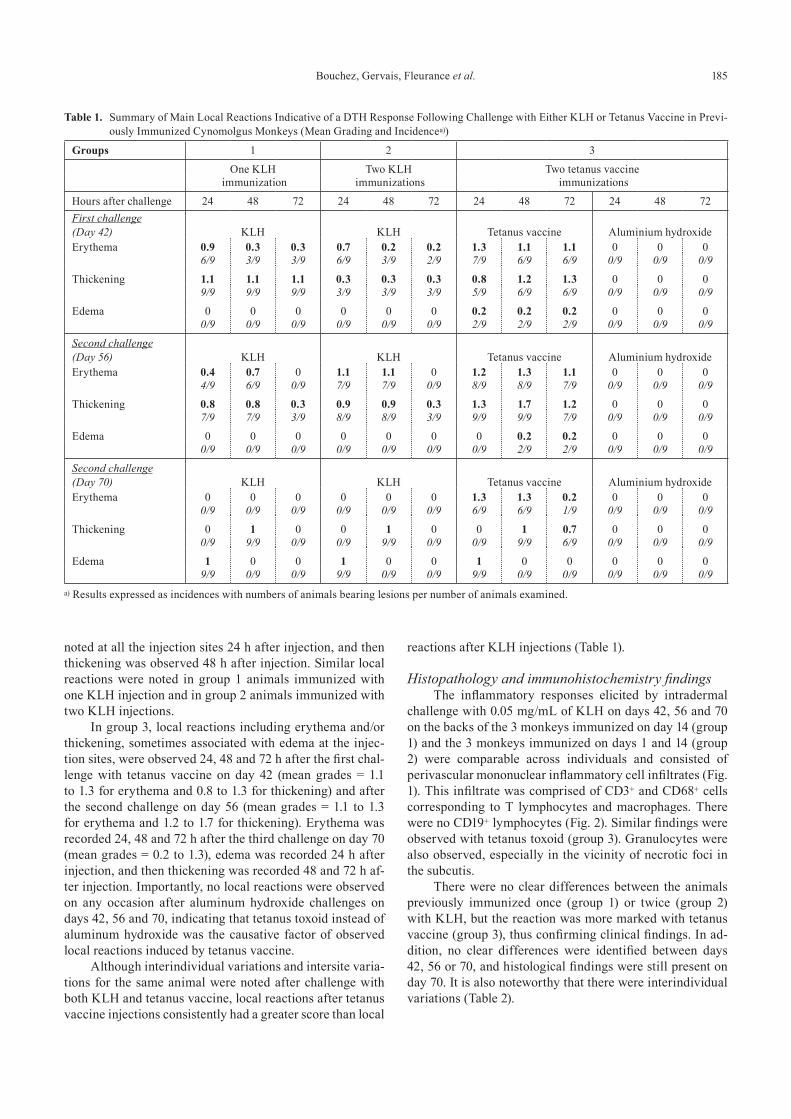
In group 3, local reactions including erythema and/or thickening, sometimes associated with edema at the injec-tion sites, were observed 24, 48 and 72 h after the first chal-lenge with tetanus vaccine on day 42 (mean grades = 1.1 to 1.3 for erythema and 0.8 to 1.3 for thickening) and after the second challenge on day 56 (mean grades = 1.1 to 1.3 for erythema and 1.2 to 1.7 for thickening). Erythema was recorded 24, 48 and 72 h after the third challenge on day 70 (mean grades = 0.2 to 1.3), edema was recorded 24 h after injection, and then thickening was recorded 48 and 72 h af-ter injection. Importantly, no local reactions were observed on any occasion after aluminum hydroxide challenges on days 42, 56 and 70, indicating that tetanus toxoid instead of aluminum hydroxide was the causative factor of observed local reactions induced by tetanus vaccine.
Although interindividual variations and intersite varia-tions for the same animal were noted after challenge with both KLH and tetanus vaccine, local reactions after tetanus vaccine injections consistently had a greater score than local
reactions after KLH injections (Table 1).
Histopathology and immunohistochemistry findingsThe inflammatory responses elicited by intradermal
challenge with 0.05 mg/mL of KLH on days 42, 56 and 70 on the backs of the 3 monkeys immunized on day 14 (group 1) and the 3 monkeys immunized on days 1 and 14 (group 2) were comparable across individuals and consisted of perivascular mononuclear inflammatory cell infiltrates (Fig. 1). This infiltrate was comprised of CD3+ and CD68+ cells corresponding to T lymphocytes and macrophages. There were no CD19+ lymphocytes (Fig. 2). Similar findings were observed with tetanus toxoid (group 3). Granulocytes were also observed, especially in the vicinity of necrotic foci in the subcutis.
There were no clear differences between the animals previously immunized once (group 1) or twice (group 2) with KLH, but the reaction was more marked with tetanus vaccine (group 3), thus confirming clinical findings. In ad-dition, no clear differences were identified between days 42, 56 or 70, and histological findings were still present on day 70. It is also noteworthy that there were interindividual variations (Table 2).
Table 1. Summary of Main Local Reactions Indicative of a DTH Response Following Challenge with Either KLH or Tetanus Vaccine in Previ-ously Immunized Cynomolgus Monkeys (Mean Grading and Incidencea))
Groups 1 2 3
One KLH Two KLH Two tetanus vaccine immunization immunizations immunizations
Hours after challenge 24 48 72 24 48 72 24 48 72 24 48 72First challenge (Day 42) KLH KLH Tetanus vaccine Aluminium hydroxideErythema 0.9 0.3 0.3 0.7 0.2 0.2 1.3 1.1 1.1 0 0 0 6/9 3/9 3/9 6/9 3/9 2/9 7/9 6/9 6/9 0/9 0/9 0/9
Thickening 1.1 1.1 1.1 0.3 0.3 0.3 0.8 1.2 1.3 0 0 09/9 9/9 9/9 3/9 3/9 3/9 5/9 6/9 6/9 0/9 0/9 0/9
Edema 0 0 0 0 0 0 0.2 0.2 0.2 0 0 0 0/9 0/9 0/9 0/9 0/9 0/9 2/9 2/9 2/9 0/9 0/9 0/9
Second challenge (Day 56) KLH KLH Tetanus vaccine Aluminium hydroxideErythema 0.4 0.7 0 1.1 1.1 0 1.2 1.3 1.1 0 0 0 4/9 6/9 0/9 7/9 7/9 0/9 8/9 8/9 7/9 0/9 0/9 0/9
Thickening 0.8 0.8 0.3 0.9 0.9 0.3 1.3 1.7 1.2 0 0 07/9 7/9 3/9 8/9 8/9 3/9 9/9 9/9 7/9 0/9 0/9 0/9
Edema 0 0 0 0 0 0 0 0.2 0.2 0 0 00/9 0/9 0/9 0/9 0/9 0/9 0/9 2/9 2/9 0/9 0/9 0/9
Second challenge (Day 70) KLH KLH Tetanus vaccine Aluminium hydroxideErythema 0 0 0 0 0 0 1.3 1.3 0.2 0 0 0
0/9 0/9 0/9 0/9 0/9 0/9 6/9 6/9 1/9 0/9 0/9 0/9
Thickening 0 1 0 0 1 0 0 1 0.7 0 0 00/9 9/9 0/9 0/9 9/9 0/9 0/9 9/9 6/9 0/9 0/9 0/9
Edema 1 0 0 1 0 0 1 0 0 0 0 09/9 0/9 0/9 9/9 0/9 0/9 9/9 0/9 0/9 0/9 0/9 0/9
a) Results expressed as incidences with numbers of animals bearing lesions per number of animals examined.

DTH Model in the Cynomolgus Monkey186
Discussion
In the present study, both KLH and tetanus toxoid were found to induce a DTH response in Cynomolgus monkeys following the required design including a sensitizing (im-munization) phase and an eliciting (challenge) phase. KLH and tetanus toxoid were selected for this comparative study as they are often used for induction of DTH in humans10–12 as well as monkeys7,8,13,14. Dinitrochlorobenzene (DNCB) has also been proposed for induction of cell-mediated im-mune response in monkeys15,16. However, DNCB is a contact sensitizer, and different effector and control mechanisms
are involved in contact sensitivity vs. DTH17.Although both KLH and tetanus toxoid induced DTH
in the present study, a greater response was consistently achieved with tetanus toxoid, both clinically and histologi-cally. Despite interanimal variability, erythema, edema and induration assessed 24–72 h post challenge were found to be reliable clinical criteria of a DTH response. A critical issue with DTH models in monkeys is the pattern of histological changes. Perivascular mononuclear inflammatory cell infil-trates consisting of T lymphocytes and macrophages were evidenced, and this supports the conclusion that either KLH or tetanus toxoid did induce a classical DTH response in the
Fig. 1. Challenge sites on day 45 (hematoxylin and eosin, 200-fold magnification). (A) Perivascular mononuclear inflamma-tory cell infiltrates in a monkey immunized twice and challenged with KLH. (B) Perivascular infiltrates in a monkey immunized twice and challenged with tetanus vaccine. (C) Monkey injected with aluminum hydroxide.
Fig. 2. Challenge sites on day 45 from a monkey immunized and challenged with KLH (200-fold magnification). (D) Positive brown staining for T lymphocytes (CD3+). (E) Positive brown staining for macrophages (CD68+). (F) Lack of specific staining with CD19 (B lymphocytes) in the perivascular infiltrates.

Bouchez, Gervais, Fleurance et al. 187
present study8. Price9 reported that a 2-week interval be-tween immunization and elicitation was seemingly too short to induce typical DTH and that a 4-week interval would be required. In our experimental conditions, a 4-week inter-val was sufficient to induce DTH, and similar clinical and histopathological findings were observed after 6-week and 8-week intervals.
The pioneering NTP interlaboratory immunotoxicity validation study in B6C3F1 mice, which tested a battery of reference compounds using various endpoints, showed that DTH is a predictor of cell-mediated immunity compa-rable to the in vitro lymphocyte proliferation assay18. This is in agreement with earlier human data, which evidenced a good correlation between in vitro cell-mediated immune responses and DTH19–21. Although DTH models have been developed and used since the early days of immunotoxicol-ogy2,3, limited attention has been paid to these models until recently. One advantage of DTH models as compared with in vitro assays is the ability to assess signaling cascades and cell-mediated immune responses in a more complex setting involving cell interactions, inflammatory mediators and trafficking proteins. Another advantage is the possibility to evaluate dose-response relationships more reliably. In con-trast, because this is an in vivo model, DTH requires satellite groups of animals, and this reduces its cost-effectiveness.
The results of the present study show that a classical DTH reaction can be induced in Cynomolgus monkeys with either KLH or tetanus toxoid, although tetanus toxoid pro-duced a greater response. Therefore, the tetanus DTH mod-el appears to be a valid alternative for further study when
monkeys are the relevant species for assessing the potency of immunopharmacological effects or the immunological safety of drug candidates.
References
1. ICH S8 guideline: Immunotoxicity studies for human pharmaceuticals. 2005. http://www.ich.org/fileadmin/Pub-lic_Web_Site/ICH_Products/Guidelines/Safety/S8/Step4/S8_Guideline.pdf.
2. Luster MI, Dean JH, and Boorman GA. Cell-mediated im-munity and its application in toxicology. Environ Health Perspect. 43: 31–36. 1982. [Medline] [CrossRef]
3. Descotes J, Tedone R, and Evreux JC. Immunotoxicity screening of drugs and chemicals: value of contact hyper-sensitivity to picryl chloride in the mouse. Methods Find Exp Clin Pharmacol. 7: 303–305. 1985. [Medline]
4. Dietert RR, Bunn TL, and Lee JE. The delayed type hyper-sensitivity assay using protein and xenogeneic cell antigens. Methods Mol Biol. 598: 185–194. 2010. [Medline] [Cross-Ref]
5. Smith MJ, and White KL Jr. Establishment and compari-son of delayed-type hypersensitivity models in the B6C3F1 mouse. J Immunotoxicol. 7: 308–317. 2010. [Medline] [CrossRef]
6. Bussiere JL. Species selection considerations for preclinical toxicology studies for biotherapeutics. Expert Opin Drug Metab Toxicol. 4: 871–877. 2008. [Medline] [CrossRef]
7. Bleavins MR, and de la Iglesia FA. Cynomolgus monkeys (Macaca fascicularis) in preclinical immune function safety testing: development of a delayed-type hypersensi-tivity procedure. Toxicology. 95: 103–112. 1995. [Medline]
Table 2. Summary of Main Microscopic Findings Following Challenge with Either KLH or Tetanus Vaccine in Previously Immunized Cyno-molgus Monkeys (Mean Grading and Incidence; Grading: 1 = Minimal, 2 = Slight, 3 = Moderate, 4 = Marked)
Groups 1 2 3
One KLH Two KLH Two tetanus vaccineimmunization immunizations immunizations
Days of Biopsy (72 hours after each challenge) 45 59 73 45 59 73 45 56 70 45 56 70
Number of biopsies (a) 6 6 6 6 6 6 6 6 6 6 6 6
Challenge KLH KLH Tetanus vaccine Aluminium hydroxide
Serocellular crust 1 - 1 - - 1 1 4 2.8 1 - 1- Incidence 1 - 2 - - 1 1 1 5 1 - 1
Ulcer 2 - - - - - 3 3.5 3 - - -- Incidence 1 - - - - - 1 4 4 - - -
Acanthosis 1 1 1 - 1 1 1.2 1.5 1.8 - 1 -- Incidence 2 2 2 - 1 1 5 4 5 - 1 -
Granulocyte infiltrates 1.9 1.8 1.2 1.0 1.1 1 2.5 3 2 - - -- Incidence 6 5 6 6 5 6 6 6 6 - - -
Mononuclear inflammatory cell infiltrates 2.2 1.8 1.5 1.5 2.2 1.5 2.7 3.7 3.2 1 1 1- Incidence 5 5 6 6 5 6 6 6 6 3 2 2
Macrophage infiltrates - 2 1 - 1 1 2.7 3 2.5 - - -- Incidence - 1 2 - 1 1 6 6 6 - - -
Degeneration/necrosis in subcutis 1 2 1 - 1 1 2.7 3 1 - - -- Incidence 2 2 1 - 3 1 6 5 3 - - -
(a) Two biopsies per animal and per day for microscopic examination.

DTH Model in the Cynomolgus Monkey188
[CrossRef] 8. Córdoba F, Wieczorek G, Preussing E, and Bigaud M. Mod-
eling of delayed type hypersensitivity (DTH) in the non-human primate (NHP). Drug Discov Today Anim Models. 5: 63–71. 2008. [CrossRef]
9. Price K. Cellular immune response in delayed-type hyper-sensitivity tests. In: Immunotoxicology Strategies for Phar-maceutical Safety Assessment. DJ Herzyk, and JL Bussiere (eds). Wiley, New York. 87–103. 2008.
10. Palestine AG, Roberge F, Charous BL, Lane HC, Fauci AS, and Nussenblatt RB. The effect of cyclosporine on immuni-zation with tetanus and keyhole limpet hemocyanin (KLH) in humans. J Clin Immunol. 5: 115–121. 1985. [Medline] [CrossRef]
11. French AL, McCullough ME, Rice KT, Schultz ME, and Gordin FM. The use of tetanus toxoid to elucidate the delayed-type hypersensitivity response in an older, immu-nized population. Gerontology. 44: 56–60. 1998. [Medline] [CrossRef]
12. Bingham CO 3rd, Looney RJ, Deodhar A, Halsey N, Gre-enwald M, Codding C, Trzaskoma B, Martin F, Agarwal S, and Kelman A. Immunization responses in rheumatoid arthritis patients treated with rituximab: results from a controlled clinical trial. Arthritis Rheum. 62: 64–74. 2010. [Medline] [CrossRef]
13. Martin PL, Oneda S, and Treacy G. Effects of an anti-TNF-alpha monoclonal antibody, administered throughout preg-nancy and lactation, on the development of the macaque im-mune system. Am J Reprod Immunol. 58: 138–149. 2007. [Medline] [CrossRef]
14. Davis JA, Hayre M, and Linn JM. Delayed cutaneous hy-persensitivity response in tetanus toxoid sensitized rhesus monkeys: predictor of anergy and value in tuberculin skin testing. Lab Anim Sci. 38: 413–416. 1988. [Medline]
15. Bugelski PJ, Thiem PA, Solleveld HA, Morgan DG. Ef-fects of sensitization to dinitrochlorobenzene (DNCB) on clinical pathology parameters and mitogen-mediated blas-togenesis in cynomolgus monkeys (Macaca fascicularis). Toxicol Pathol. 18: 643–650. 1990. [Medline]
16. Tryphonas H, Arnold DL, Bryce F, Huang J, Hodgen M, La-douceur DT, Fernie S, Lepage-Parenteau M, and Hayward S. Effects of toxaphene on the immune system of cynomol-gus (Macaca fascicularis) monkeys. Food Chem Toxicol. 39: 947–958. 2001. [Medline] [CrossRef]
17. Vocanson M, Hennino A, Chavagnac C, Saint-Mezard P, Dubois B, Kaiserlian D, and Nicolas JF. Contribution of CD4+ and CD8+ T-cells in contact hypersensitivity and allergic contact dermatitis. Expert Rev Clin Immunol. 1: 75–86. 2005. [Medline] [CrossRef]
18. Luster MI, Portier C, Pait DG, White KL Jr, Gennings C, Munson AE, and Rosenthal GJ. Risk assessment in immu-notoxicology. I. Sensitivity and predictability of immune tests. Fundam Appl Toxicol. 18: 200–210. 1992. [Medline] [CrossRef]
19. Galant SP, Flod N, Shimizu I, Granger GA, and Groncy CE. Relationship between cutaneous delayed hypersensitivity and cell-mediated immunity in vitro responses assessed by diphtheria and tetanus toxoids. J Allergy Clin Immunol. 60: 247–253. 1977. [Medline] [CrossRef]
20. Fairshter RD, Thornton DB, Gottschalk HR, Slater LM, and Galant SP. In vivo and in vitro cell-mediated immunity to tetanus toxoid in adults. J Allergy Clin Immunol. 66: 452–457. 1980. [Medline] [CrossRef]
21. Gordon EH, Krouse HA, Kinney JL, Stiehm ER, and Klaustermeyer WB. Delayed cutaneous hypersensitivity in normals: choice of antigens and comparison to in vitro as-says of cell-mediated immunity. J Allergy Clin Immunol. 72, 487–494. 1983. [Medline] [CrossRef]
Related Documents