co ! .< ¢.n .< Z /'" _-.,-iV_ < c .... NASA TN D- 1871 i TECHNICAL NOTE D-1871 EVALUATION OF INFRARED SPECTROPHOTOMETRY FOR COMPOSITIONAL ANALYSIS OF LUNAR AND PLANETARY SOILS ByR. J. P. Lyon Prepared under Contract NASr-49(04) by STANFORD RESEARCH INSTITUTE Menlo Park, California for NATIONAL AERONAUTICS AND SPACE ADMINISTRATION WASHINGTON April 196 3 https://ntrs.nasa.gov/search.jsp?R=19630004530 2020-01-12T21:48:48+00:00Z

Welcome message from author
This document is posted to help you gain knowledge. Please leave a comment to let me know what you think about it! Share it to your friends and learn new things together.
Transcript

co
!
.<¢.n.<Z
/'"_-.,-iV_ < c....
NASA TN D- 1871i
TECHNICAL NOTE
D-1871
EVALUATION OF INFRARED SPECTROPHOTOMETRY
FOR COMPOSITIONAL ANALYSIS OF LUNAR AND PLANETARY SOILS
ByR. J. P. Lyon
Prepared under Contract NASr-49(04) bySTANFORD RESEARCH INSTITUTE
Menlo Park, Californiafor
NATIONAL AERONAUTICS AND SPACE ADMINISTRATION
WASHINGTON April 196 3
https://ntrs.nasa.gov/search.jsp?R=19630004530 2020-01-12T21:48:48+00:00Z

NATIONAL AERONAUTICS AND SPACE ADMINISTRATION
TECHNICAL NOTE D-1871
EVALUATION OF INFRARED SPECTROPHOTOMETRY
FOR COMPOSITIONAL ANALYSIS OF LUNAR AND PLANETARY SOILS
By R. J. P. Lyon
SUMMARY
A preliminary feasibility study of infrared analytical techniques for the
study of the lunar surface has been made, including absorption studies of 370
rock and mineral samples, and reflection studies of 80 rocks. Spectral informa-
tion was collected in the wavelength range 2.5 to 25 microns (4000 to _00 cm'l).
Emlttance spectra have been calculated fran the reflectance data for several of
the most important rock types.
Feasibility of absorption analysis has been established and this method is
recommended particularly for use in manned laboratories.
The feasibility of near-normal specular reflection has also been established
but its use also may be confined to manned laboratories because of the require-
ment that small, but highly polished flat surfaces must be prepared on the
samples.
The feasibility of spectral emission needs to be studied further, particular-
ly to determine the limits of applicability to materials with porous or powdery
surfaces. Rocks of low porosity, and with reasonably flat surfaces are quite
suitable for emission analysis, either from a stationary, surface-rovlng, or even
an orbiting lunar vehicle.
These analytical methods are not dependent upon the crystallinity of the
sample, and the composition of volcanic glasses, lavas, or crystalline rocks can
be determined. The presence or absence of "water" can be determined, and its form,
whether as bonded hydroxyl (OH)', or as loosely attachedwater molecules, can
be defined.
TABLE OF CONTENTS
SU_Y ............................. i
SYMBOLS, ABBREVIATIONS, AND GLOSSARY ............... vii
INTRODUCTION ........................... 1
CONCLUSIONS ........................... 3
EXPERIMENTAL METHODS ....................... 5
A. Sample Acquisition ..................... 5
B. Absorption Analysis .................... 6
1. Sample Preparation .................. 6
2. Spectrophotometer Settings .............. 9
C. Reflection Analysis .................... 9
1. Sample Preparation .................. lO
2. Spectrophotometer Settings .............. lO
D. Emission Analysis ..................... ll
E. Sample Parameters ..................... ll
1. Particle Size ..................... ll
2. Orientation and Grain Size .............. 12
3. Mineral Mixtures ................... 13
4. Synthetic versus Natural Minerals ........... 15
5. Analysis of Shock-Loaded Minerals ........... 17
6. Determination of Water in Minerals and Rocks ..... 17
7. Analytical Reproducibility .............. 20
F. Quantitative Treatment of the Data ............. 21
INFRARED ABSORPTION ANALYSIS OF MINERALS AND ROCKS ........ 22
A. Typical Results in the Silicate Mineral Groups ....... 22
1. Nesosilicates ..................... 23
2. Sorosilicates ..................... 25
3. Cyclosilicates .................... 25
4. Inosilicates ..................... 25
5• Phyllosilicates .................... 28
6. Tektosilicates .................... 30
B. Typical Results with Oxide Mineral Groups ......... 31
C. Experimental Results with Rocks .............. 32
ii
INFRAREDREFLECTIONANALYSISOFMINERALSANDROCKS........ 35
A. Previous Studies ...................... 35
B. Infrared Absorption and Reflection for s-Quartz ...... 35
C. Experimental Results for Rocks ............... 37
D. Reflection Analysis of PowderedRock Samples ........ _2
INFRAREDEMISSIONANALYSISOFMINERALSANDROCKS......... _3FEASIBILITYOFINFRAREDSPECTROPHOTOMETRYFORCOMPOSITIONALANALYSISUNDERLUNARCONDITIONS................. _5
A. Operation on the Lunar Surface ............... h5
1. Unmanned Operations .................. _5
2. Manned Operations ................... 45
3. Summary ........................ 46
B. Operations on a Vehicle Orbiting above the Lunar Surface . . 48
APPENDIX
BACKGROUND FOR THERMAL EMISSIVITY STUDIES ........... 50
ACKNOWLEDGMENTS ........................ 55
REFERENCES ............................ 57
FIGURE
9
lO
ii
12
1 Absorption spectra for plagioclase mixtures ........ 60
2. Absorption spectra for augite and olivine mixtures ..... 61
3 Absorption spectra for auglte and labradorlte mixtures . . . 62
Absorption spectra for augite, labradorlte, and olivine
mixtures .......................... 63
5 Absorption spectra for natural and synthetic kaolinite . . . 65
6 Absorption spectra for natural and synthetic albite feldspar 65
7 Absorption spectra for natural and synthetic analcite . . . 66
8 Absorption spectra for natural and synthetic (iron-bearing)
alkali feldspars ...................... 67
Absorption spectra of shock-loaded albite feldspar and quartz 68
Absorption spectra showing the "modes" of occurrence of water
in minerals and the ability of infrared analysis to differen-
tiate between them ..................... 69- +
Absorption spectra for H20 , NH4, and several types of OH' 70
Absorption spectra showing the possible presence of 1% to
55 water as OH' in black perlite rock ........... 71
iii
FIGURE
13 Absorption spectra of hidden duplicates of plagioclase
feldspars .......................... 72
14 Absorption spectra for duplicates of the rock standards used
for spectrographic calibration ............... 73
15 Absorption spectra of mineral mixtures ........... 74
16 Absorption spectra for the nesosilicates (independent SiO4tetrahedra) ......................... 75
17 Detailed absorption spectra of the olivine group near the
magnesium-rlch end ..................... 76
18 Detailed absorption spectra of the garnet group ....... 77
19 Absorption spectra of the sorosilicates (two tetrahedra
sharing one oxygen, Si207) .................. 78
20 Absorption spectra for the cyclosilicates (closed rings of
tetrahedra sharing oxygens, Si6018 ) ............. 79
21 Detailed absorption spectra for the orthopyroxene inosilicates 80
22 Absorption spectra for the clinopyroxene inosilicates (single
chains of tetrahedra each sharing two oxygens, Si03) .... 81
23 Absorption spectra for a series of Jadeite pyroxenes withincreasing amounts of jadeite ................ 82
24 Absorption spectra of the omphacite pyroxenes showing the
variation within the group ................. 83
25 Absorption spectra for a series of acmite pyroxenes ..... 84
26 Absorption spectra for the amphibole inosilicates (continuous
double chains of tetrahedra alternately sharing two and three
oxygens, Si4011 ) ...................... 85
27 Absorption spectra for a series of alkali amphiboles -- iron-
rich riebeckite and alumlnous glaucophane .......... 86
28 Absorption spectra for co-exlsting cummingtonite and hornblende
contrasted with actinollte ................. 87
29 Absorption spectra for the hornblende group of amphiboles . . 88
30 Absorption spectra for phyllosilicates (continuous sheets of
tetrahedra each sharing three oxygens, Si4010 ) ....... 89
31 Absorption spectra for the phyllosillcates biotite, paragonite,
and muscovite ........................ 90
32 Absorption spectra for a series of biotite micas ...... 91
33 Absorption spectra for a series of muscovite micas ..... 92
34 Absorption spectra for a series of lepidolite micas showingthe spectral variation with various amounts of tetrahedral
aluminum .......................... 93
iv

FIGURE
35
36
Absorption spectra for synthetic and natural paragonite withthe lithium-bearing micas phlogopite and biotite ...... 94
Absorption spectra for the iron-rich micas glauconite and
ferri-celadonite ...................... 95
BY Absorption spectra for polymorphs of SiO 2 -- quartz, coesite,stishovite, and fused silica ................ 96
38 Absorption spectra for plagloclase feldspar tektosillcates
showing the spectral changes with increasing amounts of
anorthite (An) ....................... 97
39 Absorption spectra for basic plagioclase from An60 to Ans3 . 98
40 Absorption spectra for alumina ( 7 -A1203) and its hydrates . 99
41 Comparison of absorption spectra for a group of acid rocks and
a group of basic rocks ................... i00
42 Absorption spectra for silica-rich rocks compared with those
for opal and obsidian .................... i01
43 Comparison of absorption spectra for obsidian samples and
perlite .......................... 102
44 Absorption spectra for coarse-gralned acid rocks -- granite
and syenite ......................... i03
45 Absorption spectra for fine-grained basic rocks -- basalts
and serpentine ....................... 104
46 Absorption spectra for coarse-grained basic rocks -- gabbro
and diabase ......................... 105
47 Variation of specular reflection with orientation of cut of
quartz (oscillator) plates ................. 106
48 Comparison of absorption and reflection spectra for various
silica modifications .................... 107
49 Variation in reflectance with temperature for a Z-cut quartz
plate ............................ 108
50 Reflection spectra for granites ............... 109
51 Reflection spectra for gabbro and basalt .......... ii0
52 Comparison between absorption and reflection spectra for (a)
granite from the Nevada Test Site, Mercury, Nevada, and (b)anorthosite and labradorite ................. iii
53 Comparison between absorption and reflection spectra for (a)
gabbro from San Marcos, California, and (b) obsidian from
Clear Lake, California ................... 112
54 Peak shift of 160 cm"I for the reflection spectra for rock
compositions between tektite and chondritic meteorites . • . I13
55 Reflection spectra for chondritic meteorites compared with
Stillwater gabbro ...................... Ii_
v
FIGURE
56 Comparison of reflection spectra for rock surfaces and rock
powders mounted in lucite plastic mounts .......... ll5
57 Emlttance of quartz and a blackbody as a function of wave-
length and _emperature .................... ll6
58 Absorption spectra of the principal inorganic anions .... ll7
C.1 Emittance of granite, obsidian, dunite, and a stony chondritic
meteorite as a function of wavelength at 350OK ....... ll8
TABLE
I
II
III
IV
V
VI
VII
VIII
IX
X
XI
C.I
Samples for Infrared Reflection Analyses .......... 7
Spectrophotometer Settings for Infrared Analysis ...... 9
Mineral Mixtures used for Analysis ............. 14
Peak Positions in the Olivine Group ............. 24
Peak Positions in the Garnet Group ............ 2_
Rocks Studied by Absorption Analysis ............ 33
Rocks Studied by Reflection Analysis ............ 38
Compositions of a Gabbro and Two Chondrites ......... _l
Olivine Peak Positions Shifting with Changing Iron ContentV!
Compared with Bruderheim Chondrlte ............. h2
Frequencies of the Principal Anion Absorptions ....... 47
Carbonate Group Absorption Frequencies Showing Variation with
Bonded Metal ........................ 48
Average Emissivity as a Function of Temperature for Quartz . 52
vi

SYMBOLS, ABBREVIATIONS, AND GLOSSARY
Symbols and Abbreviations
Ab
AI
An
Aug
_I
cm
albfte, plagioclase feldspar end-member
aluminum
anorthite, plag_oclase feldspar end-member
aug i te
1_ x i000
reciprocal centimeters microns
En
Fa
Fe
Fo
Hy
H20
H20 +
KBr
K-spar
Lab
NH4
0
OH'
Oli
Q
St
enstatite, pyroxene end-member
fayalite, olivine end-member
iron
forsterite, olivine end-member
hypersthene pyroxene
water lost on heating below llO°C
water lost on heating above IIO°C
potassium bromide
potash feldspar
labradortte
ammonium
oxygen
hydroxyl group
olivine
quartz (as impurity)
silicon
SiO 2 silica
vii
Y
6
P
value for AI3_Si 4+ substitution, which may range from 0 to 2
absorptivity
emissivity
microns
_i)frequency (in cm
reflectivity
Spectroscopy Glossary
Absorbance, A. Logarithm to the base i0 of the reciprocal of the trans-
mittance A = log10 (I/T). Equivalent to absorptance.
Absorptance is a property of a specimen; it is the ratio of the rate of
absorption of radiant energy to its rate of incidence.
Absorption coefficient is a fundamental property of a material. It is a
quantitative expression for the rate of decrease in radiant flux den-
sity in the direction of propagation of radiant energy through a
material. Expressed mathematically,
W = W 0 e-aX (I)
in which: W = flux density after passing through thickness x of the
nonscattering material, Wois the flux density at zero thickness just
after penetrating the surface (thus not including the reflected por-
tion of the incident radiation), a is the absorption coefficient, and
e is the base of natural logarithms.
Absorptivity is a special case of absorptance, it is a fundamental prop-
erty of a material, and is measured as the absorptance of a specimen
of the material that has an optically smooth surface and is suffi-
ciently thick to be completely opaque.
Absorptivity, Molar: Product of the absorptivity, and the molecular
weight of the substance.
Emissive power is the rate of thermal emission expressed as radiant flux
per unit surface area.
Emissivity is a special case of emittance; it is a fundamental property
of a material, and is measured as the emittance of a specimen of the
viii

material that has an optically smooth surface and is sufficiently thick
to be opaque.
Emittance is a property of a specimen; it is the ratio of its emissive
power to that of a blackbody radiator at the same temperature and
under the same conditions.
Extinction coefficient is a fundamental property of a material. It is a
quantitative expression for the rate of decrease of radiant flux den-
sity in the direction of propagation of radiant energy through a
material, due to both absorption and scattering. It is expressed
mathematically by Eq. (I), in which case a represents the extinction
coefficient.
Frequency: Number of cycles per unit time.
Infrared: The region of the electromagnetic spectrum extending from
approximately 0.78 to 300 microns._6
Micron: Unit of length equal to i0 meter.
Radiant flux is the rate of flow of radiant energy. It is analogous to
current as applied to electricity.
Reflectance is a property of a specimen and is the ratio of the rate of
reflection of radiant energy to its rate of incidence.
Reflectivity is a special case of reflectance; it is a fundamental pro-
perty of a material, and is measured as the reflectance of a specimen
of the material that has an optically smooth surface and is sufficiently
thick to be completely opaque.
Scattering coefficient is a fundamental property of a material. It is a
quantitative expression for the rate of decrease in radiant flux density
in the direction of propagation of radiant energy through a material,
due to scattering. It is expressed mathematically by Eq. (I), in which
case a represents the scattering coefficient.
Spectral: Having a stated wavelength, or at a stated wavelength.
Spectrograph: Instrument with an entrance slit and dispersing device that
uses photography to obtain a record of spectral range. The radiant
power passing through the optical system is integrated over time, and
the quantity recorded is a function of radiant energy.
Spectrometer, Optical: Instrument with an entrance slit, a dispersing
ix

device, and with one or more exit slits, with which measurements are
made at selected wavelengths within spectral range, or by scanning
over the range. The quantity detected is a function of radiant power.
Spectrophotometer: Spectrometer with associated equipment, so that it
furnishes the ratio, or a function of the ratio, of the radiant power
of two beams as a function of spectral wavelength. These two beams
may be separated in time, space, or both.
Thermal emission is the act or process by which radiant energy is
emitted by a body as a consequence of its temperature only. This is
frequently shortened to "emission".
Transmittance is a property of a specimen; it is the ratio of the rate
of transmission of radiant energy to its rate of incidence.
Wavelength: The distance, measured along the line of propagation
between two points that are in phase on adjacent waves--units A., n_,
and _.
Wavenumber: Number of waves per unit length. The usual unit of wave-
number is the reciprocal centimeter, cm -I. In terms of this unit, the
wavenumber is the reciprocal of the wavelength when the latter is in
centimeters in vacuo.
Mineralogy Glossary
Acid rock
Acmite
Actinolite
Albite
Alkali halide
Almandite
-alumina
-_ inversion
igneous rock with high percentage of Si02-rich
minerals; light colored
a pyroxene; usually NaFeSi206
an amphibole in the tremolite-actinolite series;
approximate composition, Ca2(Mg,Fe)5(SisOe2)(OH)2
soda-plagioclase; usually NaAISi308
KBr, KCI, El; materials used for pellets having
no marked absorption in the wavelength under
consideration
a garnet; usually FesAI2(Si04) 3
form of alumina, A1203, with an ilmenite structure
a change in the structure of quartz which occurs
at about 573°C
_-cristobalite
_-quartz
An_onium illite
Ammonium zeolite
Amphibole
Analcite
Anauxite
Andesite
Andradite
Anorthite
Anorthosite
Anthophyllite
Ash
Augite
Australite
Bayerite
Beidellite
Beryl
Biotite
Birefringence
Boehmite
low-temperature form of cristobalite (Si02) ;
stable below 250°C
low-temperature form of quartz (SiO2) stable
below 573°C
illite with exchangeable NH_ in the interlayer
position
a zeolite with exchangeable NH_
a mineral group with the general formula
(W,X,Y)__8(Z4OII)2(OH)2 , where W is Ca or Na,
X is Fe 2+ or Mg, Y is Ti, AI, or Fe 3+, and Z is
Si or A1
a zeolite; usually NaAISi206.H20
a form of kaolinite of uncertain composition
a volcanic rock composed mainly of andesine
plagioclase (An45) and pyroxenes or amphiboles
a garnet; usually Ca3Fe2(Si04) 3
calcic plagioclase; usually CaAl(AiSi2Os)
a plutonic rock composed mainly of plagioclase
an amphibole; approximate composition,
(_,Fe )7Sis_22 (OH)2
volcanic debris of less than 4-mm diameter
a pyroxene; approximate composition,
Ca (Mg,Fe,AI) (AI, Si) 206
a tektite found in Australia
-alumina trihydrate; an artificial product
occurring in the Bayer process
an aluminum montmorillonite clay
usually Be3AI2Si6018 and commonly containing Na,
Li, and Cs
a mica; approximate composition,
K(_,Fe)3(AISi3)OIo(OH)2; usually brow.
double refraction; the difference between the
greatest and least indices of refraction
usually AIO(OH)
xi
Calcite
Cassiterite
Chlorite
Chondrite
Chrysocolla
Clinoamphibole
Clinoenstatite
Clinohypersthene
Clinopyroxene
Coesite
Cordierite
Cristobalite
Cummingtonite
Cyclosilicate
Diabase
Diadochy
Dickite
Dioctahedral
Diopside
CaC03, with a hexagonal structure
SnO 2 with a r.utile structure
green micaceous mineral; approximate composition,
(Mg,Fe,AI) s (AI,Si)4OIo (OH)8
meteorite containing rounded bodies of silicate
materials
usually CuSiO3 .H20
an amphibole with monoclinic structure
the high temperature form of enstatite with
monoclinic structure
the high temperature form of hypersthene with
monoclinic structure
pyroxene with monoclinic structure
high density polymorph of silica (Si02) with a
specific gravity of 3.2
approximate composition, (Mg,Fe)2A14Sis018
polymorph of silica (Si02) with tetragonal
structure at low temperatures and isometric
structure at high temperatures
an amphibole; approximate composition,
(Fe,Mg)TSi8022 (OH) 2
silicate structure formed by closed rings of
tetrahedra each sharing two oxygens (SisO1s)
a rock of basaltic composition consisting mainly
of pyroxene and plagioclase, but with a coarser
texture
replacement of one atom with another in the
same place in a crystal structure
polytype of kaolinite containing two kaolin
layers in the unit cell; usually Al4Si4010(OH)s
octahedral layer in a mica or clay with only
two of the three sites occupied, usually by
aluminum
a pyroxene; usually CaMgSi20s
xii
Ellipsoid
(optical)
Enstatite
Emerald
Fayalite
Feldspar
Ferri-celadonite
Fe-sanidine
Fe-microcline
Fire clay
Forsterite
Fused silica
Gabbro
T-alumina
Garnet
GeO2-quartz
GeO2-rutile
Gibbsite
Glass
Glauconite
Glaucophane
Goethite
Grandite
a surface formed by light wavefronts after the
rays have traveled for unit time through a
material
a pyroxene; usually MgSiO 3
a gem quality beryl
olivine; usually Fe2SiO 4
a group of aluminum silicates of potassium
sodium, and calcium; abundant as rock forming
minerals
a green mica; usually KFe2Si40_o(OH)2
ferric iron analog of sanidine feldspar
ferric iron analog of microcline feldspar
structurally disordered kaolinite clay
olivine; usually Mg2SiO 4
SiO2in a glassy form
plutonic rock or calcic plagioclase and clino-
pyroxene
AI_O s with a spinel structure
mineral group; approximate composition,
XsY2(Si04)3, where X is Ca, Mg, Fe z+ , or Mn,
and Y is AI, Fe s+, or Cr
GeO 2 with a quartz structure
GeO 2 with a rutile structure
usually AI(OH)3; _-alumina trihydrate
a supercooled non-crystalline substance
a green micaceous mineral; approximate composi-
tion (Fe,Mg,A1) (AlSi30 o) (OH)
an amphibole; usually NasMg3AI_SisOs2(OH)2
usually FeO(OH)
term for the garnet group grossularite, andra-
dite, and occasionally uvarovite
xiii
Granite
Grossularite
Gypsum
Halloysite
Hedenbergite
Hornblende
Hypersthene
Idocrase
Illite
Inosilicate
Jadeite
Kaolinite
K-spar
Labradorite
Lava
Lawsonite
Li-biotite
Li-phlogopite
Lucite
Mare Imbrium
a plutonic rock; mainly alkali feldspar and
quartz; coarse grained and light colored
a garnet; usually Ca3Al2(SiO4) 3
usually CaSO4-2H20
a kaolinite clay, generally with a tubular form
a pyroxene; usually CaFeSi20 s
an amphibole; approximate composition
NaCa2(Mg,Fe,AI)5(AI,Si)sO22(OH)2
a pyroxene; approximate composition, (Mg,Fe)SiO 3
or vesuvianite; usually CaloMgeAl4(Si207)2(Si04)5
(OH)4
a group of micaceous minerals, generally
considered to be interlayed muscovite and mont-
morillonite
silicate structure formed by chains of tetra-
hedra sharing two or three oxygens; examples
are amphiboles and pyroxenes
a pyroxene; usually NaAISi20 s
aluminum silicate clay; usually Al4Si401o(OH)s
potash feldspar; a compositional term
a plagioclase feldspar intermediate between
albite and anorthite; approximate composition,
(Ca,Na)(AI,Si)AISi208
volcanic rock; originally fluid
usually CaAIeSi2Ov(OH)2.H20
lithium-bearing biotite
lithium-bearing phlogopite
plastic used for mounting specimens
name of the largest "sea" on the lunar surface
visible from the earth
xi¥
ivia r i a
Meteorites
Microcline
Mineral oil
Modal analysis
Montmorillonite
Mullite mortar
Muscovite
Nacrite
Nesosilicate
Normative
analysis
Obsidian
Octahedral
coordination
Olivine
Omphacite
@
or "sea"; a large, flat, dark area on the moon
extra-terrestrial material that has landed upon
the earth; often composed of metallic and stony
materials.
potash feldspar with a triclinic structure;
usually KAISisO s
or Nujol; used as a mulling agent for sample
preparation
the mineralogical composition of an unaltered
igneous rock, as contrasted with normative
composition
a clay; approximate composition,
(K,Na,Ca) (AI,Mg,Fe) (AI,Si)4010 (OH) 2
mortar composed of AIsSi2013 , a synthetic
mineral
a white aluminous mica; usually KAI2(AISis010 )
(oH)2
polytype of kaolinite containing six kaolin
layers in the unit cell
silicate structure composed of independent
tetrahedra
a calculated mineralogical composition of an
unaltered igneous rock using a group of
"standard" minerals rather than those which
might be present
a volcanic glass; typically of rhyolitic
composition
the six-fold coordination present at the center
of an octahedron
a mineral group; approximate composition,
(Mg,Fe)2Si04
a green pyroxene intermediate between augite
and jadeite; approximate composition, (Na,Ca)
(Mg,Fe,AI) (Si,Al)2Os
Xv

Opal
Orthoamphibole
Orthopyroxene
Paragonite
Peridotite
Perlite
Pigeonite
Phlogopite
Phyllosilicate
Plagioclase
Plutonic
Pollucite
Polymorph
Polytype
Potash feldspar
Pyralspite
Pyrex
Pyrope
Pyroxene
Quartz
amorphous silica; usually Si02.nH20
amphibole with an orthorhombic structure
pyroxene with an orthorhombic structure
sodium-muscovite mica; approximate composition,
N_I 2 (A1, Si)4010 (OH)
a non-felspathic plutonic rock consisting
mainly of olivines
volcanic glass; usually with a higher water
content than obsidian
a pyroxene intermediate between clinoenstatite
and clinohypersthene
a mica; approximate composition, KMga(AI,Si)401o
(OH)2
silicate structure of continuous sheets of
tetrahedra each sharing three oxygens
sodium-calcium feldspar; approximate composition,
(Na,ea) (Al,Si)A1Si_O 8
deep-seated origin
cesium-bearing beryl
a substance which may crystallize in several
distinct forms
a stacking condition in micas wherein different
orientations are maintained in successive layers
potassium feldspar; usually KAISiaO s of several
structural types
term for the garnet group pyrope, almandine
and spessartite
a borosilicate glass
a garnet; usually MgsAI2(Si04) a
a mineral group; approximate composition,
(W,X,Y)2Z2Os, where W is Ca or Na, X is Mg,Fe 2+,
or Mn, Y is AI, Fe 3+, or Ti, and Z may be Si or
A1
SiO 2 with a hexagonal structure
xvi
Trioctahedral
Turquoise
Tuff
Uvarovite
Volcanic
Wairakite
X-cut quartz
Y-cut quartz
Z-cut quartz
Zeolite
Zeolitic water
octahedral layer in a mica, or clay with all
three sites occupied, usually by Mg or Fe
copper phosphate
a rock formed of compacted volcanic debris,
usually less than 4 mm in diameter
a garnet; usually CasCr2(Si04) 3
of igneous extrusive origin
a zeolite
a quartz plate cut parallel to the optic axis
and normal to X
a quartz plate cut parallel to the optic axis
and normal to Y
a quartz plate cut perpendicular to the optic
axis; a basal cut
minerals of the hydrous alumino-silicate
groups; characterized by their easy and rever-
sible loss of water
the water which may be easily and reversibly
lost from a mineral
xvii

INTRODUCTION
For manyyears the predominant use of the infrared spectrophoto-
meter has been for the structural analysis of organic materials. Re-
cently it has been applied in the inorganic and mineralogical I-4 fields,
although its value as a quantitative tool 5 has been little utilized i_
compositional analysis of rocks.
The analysis of reflected infrared radiation from polished surfaces
of minerals and rocks is an almost unexplored field, except for the works
of Coblentz s'7 and Pfund, s and classic studies of Simon and McMahon, 9,1°
and Gardon 11 on the radiative cooling properties of glass slabs.
The spectral analysis of emitted infrared radiation has been given
prominence of late, with the studies of nose-cone re-entry and the
attendant problems of heat dissipation from refractory coatings. Again,
however: the wavelength range of this interest is in the near infrared:
1 to 5 microns (i0:000 to 2_000 cm-i): and little investigation has been
made in the region of diagnostic analysis for rock and mineral composi-
tion, i.e., the region of i0 to 25 microns (i000 to 400 cm -i) and beyond.
With an eye to the ultimate application of infrared instrumentation
in a system for remote mapping of surfaces like that of the moon, a need
was felt for more complete understanding of the fundamentals of infrared
spectra, either absorption: reflection, or emission, so that the compli-
cating effects of compositional change within mineral species in a rock
could be evaluated at the same time as the mineral content was determined.
From preliminary studies 2,4 it was felt that infrared analysis
offered a tool of great potential value for compositional analysis of
rocks and minerals. There were several immediate problems to be faced,
however, before we could proceed to these goals. These problems can be
readily imagined if one likens the state-of-the-art of infrared mineral-
ogical analysis to that obtaining in the x-ray diffraction field about
30 years ago. Although there had been several broad surveys, 4,12,13
little had been done to examine the change of spectra with compositions
within mineral groups. Quantitative studies were rare and mostly related
to determination of quartz, calcite and someclays; and rock analysis formineral content (modal analysis) had only been briefly studied.
Almost without exception all the data pertained to absorption
ana]yses_ and sample preparation techniques were long, involved_ andobviously not capable of being automatically performed under lunar con-ditions.
With the growing possibility of soft lunar landings_ it was felt
that samples could be presented to a spectrophotometer which was either
fixed on or moving over the lunar surface. With further study it might
be possible to suitably analyze infrared emission spectra from the lunar
surface from an orbiting vehicle. Such surveys would be aided by the
hard lunar vacuum, for on the earth the interference from atmospheric
absorption plagues measurement of this type.
In January 1961 Stanford Research Institute (SRI) undertook a pro-
gram of research _rlth the following objectives:
I. To evaluate the absorption spectra of a series of assemblages
of naturally occurring mineral phases under optimum laboratory con-
ditions;
2. To evaluate the possibility of the use of an infrared spectro-
photometer as a tool in a mapping system_ on a moving surface vehicle
or an orbiting space craft, using the reflected or emitted infrared
radiation from the moon's surface;
3. To evaluate the possibility of instrumenting an infrared
system compatible with operational specifications for a soft-landing
spacecraft in the lunar environment.

CONCLUSIONS
A total of 370 infrared absorption analyses of selected rock and
mineral specimens were run in the wavelength region 2.5 to 25 microns
(4000 to 400 cm -i ). About 80 infrared reflection curves were prepared
from polished surfaces of rock specimens over the same spectral region.
A reasonably complete atlas of these curves (300) has been included in
this report. Series of assemblages of these minerals, occurring natu-
rally as rocks, or prepared synthetically as mixtures have been studied
by these techniques.
The feasibility of infrared absorption analysis in an earth- or
moon-based laboratory is clearly shown. The usefulness of near-normal
specular reflection analysis to determine rock composition is also
shown, provided one can secure a well-polished flat surface (about one-
half of a square inch in size) on the specimen. By applying Kirchhoff's
law, under thermal equilibrium conditions, one can calculate the spectral
emittance curves of the (polished) surface at any given temperature.
Such emittance curves are important prerequisites for the interpretation
of the data from remote mapping i4'i5 of the lunar surface.
It remains to be shown that infrared spectral emission analysis
can be performed on lunar materials with porous or powdery surfaces.
Solid materials with high porosity (or loosely packed powders) emit
radiation close to that of a blackbody, and this bears little or no
spectral information. However, rocks of low porosity, and with reason-
ably flat surfaces would be quite suitable for compositional analysis,
either from a stationary, surface-roving, or even an orbiting lunar
vehicle. Spectral emission of reasonable quality has been obtainedlS, 17
from sand-sized materials, with a mobile spectrophotometer using the
emitted infrared radiation from quartz and gypsum sands. A fine dust
layer such as has been predicted to be on the lunar surface would pre-
sent a problem. However, bare rocks that are free from dust, such as
those recently broken by meteorite impact, or cliffs and slopes too
steep to hold a dust layer, should offer no problems of this type.
3
Grain size variations in rocks, ranging from those of a coarse
volcanic rock to glassy lava with only random "molecules", do not pre-
sent problems--in fact, the compositions of glassy materials can be
readily deduced from infrared analysis data. The presence or absence
of "water" may be determined, and its form, whether as bonded hydroxyl
(OH)', or as loosely attached water molecules, can be defined by these
analytical techniques.
The use of an infrared spectrophotometer as a tool in a lunar-
mapping system has been evaluated. Operational specifications have
been developed for such an instrument if placed upon an orbiting or
surface-roving vehicle, and if utilizing the spectral emittance of in-
frared radiation of the lunar surface materials. Soft-landing, unmanned
spacecraft could also utilize spectral reflectance analysis if small,
highly polished surfaces could be prepared on samples under lunar
conditions.
EXPERIMENTALMETHODS
A. Sample Acquisition
While some of the infrared analysis studies of minerals appearing
in the literature are characterized by careful research a lot of the
published data suffers from two faults--either the spectroscopist knew
little or no mineralogy, or the mineralogist was not working with some-
one familiar with the technique of spectroscopy. There are even several
cases in the literature of spectra for the wrong minerals being recorded,
and impurities (e.g., quartz) are often to be noted in the spectra.
To avoid this a most concerted effort was made to secure samples,
from research mineralogists, that they themselves were working upon.
Most samples were inspected under the binocular microscope when re-
ceived; and, if found to be impure, heavy density and magnetic separa-
tions were made to procure clean material for analysis. As only i0 to
15 mg were required for the absorption method, the material was some-
times handpicked for purity before analysis.
A study of this complexity could not have been performed without
the assistance of a large group of mineralogists and geologists who
supplied "standardized" samples. Acquisition of these 400 samples was
made possible by the generosity of the people from all over the world
who have responded to letters requesting analyzed materials for this
new type of compositional analysis.
The rock samples (G-I granite, W-i diabase, and SY-I syenite) are
the powdered "standards" familiar to all who have performed emission
spectrographic analysis. Unfortunately, no solid materials were to be
had, and only "powder-mounts" in lucite plastic molds could be used.
The bulk of the rocks still require thin-section analysis before
their true modes can be defined, but the samples covered a wide range
of rock compositions. Several samples provided by R. C. Speed of Jet
Propulsion Laboratories (JPL) were those on which he was performing
x-ray diffraction analyses. The meteorites camefrom a study collection
of A. A. Loomis, also of JPL (see Table I).
B. Absorption Analysis
i. Sample Preparation
a. Potassium Bromide (KBr) Pellets
The discovery that a solid sample may be mixed at a low level
of concentration with a powdered alkali halide, and then pressed into
a clear solid disc or pellet for analysis has revolutionized the pre-
paratory steps for insoluble materials. The KBr pellet technique has
been shown by many 4 to be the most satisfactory method for handling
rock and mineral samples for infrared analysis, and has been the prin-
cipal method used for absorption analyses in this study.
The concentration of sample in the KBr is selected to yield
the level of absorption of the infrared beam which produces almost full
scale deflection in the region of interest. For silicates this is 9 to
i0 microns, and from 0.15% to 0.25% is a suitable level.
Excellent infrared absorption spectra of minerals and rocks
can be obtained with KBr discs containing about 1/4% of the sample in
question. A well tested method of preparation involves hand grinding
i0 mg of the sample with i0 drops of absolute alcohol in a 60-mm mullite
mortar, until the alcohol evaporates. This reduces the grain size to
below 5 microns (about 50% minus 2 microns) and minimizes effects 21 due
to particle size. One and one-half milligrams of this preground sample
are added to 1.00 gm of infrared quality KBr and blended in a dentists
amalgamator (Wig-L-Bug). About 350 mg of the blend is weighed out to
form a disc of the desired thickness, and pressed in a vacuum die.
About 65 tons pressure per square inch is adequate to obtain permanently
clear discs which have been stored and re-used years later.
It has been found necessary to grind the samples under alcohol
because the structure of many minerals_ particularly those containing

Table I
SAMPLES FOR INFRARED REFLECTION ANALYSES
l
Sample I Location and Description Origin
SRIQuartz
Coesite and
Stishovite
Granite
Anorthosite
Dunite
Rhyolite
Granite
Gabbro
Katmai ash
Gabbro, Stillwater
G-I Granite and
W-I diabase
SY-I syenite
Tektite
(australite)
Obsidian
Basalt, Kilauea Iki
Basalt, Pisgah
Meteorites
(chondritic)
BrGderheim
Potter
Ladder Creek
Haven
Oscillator plates
(X-, Y-, Z-cuts)
Meteor Crater, Arizona
(residue from }IF leaching
of impacted Coconino
sandstone)
(Hard Hat) Nevada Test Site,
Nevada. Medium grained
granodiorite
San Gabriel Mtns, Calif.
Twin Sisters, Calif.
Soledad, Calif
Town Mtn., Calif
San Marcos, Calif.
Katmai, Alaska
Stillwater, Montana
Spectrographic standards
Spectrographic standards
Victoria, Australia
Clear Lake, Calif.
Kilauea Iki, Hawaii, flow
Pisgah Crater, Calif.
Various falls as below,
Alberta, Canada (1960)
Cheyenne Co., Nebraska (1841)
Greely Co., Kansas (1937)
Kansas
D. E. Milton
U.S. Geological
Survey (USGS)
N. Short
University of Cali-
fornia, Lawrence
Radiation Labora-
tory (LRL)
R. C. Speed
JPL
J. Whelan, Univ.
of Utah
D. Jackson, USGS
A. A. Loomis, JPL
Canadian Spec. Soc.
C. Baker, Common-
wealth Scientific
and Industrial Re-
search Organization
(CSIRO)
G. Parks, Stanford
University
C. Matthews, SRI
W. B. Beatty, SRI
USGS
OH'groups in the lattice, can be altered in a few minutes by vigorous
dry grinding. By using consistent preparatory grinding coupled with
adequate blending, quantitative analyses 4's can be obtained with stan-
dard mineral specimens.
b. Other Possible Preparation Techniques
Nujol Mulls and Deposited Films. Hunt I-2 described the use
of deposited films of finely divided minerals on rock salt windows for
infrared studies. Miller and Wilkins 18 compiled an excellent catalog
of inorganic spectra obtained using mineral oil mull techniques. The
value of the infrared spectra of minerals has been further demonstrated
by a number of other workers in the field who used one or the other of
these techniques or modifications of them.
The deposited film technique yielded excellent spectra beyond
about 4 microns, but Was relatively tedious and time consuming. There
was a further possibility of modification of the composition of a mix-
ture because of gravity separation of the components during settling.
The mineral oil mull technique suffered from the presence of interfering
absorption bands and lack of the desired degree of control for quantita-
tive purposes.
Samples as Pastes on Aluminum Substrates. Uhlrich Is in an
article on uses of near-normal specular infrared reflectance gives a
good review of methods for obtaining absorption spectra by reflection
off an aluminum-mirror substrate. The infrared beam impinges almost
normally, onto a thin clay coating, passes through to the mirror, is
reflected back through the clay layer again, and emerges. The beam is
then scanned by the spectrophotometer which produces an absorption spec-
tra of the clay. Hannah 2° has found this method very useful when ana-
lyzing oxide coatings, paint layers, etc., on metals.
Attenuated Total Reflectance. A very new technique has been
developed for obtaining an absorption spectrum by the reflection of an
infrared beam at a prism-sample interface. This may yield a new lunar
preparation method, but as yet we have not adequately developed our
understanding of the method. It will be difficult under lunar condi-
tions, to attain optical contact between the sample and prism, without
the use of immersion media, but more experience is needed at this stage.
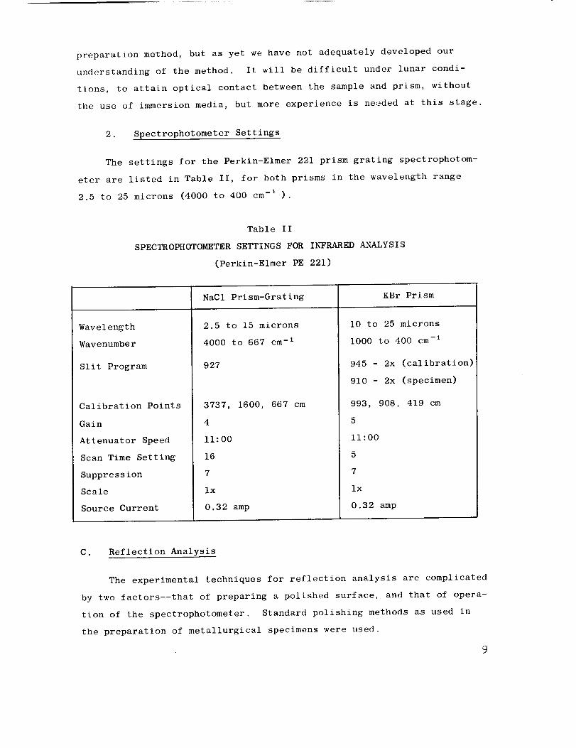
2. Spectrophotometer Settings
The settings for the Perkin-Elmer 221 prism grating spectrophotom-
eter are listed in Table II, for both prisms in the wavelength range
2.5 to 25 microns (4000 to 400 cm -I ).
Table II
SPECTROPHOTOMETER SETTINGS FOR INFRARED ANALYSIS
(Perkin-Elmer PE 221)
Wavelength
Wavenumber
Slit Program
Calibration Points
Gain
Attenuator Speed
Scan Time Setting
Suppression
Scale
Source Current
NaCI Prism-Grating KBr Prism
2.5 to 15 microns
4000 to 667 cm -I
927
3737, 1600, 667 cm
i0 to 25 microns
i000 to 400 cm -I
945 - 2x (calibration)
910 - 2x (specimen)
993, 908, 419 cm
4
ii:00
16
7
ix
0.32 amp
5
ii:00
5
7
ix
0.32 amp
C. Reflection Analysis
The experimental techniques for reflection analysis are complicated
by two factors--that of preparing a polished surface, and that of opera-
tion of the spectrophotometer. Standard polishing methods as used in
the preparation of metallurgical specimens were used.
9

I. Sample Preparation
a. Solid Samples
A polished surface of the highest quality is prepared, over a
flat surface about i/2-in. 2 . If the sample is a single crystal then the
orientation of that polished face should be recorded, as it will clearly
influence the spectrum obtained (see Section B, p° 35 and Fig. _7 for
further discussion).
b. Powdered Samples
Powdered samples can be mixed with powdered lucite* and
briquetted (as for a metallurgical specimen) and then polished in the
standard manner. Obviously there will be less reflective surface of the
sample in the beam than for a solid sample, and the reference beam may
need attenuation to secure adequate response from the spectrophotometer.
Figure 56 shows the first analyses we made for powdered dunite and gabbro
samples compared with the spectra of their solid equivalents. The use
of powdered samples is an important area for further development.
"Synthetic" rock mixtures can be readily prepared and analyzed, or com-
pared with those of unknown rock samples, by a simple matching process.
2. Spectrophotometer Settings
For infrared spectral reflection analysis it is necessary to attach
a small mirror system in one beam of the spectrophotometer. 19 This mir-
ror deflects the beam sideways onto the polished surface at an incident
angle of almost 90 deg. The radiation when reflected by the surface is
caught on a second mirror, and passes again along the original optical
path into the instrument. The setting of these mirrors, the angle of
attachment of the polished surface, and the attenuation of the reference
beam are all parameters which must be kept optimum.
A plastic must be used which has no reflection spectrum in this region.
I0
D. Emission Analysis
We have tried only the briefest experiment in emission analysis,
in which a powdered sample was heated to about 400°C outside a small
metal furnace. The reference sample was carbon-black. The work is far
too premature to be reported, but the spectral analysis of solid and
powdered samples from 400°C down to ambient temperatures should be
studied.
Discussion on p. 43 - 44 and 50 - 54 covers the most common
method of obtaining emittance data--that of subtracting the reflectance
of a polished surface, at thermal equilibrium, from the emission curve
at that temperature for a blackbody.
E. Sample Parameters
i. Particle SiZe
The physical nature of the sample being studied by infrared analysis
is very important, and this factor has been rather extensively but by no
means exhaustively studied.
Duyckaerts 21 has drawn attention to several difficulties that are
encountered when working with solid materials in making infrared absorp-
tion studies. Our experimental work 4 agrees with the conclusions of
Duyckaerts that control of particle size is of vital importance. This
control is necessary in absorption and reflection spectra measurements
regardless of whether samples are handled as suspensions in KBr, as min-
eral oil mulls, or a deposited films.
The grinding method described in Sec. i, D. i0 insures reproduci-
bility of sample particle size. We have produced several thousand infra-
red absorption curves from all types of materials over the past seven
years and have obtained good resolution of spectra. But particle size
control clearly is a vital problem away from a manually operated (ter-
restrial) laboratory, where such sample preparation is not possible.
ii
This is one reason why the emphasis in this laboratory has shifted
to studies of reflection spectral°'2Sfrom polished slab_ of minerals
and rock surfaces where scattered radiation is at a minimum. To be
of practical service to an orbiting spacecraft or even to a roving sur-
face vehicle the technique must be able to produce diagnostic spectra
without extensive preparatory steps. It is shown on p. 30 - 54 that
thermally emitted infrared radiation has most of the spectral informa-
tion as that in the absorption spectrum obtained from a powdered sample
in a KBr pellet_ or in the reflected spectrum from a polished face of
the sample.
2. Orientation and Grain-Size
An important difference exists between an powdered sample and a
single crystal of the material. As in x-ray diffraction_ powdered sam-
ples present random orientations to a beam of radiation while a single
crystal presents a unique orientation. A quartz plate cut normal to
the optic axis (Z-cut) will show a different response from one cut
parallel to the optic axis (X- or Y-cut).*
Because most infrared reflection studies of minerals (and gems)
are made upon single crystals_ one must determine the structural orienta-
tion of the crystal face (or cut-face) being examined. This is avoided
in studies of glasses due to the relatively random internal orientation
in liquids_ and this condition may be approached in fine-grained, poly-
crystalline materials if their grain size is small relative to the area
being irradiated (an area of about 1/2 x 1/4 in. in most spectrophoto-
meters). Preferred orientation of the crystals should be avoided in
the sample; otherwise spatial directions must be given. Most volcanic
and intrusive rocks show no preferred orientation. Nonfoliated sedi-
ments and metamorphic rocks may show no preferred orientation_ but those
with planar structures will show preferred orientation effects.
For a clearly drawn figure showing possible cuts in quartz crystals_
and their code designations_ see Berry and Mason, 2s p. 206.
12
3. Mineral Mixtures
a. Plagioclase Series Minerals
To explore the effect of changing composition upon spectra
the plagioclase series was investigated. The plagioclase feldspars
are often cited as examples of solid solution_ wherein the composition
may pass gradually from that of the sodic end-member_ albite (Ab)_ to
that of the calcic end-member_ anorthite (An)_ in an unbroken series.
We took the mineral labradorite (An6oAb4o) and attempted to match its
infrared absorption curve by mixing equal parts of albite (about An 9)
and anorthite (about An85) samples. The resultant curves are shown in
Fig. I. Curve 3244 is almost matched by the 50:50 mixture but differs
in enough characteristics so that its "parents" can be identified. It
is possible that if one annealed this mixture while still in the solid
state enough disorder could be introduced so that the curves would
match. This has been done by Laves and St. Hafner 23,24 on similar
materials.
b. Simulation of Rocks by Mineral Mixtures
The analysis of a granite by infrared absorption has already
been performed_ but the comparable study of a basic rock_ such as a
basalt or a diabase presents many more difficulties. In granite quartz
and microcline are two of the major components and show little composi-
tional change from sample to sample. Their spectra thus remain constant.
The composition of the other major component_ plagioclase_ often varies
markedly from sample to sample_ with an attendant variation in its
infrared spectrum. As long as only one of the major minerals is capable
of change then the mineral content (modal analysis) of the rock may be
deduced. Future study may help in the computation for more complex
variations.
13
It is this factor of spectral change with composition which
negates any simple solution to determination of the mineral content of
basic rocks. Basic rocks can contain as major components, olivine_
plagioclase, and one or more pyroxenes. Every one of these minerals
can change both in content in the rock_ and in composition within its
mineral series. In such crystalline solid solutions the composition of
an intermediate member is usually expressed as the percentage of one
end-member, e.g., olivines are expressed in terms of the amount of
forsterite (Fo), as Fo . Similar nomenclature is used for the albite-x
anorthite series, Anx, and the enstatite-hypersthene series, Enx.
To study the problem_ and to provide some examples for later
solution with the computer, mixtures were prepared in proportions listed
in Table III. Each mineral had been previously studied and its place
in its own mineral suite identified.
Table III
MINERAL MIXTURES USED FOR ANALYSIS
One-Mineral "Rocks"
(Oli) Olivine (Fo9o)
3210
(Aug) Augite
3246
(Lab) Labradorite (Anso)
3244
Two-Mineral "Rocks" Three-Mineral "Rocks"
(Fig. 2)
66% Oii:34% Aug
50% Oii:50% Aug
34% Oii:66% Aug (Fig. 4)
1Aug:l Oli:l Lab
(33% each)(Fig.3)
66% Aug:34% Lab
50% Aug:50% Lab
34% Aug:66% Lab
Absorption spectra for the I0 mixtures are shown in Figs.
2 to 4. Treating the W-I diabase (Fig. ii) as an unknown_ a reasonably
good match is noted immediately with the 50% augite:50% labradorite or
a 34% augite:66% labradorite curve. The basalts of Fig. 45 contain a
little too much glass to clearly match any of the synthetics_ but the
50% augite:50% labradorite curve would fit well if the basalt is
14
considered to be a "smoothed-out" equivalent of the synthetic mixture.
The Pisgah Crater sample (3369 in Fig. 45) shows a closer fit due to a
higher content of crystalline material.
The gabbro from Stillwater_ Montana (3367 in Fig. 46) clearly
shows the absence of olivine (see modal analysis_ Table VIII) and again
the closest fit is with 50% augite:50% labradorite. If the spectral
differences between orthopyroxene and clinopyroxene are taken into
account then the fit is better (see Figs. 21 and 22 for the two types
of pyroxene).
4. Synthetic versus Natural Minerals
There is a growing awareness2S,26that infrared absorption analysis
often is more sensitive to short-range ordering of aluminum silicates
than is x-ray diffraction. An excellent way to note this effect is by
the use of synthetic minerals which_ because of inherent difficulties
in reaching equilibrium in times available in the laboratory_ are prone
to crystallize in a disordered state. When this involves aluminum/sil-
icon (AI/Si) diadochy in the tetrahedral layers of silicate minerals_
it is often undetectable by x-rays because of the similarities in scatter-
ing power between the two elements.
Infrared analysis is specifically sensitive to such substitutions_
particularly those wherein a charge deficiency arises (AI 3+ for Si4+).
But Stubican and Roy 27 were unable to note the effects of the AI3+/Si 4+
substitutions in a series of their synthetic chlorites either by x-ray
diffraction_ or by infrared absorption analysis. Using natural chlorite
samples_ however_ one can show 3 this substitution in the strong 9 to i0
micron region quite readily. Following this initial study the same
sensitiveness to AI3+/Si 4+ tetrahedral substitution was noted in the
lepidolite (Fig. 34)_ muscovite_ biotite_ and phlogopite mica groups 25
and is strongly suggested in the pyroxene and amphibole groups whose
absorption curves were obtained during this project.
Therefore_ we studied as manysynthetic minerals as we could
obtain. Excellent spectral resolution was obtained for samples of
kaolinite (Fig. 5)_ albite feldspar (Fig. 6)_ analcite (Fig. 7)_ andparagonite (Fig. 35)_ and with a group of iron analogs of aluminous-
microcline and sanidine (Fig. 8).
In Fig. 5 the critical 1805 cm-i peak for synthetic kaolinite is
not as well resolved as for natural kaolinite; this samepoor resolution
is often found in the disordered kaolinites called fire-clays. But the
resolution of the four OH'peaks in the stretching frequencies around3700 cm-i and in the AI-O-OHbending frequency at 935 to 911 cm-i is
excellent. In Fig. 6 the synthetic albite (3045) is clearly of the
high-temperature form and thus should not resemble its low-temperaturecounterpart (KRC1498). This distinction is madeon the lack of resolu-
tion of the following areas: 1138 to 1109 cm-i_ 1023 cm-i 788 to720 cm-i and 474 to 460 cm-ij A parallel exists with the syntheticK-spar (potash feldspar) in Fig. 8. Synthetic analcite almost matches
exactly its natural form (Fig. 7) except for a lack of ordering in the640 to 540 cm-i region.
The differences between the two polymorphs of potash feldspar--
sanidine (high temperature_ disordered) and microcline (low temperature,ordered)--are clearly seen in Fig. 8. The region from 770 to 720 cm-i
is the best for indicating disorder in the feldspar lattice_ and a defi-nite peak change (from 648 to 639 cm-i) occurs between the two forms.
The ferric-iron (Fe) analogs are seen in the top two curves in
Fig. 8. The Fe-microcline clearly showsa well resolved spectrum indi-
cative of a high degree of order in the lattice. The Fe-sanidine is
quite disordered as deducedfrom the lack of resolution in its spectrum.The similarity between the two sets of analogs is striking in the ii00
to 900 cm-i (Si-O) region. The substitution for A13+by Fe3+ led to the
change of peak location from the 650 to 530 cm -I region to about 514 cm-i
The 422 cm -i peak is assigned 26 to a Si-O bond and consequently does
not change position.
16
5. Analysis of Shock-Loaded Minerals
The study of the effects of planar shock waves generated by high-
explosives has been under way for several years at the Institute.
Several mineral specimens have been shock-loaded in the pressure region
350 to 600 kilobars and the materials collected and analysed by x-ray
diffraction and infrared absorption analysis.
Two examples are given in Fig. 9. A sample of albite feldspar
shock loaded to 600 kilobars produced a glass (specific gravity_ 2.20 to
2.24; refractive index_ 1.50). A loss of resolution in the spectrum
comparable to the degree of disorder in some synthetic minerals (Figs.
6 and 8) is shown_ particularly in the 796 to 719 cm -I region. All de-
tail is lost at 1140 to 1090 cm-1_ 582 cm-1_ and 458 cm -I
A similar pattern may be seen for quartz shock-loaded to 350 kilo-
bars in a Mach-disc with cylindrically-converging shock geometry (3145)_
and to 600 kilobars with a flying-plate shock wave pattern (3015). With
the exception of the 350-kilobar spectrum_ where some quartz remnant
may be seen_ the patterns are those of a completely disordered glass.
A similarity is shown to the opal structure rather than fused silica_
based principally upon the absence of the 695 cm -I peak.
Two higher density polymorphs of silica (SiO2)--eoesite (specific
gravity_ 3.2) and stishovite (specific gravity 4.5) were also identified
and defined. A detailed discussion of this work can be found O11 l_. 30
-31.
6. Determination of Water in Minerals and Rocks
In the KBr-pellet technique_ as used for the infrared absorption
analysis of rock and mineral_ "water" can occur either on the sample or
adhering to the potassium bromide itself. It so happens that absorption
peak positions for water adhering to the KBr_ and the water adhering to
most samples_ are so nearly identical that the region of their respective
17
infrared absorption peaks overlap. Mineralogists and chemists distinguish
two types of water by their reaction upon heating--one which is lost at
or below II0°C (called H20- ) and one lost above II0°C_ sometimes several
hundred degrees higher (called H20÷ ).
In a crystal_ like alum or gypsum_ there is water which is essential
to the preservation of the structure_ and this can be found to occur in
"n" multiples of "H20." There is also "zeolitic water" which can be
driven in or out of a sample in the temperature range of i00 to 25_C_
without affecting the structure_ and there are hydroxyl_ or (OH)_ groups
of varying types bonded into varying positions within a crystal lattice.
Excluding physically adhering water_ or "moisture" (H20-)_ which may be
either on the KBr or on the sample_ this type of infrared absorption
analysis can distinguish each type of water_ because of its different
absorption peak position_ dictated by differing degrees of hydrogen
bonding in the different forms.
This is illustrated in a series of spectra* in Figs. I0_ ii_ and 12.
In the top curve in Fig. I0 one can see the effect of water on a KBr
blank disc and how it can be almost completely removed by vacuum drying
(second and third curves from the top).
The spectrum for paragonite mica (undried) in Fig. i0 shows the
moisture (shaded) and the two OH' peaks_ which have a ratio of their
absorbances of 1.48. After drying_ the "moisture" at 3425 cm -I has
been removed_ but the ratio between the two OH' peaks is almost the
same (1.50).
The spectra were analyzed on a PE 221 grating instrument linear in
wavenumber_ without any material in the reference beam. The excellent
resolution of this region may be noted_ but the peaks are much more
spread out horizontally than one is accustomed to seeing with the
prism presentation_ which is usually linear in wavelength.
18
A slightly different case is shown for the lower three spectra in
Fig. 1% which have both moisture (H20-) _ at 3425 cm-i_ and (H2 O÷)
existing in a "disordered" form_ at 3610 cm -I. This water is akin to
zeolitic water and does not show the characteristic peak of the OH'
as in the mica.
Figure ii shows zeolitic water lost on drying_ but with the contin-
ued presence of ammonium (NH_) ion in the sample. The ammonium illite
also possesses a sharp OH' band at 3632 cm -i
The synthetic kaolinite sample in Fig. Ii shows a beautiful resolu-
tion of four OH' peaks between 3694 and 3621 cm -i but without any peak
at 3435 cm -i. This is further confirmed (in Fig. 5) at 1530 cm -i where
"moisture" normally shows clearly as a wide band if a similar band
occurs at 3435 cm -i. Kaolinite thus has OH' but no H20 in its analysis.
Some acid volcanic rocks_ called perlites_ are of great interest
for their possible occurrence upon the lunar surface. On the earth's
surface they may contain up to 10% water which may be removed if the
sample is heated to about 800 to i000 C. Commercially this is called
"expanding"--or "frothing"--Perlite_ as it expands to several times its
original bulk upon the evolution of this water content as steam. It is
in great demand for lightweight aggregates_ but on the lunar surface it
may be a source of water for astronauts.
In Fig. 12 the top two spectra show a possible 2% to 5% water in
the rock (stippled area) which is not camouflaged by that present on the
KBr disc_ itself. It is this OH' water_ observed at higher wave numbers
(3515 cm -i) than the moisture_ which is retained to higher temperatures.
It is clearly discernible in the spectra of the black perlite and appar-
ently absent in that of the gray perlite.
So far we have not found the water contents of rocks and minerals
to appear in reflection spectra_ but more study is needed. The reflect-
ing power of a solid increases rapidly in the vicinity of the absorption
19
due to a strong vibrational frequency and the reflection spectra should
be as predicted from the corresponding absorption patterns (see Fig, 48).
7. Analytical Reproducibility
One is always troubled with the reproducibility of any analytical
method_ and the observed total variance between repeated analyses of the
same sample should be carefully scrutinized. In previous studies 4 we
have statistically determined the variance due to the several steps in
grinding the specimen to a standardized fine grain size for absorption
analysis. This part of the variance is quite small_ and the biggest
portion of the error between duplicate samples lies in the accuracy o£
weighing the 1.5 mg of sample to make up the 0.15% KBr disc.
It is clear_ also_ that the total variance is small_ as may be seen
in hidden duplicates_ prepared under different code numbers_ and sho_1
as Figs. 13_ 14_ 17 and 24. The total variance may be defined as the
sum of the partial variances due to the following operations:
l,
2.
3.
4.
5.
6.
Sample mixing in original bottle
Weighing 1.5 mg of the sample
Mixing with the 1.00 gram of KBr.
Disc preparation
Instrument settings
a. Absorbance adjustments
b. Wavelength adjustments
Estimation of final wavenumbers for peak position.
In general peak position can be defined to ±2 cm -I. This is a
fairly simple problem compared with that of estimating reproducibility
of analysis of a solid sample_ as in reflection or emission analysis.
Here orientation_ grain size_ and re-location of the specific area on
the sample all pose major problems. One accepts the instrumental repro-
ducibility of ±2 cm -I and relates all the other differences to sampling
variances. The peak positions in reflection analysis are reproducible
but their amplitudes are dependent upon surface features. Most polish-
ing equipment does not produce as flat a surface as that of the quartz
2O
oscillator plates_ and the reference beam of the spectrophotometer
usually must be attenuated about 40% to ensure an adequate signal from
the instrument. This "electronic" magnification does not affect peak
wavelength in any way.
F. Quantitative Treatment of the Data
A field which has been barely touched during this data gathering
phase_ is that of the quantitative treatment of the rock spectra. With
absorption data s the problem is relatively easy. For the purpose of
measurement_ a base-line is drawn tangentially to the points of minimum
absorbance (maximum transmittance) on the curve. With most silicate
minerals and rocks this base-line runs from about 1200 am -I to about
800 or 750 cm -I. The base-line absorbance at any wavenumber within
this range is considered to be the difference between the absorbance
value read from the curve and that read from the line at that wavenumber.
These base-line absorbances are additive. Standard spectrophotometric
techniques may be followed and simultaneous equations developed from
which the concentrations of several individual components are calculated.
We are considering the computer programming that would be necessary
to directly determine the mineralogical modal analysis of a rock from
its absorption curve.
For the lunar orbiter experiment we would obviously be working
with emission data_ and further complications would be introduced by the
available spectral resolution_ the digital conversion of data_ and the
down-grading of data by the spacecraft telemetry. Clearly much remains
to be done in this area.
21
INFRARED ABSORPTION ANALYSIS OF MINERALS AND ROCKS
Initial results from the first portion of the current study were
given in the Interim Report_ 28 and show how minerals with simple spectra
may be readily differentiated at selected specific wavenumbers.
The compilation of mineral absorption spectra in the region 2.5 to
25 microns (4000 cm -I to 400 cm -I ) has been carried out using both the
NaC1 and KBr prism optical regions. A file of approximately 1050 in-
frared absorption curves prepared over the past three years from 410
"standardized" mineral specimens were used for the compilation. These
samples are mainly silicate minerals and represent materials on which
full chemical analysis of superior quality has been performed and/or ma-
terials whose detailed x-ray diffraction and optical character are
known.
It was deemed esgential to perform this initial detailed study of
purified single minerals_ before undertaking mixtures_ so that a basic
understanding of the sample spectra might be obtained. Figure 15 shows
that the spectra of minerals in a mixture are additive_ and characteris-
tic absorbance peaks may be defined even when mineral percentages in the
mixture are low.
A. Typical Results in the Silicate Mineral Groups
The modern basis for crystallography 29 relies upon a realization
of the importance of the silicon-oxygen tetrahedron in its various
linkage-patterns in forming the basic framework of the silicate minerals.
We find that it is logical to group silicates of a similar basic frame-
work_ using the ratio of the silicon and oxygen atoms as the common
denominator. We find the nesosilicates_ with isolated SiO 4 tetrahedra
(Mg2SiO 4 or Fe2Si04) at one end 13 of the wavelength scale_ and the
tektosilicates_ with frameworks of SiO 4 tetrahedra but with all oxygens
shared and yielding (Si204) units at the other end. In addition_ as one
progresses toward the framework silicates there is increasing substitution
of A13+ for Si 4+ in structures which form (AISi308) groupings.
22
The following structural classes are accepted for the silicate
minerals:
i.
2.
3.
4,
5.
6o
Nesosilicate - independant tetrahedra (SiO4)
Sorosilicate - 2 tetrahedra sharing one oxygen (Si20 v)
Cyclosilicates - closed rings of tetrahedra each sharing
two oxygens (SisO1s)
Inosilicates - continuous chains of tetrahedra
a. Single chains_ sharing 2 oxygens (SiO3)
b. Double chains_ sharing alternately 2 and 3
oxygens (Si4011)
Phyllosilicates - continuous sheets of tetrahedra each
sharing 3 oxygens (Si401oOr AiSi301o)
Tektosilicates - continuous framework of tetrahedra_ each
sharing all 4 oxygens (AiSiO40r AISi3Os)
This classification is clearly shown by infrared analysis_ for the
covalent portion of the (Si-O) and (AI-O) bonds prove to be very sensitive
to infrared detection. In fact this method of analysis_ above all others_
offers the greatest hope for solving some of the problems of the order-
ing of AI-O and the Si-O tetrahedra_ and the amount of AI3+/Si 4+ diadochy. 25
i. Nesosilicates
a. Olivine
The olivines show very simple spectra comparable to many of
the inorganic anions_ with peaks changing wavenumber in a predictable
manner as their cation metal changes (Figs. 16 and 17). Almost every
detail of the spectrum persists unchanged from sample to sample but
careful calibration will show marked wavenumber changes_ as shown in
Table IV.
This mineral group has recently been the subject of an exhaus-
tive study by Duke 3°. The slight departures from complete independence
of the tetrahedra in the solid crystal were used by him as the reason
for modifying the theoretical absorption peak positions to match observed
values.
23

Table IV
PEAK POSITIONS IN THE OLIVINE GROUP
Sample Absorption Peaks (cm -I)
Forsterite_ Mg2Si04 983 950 888 838 605 504 410
Fayalite, Fe2SiO 4 947 873 829 558 482 410
b. Garnet
The garnet minerals may be divided into two main groups--the
aluminous "pyralspites" with a general formula of R_+AI2(SiO4) 3_ and
the calcic "grandites" with a general formula of Ca3R_+(Si04)3 . Within
each group complete crystalline solution exists_ but between the groups
it is a rarity. Most of the samples in Fig. 18 are pyralspites_ but the
samples DEL 3 and DEL 5 are examples of this solid solution.
Spectral peak patterns are again constant within the two sub-
groups (Figs. 16 and 18) and vary in wavenumber with the cation metal_
as shown in Table V.
Table V
PEAK POSITIONS IN THE GARNET GROUP
Mineral Formula Absorption Peaks (cm -I )
Pyralspite
Pyrope
Almandite
Spessartite
Grandites
Grossularite
Andradite
Uvarovite
Mg3Al2(SiO4) 3
Fe3Al2(SiO4) 3
Mn3Al2(SiO4) 3
Ca3Al2(Si04) 3
Ca3Fe2(Si04) 3
Ca3Cr2(SiO4) 3
970 900 872 - 575 482 460
970 902 880 638 570 480 455
955 892 870 632 562 472 452
915 860 840 618 - 540 470 450
895 840 820 592 - 512 482 438
900 840 825 609 - 540 455 425
24
2. Sorosilicates
The sorosilicate group is quite complex structurally and this is
also shown by the infrared spectra. Idocrase (Fig. 19) has both (SiO 4)
and (Silo v) groups and is halfway between the nesosilicates and the
sorosilicates. Lawsonite (Fig. 19) has 6-coordinated A1-O (and OH')
groups_ linked sideways by the Si207 groups.
3. Cyclosilicates
a. Beryl - BesA12Si6018
Minerals of the beryl group--beryl_ emerald_ and the cesium-
bearing pollucite--are very similar to one another and have very active
spectra. Compositional changes may be defined in the i015 to 1058 cm -I
region_ as seen in Fig. 20.
b. Cordierite; Al_(MgFe)2(Si5Al)Ois
Cordierite spectra are broadly comparable to beryl spectra
but differ in the 800 to 700 cm -I region in having fewer peaks. Sub-
stitution of A13+ for Si 4+ takes place in parallel with the diadochy of
Mg 2+ for A13+ in the above structural formulas. Both substitutions tend
to produce spectra characterized by fewer peaks.
4. Inosilicates
a. Single Chains - Sharing Two Oxygens (SiOs)
Orthopyroxenes. The orthorhombic pyroxenes form the series
hypersthene_ (Mg_Fe)SiOs_--enstatite_ MgSi03; and compositions are
usually expressed in terms of the enstatite end-member (e.g._ En2o is
20 percent MgSiO3). We analyzed a series of 21 orthopyroxenes and their
spectra appear in Fig. 21_ which shows the changing detail of an infrared
spectrum with cation substitution.
Clinopyroxenes. The inosilicates also include a series MgSiO 3-
FeSi03 (clinoenstatite and clinohypersthene)_ but samples of this
27
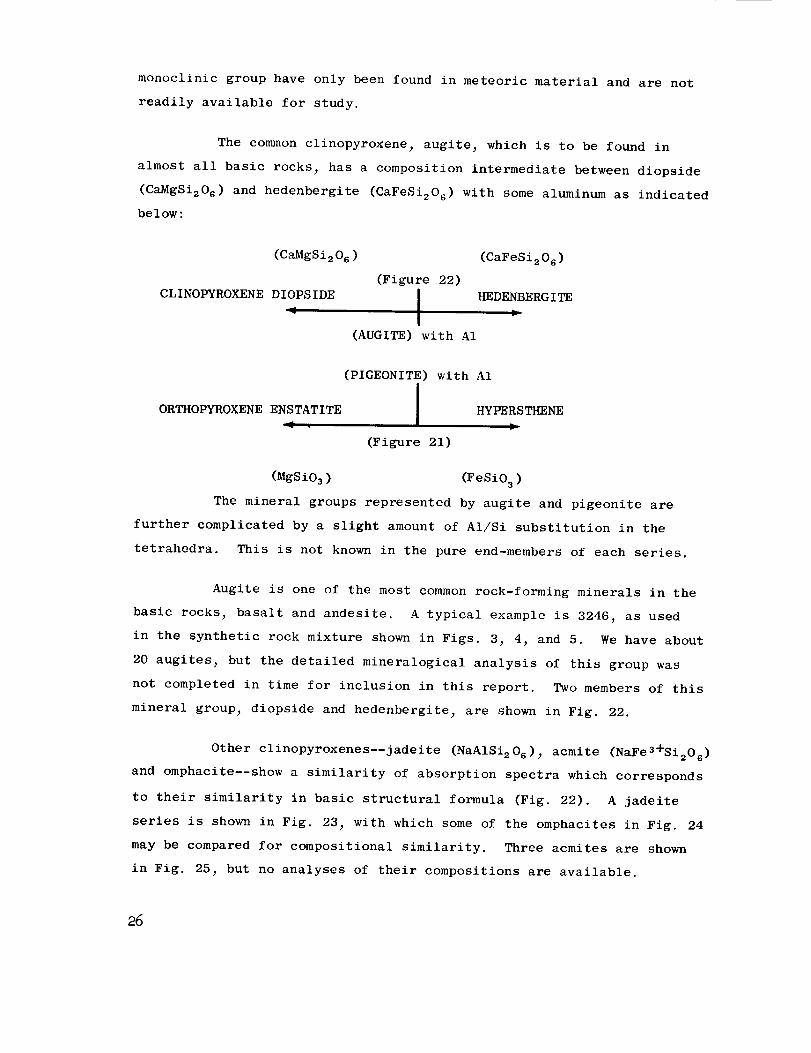
monoclinic group have only been found in meteoric material and are not
readily available for study.
The common clinopyroxene, augite 7 which is to be found in
almost all basic rocks 7 has a composition intermediate between diopside
(CaMgSi2Os) and hedenbergite (CaFeSi2Os) with some aluminum as indicated
below:
(CaMgSi206 ) (CaFeSi206 )
(Figure 22)
CLINOPYROXENE DIOPSIDE I HEDENBERGITE
(AUGITE) with A1
(PIGEONITE) with A1
ORTHOPYROXENE ENSTATITE
4
HYPERSTHENE
(Figure 21)
(MgSi03) (FeSiO 3 )
The mineral groups represented by augite and pigeonite are
further complicated by a slight amount of AI/Si substitution in the
tetrahedra. This is not known in the pure end-members of each series.
Augite is one of the most common rock-forming minerals in the
basic rocks_ basalt and andesite. A typical example is 3246, as used
in the synthetic rock mixture shown in Figs. 37 47 and 5. We have about
20 augites 7 but the detailed mineralogical analysis of this group was
not completed in time for inclusion in this report. Two members of this
mineral group_ diopside and hedenbergite 7 are shown in Fig. 22.
Other clinopyroxenes--jadeite (NaAlSi2Os) 7 acmite (NaFe3+Si2Os)
and omphacite--show a similarity of absorption spectra which corresponds
to their similarity in basic structural formula (Fig. 22). A jadeite
series is shown in Fig. 23, with which some of the omphacites in Fig. 24
may be compared for compositional similarity. Three acmites are shown
in Fig. 257 but no analyses of their compositions are available.
26
b. Double Chains - Sharing Alternately Two and Three
Oxygens (Si4011)
The amphibole group is chemically very similar to the pyroxene
group but permits a much greater AI-Si diadochy in the Si4011 chains.
Hydroxyl groups are an essential part of the structural formulas of the
amphiboles and may be seen on the infrared spectra in the region around
3600 cm -I .
The only orthorhombic member commonly found is anthopphyllite_
(Mg_Fe)TSisO22(OH)2_ where Mg is greater than Fe. Aluminum often re-
places silicon in the tetrahedra.
Amphiboles which have been studied are from the hornblende
[NaCa2(Mg_Fe)4AI(AI Si7)O22(OH)2] series and the alkali amphibole series--
glaucophane [Na2Mg3AI2SisO22(OH)2] and riebeckite [Na2Fe_÷Fe_+SisO22(OH)2 I.
Because of almost overwhelming complexities of possible substitutions
these minerals are the most difficult with which to work. Multiple
examples of these series are shown in Figs. 26 and 27. The cumming-
tonite (Fe_ Mg) 7 Sis0_2(OH) 2 series minerals are represented by sample
3420 (Fig. 28) which was found in the same rock as the hornblende 3418.
These are described in a recent paper by Vernon. 31 There is a supris-
ingly great similarity shown by these two samples_ particularly when a
larger group of hornblendes (Fig. 29) is compared.
The general relationships between the amphiboles are sketched
on the following page_ showing how hornblende is derived from actinolite
by various atomic substitutions. The close similarity may be seen
between actinolite 3247 (Fig. 28) and hornblende 3233 (Fig. 29)_ where
all the peaks except the small peak at 920 cm -I may be matched in the
two spectra.
27
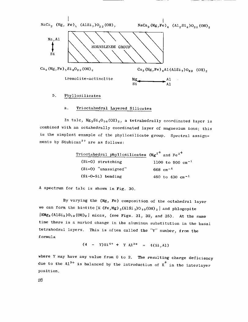
LNaCa 2 (Mg_ Fe) 5 (AISiT)O22(OH),
Na_AI
Si
Ca2(Mg_Fe)5SisO22(OH)2
tremolite-actinolite
lNaCa 2(Mg_Fe) 4 (AI2SiG)O22(OH) 2
Ca2(Mg_Fe)4AI(AISiT)O22 (OH) 2
Mg _ _ A1r
Si A1
5. Phyllosilicates
a. Trioctahedral Layered Silicates
In talc_ Mg3Si4Olo(OH)2_ a tetrahedrally coordinated layer is
combined with an octahedrally coordinated layer of magnesium ions; this
is the simplest example of the phyllosilicate group. Spectral assign-
ments by Stubican 27 are as follows:
Trioctahedral phyllosilicates (Mg 2+ and Fe 2+
(Si-O) stretching ii00 to 900 cm -I
(Si-O) "unassigned" 668 cm -I
--i(Si-O-Si) bending 460 to 430 cm
A spectrum for talc is shown in Fig. 30.
By varying the (Mg_ Fe) composition of the octahedral layer
we can form the biotite [K (Fe_Mg) 3(AISi3)OIo(OH) 2 ] and phlogopite
[KMg3(AISi3)OIo(OH)2] mieas_ (see Figs. 31_ 32_ and 35). At the same
time there is a marked change in the aluminum substitution in the basal
tetrahedral layers. This is often called the "Y" number_ from the
formula
(4 - Y)Si 4+ + Y A13+ = 4(Si_AI)
where Y may have any value from 0 to 2. The resulting charge deficiency
due to the A1 s+ is balanced by the introduction of K + in the interlayer
position.
28
b. Dioctahedral Micas
Muscovite, KAI2(AISi301o) (OH)2has two A13+ ions for each
three M_ + ions in the octahedral sheets, leaving a vacant site in the
layer. Sheet stacking gives various possible types of symmetry. Assign-
ments by Stubican 27 are as follows:
Dioctahedral phyllosilicates (AI 3+ or Fe 3+)
(Si-O) stretching ii00 to 900 cm -I
(Si-O-Si) bending 460 to 430 cm -I
Muscovite spectra are shown in Figs. 31 and 33; AI3+/Si 4+
substitution in the tetrahedral layer can markedly vary the patterns.
This substitution is very well shown by the series of lepidolites_
KLi2AI(AISi3OI0) (OH)2_ shown in Fig. 34. Here a complete series in
AI3+/Si 4+ diadochy has been examined with "Y" ranging from 0.18 up to
0.72. The marked change in the 1084 to 1130 cm -I peak_ and in the
detail of the 992 to 962 cm -I peaks has been previously reported 3,25
and is one of the best examples of this use of infrared absorption
analysis in mineralogical studies.
The following varieties of mica may be obtained by atomic
substitution in the muscovite structure:
muscovitej KAI2 (AlSi3010) (OH)2
paragonite, NaAI 2 (AlSi 30 I0) (OH)2
lepidolite_ KLi2AI(AISi3OIo) (OH)2
glauconite, K(Fe,Mg_A1) (AISi3010) (OH)2
ferri-celadonite_ KFe2 (Si4010) (OH)2
(Fig. 33)
(Fig. 35)
(Fig. 34)
(Fig. 36)
(Fig. 36)
c. Kaolinite; Al2Si40, o(OH)8
No substitution has been defined in the kaolin group, the
members of which are considered to differ from each other only in the
stacking of their plates. Most of the infrared spectra are quite
similar, but the group members--kaolinite_ dickite, halloysite and
nacrite--may be distinguished. A kaolinite spectrum appears in Fig. 30.
29
There is a suggestion in the spectrum of halloysite_ as distinct
from that of any of the others of the group_ that there is a certain
amount of tetrahedral substitution of aluminum for silicon in the basal
structural layers_ similar to the case for the micas. A value of "Y" of
up to 0.6 is suggested by direct comparison with the mica series. There
is no other real evidence as yet to support this view_ except that such
a substitution would help to explain the tubular form of halloysite re-
sulting from the misfit between the two layers in the structure (Bates3m).
6. Tektosilicates
a. Quartz and its Polymorphs
Silica is the simplest of the tektosilicates and consists of
a framework of SiO 4 tetrahedra with each oxygen linked to another in a
neighboring tetrahedron. Silica can exist in various structural groups_
several members of which are shown in Fig. 37; quartz and coesite re-
tain the tetrahedral coordination_ while stishovite shows the rutile
(six-fold coordination) structure. Stishovite is considered to be the
first mineral discovered in which silicon occurs with oxygen in a six-
fold coordination (octahedral) instead of tetrahedral coordination.
Tarte and Ringwood 33 have recently drawn attention to the
absence of SiO 6 octahedra in silicate minerals; however_ on the basis
of other work in XO 4 tetrahedral, and octahedral_ groups_ they predicted
the infrared absorption frequencies for this octahedral configuration to
be between 700 and 600 cm -I. The stishovite 34 infrared absorption curve
(Fig. 37) shows a major absorption at 769 to 628 cm-1_ effectively sup-
porting their prediction. The data show a marked change in the position
of the major absorption peaks when passing from the SiO 4 tetrahedral co-
ordinations of quartz_ coesite_ and fused silica (1098 to 1077 cm -I) to
the SiO 6 octahedral coordination in stishovite (769 to 628 cm-1; 949 to
885 cm-1). A parallel to this has been recorded by Dachille and Roy 44
5O
for GeO2- quartz and GeO2-rutile structures_ which show absorption peak
shifts from 870 cm -I for the "quartz" structure to 715 cm -I for the
"rutile" polymorph.
General assignments 35 for spectral peaks in quartz are:
(Si-O) stretching ll00 to 800 cm -I
(Si-Si) stretching 800 to 600 cm -I
(Si-O-Si) bending and
distortion 460 to 430 cm -I
b. Feldspar
The feldspars are a very complex group both compositionally
and structurally_ and to adequately cover this one group would be a
complete study in itself.
The plagioc_ase group--albite_ NaAISi308_ to anorthite_
CaAl(Si2A1)Osj-- is treated in Fig. 38 which shows the marked structural
break at An33 ( and Anss) by changes in the infrared spectra. Only the
low temperature plagioclases are shown. A detailed study of the calcic-
plagioclases (Ans0 to Ans3) is shown in Fig. 39. The diagnostic peaks
are indicated by an asterisk. These samples traverse the structural
break at Anss _ but this is not so clearly revealed as that at AD. 33. The
peak at 929 cm -I begins to be a major peak immediately after the break
and becomes dominant for An94. Peaks at 663 and 755 cm -I are also signifi-
cant above the break at Anss.
B. Typical Results with Oxide Mineral Groups
Only two oxide mineral groups have been studied--Al20 s and Ti02.
These are both of specific interest as they are known to be capable of
fairly extensive substitution for Si 4+ in the tetrahedral layers of
silicates. Only aluminum oxide will be discussed.
The spectrum for _-AI203 shown in Fig. 40 has a broad minimum from
825 to 580 cm -I. The three most common hydrates of aluminum also show
this minimum although it is generally narrower and forms a sharper peak
31
in the range 740 to 520 cm -I This is the wavenumber region where A1
in six-fold (octahedral) coordination may be found on an infrared spec-
trum. When it is in four-fold (tetrahedral) coordination its absorption
peak occurs at a much higher wavenumber. The parallel with silicon is
tabulated below.
Form Coordination Peak Position
Alumina,7-Al203
Alumino-silicates_ AISi30 s
Stishovite, SlOe
Quartz_ SiO 2
Six-fold
Four-fold
Six-fold
Four-fold
825 to 580 cm -I
i000 to 900 cm -I
949 to 560 cm -_
1160 to 1080 cm -I
C. Experimental Results with Rocks
Knowledge of single minerals has been extended to mixtures of
minerals as found in rocks. Typical materials of presumed lunar origin
(tektites) or of lunar accumulation (meteoritic chondrites_ etc.) and
over 80 rocks have been studied in the past six months. Many of the
spectra from these rocks appear in Figs. 41 to 46. The rock types
represented have also been tabulated in Table VI according to their
composition and grain size.
The techniques of absorption analysis have formed the basis for the
interpretation of the data from infrared reflection analysis of rock
surfaces. 22 In turn this provides the key to the interpretation of infra-
red thermal emission spectra from hot (or warm) specimen sources. A
series of absorption spectra (Fig. 41) for typical acid and basic rocks
found on the earth's surface has been compiled.
The absorption spectra for three "acid" rocks (Katmai ash; Town
Mtn._ Calif._ granite; and the Standard G-I granite) are shown in the
upper half of Fig. 41. For comparison the absorption spectra for three
"basic" rocks_ (Kilauea Iki basalt (1960); San Marcos_ Calif., gabbro;
and the Standard W-I diabase) are shown in the lower half of the figure.
52

Table VI
ROCKS STUDIED BY ABSORPTION ANALYSIS
L
Rock Type Sample No. Figure No.
Acid; Fine-Grained Rocks
Katmai ash
Obsidian_ Clear Lake
Obsidian_ Snowflake glass
Obsidian_ Snowflake crystals
Obsidian
Obsidian red
Obsidian Konocti
Obsidian
Perlite_ gray
Opal_ black woody
Fused Quartz
_Acid2 Medium-Grained Rocks
Rhyolite_ Soledad
Tuff_ Bishop
Acid; Coarse-Grained Rocks
Granite_ Town Mtn.
Granite_ G-I Std.
Granite_ Hard Hat
Granite_ Hard Hat
Syenite SY-I
Basic; Fine-Grained Rocks
Basalt_ Kilauea Iki (1959)
Crystals from the above
Basalt_ Kilauea Iki (old flow)
Basalt_ Pisgah Crater
Basic; Medium-Grained Rocks
Diabase_ W-I
Basic; Coarse-Grained Rocks
Gabbro_ San Mareos
Gabbro_ Stillwater
Gabbro_ Mt. Hope_ Md.
Serpentine_ San Luis Obispo
Anorthosite_ San Gabriel
3199
3358
3414
34[3
3393
3360
3359
3381
3428
3301
3019
3389
3388
3297
3165
3306
3307
3368
3298
3298
3380
3369
3166
3296
3367
3375
3384
3386
41
41,42,52
43
43
43
43
43
43
43
42
42
42
42
41
41_44
44_5244
44
41_45
45
45
45
41_46
41_46_52
46
46
45
52
33
There is a definite similarity within the two groups as to the
position of the absorption maximum around 1070 to 990 cm -I . The acid
rocks each approximate 1070 cm -I, and the basic rocks approximate 990
cm -_-- a shift of 80 cm -I. This shift appears regardless of whether
the rock being examined is glassy, crystalline, fine-grained, or
coarse-grained. The peak shift is apparently dependent on the bulk
composition of the rock, with the smaller peaks of the spectrum record-
ing the presence of specific minerals.
Simple quantitative measurements can be made as long as adequate
spectral resolution can be obtained in the resultant data. Character-
istic spectral minima can be identified for each phase (quartz, olivine_
etc._) in a mixture when superimposed peaks due to two phases can be
differentiated.
Thus there is at'least a spectral shift of 80 cm -I available on which
to base experimental work. Absorption curves have not been prepared for
olivine-rich dunites; but the minima 3° for olivine (Fos0) is at 888 cm-1_
and the peak for a rock only of this composition should be close to this
value, making a possible shift in the absorbance of around 180 cm-lbe -
tween obsidian and dunite.
34
INFRARED REFLECTION ANALYSIS OF MINERALS AND ROCKS
A. Previous Studies
The diagnostic use of infrared radiation, reflected from minerals
and rocks, is a relatively unexplored field. Apart from the pioneering
studies of Coblentz 6'7 (1906-1910) on minerals and inorganic surfaces and
coatings, and of Pfund s (1945) on the use of infrared techniques for the
identification of gems, such studies have been restricted to fused sili-
cates and glasses. In addition, Coblentz was restricted in most of his
studies to the 1 to 8 micron region, and Pfund to the 6 to 12 micron
region of the spectrum, compared with the range of 2 to 25 microns avail-
able for use today with automatically recording spectrophotometers.
There have been several excellent studies on glasses, which are
semitransparent in the infrared region. Simon and McMahon 10 and Gardon 11
have discussed the subject from the aspect of the fundamentals of radia-
tive cooling of glass slabs and sheets. These workers and others like
Saksena,3S, 36 Sevehenko and Florinskaya, 37 and Matossi and Bronder 3s
became interested in reflection measurements from polished surfaces of
silicate minerals whose crystal structures were relatively well known.
Minerals including quartz, tridymite, cristobalite, and some fused sili-
cates were investigated by these techniques.
B. Infrared Absorption and Reflection for _-Quartz
Quartz oscillator plates, with their polished surfaces, make excel-
lent samples for a study of infrared reflection; but because of the high
absorption coefficient for quartz (particularly in the 9 to I0 micron
and 21 to 25 micron regions), absorption spectra are usually prepared from
a powdered sample mixed at low concentration (0.15_) in a KBr pellet.
Absorption spectra for quartz and other SiO 2 polymorphs--coesite and
stishovite--are shown in Fig. 47, and are covered in more detail On
p. 30 - 31.
37
The techniques by which the reflection spectra used in this report
were obtained have been described in Sec. Cj p. 9-i0. A certekin alnou/%t
of reference beam attenuation is needed with some samples_ but this
appears to be more a function of grain size of the crystals in the rock
than of surface polish. The polish must be good_ however_ in order to
get adequate reflection. The samples whose spectra are shown in Fig. 54
of dunite (3390)_ tektite (3257)_ and obsidian (3358) gave sufficient
reflection without any attenuation of the reference beam.
Typical reflection spectra for quartz plates appear in Fig. 47.
The X- and Y-cut plates show a marked difference from the Z-cut plates
in the region of the 800 to 775 cm -I doublet and in the details of the
500 cm -I peak. Saksena 35,36 and Sevchenko and Florinskaya 37 have dis-
cussed the relation of the 775 cm -I peak to the extraordinary optical
ray and the 796 cm -I peak to the ordinary optical ray of _-quartz. The
Z-cut plate obviously-shows only the 796 cm -I peak_ being an optically
isotropic section through the crystal (circular section of the optical
ellipsoid, with zero birefringenee). Figure 48 has been redrawn from
the paper of Sevchenko and Florinskaya as it serves to clearly show the
relationship between absorption and reflection spectra.
A 45-deg rotation of the plate perpendicular to the plane of the
infrared beam produces an effect most marked in the 515 cm -I peak of the
X-cut and Y-cut plates (Fig. 47). The Z-cut plate shows little or no
effect upon rotation. No attempt was made to polarize the incident beam
of radiation. No attenuation of the reference beam was required with any
of these plates of quartz. The temperature dependence of reflection
spectra is displayed for a quartz Z-cut plate in Fig. 49. This has been
redrawn from the classic study of Simon and McMahon. I° The structural
change at about 543°C (or 816°K) is that of the _-_ inversion which was
accompanied by enough expansion to crack their specimen. The sharp
doublet at 1180 and Iii0 cm -I is lost at this temperature_ being replaced
by the single peak at 1120 cm-l_ a characteristic of fused quartz spectra
(Fig. 48).
36

The sharpening of spectral peaks as measured by their half-bandwidth_
with decrease in temperature is most clearly seen in the 800-cm -I peak.
This may be quite a significant effect when spectra are obtained from the
dark side of the moon_ where the lunar temperature approximates 150°K.
C. Experimental Results for Rocks
Table VII lists the rocks studied by reflection analysis under this
project. Rocks of the same composition will have the same spectra_ with
details dependent upon the degree of crystallization (Figs. 50 and 51).
The reflectance spectra for the group of rocks and meteorites pre-
sented in Figs. 54 and 55 show a progressive shift of 160 cm -I (0.85
micron) in the spectral peaks around 920 to 1080 cm -I These samples
range from acid rocks (obsidian_ rhyolite_ granite_ tektite) through
those of intermediate composition (andesite and diabase) to the basic
and ultrabasic materials (olivine-bearing dunite and meteorite chondrites).
Most importantly the spectral peaks are again dependent upon bulk-
composition and do not change in wave number with increasing grain size.
Thus_ whether a given chemical composition of rock is in the physical
form of glass_ felsite_ fine-grained volcanic flow_ or medium-grained or
coarse-grained plutonic rock_ its spectral maximum of reflection will
remain fixed in wave number. As the crystallites form in the earliest
stages of crystallization from a glass_ smaller peaks will appear as
modifications superimposed upon the spectral maxima or minima (see
Fig. 54). These modifications increase in size as the crystals grow and
as the rock loses its content of disordered silicate glass. The silicate
spectrum changes from a simple bimodal curve to the multiple-peaked spec-
tral curve typical of fully crystalline materials. Wave number will in-
dicate the bulk composition_ and the complexity of the curve will indicate
the degree of crystallinity. Knowledge of the characteristics of individ-
ual peaks enable us then to calculate the mineralogical composition or
"modal analysis" of the rocks.
It is clearly possible_ therefore_ to distinguish granite rocks from
diabase rocks by infrared analysis_ and in most instances_ to obtain
37
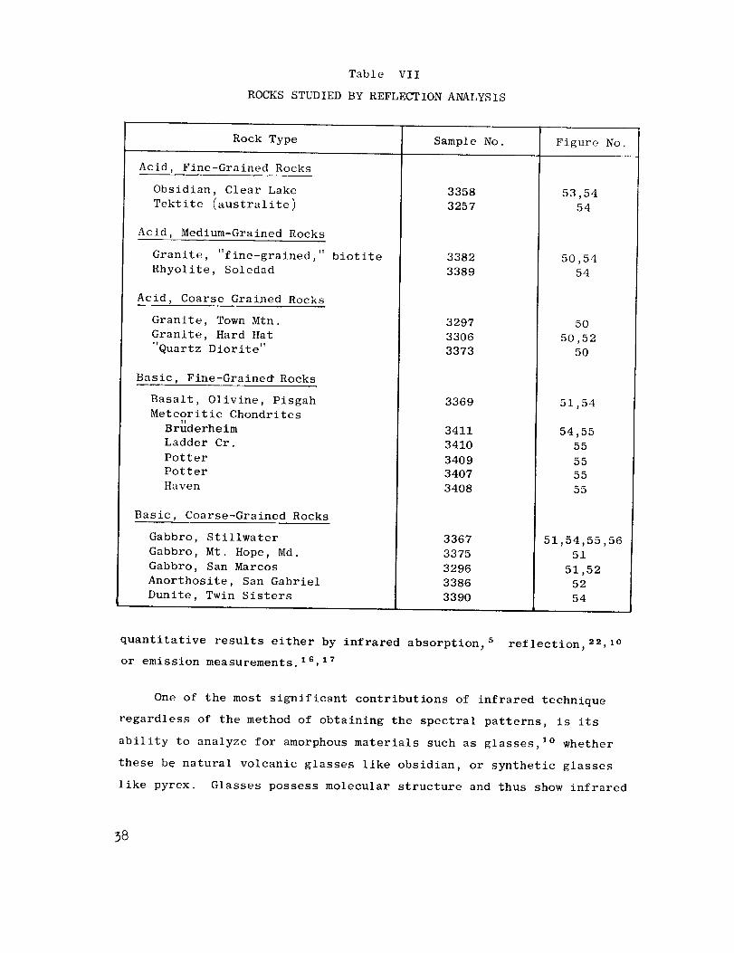
Table VII
ROCKS STUDIED BY REFLECTION ANALYSIS
Rock Type Sample No. Figure No.
Acid, Fine-Grained Rocks
Obsidian, Clear Lake
Tektite (australite)
Acid, Medium-Grained Rocks
T1 . . TI
Granite, f ine-gralned, biotite
Rhyolite, Soledad
Acid, Coarse Grained Rocks
Granite, Town Mtn.
Granite, Hard Hat
"Quartz Diorite"
Basic, Fine-Graine_ Rocks
Basalt, Olivine, Pisgah
Meteoritic Chondrites1_
Bruderhe im
Ladder Cr.
Potter
Potter
Haven
Basic, Coarse-Grained Rocks
Gabbro, Stillwater
Gabbro, Mt. Hope, Md.
Gabbro, San Marcos
Anorthosite, San Gabriel
Dunite, Twin Sisters
3358
3257
3382
3389
3297
3306
3373
3369
3411
3410
3409
3407
3408
3367
3375
3296
3386
3390
53,54
54
50,54
54
5O
50,52
50
51,54
54,55
55
55
55
55
51,54,55,56
51
51,52
52
54
quantitative results either by infrared absorption_ 5 reflection_2_, I°
or emission measurements, lS, 17
One of the most significant contributions of infrared technique
regardless of the method of obtaining the spectral patterns, is its
ability to analyze for amorphous materials such as glasses, I° whether
these be natural volcanic glasses like obsidian, or synthetic glasses
like pyrex. Glasses possess molecular structure and thus show infrared
58
spectra whose patterns change with composition and progressive ordering
(as in the beginning of crystallization). Glasses are to be treated,
therefore, as separate phases whose infrared spectra appear in addition
to those of any crystalline material present.
A series of fine-grained rocks were polished and infrared reflection
spectra obtained in a manner similar to that described for quartz plates.
Rock material adjacent to the polished face was powdered and KBr pellets
(0.15%) prepared. Infrared absorption spectra were obtained as described
earlier. The four pairs of spectra are compared in Figs. 52 and 53.
(Sample locations are listed in Table II.) For detailed discussion we
will confine ourselves to a study of the two spectra for the specimen
of "Hard Hat" granite from the Nevada Test Site, shown as 3306 in Fig. 52a.
The heavy line is the absorption spectrum for the powdered material; the
dashed line is the reflection spectrum, which has been inverted for com-
parison. The general _imilarity in shape is clearly noticeable. There
are important differences in the depths of particular peaks, e.g., the
doublet at 794 and 775 cm -I (due to quartz), which become most significant
when one attempts to determine the mineralogical composition, or "modal
analysis," of a rock. This is the most important difference between the
absorption and reflection spectral curves. Obviously methods of analysis
for absorption spectra, 5 will not produce a comparable analysis of infra-
red reflection data. It is clear that the difference is a fundamental
one, due to the mechanism of reflection and absorption, rather than merely
a slight difference in the samples--one a solid material, one a powdered
material. The same effect can be seen in Fig. 48 for a _-quartz. We
know the good degree of reproducibility for the KBr pellet technique;4, s
but the difference shown here is some orders of magnitude greater.
Furthermore, all the rocks we have studied show these differences
between spectra obtained by the two techniques. Perhaps more significant
is the discovery that mineralogically and texturally similar rocks, with
similar absorption spectra, will have similar reflection spectra, although
for each sample the absorption and reflection spectra will show basic dif-
ferences, characteristic of the two methods.
39

In a comparable manner spectra for anorthosite_ gabbr% and obsidian
specimens are also presented in Figs. 52 and 53. Unfortunately no reflec-
tion spectra are available for the labradorite sample (3244)_ but it has
been closely matched by the reflection spectrum for the rock_ anorthosite
(3886)_ composed predominantly of labradorite.
With reflection spectra from a slightly wider range of rock compo-
sitions an even more marked peak shift of 160 cm -I can be obtained_ using
surfaces polished on the rock samples. Figure 54 shows the spectral
curves_ from infrared reflection_ of a series of rocks ranging from a
tektite (australite) through granite_ obsidian_ and rhyolite_ to basalt_
gabbr% dunite_ and a stony meteorite (chondrite). Clearly marked changes
in the peak at 445 cm -I and 550 cm -I can also be noted in most of the
samples.
The simplicity of the reflection spectra of Fig. 54 compared to the
complexity of the absorption spectra of Fig. 41 again emphasizes the
previous remarks with reference to the two analytical techniques.
As a further application_ a series of chondritic meteorites (Potter_VT
Bruderheim_ Haven_ and Ladder Creek) were prepared and their reflection
spectra analyzed. These are compared with the reflection spectrum from a
sample of Stillwater gabbro. The similarities are striking. This sample
of gabbro (3367-55BEI) and its modal analysis were both kindly supplied
by E. D. Jackson_ of the U.S. Geological Survey. This is compared in
Table VIII with the two modal analyses available for Ladder Creek andr!
Bruderheim chondrites; both the samples and analyses were obtained by
the courtesy of A. A. Loomis_ Jet Propulsion Laboratory_ California
Institute of Technology.
4O
Table VIII
COMPOSITIONS OF A GABBRO AND TWO CHONDRITES
Mineral
Composition
Plagioclase
Orthopyroxene
Clinopyroxene
Olivine
Total
Gabbro
Stillwater, Montana
3367
(Mode) a
48¢ 35
17
100
Chondrite
Ladder Creek, Kansas
3410
(Norm) b
101o
20 c
6
64 c
I00
Chondrite
Bruderhelm, Canada
3411
(Normlb
32 d
6
51 d
i00
aMode--observed mineral frequencies
bNorm--Mineral content calculated from a chemical analysis of the rock
colivine and orthopyroxene calculated to be En, Foss
dOlivine and orthopyroxene calculated to be En, FoTs
There is a broad similarity between the chondritic meteorites and
the Stillwater gabbro as seen in Fig. 55; but a closer match may be ob-
tained by making a comparison with the dunite (3390) of Fig. 54, particu-
larly when we consider that the olivine in this sample is about Foso ,
and in the chondrites it is about FoTs (as calculated). A change in the
iron content, from Fo9o to FoTs would move the peaks as shown in
Table IX, to a closer match with the Bruderhelm meteorite composition.
41
Table IX
OLIVINEPEAKPOSITIONSSHIFTINGWITHCHANGINGIRONCONTENTCOMPAREDWITHBRUDERHEIMCHONDRITE
Minerals Composition Peaksj in cm -I
Absorption
Forsterite
Olivine_ 3320
(Calculated)
Fayalite
Dunite _ 3390
Reflection
Dunite, 3390tl
Bruderheim
F°9 5
FOgo
(Fo7 6)
Fo 5
ca. Fogo
ca. Fogo
983 886 605 504 410
981 884 603 502 412
(977) (880) (597) (498) -
947 873 558 482 410
985 883 605 500 405
922 525 420
921 883 510 493 405
D. Reflection Analysis of Powdered Rock Samples
One of the most difficult problems in reflection analysis arises
when one attempts to quantitatively analyze the mineral components in a
rock. In absorption analysis it is quite easy to mix up a "synthetic"
rock and obtain its spectrum to compare with that of an unknown sample.
Changes may then be made and another synthetic prepared_ closer to the
unknown in spectral pattern.
With solid rock samples this is not possible. Grain sizes and
orientation play important parts in the relative peak intensities of the
constituent minerals. To explore these parameters powdered mounts were
prepared in lucite blocks and polished. (see Sec. C., p. 9)
The results (shown in Fig. 56) are not too encouraging. Basically
the problem rests in the lowered area of reflection surface available
in a powder mount where the lucite matrix has to hold each grain in
place. But the method has a certain amount of promise in that different
rocks can be recognized by their powder mounts_ and this may serve as a
method to match their mineralogical and modal analyses quantitatively.
42
INFRARED EMISSION ANALYSIS OF MINERALS AND ROCKS
The foregoing discussions have been based upon an analysis of
reflection and absorption infrared spectra. The same type of analysis
is applicable to emission spectra for here the spectral data appear as
minima superimposed upon the familiar Planck-Wien radiative curve.
Spectral position defines the minerals; spectral shape and complexity
may be used to determine the mineral content of the rock.
Infrared emission has been previously investigated by only a few
experimenters. Some data on the "emissive power" of minerals may even
be taken from the early pioneering work of Coblentz 7 (i to 8 microns),
and more recently from the studies of Fredrickson 39 (2 to 5 microns),
and Bell et. ai.16, 17 (i to 20 microns), who have studied the thermal
emission of terrain for several years.
The work of Bell's group will be of importance to any future emis-
sion studies. Working with mobile spectrophotometers, they were able to
observe the major spectral peak for the sulfate group in the infrared
emission from gypsum sand at White Sands, N.M., and the peak for the
silica functional group in the emission from quartz sand at Cocoa Beach,
Florida. Although their data show only the major peak for each mineral,*
this study shows that compositional data may be read from the thermal
emission of sand, at ambient temperatures of only about 40°C. This is
also an important contribution to the problem of obtaining adequate
spectral data on the emission curves of powdered materials, for while
neither sample has the particle size believed to characterize lunar
"dust" (0.l-mm diameter and below), the feasibility of the method has
been proven under field operating conditions.
* A reason for the lower degree of spectral resolution in Bell's data,
may reside in the spectral resolution of his equipment, which was only
25 cm -I. This highlights a major specification which arises from our
work, that spectral resolution of 5 cm -I or less in the wavelength range
of i000 to 400 cm -I (i0 to 25 microns), is an essential prerequisite to
obtaining spectral information from emission measurements of rocks.
45
Our early theoretical studies of thermally emitted infrared radia-
tion from rocks at lunar ambient temperatures are briefly described in
Appendix C. It will suffice to say that once a catalogue of absorption
spectra for minerals has been amassed, the characteristics of the emitted
infrared radiation from these surfaces can be evaluated. The incident
energy of a particular wave numberthat falls on a surface is transmitted,
absorbed, reflected spectrally, or reflected diffusely (scattered) by
the surface. Transmission measurements made at normal incidence through
a thin layer of the sample are useful for giving the general shape of
the absorption curve; but they are dependent upon thickness. Spectral
reflection measurements, on the other hand, on polished surfaces (where
scatter is almost zero) of infrared opaque materials (where transmission
is negligible) give data which are both qualitative and quantitative.
Applying Kirchhoff's law (_ = 1 - p) and thermal equilibrium conditions
(_ = 6), where _ = _bsorptivity, p = reflectivity, and 6 = emissivity,
one can calculate the spectral emissivity of the surface at any given
temperature. This is most readily accomplished by the use of a computer,
with the printout on an X-Y plotter. Such results for a quartz X-cut
oscillator plate appear in Fig. 57.
44

FEASIBILITY OF INFRARED SPECTROPHOTOMETRY FOR
COMPOSITIONAL ANALYSIS UNDER LUNAR CONDITIONS
A. Operations on the Lunar Surface
There is nothing intrinsically different between the operation of
infrared equipment on the lunar surface and on earth. In fact the lack
of an atmosphere would be a decided advantage, obviating the necessity
of exactly balancing double-beam instruments. The sun could be used as
a "trouble-free" source of intensive infrared radiation for absorption
or reflection measurements, being kept in view by sun-seeking mirrors
during the two-week period of sunlight.
i. Unmanned Operation
It is probable that only reflection and emission measurements
could be made with an unmanned operation on the lunar surface, as the
KBr pelleting process would be too difficult to automate. Specular
reflection analysis would be best performed on small surfaces, polished
on solid rocks, but thermal emission measurements could be made either
on solid rock, or on crushed drill-cuttings. These cuttings will also
be automatically analyzed by other instruments aboard the spacecraft.
The optimum size of the cuttings could be controlled by screens if
particle size is proved to be a critical factor.
Roving vehicles would utilize spectral emission analysis in a
similar manner to that of orbiting equipment. All analytical data would
be digitized and telemetered back to earth, or to a major lunar base.
2. Manned Operation
In a manned lunar laboratory all three analytical methods could
be utilized. Samples could be analyzed automatically by emission analy-
sis, using a small solar-heated furnace with a rotating sample tray, and
results could be checked by absorption analysis of KBr pellets from time
to time. By simplified curve-matching or any other empirical correlation
with previously determined rock spectra, it may be found to be the
easiest analytical tool for use in rock analysis.
Data could be recorded in either analog or digital form, and inter-
preted by curve-fitting or by reference to a master file of peak positions
and intensities. It would be a simple process to program a computer
with an on-line X-Y plotter to reproduce an analog plot of any mineral
mixture "synthesized" by computation from the absorbances of its constitu-
ent minerals.
3. Summary
Infrared analytical techniques are capable of distinguishing miner-
als within four major groups:
(a) Minerals of relatively constant chemical composition (e.g.,
quartz)
(b) Minerals that exhibit marked differences in composition, as
between two end-members (e.g., plagioclase feldspars and
olivines), or more numerous components (e.g., pyroxenes,
and amphiboles). The infrared spectra clearly show small
differences in the amounts of major elements but not of
trace elements or low-level impurities.
(c) Minerals of constant chemical composition but possessing
different structural modifications (e.g., Si0 2 as quartz,
coesite, cristobalite, or tridymite; CaCo 3 as calcite or
aragonite; microcline and orthoclase feldspars)
(d) Minerals which possess both structural modifications and
varying compositions (soda-plagioclase with high- and low-
temperature forms which may range from Ano to An2o in
composition)
The inorganic anion groups have strong, usually simple, absorption
spectra. Some of these are listed in Table X and shown in Fig. 58. A
strong absorption within one of these bands implies that a given functional
46
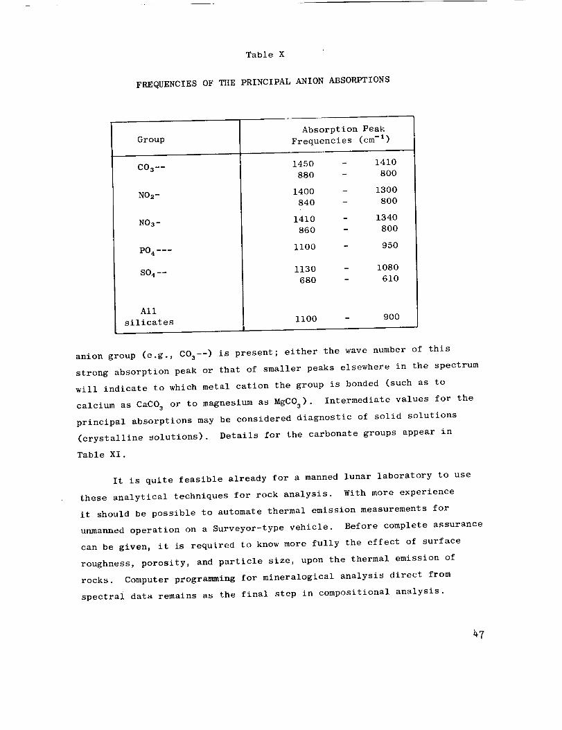
Table X
FREQUENCIES OF THE PRINCIPAL ANION ABSORPTIONS
Group
CO 3 --
NO2-
NOs -
All
silicates
Absorption Peak
Frequencies (cm -i)
1450
880
1400
840
1410
860
ii00
p
1410
800
1300
800
1340
800
950
1130 - 1080
680 - 610
1100 - 900
anion group (e.g., COs--) is present; either the wave number of this
strong absorption peak or that of smaller peaks elsewhere in the spectrum
will indicate to which metal cation the group is bonded (such as to
calcium as CaCO 3 or to magnesium as MgC03). Intermediate values for the
principal absorptions may be considered diagnostic of solid solutions
(crystalline solutions). Details for the carbonate groups appear in
Table XI.
It is quite feasible already for a manned lunar laboratory to use
these analytical techniques for rock analysis. With more experience
it should be possible to automate thermal emission measurements for
unmanned operation on a Surveyor-type vehicle. Before complete assurance
can be given, it is required to know more fully the effect of surface
roughness, porosity, and particle size, upon the thermal emission of
rocks. Computer programming for mineralogical analysis direct from
spectrai data remains as the final step in compositional analysis.
47
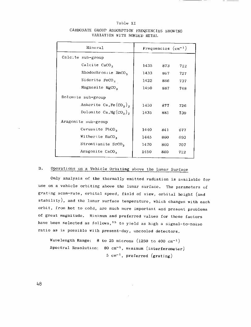
Table XI
CARBONATE GROUP ABSORPTION FREQUENCIES SHOWING
VARIATION WITH BONDED METAL
Mineral Frequencies (cm-1)
Calcite sub-group
Calcite CaCO 3
Rhodochrosite _CO 3
Siderite FeCO 3
Magnesite MgCO 3
Dolomite sub-group
Ankerite Ca,Fe(C03) 2
Dolomite Ca,_(C03) 2
Aragonite sub-group
Cerussite PbCO 3
Witherite BaCO 3
Strontianite SrCO 3
Aragonite CaCO 3
1435 873 712
1433 867 727
1422 866 737
1450 887 748
1450 877 726
1435 881 73O
1440 841 677
1445 86O 693
1470 860 707
1450 86O 712
B. Operations on a Vehicle Orbiting above the Lunar Surface
Only analysis of the thermally emitted radiation is available for
use on a vehicle orbiting above the lunar surface. The parameters of
grating scan-rate, orbital speed, field of view, orbital height (and
stability), and the lunar surface temperature, which changes with each
orbit, from hot to cold, are much more important and present problems
of great magnitude. Minimum and preferred values for these factors
have been selected as follows, Is to yield as high a signal-to-noise
ratio as is possible with present-day, uncooled detectors.
Wavelength Range: 8 to 25 microns (1250 to 400 cm -1)
Spectral Resolution: 80 cm -1, minimum (inter£erometer)
5 cm -1, preferred (grating)
48
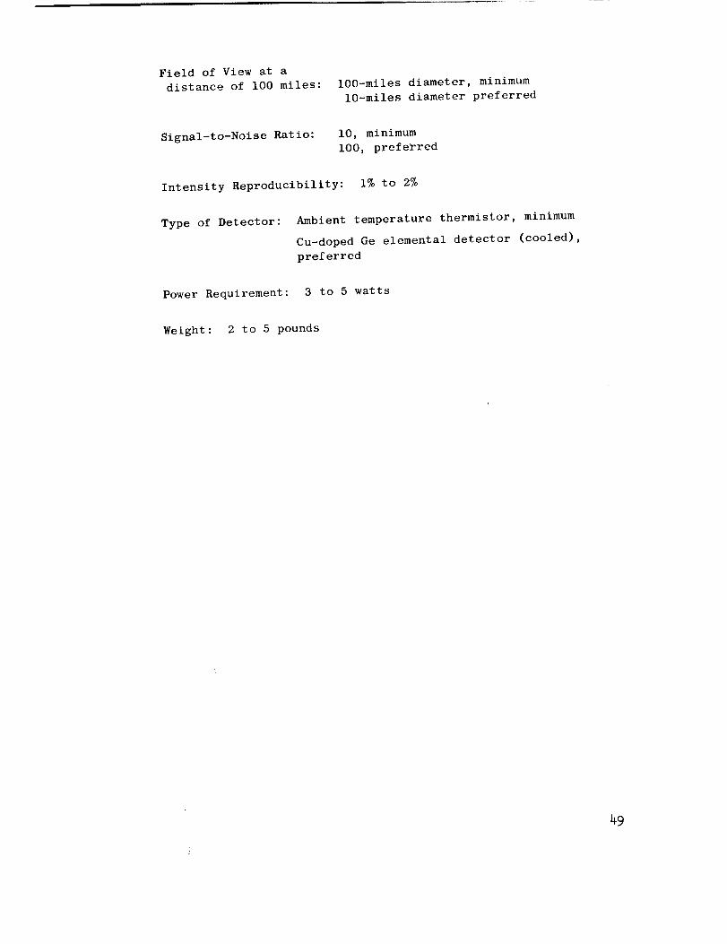
Field of View at a
distance of 100 miles: lO0-miles diameter, minimum
lO-miles diameter preferred
Signal-to-Noise Ratio: i0, minimum
i00, preferred
Intensity Reproducibility: I% to 2%
Type of Detector: Ambient temperature thermistor, minimum
Cu-doped Ge elemental detector (cooled),
preferred
Power Requirement: 3 to 5 watts
Weight: 2 to 5 pounds
49
Ae
APPENDIX
BACKGROUND FOR THERMAL EMISSIVITY STUDIES*
Relationship of Thermal Emissivity, Absorptivity, and
Dependence of Emissivity upon Wavelength and Temperature*
Because of compositional characteristics inherent in minerals, the
absorptivity and hence, emissivity, of these materials is a function of
the wavelength of the radiant energy. The radiant energy, (X, T) from
the lunar surface is equal to
T) b T) (1)
where gb is the efficiency of emission, a measure of the departure from
"blackbody" towards "graybody" character,
£(X) is the emissivity of the surface which is dependent on wave
length, and
W(_, T) is the energy of a blackbody at wavelength, k, and tem-
perature, T.
The term gb is very nearly unity in most instances, particularly if the
surface is a finely divided powder. The term E(X) is the important term
for it is dependent upon the composition of the emitting surface and
varies widely as a function of wavelength. At thermal equilibrium the
product of Ebg(X ) is equal to the absorbed energy, _(_).
To establish the composition of the lunar crust, EbE(X ) must be
determined experimentally; then interpretation by correlation wi:h pre-
viously amassed catalogs of _o(X) for minerals must be made. Qualitative
analysis is achieved readily from the general shape of the £bE(_ ) versus
curve. For quantitative work the term E b must be determined so that
it can be eliminated from subsequent calculation; this i_ readily ac-
complished at a wavelength where g(_) = 1.00.
Sections contributed by E. A. Burns, Oo-worker on this project.
_O
The catalog of So(h ) can be obtained in the laboratory in a variety
of ways. Abso!ute emission measurements give values of _b_o(X)W(_, T)
and after detailed calculation, _o(_) is obtained directly. Because a
great deal of care must be exercised in temperature control and because
energy losses in the optical system must be known to permit accurate cal-
culation of So(h), these values are more easily obtained by indirect
means. The incident energy (Io) of a particular wavelength (t) that falls
on a substrate is either transmitted (It) , absorbed (Is) , reflected spec-
trally (Ip), or reflected diffusely (scattered) (Is) by the substrate.
That is
or
Io = It + Is + Ip + Is (2)
1 = to ÷ o(x) + + So(x) (3)
Transmission measurements normal through thin layers of sample [PO(k)
and s (_) = Ol are useful for giving the general shape of the _ (_) curve;O O
however they are dependent on the thickness of sample and hence are not
useful for quantitative analysis of lunar surfaces. Spectral reflection
measurements on the other hand, on polished surfaces [ t (_) and' O
s (_) = O] give data for S (k) which are usable for both qualitative and0 0
quantitative evaluation.
In calculations of the changes in the radiant energy of quartz com-
pared with a blackbody at the same temperature, 6(k) was obtained from
reflection studies, i.e., [l-Po(_)l, and then the term 6(_)W(_, T) was
evaluated. Over the temperature range 250°K to 500°K the shape of
6(X)W(k, T) and the wavelength of maximum energy vary markedly with the
temperature of quartz. In fact, if the temperature of quartz were to
be determined using equipment designed to give only the wavelength of
maximum energy, considerable error in this measurement would exist in
this temperature range. Data indicate that a maximum error of 65°K
could result. Temperature determination by radiometric measurements
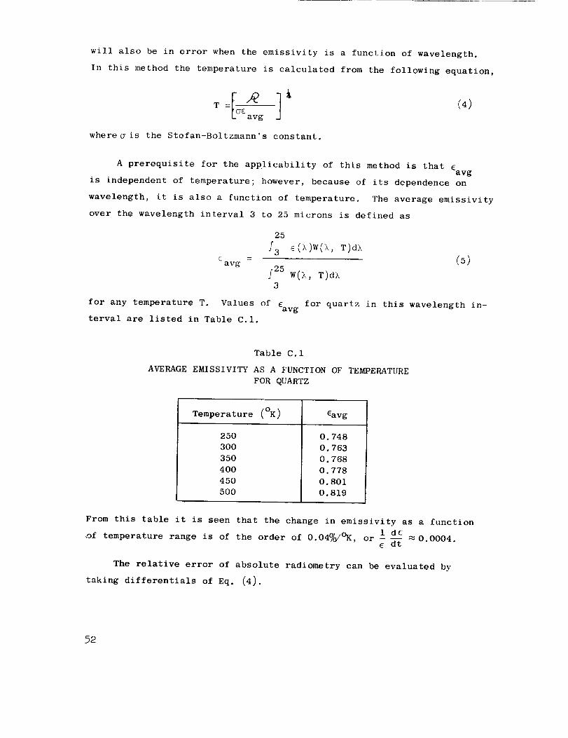
will also be in error when the emissivity is a function of wavelength.
In this method the temperature is calculated from the following equation,
T = £ avg "
whereffis the Stefan-Boltzmann's constant,
A prerequisite for the applicability of this method is that £avg
is independent of temperature; however, because of its dependence on
wavelength, it is also a function of temperature. The average emissivity
over the wavelength interval 3 to 25 microns is defined as
cavg
25
f25 W()_, T)d)_
3
(5)
for any temperature T. Values of 6avg
terval are listed in Table C.I.
for quartz in this wavelength in-
Table C. 1
AVERAGE EMISSIVITY AS A FUNCTION OF TEMPERATURE
FOR QUARTZ
Temperature (OK) 6avg
250
300
350
400
450
500
O. 748
O. 763
O. 768
O o 778
O. 801
0.819
From this table it is seen that the change in emissivity as a function
1 d6,of temperature range is of the order of 0.04%/°K, or _ 0.0004.
6 dt
The relative error of absolute radiometry can be evaluated by
taking differentials of Eq. (4).
52
d f'_ d E 4dT-- + -- (6)= 6 T
A calculated temperature change of 1.25%(4.4°K at 350°K) results from a
radiant energy change of 5%. However, this radiant energy change could
be entirely accounted for instead by a 5% change in the emissivity.
From the data of Table C.I we find that _d6 values as high as 5% are un-6
likely; hence temperature changes of the same material as observed by
radiometric methods are valid. On the other hand, absolute values of the
temperature can be significantly in error.
Because of the predominance of the strong Si-O vibrational band near
i0 microns for most rock-forming minerals, it is reasonable to conclude
that a situation similar, though perhaps not as dramatic, as that described
for quartz would exist for lunar surfaces. This statement is diametrically
opposed to the theories expounded by Pettit and Nicholson; 4° after some
detailed calculations based on an assumed magnitude of stellar radiation
they concluded that the moon was a blackbody in the wavelength range 8
to 14 microns. The classical works of Coblenz,6, 7 however, as well as
the recent studies of Bell et al. 16,17 present a strong contradiction
of the Pettit-Nicholson theories. It is impossible to confirm or deny
either theory for the emissivity of the moon even with the use of modern
infrared equipment, while situated on the earth, because of spectral in-
terference at 9.2 microns from ozone in the earth's atmosphere.
B. Interpretation of Temperature Differences
Standard methods used for the estimation of the temperature of a
radiating source are inadequate unless the relationship of the emissivity
(£) of the source as a function of wavelength (X) is established. Cal-
culations of the temperature of the moon based upon location of the wave-
length of maximum energy can be in error by as much as 65°K, and the
error for temperature differences using radiometric techniques can be of
the order of 10°K.
55
Incorporation of the measured values of _(k), (obtained from re-
flectance data) for four samples of granite, obsidian, dunite, and chon-
dritic meteorite, into the 350°K blackbody energy curves permits calcu-
lation of the emittance curves for these rock samples at a typical lunar
ambient temperature (Fig. C.l).4°, 41 From these we note that changes in
composition can yield changes in dE/E of 10%. This 10% relative change
in the emissivity between granite and the chondritic meteorite is re-
latable to a change of 2-1/2% in the temperature, or to an error of about
9°K at 350°K [see Eq. (6)].
Use of this spectral data applied to emission measurements can
readily account for the temperature differences observed by Shorthill
and Saari. 42 They noted local changes of 5°K which can be explained by
compositional changes. Explanations for the observed maximum temperature
changes of 30°K are much less certain, and are likely to be a composite
of temperature change and compositional change. It is clear that if the
variations in emissivity as a function of composition are evaluated,
then the differences in lunar surface temperatures, as already observed
from the earth, may be resolved.
Because the lunar surface temperature is estimated to vary between
l15°K and 374°K, it is particularly ill-advised to determine temperature
from the wavelength of maximum energy or by integrated radiometric
methods; however, if the radiant energy spectrum were obtained, a black-
body curve could be constructed from the slope of the W(_, T) curve at
short wavelengths [when c(k) = 1.00]. From this careful approximation,
good estimates of the temperature could be made.
74
ACKNOWLE DGi_ENTS
The samples were collected_ and then purified_ and the KBr disc
prepared in the Geochemical Laboratory_ by R. Seetharam under the
direction of Dr. R. J. P. Lyon. Spectrographic analyses were performed
under the direction of Dr. E. A. Burns_ Manager of the Analytical Labo-
ratory_ Propulsion Sciences Division_ who also provided invaluable
assistance in the many phases of emission spectrophotometry. The data
were analyzed and the report prepared by Dr. Lyon.
The many mineralogists who contributed standardized samples of
minerals are listed below. Without their assistance a study of this
degree of detail could not have been attempted.
Anderson_ J.
Baker_ G.
Beatty_ W. B.
Chao_ E. C. T.
Chinner, G.
Coleman_ R. G.
Coombs_ D. S.
DeCarlij P.
Dickinson_ W. R.
Edwards_ A. B.
Erd_ R.
Titanates
Australite
(tektite)
Basalt
Obsidian
Granite
Coesite
Stishovite
Cordierite
Muscovite
Pyroxenes
Garnets
Micas
Lawsonite
Zeolites
Phosphates
Carbonates
Wairakite
Shock-loaded
minerals
Gabbro
Muscovite
Zeolites
Phosphates
Pyroxenes
Fairbairn_ H. W.
Floyd, P.
Foster_ M. D.
Frederiksson_ R.
Fryklund, V.
Grines_ D.
Gulbrandsen_ R. A.
Hannah_ R.
Heinrich_ E. W.
Hemley_ J.
Hess_ H. H.
Howie_ R. A.
Hutton_ C. O.
Jackson_ E. D.
Mica
Obsidian
Micas
Chlorites
Chondrite
Garnet
Mica
Shock-loaded
limestone
Phosphates
AI-Hydrates
Micas
Micas
Synthetic
minerals
Pyroxenes
Pyroxenes
Pyroxenes
Micas
Feldspars
Pyroxenes
Olivines
Gabbro
Dunite
Kunze, G. W.
Lee, D.Lipman, P.Loomis, A. A.Lyons, S. C.
Mein, W. J.Milton, D.
Mountsier3 S. R.
Olsen_ R.
Pabst, AParks_ G.
Clay
GarnetsHornblendeChondritesKaolinite
Anauxite
Stishovite
Limestone
Sandstone
Coesite
Pyroxene
Talc
Zeolites
Anauxite
Perlites
Opal
Phosphate
Chrysocolla
Roberts, R. Turquoise
Sands_ L.B. Clays
Schieltz_ N.C. Celadonite
Schlocker, J. Micas
Sedlacek_ R. Rutile
Shell, H.R. Micas
Short_ N. Granite
Speed_ R. C.
Stephens_ J.
Stevens, R.
Stringham, B.
Taylor, W.
Theobald, P.
Threadgold_ I.
Tuddenham, W. M.
Vernon, R.
Weir, A.
Whelan, J.
Wilshire_ H.
Wones, D.
Yates, R
Gabbro
Granite
Serpentine
Anorthosite
Quartzite
Tuff, Bishop
Rhyolite
Dunite
Basalt
Kaolinite
Lepidolites
Clays
Zeolites
Biotites
Mica
Olivines
Hornblende
Mica
Cummingtonite
Beidellites
Clay
Phosphates
Pyroxenes
Zeolites
Synthetic
feldspar
Hornblende
Stanford Research Institute
Menlo Park, California
September 1962
76
REFERENCES
1.
20
3.
4.
5o
6o
7.
8.
9o
10.
11.
12.
13.
14.
Hunt, J. M., Wisherd, P., and Bonham, L. C., Infrared Absorption
Spectra of Minerals and other Inorganic Compounds, Anal. Chem. 22,
1478-1497 (1950)
Hunt, J. M. and Turner, D. S., Determination of Mineral Constituents
of Rocks by Infrared Spectroscopy, Anal. Chem. 25, 1169-1174 (1953)
Tuddenham, W. M. and Lyon, R. J. P., Relation of Infrared Spectra
and Chemical Analysis for some Chlorites and Related Minerals,
Anal. Chem. 31, 377-380 (1959)
Tuddenham, W. M. and Lyon, R. J. P., Infrared Techniques in the
Identification and Measurement of Minerals, Anal. Chem. 32, 1630-41
(1960)
Lyon, R. J. P., Tuddenham, W. M., and Thompson, C. S., Quantitative
Mineralogy in 30 Minutes, Econ. Geol. 54, 1047-1055 (1959)
Coblentz, W. W., Investigation of Infrared Spectra, Pub. 65, Carnegie
Institute of Washington, 1906
Coblentz, W. W., Selected Radiation from Various Solids, II, Nat.
Bur. Standards (US) Bull, 6, 301 (1910)
Pfund, A. H., The Identification of Gems, J. Opt. S.c. Amer., 35,
611-614 (1945)
McMahon, H. O., Thermal Radiation Characteristics of Some Glasses,
Jour. Am. Ceram. S,c., 34, 91-6 (1950)
Simon, J. and McMahon, H. O., Study of the Structure of Quartz,
Cristobalite, and Vitreous Silica by Reflection in Infrared,
Jour. Chem. Phys. 21, 23-30 (1953)
Gardon, R., The Emissivity of Transparent Materials, J. Am. Ceram.
S.c. 39, 278-285 (1956)
Keller, W. D., Sports, J. H., and Biggs, D. L., Infrared Spectra
of some Rock-Forming Minerals, Am. Jour. Sci. 250, 453-471 (1952)
Launer, P. J., Regularities in the Infrared Absorption Spectra
of Silicate Minerals, Am. Min. 37, 774-784 (1952)
Burns, E. A., and Lyon, R. J. P., Errors in the Measurement of the
Temperature of Radiating Lunar Materials, paper to be presented
at the First Annual Pacific Meeting of Applied Spectroscopy and
Analytical Chemistry,(Oct. 19, 1962)
77
15.
16.
17.
18.
19.
20.
21.
22.
23.
24.
25.
26.
27.
28.
Lyon, R. J. P., and Burns, E. A., Temperature and Compositional
Mapping of the Lunar Surface from an Orbiting Vehicle, SRI
Proposal PRU 62-251 to NASA, June 22,(1962)
Bell, E. E. and Eisner, I. L., Infrared Radiation from the White
Sands at White Sands National Monument, New Mexico, Jour. Opt.
Soc. Am., 46, 303-4 (1956)
Bell, E. E., Eisner, I. L. Young, J. B., Abolins, A. and Oetjen,
R. A., Infrared Techniques and Measurements, Ohio State Research
Foundation, Final Engineering Report, Contract 33(616)3312, p.105
(ASTIA - AD 151221),(1957)
Miller, F. A. and Wilkins, C. H., Infrared Spectra and Characteristic
Frequencies of Inorganic Ions, Anal. Chem. 24, 1253-94 (1952)
Uhlrich, W. F., Applications of Specular Infrared Reflectance,
The Analyzer (Beckman Inc. House Journal), July, 14-16,(1961)
Hannah, R., Perkin-Elmer Corporation, Norwalk, Conn., Private
communication.
Duyckaerts, G., The Infrared Analysis of Solid Substances,
Analyst, 84, 201-214 (1959)
Lyon, R. J. P. and Burns, E. A., Analysis of Rocks by Reflected
Infrared Radiation,(Manuscript submitted to Economic Geology
Magazine, August, 1962)
Laves, F. and Hafner, St., Order/Disorder and Infrared Absorption
I; (aluminum, silicon)--Distribution in feldspars, Zeitschr.
ILrlstallographie, 108, 52-63 (1956)
Hafner, St. and Laves, F., Order/Disorder and Infrared Absorption
II: Variation of the Position and Intensity of some Absorptions
of Feldspars; The structure of orthoclase and adularia, Zeitschr.
Kristalographie, 109, 204-225 (1957)
Lyon, R. J. P. and Tuddenham, W. M., Determination of Tetrahedral
Aluminum in Mica by Infrared Absorption, Nature, 185, 374-5 (1960)
St_bican, V. and Roy, R., Infrared Spectra of Layer-Structure
Silicates, J. Amer. Ceram. Soc., 44, 625-627 (1961)
St_bican, V. and Roy, R., Isomorphous Substitution and Infrared
Spectra of the Layer-Lattice Silicates, Am. Min. 46, 32-51 (1961)
Lyon, R. J. P., Evaluation of Infrared Spectrophotometry for
Compositional Analysis of Lunar and Planetary Soils, Stanford
Research Institute, Project No. SU-3943, Interim Report No. 1,
NASA contract NASr-49(04),(1962)
_8
29.
30.
31.
34.
35.
36.
37.
38.
39.
40.
41.
42.
43.
Berry, L. G° and Mason, B., Mineralogy-Concepts, Description and
Determinations, W. H. Freeman and Co., Press, San Francisco, pp.
630 (1959)
Duke, D. A., Infrared Investigation of the Crystal Chemistry of
Olivine and Humite Minerals, Ph.D. Thesis University of Utah,
(1962)
Vernon, R. H., Co-existing Cummingtonite and Hornblende in an
Amphibolite from Duchess, Queensland, Australia, Am. Min., 47,
360-371 (1962)
Bates, T. L., Penn. State University, Personal communication.
Tarte, P., and Ringwood, A. E., Infrared Spectrum of the Spinels
Ni2Si04, Ni2GeO 4 and their Solid Solutions, Nature 193, 971-2 (1962)
Lyon, R. J. P., Infrared Confirmation of 6-fold Coordination of
Silicon in Stishovite (SiO2): (Manuscript submitted to Nature
Magazine, July, 1962)
Saksena, B. D., Analysis of the Raman and Infrared Spectra of
_-Quartz, Proc. Indian. ACad. Sci., 12A, 92-139 (1940)
Saksena, B. D., Infrared Absorption Studies of Some Silicate
Structures, Trans. Faraday Soc., 5_/7, 242-255 (1961)
Sevchenko, N. A., and Florinskaya, V. A., The Reflection and
Transmission Spectra of Various Modifications of Silicon Dioxide
in the Wavelength Region 7-24 Microns, (in Russian), Doklady Akad.
Nauk USSR, 109, 1115-8 (1956)
Matossi, F., and Bronder, O., The Infrared Absorption Spectra of
several Silicates, in German, Z. Physik, Iii, 1-17 (1938)
Frederickson, W. R., and Ginsburg, N., Infrared Spectral Emissivity
of Terrain, Dept. of Physics, Syracuse Univ., Interim Report, No.
l, Contract AF 33(616)-5034, p. 49, (ASTIA - AD 132839), (1957)
Pettit, E., and Nicholson, S. B., Lunar Radiation and Temperature,
Astrophys. Jour., 71, p. 102-137 (1930)
Pettit, E., Radiation Measurements on the Eclipsed Moon, Astrophys.
Jour°, 91, 408 (1940)
Shorthill, R. W., and Saari, J. M., Lunar Infrared Temperature during
Sept. 4, 5, and 6, 1960, Boeing Aircraft Co., Report under Air
Force Contract 18600-1824 (Jan. 30, 1961)
Burns, E. A., and Lyon, R. J. P., Errors in the Measurement of the
Temperature of Radiating Lunar Bodies, (Manuscript submitted to
Nature Magazine, August, 1962)
59
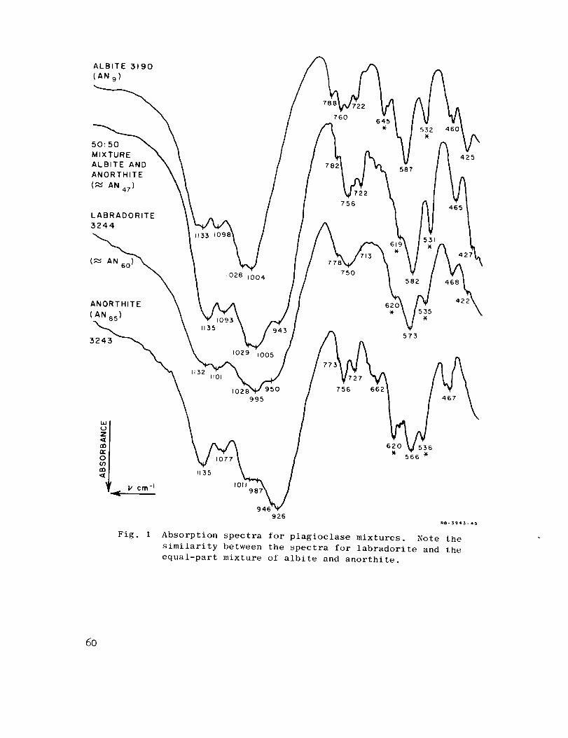
ALBITE 3190
(AN 9 )
760645
_ 532 460X
MIXTURE _. 587 _j425
ALBITE AND \
ANORTHITE
(_ AN 47)
756 465
LABRADORITE
5244
531
2\(_ AN 60 )
1028 1004750
582
42:ANORTHITE 62(
( AN 85) . 535
il35 r_ 57:3
_, ,o_,oo_ /...\ _/t
\ / VI /
926K_Bo3943-45
Fig, 1 Absorption spectra :for plagioclase mixtures, Note thesirailarity between the spectra :for labradorite and theequal-part mixture oi' albite and anorthite,
60

62(5
6O3
67O
504
5OO
424
414
410
628
1067 507
979 ,,_ 4ll
860 4651068
R8 3943 40
Fig. 2 Absorption spectra for augite and olivine mixtures
61
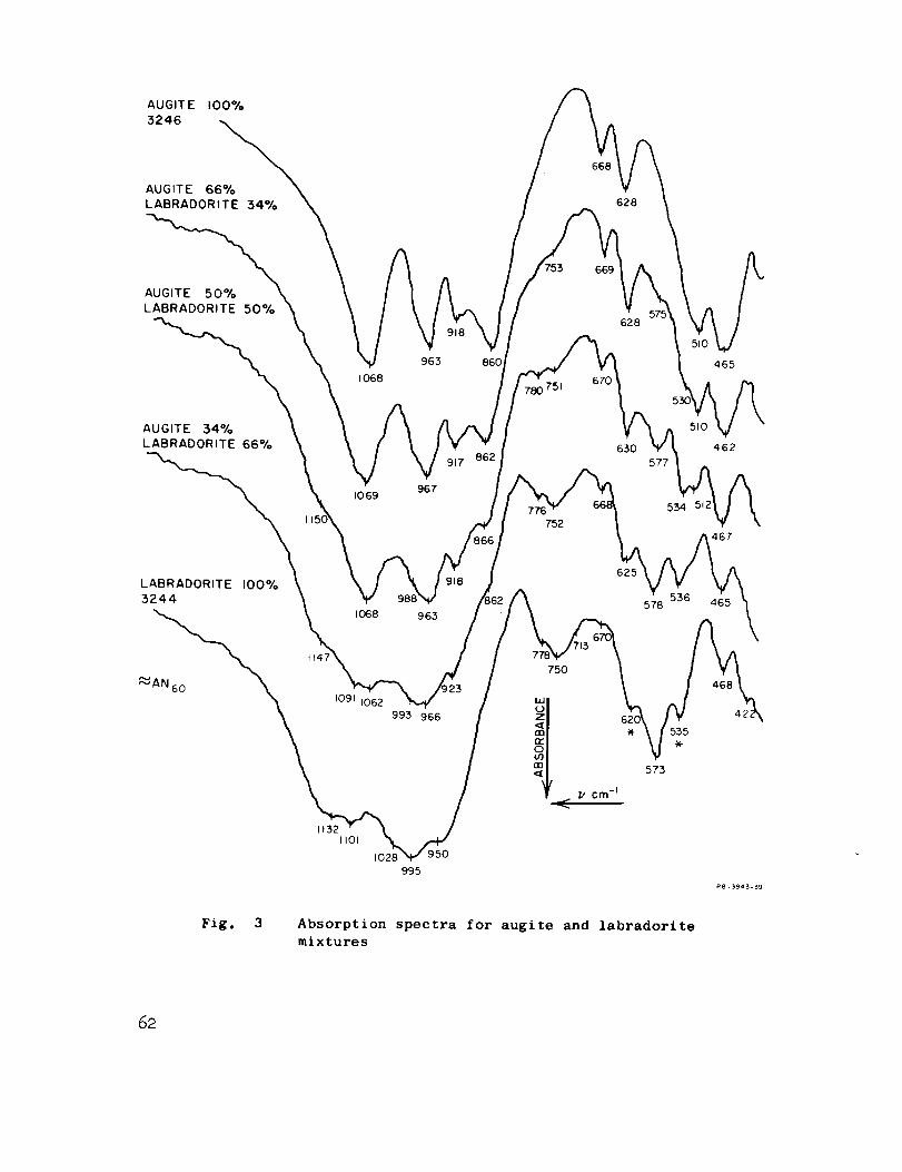
AUGIT E 100%
3246
AUGITE 66%
LABRADORITE 34%
668
628
AUGITE 50%
LABRADORITE 50%
669
628
AUGITE 34%
LABRADORITE 66%
I068
I0 69
963
967
917 862
860
780 751670
630
465
510
577_ 462
467
LABRADORITE 100%
3244
I Io68
,_---,AN _ ,.i/
11062
1132I101
102E
993 966
995
750
625
536578
Fig. 3 Absorption spectra for augite and labradorite
mixtures
62
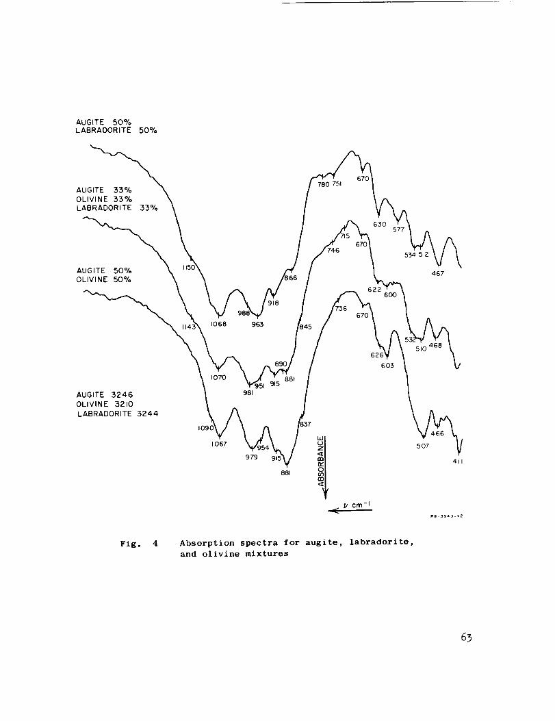
AUGITE 50%LABRAOORITE 50%
AUGITE 33%
OLIVINE 33%
LABRADORITE 33%
AUGITE ,50%
OLIVINE 50%
1068
918
963
780 75f
630
670
467
6OO
AUGITE 3246
OLIVINE 3210
LABRADORITE 3244
1070
981
603
1067
979
881
cm -I
5O7
411
R8-3943-42
Fig, 4 Absorption spectra for augite, labradorite,
and olivine mixtures
63
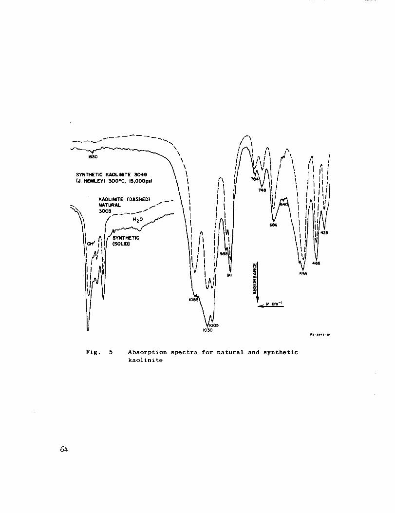
1550
SYNTHETIC KAOLINITE 3049
(J, HEMLEY) 300"C, 15,000psi
\\\\
IOIIIII
0051030
911
/I \
III
I I
428
468
RB-3943-59
Fig. 5 Absorption spectra for natural and synthetic
kaolinite
64
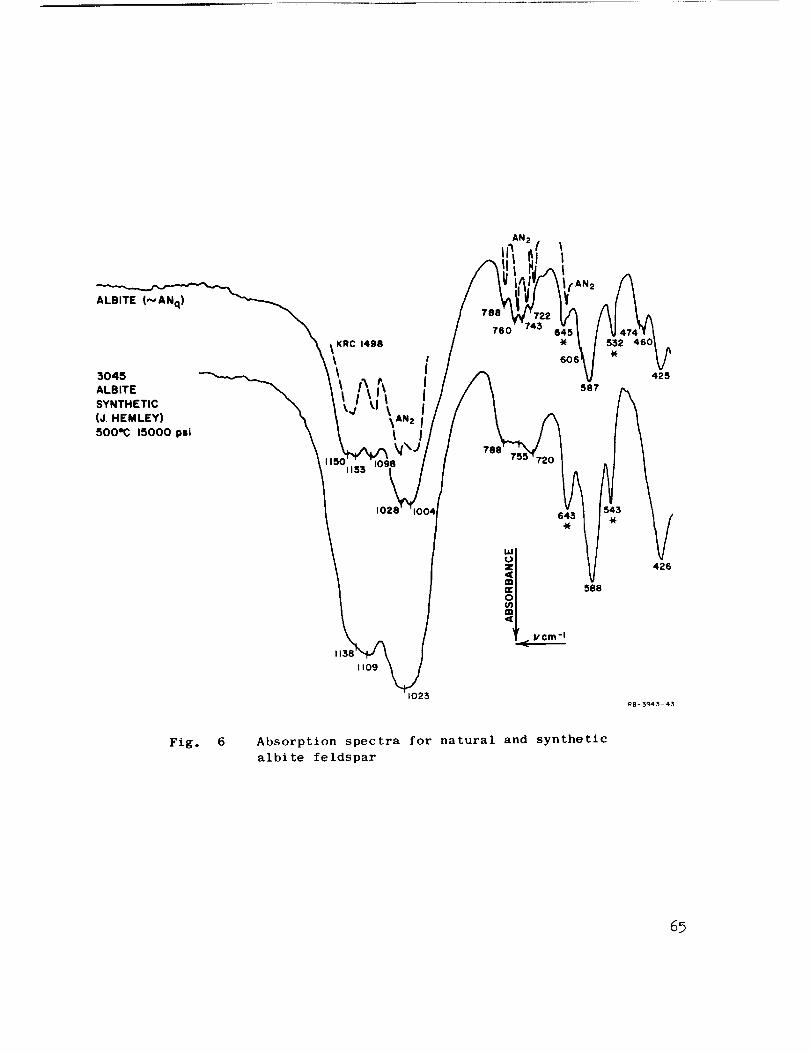
ALBITE (N ANq)
3045
ALBITE
SYNTHETIC
(J. HEMLEY)
500eC 15000 psi
KRC 1498
AN 2
760
587
532 460
425
1021
1109
1025
<: vcm-I
588
426
R8-3943-43
Flg. 6 Absorption spectra for natural and synthetic
albite feldspar
6_
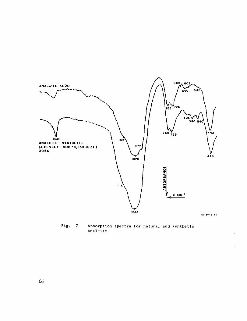
ANALCITE 3020666
26
1630
ANALCITE - SYNTHETIC
(J. HEMLEY - 400 *C, 15000 psi)3046
1020
0
586
442
1023RB'394_-44
Fig. 7 Absorpt£on spectra for natural and synthetic
analcite
66
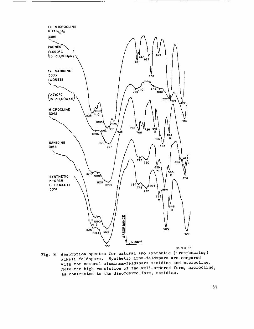
Fe - MICROCLI N E
K FeSi308
3385
Fe- SANIDINE
:5385
(WONES)
> 710°C
15-30,000
MICROCLINE
3242
SANIDINE
3154
SYNTHETICK-SPAR
(d. HEMLEY)
3051
Fig. 8
1095
1022
I
tl351094
994
1009
1030RB-_943-47
Absorption spectra for natural and synthetic (iron-bearing)
alkali feldspars. Synthetic iron-feldspars are compared
with the natural aluminum-feldspars sanidine and microcline,
Note the high resolution of the well-ordered form_ microcline_
as contrasted to the disordered form_ sanidine.
67

ALBITE 3190
(AN c
RI 1536
ALBITE
AFTER
600 Kbor
SHOCK
SG 2.20-224
RI 1,50
3187
641
719
582
1030
QUARTZ
3133
1020
QUARTZ \ \
350 Kbor _ \
SHOCK _ \
\CYL CONVER,/ \ I,70 h
SHOCK \ \
3,45 \ \ /\
1076QUARTZ
FLY,NG (OPAQUE
PLATE _,SELVAGEJ
SHOCK
600 Kbor
3015
(GLASS)
10801198
694
'75
///: _%'\
/ \
// _803_ II \ \
I \OPAL \ # \
3301 _ I \
'V '/ \ (79o) _.\
_, I [695)FUSED \ I \_
SiLiCA \ I \_
3019 _"I / _)
(804) _1 468
68
1093RB-394_- 65
Fig. 9 Absorption spectra of shock-loaded albite feldsparand quartz. The presence of glass results in theloss of detail in their spectra,

2.6 27 2.8 2.9 30 microns4000 I 3800 3600t I 3400 I 3200 cm-_
I I I I I I I I
SAME DISC AFTER I hr _0
150°C VACUUM- OVEN-3420
SAME DISC AFTER FURTHER3450
40 hrs DESSICATOR (CoCI 2)
PARAGONITE MICA
UNDRIED DISC (KBr)
SHOWING 2 OH/ PEAKS
AND WATER
0.105 ABS.
RATIO 1,48
0155 ABS.
SAME DISC AFTER
42 hrs )°C IN
VACUUM OVEN
FELDSPAR52BE35
XlO
55BE7XlO
53HBIIXlO
3636
0 140 ABS
0210 ABS
RATIO 1.503636
WAVELENGTH cm -I
Fig. 10 Absorption spectra showing the "modes" of occurrence of water
in minerals and the ability of infrared analysis to differen-
tiate between them. Normal moisture associated with a '_et"
sample occurs around 3435 cm-l_ but peaks shift to higher
wavenumbers with increased strength of bonding in the structure
of the crystai. Hydroxyl peaks for muscovite micas are about
3650 cm-t_ while that for talc is about 3700 am -1 The curves
are displaced vertically.
69
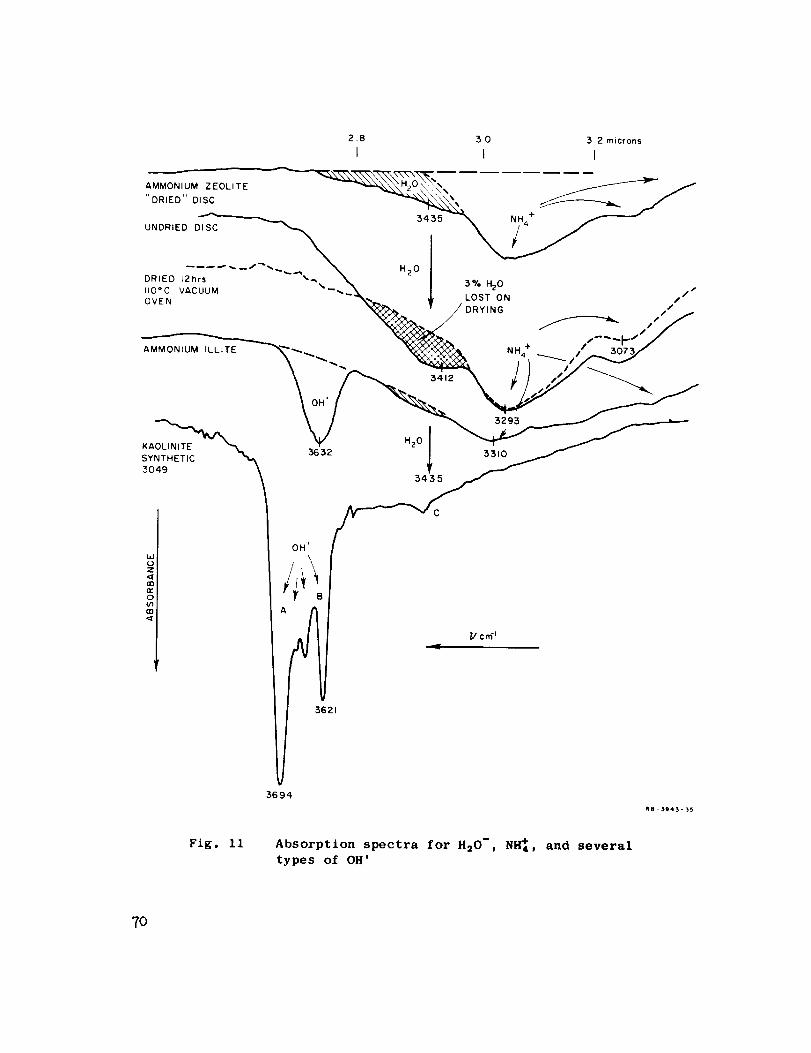
3,2 microns3.0
2.B II I
' DRIED" DISC
.......---__._\_,°I _.,._,o /DRLED 12brs ,w_. -_ | LoST ON ,,,/lid C vAcUUM ---'_:_'N_ _ / DRYING _ I ' f
ovEN _ / / -- ,'/f--p" /
AMMONIUM ILLITE "'",,..__ f / I
-- .,2 _./"
A
,694
l/cr_ _
,62t
RB-$943-_
Fig. IiAbsorption spectra for H20-, NI_4, and several
types of OH'
TO
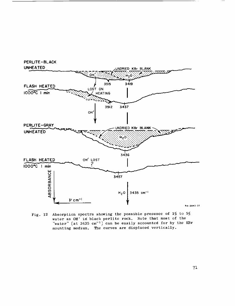
PERLITE-BLACK
UNHEATED•.. ,. /UNDRIED KBr BLANK
FLASH HEATED
OHII 3512 343[
PERLITE-GRAY UNDRIED KBr BLANK
UNHEATED
3436
FLASH HEATED OH _ LOST I
z 3457<[
oo3nn H20 3435 cm-1
_ Z/ cm-I
RA-3943-37
Fig. 12 Absorption spectra showing the possible presence of 1_ to 5_water as OH' i_ black perlite rock. Note that most of the
"water" (at 3435 cm -1) can be easily accounted for by the KBr
mounting medium. The curves are displaced vertically.
71

52BE-3570.3 % AN _'-
70.1% AN
55MV-32
73.0 % AN
73.6 % AN
1017
942
750
S84660
732 66_
77
540
621 V578
114,
102:
994
940
R8- 3943-41
Fig. 13 Absorption spectra of hidden duplicates of plagioclase
feldspars. The total variance is shown for all the steps
from the initial sampling through the disc preparation to
the numerical determination of the peak positions. The
curves are displaced vertically.
72
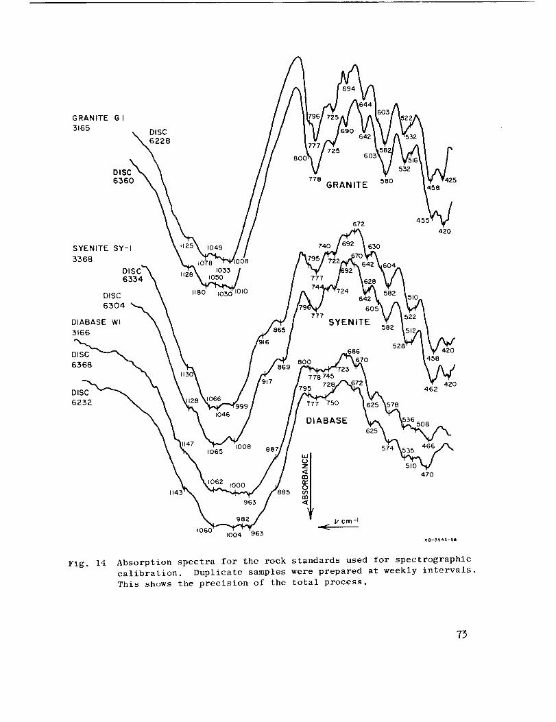
GRANITE G I
3165DISC
6228
72560
778GRANITE
580458
SYENITE SY-I
3368
DI
6334
DISC
6304 "_DIABASE Wl
3166
t078
1180
1033
740
777
777
672
630
642
605
SYENITE
420
DISC
6368
1143
917
1046
1065 1008 887
800
686
745
750
DIABASE
013963 4'
982 _,' cm-IJ
t0601004 963
52
578
625
574
420
458
420462
470
RB-3943o58
Fig. 14 Absorption spectra for the rock standards used for spectrographic
calibration. Duplicate samples were prepared at weekly intervals.
This shows the precision of the total process.
73
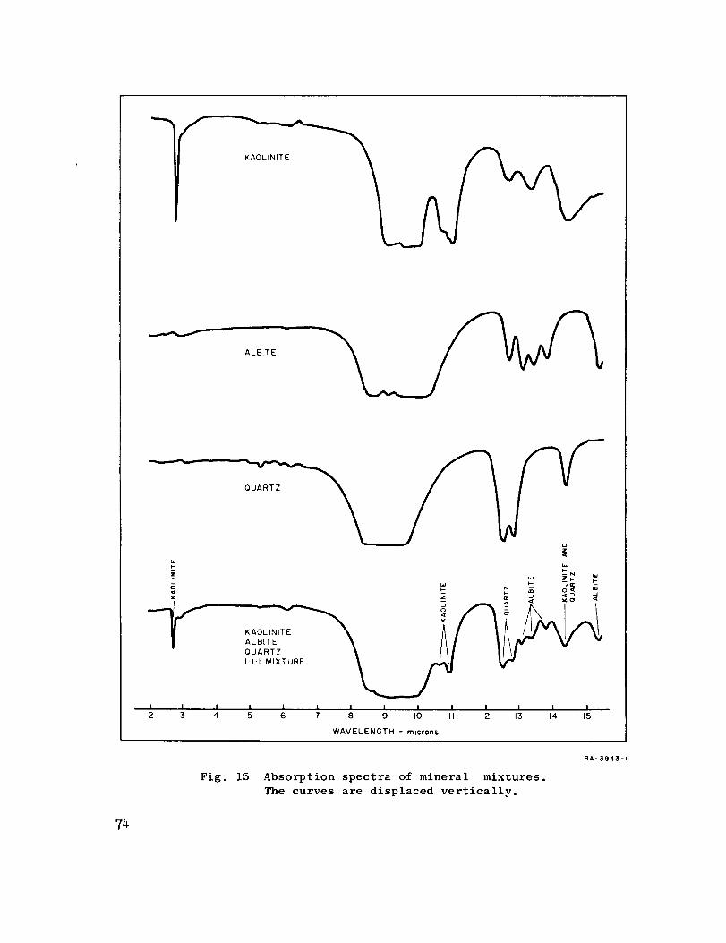
ALBITE
z
-y.1 I I2 3 4
QUARTZ
wtN
__ ._q= -_' _ _ o,_ ®
-. _
I I i I I I I I I I I5 6 7 8 9 I0 II 12 13 14 15
WAVELENGTH - microns
Fig. 15 Absorption spectra of mineral mixtures.
The curves are displaced vertically.
RA- 3943-i
T4
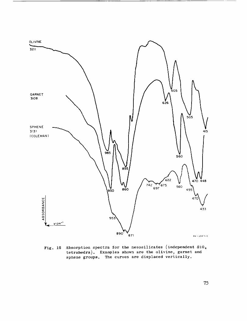
OLIVINE
3211
GARNET
3108
SPHENE
3131
( COLE MAN )
626
505
415
560
860 69756O
470 448
433
Fig. 16 Absorption spectra for the nesosilicates (independent Si0 4
tetrahedra). Examples shown are the olivine_ garnet and
sphene groups. The curves are displaced vertically.
77

FORSTERITE (%90 +) 3304OLIVINE
3320
FO 89.7
3318
Fo 85.8
DUPLICATE
3322
FO 85.8
/I
II
II//
FORSTERITE
/II
410
3319 F0 84.741'P
3317 Fo 84.0
3321 FO8o. 5
981
885
409
FAYALITE 3303 (KRC 3602)
\\
\
OLIVINE
599
//
/ k598
///FA YA_._
558
412
J 410
5o2_/482
Fig. 17 Detailed absorption spectra of the olivine group near the
magnesium-rich end. The curves are displaced vertically.
76
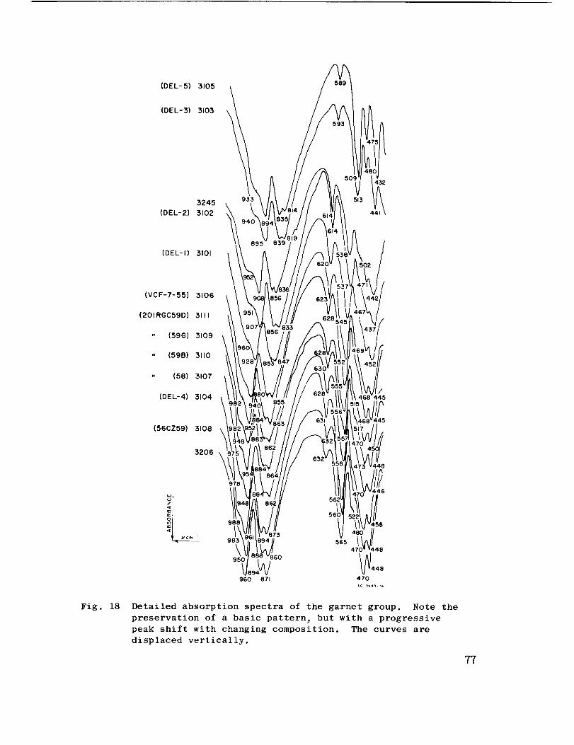
(DEL-5) 3105
(DEL-3) 3103 \
3245
(DEL-2) 3102
933
(DEL-I) 3101
(VCF-7-55) 3106
(201RGC59D) 3111
" (59G) 3109
" (59B) 3110
" (58) 3107
(DEL-4) 3104
(56CZ59) 3108
445
Fig. 18
3206 \448
o_'cm i
978
988
\95O
960 871
6O
565
470 48
_448
47'0
Detailed absorption spectra of the garnet group. Note the
preservation of a basic pattern_ but with a progressive
pear shift with changing composition. The curves are
displaced vertically.
77
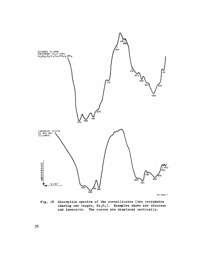
IDOCRASE GC 2538CRESTMORE CALIF (KRC)
C°_oMg2Al4{Si20_2(Si04)_(i4
_°°_V_566 M
5_ 40
480
916963
1012
LAWSONtTE GC3125
i , 420_I _ I \ A.. /.,20°_L _ I __ l_2"x/55
| _ I _n%A"%; 4,B0
995 880
94.5
RA-3943.7
Fig. 19 Absorption spectra of the sorosilicates (two tetrahedra
sharing one oxygen_ Si207). Examples shown are idocrase
and lawsonite. The curves are displaced vertically.
78
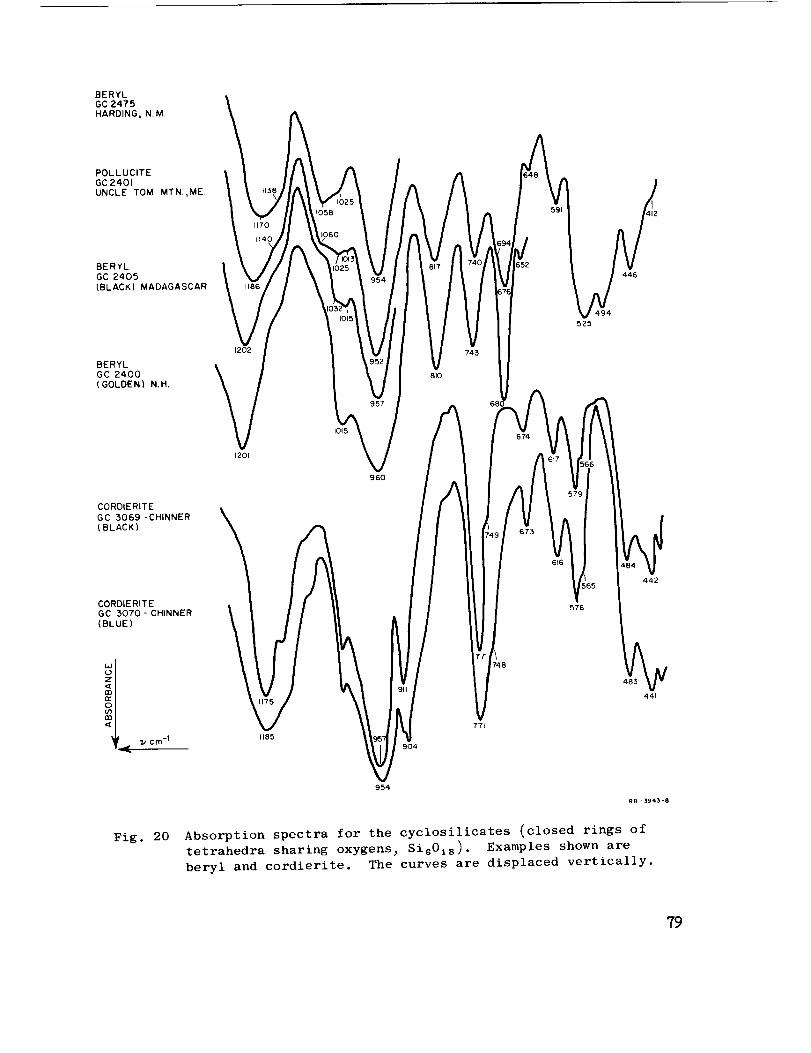
BERYL
GC2475
HARDING, NM
POLLUCITE
GC2401
UNCLE TOM MTN.,ME
BERYL
GC 2405
(BLACK) MADAGASCAR
BERYL
GC 2400
(GOLDEN) N.H.
1202
/VBIT
/V 743
810
652
494
525
446
CORDIERITE
GC 5069-CHINNER(BLACK)
CORDIERITE
GC 3070 - CHINNER
(BLUE)
OI_._, 7.,, cm -I
1201
II
r_(0(5
/96O
/A
954
748
771
616
576
442
485
441
RB _ 1394_- 8
Fig. 20 Absorption spectra for the cyclosilicates (closed rings oftetrahedra sharing oxygens_ SisOls ). Examples shown are
beryl and cordierite. The curves are displaced vertically.
79
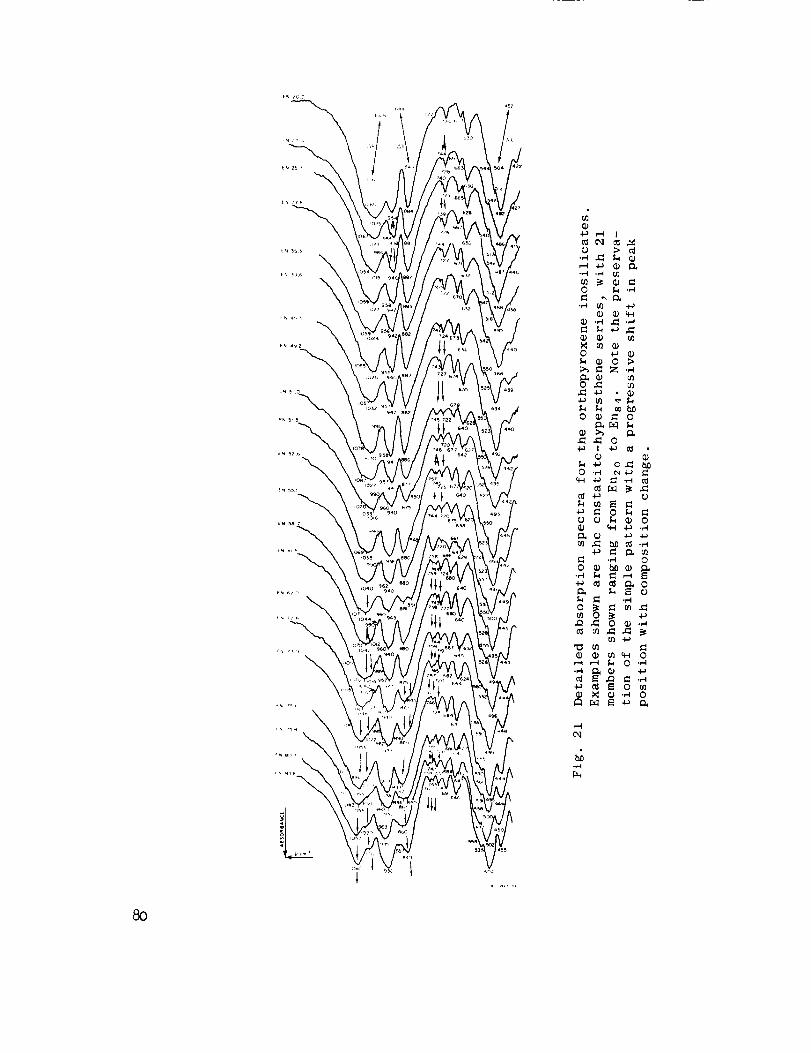
80
\
\
L1
-M

ACMITE
3250
(STANFORD)
OMPHACITE
3240
(15-CZ-60)
COLEMAN
JADEITE
3ll3
(R 1852 )
COLEMAN
DIOPSIDE
3237
(109- RGC-52 )
COLEMAN
1044
966
745
656
670
502
475
HEDENBERGITE
IRON MTN
3453
Fig. 22
405
1062
664
622
468
509
860
454
RB-3945-71
Absorption spectra for the clinopyroxene inosilicates (single
chains of tetrahedra each sharing two oxygens) Si03). Examples
shown are acmite_ omphacite_ jadeite_ diopside_ and hedenber-
gite. The curves are displaced vertically.
81
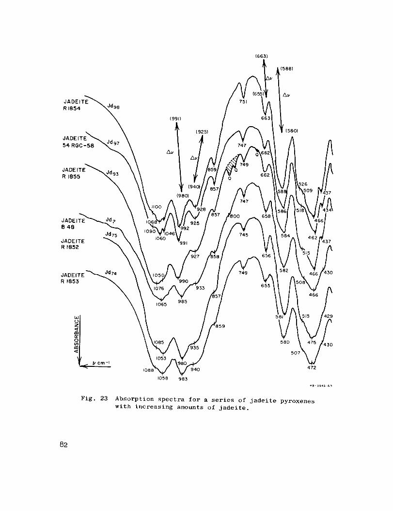
JADEITE
RI854 Jd98
JADEITE
54 RGC-58
JADEITE_
R 1855
(991)
(925)
Av
('
(980)
Q
Q
(6631
751
747
(588)
Av
663
(580}
662
JADEITE
B48
JADEITE
R 1852
Jd751060
91
JADE ITE "_74
R 1853
1065985
933
749
655
466
UJ0
o
108_ 940
1058 983
580
507
472
RB-3943-69
Fig. 23 Absorption spectra for a series of jadeite pyroxenes
with increasing amounts of jadeite.
82
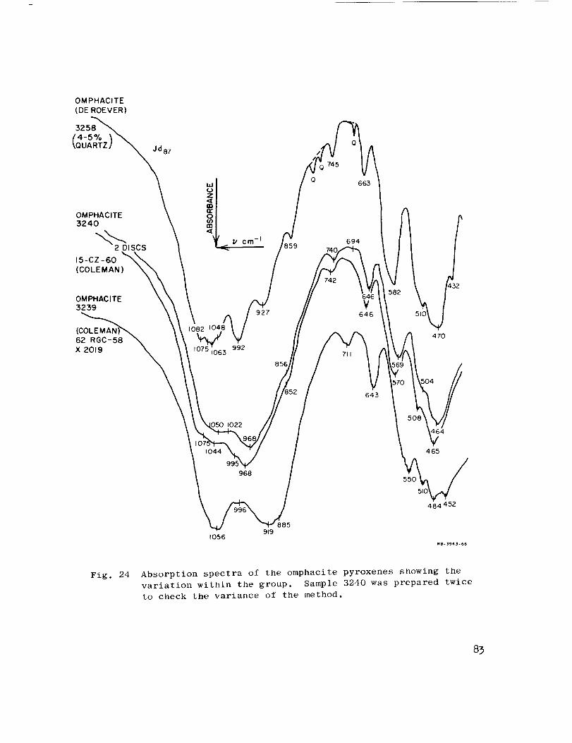
OMPHACITE
(DE ROEVER)
3258
Jd87
OMPHACITE
:5240
2 DISCS
15-CZ -60
(COLEMAN)
OM PHAC ITE
3259
X 2019
o
! cm -I
927
992
856
0
) 745
742
694
¥646
711
050 IO22
995
96855O
_64
465
IO56919
885
484 452
Fig. 24 Absorption spectra of the omphacite pyroxenes showing the
variation within the group. Sample 3240 was prepared twice
to check the variance of the method.
83

ACMITE
3250
MAGNET
STANFORD
ACMITE
3249
NEW IDRIA
STANFORD 55_
ACMITE
3248
NORWAY
STANFORD 5806
730
642
638
Fig. 25
1026
954
951
558
545
505
1054
939
Absorption spectra for a series of acmite
py roxe ne s
455
RB-3943-£7
84
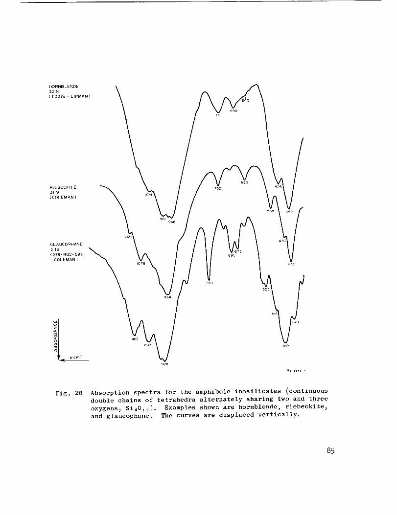
HORNBLENDE
3231
{ T 337o - L IPMAN )
RtEBECKITE
3119
( COLEMAN }
GLAUCOPHANE
3118
( 201- RGC- 59H
COLEMAN )
981
J124
964
94B
790
6,50
695
555
48O
452
II02
1045
978
48O
Fig. 26 Absorption spectra for the amphibole inosilicates (continuous
double chains of tetrahedra alternately sharing two and three
oxygens_ Si4011 ). Examples shown are hornblende_ riebeckite_
and glaucophane. The curves are displaced vertically.
85
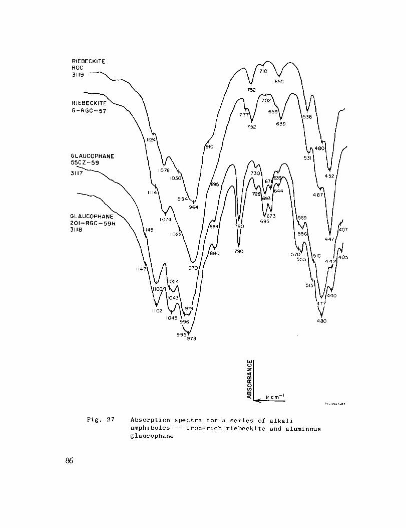
RIEBECKITE
RGC
3119
752
650
GLAUCOPHANE
55CZ-59
3117
1074
1145
114
11o2
964
1054
1045996
99. _978
88O
790
V790
695569
570555
48Z
4O5
440
477
V48O
Fig. 27
°l cmAbsorption spectra for a series of alkali
amphiboles -- iron-rich riebeckite and aluminous
glaucophane
R8- 3943-6Z
86
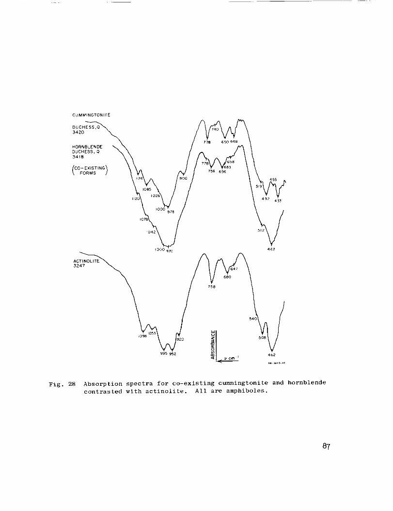
CUMMINGTONITE
DU C H E SS_"_'_
DUCHESS, O _ \
_SExISTING" x _
( ;_.Ms) \k,o,,\ /,,,,,\,°,,l./
I000 97'0 462
ACT I NO LITE
3247
995952 _1 ..... , 462<_ pcm-
• a 3943 46
Fig. 28 Absorption spectra for co-existing cummingtonite and hornblende
contrasted with actinolite. All are amphiboles.
87
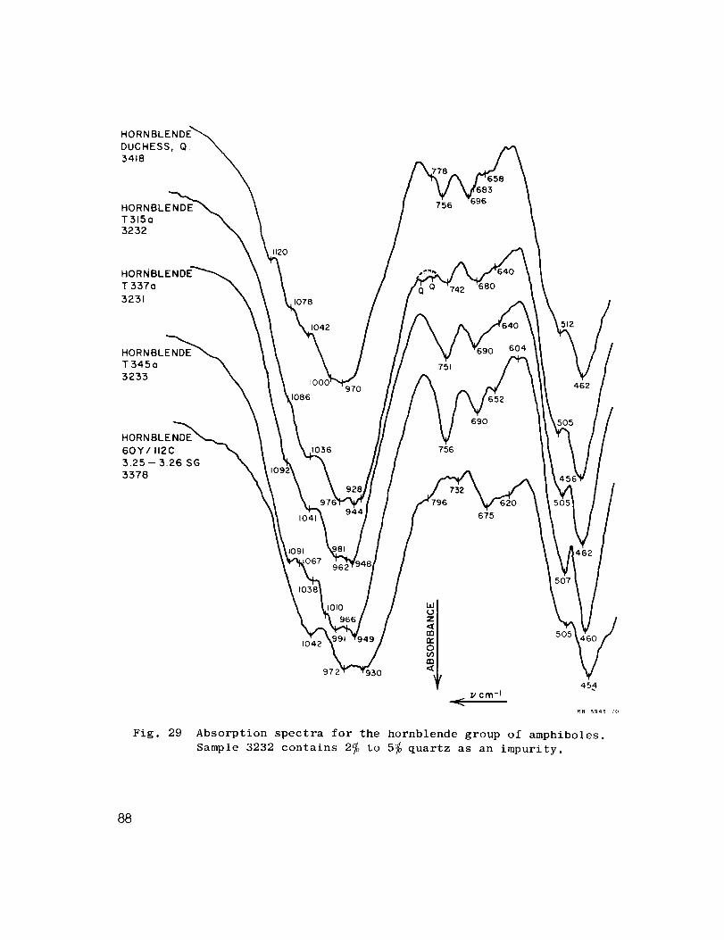
HORN
DUCHESS, Q.
34t8
HORNBLENDE
T315o
3232
HORNBLENDE_
T337o
3231
HORNBLENDE
T345a
3233
1120
1078
1042
1086
1036
756
751
756
6,52
690
604
675
Fig. 29
z
1042 (_)
972 930 (I
505
454
_. _' cm-I
R8- 394_ - to
Absorption spectra for the hornblende group of amphiboles.
Sample 3232 contains 2_ to 5_ quartz as an impurity.
88
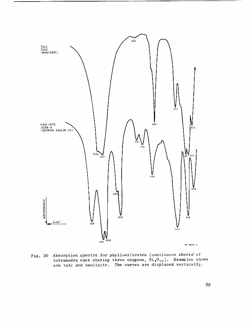
TALC
3262
(MONTSIER)
|
KAOLINITE
3058 -A
(GEORGIA KAOLIN
=o
Iv Cm "I
/
)4
1012 -
I1105
it
10061031
668
785
694
\
545
428
468
Fig. 30 Absorption spectra for phyllosilicates (continuous sheet_ of
tetrahedra each sharing three oxygens_ Si401o ). Examples shown
are talc and kaolinite. The curves are displaced vertically.
89

BIOTITE
3123
{ COLEMAN )
PARAGONITE
3048
(HEMLEY )
MUSCOVITE
3122
( COLEMAN )
770 660
449
MUSCOVITE
3098
( HEMLEY ) \
Fig. 31
994
924
422
467408
550
472
1054
1024 99_
_e 3943-13
Absorption spectra for the phyllosilicates biotite_ paragonite_
and muscovite. Note the effects of substitution in the micas.
The curves are displaced vertically.
9o
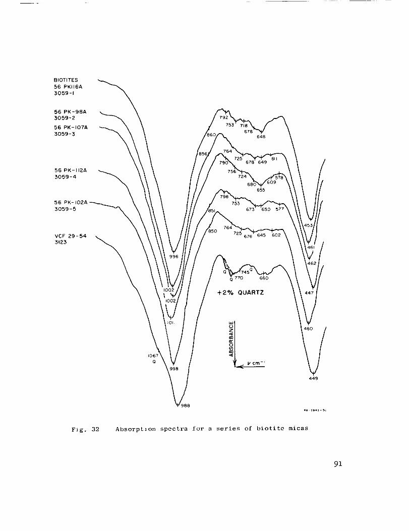
BIOTITES
56 PK116A
3059-1
56 PK-98A
3059-2
56 PK-107A
3059-3648
56 PK- 112A
3059-4
,56 PK-tO2A3059-5
VCF 29-54
3123
85O
649
724
655
753 6_
725 645 602
660
+2% QUARTZ
1067
(3
449
Fig. 32 Absorption spectra for a series of biotite micas
91
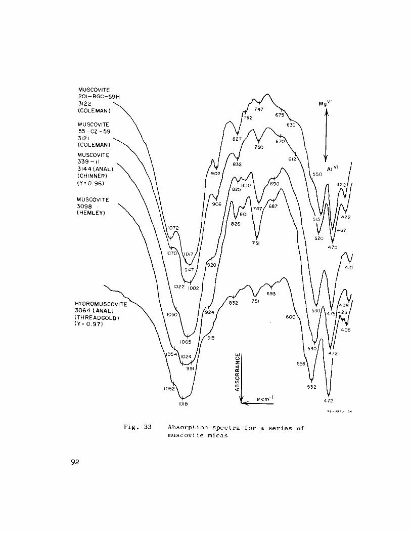
MUSCOVITE
201-RGC-59H
5122
(COLE MAN )
M U SCOVITE
55 - CZ - 59
5121
(COLEMAN)
MUSCOVITE
559 - II
5144 (ANAL) "_
(CHtNNER)
(Y=0.96)
MUSCOVITE
3098 '_
(HEMLEY)
ro72
747
750
751
Mg vf
IAI VI
55o
470
422
L67
410
HYDROMU_
,%_'2DNG%,\(Y = 0.97)_
Fig. 33
6OO
)15
t018
55E
Absorption spectra for a series of
muscovite micas
552
406
472
92
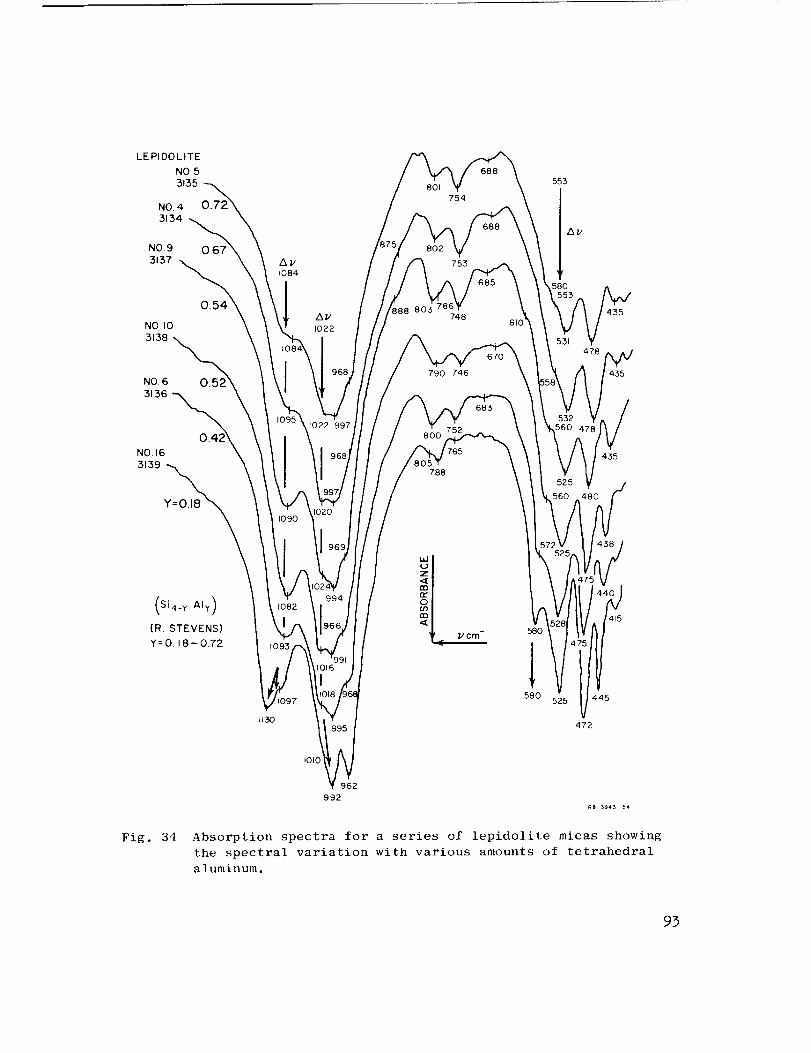
LEPIDOLITE
NO5
3135
NO. 4
3134NO.9
3137
NO. I0
3138
NO. 6
3136
NO,16
3139
Y:O.18
754
753
74861
790 746
788
553
58O
435
478
435
435
Si4_ Y AIy)
(R. STEVENS)
Y=O. 18- 0.72_cm-i 580
415
580 5 4'45
1130 472
Fig. 34
I010
962
992
Absorption spectra for a series of lepidolite micas showing
the spectral variation with various amounts of tetrahedral
aluminum.
93
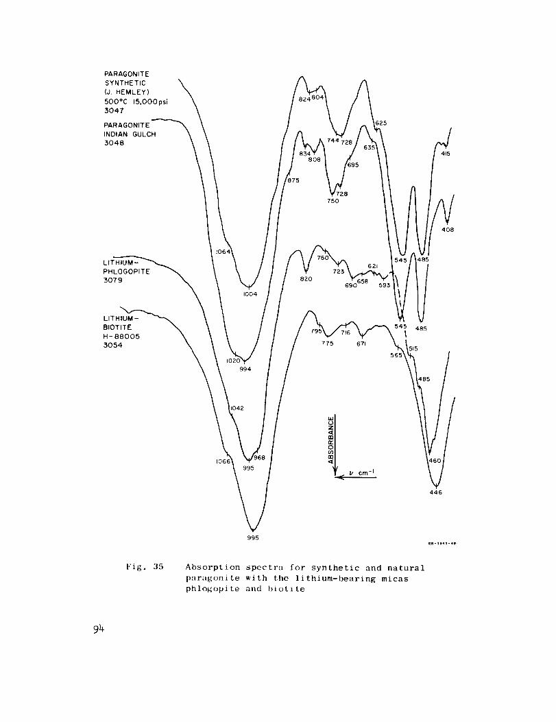
PARAGONITE
SYNTHETIC
(J. HEMLEY)
5000C 15,000 psi
3047
PARAGONITE
INDIAN GULCH
3048
975
728750
625
4O8
LITHIUM-
PHLOGOPITE
5079
994
1004
82O723
690
775 671
621485
j54_ 485
515
1066
446
995
Fig. 35 Absorption spectrn for synthetic and natural
paragonite with the lithium-bearing micas
phlogopite and biotite
94
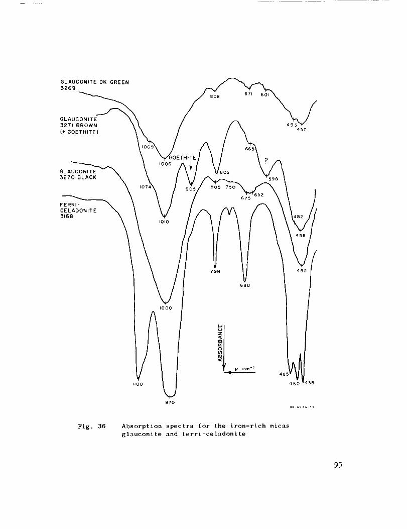
GLAUCONITE DK GREEN
3269
GLAUCONITE
3271 BROWN
(+ GOETHITE)
808
GLAUCONITE
3270 BLACK
FERRI-CELADONITE
3168
Io
1006
I010
805
805 750
798
675
680
I\\
IOOO
I_OO
cm-I
970
Fig. 36 Absorption spectra for the iron-rich micas
glauconite and ferri-celadonite
97

FUSED QUARTZ
695
QUARTZ
COESITE 1098
1078
121E 1036
STISHOVlTE
8O4
695
I
798 t79
794
683600
510
hI
49(l
[
448435
Fig. 37 Absorption spectra for polymorphs of Si02--quartz _ coesite_
stishovite, and fused silica. The curves are displaced
vertically.
96
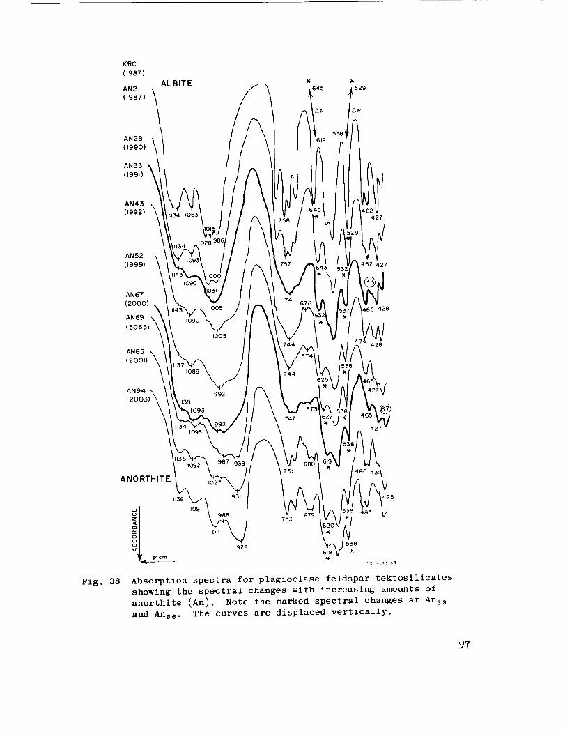
KRC
(1987)
AN2
(1987)
ALBITE
529
AN28
(1990)
AN33
(1991)
AN43 \(1992)
AN52(1999)
AN67
(2000)
AN69
(3065) x_
AN85
(2001)
AN94 \
(2003)'_k
744
747
427
462 427
465 429
428
©
ANORTHITE
Vcm I
Fig. 38
1136
1091
lOll
751
6_753
425
538929
619 -)(-
R8=4943 _8
Absorption spectra for plagioclase feldspar tektosilicates
showing the spectral changes with increasing amounts of
anorthite (An). Note the marked spectral changes at An33
and Ans6. The curves are displaced vertically.
97
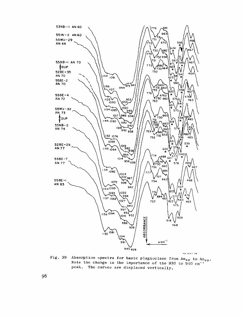
53NB-II AN 60
551N-2 AN60
55MU-29
AN 68 "__
52 BE- 35
AN 70 -" \
55BE-2 "_AN 70
55BE-4
AN 72
55MV-32
AN 73
IooP \55NB-2
AN 74
52BE-29
AN 77 _55BE -7
AN 77 -'_
55BE-I "_
AN 83 _._._.
)1107 994
1t44 1090
1019992 9
132 1076
1014956(
1072 9
942
750
763
754
757
62C-)(.
623'
621
538 465
98
Fig. 39
1135
929
945 929
539
568
_ 394_ 48
Absorption spectra for basic plagioclase £rom Anso to Arts3.
Note the change in the importance of the 930 to 940 cm -1
peak. The curves are displaced vertically.

BOEHMITE
c¢ - MONOHYDRATE
5176
GIBBSITE
(z- TRIHYDRATE
3177
(SHADED)
-TI_IHYDRATE
5178 \
)" ALUMINA
AI203
3299
1180
964
1021
799 749
660
555 515
410
420
450
flo
IV cm-I,(
Fig. 40
625
555
525
662 650 580
825 747
Absorption spectra for alumina ( y-A1203)
and its hydrates
99
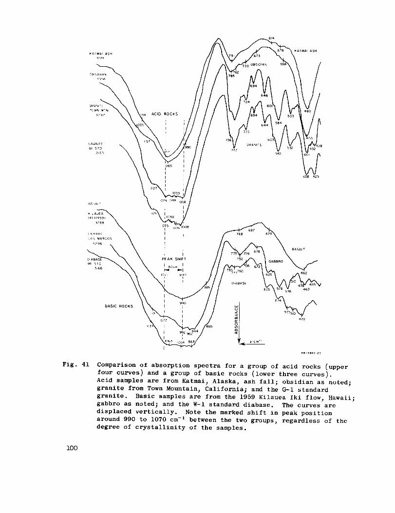
TOWN MTN
329; ACID ROCKS
1065I
I
I
I
I
777
6(
582
BASIC ROCKS
Fig. 41 Comparison of absorption spectra for a group of acid rocks (upper
four curves) and a group of basic rocks (lower three curves).
Acid samples are from Katmai, Alaska, ash fall; obsidian as noted;
granite from Town Mountain, California; and the G-I standard
granite. Basic samples are from the 1959 Kilauea Iki flow, Hawaii;
gabbro as noted; and the W-I standard diabase. The curves are
displaced vertically. Note the marked shift in peak position
around 990 to 1070 cm -I between the two groups, regardless of the
degree of crystallinity of the samples.
i00
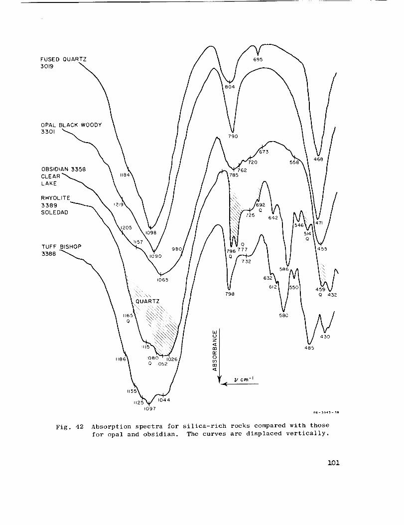
FUSED QUARTZ
3019
695
OPAL BLACK WOODY
3301790
OBSIDIAN 3358
LAKE
RHYOLITE
5589SOLEDAD
785
725 642
TUFF BISHOP
33881090
1065
QUARTZ
1115
io8o
Q 1052
10441125
1097
798
Q
752
61
58O
485
455
O 432
430
Fig. 42 Absorption spectra for silica-rich rocks compared with those
for opal and obsidian. The curves are displaced vertically.
lOl
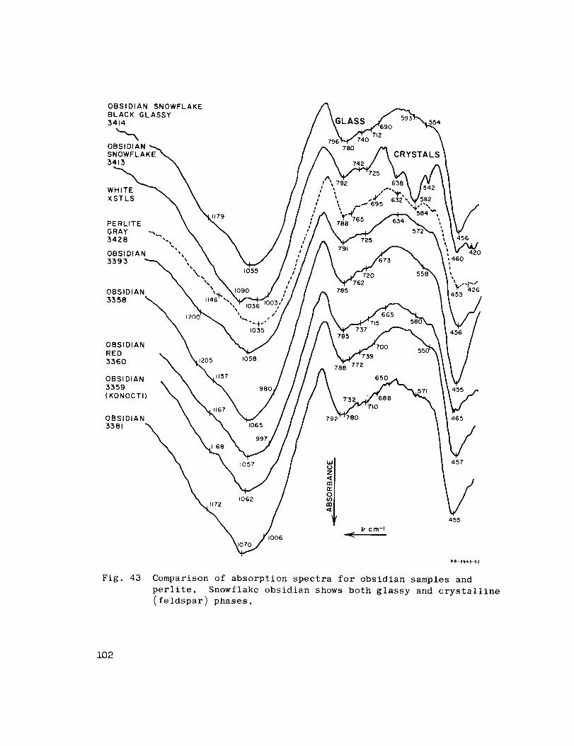
OBSIDIAN SNOWFLAKE
BLACK GLASSY5414
OBSIDIANSNOWFLAKE3413
XSTLS
PERLITE
GRAY -_,..,
3428 ,.\
OBSIDIAN "_
33 9 3 _
N,
OBSIDIAN3358
1179
OBSIDIAN
RED
3360
OBSIDIAN
3359(KONOCTI)
OBSIDIAN
338!
I055
1035
GLASS
712740
780
792 638
554
CRYSTALS
725 456
791 4201460L
720 I
785 _" 426
737785
1205 1058788 772
1157 0
980
1065780 465
I057 _t)
an
1062
457
455
Fig. 43
mB-3943-52
Comparison of absorption spectra for obsidian samples andperlite. Snowflake obsidian shows both glassy and crystalline
(feldspar) phases.
102

GRANITE
HARDHAT
3306
GRANITE
HARDHAT
3307
GRANITE
GI
3165
3297 _._
SYENITE
SY-I
3368
1162
1146
1132
10161085
1125
1127
1049
_128
1065
O8
778
P'50
725
'61
778
744
777
660
635
677 ,533 "Y,"
585514
582
584670
628
420462
Fig. 44 Absorption spectra for coarse-grained acid
rocks -- granite and syenite
z03

BASALT (HIGH GLASS)3298 (KILAUEA IKI 1959)
HE AVY
DENSITY
MINERALS IN
3298 (OLIVINE)
BASALT
3380
KILAUEA IKI
(OLD FLOW)
BASALT
PISGATH CRATER
3369
GLASS
990
98 ¢,976
922
884
748
CRYSTALS
597
621
424
499
408
531
46562
SERPENTINE
SAN LUIS OBISPO
3384
ll50 1057
1128
1085
987
578
_- FELDSPAR
996
964
I/ cm -I
Fig. 45 Absorption spectra for fine-grained basic
rocks -- basalts and serpentine
104
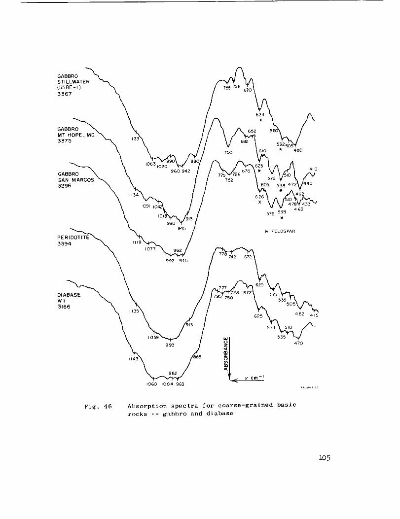
GABBROSTILLWATER
(55BE-I)
3367
1063
1091
960 942
990
750
752
624.)(-
652
610 480
605 538
462626
463576 538
Fig. 46 Absorption spectra for coarse-grained basic
rocks -- gabbro and diabase
109
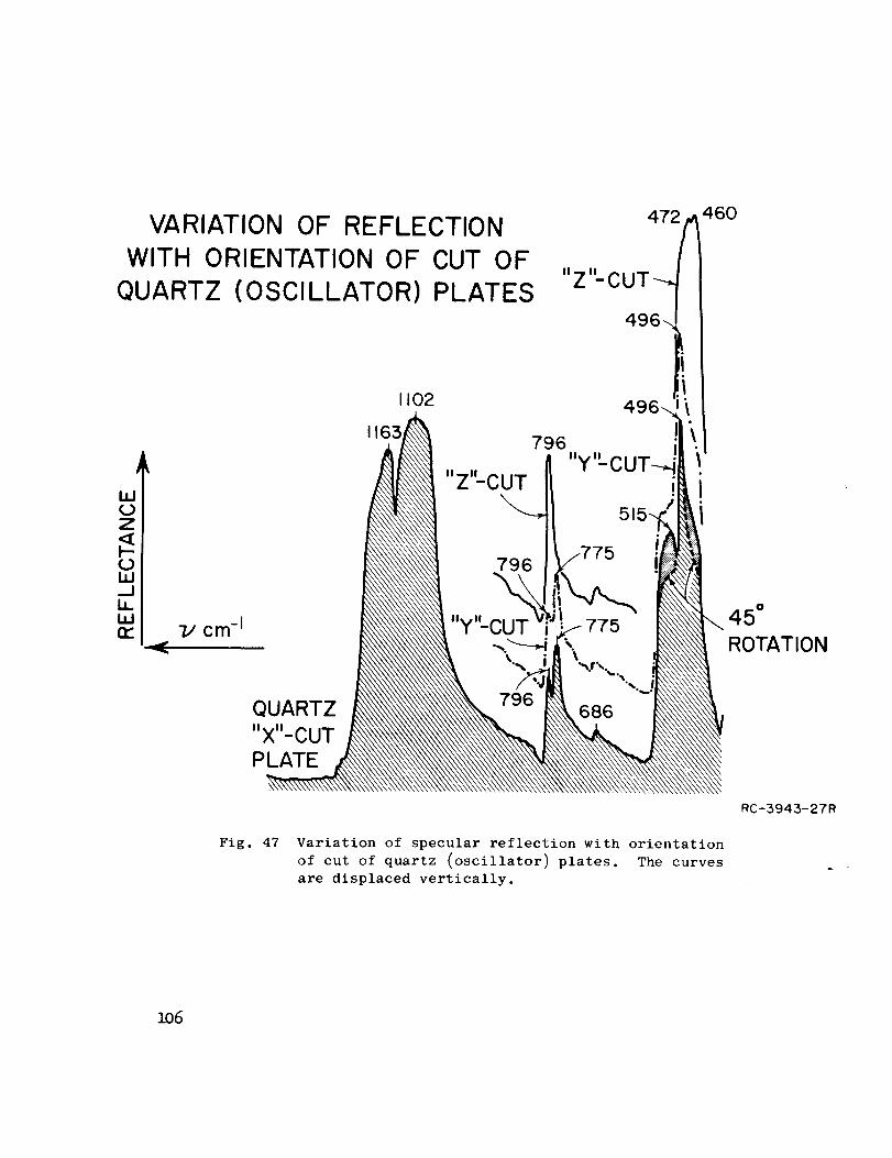
VARIATION OF REFLECTION
WITH ORIENTATION OF CUT OF
QUARTZ (OSCILLATOR) PLATES
472 460
UJ(..)Z
0
1i,I,Itt"
J
7/ cm -I
QUARTZ
PLATE
1102
1163
51
45"
ROTATION
Fig. 47 Variation of specular reflection with orientation
of cut of quartz (oscillator) plates. The curves
are displaced vertically.
RC-3943-27R
1o6

FUSED QUARTZ
CI-- CRISTOBALITE
Cl- TRIDYMITE
Q--QUARTZ
11611
L
_j
<I=,3n...b.lo j
G-QUARTZ Z-CUT j
o{- QUARTZ
X--,OR Y--CUT
i
797
I1( )_
797
688 (528)
ABSORPTION
REFLECTION
G--CRISTOBALITE
FUSED QUARTZRB-3943- 31
Fig. 48 Comparison of absorption and reflection spectra for various
silica modifications. The curves are displaced vertically.
The figure is redrawn from Figs. 1 and 2 of a paper by
Sevchenko and Florinskaya_ 1956. 37
i07
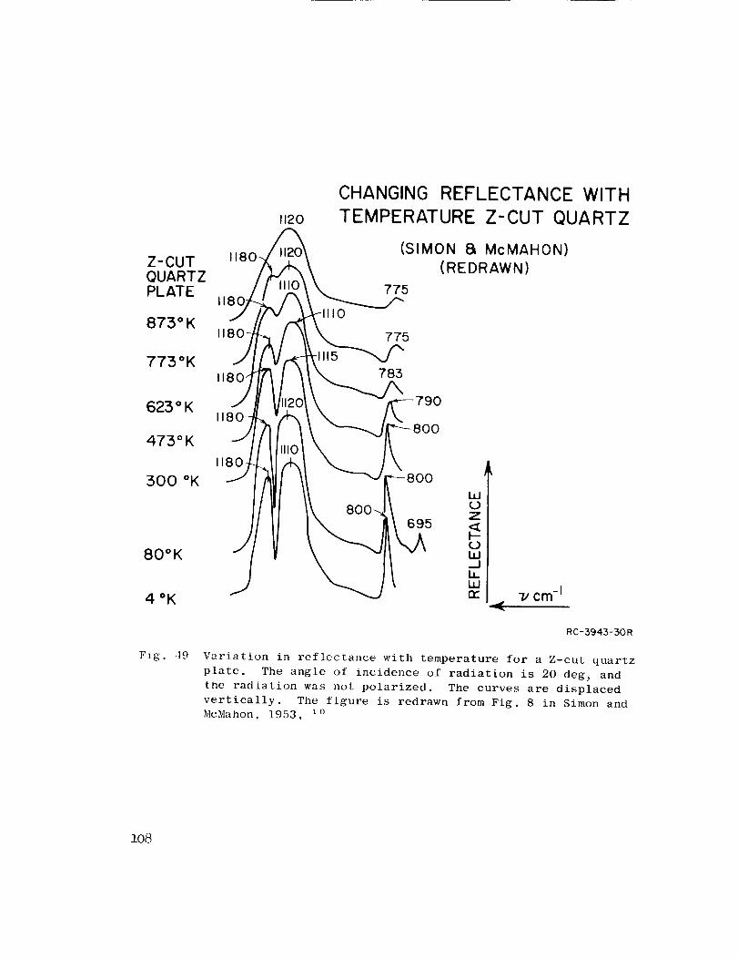
1120
CHANGING REFLECTANCE WITH
TEMPERATURE Z-CUT QUARTZ
Z-CUTQUARTZPLATE
873°KI0
(SIMON 8L McMAHON)
(REDRAWN)
775
I 775
773°K 783
1180_ _ 790623 ° K
I)0
473 ° K
300 °K
80°K
1180I00
Z
695
RC-3943-30R
Fig. ,19 Variation in reflectance with temperature for a Z-cut quartz
plate. The angle of incidence of radiation is 20 deg_ and
the radiation was not polarized. The curves are displaced
vertically. The figure is redrawn from Fig. 8 in Simon and
McMahon_ 1953. i0
:].o8
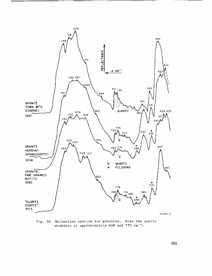
1t28
1166
1079
tl02 1067
I011
GRANITE
TOWN MTN
(COARSE)
3297
894
801 782 545
t079 ,22 ",,,UARTZc_ 5831122 1014
590
GRANITE
HARDHAT
(GRANODIORITE)
3306
GRANITE
FINE GRAINED
BIOTITIC
3382
"QUARTZ /
DIORITE"
3373 _
775
725
1125 930
,6o_ _ 6_! \ ,o2a,ooo /k
/ "- \ Q QUARTZ
* FELDSPAR
_o
795_\ 714 585 I
Fig. 50 Reflection spectra for granites. Note the quartzdoublets at approximately 800 and 775 cm -I
485
4.67
446 423
109
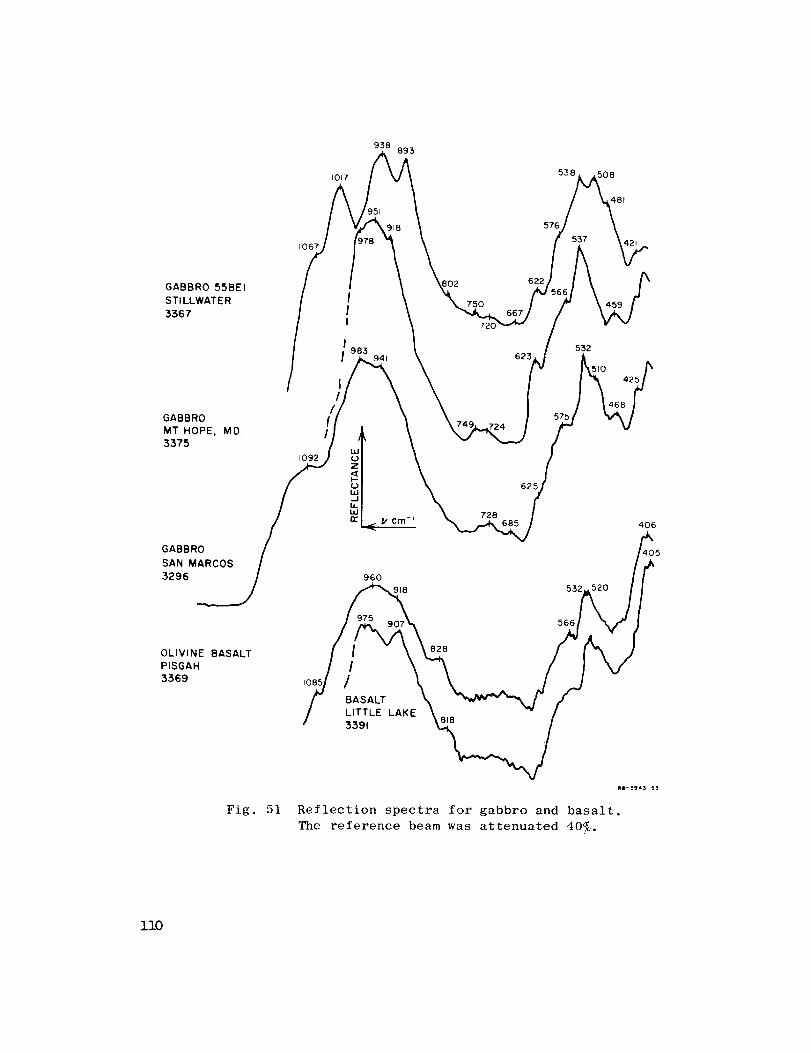
IO67
938893
GABBRO 55BEI 902
STI LLWATE R _ 750
t3367 I 720
GABBRO
MT HOPE, MD.
3375
GABBRO
SAN MARCOS
3296
OLIVINE BASALT
PISGAH
3.369
Fig. 51
538
576
,537
508
481
I983
532
510
1092 _'
625h
_. V Cm-I 728 406
960 " _
Reflection spectra for gabbro and basalt.The reference beam was attenuated 40_.
ii0

GRANITE HARDHAT
"- -_"'; / I /--' _,A533
_\.... /.,,(r94_ i.'i._4_e
\,,46_9_ (sei))'i" % _0_4#.L_046 " (5_I)iY_ \ ,,W-_o81 " i
_ (1160)\ Jr ',
I LABRADORITE '-t,.,f"il-'O00) i ;
L3244 (,,o2)(,o67) (46;;,,,/% A7,X...,_x 670 (450)
,. _.,,,,-,o, II (655)620_y i/ANORTHOSITE/ \\'"/Y.,_' /[ -* _575;
L5586 \ " \ ' _ '_' 'IIill iii
7:?. ( rs)'_ "_ (993)I-- 13Bf.3 n,-1.1.1j 01.1_ or)ILl fnrr ,,_
"V cm -1,_' RC-3943-29R
Fig. 52 Comparison between absorption (solid line) and reflection
(dashed line) spectra for (a) granite from the Nevada Test
Site_ Mercury_ Nevada_ and (b) anorthosite and labradorite.
The anorthosite is from the San Gabriel mountains_ California.
The reflection spectra have been inverted for comparison.
iii
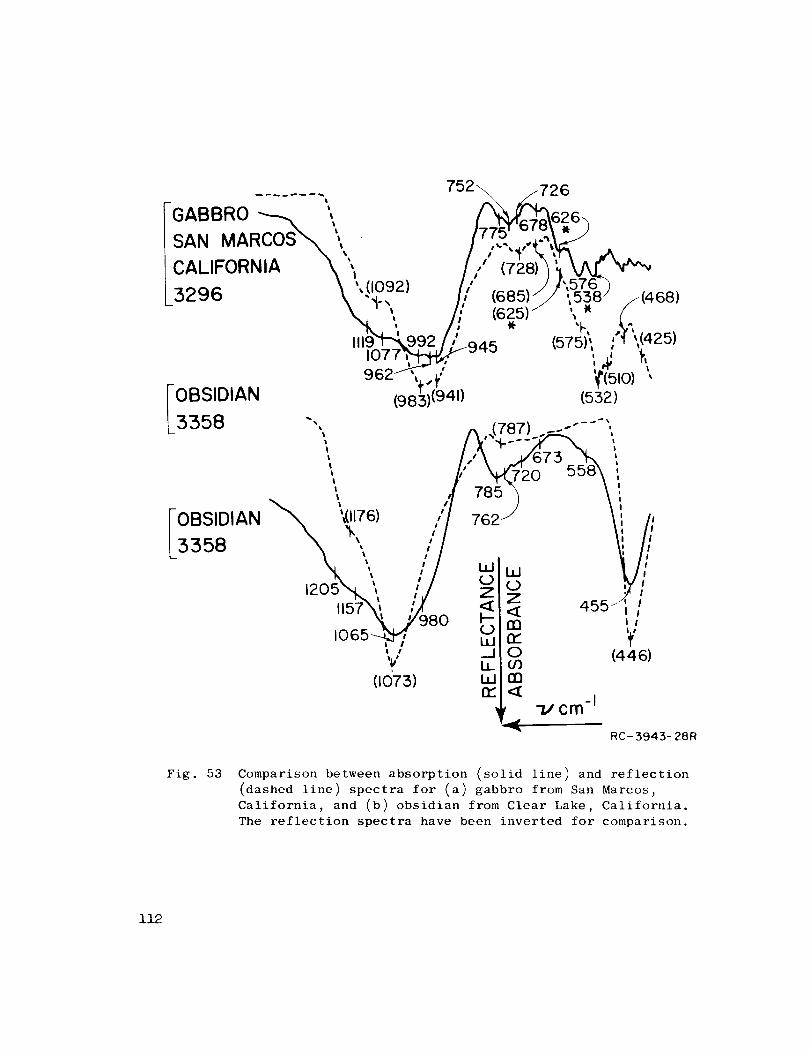
"l-077_ _'''' ..... ii "I':
_ ",.._ W,,o)',FOBSIDIAN (9815)(941) (552)
L3358 "', ,,,,(787) .--"'--" _
,,,,, t,-
_. _,(1176) I/ 762 / Ii /,IOBSIDIAN \ ', /1 ,v., ) \
' I 0 0'205N"t ' ,./ _ _ ',_I
--/is_',. '7 _ z 4_5--",7,:-o'_9_o b _1065 , / i..,i ri." 'if'
(440)0or)m
L
,e --IY ,,
(1073) I.LIn.-
RC-3943-28R
Fig. 53 Comparison between absorption (solid line) and reflection
(dashed line) spectra for (a) gabbro from San Marcos,
California_ and (b) obsidian from Clear Lake_ California.
The reflection spectra have been inverted for comparison.
112
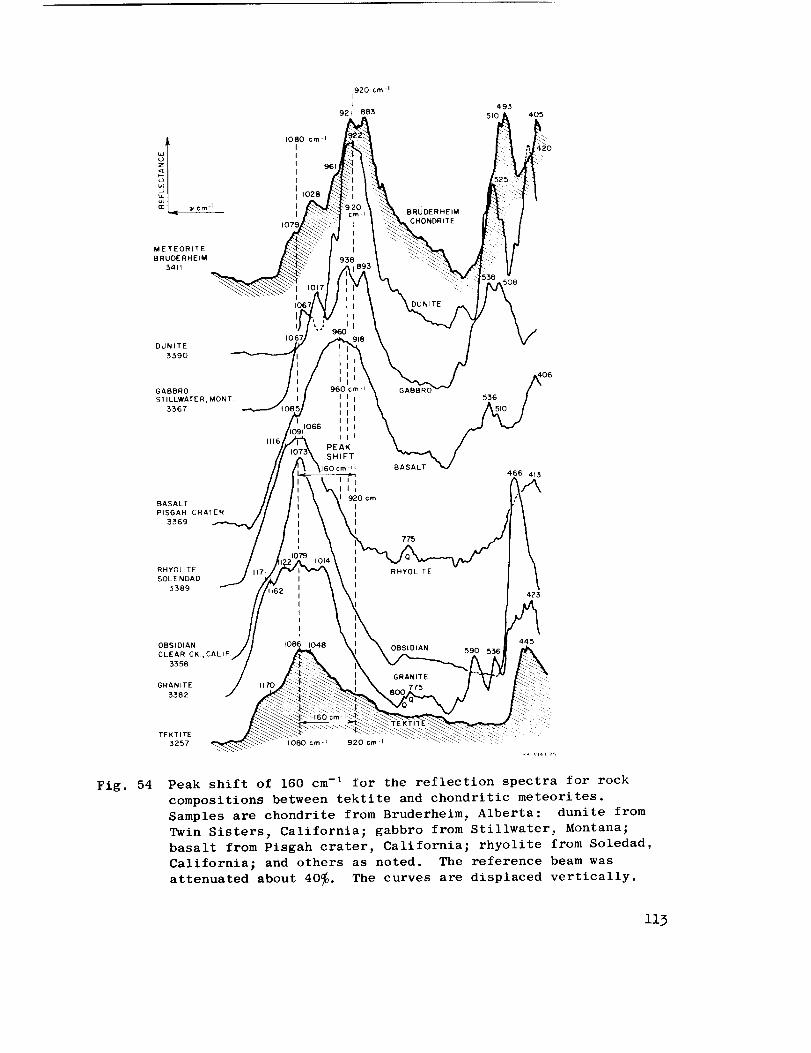
METEORITE
BRUDERHEtM
3411
1080
920 cm -II
I
921 883
106"
BRU[
CHONDRITE
.... 5oe
493
510 405
DUNITE
3390
GABBRO
STILLWATER, MONT
3367
BASALT
PISGAH CRATER
3369
RHYOLITE
SOLENDAD
3389
1116
106_
06960 cm-_
I I I 536 ]
,",,
I I I
1066 I I II
PEAK
SHIFT
;Ocrn I BASALT466 4{3
I I
II
cml
775
RHYOLITE
423
Fig. 54
OBSIDIAN
CLEAR CK,, CALIF,
3358
GRANITE
3382
GRANITE
775
590 556
TEKTITE
3257 1080cm-= 920 cm-I
Peak shift of 160 cm -I for the reflection spectra for rock
compositions between tektite and chondritic meteorites.
Samples are chondrite from Bruderheim, Alberta: dunite from
Twin Sisters, California; gabbro from Stillwater, Montana;
basalt from Pisgah crater, California; rhyolite from Soledad,
California; and others as noted. The reference beam was
attenuated about 40_. The curves are displaced vertically.
if3
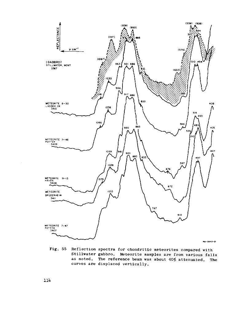
( GABBRO )
STILLWATER, MONT
3367
(I067
(1017)
(938)
(576)
(622
(536) (508)
METEORITE 8-30
LADDER CR
54101036
833408
1086596
920 883 405
METEORITE 7--49
POTTER3409
METEORITE 19-13HAVEN
3408
METEORITE
BRODERHEIM
3411
IOZ9 407
1033
676
672
497
597
767
6t0
METEORITE 7-47
POTTER
3407
Fig. 55
A0--3,45--21
Reflection spectra for chondritic meteorites compared with
Stillwater gabbro. Meteorite samples are from various falls
as noted. The reference beam was about 40_ attenuated. The
curves are displaced vertically.
if4
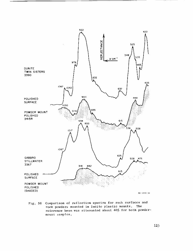
DUNITE
TWIN SISTERS
:3390
POLISHED
SU RFACE
POWDER MOUNT
POLISHED
3415R
GABBRO
STI LLWATER
3367
POLISHED
SURFACE
POWDER MOUNT
POLISHED
(SHADED)
io67
t050
1067
978
I017
922
900
632
938 615
893
918 882
625
615
548
525
498
508
508 475
420
4O5
/
Fig. 56 Comparison of reflection spectra for rock surfaces and
rock powders mounted in lucite plastic mounts. The
reference beam was attenuated about 40_ for both powder-
mount samples.
I15
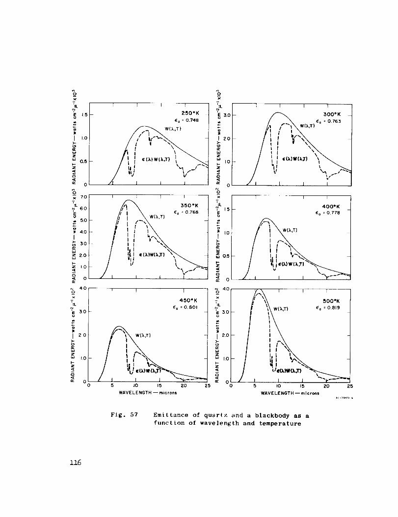
4.5-w
o
I Lo>-
nrWZ
"' 0.5p-Z
<0c 0 _
x 70T
V 60
,, 5.0-
40
I3,0
_ew
z 2.0
F-
z i.O
Q< 0_e
40 I
_ox
'_ 3.0
ie
I 2.0>-
fw,,,JZ
W t.O
I-Z
00
I I I
250"K -
_a : 0.748
t _) V'-l I I
I I I I
550= KE a : 0.768 -
/ I t 1 1
x
fw
i 't zoH
>-
n-wzw
h-z
_c oL
! I I ]
450" K
E a : 0.801
I
I 2o>-(.9n_WZ
UJ 1,0'
Z
0
5 " O0
WAVE LE NGTH -- microns
1 I 1 I
300° K _Ea : 0.763
:,t ,oo.
i,., 0.5 I
i0/_o 4.0 " I I I/,-.Xx II"_ 5ooK
I } E a : 0.819
'_ESO
5 IO 15 20 25
WAVELENGTH -- microns
Fig. 57 Emittance of quartz and a blackbody as a
function of wavelength and temperature
ii6
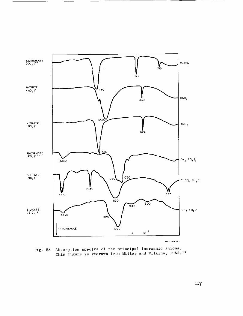
NITRITE
(NOz)-
NITRATE
(NO3)-
PHOSPHATE
(P04)
SULFATE
(S04)--
SILICATE
( SiOx)Y-
Fig. 58
715
877
83O
824
3230
380
I08(
1650
34t0 667
3330 H90_
ABSORBANCE 1090-I
,gD_ ¢ m
RA- 3943-5
Absorption spectra of the principal inorganic anions.
This figure is redrawn from Miller and Wilkins_ 1952. 18
CoCO 3
KNO 2
KNO 3
C° 3 (P04)2
CoSO4'2H20
SiO2.XH20
ii7
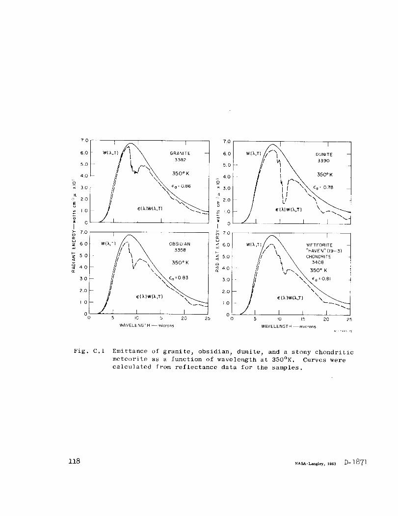
70
6,0 -
50
40 -
× 30
_'_ 2.0Eu
_ I.o
_ oI
I_ r TwC_.TIfX \ _AN,TE --
J f I i I _
_ Z.OL9
_ 6.0
_ 50
_ 4.0
3.0
2.0
I0
00
-- W(X,_) _ OBSIDIAN
.5 I0 15 20 25
WAVELENGTH --microns
7.0
6.0
5.0
4.0
× 3.O1
::L
'_ 2.0o
"' 1.0
I o
r _ r rW(X,T)//" _'i _ OUNITE --
_ 70
z 6,0
i /_ r rLU W( k,T)//_\ _ METEORITE -
_- t // \ _ "HAVEN" (19-13)
._ 5.0l /,,/ _ _ CHONDRITE
_4o // _ \ 34oB// 3so°K
2.0
E(XIW(X,
--"'..,'/'I I t J ""O'
o 5 to 15 20 25
WAVELENGTH --microns
Fi E . C. i Emittance of granite_ obsidian_ dunite, and a stony chondritic
meteorite as a function of wavelength at 350°K. Curves were
calculated from reflectance data for the samples.
118 NASA-Langley, 1903 D-1871


Related Documents