JOURNAL OF CLINICAL MICROBIOLOGY, 0095-1137/01/$04.000 DOI: 10.1128/JCM.39.10.3427–3436.2001 Oct. 2001, p. 3427–3436 Vol. 39, No. 10 Copyright © 2001, American Society for Microbiology. All Rights Reserved. MINIREVIEW Taxonomy and Identification of the Burkholderia cepacia Complex TOM COENYE, 1 * PETER VANDAMME, 2 JOHN R. W. GOVAN, 3 AND JOHN J. LIPUMA 1 Department of Pediatrics and Communicable Diseases, University of Michigan Medical School, Ann Arbor, Michigan 1 ; Laboratory of Pharmaceutical Microbiology, Ghent University, Ghent, Belgium 2 ; and Department of Medical Microbiology, University of Edinburgh, Edinburgh, Scotland, United Kingdom 3 At the beginning of this review it is essential to clarify the terminology that will be used to refer to the members of the Burkholderia cepacia complex and their relatives. The name B. cepacia will relate only to B. cepacia genomovar I. Strains resembling B. cepacia may belong to the B. cepacia complex, to other Burkholderia species (for instance, Burkholderia gladioli), or to species from other genera (for instance, Ralstonia pick- ettii) that share some phenotypic or genotypic similarities with the B. cepacia complex. B. cepacia complex bacteria and or- ganisms that may be confused with them will be altogether referred to as B. cepacia-like organisms. Most previous reports regarding these organisms were published before the recogni- tion of the complicated taxonomic relationships between the different members of the B. cepacia complex; it is therefore unclear to what category the presumed B. cepacia isolates described would belong. For that reason, when such literature is cited, the name “B. cepacia” will be shown in double quotes. Chronic microbial colonization of the major airways, leading to exacerbations of pulmonary infection, is the major cause of morbidity and mortality in patients with cystic fibrosis (CF). Typical CF pathogens include Staphylococcus aureus, Pseudo- monas aeruginosa, and Haemophilus influenzae (30). Other glu- cose nonfermenters, like Stenotrophomonas maltophilia, Alcali- genes xylosoxidans, R. pickettii, and Burkholderia gladioli, can frequently be found as well, but their role in the decline of pulmonary function is unclear (14, 19, 30). Several reports on the recovery of “B. cepacia” from CF patients appeared in the late 1970s and early 1980s (62, 63). The first detailed descrip- tion of the clinical significance of “B. cepacia” colonization and infection was published in 1984 (47). In that seminal paper, Isles et al. documented the increasing prevalence of “B. cepa- cia” colonization and infection in the Toronto, Canada, CF treatment center and described the so-called “cepacia syn- drome,” a severe progressive respiratory failure with bactere- mia that occurs in about 20% of all infected CF patients. Clustering of new cases in some centers and the decrease of colonization of new patients following segregation of colonized and noncolonized patients in other centers suggested that “B. cepacia” could be transmitted between CF patients. This was confirmed by several studies (34, 64, 67, 76, 84, 94) that showed that “B. cepacia” strains can spread between CF patients via simultaneous hospital admissions or social contact outside of the hospital. As a result of these findings, new guidelines were issued to reduce the risk of “B. cepacia” acquisition. These included discontinuing sponsorship and support of CF summer camps and segregation of colonized patients. Implementation of these draconian infection control measures has a tremen- dous impact on the lives of CF patients, and not all patients or caregivers accept such measures (35, 36, 62, 63). “B. cepacia” can also cause lung infections in chronic gran- ulomatous disease patients, and infections in these patients are associated with pneumonia and septicemia and are often lethal (2, 58, 72, 96). “B. cepacia” infections in immunocompetent patients occur only sporadically, but several cases of pseudo- epidemics and nosocomial infections, often caused by contam- inated disinfectants and anesthetic solutions, have been re- ported (3, 43, 50, 107). Despite the advances that have been made in the under- standing of the epidemiology, “B. cepacia” infections still have a considerable impact on morbidity and mortality in CF pa- tients (18, 61, 62, 63). Since “B. cepacia” is resistant to most antimicrobial agents, effective therapies are not straightfor- ward and management efforts are therefore aimed at preven- tion of infection (35, 63). Several recommendations regarding infection control measures have been made, and these include that CF patients should not share hospital rooms as inpatients and should limit contact in outpatient clinics (63). However, the efficiency of infection control measures are determined by the accuracy with which “B. cepacia” is diagnosed, and poor laboratory proficiency in identification of this organism still prevails (17, 40, 75). Although several guidelines intended to enhance accurate identification of bacterial species from spu- tum culture have been proposed by national CF organizations and by the International Burkholderia cepacia Working Group, the degree to which these are followed varies greatly among clinical microbiology laboratories (90). The problem is given an extra dimension by the fact that several “B. cepacia” strains have attracted attention as antag- onists of soilborne plant pathogens (44, 66) and as plant- growth-promoting agents that can colonize the rhizosphere of several economic crops and thereby increase the crop yield (9, 39, 74, 83). The exceptional metabolic diversity of this organ- ism (which allows it to use, e.g., constituents of crude oils and herbicides as carbon sources) could be put to use in the biore- mediation of recalcitrant xenobiotics (8, 28, 54, 57). However, most strains used or under development for biocontrol or * Corresponding author. Mailing address: Department of Pediatrics and Communicable Diseases, 8301 MSRB III, Box 0646, 1150 W. Med. Ctr. Dr., Ann Arbor, MI 48109-0646. Phone: (734) 936-9767. Fax: (734) 615-4770. E-mail: [email protected] or tomcoenye@hotmail .com. 3427

Welcome message from author
This document is posted to help you gain knowledge. Please leave a comment to let me know what you think about it! Share it to your friends and learn new things together.
Transcript

JOURNAL OF CLINICAL MICROBIOLOGY,0095-1137/01/$04.00�0 DOI: 10.1128/JCM.39.10.3427–3436.2001
Oct. 2001, p. 3427–3436 Vol. 39, No. 10
Copyright © 2001, American Society for Microbiology. All Rights Reserved.
MINIREVIEW
Taxonomy and Identification of the Burkholderia cepacia ComplexTOM COENYE,1* PETER VANDAMME,2 JOHN R. W. GOVAN,3 AND JOHN J. LIPUMA1
Department of Pediatrics and Communicable Diseases, University of Michigan Medical School, Ann Arbor, Michigan1;Laboratory of Pharmaceutical Microbiology, Ghent University, Ghent, Belgium2; and Department of
Medical Microbiology, University of Edinburgh, Edinburgh, Scotland, United Kingdom3
At the beginning of this review it is essential to clarify theterminology that will be used to refer to the members of theBurkholderia cepacia complex and their relatives. The name B.cepacia will relate only to B. cepacia genomovar I. Strainsresembling B. cepacia may belong to the B. cepacia complex, toother Burkholderia species (for instance, Burkholderia gladioli),or to species from other genera (for instance, Ralstonia pick-ettii) that share some phenotypic or genotypic similarities withthe B. cepacia complex. B. cepacia complex bacteria and or-ganisms that may be confused with them will be altogetherreferred to as B. cepacia-like organisms. Most previous reportsregarding these organisms were published before the recogni-tion of the complicated taxonomic relationships between thedifferent members of the B. cepacia complex; it is thereforeunclear to what category the presumed B. cepacia isolatesdescribed would belong. For that reason, when such literatureis cited, the name “B. cepacia” will be shown in double quotes.
Chronic microbial colonization of the major airways, leadingto exacerbations of pulmonary infection, is the major cause ofmorbidity and mortality in patients with cystic fibrosis (CF).Typical CF pathogens include Staphylococcus aureus, Pseudo-monas aeruginosa, and Haemophilus influenzae (30). Other glu-cose nonfermenters, like Stenotrophomonas maltophilia, Alcali-genes xylosoxidans, R. pickettii, and Burkholderia gladioli, canfrequently be found as well, but their role in the decline ofpulmonary function is unclear (14, 19, 30). Several reports onthe recovery of “B. cepacia” from CF patients appeared in thelate 1970s and early 1980s (62, 63). The first detailed descrip-tion of the clinical significance of “B. cepacia” colonization andinfection was published in 1984 (47). In that seminal paper,Isles et al. documented the increasing prevalence of “B. cepa-cia” colonization and infection in the Toronto, Canada, CFtreatment center and described the so-called “cepacia syn-drome,” a severe progressive respiratory failure with bactere-mia that occurs in about 20% of all infected CF patients.Clustering of new cases in some centers and the decrease ofcolonization of new patients following segregation of colonizedand noncolonized patients in other centers suggested that “B.cepacia” could be transmitted between CF patients. This wasconfirmed by several studies (34, 64, 67, 76, 84, 94) that showed
that “B. cepacia” strains can spread between CF patients viasimultaneous hospital admissions or social contact outside ofthe hospital. As a result of these findings, new guidelines wereissued to reduce the risk of “B. cepacia” acquisition. Theseincluded discontinuing sponsorship and support of CF summercamps and segregation of colonized patients. Implementationof these draconian infection control measures has a tremen-dous impact on the lives of CF patients, and not all patients orcaregivers accept such measures (35, 36, 62, 63).
“B. cepacia” can also cause lung infections in chronic gran-ulomatous disease patients, and infections in these patients areassociated with pneumonia and septicemia and are often lethal(2, 58, 72, 96). “B. cepacia” infections in immunocompetentpatients occur only sporadically, but several cases of pseudo-epidemics and nosocomial infections, often caused by contam-inated disinfectants and anesthetic solutions, have been re-ported (3, 43, 50, 107).
Despite the advances that have been made in the under-standing of the epidemiology, “B. cepacia” infections still havea considerable impact on morbidity and mortality in CF pa-tients (18, 61, 62, 63). Since “B. cepacia” is resistant to mostantimicrobial agents, effective therapies are not straightfor-ward and management efforts are therefore aimed at preven-tion of infection (35, 63). Several recommendations regardinginfection control measures have been made, and these includethat CF patients should not share hospital rooms as inpatientsand should limit contact in outpatient clinics (63). However,the efficiency of infection control measures are determined bythe accuracy with which “B. cepacia” is diagnosed, and poorlaboratory proficiency in identification of this organism stillprevails (17, 40, 75). Although several guidelines intended toenhance accurate identification of bacterial species from spu-tum culture have been proposed by national CF organizationsand by the International Burkholderia cepacia Working Group,the degree to which these are followed varies greatly amongclinical microbiology laboratories (90).
The problem is given an extra dimension by the fact thatseveral “B. cepacia” strains have attracted attention as antag-onists of soilborne plant pathogens (44, 66) and as plant-growth-promoting agents that can colonize the rhizosphere ofseveral economic crops and thereby increase the crop yield (9,39, 74, 83). The exceptional metabolic diversity of this organ-ism (which allows it to use, e.g., constituents of crude oils andherbicides as carbon sources) could be put to use in the biore-mediation of recalcitrant xenobiotics (8, 28, 54, 57). However,most strains used or under development for biocontrol or
* Corresponding author. Mailing address: Department of Pediatricsand Communicable Diseases, 8301 MSRB III, Box 0646, 1150 W. Med.Ctr. Dr., Ann Arbor, MI 48109-0646. Phone: (734) 936-9767. Fax:(734) 615-4770. E-mail: [email protected] or [email protected].
3427

bioremediation purposes are taxonomically poorly character-ized, and their potential hazard to the CF community is un-clear (33, 37, 44, 110).
The taxonomic complexity of B. cepacia-like organisms andthe lack of widespread and generally accepted identificationschemes hinder sound studies that could establish the rolesplayed by and the pathogenic significance of the different B.cepacia-like organisms. This information is crucial to proposescientifically founded policies for each of the above-mentionedproblems. The purpose of this review is to present an overviewboth of the taxonomy of the B. cepacia complex and of theavailable phenotypic and genotypic methods aimed at the cor-rect identification of these organisms.
TAXONOMY OF THE B. CEPACIA COMPLEX
Pseudomonas cepacia was originally described by Burk-holder in 1950 as the causative agent of bacterial rot of onionbulbs (13). Other names that were assigned included eugonicoxidizers group 1, Pseudomonas kingii, and Pseudomonas mul-tivorans (49, 77, 97), but several studies clearly showed thatthese could be considered as synonymous names of P. cepaciaand that the name P. cepacia had priority (5, 86, 92, 95). Thename P. cepacia was not included in the Approved List ofBacterial Names (93) and therefore lost standing in bacterial
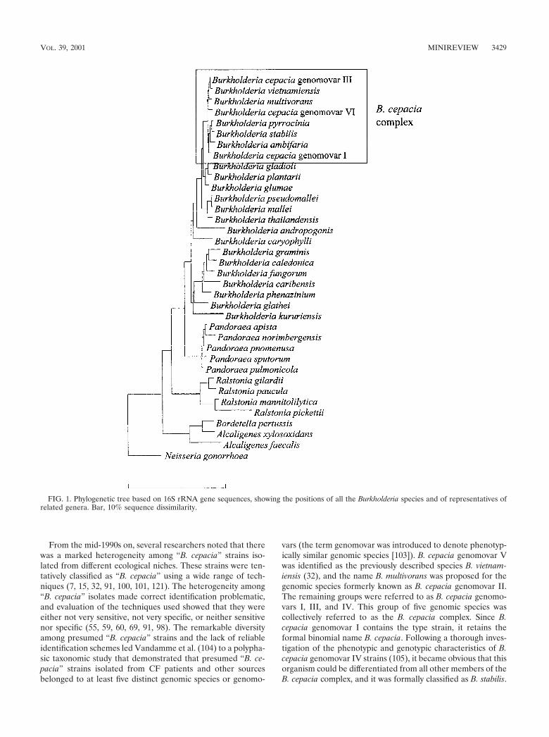
nomenclature until 1981, when it was revived by Palleroni andHolmes (81). In 1992, P. cepacia and six other species belong-ing to rRNA group II of the genus Pseudomonas (Pseudomo-nas solanacearum, Pseudomonas pickettii, Pseudomonas gladi-oli, Pseudomonas mallei, Pseudomonas pseudomallei, andPseudomonas caryophylli) (82) were transferred to the newgenus Burkholderia (119). In contrast to the genus Pseudomo-nas, the genus Burkholderia belongs to the �-subdivision of thephylum Proteobacteria (53). Since the genus name was firstassigned, the taxonomy of the genus Burkholderia has under-gone considerable changes (Table 1), and the genus now in-cludes 22 validly described species: B. cepacia (the type species),Burkholderia caryophylli, Burkholderia mallei, Burkholderiapseudomallei, Burkholderia gladioli, Burkholderia plantarii, Burk-holderia glumae, Burkholderia vietnamiensis, Burkholderia andro-pogonis, Burkholderia multivorans, Burkholderia glathei, Burkhold-eria pyrrocinia, Burkholderia thailandensis, Burkholderia graminis,Burkholderia phenazinium, Burkholderia caribensis, Burkholderiakururiensis, Burkholderia ubonensis, Burkholderia caledonica,Burkholderia fungorum, Burkholderia stabilis, and Burkholderiaambifaria (1, 10, 20, 22, 23, 24, 25, 32, 102, 104, 105, 109, 117,118, 119, 120, 122, 123). A phylogenetic tree based on 16SrRNA gene sequences, showing the positions of all the Burk-holderia species and representatives of related genera, is shownin Fig. 1.
TABLE 1. Overview of the genus Burkholderiaa
Species name originallyassignedb
Burkholderia species name ortaxon assigned
Yr ofassignment Reference Other name
subsequently assignedYr of
assignment Reference
Pseudomonas cepacia B. cepacia comb. nov. (B.cepacia genomovar I)
1992 81, 104, 119
Pseudomonas solanacearum B. solanacearum comb. nov. 1992 119 Ralstonia solanacearum comb. nov. 1995 120Pseudomonas pickettii B. picketti comb. nov. 1992 119 Ralstonia pickettii comb. nov. 1995 120Pseudomonas gladioli B. gladioli comb. nov. 1992 119Pseudomonas mallei B. mallei comb. nov. 1992 119Pseudomonas pseudomallei B. pseudomallei comb. nov. 1992 119Pseudomonas caryophylli B. caryophylli comb. nov. 1992 119Pseudomonas plantarii B. plantarii comb. nov. 1994 102Pseudomonas glumae B. glumae comb. nov. 1994 102
B. vandii sp. nov. 1994 102 Junior synonym of B. plantarii 1999 22B. vietnamiensis sp. nov. (B.
cepacia genomovar V)1995 32, 104
Pseudomonas cocovenenans B. cocovenenans comb. nov. 1995 123 Junior synonym of B. gladioli 1999 22Pseudomonas andropogonis B. andropogonis comb. nov. 1995 32
B. multivorans sp. nov. (B.cepacia genomovar II)
1997 104
Pseudomonas glathei B. glathei comb. nov. 1997 104Pseudomonas pyrrocinia B. pyrrocinia comb. nov. 1997 4, 104
B. thailandensis sp. nov. 1998 10B. graminis sp. nov. 1998 109
Pseudomonas phenazinium B. phenazinium comb. nov. 1998 109B. norimbergensis sp. nov. 1998 117 Pandoraea norimbergensis comb. nov. 2000 20B. caribensis sp. nov. 1999 1B. stabilis sp. nov. (B.
cepacia genomovar IV)2000 104, 105
B. kururiensis sp. nov. 2000 12B. ubonensis sp. nov. 2000 118B. fungorum sp. nov. 2001 23B. caledonica sp. nov. 2001 23B. ambifaria sp. nov. (B.
cepacia genomovar VII)2001 25
B. cepacia genomovar III 1997 104B. cepacia genomovar VI 2001 24
a Members of the B. cepacia complex are in boldface type.
3428 MINIREVIEW J. CLIN. MICROBIOL.
From the mid-1990s on, several researchers noted that therewas a marked heterogeneity among “B. cepacia” strains iso-lated from different ecological niches. These strains were ten-tatively classified as “B. cepacia” using a wide range of tech-niques (7, 15, 32, 91, 100, 101, 121). The heterogeneity among“B. cepacia” isolates made correct identification problematic,and evaluation of the techniques used showed that they wereeither not very sensitive, not very specific, or neither sensitivenor specific (55, 59, 60, 69, 91, 98). The remarkable diversityamong presumed “B. cepacia” strains and the lack of reliableidentification schemes led Vandamme et al. (104) to a polypha-sic taxonomic study that demonstrated that presumed “B. ce-pacia” strains isolated from CF patients and other sourcesbelonged to at least five distinct genomic species or genomo-
vars (the term genomovar was introduced to denote phenotyp-ically similar genomic species [103]). B. cepacia genomovar Vwas identified as the previously described species B. vietnam-iensis (32), and the name B. multivorans was proposed for thegenomic species formerly known as B. cepacia genomovar II.The remaining groups were referred to as B. cepacia genomo-vars I, III, and IV. This group of five genomic species wascollectively referred to as the B. cepacia complex. Since B.cepacia genomovar I contains the type strain, it retains theformal binomial name B. cepacia. Following a thorough inves-tigation of the phenotypic and genotypic characteristics of B.cepacia genomovar IV strains (105), it became obvious that thisorganism could be differentiated from all other members of theB. cepacia complex, and it was formally classified as B. stabilis.
FIG. 1. Phylogenetic tree based on 16S rRNA gene sequences, showing the positions of all the Burkholderia species and of representatives ofrelated genera. Bar, 10% sequence dissimilarity.
VOL. 39, 2001 MINIREVIEW 3429
Subsequent polyphasic taxonomic studies identified two moremembers of the B. cepacia complex (24, 25). B. cepacia geno-movar VI contains strains isolated from CF patients in theUnited States and the United Kingdom. This organism canphenotypically be differentiated from all members of the B.cepacia complex except B. multivorans. The name B. ambifaria(B. cepacia genomovar VII) was proposed for isolates fromhuman clinical and environmental specimens, including CFpatients. B. ambifaria also contains several well-characterizedbiocontrol strains. In addition, it was recently shown that thespecies B. pyrrocinia also belongs to the B. cepacia complex (4).
Within the B. cepacia complex, representatives of differentspecies generally have DNA-DNA hybridization values be-tween 30 and 60%, while values obtained from strains belong-ing to the same species are generally higher than 70%. DNA-DNA binding values obtained with other Burkholderia speciesare generally below 30% (22, 24, 25, 26, 32, 104). These valuescorrespond to the three categories described in reference 106:high DNA relatedness (70% or higher) between strains of asingle species, low but significant DNA relatedness below thespecies level, and nonsignificant DNA relatedness (30% orless). In addition, the similarities between 16S ribosomal DNA(rDNA) sequences obtained from different members of the B.cepacia complex are higher (�97.7%) than similarities be-tween such sequences and those of other Burkholderia species(�97.0%) (Fig. 1).
IDENTIFICATION OF B. CEPACIA COMPLEXORGANISMS
Introduction. The identification of organisms cultured fromrespiratory specimens obtained from CF patients is notstraightforward. Using commercial systems, members of the B.cepacia complex have been misidentified as (among others) B.gladioli, R. pickettii, Alcaligenes spp., Pseudomonas spp., S. mal-tophilia, Flavobacterium spp., and Chryseobacterium spp., andstrains of these various species have likewise been misidenti-fied as belonging to the B. cepacia complex (55, 75). Methodsfor the identification of B. cepacia-like organisms must becapable of accurately identifying such a diverse variety ofgram-negative nonfermenters, both distinguishing them fromthe B. cepacia complex and identifying the individual membersof the B. cepacia complex. In addition, these methods shouldbe relatively quick and easy to perform, given the clinicalrelevance of these organisms and the relatively large number ofisolates involved (for example, the Cystic Fibrosis Foundation[CFF] Burkholderia cepacia Research Laboratory and Repos-itory receives on average 750 B. cepacia-like isolates per year[J. J. LiPuma, Int. Burkholderia cepacia Working Group Abstr.6th Annu. Meet., 2001 {Online}]).
Phenotypical tests. In routine clinical laboratories, the iden-tification of putative B. cepacia complex isolates is generallyperformed using a combination of selective media, conven-tional biochemical analysis, and/or commercial systems (89,108). Several different media have been developed for theselective isolation of B. cepacia complex isolates from sputumof CF patients. These media include P. cepacia medium (PCagar) (containing 300 U of polymyxin B per ml and 100 �g ofticarcilline per ml) (31); oxidation-fermentation agar supple-mented with lactose, 300 U of polymyxin B per ml, and 0.2 U
of bacitracin per ml (OFPBL agar) (113); and B. cepacia se-lective agar (BCSA) (containing 1% lactose and 1% sucrose inan enriched base of casein and yeast extract with 600 U ofpolymyxin B per ml, 10 �g of gentamicin per ml, and 2.5 �g ofvancomycin per ml) (40). BCSA was reported to be superior toOFPBL and PCA in terms of rapidity (100% recovery follow-ing 72 h of incubation) and quality (70% of isolates showedgood growth following 72 h of incubation) of recovery of B.cepacia complex organisms from CF respiratory specimens andinhibition of other organisms (41). Organisms not belonging tothe B. cepacia complex that are capable of growth on BCSAinclude B. gladioli and Ralstonia spp. (41). The sensitivity andspecificity of some or all of the above-mentioned media for theisolation of environmental “B. cepacia” isolates may be muchlower (17), and therefore the use of other media, like PCATmedium (containing azelaic acid and tryptamine) (12) or TB-Tmedium (containing glucose, asparagine, trypan blue, and tet-racycline) (38) may be recommended (4, 109).
There are several reports that describe the failure of mostcommercial test systems to identify B. cepacia complex isolateswith sufficient sensitivity and specificity, with isolates com-monly misidentified as B. gladioli, S. maltophilia, or Ralstoniaspp. (55, 75, 89). Commercial test systems with relatively highpositive predictive values (including the Vitek GNI Plus andRemel Uni-N/F Tek Plate and N/F Screen [89]) are available,but there is nevertheless a general consensus that bacterialisolates presumptively identified as belonging to the B. cepaciacomplex on the basis of commercial test system results shouldbe tested for growth on BCSA, presence of lysine and ornithinedecarboxylase activity, oxidation of sucrose and adonitol, pres-ence of oxidase activity, hemolysis, pigment production, andgrowth at 42°C (42, 55, 75, 89, 108).
There are several phenotypic tests that allow the separationof B. gladioli, Pandoraea species, R. pickettii, A. xylosoxidans,and S. maltophilia from the B. cepacia complex (Table 2), andsome of the members of the B. cepacia complex can be iden-tified to the species or genomovar level based on phenotype.However, given the phenotypic variation that can occur withinspecies and the frequent discrepancies between results ob-tained with different methodologies, the identification of B.cepacia complex based on phenotypic analysis alone should beconfirmed by a reference laboratory equipped to provide morecomplete analyses (42). Consideration should also be given tothe use of reference labs for any gram-negative nonfermenterfor which species identification remains equivocal after phe-notypic analysis.
Whole-cell protein analysis. Data presented by Vandammeet al. (104) indicated that sodium dodecyl sulfate-polyacrylam-ide gel electrophoresis (SDS-PAGE) of whole-cell proteinswas a suitable technique for the identification of members ofthe B. cepacia complex. However, the comparison of the iden-tification results obtained by this method with those obtainedby other identification approaches revealed several discrepan-cies and a poor discrimination between B. cepacia genomovarsI and III, B. stabilis, and B. ambifaria was noted. The advan-tages of this technique are its applicability to a wide range oforganisms, the fact that little prior knowledge regarding theisolate is required, and its relative simplicity. A drawback ofthis method for the identification of B. cepacia-like isolates isthat the whole-cell protein patterns are often characterized by
3430 MINIREVIEW J. CLIN. MICROBIOL.

TA
BL
E2.
Characteristics
usefulforthe
differentiationof
mem
bersof
theB
.cepaciacom
plex,B.gladioli,P
andoraeaspp.,R
.pickettii,A.xylosoxidans,and
S.maltophilia
a
Result
forb:
B.cepacia
B.gladioli
Pandoraeaspecies
R.pickettii
A.xylosoxidans
S.maltophilia
Genom
ovarI
Genom
ovarII c
Genom
ovarIII
Genom
ovarIV
dG
enomovar
Ve
Genom
ovarV
IG
enomovar
VII
f
Oxidase
��
��
��
��
v�
��
Oxidation
of:Sucrose
v�
v�
��
��
��
�v
Adonitol
v�
vv
��
��
��
��
Lactose
v�
v�
��
��
��
��
Lysine
decarboxylase�
v�
��
��
��
��
�
Ornithine
decarboxylasev
�v
��
��
��
��
�
Gelatine
liquefactionv
�v
��
��
v�
��
�
Aesculine
hydrolysisv
�v
��
�v
v�
v�
�
�-G
alactosidaseactivity
��
��
��
��
��
��
Grow
that
42°Cv
�v
��
�v
�v
vN
Kv
�-H
emolysis
��
��
v�
v�
��
NK
NK
aD
atafor
mem
bersof
theB
.cepaciacom
plex,B.gladioli,and
R.pickettiifrom
reference42;data
forP
andoraeaspp.from
reference20;data
forA
.xylosoxidansfrom
references52
and111;and
datafor
S.maltophilia
fromreferences
27,80,and111.
b�
,90%or
more
ofthestrains
investigatedyielded
apositive
reaction;�,10%
orfew
erofthe
strainsinvestigated
yieldeda
positivereaction;v,betw
een10
and90%
ofthestrains
investigatedyielded
apositive
reaction;N
K,not
known.T
henum
bersof
strainsinvestigated
were
23(B
.cepaciagenom
ovarI),109
(B.m
ultivorans),139(B
.cepaciagenom
ovarIII),27
(B.stabilis),36
(B.vietnam
iensis),9(B
.cepaciagenom
ovarV
I),18(B
.am
bifaria),27(B
.gladioli),32(P
andoraeaspp.),and
12(R
.pickettii).The
numbers
ofA
.xylosoxidansand
S.maltophilia
strainsinvestigated
arenot
known.
cB.m
ultivorans.d
B.stabilis.
eB.vietnam
iensis.fB
.ambifaria.
VOL. 39, 2001 MINIREVIEW 3431

a distortion of part of the banding pattern. These distortionssignificantly influence the correlation level between the proteinpatterns. Therefore, it is essential to compare the result of thenumerical analysis of the protein patterns with the profilesthemselves in order to delineate the clusters (21, 24, 25, 104).SDS-PAGE of whole-cell proteins remains a valuable tool forthe identification of B. cepacia complex and B. cepacia-likeisolates in the research setting, where experienced personnelare present for the interpretation of the protein profiles; theabove-mentioned shortcomings, however, render it unsuitablefor use in the clinical setting.
AFLP fingerprinting. In the past decade, various nucleicacid sequence-based methods have been developed for theidentification and typing of bacterial pathogens (73, 79). Oneof these methods is amplified fragment length polymorphism(AFLP) fingerprinting, a fingerprinting technique based on theselective PCR amplification of genomic restriction fragments.This method combines broad applicability with high reproduc-ibility and discriminatory power (45, 48, 85, 87). Other data(20, 24, 25, 26) indicate that AFLP fingerprinting is a techniquethat can be used for the identification of members of the B.cepacia complex and other B. cepacia-like bacteria. However,the method is technically demanding and labor-intensive andradioactive formats are impractical for clinical use (79). Sig-nificant progress has been made with the fluorescent format(26, 29, 56), but the high setup costs associated with the pur-chase of a DNA sequencer may be prohibitive for most labo-ratories. The high reproducibility of the banding patterns for agiven strain facilitates database construction and use of such adatabase for identifying new bacterial strains. However, thepresence of high-intensity bands in the patterns of some strainsand the intermediate taxonomic position of several strains (asrevealed by DNA-DNA hybridization) may ultimately requireadditional testing before some strains can be conclusively iden-tified, again making this method unsuitable for application inroutine diagnostic microbiology laboratories. It is, however, avaluable tool in taxonomic studies and a welcome addition toSDS-PAGE of whole-cell proteins for the identification oforganisms easily misidentified by the latter method.
Whole-cell fatty acid analysis. The high degree of automa-tion, the relative simplicity, and the fairly low costs associatedwith whole-cell fatty acid analysis make it a valuable techniquefor rapid identification of isolates in clinical laboratories (112).However, Vandamme et al. (104) reported the failure ofwhole-cell fatty acid analysis to distinguish between the firstfive known species of the B. cepacia complex, and more-recentdata (105) confirmed this conclusion. It was also shown thatfatty acid analysis cannot differentiate members of the B. ce-pacia complex from B. gladioli (116; Clode, F. E., A. Louise, L.Metherel, and T. L. Pitt, Letter, Am. J. Respir. Crit. Care Med.160:374–375, 1999; M. Wilsher, J. Kolbe, A. J. Morris, D. F.Welch, and P. A. R. Vandamme, Authors’ Reply to Letter,Am. J. Resp. Crit. Care Med. 160:374–375, 1999). From thecomparison of published data, it is obvious that there arequalitative and quantitative differences in the fatty acid com-position of members of the B. cepacia complex and other B.cepacia-like species, like Pandoraea spp. (20) and Ralstoniaspp. (21), but considering standard deviations, it seems ques-tionable whether these differences will suffice to identify allnew isolates to the species level. Therefore, all organisms iden-
tified by whole-cell fatty acid analysis as belonging to the B.cepacia complex, B. gladioli, the genus Pandoraea, or the genusRalstonia should be further investigated with methods moresuitable for identification of B. cepacia-like isolates to the spe-cies level. A main advantage of this technique is the existenceof a commercial database (Microbial ID) for identification ofisolates that allows the rapid separation of B. cepacia complexorganisms and related organisms both from other gram-nega-tive nonfermenters (like P. aeruginosa and S. maltophilia) andfrom Enterobacteriaceae. The technique can also be used toassign isolates that cannot be classified with other screeningmethods to a major phylogenetic lineage.
PCR-based identification. Several candidate PCR assaysaimed at the identification of “B. cepacia” have been describedpreviously (16, 51, 78, 101) but most of these assays weredeveloped before the recognition that the B. cepacia complexconsists of several species. In addition, most relied on pub-lished DNA sequence data derived from analyses of culturecollection strains that, in retrospect, are poorly representativeof the total diversity within the B. cepacia complex. Most of thestudies regarding PCR-based identification of members of theB. cepacia complex that have been carried out so far have beenbased on the diversity within the nucleotide sequences of the16S and/or 23S rDNAs and were either aimed at the develop-ment of species- and/or genomovar-specific primers or RFLPanalysis of the PCR-amplified 16S rRNA gene (6, 11, 24, 25,65, 70, 88, 114, 115). The results from these studies clearlyindicate that B. multivorans, B. vietnamiensis, and B. cepaciagenomovar VI each can be separated from all other membersof the B. cepacia complex. B. cepacia genomovars I and III, B.stabilis, B. ambifaria, and B. pyrrocinia can be identified as agroup, but the variation within the rRNA operon is obviouslytoo small to separate all members of the B. cepacia complex,and because of this discriminatory limitation, Mahenthiral-ingam et al. (70) developed a novel PCR-based identificationassay based on the recA gene. The recA gene shows 94 to 95%similarity between the different genomovars, and typically 98 to99% similarity can be found within the genomovars. However,B. cepacia genomovar I and III each contain two subpopula-tions with a different recA allele. At the moment of this writing,recA gene-derived primer pairs are available for the identifi-cation of B. cepacia genomovar I, B. cepacia genomovar III, B.multivorans, B. stabilis, B. vietnamiensis, and B. ambifaria (noprimers are available yet for B. pyrrocinia or B. cepacia geno-movar VI) (25, 70). In addition to recA gene-derived species-specific primers, a recA gene-based RFLP approach, enablingthe recognition of multiple types within each genomovar, wasdeveloped (70).
The development of these novel molecular tools has pro-vided the scientific community with quick, easy, and scientifi-cally sound ways of identifying individual strains belonging tothis taxonomically complex group of organisms. The disadvan-tages of the PCR-based methods include the need for appro-priate measures to avoid cross-contamination (including theuse of negative controls and the use of different areas for PCRmanipulations) and the fact that PCR primers are not availablefor all B. cepacia-like organisms (e.g., no published primers areavailable yet for the identification of Pandoraea or Ralstoniaspecies). In addition, care should be taken in the interpretationof negative PCR results (i.e., in distinguishing between true-
3432 MINIREVIEW J. CLIN. MICROBIOL.

and false-negative results), and in general it can be stated thatlaboratories engaging in PCR-based identification of B. cepa-cia complex organisms should be appropriately equipped at thetechnical level and should comply with stringent quality controlrequirements to exclude misidentifications (46).
B. cepacia experimental strain panel. Recently, a panel of 30well-characterized strains representative of B. cepacia genomo-vars I and III, B. stabilis, B. multivorans, and B. vietnamiensiswas assembled (71). The main reason for the assembly of thispanel was that identification, epidemiological, and virulencestudies all would benefit from the use of a defined set ofrepresentative strains. Since the assembly of the panel, severalnew taxa belonging to the B. cepacia complex have been de-scribed, and representative strains of these new taxa will haveto be included in an updated version of the experimental strainpanel.
CONCLUSIONS
It can be concluded that most of the methods necessary toidentify B. cepacia-like organisms are available. The choice ofwhat identification tools to use depends on their availabilityand the mission of the laboratory involved. In the researchlaboratory, a polyphasic approach (aimed at the integration ofdifferent kinds of data and information) (106) seems appropri-ate. Firstly, isolates should be assigned to a major phylogeneticgroup (such as the B. cepacia complex or the genus Pandoraea)using SDS-PAGE, whole-cell fatty acid analysis, or 16S rDNAsequence analysis. In addition, members of the B. cepacia com-plex that cannot unequivocally be identified to the genomovarlevel should be included in complementary screening methodslike RFLP fingerprinting of the recA gene and/or AFLP fin-gerprinting. The identity of strains can then be confirmed usingrecA gene-based PCR assays or 16S rDNA RFLP fingerprint-ing. The mission and therefore the challenge posed by theidentification of B. cepacia-like organisms for routine clinicalmicrobiology laboratories is different. Strains isolated on se-lective media and tentatively identified as belonging to the B.cepacia complex using commercial systems should be con-firmed with the classical biochemical tests described. Thepresent state of the art indicates that isolates that are consid-ered to be putative members of the B. cepacia complex afteradditional testing should be further examined by the genotypicmethods discussed above. Laboratories equipped to augmentroutine evaluation with genotypic analyses have been estab-lished (e.g., the CFF Burkholderia cepacia Research Labora-tory and Repository, as well as the Canadian Burkholderiacepacia Research and Referral Repository [for more informa-tion, please see the website http://go.to/cepacia]). The devel-opment of additional PCR-based identification systems andtheir wider use will have an important impact on studies thatseek to elucidate the epidemiology and natural history of hu-man infections due to B. cepacia-like organisms.
The early detection of B. cepacia complex and B. cepacia-like bacteria is extremely important both for the CF patient aswell as for the CF community. However, a recent study (90)indicated that less than half of U.S. centers surveyed employ“B. cepacia”-specific selective media or incubate cultures forextended periods, both of which improve the yield of thisorganism. The use of these up-to-date culture techniques is
technically not demanding and should be the expected stan-dard of care in every CF center worldwide. Continuing educa-tion with regard to this issue is crucial. Apart from detection,correct identification of B. cepacia-like bacteria is extremelyimportant. Therefore, priority should be given to the continu-ous evaluation of existing PCR-based methods (and othermethods used for identification) with a view to keeping themup-to-date with respect to the increasing biodiversity foundwithin the B. cepacia complex. It will also be useful to developalternative PCR-based identification assays and expand theexisting assays to related taxa like R. pickettii and Pandoraeaspp. The use of up-to-date laboratory techniques for theproper detection and identification of B. cepacia complex or-ganisms in respiratory cultures of CF patients will be beneficialto patients and CF centers, enhance the accuracy of nationalCF registries, and provide the basis for further studies. Theimproved diagnosis of infections caused by members of the B.cepacia complex and other B. cepacia-like organisms will helpwith the interpretation of the results from clinical outcomestudies, and by doing so, will provide crucial information re-garding the pathogenicity and/or transmissibility of specificstrains involved.
ACKNOWLEDGMENTS
We thank the U.S. Cystic Fibrosis Foundation and the United King-dom Cystic Fibrosis Trust for their continuing support. T.C. gratefullyacknowledges support received from the Caroll Haas Research Fundin Cystic Fibrosis.
REFERENCES
1. Achouak, W., R. Christen, M. Barakat, M.-H. Martel, and T. Heulin. 1999.Burkholderia caribensis sp. nov., an exopolysaccharide-producing bacteriumisolated from vertisol microaggregates in Martinique. Int. J. Syst. Bacteriol.49:787–794.
2. Ahlin, A., M. De Boer, D. Roos, J. Leusen, C. I. Smith, U. Sundin, H.Rabbani, J. Palmblad, and G. Elinder. 1995. Prevalence, genetics andclinical presentation of chronic granulomatous disease in Sweden. ActaPaediatr. 84:1386–1394.
3. Aoun, M., P. Van der Auwera, C. Devleeshouwer, D. Daneau, N. Seraj, F.Meunier, and J. Gerain. 1992. Bacteraemia caused by non-aeruginosaPseudomonas species in a cancer centre. J. Hosp. Infect. 22:307–316.
4. Balandreau, J., V. Viallard, B. Cournoyer, T. Coenye, S. Laevens, and P.Vandamme. 2001. Burkholderia cepacia genomovar III is a common plant-associated bacterium. Appl. Environ. Microbiol. 67:982–985.
5. Ballard, R. W., N. J. Palleroni, M. Doudoroff, and R. Y. Stanier. 1970.Taxonomy of the aerobic pseudomonads: Pseudomonas cepacia, P. mar-ginata, P. alliicola and P. caryophylli. J. Gen. Microbiol. 60:199–214.
6. Bauernfeind, A., I. Schneider, R. Jungwirth, and C. Roller. 1999. Discrim-ination of Burkholderia multivorans and Burkholderia vietnamiensis fromBurkholderia cepacia genomovars I, III, and IV by PCR. J. Clin. Microbiol.37:1335–1339.
7. Bevivino, A., S. Tabacchioni, L. Chiarini, M. V. Carusi, M. Del Gallo, andP. Visca. 1994. Phenotypic comparison between rhizosphere and clinicalisolates of Burkholderia cepacia. Microbiology 140:1069–1077.
8. Bhat, M. A., M. Tsuda, K. Horiike, M. Nozaki, C. S. Vaidyanathan, and T.Nakazawa. 1994. Identification and characterization of a new plasmid car-rying genes for degradation of 2:4-dichlorophenoxyacetate from Pseudomo-nas cepacia CSV90. Appl. Environ. Microbiol. 60:307–312.
9. Bowers, J., and J. Parke. 1993. Epidemiology of Pythium damping-off andAphanomyces root rot of peas after seed treatment with bacterial agents forbiological control. Phytopathology 83:1466–1473.
10. Brett, P. J., D. DeShazer, and D. E. Woods. 1998. Burkholderia thailandensissp. nov., a Burkholderia pseudomallei-like species. Int. J. Syst. Bacteriol.48:317–320.
11. Brisse, S., C. M. Verduin, D. Milatovic, A. Fluit, J. Verhoef, S. Laevens, P.Vandamme, B. Tummler, H. A. Verbrugh, and A. van Belkum. 2000. Dis-tinguishing species of the Burkholderia cepacia complex and Burkholderiagladioli by automated ribotyping. J. Clin. Microbiol. 38:1876–1884.
12. Burbage, D. A., and M. Sasser. 1982. A medium selective for Pseudomonascepacia. Phytopathology 72:706.
13. Burkholder, W. H. 1950. Sour skin, a bacterial rot of onion bulbs. Phyto-pathology 40:115–117.
VOL. 39, 2001 MINIREVIEW 3433

14. Burns, J. L., J. Emerson, J. R. Stapp, D. L. Yim, J. Krzewinski, L. Louden,B. W. Ramsey, and C. R. Clausen. 1998. Microbiology of sputum frompatients at cystic fibrosis centers in the United States. Clin. Infect. Dis.27:158–163.
15. Butler, S. L., C. J. Doherty, J. E. Hughes, J. W. Nelson, and J. R. W. Govan.1995. Burkholderia cepacia and cystic fibrosis: do natural environmentspresent a potential hazard? J. Clin. Microbiol. 33:1001–1004.
16. Campbell, P. W., III, J. A. Phillips, III, G. J. Heidecker, M. R. S. Krish-namani, R. Zahorchak, and T. L. Stull. 1995. Detection of Pseudomonas(Burkholderia) cepacia using PCR. Pediatr. Pulmonol. 20:44–49.
17. Carson, L. R., O. C. Tablan, L. B. Cusick, W. R. Jarvis, M. S. Favero, andL. A. Bland. 1988. Comparative evaluation of selective media for isolationof Pseudomonas cepacia from cystic fibrosis patients and environmentalsources. J. Clin. Microbiol. 26:2096–2100.
18. Chaparro, C., J. Maurer, C. Gutierrez, M. Krajden, C. Chan, T. Winston,S., Keshavjee, M. Scavuzzo, E. Tullis, M. Hutcheon, and S. Kesten. 2001.Infection with Burkholderia cepacia in cystic fibrosis: outcome followinglung transplantation. Am. J. Respir. Crit. Care Med. 163:43–48.
19. Christenson, J. C., D. F. Welch, G. Mukwaya, M. J. Muszynsky, R. E.Weaver, and D. J. Brenner. 1989. Recovery of Pseudomonas gladioli fromrespiratory tract specimens of patients with cystic fibrosis. J. Clin. Micro-biol. 27:270–273.
20. Coenye, T., E. Falsen, B. Hoste, M. Ohlen, J. Goris, J. R. W. Govan, M.Gillis, and P. Vandamme. 2000. Description of Pandoraea gen. nov. withPandoraea apista sp. nov., Pandoraea pulmonicola sp. nov., Pandoraeapmomenusa sp. nov., Pandoraea sputorum sp. nov. and Pandoraea norim-bergensis comb. nov. Int. J. Syst. Evol. Microbiol. 50:887–899.
21. Coenye, T., E. Falsen, M. Vancanneyt, B. Hoste, J. R. W. Govan, K. Ker-sters, and P. Vandamme. 1999. Classification of some Alcaligenes faecalis-like isolates from the environment and human clinical samples as Ralstoniagilardii sp. nov. Int. J. Syst. Bacteriol. 49:405–413.
22. Coenye, T., B. Holmes, K. Kersters, J. R. W. Govan, and P. Vandamme.1999. Burkholderia cocovenenans (van Damme et al. 1960) Gillis et al. 1995and Burkholderia vandii Urakami et al. 1994 are junior subjective synonymsof Burkholderia gladioli (Severini 1913) Yabuuchi et al. 1993 and Burkhold-eria plantarii (Azegami et al. 1987) Urakami et al. 1994, respectively. Int. J.Syst. Bacteriol. 49:37–42.
23. Coenye, T., S. Laevens, A. Willems, M. Ohlen, W. Hannant, J. R. W. Govan,M. Gillis, E. Falsen, and P. Vandamme. 2001. Burkholderia fungorum sp.nov. and Burkholderia caledonica sp. nov., two new species isolated from theenvironment, animals and human clinical samples. Int. J. Syst. Evol. Mi-crobiol. 51:1099–1107.
24. Coenye, T., J. J. LiPuma, D. Henry, B. Hoste, K. Vandemeulebroucke, MGillis, D. P. Speert, and P. Vandamme. 2001. Burkholderia cepacia geno-movar VI, a new member of the Burkholderia cepacia complex isolated fromcystic fibrosis patients. Int. J. Syst. Evol. Microbiol. 51:271–279.
25. Coenye, T., E. Mahenthiralingam, D. Henry, J. J. LiPuma, S. Laevens, M.Gillis, D. P. Speert, and P. Vandamme. 2001. Burkholderia ambifaria sp.nov., a novel member of the Burkholderia cepacia complex including bio-control and cystic fibrosis-related isolates. Int. J. Syst. Evol. Microbiol.51:1481–1490.
26. Coenye, T., L. M. Schouls, J. R. W. Govan, K. Kersters, and P. Vandamme.1999. Identification of Burkholderia species and genomovars from cysticfibrosis patients by AFLP fingerprinting. Int. J. Syst. Bacteriol. 49:1657–1666.
27. Denton, M., and K. G. Kerr. 1998. Microbiological and clinical aspects ofinfection associated with Stenotrophomonas maltophilia. Clin. Microbiol.Rev. 11:57–80.
28. Folsom, B. R., P. J. Chapman, and P. H. Pritchard. 1990. Phenol andtrichloroethylene degradation by Pseudomonas cepacia G4: kinetics andinteractions between substrates. Appl. Environ. Microbiol. 56:1279–1285.
29. Gancheva, A., B. Pot, K. Vanhonacker, B. Hoste, and K. Kersters. 1999. Apolyphasic approach towards the identification of strains belonging to Lac-tobacillus acidophilus and related species. Syst. Appl. Microbiol. 22:573–585.
30. Gilligan, P. H. 1991. Microbiology of airway disease in patients with cysticfibrosis. Clin. Microbiol. Rev. 4:35–51.
31. Gilligan, P. H., P. A. Gage, L. M. Bradshaw, D. V. Schidlow, and B. T.DeCicco. 1985. Isolation medium for the recovery of Pseudomonas cepaciafrom respiratory secretions of patients with cystic fibrosis. J. Clin. Micro-biol. 22:5–8.
32. Gillis, M., T. Van Van, R. Bardin, M. Goor, P. Hebbar, A. Willems, P.Segers, K. Kersters, T. Heulin, and M. P. Fernandez. 1995. Polyphasictaxonomy in the genus Burkholderia leading to an emended description ofthe genus and proposition of Burkholderia vietnamiensis sp. nov. for N2-fixing isolates from rice in Vietnam. Int. J. Syst. Bacteriol. 45:274–289.
33. Govan, J. R. W., J. Balandreau, and P. Vandamme. 2000. Burkholderiacepacia—friend and foe. ASM News 66:124–125.
34. Govan, J. R. W., P. H. Brown, J. Maddison, C. Doherty, C. J. Nelson, M.Dodd, A. P. Greening, and A. K. Webb. 1993. Evidence for transmission ofPseudomonas cepacia by social contact in cystic fibrosis patients. Lancet342:15–19.
35. Govan, J. R. W., and V. Deretic. 1996. Microbial pathogenesis in cysticfibrosis: mucoid Pseudomonas aeruginosa and Burkholderia cepacia. Micro-biol. Rev. 60:539–574.
36. Govan, J. R. W., J. E. Hughes, and P. Vandamme. 1996. Burkholderiacepacia: medical, taxonomic and ecological issues. J. Med. Microbiol. 45:395–407.
37. Govan, J. R. W., and P. Vandamme. 1998. Agricultural and medical micro-biology : a time for bridging gaps. Microbiology 144:2373–2375.
38. Hagedorn, C., W. D. Gould, T. R. Bardinelli, and D. R. Gustavson. 1987. Aselective medium for enumeration and recovery of Pseudomonas cepaciabiotypes from soil. Appl. Environ. Microbiol. 53:2265–2268.
39. Hebbar, P. K., M. H. Martel, and T. Heulin. 1998. Suppression of pre- andpostemergence damping-off in corn by Burkholderia cepacia. Eur. J. Plant.Pathol. 104:29–36.
40. Henry, D. A., M. E. Campbell, J. J. LiPuma, and D. P. Speert. 1997.Identification of Burkholderia cepacia isolates from patients with cysticfibrosis and use of a simple new selective medium. J. Clin. Microbiol.35:614–619.
41. Henry, D., M. Campbell, C. McGimpsey, A. Clarke, L. Louden, J. L. Burns,M. H. Roe, P. Vandamme, and D. Speert. 1999. Comparison of isolationmedia for recovery of Burkholderia cepacia complex from respiratory se-cretions of patients with cystic fibrosis. J. Clin. Microbiol. 37:1004–1007.
42. Henry, D. A., E. Mahenthiralingam, P. Vandamme, T. Coenye, and D. P.Speert. 2001. Biochemical and molecular approaches for determining geno-movar status of the Burkholderia cepacia complex. J. Clin. Microbiol. 39:1073–1078.
43. Hobson, R., I. Gould, and J. Govan. 1995. Burkholderia (Pseudomonas)cepacia as a cause of brain abscesses secondary to chronic suppurative otitismedia. Eur. J. Clin. Microbiol. Infect. Dis. 14:908–911.
44. Holmes, A., J. Govan, and R. Goldstein. 1998. Agricultural use of Burk-holderia (Pseudomonas) cepacia: a threat to human health? Emerg. Infect.Dis. 4:221–227.
45. Huys, G., R. Coopman, P. Janssen, and K. Kersters. 1996. High-resolutiongenotypic analysis of the genus Aeromonas by AFLP fingerprinting. Int. J.Syst. Bacteriol. 46:572–580.
46. Ieven, M., and H. Goossens. 1997. Relevance of nucleic acid amplificationtechniques for diagnosis of respiratory tract infections in the clinical labo-ratory. Clin. Microbiol. Rev. 10:242–256.
47. Isles, A., I. Maclusky, M. Corey, R. Gold, C. Prober, P. Fleming, and H.Levison. 1984. Pseudomonas cepacia infection in cystic fibrosis: an emergingproblem. J. Pediatr. 104:206–210.
48. Janssen, P., R. Coopman, G. Huys, J. Swings, M. Bleeker, P. Vos, M.Zabeau, and K. Kersters. 1996. Evaluation of the DNA fingerprintingmethod AFLP as a new tool in bacterial taxonomy. Microbiology 142:1881–1893.
49. Jonsson, V. 1970. Proposal of a new species Pseudomonas kingii. Int. J. Syst.Bacteriol. 20:255–257.
50. Kaitwatcharachai, C., K. Silpapojakul, S. Jitsurong, and S. Kalnauwakul.2000. An outbreak of Burkholderia cepacia bacteremia in hemodialysispatients: an epidemiologic and molecular study. Am. J. Kidney Dis. 36:199–204.
51. Karpati, F., and J. Jonasson. 1996. Polymerase chain reaction for thedetection of Pseudomonas aeruginosa, Stenotrophomonas maltophilia andBurkholderia cepacia in sputum of patients with cystic fibrosis. Mol. Cell.Probes 10:397–403.
52. Kersters, K., and J. De Ley. 1984. Genus Alcaligenes. In N. R. Krieg andJ. G. Holt (ed.), Bergey’s manual of systematic bacteriology, vol. 1. Williamsand Wilkins, Baltimore, Md.
53. Kersters, K., W. Ludwig, M. Vancanneyt, P. De Vos, M. Gillis, and K. H.Schleifer. 1996. Recent changes in the classification of the pseudomonads:an overview. Syst. Appl. Microbiol. 19:465–477.
54. Kilbane, J. J., D. K. Chatterjee, J. S. Karns, S. T. Kellogg, and A. M.Chakrabarty. 1982. Biodegradation of 2:4,5-trichlorophenoxyacetic acid bya pure culture of Pseudomonas cepacia. Appl. Environ. Microbiol. 44:72–78.
55. Kiska, D. L., A. Kerr, M. C. Jones, J. A. Caracciolo, B. Eskridge, M. Jordan,S. Miller, D. Hughes, N. King, and P. Gilligan. 1996. Accuracy of fourcommercial systems for identification of Burkholderia cepacia and othergram-negative, nonfermenting bacilli recovered from patients with cysticfibrosis. J. Clin. Microbiol. 34:886–891.
56. Kokotovic, B., and S. L. W. On. 1999. High-resolution genomic fingerprint-ing of Campylobacter jejuni and Campylobacter coli by analysis of amplifiedfragment length polymorphisms. FEMS Microbiol. Lett. 173:77–84.
57. Krumme, M. L., K. N. Timmis, and D. F. Dwyer. 1993. Degradation oftrichloroethylene by Pseudomonas cepacia G4 and the constitutive mutantstrain G4 5223 PR1 in aquifer microcosms. Appl. Environ. Microbiol.59:2746–2749.
58. Lacy, D. E., D. A. Spencer, A. Goldstein, P. H. Weller, and P. Darbyshire.1993. Chronic granulomatous disease presenting in childhood with Pseudo-monas cepacia septicaemia. J. Infect. 27:301–304.
59. Larsen, G. Y., T. L. Stull, and J. L. Burns. 1993. Marked phenotypicvariability in Pseudomonas cepacia isolated from a patient with cystic fibro-sis. J. Clin. Microbiol. 31:788–792.
3434 MINIREVIEW J. CLIN. MICROBIOL.

60. Leff, L. G., R. M. Kernan, J. V. McArthur, and L. J. Shimkets. 1995.Identification of aquatic Burkholderia (Pseudomonas) cepacia by hybridiza-tion with species-specific rRNA gene probes. Appl. Environ. Microbiol.61:1634–1636.
61. Liou, T. G., F. R. Adler, S. C. FitzSimmons, B. C. Cahill, J. R. Hibbs, andB. C. Marshall. 2001. Predictive 5-year survivorship model of cystic fibrosis.Am. J. Epidemiol. 153:345–352.
62. LiPuma, J. J. 1998. Burkholderia cepacia: management issues and newinsights. Clin. Chest Med. 19:473–486.
63. LiPuma, J. J. 1998. Burkholderia cepacia epidemiology and pathogenesis:implications for infection control. Curr. Opin. Pulm. Med. 4:337–441.
64. LiPuma, J. J., S. E. Dasen, D. W. Nielson, R. C. Stern, and T. L. Stull. 1990.Person-to-person transmission of Pseudomonas cepacia between patientswith cystic fibrosis. Lancet 336:1094–1096.
65. LiPuma, J. J., B. J. Dulaney, J. D. McMenamin, P. W. Whitby, T. L. Stull,T. Coenye, and P. Vandamme. 1999. Development of rRNA-based PCRassays for identification of Burkholderia cepacia complex isolates recoveredfrom cystic fibrosis patients. J. Clin. Microbiol. 37:3167–3170.
66. LiPuma, J. J., and E. Mahenthiralingam. 1999. Commercial use of Burk-holderia cepacia. Emerg. Infect. Dis. 5:305–306.
67. LiPuma, J. J., K. A. Marks-Austin, D. S. Holsclaw, G. B. Winnie, P. H.Gilligan, and T. S. Stull. 1994. Inapparent transmission of Pseudomonas(Burkholderia) cepacia among patients with cystic fibrosis. Pediatr. Infect.Dis. J. 13:716–719.
68. LiPuma, J. J., T. Spilker, L. H. Gill, P. W. Campbell III, L. Liu, and E.Mahenthiralingam. 2001. Disproportionate distribution of Burkholderia ce-pacia complex species and transmissibility markers in cystic fibrosis. Am. J.Resp. Crit. Care Med. 164:92–96.
69. Liu, P.-Y. F., Z.-Y. Dhi, Y.-J. Lau, B.-S. Hu, J.-M. Shyr, W.-S. Tsai, Y.-H.Lin, and C.-Y. Tseng. 1995. Comparison of different PCR approaches forcharacterization of Burkholderia (Pseudomonas) cepacia isolates. J. Clin.Microbiol. 33:3304–3307.
70. Mahenthiralingam, E., J. Bischof, S. K. Byrne, C. Radomski, J. E. Davies,Y. Av-Gay, and P. Vandamme. 2000. DNA-based diagnostic approaches forthe identification of Burkholderia cepacia complex, Burkholderia vietnam-iensis, Burkholderia multivorans, Burkholderia stabilis, and Burkholderia ce-pacia genomovars I and III. J. Clin. Microbiol. 38:3165–3173.
71. Mahenthiralingam, E., T. Coenye, J. W. Chung, D. P. Speert, J. R. W.Govan, P. Taylor, and P. Vandamme. 2000. Diagnostically and experimen-tally useful panel of strains from the Burkholderia cepacia complex. J. Clin.Microbiol. 38:910–913.
72. Mardiney, M., S. H. Jackson, S. K. Spratt, F. Li, S. M. Holland, and H. L.Malech. 1997. Enhanced host defense after gene transfer in the murinep47phox-deficient model of chronic granulomatous disease. Blood 89:2268–2275.
73. Maslow, J. N., M. E. Mulligan, and R. D. Arbeit. 1993. Molecular epide-miology : application of contemporary techniques to the typing of micro-organisms. Clin. Infect. Dis. 17:153–164.
74. McLoughlin, T. J., J. P. Quinn, A. Bettermann, and R. Bookland. 1992.Pseudomonas cepacia suppression of sunflower wilt fungus and role ofantifungal compounds in controlling the disease. Appl. Environ. Microbiol.58:1760–1763.
75. McMenamin, J. D., T. M. Zaccone, T. Coenye, P. Vandamme, and J. J.LiPuma. 2000. Misidentification of Burkholderia cepacia in U.S. cystic fi-brosis treatment centers: an analysis of 1051 recent sputum isolates. Chest117:1661–1665.
76. Millar-Jones, L., A. Paull, Z. Saunders, and M. C. Goodchild. 1992. Trans-mission of Pseudomonas cepacia among cystic fibrosis patients. Lancet340:491.
77. Morris, M. B., and J. B. Roberts. 1959. A group of pseudomonads able tosynthesize poly-�-hydroxybutyric acid. Nature 183:1538.
78. O’Callaghan, E. M., M. S. Tanner, and G. J. Boulnois. 1994. Developmentof a PCR probe test for identifying Pseudomonas aeruginosa and Pseudo-monas (Burkholderia) cepacia. J. Clin. Pathol. 47:222–226.
79. Olive, D. M., and P. Bean. 1999. Principles and applications of methods forDNA-based typing of microbial organisms. J. Clin. Microbiol. 37:1661–1669.
80. Palleroni, N. J. 1984. Genus I. Pseudomonas Migula 1894, 237AL (nom.cons. opin. 5, Jud. Comm. 1952, 237), p. 141. In N. R. Krieg and J. G. Holt(ed.), Bergey’s manual of systematic bacteriology, vol. 1. Williams andWilkins, Baltimore, Md.
81. Palleroni, N. J., and B. Holmes. 1981. Pseudomonas cepacia, sp. nov., nom.rev. Int. J. Syst. Bacteriol. 31:479–481.
82. Palleroni, N. J., R. Kunisawa, R. Contopoulo, and M. Doudoroff. 1973.Nucleic acid homologies in the genus Pseudomonas. Int. J. Syst. Bacteriol.23:333–339.
83. Parke, J. L., R. E. Rand, A. E. Joy, and E. B. King. 1991. Biological controlof Pythium damping-off and Aphanomyces root rot of peas by application ofPseudomonas cepacia or P. fluorescens to seed. Plant. Dis. 75:987–992.
84. Pegues, D. A., L. A. Carson, O. C. Tablan, S. C. FitzSimmons, S. B. Roman,J. M. Miller, W. R. Jarvis, and the Summer Camp Study Group. 1994.Acquisition of Pseudomonas cepacia at summer camps for patients with
cystic fibrosis. J. Pediatr. 124:694–702.85. Rademaker, J. L. W., B. Hoste, F. J. Louws, K. Kersters, J. Swings, L.
Vauterin, P. Vauterin, and F. J. de Bruijn. 2000. Comparison of AFLP andrep-PCR genomic fingerprinting with DNA-DNA homology studies: Xan-thomonas as a model system. Int. J. Syst. Evol. Microbiol. 50:665–677.
86. Samuels, S. B., C. W. Moss, and R. E. Weaver. 1973. The fatty acids ofPseudomonas multivorans (Pseudomonas cepacia) and Pseudomonas kingii.J. Gen. Microbiol. 74:275–279.
87. Savelkoul, P. H. M., J. J. M. Aarts, J. De Haas, L. Dijkshoorn, B. Duim, M.Otsen, J. L. W. Rademaker, L. M. Schouls, and J. A. Lenstra. 1999. Am-plified-fragment length polymorphism analysis: the state of an art. J. Clin.Microbiol. 37:3083–3091.
88. Segonds, C., T. Heulin, N. Marty, and G. Chabanon. 1999. Differentiationof Burkholderia species by PCR-restriction fragment length polymorphismanalysis of the 16S rRNA gene and application to cystic fibrosis isolates.J. Clin. Microbiol. 37:2201–2208.
89. Shelly, D. B., T. Spilker, E. J. Gracely, T. Coenye, P. Vandamme, and J. J.LiPuma. 2000. Utility of commercial systems for identification of Burkhold-eria cepacia complex from cystic fibrosis sputum culture. J. Clin. Microbiol.38:3112–3115.
90. Shreve, M. R., S. Butler, H. J. Kaplowitz, H. R. Rabin, D. Stokes, M. Light,and W. E. Regelmann for North American Scientific Advisory Group andInvestigators for the Epidemiologic Study of Cystic Fibrosis. 1999. Impactof microbiology practice on cumulative prevalence of respiratory tract bac-teria in patients with cystic fibrosis. J. Clin. Microbiol. 37:753–757.
91. Simpson, I. N., J. Finlay, D. J. Winstanley, N. Dehwurst, J. W. Nelson, S. L.Butler, and J. R. W. Govan. 1994. Multi-resistance isolates possessingcharacteristics of both Burkholderia (Pseudomonas) cepacia and Burkhold-eria gladioli from patients with cystic fibrosis. J. Antimicrob. Chemother.34:353–361.
92. Sinsabaugh, H. A., and G. W. Howard. 1975. Emendation of the descriptionof Pseudomonas cepacia Burkholder (synonyms: Pseudomonas multivoransStanier et al., Pseudomonas kingae Jonsson; EO-1 Group). Int. J. Syst.Bacteriol. 25:187–201.
93. Skerman, V. B. D., V. McGowan, and P. A. H. Sneath. 1980. Approved listsof bacterial names. Int. J. Syst. Bacteriol. 30:225–420.
94. Smith, D. L., E. G. Smith, L. B. Gumery, and D. E. Stableforth. 1992.Pseudomonas cepacia infection in cystic fibrosis. Lancet 339:252.
95. Snell, J. J. S., L. R. Hill, S. P. Lapage, and M. A. Curtis. 1972. Identificationof Pseudomonas cepacia Burkholder and its synonymy with Pseudomonaskingii Jonsson. Int. J. Syst. Bacteriol. 22:127–138.
96. Speert, D. P., M. Bond, R. C. Woodmann, and J. T. Curnutte. 1994. Infec-tion with Pseudomonas cepacia in chronic granulomatous disease: role ofnonoxidative killing by neutrophils in host defense. J. Infect. Dis. 170:1524–1531.
97. Stanier, R. Y., N. J. Palleroni, and M. Doudoroff. 1966. The aerobic pseudo-monads: a taxonomic study. J. Gen. Microbiol. 43:159–271.
98. Stead, D. E. 1992. Grouping of plant-pathogenic and some other Pseudo-monas spp. by using cellular fatty acid profiles. Int. J. Syst. Bacteriol.42:281–295.
99. Sun, L., R. Z. Jiang, S. Steinbach, A. Holmes, C. Campanelli, J. Forstner,Y. Tan, M. Riley, and R. Goldstein. 1995. The emergence of a highlytransmissible lineage of Cbl� Pseudomonas (Burkholderia) cepacia causingCF center epidemics in North America and Britain. Nat. Med. 1:661–666.
100. Tabacchioni, S., P. Visca, L. Chiarini, A. Bevivino, C. Di Serio, S. Fancelli,and R. R. Fani. 1995. Molecular characterisation of rhizosphere and clinicalisolates of Burkholderia cepacia. Res. Microbiol. 146:531–542.
101. Tyler, S. D., C. A. Strathdee, K. R. Rozee, and W. M. Johnson. 1995.Oligonucleotide primers designed to differentiate pathogenic pseudo-monads on the basis of the sequence of genes coding for 16S–23S rRNAinternal transcribed spacers. Clin. Diagn. Lab. Immunol. 2:448–453.
102. Urakami, T., C. Ito-Yoshida, H. Araki, T. Kijima, K. Suzuki, and K. Koma-gata. 1994. Transfer of Pseudomonas plantarii and Pseudomonas glumae toBurkholderia as Burkholderia spp. and description of Burkholderia vandii sp.nov. Int. J. Syst. Bacteriol. 44:235–245.
103. Ursing, J. B., R. A. Rossello-Mora, E. Garcia-Valdes, and J. Lalucat. 1995.Taxonomic note: a pragmatic approach to the nomenclature of phenotyp-ically similar genomic groups. Int. J. Syst. Bacteriol. 45:604.
104. Vandamme, P., B. Holmes, M. Vancanneyt, T. Coenye, B. Hoste, R. Coop-man, H. Revets, S. Lauwers, M. Gillis, K. Kersters, and J. R. W. Govan.1997. Occurrence of multiple genomovars of Burkholderia cepacia in cysticfibrosis patients and proposal of Burkholderia multivorans sp. nov. Int. J.Syst. Bacteriol. 47:1188–1200.
105. Vandamme, P., E. Mahenthiralingam, B. Holmes, T. Coenye, B. Hoste, P.De Vos, D. Henry, and D. P. Speert. 2000. Identification and populationstructure of Burkholderia stabilis sp. nov (formerly Burkholderia cepaciagenomovar IV). J. Clin. Microbiol. 38:1042–1047.
106. Vandamme, P., B. Pot, M. Gillis, P. De Vos, K. Kersters, and J. Swings.1996. Polyphasic taxonomy, a consensus approach to bacterial systematics.Microbiol. Rev. 60:407–438.
107. van Laer, F., D. Raes, P. Vandamme, C. Lammens, J. P. Sion, C. Vrints, J.Snoeck, and H. Goossens. 1998. An outbreak of Burkholderia cepacia with
VOL. 39, 2001 MINIREVIEW 3435

septicemia on a cardiology ward. Infect. Control. Hosp. Epidemiol. 19:112–113.
108. van Pelt, C., C. M. Verduin, W. H. F. Goessens, M. C. Vos, B. Tummler, C.Segonds, F. Reubsaet, H. Verbrugh, and A. van Belkum. 1999. Identifica-tion of Burkholderia spp. in the clinical microbiology laboratory: compari-son of conventional and molecular methods. J. Clin. Microbiol. 37:2158–2164.
109. Viallard, V., I. Poirier, B. Cournoyer, J. Haurat, S. Wiebkin, K. Ophel-Keller, and J. Balandreau. 1998. Burkholderia graminis sp. nov., a rhizo-spheric Burkholderia species, and reassesment of [Pseudomonas] phena-zinium, [Pseudomonas] pyrrocinia and [Pseudomonas] glathei asBurkholderia. Int. J. Syst. Bacteriol. 48:549–563.
110. Vidaver, A. K., M. P. Doyle, P. J. Gerone, C. F. Gonzalez, P. Hall, J. C.Hunter-Cereva, R. Loria, R. L. Newsome, S. H. Shore, and T. Wilkins. 1999.Burkholderia cepacia—friend or foe? ASM News 65:587.
111. von Graevenitz, A. 1995. Acinetobacter, Alcaligenes, Moraxella, and othernonfermentative gram-negative bacteria. In P. R. Murray, E. J. Baron,M. A. Pfaller, F. C. Tenover, and R. H. Yolken (ed.), Manual of clinicalmicrobiology. ASM Press, Washington, D.C.
112. Welch, D. F. 1991. Application of cellular fatty acid analysis. Clin. Micro-biol. Rev. 4:422–438.
113. Welch, D. F., M. J. Muszynski, C. H. Pai, M. J. Marcon, M. M. Hribar,P. H. Gilligan, J. M. Matsen, P. G. Ahlin, B. C. Hilman, and S. A. Char-trand. 1987. Selective and differential medium for the recovery of Pseudo-monas cepacia from the respiratory tract of patients with cystic fibrosis.J. Clin. Microbiol. 25:1730–1734.
114. Whitby, P. W., K. B. Carter, K. L. Hatter, J. J. LiPuma, and T. L. Stull.2000. Identification of members of the Burkholderia cepacia complex byspecies-specific PCR. J. Clin. Microbiol. 38:2962–2965.
115. Whitby, P. W., L. C. Pope, K. B. Carter, J. J. LiPuma, and T. L. Stull. 2000.Species-specific PCR as a tool for the identification of Burkholderia gladioli.J. Clin. Microbiol. 38:282–285.
116. Wilsher, M., J. Kolbe, A. J. Morris, and D. F. Welch. 1997. Nosocomialacquisition of Burkholderia gladioli in patients with cystic fibrosis. Am. J.Resp. Crit. Care Med. 155:1136–1140.
117. Wittke, R., W. Ludwig, S. Peiffer, and D. Kleiner. 1997. Isolation andcharacterisation of Burkholderia norimbergensis sp. nov., a mildly alkaliphi-lic sulfur oxidizer. Syst. Appl. Microbiol. 20:549–553.
118. Yabuuchi, E., Y. Kawamura, T. Ezaki, M. Ikedo, S. Dejsirilert, N. Fujiwara,T. Naka, and K. Kobayashi. 2000. Burkholderia uboniae sp. nov., L-arabi-nose asimilating but different from Burkholderia thailandensis and Burk-holderia vietnamiensis. Microbiol. Immunol. 44:307–317.
119. Yabuuchi, E., Y. Kosako, H. Oyaizu, I. Yano, H. Hotta, Y. Hashimoto, T.Ezaki, and M. Arakawa. 1992. Proposal of Burkholderia gen. nov; andtransfer of seven species of the Pseudomonas homology group II to the newgenus, with the type species Burkholderia cepacia (Palleroni and Holmes1981) comb. nov. Microbiol. Immunol. 36:1251–1275.
120. Yabuuchi, E., Y. Kosako, I. Yano, H. Hotta, and Y. Nishiuchi. 1995. Trans-fer of two Burkholderia and an Alcaligenes species to Ralstonia gen. nov.:proposal of Ralstonia pickettii (Ralston, Palleroni and Doudoroff 1973)comb. nov., Ralstonia solanacearum (Smith 1896) comb. nov. and Ralstoniaeutropha (Davis 1969) comb. nov. Microbiol. Immunol. 39:897–904.
121. Yohalem, D. S., and J. W. Lorbeer. 1994. Multilocus isoenzyme diversityamong strains of Pseudomonas cepacia isolated from decayed onions, soils,and clinical sources. Syst. Appl. Microbiol. 17:116–124.
122. Zhang, H., S. Hanada, T. Shigematsu, K. Shibuya, Y. Kamagata, T. Ka-nagawa, and R. Kurane. 2000. Burkholderia kururiensis sp. nov., a trichlo-roethylene (TCE)-degrading bacterium isolated from an aquifer pollutedwith TCE. Int. J. Syst. Evol. Microbiol. 50:743–749.
123. Zhao, N., C. Qu, E. Wang, and W. Chen. 1995. Phylogenetic evidence forthe transfer of Pseudomonas cocovenenans (van Damme et al. 1960) to thegenus Burkholderia as Burkholderia cocovenenans (van Damme et al. 1960)comb. nov. Int. J. Syst. Bacteriol. 45:600–603.
3436 MINIREVIEW J. CLIN. MICROBIOL.
Related Documents