Targeting Wee1 for the treatment of pediatric high-grade gliomas Sabine Mueller, Rintaro Hashizume, Xiaodong Yang, Ilan Kolkowitz, Aleksandra K. Olow, Joanna Phillips, Ivan Smirnov, Maxwell W. Tom, Michael D. Prados, C. David James, Mitchel S. Berger, Nalin Gupta, and Daphne A. Haas-Kogan Department of Neurology, University of California, San Francisco, San Francisco, California (S.M., I.K.); Department of Pediatrics, University of California, San Francisco, San Francisco, California (S.M., M.D.P., N.G.); Brain Tumor Research Center, University of California, San Francisco Helen Diller Family Comprehensive Cancer Center, San Francisco, California (S.M., R.H., X.Y., A.K.O., J.P., M.W.T., C.D.J., M.S.B., N.G., D.A.H.-K.); Department of Neurological Surgery, Universityof California, San Francisco, San Francisco, California (S.M., J.P., I.S., M.D.P., C.D.J., M.S.B., N.G., D.A.H.-K.); Department of Radiation Oncology, University of California, San Francisco, San Francisco, California (D.A.H.-K.) Corresponding author: Sabine Mueller, MD, PhD, Departments of Neurology, Pediatrics, and Neurosurgery, University of California, San Francisco, Helen Diller Cancer Center, 1450 3rd Street San Francisco, CA 94143-0106 ([email protected]). Background. We investigated the efficacyof the Wee1 inhibitor MK-1775 in combination with radiation for the treatment of pediatric high-grade gliomas (HGGs), including diffuse intrinsic pontine gliomas (DIPGs). Methods. Gene expression analysis was performed for 38 primary pediatric gliomas (3 grade I, 10 grade II, 11 grade III, 14 grade IV) and 8 normal brain samples using the Agilent 4 × 44 K array. Clonogenic survival assays were carried out in pediatric and adult HGG cell lines (n ¼ 6) to assess radiosensitizing effects of MK-1775. DNA repair capacity was evaluated by measuring protein levels of g-H2AX, a marker of double strand DNA breaks. In vivo activity of MK-1775 with radiation was assessed in 2 distinct orthotopic engraftment models of pedi- atric HGG, including 1 derived from a genetically engineered mouse carrying a BRAF V600E mutation, and 1 xenograft model in which tumor cells were derived from a patient’s DIPG. Results. Wee1 is overexpressed in pediatric HGGs, with increasing expression positively correlated with malignancy (P ¼ .007 for grade III + IV vs I + II) and markedly high expression in DIPG. Combination treatment of MK-1775 and radiation reduced clonogenic survival and increased expression of g-H2AX to a greater extent than achieved by radiation alone. Finally, combined MK-1775 and radiation con- ferred greater survival benefit to mice bearing engrafted, orthotopic HGG and DIPG tumors, compared with treatment with radiation alone (BRAF V600E model P ¼ .0061 and DIPG brainstem model P ¼ .0163). Conclusion. Our results highlight MK-1775 as a promising new therapeutic agent for use in combination with radiation for the treatment of pediatric HGGs, including DIPG. Keywords: diffuse intrinsic pontine glioma, MK-1775, pediatric high-grade glioma, radiation, Wee1 inhibition.. High-grade gliomas (HGGs) are aggressive brain tumors in children, with 5-year survival rates ,20%. For diffuse intrinsic pontine gliomas (DIPGs), the most common type of glioma arising in the brainstem, the prognosis is even worse, with virtually no long-term survivors. 1 Despite several decades of research efforts, the out- comes for these children have not significantly changed. This lack of progress can be attributed to a lack of understanding of under- lying molecular events leading to pediatric HGG and DIPG tumori- genesis and a lack of relevant models for therapeutic drug testing. A number of genetic and molecular alterations have recently been identified in the oncogenesis of pediatric gliomas. Approxi- mately 15% of pediatric gliomas (World Health Organization grades II–IV) have an activating mutation in the Braf gene (BRAF V600E ). Inhibitors targeting components of this activated pathway are entering clinical trials for the treatment of children with BRAF V600E -positive gliomas. 2 Platelet derived growth factor re- ceptor (PDGFR) amplification has also been reported in subsets of HGGs and DIPGs, with crenolanib, an inhibitor of PDGFR kinase, cur- rently being tested in children and young adults with the latter type of tumor. 3 In addition, recent investigations have found a specific histone mutation (K27M-H3.3) that is present in most DIPGs, whose occurrence is associated with worse clinical outcome. 4,5 Investigations targeting molecular biology related to this mutation are ongoing. The molecular underpinnings of pediatric HGGs and Received 22 January 2007; accepted 14 October 2013 # The Author(s) 2013. Published by Oxford University Press on behalf of the Society for Neuro-Oncology. All rights reserved. For permissions, please e-mail: [email protected]. Neuro-Oncology Neuro-Oncology 16(3), 352 – 360, 2014 doi:10.1093/neuonc/not220 Advance Access date 4 December 2013 352 by guest on April 1, 2016 http://neuro-oncology.oxfordjournals.org/ Downloaded from

Welcome message from author
This document is posted to help you gain knowledge. Please leave a comment to let me know what you think about it! Share it to your friends and learn new things together.
Transcript

Targeting Wee1 for the treatment of pediatric high-grade gliomas
Sabine Mueller, Rintaro Hashizume, Xiaodong Yang, Ilan Kolkowitz, Aleksandra K. Olow, Joanna Phillips,Ivan Smirnov, Maxwell W. Tom, Michael D. Prados, C. David James, Mitchel S. Berger, Nalin Gupta,and Daphne A. Haas-Kogan
Department of Neurology, University of California, San Francisco, San Francisco, California (S.M., I.K.); Department of Pediatrics, University ofCalifornia, San Francisco, San Francisco, California (S.M., M.D.P., N.G.); Brain Tumor Research Center, University of California, San FranciscoHelen Diller Family Comprehensive Cancer Center, San Francisco, California (S.M., R.H., X.Y., A.K.O., J.P., M.W.T., C.D.J., M.S.B., N.G., D.A.H.-K.);Department of Neurological Surgery, University of California, San Francisco, San Francisco, California (S.M., J.P., I.S., M.D.P., C.D.J., M.S.B., N.G.,D.A.H.-K.); Department of Radiation Oncology, University of California, San Francisco, San Francisco, California (D.A.H.-K.)
Corresponding author: Sabine Mueller, MD, PhD, Departments of Neurology, Pediatrics, and Neurosurgery, University of California, San Francisco, HelenDiller Cancer Center, 1450 3rd Street San Francisco, CA 94143-0106 ([email protected]).
Background. We investigated the efficacy of the Wee1 inhibitor MK-1775 in combination with radiation for the treatment of pediatrichigh-grade gliomas (HGGs), including diffuse intrinsic pontine gliomas (DIPGs).
Methods. Gene expression analysis was performed for 38 primary pediatric gliomas (3 grade I, 10 grade II, 11 grade III, 14 grade IV) and 8normal brain samples using the Agilent 4×44 K array. Clonogenic survival assays were carried out in pediatric and adult HGG cell lines (n¼6) to assess radiosensitizing effects of MK-1775. DNA repair capacity was evaluated by measuring protein levels of g-H2AX, a marker ofdouble strand DNA breaks. In vivo activity of MK-1775 with radiation was assessed in 2 distinct orthotopic engraftment models of pedi-atric HGG, including 1 derived from a genetically engineered mouse carrying a BRAFV600E mutation, and 1 xenograft model in which tumorcells were derived from a patient’s DIPG.
Results. Wee1 is overexpressed in pediatric HGGs, with increasing expression positively correlated with malignancy (P¼ .007 for gradeIII + IV vs I + II) and markedly high expression in DIPG. Combination treatment of MK-1775 and radiation reduced clonogenic survivaland increased expression of g-H2AX to a greater extent than achieved by radiation alone. Finally, combined MK-1775 and radiation con-ferred greater survival benefit to mice bearing engrafted, orthotopic HGG and DIPG tumors, compared with treatment with radiationalone (BRAFV600E model P¼ .0061 and DIPG brainstem model P¼ .0163).
Conclusion. Our results highlight MK-1775 as a promising new therapeutic agent for use in combination with radiation for the treatmentof pediatric HGGs, including DIPG.
Keywords: diffuse intrinsic pontine glioma, MK-1775, pediatric high-grade glioma, radiation, Wee1 inhibition..
High-grade gliomas (HGGs) are aggressive brain tumors in children,with 5-year survival rates ,20%. For diffuse intrinsic pontinegliomas (DIPGs), the most common type of glioma arising in thebrainstem, the prognosis is even worse, with virtually no long-termsurvivors.1 Despite several decades of research efforts, the out-comes for these children have not significantly changed. This lackof progress can be attributed to a lack of understanding of under-lying molecular events leading to pediatric HGG and DIPG tumori-genesis and a lack of relevant models for therapeutic drug testing.
A number of genetic and molecular alterations have recentlybeen identified in the oncogenesis of pediatric gliomas. Approxi-mately 15% of pediatric gliomas (World Health Organization
grades II–IV) have an activating mutation in the Braf gene(BRAFV600E). Inhibitors targeting components of this activatedpathway are entering clinical trials for the treatment of childrenwith BRAFV600E-positive gliomas.2 Platelet derived growth factor re-ceptor (PDGFR) amplification has also been reported in subsets ofHGGs and DIPGs, with crenolanib, an inhibitor of PDGFR kinase, cur-rently being tested in children and young adults with the latter typeof tumor.3 In addition, recent investigations have found a specifichistone mutation (K27M-H3.3) that is present in most DIPGs,whose occurrence is associated with worse clinical outcome.4,5
Investigations targeting molecular biology related to this mutationare ongoing. The molecular underpinnings of pediatric HGGs and
Received 22 January 2007; accepted 14 October 2013# The Author(s) 2013. Published by Oxford University Press on behalf of the Society for Neuro-Oncology. All rights reserved.For permissions, please e-mail: [email protected].
Neuro-OncologyNeuro-Oncology 16(3), 352–360, 2014doi:10.1093/neuonc/not220Advance Access date 4 December 2013
352
by guest on April 1, 2016
http://neuro-oncology.oxfordjournals.org/D
ownloaded from

DIPGs are distinct from those underlying adult gliomas,6 and there-fore the common practice of extrapolating adult therapies to pedi-atric tumors is precarious. The development of effective strategiesfor pediatric patients requires laboratory investigations and pre-clinical testing in relevant, pediatric-specific models.
A potential specific molecular target is Wee1, which is a criticaldriver of G2-M cell cycle progression. Activated Wee1 causes inhibi-tory phosphorylation of Cdc2, preventing G2-M cell cycle progres-sion. Inhibition of Wee1 in combination with radiation has beenshown to reduce tumor growth in adult models of glioblastomamultiforme (GBM), by promoting premature mitosis in cells withdamaged DNA.7 Wee1 inhibition alone has also been shown tohave antitumor activity in sarcoma cells leading to apoptoticdeath.8 To date, possible links between Wee1 and known aberra-tions in pediatric HGG and/or DIPGs such as PDGFR amplificationor the specific K27M-H3.3 mutation remain poorly understood.
MK-1775 is a selective Wee1 kinase inhibitor and currently theonly Wee1 inhibitor to enterearly phase 1/2 clinical trials in combin-ation with conventional chemotherapy for adults with advancedsolid tumors. In this study, we investigated the expression ofWee1 in pediatric gliomas to evaluate its relevance as atherapeutictarget and whether combining MK-1775 with radiation is more ef-fective than radiation alone for the treatment of pediatric gliomas.To our knowledge this is the first investigation that reports on theexpression of Wee1 in all grades of pediatric gliomas—includingDIPG—and uses relevant pediatric glioma models to assess theeffect of MK-1775 in combination with radiation.
Materials and Methods
Cell Lines, Xenografts, and Primary TumorsU87MG and SF188 were obtained from the Brain Tumor Research CenterTissue Bank at the University of California, San Francisco (UCSF). Correctidentities for these cell lines, as well as for cell line KNS-42 (Japan HealthSciences Foundation Health Science Research Resource), SF8628 DIPGprimary cell culture, and serially passaged GBM36 xenograft,9 were deter-mined through DNA analysis by the PowerPlex 16 System (Promega).SF10776 murine glioma cells were established and propagated fromBRAFV600E, Ink4a-Arf– /2 transgenic mice, as previously described.10
SF8628 and SF10776 cells were transduced with a lentiviral vector contain-ing firefly luciferase as previously described9 to enable in vivo biolumines-cence imaging. Collection and analysis of pediatric brain tumor tissueswas in accordance with institutional review board approval.
Clonogenic Survival AssaysFor clonogenic survival assays, single-cell suspensions were generated foreach cell line and cells were seeded into 6-well tissue culture plates. Cellswere allowed to adhere for 16 h, then were treated with varying doses ofradiation. Cells were irradiated using a cesium source at a dose rate of1.97 Gy/min, then exposed to MK-1775 during colony formation. Coloniesof .50 cells were used to indicate surviving fractions. Surviving fractionswere normalized to the plating efficiency of each cell line, as previouslydescribed,11 with data presented as the mean+SD of quadruplicatesamples per treatment condition. Cell survival measurements were fitted toa linear quadratic mathematical model using GraphPad Prism 5.0 software.The dose enhancement ratio (DER) was calculated at 10% survival. TheDER is the ratio of the radiation dose required to achieve 10% cell survivalusingradiationaloneandtheradiationdoserequiredtoachievethesamebio-logicaleffect (10%cell survival) usingradiationplusMK-1775.A DERvalue.1indicates that the addition of the drug is functioning as a radiosensitizer.
Western Blot Analysis
Total protein extracts from cells were prepared using cell lysis buffer (50 mMHEPES [4-(2-hydroxyethyl)-1-piperazine ethanesulfonic acid], pH 7.0;150 mM NaCl; 10% glycerol; 1% Triton X-100; 0.5% sodium deoxycholate;1% NP-40 [nonyl phenoxypolyethoxylethanol]; 1.5 mM MgCl2; and 10 mMEDTA) containing a complete protease and phosphatase inhibitor cocktail(Roche Diagnostics). Protein lysates were separated by sodium dodecylsulfate–polyacrylamide gel electrophoresis and transferred to polyvinylidenedifluoride membranes. Afterexposure to primaryantibody, membranes wereincubated with goat anti-rabbit IgG horseradish peroxidase–conjugated sec-ondary antibody and visualized by ECL (Amersham Pharmacia). Antibodiesspecific for g-H2AX and phosphorylated (p-)Cdc2 were obtained from EMDMillipore, and b-actin antibody was obtained from Cell Signaling Technology.
Gene Expression Analysis
RNA extractions from cell lines or from primary tumor samples were per-formed using a protocol associated with the Qiagen RNA Isolation kit.Gene expression analysis was performed for 38 pediatric gliomas (3grade I, 10 grade II, 11 grade III, and 14 grade IV, including 1 DIPGtumor [SF8628], 1 DIPG derived xenograft [SF7761], and 8 normal brainsamples using the Agilent 4×44 K array [Sandler Center, UCSF]). Samplepreparation, labeling, and array hybridizations were performed accordingto standard protocols from the UCSF Sandler Center Functional GenomicsCore Facility and Agilent Technologies (http://www.arrays.ucsf.edu andhttp://www.agilent.com). Arrays were scanned using the Agilentmicroarrayscanner, and raw signal intensities were extracted with Feature Extractionv10.6 software. The dataset was normalized using the quantile normaliza-tion method proposed by Bolstad et al.12 Wee1 expression was additionallyexamined in published gene expression sets of pediatric DIPGs.13
Immunohistochemistry of Tumor Samples
Pediatric gliomas were examined on tissue microarrays and used to assessWee1 expression by immunohistochemistry (IHC). Hybridizations withWee1 antibody (Santa Cruz Biotechnology) were performed using an auto-mated IHC tissue staining process (Ventana Medical Systems BenchmarkXT) that included antigen retrieval for 90 min in Tris buffer. IHC resultswere scored by a neuropathologist (J.P.) as follows: 0 for ,2% of tumornuclei positive; 1 for 2%–10% of tumor nuclei positive; 2 for 10%–25%tumor nuclei positive; 3 for 25%–75% tumor nuclei positive; 4 for .75%tumor nuclei positive. Digital images were captured with a microscope(Olympus model BX41TF) outfitted with a digital microscope camera(Olympus model DP70).
For IHC analyses of xenografts, tumors were harvested 24 h after thelast treatment, fixed in 4% formalin overnight, and processed throughethanol dehydration series prior to paraffin embedding. Five micrometersof paraffin sections were cut, deparaffinized, rehydrated with decreasingethanol concentrations, and hybridized using anti–g-H2AX (Cell Signaling),anti–Ki-67 (Dako), or anti–p-Cdc2 (R&D Systems), then detected with dia-minobenzidine substrate (Vector Labs). Image-based quantification wasperformed for each stain. Ten 20× pictures were taken per tumor (LeicaDMLS microscope), with positively stained cells quantified. Percentages ofpositive cells were averaged per tumor, with averages used to assess differ-ences between treatment groups, by both Kruskal–Wallis and Wilcoxonrank sum tests for pairwise comparison. The Goodman–Kruskal gammastatistic was used to analyze IHC scores according to tumor grade.
Cell Cycle Analysis
Cell cycle distributions were determined using flow cytometry followingstaining with bromodeoxyuridine and 7-amino-actinomycin D. Cells weregrown exponentially and treated with MK-1775, followed immediately by
Mueller et al.: Wee1 inhibition for pediatric high-grade gliomas
Neuro-Oncology 353
by guest on April 1, 2016
http://neuro-oncology.oxfordjournals.org/D
ownloaded from
radiation; they were then fixed and stained according to instructions asso-ciated with the BD Pharmingen FITC BrdU Flow Kit 24 h after treatment.Fluorescence was measured on a fluorescence activated cell sorting(FACS) flow cytometer (Becton Dickinson), and data were analyzed usingFlowJo software (TreeStar).
Gamma-H2AX Cytometric Analysis
Gamma (g)-H2AX levels were determined using flow cytometry followingstaining with anti–phospho-histone H2AX fluorescein isothiocyanate con-jugate (Millipore). Adherent cells were treated with 200 nM MK-1775 fol-lowed immediately by 4 Gy of radiation. At 1, 3, 6, and 24 h aftertreatment with MK-1775 and radiation, the cells were fixed with 4% paraf-ormaldehyde and permeabilized with ice-cold 90% methanol, then storedin methanol at 2208C when necessary. Fluorescein isothiocyanate fluores-cence was measured on a FACS flow cytometer, and data were analyzedusing Flow Jo Software (TreeStart).
Animals and Surgical Procedures
The UCSF Institutional Animal Care and Use Committee approved all animalprotocols. Five-week-oldfemaleathymicmice(nu/nugenotype,Baggalbino/c background) were purchased from Simonsen Laboratories, and animalswere housed under aseptic conditions. Luciferase-modified tumor cellswere implanted into the brains of athymic mice as previously described.10,14
Briefly, mice were anesthetized by intraperitoneal injection of 100 mg/kgketamine and 10 mg/kg xylazine in 0.9% saline. The skulls of the mice wereexposed and a small opening made using a 25-gauge needle at 1.5 mm tothe right of midline, posterior to the lambdoid suture for injection into thepontine tagmentum, or at 3.0 mm to the right of the midline, just behindthe bregma, for tumor cell injection into the caudate putamen. A total of1.0×105 SF8628cells, inavolumeof1 mL, were injected intothe pontinetag-mentum (5.0 mm depth from the inner table of the skull), or 3.0×10⁵
SF10776 cells, in a volume of 3 mL, were injected into the caudateputamen (3.0 mm depth from the inner table of the skull).
In vivo Bioluminescence MonitoringTo monitor tumor growth in vivo, bioluminescence imaging was performedwith the Xenogen IVIS Lumina System using Living Image software for dataacquisition (Xenogen). Mice were anesthetized with 100 mg/kg ketamineand 10 mg/kg xylazine and imaged 12 min after i.p. injection of luciferin
(D-luciferin potassium salt, 150 mg/kg; Gold Biotechnology). Signal inten-sity was quantified within a region of interest over the skull, as defined bythe Living Image software.
In vivo Treatment of Tumor-bearing Athymic MiceWith MK-1775 and Radiation
Athymic mice implanted with luciferase-modified tumor cells were rando-mized to one of the following treatment groups: vehicle control (Ora-Plus,Paddock Laboratories), radiation (137Cs source, Mark I, model 68A irradiator,JL Shepherd & Associates), MK-1775, or radiation + MK-1775. Tumor-bearing mice were treated with 60 mg/kg MK-1775 twice daily by oral ad-ministration for 10 days and/or radiation at 0.5 Gy per day every otherday (Mon-Wed-Fri), for a total dose of 3.0 Gy. Mice in the drug-only groupreceived the same amount of MK-1775 compared with mice in the combin-ation group. Radiation was administered between doses of MK-1775 (inthe combined modality arm) or Ora-Plus (in the radiation alone arm). Treat-ments were initiated when intracerebral tumors entered log-phase growth,as indicated by tumor bioluminescence monitoring, which constituted day9 for SF10776 and day 13 for SF8628. All mice were examined daily for de-velopment of symptoms related to tumor growth and twice weekly by bio-luminescence imaging as outlined previously.14,15 Mice were euthanizedwhen they exhibited symptoms indicative of significant impairment ofneurological function. In addition to the mice used for therapy survivalbenefit, mice from each cohort were sacrificed 2 h after completion of thetreatment, with brains resected and placed in 4% paraformaldehyde,then processed for IHC analysis.
The Kaplan–Meier estimator was used to generate survival curves, anddifferences between survival curves were calculated using a log-rank test.Differences between bioluminescence growth curves were compared by a2-tailed unpaired t-test using GraphPad Prism software.
Results
Array Analysis of Wee1 Expression in Pediatric Gliomas
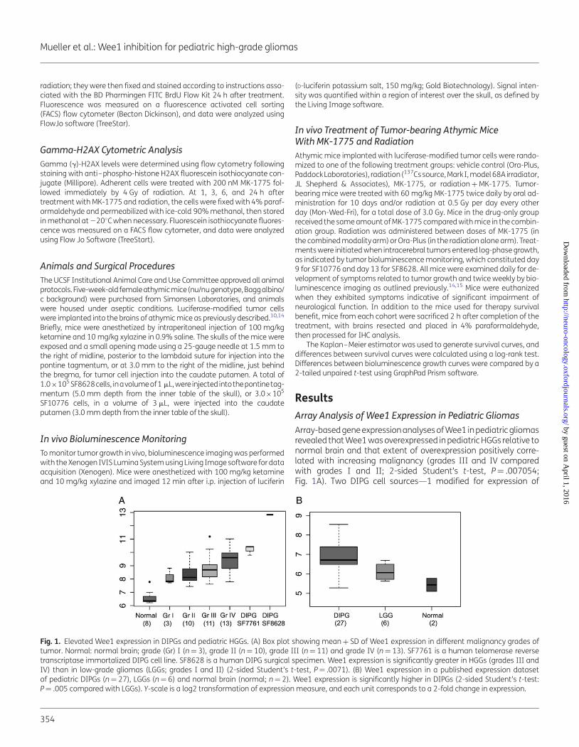
Array-based gene expression analyses of Wee1 in pediatric gliomasrevealed that Wee1 was overexpressed in pediatric HGGs relative tonormal brain and that extent of overexpression positively corre-lated with increasing malignancy (grades III and IV comparedwith grades I and II; 2-sided Student’s t-test, P¼ .007054;Fig. 1A). Two DIPG cell sources—1 modified for expression of
Fig. 1. Elevated Wee1 expression in DIPGs and pediatric HGGs. (A) Box plot showing mean + SD of Wee1 expression in different malignancy grades oftumor. Normal: normal brain; grade (Gr) I (n¼ 3), grade II (n¼ 10), grade III (n¼ 11) and grade IV (n¼ 13). SF7761 is a human telomerase reversetranscriptase immortalized DIPG cell line. SF8628 is a human DIPG surgical specimen. Wee1 expression is significantly greater in HGGs (grades III andIV) than in low-grade gliomas (LGGs; grades I and II) (2-sided Student’s t-test, P¼ .0071). (B) Wee1 expression in a published expression datasetof pediatric DIPGs (n¼ 27), LGGs (n¼ 6) and normal brain (normal; n¼ 2). Wee1 expression is significantly higher in DIPGs (2-sided Student’s t-test:P¼ .005 compared with LGGs). Y-scale is a log2 transformation of expression measure, and each unit corresponds to a 2-fold change in expression.
Mueller et al.: Wee1 inhibition for pediatric high-grade gliomas
354
by guest on April 1, 2016
http://neuro-oncology.oxfordjournals.org/D
ownloaded from

human telomerase (SF7761)14 and 1 established directly fromDIPG biopsy (SF8628) without modification—showed particularlyhigh Wee1 expression. Expression analysis of Wee1 in a publisheddataset of DIPGs13 revealed significant upregulation of Wee1 inDIPGs compared with low-grade gliomas and normal brain(2-sided Student’s t-test, P¼ .0071; Fig. 1B).
Immunohistochemical Analysis of Wee1
To further examine Wee1 protein expression in pediatric gliomas,we performed IHC analyses of Wee1 in all grades of tumor (gradeI, n¼ 18; grade II, n¼ 5; grade III, n¼ 7; grade IV, n¼ 13). Ourresults show (Fig. 2) greater Wee1 expression in higher-gradetumors (see Supplemental Table S1) and statistically significant
correlation with the array analysis (Spearman’s rho, 0.49; P¼.033). Further analysis revealed a positive correlation betweenIHC scores and tumor grade (Goodman–Kruskal gamma statisticP¼ .03; 2-sided gamma-knife gamma¼ 0.55).
Antiproliferative Effects of MK-1775 in CombinationWith Radiation
Clonogenic survival assays, for preliminary investigation ofMK-1775 as a radiation sensitizer, were carried out using 6 differentglioma cell lines (4 pediatric and 2 adult HGG cell lines). We firstexamined the timing of MK-1775 administration in relation to radi-ation therapy by testing simultaneous as well as sequential admin-istration of MK-1775, in relation to radiation treatment, with
Fig. 2. IHC staining for Wee1 in pediatric HGG. Images show representative results for IHC 0–4 positivityscoring (0, 2, and 3 are grade III tumors; 1 and 4 aregrade IV tumors). Magnification, 400×.
Fig. 3. Clonogenic survival results for pediatric and adult glioma cell lines treated with radiation+MK-1775. (A) Radiation alone: solid line. Radiation +MK-1775: dashed line. MK-1775 (200 nM) was administered 24 h after radiation (0, 2, 4, 6 Gy). Surviving fractions, shown as mean + SE (quadruplicatesamples), were normalized to plating efficiency for each cell line. XRT, radiation therapy. Clonogenic survival data points were fitted to a linearquadratic mathematical model. (B) Dose Enhancement Ratio (DER) for each cell line calculated at 10% survival levels. mt, mutant; wt, wild type.
Mueller et al.: Wee1 inhibition for pediatric high-grade gliomas
Neuro-Oncology 355
by guest on April 1, 2016
http://neuro-oncology.oxfordjournals.org/D
ownloaded from

MK-1775 administered 3, 6, and 24 h before as well as after radi-ation. The most pronounced effects were observed for MK-1775treatment at 24 h after radiation therapy (data not shown), andthis sequence of treatment was used for all subsequent clonogenicassays. Figure 3A shows the effect of MK-1775 and radiationtherapy on clonogenic survival in these 6 cell lines. Figure 3Bshows the DERs for each cell line calculated at the 10% survivallevel. No consistent differences were noted in DERs based on p53mutational status.
MK-1775 Abrogates Radiation-induced G2 Arrest in PediatricHigh-grade Gliomas
Based on the mechanism of action of Wee1 and the effects of itsinhibitor MK-1775, we hypothesized that combining MK-1775with radiation would abrogate radiation-induced G2 arrest.Indeed, our results confirm that in the pediatric glioma cell linesSF188 and SF8628 and in the adult glioma cell lines U87 andGBM36, radiation produces a G2 arrest that is abrogated by Wee1inhibitor treatment. MK-1775 alone had minimal to no effect oncell cycle distribution (Fig. 4).
Because G2 arrest allows repairof DNA damage after irradiation,we anticipated that combining MK-1775 with radiation would in-crease levels of phosphorylated H2AX (g-H2AX), a marker ofdouble strand (ds)DNA breaks, since tumor cells with damagedDNA would be less able to undergo cell cycle arrest to repairdamaged DNA. As expected, for the pediatric DIPG xenograftSF8628, a combination of MK-1775 and radiation increasedlevels ofg-H2AX to a greaterextent and for longer duration than ra-diation alone (Fig. 5). Similar effects were observed using flow
cytometric analysis in the pediatric glioma cell line KNS-42 andthe adult glioma xenograft GBM36 (Supplemental Fig. S1).
In vivo Efficacy of MK-1775 in Combination With Radiationin Supratentorial and DIPG Pediatric Models
To address the in vivo efficacy of MK-1775 and radiation, we used 2different orthotopic pediatric glioma models that both express
Fig. 4. MK-1775 decreases radiation-induced G2 cell cycle arrest. Bar graphs display distribution of cells within each cell cycle phase for 4 independent HGGcell lines treated with either radiation therapy (XRT; 4 Gy), MK-1775 (200 nM), MK-1775 + radiation (combo; 200 nM MK-1775 followed immediately by4 Gy radiation), or control. Cell cycle analyses were performed 24 h after treatment.
Fig. 5. Combination of MK-1775 and radiation leads to higher and moreprolonged expression of dsDNA marker g-H2AX. SF8628 DIPG xenograftcells treated with combination of radiation therapy (XRT, 2 Gy) andMK-1775 (200 nM), single modality only, or control. Western blots wereperformed at specified times after treatment to assess levels of g-H2AX.Gamma-H2AX levels increase 1–3 h after radiation and return to baselinewithin 6 h, whereas in combination with MK-1775, elevated g-H2AX levelsare more pronounced and prolonged (at least 24 h after radiation).
Mueller et al.: Wee1 inhibition for pediatric high-grade gliomas
356
by guest on April 1, 2016
http://neuro-oncology.oxfordjournals.org/D
ownloaded from

Wee1. For the first, genetically engineered mouse SF10776 cellscarrying the BRAFV600E mutation and modified for luciferase ex-pression were injected supratentorially into athymic mice.10 Wechose SF10776 cells to model one of the most commonly foundalterations in pediatric gliomas. Mice with intracranial SF10776tumors and treated with MK-1775 and radiation showed signifi-cantly reduced tumor burden and longer survival than mice receiv-ing single modality treatment (Fig. 6A). To test this combinationtherapy in a DIPG model, DIPG cells (SF8628) were injected directlyinto the brainstem, and mice were monitored for tumor biolumin-escence. Mice with SF8628 DIPG treated with MK-1775 and radi-ation showed decreased tumor burden and increased survivalcompared with single modality or vehicle only treatment (Fig. 6B).
As observed in our in vitro studies, combination of MK-1775 andradiation increased g-H2AX in orthotopic tumors treated in vivo(Fig. 7) to a greaterextent than in tumors exposed to MK-1775 (Wil-coxon rank sum test, P¼ .03) or radiation alone (Wilcoxon ranksum test, P¼ .03). In addition, Ki-67, a marker of proliferation,
was significantly reduced in the DIPG orthotopic tumors frommice treated with combination therapy compared with MK-1775(Wilcoxon rank sum test, P¼ .03), radiation therapy (Wilcoxonrank sum test, P¼ .03), or control treated mice (Wilcoxon ranksum test, P¼ .03). Lastly, based on the underlying mechanism ofaction of MK-1775, we expected reduced expression of p-Cdc2in xenografts from mice treated with MK-1775 or the combinationof radiation and MK-1775. As shown in Fig. 7, expression of p-CDC2was significantly reduced in the treatment groups that includedMK-1775 compared with control or radiation alone (P¼ .002).Similar results were seen for SF10776 xenografts.
DiscussionIn this investigation we demonstrated that Wee1 is significantlyupregulated in pediatric gliomas and that treatment with theWee1 inhibitor MK-1775 in combination with radiation increasesthe radiation cytotoxic effect and prolongs survival for mice with
Fig. 6. Wee1 inhibitor MK-1775 augments radiation antitumor effect in vivo. (A) Intracerebral SF10776 HGG and (B) brainstem SF8628 DIPG xenograftstreated with external beam radiation (XRT), MK (MK-1775), or XRT + MK-1775 beginning when tumors had entered log-phase growth as indicated bybioluminescence imaging tumor growth therapy response curves (left panels). Middle panels: Scatterplots of individual mice according to treatmentcohort (day 19 for SF10776 [A] and day 38 for SF8638 [B]). P values for SF10776: control vs XRT, *P¼ .0411; control vs MK-1775, **P¼ .0212; XRT vsXRT + MK-1775, ***P¼ .0137; MK-1775 vs MK-1775 + XRT, ****P¼ .0187. P values for SF8628: control vs XRT, *P¼ .0199; control vs MK-1775, **P¼.0313; XRT vs XRT + MK-1775, ***P¼ .0424; MK-1775 vs MK-1775 + XRT, ****P¼ .0149. Right panels: Kaplan–Meier survival plots; P values for SF10776:control vs XRT, P¼ 0.0032; XRT vs XRT + MK-1775, P¼ .0061; MK-1775 vs XRT + MK-1775, P¼ .0009. There is no significant difference in survivalbetween control and MK-1775, P¼ .235. P values for SF8628: control vs XRT, P¼ .0281; control vs MK-1775, P¼ .0077; XRT vs XRT + MK-1775, P¼.0163; MK-1775 vs MK-1775 + XRT, P¼ .0467.
Mueller et al.: Wee1 inhibition for pediatric high-grade gliomas
Neuro-Oncology 357
by guest on April 1, 2016
http://neuro-oncology.oxfordjournals.org/D
ownloaded from

engrafted, orthotopic gliomas. Two distinct engraftment modelswere used: one involving tumor cells from a genetically engineeredmouse with BRAFV600E mutation, and the other involving humantumor cells propagated from a patient’s DIPG. Similar resultshave been recently reported in association with the use of anadult glioma for animal modeling of DIPG.16 To our knowledge,the current report is the first using pediatric HGG models toassess the effects of Wee1 inhibition in combination with radiation.Importantly, MK-1775 is the only specific Wee1 inhibitor that hasentered clinical trials and has a favorable side effect profile. Ourresults therefore support combination therapy of MK-1775 and ra-diation as a promising new treatment strategy for children withHGGs including DIPGs.
The combination of radiation and a Wee1 inhibitor representsone of the first treatment concepts to capitalize on a specific
molecular characteristic of pediatric malignant gliomas, namelytheir high level of Wee1 expression. Our results show that Wee1 ex-pression positively correlates with glioma grade, with highest ex-pression found in DIPG samples, consistent with a publishedgene expression dataset for 27 DIPGs, in which we found thatWee1 was significantly upregulated in DIPGs compared withnormal brain.13 Recently, others have reported significantly ele-vated Wee1 expression in DIPGs compared with low-gradeglioma and normal brain samples, consistent with our findings.16
Prior studies have shown that Wee1 expression levels predict re-sponse to Wee1 inhibition.7 Consistent with this, the SF8628 DIPGxenograft we examined, which showed the highest level of Wee1expression of our entire tumor cohort, was more responsive to ra-diation + MK-1775 combination therapy than to radiation alone.Moreover, all tested HGG cell lines were sensitive to MK-1775 and
Fig. 7. IHC analyses of g-H2AX and Ki-67 in human pediatric DIPG xenografts (SF8628). (A) Representative stains of intracranial xenografts resected 24 hafter a 5-day treatment course of daily MK-1775 (60 mg/kg) for total of 5 days, with external beam radiation (XRT; 0.5 Gy) every other day (total dose1.5 Gy), or MK-1775 (60 mg/kg×5 days) + 0.5 Gy (every other day for total of 1.5 Gy), and control. Image-based quantification of (B) Ki-67, (C) g-H2AX,and (D) p-Cdc2. Percent positive tumor cells were determined in at least five 20× images per tumor, and averaged. Bars represent SE. Indicateddifferences between groups as indicated by Kruskal–Wallis test, followed by Wilcoxon rank sum test pairwise comparisons. Ki-67: combination therapyvs control P¼ .03, vs XRT P¼ .03, vs MK-1775, P¼ .03. Gamma-H2AX: combination therapy vs control P¼ .03, vs XRT P¼ .03, vs MK-1775 P¼ .03.p-Cdc2: combination therapy vs control and XRT P¼ .002, vs MK-1775, P¼ .06. Asterisk indicates statistically significant differences.
Mueller et al.: Wee1 inhibition for pediatric high-grade gliomas
358
by guest on April 1, 2016
http://neuro-oncology.oxfordjournals.org/D
ownloaded from

radiation, irrespective of p53 status, suggesting that p53 mutationis not essential for cell sensitivity to Wee1 inhibition, as previouslyreported for adult GBM7 and sarcomas.8 In contrast, others haveshown that certain types of cancer may be dependent on p53 mu-tation for response to Wee1 inhibition.17,18 Of note, our DER valueswere very similar in the various cell lines in vitro, and therefore wecannot draw any firm conclusions regarding association betweenWee1 expression and response to MK-1775.
With respect to mechanism of action, we confirm that radiationarrests pediatric glioma cells in G2 and that MK-1775 promotes ab-rogation of this cell cycle checkpoint. MK-1775–treated cells aretherefore less able to repair radiation-induced DNA damage priorto entering mitosis, as demonstrated by analysis of g-H2AX levels(Figs. 5 and 7), resulting in increased cell death.7 However, ourdata are consistent with MK-1775 increasing dsDNA breaks notonly by abrogation of G2 arrest but also due to direct effects onthe DNA repair machinery. DNA repair is highly regulated andoften specific to the type of incurred DNA damage. Nonhomolo-gous end-joining and homologous recombination are activatedby radiation-induced dsDNA breaks and involve numerousproteins, including Ku heterodimer (Ku70/Ku80), DNA proteinkinases, ligase IV, X-ray repair cross-complementing protein 4(XRCC4) and XRCC4-like factor, Artemis and DNA polymerases fornonhomologous end-joining, and Rad51 for homologous recom-bination.19 Precedence exists in the literature regarding direct in-volvement of cell cycle regulators in the DNA repair machinery,including ataxia telangiectasia mutated kinase and Chk1.20,21
Further investigations are needed to clarify whether (1) abrogationof cell cycle arrest is the main mechanism by which Wee1 inhibitionenhances the cytotoxic effects of radiation or (2) a possible directrole for Wee1 in the DNA repair machinery contributes to its co-operative antitumor effects with radiation.
Conclusions
Wee1 is overexpressed in pediatric gliomas, and Wee1 expressionlevels positively correlate with increasing glioma grade. MK-1775promotes abrogation of the radiation-induced G2 arrest, increasesand prolongs tumor g-H2AX, and thereby radiosensitizes pediatricHGGs. MK-1775 in combination with radiation extended survivalcompared with single modality arms in 2 independent orthotopicpediatric glioma models, one a supratentorial HGG and thesecond a newly established DIPG xenograft model. Results of thispreclinical study provide a strong rationale for a phase 1 trial ofthe combination of MK-1775 and radiation in children newly diag-nosed with DIPG.
Supplementary MaterialSupplementary material is available online at Neuro-Oncology(http://neuro-oncology.oxfordjournals.org/).
FundingThis research was supported in part by a National Institute of NeurologicalDisorders and Stroke grant (K12NS001692 to S.M.), the National Center forAdvancing Translational Sciences, National Institutes of Health (NIH),
through UCSF-CTSI grant KL2TR000143 (S.M.), NIH Brain Tumor SPOREgrant P50 CA097257 (S.M., D.A.H-K., M.D.P., M.S.B.), The V Foundation(S.M., D.A.H-K.), the Matthew Larson Foundation (S.M.), and the A. FrankCampini Foundation (S.M.).
AcknowledgmentsDr Rowitch (Department of Pediatrics, UCSF) generously provided SF10776cells. We would like to thank Cynthia Cowdrey for assistance with the IHCanalysis.
Conflict of interest statement. None declared.
References1. Hargrave D, Bartels U, Bouffet E. Diffuse brainstem glioma in children:
critical review of clinical trials. Lancet Oncol. 2006;7(3):241–248.
2. Nicolaides TP, Li H, Solomon DA, et al. Targeted therapy forBRAFV600E malignant astrocytoma. Clin Cancer Res. 2011;17(24):7595–7604.
3. Zarghooni M, Bartels U, Lee E, et al. Whole-genome profiling of pediatricdiffuse intrinsic pontine gliomas highlights platelet-derived growthfactor receptor alpha and poly (ADP-ribose) polymerase as potentialtherapeutic targets. J Clin Oncol. 2010;28(8):1337–1344.
4. Wu G, Broniscer A, McEachron TA, et al. Somatic histone H3 alterationsin pediatric diffuse intrinsic pontine gliomas and non-brainstemglioblastomas. Nat Genet. 2012;44(3):251–253.
5. Khuong-QuangDA, Buczkowicz P, RakopoulosP, et al. K27Mmutation inhistone H3.3 defines clinically and biologically distinct subgroups ofpediatric diffuse intrinsic pontine gliomas. Acta Neuropathol. 2012;124(3):439–447.
6. Paugh BS, Qu C, Jones C, et al. Integrated molecular genetic profiling ofpediatric high-grade gliomas reveals key differences with the adultdisease. J Clin Oncol. 2010;28(18):3061–3068.
7. Mir SE, De Witt Hamer PC, Krawczyk PM, et al. In silico analysis of kinaseexpression identifies WEE1 as a gatekeeper against mitoticcatastrophe in glioblastoma. Cancer Cell. 2010;18(3):244–257.
8. Kreahling JM, Gemmer JY, Reed D, Letson D, Bui M, Altiok S. MK1775, aselective Wee1 inhibitor, shows single-agent antitumor activityagainst sarcoma cells. Mol Cancer Ther. 2012;11(1):174–182.
9. Sarkaria JN, Yang L, Grogan PT, et al. Identification of molecularcharacteristics correlated with glioblastoma sensitivity to EGFRkinase inhibition through use of an intracranial xenograft test panel.Mol Cancer Ther. 2007;6(3):1167–1174.
10. Huillard E, Hashizume R, Phillips JJ, et al. Cooperative interactions ofBRAFV600E kinase and CDKN2A locus deficiency in pediatricmalignant astrocytoma as a basis for rational therapy. Proc NatlAcad Sci U S A. 2012;109(22):8710–8715.
11. Franken NA, Rodermond HM, Stap J, Haveman J, van Bree C. Clonogenicassay of cells in vitro. Nat Protoc. 2006;1(5):2315–2319.
12. Bolstad BM, Irizarry RA, Astrand M, Speed TP. A comparison ofnormalization methods for high density oligonucleotide arraydata based on variance and bias. Bioinformatics. 2003;19(2):185–193.
13. Paugh BS, Broniscer A, Qu C, et al. Genome-wide analyses identifyrecurrent amplifications of receptor tyrosine kinases and cell-cycleregulatory genes in diffuse intrinsic pontine glioma. J Clin Oncol.2011;29(30):3999–4006.
Mueller et al.: Wee1 inhibition for pediatric high-grade gliomas
Neuro-Oncology 359
by guest on April 1, 2016
http://neuro-oncology.oxfordjournals.org/D
ownloaded from

14. Hashizume R, Smirnov I, Liu S, et al. Characterization of a diffuseintrinsic pontine glioma cell line: implications for future investigationsand treatment. J Neurooncol. 2012;110(3):305–313.
15. Mueller S, Yang X, Sottero TL, et al. Cooperation of the HDAC inhibitorvorinostat and radiation in metastatic neuroblastoma: efficacy andunderlying mechanisms. Cancer Lett. 2011;306(2):223–229.
16. Caretti V, Hiddingh L, Lagerweij T, et al. WEE1 kinase inhibitionenhances the radiation response of diffuse intrinsic pontine gliomas.Mol Cancer Ther. 2013;12(2):141–150.
17. Hirai H, Iwasawa Y, Okada M, et al. Small-molecule inhibition of Wee1kinase by MK-1775 selectively sensitizes p53-deficient tumor cells toDNA-damaging agents. Mol Cancer Ther. 2009;8(11):2992–3000.
18. Krajewska M, Heijink AM, Bisselink YJ, et al. Forced activation of Cdk1 viawee1 inhibition impairs homologous recombination. Oncogene. 2013;32(24):3001–3008.
19. Branzei D, Foiani M. Regulation of DNA repair throughout the cell cycle.Nat Rev Mol Cell Biol. 2008;9(4):297–308.
20. Goodarzi AA, Noon AT, Deckbar D, et al. ATM signaling facilitates repairof DNA double-strand breaks associated with heterochromatin. MolCell. 2008;31(2):167–177.
21. Morgan MA, Parsels LA, Zhao L, et al. Mechanism of radiosensitizationby the Chk1/2 inhibitor AZD7762 involves abrogation of the G2checkpoint and inhibition of homologous recombinational DNArepair. Cancer Res. 2011;70(12):4972–4981.
Mueller et al.: Wee1 inhibition for pediatric high-grade gliomas
360
by guest on April 1, 2016
http://neuro-oncology.oxfordjournals.org/D
ownloaded from
Related Documents