DOI 10.1515/plm-2012-0033 Photon Lasers Med 2012; 1(4): 241–254 Review Eason Hahm, Snehlata Kulhari and Praveen R. Arany* Targeting the pain, inflammation and immune (PII) axis: plausible rationale for LLLT Schmerz, Entzündung und Immunantwort: Eine plausible Begründung für die LLLT Abstract: Low-level laser therapy (LLLT) has been used in many clinical contexts. Although its precise mecha- nism is unclear, its current use is based predominantly on its non-invasive nature and popular patient acceptance. This review attempts to provide a framework for the clini- cal disease-disorder states focusing on the etiopathology namely; pain, inflammation and immune response termed the PII axis. Following a brief introduction, the literature on the ability of LLLT to modulate the PII axis in specific disease states is reviewed. The triad of critical parameters for LLLT namely, the dose, the biological context and the mechanism are highlighted. This work suggests that LLLT could be a potent primary interventional modality when used to spe- cifically target the PII axis in clinical disease management. Keywords: low-level laser therapy; pain; inflammation; immune response. Zusammenfassung: Die Low-Level-Laser-Therapie (LLLT) ist in vielen klinischen Zusammenhängen verwendet worden. Obwohl die genauen Mechanismen der LLLT noch unklar sind, beruht ihr aktueller Einsatz vor allem auf dem minimal-invasiven Charakter der Methode und der hohen Patientenakzeptanz. Der vorliegende Review-Artikel nähert sich dem Thema ausgehend vom Krankheitsverlauf verschiedener klinischer Erkrankungen und Störungen und fokussiert hierbei auf die Schmerz- Entzündungs-Immunantwort-Achse (pain, inflammation, immune response – PPI – axis). Nach einer kurzen Ein- führung wird basierend auf der aktuellen Literatur disku- tiert, inwieweit es möglich ist, mittels LLLT die PII-Achse in bestimmten Krankheitsstadien zu modulieren. Die Triade kritischer Parameter für die LLLT, nämlich die Dosis, der biologische Kontext und der Mechanismus werden besonders herausgestellt. Im Ergebnis liegt die Vermutung nahe, dass die LLLT eine potente Interventionsmethode im Disease-Management darstellt, wenn sie zielgerichtet auf die PII-Achse ausgerichtet ist. Schlüsselwörter: Low-Level-Laser-Therapie; Schmerz; Entzündung; Immunantwort. *Corresponding author: Praveen R. Arany, current address: National Institute of Dental and Craniofacial Research (NIDCR), National Institutes of Health (NIH), 30 Convent Drive #301, Bethesda, MD 20814, USA, e-mail: [email protected] Eason Hahm: Harvard University, 58 Oxford Street, 415 ESL, Cambridge, MA 02138, USA Snehlata Kulhari: Associate Dentist, 364 Lowes Dr, Ste J, Danville, VA 24540, USA 1 Introduction The field of photomedicine encompasses a wide range of uses including photodynamic therapy (usually dye- assisted laser destruction), phototherapy (using UV and visible light sources), surgical lasers (high energy lasers as surgical cutting-coagulation tools) and low-level laser therapy (LLLT). This review will focus on the latter LLLT that is also referred to in the literature as “low inten- sity laser” or “cold laser” or “soft laser”. While LLLT is a medical subject heading (MeSH) term, it is still not precisely clear what the terms “low” and “level” refers to [1]. Given the recent popularity of light sources, espe- cially LEDs, the term “light” has often been appropri- ately substituted for “laser”. In our experience, the criti- cal parameter is the dose (energy, power and time) and this varies with the clinical-biological context [2]. The biological effects of the LLLT encompass both stimula- tory and inhibitory biological effects collectively termed “photobiomodulation” [3]. The clinical effectiveness of LLLT applications and its mechanism of action are still controversial and extensive cellular, animal and human studies need to be done to well-establish the safety of use of LLLT. This review addresses some of the most popular uses of LLLT in current clinical management. The review

Welcome message from author
This document is posted to help you gain knowledge. Please leave a comment to let me know what you think about it! Share it to your friends and learn new things together.
Transcript
DOI 10.1515/plm-2012-0033 Photon Lasers Med 2012; 1(4): 241–254
Review
Eason Hahm , Snehlata Kulhari and Praveen R. Arany *
Targeting the pain, inflammation and immune (PII) axis: plausible rationale for LLLT
Schmerz, Entz ü ndung und Immunantwort: Eine plausible Begr ü ndung f ü r die LLLT
Abstract: Low-level laser therapy (LLLT) has been used
in many clinical contexts. Although its precise mecha-
nism is unclear, its current use is based predominantly on
its non-invasive nature and popular patient acceptance.
This review attempts to provide a framework for the clini-
cal disease-disorder states focusing on the etiopathology
namely; pain, inflammation and immune response termed
the PII axis. Following a brief introduction, the literature on
the ability of LLLT to modulate the PII axis in specific disease
states is reviewed. The triad of critical parameters for LLLT
namely, the dose, the biological context and the mechanism
are highlighted. This work suggests that LLLT could be a
potent primary interventional modality when used to spe-
cifically target the PII axis in clinical disease management.
Keywords: low-level laser therapy; pain; inflammation;
immune response.
Zusammenfassung: Die Low-Level-Laser-Therapie (LLLT)
ist in vielen klinischen Zusammenh ä ngen verwendet
worden. Obwohl die genauen Mechanismen der LLLT
noch unklar sind, beruht ihr aktueller Einsatz vor allem
auf dem minimal-invasiven Charakter der Methode
und der hohen Patientenakzeptanz. Der vorliegende
Review-Artikel n ä hert sich dem Thema ausgehend vom
Krankheitsverlauf verschiedener klinischer Erkrankungen
und St ö rungen und fokussiert hierbei auf die Schmerz-
Entz ü ndungs- Immunantwort-Achse (pain, inflammation,
immune response – PPI – axis). Nach einer kurzen Ein-
f ü hrung wird basierend auf der aktuellen Literatur disku-
tiert, inwieweit es m ö glich ist, mittels LLLT die PII-Achse in
bestimmten Krankheitsstadien zu modulieren. Die Triade
kritischer Parameter f ü r die LLLT, n ä mlich die Dosis,
der biologische Kontext und der Mechanismus werden
besonders herausgestellt. Im Ergebnis liegt die Vermutung
nahe, dass die LLLT eine potente Interventionsmetho de
im Disease- Management darstellt, wenn sie zielgerichtet
auf die PII-Achse ausgerichtet ist.
Schl ü sselw ö rter: Low-Level-Laser-Therapie; Schmerz;
Entz ü ndung; Immunantwort.
*Corresponding author: Praveen R. Arany, current address: National
Institute of Dental and Craniofacial Research (NIDCR), National
Institutes of Health (NIH), 30 Convent Drive # 301, Bethesda, MD
20814, USA, e-mail: [email protected]
Eason Hahm: Harvard University , 58 Oxford Street, 415 ESL,
Cambridge, MA 02138 , USA
Snehlata Kulhari: Associate Dentist, 364 Lowes Dr , Ste J, Danville,
VA 24540 , USA
1 Introduction The field of photomedicine encompasses a wide range
of uses including photodynamic therapy (usually dye-
assisted laser destruction), phototherapy (using UV and
visible light sources), surgical lasers (high energy lasers
as surgical cutting-coagulation tools) and low-level laser
therapy (LLLT). This review will focus on the latter LLLT
that is also referred to in the literature as “ low inten-
sity laser ” or “ cold laser ” or “ soft laser ” . While LLLT is
a medical subject heading (MeSH) term, it is still not
precisely clear what the terms “ low ” and “ level ” refers
to [1] . Given the recent popularity of light sources, espe-
cially LEDs, the term “ light ” has often been appropri-
ately substituted for “ laser ” . In our experience, the criti-
cal parameter is the dose (energy, power and time) and
this varies with the clinical-biological context [2] . The
biological effects of the LLLT encompass both stimula-
tory and inhibitory biological effects collectively termed
“ photobiomodulation ” [3] . The clinical effectiveness of
LLLT applications and its mechanism of action are still
controversial and extensive cellular, animal and human
studies need to be done to well-establish the safety of use
of LLLT. This review addresses some of the most popular
uses of LLLT in current clinical management. The review
242 E. Hahm et al.: LLLT and the PII axis
is outlined in three sections: first an introduction to the
connections between pain, inflammation and immune
response, termed the PII axis, is presented. The second
section overviews the evidence for LLLT specifically tar-
geting the PII axis in mediating its therapeutic effects. In
the final section, the literature is reviewed in each condi-
tion supporting the use of LLLT for therapy. It should be
pointed out that LLLT is currently used as an adjuvant in
combination with physical or pharmacological therapy.
The primary goal of this review is to provide an integrated
view for the use of key parameters of LLLT namely; dose,
contexts and mechanisms based on its ability to primary
modulate the PII axis for therapy.
2 The nexus of pain, inflammation and immune response – PII axis
The defense mechanism evolved by the body to protect
against damage by deleterious agents including physical
injury and microbial infection involves a complex set of
biochemical and cellular phases constituting the immune
system. Broadly, the immune system is divided into innate
immune response that is generic and non-specific to spe-
cific damage agents; while a more tailored, agent-specific
response is part of the adaptive immune response. In
order to provide a broad conceptual framework to assess
the efficacy of LLLT in clinical disease management, we
propose an integrated overview of the disease etiopathol-
ogy termed the pain, inflammation and immune or the PII
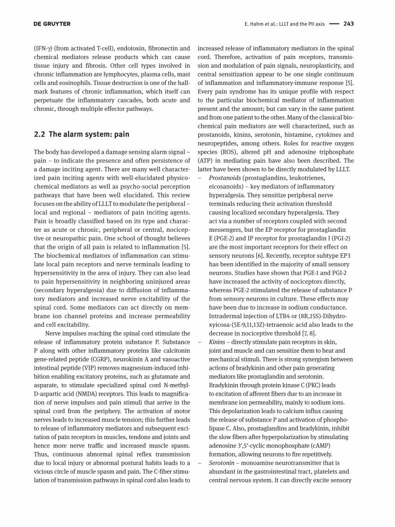
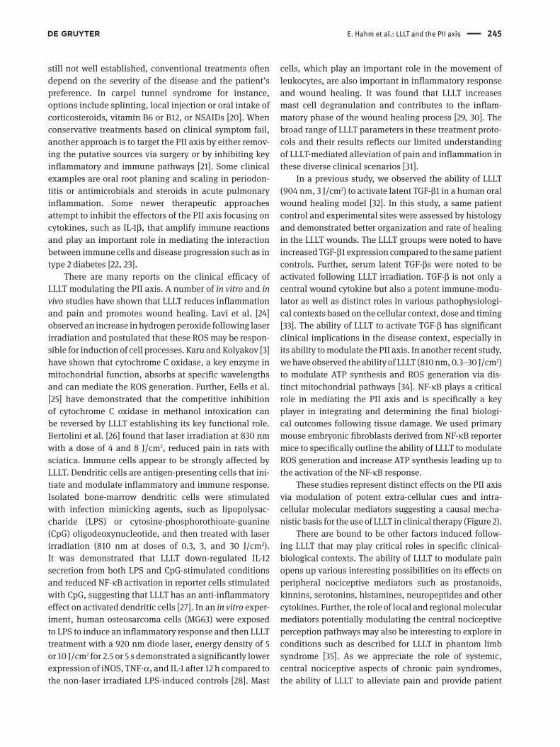
axis (Figure 1 ). The major purpose of proposing this con-
ceptual outline is to present the combinatorial “ causal ”
agents and “ effector ” mediators that have been dem-
onstrated in individual diseases disorders. Further, this
provides the rationale to highlight the literature show-
casing the ability of LLLT to specifically modulate these
pathways. It is hoped that this review will also highlight
specific biomarkers or disease indicators that could be
assessed in future LLLT studies to establish their efficacy
with regards to various clinical disease-disorder states.
2.1 The effector process: inflammation
Inflammation is a key component of the innate response
and is the protective response involving a complex reac-
tion in vascularized connective tissue to rid the host of
both the causes of cell injury and its associated conse-
quences [4] . It is divided into two patterns – acute and
chronic inflammation. Acute inflammation is the imme-
diate response to an injurious agent. It involves vascular
and cellular events. Vascular changes begin shortly after
injury and occur in the following order: transient vaso-
constriction followed by vasodilation that increases blood
flow resulting mainly from arteriolar dilation and opening
of capillary beds, slowing of circulation or stasis due to
increased permeability of microvasculature and finally,
leukocytic margination. The hallmark of acute inflam-
mation is increased vascular permeability leading to the
escape of protein rich fluid into interstitial tissues termed
edema. Cellular events involve adhesion of leukocytes
(initially predominantly neutrophils) to the endothe-
lium and then transmigration or diapedesis across the
endothelium to the interstitial tissues and migration
towards the site of injury by a process called chemotaxis,
followed by phagocytosis of the injurious agent. Chemo-
tactic agents can be both endogenous, e.g., complement
component 5a, leukotriene B4 (LTB4), interleukin 8 (IL-8),
and exogenous (most common are bacterial products).
During chemotaxis and phagocytosis, activated leuko-
cytes may release products into the extracellular matrix
that can lead to significant tissue damage. Some of the
important products released by neutrophils are a) lyso-
somal enzymes, b) oxygen derived active metabolite, and
c) products of arachidionic acid metabolism, including
prostaglandin and leukotrienes. Acute inflammation can
have any one of the four outcomes: a) complete resolution
or healing, b) abscess formation, c) healing by connec-
tive tissue replacement or scarring, and d) progression to
chronic inflammation.
Chronic inflammation, on the other hand, is of longer
duration and is characterized by the presence of mononu-
clear cells (macrophages, lymphocytes, and plasma cells),
tissue destruction and attempts at healing by connective
tissue replacement by angiogenesis (proliferation of blood
vessels) and fibrosis. The functional transformation of the
monocytes to macrophage at the sites of tissue damage is
a main feature of chronic inflammation. Macro phages are
activated by signals such as the cytokine interferon-gamma
Figure 1 Depiction of the two effector processes of the immune
response, pain and inflammation that form the etiopathology of
many common clinical disease-disorder states termed the PII axis.
E. Hahm et al.: LLLT and the PII axis 243
(IFN- γ ) (from activated T-cell), endotoxin, fibronectin and
chemical mediators release products which can cause
tissue injury and fibrosis. Other cell types involved in
chronic inflammation are lymphocytes, plasma cells, mast
cells and eosinophils. Tissue destruction is one of the hall-
mark features of chronic inflammation, which itself can
perpetuate the inflammatory cascades, both acute and
chronic, through multiple effector pathways.
2.2 The alarm system: pain
The body has developed a damage sensing alarm signal –
pain – to indicate the presence and often persistence of
a damage inciting agent. There are many well character-
ized pain inciting agents with well-elucidated physico-
chemical mediators as well as psycho-social perception
pathways that have been well elucidated. This review
focuses on the ability of LLLT to modulate the peripheral –
local and regional – mediators of pain inciting agents.
Pain is broadly classified based on its type and charac-
ter as acute or chronic, peripheral or central, nocicep-
tive or neuropathic pain. One school of thought believes
that the origin of all pain is related to inflammation [5] .
The biochemical mediators of inflammation can stimu-
late local pain receptors and nerve terminals leading to
hypersensitivity in the area of injury. They can also lead
to pain hypersensitivity in neighboring uninjured areas
(secondary hyperalgesia) due to diffusion of inflamma-
tory mediators and increased nerve excitability of the
spinal cord. Some mediators can act directly on mem-
brane ion channel proteins and increase permeability
and cell excitability.
Nerve impulses reaching the spinal cord stimulate the
release of inflammatory protein substance P. Substance
P along with other inflammatory proteins like calcitonin
gene-related peptide (CGRP), neurokinin A and vasoactive
intestinal peptide (VIP) removes magnesium-induced inhi-
bition enabling excitatory proteins, such as glutamate and
asparate, to stimulate specialized spinal cord N-methyl-
D -aspartic acid (NMDA) receptors. This leads to magnifica-
tion of nerve impulses and pain stimuli that arrive in the
spinal cord from the periphery. The activation of motor
nerves leads to increased muscle tension; this further leads
to release of inflammatory mediators and subsequent exci-
tation of pain receptors in muscles, tendons and joints and
hence more nerve traffic and increased muscle spasm.
Thus, continuous abnormal spinal reflex transmission
due to local injury or abnormal postural habits leads to a
vicious circle of muscle spasm and pain. The C-fiber stimu-
lation of transmission pathways in spinal cord also leads to
increased release of inflammatory mediators in the spinal
cord. Therefore, activation of pain receptors, transmis-
sion and modulation of pain signals, neuroplasticity, and
central sensitization appear to be one single continuum
of inflammation and inflammatory-immune response [5] .
Every pain syndrome has its unique profile with respect
to the particular biochemical mediator of inflammation
present and the amount; but can vary in the same patient
and from one patient to the other. Many of the classical bio-
chemical pain mediators are well characterized, such as
prostanoids, kinins, serotonin, histamine, cytokines and
neuropeptides, among others. Roles for reactive oxygen
species (ROS), altered pH and adenosine triphosphate
(ATP) in mediating pain have also been described. The
latter have been shown to be directly modulated by LLLT.
– Prostanoids (prostaglandins, leukotrienes,
eicosanoids) – key mediators of inflammatory
hyperalgesia. They sensitize peripheral nerve
terminals reducing their activation threshold
causing localized secondary hyperalgesia. They
act via a number of receptors coupled with second
messengers, but the EP receptor for prostaglandin
E (PGE-2) and IP receptor for prostaglandin I (PGI-2)
are the most important receptors for their effect on
sensory neurons [6] . Recently, receptor subtype EP3
has been identified in the majority of small sensory
neurons. Studies have shown that PGE-1 and PGI-2
have increased the activity of nociceptors directly,
whereas PGE-2 stimulated the release of substance P
from sensory neurons in culture. These effects may
have been due to increase in sodium conductance.
Intradermal injection of LTB4 or (8R,15S)-Dihydro-
xyicosa-(5E-9,11,13Z)-tetraenoic acid also leads to the
decrease in nociceptive threshold [7, 8] .
– Kinins – directly stimulate pain receptors in skin,
joint and muscle and can sensitize them to heat and
mechanical stimuli. There is strong synergism between
actions of bradykinin and other pain generating
mediators like prostaglandin and serotonin.
Bradykinin through protein kinase C (PKC) leads
to excitation of afferent fibers due to an increase in
membrane ion permeability, mainly to sodium ions.
This depolarization leads to calcium influx causing
the release of substance P and activation of phospho-
lipase C. Also, prostaglandins and bradykinin, inhibit
the slow fibers after hyperpolarization by stimulating
adenosine 3 ′ ,5 ′ -cyclic monophosphate (cAMP)
formation, allowing neurons to fire repetitively.
– Serotonin – monoamine neurotransmitter that is
abundant in the gastrointestinal tract, platelets and
central nervous system. It can directly excite sensory
244 E. Hahm et al.: LLLT and the PII axis
neurons by increasing sodium permeability via 5-HT3
receptor activation.
– Histamine – histamine H1 receptor activation on
sensory neurons leads to an increase in membrane
calcium permeability, which leads to the release
of sensory neuropeptides, prostaglandins and
5-hydroxyeicosatetraenoic acid from endothelial
cells leading to hyperalgesia and other pro-inflam-
matory affects.
– Cytokines (IL-1 β , IL-6, IL-8, IL-10, TNF- α ) – can cause
hyperalgesia via various indirect mechanisms like
increased production of prostaglandin or increas-
ing the expression of bradykinin or nerve growth
factor (NGF) receptors. Tumor necrosis factor-alpha
(TNF- α ) release leads to increased production of
prostaglandin which further leads to increased
production of glutamate and thus increase in nerve
cell communication. It also leads to excitation of
pain receptors and stimulation of specialized nerves
such as the C-fibers and A δ -fibers.
– Neuropeptides (neurokinins and neurotropins) –
during inflammation neurokinins substance P and
neurokinin A (NKA) contribute directly and
indirectly to neurogenic inflammation and hyper-
algesia in periphery and excitability in dorsal horn
cells of spinal cord associated with transmission of
pain signals. Neurokinins can also reduce potassium
permeability and can directly depolarize sensory
neurons. During inflammation, neurotrophins like
NGF increase the synthesis of neurokinis and CGRP.
Substance P also leads to an increase in TNF- α
production.
– Free radicals and reactive oxygen species –
Hydrogen peroxide has been shown to enhance the
effects of bradykinin and PGE-2. The intra-dermal
injection of nitric oxide (NO) induces a delayed
burning pain [9] . During inflammation or nerve
injury, an inducible and calcium dependant form
of nitric oxide synthase (NOS) leads to an increase
in NO synthesis. Inducible NOS (iNOS) has a role
in upregulation of cyclooxygenase (COX) activity
and hence the production of pro-inflammatory
prostanoids [10] . NO may also alter the response of
sensory neurons to bradykinin and contributes to
hyperalgesia by increasing sensitization to central
and peripheral stimuli.
– Altered pH (protons) – Change in tissue pH due to
inflammation generates positively charged sub-
atomic particles called protons. They are associated
with inflammatory hyperalgesia and pain-discomfort
due to hypoxia observed during muscle exercise.
Intradermal injection of acidic solution leads to
sharp stinging pain due to direct activation of
nociceptors and enhancing the effects of other
inflammatory mediators.
– Adenosine triphosphate – can activate sensory
neurons and increase their permeability to cations.
Adenosine formed on breakdown of ATP, also
provokes pain and hyperalgesia due to stimulation
of adenosine A2 receptors which are coupled with
cAMP. The production of cAMP and decrease in
potassium ion permeability accounts for hyper-
excitability of sensory neurons.
2.3 Damage-associated molecular patterns
Damaged-associated molecular patterns (DAMPs) play
an important role in signaling to the immune system
[11, 12] . Inflammatory response occurs when pattern
recognition receptors (PRRs) on the surface of innate
immune cells detect the release of DAMPs from injured
tissue in the absence of microbial invasion [13] . For
example, ROS in high concentrations can cause tissue
damage that can induce DAMPs which are recog-
nized by PRRs [14] . Toll-like receptors are activated by
DAMPs, which induces inflammatory gene expression
in an effort to mediate the repair of damaged tissue [15] .
As a result, DAMPs have been thought to play a key role
in inflammatory diseases, such as rheumatoid arthri-
tis and oral mucositis, as they promote nuclear factor
kappa B (NF- κ B) signaling and upregulate the expres-
sion of pro-inflammatory cytokines [16] . Thus, DAMPs
play important roles in both induction and perpetua-
tion of a pro-inflammatory responses and have been
linked to many autoimmune diseases.
3 Can the PII axis be modulated by LLLT ?
Conventional approaches to managing pain and inflam-
mation include the use of pharmacologic drugs, most com-
monly non-steroidal anti-inflammatory drugs (NSAIDs)
[17] . However, NSAIDs generally only treat the symptoms
of inflammation; they do not directly target the cause of
disease and many of the negative side effects related to
the long-term use of these drugs have been documented
[18] . For example in neck pain, the standard treatment
includes simple analgesics, NSAIDs, and physical therapy
[19] . For disease where definitive treatment strategies are
E. Hahm et al.: LLLT and the PII axis 245
still not well established, conventional treatments often
depend on the severity of the disease and the patient ’ s
preference. In carpel tunnel syndrome for instance,
options include splinting, local injection or oral intake of
corticosteroids, vitamin B6 or B12, or NSAIDs [20] . When
conservative treatments based on clinical symptom fail,
another approach is to target the PII axis by either remov-
ing the putative sources via surgery or by inhibiting key
inflammatory and immune pathways [21] . Some clinical
examples are oral root planing and scaling in periodon-
titis or antimicrobials and steroids in acute pulmonary
inflammation. Some newer therapeutic approaches
attempt to inhibit the effectors of the PII axis focusing on
cytokines, such as IL-1 β , that amplify immune reactions
and play an important role in mediating the interaction
between immune cells and disease progression such as in
type 2 diabetes [22, 23] .
There are many reports on the clinical efficacy of
LLLT modulating the PII axis. A number of in vitro and in vivo studies have shown that LLLT reduces inflammation
and pain and promotes wound healing. Lavi et al. [24]
observed an increase in hydrogen peroxide following laser
irradiation and postulated that these ROS may be respon-
sible for induction of cell processes. Karu and Kolyakov [3]
have shown that cytochrome C oxidase, a key enzyme in
mitochondrial function, absorbs at specific wavelengths
and can mediate the ROS generation. Further, Eells et al.
[25] have demonstrated that the competitive inhibition
of cytochrome C oxidase in methanol intoxication can
be reversed by LLLT establishing its key functional role.
Bertolini et al. [26] found that laser irradiation at 830 nm
with a dose of 4 and 8 J/cm 2 , reduced pain in rats with
sciatica. Immune cells appear to be strongly affected by
LLLT. Dendritic cells are antigen- presenting cells that ini-
tiate and modulate inflammatory and immune response.
Isolated bone-marrow dendritic cells were stimulated
with infection mimicking agents, such as lipopolysac-
charide (LPS) or cytosine-phosphorothioate-guanine
(CpG) oligodeoxynucleotide, and then treated with laser
irradiation (810 nm at doses of 0.3, 3, and 30 J/cm 2 ).
It was demonstrated that LLLT down-regulated IL-12
secretion from both LPS and CpG-stimulated conditions
and reduced NF- κ B activation in reporter cells stimulated
with CpG, suggesting that LLLT has an anti-inflammatory
effect on activated dendritic cells [27] . In an in vitro exper-
iment, human osteosarcoma cells (MG63) were exposed
to LPS to induce an inflammatory response and then LLLT
treatment with a 920 nm diode laser, energy density of 5
or 10 J/cm 2 for 2.5 or 5 s demonstrated a significantly lower
expression of iNOS, TNF- α , and IL-1 after 12 h compared to
the non-laser irradiated LPS-induced controls [28] . Mast
cells, which play an important role in the movement of
leukocytes, are also important in inflammatory response
and wound healing. It was found that LLLT increases
mast cell degranulation and contributes to the inflam-
matory phase of the wound healing process [29, 30] . The
broad range of LLLT parameters in these treatment proto-
cols and their results reflects our limited understanding
of LLLT-mediated alleviation of pain and inflammation in
these diverse clinical scenarios [31] .
In a previous study, we observed the ability of LLLT
(904 nm, 3 J/cm 2 ) to activate latent TGF- β 1 in a human oral
wound healing model [32] . In this study, a same patient
control and experimental sites were assessed by histology
and demonstrated better organization and rate of healing
in the LLLT wounds. The LLLT groups were noted to have
increased TGF- β 1 expression compared to the same patient
controls. Further, serum latent TGF- β s were noted to be
activated following LLLT irradiation. TGF- β is not only a
central wound cytokine but also a potent immune-modu-
lator as well as distinct roles in various pathophysiologi-
cal contexts based on the cellular context, dose and timing
[33] . The ability of LLLT to activate TGF- β has significant
clinical implications in the disease context, especially in
its ability to modulate the PII axis. In another recent study,
we have observed the ability of LLLT (810 nm, 0.3 – 30 J/cm 2 )
to modulate ATP synthesis and ROS generation via dis-
tinct mitochondrial pathways [34] . NF- κ B plays a critical
role in mediating the PII axis and is specifically a key
player in integrating and determining the final biologi-
cal outcomes following tissue damage. We used primary
mouse embryonic fibroblasts derived from NF- κ B reporter
mice to specifically outline the ability of LLLT to modulate
ROS generation and increase ATP synthesis leading up to
the activation of the NF- κ B response.
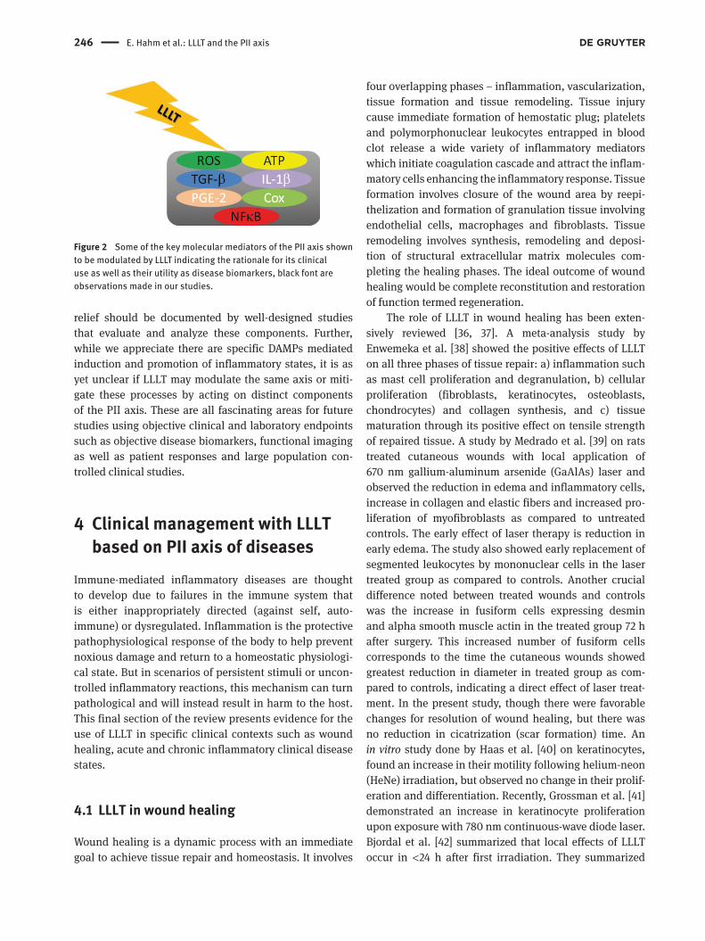
These studies represent distinct effects on the PII axis
via modulation of potent extra-cellular cues and intra-
cellular molecular mediators suggesting a causal mecha-
nistic basis for the use of LLLT in clinical therapy (Figure 2 ).
There are bound to be other factors induced follow-
ing LLLT that may play critical roles in specific clinical-
biological contexts. The ability of LLLT to modulate pain
opens up various interesting possibilities on its effects on
peripheral nociceptive mediators such as prostanoids,
kinnins, serotonins, histamines, neuropeptides and other
cytokines. Further, the role of local and regional molecular
mediators potentially modulating the central nociceptive
perception pathways may also be interesting to explore in
conditions such as described for LLLT in phantom limb
syndrome [35] . As we appreciate the role of systemic,
central nociceptive aspects of chronic pain syndromes,
the ability of LLLT to alleviate pain and provide patient
246 E. Hahm et al.: LLLT and the PII axis
relief should be documented by well-designed studies
that evaluate and analyze these components. Further,
while we appreciate there are specific DAMPs mediated
induction and promotion of inflammatory states, it is as
yet unclear if LLLT may modulate the same axis or miti-
gate these processes by acting on distinct components
of the PII axis. These are all fascinating areas for future
studies using objective clinical and laboratory endpoints
such as objective disease biomarkers, functional imaging
as well as patient responses and large population con-
trolled clinical studies.
4 Clinical management with LLLT based on PII axis of diseases
Immune-mediated inflammatory diseases are thought
to develop due to failures in the immune system that
is either inappropriately directed (against self, auto-
immune) or dysregulated. Inflammation is the protective
pathophysiological response of the body to help prevent
noxious damage and return to a homeostatic physiologi-
cal state. But in scenarios of persistent stimuli or uncon-
trolled inflammatory reactions, this mechanism can turn
pathological and will instead result in harm to the host.
This final section of the review presents evidence for the
use of LLLT in specific clinical contexts such as wound
healing, acute and chronic inflammatory clinical disease
states.
4.1 LLLT in wound healing
Wound healing is a dynamic process with an immediate
goal to achieve tissue repair and homeostasis. It involves
Figure 2 Some of the key molecular mediators of the PII axis shown
to be modulated by LLLT indicating the rationale for its clinical
use as well as their utility as disease biomarkers, black font are
observations made in our studies.
four overlapping phases – inflammation, vascularization,
tissue formation and tissue remodeling. Tissue injury
cause immediate formation of hemostatic plug; platelets
and polymorphonuclear leukocytes entrapped in blood
clot release a wide variety of inflammatory mediators
which initiate coagulation cascade and attract the inflam-
matory cells enhancing the inflammatory response. Tissue
formation involves closure of the wound area by reepi-
thelization and formation of granulation tissue involving
endothelial cells, macrophages and fibroblasts. Tissue
remodeling involves synthesis, remodeling and deposi-
tion of structural extracellular matrix molecules com-
pleting the healing phases. The ideal outcome of wound
healing would be complete reconstitution and restoration
of function termed regeneration.
The role of LLLT in wound healing has been exten-
sively reviewed [36, 37] . A meta-analysis study by
Enwemeka et al. [38] showed the positive effects of LLLT
on all three phases of tissue repair: a) inflammation such
as mast cell proliferation and degranulation, b) cellular
proliferation (fibroblasts, keratinocytes, osteoblasts,
chondrocytes) and collagen synthesis, and c) tissue
maturation through its positive effect on tensile strength
of repaired tissue. A study by Medrado et al. [39] on rats
treated cutaneous wounds with local application of
670 nm gallium-aluminum arsenide (GaAlAs) laser and
observed the reduction in edema and inflammatory cells,
increase in collagen and elastic fibers and increased pro-
liferation of myofibroblasts as compared to untreated
controls. The early effect of laser therapy is reduction in
early edema. The study also showed early replacement of
segmented leukocytes by mononuclear cells in the laser
treated group as compared to controls. Another crucial
difference noted between treated wounds and controls
was the increase in fusiform cells expressing desmin
and alpha smooth muscle actin in the treated group 72 h
after surgery. This increased number of fusiform cells
corresponds to the time the cutaneous wounds showed
greatest reduction in diameter in treated group as com-
pared to controls, indicating a direct effect of laser treat-
ment. In the present study, though there were favorable
changes for resolution of wound healing, but there was
no reduction in cicatrization (scar formation) time. An
in vitro study done by Haas et al. [40] on keratinocytes,
found an increase in their motility following helium-neon
(HeNe) irradiation, but observed no change in their prolif-
eration and differentiation. Recently, Grossman et al. [41]
demonstrated an increase in keratinocyte proliferation
upon exposure with 780 nm continuous-wave diode laser.
Bjordal et al. [42] summarized that local effects of LLLT
occur in < 24 h after first irradiation. They summarized
E. Hahm et al.: LLLT and the PII axis 247
that the effects of LLLT on biochemistry of inflammatory
process include reduction in levels of PGE-2, TNF, IL-1,
COX-2 and plasminogen activator. Also, the effects of LLLT
on cells and soft tissues include reduction in edema and
hemorrhage formation, neutrophil influx, cell apoptosis
and improved microcirculation.
Various studies have been done on the effects of
LLLT on healing on the animal models. Stadler et al. [43]
demonstrated an increase in cutaneous wound tensile
strength on a diabetic mouse following irradiation with
830 nm diode laser. Contrary to these studies, a study
done by Lowe et al. [44] observed no significant improve-
ment on wound healing in mice expose to ionizing irradia-
tion upon treatment with 890 nm laser. Similarly, Walker
et al. [45] also found no changes in wound healing process
in irradiation – impaired mice upon treatment with 660
nm GaAlAs laser. These negative results emphasize the
need to pay close attention to the experimental models
used. The specific use of ionizing radiation to produce the
injury may have secondary deleterious cellular effects as
has been well documented in the literature. Further, acti-
vation of specific growth factor and cytokine pathways,
such as TGF- β s, by ionizing radiation may bias the healing
milieu preventing the beneficial effects of non-ionizing
LLLT. Similarly, a study done by Hunter et al. [46] on pigs
demonstrated no hastening of wound healing on expo-
sure with HeNe laser. Researchers argue that the healing
mechanisms of animals like mice, rats and guinea pigs
occur predominantly by contraction due to loose elastic
skin and panniculus carnosis as compared to humans and
pigs where healing is mainly by true reepithelization [36] .
Studies on LLLT in humans were pioneered by Mester
et al. [47] who found healing of chronic soft tissue ulcer
upon treatment with ruby laser at 1 – 4 J/cm 2 energy density.
They also found improvement in 70 % of recalcitrant ulcers
they examined, upon treatment with laser at approximately
4 J/cm 2 . Schindl et al. [48] demonstrated improved healing
in a persistent radiation ulcer upon exposure with HeNe
laser at 31.5 J/cm 2 . Another case report found an improve-
ment in the healing of diabetic neuropathic foot ulcers
following treatment with 670 nm diode laser along with
oral antibiotics and dressing change [49] . A study done
on 30 patients with diabetic microangiopathy, postulated
that laser irradiation caused cytokine release which might
be beneficial in the treatment of diabetic microangio pathy
[50] . A study on humans by Pourreau-Schneider et al. [51]
showed the early appearance of myofibroblasts in an intra
oral area after the laser irradiation as compared to the
control site. The myofibroblasts appeared within 48 h of
laser application in treated sites, whereas the control site
did not show these cells within at the same time. Contrary
to the results in these studies above, there are reports of
LLLT being inefficacious in certain clinical scenarios.
Lundeberg and Malm [52] found no significant difference
in the percentage of venous leg ulcer area healed upon
treatment with an HeNe laser at 4 J/cm 2 as compared to
the placebo. Similarly, Malm and Lundeberg [53] found no
difference in the rate of healing of venous ulcers follow-
ing exposure with 904 nm gallium arsenide (GaAs) laser.
Lagan et al. [54] also observed no difference in wound
healing rate or pain levels in patient with post-surgical
wounds upon treatment with 830 nm GaAlAs laser.
4.2 LLLT in acute pulmonary inflammatory disease
In an acute pulmonary inflammatory model in rats,
animals received saline (control), LLLT, LPS, LPS + LLLT
or LPS + dexamethasone treatment. Rats exposed to LLLT
(650 nm, 1.3 J/cm 2 ) after induction of inflammation by
LPS after 1 h demonstrated significant down-regulation of
pro-inflammatory cytokines TNF- α and IL-1 β and inhibi-
tion of pulmonary edema and neutrophilic inflammation
[55] . A similar study by Mafra de Lima et al. [56] found
that LLLT (660 nm, 30 mW, 0.08 cm 2 ) can attenuate acute
lung inflammation induced by intestinal ischemia and
reperfusion in rats pre-treated with either anti-TNF- α or
IL-10 antibodies by significantly down-regulating TNF- α
and upregulating IL-10. TNF- α inhibitors are the standard
treatment for rheumatoid arthritis (RA). Two controlled
studies by Aimbire et al. [57] conducted on animals with
induced lung injury, showed dose-dependent reduction
in TNF- α expression following irradiation with a 650 nm
GaAlAs laser.
4.3 LLLT in gingivitis and periodontitis
Gingivitis is the inflammation of the gingiva with redness,
swelling and an increased tendency of the gingiva to
bleed on gentle probing. Periodontitis is characterized by
clinical attachment loss, deep pockets and crestal bone
loss. The progression from health to gingivitis and peri-
odontitis can be divided into four phases – initial, early,
established and advanced. The initial lesion which occurs
within 4 days of plaque accumulation involves an acute
inflammatory response to plaque. The progression from
gingivitis to periodontits is marked by change in T-cell
to B-cell predominance [58] . The bacterial products (like
LPS) and the inflammatory mediators from host derived
immune response [like TNF- α , IL-1, PGE-2, IFN- γ , matrix
248 E. Hahm et al.: LLLT and the PII axis
metalloproteinases (MMPs)] contribute to the pathogen-
esis of periodontal disease [59] .
Since gingival fibroblasts are important in the late
wound healing phase, their stimulation might help in the
healing process. A study done by Kreisler et al. [60] used
primary cells from gingival connective tissue explants
that were irradiated with a 809 nm laser and noted an
increase in the proliferation rate of these cells but noted
a limited response leading them to conclude that multiple
laser irradiation may be desirable for clinical benefits on
healing. Tuby et al. [61] has shown an increase in expres-
sion of fibroblast growth factor by macrophages and fibro-
blasts upon irradiation with LLLT.
In an elegant study, Shimizu et al. [62] demon-
strated inhibition of the production of PGE-2 and IL-1 β
in an in vitro study conducted on stretched human peri-
odontal ligament cells derived from healthy premolars
and irradiated with an 830 nm GaAlAs low power diode
laser, concluding the role of low power laser in reduc-
ing pain accompanying tooth movement in orthodontic
treatment. The study showed complete inhibition of pro-
duction of PGE-2 in a dose-dependent manner, though
the reduction of IL-1 β was only partial. The study dem-
onstrated down-regulation in the activity of IL-1 β con-
verting enzyme by laser irradiation; the enzyme cleaved
IL-1 β precursor to mature IL-1 β . Since IL-1 is a powerful
stimulator of PGE-2, and there is only partial inhibition
in the production of IL-1, this study went on to show the
down-regulation of COX activity that can inhibit PGE-2
production. Since PGE-2 and IL-1 β play crucial roles in
bone resorption, the LLLT may reduce bone resorption
via inhibition of PGE-2 and IL-1 β production. A con-
trolled clinical pilot study done by Ozcelik et al. [63]
on 20 patients with inflammatory gingival hyperplasia,
demonstrated an improvement in epithelization and
wound healing following gingivectomy and gingivo-
plasty procedures.
4.4 LLLT for carpal tunnel syndrome
Carpal tunnel syndrome (CTS) is an inflammatory disorder
associated with compression of the median nerve at the
wrist. Studies have shown increased expression of vas-
cular endothelial growth factor (VEGF) and PGE-2 in the
tenosynovium of CTS patients, which is thought to lead
to thickening and play a role in the development of CTS
[64] . Tucci et al. [65] found a significant increase in IL-6
and malionaldehyde bis-(diethyl acetal), and a five-fold
elevation in PGE-2 in tissue samples from CTS patients
compared to control tissues.
Clinical studies have investigated the effects of low
power laser therapy for the treatment of CTS to harness the
laser ’ s anti-inflammatory effects and ability to improve
microcirculation. A study by Shooshtari et al. [66] demon-
strated significant improvement in clinical symptoms and
hand grip in patients that were treated with laser irradia-
tion at a dose of 9 – 11 J/cm 2 compared to those who received
sham laser treatment. Chang et al. [67] also investigated
the therapeutic benefits of laser irradiation at 9.7 J/cm 2
using an 830 nm diode laser on CTS patients for 2 weeks.
The study demonstrated statistically significant improve-
ments in grip strength and clinical symptoms after the
2-week follow-up; however there was no significant differ-
ence in nerve conduction studies between the treatment
and control groups. In a more recent study, researchers
demonstrated clinical improvement and pain reduction
in CTS patients treated with LLLT using a 904 nm GaAs
laser at 6 J/cm 2 , with significant benefits persisting for up
to 6 months [68] . As in other clinical scenarios, the clinical
evidence for using LLLT to treat CTS is also inconsistent.
Tascioglu et al. [69] concluded that there were no signifi-
cant improvements in CTS symptoms or nerve conduction
based on electroneuromyographic and ultrasonographic
testing for patients treated with LLLT using an 830 nm
GaAlAs diode laser at 6 J/cm 2 and 3 J/cm 2 compared to
untreated controls.
4.5 LLLT in rheumatoid arthritis
RA is an autoimmune disease provoked by CD4 + T-cells,
in particular IL-17 producing helper T (Th17) cells, that
results in local joint inflammation and the development of
arthritis [70, 71] . IL-17 plays a role in the migration of innate
immune cells and the production of other pro-inflamma-
tory cytokines, control of extracellular pathogens, and
induction of matrix destruction. IL-17 targets osteoblasts
and chondrocytes, releasing receptor activator for NF- κ B
ligand (RANKL), MMPs, and osteoclastogenesis, leading
to bone erosion and cartilage damage and resulting in
RA [72] . TGF- β 1 is another important regulatory molecule
in T-cells that has the ability to exacerbate inflammatory
effects in collagen-induced arthritis RA models due to the
increased production of IFN- γ and TNF- α . Due to the plei-
otropic effects of TGF- β 1, there is also evidence that this
molecule can also play an anti-inflammatory role as well
[73] . Pro-inflammatory cytokines, such as IL-1, IL-6, and
TNF- α , contribute to the development and progression of
RA in animal models [71] .
Clinically, low power laser irradiation has been used
for the relief of pain in RA. LLLT has also been tested for
E. Hahm et al.: LLLT and the PII axis 249
anti-inflammatory effects in RA models. In a collagen-
induced arthritis rat model, researchers subjected rats to
LLLT (830 nm, GaAlAs diode, 7.64 J/cm 2 , 20 min, 3 times
a week for 2 weeks) and concluded that LLLT decreases
the synthesis of chemokine (C-C motif) ligand 2 (CCL2)
in RA synovial membrane tissues [74] . In a collagenase-
induced tendinitis rat model, rats were subjected to LLLT
(780 nm, 75 s, 7.7 J/cm 2 ) for 12 h and 7 days post-induction.
The LLLT group had significantly less IL-6, COX-2, and
TGF- β expression compared to the control group [75] . In a
zymosan-induced inflammatory arthritis model, Castano
et al. [76] suggest the importance of LLLT time over other
irradiation parameters such as irradiance and fluence.
Although time is a critical parameter due to physical
attributes of the target tissue and cumulative rate con-
stants of routine biological reactions, the significance of
both irradiance and fluence must also be carefully appre-
ciated [77] .
4.6 LLLT in myofascial and musculoskeletal disorders
Chronic myofascial pain syndrome (MPS) is a condition
characterized by regional pain and muscle tenderness
and presence of hypersensitive nodules called myofas-
cial trigger points. Local tenderness associated with
acute muscle pain is caused by peripheral sensitization
of local muscle nociceptors. Nociceptive terminals in
muscles have great numbers of receptors in their mem-
branes including receptors for bradykinin, serotonin,
altered pH (protons) and prostaglandins. Also, continu-
ous activation of muscle nociceptors by these inflamma-
tory mediators or other endogenous substances can lead
to central sensitization of dorsal horn cells. The continu-
ous presence of these mediators released from damaged
tissues and other biochemical mediators may be respon-
sible for persistent pain conditions like MPS [78] . Chronic
musculo skeletal pain, including back and neck pain, are
a class of inflammatory pain syndromes that commonly
result from injury to the muscle, disk, nerve, ligament
or facet joint with a subsequent inflammatory reaction.
Research suggests that back and neck pain is associ-
ated with the release of proinflammatory cytokines, in
particular TNF- α , which upregulates prostaglandin, NO,
and phospholipase A2 [79] .
A growing amount of literature suggests that muscle
regeneration requires cell proliferation, migration, and
differentiation and is regulated by growth factors and
cytokines. Increased local presence of proinflamma-
tory cytokines, in particular TNF- α , IL-1 β , and IL-6, and
oxidative stress were associated with muscle wasting [80,
81] . In particular, studies suggest that enhanced levels
of TNF- α lead to skeletal muscle atrophy in conditions
such as chronic heart failure, cancer, AIDS, and cachexia
induced by bacteria. Recent studies have also shown
evidence that the activation of NF- κ B leads to skeletal
muscle wasting and the inhibition of signaling pathway
prevents the loss of skeletal muscle mass [82] .
In a study by Mesquita-Ferrari et al. [83] researchers
investigated the effect of LLLT on the expression of TNF- α
and TGF- β in the tibialis anterior of Wistar rats with a cryo-
injury. The rats were divided into the following experimen-
tal groups: control, cryoinjury without LLLT group, and
cryoinjury with LLLT [aluminium gallium indium phos-
phide (AlGaInP) laser, 660 nm, 5 J/cm 2 , 10 s, 3 times per
week]. Compared to the control, LLLT was able to down-
regulate TNF- α and TGF- β , demonstrating the ability for
LLLT to modulate cytokine expression and contribute to
muscle repair. In a recent study, Luo et al. [84] studied the
effects of LLLT (635 nm GaAlAs laser, 7.0 mW, 17.5 mW/cm 2 )
on skeletal muscle repair by measuring ROS generation
and expression of insulin-like growth factor 1 (IGF-1)
and TGF- β 1 in the gastrocnemius muscles of adult male
Sprague-Dawley rats following contusion. The results
demonstrated that LLLT promoted the regeneration
of muscle, reduced scar formation, enhanced muscle
superoxide dismutase activity, and decreased muscle
malon dialdehyde levels. LLLT was found to modulate the
expression of IGF-1 and TGF- β 1, which play an important
role in the repair process. LLLT upregulated IGF-1 on days
2, 3 and 7 following after injury while down-regulating
expression on day 21 and 28. In contrast, LLLT down-
regulated TGF- β 1 levels on day 3 and 28 after injury, but
upregulated it at day 7 and 14 [84] .
To better understand the effects of LLLT on the colla-
gen component of the extracellular matrix during skeletal
muscle remodeling, a study by de Souza et al. [85] used
LLLT (660 nm AlGaInP, 20 mW, 0.5 mW/cm 2 ) to treat rats
following cryoinjury. This study revealed that at day 7,
there was a significant reduction in myonecrosis associ-
ated with angiogenesis and significant upregulation of
type I and III collagen in the laser-treated group compared
to cryoinjured, non-laser-treated group, suggesting the
ability for LLLT to stimulate the regenerative and fibrotic
phases of skeletal muscle repair.
4.7 LLLT in chronic back pain
A study by Gur et al. [86] on 75 patients demonstrated the
beneficial effects of low power laser therapy (GaAs laser)
250 E. Hahm et al.: LLLT and the PII axis
in reducing pain and functional disability in chronic low
back pain. In a separate study, they conducted a pro-
spective, double-blind, randomized and controlled trial
on patients with chronic MPS [87] . They concluded that
there was improvement in functional ability, quality of life
and pain relief in MPS patient who received short-period
application of LLLT [87] .
4.8 LLLT in neck pain
LLLT has also been used for the management of other
types of musculoskeletal pain and MPS, including neck
pain. In 2006, Chow et al. [88] conducted a double-blind,
randomized, placebo-controlled study on 90 patients
with chronic neck pain using a 300 mW, 830 nm laser con-
sisting of 14 treatments over 7 weeks. Based on the visual
analogue scale (VAS) for pain and outcome measures, the
researchers concluded that LLLT significantly reduced
pain in the active group compared to the placebo group
over 3 months. More recently, the efficacy of LLLT (830 nm
GaAsAl laser, 450 mW) in treating MPS in the neck was
tested in a double-blind, randomized controlled trial with
64 patients. Using the VAS assessment as the primary pain
outcome measure, after 4 weeks there was no statistically
significant improvement in neck pain compared to the
placebo group [89] .
4.9 LLLT in tendinitis
Among the major effectors of inflammation are MMPs
that mediate matrix turnover. The balance of MMPs and
their inhibitors play an important role in tendon matrix
morphology, and an imbalance can often lead to tend-
initis. The expression of pro-inflammatory cytokines,
such as IL-1 β and TNF- α , can stimulate the synthesis of
MMPs, which directly affects tendon growth, remodeling,
and healing. Tendinitis and other tendinopathies have
increased the expression of MMP-1, MMP-9, and MMP-13,
along with decreased type II collagen expression due to
degradation of collagen during inflammation. A recent
study by Marcos et al. [18] treated collagenase-induced
Achilles tendinitis rats with two doses of LLLT (810 nm,
35.71 J/cm 2 , 10 s and 107.14 J/cm 2 , 30 s). Following laser
irradiation, it was found that LLLT significantly down-
regulated COX-2, TNF- α , MMP-3, MMP-9, and MMP-13 gene
expression, as well as PGE-2 compared to rats without
LLLT. These results suggest that LLLT has the ability to
reduce short-term tendon inflammation and the potential
to effectively treat Achilles tendinitis.
4.10 LLLT in chronic vascular obstructive disease
Hsieh et al. [90] used a continuous 660 nm GaAlAs diode
laser at a dose of 9 J/cm 2 for 7 days in a chronic constric-
tive injury model in rats and demonstrated that the use
of LLLT could stimulate regeneration, decrease inflam-
mation, and accelerate functional recovery through
immunomodulation. Compared to injured, non-treated
animals, LLLT irradiation demonstrated a significant
reduction in the accumulation of hypoxia-inducible factor
(HIF)-1 α , down-regulation of pro-inflammatory cytokines
TNF- α and IL-1 β , and increased expression of VEGF and
NGF. Mirsky et al. [91] demonstrated an increase in angio-
genesis and endothelial cell proliferation in infarcted rat
heart and chick chorioallantoic membrance following
irradiation with 804 nm GaAs diode laser.
4.11 Laser acupuncture
Laser acupuncture is defined as the stimulation of acu-
puncture points using low intensity, non-thermal laser
radiation. While it is apparent that LLLT can have distinct
biological effects by itself, the use of LLLT at specific
anatomical sites may provide additional utility especially
since the precise biological mediators are unclear.
A double-blind clinical study by Ceccherelli et al. [92]
using a pulsed infrared beam applied to the four most
painful muscular trigger points and five bilateral homo-
metameric acupuncture points in patients with cervical
myofascial pain, found statistically significant pain atten-
uation in the treatment group. Similarly, Kreczi and Klin-
gler [93] reported statistically significant reduction in pain
levels following laser treatment in 21 patients with radicu-
lar and pseudoradicular pain syndromes. A large clini-
cal trial conducted with 610 cases by Zhou [94] using a
2.8 – 6 mW HeNe laser for acupuncture anesthesia for minor
operations in the oral maxillofacial region observed satis-
factory analgesic effects. However, there are some studies
that have reported little, if any, improvements with LLLT
on acupuncture trigger points. A study by Waylonis et al.
[95] found no statistical difference between the treatment
group and placebo groups of 62 patients with chronic myo-
fascial pain using low output HeNe laser therapy. Similarly
Haker and Lundeberg [96] conducted a double-blind study
on 49 patients suffering from lateral humeral epicondylal-
gia and applied 940-nm GaAs laser treatment to acupunc-
ture points. They found no statistical difference between
the treatment and placebo group. In another study, Lun-
deberg et al. [97] found no significant changes in evoked
E. Hahm et al.: LLLT and the PII axis 251
sensory potential of radial nerve and the subcutaneous
temperatures in the tissue surrounding the treated radial
nerve than placebo in treating cases with tennis elbow.
While these contrasting results highlight the importance
of dose and methodologies in various studies, the dem-
onstration of a direct ability of LLLT to modulate evoked
action potential in dorsal root ganglion neurons estab-
lishes distinct physiological (e.g., analgesic) effects [98,
99] . Besides pain, laser acupuncture has also been used to
treat postoperative emesis [100] , nocturnal enuresis [101] ,
visceral postmenopausal obesity [102] and headaches
[103] . Laser acupuncture is rapidly gaining popularity and
there are equivocal studies on its clinical applications.
5 Summary and conclusions Taken together, the results of these experiments demonstrate
the potential for LLLT to be used to treat inflammatory con-
ditions. While the exact mechanism of LLLT in modulating
pain and inflammation is not fully understood, there is a
growing body of evidence supporting the beneficial effects
of LLLT from both clinical and basic research studies.
Specifically LLLT has been shown to implicate key players
in the PII axis such as ROS, ATP, TGF- β , IL-1 β , COX, PGE-2
and NF- κ B among others. As evident from the cited litera-
ture, LLLT appears to have potent clinical efficacy in all of
diseased states that have a significant component of PII
axis in their etiopathology. Nonetheless, there are also
reports of inefficacious use of LLLT. The LLLT dose specifi-
cally fluence (J/cm 2 ), irradiance (W/cm 2 ) and time along
with the biological context and operative mechanisms are
key determinants of clinical efficacy [2, 77, 104] . The use
of standardized instrument parameters for LLLT is also a
key ingredient for clinical success [105, 106] . Along with
well-designed clinical studies, the continued exploration
of molecular mechanisms will be essential in promoting
the progression of LLLT from a mere adjuvant modality to
mainstream medicine.
Acknowledgements: All authors have contributed equally
to this work.
Received August 18, 2012; revised September 18, 2012; accepted
September 24, 2012; previously published online October 27, 2012
References [1] http://www.nlm.nih.gov/mesh/. Last accessed 23 Sept 2012.
[2] Arany PR. Photobiomodulation: poised from the fringes.
Photomed Laser Surg 2012;30(9):507 – 9.
[3] Karu TI, Kolyakov SF. Exact action spectra for cellular
responses relevant to phototherapy. Photomed Laser Surg
2005;23(4):355 – 61.
[4] Cotran RS, Kumar V, Collins T, editors. Robbins pathologic
basis of disease. 6th ed. Philadelphia: WB Saunders Co; 1999.
[5] Omoigui S. The biochemical origin of pain – proposing a new
law of pain: the origin of all pain is inflammation and the
inflammatory response. Part 1 of 3 – A unifying law of pain.
Med Hypotheses 2007;69(1):70 – 82.
[6] Dray A. Inflammatory mediators of pain. Br J Anaesth
1995;75(2):125 – 31.
[7] Camp RD, Coutts AA, Greaves MW, Kay AB, Walport MJ.
Responses of human skin to intradermal injection of
leukotrienes C4, D4 and B4. Br J Pharmacol 1983;80(3):
497 – 502.
[8] Levine JD, Fields HL, Basbaum AI. Peptides and the primary
afferent nociceptor. J Neurosci 1993;13(6):2273 – 86.
[9] Humphrey PP, Feniuk W. Mode of action of the anti-migraine
drug sumatriptan. Trends Pharmacol Sci 1991;12(12):
444 – 6.
[10] Salvemini D, Misko TP, Masferrer JL, Seibert K, Currie MG,
Needleman P. Nitric oxide activates cyclooxygenase enzymes.
Proc Natl Acad Sci USA 1993;90(15):7240 – 4.
[11] Matzinger P. The danger model: a renewed sense of self.
Science 2002;296(5566):301 – 5.
[12] Janeway CA Jr, Medzhitov R. Innate immune recognition. Annu
Rev Immunol 2002;20:197 – 216.
[13] Newton K, Dixit VM. Signaling in innate immunity and
inflammation. Cold Spring Harb Perspect Biol 2012.
DOI: 10.1101/cshperspect.a006049.
[14] Land WG. Emerging role of innate immunity in organ
transplantation part II: potential of damage-associated
molecular patterns to generate immunostimulatory
dendritic cells. Transplant Rev (Orlando) 2012;26(2):
73 – 87.
[15] Piccinini AM, Midwood KS. DAMPening inflammation by
modulating TLR signalling. Mediators Inflamm 2010.
DOI: 10.1155/2010/672395.
[16] Sonis ST. New thoughts on the initiation of mucositis. Oral Dis
2010;16(7):597 – 600.
[17] McCormack K. The spinal actions of nonsteroidal
anti-inflammatory drugs and the dissociation between
their anti-inflammatory and analgesic effects. Drugs
1994;47(Suppl 5):28 – 45; discussion 46 – 7.
[18] Marcos RL, Leal-Junior EC, Arnold G, Magnenet V, Rahouadj
R, Wang X, Demeurie F, Magdalou J, de Carvalho MH, Lopes-
Martins RA. Low-level laser therapy in collagenase-induced
achilles tendinitis in rats: Analyses of biochemical and
biomechanical aspects. J Orthop Res 2012. DOI: 10.1002/
jor.22156.
[19] Chow RT, Barnsley L. Systematic review of the literature of
low-level laser therapy (LLLT) in the management of neck pain.
Lasers Surg Med 2005;37(1):46 – 52.

252 E. Hahm et al.: LLLT and the PII axis
[20] Uchiyama S, Itsubo T, Nakamura K, Kato H, Yasutomi T,
Momose T. Current concepts of carpal tunnel syndrome:
pathophysiology, treatment, and evaluation. J Orthop Sci
2010;15(1):1 – 13.
[21] Saski R, Pizer LI. Regulatory properties of purified 3-phospho-
glycerate dehydrogenase from Bacillus subtilis. Eur J Biochem
1975;51(2):415 – 27.
[22] Goldbach-Mansky R. Immunology in clinic review series;
focus on autoinflammatory diseases: update on monogenic
autoinflammatory diseases: the role of interleukin (IL)-1 and
an emerging role for cytokines beyond IL-1. Clin Exp Immunol
2012;167(3):391 – 404.
[23] Donath MY, Shoelson SE. Type 2 diabetes as an inflammatory
disease. Nat Rev Immunol 2011;11(2):98 – 107.
[24] Lavi R, Sinyakov M, Samuni A, Shatz S, Friedmann H,
Shainberg A, Breitbart H, Lubart R. ESR detection of 1O2
reveals enhanced redox activity in illuminated cell cultures.
Free Radic Res 2004;38(9):893 – 902.
[25] Eells JT, Henry MM, Summerfelt P, Wong-Riley MT, Buchmann
EV, Kane M, Whelan NT, Whelan HT. Therapeutic photobiomod-
ulation for methanol-induced retinal toxicity. Proc Natl Acad Sci
USA 2003;100(6):3439 – 44.
[26] Bertolini GR, Artifon EL, Silva TS, Cunha DM, Vigo PR.
Low-level laser therapy, at 830 nm, for pain reduction in
experimental model of rats with sciatica. Arq Neuropsiquiatr
2011;69(2B):356 – 9.
[27] Chen AC, Huang YY, Sharma SK, Hamblin MR. Effects of
810-nm laser on murine bone-marrow-derived dendritic cells.
Photomed Laser Surg 2011;29(6):383 – 9.
[28] Huang TH, Lu YC, Kao CT. Low-level diode laser therapy reduces
lipopolysaccharide (LPS)-induced bone cell inflammation.
Lasers Med Sci 2012;27(3):621 – 7.
[29] el Sayed SO, Dyson M. Effect of laser pulse repetition rate and
pulse duration on mast cell number and degranulation. Lasers
Surg Med 1996;19(4):433 – 7.
[30] Sawasaki I, Geraldo-Martins VR, Ribeiro MS, Marques MM.
Effect of low-intensity laser therapy on mast cell degranulation
in human oral mucosa. Lasers Med Sci 2009;24(1):113 – 6.
[31] Chow RT, Johnson MI, Lopes-Martins RA, Bjordal JM.
Efficacy of low-level laser therapy in the management
of neck pain: a systematic review and meta-analysis of
randomised placebo or active-treatment controlled trials.
Lancet 2009;374(9705):1897 – 908. Erratum in Lancet
2010;375(9718):894.
[32] Arany PR, Nayak RS, Hallikerimath S, Limaye AM, Kale AD,
Kondaiah P. Activation of latent TGF-beta1 by low-power
laser in vitro correlates with increased TGF-beta1 levels in
laser-enhanced oral wound healing. Wound Repair Regen
2007;15(6):866 – 74.
[33] Blobe GC, Schiemann WP, Lodish HF. Role of transforming
growth factor beta in human disease. N Engl J Med
2000;342(18):1350 – 8.
[34] Chen AC, Arany PR, Huang YY, Tomkinson EM, Sharma SK,
Kharkwal GB, Saleem T, Mooney D, Yull FE, Blackwell TS,
Hamblin MR. Low-level laser therapy activates NF-kB via
generation of reactive oxygen species in mouse embryonic
fibroblasts. PLoS One 2011;6(7):e22453.
[35] Jacobs MB, Niemtzow RC. Treatment of phantom limb pain with
laser and needle auricular acupuncture: a case report. Medical
Acupuncture 2011;23(1):57 – 60.
[36] Posten W, Wrone DA, Dover JS, Arndt KA, Silapunt S, Alam M.
Low-level laser therapy for wound healing: mechanism and
efficacy. Dermatol Surg 2005;31(3):334 – 40.
[37] Peplow PV, Chung TY, Baxter GD. Photodynamic modulation
of wound healing: a review of human and animal studies.
Photomed Laser Surg 2012;30(3):118 – 48.
[38] Enwemeka CS, Parker JC, Dowdy DS, Harkness EE, Sanford LE,
Woodruff LD. The efficacy of low-power lasers in tissue repair
and pain control: a meta-analysis study. Photomed Laser Surg
2004;22(4):323 – 9.
[39] Medrado AR, Pugliese LS, Reis SR, Andrade ZA. Influence of
low level laser therapy on wound healing and its biological
action upon myofibroblasts. Lasers Surg Med 2003;32(3):
239 – 44.
[40] Haas AF, Isseroff RR, Wheeland RG, Rood PA, Graves PJ.
Low-energy helium-neon laser irradiation increases the
motility of cultured human keratinocytes. J Invest Dermatol
1990;94(6):822 – 6.
[41] Grossman N, Schneid N, Reuveni H, Halevy S, Lubart R. 780 nm
low power diode laser irradiation stimulates proliferation of
keratinocyte cultures: involvement of reactive oxygen species.
Lasers Surg Med 1998;22(4):212 – 8.
[42] Bjordal JM, Johnson MI, Iversen V, Aimbire F, Lopes-Martins
RA. Low-level laser therapy in acute pain: a systematic review
of possible mechanisms of action and clinical effects in
randomized placebo-controlled trials. Photomed Laser Surg
2006;24(2):158 – 68.
[43] Stadler I, Lanzafame RJ, Evans R, Narayan V, Dailey B, Buehner
N, Naim JO. 830-nm irradiation increases the wound tensile
strength in a diabetic murine model. Lasers Surg Med
2001;28(3):220 – 6.
[44] Lowe AS, Walker MD, O ’ Byrne M, Baxter GD, Hirst DG. Effect
of low intensity monochromatic light therapy (890 nm) on
a radiation-impaired, wound-healing model in murine skin.
Lasers Surg Med 1998;23(5):291 – 8.
[45] Walker MD, Rumpf S, Baxter GD, Hirst DG, Lowe AS. Effect
of low-intensity laser irradiation (660 nm) on a radiation-
impaired wound-healing model in murine skin. Lasers Surg
Med 2000;26(1):41 – 7.
[46] Hunter J, Leonard L, Wilson R, Snider G, Dixon J. Effects of low
energy laser on wound healing in a porcine model. Lasers Surg
Med 1984;3(4):285 – 90.
[47] Mester E, Kor é nyi-Both A, Spiry T, Scher A, Tisza S. Stimulation
of wound healing by means of laser rays. (Clinical and electron
microscopical study). Acta Chir Acad Sci Hung 1973;14(4):
347 – 56.
[48] Schindl A, Schindl M, Schindl L. Successful treatment of a
persistent radiation ulcer by low power laser therapy. J Am
Acad Dermatol 1997;37(4):646 – 8.
[49] Schindl A, Schindl M, Schindl L, Jurecka W, H ö nigsmann H,
Breier F. Increased dermal angiogenesis after low-intensity
laser therapy for a chronic radiation ulcer determined
by a video measuring system. J Am Acad Dermatol
1999;40(3):481 – 4.
[50] Schindl A, Schindl M, Sch ö n H, Knobler R, Havelec L, Schindl L.
Low-intensity laser irradiation improves skin circulation
in patients with diabetic microangiopathy. Diabetes Care
1998;21(4):580 – 4.
[51] Pourreau-Schneider N, Ahmed A, Soudry M, Jacquemier J,
Kopp F, Franquin JC, Martin PM. Helium-neon laser treatment
E. Hahm et al.: LLLT and the PII axis 253
transforms fibroblasts into myofibroblasts. Am J Pathol
1990;137(1):171 – 8.
[52] Lundeberg T, Malm M. Low-power HeNe laser treatment of
venous leg ulcers. Ann Plast Surg 1991;27(6):537 – 9.
[53] Malm M, Lundeberg T. Effect of low power gallium arsenide
laser on healing of venous ulcers. Scand J Plast Reconstr Surg
Hand Surg 1991;25(3):249 – 51.
[54] Lagan KM, Clements BA, McDonough S, Baxter GD. Low
intensity laser therapy (830nm) in the management of minor
postsurgical wounds: a controlled clinical study. Lasers Surg
Med 2001;28(1):27 – 32.
[55] Mafra de Lima F, Villaverde AB, Salgado MA, Castro-Faria-Neto
HC, Munin E, Albertini R, Aimbire F. Low intensity laser therapy
(LILT) in vivo acts on the neutrophils recruitment and chemokines/
cytokines levels in a model of acute pulmonary inflammation
induced by aerosol of lipopolysaccharide from Escherichia coli in
rat. J Photochem Photobiol B 2010;101(3):271 – 8.
[56] Mafra de Lima F, Villaverde AB, Albertini R, Corr ê a JC, Carvalho
RL, Munin E, Ara ú jo T, Silva JA, Aimbire F. Dual Effect of
low-level laser therapy (LLLT) on the acute lung inflammation
induced by intestinal ischemia and reperfusion: action on
anti- and pro-inflammatory cytokines. Lasers Surg Med
2011;43(5):410 – 20.
[57] Aimbire F, Albertini R, Pacheco MT, Castro-Faria-Neto HC,
Leonardo PS, Iversen VV, Lopes-Martins RA, Bjordal JM.
Low-level laser therapy induces dose-dependent reduction of
TNFalpha levels in acute inflammation. Photomed Laser Surg
2006;24(1):33 – 7.
[58] Kinane DF. Causation and pathogenesis of periodontal disease.
Periodontol 2000 2001;25(1):8 – 20.
[59] Alexander MB, Damoulis PD. The role of cytokines in the
pathogenesis of periodontal disease. Curr Opin Periodontol
1994;1:39 – 53.
[60] Kreisler M, Christoffers AB, Al-Haj H, Willershausen B, d ’ Hoedt
B. Low level 809-nm diode laser-induced in vitro stimulation
of the proliferation of human gingival fibroblasts. Lasers Surg
Med 2002;30(5):365 – 9.
[61] Tuby H, Maltz L, Oron U. Modulations of VEGF and iNOS in
the rat heart by low level laser therapy are associated with
cardioprotection and enhanced angiogenesis. Lasers Surg Med
2006;38(7):682 – 8.
[62] Shimizu N, Yamaguchi M, Goseki T, Shibata Y, Takiguchi H,
Iwasawa T, Abiko Y. Inhibition of prostaglandin E2 and
interleukin 1-beta production by low-power laser irradiation
in stretched human periodontal ligament cells. J Dent Res
1995;74(7):1382 – 8.
[63] Ozcelik O, Cenk Haytac M, Kunin A, Seydaoglu G. Improved
wound healing by low-level laser irradiation after gingivectomy
operations: a controlled clinical pilot study. J Clin Periodontol
2008;35(3):250 – 4.
[64] Hirata H, Nagakura T, Tsujii M, Morita A, Fujisawa K, Uchida A.
The relationship of VEGF and PGE2 expression to extracellular
matrix remodelling of the tenosynovium in the carpal tunnel
syndrome. J Pathol 2004;204(5):605 – 12.
[65] Tucci MA, Barbieri RA, Freeland AE. Biochemical and
histological analysis of the flexor tenosynovium in
patients with carpal tunnel syndrome. Biomed Sci Instrum
1997;33:246 – 51.
[66] Shooshtari SM, Badiee V, Taghizadeh SH, Nematollahi AH,
Amanollahi AH, Grami MT. The effects of low level laser
in clinical outcome and neurophysiological results of
carpal tunnel syndrome. Electromyogr Clin Neurophysiol
2008;48(5):229 – 31.
[67] Chang WD, Wu JH, Jiang JA, Yeh CY, Tsai CT. Carpal tunnel
syndrome treated with a diode laser: a controlled treatment
of the transverse carpal ligament. Photomed Laser Surg
2008;26(6):551 – 7.
[68] Dakowicz A, Kuryliszyn-Moskal A, Koszty ł a-Hojna B, Moskal
D, Latosiewicz R. Comparison of the long-term effectiveness
of physiotherapy programs with low-level laser therapy and
pulsed magnetic field in patients with carpal tunnel syndrome.
Adv Med Sci 2011;56(2):270 – 4.
[69] Tascioglu F, Degirmenci NA, Ozkan S, Mehmetoglu O. Low-level
laser in the treatment of carpal tunnel syndrome: clinical,
electrophysiological, and ultrasonographical evaluation.
Rheumatol Int 2012;32(2):409 – 15.
[70] Komatsu N, Takayanagi H. Inflammation and bone destruction
in arthritis: synergistic activity of immune and mesenchymal
cells in joints. Front Immunol 2012;3:77.
[71] Komatsu N, Takayanagi H. Autoimmune arthritis: the interface
between the immune system and joints. Adv Immunol
2012;115:45 – 71.
[72] Miossec P, Korn T, Kuchroo VK. Interleukin-17 and type 17
helper T cells. N Engl J Med 2009;361(9):888 – 98.
[73] Li MO, Wan YY, Sanjabi S, Robertson AK, Flavell RA.
Transforming growth factor-beta regulation of immune
responses. Annu Rev Immunol 2006;24:99 – 146.
[74] Zhang L, Zhao J, Kuboyama N, Abiko Y. Low-level laser
irradiation treatment reduces CCL2 expression in rat
rheumatoid synovia via a chemokine signaling pathway. Lasers
Med Sci 2011;26(5):707 – 17.
[75] Pires D, Xavier M, Ara ú jo T, Silva JA Jr, Aimbire F, Albertini R.
Low-level laser therapy (LLLT; 780 nm) acts differently on mRNA
expression of anti- and pro-inflammatory mediators in an
experimental model of collagenase-induced tendinitis in rat.
Lasers Med Sci 2011;26(1):85 – 94.
[76] Castano AP, Dai T, Yaroslavsky I, Cohen R, Apruzzese WA,
Smotrich MH, Hamblin MR. Low-level laser therapy for
zymosan-induced arthritis in rats: Importance of illumination
time. Lasers Surg Med 2007;39(6):543 – 50.
[77] Arany PR. Laser photobiomodulation: models and
mechanisms. J Laser Dent 2011;19(2):231 – 7.
[78] Shah JP, Danoff JV, Desai MJ, Parikh S, Nakamura LY, Phillips
TM, Gerber LH. Biochemicals associated with pain and
inflammation are elevated in sites near to and remote from
active myofascial trigger points. Arch Phys Med Rehabil
2008;89(1):16 – 23.
[79] Omoigui S. The biochemical origin of pain: the origin of all
pain is inflammation and the inflammatory response. Part 2 of
3 – Inflammatory profile of pain syndromes. Med Hypotheses
2007;69(6):1169 – 78.
[80] Sp ä te U, Schulze PC. Proinflammatory cytokines and skeletal
muscle. Curr Opin Clin Nutr Metab Care 2004;7(3):265 – 9.
[81] Frost RA, Lang CH. Skeletal muscle cytokines: regulation by
pathogen-associated molecules and catabolic hormones. Curr
Opin Clin Nutr Metab Care 2005;8(3):255 – 63.
[82] Bhatnagar S, Panguluri SK, Gupta SK, Dahiya S, Lundy RF,
Kumar A. Tumor necrosis factor- α regulates distinct molecular
pathways and gene networks in cultured skeletal muscle cells.
PLoS One 2010;5(10):e13262.
254 E. Hahm et al.: LLLT and the PII axis
[83] Mesquita-Ferrari RA, Martins MD, Silva JA Jr, da Silva TD,
Piovesan RF, Pavesi VC, Bussadori SK, Fernandes KP. Effects
of low-level laser therapy on expression of TNF- α and TGF- β
in skeletal muscle during the repair process. Lasers Med Sci
2011;26(3):335 – 40.
[84] Luo L, Sun Z, Zhang L, Li X, Dong Y, Liu TC. Effects of low-level
laser therapy on ROS homeostasis and expression of IGF-1 and
TGF- β 1 in skeletal muscle during the repair process. Lasers
Med Sci 2012. DOI: 10.1007/s10103-012-1133-0.
[85] de Souza TO, Mesquita DA, Ferrari RA, Dos Santos Pinto D Jr,
Correa L, Bussadori SK, Fernandes KP, Martins MD.
Phototherapy with low-level laser affects the remodeling of
types I and III collagen in skeletal muscle repair. Lasers Med
Sci 2011;26(6):803 – 14.
[86] Gur A, Karakoc M, Cevik R, Nas K, Sarac AJ, Karakoc M. Efficacy
of low power laser therapy and exercise on pain and functions
in chronic low back pain. Lasers Surg Med 2003;32(3):233 – 8.
[87] Gur A, Sarac AJ, Cevik R, Altindag O, Sarac S. Efficacy of 904 nm
gallium arsenide low level laser therapy in the management
of chronic myofascial pain in the neck: a double-blind and
randomize-controlled trial. Lasers Surg Med 2004;35(3):
229 – 35.
[88] Chow RT, Heller GZ, Barnsley L. The effect of 300 mW, 830
nm laser on chronic neck pain: a double-blind, randomized,
placebo-controlled study. Pain 2006;124(1 – 2):201 – 10.
[89] Dundar U, Evcik D, Samli F, Pusak H, Kavuncu V. The effect of
gallium arsenide aluminum laser therapy in the management
of cervical myofascial pain syndrome: a double blind, placebo-
controlled study. Clin Rheumatol 2007;26(6):930 – 4.
[90] Hsieh YL, Chou LW, Chang PL, Yang CC, Kao MJ, Hong CZ.
Low-level laser therapy alleviates neuropathic pain and
promotes function recovery in rats with chronic constriction
injury: possible involvements in hypoxia-inducible factor 1 α
(HIF-1 α ). J Comp Neurol 2012;520(13):2903 – 16.
[91] Mirsky N, Krispel Y, Shoshany Y, Maltz L, Oron U. Promotion of
angiogenesis by low energy laser irradiation. Antioxid Redox
Signal 2002;4(5):785 – 90.
[92] Ceccherelli F, Altafini L, Lo Castro G, Avila A, Ambrosio F, Giron
GP. Diode laser in cervical myofascial pain: a double-blind
study versus placebo. Clin J Pain 1989;5(4):301 – 4.
[93] Kreczi T, Klingler D. A comparison of laser acupuncture versus
placebo in radicular and pseudoradicular pain syndromes
as recorded by subjective responses of patients. Acupunct
Electrother Res 1986;11(3 – 4):207 – 16.
[94] Zhou YC. An advanced clinical trial with laser acupuncture
anesthesia for minor operations in the oro-maxillofacial
region. Lasers Surg Med 1984;4(3):297 – 303.
[95] Waylonis GW, Wilke S, O ’ Toole D, Waylonis DA, Waylonis
DB. Chronic myofascial pain: management by low-output
helium-neon laser therapy. Arch Phys Med Rehabil
1988;69(12):1017 – 20.
[96] Haker E, Lundeberg T. Laser treatment applied to acupuncture
points in lateral humeral epicondylalgia. A double-blind
study. Pain 1990;43(2):243 – 7.
[97] Lundeberg T, Haker E, Thomas M. Effect of laser
versus placebo in tennis elbow. Scand J Rehabil Med
1987;19(3):135 – 8.
[98] Chow RT, David MA, Armati PJ. 830 nm laser irradiation
induces varicosity formation, reduces mitochondrial
membrane potential and blocks fast axonal flow in small
and medium diameter rat dorsal root ganglion neurons:
implications for the analgesic effects of 830 nm laser.
J Peripher Nerv Syst 2007;12(1):28 – 39.
[99] Yan W, Chow R, Armati PJ. Inhibitory effects of visible 650-nm
and infrared 808-nm laser irradiation on somatosensory
and compound muscle action potentials in rat sciatic nerve:
implications for laser-induced analgesia. J Peripher Nerv Syst
2011;16(2):130 – 5.
[100] Schlager A, Off er T, Baldissera I. Laser stimulation of
acupuncture point P6 reduces postoperative vomiting in
children undergoing strabismus surgery. Br J Anaesth
1998;81(4):529 – 32.
[101] Radmayr C, Schlager A, Studen M, Bartsch G. Prospective
randomized trial using laser acupuncture versus desmo-
pressin in the treatment of nocturnal enuresis. Eur Urol
2001;40(2):201 – 5.
[102] Wozniak P, Stachowiak G, Pi ê ta-Doli ñ ska A, Oszukowski P.
Laser acupuncture and low-calorie diet during visceral
obesity therapy aft er menopause. Acta Obstet Gynecol Scand
2003;82(1):69 – 73.
[103] Gottschling S, Meyer S, Gribova I, Distler L, Berrang J, Gortner
L, Graf N, Shamdeen MG. Laser acupuncture in children with
headache: a double-blind, randomized, bicenter, placebo-
controlled trial. Pain 2008;137(2):405 – 12.
[104] Huang YY, Chen AC, Carroll JD, Hamblin MR. Biphasic
dose response in low level light therapy. Dose Response
2009;7(4):358 – 83.
[105] Jenkins PA, Carroll JD. How to report low-level laser
therapy (LLLT)/photomedicine dose and beam parameters
in clinical and laboratory studies. Photomed Laser Surg
2011;29(12):785 – 7.
[106] Enwemeka CS. The relevance of accurate comprehensive
treatment parameters in photobiomodulation. Photomed
Laser Surg 2011;29(12):783 – 4.
Related Documents


