JOURNAL OF VIROLOGY, Sept. 2008, p. 8487–8499 Vol. 82, No. 17 0022-538X/08/$08.000 doi:10.1128/JVI.00851-08 Copyright © 2008, American Society for Microbiology. All Rights Reserved. Targeting the Apoptotic Pathway with BCL-2 Inhibitors Sensitizes Primary Chronic Lymphocytic Leukemia Cells to Vesicular Stomatitis Virus-Induced Oncolysis Vanessa Fonseca Tumilasci, 1,2 Stephanie Olie `re, 1,2 † Thi Lien-Ahn Nguye ˆn, 1,2 † April Shamy, 3 John Bell, 4 and John Hiscott 1,2,3 * Molecular Oncology Group, Lady Davis Institute–Jewish General Hospital, 1 and Department of Microbiology and Immunology, McGill University, 2 Montreal, Quebec H3T 1E2, Canada; Department of Medicine, Jewish General Hospital, McGill University, Montreal, Quebec H3T 1E2, Canada 3 ; and Ottawa Health Research Institute, University of Ottawa, Ottawa, Ontario K1Y 4E9, Canada 4 Received 22 April 2008/Accepted 17 June 2008 Chronic lymphocytic leukemia (CLL) is characterized by clonal accumulation of CD5 CD19 B lympho- cytes that are arrested in the G 0 /G 1 phase of the cell cycle and fail to undergo apoptosis because of overex- pression of the antiapoptotic B-cell CLL/lymphoma 2 (BCL-2) protein. Oncolytic viruses, such as vesicular stomatitis virus (VSV), have emerged as potential anticancer agents that selectively target and kill malignant cells via the intrinsic mitochondrial pathway. Although primary CLL cells are largely resistant to VSV oncolysis, we postulated that targeting the apoptotic pathway via inhibition of BCL-2 may sensitize CLL cells to VSV oncolysis. In the present study, we examined the capacity of EM20-25—a small-molecule antagonist of the BCL-2 protein—to overcome CLL resistance to VSV oncolysis. We demonstrate a synergistic effect of the two agents in primary ex vivo CLL cells (combination index of 0.5; P < 0.0001). In a direct comparison of peripheral blood mononuclear cells from healthy volunteers with primary CLL, the two agents combined showed a therapeutic index of 19-fold; furthermore, the combination of VSV and EM20-25 increased apoptotic cell death in Karpas-422 and Granta-519 B-lymphoma cell lines (P < 0.005) via the intrinsic mitochondrial pathway. Mechanistically, EM20-25 blocked the ability of the BCL-2 protein to dimerize with proapoptotic BAX protein, thus sensitizing CLL to VSV oncolytic stress. Together, these data indicate that the use of BCL-2 inhibitors may improve VSV oncolysis in treatment-resistant hematological malignancies, such as CLL, with characterized defects in the apoptotic response. Chronic lymphocytic leukemia (CLL) is one of the most common leukemias in the Western hemisphere, accounting for up to 30% of all diagnosed leukemias. CLL is characterized by a progressive accumulation of a monoclonal CD5 CD19 B-lymphocyte population in the peripheral blood, bone mar- row, and lymphoid organs as well as low levels of cell surface immunoglobulin, and CLL cells ultimately acquire an aggres- sive and lethal phenotype (12). Malignant B cells are arrested in G 0 /G 1 phase of the cell cycle and fail to undergo apoptosis due to overexpression of B-cell CLL/lymphoma 2 (BCL-2) protein in malignant CLL cells (18, 51). The antiapoptotic BCL-2 protein plays a key role in the control of the intrinsic mitochondrial pathway and promotes cell survival by inhibiting the function of proapoptotic proteins, such as BAX and BAK (4, 39, 46). Although chromosomal translocation events, such as t(14:18), have been associated with BCL-2 overexpression in several types of follicular B-cell lymphomas, the mechanisms that mediate BCL-2 expression in CLL cells remain unclear (4, 26, 40). Despite advances in cancer therapeutics, CLL disease remains resistant to existing treatments; the majority of ther- apies are palliative, with only a small percentage of patients achieving a complete response (1, 2). Viral oncolytic therapy, involving the use of replication- competent viruses that specifically target and kill cancer cells, while sparing normal tissues, is a promising new strategy for cancer treatment (32, 37). This selectivity is achieved by ex- ploiting cell surface or intracellular aberrations in gene expres- sion that arise during the development of malignancies and appear to favor cancer cell proliferation at the expense of the host antiviral program (reviewed in references 5, 37, and 41). Vesicular stomatitis virus (VSV) is an enveloped, single- stranded RNA virus and member of the Rhabdoviridae family possessing intrinsic oncolytic properties (37, 52, 53). Aspects of interferon signaling and the action of downstream effectors, including translational control, are compromised in malignant cells, thus affording a cellular environment that facilitates viral replication and cell killing—uninterrupted by the host antiviral response (58). Naturally attenuated VSV strains (termed AV1 and AV2) harboring mutations in the matrix protein have a potentially greater therapeutic margin compared to wild-type VSV (49), because these attenuated strains fail to block the nuclear to cytoplasmic transport of host mRNA, including interferon and cytokine mRNA, and therefore generate an antiviral response (20) that contributes to a strong protective effect in normal tissue. It has been generally accepted that VSV induces apoptosis in a caspase-3- and caspase-9-dependent manner (22, 53). De- * Corresponding author. Mailing address: Lady Davis Institute for Medical Research, 3755 Cote Ste. Catherine, Montreal, Quebec, Can- ada H3T 1E2. Phone: (514) 340-8222, ext. 5265. Fax: (514) 340-7576. E-mail: [email protected]. † These two authors contributed equally to this work. Published ahead of print on 25 June 2008. 8487 on December 1, 2014 by guest http://jvi.asm.org/ Downloaded from

Welcome message from author
This document is posted to help you gain knowledge. Please leave a comment to let me know what you think about it! Share it to your friends and learn new things together.
Transcript
JOURNAL OF VIROLOGY, Sept. 2008, p. 8487–8499 Vol. 82, No. 170022-538X/08/$08.00�0 doi:10.1128/JVI.00851-08Copyright © 2008, American Society for Microbiology. All Rights Reserved.
Targeting the Apoptotic Pathway with BCL-2 Inhibitors SensitizesPrimary Chronic Lymphocytic Leukemia Cells to Vesicular
Stomatitis Virus-Induced Oncolysis�
Vanessa Fonseca Tumilasci,1,2 Stephanie Oliere,1,2† Thi Lien-Ahn Nguyen,1,2† April Shamy,3John Bell,4 and John Hiscott1,2,3*
Molecular Oncology Group, Lady Davis Institute–Jewish General Hospital,1 and Department of Microbiology and Immunology,McGill University,2 Montreal, Quebec H3T 1E2, Canada; Department of Medicine, Jewish General Hospital,
McGill University, Montreal, Quebec H3T 1E2, Canada3; and Ottawa Health Research Institute,University of Ottawa, Ottawa, Ontario K1Y 4E9, Canada4
Received 22 April 2008/Accepted 17 June 2008
Chronic lymphocytic leukemia (CLL) is characterized by clonal accumulation of CD5� CD19� B lympho-cytes that are arrested in the G0/G1 phase of the cell cycle and fail to undergo apoptosis because of overex-pression of the antiapoptotic B-cell CLL/lymphoma 2 (BCL-2) protein. Oncolytic viruses, such as vesicularstomatitis virus (VSV), have emerged as potential anticancer agents that selectively target and kill malignantcells via the intrinsic mitochondrial pathway. Although primary CLL cells are largely resistant to VSVoncolysis, we postulated that targeting the apoptotic pathway via inhibition of BCL-2 may sensitize CLL cellsto VSV oncolysis. In the present study, we examined the capacity of EM20-25—a small-molecule antagonist ofthe BCL-2 protein—to overcome CLL resistance to VSV oncolysis. We demonstrate a synergistic effect of thetwo agents in primary ex vivo CLL cells (combination index of 0.5; P < 0.0001). In a direct comparison ofperipheral blood mononuclear cells from healthy volunteers with primary CLL, the two agents combinedshowed a therapeutic index of 19-fold; furthermore, the combination of VSV and EM20-25 increased apoptoticcell death in Karpas-422 and Granta-519 B-lymphoma cell lines (P < 0.005) via the intrinsic mitochondrialpathway. Mechanistically, EM20-25 blocked the ability of the BCL-2 protein to dimerize with proapoptoticBAX protein, thus sensitizing CLL to VSV oncolytic stress. Together, these data indicate that the use of BCL-2inhibitors may improve VSV oncolysis in treatment-resistant hematological malignancies, such as CLL, withcharacterized defects in the apoptotic response.
Chronic lymphocytic leukemia (CLL) is one of the mostcommon leukemias in the Western hemisphere, accounting forup to 30% of all diagnosed leukemias. CLL is characterized bya progressive accumulation of a monoclonal CD5� CD19�
B-lymphocyte population in the peripheral blood, bone mar-row, and lymphoid organs as well as low levels of cell surfaceimmunoglobulin, and CLL cells ultimately acquire an aggres-sive and lethal phenotype (12). Malignant B cells are arrestedin G0/G1 phase of the cell cycle and fail to undergo apoptosisdue to overexpression of B-cell CLL/lymphoma 2 (BCL-2)protein in malignant CLL cells (18, 51). The antiapoptoticBCL-2 protein plays a key role in the control of the intrinsicmitochondrial pathway and promotes cell survival by inhibitingthe function of proapoptotic proteins, such as BAX and BAK(4, 39, 46). Although chromosomal translocation events, suchas t(14:18), have been associated with BCL-2 overexpression inseveral types of follicular B-cell lymphomas, the mechanismsthat mediate BCL-2 expression in CLL cells remain unclear (4,26, 40). Despite advances in cancer therapeutics, CLL diseaseremains resistant to existing treatments; the majority of ther-
apies are palliative, with only a small percentage of patientsachieving a complete response (1, 2).
Viral oncolytic therapy, involving the use of replication-competent viruses that specifically target and kill cancer cells,while sparing normal tissues, is a promising new strategy forcancer treatment (32, 37). This selectivity is achieved by ex-ploiting cell surface or intracellular aberrations in gene expres-sion that arise during the development of malignancies andappear to favor cancer cell proliferation at the expense of thehost antiviral program (reviewed in references 5, 37, and 41).Vesicular stomatitis virus (VSV) is an enveloped, single-stranded RNA virus and member of the Rhabdoviridae familypossessing intrinsic oncolytic properties (37, 52, 53). Aspects ofinterferon signaling and the action of downstream effectors,including translational control, are compromised in malignantcells, thus affording a cellular environment that facilitates viralreplication and cell killing—uninterrupted by the host antiviralresponse (58). Naturally attenuated VSV strains (termed AV1and AV2) harboring mutations in the matrix protein have apotentially greater therapeutic margin compared to wild-typeVSV (49), because these attenuated strains fail to block thenuclear to cytoplasmic transport of host mRNA, includinginterferon and cytokine mRNA, and therefore generate anantiviral response (20) that contributes to a strong protectiveeffect in normal tissue.
It has been generally accepted that VSV induces apoptosisin a caspase-3- and caspase-9-dependent manner (22, 53). De-
* Corresponding author. Mailing address: Lady Davis Institute forMedical Research, 3755 Cote Ste. Catherine, Montreal, Quebec, Can-ada H3T 1E2. Phone: (514) 340-8222, ext. 5265. Fax: (514) 340-7576.E-mail: [email protected].
† These two authors contributed equally to this work.� Published ahead of print on 25 June 2008.
8487
on Decem
ber 1, 2014 by guesthttp://jvi.asm
.org/D
ownloaded from
spite discrepancies about the particular involvement of eitherthe intrinsic or extrinsic pathway in VSV-induced apoptosis(23, 24), the proapoptotic protein BAX represents the conver-gence point of VSV-mediated cell death, triggering mitochon-drial membrane potential depolarization (50). We previouslyreported (11) that primary ex vivo CLL cells are resistant toVSV-induced apoptosis; given the importance of mitochon-drial pathway in VSV oncolysis, we hypothesized that inhibi-tion of BCL-2 function may restore activation of the intrinsicapoptotic pathway in VSV-infected malignant CLL cells. In-deed, we demonstrate that primary CLL cells that are refrac-tory to VSV-induced apoptosis can be rendered sensitive toVSV oncolysis by combination treatment with VSV-AV1 and aBCL-2 inhibitor. Impressively, our data also demonstrate thatinduction of apoptosis by combination treatment is not toxicfor normal peripheral blood marrow cells (PBMCs), suggestingthat the use of VSV and a BCL-2 inhibitor constitutes a prom-ising, therapeutic approach for the treatment of chronic lym-phocytic leukemia.
MATERIALS AND METHODS
Patients and PBMC isolation. PBMCs were obtained from healthy individualsand CLL patients at the Jewish General Hospital, Montreal, Quebec, Canada,following written, informed consent, in agreement with the Jewish GeneralHospital and McGill University Research Ethics Committee. Blood mononu-clear cells were isolated by centrifugation (400 � g at 20°C for 25 min) of bloodsamples on a Ficoll-Hypaque density gradient (GE Healthcare Bio-Sciences Inc.,Oakville, Ontario, Canada). PBMCs were cultured in RPMI 1640 medium sup-plemented with 15% heat-inactivated fetal bovine serum (Wisent Inc., St-Bruno,Quebec, Canada) and 100 U of penicillin and streptomycin per ml. PBMCs werecultured at 37°C in a humidified, 5% CO2 incubator.
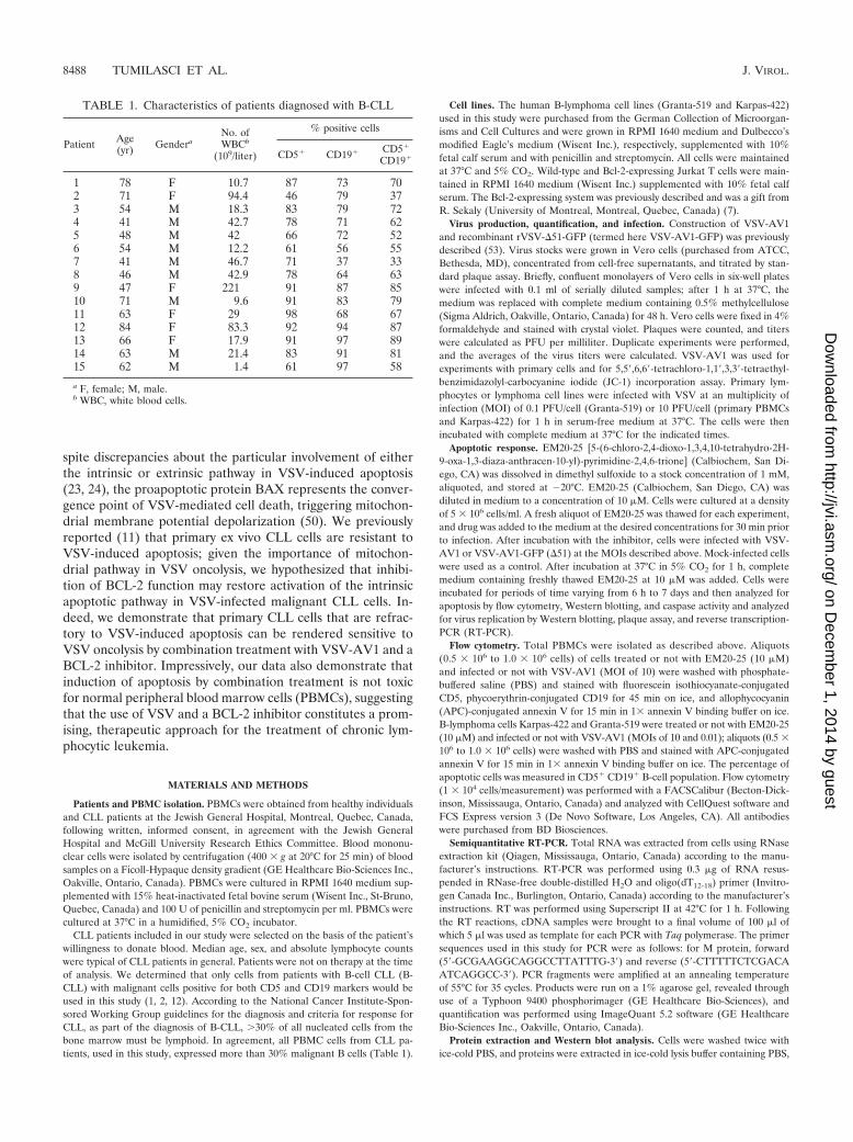
CLL patients included in our study were selected on the basis of the patient’swillingness to donate blood. Median age, sex, and absolute lymphocyte countswere typical of CLL patients in general. Patients were not on therapy at the timeof analysis. We determined that only cells from patients with B-cell CLL (B-CLL) with malignant cells positive for both CD5 and CD19 markers would beused in this study (1, 2, 12). According to the National Cancer Institute-Spon-sored Working Group guidelines for the diagnosis and criteria for response forCLL, as part of the diagnosis of B-CLL, �30% of all nucleated cells from thebone marrow must be lymphoid. In agreement, all PBMC cells from CLL pa-tients, used in this study, expressed more than 30% malignant B cells (Table 1).
Cell lines. The human B-lymphoma cell lines (Granta-519 and Karpas-422)used in this study were purchased from the German Collection of Microorgan-isms and Cell Cultures and were grown in RPMI 1640 medium and Dulbecco’smodified Eagle’s medium (Wisent Inc.), respectively, supplemented with 10%fetal calf serum and with penicillin and streptomycin. All cells were maintainedat 37°C and 5% CO2. Wild-type and Bcl-2-expressing Jurkat T cells were main-tained in RPMI 1640 medium (Wisent Inc.) supplemented with 10% fetal calfserum. The Bcl-2-expressing system was previously described and was a gift fromR. Sekaly (University of Montreal, Montreal, Quebec, Canada) (7).
Virus production, quantification, and infection. Construction of VSV-AV1and recombinant rVSV-�51-GFP (termed here VSV-AV1-GFP) was previouslydescribed (53). Virus stocks were grown in Vero cells (purchased from ATCC,Bethesda, MD), concentrated from cell-free supernatants, and titrated by stan-dard plaque assay. Briefly, confluent monolayers of Vero cells in six-well plateswere infected with 0.1 ml of serially diluted samples; after 1 h at 37°C, themedium was replaced with complete medium containing 0.5% methylcellulose(Sigma Aldrich, Oakville, Ontario, Canada) for 48 h. Vero cells were fixed in 4%formaldehyde and stained with crystal violet. Plaques were counted, and titerswere calculated as PFU per milliliter. Duplicate experiments were performed,and the averages of the virus titers were calculated. VSV-AV1 was used forexperiments with primary cells and for 5,5�,6,6�-tetrachloro-1,1�,3,3�-tetraethyl-benzimidazolyl-carbocyanine iodide (JC-1) incorporation assay. Primary lym-phocytes or lymphoma cell lines were infected with VSV at an multiplicity ofinfection (MOI) of 0.1 PFU/cell (Granta-519) or 10 PFU/cell (primary PBMCsand Karpas-422) for 1 h in serum-free medium at 37°C. The cells were thenincubated with complete medium at 37°C for the indicated times.
Apoptotic response. EM20-25 [5-(6-chloro-2,4-dioxo-1,3,4,10-tetrahydro-2H-9-oxa-1,3-diaza-anthracen-10-yl)-pyrimidine-2,4,6-trione] (Calbiochem, San Di-ego, CA) was dissolved in dimethyl sulfoxide to a stock concentration of 1 mM,aliquoted, and stored at �20°C. EM20-25 (Calbiochem, San Diego, CA) wasdiluted in medium to a concentration of 10 �M. Cells were cultured at a densityof 5 � 106 cells/ml. A fresh aliquot of EM20-25 was thawed for each experiment,and drug was added to the medium at the desired concentrations for 30 min priorto infection. After incubation with the inhibitor, cells were infected with VSV-AV1 or VSV-AV1-GFP (�51) at the MOIs described above. Mock-infected cellswere used as a control. After incubation at 37°C in 5% CO2 for 1 h, completemedium containing freshly thawed EM20-25 at 10 �M was added. Cells wereincubated for periods of time varying from 6 h to 7 days and then analyzed forapoptosis by flow cytometry, Western blotting, and caspase activity and analyzedfor virus replication by Western blotting, plaque assay, and reverse transcription-PCR (RT-PCR).
Flow cytometry. Total PBMCs were isolated as described above. Aliquots(0.5 � 106 to 1.0 � 106 cells) of cells treated or not with EM20-25 (10 �M)and infected or not with VSV-AV1 (MOI of 10) were washed with phosphate-buffered saline (PBS) and stained with fluorescein isothiocyanate-conjugatedCD5, phycoerythrin-conjugated CD19 for 45 min on ice, and allophycocyanin(APC)-conjugated annexin V for 15 min in 1� annexin V binding buffer on ice.B-lymphoma cells Karpas-422 and Granta-519 were treated or not with EM20-25(10 �M) and infected or not with VSV-AV1 (MOIs of 10 and 0.01); aliquots (0.5 �106 to 1.0 � 106 cells) were washed with PBS and stained with APC-conjugatedannexin V for 15 min in 1� annexin V binding buffer on ice. The percentage ofapoptotic cells was measured in CD5� CD19� B-cell population. Flow cytometry(1 � 104 cells/measurement) was performed with a FACSCalibur (Becton-Dick-inson, Mississauga, Ontario, Canada) and analyzed with CellQuest software andFCS Express version 3 (De Novo Software, Los Angeles, CA). All antibodieswere purchased from BD Biosciences.
Semiquantitative RT-PCR. Total RNA was extracted from cells using RNaseextraction kit (Qiagen, Mississauga, Ontario, Canada) according to the manu-facturer’s instructions. RT-PCR was performed using 0.3 �g of RNA resus-pended in RNase-free double-distilled H2O and oligo(dT12-18) primer (Invitro-gen Canada Inc., Burlington, Ontario, Canada) according to the manufacturer’sinstructions. RT was performed using Superscript II at 42°C for 1 h. Followingthe RT reactions, cDNA samples were brought to a final volume of 100 �l ofwhich 5 �l was used as template for each PCR with Taq polymerase. The primersequences used in this study for PCR were as follows: for M protein, forward(5�-GCGAAGGCAGGCCTTATTTG-3�) and reverse (5�-CTTTTTCTCGACAATCAGGCC-3�). PCR fragments were amplified at an annealing temperatureof 55°C for 35 cycles. Products were run on a 1% agarose gel, revealed throughuse of a Typhoon 9400 phosphorimager (GE Healthcare Bio-Sciences), andquantification was performed using ImageQuant 5.2 software (GE HealthcareBio-Sciences Inc., Oakville, Ontario, Canada).
Protein extraction and Western blot analysis. Cells were washed twice withice-cold PBS, and proteins were extracted in ice-cold lysis buffer containing PBS,
TABLE 1. Characteristics of patients diagnosed with B-CLL
Patient Age(yr) Gendera
No. ofWBCb
(109/liter)
% positive cells
CD5� CD19� CD5�
CD19�
1 78 F 10.7 87 73 702 71 F 94.4 46 79 373 54 M 18.3 83 79 724 41 M 42.7 78 71 625 48 M 42 66 72 526 54 M 12.2 61 56 557 41 M 46.7 71 37 338 46 M 42.9 78 64 639 47 F 221 91 87 8510 71 M 9.6 91 83 7911 63 F 29 98 68 6712 84 F 83.3 92 94 8713 66 F 17.9 91 97 8914 63 M 21.4 83 91 8115 62 M 1.4 61 97 58
a F, female; M, male.b WBC, white blood cells.
8488 TUMILASCI ET AL. J. VIROL.
on Decem
ber 1, 2014 by guesthttp://jvi.asm
.org/D
ownloaded from
0.05% NP-40, 0.1% glycerol, 30 mM NaF, 40 mM �-glycerophosphate, 10 mMNa3VO4, and protease inhibitor cocktail (Sigma-Aldrich, Oakville, Ontario, Can-ada) at a dilution of 1/1,000. Extracts were kept on ice for 15 min and centrifugedat 10,000 � g for 25 min (4°C), and supernatants were stored at �80°C. Proteinconcentration was determined with Bio-Rad protein assay reagent (Bio-Rad,Hercules, CA). Mitochondrial versus cytosol fractions of cells were preparedusing a mitochondria isolation kit for cultured cells (Pierce, Rockford, IL)according to the manufacturer’s instructions. Protein extracts (30 �g) were re-solved using 12 to 14% sodium dodecyl sulfate (SDS)-polyacrylamide gel elec-trophoresis and transferred to nitrocellulose membranes (Hybond C Super; GEHealthcare Bio-Sciences Inc.). The membranes were blocked for 1 h in 5%nonfat dried milk in Tris-buffered saline containing 0.5% Tween 20. The mem-branes were then incubated with any of the following primary antibodies: anti-rabbit VSV (1:5,000), anti-cleaved caspase-3 (Cell Signaling, Danvers, MA)(1:2,000), anti-rabbit �-actin (Cell Signaling, Danvers, MA) (1:1,000), oranti-mouse BCL-2 (Santa Cruz Biotechnology, Santa Cruz, CA) (1:2,000).The immunocomplexes were detected with horseradish peroxidase-conju-gated secondary antibodies and enhanced chemiluminescence according tothe manufacturer’s specifications (ECL kit; GE Healthcare Bio-Sciences Inc.).Cytochrome c was analyzed with anti-rabbit cytochrome c monoclonal antibody(MAb) or control antibody anti-mouse �-actin (Cell Signaling, Danvers, MA).Western blot quantification was assessed by densitometric analyses of scannedfilms with the use of the Scion Image 4.0. software.
Coimmunoprecipitation assay. Cells were lysed with 1% CHAPS lysis buffer{10 mM HEPES (pH 7.4), 150 mM NaCl, 1% 3-[(3-cholamidopropyl)-dimeth-ylammonio]-1-propanesulfonate (CHAPS)} containing protease inhibitors. To-tal protein (200 �g) was incubated with 2 �g of anti-BCL-2 MAb (Santa CruzBiotechnology, Santa Cruz, CA) or 2 �g of anti-BAX MAb (clone 6A7) (SigmaAldrich, Oakville, Ontario, Canada) in CHAPS lysis buffer at 4°C overnight ona rotator. Immunoprecipitates were collected by incubating with 20 �l proteinL-agarose (Santa Cruz Biotechnology, Santa Cruz, CA) for 4 h at 4°C, followedby centrifugation for 1 min. The pellets were washed three times with CHAPSlysis buffer, and beads were boiled in 2� SDS buffer and analyzed by Westernblotting using the anti-BAX MAb (Santa Cruz Biotechnology, Santa Cruz, CA).
Measurement of mitochondrial potential by JC-1 staining. Cells (0.5 � 106)were cultured in six-well plates. After treatment with EM20-25 and/or VSV-AV1, the cells were collected, washed in PBS, and resuspended in mediumcontaining JC-1 (Molecular Probes-Invitrogen Canada Inc., Ontario, Canada) ata final concentration of 1 mM/liter and incubated at 37°C for 15 min. Afterincubation, cells were subjected to flow cytometry analysis on a FACSCalibur(Becton-Dickinson) and analyzed with CellQuest software.
Quantitative measurement of caspase activity. Cells were lysed in buffer con-taining 50 mM HEPES (pH 7.4), 100 mM NaCl, 0.1% CHAPS, 0.1 mM EDTA,and 1 mM dithiothreitol. The lysates were clarified by centrifugation, and thesupernatants were used for enzyme assays. Enzymatic reactions were carried outin 2.5� piperazine-N,N�-bis(2-ethanesulfonic acid) (PIPES) buffer containing 20�g of protein lysate and 5 �M Asp-Glu-Val-Asp (DEVD)–7-amino-4-triflu-oromethyl coumarin (AFC) or 5 �M Leu-Glu-His-Asp (LEHD)-AFC (bothfrom BioMol International, Plymouth Meeting, PA). Fluorescent AFC forma-tion was measured at a excitation wavelength of 390 nm and emission wavelengthof 538 nm for 30 cycles using a Bio-Rad Fluoromark.
Viability assay. Cell viability was assessed by 3-(4,5-dimethylthiazol)-2,5-di-phenyl tetrazolium (MTT) dye absorbance according to the manufacturer’s in-structions (Chemicon International, Billerica, MA). Cells were seeded in 96-wellplates at a density of 5 � 105 cells (PBMCs) per well or 100,000 cells per well(Karpas-422 and Granta-519 cells). For drug combination studies, cells weretreated or not with EM20-25 (10 �g) and infected or not with VSV-AV1 (MOIof 10) where indicated. For 50% inhibitory concentration (IC50) assay, increasingconcentrations of EM20-25 (300 nm to 200 �M) were used. Plates were incu-bated at 37°C and 5% CO2 for 7 days. Each experimental condition was testedin quadruplicate experiments.
Statistical, synergism, and therapeutic index analyses. The statistical analysiswas performed using the Student’s t test. P values that were 0.05 were consid-ered statistically significant. Average values were expressed as means standarddeviations (SDs). The following equation was used to determine the combinedcytotoxic effects of EM20-25 according to the method of Chou and Talalay (15):CI1⁄4[(D)1/(Dx)1]þ[(D)2/(Dx)2]þ(D)1(D)2/(Dx)1(Dx)2, where (D)1 and (D)2 arethe doses of treatments 1 and 2 that produce the x effect when used in combi-nation and (Dx)1 and (Dx)2 represent the doses of treatments 1 and 2 thatproduce the same x effect when used alone. The combination is additive when theCI equals 1.0, synergistic when the CI is 1.0, and antagonistic when the CI is�1.0. The therapeutic index is the capacity or propensity of a drug to affect onecell population in preference to others, i.e., the ability of a drug to affect one kind
of cell and produce effects in doses lower than those required to affect other cells.The therapeutic index was calculated by measuring 50% lethal dose/50% ther-apeutically effective dose, where the lethal dose applies to the normal populationand the therapeutically effective dose applies to the malignant population (14).
RESULTS
Primary CLL cells are resistant to VSV-AV1-induced apop-tosis. Overexpression of the antiapoptotic BCL-2 protein hasbeen associated with an apoptosis-resistant phenotype in CLLand other malignances (31, 40, 42). To analyze VSV-inducedoncolysis in BCL-2-overexpressing malignant cells, primaryCLL cells from seven patients were infected with a naturallyattenuated variant of VSV (AV1) at an MOI of 10, and by 96 hpostinfection, cells were analyzed for virus replication and ap-optosis; all samples showed high levels of BCL-2 protein com-pared to samples from healthy volunteers (Fig. 1a). Despitethe fact that viral replication was detected in all samples (Fig.1c), VSV-AV1 did not induce significant apoptosis in the CLLsamples assayed by annexin V staining (Fig. 1b). Indeed, in-fection at higher MOIs of VSV-AV1 (MOIs of 10, 30, and 60)did not further increase apoptosis (Fig. 1d and e), suggestingthat BCL-2 overexpression may play a role in resistance toVSV-induced apoptosis in CLL cells.
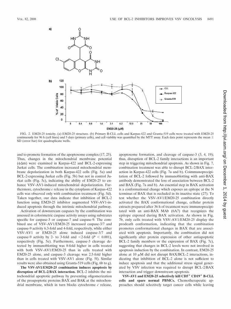
BCL-2 inhibitor EM20-25 sensitizes ex vivo CLL cells toVSV-AV1 oncolysis. Small organic molecules, such as EM20-25(36, 45) (Fig. 2a), have been shown to sensitize apoptosis-resistant cells to cytotoxic drugs by binding to BCL-2 anddisrupting interactions with proapoptotic proteins, such asBAX and BAK. As such, investigations are under way to ex-plore the use of small-molecule inhibitors as therapeuticagents for the treatment of malignancies that overexpressBCL-2 (33, 34, 59). To examine the effect of EM20-25 on VSVoncolysis in CLL cells, primary human CLL cells from sevendifferent patients (CLL8 to CLL11 and CLL13 to CLL15) werepretreated with 10 �M EM20-25 (IC50 approximately 20 �M[Fig. 2b]) and infected with VSV-AV1 (MOI of 10). Pretreat-ment with EM20-25 for 30 min dramatically diminished theresistance of CLL cells to VSV-induced apoptosis (Fig. 3a). By96 h postinfection, the combination increased cell death morethan fivefold compared to either VSV-AV1 or EM20-25 alone(1.1- and 1.4-fold, respectively) (Fig. 3b). Importantly, syner-gism measured using a CI of 0.5 (see Materials and Methods)was achieved in all seven samples tested (P 0.0001 for com-parison to VSV-AV1 treatment alone or P 0.001 for com-parison to EM20-25 treatment alone) (Fig. 3c). Induction ofapoptosis by VSV-AV1 and EM20-25 in malignant CLL cellswas further confirmed by detection of cleaved caspase-3; thelevel of cleaved caspase-3 was 7- to 10-fold higher in cellstreated with both VSV-AV1 and EM20-25 than in cells treatedwith either agent alone (Fig. 3d). Analysis of VSV-AV1 repli-cation in malignant primary CLL cells by viral protein produc-tion (Fig. 3e), viral M mRNA synthesis (Fig. 3f), and virus titer(Fig. 3g) demonstrated that VSV-AV1 replicates in malignantCLL cells and that EM20-25 did not significantly alter viralreplication. Together these data demonstrate that VSV-AV1in combination with EM20-25 synergistically overcomes apop-tosis resistance in primary malignant CLL cells without in-creasing VSV-AV1 replication.
VOL. 82, 2008 USE OF BCL-2 INHIBITORS IMPROVES VSV ONCOLYSIS 8489
on Decem
ber 1, 2014 by guesthttp://jvi.asm
.org/D
ownloaded from

VSV-AV1 in combination with EM20-25 induces apoptosisthrough the intrinsic mitochondrial pathway. To further cor-roborate the data obtained in primary CLL cells, we sought toidentify cell lines that expressed BCL-2 protein at levels similarto those in CLL cells. Among 12 B-lymphoma cell lines tested,Karpas-422 and Granta-519 naturally overexpress BCL-2 tolevels comparable to those in ex vivo PBMCs from CLL pa-tients (Fig. 4a; also see Fig. 6a). As with CLL cells, bothKarpas-422 and Granta-519 cells had similar EM20-25 toxicitylevels (IC50s of approximately 20 �M) (Fig. 2b). Karpas-422cells were treated with EM20-25 (10 �M) for 30 min and theninfected with VSV-AV1 at an MOI of 10. The combination ledto a threefold increase in VSV-induced apoptosis compared tothe effect of virus alone (P 0,05), as demonstrated by annexinV-APC staining (Fig. 4b). When examined by the MTT reduc-tion assay, viability of Karpas-422 cells was reduced threefoldwith the combination, while the loss of viability was only 1.4-fold when Karpas-422 cells were treated with EM20-25 or VSValone (Fig. 4c). Interestingly, VSV replication was not signifi-cantly affected (Fig. 4d), indicating that apoptosis observed
with VSV-AV1/EM20-25 was not accompanied by increasedVSV replication.
To link BCL-2 overexpression with resistance to VSV-in-duced apoptosis, a Jurkat T-cell line expressing BCL-2 underthe control of doxycycline (DOX) was used (7). In this cellmodel, a fourfold increase in BCL-2 was detected 24 h afterDOX treatment (Fig. 4e). At 48 h after VSV infection, 85% ofcontrol Jurkat cells underwent apoptosis, whereas in BCL-2-expressing Jurkat cells, VSV-AV1-induced apoptosis reached amaximum of 40 to 50% (P 0.05) (Fig. 4f). Pretreatment ofBCL-2-expressing Jurkat cells with EM20-25 restored VSV-AV1-induced apoptosis to levels similar to those seen in con-trol Jurkat cells (Fig. 4f). Confirming our results with Kar-pas-422 cells, treatment with EM20-25 did not affect VSVreplication in Jurkat cells (control or BCL-2-expressingcells) (Fig. 4g).
We and others previously reported the importance of themitochondrial apoptotic pathway in VSV-induced oncolysis(22, 50). The loss of mitochondrial transmembrane potential isknown to trigger cytochrome c release from the mitochondria
FIG. 1. Primary CLL cells are resistant to VSV-AV1-induced apoptosis. (a) PBMCs were isolated from CLL patients and from two healthyvolunteers. Whole-cell extracts were analyzed for BCL-2 protein expression by immunoblotting. The BCL-2 protein expression level was quantifiedand normalized to the �-actin level. (b) VSV-AV1-induced apoptosis in PBMCs isolated from CLL patients. PBMCs from a healthy volunteer wereused as a BCL-2 expression control. At 96 h postinfection, apoptosis was measured using annexin V staining by flow cytometry. Mock-infected cellswere used as a control. (c) VSV-AV1 replication in PBMCs isolated from CLL patients and from two healthy volunteers infected with VSV-AV1(�) or not infected with VSV-AV1 (�) was examined by Western blotting. VSV-AV1 viral protein expression was monitored by Western blotanalysis with anti-rabbit VSV antibody. L, large protein; G, glycoprotein; N/P, nucleocapsid. (d and e) PBMCs isolated from a CLL patient (CLL3)were infected with VSV-AV1 at different MOIs (MOIs of 10, 30, and 60). Mock-infected cells were used as a control. (d) VSV viral proteinexpression was monitored as described above for panel c. The time postinfection (24, 48, and 72 hours after infection) is shown above the lanes.IB, immunoblotting; N, nucleocapsid. (e) VSV-AV1-induced apoptosis in PBMCs from a CLL patient measured by annexin V staining.Mock-infected cells were used as a control.
8490 TUMILASCI ET AL. J. VIROL.
on Decem
ber 1, 2014 by guesthttp://jvi.asm
.org/D
ownloaded from
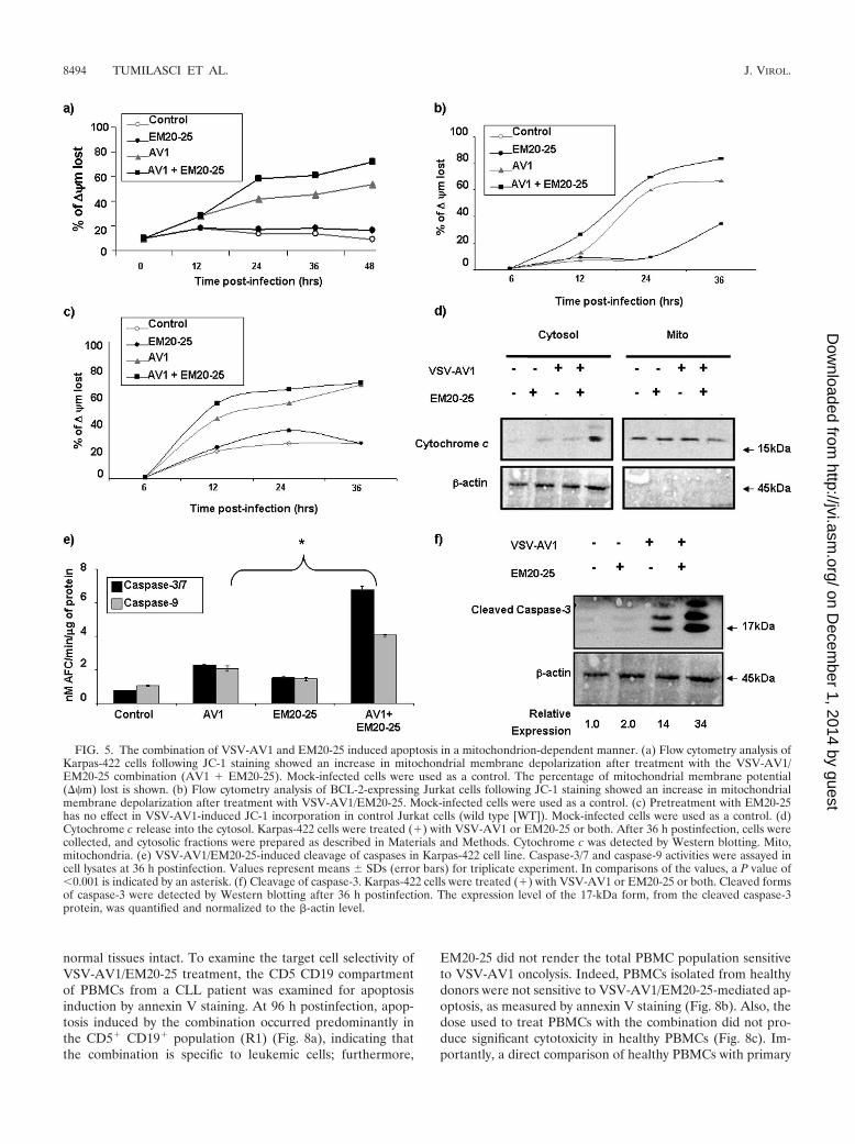
and to promote formation of the apoptosome complex (17, 25).Thus, changes in the mitochondrial membrane potential(��m) were examined in Karpas-422 and BCL-2-expressingJurkat cells. The combination increased mitochondrial mem-brane depolarization in both Karpas-422 cells (Fig. 5a) andBCL-2-expressing Jurkat cells (Fig. 5b) but not in control Ju-rkat cells (Fig. 5c), indicating the ability of EM20-25 to en-hance VSV-AV1-induced mitochondrial depolarization. Fur-thermore, cytochrome c release in the cytoplasm of Karpas-422cells was observed only with combination treatment (Fig. 5d).Taken together, our data indicate that inhibition of BCL-2function using EM20-25 inhibitor augmented VSV-AV1-in-duced apoptosis through the intrinsic mitochondrial pathway.
Activation of downstream caspases by the combination wasassessed in colorimetric caspase activity assays using substratesspecific for caspase-3 or caspase-7 and caspase-9. The com-bined use of VSV-AV1/EM20-25 increased caspase-3/7 andcaspase-9 activity 6.3-fold and 4-fold, respectively, while eitherVSV-AV1 or EM20-25 alone induced caspase-3/7 andcaspase-9 activity by 2- to 3-fold and 2-fold (P 0.001),respectively (Fig. 5e). Furthermore, caspase-3 cleavage de-tected by immunoblotting was 8-fold higher in cells treatedwith both VSV-AV1/EM20-25 than in cells treated withEM20-25 alone, and caspase-3 cleavage was 2.5-fold higherthan in cells treated with VSV-AV1 alone (Fig. 5f). Similarresults were also obtained using Granta-519 cells (Fig. 6b to g).
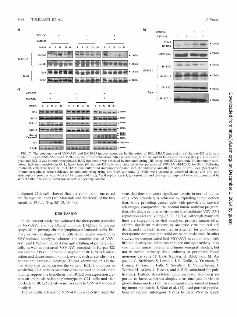
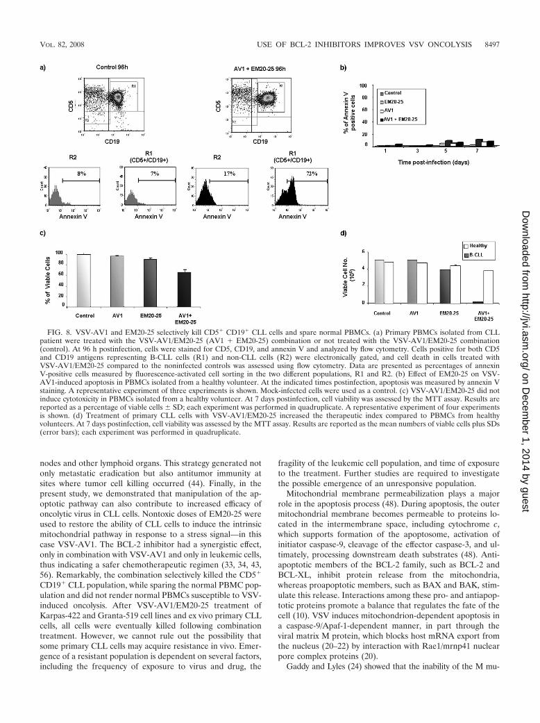
The VSV-AV1/EM20-25 combination induces apoptosis bydisruption of BCL-2/BAX interaction. BCL-2 inhibits the mi-tochondrial apoptotic pathway by preventing oligomerizationof the proapoptotic proteins BAX and BAK at the mitochon-drial membrane, which in turn blocks cytochrome c release,
apoptosome formation, and cleavage of caspase-3 (3, 4, 19);thus, disruption of BCL-2 family interactions is an importantstep in triggering mitochondrial apoptosis. As shown in Fig. 7,combination treatment was able to disrupt BCL-2/BAX inter-action in Karpas-422 cells (Fig. 7a and b). Coimmunoprecipi-tation of BCL-2 followed by immunoblotting with anti-BAXantibody demonstrated the loss of association between BCL-2and BAX (Fig. 7a and b). An essential step in BAX activationis a conformational change which exposes an epitope at the Nterminus of BAX that is occluded in its inactive state (27). Totest whether the VSV-AV1/EM20-25 combination directlyactivated the BAX conformational change, cellular proteinextracts prepared after 36 h of treatment were immunoprecipi-tated with an anti-BAX MAb (6A7) that recognizes theepitope exposed during BAX activation. As shown in Fig.7b, only cells treated with VSV-AV1/EM20-25 display theprodeath conformation, indicating that the combinationpromotes conformational changes in BAX that are associ-ated with apoptosis. Importantly, the combination did notsignificantly alter protein expression of other antiapoptoticBCL-2 family members or the expression of BAX (Fig. 7c),suggesting that changes in BCL-2 levels were not involved inapoptosis induction by the combination. In contrast, EM20-25alone at 10 �M did not disrupt BAX/BCL-2 interactions, in-dicating that inhibition of BCL-2 alone is not sufficient toinduce apoptosis and that the additional stress signal gener-ated by VSV infection was required to disrupt BCL-2/BAXinteraction and trigger downstream apoptosis.
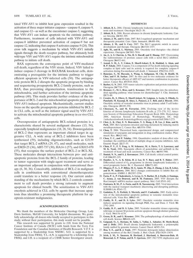
VSV-AV1 and EM20-25 selectively kill CD5� CD19� B-CLLcells and spare normal PBMCs. Chemotherapeutic ap-proaches should selectively target cancer cells while leaving
FIG. 2. EM20-25 toxicity. (a) EM20-25 structure. (b) Primary B-CLL cells and Karpas-422 and Granta-519 cells were treated with EM20-25continuously for 96 h (cell lines) and 5 days (primary cells), and cell viability was quantified by the MTT assay. Each data point represents the mean SD (error bar) for quadruplicate wells.
VOL. 82, 2008 USE OF BCL-2 INHIBITORS IMPROVES VSV ONCOLYSIS 8491
on Decem
ber 1, 2014 by guesthttp://jvi.asm
.org/D
ownloaded from
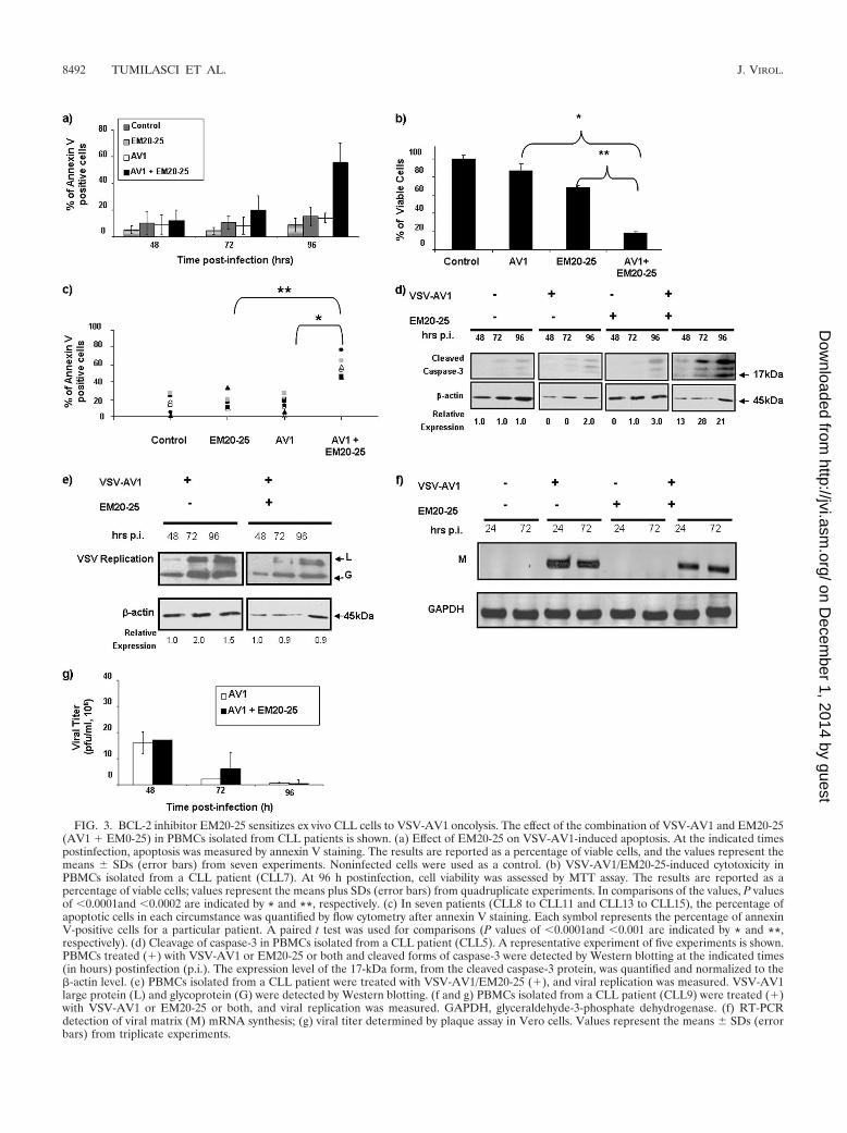
FIG. 3. BCL-2 inhibitor EM20-25 sensitizes ex vivo CLL cells to VSV-AV1 oncolysis. The effect of the combination of VSV-AV1 and EM20-25(AV1 � EM0-25) in PBMCs isolated from CLL patients is shown. (a) Effect of EM20-25 on VSV-AV1-induced apoptosis. At the indicated timespostinfection, apoptosis was measured by annexin V staining. The results are reported as a percentage of viable cells, and the values represent themeans SDs (error bars) from seven experiments. Noninfected cells were used as a control. (b) VSV-AV1/EM20-25-induced cytotoxicity inPBMCs isolated from a CLL patient (CLL7). At 96 h postinfection, cell viability was assessed by MTT assay. The results are reported as apercentage of viable cells; values represent the means plus SDs (error bars) from quadruplicate experiments. In comparisons of the values, P valuesof 0.0001and 0.0002 are indicated by * and **, respectively. (c) In seven patients (CLL8 to CLL11 and CLL13 to CLL15), the percentage ofapoptotic cells in each circumstance was quantified by flow cytometry after annexin V staining. Each symbol represents the percentage of annexinV-positive cells for a particular patient. A paired t test was used for comparisons (P values of 0.0001and 0.001 are indicated by * and **,respectively). (d) Cleavage of caspase-3 in PBMCs isolated from a CLL patient (CLL5). A representative experiment of five experiments is shown.PBMCs treated (�) with VSV-AV1 or EM20-25 or both and cleaved forms of caspase-3 were detected by Western blotting at the indicated times(in hours) postinfection (p.i.). The expression level of the 17-kDa form, from the cleaved caspase-3 protein, was quantified and normalized to the�-actin level. (e) PBMCs isolated from a CLL patient were treated with VSV-AV1/EM20-25 (�), and viral replication was measured. VSV-AV1large protein (L) and glycoprotein (G) were detected by Western blotting. (f and g) PBMCs isolated from a CLL patient (CLL9) were treated (�)with VSV-AV1 or EM20-25 or both, and viral replication was measured. GAPDH, glyceraldehyde-3-phosphate dehydrogenase. (f) RT-PCRdetection of viral matrix (M) mRNA synthesis; (g) viral titer determined by plaque assay in Vero cells. Values represent the means SDs (errorbars) from triplicate experiments.
8492 TUMILASCI ET AL. J. VIROL.
on Decem
ber 1, 2014 by guesthttp://jvi.asm
.org/D
ownloaded from
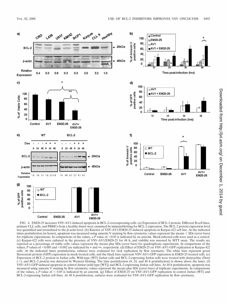
FIG. 4. EM20-25 increases VSV-AV1-induced apoptosis in BCL-2-overexpressing cells. (a) Expression of BCL-2 protein. Different B-cell lines,primary CLL cells, and PBMCs from a healthy donor were examined by immunoblotting for BCL-2 expression. The BCL-2 protein expression levelwas quantified and normalized to the �-actin level. (b) Kinetics of VSV-AV1/EM20-25-induced apoptosis in Karpas-422 cell line. At the indicatedtimes postinfection (in hours), apoptosis was measured using annexin V staining by flow cytometry; values represent the means SDs (error bars)for triplicate experiments. In comparisons of the values, a P value of 0.05 is indicated by an asterisk. Mock-infected cells were used as a control.(c) Karpas-422 cells were cultured in the presence of VSV-AV1/EM20-25 for 48 h, and viability was assessed by MTT assay. The results arereported as a percentage of viable cells; values represent the means plus SDs (error bars) for quadruplicate experiments. In comparisons of thevalues, P values of 0.005 and 0.002 are indicated by * and **, respectively. (d) Effect of EM20-25 on VSV-AV1-GFP replication in Karpas-422cells. At the indicated times postinfection, cultures were evaluated for viral replication by flow cytometry. The white bars represent greenfluorescent protein (GFP) expression in mock-treated cells, and the black bars represent VSV-AV1-GFP expression in EM20-25-treated cells. (e)Expression of BCL-2 protein in Jurkat cells. Wild-type (WT) Jurkat cells and BCL-2-expressing Jurkat cells were treated with doxycycline (Dox)(�), and BCL-2 protein was detected by Western blotting. The time postinfection (0, 24, and 48 h postinfection) is shown above the lanes. (f)VSV-AV1-GFP-induced apoptosis in control Jurkat (wild type [WT]) and BCL-2-expressing Jurkat cell lines. At 48 h postinfection, apoptosis wasmeasured using annexin V staining by flow cytometry; values represent the means plus SDs (error bars) of triplicate experiments. In comparisonsof the values, a P value of 0.05 is indicated by an asterisk. (g) Effect of EM20-25 on VSV-AV1-GFP replication in control Jurkat (WT) andBCL-2-expressing Jurkat cell lines. At 48 h postinfection, cultures were evaluated for VSV-AV1-GFP replication by flow cytometry.
VOL. 82, 2008 USE OF BCL-2 INHIBITORS IMPROVES VSV ONCOLYSIS 8493
on Decem
ber 1, 2014 by guesthttp://jvi.asm
.org/D
ownloaded from
normal tissues intact. To examine the target cell selectivity ofVSV-AV1/EM20-25 treatment, the CD5 CD19 compartmentof PBMCs from a CLL patient was examined for apoptosisinduction by annexin V staining. At 96 h postinfection, apop-tosis induced by the combination occurred predominantly inthe CD5� CD19� population (R1) (Fig. 8a), indicating thatthe combination is specific to leukemic cells; furthermore,
EM20-25 did not render the total PBMC population sensitiveto VSV-AV1 oncolysis. Indeed, PBMCs isolated from healthydonors were not sensitive to VSV-AV1/EM20-25-mediated ap-optosis, as measured by annexin V staining (Fig. 8b). Also, thedose used to treat PBMCs with the combination did not pro-duce significant cytotoxicity in healthy PBMCs (Fig. 8c). Im-portantly, a direct comparison of healthy PBMCs with primary
FIG. 5. The combination of VSV-AV1 and EM20-25 induced apoptosis in a mitochondrion-dependent manner. (a) Flow cytometry analysis ofKarpas-422 cells following JC-1 staining showed an increase in mitochondrial membrane depolarization after treatment with the VSV-AV1/EM20-25 combination (AV1 � EM20-25). Mock-infected cells were used as a control. The percentage of mitochondrial membrane potential(��m) lost is shown. (b) Flow cytometry analysis of BCL-2-expressing Jurkat cells following JC-1 staining showed an increase in mitochondrialmembrane depolarization after treatment with VSV-AV1/EM20-25. Mock-infected cells were used as a control. (c) Pretreatment with EM20-25has no effect in VSV-AV1-induced JC-1 incorporation in control Jurkat cells (wild type [WT]). Mock-infected cells were used as a control. (d)Cytochrome c release into the cytosol. Karpas-422 cells were treated (�) with VSV-AV1 or EM20-25 or both. After 36 h postinfection, cells werecollected, and cytosolic fractions were prepared as described in Materials and Methods. Cytochrome c was detected by Western blotting. Mito,mitochondria. (e) VSV-AV1/EM20-25-induced cleavage of caspases in Karpas-422 cell line. Caspase-3/7 and caspase-9 activities were assayed incell lysates at 36 h postinfection. Values represent means SDs (error bars) for triplicate experiment. In comparisons of the values, a P value of0.001 is indicated by an asterisk. (f) Cleavage of caspase-3. Karpas-422 cells were treated (�) with VSV-AV1 or EM20-25 or both. Cleaved formsof caspase-3 were detected by Western blotting after 36 h postinfection. The expression level of the 17-kDa form, from the cleaved caspase-3protein, was quantified and normalized to the �-actin level.
8494 TUMILASCI ET AL. J. VIROL.
on Decem
ber 1, 2014 by guesthttp://jvi.asm
.org/D
ownloaded from

FIG. 6. The resistance of Granta-519 cells to VSV oncolysis is overcome by EM20-25. (a) Expression of BCL-2 protein in six B-lymphoma celllines compared with ex vivo B-CLL cells (CLL 9) and PBMCs isolated from a healthy donor. Expression of BCL-2 was analyzed in whole-cellextracts by Western blotting with antibody against BCL-2. �-Actin was included as a loading control. The BCL-2 protein expression level wasquantified and normalized to the �-actin level. (b) Apoptosis was measured by flow cytometry in Granta-519 cells treated with EM20-25/VSV-�51-GFP. Results are presented as bars indicating the percentage of annexin V-positive cells. Mock-infected (control) cells or cells treated withEM20-25 were included as a control. In comparisons of the values, a P value of 0.05 is indicated by an asterisk. (c) Analysis of cytotoxicity byVSV-AV1/EM20-25 in Granta-519 cells. Results are presented as bars indicating the percentage of cell viability. In comparisons of the values, aP value of 0.0001 are indicated by an asterisk. (d) Expression of VSV-�51-GFP virus in infected cells was determined by fluorescence-activatedcell sorting. Results are presented as a percentage of green fluorescent protein-positive cells in Granta-519 cells. In panels b to d, Data arepresented as means � SDs (error bars). (e) Mitochondrial membrane depolarization (� �m) in response to VSV-AV1/EM20-25 treatment wasassessed by flow cytometry using JC-1 staining. Cells were treated with EM20-25/VSV-AV1 prior to JC-1 staining and flow cytometry analysis.Mock-infected cells were used as a control. (f and g) Granta-519 cells were treated as described above. At 36 h postinfection, cells were harvested,and the enzymatic activity of caspase-3/7 (f) and caspase-9 (g) proteases was determined. Results are presented as bars indicating the amount ofcaspase activity induced compared to the control sample (mock-infected cells). In comparisons of the values, a P value of 0.05 is indicated byan asterisk.
VOL. 82, 2008 USE OF BCL-2 INHIBITORS IMPROVES VSV ONCOLYSIS 8495
on Decem
ber 1, 2014 by guesthttp://jvi.asm
.org/D
ownloaded from
malignant CLL cells showed that the combination increasedthe therapeutic index (see Materials and Methods) of the twoagents by 19-fold (Fig. 8d) (8, 16, 49).
DISCUSSION
In the present study, we evaluated the therapeutic potentialof VSV-AV1 and the BCL-2 inhibitor EM20-25 to induceapoptosis in primary chronic lymphocytic leukemia cells. Pri-mary ex vivo malignant CLL cells were largely resistant toVSV-induced oncolysis, whereas the combination of VSV-AV1 and EM20-25 induced synergistic killing of primary CLLcells, as well as increased VSV-AV1 oncolysis in Karpas-422and Granta-519 cell lines and disruption of BCL-2/BAX inter-action and downstream apoptotic events, such as cytochrome crelease and caspase-3 cleavage. To our knowledge, this is thefirst study that demonstrates the value of BCL-2 inhibitors insensitizing CLL cells to oncolytic virus-induced apoptosis. Ourfindings support the hypothesis that BCL-2 overexpression cre-ates an apoptosis-resistant phenotype in CLL cells and thatblockade of BCL-2 activity sensitizes cells to VSV-AV1-indcedoncolysis.
The naturally attenuated VSV-AV1 is a selective oncolytic
virus that does not cause significant toxicity in normal humancells. VSV selectivity is achieved by exploiting tumor defectsthat, while providing cancer cells with growth and survivaladvantages, compromise the normal innate antiviral program,thus affording a cellular environment that facilitates VSV-AV1replication and cell killing (9, 32, 37, 53). Although many celllines are susceptible to viral oncolysis, primary tumors oftenexhibit significant resistance to oncolytic virus-induced celldeath, and this fact has resulted in a search for combinationtherapeutic strategies that could overcome resistance. In otherstudies, we demonstrated that VSV-AV1 in combination withhistone deacetylase inhibitors enhance oncolytic activity in exvivo human tumor material and tumor xenograft models, butnot in normal primary tissue cultures or peripheral bloodmononuclear cells (T. L.-A. Nguyen, H. Abdelbary, M. Ar-guello, C. Breitbach, S. Leveille, J.-S. Diallo, A. Yasmeen, T.Bismar, D. Kirn, T. Falls, V. Snoulton, B. Vanderhyden, J.Werier, H. Atkins, J. Hiscott, and J. Bell, submitted for pub-lication). Histone deacetylase inhibitors have also been re-ported to increase herpes simplex virus oncolysis in vivo inglioblastoma models (35). In an elegant study aimed at target-ing tumor metastasis, J. Qiao et al. (44) used purified popula-tions of normal autologous T cells to carry VSV to lymph
FIG. 7. The combination of VSV-AV1 and EM20-25 induces apoptosis by disruption of BCL-2/BAX interaction. (a) Karpas-422 cells weretreated (�) with VSV-AV1 and EM20-25 alone or in combination. After infection (0, 6, 12, 24, and 48 hours postinfection [hrs p.i.]), cells werelysed and BCL-2 was immunoprecipitated. BAX interaction was revealed by immunoblotting (IB) using anti-BAX antibody. IP, immunoprecipi-tation. IgG, immunoglobulin G; L, light chain. (b) Karpas-422 cells were cultured in the presence of VSV-AV1/EM20-25 for 36 h. Followingtreatment, cells were lysed in 1% CHAPS lysis buffer and immunoprecipitated with the indicated anti-BCL-2 MAb or anti-BAX (6A7) MAb.Immunoprecipitates were subjected to immunoblotting using anti-BAX antibody. (c) Cells were treated as described above, and pro- andantiapoptotic proteins were detected by immunoblotting. Viral replication (G, glycoprotein) and cleavage of caspase-3 were also monitored byWestern blot analysis. �-Actin was added as a loading control.
8496 TUMILASCI ET AL. J. VIROL.
on Decem
ber 1, 2014 by guesthttp://jvi.asm
.org/D
ownloaded from
nodes and other lymphoid organs. This strategy generated notonly metastatic eradication but also antitumor immunity atsites where tumor cell killing occurred (44). Finally, in thepresent study, we demonstrated that manipulation of the ap-optotic pathway can also contribute to increased efficacy ofoncolytic virus in CLL cells. Nontoxic doses of EM20-25 wereused to restore the ability of CLL cells to induce the intrinsicmitochondrial pathway in response to a stress signal—in thiscase VSV-AV1. The BCL-2 inhibitor had a synergistic effect,only in combination with VSV-AV1 and only in leukemic cells,thus indicating a safer chemotherapeutic regimen (33, 34, 43,56). Remarkably, the combination selectively killed the CD5�
CD19� CLL population, while sparing the normal PBMC pop-ulation and did not render normal PBMCs susceptible to VSV-induced oncolysis. After VSV-AV1/EM20-25 treatment ofKarpas-422 and Granta-519 cell lines and ex vivo primary CLLcells, all cells were eventually killed following combinationtreatment. However, we cannot rule out the possibility thatsome primary CLL cells may acquire resistance in vivo. Emer-gence of a resistant population is dependent on several factors,including the frequency of exposure to virus and drug, the
fragility of the leukemic cell population, and time of exposureto the treatment. Further studies are required to investigatethe possible emergence of an unresponsive population.
Mitochondrial membrane permeabilization plays a majorrole in the apoptosis process (48). During apoptosis, the outermitochondrial membrane becomes permeable to proteins lo-cated in the intermembrane space, including cytochrome c,which supports formation of the apoptosome, activation ofinitiator caspase-9, cleavage of the effector caspase-3, and ul-timately, processing downstream death substrates (48). Anti-apoptotic members of the BCL-2 family, such as BCL-2 andBCL-XL, inhibit protein release from the mitochondria,whereas proapoptotic members, such as BAX and BAK, stim-ulate this release. Interactions among these pro- and antiapop-totic proteins promote a balance that regulates the fate of thecell (10). VSV induces mitochondrion-dependent apoptosis ina caspase-9/Apaf-1-dependent manner, in part through theviral matrix M protein, which blocks host mRNA export fromthe nucleus (20–22) by interaction with Rae1/mrnp41 nuclearpore complex proteins (20).
Gaddy and Lyles (24) showed that the inability of the M mu-
FIG. 8. VSV-AV1 and EM20-25 selectively kill CD5� CD19� CLL cells and spare normal PBMCs. (a) Primary PBMCs isolated from CLLpatient were treated with the VSV-AV1/EM20-25 (AV1 � EM20-25) combination or not treated with the VSV-AV1/EM20-25 combination(control). At 96 h postinfection, cells were stained for CD5, CD19, and annexin V and analyzed by flow cytometry. Cells positive for both CD5and CD19 antigens representing B-CLL cells (R1) and non-CLL cells (R2) were electronically gated, and cell death in cells treated withVSV-AV1/EM20-25 compared to the noninfected controls was assessed using flow cytometry. Data are presented as percentages of annexinV-positive cells measured by fluorescence-activated cell sorting in the two different populations, R1 and R2. (b) Effect of EM20-25 on VSV-AV1-induced apoptosis in PBMCs isolated from a healthy volunteer. At the indicated times postinfection, apoptosis was measured by annexin Vstaining. A representative experiment of three experiments is shown. Mock-infected cells were used as a control. (c) VSV-AV1/EM20-25 did notinduce cytotoxicity in PBMCs isolated from a healthy volunteer. At 7 days postinfection, cell viability was assessed by the MTT assay. Results arereported as a percentage of viable cells SD; each experiment was performed in quadruplicate. A representative experiment of four experimentsis shown. (d) Treatment of primary CLL cells with VSV-AV1/EM20-25 increased the therapeutic index compared to PBMCs from healthyvolunteers. At 7 days postinfection, cell viability was assessed by the MTT assay. Results are reported as the mean numbers of viable cells plus SDs(error bars); each experiment was performed in quadruplicate.
VOL. 82, 2008 USE OF BCL-2 INHIBITORS IMPROVES VSV ONCOLYSIS 8497
on Decem
ber 1, 2014 by guesthttp://jvi.asm
.org/D
ownloaded from
tated VSV-AV1 to inhibit host gene expression resulted in theactivation of three major initiator caspases—caspase-8, caspase-9,and caspase-12—as well as the executioner caspase-3, suggestingthat VSV-AV1 can induce apoptosis via the extrinsic pathway.Furthermore, treatment of cells infected with VSV-AV1 andcaspase-8 inhibitor prevented activation of caspase-9 but notcaspase-12, indicating that caspase-8 activates caspase-9 (24). Thiscross talk suggests a mechanism by which VSV-AV1 initiallysignals through the death receptor pathway, but ultimately bothwild-type VSV and VSV-AV1 require the intrinsic mitochondrialpathway to initiate cell death.
BAX represents the convergence point of VSV-mediatedcell death, regardless of the VSV strain. Indeed, VSV failed toinduce caspase-3 cleavage in BAX/BAK knockout cells, dem-onstrating a prerequisite for the intrinsic pathway to triggerefficient apoptosis in VSV-infected cells (50). The antiapop-totic protein BCL-2 disrupts the apoptotic program by bindingand sequestering proapoptotic BCL-2 family proteins, such asBAX, thus preventing oligomerization, translocation to themitochondria, and further activation of the intrinsic apoptoticpathway (40). This study provides further evidence of the im-portance of BCL-2 and the mitochondrial apoptotic pathway inVSV-AV1-induced apoptosis. Mechanistically, current studiesfocus on the specific proapoptotic proteins inhibited by BCL-2in CLL cells, as well as the initiator signal used by VSV-AV1to activate the mitochondrial apoptotic pathway in ex vivo CLLcells.
Overexpression of antiapoptotic BCL-related proteins is acharacteristic shared by several malignant diseases (13, 54),especially lymphoid malignancies (18, 28, 54). Downregulationof BCL-2 thus represents an important clinical target in ag-gressive CLL. A wide array of BCL-2 inhibitors have beensynthesized (59), including oligonucleotides, such as G3139that target BCL-2 mRNA (29, 47), and small molecules, suchas EM20-25 (36), ABT-737 (30), HA14-1 (57), and GX015-070(55), that recognize the surface pocket of BCL-2 or BCL-XL.These molecules disrupt interactions between pro- and anti-apoptotic proteins from the BCL-2 family of proteins, leadingto tumor regression with single-agent treatment and serve asan important adjuvant in conjunction with conventional ther-apy (6, 30, 36). Conceivably, inhibition of BCL-2 in malignantcells in combination with conventional chemotherapeuticscould translate to a better response (4). Our current under-standing of the mechanisms by which BCL-2 controls commit-ment to cell death provides a strong rationale to augmentapoptosis for clinical benefit. The sensitization to VSV-AV1oncolysis achieved in CLL cells by agents that increase apop-tosis thus identifies a promising therapeutic platform for ap-optosis-resistant malignancies.
ACKNOWLEDGMENTS
We thank the members of the Molecular Oncology Group, LadyDavis Institute, McGill University, for helpful discussions. We grate-fully acknowledge all donors who kindly accepted to participate in thisstudy; without their participation, this study would not be possible.
This research was supported by grants to J.H. from the NationalCancer Institute of Canada (NCIC) with funds from the Terry FoxFoundation and the Canadian Institutes of Health Research. V.F.T. issupported by a Studentship from NSERC, S.O. is supported by aStudentship from FRSQ, T.L.-A.N. is supported by a fellowship fromFRSQ, and J.H. is a recipient of a CIHR Senior Investigator award.
REFERENCES
1. Abbott, B. L. 2006. Chronic lymphocytic leukemia: recent advances in diag-nosis and treatment. Oncologist 11:21–30.
2. Abbott, B. L. 2006. Recent advances in chronic lymphocytic leukemia. Can-cer Investig. 24:302–309.
3. Adams, J. M., and S. Cory. 2007. Bcl-2-regulated apoptosis: mechanism andtherapeutic potential. Curr. Opin. Immunol. 19:488–496.
4. Adams, J. M., and S. Cory. 2007. The Bcl-2 apoptotic switch in cancerdevelopment and therapy. Oncogene 26:1324–1337.
5. Aghi, M., and R. L. Martuza. 2005. Oncolytic viral therapies—the clinicalexperience. Oncogene 24:7802–7816.
6. An, J., A. S. Chervin, A. Nie, H. S. Ducoff, and Z. Huang. 2007. Overcomingthe radioresistance of prostate cancer cells with a novel Bcl-2 inhibitor.Oncogene 26:652–661.
7. Aouad, S. M., L. Y. Cohen, E. Sharif-Askari, E. K. Haddad, A. Alam, andR. P. Sekaly. 2004. Caspase-3 is a component of Fas death-inducing signalingcomplex in lipid rafts and its activity is required for complete caspase-8activation during Fas-mediated cell death. J. Immunol. 172:2316–2323.
8. Banerjee, S., M. Hussain, A. Wang, A. Saliganan, M. Che, D. Bonfil, M.Cher, and F. H. Sarkar. 2007. In vitro and in vivo molecular evidence forbetter therapeutic efficacy of ABT-627 and taxotere combination in prostatecancer. Cancer Res. 67:3818–3826.
9. Barber, G. N. 2005. VSV-tumor selective replication and protein translation.Oncogene 24:7710–7719.
10. Brenner, C., M. L. Bras, and G. Kroemer. 2003. Insights into the mitochon-drial signaling pathway: what lessons for chemotherapy? J. Clin. Immunol.23:73–80.
11. Cesaire, R., S. Oliere, E. Sharif-Askari, M. Loignon, A. Lezin, S. Olindo, G.Panelatti, M. Kazanji, R. Aloyz, L. Panasci, J. C. Bell, and J. Hiscott. 2006.Oncolytic activity of vesicular stomatitis virus in primary adult T-cell leuke-mia. Oncogene 25:349–358.
12. Chiorazzi, N., and M. Ferrarini. 2006. Evolving view of the in-vivokinetics of chronic lymphocytic leukemia B cells, p. 273–278, 512. Amer-ican Society of Hematology Education Program Book 2006. Hematology2006. American Society of Hematology, Washington, DC. http://asheducationbook.hematologylibrary.org/cgi/content/full/2006/1/273.
13. Choi, J., K. Choi, E. N. Benveniste, Y. S. Hong, J. H. Lee, J. Kim, and K.Park. 2005. Bcl-2 promotes invasion and lung metastasis by inducing matrixmetalloproteinase-2. Cancer Res. 65:5554–5560.
14. Chou, T. 2006. Theoretical basis, experimental design, and computerizedsimulation of synergism and antagonism in drug combination studies. Phar-macol. Rev. 58:621–681.
15. Chou, T. C., and P. Talalay. 1984. Quantitative analysis of dose-effect rela-tionships: the combined effects of multiple drugs or enzyme inhibitors. Adv.Enzyme Regul. 22:27–55.
16. Chun, P. Y., F. Y. Feng, A. M. Scheurer, M. A. Davis, T. S. Lawrence, andM. K. Nyati. 2006. Synergistic effects of gemcitabine and gefitinib in thetreatment of head and neck carcinoma. Cancer Res. 66:981–988.
17. Danial, N. N., and S. J. Korsmeyer. 2004. Cell death: critical control points.Cell 116:205–219.
18. Danilov, A. V., A. K. Klein, H. J. Lee, D. V. Baez, and B. T. Huber. 2005.Differential control of G0 programme in chronic lymphocytic leukaemia: anovel prognostic factor. Br. J. Haematol. 128:472–481.
19. Dlugosz, P. G., L. P. Billen, M. G. Annis, W. Zhu, Z. Zhang, J. Lin, B. Leber,and D. W. Andrews. 2006. Bcl-2 changes conformation to inhibit Bax oli-gomerization. EMBO J. 25:2287–2296.
20. Faria, P. A., P. Chakraborty, A. Levay, G. N. Barber, H. J. Ezelle, J. Enninga,C. Arana, J. van Deursen, and B. M. Fontoura. 2005. VSV disrupts theRae1/mrnp41 mRNA nuclear export pathway. Mol. Cell 17:93–102.
21. Fontoura, B. M., P. A. Faria, and D. R. Nussenzveig. 2005. Viral interactionswith the nuclear transport machinery: discovering and disrupting pathways.IUBMB Life 57:65–72.
22. Gadaleta, P., X. Perfetti, S. Mersich, and F. Coulombie. 2005. Early activa-tion of the mitochondrial apoptotic pathway in vesicular stomatitis virus-infected cells. Virus Res. 109:65–69.
23. Gaddy, D. F., and D. S. Lyles. 2007. Oncolytic vesicular stomatitis virusinduces apoptosis via signaling through PKR, Fas, and Daxx. J. Virol. 81:2792–2804.
24. Gaddy, D. F., and D. S. Lyles. 2005. Vesicular stomatitis viruses expressingwild-type or mutant M proteins activate apoptosis through distinct pathways.J. Virol. 79:4170–4179.
25. Green, D. R., and G. Kroemer. 2004. The pathophysiology of mitochondrialcell death. Science 305:626–629.
26. Guipaud, O., L. Deriano, H. Salin, L. Vallat, L. Sabatier, H. Merle-Beral,and J. Delic. 2003. B-cell chronic lymphocytic leukaemia: a polymorphicfamily unified by genomic features. Lancet Oncol. 4:505–514.
27. Hsu, Y.-T., and R. J. Youle. 1997. Nonionic detergents induce dimerizationamong members of the Bcl-2 family. J. Biol. Chem. 272:13829–13834.
28. Irish, J. M., N. Ånensen, R. Hovland, J. Skavland, A. Børresen-Dale, Ø.Bruserud, G. P. Nolan, and B. T. Gjertsen. 2007. Flt3 Y591 duplication and
8498 TUMILASCI ET AL. J. VIROL.
on Decem
ber 1, 2014 by guesthttp://jvi.asm
.org/D
ownloaded from
Bcl-2 overexpression are detected in acute myeloid leukemia cells with highlevels of phosphorylated wild-type p53. Blood 109:2589–2596.
29. Kim, R., M. Emi, K. Matsuura, and K. Tanabe. 2007. Antisense and non-antisense effects of antisense Bcl-2 on multiple roles of Bcl-2 as a chemo-sensitizer in cancer therapy. Cancer Gene Ther. 14:1–11.
30. Kline, M. P., S. V. Rajkumar, M. M. Timm, T. K. Kimlinger, J. L. Haug, J. A.Lust, P. R. Greipp, and S. Kumar. 2007. ABT-737, an inhibitor of Bcl-2family proteins, is a potent inducer of apoptosis in multiple myeloma cells.Leukemia 21:1549–1560.
31. Letai, A., M. D. Sorcinelli, C. Beard, and S. J. Korsmeyer. 2004. Antiapop-totic BCL-2 is required for maintenance of a model leukemia. Cancer Cell6:241–249.
32. Lichty, B. D., A. T. Power, D. F. Stojdl, and J. C. Bell. 2004. Vesicularstomatitis virus: re-inventing the bullet. Trends Mol. Med. 10:210–216.
33. Lickliter, J. D., J. Cox, J. McCarron, N. R. Martinez, C. W. Schmidt, H. Lin,M. Nieda, and A. J. Nicol. 2007. Small-molecule Bcl-2 inhibitors sensitisetumour cells to immune-mediated destruction. Br. J. Cancer 96:600–608.
34. Lickliter, J. D., N. J. Wood, L. Johnson, G. McHugh, J. Tan, F. Wood, J. Cox,and N. W. Wickham. 2003. HA14-1 selectively induces apoptosis in Bcl-2-overexpressing leukemia/lymphoma cells, and enhances cytarabine-inducedcell death. Leukemia 17:2074–2080.
35. Liu, T.-C., P. Castelo-Branco, S. D. Rabkin, and R. L. Martuza. 2008.Trichostatin A and oncolytic HSV combination therapy shows enhancedantitumoral and antiangiogenic effects. Mol. Ther. 16:1041–1047.
36. Milanesi, E., P. Costantini, A. Gambalunga, R. Colonna, V. Petronilli, A.Cabrelle, S. Semenzato, A. M. Cesura, E. Pinard, and P. Bernardi. 2006. Themitochondrial effects of small organic ligands of BCL-2: sensitization ofBCL-2-overexpressing cells to apoptosis by a pyrimidine-2,4,6-trione deriv-ative. J. Biol. Chem. 281:10066–10072.
37. Nakhaei, P., S. Paz, S. Oliere, V. Tumilasci, J. C. Bell, and J. Hiscott. 2005.Oncolytic virotherapy of cancer with vesicular stomatitis virus. Gene Ther.Mol. Biol. 9:269–280.
38. Reference deleted.39. O’Neill, J., M. Manion, P. Schwartz, and D. M. Hockenbery. 2004. Promises
and challenges of targeting Bcl-2 anti-apoptotic proteins for cancer therapy.Biochim. Biophys. Acta 1705:43–51.
40. Packham, G., and F. K. Stevenson. 2005. Bodyguards and assassins: Bcl-2family proteins and apoptosis control in chronic lymphocytic leukaemia.Immunology 114:441–449.
41. Parato, K. A., D. Senger, P. A. Forsyth, and J. C. Bell. 2005. Recent progressin the battle between oncolytic viruses and tumours. Nat. Rev. Cancer 5:965–976.
42. Pepper, C., A. Thomas, J. Hidalgo de Quintana, S. Davies, T. Hoy, and P.Bentley. 1999. Pleiotropic drug resistance in B-cell chronic lymphocytic leu-kaemia—the role of Bcl-2 family dysregulation. Leuk. Res. 23:1007–1014.
43. Perez-Galan, P., G. Roue, N. Villamor, E. Campo, and D. Colomer. 2007.The BH3-mimetic GX15-070 synergizes with bortezomib in mantle cell lym-phoma by enhancing Noxa-mediated activation of Bak. Blood 109:4441–4449.
44. Qiao, J., T. Kottke, C. Willmon, F. Galivo, P. Wongthida, R. M. Diaz,J. Thompson, P. Ryno, G. N. Barber, J. Chester, P. Selby, K. Harrington, A.Melcher, and R. G. Vile. 2008. Purging metastases in lymphoid organs usinga combination of antigen-nonspecific adoptive T cell therapy, oncolytic vi-rotherapy and immunotherapy. Nat. Med. 14:37–44.
45. Rasola, A., and P. Bernardi. 2007. The mitochondrial permeability transitionpore and its involvement in cell death and in disease pathogenesis. Apoptosis12:815–833.
46. Reed, J. C. 2006. Proapoptotic multidomain Bcl-2/Bax-family proteins:mechanisms, physiological roles, and therapeutic opportunities. Cell DeathDiffer. 13:1378–1386.
47. Rheingold, S. R., M. D. Hogarty, S. M. Blaney, J. A. Zwiebel, C. Sauk-Schubert, R. Chandula, M. D. Krailo, and P. C. Adamson. 2007. Phase ITrial of G3139, a bcl-2 antisense oligonucleotide, combined with doxorubicinand cyclophosphamide in children with relapsed solid tumors: a Children’sOncology Group Study. J. Clin. Oncol. 25:1512–1518.
48. Riedl, S., and G. Salvesen. 2007. The apoptosome: signalling platform of celldeath. Nat. Rev. Mol. Cell Biol. 8:405–413.
49. Rots, M. G., D. T. Curielb, W. R. Gerritsenc, and H. J. Haismaa. 2003.Targeted cancer gene therapy: the flexibility of adenoviral gene therapyvectors. J. Controlled Release 87:159–165.
50. Sharif-Askari, E., P. Nakhaei, S. Oliere, V. Tumilasci, E. Hernandez, P.Wilkinson, R. Lin, J. C. Bell, and J. Hiscott. 2007. Bax-dependent mitochon-drial membrane permeabilization enhances IRF3-mediated innate immuneresponse during VSV infection. Virology 365:20–33.
51. Sorenson, C. M. 2004. Bcl-2 family members and disease. Biochim. Biophys.Acta 1644:169–177.
52. Stojdl, D. F., B. Lichty, S. Knowles, R. Marius, H. Atkins, N. Sonenberg, andJ. C. Bell. 2000. Exploiting tumor-specific defects in the interferon pathwaywith a previously unknown oncolytic virus. Nat. Med. 6:821–825.
53. Stojdl, D. F., B. D. Lichty, B. R. tenOever, J. M. Paterson, A. T. Power, S.Knowles, R. Marius, J. Reynard, L. Poliquin, H. Atkins, E. G. Brown, R. K.Durbin, J. E. Durbin, J. Hiscott, and J. C. Bell. 2003. VSV strains withdefects in their ability to shutdown innate immunity are potent systemicanti-cancer agents. Cancer Cell 4:263–275.
54. Trisciuoglio, D., M. Desideri, L. Ciuffreda, M. Mottolese, D. Ribatti, A.Vacca, M. Del Rosso, L. Marcocci, G. Zupi, and D. Del Bufalo. 2005. Bcl-2overexpression in melanoma cells increases tumor progression-associatedproperties and in vivo tumor growth. J. Cell. Physiol. 205:414–421.
55. Trudel, S., Z. Li, J. Rauw, R. Tiedemann, X. Wen, and K. Stewart. 2007.Preclinical studies of the pan-Bcl inhibitor obatoclax (GX015-070) in mul-tiple myeloma. Blood 109:5430–5438.
56. Trudel, S., A. K. Stewart, Z. Li, Y. Shu, S. B. Liang, Y. Trieu, D. Reece, J.Paterson, D. Wang, and X. Y. Wen. 2007. The Bcl-2 family protein inhibitor,ABT-737, has substantial antimyeloma activity and shows synergistic effectwith dexamethasone and melphalan. Clin. Cancer Res. 13:621–629.
57. Wlodkowic, D., J. Skommer, and J. Pelkonen. 2007. Brefeldin A triggersapoptosis associated with mitochondrial breach and enhances HA14-1- andanti-Fas-mediated cell killing in follicular lymphoma cells. Leuk. Res. 31:1687–1700.
58. Wollmann, G., M. D. Robek, and A. N. van den Pol. 2007. Variable defi-ciencies in the interferon response enhance susceptibility to vesicular sto-matitis virus oncolytic actions in glioblastoma cells but not in normal humanglial cells. J. Virol. 81:1479–1491.
59. Zhai, D., C. Jin, A. C. Satterthwait, and J. C. Reed. 2006. Comparison ofchemical inhibitors of antiapoptotic Bcl-2-family proteins. Cell Death Differ.13:1419–1421.
VOL. 82, 2008 USE OF BCL-2 INHIBITORS IMPROVES VSV ONCOLYSIS 8499
on Decem
ber 1, 2014 by guesthttp://jvi.asm
.org/D
ownloaded from
Related Documents