OPEN Targeting of preexisting and induced breast cancer stem cells with trastuzumab and trastuzumab emtansine (T-DM1) J Diessner* ,1 , V Bruttel 1 , RG Stein 1,2 , E Horn 1 , SF M Ha ¨ usler 1,3 , J Dietl 1 , A Ho ¨ nig 1,3 and J Wischhusen* ,1,3 The antibody trastuzumab (Herceptin) has substantially improved overall survival for patients with aggressive HER2-positive breast cancer. However, about 70% of all treated patients will experience relapse or disease progression. This may be related to an insufficient targeting of the CD44 high CD24 low breast cancer stem cell subset, which is not only highly resistant to chemotherapy and radiotherapy but also a poor target for trastuzumab due to low HER2 surface expression. Hence, we explored whether the new antibody-drug conjugate T-DM1, which consists of the potent chemotherapeutic DM1 coupled to trastuzumab, could improve the targeting of these tumor-initiating or metastasis-initiating cells. To this aim, primary HER2-overexpressing tumor cells as well as HER2-positive and HER2-negative breast cancer cell lines were treated with T-DM1, and effects on survival, colony formation, gene and protein expression as well as antibody internalization were assessed. This revealed that CD44 high CD24 low HER2 low stem cell-like breast cancer cells show high endocytic activity and are thus particularly sensitive towards the antibody-drug conjugate T-DM1. Consequently, preexisting CD44 high CD24 low cancer stem cells were depleted by concentrations of T-DM1 that did not affect the bulk of the tumor cells. Likewise, colony formation was efficiently suppressed. Moreover, when tumor cells were cocultured with natural killer cells, antibody-dependent cell-mediated cytotoxicity was enhanced, and EMT-mediated induction of stem cell-like properties was prevented in differentiated tumor cells. Thus our study reveals an unanticipated targeting of stem cell-like breast cancer cells by T-DM1 that may contribute to the clinical efficacy of this recently approved antibody-drug conjugate. Cell Death and Disease (2014) 5, e1149; doi:10.1038/cddis.2014.115; published online 27 March 2014 Subject Category: Cancer The Her-2/neu (c-erbB2, HER2) proto-oncogene is a trans- membrane receptor tyrosine kinase 1 that mediates critical cellular functions like growth, differentiation and survival in malignant and normal breast epithelial cells. Homodimeriza- tion of HER2 as well as heterodimerization of HER2 with the related receptors HER1/EGFR1/ErbB1, HER3 or HER4 triggers oncogenic signaling. 2,3 Overexpression of the HER2 gene is associated with an aggressive clinical phenotype, increased disease recurrence and unfavorable prognosis. 4 Although 20–30% of breast cancers show HER2 amplifica- tion, genome-wide sequencing has revealed activating HER2 mutations as an alternative mechanism for HER2 activation in breast cancer. 5 Moreover, cancer stem cell (CSCs) were found to overexpress HER2 even in breast cancers not classified as HER2-positive. 6 Thus HER2-directed treatments could be beneficial for numerous patients. The humanized monoclonal antibody trastuzumab (Herceptin, Genentech), which binds to the extracellular domain of HER2, was found to reverse many oncogenic effects caused by the overexpression of HER2. In patients with HER2-positive breast and gastric cancer, trastuzumab could thus significantly improve disease-free and overall survival in the adjuvant and metastasized setting. 7–9 Used as monotherapy, trastuzumab inhibits tumor cell growth. In combination with chemotherapeutics or ionizing radiation, duration of response and progression-free survival are improved. 10,11 Mechanistically, trastuzumab does not block only signal transduction via the HER2 receptor and its interaction partners. By binding to HER2-overexpressing target cells, trastuzumab also ‘labels’ tumor cells for recognition by immune effectors like natural killer (NK) cells. These bind antibodies via Fcg-receptors, which then trigger the so-called ‘antibody-dependent cell-mediated cytotoxicity’ (ADCC). 12,13 The functional relevance of NK cell recruitment for the activity of trastuzumab is supported by a clinical correlation between NK cell function and therapeutic response, 12 by a loss of activity in Fcg-RIIIA knockout animals mice 14 and by an improved in vivo effect of trastuzumab upon additional NK cell stimulation. 15 Importantly, the Fcg-part of trastuzumab, which mediates these immune-stimulatory effects, was retained in trastuzumab emtansine (T-DM1). 1 Department for Obstetrics and Gynecology, University of Wu ¨ rzburg Medical School, Josef-Schneider-Street 4, Wu ¨ rzburg 97080, Germany; 2 Else-Kro ¨ ner Research Training Program for Clinician Scientists, University of Wu ¨ rzburg Medical School, Josef-Schneider-Street 4, Wu ¨ rzburg 97080, Germany and 3 Interdisciplinary Center for Clinical Research (IZKF), University of Wu ¨ rzburg Medical School, Josef-Schneider-Street 4, Wu ¨ rzburg 97080, Germany *Corresponding author: J Diessner or J Wischhusen, Department for Obstetrics and Gynecology, University of Wu ¨ rzburg Medical School, Josef-Schneider-Street 4, Wu ¨ rzburg 97080, Germany. Tel: +49 0931 201 25253; Fax: +49 0931 201 25406; E-mail: [email protected] or [email protected] Received 17.6.13; revised 24.1.14; accepted 20.2.14; Edited by Y Shi Keywords: targeted therapy; HER2; T-DM1; tumor stem cells; breast cancer Abbreviations: ADCC, antibody-dependent cell-mediated cytotoxicity; ALDH, aldehyde dehydrogenase 1; CFSE, carboxyfluorescein diacetate succinimidyl ester; CSC, cancer stem cell; EMT, epithelial-to-mesenchymal transition; NK, natural killer; T-DM1, trastuzumab emtansine Citation: Cell Death and Disease (2014) 5, e1149; doi:10.1038/cddis.2014.115 & 2014 Macmillan Publishers Limited All rights reserved 2041-4889/14 www.nature.com/cddis

Welcome message from author
This document is posted to help you gain knowledge. Please leave a comment to let me know what you think about it! Share it to your friends and learn new things together.
Transcript
OPEN
Targeting of preexisting and induced breast cancerstem cells with trastuzumab and trastuzumabemtansine (T-DM1)
J Diessner*,1, V Bruttel1, RG Stein1,2, E Horn1, SF M Hausler1,3, J Dietl1, A Honig1,3 and J Wischhusen*,1,3
The antibody trastuzumab (Herceptin) has substantially improved overall survival for patients with aggressive HER2-positivebreast cancer. However, about 70% of all treated patients will experience relapse or disease progression. This may be related toan insufficient targeting of the CD44highCD24low breast cancer stem cell subset, which is not only highly resistant tochemotherapy and radiotherapy but also a poor target for trastuzumab due to low HER2 surface expression. Hence, we exploredwhether the new antibody-drug conjugate T-DM1, which consists of the potent chemotherapeutic DM1 coupled to trastuzumab,could improve the targeting of these tumor-initiating or metastasis-initiating cells. To this aim, primary HER2-overexpressingtumor cells as well as HER2-positive and HER2-negative breast cancer cell lines were treated with T-DM1, and effects on survival,colony formation, gene and protein expression as well as antibody internalization were assessed. This revealed thatCD44highCD24lowHER2low stem cell-like breast cancer cells show high endocytic activity and are thus particularly sensitivetowards the antibody-drug conjugate T-DM1. Consequently, preexisting CD44highCD24low cancer stem cells were depleted byconcentrations of T-DM1 that did not affect the bulk of the tumor cells. Likewise, colony formation was efficiently suppressed.Moreover, when tumor cells were cocultured with natural killer cells, antibody-dependent cell-mediated cytotoxicity wasenhanced, and EMT-mediated induction of stem cell-like properties was prevented in differentiated tumor cells. Thus our studyreveals an unanticipated targeting of stem cell-like breast cancer cells by T-DM1 that may contribute to the clinical efficacy of thisrecently approved antibody-drug conjugate.Cell Death and Disease (2014) 5, e1149; doi:10.1038/cddis.2014.115; published online 27 March 2014Subject Category: Cancer
The Her-2/neu (c-erbB2, HER2) proto-oncogene is a trans-membrane receptor tyrosine kinase1 that mediates criticalcellular functions like growth, differentiation and survival inmalignant and normal breast epithelial cells. Homodimeriza-tion of HER2 as well as heterodimerization of HER2 with therelated receptors HER1/EGFR1/ErbB1, HER3 or HER4triggers oncogenic signaling.2,3 Overexpression of the HER2gene is associated with an aggressive clinical phenotype,increased disease recurrence and unfavorable prognosis.4
Although 20–30% of breast cancers show HER2 amplifica-tion, genome-wide sequencing has revealed activating HER2mutations as an alternative mechanism for HER2 activation inbreast cancer.5 Moreover, cancer stem cell (CSCs) werefound to overexpress HER2 even in breast cancers notclassified as HER2-positive.6 Thus HER2-directed treatmentscould be beneficial for numerous patients.
The humanized monoclonal antibody trastuzumab(Herceptin, Genentech), which binds to the extracellulardomain of HER2, was found to reverse many oncogeniceffects caused by the overexpression of HER2. In patientswith HER2-positive breast and gastric cancer, trastuzumab
could thus significantly improve disease-free and overallsurvival in the adjuvant and metastasized setting.7–9
Used as monotherapy, trastuzumab inhibits tumor cellgrowth. In combination with chemotherapeutics or ionizingradiation, duration of response and progression-freesurvival are improved.10,11
Mechanistically, trastuzumab does not block only signaltransduction via the HER2 receptor and its interactionpartners. By binding to HER2-overexpressing target cells,trastuzumab also ‘labels’ tumor cells for recognition byimmune effectors like natural killer (NK) cells. These bindantibodies via Fcg-receptors, which then trigger the so-called‘antibody-dependent cell-mediated cytotoxicity’ (ADCC).12,13
The functional relevance of NK cell recruitment for the activityof trastuzumab is supported by a clinical correlation betweenNK cell function and therapeutic response,12 by a loss ofactivity in Fcg-RIIIA knockout animals mice14 and by animproved in vivo effect of trastuzumab upon additional NK cellstimulation.15 Importantly, the Fcg-part of trastuzumab, whichmediates these immune-stimulatory effects, was retained intrastuzumab emtansine (T-DM1).
1Department for Obstetrics and Gynecology, University of Wurzburg Medical School, Josef-Schneider-Street 4, Wurzburg 97080, Germany; 2Else-Kroner ResearchTraining Program for Clinician Scientists, University of Wurzburg Medical School, Josef-Schneider-Street 4, Wurzburg 97080, Germany and 3Interdisciplinary Center forClinical Research (IZKF), University of Wurzburg Medical School, Josef-Schneider-Street 4, Wurzburg 97080, Germany*Corresponding author: J Diessner or J Wischhusen, Department for Obstetrics and Gynecology, University of Wurzburg Medical School, Josef-Schneider-Street 4,Wurzburg 97080, Germany. Tel: +49 0931 201 25253; Fax: +49 0931 201 25406; E-mail: [email protected] or [email protected]
Received 17.6.13; revised 24.1.14; accepted 20.2.14; Edited by Y Shi
Keywords: targeted therapy; HER2; T-DM1; tumor stem cells; breast cancerAbbreviations: ADCC, antibody-dependent cell-mediated cytotoxicity; ALDH, aldehyde dehydrogenase 1; CFSE, carboxyfluorescein diacetate succinimidyl ester;CSC, cancer stem cell; EMT, epithelial-to-mesenchymal transition; NK, natural killer; T-DM1, trastuzumab emtansine
Citation: Cell Death and Disease (2014) 5, e1149; doi:10.1038/cddis.2014.115& 2014 Macmillan Publishers Limited All rights reserved 2041-4889/14
www.nature.com/cddis
In preliminary studies, we have already demonstrated theinduction of NK cell–mediated ADCC by trastuzumab in vitro.Moreover, we could provide in vivo evidence that HER2-positive cells surviving an ADCC challenge with NK cells andtrastuzumab preferentially show a ‘CSC-like’ phenotype.16
CSCs or tumor-initiating or metastasis-initiating cells are, inbreast cancer, characterized by a CD44highCD24low pheno-type, expression of ganglioside GD2, aldehyde dehydrogenase 1(ALDH), high clonogenicity, high tumorigenicity and increasedmetastatic potential.17–19 Current concepts suggest that CSCsoften resist cytotoxic treatments and are therefore responsiblefor recurrence.20,21 Moreover, recent data indicate that stressstimuli,22 unsuccessful treatments23 and inflammation24–26 cantrigger the conversion of (partly) differentiated cancer cellstowards a CSC-like phenotype. The underlying process that isknown as ‘epithelial-to-mesenchymal transition’ (EMT) enablestransformation of epithelial into highly mobile mesenchymalcells, which is required for embryonic development. In cancer,however, highly mobile cells that have lost their epithelialphenotype may cause metastasis.
A promising new tool for the treatment of HER2-positivebreast cancer is the antibody-drug conjugate T-DM1.It consists of the antibody trastuzumab and the potentMaytansine-derivative DM1, which inhibits cell division andinduces cell death27 by blocking the spindle apparatus.28,29
Due to its high toxicity as free drug and low activity at toleratedlevels, Maytansine requires a specific targeting to becomeapplicable for antitumor therapy.30–33 DM1 was thuschemically linked to trastuzumab with a drug-to-antibody ratioof 3.5 : 1.31 HER2 represents an excellent target for anantibody-drug conjugate as it is highly overexpressed onHER2-positive cancer cells34 and also found on CSCs intumors that do not show general positivity for HER2.6 Afterbinding to HER2, T-DM1 is internalized by endocytosis anddegraded in lysosomes, causing the release of the activemetabolite DM1.35
Importantly, the mechanisms of action of trastuzumab(such as ADCC and the blockade of HER2-specific signaling)are preserved in the T-DM1 conjugate, but the targeteddelivery of a highly toxic chemotherapeutic to HER2-positivetumor cells provides an additional benefit that has alreadyyielded impressive clinical results in the first studies.36 In thisset of experiments, we now investigated whether the newHER2-specific antibody-drug conjugate T-DM1 could alsoimprove the targeting of CSCs.
Results
T-DM1 induces dose- and time-dependent cell deathin HER2-positive tumor cells. To quantify the surface
expression of HER2, six breast cancer cell lines (BT-474,SK-BR-3, MCF-7, MDA-MB-231, HCC1806 and HCC1937)were either enzymatically detached with Accutase ormechanically scraped before being stained with trastuzumabfollowed by a Cy5-conjugated detection antibody. Irrespec-tive of the mode of harvesting, quantification by flowcytometry (Figure 1a) confirmed high levels of HER2 onBT474 and SK-BR-3 cells (commonly classified asHER2þþþ ) and revealed significant HER2 surfaceexpression on MCF-7 and MDA-MB-231 cells, whereasHCC1806 and HCC1937 cells were negative for HER2.Considering that the common description of MCF-7 andMDA-MB-231 as HER20/þ 37 is based on analysis byimmunohistochemistry (compare Table 1), we presume thatflow cytometric analysis is more sensitive. In fact, severalprevious studies could also show that HER2 represents apossible target in MCF-7 cells.6,38
Next, the sensitivity of HER2high (SK-BR-3 and BT474),HER2þ (MCF-7 and MDA-MB-231) and HER2� (HCC1806and HCC1937) breast cancer cell lines towards T-DM1 wastested over 96 h. Light microscopy showed that dead cellsround off and detach. Cell density could thus be determined bycrystal violet staining and used as a surrogate parameter forcell death (Figure 1b). Data were confirmed by the metabolicWST-1 assay (not shown). Finally, the effect of HER2-specifictherapeutics on breast cancer cells was assessed by cellcycle analysis. After staining of the cellular DNA withpropidium iodide, cell cycle arrest is typically indicated by anincrease of cells in G0/G1 or G2/M-phase, and cell death isevidenced by the appearance of a sub-G0 fraction (Figure 1c).In all three assays, T-DM1 dose-dependently induced celldeath in HER2-expressing breast cancer cells but not in theHER2-negative triple-negative breast cancer cell linesHCC1806 and HCC1937. Likewise, we could not detect anysignificant effect of T-DM1 on primary lymphocytes, which areHER2-negative (data not shown). This confirmed thatcytotoxic effects of T-DM1 are HER2 dependent. Trastuzu-mab, in contrast, had hardly any influence on cell cycledistribution or cell density in our assays (Figure 1c and datanot shown).
T-DM1 efficiently suppresses clonogenicity of breastcancer cells, even though HER2 surface expressionis low on the CD44highCD24low CSC subset. Ratherthan acute cytotoxicity, colony formation is often a betterin vitro test for the efficacy of anti-cancer drugs.39 Following48 h of incubation with T-DM1, HER2-positive breastcancer cell lines like SK-BR-3 and BT-474 dose-dependentlylost their clonogenic potential (Figure 2a), which suggestsa selective depletion of clonogenic cells and thus a
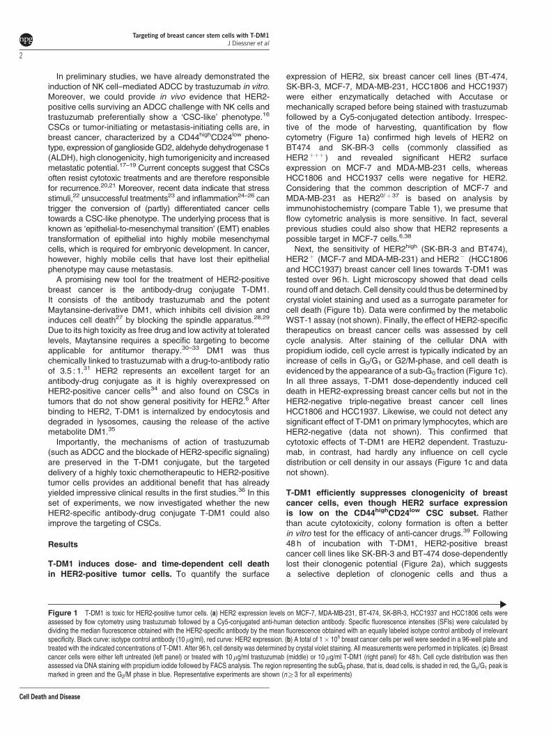
Figure 1 T-DM1 is toxic for HER2-positive tumor cells. (a) HER2 expression levels on MCF-7, MDA-MB-231, BT-474, SK-BR-3, HCC1937 and HCC1806 cells wereassessed by flow cytometry using trastuzumab followed by a Cy5-conjugated anti-human detection antibody. Specific fluorescence intensities (SFIs) were calculated bydividing the median fluorescence obtained with the HER2-specific antibody by the mean fluorescence obtained with an equally labeled isotype control antibody of irrelevantspecificity. Black curve: isotype control antibody (10 mg/ml), red curve: HER2 expression. (b) A total of 1� 105 breast cancer cells per well were seeded in a 96-well plate andtreated with the indicated concentrations of T-DM1. After 96 h, cell density was determined by crystal violet staining. All measurements were performed in triplicates. (c) Breastcancer cells were either left untreated (left panel) or treated with 10 mg/ml trastuzumab (middle) or 10mg/ml T-DM1 (right panel) for 48 h. Cell cycle distribution was thenassessed via DNA staining with propidium iodide followed by FACS analysis. The region representing the subG0 phase, that is, dead cells, is shaded in red, the Go/G1 peak ismarked in green and the G2/M phase in blue. Representative experiments are shown (nZ3 for all experiments)
Targeting of breast cancer stem cells with T-DM1J Diessner et al
2
Cell Death and Disease

Targeting of breast cancer stem cells with T-DM1J Diessner et al
3
Cell Death and Disease
preferential targeting of CSC39 by T-DM1. Trastuzumab, incontrast, had no such effect.
To confirm this hypothesis, we used MCF-7 cells from whicha highly aggressive and poorly immunogenic40 CD44high
CD24low CSC-like subset16,17 can be purified. Treatment ofeither unsorted MCF-7 cells or of purified CD44highCD24low
cells did not only show the higher clonogenicity of thissubpopulation but also confirmed the strong effect of T-DM1on the clonogenicity of HER2-positive breast cancer cells(Figure 2b, upper panel). Similar data were obtained withMDA-MB-231 cells (Figure 2b, lower panel). Trastuzumab, incontrast, did not inhibit clonogenicity. For BT-474 and SK-BR-3 cells, flow cytometric or magnetic bead-based enrichment ofthe CD44highCD24low subpopulation failed that precluded theiruse in this assay.
As T-DM1 concentrationsr100 ng/ml almost abrogatedclonogenicity in SK-BR-3, BT-474 and MCF-7 cells, wecompared EC50 values for clonogenicity and cytotoxicity forall 6 cell lines (Figure 2c). The striking differences betweenthese values show that T-DM1 preferentially targets clono-genic cells rather than the bulk of the tumor cell population.Again, HER2 positivity was a prerequisite for a discernibleeffect of T-DM1.
Importantly, clonogenicity (which, depending on the cellline, ranged from 5% to 26%) can be used as a surrogatemarker for CSCs. Our own previous findings, however, hadshown lower HER2 surface expression on the CD44high
CD24low presumed stem cell-like subset than on their moredifferentiated counterparts.41 In line with this observation,stem cell-like CD44highCD24lowHER2low cells escaped fromtargeting with trastuzumab and NK cells.16 The reducedHER2 surface expression on CD44highCD24low cells was alsonow confirmed with primary tumor cells obtained from pleuraleffusions of five patients with metastasized HER2-positivebreast cancer (Figure 2d and data not shown).
Effect of T-DM1 in the presence of NK cells. Thetherapeutic effect of anti-HER2 antibody treatment wasfound to correlate with the recruitment of NK cells in to thetumor microenvironment12 and to depend on both innate andadaptive immunity.42 One important mechanism is ADCC,16
which we found to be similar for trastuzumab and T-DM1(Figure 3a). However, recruitment of immune cells into atumor and inflammation can also promote EMT, which leads
to dedifferentiation of cancer cells towards a CSC-likephenotype.24–26 In fact, coculture of MCF-7 breast cancercells with NK cells did not induce morphological changestoward an elongated fibroblast-like shape,43,44 therebyindicating the transition from an epithelial to a mesenchymalphenotype (Figure 3b). Cell surface expression of theepithelial adherence factor E-Cadherin also decreased(Figure 3c). Western blotting revealed the induction ofb-Catenin, Vimentin and (non-junctional) Claudin-145
(Figure 3d), which have been described during EMT. Thecharacteristic transcription factors for EMT, Twist, Snail andSlug could, unfortunately, not be visualized by immuno-blotting that may be due to their low and transient expressionin MCF-7 cells. However, qRT-PCR analysis showed aninduction of these factors on mRNA level that could beprevented by T-DM1 (Figure 3e). Accordingly, immune-mediated induction of EMT could be prevented by T-DM1.
Effect of T-DM1 on induced breast CSCs. When T cells26
or NK cells16 induce EMT in breast cancer cells, induction ofCSC-like properties can be the unwanted consequence. Inline with these findings, coculture with NK cells increased theproportion of CD44highCD24low cells in the MCF-7 cell line(Figure 4a). As an additional control, use of the ALDEFLUORreagent system19 confirmed both stem cell induction incoculture with NK cells and the prevention of this effectby T-DM1 (Figure 4b). The enrichment of cells with aCSC signature was reflected by enhanced clonogenicity(Figure 4c). Both these effects could, again, be prevented byaddition of T-DM1 to the cocultures, whereas trastuzumabshowed no such effect (Figures 4a–c and data not shown).Similar data were also obtained in other cell lines (shown forSK-BR-3 in Figures 4d and e). Accordingly, T-DM1 seems tobe able to block the immune-mediated induction of tumorstem cells.
Maintenance of the CD44highCD24low breast CSCphenotype requires autophagy. Our data show that theHER2-specific antibody-drug conjugate T-DM1 exerts strongeffects on preexisting (Figure 2b) and induced (Figure 4a)CD44highCD24low breast cancer CSCs, even though HER2surface levels are low on this target cell population (Figure 2d).This apparent contradiction might be resolved by consideringthe high level of autophagy exerted by these cells. In fact,
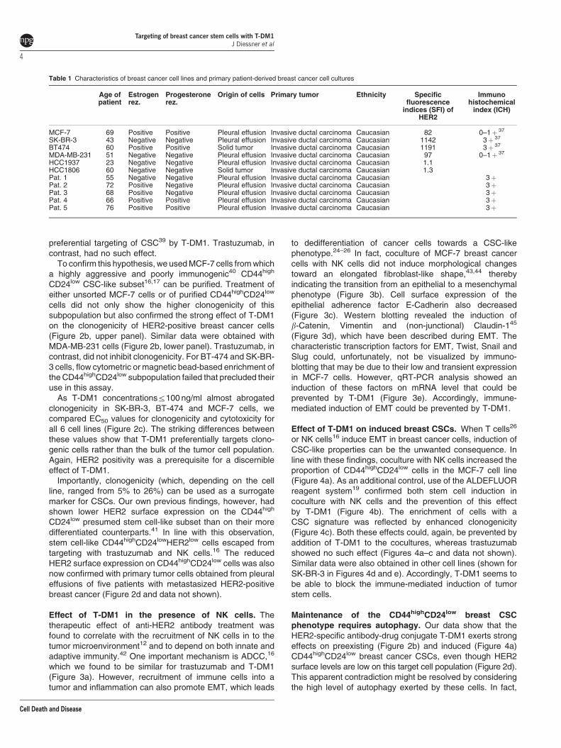
Table 1 Characteristics of breast cancer cell lines and primary patient-derived breast cancer cell cultures
Age ofpatient
Estrogenrez.
Progesteronerez.
Origin of cells Primary tumor Ethnicity Specificfluorescence
indices (SFI) ofHER2
Immunohistochemical
index (ICH)
MCF-7 69 Positive Positive Pleural effusion Invasive ductal carcinoma Caucasian 82 0–1þ 37
SK-BR-3 43 Negative Negative Pleural effusion Invasive ductal carcinoma Caucasian 1142 3þ 37
BT474 60 Positive Positive Solid tumor Invasive ductal carcinoma Caucasian 1191 3þ 37
MDA-MB-231 51 Negative Negative Pleural effusion Invasive ductal carcinoma Caucasian 97 0–1þ 37
HCC1937 23 Negative Negative Pleural effusion Invasive ductal carcinoma Caucasian 1.1HCC1806 60 Negative Negative Solid tumor Invasive ductal carcinoma Caucasian 1.3Pat. 1 55 Negative Negative Pleural effusion Invasive ductal carcinoma Caucasian 3þPat. 2 72 Positive Negative Pleural effusion Invasive ductal carcinoma Caucasian 3þPat. 3 68 Positive Negative Pleural effusion Invasive ductal carcinoma Caucasian 3þPat. 4 66 Positive Positive Pleural effusion Invasive ductal carcinoma Caucasian 3þPat. 5 76 Positive Positive Pleural effusion Invasive ductal carcinoma Caucasian 3þ
Targeting of breast cancer stem cells with T-DM1J Diessner et al
4
Cell Death and Disease
autophagy was reported to be critical for the tumorigenicityof breast cancer stem-like/progenitor cells46–48 and foundto positively regulate the CD44highCD24low phenotype.49
In order to exclude artifacts depending on side effects ofautophagy inhibitors, we used three different substances(3-methyl adenine, chloroquine and artesunate), which all
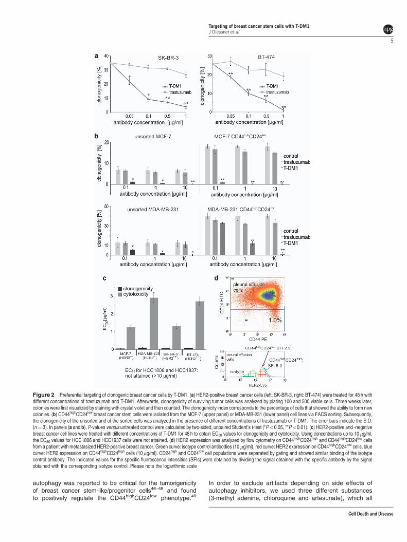
Figure 2 Preferential targeting of clonogenic breast cancer cells by T-DM1. (a) HER2-positive breast cancer cells (left: SK-BR-3, right: BT-474) were treated for 48 h withdifferent concentrations of trastuzumab and T-DM1. Afterwards, clonogenicity of surviving tumor cells was analyzed by plating 100 and 500 viable cells. Three weeks later,colonies were first visualized by staining with crystal violet and then counted. The clonogenicity index corresponds to the percentage of cells that showed the ability to form newcolonies. (b) CD44highCD24low breast cancer stem cells were isolated from the MCF-7 (upper panel) or MDA-MB-231 (lower panel) cell lines via FACS sorting. Subsequently,the clonogenicity of the unsorted and of the sorted cells was analyzed in the presence of different concentrations of trastuzumab or T-DM1. The error bars indicate the S.D.(n¼ 3). In panels (a and b), P-values versus untreated control were calculated by two-sided, unpaired Student’s t-test (*Po0.05, **Po0.01). (c) HER2-positive and -negativebreast cancer cell lines were treated with different concentrations of T-DM1 for 48 h to obtain EC50 values for clonogenicity and cytotoxicity. Using concentrations up to 10mg/ml,the EC50 values for HCC1806 and HCC1937 cells were not attained. (d) HER2 expression was analyzed by flow cytometry on CD44highCD24high and CD44highCD24low cellsfrom a patient with metastasized HER2-positive breast cancer. Green curve: isotype control antibodies (10 mg/ml), red curve: HER2 expression on CD44highCD24low cells, bluecurve: HER2 expression on CD44highCD24high cells (10 mg/ml). CD24high and CD24low cell populations were separated by gating and showed similar binding of the isotypecontrol antibody. The indicated values for the specific fluorescence intensities (SFIs) were obtained by dividing the signal obtained with the specific antibody by the signalobtained with the corresponding isotype control. Please note the logarithmic scale
Targeting of breast cancer stem cells with T-DM1J Diessner et al
5
Cell Death and Disease

Figure 3 During coculture with NK cells, T-DM1 can induce ADCC in breast cancer cells and inhibit EMT. (a) HCC1806, SK-BR-3 or MCF-7 breast cancer cells werecocultured with NK cells for 4 h, at a tumor-to-NK cell ratio of 1 : 2. For better discrimination, immune cells were stained with eFluor 670Cell Proliferation Dye, and tumor cellswere stained with CFSE. Target cell lysis of breast cancer cells (as evidenced by CFSE loss) was determined after 4 h by FACS analysis. Normal human immunoglobulins G(IgGs) served as a control for the humanized antibody trastuzumab and its conjugate T-DM1 (all used at 50 ng/ml). Data analysis was performed using an unpaired, two-sidedStudent’s t-test. Results from three independent experiments were analyzed (*Po0.05, **Po0.01). (b) MCF-7 breast cancer cells were photographed before (left panel) andafter (right panel) 16 h coculture with NK cells to visualize the changes regarding cellular morphology from the epithelial to a fibroblastic shape. (c) Flow cytometry was used todetermine E-cadherin surface expression on MCF-7 breast cancer cells before and after 24 h of coculture with NK cells. Although the binding of a PerCP/Cy5.5-labeled isotypecontrol of irrelevant specificity remained unaltered after coculture (green curve), the signals obtained with a similarly labeled anti-E-Cadherin antibody (clone 67A4) before andafter coculture are shown in blue (before) and red (after), respectively. SFI values are indicated. (d) MCF-7 breast cancer cells were incubated for 24 h with NK cells, using atumor cell-to-NK cell ratio of 1 : 2. After removal of non-adherent (i.e., NK) cells, protein lysates were prepared from the remaining adherent MCF-7 cells and tested forexpression of b-catenin, vimentin and claudin by western blotting. b-Actin was used as a loading control. (e) Cocultures were set up as in panel (d). Where indicated, T-DM1was added at a concentration of 50 ng/ml. After 16 h, NK cells were removed by washing, and RNA was extracted from the remaining adherent MCF-7 cells. The regulation ofEMT markers was analyzed by Sybr-Green-based quantitative real-time PCR, using 18S rRNA as internal control
Targeting of breast cancer stem cells with T-DM1J Diessner et al
6
Cell Death and Disease

caused an accumulation of the macroautophagy substratesLC3-II and p62 in MCF-7 cells. Quantification of the obtainedwestern blotting data showed the strongest effect withchloroquine and a somewhat weaker efficiency for3-methyladenine (Figure 5a). Consequently, 3-methylade-nine,50 chloroquine51 and artesunate52 all reduced the size ofthe CD44highCD24low population (Figure 5b) and the clono-genicity of MCF-7 cells (Figure 5c), up to a complete loss ofclonogenicity and of CD44highCD24low cells in the absence ofacute cytotoxicity. Using artesunate, similar data were also
obtained with primary breast cancer cells from pleuraleffusions (Figure 5d).
CD44highCD24low breast CSCs show increased autop-hagy leading to enhanced internalization of HER2 fromthe cell surface. Interestingly, it has already been hypothe-sized that autophagy might facilitate resistance against thesmall molecule HER2 receptor kinase inhibitor Lapatinib inHER2-positive breast cancer cells.53 Thus, we wonderedwhether the low HER2 surface levels on CD44highCD24low
Figure 4 T-DM1 blocks the immune-mediated enrichment of CD44highCD24low and ALDHþ breast cancer CSCs. (a–e) MCF-7 (a–c) and SK-BR-3 (d and e) breastcancer cells were cocultured with NK cells for 16 h, using a tumor cell-to-NK cell ratio of 1 : 2. Trastuzumab or T-DM1 was added where indicated. (a and d) Usingfluorescently labeled antibodies against CD24 and CD44, the size of the CD44highCD24low population was analyzed before and after incubation with polyclonal NK cells. Inpanel (b), the proportion of ALDH-expressing cells was assessed before (grey) and after (black) coculture with NK cells using the ALDEFLUOR reagent system. Cellsdisplaying high levels of ALDH are identified via the enzymatic conversion of a fluorogenic dye that can be visualized by flow cytometry. Positivity in this assay is consideredto be a characteristic trait of CSCs. In panels (a, b and d), representative experiments are shown. (c and e) MCF-7 (upper bar chart) and SK-BR-3 (lower bar chart) breastcancer cells were incubated for 16 h with NK cells, using a tumor cell-to-NK cell ratio of 1 : 2. T-DM1 or trastuzumab were added at the indicated concentrations. After 16 h,clonogenicity of the surviving tumor cells was assessed as in Figure 2b. Clonogenicity indices for cells that had not been cocultured with NK cells are shown in grey.P-values for the effects of trastuzumab or T-DM1 were calculated by the two-sided unpaired Student’s t-test using the respective coculture without antibody as relevantcontrol (n¼ 3, **Po0.01)
Targeting of breast cancer stem cells with T-DM1J Diessner et al
7
Cell Death and Disease

cells might be due to increased internalization of thereceptor, which is a phenomenon known to occur in responseto receptor activation.35 In line with this hypothesis, weobserved a loss of the CD24lowHER2low subpopulation whenautophagy was inhibited. HER2 expression on CD24high
cells, however, remained unaltered (data not shown).We thus assessed the internalization of the HER2 surface
antigen in CD44highCD24high and CD44highCD24low cellsusing trastuzumab labeled with the fluorogenic dye pHrodo.This pH-dependent dye experiences a steep increase in
fluorescence upon acidification. As clathrin-coated or othervesicles used for endocytosis are characterized by alow pH, this provided us with a tool to visualize internaliza-tion of the conjugated anti-HER2 antibody. Co-staining oftrastuzumab-pHrodo-treated cells after 6 h confirmedthat450% of the CD44highCD24low cells, but only o2% ofthe more differentiated CD44highCD24high cells, internalizedpHrodo-labeled trastuzumab. Internalization could beprevented by addition of artesunate (Figure 6a). Irrespectiveof the tumor cell subset, no internalization was observed
Figure 5 Enhanced autophagy is an essential requirement for clonogenic CD44highCD24low stem cell-like breast cancer cells. (a–c) MCF-7 breast cancer cells wereincubated with the autophagy inhibitors 3-methyladenine, chloroquine and artesunate for 24 h. (a) Cell lysates were prepared from untreated and treated MCF-7 breast cancercells and assayed for the expression of LC3-II and p62. b-Actin served as a loading control. Signals were quantified using Image J (U.S. National Institutes of Health,Bethesda, MD, USA; http://imagej.nih.gov/ij/), normalized to b-actin and the relative increase in non-degraded LC3-II and p62 was calculated. (b) The size of theCD44highCD24low CSC population was analyzed by flow cytometry before and after treatment. (c) MCF-7 breast cancer cells were treated with different concentrations of3-methyladenine, chloroquine and artesunate for 24 h. Thereafter, the ability of viable cells to form new colonies was analyzed as in Figure 2b (*Po0.05, **Po0.01).(d) Primary tumor cells obtained from a pleural effusion of a patient with metastasized HER2-positive breast cancer, treated with different concentrations of artesunate andanalyzed by flow cytometry for the presence of a CD44highCD24low CSC population
Targeting of breast cancer stem cells with T-DM1J Diessner et al
8
Cell Death and Disease

with the non-binding Rituximab-pHrodo conjugate (datanot shown).
To further confirm that maintenance of the CD44high
CD24low CSC-like state and internalization of pHrodo-labeledtrastuzumab by these cells depends on autophagy, we soughtto inhibit autophagy by siRNAs against Beclin and LC3(Figure 6b). Although downregulation of these essential
mediators of autophagy was incomplete, siRNA treatmentwas still sufficient to reduce the extent of the CD44highCD24low
population and the percentage of pHrodo-positive cells(Figure 6c). The fact that the remaining CD44highCD24low
cells showed a hardly reduced uptake of pHrodo mightsuggest that the loss of the CD24low signature precedes orcoincides with the reduced level of internalization.
Figure 6 Internalization by increased autophagy represents an ‘Achilles’ heel’ of CSCs with regard to T-DM1. (a) The HER2-specific antibody trastuzumab was labeledwith the fluorogenic dye pHrodo. pHrodo shows a strong fluorescence in the FL-2H channel once the pH of its surroundings becomes more acidic, for example, uponinternalization in endocytotic vesicles. Shown is the fluorescent signal from internalized trastuzumab-pHrodo against CD24 in CD44high cells, with CD44highCD24low
cells representing the stem cell-like cancer cell population. Upper left: control MCF-7 cells, upper right: MCF-7 cells were incubated with pHrodo-labeled immunoglobulinG1 (IgG1) isotype control antibodies and counter-stained with anti-CD24-FITC. Lower left: MCF-7 cells were incubated with pHrodo-labeled trastuzumab and stained withanti-CD24-FITC. Lower right: MCF-7 cells were incubated with pHrodo-labeled trastuzumab in the presence of the autophagy inhibitor artesunate (20 mg/ml) and stained withanti-CD24-FITC. Bar chart: Under the various applied conditions, the respective percentage of trastuzumab-pHrodo-internalizing cells was determined and evaluatedseparately for the CD24high and CD24low subsets. (b) Using TransIT TKO transfection reagent, MCF-7 breast cancer cells were transfected with 25 nM of siRNA against Beclin(BECN1 no. s16537, Ambion) or LC3 (no. MAP1LC3A, Ambion) or with a universal negative siRNA control (Sigma). Forty-eight hours later, cells were lysed and the expressionof Beclin, LC3 and b-actin was assayed in the various groups by western blotting. (c) MCF-7 cells were transfected with siRNA as in panel (c) before being incubated withtrastuzumab-pHrodoas in panel (a). Staining for CD44 and CD24 followed by flow cytometric analysis enabled the quantification of CD44highCD24low cells, of trastuzumab-internalising pHrodopos cells and of pHrodopos CD24low cells shown in panel (c)
Targeting of breast cancer stem cells with T-DM1J Diessner et al
9
Cell Death and Disease

Importantly, as pHrodo and emtansine are similarly small insize, and as they are both coupled to the same large antibody,it may be assumed that T-DM1 will show a similar behavior asthis fluorogenic conjugate. We thus propose that CD44high
CD24low breast cancer CSCs show a high level of internaliza-tion with regard to HER2 and thus evade effector mechan-isms, such as ADCC. However, by this same property thesecells will preferentially internalize antibody-drug conjugates.This can not only explain the highly efficient targeting ofclonogenic CD44highCD24low cells by T-DM1 but also providea more general rationale for the targeting of breast cancerCSCs with antibody-drug conjugates against HER2 or otheroncogenic growth factor receptors.
Discussion
The HER2-specific antibody trastuzumab10 has significantlyincreased overall survival in patients suffering from HER2-positive breast cancer. Moreover, a recent study has alsorevealed a role for HER2 in the CSC subset of cell lines(including MCF-7) which are not commonly classifiedas HER2 overexpressing.6 Likewise, synergy betweentrastuzumab and paclitaxel or docetaxel had also beenreported in such cell lines.38 Accordingly, more patients thanpreviously anticipated might benefit from treatment withanti-HER2 antibodies. Unfortunately, however, the majorityof patients treated with trastuzumab experience relapse atsome time after treatment. Thus, a better understanding of themechanism(s) underlying this resistance might pave the waytowards more effective strategies. In our previous studies, wecould demonstrate in vitro that the binding of trastuzumab‘labels’ HER2-positive breast cancer cells for Fcg-dependentrecognition by NK cells,16 thereby inducing ADCC.12,13 Thisimportant immune-stimulatory effect has been preserved inthe antibody-drug conjugate T-DM1, which has retained theFcg-part of trastuzumab (Figure 3a). Also inhibition of HER2signaling, as evidenced by reduced activation of the phos-phatidylinositol 30-kinase-AKT pathway, is similarly achievedby both trastuzumab and T-DM1 (data not shown). However,T-DM1 combines the mechanisms of action of trastuzumabwith the additional specific application of the potentchemotherapeutic DM1.31,32
Unlike trastuzumab, T-DM1 thus induced cell cycle arrestand significant cell death in HER2-positive tumor cells that islikely due to the toxin DM1 contained in T-DM1. EC50 valuesover 48 h were mostlyZ10 mg/ml (Figures 1b and c), whichmight be hard to achieve in patients. Using clinically relevantconcentrations (3mg/ml)54 of T-DM1, a significant proportionof target cells survived in vitro that could point towardschemoresistance. However, HER2-positive tumor cellssurviving a treatment with T-DM1 displayed drasticallyreduced clonogenicity (Figures 2a–c), even at T-DM1concentrations as low as 50 ng/ml. Likewise, T-DM1 incuba-tion almost abrogated the clonogenicity of sorted CD44high
CD24low cells, whereas trastuzumab treatment showed nocomparable effect on this highly tumorigenic and poorlyimmunogenic subset (Figure 2b). This suggests that T-DM1 isfar more effective against CSC than trastuzumab. In fact, themajor limitation encountered in our previous study was theinsufficient targeting of CD44highCD24lowHER2low cells
either with trastuzumab alone or in conjunction with NKcells.16 Based on the evidence that this subset possessescharacteristics of the so-called ‘tumor-initiating’ or ‘metasta-sis-initiating’ or ‘CSCs’,17 a lack of activity against thissubpopulation may explain the high rate of recurrence aftertreatment.
CSCs, however, constitute a rather dynamic subset. Weand others have observed26 that ‘unproductive’ (i.e., non-lytic)interactions between immune and tumor cells induce EMTin breast cancer cells, which reprograms previously differ-entiated cancer cells towards a CSC-like phenotype.22
Accordingly, tumor cell cultures that had interacted with NKcells showed morphological changes towards a mesenchymalphenotype (Figure 3b), downregulation of the epithelial celladhesion molecule E-Cadherin (Figure 3c), upregulation ofEMT markers (Figures 3d and e), more CD44highCD24low
(Figures 4a and d) and ALDHþ (Figure 4b) cells andenhanced clonogenicity (Figures 4c and e). Addition ofT-DM1 to the coculture, however, prevented this expansionof the CSC population (Figures 3e and 4). This left us with twopossible explanations: either tumor cells that had survived thetreatment were ‘sufficiently damaged’ to lose all stem cell-likeproperties or the antibody-drug conjugate might preferentiallytarget highly clonogenic ‘CSCs’ - in spite of their low HER2surface expression (Figure 2d). This prompted us toinvestigate the possibility of enhanced internalization byCSCs. In fact, the CSC phenotype has repeatedly beenlinked to autophagy,49,55,56 a tightly regulated catabolicprocess involving the degradation of a cell’s own componentsthrough the lysosomal machinery. On the one hand, auto-phagy helps to keep up a balance between degradation,synthesis and recycling of cellular products and allowsstressed or starving cells to recycle nutrients from unneces-sary processes to more essential metabolic tasks. On theother hand, autophagy leads to a constant recycling of cellularcomponents and thus enables longevity57 and maintenance ofa stem cell-like phenotype not only in hematopoietic58 but alsoin breast cancer CSCs.48 In line with these reports, autophagyinhibitors (chloroquine, 3’-methyladenine or artesunate)51,59
abrogated CD44highCD24lowHer2low cells and clonogenicity inprimary breast cancer cells from pleural effusions and inbreast cancer cell lines (Figures 5b and d). Most importantly,however, we measured internalization of trastuzumab bycovalently coupling the fluorogenic dye pHrodo to theantibody. This revealed a preferential internalization of theconjugated anti-HER2 antibody by CD44highCD24low cells thatcould be abrogated by inhibiting autophagy (Figure 6a).Unspecific binding via the pHrodo moiety or the human IgGbackbone was excluded by use of the pHrodo-labeledhumanized anti-CD20 antibody rituximab. The link betweenautophagy, CD44highCD24low CSC and internalization oflabeled trastuzumab was further confirmed by siRNA-mediated downregulation of LC3 or Beclin (Figure 6b), whichresulted in a significantly smaller CD44highCD24low CSCsubset and in reduced internalization of pHrodo-labeledtrastuzumab. Interestingly, those cells that retained theCD24low phenotype also showed unaltered internalization.We thus propose that the low HER2 (and, most likely also thelow CD24) surface expression on CD44highCD24low cells ismainly due to internalization via autophagy. This, once again,
Targeting of breast cancer stem cells with T-DM1J Diessner et al
10
Cell Death and Disease
confirms that autophagy enables constant renewal of thesecells and may be protective against most therapeutic insults,including immune-mediated destruction via ADCC. However,when it comes to antibody-drug conjugates, this mechanismconstitutes an ‘Achilles’ heel’ that may be therapeuticallyexploited by T-DM1. Considering that the targeting of thesecells is thought to be essential for a successful cancer therapy,our in vitro data thus suggest that T-DM1 holds great promisefor future treatment of early and metastatic HER2-positivebreast cancer. In fact, first results from the EMILIA trial, aphase III study in which 991 women with locally advanced ormetastatic, unresectable, trastuzumab- and taxane-treated,HER2-positive breast cancer were enrolled showed longerprogression-free survival (9.6 versus 6.4 months) and asuperior safety profile for T-DM1 against the combination ofcapecitabine with lapatinib.60
Further trials (MARIANNE, TH3RESA) are ongoing. From amechanistic point of view, it is interesting to note that a tubulininhibitor can prevent the unwanted induction of tumor stemcell characteristics. In line with this notion, pretreatment ofcancer cells with paclitaxel (Taxol) likewise prevented theinduction of EMT and of stem cell-like features (data notshown). Thus, tubulin-dependent processes seem to have aseminal role for CSC induction. It may thus be speculatedwhether the induced CD44highCD24low cells actually originatefrom more differentiated tumor cells via (induced) asymmetriccell divisions. In any case, these data show that the coupling ofa potent inhibitor of the mitotic spindle to trastuzumab was agood choice for T-DM1. Moreover, the combination of theanti-proliferative and immune-activating effect of trastuzumabat the cell surface with intracellular delivery of a potent toxinupon endocytosis could serve as a template for furtherantibody-drug conjugates. Such novel therapeutics mightnot only be well tolerated but also be particularly effectiveagainst CSCs with their high internalization rate.
Materials and MethodsCell culture and reagents. MCF-7, MDA-MB-231, BT-474, HCC1937,HCC1806 and SK-BR-3 breast cancer cells were obtained from the AmericanType Culture Collection (Manassas, VA, USA) and the Deutsche Sammlung furMikroorganismen und Zellkulturen (DSMZ, Braunschweig, Germany), authenti-cated and cultured as indicated by the supplier. Primary tumor cells were obtainedfrom pleural effusions of patients undergoing puncture for the palliative treatmentof metastasized breast cancer at our institution. Further investigation of these cellswas approved by both the patients and the local ethics committee. Cells werecentrifuged, washed with PBS, and transferred to L-valine-free, D-valine-containing Dulbecco’s Modified Eagles Medium supplemented with 2% heat-inactivated FCS (Biochrom, Berlin, Germany), penicillin (100 IU/ml) andstreptomycin (100 IU/ml) (all from PAA, Colbe, Germany). Non-adherent cellswere removed by washing after 24 h. Fibroblast growth was suppressed due to thelack of L-valine. Autophagy inhibitors (3-methyl adenine, artesunate andchloroquine) were obtained from Sigma (Deisenhofen, Germany).
Cytotoxicity and cell cycle assays. Breast cancer cells were treated withdifferent concentrations of T-DM1 and trastuzumab as indicated. Cell density afterdrug treatment was assessed by crystal violet staining. As this assay is based onthe quantification of adherent cells, microscopy was used to confirm that dyingcells became round and detached. As an additional control, the metabolic WST-1assay (Roche, Mannheim, Germany) was used according to the manufacturer’sinstructions. For cell cycle analysis, cells were detached, fixed and permeabilizedfor at least 1 h in ice-cold 70% ethanol and washed with PBS before their DNAwas stained with propidium iodide (PI). RNA was simultaneously digested withDNAse-free RNAse A (Carl Roth, Karlsruhe, Germany). PI intercalates into DNA
with no sequence preference and thus enables the assessment of the cellularDNA content in the FL-2A channel of the FACSCalibur flow cytometer(BD Biosciences, Heidelberg, Germany). Doublets were excluded via the FL-2Wchannel. Cells containing less than a complete set of chromosomes (i.e., 2n) areconsidered to be dead.
Clonogenicity assays. Clonogenicity assays are in vitro cell survival assaysbased on the ability of single cells to establish a new colony. The assay essentiallyinvestigates the proportion of cells that can undergo ‘unlimited’ division. Breastcancer cells were treated as indicated before being seeded at low density onlow-attachment plastic material (Greiner BioOne, Frickenhausen, Germany). After2–3 weeks, colonies were fixed, stained with crystal violet (0.5% w/v) and counted.Stained colonies consisting of at least 50 cells were visible without the use of amicroscope.
Flow cytometric analysis of surface expression levels and cellsorting. Cells were either mechanically or enzymatically detached usingAccutase (PAA), blocked and stained with 10 mg/ml Trastuzumab or 10mg/mlT-DM1 (Genentech, Burlingame, CA, USA) followed by a Cy5-conjugated goatanti-human IgG (Rockland Immunochemicals, Gilbertsville, PA, USA) detectionantibody. Simultaneously, CD44-RPE (Clone 2BJ18, BioLegend, San Diego, CA,USA), CD24-FITC (clone SWA-11, kindly provided by Professor Peter Altevogt,German Cancer Research Centre, Heidelberg, Germany) and the life stain7-aminoactinomycin D (Sigma) were applied and analyzed on a FACSCalibur oran Attune flow cytometer (Life Technologies, Darmstadt, Germany). ForE-Cadherin surface staining, a PerCP/Cy5.5-labeled anti-human CD324/E-Cadherin antibody (Clone 67A4) and a corresponding isotype control (CloneMOPC-21), both from BioLegend, were used. Where appropriate, expressionlevels are indicated as specific fluorescence intensity values, which are obtainedby dividing the fluorescence intensity detected with the specific antibody by thesignal measured with the isotype-matched control antibody. For fluorescence-activated cell sorting, the stained cells were separated twice on a DigitalFACSVantage (BD Biosciences), first in yield and then in purity mode.
ALDH assay. The ALDEFLUOR assay (STEMCELL Technologies, Grenoble,France) enables the identification of stem and progenitor cells based on their highexpression of ALDH1. This enzyme converts the fluorogenic substrate BODIPY-aminoacetaldehyde into a green fluorescent dye that can be visualized by flowcytometry. As the ALDEFLUOR reaction product will leak out of dead cells, onlyviable cells are identified by this assay. Transporter-mediated exclusion of the dyefrom stem cells is prevented by multi-drug reporter inhibition with a verapamil-containing inhibitor contained in the kit. Tumor cells were treated with differentconcentrations of drugs for variable intervals of time. Afterwards, the ALDEFLUORtest was conducted according to the manufacturer’s instructions.
NK cell preparation and cytotoxicity assays. Peripheral bloodlymphocytes were obtained from healthy volunteers by density gradientcentrifugation (Biocoll, Biochrom) and cultured for 8-11 days on irradiated(30 Gy) RPMI8866 feeder cells to obtain polyclonal NK cell populations (30). NKcell-mediated lysis of MCF-7 or MDA-MB-231 cells was assessed by modifiedfluorometric assessment of T lymphocyte antigen specific lysis assays.41 NK cellswere thus labeled with eFluor 670 Cell Proliferation Dye (ebioscience, Frankfurt,Germany) while target cells (2� 105/well) were stained with carboxyfluoresceindiacetate succinimidyl ester (CFSE) (Invitrogen, Karlsruhe, Germany). Cocultureswere set up using different effector:target ratios, and lytic activity was assessedafter 16 h by flow cytometric detection of CFSEdim cells among the eFluor670-negative target cell population. Values were corrected for spontaneousleakage of CFSE.
Immunoblot analysis. Regulation of EMT-related proteins in MCF-7 cellsthat had been cocultured with NK cells was analyzed by immunoblot. In short, NKcells were removed by washing. Adherent cells were detached, collected bycentrifugation, washed with PBS and lysed in protein lysis buffer containing 50 mMTris-HCl (tris(hydroxymethyl)aminomethane-hydrochloric acid) pH 8.0 (Sigma)),120 mM sodium chloride, 5 mM EDTA, 2 mg/ml Aprotinin, 10mg/ml Leupeptin,1 mM PMSF, 10 nM sodium fluoride (all purchased from Carl Roth, Karlsruhe,Germany), 0.5% Nonidet P-40 and 1 mM sodium orthovanadate (both fromApplichem GmbH, Darmstadt, Germany). Proteins were separated on 8–15%polyacrylamide (Carl Roth) gels under reducing conditions, transferred to
Targeting of breast cancer stem cells with T-DM1J Diessner et al
11
Cell Death and Disease
Nitrocellulose (Protran Transfer Membrane, Whatman, Dassel, Germany), whichwas blocked in PBS containing 5% skimmed milk powder and 0.05% Tween20(both from Carl Roth), and incubated overnight at 4 1C with the followingantibodies (all from Cell Signaling Technology, Danvers, MA, USA): anti-humanLC3 (clone D3U4C), anti-human Beclin (clone D40C5), anti-human �-Catenin(clone D10A8), anti-human Claudin (no. 4933), anti-human Vimentin (cloneD21H3), anti-human pAKT (clone Ser473), and anti-human AKT (no. 9272).Antibodies against p62 (Poly6477) and against b-actin (Poly6221), which servedas loading control, were from BioLegend. Proteins were visualized usinghorseradish peroxidase-coupled anti-mouse or anti-rabbit IgG secondary antibody(Cell Signaling) and homemade enhanced chemiluminescence (ECL) solution.ECL solution was prepared by mixing 0.25 mg/ml Luminol (Carbosynth Ltd,Berkshire, UK) in 0.1 M Tris-HCL (pH 8.6) with 1.1 mg/ml para-hydroxycoumaricacid (Sigma-Aldrich, Taufkirchen, Germany) and 0.03% hydrogen peroxide (CarlRoth) at a 10 : 1 : 0.003 ratio.
qRT-PCR. Total RNA was prepared using TriFast (Peqlab, Erlangen, Germany)and transcribed with the iScript cDNA Synthesis kit (Bio-Rad, Munich, Germany).For real-time PCR, cDNA amplification was monitored using the ABsolute QPCRSYBR Green Low Rox mix (ABgene, Epsom, UK) on the ABI PRISM 7500Sequence Detection System (Applied Biosystems, Weiterstadt, Germany). To thisaim, 40 cycles of a two-step PCR protocol (95 1C for 15 s, 60 1C for 1 min) wereperformed. The following primers were used:
18S up: 50-CGGCTACCACATCCAAGGAA-30; 18S down: 50-GCTGGAATTACCGCGGCT-30; Snail up: 50-CTGCTCCACAAGCACCAAGAGTC-30; Snaildown: 50-CCAGCTGCCCTCCCTCCAC-30; b-actin up: 50-TGTTTGAGACCTTCAACACCC-30; b-actin down: 50-AGCACTGTGTTGGCGTACAG-30; Slug up:50-GGGGAGAAGCCTTTTTCT TG-30; Slug down: 50-TCCTCATGTTTGTGCAGGAG-30; Vimentin up: 50-GAGAATTTGCCGTTGAAGC-30; Vimentin down:50-GCTTCCTGTAGGTGGCAATC-30; Twist up: 50-GGAGTCCGCAGTCTTACGAG-30; Twist down: 50-TCTGGAGGACCTGGTAGAG-30; E-Cadherin up:50-CCGAGATGGGGTTGATAATG-30; and E-Cadherin down: 50-ACAGTGGCCACCTACAAAG-30. Data analysis was done using the DDCT method for relativequantification. Dissociation curves confirmed the presence of a single specific PCRproduct.
pHrodo assay. HER2-specific antibodies were labeled with the fluorogenicdye pHrodo (Life Technologies, INVITROGEN CORPORATION, Burlington, ON,Canada) according to the manufacturer’s instructions. pHrodo shows increasedfluorescence in the FL-2H channel as the pH of its surroundings becomes moreacidic, for example, upon internalization in endocytotic vesicles. With respect tobreast cancer cells, pHrodo-labeled humanized anti-CD20 antibody Rituximab(Rituxan) was used as a non-binding control.
siRNA transfection. In all, 2� 105 MCF-7 cells were seeded in a six-wellplate. Twenty-four hours later, they were transfected with 25 nM of siRNA againstBeclin (BECN1 no.s16537) or LC3 (MAP1LC3A, no. s39155, both from Ambion,Life Technologies, Darmstadt, Germany) or with a universal negative siRNAcontrol (Sigma), using 10ml of TransIT-TKO Transfection Reagent (Mirus,Madison, WI, USA). Forty-eight hours later, cells were lysed, and the expressionlevels of Beclin, LC3 and b-actin were assayed in the various groups by westernblotting. Incubation with trastuzumab-pHrodo and subsequent flow cytometricanalysis were performed as described above.
Statistics. Experiments were performed at least three times with similarresults, and representative experiments are shown. S.Ds. for flow cytometry datawere calculated by using the Summit software (Beckman Coulter, Krefeld,Germany). Analysis of significance was performed using an unpaired, two-sidedStudent’s t-test.
Conflict of InterestAH participated as a consultant in advisory boards for Roche and receivedhonoraria from Roche. The remaining authors declare no conflict of interest.
Acknowledgements. We thank Dr. Peter Altevogt (Deutsches Krebs-forschungszentrum, Heidelberg, Germany) for providing the CD24 antibody,Christian Linden for cell sorting, Birgitt Fischer for excellent technical assistance
and Itsaso Montalban, Anoop Chandran and Markus Junker for valuablediscussions. Grant support: This work was supported by a grant to JW and AHfrom F Hoffmann-La Roche/Genentech, a member of the Roche Group. VB is afellow the German research council (DFG)-sponsored Graduate School for LifeSciences (GSLS) Wurzburg. RGS is a member of the clinician scientist trainingprogram ‘Interdisciplinary Translational Immunology’ (Else-Kroner Forschungskol-leg Wurzburg), which is supported by the Else-Kroner Fresenius foundation.
1. Gschwind A, Fischer OM, Ullrich A. The discovery of receptor tyrosine kinases: targets forcancer therapy. Nat Rev Cancer 2004; 4: 361–370.
2. Monsey J, Shen W, Schlesinger P, Bose R. Her4 and Her2/neu tyrosine kinase domainsdimerize and activate in a reconstituted in vitro system. J Biol Chem 2010; 285:7035–7044.
3. Wiseman SM, Makretsov N, Nielsen TO, Gilks B, Yorida E, Cheang M et al. Coexpressionof the type 1 growth factor receptor family members HER-1, HER-2, and HER-3 has asynergistic negative prognostic effect on breast carcinoma survival. Cancer 2005; 103:1770–1777.
4. Slamon DJ, Clark GM, Wong SG, Levin WJ, Ullrich A, McGuire WL. Human breast cancer:correlation of relapse and survival with amplification of the HER-2/neu oncogene. Science1987; 235: 177–182.
5. Bose R, Kavuri SM, Searleman AC, Shen W, Shen D, Koboldt DC et al. Activating HER2mutations in HER2 gene amplification negative breast cancer. Cancer Discov 2012; 3:224–237.
6. Ithimakin S, Day KC, Malik F, Zen Q, Dawsey SJ, Bersano-Begey TF et al. HER2 drivesluminal breast cancer stem cells in the absence of HER2 amplification: implications forefficacy of adjuvant trastuzumab. Cancer Res 2013; 73: 1635–1646.
7. Bang YJ, Van Cutsem E, Feyereislova A, Chung HC, Shen L, Sawaki A et al. Trastuzumabin combination with chemotherapy versus chemotherapy alone for treatment ofHER2-positive advanced gastric or gastro-oesophageal junction cancer (ToGA): a phase 3,open-label, randomised controlled trial. Lancet 2010; 376: 687–697.
8. Cobleigh MA, Vogel CL, Tripathy D, Robert NJ, Scholl S, Fehrenbacher L et al.Multinational study of the efficacy and safety of humanized anti-HER2 monoclonalantibody in women who have HER2-overexpressing metastatic breast cancer thathas progressed after chemotherapy for metastatic disease. J Clin Oncol 1999; 17:2639–2648.
9. Joensuu H, Kellokumpu-Lehtinen PL, Bono P, Alanko T, Kataja V, Asola R et al. Adjuvantdocetaxel or vinorelbine with or without trastuzumab for breast cancer. N Engl J Med 2006;354: 809–820.
10. Slamon DJ, Leyland-Jones B, Shak S, Fuchs H, Paton V, Bajamonde A et al. Use ofchemotherapy plus a monoclonal antibody against HER2 for metastatic breast cancer thatoverexpresses HER2. N Engl J Med 2001; 344: 783–792.
11. Liang K, Lu Y, Jin W, Ang KK, Milas L, Fan Z. Sensitization of breast cancer cells toradiation by trastuzumab. Mol Cancer Ther 2003; 2: 1113–1120.
12. Beano A, Signorino E, Evangelista A, Brusa D, Mistrangelo M, Polimeni MA et al.Correlation between NK function and response to trastuzumab in metastatic breast cancerpatients. J Transl Med 2008; 6: 25.
13. Hudis CA. Trastuzumab—mechanism of action and use in clinical practice. N Engl J Med2007; 357: 39–51.
14. Clynes RA, Towers TL, Presta LG, Ravetch JV. Inhibitory Fc receptors modulate in vivocytotoxicity against tumor targets. Nat Med 2000; 6: 443–446.
15. Kohrt HE, Houot R, Weiskopf K, Goldstein MJ, Scheeren F, Czerwinski D et al. Stimulationof natural killer cells with a CD137-specific antibody enhances trastuzumab efficacy inxenotransplant models of breast cancer. J Clin Invest 2012; 122: 1066–1075.
16. Reim F, Dombrowski Y, Ritter C, Buttmann M, Hausler S, Ossadnik M et al.Immunoselection of breast and ovarian cancer cells with trastuzumab and natural killercells: selective escape of CD44high/CD24low/HER2low breast cancer stem cells. CancerRes 2009; 69: 8058–8066.
17. Al-Hajj M, Wicha MS, Benito-Hernandez A, Morrison SJ, Clarke MF. Prospectiveidentification of tumorigenic breast cancer cells. Proc Natl Acad Sci USA 2003; 100:3983–3988.
18. Battula VL, Shi Y, Evans KW, Wang RY, Spaeth EL, Jacamo RO et al. Ganglioside GD2identifies breast cancer stem cells and promotes tumorigenesis. J Clin Invest 2012; 122:2066–2078.
19. Charafe-Jauffret E, Ginestier C, Iovino F, Tarpin C, Diebel M, Esterni B et al. Aldehydedehydrogenase 1-positive cancer stem cells mediate metastasis and poor clinical outcomein inflammatory breast cancer. Clin Cancer Res 2010; 16: 45–55.
20. Al-Ejeh F, Smart CE, Morrison BJ, Chenevix-Trench G, Lopez JA, Lakhani SR et al. Breastcancer stem cells: treatment resistance and therapeutic opportunities. Carcinogenesis2011; 32: 650–658.
21. Sutherland CM, Mather FJ, Carter RD, Cerise EJ, Krementz ET. Breast cancer as analyzedby the human tumor stem cell assay. Surgery 1983; 94: 370–375.
22. Polyak K, Weinberg RA. Transitions between epithelial and mesenchymal states:acquisition of malignant and stem cell traits. Nat Rev Cancer 2009; 9: 265–273.
23. Harless WW. Cancer treatments transform residual cancer cell phenotype. Cancer Cell Int2011; 11: 1.
Targeting of breast cancer stem cells with T-DM1J Diessner et al
12
Cell Death and Disease

24. Korkaya H, Kim GI, Davis A, Malik F, Henry NL, Ithimakin S et al. Activation of an IL6inflammatory loop mediates trastuzumab resistance in HER2þ breast cancer byexpanding the cancer stem cell population. Mol Cell 2012; 47: 570–584.
25. Landsberg J, Kohlmeyer J, Renn M, Bald T, Rogava M, Cron M et al. Melanomas resistT-cell therapy through inflammation-induced reversible dedifferentiation. Nature 2012; 490:412–416.
26. Santisteban M, Reiman JM, Asiedu MK, Behrens MD, Nassar A, Kalli KR et al.Immune-induced epithelial to mesenchymal transition in vivo generates breast cancer stemcells. Cancer Res 2009; 69: 2887–2895.
27. Yu TW, Bai L, Clade D, Hoffmann D, Toelzer S, Trinh KQ et al. The biosynthetic genecluster of the maytansinoid antitumor agent ansamitocin from Actinosynnema pretiosum.Proc Natl Acad Sci USA 2002; 99: 7968–7973.
28. Lambert JM. Drug-conjugated monoclonal antibodies for the treatment of cancer.Curr Opin Pharmacol 2005; 5: 543–549.
29. Wang L, Amphlett G, Blattler WA, Lambert JM, Zhang W. Structural characterizationof the maytansinoid-monoclonal antibody immunoconjugate, huN901-DM1, by massspectrometry. Protein Sci 2005; 14: 2436–2446.
30. Chari RV, Martell BA, Gross JL, Cook SB, Shah SA, Blattler WA et al. Immunoconjugatescontaining novel maytansinoids: promising anticancer drugs. Cancer Res 1992; 52: 127–131.
31. Erickson HK, Park PU, Widdison WC, Kovtun YV, Garrett LM, Hoffman K et al.Antibody-maytansinoid conjugates are activated in targeted cancer cells by lysosomaldegradation and linker-dependent intracellular processing. Cancer Res 2006; 66: 4426–4433.
32. Kovtun YV, Audette CA, Mayo MF, Jones GE, Doherty H, Maloney EK et al.Antibody-maytansinoid conjugates designed to bypass multidrug resistance. Cancer Res2010; 70: 2528–2537.
33. Remillard S, Rebhun LI, Howie GA, Kupchan SM. Antimitotic activity of the potent tumorinhibitor maytansine. Science 1975; 189: 1002–1005.
34. Pegram M, Hsu S, Lewis G, Pietras R, Beryt M, Sliwkowski M et al. Inhibitory effects ofcombinations of HER-2/neu antibody and chemotherapeutic agents used for treatment ofhuman breast cancers. Oncogene 1999; 18: 2241–2251.
35. Austin CD, De Maziere AM, Pisacane PI, van Dijk SM, Eigenbrot C, Sliwkowski MX et al.Endocytosis and sorting of ErbB2 and the site of action of cancer therapeutics trastuzumaband geldanamycin. Mol Biol Cell 2004; 15: 5268–5282.
36. Krop IE, LoRusso P, Miller KD, Modi S, Yardley D, Rodriguez G et al. A phase II study oftrastuzumab emtansine in patients with human epidermal growth factor receptor 2-positivemetastatic breast cancer who were previously treated with trastuzumab, lapatinib, ananthracycline, a taxane, and capecitabine. J Clin Oncol 2012; 30: 3234–3241.
37. Subik K, Lee JF, Baxter L, Strzepek T, Costello D, Crowley P et al. The expression patternsof ER, PR, HER2, CK5/6, EGFR, Ki-67 and AR by immunohistochemical analysis in breastcancer cell lines. Breast Cancer (Auckl) 2010; 4: 35–41.
38. Merlin JL, Barberi-Heyob M, Bachmann N. In vitro comparative evaluation of trastuzumab(Herceptin) combined with paclitaxel (Taxol) or docetaxel (Taxotere) in HER2-expressinghuman breast cancer cell lines. Ann Oncol 2002; 13: 1743–1748.
39. Beier D, Rohrl S, Pillai DR, Schwarz S, Kunz-Schughart LA, Leukel P et al. Temozolomidepreferentially depletes cancer stem cells in glioblastoma. Cancer Res 2008; 68: 5706–5715.
40. Brisken C, Duss S. Stem cells and the stem cell niche in the breast: an integrated hormonaland developmental perspective. Stem Cell Rev 2007; 3: 147–156.
41. Diessner J, Bruttel V, Becker K, Pawlik M, Stein R, Hausler S et al. Targeting breast cancerstem cells with HER2-specific antibodies and natural killer cells. Am J Cancer Res 2013; 3:211–220.
42. Park S, Jiang Z, Mortenson ED, Deng L, Radkevich-Brown O, Yang X et al. The therapeuticeffect of anti-HER2/neu antibody depends on both innate and adaptive immunity. CancerCell 2010; 18: 160–170.
43. Maeda M, Johnson KR, Wheelock MJ. Cadherin switching: essential for behavioral but notmorphological changes during an epithelium-to-mesenchyme transition. J Cell Sci 2005;118(Pt 5): 873–887.
44. Piek E, Moustakas A, Kurisaki A, Heldin CH, ten Dijke P. TGF-(beta) type I receptor/ALK-5and Smad proteins mediate epithelial to mesenchymal transdifferentiation in NMuMGbreast epithelial cells. J Cell Sci 1999; 112(Pt 24): 4557–4568.
45. Dhawan P, Singh AB, Deane NG, No Y, Shiou SR, Schmidt C et al. Claudin-1 regulatescellular transformation and metastatic behavior in colon cancer. J Clin Invest 2005; 115:1765–1776.
46. Choi AM, Ryter SW, Levine B. Autophagy in human health and disease. N Engl J Med2013; 368: 651–662.
47. Espina V, Mariani BD, Gallagher RI, Tran K, Banks S, Wiedemann J et al. Malignantprecursor cells pre-exist in human breast DCIS and require autophagy for survival. PloSOne 2010; 5: e10240.
48. Gong C, Bauvy C, Tonelli G, Yue W, Delomenie C, Nicolas V et al. Beclin 1 and autophagyare required for the tumorigenicity of breast cancer stem-like/progenitor cells. Oncogene2012; 32: 22261–22272.
49. Cufi S, Vazquez-Martin A, Oliveras-Ferraros C, Martin-Castillo B, Vellon L, Menendez JA.Autophagy positively regulates the CD44(þ ) CD24(-/low) breast cancer stem-likephenotype. Cell Cycle 2011; 10: 3871–3885.
50. Wu YT, Tan HL, Shui G, Bauvy C, Huang Q, Wenk MR et al. Dual role of 3-methyladeninein modulation of autophagy via different temporal patterns of inhibition on class I and IIIphosphoinositide 3-kinase. J Biol Chem 2010; 285: 10850–10861.
51. Kimura T, Takabatake Y, Takahashi A, Isaka Y. Chloroquine in cancer therapy:a double-edged sword of autophagy. Cancer Res 2013; 73: 3–7.
52. Hamacher-Brady A, Stein HA, Turschner S, Toegel I, Mora R, Jennewein N et al.Artesunate activates mitochondrial apoptosis in breast cancer cells via iron-catalyzedlysosomal reactive oxygen species production. J Biol Chem 2011; 286:6587–6601.
53. Chen S, Li X, Feng J, Chang Y, Wang Z, Wen A. Autophagy facilitates theLapatinib resistance of HER2 positive breast cancer cells. Med Hypotheses 2011; 77:206–208.
54. Girish S, Gupta M, Wang B, Lu D, Krop IE, Vogel CL et al. Clinical pharmacologyof trastuzumab emtansine (T-DM1): an antibody-drug conjugate in developmentfor the treatment of HER2-positive cancer. Cancer Chemother Pharmacol 2012; 69:1229–1240.
55. Vazquez-Martin A, Lopez-Bonetc E, Cufi S, Oliveras-Ferraros C, Del Barco S,Martin-Castillo B et al. Repositioning chloroquine and metformin to eliminate cancer stemcell traits in pre-malignant lesions. Drug Resist Updat 2011; 14: 212–223.
56. Li Z, Rich JN. Hypoxia and hypoxia inducible factors in cancer stem cell maintenance. CurrTop Microbiol Immunol 2010; 345: 21–30.
57. Madeo F, Tavernarakis N, Kroemer G. Can autophagy promote longevity? Nat Cell Biol2010; 12: 842–846.
58. Mortensen M, Soilleux EJ, Djordjevic G, Tripp R, Lutteropp M, Sadighi-Akha E et al.The autophagy protein Atg7 is essential for hematopoietic stem cell maintenance.J Exp Med 2011; 208: 455–467.
59. Amaravadi RK, Lippincott-Schwartz J, Yin XM, Weiss WA, Takebe N, Timmer W et al.Principles and current strategies for targeting autophagy for cancer treatment. Clin CancerRes 2011; 17: 654–666.
60. Verma S, Miles D, Gianni L, Krop IE, Welslau M, Baselga J et al. Trastuzumab emtansinefor HER2-positive advanced breast cancer. N Engl J Med 2012; 367: 1783–1791.
Cell Death and Disease is an open-access journalpublished by Nature Publishing Group. This work is
licensed under a Creative Commons Attribution-NonCommercial-ShareAlike 3.0 Unported License. To view a copy of this license, visithttp://creativecommons.org/licenses/by-nc-sa/3.0/
Targeting of breast cancer stem cells with T-DM1J Diessner et al
13
Cell Death and Disease
Related Documents


