doi:10.1182/blood-2004-10-3869 Prepublished online December 23, 2004; Alexander Y Rudensky, Tak W Mak, Jonathan S Serody and Bruce R Blazar Patricia A Taylor, Angela Panoskaltsis-Mortari, Gordon J Freeman, Arlene H Sharpe, Randolph J Noelle, engraftment of allogeneic bone marrow (BM) cells downregulates graft-versus-host disease (GVHD) and facilitates Targeting of inducible costimulator (ICOS) expressed on alloreactive T (1880 articles) Transplantation (5019 articles) Immunobiology (1730 articles) Free Research Articles Articles on similar topics can be found in the following Blood collections http://bloodjournal.hematologylibrary.org/site/misc/rights.xhtml#repub_requests Information about reproducing this article in parts or in its entirety may be found online at: http://bloodjournal.hematologylibrary.org/site/misc/rights.xhtml#reprints Information about ordering reprints may be found online at: http://bloodjournal.hematologylibrary.org/site/subscriptions/index.xhtml Information about subscriptions and ASH membership may be found online at: digital object identifier (DOIs) and date of initial publication. the indexed by PubMed from initial publication. Citations to Advance online articles must include final publication). Advance online articles are citable and establish publication priority; they are appeared in the paper journal (edited, typeset versions may be posted when available prior to Advance online articles have been peer reviewed and accepted for publication but have not yet Copyright 2011 by The American Society of Hematology; all rights reserved. 20036. the American Society of Hematology, 2021 L St, NW, Suite 900, Washington DC Blood (print ISSN 0006-4971, online ISSN 1528-0020), is published weekly by For personal use only. by guest on June 2, 2013. bloodjournal.hematologylibrary.org From

Welcome message from author
This document is posted to help you gain knowledge. Please leave a comment to let me know what you think about it! Share it to your friends and learn new things together.
Transcript

doi:10.1182/blood-2004-10-3869Prepublished online December 23, 2004;
Alexander Y Rudensky, Tak W Mak, Jonathan S Serody and Bruce R BlazarPatricia A Taylor, Angela Panoskaltsis-Mortari, Gordon J Freeman, Arlene H Sharpe, Randolph J Noelle, engraftment of allogeneic bone marrow (BM)cells downregulates graft-versus-host disease (GVHD) and facilitates Targeting of inducible costimulator (ICOS) expressed on alloreactive T
(1880 articles)Transplantation � (5019 articles)Immunobiology �
(1730 articles)Free Research Articles �Articles on similar topics can be found in the following Blood collections
http://bloodjournal.hematologylibrary.org/site/misc/rights.xhtml#repub_requestsInformation about reproducing this article in parts or in its entirety may be found online at:
http://bloodjournal.hematologylibrary.org/site/misc/rights.xhtml#reprintsInformation about ordering reprints may be found online at:
http://bloodjournal.hematologylibrary.org/site/subscriptions/index.xhtmlInformation about subscriptions and ASH membership may be found online at:
digital object identifier (DOIs) and date of initial publication. theindexed by PubMed from initial publication. Citations to Advance online articles must include
final publication). Advance online articles are citable and establish publication priority; they areappeared in the paper journal (edited, typeset versions may be posted when available prior to Advance online articles have been peer reviewed and accepted for publication but have not yet
Copyright 2011 by The American Society of Hematology; all rights reserved.20036.the American Society of Hematology, 2021 L St, NW, Suite 900, Washington DC Blood (print ISSN 0006-4971, online ISSN 1528-0020), is published weekly by
For personal use only. by guest on June 2, 2013. bloodjournal.hematologylibrary.orgFrom

1
Targeting of Inducible Costimulator (ICOS) Expressed on Alloreactive
T cells Downregulates Graft-versus-Host Disease (GVHD) and
Facilitates Engraftment of Allogeneic Bone Marrow (BM)
Patricia A. Taylor*
Angela Panoskaltsis-Mortari*
Gordon J. Freeman||
Arlene H. Sharpe‡
Randolph J. Noelle%
Alexander Y. Rudensky@
Tak W. Mak§
Jonathan S. Serody&
Bruce R. Blazar*
* University of Minnesota Cancer Center and Department of Pediatrics, Division of
BMT, Minneapolis, MN 55455, USA ||Department of Medical Oncology, Dana-Farber
Cancer Institute, Harvard Medical School, Boston, MA 02215 ‡Department of
Pathology, Brigham and Women’s Hospital, Boston, MA 02215 %
Department of
Microbiology and Immunology, Darmouth Medical School, Lebanon, NH 03756
@Department of Immunology, University of Washington School of Medicine, Seattle,
Blood First Edition Paper, prepublished online December 23, 2004; DOI 10.1182/blood-2004-10-3869
Copyright © 2004 American Society of Hematology
For personal use only. by guest on June 2, 2013. bloodjournal.hematologylibrary.orgFrom

2
WA 98195 §Department of Medical Biophysics and Immunology, Advanced Medical
Discovery Institute, Toronto, Ontario, Canada & Lineberger Comprehensive Cancer
Center, University of North Carolina, Chapel Hill, NC 27599.
Correspondence: Bruce R. Blazar, M.D; Tel: 612-626-2734; FAX: 612-624-3913;
email: [email protected]
Running title: ICOS blockade inhibits GVHD and graft rejection
Key words: transplantation, in vivo animal models, T cells; ICOS
Abstract – 198 words
Text - 5000 words, excluding abstract, figure legends, tables and references
For personal use only. by guest on June 2, 2013. bloodjournal.hematologylibrary.orgFrom
3
Abstract
ICOS, a CD28/CTLA-4 family member, is expressed on activated T cells. ICOS Ligand,
a B7 family member, is constitutively expressed on B cells, macrophages and dendritic
cells and is upregulated on APCs and some nonlymphoid tissues by TNFα or LPS. Thus,
ICOS:ICOSL blockade could reduce alloreactive T cell-APC interactions responsible for
GVHD and BM graft rejection. ICOS blockade, achieved with ICOS-/- mice or anti-
ICOS mAb administration, resulted in significant inhibition of GVHD in multiple strain
combinations whether mediated by CD4+ and/or CD8+ T cells, alloantigen-specific TCR
Tg T cells or CD28-, Th1- or Th2-deficient T cells. Anti-ICOS significantly delayed
GVHD mortality even when mAb infusions were delayed until d5 after transplantation.
ICOS blockade reduced the number of alloantigen-specific effector cells but did not
prevent their activation. Imaging of GFP+ effectors indicated that ICOS blockade
inhibited expansion of GVHD-causing effector T cells in secondary lymphoid and
GVHD target organs. Engraftment rates were significantly higher in ICOS-/- vs WT mice
receiving allogeneic BM; and ICOS blockade significantly inhibited expansion of host
antidonor alloantigen-specific BM graft rejecting T cells. These data suggest that the
ICOS pathway may be a beneficial therapeutic target for GVHD inhibition, GVHD
therapy and BM graft promotion.
For personal use only. by guest on June 2, 2013. bloodjournal.hematologylibrary.orgFrom

4
Introduction
ICOS, a member of the CD28 superfamily, is induced on CD4+ and CD8+ T cells during
T cell activation1. ICOSL (also known as B7h) is constitutively expressed at low levels
on B cells, macrophages, and dendritic cells and is upregulated on APCs and induced on
some nonlymphoid tissues (including heart, lung, kidney and testes) by TNFα or
lipopolysaccharide2,3, often produced during GVHD responses. In vitro studies indicate
that ICOS stimulates production of IL-4, IFNγ and especially IL-10, but not IL-2, which
can occur independent of CD28 signaling1. It is thought that CD28 and ICOS function in
concert with CD28 being responsible for T cell priming and early activation events and
ICOS regulating effector responses4.
ICOS-/- T cells produce greatly reduced amounts of IL-2 after activation resulting
in defective T cell proliferation5. Upon restimulation, differentiated ICOS-/- T cells fail
to express IL-4 and are defective in IL-2 production suggesting ICOS may regulate IL-2
expression during T cell activation and also in the effector phase5. Differentiated ICOS-/-
T cells are able to produce IFNγ and IL-105. ICOS-/- mice exhibit severely deficient T
cell-dependent humoral responses due to impaired germinal center formation and
defective immunoglobulin isotype class switching5-8.
Several studies indicate that ICOS plays a role in solid organ graft rejection.
Harada et al found that ICOS blockade prolonged cardiac allograft survival9 while
another study demonstrated prolonged liver allograft survival10. Nanji et al demonstrated
that the addition of a blocking anti-ICOS mAb to CTLA4-Ig, anti-CD40L mAb or
rapamycin improved pancreatic islet cell allograft survival11. Fewer studies have
For personal use only. by guest on June 2, 2013. bloodjournal.hematologylibrary.orgFrom

5
examined the role of the ICOS pathway in BMT although one report demonstrated that
ICOS blockade inhibited Th2-mediated chronic GVHD but exacerbated Th1-mediated
acute GVHD using non-irradiated parent-into-F1 models12.
This report examines the role of ICOS in regulating alloreactivity as assessed in
several different irradiation models of GVHD and BM graft rejection characterized by
distinct pathophysiological mechanisms. To further explore the effect of ICOS blockade
on the mechanisms of GVHD and BM graft rejection, studies were performed in TCR
transgenic (Tg) systems of both disease processes. Additionally, imaging of green
fluorescent protein (GFP)+ T cell effectors in GVHD control mice and anti-ICOS mAb-
treated mice illustrate the multi-organ involvement of GVHD and the effect of ICOS
blockade on GVHD effector cell expansion and target organ infiltration. Collectively,
these data indicate that the ICOS pathway plays an important role in GVHD and graft
rejection and provide insights as to the mechanisms responsible for the effect of ICOS
blockade on alloreactive T cell responses.
For personal use only. by guest on June 2, 2013. bloodjournal.hematologylibrary.orgFrom

6
Materials and methods
Mice
B6.C-H2bm12 (termed bm12) (H2b), B6.C-H2bm1 (termed bm1) (H2b), B10.BR (termed
BR) (H2k), BALB/c STAT4 deficient (-/-), BALB/c STAT6-/-, C57BL/6 CD28-/- and B6
Rag-/- mice were purchased from The Jackson Laboratory (Bar Harbor, ME). C57BL/6
(termed B6) (H2b), BALB/c (H2d) and BALB/c x C57BL/6 F1 (termed CB6) mice were
purchased from the National Institutes of Health (Bethesda, MD). B6 and bm12 mice
(both H2b) differ at 3 amino acids due to mutations in the MHC class II IA region. B6
and bm1 mice (both H2b) differ at 1 amino acid in the MHC class I K region. ICOS-/-
mice13, on the B6 background, were obtained from Dr. Tak Mak and bred at the
University of Minnesota. GFP transgenic (Tg) mice, on the B6 background, were
obtained from the laboratory of Dr. Jonathan Serody. B6 TEa CD4+ TCR Tg mice14
express a TCR that recognizes the peptide ASFEAQGLANIAVDKA from the α-chain of
I-E class II molecules expressed in all APCs from H-2b/I-E+ strains in the context of I-Ab
and were obtained from Dr. Randolph Noelle. B6 2C CD8+ TCR Tg mice15 express a
TCR that recognizes QLSPFPFDL (QL9) peptide bound to class I Ld and were obtained
from Dr. Matthew Mescher (University of Minnesota, Minneapolis, MN). All Tg mice
were bred at the University of Minnesota. TEa GFP and 2C GFP double Tg mice,
obtained by mating parental strains, were generated at the University of Minnesota. Mice
were used at 6-14 weeks of age. All mice were housed in a specific pathogen-free
facility in microisolator cages.
For personal use only. by guest on June 2, 2013. bloodjournal.hematologylibrary.orgFrom
7
GVHD induction
Bm12, bm1 and CB6 mice were sublethally irradiated with 6.0-6.5 Gy (137Cs) total body
irradiation (TBI) 4 hours before cell infusion. Bm12 and bm1 mice were infused
intravenously with purified CD4+ or CD8+ T cells, respectively, from class II- or class I-
disparate, respectively, B6 or B6 ICOS-/- mice. In order to obtain a more uniform
effector population, CD4+ T cells were further depleted of CD25+ regulatory cells that
have been shown to inhibit GVHD responses16,17. T cells were purified as previously
described 18 and verified by flow cytometric phenotyping to be >95% of the desired
phenotype. In other experiments, BR or B6 mice were lethally irradiated with 8.0-9.0 Gy
by x-ray on the day prior to transplantation with 20x106 allogeneic, T cell-depleted
(TCD) bone marrow (BM) and whole splenocytes from the designated strain at the
indicated cell number. In a Tg model of GVHD19, sublethally irradiated CB6 mice were
infused with lymph node (LN) cells from 2C and TEa mice adjusted for Tg CD8+ and
CD4+ T cell content (4x10⁶ Tg T cells each). In the case of experiments comparing
survival of recipients receiving wild-type vs gene-deficient splenocytes, splenic
phenotyping determined that CD4+ and CD8+ T cell proportions and ratios were similar
among donor spleens (not shown). In some experiments, irrelevant rat IgG (rIgG)
(Rockland, Gilbertsville, PA) or anti-ICOS mAb (hybridoma 7E.17G9.G1, rIgG2b6)
(produced at National Cell Culture, Minneapolis, MN) was administered intraperitoneally
at a doseage of 200 µg daily from d-1 through +5, then 3x weekly until day 28 after BMT
unless otherwise indicated. Mice were monitored daily for survival and weighed twice
For personal use only. by guest on June 2, 2013. bloodjournal.hematologylibrary.orgFrom
8
weekly for the first month, then once weekly thereafter as well as examined for the
clinical appearance of GVHD. In some experiments, representative long-term survivors
were electively killed and hematoxylin & eosin-stained slides of liver, lung, colon, skin
and spleen were histologically assessed using a semi-quantitative GVHD scoring system
(0 to 4.0 grades in 0.5 increments) as published20. Coded sections were graded by one of
us (APM) without knowledge of the treatment.
Assessment of anti-ICOS mAb on alloantigen-specific T cell expansion and
phenotype in vivo
To determine effect of anti-ICOS on alloantigen-specific T cell expansion and activation
status, TEa and 2C LN cells (adjusted for 4x10⁶ Tg T cells each) were infused into
sublethally irradiated CB6 and rIgG or anti-ICOS was administered. Spleens were
harvested on day 4 and Tg T cells were enumerated and activation status assessed by
flow cytometry. TEa Tg T cells were identified with fluorochrome-conjugated mAb to
CD4 and Vα2 and 2C Tg T cells were identified using conjugated mAb to CD8 and the
anti-clonotypic mAb 1B2. Activation status of Tg T cells was assessed by directly
labeled fluorochromes to anti-CD25, -ICOS, -CD28, -L Sel, -CD44, -CD122 (IL-2Rβ), -
CD127 (IL-7Rα) and isotype controls (Pharmingen, San Diego, CA) and analyzed using
CellQuest software on a FACSCalibur flow cytometer (Becton Dickinson, Mountain
View, CA). Intracellular granzyme B was assessed using Cytofix/Cytoperm kit
(Pharmingen) and murine-cross reactive anti-human Granzyme B PE (Caltag
Laboratories, Burlingame, CA)21.
For personal use only. by guest on June 2, 2013. bloodjournal.hematologylibrary.orgFrom
9
In vivo imaging
Images were taken with a Magnafire color camera and software (Optronics, Goleta, CA)
or a Retiga Exi color camera and QCapture software (Qimaging, Burnaby, B.C., Canada)
mounted onto a Leica MZFLIII stereomicroscope using a GFP2- or a GFP2/dsRED-
bandpass filter and a 0.63X transfer lens (Leica Microsystems, Bannockburn, IL)22.
Zoom factors from 1.0-10X were used. In one imaging model, purified 2C GFP+ CD8+
and TEa GFP+ CD4+ T cells (2x10⁶ each) were infused into sublethally irradiated CB6
recipients. In the other imaging model, purified B6 GFP+ T cells (3x10⁶) were infused
with non-GFP B6 BM into lethally irradiated BR mice. Exposure times for each organ
were optimized for GVHD control mice receiving rIgG and identical times were used for
all other groups. Mice receiving allogeneic BM only (non-GFP) served as concurrent
negative controls for background autofluorescence (only dark images were seen as
previously reported) (data not shown). To obtain optimal images, mice were killed and
dissected for imaging. Three mice per group were examined at 1 and 2 (and 3 wk for
non-Tg GVHD model) weeks after BMT. Imaging of mice in Tg GVHD model on d4
was determined to be suboptimal for detection of widespread effector expansion and
trafficking (data not shown). Mice within a group yielded very similar results at each
time point so a representative image is illustrated.
Engraftment/rejection models
For personal use only. by guest on June 2, 2013. bloodjournal.hematologylibrary.orgFrom

10
B6 WT and B6 ICOS-/- mice were sublethally irradiated with 5.5 Gy TBI by x-ray on the
day prior to transplantation with 10x106 TCD BALB/c BM cells. Survival was
monitored daily, and mice were weighed twice weekly for the first month after
transplantation, then once weekly thereafter. Documentation of donor chimerism was
done by phenotyping of peripheral blood leukocytes (PBLs) obtained by retro-orbital
venipuncture at 6 weeks and 5 months after transplantation. Cells were stained with
flurochrome-conjugated antibodies (anti-CD8, -CD4, -MAC-1, -CD19, -H2d, -H2b and
isotype controls; Pharmingen) and analyzed using CellQuest software on a FACSCalibur
flow cytometer (Becton Dickinson).
A new model to determine effect of ICOS blockade on BM graft rejection by
antigen-specific host T cells was established. TEa and 2C LN cells (10x106 each) were
adoptively transferred into B6 Rag-/- mice on d –2. Mice were irradiated with 2.0 Gy TBI
by x-ray on d-1 and cohorts infused with 40x106 BALB/c BM cells on day 0. Rat IgG or
anti-ICOS mAb was infused from d-1 to +9. Spleens were harvested on day 10 and
numbers of Tg TEa CD4+ and 2C CD8+ cells were determined by flow cytometric
analysis.
Statistics
Survival data in GVHD experiments were analyzed by life-table methods, and actuarial
survival rates are shown. Group comparisons were made by log-rank test statistics.
P<.05 was considered significant. Group comparisons of GVHD scores and total splenic
effector CD4+ and CD8+ T cells were analyzed by Student t-test. P< .05 was considered
For personal use only. by guest on June 2, 2013. bloodjournal.hematologylibrary.orgFrom

11
significant. To assess engraftment data, group comparisons of percent donor chimerism
were analyzed by Student t-test. Group comparisons of rates of engraftment were
analyzed by chi2 test. P< .05 was considered significant.
For personal use only. by guest on June 2, 2013. bloodjournal.hematologylibrary.orgFrom

12
Results
ICOS regulates GVHD mediated by CD4+ or CD8+ T cells
Initial experiments evaluated the role of ICOS in GVHD models mediated solely by
CD4+ or CD8+ T cells. To determine whether ICOS played a role in alloresponses
mediated by only CD4+ T cells, bm12 mice were sublethally irradiated and infused with 2
different doses of purified CD25-depleted CD4+ T cells from B6 WT or ICOS-/- donors
(Figure 1A). All recipients of WT CD4+ cells died within 25 days after transfer of cells.
In contrast, 50% of recipients of 3x10⁴ and 13% of 105 ICOS-/- CD4+ cells survived long-
term (P ≤ .007 vs WT at same dose). To determine whether ICOS played a role in a
GVHD model mediated solely by CD8+ T cells, bm1 mice were sublethally irradiated and
infused with purified CD8+ T cells from B6 WT or ICOS-/- donors (Figure 1B). Fifty-six
percent of recipients of WT CD8+ T cells died of GVHD by 3 weeks after transfer of
cells. In contrast, only 11% of recipients of ICOS-/- CD8+ T cells died of GVHD (P =
.002). These data indicated that ICOS plays a role in alloresponses mediated by both
CD4+ and CD8+ T cells. The role of ICOS was next evaluated in an MHC class I + II-
disparate GVHD model mediated by both CD4+ and CD8+ T cells. BR mice were
lethally irradiated and infused with B6 BM and 3 different doses of splenocytes from
either B6 WT or B6 ICOS-/- mice (Figure 1C). Recipients of ICOS-/- splenocytes had
significantly increased survival as compared to recipients of WT splenocytes at all 3 cell
doses (P≤.0001). At the lowest cell dose, median survival time (MST) of recipients of
WT spleen was 30 days vs 75 days for recipients of ICOS-/- spleen. At the highest cell
For personal use only. by guest on June 2, 2013. bloodjournal.hematologylibrary.orgFrom

13
dose, MST for recipients of WT vs ICOS-/- spleen was 21 vs 30 days, respectively (Figure
1C). Long-term survivors of ICOS-/- spleen had clinical evidence of GVHD (weight loss,
generalized erythema of the skin, poor fur quality, hunched posture and diarrhea).
0
0 . 2
0 . 4
0 . 6
0 . 8
1
Pro
por
tion
Su
rviv
ing
0 10 20 30 40 50 60Days Post Transfer
ICOS-/- 105
ICOS-/- 3X10⁴
B6 105
B6 3X10⁴
B6 vs ICOS-/- CD4+ T cells -> bm12
Figure 1A
For personal use only. by guest on June 2, 2013. bloodjournal.hematologylibrary.orgFrom

14
0
0 . 2
0 . 4
0 . 6
0 . 8
1
Pro
por
tion
Su
rviv
ing
0 10 20 30 40 50 60Days Post Transfer
ICOS-/-
B6
B6 vs ICOS-/- CD8+ T cells -> bm1
Figure 1B
0
0 . 2
0 . 4
0 . 6
0 . 8
1
Pro
por
tion
Su
rviv
ing
0 20 40 60 80 100Days Post Transplant
ICOS-/- 25S
ICOS-/- 15S
ICOS-/- 5S
B6 25S
B6 15S
B6 5S
BM only
B6 vs ICOS-/- Spleen -> BR
Figure 1C
For personal use only. by guest on June 2, 2013. bloodjournal.hematologylibrary.orgFrom

15
Although the substantially reduced capacity of ICOS-/- effectors to mediate
GVHD indicated that the ICOS pathway is an important regulator of GVHD responses,
we also tested the effect of a blocking anti-ICOS mAb. BR mice were lethally irradiated
and transplanted with B6 BM and 2 different doses of whole splenocytes. Rat IgG or
anti-ICOS mAb was infused from d-1 to d+28 (Figure 1D). The administration of anti-
ICOS mAb significantly prolonged survival at both cell doses (Figure 1D, rIgG vs anti-
ICOS; MST of 26 vs 80 d at 15x106 spleen cells; 9 vs 40 d at 25x106 spleen cells;
P<.0001) to a similar degree as the use of ICOS-/- donors (compare Figure 1D to 1C).
Evaluation of weight curves of mice receiving 15x106 spleen cells (Figure 1E) revealed
that although anti-ICOS mAb-treated mice had weights superior to rIgG-treated mice,
their weights were lower than those of transplanted controls receiving only TCD BM
indicating that mice had sublethal GVHD. Histological examination of GVHD target
organs at 4 months after BMT also indicated that anti-ICOS mAb-treated survivors had
significantly more GVHD pathology than BM-only controls in colon (infiltrates in
intermucosal area with some displacement of mucosa), liver (large perivascular infiltrates
associated with bile ducts and small necrotic lesions) and spleen (disruption of splenic
architecture) (Table 1, Exp 1).
For personal use only. by guest on June 2, 2013. bloodjournal.hematologylibrary.orgFrom

16
0
0 . 2
0 . 4
0 . 6
0 . 8
1P
rop
orti
on S
urv
ivin
g
0 20 40 60 80 100Days Post Transplant
25S, anti-ICOS
25S, irrel mAb
15S, anti-ICOS
15S, irrel mAb
BM only
B6 -> BR
Figure 1D
10
15
20
25
Wt.
in
gra
ms
0 20 40 60 80 100Days Post Transplant
Anti-ICOS
Irrel mAb
BM only
Figure 1E
For personal use only. by guest on June 2, 2013. bloodjournal.hematologylibrary.orgFrom

17
Table 1. Long-term survivors of anti-ICOS mAb have sublethal GVHD.
Exp. Group Organ GVHD scores Ave + SD
1 BM only colon 1, 1, 1, 1 1.0 ± 0.00
1 Anti-ICOS colon 2, 1.5, 2, 2 1.9 ± 0.25*
1 BM only liver 3, 2, 1, 1.5 1.9 ± 0.85
1 Anti-ICOS liver 3, 3.5, 3, 3.5 3.3 ± 0.29*
1 BM only lung 3, 2, 2.5, 2 2.4 ± 0.48
1 Anti-ICOS lung 2, 3.5, 3, 3.5 3.0 ± 0.71
1 BM only skin 0.5, 1, 1, 0.5 0.8 ± 0.29
1 Anti-ICOS skin 0.5, 0.5, 1.5, 1 0.9 ± 0.48
1 BM only spleen 1.5, 2, 1, 0.5 1.3 ± 0.65
1 Anti-ICOS spleen 2.5, 3, 3, 3.5 3.0 ± 0.41*
2 Rad. Control colon 1, 1.5, 1, 1.5 1.3 ± 0.29
2 Anti-ICOS colon 1.5, 1, 2, 3 1.9 ± 0.85
2 Rad. Control liver 0.5, 0.5, 0, 0.5 0.4 ± 0.25
2 Anti-ICOS liver 3, 0.5, 2, 2 1.9 ± 1.03∗
2 Rad. Control lung 1, 1, 0.5, 0.5 0.8 ± 0.29
2 Anti-ICOS lung 2, 1, 3, 2.5 2.1 ± 0.85∗
2 Rad. Control skin 0.5, 0.5, 0.5, 0.5 0.5 ± 0.00
2 Anti-ICOS skin 0, 0, 0.5, 0.5 0.3 ± 0.29
2 Rad. Control spleen 0.5, 0.5, 0.5, 0.5 0.5 ± 0.00
2 Anti-ICOS spleen 2, 1, 1, 1, 1.3 ± 0.50∗
For personal use only. by guest on June 2, 2013. bloodjournal.hematologylibrary.orgFrom

18
GVHD target tissues were harvested 4 months (Exp 1) and 7 months (Exp 2) from Figure
1D (Exp 1) and 3A (Exp2). Tissues were sectioned and stained with hematoxylin and
Eosin and scored for GVHD histopathology. Shown are individual and average scores
out of a maximum score of 4.0. *p ≤ .03.
Anti-ICOS mAb delays GVHD mortality even when initiated after T cell priming
It is thought that CD28 and ICOS function in concert with CD28 being responsible for T
cell priming and early activation events and ICOS regulating effector function at a later
stage of activation. Therefore, we examined the effect of delaying anti-ICOS mAb until
d5 after transplantation, a time when donor anti-host T cell activation and massive T cell
proliferation have occurred and GVHD inhibition has historically been difficult to
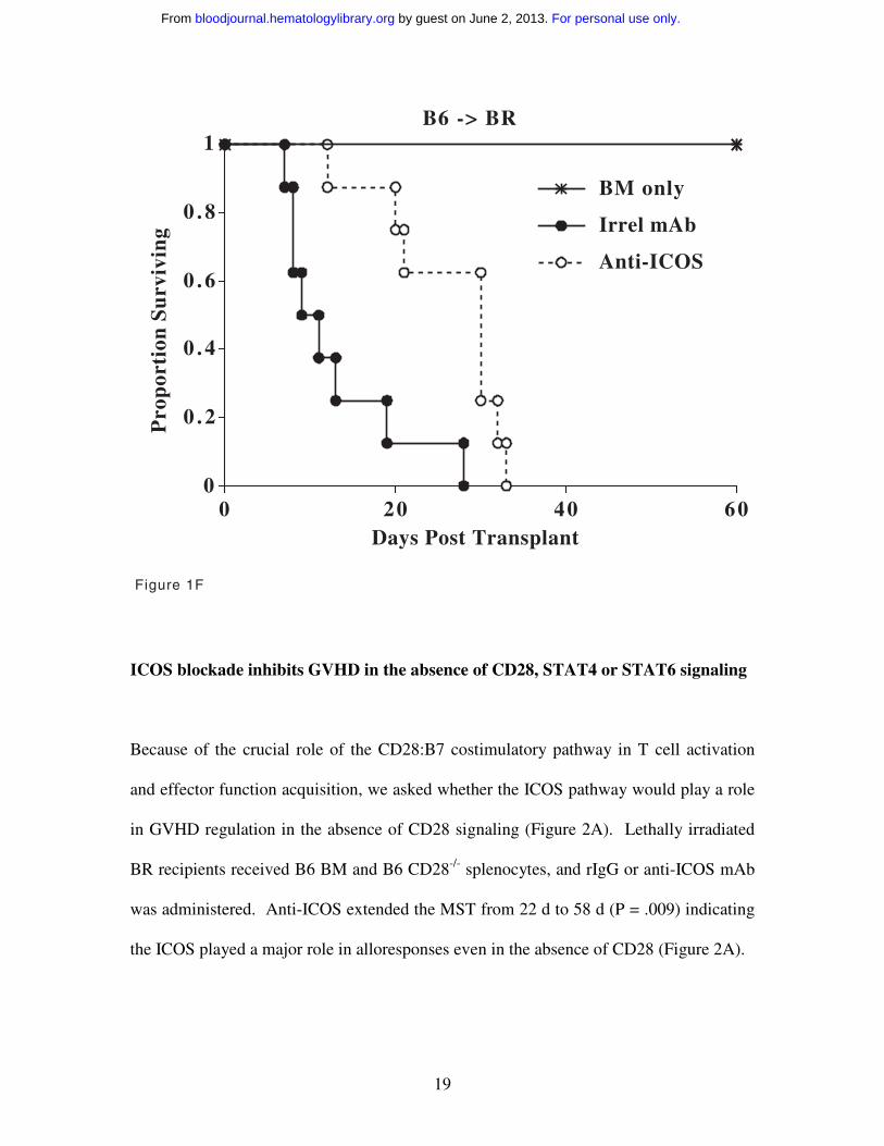
achieve. Delayed administration of anti-ICOS mAb significantly increased MST from
11d to 30.5d (Figure 1F, P = .0024) similar to the increase in MST seen when anti-ICOS
infusions were initiated on d-1 (9d vs 40d, rIgG vs anti-ICOS, Figure 1D, 25x106 spleen
dose).
For personal use only. by guest on June 2, 2013. bloodjournal.hematologylibrary.orgFrom
19
0
0 . 2
0 . 4
0 . 6
0 . 8
1P
rop
orti
on S
urv
ivin
g
0 20 40 60Days Post Transplant
Anti-ICOS
Irrel mAb
BM only
B6 -> BR
Figure 1F
ICOS blockade inhibits GVHD in the absence of CD28, STAT4 or STAT6 signaling
Because of the crucial role of the CD28:B7 costimulatory pathway in T cell activation
and effector function acquisition, we asked whether the ICOS pathway would play a role
in GVHD regulation in the absence of CD28 signaling (Figure 2A). Lethally irradiated
BR recipients received B6 BM and B6 CD28-/- splenocytes, and rIgG or anti-ICOS mAb
was administered. Anti-ICOS extended the MST from 22 d to 58 d (P = .009) indicating
the ICOS played a major role in alloresponses even in the absence of CD28 (Figure 2A).
For personal use only. by guest on June 2, 2013. bloodjournal.hematologylibrary.orgFrom

20
0
0 . 2
0 . 4
0 . 6
0 . 8
1
Pro
por
tion
Su
rviv
ing
0 20 40 60 80 100Days Post Transplant
Anti-ICOS
Irrel mAb
BM only
B6 CD28 KO -> BR
Figure 2A
ICOS has been reported to play a pivotal costimulatory role in Th1/Th2
differentiation. Because optimal GVHD has been reported to require both Th1 and Th2
cells23, amelioration of GVHD by ICOS blockade could be the result of specific Th
skewing. To determine whether ICOS blockade inhibited GVHD mediated by either Th1
or Th2 T cells, lethally irradiated B6 mice were transplanted with BALB/c BM and
splenocytes from either BALB/c WT, STAT4-/- or STAT6-/- mice (Figure 2B). Whereas
7 of 8 rIgG-treated mice receiving WT splenocytes died by day 10, anti-ICOS treatment
resulted in long-term (d 100) survival in 6 of 8 mice reproducing the protective effect of
ICOS blockade in a second strain combination that is disparate at multiple minor as well
as MHC class I and II antigens (P = .0002). ICOS blockade of mice receiving STAT4-/-
For personal use only. by guest on June 2, 2013. bloodjournal.hematologylibrary.orgFrom

21
splenocytes resulted in long-term survival in 7 of 8 mice while all rIgG-treated mice died
by d50 (P = .001). Although ICOS blockade resulted in only 1 long-term survivor of
STAT 6-/- splenocytes, MST was significantly extended to 59 d vs 15 d for rIgG-treated
controls (P = .0075). These data indicate that ICOS blockade significantly inhibited
GVHD mediated by either Th1 or Th2 T cells, although a greater survival benefit was
observed by ICOS blockade in a setting of an intact STAT6 (Th2) signaling pathway.
0
0 . 2
0 . 4
0 . 6
0 . 8
1
Pro
por
tion
Su
rviv
ing
0 20 40 60 80 100Days Post Transplant
Stat 6 KO, anti-ICOS
Stat 6 KO, irrel mAbStat 4 KO, anti-ICOS
Stat 4 KO, irrel mAb
WT, anti-ICOS
WT, irrel mAb
BM only
BALB/c -> B6
Figure 2B
For personal use only. by guest on June 2, 2013. bloodjournal.hematologylibrary.orgFrom

22
Anti-ICOS mAb blocks expansion, but not activation, of alloantigen-specific cells
To determine the effect of anti-ICOS mAb on alloantigen-specific T cells, 2C and TEa
LN cells were infused into sublethally irradiated CB6 recipients and rIgG or anti-ICOS
was administered (Figure 3A). All rIgG-treated mice died by d 21. In contrast, 7 of 8
anti-ICOS-treated mice survived long-term (P = .0002 vs rIgG controls). Despite the
profound survival advantage, anti-ICOS-treated mice had clinical evidence of GVHD at 5
months as evidenced by generalized erythema, poor fur quality, diarrhea, hunched
posture and a 20% weight reduction as compared to irradiated control mice that did not
receive Tg LN cells (data not shown). Evaluation of GVHD target tissues revealed
significant GVHD pathology of liver (large perivascular infiltrates associated with bile
ducts), lung (perivascular and peribronchiolar cuffing) and spleen (disruption of splenic
architecture and increased neutrophil infiltration) in anti-ICOS-treated survivors
compared to irradiated control mice (Table 1, Exp 2).
For personal use only. by guest on June 2, 2013. bloodjournal.hematologylibrary.orgFrom

23
0
0 . 2
0 . 4
0 . 6
0 . 8
1
Pro
por
tion
Su
rviv
ing
0 30 60 90 120 150Days Post Transfer
Rad. control
Anti-ICOS
Irrel mAb
2C/TEa -> CB6
Figure 3A
To evaluate the effect of ICOS blockade on activation and expansion of
alloantigen-specific Tg T cells, 2C Tg CD8+ and TEa Tg CD4+ T cells, recovered from
the spleens of irradiated host CB6 mice, were enumerated and phenotyped 4d after Tg
LN transfer (Figure 3B). Anti-ICOS reduced the number of 2C CD8+ and TEa CD4+ Tg
T cells by 63% and 26%, respectively (P = .0027 for CD8+ and P = .0062 for CD4+ T
cells; rIgG vs anti-ICOS). ICOS was expressed at high levels on all Tg CD8+ and CD4+
T cells recovered from rIgG-treated controls. In contrast, ICOS expression was reduced
approximately 10-fold in T cells from anti-ICOS mAb-treated mice (Figure 3C, top
histograms, bold line illustrates anti-ICOS, thin solid line illustrates rIgG). Analysis of
several cell surface activation antigens and intracellular expression of the cytotoxic
effector molecule, granzyme B, all indicated that ICOS blockade did not inhibit the in
For personal use only. by guest on June 2, 2013. bloodjournal.hematologylibrary.orgFrom

24
vivo activation of alloantigen-specific Tg T cells (Figure 3C). CD28, CD25, CD44,
CD122, CD127 and CD69 were significantly upregulated and L Sel was downregulated
in both rIgG- and anti-ICOS-treated mice (albeit to slightly different degrees for CD28,
CD25 and CD127) indicative of a high degree of activation (Figure 3C and CD69 not
shown). Moreover, the cytotoxic molecule granzyme B was detected intracellularly at
high levels in Tg CD8+ T cells recovered from rIgG-treated controls (27% ± 3%, ave ±
1SD) and anti-ICOS-treated mice (29% ± 5%). Also indicative of activation, flow
cytometric forward and side scatter parameters were consistently higher in both Tg CD8+
and Tg CD4+ cells in anti-ICOS-treated mice vs rIgG-treated controls (data not shown, P
≤ .004).
For personal use only. by guest on June 2, 2013. bloodjournal.hematologylibrary.orgFrom

25
For personal use only. by guest on June 2, 2013. bloodjournal.hematologylibrary.orgFrom
26
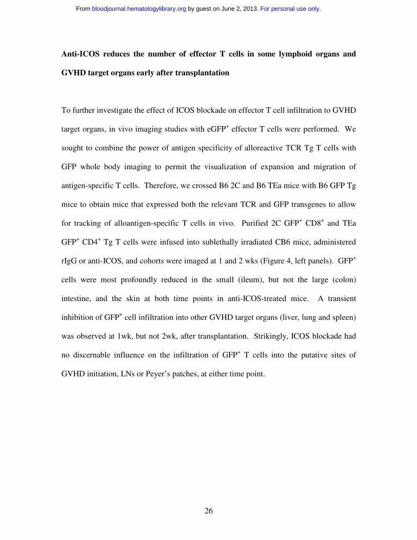
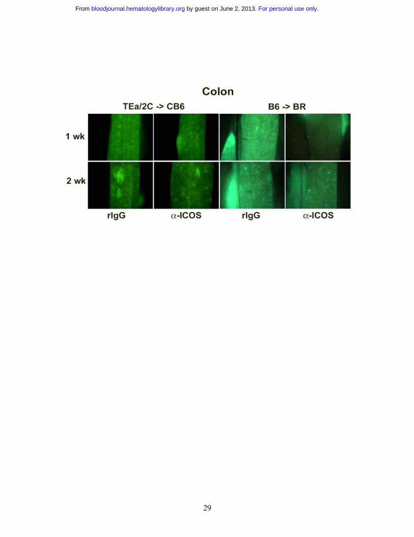
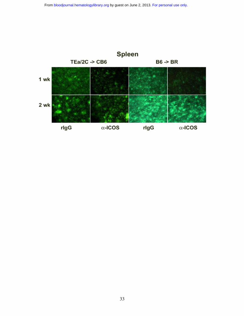
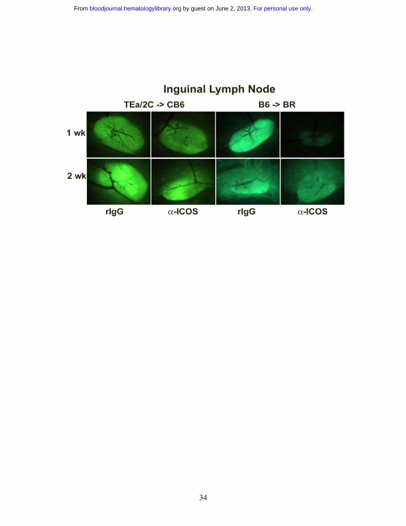
Anti-ICOS reduces the number of effector T cells in some lymphoid organs and
GVHD target organs early after transplantation
To further investigate the effect of ICOS blockade on effector T cell infiltration to GVHD
target organs, in vivo imaging studies with eGFP+ effector T cells were performed. We
sought to combine the power of antigen specificity of alloreactive TCR Tg T cells with
GFP whole body imaging to permit the visualization of expansion and migration of
antigen-specific T cells. Therefore, we crossed B6 2C and B6 TEa mice with B6 GFP Tg
mice to obtain mice that expressed both the relevant TCR and GFP transgenes to allow
for tracking of alloantigen-specific T cells in vivo. Purified 2C GFP+ CD8+ and TEa
GFP+ CD4+ Tg T cells were infused into sublethally irradiated CB6 mice, administered
rIgG or anti-ICOS, and cohorts were imaged at 1 and 2 wks (Figure 4, left panels). GFP+
cells were most profoundly reduced in the small (ileum), but not the large (colon)
intestine, and the skin at both time points in anti-ICOS-treated mice. A transient
inhibition of GFP+ cell infiltration into other GVHD target organs (liver, lung and spleen)
was observed at 1wk, but not 2wk, after transplantation. Strikingly, ICOS blockade had
no discernable influence on the infiltration of GFP+ T cells into the putative sites of
GVHD initiation, LNs or Peyer’s patches, at either time point.
For personal use only. by guest on June 2, 2013. bloodjournal.hematologylibrary.orgFrom

27
For personal use only. by guest on June 2, 2013. bloodjournal.hematologylibrary.orgFrom

28
For personal use only. by guest on June 2, 2013. bloodjournal.hematologylibrary.orgFrom
29
For personal use only. by guest on June 2, 2013. bloodjournal.hematologylibrary.orgFrom

30
For personal use only. by guest on June 2, 2013. bloodjournal.hematologylibrary.orgFrom
31
For personal use only. by guest on June 2, 2013. bloodjournal.hematologylibrary.orgFrom

32
For personal use only. by guest on June 2, 2013. bloodjournal.hematologylibrary.orgFrom
33
For personal use only. by guest on June 2, 2013. bloodjournal.hematologylibrary.orgFrom
34
For personal use only. by guest on June 2, 2013. bloodjournal.hematologylibrary.orgFrom

35
To determine whether the above imaging findings would be observed in a
polyclonal model of GVHD, studies were performed in which lethally irradiated BR mice
were infused with non-GFP B6 BM and purified B6 GFP+ T cells, given rIgG or anti-
ICOS mAb, and imaged at 1, 2 and 3 weeks (Figure 4, right panels and data not shown).
Survival studies indicated that anti-ICOS mAb administration resulted in significant
inhibition of GVHD in this strain combination (Figure 1C) as well as in the Tg GVHD
model (Figure 2A). Similar to the Tg GVHD model, anti-ICOS resulted in the most
profound and sustained reduction of polyclonal GFP+ effectors in ileum and skin (Figure
4). The widespread intestinal infiltration of GFP+ effectors is best illustrated by the low
zoom image of the loops of small intestine in the abdomen. By 1 wk, the small intestine
is diffusely infiltrated with GFP+ effectors in rIgG-treated mice. Anti-ICOS mAb
For personal use only. by guest on June 2, 2013. bloodjournal.hematologylibrary.orgFrom

36
profoundly reduced this infiltrative process although GFP+ infiltration increased from 1
to 2 wk in both groups of mice indicating that anti-ICOS mAb inhibited, but did not
prevent, donor T cell expansion. As in the TCR Tg model, anti-ICOS mAb reduced
GFP+ cells in the liver, lung and spleen although such reductions were more profound at
1 vs 2 wk. A slight decrease was also noted in the colon and Peyer’s patches. In contrast
to the TCR Tg model, GFP+ T cells were transiently reduced at 1 wk in the LNs. Thus
the dominant effects of anti-ICOS mAb administration involved the small intestine and
skin with more modest and transient effects on the liver, lung and spleen and variable
effects on other secondary lymphoid organs.
ICOS pathway plays a role in graft rejection
To investigate the role of ICOS pathway in host antidonor T cell-mediated BM graft
rejection, B6 WT and B6 ICOS-/- mice were sublethally irradiated and infused with
allogeneic TCD BALB/c BM. PBLs were phenotyped 6 weeks after transplantation for
determination of percentage donor chimerism (Table 2). Engraftment rates of WT and
ICOS-/- mice were 55% and 100%, respectively (P = .00065). Average percentage donor
chimerism in WT and ICOS-/- was 29% and 61%, respectively (P = .00019). Engraftment
was stable, multilineage and long-term (> 4 months) and donor chimerism increased over
time in ICOS-/- recipients (Table 2). These data indicated that ICOS-/- host T cells were
less competent graft rejecting effectors than WT host T cells and implicated the ICOS
pathway in BM graft rejection.
For personal use only. by guest on June 2, 2013. bloodjournal.hematologylibrary.orgFrom
37
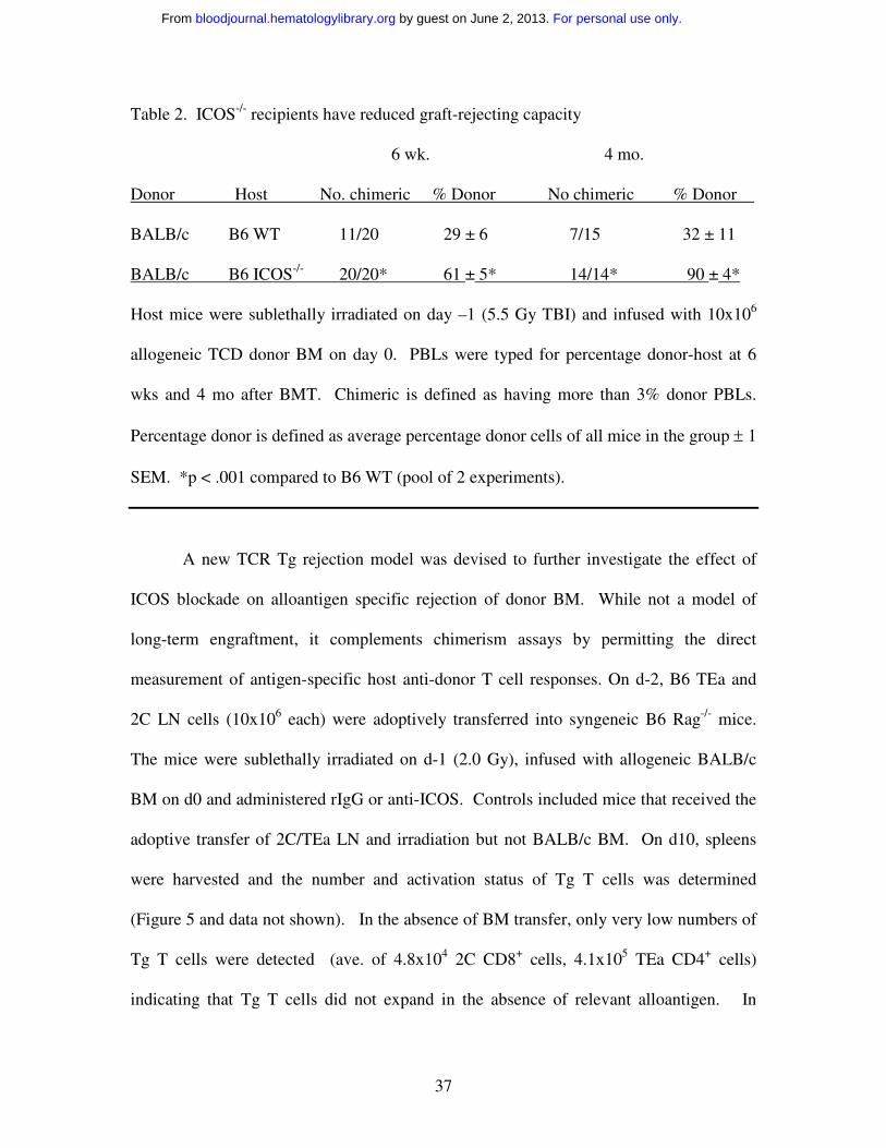
Table 2. ICOS-/- recipients have reduced graft-rejecting capacity
6 wk. 4 mo.
Donor Host No. chimeric % Donor No chimeric % Donor
BALB/c B6 WT 11/20 29 ± 6 7/15 32 ± 11
BALB/c B6 ICOS-/- 20/20* 61 ± 5* 14/14* 90 ± 4*
Host mice were sublethally irradiated on day –1 (5.5 Gy TBI) and infused with 10x106
allogeneic TCD donor BM on day 0. PBLs were typed for percentage donor-host at 6
wks and 4 mo after BMT. Chimeric is defined as having more than 3% donor PBLs.
Percentage donor is defined as average percentage donor cells of all mice in the group ± 1
SEM. *p < .001 compared to B6 WT (pool of 2 experiments).
A new TCR Tg rejection model was devised to further investigate the effect of
ICOS blockade on alloantigen specific rejection of donor BM. While not a model of
long-term engraftment, it complements chimerism assays by permitting the direct
measurement of antigen-specific host anti-donor T cell responses. On d-2, B6 TEa and
2C LN cells (10x106 each) were adoptively transferred into syngeneic B6 Rag-/- mice.
The mice were sublethally irradiated on d-1 (2.0 Gy), infused with allogeneic BALB/c
BM on d0 and administered rIgG or anti-ICOS. Controls included mice that received the
adoptive transfer of 2C/TEa LN and irradiation but not BALB/c BM. On d10, spleens
were harvested and the number and activation status of Tg T cells was determined
(Figure 5 and data not shown). In the absence of BM transfer, only very low numbers of
Tg T cells were detected (ave. of 4.8x10⁴ 2C CD8+ cells, 4.1x10⁵ TEa CD4+ cells)
indicating that Tg T cells did not expand in the absence of relevant alloantigen. In
For personal use only. by guest on June 2, 2013. bloodjournal.hematologylibrary.orgFrom

38
contrast, there was a 387- and 11-fold increase of Tg CD8+ and CD4+ cells, respectively,
in spleens of mice that received BALB/c BM and rIgG. Anti-ICOS reduced the number
of donor BM-specific Tg CD8+ and CD4+ cells by 52% and 63%, respectively, indicating
that in vivo ICOS blockade significantly inhibited, but did not abrogate, expansion of
alloantigen-specific host T cells (Figure 5). Although ICOS blockade reduced the
expansion of host antidonor T cells, it did not inhibit their activation (data not shown).
These data indicating the inhibition of alloantigen-specific host T cell expansion by ICOS
blockade complement the data demonstrating increased engraftment in ICOS-/- recipients.
Together, these data indicate that the ICOS pathway plays a role in BM graft rejection.
For personal use only. by guest on June 2, 2013. bloodjournal.hematologylibrary.orgFrom

39
Discussion
These studies indicate that the ICOS costimulatory pathway plays an important role in
both donor antihost (GVHD) and host antidonor (rejection) alloresponses. ICOS
blockade, achieved either through gene deficient mice or the in vivo administration of
anti-ICOS mAb, resulted in significant amelioration of GVHD in multiple strain
combinations whether mediated by polyclonal CD4+ and/or CD8+ T cells, alloantigen-
specific TCR Tg T cells or CD28-, Th1- or Th2-deficient T cells. Consistent with the
known role of ICOS in regulation of T cell responses after activation1, ICOS blockade
inhibited in vivo alloreactive T cell expansion but did not prevent T cell activation nor
the generation of the cytotoxic effector molecule, granzyme B24. Imaging studies
indicated that ICOS blockade resulted in striking reductions of GVHD effector cell
infiltrations into the small intestine and skin with more modest or transient effects in
other GVHD target organs. The differential results of ICOS blockade on organ
infiltration suggest effects on T cell homing and migration in addition to inhibition of T
cell expansion.
These studies used an established Tg model of GVHD in which BALB/c-reactive
Tg (2C CD8+ and TEa CD4+) T cells were infused into irradiated CB6 recipients to allow
for the study of donor antihost alloresponses19. Additionally, this paper describes a new
Tg model of rejection in which B6 host-type, BALB/c-reactive Tg T cells were
transferred into syngeneic B6 Rag-/- recipients that were irradiated and given allogeneic
BALB/c BM to allow for the study of host antidonor alloantigen-specific responses.
Somewhat gratifying to us, ICOS blockade inhibited 2C CD8+ cells similarly in both
For personal use only. by guest on June 2, 2013. bloodjournal.hematologylibrary.orgFrom

40
models (63% inhibition in GVHD and 52% inhibition in rejection model; Figures 3B and
5). The degree of inhibition mediated by ICOS blockade on TEa CD4+ cells was more
variable (26% inhibition in GVHD and 63% inhibition in rejection model). This partial
inhibition of T cell expansion mediated by anti-ICOS mAb in a Tg model of GVHD was
sufficient to result in a profound survival advantage (Figure 3A). GVHD inhibition by
ICOS blockade (either by use of mAb or ICOS-/- donors) was confirmed in several
different polyclonal models of GVHD. Although ICOS-/- mice had enhanced donor
engraftment compared to WT mice, we cannot rule out possible contributions from non-T
cell mechanisms such as NK defects or reduced stem cell competition in ICOS-/-
recipients. Therefore, the percent inhibition of expansion of adoptively transferred T
cells in the Tg rejection model may not correlate perfectly with inhibition of graft
rejection. Further studies to directly measure T cell-mediated rejection in polyclonal
models are warranted.
Imaging provides a vivid illustration of the widespread organ infiltration by
effector T cells during GVHD18,22. Imaging data indicate that ICOS blockade resulted in
an inhibition and/or delay in the expansion of effector T cells rather than an abrogation of
priming which might be expected to result in a more sustained reduction of GFP+
effectors in all GVHD target organs. In addition to the inhibition of T cell expansion, we
hypothesize that ICOS blockade may have additional effects on T cell homing and
migration as is evidenced by the profound and sustained reduction of GFP+ effectors into
the ileum and skin. These data illustrating the effect of ICOS blockade on GFP+ effectors
contrast with the effect of Treg infusions which resulted in a more profound and
For personal use only. by guest on June 2, 2013. bloodjournal.hematologylibrary.orgFrom

41
sustained inhibition of GFP+ effectors in a wider range of GVHD organs; a result thought
to be due to inhibition of earlier events in the immune response18.
Of clinical interest, anti-ICOS significantly increased MST even when mAb
infusions were delayed until d5 after transplantation. Historically, delaying therapeutic
intervention until d5 has rarely led to GVHD inhibition of our models, likely due to the
extent of priming and early expansion that has already occurred by this time22.
Furthermore, in contrast to ICOS:ICOSL blockade, neither CD28:B7, CD40L:CD40, nor
OX40:OX40L blockade extended survival time when antibodies were delayed until d5
(data not shown, P.A.T.). The differential effect of ICOS on regulating effector
responses make it an attractive candidate for combined blockade with the CD28:B7 or
CD40L:CD40 pathway which are more critical for initial activation events. Consistent
with this, Nanji et al found that combined blockade of ICOS with either CD40L:CD40 or
CD28:B7 blockade resulted in enhanced islet allograft survival11. Additionally, the role
of ICOS in regulation of effector T cells after activation suggests that, in addition to
GVHD therapy, ICOS blockade may be useful for the therapeutic targeting of primed or
memory cells that are less likely to be governed by other costimulatory pathways.
Our results indicating GVHD inhibition by ICOS blockade in all of our models
contrast somewhat with those of Ogawa et al12. They found that ICOS blockade
accelerated acute GVHD and ameliorated chronic GVHD, effects postulated to be the
result of selective inhibition of IL-4 production and Th1 polarization12. However, they
used a non-irradiated parent-into-F1 GVHD model that may be more amenable to Th
polarization by ICOS blockade whereas all of our models involved host conditioning with
total body irradiation. Consistent with this hypothesis, ICOS blockade during immune
For personal use only. by guest on June 2, 2013. bloodjournal.hematologylibrary.orgFrom

42
priming exacerbated EAE that was thought to be due to Th1 polarization due to
impairment of Th2 development25. In contrast and despite the fact that ICOS signaling
preferentially induces IL-4 and IL-101, our data indicate that ICOS blockade inhibited
GVHD mediated by either STAT4-/- (Th1-deficient) or by STAT6-/- (Th2-deficient)
effectors (Figure 2B). However, a more profound effect on GVHD inhibition by ICOS
blockade was observed with either WT or STAT4-/- donor splenocytes compared to
STAT6-/- donor splenocytes indicating a greater benefit on inhibiting Th2-mediated
GVHD.
This study demonstrates that ICOS blockade had positive effects for GVHD
inhibition, GVHD therapy and allogeneic BM graft promotion. These data together with
the intriguing report that ICOS expression most accurately defines inflammatory effector
T cells26 suggest that the ICOS pathway could be an important therapeutic target.
For personal use only. by guest on June 2, 2013. bloodjournal.hematologylibrary.orgFrom

43
Figure Legends
Figure 1. ICOS blockade inhibits GVHD. A) Sublethally irradiated (6.0 Gy Cs) bm12
mice were infused with the indicated number of purified CD25-depleted CD4+ T cells
from B6 WT or B6 ICOS-/- mice. Survival is shown. (n = 16/group, pool of 2
experiments; P = .001 for 3x10⁴ cells, .P = .007 for 105 cells). B) Sublethally irradiated
(6.0 Gy Cs) bm1 mice were infused with 106 purified CD25-depleted CD8+ T cells from
B6 WT or B6 ICOS-/- mice. Survival is shown. (n = 18/group, pool of 2 experiments; P
= .002). C) Lethally irradiated (8.0 Gy) BR mice received 20x106 B6 WT BM and either
5x10⁶, 15x106 or 25x106 splenocytes (indicated as 5S, 15S or 25S) from B6 WT or B6
ICOS-/- mice. Survival is shown. (n = 16/group, pool of 2 experiments; P ≤ .0001, B6 vs
ICOS-/- at each spleen dose). D) Lethally irradiated (8.0 Gy) BR mice received 20x106
B6 BM and either 15x106 or 25x106 B6 splenocytes (indicated as 15S or 25S). Irrelevant
rIgG or anti-ICOS mAb was administered from d-1 as indicated in Materials & Methods.
Survival is shown. (n = 10-16 mice/group, 1 exp for 25S, pool of 2 experiments for 15S
and BM only; P ≤ .0006, irrel mAb vs anti-ICOS at each spleen dose). E) Average
weights are shown for mice from Figure 1D receiving 15x106 splenocytes. F) Lethally
irradiated (8.0 Gy) BR mice received 20x106 B6 BM and 25x106 B6 splenocytes and
rIgG (Irrel mAb) or anti-ICOS was administered starting d5 after transplantation.
Survival is shown. (n = 8/group; P = .002).
Figure 2. ICOS blockade inhibits GVHD independent of CD28, STAT4 or STAT6
signaling. A) Lethally irradiated (8.0 Gy) BR mice received 20x106 B6 BM and 15x106
For personal use only. by guest on June 2, 2013. bloodjournal.hematologylibrary.orgFrom

44
B6 CD28-/- splenocytes. Irrelevant rIgG or anti-ICOS was administered from d-1 through
d28. Survival is shown. (n = 8/group; P = .009). B) Lethally irradiated B6 (9.0 Gy) mice
received 20x106 BALB/c BM and 25x106 splenocytes from BALB/c WT, STAT4-/- or
STAT6-/- mice. Irrelevant mAb or anti-ICOS was administered from d-1 through d28.
Survival is shown. (n = 8/group; P ≤ .0075, Irrel mAb vs anti-ICOS for each spleen
donor).
Figure 3. Anti-ICOS inhibits GVHD mediated by alloantigen-specific Tg T cells. A)
CB6 mice were sublethally irradiated (6.5 cGy Cs), infused with 2C and TEa LN adjusted
for Tg T cell content (4x10⁶ T cells each) and irrelevant mAb or anti-ICOS was
administered from d-1 through d+28). Radiation controls did not receive Tg T cells or
Ab. Survival is shown. (n = 8/group; P = .0002, irrel mAb vs anti-ICOS). B) Spleens
were harvested from mice described in A on d4, counted, and proportion of Tg T cells
was determined by flow cytometric analysis. Shown is ave of absolute counts of 2C
CD8+ and TEa CD4+ Tg T cells per spleen + 1 SEM (n = 19/group, pool of 4 separate
experiments; P = .0027 for 2C CD8+, P = .0062 for TEa CD4+, rIgG vs anti-ICOS). C)
Spleens from mice treated as described in A and B were phenotyped for indicated cell
surface activation antigens and intracellular cytotoxic effector molecule (granzyme B).
2C and TEa Tg cells were gated on to obtain histograms. Dotted line indicates negative
control. Bold solid line and thin solid line indicate anti-ICOS and rIgG treatment,
respectively. The dash line shown in the CD28, L Sel and CD44 histograms indicates the
level of expression on freshly harvested Tg T cells. Shown is a representative sample.
For personal use only. by guest on June 2, 2013. bloodjournal.hematologylibrary.orgFrom

45
Figure 4. ICOS blockade inhibits expansion of GFP+ effectors in secondary
lymphoid organs and GVHD target tissues. Effect of ICOS blockade in both a Tg and
a polyclonal GVHD model is shown. Images of sublethally irradiated (5.5 Gy Cs) CB6
mice infused with purified 2C GFP+ CD8+ and TEa GFP+ CD4+ T cells (2x10+ each) are
shown in left-hand panels. Images of lethally irradiated (8.0 Gy) BR mice infused with
B6 non-GFP BM (20x10+) and purified B6 GFP+ T cells (3x10+) are shown in right-hand
panels. Rat IgG or anti-ICOS mAb was administered. Representative images from 1 of 3
mice per group imaged at 1 and 2 wk are shown. Stereomicroscope was set to x 1.0
zoom factor for intestinal loops in abdomen; x 3.2 for inguinal LN, skin, colon and ileum;
x 4.5 for Peyer’s patch; x 7.0 for liver and spleen; and x 10.0 for lung. Exposure times
were optimized for rIgG-treated mice for each organ and identical times were used for
anti-ICOS-treated mice. Control mice not receiving GFP+ effectors to verify lack of
autofluorescence resulted in dark images (not shown).
Figure 5. ICOS blockade blocks expansion of alloantigen-specific BM graft
rejecting cells. 2C and TEa LN cells (10x106 each) were adoptively transferred into B6
Rag-/- mice on d-2, irradiated on d-1 (2.0 Gy), infused with BALB/c BM (40x106) on d0
and administered rIgG or anti-ICOS mAb from d-1 to d+9. Control of irradiated mice
receiving adoptive transfer of 2C and TEa LN, but not BALB/c BM is also shown
(designated as AT Only). On d10, spleens were harvested and the number of Tg T cells
was determined. Shown is average absolute number of CD8+ and CD4+ Tg T cells per
spleen ± 1 SEM. (n = 10/group, pool of 2 experiments; P = .0197 for 2C Tg CD8+ cells, P
= .0006 for TEa Tg CD4+ cells; rIgG vs anti-ICOS).
For personal use only. by guest on June 2, 2013. bloodjournal.hematologylibrary.orgFrom

46
Acknowledgements
The authors acknowledge Jessica M. Swedin, Michael J. Ehrhardt, Christopher J. Lees
and Matthew M. Roforth for excellent technical assistance.
These studies were supported in part by National Institutes of Health grants RO1
AI34495, 2R37HL56067, RO1 HL63452 and PO1 AI056299.
For personal use only. by guest on June 2, 2013. bloodjournal.hematologylibrary.orgFrom

47
References
1. Hutloff A, Dittrich AM, Beier KC, Eljaschewitsch B, Kraft R,
Anagnostopoulos I, Kroczek RA. ICOS is an inducible T-cell co-stimulator structurally
and functionally related to CD28. Nature. 1999;397:263-266
2. Swallow MM, Wallin JJ, Sha WC. B7h, a novel costimulatory homolog of
B7.1 and B7.2, is induced by TNFalpha. Immunity. 1999;11:423-432
3. Gonzalo JA, Delaney T, Corcoran J, Goodearl A, Gutierrez-Ramos JC, Coyle
AJ. Cutting edge: the related molecules CD28 and inducible costimulator deliver both
unique and complementary signals required for optimal T cell activation. J Immunol.
2001;166:1-5
4. Coyle AJ, Gutierrez-Ramos JC. The expanding B7 superfamily: increasing
complexity in costimulatory signals regulating T cell function. Nat Immunol. 2001;2:203-
209
5. Dong C, Juedes AE, Temann UA, Shresta S, Allison JP, Ruddle NH, Flavell
RA. ICOS co-stimulatory receptor is essential for T-cell activation and function. Nature.
2001;409:97-101
6. McAdam AJ, Greenwald RJ, Levin MA, Chernova T, Malenkovich N, Ling V,
Freeman GJ, Sharpe AH. ICOS is critical for CD40-mediated antibody class switching.
Nature. 2001;409:102-105
7. Mak TW, Shahinian A, Yoshinaga SK, Wakeham A, Boucher LM, Pintilie M,
Duncan G, Gajewska BU, Gronski M, Eriksson U, Odermatt B, Ho A, Bouchard D,
Whorisky JS, Jordana M, Ohashi PS, Pawson T, Bladt F, Tafuri A. Costimulation
For personal use only. by guest on June 2, 2013. bloodjournal.hematologylibrary.orgFrom
48
through the inducible costimulator ligand is essential for both T helper and B cell
functions in T cell-dependent B cell responses. Nat Immunol. 2003;4:765-772
8. Nurieva RI, Mai XM, Forbush K, Bevan MJ, Dong C. B7h is required for T
cell activation, differentiation, and effector function. Proc Natl Acad Sci U S A.
2003;100:14163-14168
9. Harada H, Salama AD, Sho M, Izawa A, Sandner SE, Ito T, Akiba H, Yagita
H, Sharpe AH, Freeman GJ, Sayegh MH. The role of the ICOS-B7h T cell costimulatory
pathway in transplantation immunity. J Clin Invest. 2003;112:234-243
10. Guo L, Li XK, Funeshima N, Fujino M, Nagata Y, Kimura H, Amemiya H,
Enosawa S, Tsuji T, Harihara Y, Makuuchi M, Suzuki S. Prolonged survival in rat liver
transplantation with mouse monoclonal antibody against an inducible costimulator
(ICOS). Transplantation. 2002;73:1027-1032
11. Nanji SA, Hancock WW, Anderson CC, Adams AB, Luo B, Schur CD,
Pawlick RL, Wang L, Coyle AJ, Larsen CP, Shapiro AM. Multiple combination therapies
involving blockade of ICOS/B7RP-1 costimulation facilitate long-term islet allograft
survival. Am J Transplant. 2004;4:526-536
12. Ogawa S, Nagamatsu G, Watanabe M, Watanabe S, Hayashi T, Horita S,
Nitta K, Nihei H, Tezuka K, Abe R. Opposing effects of anti-activation-inducible
lymphocyte-immunomodulatory molecule/inducible costimulator antibody on the
development of acute versus chronic graft-versus-host disease. J Immunol.
2001;167:5741-5748
13. Tafuri A, Shahinian A, Bladt F, Yoshinaga SK, Jordana M, Wakeham A,
Boucher LM, Bouchard D, Chan VS, Duncan G, Odermatt B, Ho A, Itie A, Horan T,
For personal use only. by guest on June 2, 2013. bloodjournal.hematologylibrary.orgFrom

49
Whoriskey JS, Pawson T, Penninger JM, Ohashi PS, Mak TW. ICOS is essential for
effective T-helper-cell responses. Nature. 2001;409:105-109
14. Grubin CE, Kovats S, deRoos P, Rudensky AY. Deficient positive selection
of CD4 T cells in mice displaying altered repertoires of MHC class II-bound self-
peptides. Immunity. 1997;7:197-208
15. Sha WC, Nelson CA, Newberry RD, Kranz DM, Russell JH, Loh DY.
Positive and negative selection of an antigen receptor on T cells in transgenic mice.
Nature. 1988;336:73-76
16. Taylor PA, Lees CJ, Blazar BR. The infusion of ex vivo activated and
expanded CD4(+)CD25(+) immune regulatory cells inhibits graft-versus-host disease
lethality. Blood. 2002;99:3493-3499
17. Cohen JL, Trenado A, Vasey D, Klatzmann D, Salomon BL.
CD4(+)CD25(+) immunoregulatory T Cells: new therapeutics for graft-versus-host
disease. J Exp Med. 2002;196:401-406
18. Taylor PA, Panoskaltsis-Mortari A, Swedin JM, Lucas PJ, Gress RE, Levine
BL, June CH, Serody JS, Blazar BR. LSelectinhi but not the LSelectinlo CD4+25+ T
regulatory cells are potent inhibitors of GVHD and BM graft rejection. Blood. 2004
19. Gonzalez M, Quezada SA, Blazar BR, Panoskaltsis-Mortari A, Rudensky
AY, Noelle RJ. The balance between donor T cell anergy and suppression versus lethal
graft-versus-host disease is determined by host conditioning. J Immunol. 2002;169:5581-
5589
20. Blazar BR, Taylor PA, McElmurry R, Tian L, Panoskaltsis-Mortari A, Lam
S, Lees C, Waldschmidt T, Vallera DA. Engraftment of severe combined immune
For personal use only. by guest on June 2, 2013. bloodjournal.hematologylibrary.orgFrom

50
deficient mice receiving allogeneic bone marrow via In utero or postnatal transfer. Blood.
1998;92:3949-3959
21. Grossman WJ, Verbsky JW, Tollefsen BL, Kemper C, Atkinson JP, Ley TJ.
Differential expression of granzymes A and B in human cytotoxic lymphocyte subsets
and T regulatory cells. Blood. 2004
22. Panoskaltsis-Mortari A, Price A, Hermanson JR, Taras E, Lees C, Serody JS,
Blazar BR. In vivo imaging of graft-versus-host-disease in mice. Blood. 2004;103:3590-
3598
23. Nikolic B, Lee S, Bronson RT, Grusby MJ, Sykes M. Th1 and Th2 mediate
acute graft-versus-host disease, each with distinct end-organ targets. J Clin Invest.
2000;105:1289-1298
24. Graubert TA, Russell JH, Ley TJ. The role of granzyme B in murine models
of acute graft-versus-host disease and graft rejection. Blood. 1996;87:1232-1237
25. Rottman JB, Smith T, Tonra JR, Ganley K, Bloom T, Silva R, Pierce B,
Gutierrez-Ramos JC, Ozkaynak E, Coyle AJ. The costimulatory molecule ICOS plays an
important role in the immunopathogenesis of EAE. Nat Immunol. 2001;2:605-611
26. Lohning M, Hutloff A, Kallinich T, Mages HW, Bonhagen K, Radbruch A,
Hamelmann E, Kroczek RA. Expression of ICOS in vivo defines CD4+ effector T cells
with high inflammatory potential and a strong bias for secretion of interleukin 10. J Exp
Med. 2003;197:181-193
For personal use only. by guest on June 2, 2013. bloodjournal.hematologylibrary.orgFrom
Related Documents