Targeting neonatal ischemic brain injury with a pentapeptide-based irreversible caspase inhibitor D Chauvier 1,2 , S Renolleau 3,4,5 , S Holifanjaniaina 6,7 , S Ankri 1,2 , M Bezault 1,2 , L Schwendimann 6,7 , C Rousset 8,9 , R Casimir 1,2,10 , J Hoebeke 10 , M Smirnova 1,2 , G Debret 11,12 , A-P Trichet 2 , Y Carlsson 9,13 , X Wang 9 , E Bernard 2 , M He ´bert 2 , J-M Rauzier 14,15 , S Matecki 16,17 , A Lacampagne 15,17 , P Rustin 6,7 , J Mariani 3,4,18 , H Hagberg 8,9,13 , P Gressens 6,7,8 , C Charriaut-Marlangue 3,4 and E Jacotot* ,1,2,6,7,8 Brain protection of the newborn remains a challenging priority and represents a totally unmet medical need. Pharmacological inhibition of caspases appears as a promising strategy for neuroprotection. In a translational perspective, we have developed a pentapeptide-based group II caspase inhibitor, TRP601/ORPHA133563, which reaches the brain, and inhibits caspases activation, mitochondrial release of cytochrome c, and apoptosis in vivo. Single administration of TRP601 protects newborn rodent brain against excitotoxicity, hypoxia–ischemia, and perinatal arterial stroke with a 6-h therapeutic time window, and has no adverse effects on physiological parameters. Safety pharmacology investigations, and toxicology studies in rodent and canine neonates, suggest that TRP601 is a lead compound for further drug development to treat ischemic brain damage in human newborns. Cell Death and Disease (2011) 2, e203; doi:10.1038/cddis.2011.87; published online 1 September 2011 Subject Category: Neuroscience Worldwide estimations indicate more than 2 million non- infectious neonatal deaths per year. 1 Neonatal ischemic brain injuries, such as stroke (focal cerebral ischemia involving middle cerebral artery occlusion (MCAO)) or hypoxia–ischemia (HI) (involving systemic asphyxia), are major and untreated causes of newborn morbidity and mortality. 2,3 Of the infants who survive the first few hours after intrapartum-related neonatal deaths (previously called ‘birth asphyxia’), as many as an annual 1 million may develop cerebral palsy, learning difficulties or other disabilities. 4 Recent clinical trials show that therapeutic inter- vention by brain cooling beginning up to 6 h after perinatal asphyxia reduces cerebral injury and improves outcomes in term infants. 5 Although these studies provide proof of concept that in this context cell death is both delayed and preventable, the protection is limited and there is still no treatment available for perinatal stroke or brain injury occurring in preterm infants. 6 Ischemic brain injury in the developing brain involves several factors such as excitotoxicity, oxidative stress, and inflammation, which accelerate cell death through either apoptosis or necrosis, depending on the region of the brain affected and on the severity of the insult. 7,8 Accumulating data suggest that apoptotic mechanisms have a more prominent role in the evolution of ischemic brain injury in neonatal rodents 9–11 and humans 12 than in adult brain ischemia, 13,14 and that apoptosis involves the mitochondrial release of cytochrome c 11 and apoptosis-inducing factor (AIF), 11,15,16 which activate caspase-dependent 10,16 and -independent execution pathways, 13,16 respectively. Caspases are a class of cysteine endoproteases that have strict specificity for an aspartic residue at the S1 subsite. 17,18 Some caspases are important mediators of inflammation and others are involved in the apoptosis of mammalian cells, where they participate in signaling and effector pathways. 19 On the basis of the optimal four-amino-acid sequence to the left of the cleavage site, caspases may be classified into three groups: group I contains caspase-1, -4, and -5 (optimal tetrapeptide: WEHD), group II contains caspase-2 (Casp2), -3 (Casp3), and -7 (Casp7) (optimal tetrapeptide: D[E/A]XD), and group III contains caspase-6, -8 (Casp8), -9 (Casp9), and -10 (optimal tetrapeptide: [V/L]EXD). 20–22 Received 20.7.11; accepted 21.7.11; Edited by D Bano 1 Theraptosis Research Laboratory, Theraptosis SA, Pasteur BioTop, Institut Pasteur, Paris 75015, France; 2 Theraptosis R&D Laboratories, Theraptosis SA, Biocitech Technology Park, Romainville 93230, France; 3 CNRS, UMR 7102 NPA, Paris 75005, France; 4 Pierre and Marie Curie University Paris 6, UMR 7102 NPA, Paris 75005, France; 5 Service de Re ´ animation Ne ´ onatale et Pe ´ diatrique, Ho ˆ pital Armand Trousseau - APHP, Paris 75012, France; 6 Inserm, U676, Paris 75019, France; 7 Universite ´ Paris Diderot, UMR 676, Paris 75019, France; 8 Centre for the Developing Brain, Institute of Reproductive and Developmental Biology, Imperial College London, Hammersmith Hospital, London W12 0NN, UK; 9 Department of Physiology, Perinatal Center, Sahlgrenska Academy, Go ¨ teborg University, Gothenburg 40530, Sweden; 10 CNRS UPR 9021, Institut de Biologie Mole ´ culaire et Cellulaire, Strasbourg 67084, France; 11 Laboratoire de Physique The ´orique de la Matie ` re Condense ´e, CNRS UMR 7600, Paris 75005, France; 12 Pierre and Marie Curie University Paris 6 UMR 7600 LPTMC, Paris 75005, France; 13 Department of Obstetrics and Gynaecology, Sahlgrenska University Hospital, Gothenburg 41345, Sweden; 14 CHU de Nı ˆmes, Nı ˆmes 30000, France; 15 Inserm U1046, Montpellier 34925, France; 16 CHU de Montpellier, Montpellier 34925, France; 17 Universite ´ Montpellier 1 et 2, Montpellier 34925, France and 18 Ho ˆ pital Charles Foix, UEF, Ivry-sur-Seine 94205, France *Corresponding author: E Jacotot, Inserm, U676, Ho ˆ pital Robert Debre ´, 48 Bd Se ´ rurier, Paris 75019, France. Tel: þ 33 1 4003 1932; Fax: þ 44 20 7594 2154; E-mail: [email protected] or [email protected] Keywords: caspase inhibitor; perinatal brain damage; ischemia; neuroprotection Abbreviations: AIF, apoptosis-inducing factor; Casp2, caspase-2; Casp3, caspase-3; Casp8, caspase-8; Casp9, caspase-9; Casp7, caspase-7; D2Me-TRP601, TRP601 devoid of methyl ester groups on Asp lateral chains; ECG, electrocardiogram; HI, hypoxia–ischemia; i.p., intraperitoneal; i.v., intravenous; MCAO, middle cerebral artery occlusion; Q-VD-Oph, quinoline-Val-Asp-CH2-OPh; TUNEL, terminal deoxynucleotidyl transferase dUTP nick-end labeling; SD, serum deprivation; z-VAD-fmk, N-benzyloxycarbonyl-Val-Ala-Asp(Ome)-fluoromethylketone Citation: Cell Death and Disease (2011) 2, e203; doi:10.1038/cddis.2011.87 & 2011 Macmillan Publishers Limited All rights reserved 2041-4889/11 www.nature.com/cddis

Welcome message from author
This document is posted to help you gain knowledge. Please leave a comment to let me know what you think about it! Share it to your friends and learn new things together.
Transcript
Targeting neonatal ischemic brain injury with apentapeptide-based irreversible caspase inhibitor
D Chauvier1,2, S Renolleau3,4,5, S Holifanjaniaina6,7, S Ankri1,2, M Bezault1,2, L Schwendimann6,7, C Rousset8,9, R Casimir1,2,10,
J Hoebeke10, M Smirnova1,2, G Debret11,12, A-P Trichet2, Y Carlsson9,13, X Wang9, E Bernard2, M Hebert2, J-M Rauzier14,15,
S Matecki16,17, A Lacampagne15,17, P Rustin6,7, J Mariani3,4,18, H Hagberg8,9,13, P Gressens6,7,8, C Charriaut-Marlangue3,4
and E Jacotot*,1,2,6,7,8
Brain protection of the newborn remains a challenging priority and represents a totally unmet medical need. Pharmacologicalinhibition of caspases appears as a promising strategy for neuroprotection. In a translational perspective, we have developed apentapeptide-based group II caspase inhibitor, TRP601/ORPHA133563, which reaches the brain, and inhibits caspasesactivation, mitochondrial release of cytochrome c, and apoptosis in vivo. Single administration of TRP601 protects newbornrodent brain against excitotoxicity, hypoxia–ischemia, and perinatal arterial stroke with a 6-h therapeutic time window, and hasno adverse effects on physiological parameters. Safety pharmacology investigations, and toxicology studies in rodent andcanine neonates, suggest that TRP601 is a lead compound for further drug development to treat ischemic brain damage inhuman newborns.Cell Death and Disease (2011) 2, e203; doi:10.1038/cddis.2011.87; published online 1 September 2011Subject Category: Neuroscience
Worldwide estimations indicate more than 2 million non-infectious neonatal deaths per year.1 Neonatal ischemic braininjuries, such as stroke (focal cerebral ischemia involving middlecerebral artery occlusion (MCAO)) or hypoxia–ischemia (HI)(involving systemic asphyxia), are major and untreated causesof newborn morbidity and mortality.2,3 Of the infants who survivethe first few hours after intrapartum-related neonatal deaths(previously called ‘birth asphyxia’), as many as an annual1 million may develop cerebral palsy, learning difficulties or otherdisabilities.4 Recent clinical trials show that therapeutic inter-vention by brain cooling beginning up to 6 h after perinatalasphyxia reduces cerebral injury and improves outcomes in terminfants.5 Although these studies provide proof of concept that inthis context cell death is both delayed and preventable, theprotection is limited and there is still no treatment available forperinatal stroke or brain injury occurring in preterm infants.6
Ischemic brain injury in the developing brain involvesseveral factors such as excitotoxicity, oxidative stress, andinflammation, which accelerate cell death through eitherapoptosis or necrosis, depending on the region of the brain
affected and on the severity of the insult.7,8 Accumulating datasuggest that apoptotic mechanisms have a more prominentrole in the evolution of ischemic brain injury in neonatalrodents9–11 and humans12 than in adult brain ischemia,13,14
and that apoptosis involves the mitochondrial release ofcytochrome c11 and apoptosis-inducing factor (AIF),11,15,16
which activate caspase-dependent10,16 and -independentexecution pathways,13,16 respectively.
Caspases are a class of cysteine endoproteases that havestrict specificity for an aspartic residue at the S1 subsite.17,18
Some caspases are important mediators of inflammation andothers are involved in the apoptosis of mammalian cells,where they participate in signaling and effector pathways.19
On the basis of the optimal four-amino-acid sequence to theleft of the cleavage site, caspases may be classified into threegroups: group I contains caspase-1, -4, and -5 (optimaltetrapeptide: WEHD), group II contains caspase-2 (Casp2), -3(Casp3), and -7 (Casp7) (optimal tetrapeptide: D[E/A]XD),and group III contains caspase-6, -8 (Casp8), -9 (Casp9), and-10 (optimal tetrapeptide: [V/L]EXD).20–22
Received 20.7.11; accepted 21.7.11; Edited by D Bano
1Theraptosis Research Laboratory, Theraptosis SA, Pasteur BioTop, Institut Pasteur, Paris 75015, France; 2Theraptosis R&D Laboratories, Theraptosis SA, BiocitechTechnology Park, Romainville 93230, France; 3CNRS, UMR 7102 NPA, Paris 75005, France; 4Pierre and Marie Curie University Paris 6, UMR 7102 NPA, Paris 75005,France; 5Service de Reanimation Neonatale et Pediatrique, Hopital Armand Trousseau - APHP, Paris 75012, France; 6Inserm, U676, Paris 75019, France; 7UniversiteParis Diderot, UMR 676, Paris 75019, France; 8Centre for the Developing Brain, Institute of Reproductive and Developmental Biology, Imperial College London,Hammersmith Hospital, London W12 0NN, UK; 9Department of Physiology, Perinatal Center, Sahlgrenska Academy, Goteborg University, Gothenburg 40530, Sweden;10CNRS UPR 9021, Institut de Biologie Moleculaire et Cellulaire, Strasbourg 67084, France; 11Laboratoire de Physique Theorique de la Matiere Condensee, CNRSUMR 7600, Paris 75005, France; 12Pierre and Marie Curie University Paris 6 UMR 7600 LPTMC, Paris 75005, France; 13Department of Obstetrics and Gynaecology,Sahlgrenska University Hospital, Gothenburg 41345, Sweden; 14CHU de Nımes, Nımes 30000, France; 15Inserm U1046, Montpellier 34925, France; 16CHU deMontpellier, Montpellier 34925, France; 17Universite Montpellier 1 et 2, Montpellier 34925, France and 18Hopital Charles Foix, UEF, Ivry-sur-Seine 94205, France*Corresponding author: E Jacotot, Inserm, U676, Hopital Robert Debre, 48 Bd Serurier, Paris 75019, France. Tel: þ 33 1 4003 1932; Fax: þ 44 20 7594 2154;E-mail: [email protected] or [email protected]: caspase inhibitor; perinatal brain damage; ischemia; neuroprotectionAbbreviations: AIF, apoptosis-inducing factor; Casp2, caspase-2; Casp3, caspase-3; Casp8, caspase-8; Casp9, caspase-9; Casp7, caspase-7; D2Me-TRP601,TRP601 devoid of methyl ester groups on Asp lateral chains; ECG, electrocardiogram; HI, hypoxia–ischemia; i.p., intraperitoneal; i.v., intravenous; MCAO, middlecerebral artery occlusion; Q-VD-Oph, quinoline-Val-Asp-CH2-OPh; TUNEL, terminal deoxynucleotidyl transferase dUTP nick-end labeling; SD, serum deprivation;z-VAD-fmk, N-benzyloxycarbonyl-Val-Ala-Asp(Ome)-fluoromethylketone
Citation: Cell Death and Disease (2011) 2, e203; doi:10.1038/cddis.2011.87& 2011 Macmillan Publishers Limited All rights reserved 2041-4889/11
www.nature.com/cddis
Both pancaspase inhibition and Casp3-selective inhibitionhave been reported to be neuroprotective in various rodentmodels of neonatal brain injury,13,23,24 opening the possibilityfor pharmacological intervention.6 However, lack of protectionwith caspase inhibitors was also reported,25,26 possiblyreflecting differences between experimental models or set-tings (e.g., the age of animals26), specific in vivo properties ofthe used inhibitors (e.g., brain penetration27), and/or a shift tocaspase-independent cell death pathways (e.g., AIF, auto-phagic death, necroptosis).
The role of individual caspases in the developing brain is notfully understood. Genetic analysis using constitutive defi-ciency revealed that Casp3 and Casp9 execute programmed(physiological) cell death in the central nervous system,28,29
whereas Casp2 does not.30 Aggravation of HI-induced lesionswas reported in Casp3-null mice.31 In contrast, geneticinhibition of Casp2 is neuroprotective in newborn miceexposed to HI or excitotoxic challenges.32
In a translational attempt to generate an efficient and safeCasp2/group-II caspase inhibitor, we have developed a potentpentapeptide-based irreversible caspase inhibitor. We reporthere the preclinical evaluation of this compound and presentdata supporting a potent neuroprotective role against perina-tal ischemic brain damage in a variety of in vivo models,potentially opening an avenue for treatment.
Results
Design of a caspase inhibitor adapted for neuroprotectionin neonates. We previously showed that the pancaspaseinhibitor quinolyl-carbonyl-Val-Asp-difluorophenoxymethyl-ketone (Q-VD-OPh) has enhanced in vitro and in vivopharmacological properties,33 together with potent neuro-protective effects in neonatal brain injury experimentalmodels.10,16,34 We reasoned that an in vivo efficient groupII-selective caspase inhibitor might combine an amino-terminalquinolyl-carbonyl and a C-terminal fluorophenoxymethyl
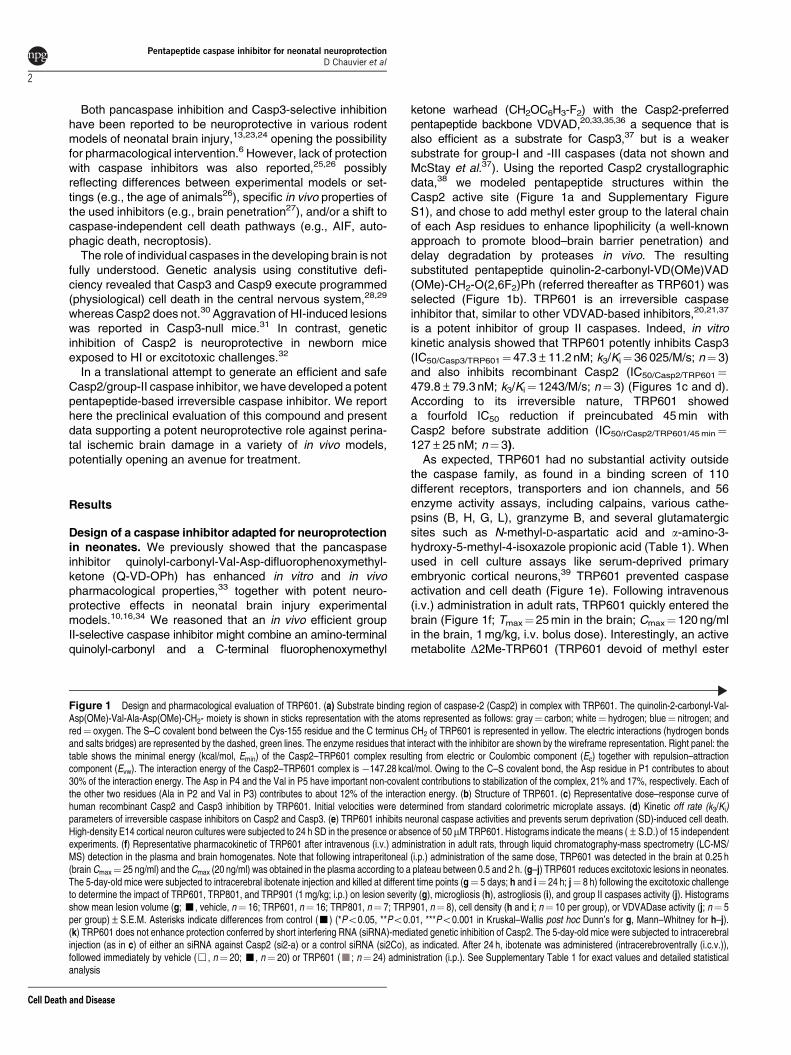
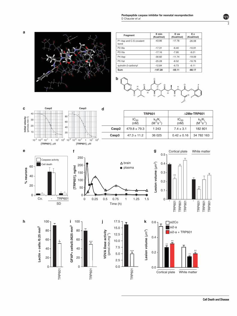
ketone warhead (CH2OC6H3-F2) with the Casp2-preferredpentapeptide backbone VDVAD,20,33,35,36 a sequence that isalso efficient as a substrate for Casp3,37 but is a weakersubstrate for group-I and -III caspases (data not shown andMcStay et al.37). Using the reported Casp2 crystallographicdata,38 we modeled pentapeptide structures within theCasp2 active site (Figure 1a and Supplementary FigureS1), and chose to add methyl ester group to the lateral chainof each Asp residues to enhance lipophilicity (a well-knownapproach to promote blood–brain barrier penetration) anddelay degradation by proteases in vivo. The resultingsubstituted pentapeptide quinolin-2-carbonyl-VD(OMe)VAD(OMe)-CH2-O(2,6F2)Ph (referred thereafter as TRP601) wasselected (Figure 1b). TRP601 is an irreversible caspaseinhibitor that, similar to other VDVAD-based inhibitors,20,21,37
is a potent inhibitor of group II caspases. Indeed, in vitrokinetic analysis showed that TRP601 potently inhibits Casp3(IC50/Casp3/TRP601¼ 47.3±11.2 nM; k3/Ki¼ 36 025/M/s; n¼ 3)and also inhibits recombinant Casp2 (IC50/Casp2/TRP601¼479.8±79.3 nM; k3/Ki¼ 1243/M/s; n¼ 3) (Figures 1c and d).According to its irreversible nature, TRP601 showeda fourfold IC50 reduction if preincubated 45 min withCasp2 before substrate addition (IC50/rCasp2/TRP601/45 min¼127±25 nM; n¼ 3).
As expected, TRP601 had no substantial activity outsidethe caspase family, as found in a binding screen of 110different receptors, transporters and ion channels, and 56enzyme activity assays, including calpains, various cathe-psins (B, H, G, L), granzyme B, and several glutamatergicsites such as N-methyl-D-aspartatic acid and a-amino-3-hydroxy-5-methyl-4-isoxazole propionic acid (Table 1). Whenused in cell culture assays like serum-deprived primaryembryonic cortical neurons,39 TRP601 prevented caspaseactivation and cell death (Figure 1e). Following intravenous(i.v.) administration in adult rats, TRP601 quickly entered thebrain (Figure 1f; Tmax¼ 25 min in the brain; Cmax¼ 120 ng/mlin the brain, 1 mg/kg, i.v. bolus dose). Interestingly, an activemetabolite D2Me-TRP601 (TRP601 devoid of methyl ester
Figure 1 Design and pharmacological evaluation of TRP601. (a) Substrate binding region of caspase-2 (Casp2) in complex with TRP601. The quinolin-2-carbonyl-Val-Asp(OMe)-Val-Ala-Asp(OMe)-CH2- moiety is shown in sticks representation with the atoms represented as follows: gray¼ carbon; white¼ hydrogen; blue¼ nitrogen; andred¼ oxygen. The S–C covalent bond between the Cys-155 residue and the C terminus CH2 of TRP601 is represented in yellow. The electric interactions (hydrogen bondsand salts bridges) are represented by the dashed, green lines. The enzyme residues that interact with the inhibitor are shown by the wireframe representation. Right panel: thetable shows the minimal energy (kcal/mol, Emin) of the Casp2–TRP601 complex resulting from electric or Coulombic component (Ec) together with repulsion–attractioncomponent (Evw). The interaction energy of the Casp2–TRP601 complex is �147.28 kcal/mol. Owing to the C–S covalent bond, the Asp residue in P1 contributes to about30% of the interaction energy. The Asp in P4 and the Val in P5 have important non-covalent contributions to stabilization of the complex, 21% and 17%, respectively. Each ofthe other two residues (Ala in P2 and Val in P3) contributes to about 12% of the interaction energy. (b) Structure of TRP601. (c) Representative dose–response curve ofhuman recombinant Casp2 and Casp3 inhibition by TRP601. Initial velocities were determined from standard colorimetric microplate assays. (d) Kinetic off rate (k3/Ki)parameters of irreversible caspase inhibitors on Casp2 and Casp3. (e) TRP601 inhibits neuronal caspase activities and prevents serum deprivation (SD)-induced cell death.High-density E14 cortical neuron cultures were subjected to 24 h SD in the presence or absence of 50 mM TRP601. Histograms indicate the means (±S.D.) of 15 independentexperiments. (f) Representative pharmacokinetic of TRP601 after intravenous (i.v.) administration in adult rats, through liquid chromatography-mass spectrometry (LC-MS/MS) detection in the plasma and brain homogenates. Note that following intraperitoneal (i.p.) administration of the same dose, TRP601 was detected in the brain at 0.25 h(brain Cmax¼ 25 ng/ml) and the Cmax (20 ng/ml) was obtained in the plasma according to a plateau between 0.5 and 2 h. (g–j) TRP601 reduces excitotoxic lesions in neonates.The 5-day-old mice were subjected to intracerebral ibotenate injection and killed at different time points (g¼ 5 days; h and i¼ 24 h; j¼ 8 h) following the excitotoxic challengeto determine the impact of TRP601, TRP801, and TRP901 (1 mg/kg; i.p.) on lesion severity (g), microgliosis (h), astrogliosis (i), and group II caspases activity (j). Histogramsshow mean lesion volume (g; ’, vehicle, n¼ 16; TRP601, n¼ 16; TRP801, n¼ 7; TRP901, n¼ 8), cell density (h and i; n¼ 10 per group), or VDVADase activity (j; n¼ 5per group)±S.E.M. Asterisks indicate differences from control (’) (*Po0.05, **Po0.01, ***Po0.001 in Kruskal–Wallis post hoc Dunn’s for g, Mann–Whitney for h–j).(k) TRP601 does not enhance protection conferred by short interfering RNA (siRNA)-mediated genetic inhibition of Casp2. The 5-day-old mice were subjected to intracerebralinjection (as in c) of either an siRNA against Casp2 (si2-a) or a control siRNA (si2Co), as indicated. After 24 h, ibotenate was administered (intracerebroventrally (i.c.v.)),followed immediately by vehicle (&, n¼ 20; ’, n¼ 20) or TRP601 (’; n¼ 24) administration (i.p.). See Supplementary Table 1 for exact values and detailed statisticalanalysis
Pentapeptide caspase inhibitor for neonatal neuroprotectionD Chauvier et al
2
Cell Death and Disease
TRP601 Δ2Me-TRP601
IC50(nM)
k3/Ki(M-1s-1)
k3/Ki(M-1s-1)
IC50(nM)
30
4080
Casp2 Casp3
2500.5
White matterCortical plate
Init
ial v
elo
city
(pm
oles
.min
-1)
Casp2 479.8 ± 79.3 7.4 ± 3.1
Casp3 47.3 ± 11.2 0.42 ± 0.1610
20
10-3 10-2 1 10210-1
[TRP601], µM
40
20
60
[TRP601], µM
20
0
40
60
% n
euro
ns
Caspase activity
Cell death
50
100
150
200
[TR
P60
1], n
g/m
l
brain
plasma
Les
ion
vo
lum
e (µ
m3 )
0.4
0.1
0.2
0.3**
**
SD
TRP6010
0 0.5 0.75 1.25
Time (h)0
TR
P60
1
TR
P90
1
TR
P80
1
TR
P60
1
TR
P90
1
TR
P80
1- -
0.617.5100 100 si2Co
0.2
0.4
Les
ion
vo
lum
e (µ
m3 )
***5.0
7.5
10.0
12.5
15.0
Lec
tin
+ c
ells
/0.2
5 m
m2
80
60
40
20
*
40
60
80
20
***
si2-a
si2-a + TRP601
****
****
0.0
VD
VA
Das
e ac
tivi
ty(p
mol
.min
.mg-1
)
0.0
2.5
TR
P60
1
0
TR
P60
1
GF
AP
+ ce
lls/0
.062
5 m
m2
TR
P60
1
0
White matterCortical plate
Fragment E min(Kcal/mol)
E c(Kcal/mol)
E vw(Kcal/mol)
P2 Ala
-43.86 -17.78
-6.40
-7.95
-11.74
-8.52
-6.73 -6.11
-16.76
-19.09
-9.21
-10.91
-26.08
-17.31
-17.16
-30.82
-25.28
-12.84
P3 Vla
P4 Asp
P5 Val
NHN
NH
NH
HN
HN
O
O O O
OO
O
O
O
O
O
F
F
Sum -147.28 -59.11 -88.17
quinolin-2-carbonyl
P1 Asp and C-S covalentbond
182 801
34 782 163
1 243
36 025
0.25 1 1.5
101 10-3 10-2 1 10210-1 101
Co. -
Pentapeptide caspase inhibitor for neonatal neuroprotectionD Chauvier et al
3
Cell Death and Disease
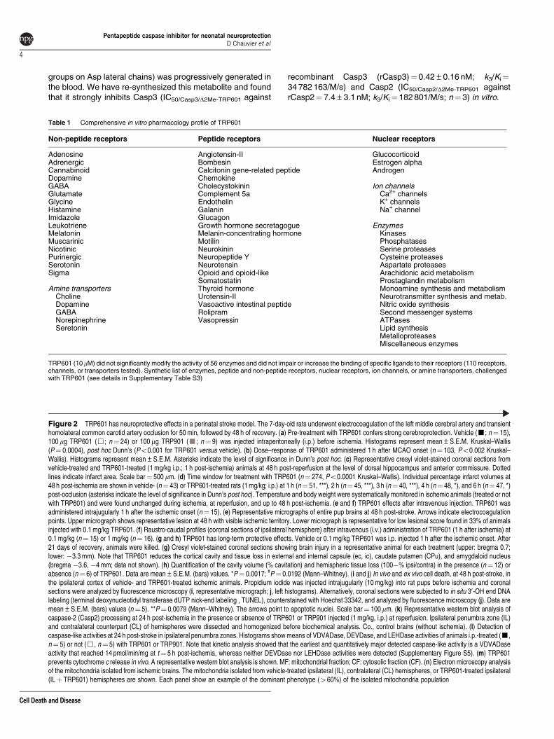
groups on Asp lateral chains) was progressively generated inthe blood. We have re-synthesized this metabolite and foundthat it strongly inhibits Casp3 (IC50/Casp3/D2Me-TRP601 against
recombinant Casp3 (rCasp3)¼ 0.42±0.16 nM; k3/Ki¼34 782 163/M/s) and Casp2 (IC50/Casp2/D2Me-TRP601 againstrCasp2¼ 7.4±3.1 nM; k3/Ki¼ 182 801/M/s; n¼ 3) in vitro.
Table 1 Comprehensive in vitro pharmacology profile of TRP601
Non-peptide receptors Peptide receptors Nuclear receptors
Adenosine Angiotensin-II GlucocorticoidAdrenergic Bombesin Estrogen alphaCannabinoid Calcitonin gene-related peptide AndrogenDopamine ChemokineGABA Cholecystokinin Ion channelsGlutamate Complement 5a Ca2+ channelsGlycine Endothelin K+ channelsHistamine Galanin Na+ channelImidazole GlucagonLeukotriene Growth hormone secretagogue EnzymesMelatonin Melanin-concentrating hormone KinasesMuscarinic Motilin PhosphatasesNicotinic Neurokinin Serine proteasesPurinergic Neuropeptide Y Cysteine proteasesSerotonin Neurotensin Aspartate proteasesSigma Opioid and opioid-like Arachidonic acid metabolism
Somatostatin Prostaglandin metabolismAmine transporters Thyroid hormone Monoamine synthesis and metabolism
Choline Urotensin-II Neurotransmitter synthesis and metab.Dopamine Vasoactive intestinal peptide Nitric oxide synthesisGABA Rolipram Second messenger systemsNorepinephrine Vasopressin ATPasesSeretonin Lipid synthesis
MetalloproteasesMiscellaneous enzymes
TRP601 (10mM) did not significantly modify the activity of 56 enzymes and did not impair or increase the binding of specific ligands to their receptors (110 receptors,channels, or transporters tested). Synthetic list of enzymes, peptide and non-peptide receptors, nuclear receptors, ion channels, or amine transporters, challengedwith TRP601 (see details in Supplementary Table S3)
Figure 2 TRP601 has neuroprotective effects in a perinatal stroke model. The 7-day-old rats underwent electrocoagulation of the left middle cerebral artery and transienthomolateral common carotid artery occlusion for 50 min, followed by 48 h of recovery. (a) Pre-treatment with TRP601 confers strong cerebroprotection. Vehicle (’; n¼ 15),100mg TRP601 (&; n¼ 24) or 100mg TRP901 (’; n¼ 9) was injected intraperitoneally (i.p.) before ischemia. Histograms represent mean±S.E.M. Kruskal–Wallis(P¼ 0.0004), post hoc Dunn’s (Po0.001 for TRP601 versus vehicle). (b) Dose–response of TRP601 administered 1 h after MCAO onset (n¼ 103, Po0.002 Kruskal–Wallis). Histograms represent mean±S.E.M. Asterisks indicate the level of significance in Dunn’s post hoc. (c) Representative cresyl violet-stained coronal sections fromvehicle-treated and TRP601-treated (1 mg/kg i.p.; 1 h post-ischemia) animals at 48 h post-reperfusion at the level of dorsal hippocampus and anterior commissure. Dottedlines indicate infarct area. Scale bar¼ 500 mm. (d) Time window for treatment with TRP601 (n¼ 274, Po0.0001 Kruskal–Wallis). Individual percentage infarct volumes at48 h post-ischemia are shown in vehicle- (n¼ 43) or TRP601-treated rats (1 mg/kg; i.p.) at 1 h (n¼ 51, ***), 2 h (n¼ 45, ***), 3 h (n¼ 40, ***), 4 h (n¼ 48, *), and 6 h (n¼ 47, *)post-occlusion (asterisks indicate the level of significance in Dunn’s post hoc). Temperature and body weight were systematically monitored in ischemic animals (treated or notwith TRP601) and were found unchanged during ischemia, at reperfusion, and up to 48 h post-ischemia. (e and f) TRP601 effects after intravenous injection. TRP601 wasadministered intrajugularly 1 h after the ischemic onset (n¼ 15). (e) Representative micrographs of entire pup brains at 48 h post-stroke. Arrows indicate electrocoagulationpoints. Upper micrograph shows representative lesion at 48 h with visible ischemic territory. Lower micrograph is representative for low lesional score found in 33% of animalsinjected with 0.1 mg/kg TRP601. (f) Raustro-caudal profiles (coronal sections of ipsilateral hemisphere) after intravenous (i.v.) administration of TRP601 (1 h after ischemia) at0.1 mg/kg (n¼ 15) or 1 mg/kg (n¼ 16). (g and h) TRP601 has long-term protective effects. Vehicle or 0.1 mg/kg TRP601 was i.p. injected 1 h after the ischemic onset. After21 days of recovery, animals were killed. (g) Cresyl violet-stained coronal sections showing brain injury in a representative animal for each treatment (upper: bregma 0.7;lower: �3.3 mm). Note that TRP601 reduces the cortical cavity and tissue loss in external and internal capsule (ec, ic), caudate putamen (CPu), and amygdaloid nucleus(bregma �3.6, �4 mm; data not shown). (h) Quantification of the cavity volume (% cavitation) and hemispheric tissue loss (100�% ipsi/contra) in the presence (n¼ 12) orabsence (n¼ 6) of TRP601. Data are mean±S.E.M. (bars) values. *P¼ 0.0017; #P¼ 0.0192 (Mann–Whitney). (i and j) In vivo and ex vivo cell death, at 48 h post-stroke, inthe ipsilateral cortex of vehicle- and TRP601-treated ischemic animals. Propidium iodide was injected intrajugularly (10 mg/kg) into rat pups before ischemia and coronalsections were analyzed by fluorescence microscopy (i, representative micrograph; j, left histograms). Alternatively, coronal sections were subjected to in situ 30-OH end DNAlabeling (terminal deoxynucleotidyl transferase dUTP nick-end labeling , TUNEL), counterstained with Hoechst 33342, and analyzed by fluorescence microscopy (j). Data aremean±S.E.M. (bars) values (n¼ 5). **P¼ 0.0079 (Mann–Whitney). The arrows point to apoptotic nuclei. Scale bar¼ 100mm. (k) Representative western blot analysis ofcaspase-2 (Casp2) processing at 24 h post-ischemia in the presence or absence of TRP601 or TRP901 injected (1 mg/kg, i.p.) at reperfusion. Ipsilateral penumbra zone (IL)and contralateral counterpart (CL) of hemispheres were dissected and homogenized before biochemical analysis. Co., control brains (without ischemia). (l) Detection ofcaspase-like activities at 24 h post-stroke in ipsilateral penumbra zones. Histograms show means of VDVADase, DEVDase, and LEHDase activities of animals i.p.-treated (’,n¼ 5) or not (&, n¼ 5) with TRP601 or TRP901. Note that kinetic analysis showed that the earliest and quantitatively major detected caspase-like activity is a VDVADaseactivity that reached 14 pmol/min/mg at t¼ 5 h post-ischemia, whereas neither DEVDase nor LEHDase activities were detected (Supplementary Figure S5). (m) TRP601prevents cytochrome c release in vivo. A representative western blot analysis is shown. MF: mitochondrial fraction; CF: cytosolic fraction (CF). (n) Electron microscopy analysisof the mitochondria isolated from ischemic brains. The mitochondria isolated from vehicle-treated ipsilateral (IL), contralateral (CL) hemispheres, or TRP601-treated ipsilateral(ILþ TRP601) hemispheres are shown. Each panel show an example of the dominant phenotype (460%) of the isolated mitochondria population
Pentapeptide caspase inhibitor for neonatal neuroprotectionD Chauvier et al
4
Cell Death and Disease
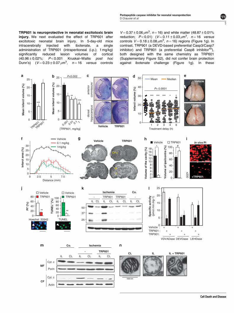
TRP601 is neuroprotective in neonatal excitotoxic braininjury. We next evaluated the effect of TRP601 afterexcitotoxic neonatal brain injury. In 5-day-old miceintracerebrally injected with ibotenate, a singleadministration of TRP601 (intraperitoneal (i.p.); 1 mg/kg)significantly reduced lesion volumes of cortical(40.96±0.02%; Po0.001 Kruskal–Wallis post hocDunn’s) (V¼ 0.23±0.07mm3, n¼ 16 versus controls
V¼ 0.37±0.06mm3, n¼ 16) and white matter (48.87±0.01%reduction; Po0.01) (V¼ 0.11±0.03mm3, n¼ 16 versuscontrols V¼ 0.18±0.08 mm3, n¼ 16) regions (Figure 1g). Incontrast, TRP901 (a DEVD-based preferential Casp3/Casp7inhibitor) and TRP801 (a preferential Casp8 inhibitor40),both designed with the same chemistry as TRP601(Supplementary Figure S2), did not confer brain protectionagainst ibotenate challenge (Figure 1g). In these
40
50 Mean Median
20
25Vehicle
20
25
P< 0.0001
P<0.002
ante
rior
com
mis
sure
dors
alhi
ppoc
ampu
s
0
10
20
30
Infa
rct
volu
me
(%)
1 6
*** *** *** * *
Mea
n in
farc
t vo
lum
e (%
)
0
5
10
15
***TRP601
Mea
n in
farc
t vo
lum
e (%
)
0
5
10
15 **
***
Vehicle TRP601Treatment delay (h)
2 3 4
Vehicle
*18
Vehicle TRP601#100
[TRP601, mg/kg]
Vehicle0.1 mg/kg1mg/kg30
In vivo PI
ec
ic
CPu
Vo
lum
e o
f th
e ca
vity
(%
)
02468
10121416
Vo
lum
e o
f ip
si/c
on
tra
(%)
0
20
40
60
80
0
Infa
rct
area
(%
)
0
5
10
15
20
25
2.5 +TRP601
x200
50
IL CL IL CL IL CL IL CL
- -
Ischemia Co.
-
15
20
25
Distance (mm)
TRP601
40
60
80Vehicle
TRP601Vehicle
6080
100
37
25
-
-
Vehicle :
TRP901:TRP601 :
0
5
10
Sp
ecif
ic a
ctiv
ity
(pm
ol.m
in.m
g-1)
PI+
(%)
0
20
Hoechst 33342
TU
NE
L+
(%)
TUNEL0
2040
****
MF
Ischemia
-
Co.
Porin
Cyt. c
++ + +
+ + +
+ +
VDVADase
IL IL + TRP601CL
CFActin
Cyt. c
5 7.5
CLILCLIL CLIL
TRP601
LEHDaseDEVDase
TRP601
TRP901 TRP601
Pentapeptide caspase inhibitor for neonatal neuroprotectionD Chauvier et al
5
Cell Death and Disease
experimental conditions, TRP601 also reduced macrophageactivation (43.51±4.16% reduction; Po0.0001 Mann–Whitney) and astrogliosis (37.69±3.15% reduction;Po0.0001 Mann–Whitney) as demonstrated by histologicalevaluation of lectine- (Figure 1h) and glial fibrillary acidicprotein -positive (Figure 1i) cell densities, respectively. Inaddition, TRP601 strongly reduced the VDVADase-specificactivity (73% reduction; P¼ 0.0079 Mann–Whitney) whenstudied 8 h after ibotenate treatment (Figure 1j).
Having found recently that genetic inhibition of caspase-2(Casp2) protects newborn mice from ischemic brain injury,32
we further investigated the protective effect of TRP601 duringCasp2 silencing. When TRP601 was administered in miceneonates treated with Casp2-specific siRNA, no significantadditional protection was found against ibotenate, neither inthe white matter nor in the cortical plate (Figure 1k andSupplementary Table S1). This suggests that, at least in thisexperimental setting, TRP601-mediated neuroprotection isCasp2 dependent.
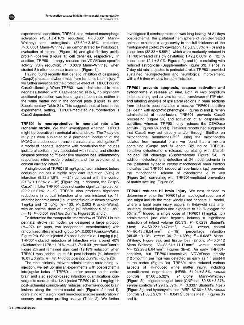
TRP601 is neuroprotective in neonatal rats afterischemic stroke. We then investigated whether TRP601might be operative in perinatal arterial stroke. The 7-day-oldrat pups were subjected to a permanent occlusion of theMCAO and subsequent transient unilateral carotid ligation,41
a model of neonatal ischemia with reperfusion that inducesipsilateral cortical injury associated with initiator and effectorcaspase processing,42 extensive neuronal loss, inflammatoryresponses, nitric oxide production and the evolution of acortical cavitary infarct.41
A single dose of TRP601 (5 mg/kg; i.p.) administered beforeocclusion induces a highly significant reduction (59%) ofinfarction (8.83±1.8%; n¼ 24) compared with the control(21.67±1.65%; n¼ 15) (Figure 2a). In contrast, the Casp3/Casp7 inhibitor TRP901 does not confer significant protection(22.2±5.67%; n¼ 9). TRP601 also produces significantreductions in cortical infarction when administered i.p. 1 hafter the ischemic onset (i.e., at reperfusion) at doses between1mg/kg and 10 mg/kg (n¼ 103; Po0.002 Kruskal–Wallis),with an optimal dose of 1 mg/kg (% infarction: 9.73±1.9%;n¼ 18, Po0.001 post hoc Dunn’s; Figures 2b and c).
To determine the therapeutic time window of TRP601 in thisperinatal stroke rat model, we designed a large protocol(n¼ 274 rat pups, two independent experimenters) withrandomized litters in each group (Po0.0001 Kruskal–Wallis;Figure 2d). When injected 2 h post-ischemia at 1 mg/kg (i.p.),TRP601-induced reduction of infarction was around 40%(% infarction: 11.78±1.01%; n¼ 47, Po0.001 post hoc Dunn’s;Figure 2d) and remained significant (19.18% reduction) whenTRP601 was added up to 6 h post-ischemia (% infarction:16.01±0.92%; n¼ 47, Po0.05 post hoc Dunn’s; Figure 2d).
The most clinically relevant administration route being i.v.injection, we set up similar experiments with post-ischemiaintrajugular bolus of TRP601. Lesion scores on the entirebrain and also section-based infarction quantifications con-verged to conclude that i.v. injected TRP601 (0.1–1 mg/kg; 1 hpost-ischemia) considerably reduces ischemia-induced brainlesions along the rostro-caudal axis (Figures 2e and f),correlating with a significant neurological score amelioration insensory and motor profiling assays (Table 2). We further
investigated if cerebroprotection was long-lasting. At 21 dayspost-ischemia, the ipsilateral hemisphere of vehicle-treatedanimals exhibited a large cavity in the full thickness of thefrontoparietal cortex (% cavitation: 12.5±3.53%; n¼ 6) and atissue loss (32.33±5.56%), which were markedly reduced inTRP601-treated rats (% cavitation: 1.42±0.68%; n¼ 12; %tissue loss: 12.1±3.9%; Figures 2g and h), correlating withreduced astrogliosis (Supplementary Figure S3). Hence, in7-day-old rats subjected to perinatal stroke, TRP601 providedsustained neuroprotection and neurological improvement,with a 6 h time window for administration.
TRP601 prevents apoptosis, caspase activation andcytochrome c release in vivo. Both in vivo propidiumiodide staining and ex vivo terminal transferase dUTP nick-end labeling analysis of ipsilateral regions in brain sectionsfrom ischemic pups revealed a massive TRP601-sensitivecell death with apoptotic phenotype (Figures 2i and j). Whenadministered at reperfusion, TRP601 prevents Casp2processing (Figure 2k) and activation of all caspase-likeactivities, whereas TRP901 only reduces the DEVDaseactivity (Figures 2k and l). Previous reports had suggestedthat Casp2 may act directly and/or through Bid/Bax onmitochondrial membranes.43,44 Using the mitochondriaisolated from neonatal brain, we found that a mixturecontaining rCasp2 and full-length Bid induce TRP601-sensitive cytochrome c release, correlating with Casp2-induced Bid cleavage (Supplementary Figure S4). Inaddition, cytochrome c detection at 24 h post-ischemia inthe ipsilateral cytosolic versus mitochondrial brain fractionindicates that TRP601 (added at reperfusion; i.p.) preventsthe mitochondrial release of cytochrome c in vivo(Figure 2m), correlating with TRP601-mediated preventionof matrix swelling (Figure 2n).
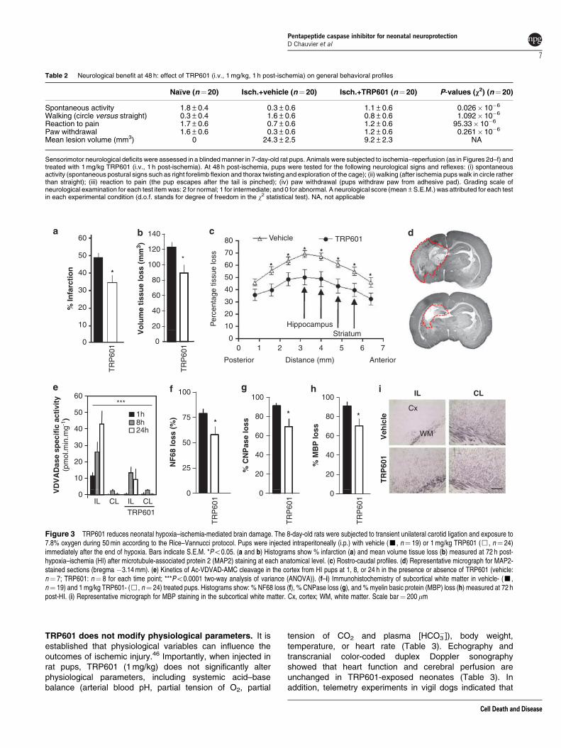
TRP601 reduces HI brain injury. We next decided todetermine whether the TRP601 pharmacological spectrum ofuse might include the most widely used neonatal HI model,where a focal brain injury occurs in 8-day-old rats afterunilateral carotid ligation and exposure to 7.8 % oxygen for50 min.45 Indeed, a single dose of TRP601 (1 mg/kg; i.p.)administered just after hypoxia induces a significantreduction of infarct volume (30.3%; P¼ 0.0239 Student’st-test; V¼ 60.22±8.47 mm3, n¼ 24 versus controlV¼ 86.43±6.54 mm3, n¼ 19), percentage infarction(48.48±3.13% versus 34.17±4.69%; P¼ 0.0287 Mann–Whitney; Figure 3a), and tissue loss (27.5%; P¼ 0.0412Mann–Whitney; V¼ 88.64±11.17 mm3 versus controlV¼ 122.29±6.84 mm3; Figures 3b–d). An early TRP601-sensitive, but TRP901-insensitive, VDVADase activity(12 pmol/min per mg) was detected as early as 1 h post-HIin the cortex (Figure 3e). TRP601 also reduced variousaspects of HI-induced white matter injury, includingneurofilament degradation (NF68: 64.24±8.5% versuscontrols 87.66±5.32%; P¼ 0.049 Mann–Whitney)(Figure 3f), oligodendroglial loss (CNPase: 69.56±8.37%versus controls 91.29±2.59%; P¼ 0.0307 Student’s t-test)(Figure 3g) and hypomyelination (MBP: 87.66±8.8% versuscontrols 91.03±2.6%; P¼ 0.041 Student’s t-test) (Figures 3hand i).
Pentapeptide caspase inhibitor for neonatal neuroprotectionD Chauvier et al
6
Cell Death and Disease
TRP601 does not modify physiological parameters. It isestablished that physiological variables can influence theoutcomes of ischemic injury.46 Importantly, when injected inrat pups, TRP601 (1 mg/kg) does not significantly alterphysiological parameters, including systemic acid–basebalance (arterial blood pH, partial tension of O2, partial
tension of CO2 and plasma [HCO3�]), body weight,
temperature, or heart rate (Table 3). Echography andtranscranial color-coded duplex Doppler sonographyshowed that heart function and cerebral perfusion areunchanged in TRP601-exposed neonates (Table 3). Inaddition, telemetry experiments in vigil dogs indicated that
Table 2 Neurological benefit at 48 h: effect of TRP601 (i.v., 1 mg/kg, 1 h post-ischemia) on general behavioral profiles
Naıve (n¼ 20) Isch.+vehicle (n¼20) Isch.+TRP601 (n¼ 20) P-values (v2) (n¼20)
Spontaneous activity 1.8±0.4 0.3±0.6 1.1±0.6 0.026�10�6
Walking (circle versus straight) 0.3±0.4 1.6±0.6 0.8±0.6 1.092�10�6
Reaction to pain 1.7±0.6 0.7±0.6 1.2±0.6 95.33� 10�6
Paw withdrawal 1.6±0.6 0.3±0.6 1.2±0.6 0.261�10�6
Mean lesion volume (mm3) 0 24.3±2.5 9.2±2.3 NA
Sensorimotor neurological deficits were assessed in a blinded manner in 7-day-old rat pups. Animals were subjected to ischemia–reperfusion (as in Figures 2d–f) andtreated with 1 mg/kg TRP601 (i.v., 1 h post-ischemia). At 48 h post-ischemia, pups were tested for the following neurological signs and reflexes: (i) spontaneousactivity (spontaneous postural signs such as right forelimb flexion and thorax twisting and exploration of the cage); (ii) walking (after ischemia pups walk in circle ratherthan straight); (iii) reaction to pain (the pup escapes after the tail is pinched); (iv) paw withdrawal (pups withdraw paw from adhesive pad). Grading scale ofneurological examination for each test item was: 2 for normal; 1 for intermediate; and 0 for abnormal. A neurological score (mean±S.E.M.) was attributed for each testin each experimental condition (d.o.f. stands for degree of freedom in the w2 statistical test). NA, not applicable
14060
**
* **
**
Hippocampus
Vo
lum
e ti
ssu
e lo
ss (
mm
3 )
40
60
80
100
120*
% In
farc
tio
n *
10
20
30
40
50
60
Posterior Anterior
Striatum0
20
TR
P60
1
IL
0
100100 100
***
TR
P60
1
10
20
30
40
50
VD
VA
Das
e sp
ecif
ic a
ctiv
ity
(pm
ol.m
in.m
g-1) 1h
8h24h
Veh
icle
TR
P60
1
% C
NP
ase
loss
% M
BP
loss
Cx
WM
*
NF
68 lo
ss (
%)
25
50
75 *
20
40
60
80
20
40
60
80 *
0IL
TRP601
0
TR
P60
1
TR
P60
1
0
TR
P60
1
0
80 Vehicle TRP601
70
60
50
Per
cent
age
tissu
e lo
ss
40
30
20
10
00 1 2 3 4 5 6 7
Distance (mm)
CL IL CL
CL
Figure 3 TRP601 reduces neonatal hypoxia–ischemia-mediated brain damage. The 8-day-old rats were subjected to transient unilateral carotid ligation and exposure to7.8% oxygen during 50 min according to the Rice–Vannucci protocol. Pups were injected intraperitoneally (i.p.) with vehicle (’, n¼ 19) or 1 mg/kg TRP601 (&, n¼ 24)immediately after the end of hypoxia. Bars indicate S.E.M. *Po0.05. (a and b) Histograms show % infarction (a) and mean volume tissue loss (b) measured at 72 h post-hypoxia–ischemia (HI) after microtubule-associated protein 2 (MAP2) staining at each anatomical level. (c) Rostro-caudal profiles. (d) Representative micrograph for MAP2-stained sections (bregma �3.14 mm). (e) Kinetics of Ac-VDVAD-AMC cleavage in the cortex from HI pups at 1, 8, or 24 h in the presence or absence of TRP601 (vehicle:n¼ 7; TRP601: n¼ 8 for each time point; ***Po0.0001 two-way analysis of variance (ANOVA)). (f–i) Immunohistochemistry of subcortical white matter in vehicle- (’,n¼ 19) and 1 mg/kg TRP601- (&, n¼ 24) treated pups. Histograms show: % NF68 loss (f), % CNPase loss (g), and % myelin basic protein (MBP) loss (h) measured at 72 hpost-HI. (i) Representative micrograph for MBP staining in the subcortical white matter. Cx, cortex; WM, white matter. Scale bar¼ 200mm
Pentapeptide caspase inhibitor for neonatal neuroprotectionD Chauvier et al
7
Cell Death and Disease
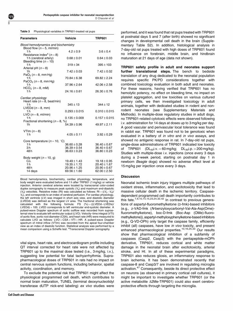
vital signs, heart rate, and electrocardiogram profile includingQT interval corrected for heart rate were not affected byTRP601 up to the maximal dose tested (i.e., 3 mg/kg, i.v.),suggesting low potential for fatal tachyarrhythmia. Supra-pharmacological doses of TRP601 in rats had no impact oncentral nervous system functions, including behavior, spatialactivity, coordination, and memory.
To exclude the potential risk that TRP601 might affect thephysiological programmed cell death, which contributes tonormal brain maturation, TUNEL (terminal deoxynucleotidyltransferase dUTP nick-end labeling) ex vivo studies were
performed, and it was found that rat pups treated with TRP601at postnatal days 5 and 7 (after birth) showed no significantchanges in developmental cell death in the brain (Supple-mentary Table S2). In addition, histological analysis in7-day-old rat pups treated with high doses of TRP601 foundno influence on forebrain, middle brain, and hindbrainmaturation at 21 days of age (data not shown).
TRP601 safety profile in adult and neonates supportfurther translational steps. The bench to bedsidetranslation of any drug dedicated to the neonatal populationrequires specific PK/PD considerations together withcombined toxicology evaluation in both adult and neonates.For these reasons, having verified that TRP601 has nohemolytic potency, no effect on bleeding time, no impact onplatelet aggregation, and low toxicities on various culturedprimary cells, we then investigated toxicology in adultanimals, together with dedicated studies in rodent and non-rodent neonates (see Supplementary Materials andMethods). In multiple-dose regulatory studies in adult dogs,no TRP601-related cytotoxic effects were observed followingi.v. administration for 14 days at doses up to 3 mg/kg per day.A good vascular and perivascular local tolerance was foundin rabbit ear. TRP601 was found not to be genotoxic whenevaluated in a battery of in vitro and in vivo assays, andshowed no antigenic response in rat. In 7-day-old rat pups,single-dose administrations of TRP601 indicated low toxicityof TRP601 (DL50i.v.¼ 60 mg/kg; DL30i.p.4200 mg/kg).Studies with multiple-dose i.v. injections (once every 3 daysduring a 2-week period, starting on postnatal day 1 innewborn Beagle dogs) showed no adverse effect level at15 mg/kg per injection once every 3 days.
Discussion
Neonatal ischemic brain injury triggers multiple pathways ofoxidant stress, inflammation, and excitotoxicity that lead tomassive cellular death in the ischemic territory. Caspase-dependent programmed cell death importantly contributes tothis fate.7,9,10,14,15,23,24,32,42 In contrast to previous genera-tions of aspartyl-fluoromethylketone (D-fmk)-based inhibitors(e.g., z-VAD-fmk (N-benzyloxycarbonyl-Val-Ala-Asp(Ome)-fluoromethylketone), boc-D-fmk (Boc-Asp (OMe)-fluoro-methylketone)), aspartyl-methyloxyphenylketone-based inhibitors(e.g., Q-VD-OPh) only target the caspase family, efficientlyinhibit (all) caspases, have low in vivo toxicity, and presentenhanced pharmacological properties.10,16,25,33 Our resultsshow that pharmacological inhibition of a subfamily ofcaspases (Casp2, Casp3) with the pentapeptide-mOPhderivative, TRP601, reduces cortical and white matterdamage in the neonatal brain after excitotoxicity, arterialstroke, and HI. In all of these experimental paradigms,TRP601 also reduces gliosis, an inflammatory response tobrain ischemia. It has been demonstrated recently thatcaspase-8 and Casp3/7 are involved in regulating microgliaactivation.47 Consequently, beside its direct protective effecton neurons (as observed in primary cortical cell cultures), itmight be important to investigate whether TRP601 (or theactive metabolite D2Me-TRP601) could also exert cerebro-protective effects through targeting the microglia.
Table 3 Physiological variables in TRP601-treated rat pups
Parameters Vehicle TRP601
Blood hemodynamics and biochemistryBlood flow (n¼5, ml/min)
1 h 4.2±0.9 3.6±0.4Resistance indexa (n¼8)
1 h (cerebral artery) 0.68±0.01 0.64±0.03Bleeding time (n¼ 10)
1 h 319±34 395±103Arterial pH (n¼8)
1h 7.42±0.03 7.42±0.02PaO2 (n¼ 8, mm Hg)
1 h 70.84±6.38 69.82±2.24PaCO2 (n¼ 8, mm Hg)
1 h 37.96±2.64 42.06±2.58HCO3
� (n¼ 8, mM)1 h 24.16±0.61 26.30±0.76
Cardiac physiologyHeart rate (n¼8, beat/min)
1 h 345±13 344±12LVEDd (n¼ 8, mm)
1 h 0.293±0.015 0.310±0.015LVO (n¼ 8, ml/min)
1 h 0.135±0.009 0.157±0.015Fractional shortening (n¼8, %)
1h 54.39±0.96 49.97±2.11
VTIm (n¼8)1 h 4.05±0.11 3.92±0.29
Core temperature (n¼10, 1C)1 h 36.60±0.28 36.40±0.673 h 36.39±0.54 36.40±0.6748 h 36.63±0.23 36.80±0.23
Body weight (n¼10, g)Co. 19.49±1.43 19.18±0.951 h 19.35±1.72 20.40±1.6748 h 20.96±1.25 20.90±0.6714 days 69.56±1.60 62.00±2.50
Blood hemodynamics, biochemistry, cardiac physiology, temperature, andbody weight were evaluated before and 1 h after TRP601 (1 mg/kg) or vehicleinjection. Anterior cerebral arteries were located by transcranial color-codedduplex sonography to measure peak systolic (Vs) and maximum end-diastolic(Vd) velocities. Resistive index (RI) was calculated as follows: RI¼ ((Vs�Vd)/Vs), and corresponds to an index of cerebral perfusion. For cardiac physiology,the left ventricle (LV) was imaged in short axis view, and diastolic diameter(LVEDd) was defined as the largest LV area. The fractional shortening wascalculated with the following formula: FS (%)¼ ((LVEDd�LVEDs)/LVEDd)� 100. LVED corresponds to left ventricular end-systolic diameter. Apulsed-wave Doppler spectrum of aortic outflow was recorded from supras-ternal view to evaluate left ventricular output (LVO). Velocity–time integral (VTI)of aortic flow, aortic root diameter (CSA), and heart rate (HR) were measured tocalculate LVO as follows: LVO¼CSA�VTI�HR. A pulsed-wave Dopplerspectrum of mitral inflow (VTIm) was recorded from the apical four-chamberview as an index of diastolic function. Statistical analysis was performed by amean comparison using a Scheffe test. aTranscranial Doppler sonography
Pentapeptide caspase inhibitor for neonatal neuroprotectionD Chauvier et al
8
Cell Death and Disease
Our results indicate that TRP601 and its active metabo-lite(s) (e.g., D2Me-TRP601) inhibit Casp3 and Casp2 in vitroand in vivo. We also provide evidences suggesting thatTRP601 interrupts the mitochondrial (intrinsic) pathway ofapoptosis in the immature brain, possibly at the level of Casp-2-mediated Bid cleavage, and upstream cytochrome crelease.39 We have recently reported that Casp2�/� micesubjected to neonatal HI presented significantly lowercerebral infarction, reduced white matter injury, and reducedCasp3 activation in the thalamus and hippocampus.32 Lesionsizes and Casp3 activation were also found reduced inCasp2-deficient newborn mice subjected to excitotoxicity.32 Inaddition, we show here that combined administration ofTRP601 with Casp2 silencing did not show any additive effectagainst ibotenate challenge. This suggests that the TRP601-mediated neuroprotection is at least, in part, Casp2-dependent.Taken together, our data suggest that the in vivo observedneuroprotection and Casp3 inhibition by TRP601 are at least,in part, Casp2-dependent. However, we could not formallyexclude that part of the neuroprotective effect of TRP601could be related to a direct Casp3 inactivation by TRP601 orD2Me-TRP601 (this latter being a very powerful Casp3inhibitor in vitro). As the selective Casp3 inhibitor M826 waspreviously found neuroprotective against neonatal HI,24 onecan suggest that the use of a group-II caspase inhibitor mightbe of therapeutic interest to cover different conditions,developmental stages, or regional mechanisms, whereCasp3/7 and Casp2 are variably expressed and activatedthrough different pathways.
Irrespective of these mechanistic considerations, TRP601is a lead candidate for neuroprotective strategy in a variety ofperinatal brain injury conditions. The lack of detectable sideeffects following in vivo administration of TRP601 to newbornrodents and dogs support its further evaluation, possiblyin combination with hypothermia5 or other candidate treat-ments.6,48
Materials and MethodsAnimal models. Three different animal models were used: neonatal stroke,excitotoxicity, and HI. Neonatal stroke (focal brain ischemia with reperfusion in the7-day-old rat) was carried out according to Renolleau et al.10,41 For HI, 8-day-oldrats or alternatively 9-day-old mice were subjected to unilateral ligation of the leftcarotid artery, followed by hypoxia (7.8% O2 for rats and 10% O2 for mice, 361C) for50 min, as described.32 For drug-induced excitotoxicity, ibotenate was administeredi.c.v. to 5-day-old mice, as described.32 Details for each animal models are given inSupplementary Material and Methods. SiRNAs were prepared as a solution in JetSIaccording to the manufacturer’s recommendations (PolyPlus-transfection, Illkirch,France). Final solution contained RNAse free water, 0.08 mM DOPE, 0.24%chloroform/7% EtOH, glucose 12.5%, and 0.04 mM JetSI. For intracerebral siRNAadministration, 0.4mg siRNA/mice were administered (volume of injection¼ 2 ml)24 h before ibotenate. SiRNA sequences and primers for PCR were reportedpreviously.32
Primary cortical neuron culture and cell death analysis. Primarycortical neurons from E14 Swiss mice embryos (Janvier, Le Genest-St-Isle, France)were cultured in N5 complete medium up to 6 DIV and subjected to serumdeprivation in the absence or presence of TRP601, or alternatively were transfectedwith siRNAs using Lipofectamine 2000 (Invitrogen, Carlsbad, CA, USA) 24 h beforeserum withdrawal. To monitor cell death parameters, neurons wereco-stained with Hoechst 33342 (nuclear condensation; Sigma, Saint Louis, MO,USA), JC-1 (5,50,6,60-tetrachloro-1,10, 3,30-tetraethyl-benzimidazolylcarbocyanineiodide) (mitochondrial transmembrane potential-sensitive probe), and 7-aminoactinomycin D (plasma membrane permeabilization; Sigma), and then subjected to
fluorescence microscopy (DM IRB; Leica, Rueil-Malmaison, France) and flowcytometry (FACSCalibur; Becton Dickinson, San Jose, CA, USA) analysis, asdescribed previously.39 In cellula caspase activities were detected using FAM-conjugated peptides (CaspaTag fluorescein Caspase Activity Kits; Q-Biogen, MPBiomedicals, Illkirch, France), as described.39
Preparation and use of drug solutions. Synthesis of TRP601/ORPHA133563 (also known as OPH016) is described in SupplementaryMaterials and Methods). Research batches where synthesized in-house(Dr. Richard Casimir). Additional batches were custom synthesized by MPBiomedicals or alternatively by NeoMPs (PolyPeptide group, Strasbourg, France).For regulatory safety studies, TRP601 process development and manufacturing ofGMP batches were subcontracted to NeoMPs (PolyPeptide group). For in vitrostudies TRP601 (C40H48F2N6O11; MW: 826.84), D2Me-TRP601 (C38H44F2N6O11;MW: 798.79), TRP801 (MW: 771.76), and TRP901 (MW: 799.77) were initiallysolubilized in 100% DMSO. The final concentration of DMSO was r1%. For in vivoefficacy experiments, TRP601, TRP801, and TRP901 were formulated in 0.9%NaCl containing 10% DMSO for i.p. administration or 2.4% Tween 20 for i.v.administration. I.v. administration of TRP601 to 7-day-old rats was carried outintrajugularly using 27-gauge needles. All stock solutions and formulations wereverified using RP-HPLC. TRP601 has a high degree of lipophilicity (partitioncoefficient, Log Poctanol/water¼ 3.89) and exhibits one pKa of 1.1 (assigned to thequinoleine group), strongly suggesting that it remains neutral in the plasma.Bioanalysis of TRP601 in fluids and organ homogenates was conducted by LC/MS/MS analysis with a precision of 1.00 ng/ml in the plasma and 3.00 ng/g inexsanguined brain homogenates (ADME Bioanalyses, Vergeze, France).
Molecular modeling. Starting from the crystal structure of the caspase-2-inhibitor (Ac–LDESD–CHO) complex (1PYO), pentapeptide inhibitors (includingAc–VDVAD–CHO and TRP601) were studied in an uncharged form and at adielectric constant of 1, using either Insight II or Discovery Studio 2.0 softwares(Accelrys, San Diego, CA, USA). See details in Supplementary Materials andMethods.
In vitro enzymatic assays. Kinetics were performed according to thecontinuous method. Assays of chromogenic substrate cleavage contained in 100mlcaspase buffer (20 mM HEPES, pH 7.4, 0.1% CHAPS, 5 mM DTT, 2 mM EDTA),117 ng of human active recombinant caspase-2 (Biomol, Plymouth, PA, USA) or20 ng of human active recombinant caspase-3 (Sigma; no. C5974), and 100mM ofchromogenic substrate (Ac–DEVD–para-nitroaniline (pNA) for Casp3 andAc–VDVAD–pNA for Casp2) in the presence of a range of concentrations ofinhibitors. For Casp2 assays, the buffer was supplemented with 1 M succinate(kosmotrope agent). Cleavage of the chromogenic substrate as a function of timewas monitored at 405 nm, and the initial velocity (V0) was determined from the linearportion of the progress curve. The formation of pNA was followed every 30 s for150 min at 371C with a microtiter plate reader (Paradigm Multi-Mode MicroplateDetection Platform, SoftMax Pro 5.2 software; Molecular Devices, St. GregoireCedex, France). V0, relative velocities, Km, and IC50 were determined fromexperimental data using the GraphPad Prism 5.01 software program. Standarddeviations of the reported values were below 10%. Inhibitor binding affinity(dissociation constant, Ki) and first-order rate constant (k3) parameters wereestimated using R (http://www.R-project.org) to fit the following equation fromWu and Fritz49:
½P�t ¼ ½E�0½S�K i
½I�K s
� �ks
k3
� �1� e�k3t= 1þðKi=½I�Þð1þ½S�=K sÞð Þh i
Other in vitro assays for calpains, cathepsins, and granzyme activities wereperformed as described.33
Ex vivo biochemistry. Brains of rat or mice pups were removed just afterdecapitation. Contralateral and ipsilateral hemispheres were frozen immediatelyand kept at �801C. Alternatively, the ipsilateral hemisphere was rapidly micro-dissected (at 41C) to mainly select the lesioned area (i.e., the estimated penumbrazone for the neonatal stroke model). Brain samples processing and relatedbiochemical analyses are described in Supplementary Material and Methods.
Physiological parameters. Blood gas analyses and hemodynamicevaluations conducted in newborn rats are described in Supplementary Materialsand Methods.
Pentapeptide caspase inhibitor for neonatal neuroprotectionD Chauvier et al
9
Cell Death and Disease
Safety pharmacology and toxicology. Safety experiments conducted innewborn dogs and rats are described in Supplementary Materials and Methods.
Statistics. Statistical analyses were conducted with Prism 4.0. Data arepresented as mean±S.E.M. and were first analyzed with a KS normality test. Whenthe data passed the normality test, the appropriate parametric test has been applied(Student’s t-test or ANOVA), followed by the suitable post hoc test if necessary(Bonferroni test). When the data did not pass the normality test, the appropriate non-parametric test has been applied (Mann–Whitney or Kruskal–Wallis tests), followedby the suitable post hoc test if necessary (Dunn’s test). Neurological scores werestatistically analyzed with a w2. Statistical significance is indicated by a singleasterisk (Po0.05), and two or three asterisks (Po0.01 or o0.001, respectively).
Conflict of Interest
Some of the authors (DC, SA, MB, RC, MS, A-PT, EB, and EJ) were formeremployees of a biopharmaceutical company (Theraptosis SA, Paris, France) thatclosed down in late 2008. Other authors (SR, SH, LS, CR, JH, GD, YC, XW, MH,J-MR, SM, AL, PR, JM, HH, PG, CC-M) declare no conflict of interest. First publicdisclosure of TRP601-related data occurred in June 2008 (6th Hershey Conference,Ecquevilly, France). TRP601 rights were acquired in 2009 by Chiesi PharmaceuticiSpA, Parma, Italy (http://www.chiesigroup.com/web/guest/chi-siamo/key-figures/report-finanziari; Annual Report 2009, p 50).
Acknowledgements. We thank Professor A David Edwards, Professor CarolTroy, Professor Marie-Lise Gougeon, Professor Michele Reboud-Ravaux, Dr. GillesGuichard, and Tinoray Jacotot for discussion and advice. We also thank AlainLangonne, Christelle Garraud-Moquereau, and Anna-Lena Leverin for technicalassistance, and Dr. Isabelle Margail (University Paris V, Paris, France) for MCAO inadult mice; Dr. Christelle Gelis and Dr. Patrick Duchene (ADME Bioanalyse,Vergeze, France) for bioanalysis; Dr. Jean-Michel Caillaud (Biodoxis SA,Romainville, France) and Dr. Palate (CIT, Evreux, France) for histopathologyexaminations; Dr. Olivier Foulon, Dr. Rhian Davies, Dr. Pierre-Olivier Guillaumat,and Dr. Valerie Haag (CIT) for animal toxicology contributions and expertise; and Dr.Francoise Brunner for excellent advice during drug development. This work wassupported by grants from the French Ministry of Research (No. 01H0476 to EJ; No.01H0477 to JM), Inserm, Foundation Grace de Monaco (to CC-M and PG), AgenceNationale pour la Valorisation de la Recherche (ANVAR; No. R0209330Q to EJ),Medical Research Council strategic award (MRC, UK, P19381 to HH), UniversityParis 7, Premup, Medical Research Council (VR, Sweden, 2006-3396 to HH), ALF-LUA (Sweden, ALFGBG2863 to HH), and the Sixth Framework Programme of theEuropean Commission (STREP – Neobrain consortium, Contract No. LSHM-CT-2006-036534 to HH, PG, and EJ).
1. Lawn JE, Lee AC, Kinney M, Sibley L, Carlo WA, Paul VK et al. Two million intrapartum-related stillbirths and neonatal deaths: where, why, and what can be done? Int J GynaecolObstet 2009; 107: S5.
2. du Plessis AJ, Volpe JJ. Perinatal brain injury in the preterm and term newborn. Curr OpinNeurol 2002; 15: 151–157.
3. Ferriero DM. Neonatal brain injury. N Engl J Med 2004; 351: 1985–1995.4. Lawn JE, Bahl R, Bergstrom S, Bhutta ZA, Darmstadt GL, Ellis M et al. Setting research
priorities to reduce almost one million deaths from birth asphyxia by 2015. PLoS Med 2011;8: e1000389.
5. Azzopardi DV, Strohm B, Edwards AD, Dyet L, Halliday HL, Juszczak E et al.Moderate hypothermia to treat perinatal asphyxial encephalopathy. N Engl J Med 2009;361: 1349–1358.
6. Gonzalez FF, Ferriero DM. Neuroprotection in the newborn infant. Clin Perinatol 2009; 36:859–880.
7. Northington FJ, Zelaya ME, O’Riordan DP, Blomgren K, Flock DL, Hagberg H et al. Failureto complete apoptosis following neonatal hypoxia-ischemia manifests as ‘continuum’phenotype of cell death and occurs with multiple manifestations of mitochondrialdysfunction in rodent forebrain. Neuroscience 2007; 149: 822–833.
8. Kaindl AM, Favrais G, Gressens P. Molecular mechanisms involved in injury to the pretermbrain. J Child Neurol 2009; 24: 1112–1118.
9. Gill R, Soriano M, Blomgren K, Hagberg H, Wybrecht R, Miss MT et al. Role of caspase-3activation in cerebral ischemia-induced neurodegeneration in adult and neonatal brain.J Cereb Blood Flow Metab 2002; 22: 420–430.
10. Renolleau S, Fau S, Goyenvalle C, Joly LM, Chauvier D, Jacotot E et al. Specific caspaseinhibitor Q-VD-OPh prevents neonatal stroke in P7 rat: a role for gender. J Neurochem2007; 100: 1062–1071.
11. Wang X, Carlsson Y, Basso E, Zhu C, Rousset CI, Rasola A et al. Developmentalshift of cyclophilin D contribution to hypoxic–ischemic brain injury. J Neurosci 2009; 29:2588–2596.
12. Edwards AD, Yue X, Cox P, Hope PL, Azzopardi DV, Squier MV et al. Apoptosis in thebrains of infants suffering intrauterine cerebral injury. Pediatr Res 1997; 42: 684–689.
13. Hagberg H, Mallard C, Rousset CI, Wang X. Apoptotic mechanisms in the immature brain:involvement of mitochondria. J Child Neurol 2009; 24: 1141–1146.
14. Zhu C, Wang X, Xu F, Bahr BA, Shibata M, Uchiyama Y et al. The influence of age onapoptotic and other mechanisms of cell death after cerebral hypoxia–ischemia. Cell DeathDiffer 2005; 12: 162–176.
15. Wang X, Han W, Du X, Zhu C, Carlsson Y, Mallard C et al. Neuroprotective effect ofBax-inhibiting peptide on neonatal brain injury. Stroke 2010; 41: 2050–2055.
16. Zhu C, Wang X, Huang Z, Qiu L, Xu F, Vahsen N et al. Apoptosis-inducing factor is a majorcontributor to neuronal loss induced by neonatal cerebral hypoxia–ischemia. Cell DeathDiffer 2007; 14: 775–784.
17. Thornberry NA, Lazebnik Y. Caspases: enemies within. Science 1998; 281: 1312–1316.18. Talanian RV, Brady KD, Cryns VL. Caspases as targets for anti-inflammatory and
anti-apoptotic drug discovery. J Med Chem 2000; 43: 3351–3371.19. Yuan J, Yankner BA. Apoptosis in the nervous system. Nature 2000; 407: 802–809.20. Talanian RV, Quinlan C, Trautz S, Hackett MC, Mankovich JA, Banach D et al. Substrate
specificities of caspase family proteases. J Biol Chem 1997; 272: 9677–9682.21. Garcia-Calvo M, Peterson EP, Leiting B, Ruel R, Nicholson DW, Thornberry NA et al.
Inhibition of human caspases by peptide-based and macromolecular inhibitors. J BiolChem 1998; 273: 32608–32613.
22. Garcia-Calvo M, Peterson EP, Rasper DM, Vaillancourt JP, Zamboni R, Nicholson DWet al. Purification and catalytic properties of human caspase family members. Cell DeathDiffer 1999; 6: 362–369.
23. Cheng Y, Deshmukh M, D’Costa A, Demaro JA, Gidday JM, Shah A et al. Caspaseinhibitor affords neuroprotection with delayed administration in a rat model of neonatalhypoxic–ischemic brain injury. J Clin Invest 1998; 101: 1992–1999.
24. Han BH, Xu D, Choi J, Han Y, Xanthoudakis S, Roy S et al. Selective, reversible caspase-3inhibitor is neuroprotective and reveals distinct pathways of cell death after neonatalhypoxic–ischemic brain injury. J Biol Chem 2002; 277: 30128–30136.
25. Joly LM, Mucignat V, Mariani J, Plotkine M, Charriaut-Marlangue C. Caspase inhibitionafter neonatal ischemia in the rat brain. J Cereb Blood Flow Metab 2004; 24: 124–131.
26. Puyal J, Vaslin A, Mottier V, Clarke PG. Postischemic treatment of neonatal cerebralischemia should target autophagy. Ann Neurol 2009; 66: 378–389.
27. Karatas H, Aktas Y, Gursoy-Ozdemir Y, Bodur E, Yemisci M, Caban S et al.A nanomedicine transports a peptide caspase-3 inhibitor across the blood–brain barrierand provide1neuroprotection. J Neurosci 2009; 29: 13761–13769.
28. Kuida K, Zheng TS, Na S, Kuan C, Yang D, Karasuyama H et al. Decreased apoptosis inthe brain and premature lethality in CPP32-deficient mice. Nature 1996; 384: 368–372.
29. Kuida K, Haydar TF, Kuan CY, Gu Y, Taya C, Karasuyama H et al. Reduced apoptosis andcytochrome c-mediated caspase activation in mice lacking caspase 9. Cell 1998; 94: 325–337.
30. Bergeron L, Perez GI, Macdonald G, Shi L, Sun Y, Jurisicova A et al. Defects in regulationof apoptosis in caspase-2-deficient mice. Genes Dev 1998; 12: 1304–1314.
31. West T, Atzeva M, Holtzman DM. Caspase-3 deficiency during development increasesvulnerability to hypoxic–ischemic injury through caspase-3-independent pathways.Neurobiol Dis 2006; 22: 523–537.
32. Carlsson Y, Schwendimann L, Vontell R, Rousset CI, Wang X, Lebon S et al. Geneticinhibition of caspase-2 reduces hypoxic–ischemic and excitotoxic neonatal brain injury.Ann Neurol 2011; e-pub ahead of print 28 March 2011; doi:10.1002/ana.22431.
33. Chauvier D, Ankri S, Charriaut-Marlangue C, Casimir R, Jacotot E. Broad-spectrumcaspase inhibitors: from myth to reality? Cell Death Differ 2007; 14: 387–391.
34. Zhu C, Qiu L, Wang X, Hallin U, Cande C, Kroemer G et al. Involvement of apoptosis-inducing factor in neuronal death after hypoxia–ischemia in the neonatal rat brain.J Neurochem 2003; 86: 306–317.
35. Linton SD, Aja T, Armstrong RA, Bai X, Chen LS, Chen N et al. First-in-class pan caspaseinhibitor developed for the treatment of liver disease. J Med Chem 2005; 48: 6779–6782.
36. Leung-Toung R, Zhao Y, Li W, Tam TF, Karimian K, Spino M et al. Thiol proteases:inhibitors and potential therapeutic targets. Curr Med Chem 2006; 13: 547–581.
37. McStay GP, Salvesen GS, Green DR. Overlapping cleavage motif selectivity of caspases:implications for analysis of apoptotic pathways. Cell Death Differ 2008; 15: 322–331.
38. Schweizer A, Briand C, Grutter MG. Crystal structure of caspase-2, apical initiator of theintrinsic apoptotic pathway. J Biol Chem 2003; 278: 42441–42447.
39. Chauvier D, Lecoeur H, Langonne A, Borgne-Sanchez A, Mariani J, Martinou JC et al.Upstream control of apoptosis by caspase-2 in serum-deprived primary neurons. Apoptosis2005; 10: 1243–1259.
40. Dzietko M, Boos V, Sifringer M, Polley O, Gerstner B, Genz K et al. A critical role forFas/CD-95 dependent signaling pathways in the pathogenesis of hyperoxia-induced braininjury. Ann Neurol 2008; 64: 664–673.
41. Renolleau S, Aggoun-Zouaoui D, Ben-Ari Y, Charriaut-Marlangue C. A model of transientunilateral focal ischemia with reperfusion in the P7 neonatal rat: morphological changesindicative of apoptosis. Stroke 1998; 29: 1454–1461.
42. Villapol S, Bonnin P, Fau S, Baud O, Renolleau S, Charriault-Marlangue C. Unilateral bloodflow decrease induces bilateral and symmetric responses in the immature brain. Am JPathol 2009; 175: 2111–2120.
Pentapeptide caspase inhibitor for neonatal neuroprotectionD Chauvier et al
10
Cell Death and Disease
43. Guo Y, Srinivasula SM, Druilhe A, Fernandes-Alnemri T, Alnemri ES. Caspase-2 induces apoptosisby releasing proapoptotic proteins from mitochondria. J Biol Chem 2002; 277: 13430–13437.
44. Robertson JD, Enoksson M, Suomela M, Zhivotovsky B, Orrenius S. Caspase-2 actsupstream of mitochondria to promote cytochrome c release during etoposide-inducedapoptosis. J Biol Chem 2002; 277: 29803–29809.
45. Andine P, Thordstein M, Kjellmer I, Nordborg C, Thiringer K, Wennberg E et al. Evaluationof brain damage in a rat model of neonatal hypoxic–ischemia. J Neurosci Methods 1990;35: 253–260.
46. Hunter AJ, Green AR, Cross AJ. Animal models of acute ischaemic stroke: can they predictclinically successful neuroprotective drugs? Trends Pharmacol Sci 1995; 16: 123–128.
47. Burguillos MA, Deierborg T, Kavanagh E, Persson A, Hajji N, Garcia-Quintanilla A et al.Caspase signalling controls microglia activation and neurotoxicity. Nature 2011; 472: 319–324.
48. Gonzalez FF, Ferriero DM. Therapeutics for neonatal brain injury. Pharmacol Ther 2008;120: 43–53.
49. Wu JC, Fritz LC. Irreversible caspase inhibitors: tools for studying apoptosis. Methods1999; 17: 320–328.
Cell Death and Disease is an open-access journalpublished by Nature Publishing Group. This work is
licensed under the Creative Commons Attribution-Noncommercial-NoDerivative Works 3.0 Unported License. To view a copy of this license,visit http://creativecommons.org/licenses/by-nc-nd/3.0/
Supplementary Information accompanies the paper on Cell Death and Disease website (http://www.nature.com/cddis)
Pentapeptide caspase inhibitor for neonatal neuroprotectionD Chauvier et al
11
Cell Death and Disease
Related Documents