Current Medicinal Chemistry, 2000, 7, 17-37 17 Targeting DNA with Triplexes Keith R. Fox* Division of Biochemistry & Molecular Biology, School of Biological Sciences, University of Southampton, Bassett Crescent East, Southampton SO16 7PX, U.K. Abstract : The formation of intermolecular DNA triple helices offers the possibility of designing compounds with extensive sequence recognition properties which may be useful as antigene agents or tools in molecular biology. In these structures a third strand oligonucleotide binds in the DNA major groove, making specific contacts with substituents on the exposed faces of the base pairs. Although triplexes form with exquisite specificity their use suffers from several drawbacks. Two limitations of this approach, which are considered in this review are, firstly that conditions of low pH are necessary for formation of the C + ● GC triplet, and secondly that these structures are often less stable than their duplex counterparts. This review outlines the strategies that have been employed to overcome these drawbacks. The pH problem is addressed by considering the various DNA base analogues that have been used to recognise GC base pairs in a pH independent fashion, and discusses the benefits and limitations of each analogue. Triplex stability can be increased by using novel base analogues, backbone modifications and the use of triplex-specific binding ligands. Background which selectively bind to the individual bases (or base pairs), and which are attached to a regular backbone which is able to wind around the DNA helix, possessing the same axial rise as duplex DNA. A simple solution to these problems is to use oligonucleotides as sequence specific agents, since their repeating unit is the same as that of duplex DNA, and they can easily adopt a helical structure. It has been known for several years that the major groove of duplex DNA is large enough to accommodate a third oligonucleotide strand, forming an intermolecular triplex. By exploring the rules governing the formation of these complexes it is hoped to generate a versatile recognition code for designing agents to interact with any desired sequence. Compounds which interact with duplex DNA in a sequence-specific fashion have the potential to inhibit the activity of individual genes, and may be useful for treating a variety of diseases including cancer or viral infections. In principle it should be simpler to design drugs targeted to DNA than against many other pharmacological receptors, since the structure of the target is well known in precise molecular detail. In addition there are only two copies of each DNA target site per diploid cell, and it may therefore be possible to use low doses of such agents, provided that they maintain high affinity and stringency. In order to be selective for a unique DNA sequence within the human genome of 3 x 10 9 base pairs, such agents need to recognise at least 16-17 consecutive bases. Sequence specific recognition of DNA is achieved by making specific contacts with the regular array of hydrogen bond donors and acceptors on the base pairs which are exposed in the major and minor grooves. Since these recognition elements are positioned in a helical arrangement, turning through about 360° every 10 base pairs, DNA sequence- reading agents need to follow this helical pattern. A versatile code for generating sequence-specific DNA- reading agents will therefore require monomer units The formation of DNA and RNA triple helices was first demonstrated in 1957 by mixing polyU and polyA in the ratio of 2:1 [1,2]. This and other triplexes, formed with synthetic polynucleotides, remained an obscure part of DNA chemistry until 1987 when it was realised that they offered a means for designing DNA sequence specific agents [3,4]. Since then a variety of biological activities have been proposed for these structures [5- 10]. In these complexes the third strand lies in the major groove of the target DNA duplex, where it makes specific hydrogen bond contacts with substituents on the exposed faces of the duplex DNA base pairs. Two types of triplexes have been characterised, which differ in the orientation of the third strand. Those in which the third strand runs parallel to the duplex purine strand are characterised by T ● AT and C + ● GC triplets, Fig. (1a ), *Address correspondence to this author at the Division of Biochemistry & Molecular Biology, School of Biological Sciences, University of Southampton, Bassett Crescent East, Southampton SO16 7PX, U.K.; Tel. +1703-594374; Fax. + 1703-594459; Email: [email protected] 0929-8673/00 $19.00+.00 © 2000 Bentham Science Publishers B.V.

Welcome message from author
This document is posted to help you gain knowledge. Please leave a comment to let me know what you think about it! Share it to your friends and learn new things together.
Transcript
Current Medicinal Chemistry, 2000, 7, 17-37 17
Targeting DNA with Triplexes
Keith R. Fox*
Division of Biochemistry & Molecular Biology, School of Biological Sciences, Universityof Southampton, Bassett Crescent East, Southampton SO16 7PX, U.K.
Abstract: The formation of intermolecular DNA triple helices offers thepossibility of designing compounds with extensive sequence recognitionproperties which may be useful as antigene agents or tools in molecularbiology. In these structures a third strand oligonucleotide binds in the DNAmajor groove, making specific contacts with substituents on the exposedfaces of the base pairs. Although triplexes form with exquisite specificity their use suffers fromseveral drawbacks. Two limitations of this approach, which are considered in this review are,firstly that conditions of low pH are necessary for formation of the C+● GC triplet, and secondlythat these structures are often less stable than their duplex counterparts. This review outlinesthe strategies that have been employed to overcome these drawbacks. The pH problem isaddressed by considering the various DNA base analogues that have been used to recogniseGC base pairs in a pH independent fashion, and discusses the benefits and limitations of eachanalogue. Triplex stability can be increased by using novel base analogues, backbonemodifications and the use of triplex-specific binding ligands.
Background which selectively bind to the individual bases (or basepairs), and which are attached to a regular backbonewhich is able to wind around the DNA helix, possessingthe same axial rise as duplex DNA. A simple solution tothese problems is to use oligonucleotides assequence specific agents, since their repeating unit isthe same as that of duplex DNA, and they can easilyadopt a helical structure. It has been known for severalyears that the major groove of duplex DNA is largeenough to accommodate a third oligonucleotidestrand, forming an intermolecular triplex. By exploringthe rules governing the formation of these complexes itis hoped to generate a versatile recognition code fordesigning agents to interact with any desiredsequence.
Compounds which interact with duplex DNA in asequence-specific fashion have the potential to inhibitthe activity of individual genes, and may be useful fortreating a variety of diseases including cancer or viralinfections. In principle it should be simpler to designdrugs targeted to DNA than against many otherpharmacological receptors, since the structure of thetarget is well known in precise molecular detail. Inaddition there are only two copies of each DNA targetsite per diploid cell, and it may therefore be possible touse low doses of such agents, provided that theymaintain high affinity and stringency. In order to beselective for a unique DNA sequence within the humangenome of 3 x 10⁹ base pairs, such agents need torecognise at least 16-17 consecutive bases.Sequence specific recognition of DNA is achieved bymaking specific contacts with the regular array ofhydrogen bond donors and acceptors on the basepairs which are exposed in the major and minorgrooves. Since these recognition elements arepositioned in a helical arrangement, turning throughabout 360° every 10 base pairs, DNA sequence-reading agents need to follow this helical pattern. Aversatile code for generating sequence-specific DNA-reading agents will therefore require monomer units
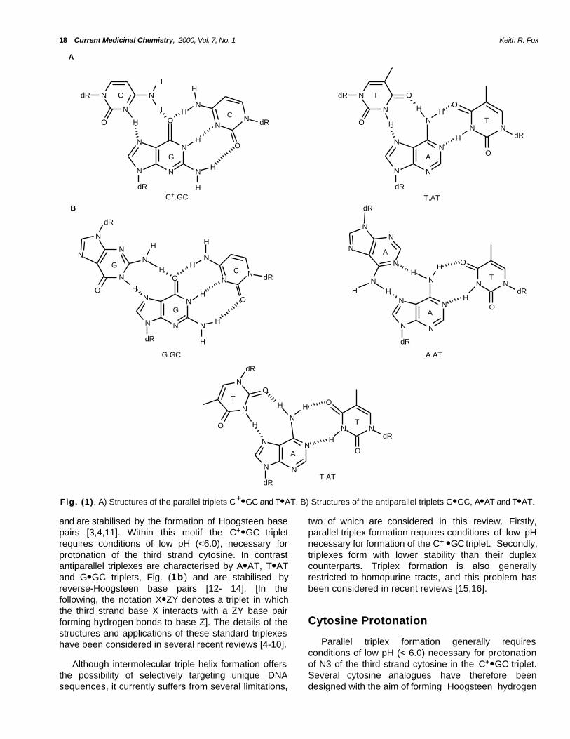
The formation of DNA and RNA triple helices wasfirst demonstrated in 1957 by mixing polyU and polyAin the ratio of 2:1 [1,2]. This and other triplexes, formedwith synthetic polynucleotides, remained an obscurepart of DNA chemistry until 1987 when it was realisedthat they offered a means for designing DNA sequencespecific agents [3,4]. Since then a variety of biologicalactivities have been proposed for these structures [5-10]. In these complexes the third strand lies in themajor groove of the target DNA duplex, where it makesspecific hydrogen bond contacts with substituents onthe exposed faces of the duplex DNA base pairs. Twotypes of triplexes have been characterised, which differin the orientation of the third strand. Those in which thethird strand runs parallel to the duplex purine strand arecharacterised by T● AT and C+● GC triplets, Fig. (1a),
*Address correspondence to this author at the Division of Biochemistry &Molecular Biology, School of Biological Sciences, University ofSouthampton, Bassett Crescent East, Southampton SO16 7PX, U.K.; Tel.+1703-594374; Fax. + 1703-594459; Email: [email protected]
0929-8673/00 $19.00+.00 © 2000 Bentham Science Publishers B.V.
18 Current Medicinal Chemistry, 2000, Vol. 7, No. 1 Keith R. Fox
N+
NdR
O H
N
H
N
OH
N
N
N
H
N
N
N
O
dR
NH
HdR
HH
C+
C
G
N
NdR
O H
O
H
N
NH
N
N
N
HN
O
N
O
dR
dR
T
T
C+.GC T.AT
N
NN
N
O
dR
NH
H
H
HH
N
NN
O
NN
N
N
N
dR
O H
H H
dR
GC
G
G.GC
N
NN
N
N
dR
H
HO
N N
N
N
N
N
dR
NdR
TH
HH
A
A
A.AT
N
NN
N
N
dR
H
HO
N N
O
N
N
dR
T
T
A
O
H
HO
dR
T.AT
A
B
A
O
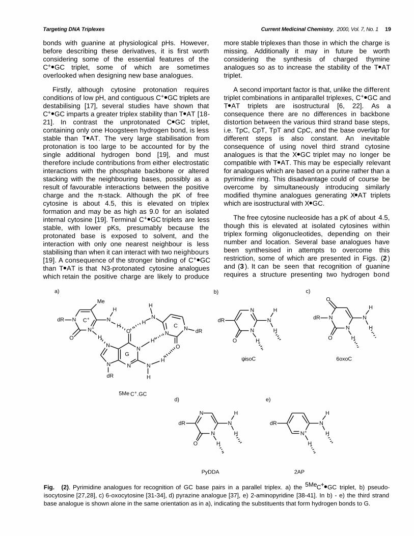
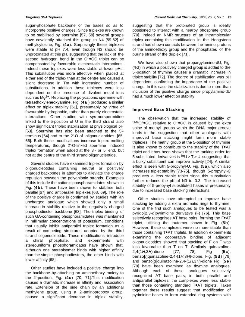
Fig. (1) . A) Structures of the parallel triplets C+● GC and T● AT. B) Structures of the antiparallel triplets G● GC, A● AT and T● AT.
and are stabilised by the formation of Hoogsteen basepairs [3,4,11]. Within this motif the C+● GC tripletrequires conditions of low pH (<6.0), necessary forprotonation of the third strand cytosine. In contrastantiparallel triplexes are characterised by A● AT, T● ATand G● GC triplets, Fig. (1b ) and are stabilised byreverse-Hoogsteen base pairs [12- 14]. [In thefollowing, the notation X● ZY denotes a triplet in whichthe third strand base X interacts with a ZY base pairforming hydrogen bonds to base Z]. The details of thestructures and applications of these standard triplexeshave been considered in several recent reviews [4-10].
two of which are considered in this review. Firstly,parallel triplex formation requires conditions of low pHnecessary for formation of the C+ ● GC triplet. Secondly,triplexes form with lower stability than their duplexcounterparts. Triplex formation is also generallyrestricted to homopurine tracts, and this problem hasbeen considered in recent reviews [15,16].
Cytosine Protonation
Parallel triplex formation generally requiresconditions of low pH (< 6.0) necessary for protonationof N3 of the third strand cytosine in the C+● GC triplet.Several cytosine analogues have therefore beendesigned with the aim of forming Hoogsteen hydrogen
Although intermolecular triple helix formation offersthe possibility of selectively targeting unique DNAsequences, it currently suffers from several limitations,
Targeting DNA Triplexes Current Medicinal Chemistry, 2000, Vol. 7, No. 1 19
bonds with guanine at physiological pHs. However,before describing these derivatives, it is first worthconsidering some of the essential features of theC+● GC triplet, some of which are sometimesoverlooked when designing new base analogues.
more stable triplexes than those in which the charge ismissing. Additionally it may in future be worthconsidering the synthesis of charged thymineanalogues so as to increase the stability of the T● ATtriplet.
Firstly, although cytosine protonation requiresconditions of low pH, and contiguous C+● GC triplets aredestabilising [17], several studies have shown thatC+● GC imparts a greater triplex stability than T● AT [18-21]. In contrast the unprotonated C● GC triplet,containing only one Hoogsteen hydrogen bond, is lessstable than T● AT. The very large stabilisation fromprotonation is too large to be accounted for by thesingle additional hydrogen bond [19], and musttherefore include contributions from either electrostaticinteractions with the phosphate backbone or alteredstacking with the neighbouring bases, possibly as aresult of favourable interactions between the positivecharge and the π-stack. Although the pK of freecytosine is about 4.5, this is elevated on triplexformation and may be as high as 9.0 for an isolatedinternal cytosine [19]. Terminal C+● GC triplets are lessstable, with lower pKs, presumably because theprotonated base is exposed to solvent, and theinteraction with only one nearest neighbour is lessstabilising than when it can interact with two neighbours[19]. A consequence of the stronger binding of C+● GCthan T● AT is that N3-protonated cytosine analogueswhich retain the positive charge are likely to produce
A second important factor is that, unlike the differenttriplet combinations in antiparallel triplexes, C+● GC andT● AT triplets are isostructural [6, 22]. As aconsequence there are no differences in backbonedistortion between the various third strand base steps,i.e. TpC, CpT, TpT and CpC, and the base overlap fordifferent steps is also constant. An inevitableconsequence of using novel third strand cytosineanalogues is that the X● GC triplet may no longer becompatible with T● AT. This may be especially relevantfor analogues which are based on a purine rather than apyrimidine ring. This disadvantage could of course beovercome by simultaneously introducing similarlymodified thymine analogues generating X● AT tripletswhich are isostructural with X● GC.
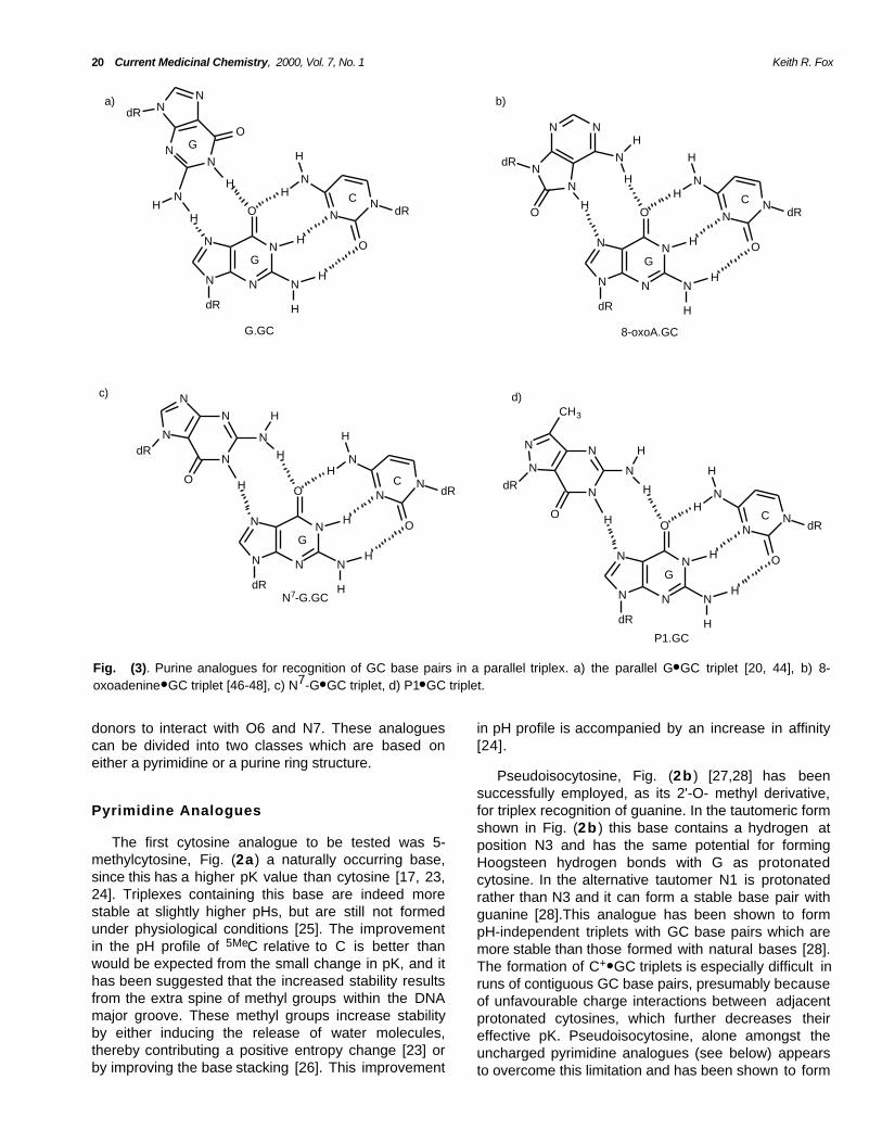
The free cytosine nucleoside has a pK of about 4.5,though this is elevated at isolated cytosines withintriplex forming oligonucleotides, depending on theirnumber and location. Several base analogues havebeen synthesised in attempts to overcome thisrestriction, some of which are presented in Figs. (2 )and (3 ). It can be seen that recognition of guaninerequires a structure presenting two hydrogen bond
N
NN
N
O
dR
HH
N
H
N
N+
Me
O
dR
H
N
H
NN
O
dR
H
N
H
HG
C+
5Me C+.GC
a) b)
N
N
N
H
H
HO
dR N
N
N
H
H
HO
dR
O
c)
ψisoC 6oxoC
d)
N
N
N
H
H
HO
dR
PyDDA
e)
N+
N
H
H
H
dR
2AP
C
Fig. (2). Pyrimidine analogues for recognition of GC base pairs in a parallel triplex. a) the 5MeC+● GC triplet, b) pseudo-isocytosine [27,28], c) 6-oxocytosine [31-34], d) pyrazine analogue [37], e) 2-aminopyridine [38-41]. In b) - e) the third strandbase analogue is shown alone in the same orientation as in a), indicating the substituents that form hydrogen bonds to G.
20 Current Medicinal Chemistry, 2000, Vol. 7, No. 1 Keith R. Fox
N
NN
N
O
dR
H
H
N
H
NN
O
dR
H
N
H
H
HN
HC
G
G.GC
N
NN
N
O
dR
HN
H
NN
O
dR
H
N
H
H
NN
C
G
8-oxoA.GC
N
N
O
dR
H
N
H
H
N
NN
N
O
dR
HH
N
H
NN
O
dR
H
N
H
H
H
C
G
N7-G.GC
N
HN
N
O
N
NdR
N
NN
N
O
dR
HH
N
H
NN
O
dR
H
N
H
H
H
C
G
P1.GC
N
HN
N
O
N
N
dR
CH3
a) b)
c) d)
NN
NNdR
OG
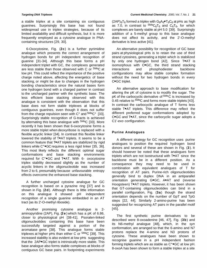
Fig. (3). Purine analogues for recognition of GC base pairs in a parallel triplex. a) the parallel G● GC triplet [20, 44], b) 8-oxoadenine● GC triplet [46-48], c) N7-G● GC triplet, d) P1● GC triplet.
donors to interact with O6 and N7. These analoguescan be divided into two classes which are based oneither a pyrimidine or a purine ring structure.
in pH profile is accompanied by an increase in affinity[24].
Pseudoisocytosine, Fig. (2b ) [27,28] has beensuccessfully employed, as its 2'-O- methyl derivative,for triplex recognition of guanine. In the tautomeric formshown in Fig. (2b ) this base contains a hydrogen atposition N3 and has the same potential for formingHoogsteen hydrogen bonds with G as protonatedcytosine. In the alternative tautomer N1 is protonatedrather than N3 and it can form a stable base pair withguanine [28].This analogue has been shown to formpH-independent triplets with GC base pairs which aremore stable than those formed with natural bases [28].The formation of C+● GC triplets is especially difficult inruns of contiguous GC base pairs, presumably becauseof unfavourable charge interactions between adjacentprotonated cytosines, which further decreases theireffective pK. Pseudoisocytosine, alone amongst theuncharged pyrimidine analogues (see below) appearsto overcome this limitation and has been shown to form
Pyrimidine Analogues
The first cytosine analogue to be tested was 5-methylcytosine, Fig. (2a) a naturally occurring base,since this has a higher pK value than cytosine [17, 23,24]. Triplexes containing this base are indeed morestable at slightly higher pHs, but are still not formedunder physiological conditions [25]. The improvementin the pH profile of 5MeC relative to C is better thanwould be expected from the small change in pK, and ithas been suggested that the increased stability resultsfrom the extra spine of methyl groups within the DNAmajor groove. These methyl groups increase stabilityby either inducing the release of water molecules,thereby contributing a positive entropy change [23] orby improving the base stacking [26]. This improvement
Targeting DNA Triplexes Current Medicinal Chemistry, 2000, Vol. 7, No. 1 21
a stable triplex at a site containing six contiguousguanines. Surprisingly this base has not foundwidespread use in triplex strategies, because of itslimited availability and difficult synthesis, but it is morefrequently employed as a cytosine analogue in PNA-containing structures [29, 30].
(2AP)6T6 formed a triplex with G6A6● T6C6 at pHs as highas 7.0, in contrast to 5MeC6T6 and C6T6, for whichcomplexes are barely stable at pH 5.0 [39]. Surprisinglyaddition of a 5-methyl group to this base analoguedoes not affect its activity, and the 2'-O-methylderivative is less active [41].
6-Oxocytosine, Fig. (2c) is a further pyrimidineanalogue which presents the correct arrangement ofhydrogen bonds for pH independent recognition ofguanine [31-34]. Although this base forms a pHindependent triplet with GC, the complexes generatedare less stable than those observed with C or 5MeC atlow pH. This could reflect the importance of the positivecharge noted above, affecting the energetics of basestacking, or might be due to changes in the hydrogenbonding characteristics since the natural bases formone hydrogen bond with a charged partner in contrastto the uncharged partner with the synthetic base. Theless efficient base stacking observed with thisanalogue is consistent with the observation that thisbase does not form stable triplexes at blocks ofcontiguous guanines, even though it does not sufferfrom the charge repulsion seen with C+ and 5MeC+ [33].Surprisingly stable recognition of G-tracts is achievedby alternating this base analogue with 5MeC [33]. Morerecently it has been shown that 6-oxocytosine forms amore stable triplet when deoxyribose is replaced with aflexible acyclic linker [34]. In contrast this flexible linkerlowered the stability of T● AT triplets. It seems to be acommon feature that T● AT triplets are stabilized by rigidlinkers while C+● GC requires a less rigid linker [35, 36].This most likely reflects the different optimal sugarconformations and base stacking environmentsrequired for C+● GC and T● AT. With 6- oxocytosinetriplex stability decreased slightly as the number ofacyclic linkers in the oligonucleotide was increasedfrom 2 to 6, presumably because unfavourable entropyeffects overcome the enhanced base stacking.
An alternative possibility for recognition of GC basepairs at physiological pHs is to retain the use of thirdstrand cytosines, generating a triplet which is stabilizedby only one hydrogen bond [42]. Since T● AT isisomorphous with C● GC, the third strand stackinginteractions and phosphodiester backboneconfigurations may allow stable complex formationwithout the need for two hydrogen bonds in everyC● GC triplet.
An alternative approach to base modification foraltering the pK of cytosine is to modify the sugar. ThepK of the carbocyclic derivative of 5MeC is increased by0.45 relative to 5MeC and forms more stable triplets [43].In contrast the carbocyclic analogue of T forms lessstable T● AT triplets. This difference may reflect thedifferent preferred sugar conformations adopted byC● GC and T● AT, since the carbocyclic sugar adopts aC1'-exo configuration.
Purine Analogues
A different strategy for GC recognition uses purineanalogues to position the required hydrogen bonddonors and several of these are shown in Fig. (3 ). Itshould however be noted that these analogues formtriplets which are not isostructural with T● AT since thebackbone must be in a different position. As aconsequence they may need to be used incombination with equivalent analogues of T forrecognition of AT pairs. Purine-rich oligonucleotidesgenerally bind to duplex DNA in an antiparallelorientation generating G● GC, A● AT and (reverseHoogsteen) T● AT triplets. However, it has been shownthat GT-containing oligonucleotides can bind in aparallel configuration, Fig. (3a) though the strandorientation depends on the number of GpT and TpGsteps [22, 44]. Similarly 2-amino-purine has beensuggested for recognizing AT pairs in the parallel motif[45].
A further uncharged cytosine analogue for GCrecognition is based on a pyrazine ring [37] and isshown in Fig. (2d ). Although there is little informationon this analogue it showed pH independentrecognition of a single guanine embedded in an ATtract (as its 2'-O-methyl riboside).
Another promising cytosine analogue is 2-aminopyridine (2AP), Fig. (2 e ) which has a pK of 6.86,closer to physiological pH [38-41]. Psoralen-linkedoligonucleotides containing this base have beensuccessfully targeted against a portion of thearomatase gene [38]. This analogue forms stabletriplexes at higher pHs than either C or 5MeC [39]. Thisincreased stability is also evident at low pHs suggestingthat the 2AP● GC triplet is intrinsically more stable. Thisbase analogue also forms stable complexes at blocks ofcontiguous GC base pairs. In footprinting experiments
The first synthetic purine derivatives to bedescribed were 8-oxoadenine [46, 47], Fig. (3b ) andits N6-methyl analogue [48], which, in the synconformation, are arranged so that the 6-amino and N7protons replace the 4-amino and N3 protons ofcytosine. These analogues have been shown torecognise guanine in a pH independent fashionforming triplets which are as stable as C+● GC at low pH.8-oxoA has been shown to form a stable triplex at a site
22 Current Medicinal Chemistry, 2000, Vol. 7, No. 1 Keith R. Fox
containing four contiguous guanines, which cannot betargeted with cytosine-containing oligonucleotides.Since this base has been tested in oligonucleotidescontaining T for recognition of AT pairs, it is clear thatthe third strand backbone must be distorted at each GAand AG step in the target. A more even backbonemight be achieved by using this base in combinationwith 8-oxoguanine, which in the syn conformation,should be able to form Hoogsteen hydrogen bonds toA.
analogues for GC recognition suggest that it may bepossible to generate a new parallel stranded motifbased on N7 purines.
A further analogue suggested for recognition of GCbase pairs within the parallel motif is N7-inosine. It hasbeen suggested that this base, which can be used forpH independent recognition of GC in both alternatingGA and oligo G-tracts, binds by forming a singlehydrogen bond between N1H of inosine and guanineN7. Interaction with other base pairs is prohibited byunfavourable repulsive Van der Waal’s interaction [56].Other purine analogues, N7-G, Fig. (3c) [49-51],
and P1, Fig. (3d ) [52-54] bind in an anti configurationrecognizing GC in a pH independent fashion. Althoughisolated P1.GC and N7-G.GC triplets show a similarstability to 5MeC+● GC, recognition of alternating GAsequences by alternating P1 and T or N7-G and T isabout three orders of magnitude lower than witholigonucleotides containing 5MeC and T. In contrast,tracts of six contiguous guanines are bound by P1 orN7-G about four orders of magnitude better than 5MeC.These differences are probably caused by the lack ofstructural isomorphism between N7-G● GC and T● AT orP1● GC and T● AT, whereas no such structural distortionis present within tracts of identical triplets. A similareffect is seen when N7-G is connected by a flexibleacyclic linker [55]. The location of the phosphodiesterbackbone is seen to be important since on moving theglycosidic linkage in P1 from N7 to N8, generating baseP2, no triplex formation was observed. These base
Strength Of Binding
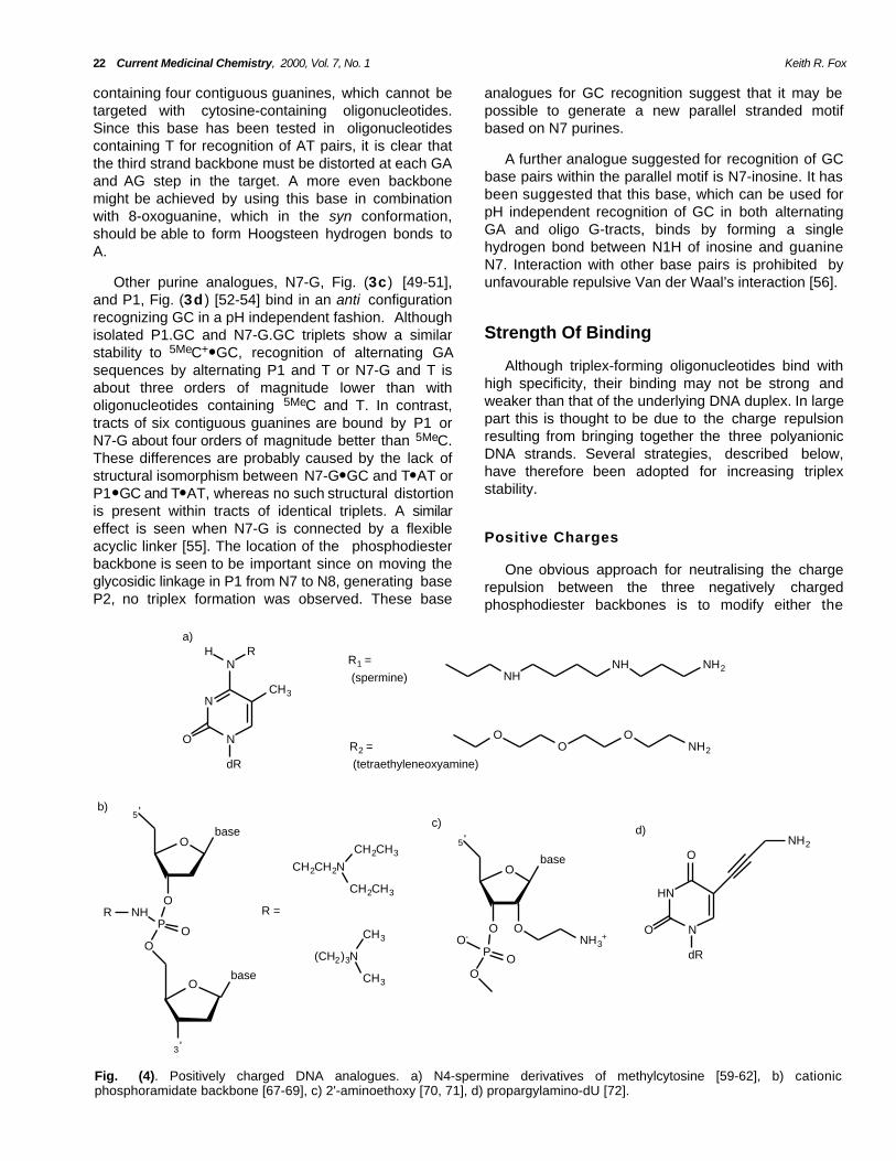
Although triplex-forming oligonucleotides bind withhigh specificity, their binding may not be strong andweaker than that of the underlying DNA duplex. In largepart this is thought to be due to the charge repulsionresulting from bringing together the three polyanionicDNA strands. Several strategies, described below,have therefore been adopted for increasing triplexstability.
Positive Charges
One obvious approach for neutralising the chargerepulsion between the three negatively chargedphosphodiester backbones is to modify either the
N
N
NRH
O
dR
CH3
NHNH NH2
OO
ONH2
R1 =
(spermine)
R2 =
(tetraethyleneoxyamine)
a)
Obase
5 '
O
PO
NHR
O
O
3 '
base
CH2CH2N
CH2CH3
CH2CH3
(CH2)3N
CH3
CH3
Obase
5 '
O
PO
O-
O
ONH3
+
HN
N
O
dR
O
NH2
b)c)
d)
R =
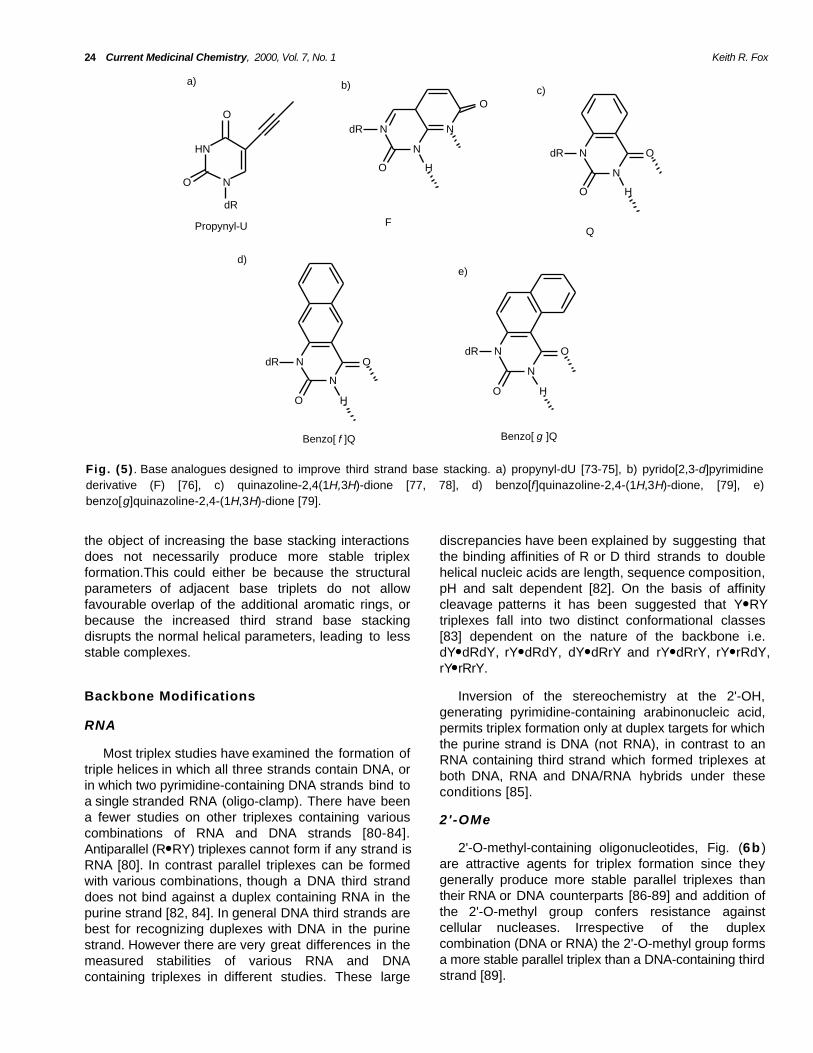
Fig. (4). Positively charged DNA analogues. a) N4-spermine derivatives of methylcytosine [59-62], b) cationicphosphoramidate backbone [67-69], c) 2'-aminoethoxy [70, 71], d) propargylamino-dU [72].
Targeting DNA Triplexes Current Medicinal Chemistry, 2000, Vol. 7, No. 1 23
sugar-phosphate backbone or the bases so as toincorporate positive charges. Since triplexes are knownto be stabilised by spermine [57, 58] several groupshave covalently attached this group to N4 [59-62] ofmethylcytosine, Fig. (4a). Surprisingly these triplexeswere stable at pH 7.4, even though N3 should beunprotonated at this pH, suggesting that the lack of thesecond hydrogen bond in the C+● GC triplet can becompensated by favourable electrostatic interactions.Indeed these triplexes were less stable at lower pHs.This substitution was more effective when placed ateither end of the triplex than at the centre and caused aslight decrease in Tm with increasing number ofsubstitutions. In addition these triplexes were lessdependent on the presence of divalent metal ionssuch as Mg2+. Replacing the polycationic spermine withtetraethoxyleneoxyamine, Fig. (4a ) produced a similareffect on triplex stability [61], presumably by virtue offavourable hydrophobic, rather than purely electrostaticinteractions. Other studies with syn-norspermidinelinked to the 5-position of U in the third strand alsoshow significant triplex stabilization at physiological pH[63]. Spermine has also been attached to the 5'-terminus [64] and to the 2'-O of oligonucleotides [65,66]. Both these modifications increase triplex meltingtemperatures, though 2'-O-linked spermine inducedtriplex formation when added at the 3'- or 5' end, butnot at the centre of the third strand oligonucleotide.
suggesting that the protonated group is ideallypositioned to interact with a nearby phosphate group[70]. Indeed an NMR structure of an intramoleculartriplex containing this modification in the Hoogsteenstrand has shown contacts between the amino protonsof the aminoethoxy group and the phosphates of thepurine strand of the duplex [71].
We have also shown that propargylamino-dU, Fig.(4d ) in which a positively charged group is added to the5'-position of thymine causes a dramatic increase intriplex stability [72]. The degree of stabilization was pHdependent, confirming the importance of the positivecharge. In this case the stabilization is due to more thaninclusion of the positive charge since propylamino-dUhad a much lower effect on stability.
Improved Base Stacking
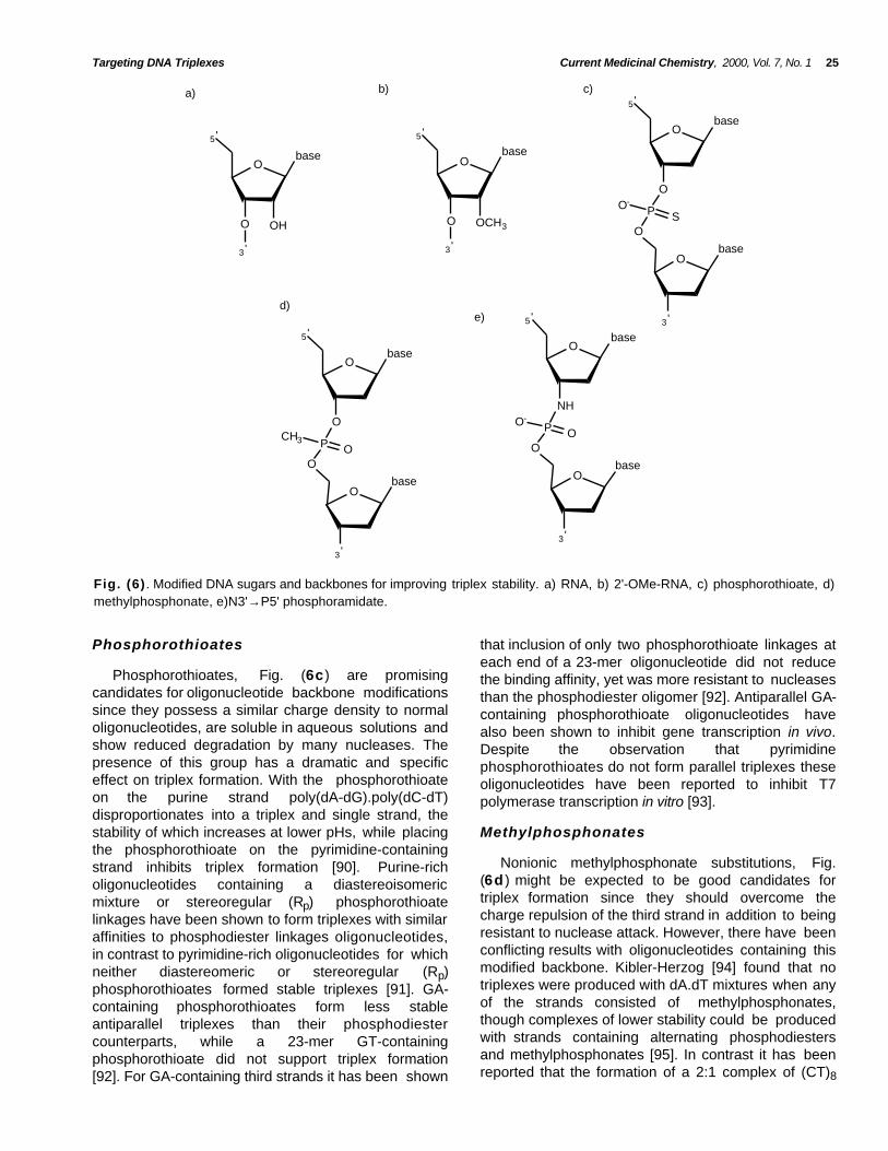
The observation that the increased stability of5MeC+● GC relative to C+● GC is caused by the extraspine of methyl groups within the DNA major grooveleads to the suggestion that other analogues withimproved base stacking might form more stabletriplexes. The methyl group at the 5-position of thymineis also known to contribute to the stability of the T● ATtriplet and it has been shown that the ranking order for5-substituted derivatives is BrU > T > U, suggesting thata bulky substituent can improve activity [24]. A similareffect is seen with 5-propynyl-U, Fig. (5a), which alsoincreases triplet stability [73-75], though 5-propynyl-Cproduces a less stable triplet since this substitutionfurther reduces the pK of N3 to 3.3. The increasedstability of 5-propynyl substituted bases is presumablydue to increased base stacking interactions.
Several studies have examined triplex formation byoligonucleotides containing neutral or positivelycharged backbones in attempts to alleviate the chargerepulsion between the polyanionic strands. Examplesof this include the cationic phosphoramidates shown inFig. (4b ). These have been shown to stabilise bothparallel [67] and antiparallel triplexes [68, 69]. The roleof the positive charge is confirmed by studies with anuncharged analogue which showed only a smallincrease in stability relative to the negatively chargedphosphodiester backbone [68]. The triplex binding ofsuch GA-containing phosphoramidates was maintainedin millimolar concentrations of potassium, conditionsthat usually inhibit antiparallel triplex formation as aresult of competing structures adopted by the thirdstrand oligonucleotide. These modifications introducea chiral phosphate, and experiments withstereouniform phosphoramidates have shown that,although one stereoisomer binds with higher affinitythan the simple phosphodiesters, the other binds withlower affinity [68].
Other studies have attempted to improve basestacking by adding a extra aromatic rings to thymine.One of the first such analogues to be tested was apyrido[2,3-d]pyrimidine derivative (F) [76]. This baseselectively recognises AT base pairs, forming the F● ATtriplet, utilising the tautomer shown in Fig. (5b ).However, these complexes were no more stable thanthose containing T● AT triplets. In addition experimentsexamining the cooperative binding of adjacentoligonucleotides showed that stacking of F on F wasless favourable than T on T. Similarly quinazoline-2,4(1H,3H)-dione [77, 78], Fig (5c),benzo[f]quinazoline-2,4-(1H,3H)-dione, Fig. (5d ) [79]and benzo[g]quinazoline-2,4-(1H,3H)-dione Fig. (5 e )[79] have been examined as thymine analogues.Although each of these analogues selectivelyrecognized AT base pairs, in both parallel andantiparallel triplexes, the complexes were less stablethan those containing standard T● AT triplets. Takentogether these results suggest that modification ofpyrimidine bases to form extended ring systems with
Other studies have included a positive charge intothe backbone by attaching an aminoethoxy moiety tothe 2'-position, Fig. (4c) [70, 71].This modificationcauses a dramatic increase in affinity and associationrate. Extension of the side chain by an additionalmethylene group, using a 2'-aminopropoxy group,caused a significant decrease in triplex stability,
24 Current Medicinal Chemistry, 2000, Vol. 7, No. 1 Keith R. Fox
HN
N
O
dR
O
N
N
N
O
dR
H
O
N
N
O
O
dR
N
N
O
dR ON
N
OdR
a) b) c)
d)e)
Propynyl-U FQ
Benzo[ f ]Q Benzo[ g ]Q
OH
H
H
Fig. (5) . Base analogues designed to improve third strand base stacking. a) propynyl-dU [73-75], b) pyrido[2,3-d]pyrimidinederivative (F) [76], c) quinazoline-2,4(1H,3H)-dione [77, 78], d) benzo[f]quinazoline-2,4-(1H,3H)-dione, [79], e)benzo[g]quinazoline-2,4-(1H,3H)-dione [79].
the object of increasing the base stacking interactionsdoes not necessarily produce more stable triplexformation.This could either be because the structuralparameters of adjacent base triplets do not allowfavourable overlap of the additional aromatic rings, orbecause the increased third strand base stackingdisrupts the normal helical parameters, leading to lessstable complexes.
discrepancies have been explained by suggesting thatthe binding affinities of R or D third strands to doublehelical nucleic acids are length, sequence composition,pH and salt dependent [82]. On the basis of affinitycleavage patterns it has been suggested that Y● RYtriplexes fall into two distinct conformational classes[83] dependent on the nature of the backbone i.e.dY● dRdY, rY● dRdY, dY● dRrY and rY● dRrY, rY● rRdY,rY● rRrY.
Backbone Modifications Inversion of the stereochemistry at the 2'-OH,generating pyrimidine-containing arabinonucleic acid,permits triplex formation only at duplex targets for whichthe purine strand is DNA (not RNA), in contrast to anRNA containing third strand which formed triplexes atboth DNA, RNA and DNA/RNA hybrids under theseconditions [85].
RNA
Most triplex studies have examined the formation oftriple helices in which all three strands contain DNA, orin which two pyrimidine-containing DNA strands bind toa single stranded RNA (oligo-clamp). There have beena fewer studies on other triplexes containing variouscombinations of RNA and DNA strands [80-84].Antiparallel (R● RY) triplexes cannot form if any strand isRNA [80]. In contrast parallel triplexes can be formedwith various combinations, though a DNA third stranddoes not bind against a duplex containing RNA in thepurine strand [82, 84]. In general DNA third strands arebest for recognizing duplexes with DNA in the purinestrand. However there are very great differences in themeasured stabilities of various RNA and DNAcontaining triplexes in different studies. These large
2'-OMe
2'-O-methyl-containing oligonucleotides, Fig. (6b )are attractive agents for triplex formation since theygenerally produce more stable parallel triplexes thantheir RNA or DNA counterparts [86-89] and addition ofthe 2'-O-methyl group confers resistance againstcellular nucleases. Irrespective of the duplexcombination (DNA or RNA) the 2'-O-methyl group formsa more stable parallel triplex than a DNA-containing thirdstrand [89].
Targeting DNA Triplexes Current Medicinal Chemistry, 2000, Vol. 7, No. 1 25
Obase
5 '
OHO
3 '
Obase
5 '
OCH3O
3 '
Obase
5 '
O
P SO-
O
Obase
3 '
Obase
5 '
O
P OCH3
O
Obase
3 '
Obase
5 '
NH
P OO-
O
Obase
3 '
e)d)
c)b)a)
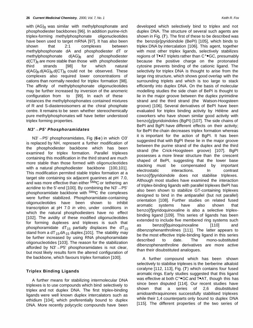
Fig. (6) . Modified DNA sugars and backbones for improving triplex stability. a) RNA, b) 2'-OMe-RNA, c) phosphorothioate, d)methylphosphonate, e)N3'→P5' phosphoramidate.
Phosphorothioates that inclusion of only two phosphorothioate linkages ateach end of a 23-mer oligonucleotide did not reducethe binding affinity, yet was more resistant to nucleasesthan the phosphodiester oligomer [92]. Antiparallel GA-containing phosphorothioate oligonucleotides havealso been shown to inhibit gene transcription in vivo.Despite the observation that pyrimidinephosphorothioates do not form parallel triplexes theseoligonucleotides have been reported to inhibit T7polymerase transcription in vitro [93].
Phosphorothioates, Fig. (6c) are promisingcandidates for oligonucleotide backbone modificationssince they possess a similar charge density to normaloligonucleotides, are soluble in aqueous solutions andshow reduced degradation by many nucleases. Thepresence of this group has a dramatic and specificeffect on triplex formation. With the phosphorothioateon the purine strand poly(dA-dG).poly(dC-dT)disproportionates into a triplex and single strand, thestability of which increases at lower pHs, while placingthe phosphorothioate on the pyrimidine-containingstrand inhibits triplex formation [90]. Purine-richoligonucleotides containing a diastereoisomericmixture or stereoregular (Rp) phosphorothioatelinkages have been shown to form triplexes with similaraffinities to phosphodiester linkages oligonucleotides,in contrast to pyrimidine-rich oligonucleotides for whichneither diastereomeric or stereoregular (Rp)phosphorothioates formed stable triplexes [91]. GA-containing phosphorothioates form less stableantiparallel triplexes than their phosphodiestercounterparts, while a 23-mer GT-containingphosphorothioate did not support triplex formation[92]. For GA-containing third strands it has been shown
Methylphosphonates
Nonionic methylphosphonate substitutions, Fig.(6d ) might be expected to be good candidates fortriplex formation since they should overcome thecharge repulsion of the third strand in addition to beingresistant to nuclease attack. However, there have beenconflicting results with oligonucleotides containing thismodified backbone. Kibler-Herzog [94] found that notriplexes were produced with dA.dT mixtures when anyof the strands consisted of methylphosphonates,though complexes of lower stability could be producedwith strands containing alternating phosphodiestersand methylphosphonates [95]. In contrast it has beenreported that the formation of a 2:1 complex of (CT)8
26 Current Medicinal Chemistry, 2000, Vol. 7, No. 1 Keith R. Fox
with (AG)8 was similar with methylphosphonate andphosphodiester backbones [96]. In addition purine-richtriplex-forming methylphosphonate oligonucleotideshave been used to target mRNA [97]. It has also beenshown that 2:1 complexes betweenmethylphosphonate dA and phosphodiester dT ormethylphosphonate d(AG)8 and phosphodiesterd(CT)8 are more stable than those with phosphodiesterthird strands [98] for which naturald(AG)8.d(AG)8.d(CT)8 could not be observed. Thesecomplexes also required lower concentrations ofcations than normally needed for triplex formation [98].The affinity of methylphosphonate oligonucleotidesmay be further increased by inversion of the anomericconfiguration from to [99]. In each of theseinstances the methylphosphonates contained mixturesof R and S-diastereoisomers at the chiral phosphatecentre. It remains to be seen whether stereochemicallypure methylphosphonates will have better understoodtriplex forming properties.
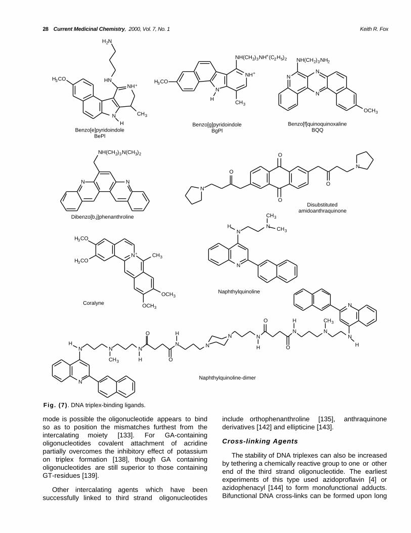
developed which selectively bind to triplex and notduplex DNA. The structure of several such agents areshown in Fig. (7 ). The first of these to be described wasthe benzo[e]pyridoindole (BePI) [105], which binds totriplex DNA by intercalation [106]. This agent, togetherwith most other triplex ligands, selectively stabilizesregions of T● AT triplets rather than C+● GC, presumablybecause the positive charge on the protonatedcytosine prevents binding of the cationic ligand. Theselectivity for triplex DNA is thought to arise from thelarge ring structure, which shows good overlap with thesurrounding triplets and which is too large to stackefficiently into duplex DNA. On the basis of molecularmodelling studies the side chain of BePI is thought tolie in the major groove between the duplex pyrimidinestrand and the third strand (the Watson-Hoogsteengroove) [106]. Several derivatives of BePI have beenevaluated for triplex binding activity by Hélène andcoworkers who have shown similar good activity withbenzo[g]pyridoindoles (BgPI) [107]. The side chains ofBePI and BgPI have different effects on their activity;for BePI the chain decreases triplex formation whereasit is important for the action of BgPI. It has beensuggested that with BgPI these lie in the major groovebetween the purine strand of the duplex and the thirdstrand (the Crick-Hoogsteen groove) [107]. BgPIpossesses a more linear structure than the crescentshaped of BePI, suggesting that the lower basestacking must be compensated by importantelectrostatic interactions. In contrastbenzo[f]pyridoindole does not stabilise triplexes.Although most studies have examined the interactionof triplex-binding ligands with parallel triplexes BePI hasalso been shown to stabilize GT-containing triplexesdesigned to bind in the antiparallel (but not parallel)orientation [108]. Further studies on related fusedaromatic systems have also shown thatbenzo[f]pyridoquinoxaline is also a selective triplex-binding ligand [109]. This series of ligands has beenextended to include five membered ring systems suchas benzo[f]quinoquinoxaline [110] anddibenzophenanthrolines [111]. The latter appears tobe the most effective triple-binding ligand in this seriesdescribed to date. The mono-substituteddibenzophenanthroline derivatives are more activethan their disubstituted analogues.
N3'→P5' Phosphoramidates
N3'→P5' phosphoramidates, Fig (6 e ) in which O3'is replaced by NH, represent a further modification ofthe phosphodiester backbone which has beenexamined for triplex formation. Parallel triplexescontaining this modification in the third strand are muchmore stable than those formed with oligonucleotideswith a natural phosphodiester backbone [100,101].This modification permited stable triplex formation at atarget site containing six adjacent guanines at pH 7.0,and was more effective than either 5MeC or tethering anacridine to the 5'-end [100]. By combining the N3'→P5'phosphoramidate backbone with 5MeC the complexeswere further stabilized. Phosphoramidate-containingoligonucleotides have been shown to inhibittranscription at pH 7.0 in vitro, under conditions inwhich the natural phosphodiesters have no effect[102]. The avidity of these modified oligonucleotidesfor forming duplexes and triplexes is such thatphosphoramidate dT15 partially displaces the dT15stand from a dT15.dA15 duplex.[101]. The stability maybe further increased by using RNA phosphoramidateoligonucleotides [103]. The reason for the stabilizationafforded by N3'→P5' phosphoramidates is not clear,but most likely results form the altered configuration ofthe backbone, which favours triplex formation [100]. A further compound which has been shown
selectively to stabilise triplexes is the berberine alkaloidcoralyne [112, 113], Fig. (7 ) which contains four fusedaromatic rings. Early studies suggested that this ligandwas effective at both C+● GC and T● AT, though this hassince been disputed [114]. Our recent studies haveshown that a series of 2,6 disubstitutedamidoanthraquinones successfully stabilised triplexeswhile their 1,4 counterparts only bound to duplex DNA[115]. The different properties of the two series of
Triplex Binding Ligands
A further means for stabilizing intermolecular DNAtriplexes is to use compounds which bind selectively totriplex and not duplex DNA. The first triplex-bindingligands were well known duplex intercalators such asethidium [104], which preferentially bound to duplexDNA. More recently polycyclic compounds have been
Targeting DNA Triplexes Current Medicinal Chemistry, 2000, Vol. 7, No. 1 27
compounds are thought to arise from differences in thestacking of the base triplets with the chromophore,combined with DNA groove accessibility. Confirmationof this binding selectivity and a thermodynamic basis forthe different triplex/duplex preferences of the 2,6 and1,4 isomers was provided by calorimetric andspectrophotometric techniques [116]. This studyrevealed that the 1,4 compounds destabilize DNAtriplexes, leading to dissociation of the third strand,whereas their 2,6 isomeric analogues bind to andstabilize triple-stranded structures. A related study hasalso shown that selective stabilization of triplex DNAcan be achieved with a series of disubstitutedanthraquinone sulfonamides and suggested that 2,7-functionalised compounds are more potent than their2,6 counterparts [117]. We have also compared theactivity of four di-substituted and two monosubstitutedamidoanthraquinone compounds, and have examinedhow the position of the cationic substituents affectstriplex affinity [118]. Triplex affinity for the differentsubstituent patterns decreases in order 2,7 > 1,8 = 1,5> 2,6, with the equivalent monosubstitutedcompounds being at least an order of magnitude lessefficient.
absence of BePI [126]. Duplex regions of (AT)n havealso been targeted with GT-containingoligonucleotides in the presence of thenaphthylquinoline triplex-binding ligand, generatingcomplexes containing alternating G● TA and T● ATtriplets [127]. Although blocks of alternating T● AT andG● TA triplets alone are not stable, even in the presencea triplex-binding ligand, these complexes can bestabilized by attaching this region to a block ofconsecutive T● AT triplets, to which the ligand isthought to bind. In this way the duplexA11(AT)6.(AT)6T11 can be targeted with the third strandT11(TG)6, though this interaction still requires thepresence of Mn2+ or a triplex binding ligand. Thesecomplexes can be extended to longer (AT)n tracts (upto n = 11) [128] and can be stabilised with shorter T● ATtracts. It should also be noted that these complexescan be further stabilized by including a few C+● GCtriplets in the anchoring tail and form in the presence ofMg2+, without addition of a stabilizing ligand [128].
Tethered DNA Binding Agents
One of the first methods to be attempted forincreasing triplex stability was to attach a DNA bindingligand to one end of the oligonucleotide. In this waysequence selectivity is achieved by the triplex formingoligonucleotide while the ligand acts as a non-selectiveanchor increasing the affinity. Most of these studieshave used duplex intercalators as DNA binding ligands,though some recent examples have employed triplexbinding ligands.
Naphthylquinoline derivatives, Fig (7 ) alsoselectively bind to triplex DNA by intercalation [119-123]. These compounds possess a large aromatic areato stack with the three bases in the triplex, yet, sincethe aromatic portions are not fused, they possesstorsional flexibility and can accommodate the propellertwist of the triplets in which the three bases may not becoplanar. These compounds are also selective for T● ATover C+● GC and are more effective at parallel thanantiparallel triplexes [121]. A recent study has alsosuggested that a naphthylquinoline dimer may bind bybis-intercalation [124].
Intercalators
Several groups have shown that covalentattachment of an acridine moiety (usually [2-methoxy,6-chloro,9-amino]acridine) to the end of anoligonucleotide significantly increases triplex stability[129-133]. The acridine is proposed to intercalate atthe triplex-duplex junction. Attachment to the 5'-endhas a greater effect than at the 3'-end [130, 134],possibly reflecting a structural difference between the5'-and 3'-triplex-duplex junctions, since the junction atthe 5'-end of the third strand is thought to present astrong intercalation site [135, 136]. Most of thesestudies have used parallel, pyrimidine-containingoligonucleotides, though they have also been used tostabilise antiparallel GA and GT-containing triplexes [92,137-140] as well as alternate strand triplexes containingboth parallel and antiparallel motifs [141]. Attachmentgenerally increases triplex stability by at least 100-fold,though the interaction with related non-cognatesequences is also increased [133]. Howeverattachment of the acridine does not seem substantiallyto compromise the sequence specificity of binding[139]. At secondary sites where more than one binding
Triplex-binding ligands have also been used forincreasing the strength of binding to sites whichcontain pyrimidine interruptions. In general theseligands do not affect the stringency of triplex formation,and the relative binding strengths of differentoligonucleotide substitutions are not affected [122].However by increasing the strength of binding, by upto 1000-fold, complexes can be formed at sequencesfor which there are no clear rules. Thenaphthylquinoline derivatives have been shown topromote the formation of triplexes at sites containingup to three consecutive base pair inversion using T● CGand G● TA triplets [125]. Studies with BePI have shownthat the least destabilizing triplets are the same in boththe presence and absence of the ligand and that thethird strand base is less important in the presence ofthe ligand [126]. However addition of the ligand doesprovide some discrimination between different invertedbase pairs. In particular 3-nitropyrrole discriminates CGfrom GC, TA and AT pairs in the presence, but not the
28 Current Medicinal Chemistry, 2000, Vol. 7, No. 1 Keith R. Fox
N
NH+HN
H2N
CH3
H
H3CONH+
N
CH3
NH(CH2)3NH+(C2H5)2
H
H3CON
N
N
OCH3
NH(CH2)3NH2
N N
NH(CH2)3N(CH3)2
O
NO
N
O
O
N+
OCH3
OCH3
CH3
H3CO
H3CON
NN
CH3
CH3H
N
N N NNH
CH3
O
O
N
N NN N
N
H
O
O
H CH3
H
N
H
H
Benzo[e]pyridoindoleBePl
Benzo[g]pyridoindoleBgPl
Benzo[f]quinoquinoxalineBQQ
Dibenzo[b,j]phenanthroline
Disubstitutedamidoanthraquinone
Coralyne
Naphthylquinoline
Naphthylquinoline-dimer
Fig. (7) . DNA triplex-binding ligands.
mode is possible the oligonucleotide appears to bindso as to position the mismatches furthest from theintercalating moiety [133]. For GA-containingoligonucleotides covalent attachment of acridinepartially overcomes the inhibitory effect of potassiumon triplex formation [138], though GA containingoligonucleotides are still superior to those containingGT-residues [139].
include orthophenanthroline [135], anthraquinonederivatives [142] and ellipticine [143].
Cross-linking Agents
The stability of DNA triplexes can also be increasedby tethering a chemically reactive group to one or otherend of the third strand oligonucleotide. The earliestexperiments of this type used azidoproflavin [4] orazidophenacyl [144] to form monofunctional adducts.Bifunctional DNA cross-links can be formed upon long
Other intercalating agents which have beensuccessfully linked to third strand oligonucleotides
Targeting DNA Triplexes Current Medicinal Chemistry, 2000, Vol. 7, No. 1 29
wavelength irradiation of psoralen, conjugated to anoligonucleotide via either its 4' or 5 position, and can beused as a means of covalently attaching a triplex-forming oligonucleotide to its target site. This not onlyachieves selective gene inactivation, but provides ameans for delivering a tethered mutagen to a targetsite, for the introduction of site-specific DNA damage.The most reactive sequence for photocrosslinking bypsoralen is TpA, linking the thymines across the twostrands [145]. Psoralen-linked oligonucleotides aretherefore effective at triplex sites which are borderedby a TpA step. The first experiments of this typeattached psoralen to the 5'-end of pyrimidine-containing third strand oligonucleotides [146, 147]. Inthis way successful complex formation wasdemonstrated at a 16 base oligopurine tract in the HIVproviral sequence at pH 6.0, even though this targetsequence contained a run of six contiguous GC basepairs. However at elevated oligonucleotideconcentrations secondary binding was observed at asite containing only eight matching base pairs,suggesting that the covalent reaction might induce theformation of some non-productive complexes. Bindingwas further increased by replacing the third strandcytosines with guanines forming parallel G● GC triplets.Similarly a 20 base pair sequence within the aromatasegene has been targeted with a psoralen-linkedoligonucleotide, even though this polypurine tract isinterrupted by three CG base pairs, for which T● CGtriplets were employed [148, 149].
Tethered Triplex-binding Ligands
Benzopyridoindole and benzopyridoquinolinetriplex-binding ligands have been attached to either the5'-end or to an internucleotide position of triplex-forming oligonucleotides [164, 165]. For internalattachment the ligands were incorporated as anadditional nucleotide, rather than replacing an existingone. All the derivatives stabilised triple helicalstructures more than either attachment of an acridinemoiety or addition of the free ligand at an equivalentconcentration. Comparison of their effect on twodifferent parallel triplexes showed that the attachedligand had a greater stabilising effect on the sequencecontaining a longer run of contiguous thymines.Benzo[h]quinoquinoxaline was the most effectivecompound when attached to the 5'-end of the thirdstrand, while BePI was the best ligand for the internalsite. In addition these modifications did not alter thestringency of triplex formation, and introduction of asingle mismatch caused a large decrease in bindingaffinity. We have also prepared oligonucleotides with anaphthylquinoline-derivative tethered to the 5'-endand have shown that these successfully stabilisetriplexes to a greater extent than an equivalentconcentration of free ligand [166]. It should also benoted, however, that with the tethered complex thereis only one ligand stabilising each triplex, whereas withthe free ligand several molecules may be able to bindsimultaneously at different positions along the triplex.
Inclusion of a duplex binding agent within the thirdstrand oligonucleotide has also be used to increasebinding to sites containing pyrimidine interruptions, byplacing an internal acridine group adjacent to the basefacing the inverted pyrimidine.purine base pair [167,168]. In this way, the loss of triplex stability at theinversion is partly overcome by the additional bindingfree energy of the intercalator, in a similar fashion to thatachieved by attaching an intercalator to the 5'-end ofthe third strand. In the absence of a base inversioninclusion of an internal acridine has little or no effect ontriplex stability, possibly because it is a duplex- ratherthan a triplex-specific intercalator. For targetscontaining CG or TA inversions the acridine moietyincreases the stability of triplexes with either natural orsynthetic bases opposing the pyrimidine base.Recognition of the TA base pair is strongest usingeither acridine or propanediol with an acridine on its 3'-side. Recognition of CG is greatest with either cytosinewith acridine on its 3'-side or guanine with acridine onits 5'-side [167]. Similarly acridine-conjugatedoligonucleotides have been used to stabilize theformation of triplexes at oligopurine tracts containingpyrimidine interruptions [140]. In these studies variousnatural or synthetic base analogues were placedopposite the pyrimidine residue, while the acridine wastethered to the 5'-end. These base ‘mismatches’ had a
Psoralen-linked GA-containing oligonucleotideshave also been used to introduce a mutation into aselected triplex target sequence. Most of the mutationsdetected are TA → AT transversions and are targetedat the psoralen intercalation site [151-152]. Mutationsare generated by transcription-coupled repair [153] andinvolve the error-prone repair pathway, either alone orin combination with nucleotide excision repair [154].This mechanism may depend on the sequence contextsince other studies have shown that a 19-mer GT-containing oligonucleotide with a 5'-psoralen is notrepaired [155]. Double psoralen adducts, located atboth ends of a triplex-forming oligonucleotide cannotbe repaired efficiently in human cells [156]. Theefficiency of triplex formation and gene inactivation canbe further increased by tethering an acridine to theother end of the oligonucleotide [157].
Other DNA-binding agents which have beensuccessfully tethered to triplex-formingoligonucleotides include the AT-selective minorgroove binding ligand Hoechst 33258 [158, 159], thetopoisomerase I inhibitor camptoethecin [160],duocarmycin [161], the DNA cleavage agent bleomycin[162] and a platinum-containing DNA cross-linkingagent [163].
30 Current Medicinal Chemistry, 2000, Vol. 7, No. 1 Keith R. Fox
greater destabilizing effect when place close to theintercalator end of the oligonucleotide.
reverse Hoogsteen T● AT [11]. The kinetics of triplexformation are also faster for antiparallel than parallelcomplexes [174-177].
Antiparallel TriplexesLength
Since pyrimidine-containing oligonucleotides,forming parallel DNA triplexes, require conditions of lowpH, necessary for protonation of the third strandcytosine, it might be expected that purine-containingoligonucleotides designed to for antiparallel triplexeswould be superior. Nonetheless these have not beenso widely used for several reasons. Firstly the stabilityof antiparallel triplexes seems to vary widely from onestudy to another. Secondly these structures aredominated by G.GC, which usually constitutes over60% of the triplets present, and G-rich oligonucleotidesare known to adopt unusual structures which competefor triplex formation. Thirdly there are fewer studies onthe recognition of pyrimidine interruptions and there isno specific means of recognising TA (see below).
Although triplex stability generally increases with thelength of the third strand, this is not necessarily thecase for antiparallel triplexes. Cheng & Van Dyke [178]showed that, for the GT-motif short third strands (12-mers) could produce more stable complexes thanlonger oligonucleotides and that, in general,extensions and mutations at the 3'-end had greatereffect than those at the 5'-end. Based on thisobservation they suggested that antiparallel triplexformation proceeded in the 3'→5' direction. Similarly ina REPSA selection assay for oligonucleotides whichgenerate stable triplexes at a 19-mer target site, perfectmatches were observed for the 13 bases at the 3'-end,while mismatches were selected at the 5'-end, andshorter oligonucleotides generated complexes withmuch reduced stability [179]. The greater effect of the3'-end of the third strand has also been observed infootprinting experiments with oligonucleotidescontaining (GGA)n repeats, for which slipped structureswere observed [171]. More recently this effect hasbeen confirmed by Arimondo et al. [180] whocompared the strength of triplex formation by (AGG)4and (GGA)4. Although both complexes contained 4 xA● AT and 8 x G● GC triplets the oligonucleotide with a3'-G [i.e. (AGG)4] bound about 6-times better than theone with a 3'-A [i.e. (GGA)4]. The addition of A● AT andG● GC triplets to the 5'-end of (AGG)4 caused anincrease in triplex stability as expected. In contrast,although the addition of a single G to the 3'-end of(GGA)4 increased stability by about 10-fold, the additionof further As caused a decrease in stability.Furthermore overhanging mismatched bases had amuch greater effect on stability when added to the 3'-than the 5'-end. Taken together these results suggestthat antiparallel triplex formation proceeds from the 3'-end of the third strand and that nucleation is moreefficient when the first base is a guanine.
Stability
In some studies antiparallel triplexes are shown tohave very high affinities; short oligonucleotides exhibitnanomolar binding constants [169, 170], while in otherstudies longer oligonucleotides, under similarconditions, fail to show binding at micromolarconcentrations [137, 170-173]. The simplestexplanation for this differences is that the formation ofantiparallel triplexes is very sequence dependent. Thismay be due to the different positions of the third strandbackbone in G● GC, A● AT and T● AT triplets; unlikeparallel T● AT and C+● GC triplets which are isohelical.The most stable complexes are likely to be those withthe lowest number of ApG and GpA steps in thehomopurine target site. This sequence dependence isfurther compounded by the observation that the affinityof antiparallel triplexes is dominated by the G● GC triplet,which imparts a much greater stability that either A● ATor T● AT [137]. However, G-rich oligonucleotides formstrong inter- and intramolecular structures, possiblyinvolving G-quartet formation, which reduce theeffective concentration of the oligonucleotide bycompeting with triplex formation. The best antiparalleltriplexes may therefore be those in which the thirdstrand has a high G-content, but in which the guaninesare arranged in such a way as to prevent the formationof unusual structures.
Self Association
G-rich oligonucleotides are known to be able to formstable G-quartet structures, which compete for triplexformation in the presence of physiologicalconcentration of monovalent cations, particularly K+
[181, 182]. In addition GA- (but not GT) containingoligonucleotides can form homoduplexes, stabilised byinternal GA repeats [183, 184]. The situation is furthercomplicated by the observation that different inter- orintra-molecular structures can be formed with differentsequences of (G,A)-containing oligonucleotides [185].
There have been few studies directly comparing thestability of triplexes formed with CT- GT- and GA-containing oligonucleotides. It is generally agreed thatGT triplexes are less stable than their GA counterparts[171, 174-176], possibly because the antiparallel G● GCtriplet is more closely isomorphous to A● AT than
Targeting DNA Triplexes Current Medicinal Chemistry, 2000, Vol. 7, No. 1 31
Self association of these oligonucleotides caneffectively compete with triple helix formation andprevent complete triplex formation since, at highconcentrations, more of the oligonucleotide is trappedin self-associated structures. The self-structuresadopted by GA-containing oligonucleotides explain theopposite temperature dependence of triplexes formedwith GA- and GT-containing oligonucleotides. Thebinding of GT-oligonucleotides decreases on raisingthe temperature from 4 to 37°C, in contrast to GAoligonucleotides for which the binding was increased,as the self-associated structure become less stable[183, 184].
deazaxanthine (c7X) in place of A or T in antiparalleloligonucleotides also achieved high affinity triplexformation under physiological potassiumconcentrations [187, 190].
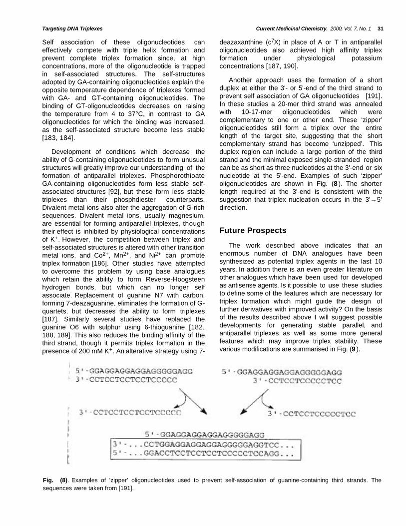
Another approach uses the formation of a shortduplex at either the 3'- or 5'-end of the third strand toprevent self association of GA oligonucleotides [191].In these studies a 20-mer third strand was annealedwith 10-17-mer oligonucleotides which werecomplementary to one or other end. These ‘zipper’oligonucleotides still form a triplex over the entirelength of the target site, suggesting that the shortcomplementary strand has become ‘unzipped’. Thisduplex region can include a large portion of the thirdstrand and the minimal exposed single-stranded regioncan be as short as three nucleotides at the 3'-end or sixnucleotide at the 5'-end. Examples of such ‘zipper’oligonucleotides are shown in Fig. (8 ). The shorterlength required at the 3'-end is consistent with thesuggestion that triplex nucleation occurs in the 3'→5'direction.
Development of conditions which decrease theability of G-containing oligonucleotides to form unusualstructures will greatly improve our understanding of theformation of antiparallel triplexes. PhosphorothioateGA-containing oligonucleotides form less stable self-associated structures [92], but these form less stabletriplexes than their phosphdiester counterparts.Divalent metal ions also alter the aggregation of G-richsequences. Divalent metal ions, usually magnesium,are essential for forming antiparallel triplexes, thoughtheir effect is inhibited by physiological concentrationsof K+. However, the competition between triplex andself-associated structures is altered with other transitionmetal ions, and Co2+, Mn2+, and Ni2+ can promotetriplex formation [186]. Other studies have attemptedto overcome this problem by using base analogueswhich retain the ability to form Reverse-Hoogsteenhydrogen bonds, but which can no longer selfassociate. Replacement of guanine N7 with carbon,forming 7-deazaguanine, eliminates the formation of G-quartets, but decreases the ability to form triplexes[187]. Similarly several studies have replaced theguanine O6 with sulphur using 6-thioguanine [182,188, 189]. This also reduces the binding affinity of thethird strand, though it permits triplex formation in thepresence of 200 mM K+. An alterative strategy using 7-
Future Prospects
The work described above indicates that anenormous number of DNA analogues have beensynthesized as potential triplex agents in the last 10years. In addition there is an even greater literature onother analogues which have been used for developedas antisense agents. Is it possible to use these studiesto define some of the features which are necessary fortriplex formation which might guide the design offurther derivatives with improved activity? On the basisof the results described above I will suggest possibledevelopments for generating stable parallel, andantiparallel triplexes as well as some more generalfeatures which may improve triplex stability. Thesevarious modifications are summarised in Fig. (9 ).
Fig. (8). Examples of ‘zipper’ oligonucleotides used to prevent self-association of guanine-containing third strands. Thesequences were taken from [191].

32 Current Medicinal Chemistry, 2000, Vol. 7, No. 1 Keith R. Fox
O
O
O
O
O
ONH3
+
ligandbase
PO
O-
O
3'
base
base
NH
P O
O-
O
Tethered ligands
Psoralen
Triplex ligand
Spermine
Base modification
Cytosine analogues
Charged bases
Improved base stacking
Guanine analogues
Parallel or antiparallel
GT or GA
Backbone modification
2' -substitutionRNA
OMe
Aminoethoxy
Sugar modification
Fig. (9) . Summary of potential sites of oligonucleotide modification for generating triplexes with enhanced stability.
Parallel Triplexes necessary that the X● GC triplet should be isostructuralwith T● AT so as to minimise any distortions in the thirdstrand backbone between adjacent base analogues.This may not be possible with purine analogues forrecognition of GC when used alongside T● AT triplets,and similar derivatives, such as 2-aminopurine [45] or 8-oxoguanine may need to be developed for recognitionof AT in these contexts. Thirdly triplex stability may beenhanced by increasing the base stacking in the thirdstrand. However, simple addition of aromatic rings tothe pyrimidine nucleus does not seem to be of benefit[76-79], possibly because the improved stacking of thesingle stranded oligonucleotide hinders triplexformation. Other stacking interactions, such as thoseinvolving appended propynyl or propargylaminogroups [72-75], may be a better option. Lastly it ispossible that different sugars should be used for thirdstrand C or T, since it appears that rigid linkers improvethe stability of T● AT, while flexible linkers improve thestability of C+● GC [35, 36].
One major problem for generating stable paralleltriplexes under physiological conditions is the pHdependency of the C+● GC triplet. Although severalanalogues have been shown to alleviate this problemnone of these has found widespread use, possiblybecause of their limited availability, but also because ofthe large number of choices. In addition although manyanalogues remove the pH dependency they generateless stable triplexes than those formed with C+● GC atlow pH. Several features seem important in this regard.Firstly, since it is known that C+● GC is more stable thanT● AT [18-21], novel cytosine analogues with ahydrogen bond donor at a position equivalent to N3should retain the positive charge either on the ringsystem, as in 2-aminopyridine [38-41], or added to aside group which may be attached to either N4 or C5[59-62]. The importance of the positive chargesuggests that the stability of T● AT might be improvedby generating a charged analogue of T. However, sinceadjacent C+● GC triplets are known to be destabilising,due to repulsion between the charged groups, it maybe necessary to prepare both charged and unchargedanalogues of T and C, and determine the optimumarrangement of the charged derivatives. Secondly it is
Antiparallel Triplexes
There have been fewer studies developing baseanalogues for use in antiparallel triplets, though there
Targeting DNA Triplexes Current Medicinal Chemistry, 2000, Vol. 7, No. 1 33
are several outstanding problems which need to beaddressed. Firstly there is the question of whether touse GA- or GT- containing oligonucleotide. GAsequences are more prone to self association [184,185], yet the A● AT triplet is more closely isostructural toG● GC than T● AT [11]. Since these structures aredominated by the stability of the G● GC triplet, furtheranalogues for isostructural recognition of AT (xanthine)may be useful. Secondly novel analogues (such as 6-thioguanine and 7-deazaguanine) will be needed toovercome the tendency of G-rich oligonucleotides toaggregate [188-190]. These limitations require furtherwork optimizing the choice of third strandoligonucleotide. Thirdly, by analogy with C+● GC, it maybe beneficial to introduce positively charged groupsonto the purine ring.
Acknowledgements
Work in the author's laboratory is funded by theCancer Research Campaign.
References
[1] Felsenfeld, G.; Rich, A. Biochim. Biophys. Acta 1957, 26, 457.
[2] Felsenfeld, G.; Davis, D.R.; Rich, A. J. Am. Chem. Soc. 1957,79, 2023.
[3] Moser, H.E.; Dervan, P.B. Science 1987, 238, 645.
[4] Le Doan, T.; Perrouault, L.; Praseuth, D.; Habhoub, N.; Decout,J.L.;Thuong, N.T.; Lhomme, J.; Hélène, C. Nucleic Acids. Res.1987, 15, 7749.
[5] Soyfer, V.N.; Potaman, V.N. Triple Helical Nucleic AcidsSpringer-Verlag: New York, Berlin, 1996.
Stability[6] Thuong, N.T.; Hélène, C. Angew. Chem. Int. Ed. Engl. 1993, 32,
666.It is generally agreed that the lower stability of
triplexes, relative to duplexes, can in part be attributedto charge repulsion between the three negativelycharged phosphodiester backbones. Although severalstudies have shown that the introduction of positivelycharged groups into the backbone [59-66], sugar [70,71] or bases [67-69, 72] can produce stable triplexes,these have usually been used in isolation. It is worthremembering that each triplet carries three negativelycharged phosphates; so that yet more stable triplexesmay be produced by combining several of thesemodifications in a single oligonucleotide. A furtherbenefit of these modified oligonucleotides is that theyoften confer increased biological stability andresistance against nucleases. Triplex binding ligandsoffer a further method for increasing triplex stabilitywhich may also be useful when combined with some ofthe other approaches, or tethered to the end of thethird strand. A further limiting factor for both triplexmotifs is that there is still no good method forrecognizing pyrimidine interruptions [15, 16]. This ismore acute for antiparallel complexes for which there isno method for recognizing a TA base pair. Any novelbases for recognizing TA or CG will also need to bestructurally compatible the existing method fortargeting GC and TA. Triplex (or duplex) ligands mayalso be used for stabilizing weaker complexes acrosstargets containing pyrimidine interruptions.
[7] Vasquez, K.M.; Wilson, K.H. Trends in Biochem. 1998, 23, 4.
[8] Chubb, J.M.; Hogan, M.E. Trends in Biotechnol. 1992, 10, 132.
[9] Chan, P.P ; Glazer, P.M. J. Mol. Med. 1997, 75, 267.
[10] Neidle, S. Anti-Cancer Drug Des. 1997, 12, 433.
[11] Radhakrishnan, I.; Patel, D.J. Structure 1994, 2, 17.
[12] Beal, P.A.; Dervan, P.B. Science 1991, 251, 1360.
[13] Chen, F.-M. Biochemistry 1991, 30, 4472.
[14] Radhakrishnan, I.; Patel, D.J.Structure 1993, 1, 135.
[15] Doronina, S.O.; Behr, J.P. Chem. Soc. Rev. 1997, 26, 63.
[16] Gowers, D.M.; Fox, K.R. Nucleic Acids Res. 1999, 27, 1569.
[17] Lee, J.S.; Wordsworth, M.L.; Latimer, L.J.P.; Morgan, A.R.Nucleic. Acids. Res. 1984, 12, 6603.
[18] Roberts, R.W.; Crothers, D.M. Proc. Natl. Acad. Sci. U.S.A.1996, 93, 4320.
[19] Asensio, J.L.; Lane, A.N.; Dhesi, J.; Bergvist, S.; Brown, T. J.Mol. Biol. 1998, 275, 811.
[20] Keppler, M.D.; Fox, K.R. Nucleic Acids Res. 1997, 25, 4464.
[21] Volker, J.; Klump, H.H. Biochemistry 1994, 33, 13502.
[22] Giovannangeli, C.; Rougée, M.; Garestier, T.; Thuong, N.T.;Hélène, C. Proc. Natl. Acad. Sci. U.S.A. 1992, 89, 8631.
[23] Xodo, L.E.; Manzini, G.; Quadrifoglio, F.; van der Marel, G.; vanBoom, J. Nucleic Acids Res. 1991, 19, 5625.
Although there have been many significantadvances in the use of triplex forming oligonucleotidesthere are still many unanswered questions. Resolvingthese problems will require further close collaborationsbetween chemists and molecular biologists before thepotential of this strategy for targeting DNA is fullyrealised.
[24] Povsic, T.J.; Dervan, P.B. J. Am. Chem. Soc. 1989, 111, 3059.
[25] Koh, J.S.; Dervan, P.B. J. Am. Chem. Soc. 1992, 114, 1470.
[26] Singleton, S.F.; Dervan, P.B. Biochemistry 1992, 31, 10995.
[27] Ono, A.; Ts'o, P.O.P.; Kan, L. J. Am. Chem. Soc. 1991, 113,4032.
34 Current Medicinal Chemistry, 2000, Vol. 7, No. 1 Keith R. Fox
[28] Ono, A.; Ts'o, P.O.P.; Kan, L. J. Org. Chem. 1992, 57, 3225. [55] St. Clair, A.; Xiang, G.; McLaughlin, L.W. NucleosidesNucleotides 1998, 17, 925.
[29] Egholm, M.; Christensen, L.; Dueholm, K.L.; Buchardt, O.; Coull,J.; Nielsen, P.E. Nucleic Acids Res. 1995, 23, 217. [56] Marfut, J.; Parel, S.P.; Leumann, C.J. Nucleic Acids Res. 1997,
25, 1875.[30] Kuhn, H.; Demidov, V.V.; Frank-Kamenetskii, M.D.; Nielsen,
P.E. Nucleic Acids Res. 1998 26, 582. [57] Hampel, K.J.; Crosson, P.; Lee, J.S. Biochemistry 1991, 30,4455.
[31] Berressem, R.; Engels, J.W. Nucleic Acids Res. 1995, 23, 3465.[58] Thomas, T.; Thomas, T.J. Biochemistry 1993, 32, 14068.
[32] Xiang, G.; Soussou, W.; McLaughlin, L.W. J. Am. Chem. Soc.1994, 116, 11155. [59] Barawkar, D.A.; Kumar, V.A.; Garesh, K.N. Biochem. Biophys.
Res. Commun. 1994, 205, 1665.[33] Xiang, G.; Bogacki, R.; McLaughlin, L.W. Nucleic Acids Res.
1996, 24, 1963. [60] Barawkar, D.A.; Rajeev, K.G.; Kumar, V.A.; Ganesh, K.N.Nucleic Acids Res. 1996, 24, 1229.
[34] Xiang, G.; McLaughlin, L.W. Tetrahedron 1998, 54, 375.[61] Rajeev, K.G.; Jadhav, V.R.; Ganesh, K.N. Nucleic Acids Res.
1997, 25, 4187.[35] Tarkoy, M.; Leumann, C. J. Org. Chem. 1993, 32, 1432.
[36] Jones, R.L.; Swaminathan, S.; Milligan, J.F.; Waswani, S.;Froehler, B.C.; Matteucci, M.D. J. Am. Chem. Soc. 1993, 115,9816.
[62] Ganesh, K.N.; Rajeev, K.G.; Pallan, P.S.; Rana, V.S.; Barawkar,D.A.; Kumar, V.A. Nucleosides Nucleotides 1997, 16, 1271.
[63] Nara, H.; Ono, A.; Matsuda, A. Bioconjugate Chem. 1995, 6, 54.[37] von Krosigk, U.; Benner, S.A. J. Am. Chem Soc. 1995, 117,
5361. [64] Tung, C.H.; Breslauer, K.J.; Stein, S. Nucleic Acids Res. 1993,21, 5489.
[38] Bates, P.J.; Laughton, C.A.; Jenkins, T.C.; Capaldi, D.C.; Roselt,P.D.; Reese, C.B.; Neidle, S. Nucleic Acids Res. 1996, 24, 4176. [65] Sund C.; Puri, N.; Chattopadhyaya, J. Tetrahedron 1996, 52,
12275.[39] Cassidy, S.A.; Slickers, P.; Trent, J.O.; Capaldi, D.C.; Roselt,
P.D.; Reese, C.B.; Neidle, S.; Fox, K.R. Nucleic Acids Res.1997, 25, 4891.
[66] Sund C.; Puri, N.; Chattopadhyaya, J. Nucleosides Nucleotides1997, 16, 755.
[40] Hildbrand, S.; Leumann, C. Angew. Chem. Int. Ed. Engl. 1996,35, 1968.
[67] Chaturvedi, S.; Horn, T.; Letsinger, R.L. Nucleic Acids Res.1996, 24, 2318.
[41] Hildbrand, S.; Blaser, A.; Parel, S.P.; Leumann, C.J. J. Am.Chem. Soc. 1997, 119, 5499.
[68] Dagle, J.M.; Weeks, D.L. Nucleic Acids Res. 1996, 24, 2143.
[69] Bailey, C.P.; Dagle, J.M.; Weeks, D.L. Nucleic Acids Res. 1998,26, 4860.[42] Horne, D.A.; Dervan, P.B. Nucleic Acids Res. 1991, 19, 4963.
[43] Froehler, B.C.; Ricca, D.J. J. Am. Chem. Soc. 1992, 114, 8320. [70] Cuenoud, B.; Casset, F.; Husken, D.; Natt, F.; Wolf, R.M.;Altmann, K.H.; Martin, P.; Moser, H.E. Angew. Chem. Int. Ed.Engl. 1998, 37, 1288.[44] Sun, J.-S.; De Bizemont, T.; Duval-Valentin, G.; Monteny-
Garestier, T.; Hélène, C. C.R.Acad. Sci. Paris Serie III 1991,313, 585. [71] Blommers, T.J.J.; Natt, F.; Jahnke, W.; Cuenoud, B.
Biochemistry 1998, 37, 17714.[45] Roig, V.; Kurfurst, R.; Thuong, N.T. Tetrahedron Lett. 1993, 34,
1601. [72] Bijapur, J.; Keppler, M.D.; Bergqvist, S.; Brown, T.; Fox, K.R.Nucleic Acids Res. 1999, 27, 1802.
[46] Miller, P.S.; Cushman, C.D. Biochemistry 1992, 31, 2999.[73] Colocci, N.; Dervan P.B. J. Am. Chem. Soc. 1994, 116, 785.
[47] Jetter, M.C.; Hobbs, F.W. Biochemistry 1993, 32, 3249.[74] Froehler, B.C.; Wadwani, S.; Terhorst, T.J.; Gerrard, S.R.
Tetrahedron Lett. 1992, 33, 5307.[48] Krawczyk, S.H.; Milligan, J.F.; Wadwani, S.; Moulds, C.;Froehler, B.C.; Matteucci, M.D. Proc. Natl. Acad. Sci. U.S.A.1992, 89, 3761. [75] Phipps, A.K.; Tarkoy, M.; Shultze, P.; Feigon, J. Biochemistry
1998, 37, 5820.[49] Brunar H.; Dervan, P.B. Nucleic Acids Res. 1996, 24, 1987.
[76] Staubli, A.B.; Dervan, P.B. Nucleic Acids Res. 1994, 22, 2637.[50] Hunziker, J.; Priestley, E.S.; Brunar, H.; Dervan, P.B. J.
Am.Chem. Soc. 1995, 117, 2661. [77] Michel, J.; Toulme, J.-J.; Vercauteren, J.; Moreau, S. NucleicAcids Res. 1996, 24, 1127.
[51] Koshlap, K.M.; Schultze, P.; Brunar, H.; Dervan, P.B.; Feigon, J.Biochemistry 1997, 36, 2659. [78] Michel, J.; Gueguen, G.; Vercauteren, J.; Moreau, S.
Tetrahedron 1997, 53, 8457.[52] Koh, J.S.; Dervan, P.B. J. Am. Chem. Soc. 1992, 114, 1470.
[79] Godde, F.; Toulme, J.-J.; Moreau, S. Biochemistry 1998, 37,13765.[53] Radhakrishnan, I.; Patel, D.J.; Priestley, E.S.; Nash, H.M.;
Dervan, P.B. Biochemistry 1993, 32, 11228.[80] Semerad, C.L.; Maher L.J. Nucleic Acids Res. 1994, 22, 5231.
[54] Priestley, E.S.; Dervan, P.B. J. Am. Chem. Soc. 1995, 117, 4761.
Targeting DNA Triplexes Current Medicinal Chemistry, 2000, Vol. 7, No. 1 35
[81] Escudé C.; Francois, J.-C.; Sun, J.-S.; Oitt, G.; Sprinz, M.;Garestier, T.; Hélène, C. Nucleic Acids Res. 1993, 21, 5547.
[106] Pilch, D.S.; Waring, M.J.; Sun, J.-S.; Rougee, M.; Nguyen, C.-H.; Bisagni, E.; Garestier, T.; Hélène, C. J. Mol. Biol. 1993, 232,926.
[82] Han, H.; Dervan, P.B. Proc. Natl. Acad. Sci. U.S.A. 1993, 90,3806. [107] Escudé, C.; Nguyen, C.H.; Mergny, J.-L.; Sun, J.-S.; Bisagni, E.;
Garestier, T.; Hélène, C. J. Am. Chem. Soc. 1995, 117, 10212.[83] Han, H.; Dervan, P.B. Nucleic Acids Res. 1994, 22, 2837.
[108] Escudé, C.; Sun, J.-S.; Nguyen, C.H.; Bisagni, E.; Garestier, T.;Hélène, C. Biochemistry 1996, 35, 5735.[84] Roberts, R.W.;Crothers, D.M. Science 1992, 258, 1473.
[85] Noronha, A.; Damha, M.J. Nucleic Acids Res. 1998, 26, 2665. [109] Escudé, C.; Nguyen, C.H.; Kukreti, S.; Janin, Y.; Sun, J.-S.;Bisagni, E.; Garestier, T.; Hélène, C. Proc. Natl. Acad. Sci.U.S.A. 1998, 95, 3591.[86] Wang, S.; Kool, E.T. Nucleic Acids Res. 1995, 23, 1157.
[87] Shimizu, M.; Konishi, A.; Shimada, Y.; Inoue, H.; Ohtsuka, E.FEBS Lett. 1992, 302, 155.
[110] Marchand, C.; Bailly, C.; Nguyen, C.H.; Bisagni, E.; Garestier,T.; Hélène, C.; Waring, M.J. Biochemistry 1996, 35, 5022.
[88] Escudé, C.; Sun, J.-S.; Rougée, M.;Garestier, T.; Hélène, C.C.R. Acad. Sci. Paris Série III 1992, 315, 521.
[111] Baudoin, O.; Marchand, C.; Teulande, M.-P.; Vigneron, J.-P.;Sun, J.-S.; Garestier, T.; Hélène, C.; Lehn, J.-M. Chem. Eur. J.1998, 4, 1504.[89] Kandimalla, E.R.; Venkataraman, G.; Sasisekharan, V.; Agrawal,
S. J. Biomol. Struct. & Dynam. 1997, 14, 715. [112] Lee, J.S.; Latimer, L.J.P.; Hampel, K.J. Biochemistry 1993, 32,5591.[90] Latimer, L.J.P.; Hampel, K.; Lee, J.S. Nucleic Acids Res. 1989,
17, 1549. [113] Latimer, L.J.P.; Payton, N.; Forsyth G.; Lee, J.S. Biochem. CellBiol. 1995, 73, 11.[91] Hacia, J.G.; Wold, B.J.; Dervan, P.B. Biochemistry 1994, 33,
5367. [114] Moraru-Allen, A.A.; Cassidy, S.; Alvarez, J.-L.A.; Fox, K.R.;Brown, T.; Lane, A.N. Nucleic Acids Res. 1997, 25, 1890.[92] Lacoste, J.; Francois, J.-C.; Hélène, C. Nucleic Acids Res.
1997, 25, 1991. [115] Fox, K.R.; Polucci, P.; Jenkins, T.C.; Neidle, S. Proc. Natl. Acad.Sci. U.S.A. 1995, 92, 7887.[93] Alunni-Fabbroni, M.G.; Manfioletti, G.; Manzini, G.; Xodo, L.E.
Eur. J. Biochem. 1996, 226, 831. [116] Haq, I.; Ladbury, J.E.; Chowdhry, B.Z.; Jenkins, T.C. J. Am.Chem. Soc. 1996, 118, 10693.[94] Kibler-Herzog, L.; Kell, B.; Zon, G.; Shinozuka, K.; Mizan, S.;
Wilson, W.D. Nucleic Acids Res. 1990, 18, 3534. [117] Kan, Y.Z.; Armitage, B.; Schuster, G.B. Biochemistry 1997, 36,1461.[95] Kibler-Herzog, L.; Zon, G.; Whittier, G.; Mizan, S.; Wilson, W.D.
Anti-Cancer Drug Des. 1993, 8, 65. [118] Keppler, M.D.; Read, M.A.; Perry, P.J.; Jenkins, T.C.; Trent,J.O.; Reszka, A.P.; Neidle, S.; Fox, K.R. Eur. J. Biochem. 1999,263, 817.
[96] Callahan, D.E.; Trapane, T.L.; Miller, P.S.; Ts'o, P.O.P.; Kan, L.-S. Biochemistry 1991, 30, 1650.
[119] Wilson, W.D.; Tanious, F.A.; Mizan, S.; Yao, S.; Kiselyov, A.S.;Zon, G.; Strekowski, L. Biochemistry 1993, 32, 10614.
[97] Reynolds, M.A.; Arnold, L.J.; Almazan, M.Y.; Beck, T.A.;Hogrefe, R.I.; Metzler, M.D.; Stoughton, S.R.; Tseng, B.Y.;Trapane, T.L.; Ts'o, P.O.P.; Woolf, T.M. Proc. Natl. Acad. Sci.U.S.A. 1994, 91, 12433.
[120] Cassidy, S.A.; Strekowski, L.; Wilson, W.D.; Fox, K.R.Biochemistry 1994, 33, 15338.
[98] Trapane, T.L.; Hogrefe, R.I.; Reynold, M.A.; Kan, L.S.; Ts'o,P.O.P. Biochemistry 1996, 35, 5495.
[121] Cassidy, S.A.; Strekowski, L; Fox, K.R. Nucleic. Acids Res.1996, 24, 4133.
[99] Debart, F.; Meyer, A.; Vasseur, J.J.; Rayner, B. Nucleic AcidsRes. 1998, 26, 4551.
[122] Chandler, S.P.; Strekowski, L.; Wilson, W.D.; Fox, K.R.Biochemistry 1995, 34, 7234.
[100] Escudé, C.; Giovannangeli, C.; Sun, J.-S.; Lloyd, D.H.; Chen, J.-K.; Gryaznov, S.M.; Garestier, T.; Hélène, C. Proc. Natl. Acad.Sci. U.S.A. 1996, 93, 4365.
[123] Strekowski, L.; Gulevich, Y.; Baranowski, T.C.; Parker, A.N.;Kiselyov, A.S.; Lin, S.-Y.; Tanious, F.; Wilson, W.D. J. Med.Chem. 1996, 39, 3980.
[101] Zhou-Sun, B.W.; Sun, J.S.; Gryaznov, S.N.; Liquier, J.;Garestier, T.; Hélène, C.; Tailandier, E. Nucleic Acids Res.1997, 25, 1782.
[124] Keppler, M.D.; Zegrocka, O.; Strekowski, L.; Fox, K.R. FEBSLett. 1999, 447, 223.
[125] Gowers, D.M.; Fox, K.R. Nucleic Acids Res. 1997, 25, 3787.[102] Giovannangeli, C.; Perrouault, L.; Escudé, C.; Gryaznov, S.;
Hélène, C. J. Mol. Biol. 1996, 261, 386. [126] Kukreti, S.; Sun, J.S.; Loakes, D.; Brown, D.M.; Nguyen, C.H.;Bisagni, E.; Garestier, T.; Hélène, C. Nucleic Acids Res. 1998,26, 2179.[103] Gryaznov, S.M.; Winter, H. Nucleic Acids Res. 1998, 26, 4160.
[104] Scaria, P.V.; Shafer, R.H. J. Biol. Chem. 1991, 266, 5417. [127] Chandler, S.P.; Fox, K.R. FEBS Letts. 1995, 360, 21.
[105] Mergny, J.L.; Duval-Valentin, G.; Nguyen, C.H.; Perrouault, L.;Faucon, B.; Rougée, M.; Montenay-Garestier, T.; Nisagni, E.;Hélène, C. Science 1992, 256, 1681.
[128] Gowers, D.M.; Fox, K.R. Nucleic Acids Res. 1998, 26, 3626.
36 Current Medicinal Chemistry, 2000, Vol. 7, No. 1 Keith R. Fox
[129] Sun, J.C.; Giovannangeli, C.; Francois, J.C.; Kurfurst, K.;Monteny-Garestier, T.; Asseline, U.; Saison-Behmoaras, T.;Thuong, N.T.; Hélène, C. Proc. Natl. Acad. Sci. U.S.A. 1991, 88,6023.
[150] Raha, M.; Wang, G.; Seidman, M.M.; Glazer, P.M. Proc. Natl.Acad. Sci. U.S.A. 1996, 93, 2941.
[151] Wang, G.; Levy, D.D.; Seidman, M.M.; Glazer, P.M. Mol. Cell.Biol. 1995, 15, 1759.
[130] Sun, J.-S.; Francois, J.-C.; Montenay-Garestier, T.; Saison-Behmoaras, T.; Roig, V.; Thuong, N.T.; Hélène, C. Proc. Natl.Acad. Sci. U.S.A. 1989, 86, 9198.
[152] Gasparro, F.P.; Havre, P.A.; Olack, G.A.; Gunther, E.J.; Glazer,P.M. Nucleic Acids Res. 1994, 22, 2845.
[153] Wang, G.; Seidman, M.M.; Glazer, P.M. Science 1996, 271, 802.[131] Birg, F.; Praseuth, D.; Zerial, A.; Thuong, N.T.; Asseline, U.; LeDoan, T.; Hélène, C. Nucleic Acids Res. 1990, 18, 2901.
[154] Barre, F.-X.; Giovannangeli, C.; Hélène, C.; Harel-Bellan, A.Nucleic Acids Res. 1999, 27, 743.[132] Grigoriev, M.; Praseuth, D.; Robin, P.; Hemar, A.; Saiaon-
Behmoaras, T.; Dautry-Varsat, A.; Thuong, N.T.; Hélène, C.;Harel-Bellan, A. J. Biol. Chem. 1992, 267, 3389. [155] Musso, M.; Wang, J.C.; Van Dyke, M.W. Nucleic Acids Res.
1996, 24, 4924.
[133] Stonehouse T.J.; Fox, K.R. Biochim. Biophys. Acta 1994, 1218,322. [156] Sandor, Z.; Bredberg, A. FEBS Lett. 1995, 374, 287.
[157] Duval-Valentin, G.M.; Takasugi, C.; Hélène, C.; Sage, E. J. Mol.Biol. 1998, 278, 815.
[134] Montenay-Garestier, T.; Sun, J.S.; Chomilier, J.; Mergny, J.L.;Takasugi, M.; Asseline, U.; Thuoung, N.T.; Rougée, M.; Hélène,C. In: Molecular Basis of Specificity in Nucleic Acid-DrugInteractions, B.Pullman & J. Jortner (eds.) Kluwer AcademicPublishers, Holland. 1990, 275.
[158] Robles, J.; Rajur, S.;McLaughlin, L.W. J. Am. Chem. Soc. 1996,118, 5820.
[159] Rajur, S.B.; Robles, J.; Wiederholt, K.; Kuimelis, R.G.;McLaughlin, L.W. J. Org. Chem. 1997, 62, 523.[135] Collier, D.A.; Mergny, J.-L.; Thuong, N.T. Hélène, C. Nucleic
Acids Res. 1991, 15, 4219.
[160] Matteucci, M.; Lin, K.Y.; Huang, T.; Wagner, R.; Sternbach D.D;Mehrotra, M.; Besterman, J.M. J. Am. Chem. Soc. 1997, 119,6939.
[136] Mouscadet, J.-F.; Ketterlé, C.; Goulaouic, H.; Carteau, S.; Subra,F.; Le Bret, M.; Auclair, C. Biochemistry 1994, 33, 4187.
[137] Fox, K.R. Nucleic Acids Res. 1994, 22, 2016. [161] Lin, C.H.; Patel, D.J. J. Amer. Chem. Soc. 1992, 114, 10658.
[138] Gamper, H.B.; Kutyavin, I.V.; Rhinehart, R.L.; Lokhov, S.G.;Reed, M.W.; Meyer, R.B. Biochemistry 1997, 36, 14816.
[162] Kane, S.A.; Hecht, S.M.; Sun, J.-S.; Garestier, T.;Hélène, C.Biochemistry 1995, 34, 16715.
[139] Klysik, J.; Kinset, B.M.; Hua, P.; Glass, G.A.; Orson, F.A.Bioconjugate Chem. 1997, 8, 318.
[163] Colombier, C.; Lippert, B.; Leng, M. Nucleic Acids Res . 1996,24, 4519.
[140] Orson, F.M.; Klysik, J.; Bergstrom, D.E.; Ward, B.; Glass, G.A.;Hua, P.; Kinesy, B.M. Nucleic Acids Res. 1999, 27, 810.
[164] Silver, G.C.; Nguyen, C.H.; Boutorine, A.S.; Bisagni, E.;Garestier, T.; Hélène, C. Bioconjugate Chem.1997, 8, 15.
[141] Washbrook, E.; Fox, K.R. Biochem. J. 1994, 301, 569. [165] Silver, G.C.; Sun, J.-S.; Nguyen, C.H.; Boutorine, A.S.; Bisagni,E.; Hélène, C. J. Am. Chem Soc. 1998, 119, 263.[142] Gibson, V.; Anderson, R.J.; Brown, J.R.; Hartley, J.A.; Cassidy,
S.A.; Fox, K.R.; Cairns, D. Pharm. Sci. 1996, 2, 49. [166] Keppler, M.D.; McKeen, C.M.; Zegrocka, O.; Strekowski, L.;Brown, T.; Fox, K.R. Biochem. Biophys. Acta 1999, in press.[143] Perrouault, L.; Asseline, U.; Rivalle, C.; Thuong, N.T; Bisagni,
E.; Giovannangeli, C.; Le Doan, T.; Hélène, C. Nature 1990, 344,358.
[167] Zhou, B.; Puga, E.; Sun, J.; Garestier, T.; Hélène, C. J. Am.Chem Soc. 1995, 117, 10425.
[144] Praseuth, D.; Perroualt, L.; Le Doan, T.; Chassignol, M.;Thouong, N.; Hélène, C. Proc. Natl. Acad. Sci. U.S.A. 1988, 85,1349.
[168] Kukreti, S.; Sun, J.S.; Garestier,T.; Hélène,C. Nucleic AcidsRes. 1997, 25, 4264.
[169] Svinarchuk, F.; Bertrand, J.-R.; Malvy, C. Nucleic Acids Res.1994, 22, 3742.[145] Sage, E.; Moustacchi, E. Biochemistry 1987, 26, 3307.
[146] Giovannangeli, C.; Thuong, N.T.; Hélène, C. Nucleic Acids Res.1992, 20, 4275.
[170] Svinarchuk, F.; Paoletti, J.; Malvy, C. J. Biol. Chem. 1995, 270,14068.
[147] Takasugi, M.; Guendouz, A.; Chassignol, M.; Decout, J.L.;Lhomme, J.; Thuong, N.T.; Hélène, C. Proc. Natl. Acad. Sci.U.S.A. 1991, 88, 5602.
[171] Chandler, S.P.; Fox, K.R. Biochemistry 1996, 35, 15038.
[172] Brown, P.M.; Fox, K.R. Biochem. J. 1996, 319, 607.
[148] Macaulay, V.M.; Bates, P.J.; McLean, M.J.; Rowlands, M.G.;Jenkins, T.C.; Ashworth, A.; Neidle, S. FEBS Lett. 1995, 372,222.
[173] Brown, P.M.; Madden, C.A.; Fox, K.R. Biochemistry 1998, 37,16139.
[174] Faucon, B.; Mergny, J.-L.; Hélène, C. Nucleic Acids Res. 1996,24, 3181.[149] Bates, P.J.; Macaulay, V.M.; McLean, M.J.; Jenkins, T.C.;
Reska, A.P.; Laughton, C.A.; Neidle, S. Nucleic Acids Res. 1995,23, 4283. [175] Roy, C. Nucleic Acids Res. 1993, 21, 2845.
Targeting DNA Triplexes Current Medicinal Chemistry, 2000, Vol. 7, No. 1 37
[176] Klysik, J.; Kinset, B.M.; Hua, P.; Glass, G.A.; Orson, F.A.Bioconjugate Chem. 1997, 8, 318.
[184] Noonberg, S.B.; Francois, J.-C.; Praseuth, D.; Guieyesse-Peugot, A.-L.; Lacoste, J.; Garestier, T.; Hélène, C. NucleicAcids Res. 1995, 23, 4042.
[177] Paes, H.M.; Fox, K.R. Nucleic Acids Res. 1997, 25, 3269.[185] Lee, J.S. Nucleic Acids Res. 1990, 18, 6057.
[178] Cheng, A.-J.; van Dyke, M.W. Nucleic Acids Res. 1994, 22,4742. [186] Blume, S.W.; Guarcello, V.; Zacharias, W.; Miller, D.M. Nucleic
Acids Res. 1997, 25, 617.[179] Hardenbol, P.; Van Dyke M.W. Proc. Natl. Acad. Sci. U.S.A.
1996, 93, 2811. [187] Milligan, J.F.; Krawczyk, S.H.; Wadwani, S.; Matteucci, M.D.Nucleic Acids Res. 1993, 21, 327.
[180] Arimondo, P.B.; Barcelo, F.; Sun, J.-S.; Maurizot, J.-C.;Garestier, T.; Hélène, C. Biochemistry 1998, 37, 16627. [188] Gee, J.E.; Revankar, G.R.; Rao, T.S; Hogan, M.E. Biochemistry
1995, 34, 2042.[181] Cheng, A.-J.; Van Dyke, M.W. Nucleic Acids Res. 1993, 21,
5630. [189] Rao, T.S.; Durland, R.H.; Seth, D.M.; Myrick, M.A.; Bodpeudi,V.; Revankar, G.R. Biochemistry 1995, 34, 765.
[182] Olivas, W.M.; Maher, L.J. Nucleic Acids Res. 1995, 23, 1936.[190] Faruqi, A.F.; Krawczyk, S.H.; Metteucci, M.D.; Glazer, P.M.
Nucleic Acids Res. 1997, 25, 633.[183] Noonberg, S.B.; Francoise, J.C.; Garestier, T.; Hélène, C.Nucleic Acids Res. 1995, 23, 1956.
[191] Svinarchuk, F.; Cherny, D.; Debin, A.; Delain, E.; Malvy, C.Nucleic Acids Res. 1996, 24, 385.
Related Documents