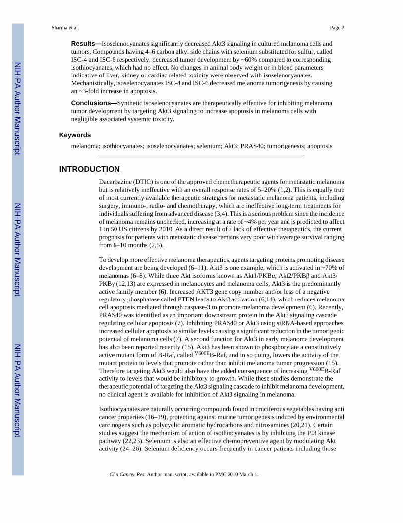
Targeting Akt3 Signaling in Malignant Melanoma Using Isoselenocyanates Arati Sharma 1,5 , Arun K. Sharma 1,5 , SubbaRao V. Madhunapantula 1 , Dhimant Desai 1,5 , Sung Jin Huh 1 , Paul Mosca 5,6 , Shantu Amin 1,5 , and Gavin P. Robertson 1,2,3,4,5 1 Department of Pharmacology, The Pennsylvania State University College of Medicine, Hershey, PA 17033 2 Department of Pathology, The Pennsylvania State University College of Medicine, Hershey, PA 17033 3 Department of Dermatology, The Pennsylvania State University College of Medicine, Hershey, PA 17033 4 The Foreman Foundation for Melanoma Research, The Pennsylvania State University College of Medicine, Hershey, PA 17033 5 Penn State Melanoma Therapeutics Program, The Pennsylvania State University College of Medicine, Hershey, PA 17033 6 Department of Surgery, Lehigh Valley and Health Network, Allentown, PA 18034 Abstract Purpose—Melanoma is the most invasive and deadly form of skin cancer. Few agents are available for treating advanced disease to enable long-term patient survival, which is driving the search for new compounds inhibiting deregulated pathways causing melanoma. Akt3 is an important target in melanomas since its activity is increased in ~70% of tumors, decreasing apoptosis in order to promote tumorigenesis. Experimental Design—Since naturally occurring products can be effective anti-cancer agents, a library was screened to identify Akt3 pathway inhibitors. Isothiocyanates were identified as candidates but low potency requiring high concentrations for therapeutic efficacy made them unsuitable. Therefore, more potent analogs called isoselenocyanates were created using the isothiocyanate backbone but increasing the alkyl chain length and replacing sulfur with selenium. Efficacy was measured on cultured cells and tumors by quantifying proliferation, apoptosis, angiogenesis, toxicity and Akt3 pathway inhibition. Correspondence to: Gavin P. Robertson, Department of Pharmacology – R130, The Pennsylvania State University College of Medicine, 500 University Drive, Hershey, PA 17033. Phone: (717) 531-8098; Fax: (717) 531-5013. ([email protected]). STATEMENT OF TRANSLATIONAL RELEVANCE This manuscript is clinically relevant since melanoma is the most invasive and deadly form of skin cancer with few agents available to treat advanced metastatic disease. Thus, research scientists are vigorously searching for new therapeutic agents targeting pathways important in melanoma to treat the disease. One gene involved in ~70% of sporadic melanomas is AKT3, promoting tumorigenesis by decreasing apoptosis. Here we detail identification of naturally occurring isothiocyanates, present in cruciferous vegetables, as inhibitors of the Akt3 pathway in melanoma. However, low potency requiring high concentrations for therapeutic efficacy made them unsuitable therapeutics. Therefore, more potent analogs have been developed using the isothiocyanate backbone but increasing the alkyl chain length and replacing sulfur with selenium to create compounds called isoselenocyanates. Isoselenocyanates decreased Akt3 signaling in cultured melanoma cells and tumors to significantly reduce melanoma tumor development without changes in animal body weight or in blood parameters indicative of liver, kidney or cardiac related toxicity. NIH Public Access Author Manuscript Clin Cancer Res. Author manuscript; available in PMC 2010 March 1. Published in final edited form as: Clin Cancer Res. 2009 March 1; 15(5): 1674–1685. doi:10.1158/1078-0432.CCR-08-2214. NIH-PA Author Manuscript NIH-PA Author Manuscript NIH-PA Author Manuscript

Welcome message from author
This document is posted to help you gain knowledge. Please leave a comment to let me know what you think about it! Share it to your friends and learn new things together.
Transcript
Targeting Akt3 Signaling in Malignant Melanoma UsingIsoselenocyanates
Arati Sharma1,5, Arun K. Sharma1,5, SubbaRao V. Madhunapantula1, Dhimant Desai1,5,Sung Jin Huh1, Paul Mosca5,6, Shantu Amin1,5, and Gavin P. Robertson1,2,3,4,51 Department of Pharmacology, The Pennsylvania State University College of Medicine, Hershey,PA 170332 Department of Pathology, The Pennsylvania State University College of Medicine, Hershey, PA170333 Department of Dermatology, The Pennsylvania State University College of Medicine, Hershey, PA170334 The Foreman Foundation for Melanoma Research, The Pennsylvania State University College ofMedicine, Hershey, PA 170335 Penn State Melanoma Therapeutics Program, The Pennsylvania State University College ofMedicine, Hershey, PA 170336 Department of Surgery, Lehigh Valley and Health Network, Allentown, PA 18034
AbstractPurpose—Melanoma is the most invasive and deadly form of skin cancer. Few agents are availablefor treating advanced disease to enable long-term patient survival, which is driving the search fornew compounds inhibiting deregulated pathways causing melanoma. Akt3 is an important target inmelanomas since its activity is increased in ~70% of tumors, decreasing apoptosis in order to promotetumorigenesis.
Experimental Design—Since naturally occurring products can be effective anti-cancer agents, alibrary was screened to identify Akt3 pathway inhibitors. Isothiocyanates were identified ascandidates but low potency requiring high concentrations for therapeutic efficacy made themunsuitable. Therefore, more potent analogs called isoselenocyanates were created using theisothiocyanate backbone but increasing the alkyl chain length and replacing sulfur with selenium.Efficacy was measured on cultured cells and tumors by quantifying proliferation, apoptosis,angiogenesis, toxicity and Akt3 pathway inhibition.
Correspondence to: Gavin P. Robertson, Department of Pharmacology – R130, The Pennsylvania State University College of Medicine,500 University Drive, Hershey, PA 17033. Phone: (717) 531-8098; Fax: (717) 531-5013. ([email protected]).STATEMENT OF TRANSLATIONAL RELEVANCEThis manuscript is clinically relevant since melanoma is the most invasive and deadly form of skin cancer with few agents available totreat advanced metastatic disease. Thus, research scientists are vigorously searching for new therapeutic agents targeting pathwaysimportant in melanoma to treat the disease. One gene involved in ~70% of sporadic melanomas is AKT3, promoting tumorigenesis bydecreasing apoptosis. Here we detail identification of naturally occurring isothiocyanates, present in cruciferous vegetables, as inhibitorsof the Akt3 pathway in melanoma. However, low potency requiring high concentrations for therapeutic efficacy made them unsuitabletherapeutics. Therefore, more potent analogs have been developed using the isothiocyanate backbone but increasing the alkyl chain lengthand replacing sulfur with selenium to create compounds called isoselenocyanates. Isoselenocyanates decreased Akt3 signaling in culturedmelanoma cells and tumors to significantly reduce melanoma tumor development without changes in animal body weight or in bloodparameters indicative of liver, kidney or cardiac related toxicity.
NIH Public AccessAuthor ManuscriptClin Cancer Res. Author manuscript; available in PMC 2010 March 1.
Published in final edited form as:Clin Cancer Res. 2009 March 1; 15(5): 1674–1685. doi:10.1158/1078-0432.CCR-08-2214.
NIH
-PA Author Manuscript
NIH
-PA Author Manuscript
NIH
-PA Author Manuscript
Results—Isoselenocyanates significantly decreased Akt3 signaling in cultured melanoma cells andtumors. Compounds having 4–6 carbon alkyl side chains with selenium substituted for sulfur, calledISC-4 and ISC-6 respectively, decreased tumor development by ~60% compared to correspondingisothiocyanates, which had no effect. No changes in animal body weight or in blood parametersindicative of liver, kidney or cardiac related toxicity were observed with isoselenocyanates.Mechanistically, isoselenocyanates ISC-4 and ISC-6 decreased melanoma tumorigenesis by causingan ~3-fold increase in apoptosis.
Conclusions—Synthetic isoselenocyanates are therapeutically effective for inhibiting melanomatumor development by targeting Akt3 signaling to increase apoptosis in melanoma cells withnegligible associated systemic toxicity.
Keywordsmelanoma; isothiocyanates; isoselenocyanates; selenium; Akt3; PRAS40; tumorigenesis; apoptosis
INTRODUCTIONDacarbazine (DTIC) is one of the approved chemotherapeutic agents for metastatic melanomabut is relatively ineffective with an overall response rates of 5–20% (1,2). This is equally trueof most currently available therapeutic strategies for metastatic melanoma patients, includingsurgery, immuno-, radio- and chemotherapy, which are ineffective long-term treatments forindividuals suffering from advanced disease (3,4). This is a serious problem since the incidenceof melanoma remains unchecked, increasing at a rate of ~4% per year and is predicted to affect1 in 50 US citizens by 2010. As a direct result of a lack of effective therapeutics, the currentprognosis for patients with metastatic disease remains very poor with average survival rangingfrom 6–10 months (2,5).
To develop more effective melanoma therapeutics, agents targeting proteins promoting diseasedevelopment are being developed (6–11). Akt3 is one example, which is activated in ~70% ofmelanomas (6–8). While three Akt isoforms known as Akt1/PKBα, Akt2/PKBβ and Akt3/PKBγ (12,13) are expressed in melanocytes and melanoma cells, Akt3 is the predominantlyactive family member (6). Increased AKT3 gene copy number and/or loss of a negativeregulatory phosphatase called PTEN leads to Akt3 activation (6,14), which reduces melanomacell apoptosis mediated through caspase-3 to promote melanoma development (6). Recently,PRAS40 was identified as an important downstream protein in the Akt3 signaling cascaderegulating cellular apoptosis (7). Inhibiting PRAS40 or Akt3 using siRNA-based approachesincreased cellular apoptosis to similar levels causing a significant reduction in the tumorigenicpotential of melanoma cells (7). A second function for Akt3 in early melanoma developmenthas also been reported recently (15). Akt3 has been shown to phosphorylate a constitutivelyactive mutant form of B-Raf, called V600EB-Raf, and in so doing, lowers the activity of themutant protein to levels that promote rather than inhibit melanoma tumor progression (15).Therefore targeting Akt3 would also have the added consequence of increasing V600EB-Rafactivity to levels that would be inhibitory to growth. While these studies demonstrate thetherapeutic potential of targeting the Akt3 signaling cascade to inhibit melanoma development,no clinical agent is available for inhibition of Akt3 signaling in melanoma.
Isothiocyanates are naturally occurring compounds found in cruciferous vegetables having anticancer properties (16–19), protecting against murine tumorigenesis induced by environmentalcarcinogens such as polycyclic aromatic hydrocarbons and nitrosamines (20,21). Certainstudies suggest the mechanism of action of isothiocyanates is by inhibiting the PI3 kinasepathway (22,23). Selenium is also an effective chemopreventive agent by modulating Aktactivity (24–26). Selenium deficiency occurs frequently in cancer patients including those
Sharma et al. Page 2
Clin Cancer Res. Author manuscript; available in PMC 2010 March 1.
NIH
-PA Author Manuscript
NIH
-PA Author Manuscript
NIH
-PA Author Manuscript
diagnosed with metastatic melanoma (27). Recently, selenium has been shown to inducedestabilization of Akt activity in prostate cancer cells (28,29). Therefore, incorporatingselenium into the structure of compounds could increase compound efficacy and thesecompounds would be safe since melanoma patients frequently have selenium deficiency.
In this study, isothiocyanate analogs having a longer carbon chain lengths and seleniumsubstituted for sulfur have been developed and therapeutic efficacy for killing culturedmelanoma cells or inhibiting tumor development in animals evaluated. While increasing chainlength did not increase tumor inhibitory potency of sulfur containing isothiocyanates,incorporation of selenium with increasing chain length significantly enhanced anti-tumorpotency by elevating rates of tumor cell apoptosis. Thus, novel isoselenocyanates have beendeveloped that target Akt3 signaling in melanoma cells leading to robust anti-melanomaactivity.
MATERIALS AND METHODSCell lines and culture conditions
The human fibroblast cells (FF2441) and metastatic melanoma cell lines UACC 903 and 1205Lu were maintained in DMEM (Invitrogen, Carlsbad, CA), supplemented with 10% FBS(Hyclone, Logan, UT) at 37°C with 5% CO2 atmosphere in a humidified incubator. Verticalgrowth phase (VGP) melanoma cell line WM115 was maintained in Tu2% medium asdescribed previously (6).
Western blot analysisFor Western blot analysis, floating and adherent cells treated with compounds or control vehicle(DMSO) were harvested by addition of lyses buffer containing 50 mM HEPES (pH 7.5), 150mM NaCl, 10 mM EDTA, 10% glycerol, 1% Triton X-100, 1 mM sodium orthovanadate,0.1mM sodium molybdate, 1mM phenylmethylsulfonyl fluoride, 20 μg/ml aprotinin, and 5μg/ml leupeptin. Whole cell lysates were centrifuged (≥10,000 X g) for 10 min at 4°C to removecell debris. Protein concentrations were quantitated using the BCA assay from Pierce(Rockford, IL), and 30 μg of lysate loaded per lane onto NuPAGE Gels from Life Technologies(Carlsbad, CA). Following electrophoresis, samples were transferred to a polyvinylidenedifluoride membrane (Pall Corporation, Pensacola, FL). Blots were probed with antibodiesaccording to each supplier’s recommendations: phosphorylated-PRAS40 (Thr246) fromInvitrogen (Carlsbad, CA); Erk2, α-enolase and secondary antibodies conjugated withhorseradish peroxidase from Santa Cruz Biotechnology (Santa Cruz, CA); and Immunoblotswere developed using the enhanced chemiluminescence detection system (PierceBiotechnology, Rockford, IL).
Synthesis of isothiocyanates (ITC), isoselenocyanates (ISC) and phenylhexyl selenocyanate(PHSC)
BITC and PEITC were purchased from Sigma-Aldrich (St. Louis, MO). PBITC and PHITCwere synthesized by using previously reported methodology (30). Isoselenocyanates weresynthesized using a described methoda. Briefly, a solution of triphosgene (5.0 mmol) inCH2Cl2 (15 mL) was added over a refluxing mixture of formamides (10.0 mmol), triethylamine(43.0 mmol) and 4 Å molecular sieves in CH2Cl2 (35 mL). Mixture was then refluxed for anadditional 2.5 h. Selenium powder (20 mmol) was then added and resulting mixture refluxedfor 6–8 h. Mixture was cooled, filtered, and solvent evaporated yielding a crude mixture, whichwas purified by silica gel column chromatography generating pure isoselenocyanates. 6-
aSharma AK, Sharma A, Desai D, Madhunapantula SV, Huh SJ, Robertson GP, Amin S. Synthesis and anticancer activity comparisonof isoselenocyanates with isothiocyanates present in cruciferous vegetables (submitted)
Sharma et al. Page 3
Clin Cancer Res. Author manuscript; available in PMC 2010 March 1.
NIH
-PA Author Manuscript
NIH
-PA Author Manuscript
NIH
-PA Author Manuscript
phenylhexylselenocyanate (PHSC) was prepared by reacting 0.3 g (1.25 mmol) of 6-phenylhexylbromide with 0.19 g (1.32 mmol) of KSeCN in 10 mL of acetonitrile, in a nitrogenatmosphere. After stirring overnight at room temperature, the residue was partitioned betweenEtOAc and water. The organic phase was separated, washed with brine and water, dried overMgSO4, filtered and the solvent evaporated to yield 0.26 g (74% yield). Compounds identitieswere confirmed by NMR as well as Mass Spectra analysis and purity (>99%) quantified byHPLC analysis.
SiRNA protein knockdown studiesDuplexed “Stealth” siRNA from Invitrogen (Carlsbad, CA) were: AKT3- 5′-GGA CUA UCUACA UUC CGG AAA GAU U-3′ and scrambled- 5′-AAU UCU CCG AAC GUG UCA CGUGAG A-3′. Nucleofection using Amaxa Nucleofector (Koeln, Germany) was used to introducesiRNA into UACC 903 cells (Reagent R, program K17). SiRNA (100 pmoles) against Akt3or scrambled siRNA or buffer were nucleofected into 1X10⁶ UACC 903 cells, which werethen replated in DMEM supplemented with 10% FBS and allowed to recover for 1.5 days.Transfection efficiency was >90% with ~80% viability. For animal experiments, thirty-sixhours later, 1X10⁶ viable UACC 903 cells in 0.2 ml of DMEM supplemented with 10% FBSwere injected subcutaneously into the left and right flanks of 3-to-4 wk old female AthymicNude-Foxn1nu mice. Dimensions of developing tumors were measured on alternate days usingcalipers up to day 17.5. To test duration of siRNA-mediated knockdown, protein lysates werecollected at 2, 4, 6, and 8 d following nucleofection and measured for Akt3 protein expressionby Western blot analysis and quantitated by densitometry as described previously (6,14).
Cell viability, proliferation, apoptosis determination and cell cycle analysisViability and IC50 of melanoma cells following treatment with compounds was measured usingthe 3-(4,5-dimethylthiazol-2-yl)-5-(3-carboxymethoxyphenyl)-2-(4-sulfophenyl)-2H-tetrazolium (MTS) assay (Promega, Madison, WI). Briefly, 5 X 10³ melanoma (UACC 903,1205 Lu or WM115) or human fibroblast (FF2441) cells per well in 100 μL DMEM containing10% FBS were grown in a 96-well plate for 24 or 76 h and treated with either control DMSOvehicle or increasing concentrations (2–100 μM) of compounds for 24 h. Cellular viabilitycompared to vehicle control treated cells was measured using the MTS assay. IC50 values foreach compound in respective cell lines was determined from three independent experimentsusing GraphPad Prism version 4.01 (GraphPad software, San Diego, CA).
Cellular proliferation and apoptosis rates were measured by seeding 5 X 10³ cells/well in 96-well plates, followed by treatment for 24 h with each respective agent. Proliferation andapoptosis were measured using a BrdUrd ELISA kit (Roche Applied Sciences, Indianapolis,IN) or Apo-ONE Homogenous caspase-3/7 Assay kit (Promega Corporation, Madison, WI),respectively.
Cell cycle analysis was undertaken by plating 1.5 X 10⁶ melanoma cells in 100-mm culturedish and following treatment with respective compounds for 24 h, total cells (floating andadherent) were trypsinized, centrifuged (500 X g, for 5 min) and treated with 1 mL of propidiumiodide staining solution containing 100 μg/mL PI; Sigma, St Louis, MO), 20 μg/mLRibonuclease A (Roche Applied Sciences, Indianapolis, IN) 3 μg/mL Triton X-100 dissolvedin 0.1% (w/v sodium citrate for 30 m at 4°C (31). Cells were analyzed using the FACScananalyzer (Becton Dickinson, San Jose, CA) and data processed using ModFit LT software(Verity software house, Topsham, ME).
Sharma et al. Page 4
Clin Cancer Res. Author manuscript; available in PMC 2010 March 1.
NIH
-PA Author Manuscript
NIH
-PA Author Manuscript
NIH
-PA Author Manuscript
Tumorigenicity assessment, knockdown of protein expression and measurement ofproliferation/apoptosis rates in tumors
Animal experimentation was performed according to protocols approved by the InstitutionalAnimal Care and Use Committee at The Pennsylvania State University College of Medicine.Tumor kinetics were measured by subcutaneous injection of 2.5–5X10⁶ 1205 Lu or UACC903 melanoma cells in 0.2 ml of DMEM supplemented with 10% FBS above both left andright rib cages of 3-to-4 wk old female Athymic Nude-Foxn1nu mice (Harlan Sprague Dawley,Indianapolis, IN). Six days later when a fully vascularized tumor (50–75 mm3) had formed,mice were randomly divided in to DMSO vehicle control and experimental (BITC, PEITC,PBITC, PHITC, ISC-1, ISC-2, ISC-4 or ISC-6) groups (5 mice/group; 2 tumors/mouse) andtreated i. p. with isothiocyanate or isoselenocyanate compounds (2.5 or 0.76 μmoles (equivalentto 3 ppm selenium) / 20g mice in 50 μL DMSO vehicle) on Monday, Wednesday and Fridayfor ~ 3 weeks. Control mice received an equivalent volume of the vehicle. Body weight (grams)and dimensions of the developing tumors (mm3) were measured at the time of treatment.
To ascertain mechanism underlying tumor inhibition, 5X10⁶ UACC 903 cells were injectedinto nude mice, 6-days later mice were treated i. p. with PBITC or PHITC (0.76 μmoles), ISC-4or ISC-6 (0.76 μmoles, equivalent to 3 ppm selenium) on alternate days. Size and time matchedtumors were harvested at days 11 and 13 to assess changes in cell proliferation and apoptosis.A small portion of the tumor was also flash frozen in liquid nitrogen, pulverized and lysed inprotein lysis buffer (600–800 μl, 50 mM Tris-HCl, pH 7.5 containing 0.1% Triton X-100, 1mM EDTA, 1 mM EGTA, 50 mM sodium fluoride, 10 mM sodium β-glycerol phosphate, 5mM sodium pyrophosphate, 1 mM activated sodium orthovanadate, protease inhibitor cocktailfrom Sigma and 0.1% (v/v) 2-mercaptoethanol). Protein concentration was determined usingBio-Rad protein assay reagent (Bio-Rad laboratories, Hercules, CA), analyzed by Westernblotting to measure levels of pAkt and downstream pPRAS40 in tumors and quantitated bydensitometry as described previously (6,14).
Cell proliferation and apoptosis were measured in formalin-fixed, paraffin-embedded tumorsections using the TUNEL TMR Red Apoptosis kit from Roche (Manheim, Germany) orpurified mouse anti-human Ki-67 from PharMingen (San Diego, CA), respectively. Aminimum of 6 different tumors with 4–6 fields per tumor was analyzed and results representedas the average ± S.E.M.
Toxicity assessments4-to-6 wks old female nude mice (Harlan Sprague Dawley, Indianapolis, IN) were injected i.p. with either control DMSO vehicle, PBITC or PHITC (0.76 μmoles) or ISC-4 or ISC-6 (0.76μmoles equivalent to 3 ppm Se) on Monday, Wednesday and Friday for 3 weeks. Animals weresacrificed by CO2 asphyxiation and blood collected from each animal in plasma separator tubeswith lithium heparin (BD, Franklin Lakes, NJ) following cardiac puncture and analyzed forSGOT (serum glutamic oxaloacetic transaminase), SGPT (serum glutamate pyruvatetransaminase), alkaline phosphatase, glucose and creatinine to ascertain liver, heart, kidneyand pancreas related toxicity. For morphological examination of blood cells, whole blood wascollected in microtainer tubes containing K2EDTA (BD, Franklin Lakes, NJ) and RBC, WBC,lymphocytes, monocytes, eosinophils, platelets, total hemoglobin and hematocrit percentageanalyzed. Blood was also microscopically examined for segregates, polychromatin bodies, andsmudge cells. A portion of liver, heart, kidney, spleen, intestine, pancreas and adrenal fromeach animal was formalin fixed and paraffin-embedded to examine toxicity-related changes incell or organ morphology by H&E staining.
Sharma et al. Page 5
Clin Cancer Res. Author manuscript; available in PMC 2010 March 1.
NIH
-PA Author Manuscript
NIH
-PA Author Manuscript
NIH
-PA Author Manuscript
Statistical analysisStatistical analysis was carried out using Prism 4.0 (GraphPad Software). One-way or Two-way Analysis Of Variance (ANOVA) was used for groupwise comparisons, followed by theTukey’s or Bonferroni’s post hoc tests. All the data represented as ± S.E.M. Results wereconsidered significant at a P value less than 0.05 (95% CI).
RESULTSsiRNA-mediated inhibition of Akt3 signaling reduced the tumorigenic potential of melanomacells
To confirm prior studies documenting the therapeutic potential of inhibiting Akt3 signaling inmelanoma tumorigenesis, a siRNA-based approach was initially used to inhibit proteinexpression and thereby activity (6,7,15). UACC 903 cells were nucleofected with siRNAtargeting Akt3, or a scrambled siRNA or a buffer control using the Amaxa nucleofectionsystem. 36 h later, viable cells were subcutaneously injected in to nude mice and tumordevelopment measured at every other day. Decreased expression (activity) of Akt3 reducedthe tumorigenic potential of melanoma cells by ~60% (One Way ANOVA; P< 0.001) comparedto control cells nucleofected with scrambled siRNA or nucleofection buffer (Fig. 1A). A tumorremoved from animals at day 9.5 showed significantly less Akt3 protein than control(scrambled siRNA) tumors demonstrating effective knockdown of Akt3 protein expression(Fig. 1B). Duration of Akt3 protein knockdown following exposure to siRNA targeting Akt3persisted up to 8-d in culture (Fig. 1C) as reported previously (6). Thus, targeting Akt3 signalingled to significant melanoma tumor inhibition, which has laid the foundation to search forpharmacological agents that could inhibit melanoma development by reducing the activity ofthis important signaling cascade involved in ~70% of sporadic melanomas (6).
Development of isothiocyanate analogs with longer alkyl chain lengths and seleniumsubstituted for sulfur
In order to identify a pharmacological agent inhibiting Akt3 activity and melanoma cellsurvival, a natural product library was screened and isothiocyanates identified as possiblecandidates. However, the parent isothiocyanate compound had low potency in vivo requiringhigh concentrations for efficacy. Therefore, more potent analogs were created that could serveas therapeutic agents using the isothiocyanate backbone. The goal was to identify optimalcarbon chain length for maximal tumor inhibition by comparing arylalkyl isothiocyanatecompounds with increasing alkyl chain length and by replacing sulfur with selenium. Figure2A shows the structures of isothiocyanates containing 1(benzyl), 2 (phenethyl), 4 (phenylbutyl)and 6 (phenylhexyl) carbon spacers. Corresponding isosteric selenium analogs are also shownin which sulfur was replaced with selenium. Finally, a 6 carbon selenocyanate, calledphenylhexyl selenocyanate (PHSC), was created to serve as a control to show that seleniumalone was not accounting for inhibition but rather that the structure of the compound containingselenium was critical for tumor reduction (Fig. 2A).
Isoselenocyanates are more effective at inhibiting cultured melanoma cells thanisothiocyanates
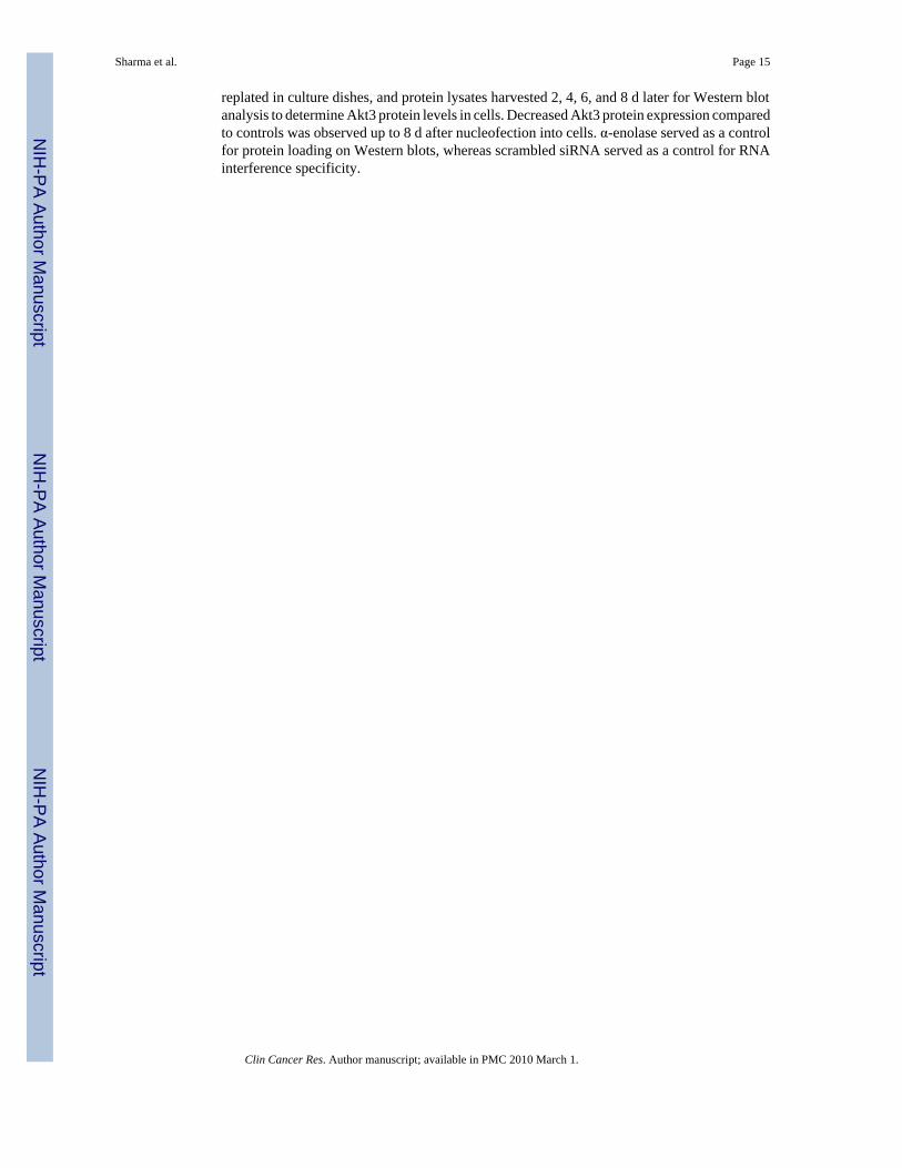
Initially, the MTS assay was used to quantify viable cells of three human melanoma cell lines(UACC 903, 1205 Lu and WM115) following treatment with increasing concentrations of eachagent to measure the IC50 of respective compounds. Figure 2B shows a representative exampleof this analysis where inhibitory effectiveness of the 4 carbon PBITC and ISC-4 as well as 6carbon PHITC and ISC-6 were compared to DMSO vehicle, Akt inhibitor API-2 (1,5-Dihydro-5-methyl-1-b-D-ribofuranosyl-1,4,5,6,8-penta azaacenaphthylen-3-amine) (7, 32) orcontrol PHSC. Selenium containing ISC-4 and ISC-6 were more effective at inhibiting growth
Sharma et al. Page 6
Clin Cancer Res. Author manuscript; available in PMC 2010 March 1.
NIH
-PA Author Manuscript
NIH
-PA Author Manuscript
NIH
-PA Author Manuscript
of melanoma cells than sulfur containing PBITC, PHITC, control PHSC or API-2. Figure 2Cshows a detailed comparison where the IC50 of 1, 2, 4 or 6 carbon isothiocyanates are comparedto selenium containing analogs in three independently derived melanoma cell lines, UACC903, 1205 Lu and WM115. A general trend was observed in which increasing carbon chainlength and substitution of selenium for sulfur decreased the IC50 for all cell lines but thedifferences were subtle. Increased potency ranged from 30–70% with increasing chain lengthand/or sulfur substituted for selenium. Thus, isothiocyanate analogs with longer alkyl chainlengths and sulfur substituted for selenium had increased killing efficiency for culturedmelanoma cells.
Isoselenocyanates inhibits melanoma cell growth more effectively than normal cellsSensitivity of melanoma and normal cells to PBITC or ISC-4 was compared to determinewhether cancer cells were more sensitive to the compounds. Normal human fibroblast, FF2441,and melanoma (UACC 903) cells were treated with 2–100 μM of PBITC or ISC-4 and IC50measured at 12 and 24 h (Fig. 2D). Consistently, 2–4-fold higher drug concentrations wererequired to kill fibroblasts compared to melanoma cells (Fig. 2D). Thus, cultured cancer cellsare more sensitive to PBITC or ISC-4 than normal cells.
Isoselenocyanates have increased in vivo potency compared to correspondingisothiocyanates and effectively reduce melanoma development
Effectiveness of isoselenocyanates for inhibiting the growth of pre-existing tumors wasevaluated in nude mice. UACC 903 melanoma cells having high Akt3 signaling activity wereinjected subcutaneously and 6 days later when a vascularized tumor had developed, mice wereinjected intraperitoneally with 2.5 μmoles of each isothiocyanate or 0.76 μmoles ofisoselenocyanate (Fig. 3A). While 3-fold less isoselenocyanate was administered, ~50% tumorinhibition was observed at day 24, which indicates enhanced tumor inhibitory effectiveness ofselenium containing analogs. Increasing carbon chain length of isothiocyanates seemed to beless effective at tumor inhibition (Fig. 3A; left panel). In contrast, increasing carbon chainlength of isoselenocyanates was associated with more effective tumor inhibition (P<0.001,One-way ANOVA, ISC-1 vs. ISC-2, ISC-4, ISC-6) (Fig. 3A; right panel). Thus, PBITC andPHITC reduced tumor development but at concentrations 3-fold higher than correspondingisoselenocyanates. Therefore, 4–6 carbon chain isoselenocyanates appeared to be the mostrobust inhibitors of melanoma tumorigenesis. Based on these findings subsequent studiesfocused on comparing ISC-4 and ISC-6 to PBITC and PHITC, respectively.
UACC 903 and 1205 Lu melanoma cells having high Akt3 signaling activity were injectedsubcutaneously and following 6 days when tumor angiogenesis had occurred, mice wereexposed to 0.76 μmoles representative isothiocyanate compound PBITC versus ISC-4 orPHITC versus ISC-6, 3 times per week and tumor development measured (Figs. 3B & 3C).Animals were also weighed to ascertain possible toxicity. While PBITC and PHITC areineffective at reducing tumor burden of UACC 903 (Fig. 3B) or 1205 Lu (Fig. 3C) at thisconcentration, ISC-4 and ISC-6 led to significant (P<0.001; Two-way ANOVA) reductionsin tumor size beginning from day 13 for UACC 903 cells or from day 10 for 1205 Lu cells.Thus, isoselenocyanates are effective at reducing melanoma tumor development by 50–60%at significantly lower concentrations than corresponding isothiocyanates, which is similar tosiRNA mediated inhibition of Akt3 (Fig. 1A).
Synthetic isoselenocyanate compounds causes negligible organ related toxicity followingsystemic administration
Systemic toxicity of PBITC, PHITC, ISC-4 or ISC-6 administration was evaluated in nudemice. Body weights of mice treated with isothiocyanate or isoselenocyanate compoundscompared to control DMSO vehicle showed no significant differences between groups (Fig.
Sharma et al. Page 7
Clin Cancer Res. Author manuscript; available in PMC 2010 March 1.
NIH
-PA Author Manuscript
NIH
-PA Author Manuscript
NIH
-PA Author Manuscript
4A). Furthermore, blood parameters (SGOT, SGPT, alkaline phosphatase, blood urea, glucoseand creatinine) indicative of systemic toxicity did not detect significant liver, kidney or cardiacrelated toxicity (Fig. 4B). Levels of cellular metabolites basal urea nitrogen (BUN), creatinineand glucose in animals were also not significantly different between ISC-4 or PBITC treatedand control animals. Histological examination of hematoxylin and eosin stained vital organsections, including the liver (Fig. 4C), revealed that ISC-4 treatment did not significantly altercell morphology or structure of kidney, adrenal, lung, spleen, heart, pancreatic or intestinaltissue (data not shown). Similar results were observed following treatment with ISC-6 inanimals (data not shown). Thus, treatment using synthetic selenium containing analogs ofisothiocyanate ISC-4 or ISC-6 led to negligible associated systemic toxicity with significanttherapeutic potential.
Isoselenocyanates decreased Akt3 signaling in cultured melanoma cells and tumorsCells were next treated with isoselenocyanates ISC-4 and ISC-6 and effect on Akt3 signalingexamined by Western blotting. Both compounds inhibited Akt3 signaling as demonstratedthrough decreased pAkt and downstream pPRAS40 levels (Fig. 5A). However, ISC-4 andISC-6 were effective at lower concentrations, completely inhibiting the pathway at ~10 μMcompared to corresponding isothiocyanates requiring ≥15 μM for similar inhibition (Fig. 5A).As reported previously, Akt3 pathway inhibition led to apoptosis, which was indicated by highlevels of cleaved PARP (Fig. 5A) (7). Higher cleaved PARP levels were observed at lowerconcentrations of ISC-4 than corresponding isothiocyanate PBITC suggestingisoselenocyanate compounds were more effective than corresponding sulfur containingisothiocyanates. Similar Akt pathway inhibition by ISC-4 or ISC-6 (not shown) also occurredfor human melanoma cell lines 1205 Lu and WM115 (Fig. 5B).
Western blot analysis of size and time matched tumors harvested at day 13 from animals treatedwith DMSO, PBIT or ISC-4 also showed significantly decreased phosphorylated (active) Akt(P<0.05; One-way ANOVA) and downstream PRAS40 (P<0.001; One-way ANOVA) inISC-4 tumor lysate’s compared to DMSO control or PBITC treated tumors (Fig. 5C). Thus,isoselenocyanates ISC-4 and ISC-6 were more robust inhibitors of the Akt3 signaling cascadein cultured melanoma cells as well as in xenografted melanoma tumors than correspondingsulfur containing isothiocyanates.
Isoselenocyanates induced apoptosis in cultured melanoma cells as well as in xenograftedmelanoma tumors
To identify the underlying mechanism by which isothiocyanates or isoselenocyanates inhibitedmelanoma cell survival, rates of apoptosis and proliferation were examined followingtreatment. In contrast to API-2 or PHSC, increasing concentrations of PBITC, ISC-4, PHITCor ISC-6 led to increased cellular apoptosis (Fig. 6A) and decreased proliferative potential (Fig.6B) of UACC 903 melanoma cells. ISC-4 or ISC-6 was ~2-fold more effective than PBITC orPHITC at inducing apoptosis and inhibiting cellular proliferation (Figs. 6A & 6B). Cell cycleanalysis of asynchronously growing UACC 903 cells showed a significant increase in the sub-G0-G1 population in PBITC or ISC-4 and PHITC or ISC-6 treated cells compared to controls(Fig. 6C), which is indicative of cellular apoptosis. Analysis of cells in each stage of the cellcycle, in Supplementary Table, showed a 30–40% decrease in the G0-G1 phase cells with a50–60% increase in the G2-M phase cell population. Marginal changes were observed in S-phase cells. The most significant change was an ~15-fold increase in the sub-G0-G1 cellpopulation indicating a dramatic increase in cellular apoptosis.
To confirm that a similar mechanism led to tumor inhibition in animals following ISC-4treatment, rates of apoptosis (TUNEL staining) and proliferation (Ki-67immunohistochemistry) were compared in size and time matched melanoma tumors from
Sharma et al. Page 8
Clin Cancer Res. Author manuscript; available in PMC 2010 March 1.
NIH
-PA Author Manuscript
NIH
-PA Author Manuscript
NIH
-PA Author Manuscript
ISC-4 or PBITC treated animals and compared to DMSO vehicle. Tumors harvested at day 11and 13 from mice treated with ISC-4, showed ~3-fold (Fig. 6D, right panel, P< 0.01; One-wayANOVA) more TUNEL positive cells compared to control animals treated with DMSO orPBITC. Slightly fewer proliferating tumor cells were observed in ISC-4 treated tumorscompared to PBITC, but this difference was not statistically significant (Fig. 6D, left panel,P> 0.05; One-way ANOVA). Thus, the superior anti-melanoma activity of ISC-4 relative toPBITC appears primarily to be due to an effect on tumor cell apoptosis rather than on cellularproliferation, which is consistent with effects observed following treatment of cultured cells(Fig. 5A & Supplementary Table).
DISCUSSIONFor several decades, no substantial progress has been made in developing drugs effective forthe long-term survival of patient’s with advanced-stage melanoma (33). Current systemictherapies for metastatic disease still achieve only a modest ~20% overall response rates, andduration of efficacy is typically months and not years (1,33). The median progression-freesurvival following initiation of systemic therapy for stage IV melanoma is typically about 1.7months, and the median survival is 6.2 months (34). Clearly, a pipeline of novel, more effectivetherapeutics is needed to increase the long-term survival of metastatic melanoma patients,which is the goal of this report.
Targeted agents that inhibit the activity of aberrant melanoma causing genes have potential tosignificantly increase patient survival (1,9,11). Agents of this type have been shown to beeffective, for example: imatinib targeting receptor-type KIT tyrosine kinase and BCR-ABLtyrosine kinase in chronic myelogenous leukemia and gastrointestinal stromal tumors;bevacizumab targeting vascular endothelial growth factor (VEGF) in colorectal cancer;sunitinib targeting VEGF receptors, FMS-like tyrosine kinase 3, c-KIT & platelet-derivedgrowth factor in renal cell carcinoma; and sorafenib targeting Raf kinases in renal cell as wellas hepatocellular carcinomas (35–39). Thus, it is reasonable to assume that agents could bedeveloped to inhibit proteins deregulated during melanoma development that would be moreeffective than currently available drugs for treating this disease.
The Akt3 pathway is an important pathway deregulated in ~70% of melanomas, important totherapeutically target alone or in combination with other targeted agents (6–8). A second keypathway is that of the MAP kinase signaling cascade, which is constitutively activated throughRas mutations in 10–15% and B-Raf mutations in ~60% of melanomas (40,41). A T to Amutation at nucleotide 1799 of B-Raf, leads to substitution of a valine for a glutamic acid atcodon 600 (V600E) in exon 15 in the vast majority of melanomas in which B-Raf is mutated(41). This alteration is acquired during development of sporadic melanomas and not inherited(41). Since B-Raf is the most mutated gene in melanomas, it is an attractive therapeutic target(40). Sorafenib, which was identified as a Raf kinase inhibitor, was initially hoped to beeffective for treating melanoma (10,40). However, off-target effects have limited its efficacyfor treating melanoma. Sorafenib inhibits Raf, but it also decreases activity of VEGFR1,VEGFR2, VEGFR3, PDGFR Beta, Flt-3, p38, c-Kit and FGFR1 (42). Therefore, while ittargets constitutively active mutant V600EB-Raf present in ~60% of metastatic melanomas, itis ineffective because its primary mechanism of action is as an angiogenesis inhibitor and notas a regulator of cellular proliferation, which is required for effective melanoma inhibitionwhen targeting this aberrant signaling cascade (9,10,43). More specific B-Raf inhibitors arebeing developed and evaluated to circumvent these limitations but clinical efficacy is currentlyunknown (11).
Inhibitors of the Akt3 pathway were developed in this report by initially screening a naturalproduct library for candidate anti-neoplastic compounds that inhibited this signaling cascade.
Sharma et al. Page 9
Clin Cancer Res. Author manuscript; available in PMC 2010 March 1.
NIH
-PA Author Manuscript
NIH
-PA Author Manuscript
NIH
-PA Author Manuscript
The screen was based on reports showing that targeting Akt3 signaling significantly reducedthe tumorigenic potential of melanoma cells (6,7,15). Naturally occurring isothiocyanates wereidentified as potential inhibitors of Akt3 signaling. Numerous naturally occurring compoundsexhibiting anti-neoplastic properties are being exploited as potential chemotherapeutic agents(44,45). Some are well established components of standard systemic chemotherapeuticregimens, such as the taxanes, vinca alkaloids, and camptothecins (44,46,47). However, nosuccessful agent or combination of agents has been identified that dramatically extendsmelanoma patient survival (33). Thus, naturally occurring agents targeting key signalingpathways, such as Akt3, could be important breakthroughs for more effective melanomatherapies.
While naturally occurring isothiocyanates were found to inhibit Akt3 signaling, impracticalquantities were required for melanoma anti-tumor activity. Therefore, replacing the sulfurgroup with selenium and lengthening the carbon chains was evaluated to enhance potency astherapeutic agents. The resulting family of compounds, called isoselenocyanates, had greaterefficacy killing cultured cells as well as inhibiting tumor development in animals compared tosulfur containing isothiocyanates. Two isoselenocyanates, ISC-4 and ISC-6, had particularlyrobust anti-melanoma activity with enhanced potency due primarily to enhanced tumor cellapoptosis following treatment. Thus, isoselenocyantes represents a significant development inthe natural product drug pipeline by targeting a key signaling cascade deregulated in ~70% ofmelanomas.
Incorporating selenium into the structure of isoselenocyanates is a further significantdevelopment. Selenium plays a role in cancer chemoprevention but the exact mechanistic basisfor inhibition remains to be identified (28,29,48,49). Several clinical trials are examining therole of selenium in the prevention of colorectal cancer (NCT00078897), breast cancer(NCT00555386), lung cancer (NCT00008385), and bladder cancer (NCT00553345).Furthermore, incorporating selenium into the structure of drugs, as in the case ofisoselenocyanates, can increased compound potency making ineffective agents bettertherapeutics. Recently, a selenium containing analog of the iNOS inhibitor PBIT, called PBISe,which is ineffective at killing melanoma cells was made >10-fold more potent by incorporatingselenium into its structure to more effectively decrease melanoma tumor development inanimals (50). Thus, incorporating selenium into the structure of cancer therapeutics is onefeasible approach to increase the tumor inhibitory efficacy of therapeutic agents.
Since control compounds containing selenium had little effect on melanoma cell survival,isoselenocyanate-mediated inhibition of melanoma is independent of selenium, suggesting thatthe structure of the compound in combination with selenium is necessary for enhancedinhibitory activity. Compared to the chemopreventive effects of natural selenium enrichedproducts, isoselenocyanates are unique in that the selenium containing compounds target Akt3signaling in melanoma to promote apoptosis, block the growth of tumors, and are associatedwith negligible toxicity at biologically effective doses. Therefore, isoselenocyanates representa promising adjunct to rational, targeted, single- or perhaps multi-agent therapy for advancedmelanoma.
Supplementary MaterialRefer to Web version on PubMed Central for supplementary material.
AcknowledgmentsGrant support: This work was supported by The American Cancer Society [RSG-04- 053-01-GMC to G.P.R.];National Institutes of Health [CA-127892-01A to G.P.R.]; National Institute of Health and National Cancer Institute
Sharma et al. Page 10
Clin Cancer Res. Author manuscript; available in PMC 2010 March 1.
NIH
-PA Author Manuscript
NIH
-PA Author Manuscript
NIH
-PA Author Manuscript
contract [NO2-CB-56603 to S.A], The Foreman Foundation for Melanoma Research to G.P.R.; Elsa U. PardeeFoundation to A.S.; and Melanoma Research Foundation to A.S.
We thank Dr. Raghvendra Gowda for providing technical assistance.
References1. Katipamula R, Markovic SN. Emerging therapies for melanoma. Expert Rev Anticancer Ther
2008;8:553–60. [PubMed: 18402522]2. McDermott DF, Sosman JA, Gonzalez R, et al. Double-Blind Randomized Phase II Study of the
Combination of Sorafenib and Dacarbazine in Patients With Advanced Melanoma: A Report Fromthe 11715 Study Group. J Clin Oncol 2008;26:2178–85. [PubMed: 18445842]
3. Helmbach H, Rossmann E, Kern MA, Schadendorf D. Drug-resistance in human melanoma.International Journal of Cancer 2001;93:617–22.
4. Markovic SN, Erickson LA, Rao RD, et al. Malignant melanoma in the 21st century, part 1:epidemiology, risk factors, screening, prevention, and diagnosis. Mayo Clin Proc 2007;82:364–80.[PubMed: 17352373]
5. Gray-Schopfer V, Wellbrock C, Marais R. Melanoma biology and new targeted therapy. Nature2007;445:851–7. [PubMed: 17314971]
6. Stahl JM, Sharma A, Cheung M, et al. Deregulated Akt3 activity promotes development of malignantmelanoma. Cancer Res 2004;64:7002–10. [PubMed: 15466193]
7. Madhunapantula SV, Sharma A, Robertson GP. PRAS40 deregulates apoptosis in malignantmelanoma. Cancer Res 2007;67:3626–36. [PubMed: 17440074]
8. Robertson GP. Functional and therapeutic significance of Akt deregulation in malignant melanoma.Cancer Metastasis Rev 2005;24:273–85. [PubMed: 15986137]
9. Sharma A, Tran MA, Liang S, et al. Targeting mitogen-activated protein kinase/extracellular signal-regulated kinase kinase in the mutant (V600E) B-Raf signaling cascade effectively inhibits melanomalung metastases. Cancer Res 2006;66:8200–9. [PubMed: 16912199]
10. Sharma A, Trivedi NR, Zimmerman MA, Tuveson DA, Smith CD, Robertson GP. Mutant V599EB-Raf regulates growth and vascular development of malignant melanoma tumors. Cancer Res2005;65:2412–21. [PubMed: 15781657]
11. Tsai J, Lee JT, Wang W, et al. Discovery of a selective inhibitor of oncogenic B-Raf kinase withpotent antimelanoma activity. Proc Natl Acad Sci U S A 2008;105:3041–6. [PubMed: 18287029]
12. Brazil DP, Hemmings BA. Ten years of protein kinase B signalling: a hard Akt to follow. TrendsBiochem Sci 2001;26:657–64. [PubMed: 11701324]
13. Nicholson KM, Anderson NG. The protein kinase B/Akt signalling pathway in human malignancy.Cell Signal 2002;14:381–95. [PubMed: 11882383]
14. Stahl JM, Cheung M, Sharma A, Trivedi NR, Shanmugam S, Robertson GP. Loss of PTEN promotestumor development in malignant melanoma. Cancer Res 2003;63:2881–90. [PubMed: 12782594]
15. Cheung M, Sharma A, Madhunapantula SV, Robertson GP. Akt3 and mutant V600E B-Raf cooperateto promote early melanoma development. Cancer Res 2008;68:3429–39. [PubMed: 18451171]
16. Zhang Y, Kensler TW, Cho CG, Posner GH, Talalay P. Anticarcinogenic activities of sulforaphaneand structurally related synthetic norbornyl isothiocyanates. Proc Natl Acad Sci U S A 1994;91:3147–50. [PubMed: 8159717]
17. Hecht SS. Chemoprevention by isothiocyanates. J Cell Biochem Suppl 1995;22:195–209. [PubMed:8538199]
18. Zhang Y, Yao S, Li J. Vegetable-derived isothiocyanates: anti-proliferative activity and mechanismof action. Proc Nutr Soc 2006;65:68–75. [PubMed: 16441946]
19. El-Bayoumy K, Sinha R, Pinto JT, Rivlin RS. Cancer chemoprevention by garlic and garlic-containingsulfur and selenium compounds. J Nutr 2006;136:864S–9S. [PubMed: 16484582]
20. Miyoshi N, Uchida K, Osawa T, Nakamura Y. A link between benzyl isothiocyanate-induced cellcycle arrest and apoptosis: involvement of mitogen-activated protein kinases in the Bcl-2phosphorylation. Cancer Res 2004;64:2134–42. [PubMed: 15026354]
Sharma et al. Page 11
Clin Cancer Res. Author manuscript; available in PMC 2010 March 1.
NIH
-PA Author Manuscript
NIH
-PA Author Manuscript
NIH
-PA Author Manuscript
21. Chiao JW, Wu H, Ramaswamy G, et al. Ingestion of an isothiocyanate metabolite from cruciferousvegetables inhibits growth of human prostate cancer cell xenografts by apoptosis and cell cycle arrest.Carcinogenesis 2004;25:1403–8. [PubMed: 15016658]
22. Keum YS, Jeong WS, Kong AN. Chemoprevention by isothiocyanates and their underlying molecularsignaling mechanisms. Mutat Res 2004;555:191–202. [PubMed: 15476860]
23. Zhang Y. Cancer-preventive isothiocyanates: measurement of human exposure and mechanism ofaction. Mutat Res 2004;555:173–90. [PubMed: 15476859]
24. Bandura L, Drukala J, Wolnicka-Glubisz A, Bjornstedt M, Korohoda W. Differential effects ofselenite and selenate on human melanocytes, keratinocytes, and melanoma cells. Biochem Cell Biol2005;83:196–211. [PubMed: 15864328]
25. Brigelius-Flohe R. Selenium compounds and selenoproteins in cancer. Chem Biodivers 2008;5:389–95. [PubMed: 18357548]
26. Hu H, Jiang C, Li G, Lu J. PKB/AKT and ERK regulation of caspase-mediated apoptosis bymethylseleninic acid in LNCaP prostate cancer cells. Carcinogenesis 2005;26:1374–81. [PubMed:15845651]
27. Reinhold U, Biltz H, Bayer W, Schmidt KH. Serum selenium levels in patients with malignantmelanoma. Acta Derm Venereol 1989;69:132–6. [PubMed: 2564230]
28. Lee JH, Shin SH, Kang S, Lee YS, Bae S. A novel activation-induced suicidal degradation mechanismfor Akt by selenium. Int J Mol Med 2008;21:91–7. [PubMed: 18097621]
29. Wu Y, Zu K, Warren MA, Wallace PK, Ip C. Delineating the mechanism by which seleniumdeactivates Akt in prostate cancer cells. Mol Cancer Ther 2006;5:246–52. [PubMed: 16505097]
30. Morse MA, Eklind KI, Hecht SS, et al. Structure-activity relationships for inhibition of 4-(methylnitrosamino)-1-(3-pyridyl)-1-butanone lung tumorigenesis by arylalkyl isothiocyanates in A/J mice. Cancer Res 1991;51:1846–50. [PubMed: 2004368]
31. Krishan A. Rapid flow cytofluorometric analysis of mammalian cell cycle by propidium iodidestaining. J Cell Biol 1975;66:188–93. [PubMed: 49354]
32. Yang L, Dan HC, Sun M, et al. Akt/protein kinase B signaling inhibitor-2, a selective small moleculeinhibitor of Akt signaling with antitumor activity in cancer cells overexpressing Akt. Cancer Res2004;64:4394–9. [PubMed: 15231645]
33. Lui P, Cashin R, Machado M, Hemels M, Corey-Lisle PK, Einarson TR. Treatments for metastaticmelanoma: synthesis of evidence from randomized trials. Cancer Treat Rev 2007;33:665–80.[PubMed: 17919823]
34. Korn EL, Liu PY, Lee SJ, et al. Meta-analysis of phase II cooperative group trials in metastatic stageIV melanoma to determine progression-free and overall survival benchmarks for future phase II trials.J Clin Oncol 2008;26:527–34. [PubMed: 18235113]
35. Mauro MJ, O’Dwyer M, Heinrich MC, Druker BJ. STI571: a paradigm of new agents for cancertherapeutics. J Clin Oncol 2002;20:325–34. [PubMed: 11773186]
36. Hurwitz H, Fehrenbacher L, Novotny W, et al. Bevacizumab plus irinotecan, fluorouracil, andleucovorin for metastatic colorectal cancer. N Engl J Med 2004;350:2335–42. [PubMed: 15175435]
37. Motzer RJ, Hutson TE, Tomczak P, et al. Sunitinib versus interferon alfa in metastatic renal-cellcarcinoma. N Engl J Med 2007;356:115–24. [PubMed: 17215529]
38. Escudier B, Eisen T, Stadler WM, et al. Sorafenib in advanced clear-cell renal-cell carcinoma. N EnglJ Med 2007;356:125–34. [PubMed: 17215530]
39. Zhu AX. Development of sorafenib and other molecularly targeted agents in hepatocellularcarcinoma. Cancer 2008;112:250–9. [PubMed: 18041064]
40. Madhunapantula SV, Robertson GP. Is B-Raf a good therapeutic target for melanoma and othermalignancies? Cancer Res 2008;68:5–8. [PubMed: 18172288]
41. Davies H, Bignell GR, Cox C, et al. Mutations of the BRAF gene in human cancer. Nature2002;417:949–54. [PubMed: 12068308]
42. Wilhelm S, Carter C, Lynch M, et al. Discovery and development of sorafenib: a multikinase inhibitorfor treating cancer. Nat Rev Drug Discov 2006;5:835–44. [PubMed: 17016424]
43. Eisen T, Ahmad T, Flaherty KT, et al. Sorafenib in advanced melanoma: a Phase II randomiseddiscontinuation trial analysis. Br J Cancer 2006;95:581–6. [PubMed: 16880785]
Sharma et al. Page 12
Clin Cancer Res. Author manuscript; available in PMC 2010 March 1.
NIH
-PA Author Manuscript
NIH
-PA Author Manuscript
NIH
-PA Author Manuscript
44. Newman DJ, Cragg GM. Natural products as sources of new drugs over the last 25 years. J Nat Prod2007;70:461–77. [PubMed: 17309302]
45. Gullo VP, McAlpine J, Lam KS, Baker D, Petersen F. Drug discovery from natural products. J IndMicrobiol Biotechnol 2006;33:523–31. [PubMed: 16544162]
46. Efferth T, Li PC, Konkimalla VS, Kaina B. From traditional Chinese medicine to rational cancertherapy. Trends Mol Med 2007;13:353–61. [PubMed: 17644431]
47. Gallagher BM Jr. Microtubule-stabilizing natural products as promising cancer therapeutics. CurrMed Chem 2007;14:2959–67. [PubMed: 18220732]
48. Chen KM, Spratt TE, Stanley BA, et al. Inhibition of nuclear factor-kappaB DNA binding byorganoselenocyanates through covalent modification of the p50 subunit. Cancer Res 2007;67:10475–83. [PubMed: 17974991]
49. Tanaka T, Kohno H, Murakami M, Kagami S, El-Bayoumy K. Suppressing effects of dietarysupplementation of the organoselenium 1,4-phenylenebis(methylene)selenocyanate and the Citrusantioxidant auraptene on lung metastasis of melanoma cells in mice. Cancer Res 2000;60:3713–6.[PubMed: 10919638]
50. Madhunapantula SV, Desai D, Sharma A, Huh S, Amin S, Robertson GP. PBIse, a novel seleniumcontaining drug for the treatment of malignant melanoma. Mol Cancer Ther 2008;7:1297–308.[PubMed: 18483317]
Sharma et al. Page 13
Clin Cancer Res. Author manuscript; available in PMC 2010 March 1.
NIH
-PA Author Manuscript
NIH
-PA Author Manuscript
NIH
-PA Author Manuscript
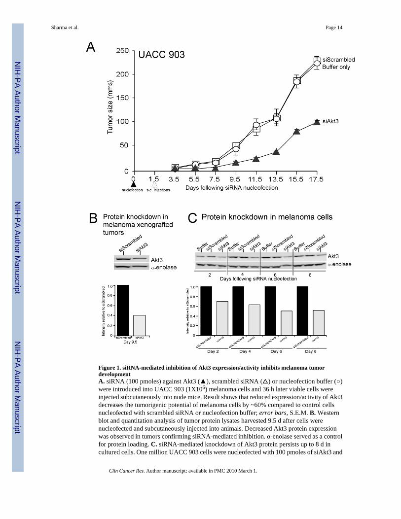
Figure 1. siRNA-mediated inhibition of Akt3 expression/activity inhibits melanoma tumordevelopmentA. siRNA (100 pmoles) against Akt3 (▲), scrambled siRNA (△) or nucleofection buffer (○)were introduced into UACC 903 (1X10⁶) melanoma cells and 36 h later viable cells wereinjected subcutaneously into nude mice. Result shows that reduced expression/activity of Akt3decreases the tumorigenic potential of melanoma cells by ~60% compared to control cellsnucleofected with scrambled siRNA or nucleofection buffer; error bars, S.E.M. B. Westernblot and quantitation analysis of tumor protein lysates harvested 9.5 d after cells werenucleofected and subcutaneously injected into animals. Decreased Akt3 protein expressionwas observed in tumors confirming siRNA-mediated inhibition. α-enolase served as a controlfor protein loading. C. siRNA-mediated knockdown of Akt3 protein persists up to 8 d incultured cells. One million UACC 903 cells were nucleofected with 100 pmoles of siAkt3 and
Sharma et al. Page 14
Clin Cancer Res. Author manuscript; available in PMC 2010 March 1.
NIH
-PA Author Manuscript
NIH
-PA Author Manuscript
NIH
-PA Author Manuscript
replated in culture dishes, and protein lysates harvested 2, 4, 6, and 8 d later for Western blotanalysis to determine Akt3 protein levels in cells. Decreased Akt3 protein expression comparedto controls was observed up to 8 d after nucleofection into cells. α-enolase served as a controlfor protein loading on Western blots, whereas scrambled siRNA served as a control for RNAinterference specificity.
Sharma et al. Page 15
Clin Cancer Res. Author manuscript; available in PMC 2010 March 1.
NIH
-PA Author Manuscript
NIH
-PA Author Manuscript
NIH
-PA Author Manuscript
Figure 2. Characterization of isothiocyanates and isoselenocyanates as inhibitors of melanomaA. Structures of isothiocyanates and selenium containing isoselenocyanates. Chemicalstructures of isothiocyanates containing 1(benzyl), 2 (phenethyl), 4 (phenylbutyl) and 6(phenylhexyl) carbon spacers and corresponding isosteric selenium analogs in which sulfurwas replaced with selenium. A 6-carbon selenocyanate (phenylhexyl selenocyanate, PHSC),served as a control to show compound structure and not selenium caused the inhibitory effect.B and C. Comparison of melanoma cell survival following exposure to isothiocyanates versusisoselenocyanates. Cell viability was measured using the MTS assay and IC50 (μM) valuesplotted against carbon chain length. 5X10³ melanoma cells were plated in 96-well plates andallowed to attach for 24 h. Increasing concentrations of isothiocyanates and selenium analogs
Sharma et al. Page 16
Clin Cancer Res. Author manuscript; available in PMC 2010 March 1.
NIH
-PA Author Manuscript
NIH
-PA Author Manuscript
NIH
-PA Author Manuscript
were added in culture medium. Values represent averages of percentage of control DMSOtreated cells. API-2 and PHSC served as an Akt inhibitor and selenium control respectively.Results show that ISC-4 and ISC-6 were the most effective inhibitors. D. ISC-4 kills melanomacells at 2–4 fold lower concentrations than normal cells. 5X10³ normal human fibroblasts(FF2441) or metastatic melanoma cells (UACC 903) were plated in 96-well plates in 100 μlDMEM media containing 10% FBS and grown for 72 and 36 h respectively. Exponentiallygrowing cells were treated with increasing concentrations (2–100 μM) of ISC-4 or PBITC for12 and 24 h and IC50 (μM) values determined. ISC-4 was found to consistently inhibitmelanoma cells growth at concentrations 2–4 fold lower than fibroblast cells (One wayANOVA ***P<0.001); error bars, S.E.M.
Sharma et al. Page 17
Clin Cancer Res. Author manuscript; available in PMC 2010 March 1.
NIH
-PA Author Manuscript
NIH
-PA Author Manuscript
NIH
-PA Author Manuscript
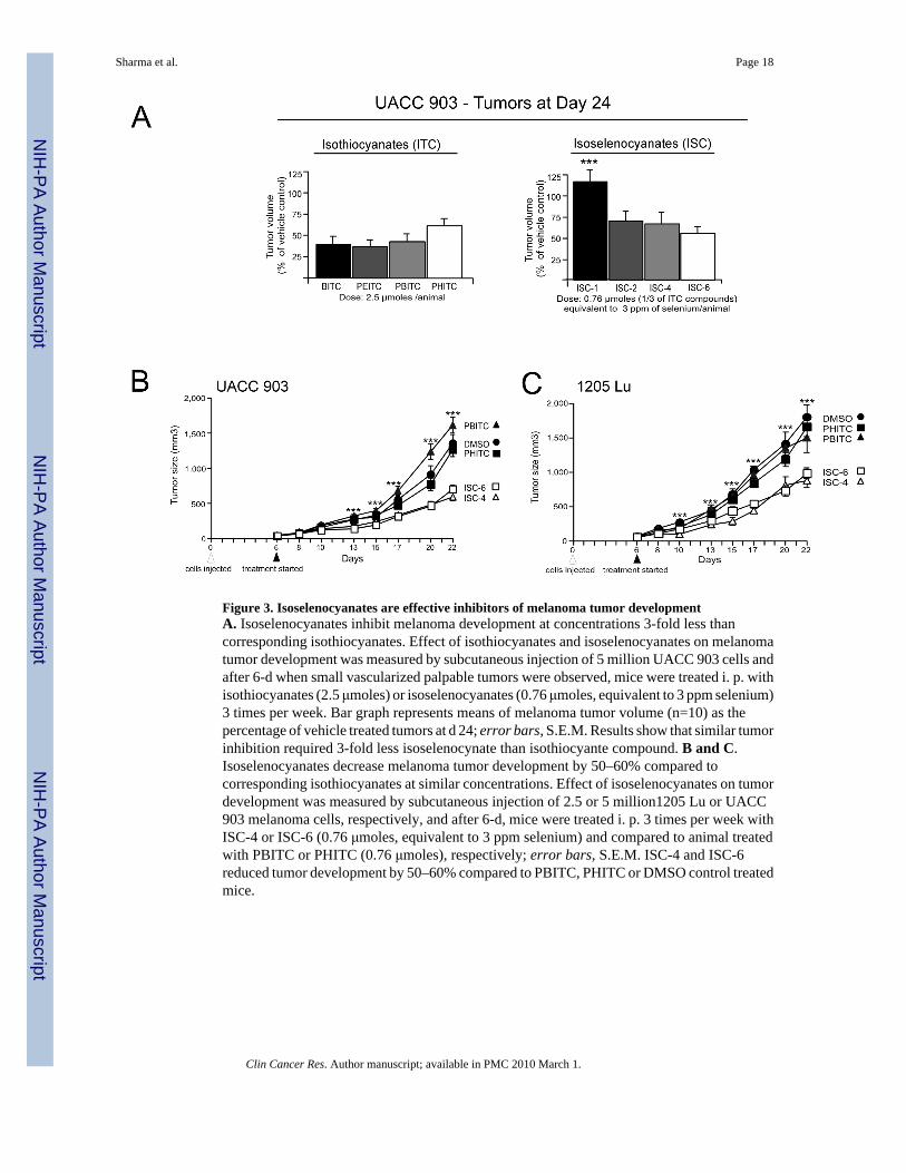
Figure 3. Isoselenocyanates are effective inhibitors of melanoma tumor developmentA. Isoselenocyanates inhibit melanoma development at concentrations 3-fold less thancorresponding isothiocyanates. Effect of isothiocyanates and isoselenocyanates on melanomatumor development was measured by subcutaneous injection of 5 million UACC 903 cells andafter 6-d when small vascularized palpable tumors were observed, mice were treated i. p. withisothiocyanates (2.5 μmoles) or isoselenocyanates (0.76 μmoles, equivalent to 3 ppm selenium)3 times per week. Bar graph represents means of melanoma tumor volume (n=10) as thepercentage of vehicle treated tumors at d 24; error bars, S.E.M. Results show that similar tumorinhibition required 3-fold less isoselenocynate than isothiocyante compound. B and C.Isoselenocyanates decrease melanoma tumor development by 50–60% compared tocorresponding isothiocyanates at similar concentrations. Effect of isoselenocyanates on tumordevelopment was measured by subcutaneous injection of 2.5 or 5 million1205 Lu or UACC903 melanoma cells, respectively, and after 6-d, mice were treated i. p. 3 times per week withISC-4 or ISC-6 (0.76 μmoles, equivalent to 3 ppm selenium) and compared to animal treatedwith PBITC or PHITC (0.76 μmoles), respectively; error bars, S.E.M. ISC-4 and ISC-6reduced tumor development by 50–60% compared to PBITC, PHITC or DMSO control treatedmice.
Sharma et al. Page 18
Clin Cancer Res. Author manuscript; available in PMC 2010 March 1.
NIH
-PA Author Manuscript
NIH
-PA Author Manuscript
NIH
-PA Author Manuscript
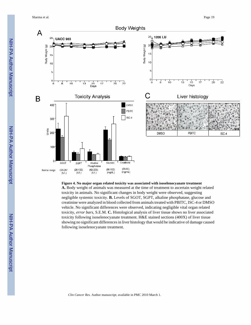
Figure 4. No major organ related toxicity was associated with isoselenocyanate treatmentA. Body weight of animals was measured at the time of treatment to ascertain weight relatedtoxicity in animals. No significant changes in body weight were observed, suggestingnegligible systemic toxicity. B. Levels of SGOT, SGPT, alkaline phosphatase, glucose andcreatinine were analyzed in blood collected from animals treated with PBITC, ISC-4 or DMSOvehicle. No significant differences were observed, indicating negligible vital organ relatedtoxicity, error bars, S.E.M. C. Histological analysis of liver tissue shows no liver associatedtoxicity following isoselenocyanate treatment. H&E stained sections (400X) of liver tissueshowing no significant differences in liver histology that would be indicative of damage causedfollowing isoselenocyanate treatment.
Sharma et al. Page 19
Clin Cancer Res. Author manuscript; available in PMC 2010 March 1.
NIH
-PA Author Manuscript
NIH
-PA Author Manuscript
NIH
-PA Author Manuscript
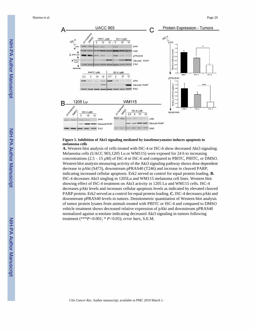
Figure 5. Inhibition of Akt3 signaling mediated by isoselenocyanates induces apoptosis inmelanoma cellsA. Western blot analysis of cells treated with ISC-4 or ISC-6 show decreased Akt3 signaling.Melanoma cells (UACC 903,1205 Lu or WM115) were exposed for 24 h to increasingconcentrations (2.5 – 15 μM) of ISC-4 or ISC-6 and compared to PBITC, PHITC, or DMSO.Western blot analysis measuring activity of the Akt3 signaling pathway shows dose dependentdecrease in pAkt (S473), downstream pPRAS40 (T246) and increase in cleaved PARP,indicating increased cellular apoptosis. Erk2 served as control for equal protein loading. B.ISC-4 decreases Akt3 singling in 1205Lu and WM115 melanoma cell lines. Western blotshowing effect of ISC-4 treatment on Akt3 activity in 1205 Lu and WM115 cells. ISC-4decreases pAkt levels and increases cellular apoptosis levels as indicated by elevated cleavedPARP protein. Erk2 served as a control for equal protein loading. C. ISC-4 decreases pAkt anddownstream pPRAS40 levels in tumors. Densitometric quantitation of Western blot analysisof tumor protein lysates from animals treated with PBITC or ISC-4 and compared to DMSOvehicle treatment shows decreased relative expression of pAkt and downstream pPRAS40normalized against α-enolase indicating decreased Akt3 signaling in tumors followingtreatment (***P<0.001; * P<0.05); error bars, S.E.M.
Sharma et al. Page 20
Clin Cancer Res. Author manuscript; available in PMC 2010 March 1.
NIH
-PA Author Manuscript
NIH
-PA Author Manuscript
NIH
-PA Author Manuscript
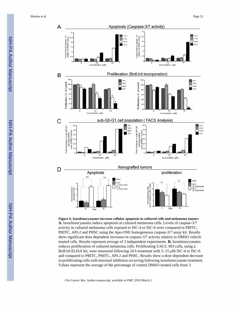
Figure 6. Isoselenocyanates increase cellular apoptosis in cultured cells and melanoma tumorsA. Isoselenocyanates induce apoptosis in cultured melanoma cells. Levels of caspase-3/7activity in cultured melanoma cells exposed to ISC-4 or ISC-6 were compared to PBITC,PHITC, API-2 and PHSC using the Apo-ONE homogeneous caspase-3/7 assay kit. Resultsshow significant dose dependent increases in caspase-3/7 activity relative to DMSO vehicletreated cells. Results represent average of 3 independent experiments. B. Isoselenocyanatesreduces proliferation of cultured melanoma cells. Proliferating UACC 903 cells, using aBrdUrd ELISA kit, were measured following 24 h treatment with 5–15 μM ISC-4 or ISC-6and compared to PBITC, PHITC, API-2 and PHSC. Results show a dose dependent decreasein proliferating cells with maximal inhibition occurring following isoselenocyanate treatment.Values represent the average of the percentage of control DMSO treated cells from 3
Sharma et al. Page 21
Clin Cancer Res. Author manuscript; available in PMC 2010 March 1.
NIH
-PA Author Manuscript
NIH
-PA Author Manuscript
NIH
-PA Author Manuscript
independent experiments. C. Isoselenocyanates increase sub-G0-G1 cell population indicatingincreased cellular apoptosis. Asynchronously growing UACC 903 cells were treated withISC-4, ISC-6, PBITC, PHITC, API-2 or PHSC, and 24 h later, cells stained with propidiumiodide analyzed for cell cycle distribution using a FACScan analyzer. ISC-4 or ISC-6 treatmentsignificantly increased the sub G0-G1 cell population indicating apoptotic cells. Resultsrepresent average of 2 independent experiments. D. Isoselenocyanate treatment increaseslevels of cellular apoptosis in melanoma tumors. Rates of apoptosis and proliferation in sizeand time matched tumors from mice treated i. p. with ISC-4 (3 ppm equivalent to 0.76 μmoles),starting 6 d after subcutaneous injection of cells and on alternate days thereafter up to d 13,were compared to mice treated with PBITC (0.76 μmoles) or DMSO (50 μl). Results show a3-fold increase in number of apoptotic cells following treatment of UACC 903 tumors withISC-4 at d 11 and 13 compared to PBITC or DMSO control. No statistically significantdifference was observed in proliferation rate. Values represent means from 2 separateexperiments with 4–6 fields analyzed from each of 6 tumors per experiment. (** P<0.01; NS,non significant); error bars, S.E.M.
Sharma et al. Page 22
Clin Cancer Res. Author manuscript; available in PMC 2010 March 1.
NIH
-PA Author Manuscript
NIH
-PA Author Manuscript
NIH
-PA Author Manuscript
Related Documents