Targeted Sprouty1 overexpression in cardiac myocytes does not alter myocardial remodeling or function Nathan J. Charles, Lillehei Heart Institute, University of Minnesota, Minneapolis, MN, USA Robert C. Huebert, Division of Gastroentology and Hepatology, Mayo Clinic and Foundation, Rochester, MN, USA Sangjin Lee, Lillehei Heart Institute, University of Minnesota, Minneapolis, MN, USA Department of Medicine, University of Minnesota, Minneapolis, MN, USA Neeta Adhikari, Lillehei Heart Institute, University of Minnesota, Minneapolis, MN, USA Department of Medicine, University of Minnesota, Minneapolis, MN, USA Sean Polster, Lillehei Heart Institute, University of Minnesota, Minneapolis, MN, USA Department of Medicine, University of Minnesota, Minneapolis, MN, USA James E. Rider, Lillehei Heart Institute, University of Minnesota, Minneapolis, MN, USA Department of Medicine, University of Minnesota, Minneapolis, MN, USA Elizabeth Braunlin, Division of Pediatric Cardiology, Department of Pediatrics, University of Minnesota, Minneapolis, MN, USA Ami Mariash, Lillehei Heart Institute, University of Minnesota, Minneapolis, MN, USA Department of Medicine, University of Minnesota, Minneapolis, MN, USA Maggie Robledo, Lillehei Heart Institute, University of Minnesota, Minneapolis, MN, USA David Schuweiler, and Lillehei Heart Institute, University of Minnesota, Minneapolis, MN, USA Jennifer L. Hall Lillehei Heart Institute, University of Minnesota, Minneapolis, MN, USA Department of Medicine, University of Minnesota, Minneapolis, MN, USA Developmental Biology Center, University of Minnesota, Minneapolis, MN, USA © Springer Science+Business Media, LLC. 2010 J. L. Hall, Lillehei Heart Institute, Department of Medicine, University of Minnesota, 312 Church Street, 4-106, Minneapolis, MN 55455, USA, [email protected]. NIH Public Access Author Manuscript Mol Cell Biochem. Author manuscript; available in PMC 2011 September 1. Published in final edited form as: Mol Cell Biochem. 2010 September ; 342(1-2): 57–62. doi:10.1007/s11010-010-0468-8. NIH-PA Author Manuscript NIH-PA Author Manuscript NIH-PA Author Manuscript

Welcome message from author
This document is posted to help you gain knowledge. Please leave a comment to let me know what you think about it! Share it to your friends and learn new things together.
Transcript

Targeted Sprouty1 overexpression in cardiac myocytes does notalter myocardial remodeling or function
Nathan J. Charles,Lillehei Heart Institute, University of Minnesota, Minneapolis, MN, USA
Robert C. Huebert,Division of Gastroentology and Hepatology, Mayo Clinic and Foundation, Rochester, MN, USA
Sangjin Lee,Lillehei Heart Institute, University of Minnesota, Minneapolis, MN, USA
Department of Medicine, University of Minnesota, Minneapolis, MN, USA
Neeta Adhikari,Lillehei Heart Institute, University of Minnesota, Minneapolis, MN, USA
Department of Medicine, University of Minnesota, Minneapolis, MN, USA
Sean Polster,Lillehei Heart Institute, University of Minnesota, Minneapolis, MN, USA
Department of Medicine, University of Minnesota, Minneapolis, MN, USA
James E. Rider,Lillehei Heart Institute, University of Minnesota, Minneapolis, MN, USA
Department of Medicine, University of Minnesota, Minneapolis, MN, USA
Elizabeth Braunlin,Division of Pediatric Cardiology, Department of Pediatrics, University of Minnesota, Minneapolis,MN, USA
Ami Mariash,Lillehei Heart Institute, University of Minnesota, Minneapolis, MN, USA
Department of Medicine, University of Minnesota, Minneapolis, MN, USA
Maggie Robledo,Lillehei Heart Institute, University of Minnesota, Minneapolis, MN, USA
David Schuweiler, andLillehei Heart Institute, University of Minnesota, Minneapolis, MN, USA
Jennifer L. HallLillehei Heart Institute, University of Minnesota, Minneapolis, MN, USA
Department of Medicine, University of Minnesota, Minneapolis, MN, USA
Developmental Biology Center, University of Minnesota, Minneapolis, MN, USA
© Springer Science+Business Media, LLC. 2010J. L. Hall, Lillehei Heart Institute, Department of Medicine, University of Minnesota, 312 Church Street, 4-106, Minneapolis, MN55455, USA, [email protected].
NIH Public AccessAuthor ManuscriptMol Cell Biochem. Author manuscript; available in PMC 2011 September 1.
Published in final edited form as:Mol Cell Biochem. 2010 September ; 342(1-2): 57–62. doi:10.1007/s11010-010-0468-8.
NIH
-PA Author Manuscript
NIH
-PA Author Manuscript
NIH
-PA Author Manuscript

AbstractThe mitogen activated protein kinase (MAPK) signaling pathway regulates multiple eventsleading to heart failure including ventricular remodeling, contractility, hypertrophy, apoptosis, andfibrosis. The regulation of conserved intrinsic inhibitors of this pathway is poorly understood. Werecently identified an up-regulation of Sprouty1 (Spry1) in a targeted approach for novel inhibitorsof the MAPK signaling pathway in failing human hearts following reverse remodeling. The goalof this study was to test the hypothesis that up-regulated expression of Spry1 in cardiac myocyteswould be sufficient to inhibit ERK1/2 activation and tissue remodeling. We established a murinemodel with up-regulated Spry1 expression in cardiac myocytes using the alpha-myosin heavychain promoter (α-MHC). Heart weight and cardiac myocyte morphology were unchanged in adultmale α-MHC–Spry1 mice compared to control mice. Ventricular function of α-MHC–Spry1 micewas unaltered at 8 weeks or 1 year of age. These findings were consistent with the lack of an effectof Spry1 on ERK1/2 activity. In summary, targeted up-regulation of Spry1 in cardiac myocytes isnot sufficient to alter cell or tissue remodeling consistent with the lack of an effect on ERK1/2activity.
KeywordsSpry1; Myocyte; Echocardiography; MAPkinase
IntroductionEvidence to date suggests a role for the mitogen activated protein kinase (MAPK) signalingcascade in cardiac remodeling, hypertrophy, and heart failure [1,2]. Unloading of the failingleft ventricle with a left ventricular assist device (LVAD) leads to reverse remodeling,including partial normalization of myocardial structure and function by decreasing cardiacmyocyte hypertrophy [3,4]. Our lab and others have seen an association between reductionsin cardiac myocyte hypertrophy and decreased activity of extracellular signal-regulatedkinase (ERK1/2), following LVAD support [5–7]. We previously screened a compendium ofgene expression datasets from end-stage heart failure patients before and after placement ofan LVAD with the goal of identifying inhibitors of the MAPK signaling pathway [7]. Fromthis screen, we identified Sprouty1 (Spry1), a conserved gene shown to inhibit MAPKsignaling in model organisms and cells in culture. We further demonstrated that Spry1 wassignificantly increased after reverse remodeling of the failing left ventricle [7].
Spry1 is one of four mammalian Spry genes (Spry1–4) and was originally defined as anantagonist of branching morphogenesis in the lung of Drosophila [8]. Spry1−/− mice exhibitdefects in kidney development due to increased ureteric branching, with no reported defectsin cardiovascular development [9]. Studies overexpressing Spry1 in neonatal cardiacmyocytes and the 293T, Swiss 3T3, C2C12, and Cos cell lines have reported decreased anddelayed ERK1/2 activation in response to fibroblast growth factor-2 (FGF2) stimulation,while siRNA directed against Spry1 has been shown to increase ERK1/2 activity [7,10,11].
The primary goal of this study was to test the hypothesis that increased in vivo expression ofSpry1 in cardiac myocytes would decrease ERK1/2 activity and alter myocyte morphologyleading to decreased heart size. To test this hypothesis, we established a cardiac-specificSpry1 transgenic mouse model.
Charles et al. Page 2
Mol Cell Biochem. Author manuscript; available in PMC 2011 September 1.
NIH
-PA Author Manuscript
NIH
-PA Author Manuscript
NIH
-PA Author Manuscript
Materials and methodsConstruct design and founder validation
The mSpry1 coding sequence (GenBank: AF176903) was PCR amplified from the pxj40-FLAG-Spry1 plasmid (provided by G. Guy) with primers incorporating 5′ SalI (5′-tatatagtcgacatggattccccaagtcagca-3′) and 3’ HindIII restriction sites (5′-tatataaagctttcatgacagtttgccctgag-3′). The sequence was ligated into the SalI/HindIII sites ofthe pnc26 plasmid (provided by J. Robbins) containing the murine α-MHC promoter(GenBank: U71441) and an hGH poly-A tail. The α-MHC/Spry1/hGH poly-A construct wasexcised with Not I and purified by electroelution. Transgenic founders were generated usingstandard pronuclear microinjection of C57/BL6 fertilized eggs implanted into foster females.PCR was performed using murine α-MHC (5′-gcccggcactcttagcaaacctca-3′) and Spry1primers (5′-tctaacctctgccggccttccaca-3′) which amplified a 306 bp product establishingpresence of the transgene; initial denature at 95°C for 4 min; 30×: denature at 94°C for 1min, anneal at 65°C for 30 s, extend at 72°C for 30 s, and final extension at 72°C for 5 min.A Southern blot was performed with 5 µg of genomic DNA isolated from mouse liver tissueusing the DNeasy Tissue Kit (Qiagen) and restriction digested with Bst XI. DNA fragmentswere separated by 1% PAGE and transferred to Hybond™-N+ nylon membrane (AmershamBiosciences) at RT overnight in 20× SSC buffer. A DNA probe was generated by PCR from10 ng of α-MHC/Spry1/hGH poly-A construct using mSpry1 primers (described below inqRT-PCR). PAGE was used to confirm the correct product size (227 bp) and the probe waspurified with the QiaQuick Gel Extraction Kit (Qiagen). The probe was radiolabeled with α-P32-dGTP and hybridized to the membrane overnight in Rapid-hyb™ buffer (AmershamBiosciences). The membrane was washed in 50°C 2× SSC/0.05% SDS and 0.1× SSC/0.1%SDS buffers, placed at −80°C overnight and exposed to radiography film.
Western blottingTotal protein was extracted from frozen whole mouse hearts pulverized with a modifiedNP40 RIPA extraction buffer containing PMSF (1 mM), NaF (50 mM), Na3VO4 (0.2 mM),BME (14.3 mM), and one complete Mini-Protease Inhibitor Cocktail tablet (Roche). Proteinconcentration was quantified with Bio-Rad Protein Assay (Bio-Rad), and protein wastransferred to Immobilon™ PVDF membrane (Millipore) after fractionation by 10% SDS-PAGE as previously described [12]. Blots were probed overnight at 4°C with the followingprimary antibodies diluted in Tris-buffered saline/0.1% Tween-20 (v/v) containing 1% driedmilk (Bio-Rad): anti-Spry1 (Invitrogen-Zymed; Cat. No. 40–1800), anti-Vinculin (Sigma-Aldrich; Cat. No. V9131), anti-Phospho-p44/42 MAP Kinase (Cell Signaling; Cat. No.9101), anti-p44/42 MAP Kinase (Cell Signaling; Cat. No. 9102). HRP-conjugated goat anti-rabbit and anti-mouse secondary antibodies (Santa Cruz; Cat. No. 2004 and 2005) were usedto detect their respective primary antibodies, and immunoreactive proteins were visualizedby Super Signal® West Femto Maximum Sensitivity Substrate ECL detection (Pierce).Densitometry was performed using an AlphaImager® fluorimeter and analyzed withAlphaEase® FC Image Analysis Software (Alpha Innotech).
Heart weightMice were anesthetized by CO2 asphyxiation and immediately weighed according to theapproved IACUC protocol at the University of Minnesota. Whole hearts were dissectedfrom animals at the aortic root and rinsed three times in Dulbecco’s PBS. Hearts wereblotted dry and then weighed.
Charles et al. Page 3
Mol Cell Biochem. Author manuscript; available in PMC 2011 September 1.
NIH
-PA Author Manuscript
NIH
-PA Author Manuscript
NIH
-PA Author Manuscript
HistologyFormalin-fixed mouse hearts were embedded in paraffin and 6 µm thick sections weredeparaffinized with xylene and rehydrated in a graded ethanol series. Whole heart sectionswere stained with hematoxylin (Surgipath) for 2 min, dehydrated, and counterstained witheosin for 1 min (Surgipath). Individual cardiac myocytes were treated with TRITC-conjugated wheat germ agglutinin (Invitrogen) at 50 µg/ml for 90 min after being blockedfor 60 min with 1% BSA in 1× PBS. Cardiac vascularity was determined by counting allvessels appearing in cross-section per high-power filed (400×) using TRITC stainedsections. Trichrome staining was conducted according to manufacturer’s guidelines(Newcomer Supply).
ApoptosisHL-1 cardiac myocyte cells [13] were cultured at 37°C and 5% CO2 on fibronectin (Sigma-Aldrich) coated 6-well plates (5 µg/ml) using Claycomb Media (JRH Biosciences; Cat. No.51800C) supplemented with 10% fetal bovine serum (Gibco®), 0.1 mM norepinephrine(Sigma-Aldrich), 1% L-glutamine (Gibco®), and penicillin–streptomycin (Gibco®). HL-1cells were infected with either eGFP or Spry1 adenovirus (described in [7]) at an MOI of10.0 for 6 h at 37°C. 48 h later, 5 µg of Hoechst 33342 dye was added to each well for 5 minat 37°C before cells were collected by trypsinization and placed onto glass microscope slidesfor apoptotic scoring as previously described for condensed and coalesced nuclei [14].
EchocardiographyHigh resolution ultrasound biomicroscopy (VisualSonics 770, Toronto, ON) was performedusing a 30-MHz linear transducer. After anesthesia was induced by inhalation of 3.5%isoflurane and supplemental oxygen, isoflurane concentration was diminished to 1.5% tomaintain anesthesia. Mice were then placed upon a heated examination platform and theanterior chest was prepared for imaging with a depilatory cream. Two-dimensional (2-D)and M-mode images were obtained from standard imaging positions to determine leftventricular chamber dimensions and wall thicknesses [15,16]. Shortening fraction andejection fraction were calculated from M-mode and ECG-gated 2-D images, respectively[15,16].
Statistical analysisStatistical analyses were performed using a paired Student’s t-test. Data is presented asmean ± standard deviation. Significance was determined with 95% confidence.
ResultsThe murine Spry1 transgene was ligated in frame between the α-MHC promoter and ahuman growth hormone (hGH) poly-A tail (Fig. 1a). Establishing transgenic lines of foundermice was performed by Southern blot analysis (Fig. 1b). All animals were studied asheterozygous for the Spry1 transgene and followed normal Mendelian inheritance, wherethrough five generations 55% of all male animals (n = 150) possessed the Spry1 transgene asdetermined by PCR-based genotyping (Fig. 1c). Spry1 protein expression was markedlyincreased in the hearts of transgenic animals (Fig. 1d). Spry1 overexpression was cardiac-specific and did not affect gene expression of the other Spry1 isoforms, Spry2–4 (data notshown). This initial characterization was also performed on a second founding line oftransgenic mice demonstrating lesser Spry1 overexpression not discussed herein.
To determine if baseline ERK1/2 activity was altered in the hearts of Spry1 transgenic mice,we performed immunoblotting for phosphorylated ERK1/2 and total ERK1/2 in 12-week oldmale mice (Fig. 2a). No significant differences in normalized ERK1/2 phosphorylation or
Charles et al. Page 4
Mol Cell Biochem. Author manuscript; available in PMC 2011 September 1.
NIH
-PA Author Manuscript
NIH
-PA Author Manuscript
NIH
-PA Author Manuscript
total EKR1/2 expression were seen between groups following Western blotting anddensitometry (Fig. 2b). Taken together, these findings suggest that in adult male mice,upregulation of Spry1 in the heart is not sufficient to decrease basal ERK1/2 activity or altertotal ERK1/2 expression levels.
Hearts from 12-week old males demonstrated no gross morphological changes incomparison to age-matched wild-type littermate control animals (Fig. 3a). Furthermore,there was no significant difference in normalized whole heart weight between the twogroups (Fig. 3b). Histological analysis of heart tissue from control and transgenic animalsrevealed no differences in cardiac myocyte structure (Fig. 3c) or cardiac myocyte size (Fig.3d). Spry1 overexpression in the heart did not influence cardiac vascularity between control(CL) and transgenic (TG) animals measured as the mean number of vessels present per high-power field (CL = 8.0 ± 2.1 vs. TG = 6.0 ± 1.1, n = 5, p = NS). Additionally, cardiac fibrosiswas not present in either control or transgenic animals as determined by tri-chrome staining(Fig. 3e). Finally, we did not observe any changes in cardiac myocyte morphology betweencontrol and Spry1 transgenic mice that would indicate a difference in cardiac myocytesurvival (Fig. 3c). To confirm these findings, we overexpressed Spry1 in HL-1 cardiacmyocytes and determined the percentage of apoptotic nuclei. Spry1 overexpression (SP) didnot alter the percentage of apoptotic nuclei (CL = 1% ± 1% vs. SP = 1% ± 0%, n = 3 pergroup, p = NS).
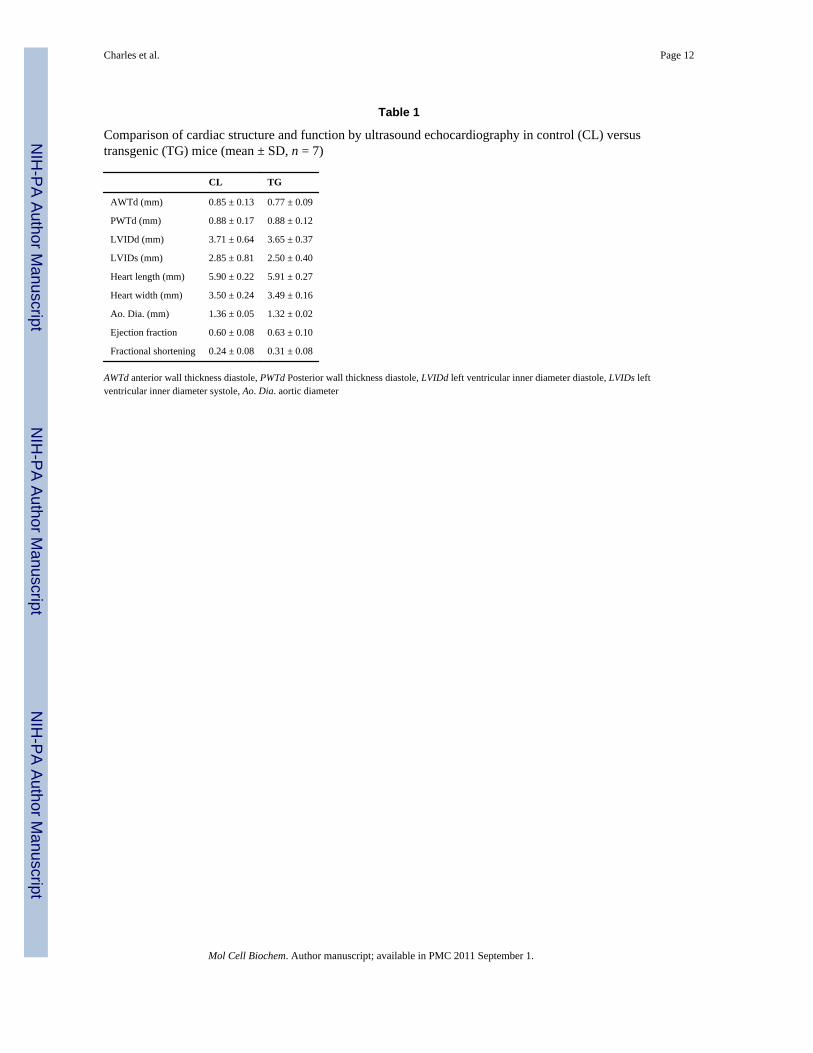
Structural analysis of hearts from both control and transgenic animals showed no grossdifferences in chamber size or wall thickness both horizontally (Fig. 4a) and longitudinally(Fig. 4b) or with regards to diastole or systole (Table 1). Functional analysis of cardiacperformance by ultrasound echocardiography revealed no significant differences in eitherleft ventricular ejection fraction or fractional shortening in 8-week old mice (Table 1).Additionally, baseline echocardiographic analysis of one-year old control (CL) versustransgenic (TG) mice demonstrated no significant changes in heart function of aged animalsas determined by comparisons in ejection fraction (CL = 0.59 ± 0.00 vs. TG = 0.61 ± 0.08)and fractional shortening (CL = 0.31 ± 0.12 vs. TG = 0.29 ± 0.06).
DiscussionSpry protein family members have been classically defined by their ability to inhibitreceptor tyrosine kinase (RTK) signaling [17]. Of great interest, is their potential to preventor limit activation of the downstream RTK target ERK1/2, which is well known to play asignificant role in a variety of key cellular processes in cardiac myocytes [1]. We wereespecially interested in Spry1 given the increased expression of this conserved ERK1/2pathway inhibitor in failing human hearts following therapy with a LVAD that lead toreverse remodeling [7]. We hypothesized that increased expression of Spry1 in cardiacmyocytes would decrease ERK1/2 activity leading to a reduction in myocyte size.
The major findings from this study are that overexpression of Spry1 in cardiac myocytesdoes not affect cardiovascular development, myocyte size, or ERK1/2 activity. Combinedwith data from the Spry1−/− mouse that did not show a cardiovascular phenotype, these datasuggest that Spry1 does not play a major role under normal physiological conditions in theheart.
Previous work from our lab has shown expression of Spry1 protein in the human heart [7].Immunohistochemical staining demonstrated the presence of Spry1 in cardiac myocytes aswell as other cardiac cell types [7]. We also reported an increase in Spry1 mRNA andprotein expression with an associated decrease in phosphorylated ERK1 and ERK2 in failinghuman hearts following unloading with a LVAD [7]. To test if up-regulation of Spry1 was
Charles et al. Page 5
Mol Cell Biochem. Author manuscript; available in PMC 2011 September 1.
NIH
-PA Author Manuscript
NIH
-PA Author Manuscript
NIH
-PA Author Manuscript
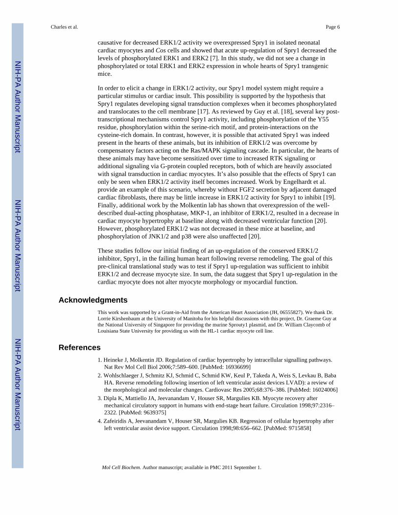
causative for decreased ERK1/2 activity we overexpressed Spry1 in isolated neonatalcardiac myocytes and Cos cells and showed that acute up-regulation of Spry1 decreased thelevels of phosphorylated ERK1 and ERK2 [7]. In this study, we did not see a change inphosphorylated or total ERK1 and ERK2 expression in whole hearts of Spry1 transgenicmice.
In order to elicit a change in ERK1/2 activity, our Spry1 model system might require aparticular stimulus or cardiac insult. This possibility is supported by the hypothesis thatSpry1 regulates developing signal transduction complexes when it becomes phosphorylatedand translocates to the cell membrane [17]. As reviewed by Guy et al. [18], several key post-transcriptional mechanisms control Spry1 activity, including phosphorylation of the Y55residue, phosphorylation within the serine-rich motif, and protein-interactions on thecysteine-rich domain. In contrast, however, it is possible that activated Spry1 was indeedpresent in the hearts of these animals, but its inhibition of ERK1/2 was overcome bycompensatory factors acting on the Ras/MAPK signaling cascade. In particular, the hearts ofthese animals may have become sensitized over time to increased RTK signaling oradditional signaling via G-protein coupled receptors, both of which are heavily associatedwith signal transduction in cardiac myocytes. It’s also possible that the effects of Spry1 canonly be seen when ERK1/2 activity itself becomes increased. Work by Engelhardt et al.provide an example of this scenario, whereby without FGF2 secretion by adjacent damagedcardiac fibroblasts, there may be little increase in ERK1/2 activity for Spry1 to inhibit [19].Finally, additional work by the Molkentin lab has shown that overexpression of the well-described dual-acting phosphatase, MKP-1, an inhibitor of ERK1/2, resulted in a decrease incardiac myocyte hypertrophy at baseline along with decreased ventricular function [20].However, phosphorylated ERK1/2 was not decreased in these mice at baseline, andphosphorylation of JNK1/2 and p38 were also unaffected [20].
These studies follow our initial finding of an up-regulation of the conserved ERK1/2inhibitor, Spry1, in the failing human heart following reverse remodeling. The goal of thispre-clinical translational study was to test if Spry1 up-regulation was sufficient to inhibitERK1/2 and decrease myocyte size. In sum, the data suggest that Spry1 up-regulation in thecardiac myocyte does not alter myocyte morphology or myocardial function.
AcknowledgmentsThis work was supported by a Grant-in-Aid from the American Heart Association (JH, 06555827). We thank Dr.Lorrie Kirshenbaum at the University of Manitoba for his helpful discussions with this project, Dr. Graeme Guy atthe National University of Singapore for providing the murine Sprouty1 plasmid, and Dr. William Claycomb ofLouisiana State University for providing us with the HL-1 cardiac myocyte cell line.
References1. Heineke J, Molkentin JD. Regulation of cardiac hypertrophy by intracellular signalling pathways.
Nat Rev Mol Cell Biol 2006;7:589–600. [PubMed: 16936699]2. Wohlschlaeger J, Schmitz KJ, Schmid C, Schmid KW, Keul P, Takeda A, Weis S, Levkau B, Baba
HA. Reverse remodeling following insertion of left ventricular assist devices LVAD): a review ofthe morphological and molecular changes. Cardiovasc Res 2005;68:376–386. [PubMed: 16024006]
3. Dipla K, Mattiello JA, Jeevanandam V, Houser SR, Margulies KB. Myocyte recovery aftermechanical circulatory support in humans with end-stage heart failure. Circulation 1998;97:2316–2322. [PubMed: 9639375]
4. Zafeiridis A, Jeevanandam V, Houser SR, Margulies KB. Regression of cellular hypertrophy afterleft ventricular assist device support. Circulation 1998;98:656–662. [PubMed: 9715858]
Charles et al. Page 6
Mol Cell Biochem. Author manuscript; available in PMC 2011 September 1.
NIH
-PA Author Manuscript
NIH
-PA Author Manuscript
NIH
-PA Author Manuscript

5. Razeghi P, Bruckner BA, Sharma S, Youker KA, Frazier OH, Taegtmeyer H. Mechanical unloadingof the failing human heart fails to activate the protein kinase B/Akt/glycogen synthase kinase-3betasurvival pathway. Cardiology 2003;100:17–22. [PubMed: 12975541]
6. Baba HA, Stypmann J, Grabellus F, Kirchhof P, Sokoll A, Schafers M, Takeda A, Wilhelm MJ,Scheld HH, Takeda N, Breithardt G, Levkau B. Dynamic regulation of MEK/Erks and Akt/GSK-3beta in human end-stage heart failure after left ventricular mechanical support: myocardialmechanotransduction-sensitivity as a possible molecular mechanism. Cardiovasc Res 2003;59:390–399. [PubMed: 12909322]
7. Huebert RC, Li Q, Adhikari N, Charles NJ, Han X, Ezzat MK, Grindle S, Park S, Ormaza S, FerminD, Miller LW, Hall JL. Identification and regulation of Sprouty1, a negative inhibitor of the ERKcascade, in the human heart. Physiol Genomics 2004;18:284–289. [PubMed: 15306693]
8. Hacohen N, Kramer S, Sutherland D, Hiromi Y, Krasnow MA. Sprouty encodes a novel antagonistof FGF signaling that patterns apical branching of the Drosophila airways. Cell 1998;92:253–263.[PubMed: 9458049]
9. Basson MA, Akbulut S, Watson-Johnson J, Simon R, Carroll TJ, Shakya R, Gross I, Martin GR,Lufkin T, McMahon AP, Wilson PD, Costantini FD, Mason IJ, Licht JD. Sprouty1 is a criticalregulator of GDNF/RET-mediated kidney induction. Dev Cell 2005;8:229–239. [PubMed:15691764]
10. Hanafusa H, Torii S, Yasunaga T, Nishida E. Sprouty1 and Sprouty2 provide a control mechanismfor the Ras/MAPK signalling pathway. Nat Cell Biol 2002;4:850–858. [PubMed: 12402043]
11. Ozaki K, Miyazaki S, Tanimura S, Kohno M. Efficient suppression of FGF-2-induced ERKactivation by the cooperative interaction among mammalian Sprouty isoforms. J Cell Sci2005;118:5861–5871. [PubMed: 16339969]
12. Basi DL, Adhikari N, Mariash A, Li Q, Kao E, Mullegama SV, Hall JL. Femoral artery neointimalhyperplasia is reduced after wire injury in Ref-1+/− mice. Am J Physiol Heart Circ Physiol2007;292:H516–H521. [PubMed: 16936011]
13. Claycomb WC, Lanson NA Jr, Stallworth BS, Egeland DB, Delcarpio JB, Bahinski A, Izzo NJ Jr.HL-1 cells: a cardiac muscle cell line that contracts and retains phenotypic characteristics of theadult cardiomyocyte. Proc Natl Acad Sci USA 1998;95:2979–2984. [PubMed: 9501201]
14. Hall JL, Matter CM, Wang X, Gibbons GH. Hyperglycemia inhibits vascular smooth muscle cellapoptosis through a protein kinase C-dependent pathway. Circ Res 2000;87:574–580. [PubMed:11009562]
15. Collins KA, Korcarz CE, Lang RM. Use of echocardiography for the phenotypic assessment ofgenetically altered mice. Physiol Genomics 2003;13:227–239. [PubMed: 12746467]
16. Yang XP, Liu YH, Rhaleb NE, Kurihara N, Kim HE, Carretero OA. Echocardiographic assessmentof cardiac function in conscious and anesthetized mice. Am J Physiol 1999;277:H1967–H1974.[PubMed: 10564153]
17. Kim HJ, Bar-Sagi D. Modulation of signalling by Sprouty: a developing story. Nat Rev Mol CellBiol 2004;5:441–450. [PubMed: 15173823]
18. Guy GR, Jackson RA, Yusoff P, Chow SY. Sprouty proteins: modified modulators, matchmakersor missing links? J Endocrinol 2009;203:191–202. [PubMed: 19423641]
19. Thum T, Gross C, Fiedler J, Fischer T, Kissler S, Bussen M, Galuppo P, Just S, Rottbauer W,Frantz S, Castoldi M, Soutschek J, Koteliansky V, Rosenwald A, Basson MA, Licht JD, Pena JT,Rouhanifard SH, Muckenthaler MU, Tuschl T, Martin GR, Bauersachs J, Engelhardt S.MicroRNA-21 contributes to myocardial disease by stimulating MAP kinase signalling infibroblasts. Nature 2008;456:980–984. [PubMed: 19043405]
20. Bueno OF, De Windt LJ, Lim HW, Tymitz KM, Witt SA, Kimball TR, Molkentin JD. The dual-specificity phosphatase MKP-1 limits the cardiac hypertrophic response in vitro and in vivo. CircRes 2001;88:88–96. [PubMed: 11139479]
Charles et al. Page 7
Mol Cell Biochem. Author manuscript; available in PMC 2011 September 1.
NIH
-PA Author Manuscript
NIH
-PA Author Manuscript
NIH
-PA Author Manuscript
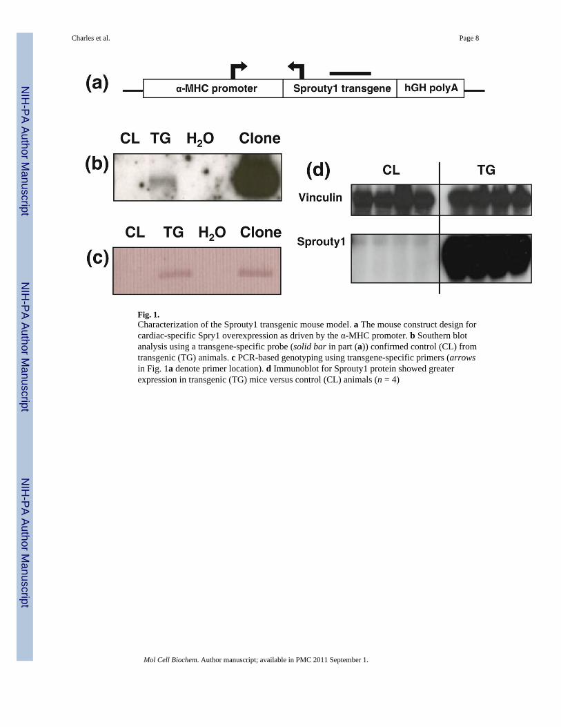
Fig. 1.Characterization of the Sprouty1 transgenic mouse model. a The mouse construct design forcardiac-specific Spry1 overexpression as driven by the α-MHC promoter. b Southern blotanalysis using a transgene-specific probe (solid bar in part (a)) confirmed control (CL) fromtransgenic (TG) animals. c PCR-based genotyping using transgene-specific primers (arrowsin Fig. 1a denote primer location). d Immunoblot for Sprouty1 protein showed greaterexpression in transgenic (TG) mice versus control (CL) animals (n = 4)
Charles et al. Page 8
Mol Cell Biochem. Author manuscript; available in PMC 2011 September 1.
NIH
-PA Author Manuscript
NIH
-PA Author Manuscript
NIH
-PA Author Manuscript
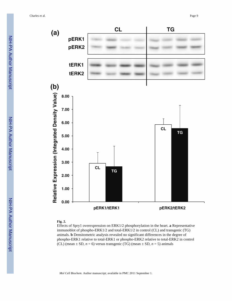
Fig. 2.Effects of Spry1 overexpression on ERK1/2 phosphorylation in the heart. a Representativeimmunoblot of phospho-ERK1/2 and total-ERK1/2 in control (CL) and transgenic (TG)animals. b Densitometric analysis revealed no significant differences in the degree ofphospho-ERK1 relative to total-ERK1 or phospho-ERK2 relative to total-ERK2 in control(CL) (mean ± SD, n = 6) versus transgenic (TG) (mean ± SD, n = 5) animals
Charles et al. Page 9
Mol Cell Biochem. Author manuscript; available in PMC 2011 September 1.
NIH
-PA Author Manuscript
NIH
-PA Author Manuscript
NIH
-PA Author Manuscript

Fig. 3.Effects of Spry1 overexpression on cardiac morphometry and cardiac myocyte morphology.a Control (CL) and transgenic (TG) mouse hearts showed no differences in gross cardiacmorphometry (n = 3). b There were no significant differences in normalized heart mass ofcontrol (CL) versus transgenic (TG) animals (mean ± SD, n = 6). c–e Histologic analysisrevealed no deviation in control (CL) and transgenic (TG) hearts with respect to: c cardiacmyocyte morphology by H&E staining (×400) (n = 5), d cardiac myocyte size or cardiacvessel number (asterisks) by WGA-TRITC staining (×400) (n = 5), or e cardiac fibrosis bytri-chrome staining (×200) (n = 5)
Charles et al. Page 10
Mol Cell Biochem. Author manuscript; available in PMC 2011 September 1.
NIH
-PA Author Manuscript
NIH
-PA Author Manuscript
NIH
-PA Author Manuscript

Fig. 4.Effects of Spry1 overexpression on cardiac structure. a, b Control (CL) and transgenic (TG)hearts revealed no gross differences in chamber dimensions or wall thicknesses by H&Estaining in either: a cross-section (n = 5) or b longitudinal-section (n = 5)
Charles et al. Page 11
Mol Cell Biochem. Author manuscript; available in PMC 2011 September 1.
NIH
-PA Author Manuscript
NIH
-PA Author Manuscript
NIH
-PA Author Manuscript
NIH
-PA Author Manuscript
NIH
-PA Author Manuscript
NIH
-PA Author Manuscript
Charles et al. Page 12
Table 1
Comparison of cardiac structure and function by ultrasound echocardiography in control (CL) versustransgenic (TG) mice (mean ± SD, n = 7)
CL TG
AWTd (mm) 0.85 ± 0.13 0.77 ± 0.09
PWTd (mm) 0.88 ± 0.17 0.88 ± 0.12
LVIDd (mm) 3.71 ± 0.64 3.65 ± 0.37
LVIDs (mm) 2.85 ± 0.81 2.50 ± 0.40
Heart length (mm) 5.90 ± 0.22 5.91 ± 0.27
Heart width (mm) 3.50 ± 0.24 3.49 ± 0.16
Ao. Dia. (mm) 1.36 ± 0.05 1.32 ± 0.02
Ejection fraction 0.60 ± 0.08 0.63 ± 0.10
Fractional shortening 0.24 ± 0.08 0.31 ± 0.08
AWTd anterior wall thickness diastole, PWTd Posterior wall thickness diastole, LVIDd left ventricular inner diameter diastole, LVIDs leftventricular inner diameter systole, Ao. Dia. aortic diameter
Mol Cell Biochem. Author manuscript; available in PMC 2011 September 1.
Related Documents