Targeted mutagenesis of Lis1 disrupts cortical development and LIS1 homodimerization Aviv Cahana*, Teresa Escamez , Richard S. Nowakowski ‡ , Nancy L. Hayes ‡ , MaiBritt Giacobini §¶ , Alexander von Holst §¶ , Orit Shmueli*, Tamar Sapir*, Susan K. McConnell i , Wolfgang Wurst §¶ , Salvador Martinez , and Orly Reiner* , ** *Department of Molecular Genetics, Weizmann Institute of Science, 76100 Rehovot, Israel; ² Instituto de Nuerociencias, Campus de San Juan, Alicante 30071, Spain; ‡ Department of Neuroscience and Cell Biology, University of Medicine and Dentistry of New Jersey—Robert Wood Johnson Medical School, Piscataway, NJ 08854; § Max Planck Institute of Psychiatry, Munich 80804, Germany; ¶ GSF-Research Center Clinical Neurogenetics, Institute for Mammalian Genetics, Munich 80804, Germany; and i Department of Biological Sciences, Stanford University, Stanford, CA 94305-5020 Communicated by C. Thomas Caskey, Cogene Biotech Ventures, Ltd., Houston, TX, March 12, 2001 (received for review December 6, 2000) Lissencephaly is a severe brain malformation in humans. To study the function of the gene mutated in lissencephaly (LIS1), we deleted the first coding exon from the mouse Lis1 gene. The deletion resulted in a shorter protein (sLIS1) that initiates from the second methionine, a unique situation because most LIS1 muta- tions result in a null allele. This mutation mimics a mutation described in one lissencephaly patient with a milder phenotype. Homozygotes are early lethal, although heterozygotes are viable and fertile. Most strikingly, the morphology of cortical neurons and radial glia is aberrant in the developing cortex, and the neurons migrate more slowly. This is the first demonstration, to our knowl- edge, of a cellular abnormality in the migrating neurons after Lis1 mutation. Moreover, cortical plate splitting and thalomocortical innervation are also abnormal. Biochemically, the mutant protein is not capable of dimerization, and enzymatic activity is elevated in the embryos, thus a demonstration of the in vivo role of LIS1 as a subunit of PAF-AH. This mutation allows us to determine a hierarchy of functions that are sensitive to LIS1 dosage, thus promoting our understanding of the role of LIS1 in the developing cortex. brain development u lissencephaly u platelet-activating factor u acetylhydrolase u gene targeting L IS1 was identified as the gene mutated in a severe human developmental brain malformation known as lissencephaly (‘‘smooth brain’’) type I (1). Patients with lissencephaly often are severely retarded, epileptic, and die at a young age. The most striking feature of the brains of affected individuals is that they are smooth and largely devoid of the sulci and gyri that char- acterize the normal brain. The lissencephalic brain exhibits defects in neuronal migration that result in poor organization of cortical layering. A reduced surface area and lack of cortical folds are also seen, possibly because of an overall reduced number of neurons (2). Mutations in two different genes may result in type I lissencephaly: LIS1, an autosomal gene located on chromosome 17p13.3 (1), and doublecortin, an X-linked gene (3, 4). The pattern of expression of LIS1 in the nervous system suggested that the mouse would be a suitable organism for studying the role of LIS1 during brain development (5). Mouse embryos homozygous for the null Lis1 allele (Lis12y2) die after implantation, whereas heterozygotes are viable and fertile (6). A half dosage of LIS1 affects neuronal migration only slightly in the developing cortex, whereas adult layer organiza- tion appears normal. Further gene dosage reduction severely obstructs cortical and hippocampal organization (6). LIS1 in- teracts with many proteins and is involved in several basic cellular functions, including mitosis, nuclear positioning, and microtubule regulation (for review, see ref. 7). To better under- stand the function of LIS1 and its role in neuronal migration, we produced Lis1 mutant mice by using cre recombinase-mediated loxP deletion. Our mutation resulted in a shorter LIS1 protein that initiates from the second methionine (M63), thus lacking two-thirds of the coiled-coil N terminus. The shorter protein enabled us to study biochemical parameters of the mutated protein in addition to the developmental phenotype of mutant embryos. The LIS1 mutants described here show a transient delay in the organization and maturation of the dorsal–caudal portion of the cortex, with abnormal morphology of both cortical neurons and radial glia during corticogenesis. Materials and Methods Monoclonal anti-LIS1 antibodies have been described (8). Poly- clonal antibody specific for the N-terminal domain of LIS1 was generated by injecting rabbits with a peptide corresponding to amino acids 5–13 of LIS1 (amino acid sequence: QRQRDEL- NRAIAD), coupled to keyhole limpet hemacyanin (Sigma). Histological and in Situ Hybridization Analyses. Embryos were col- lected at the stages indicated, and brains were either dissected from the head or left in situ. All sections discussed in this paper are coronal. Samples for whole-mount RNA in situ hybridization were fixed in 4% paraformaldehyde at 4°C and then processed essentially as described (9). For DiI (Molecular Probes) labeling, embryos were fixed in 4% paraformaldehyde, and then a DiI crystal (saturated solution in DMSO and air dried) was placed in the cortex or within the thalamus later sectioned by the vibratome in 100-mm-thick sections. BrdUrdypropidium iodide staining of cortical neurons and FACS analysis were performed according to ref. 10. For analysis of cell cycle kinetics and interkinetic nuclear movements, im- munocytochemistry and autoradiography were performed on 4 mm coronal sections, as described previously (11). Gel filtration (12) and GST pulldown (13) were performed as described. Platelet-activating factor acetylhydrolase enzymatic activity was tested as described in ref. 14. Microtubule assembly was per- formed as described previously (15) with the aid of taxol. Results and Discussion Our targeting construct involved insertion of two loxP sites, with a neomycin resistance gene in Lis1 introns flanking the first coding methionine (Fig. 1A). Heterozygotes of the loxP-neo allele were mated with PGK-Cre mice (16). Offspring of these mice exhibited the expected first coding exon deletion (‘‘floxed locus’’) in all tissues examined (data not shown); however, no homozygotes were born (Fig. 1B), and lethality was at the implantation stage. Heterozygotes were viable and fertile. West- ern blot analysis of brain extracts from wild-type and heterozy- gous mice revealed two LIS1 immunoreactive bands in heterozy- gotes, but only one band was present in wild-type mice (Fig. 1D); Abbreviations: En, embryonic day n; CP, cortical plate. **To whom reprint requests should be addressed. E-mail: [email protected]. The publication costs of this article were defrayed in part by page charge payment. This article must therefore be hereby marked “advertisement” in accordance with 18 U.S.C. §1734 solely to indicate this fact. www.pnas.orgycgiydoiy10.1073ypnas.101122598 PNAS u May 22, 2001 u vol. 98 u no. 11 u 6429 – 6434 NEUROBIOLOGY

Welcome message from author
This document is posted to help you gain knowledge. Please leave a comment to let me know what you think about it! Share it to your friends and learn new things together.
Transcript
Targeted mutagenesis of Lis1 disrupts corticaldevelopment and LIS1 homodimerizationAviv Cahana*, Teresa Escamez†, Richard S. Nowakowski‡, Nancy L. Hayes‡, MaiBritt Giacobini§¶, Alexander von Holst§¶,Orit Shmueli*, Tamar Sapir*, Susan K. McConnelli, Wolfgang Wurst§¶, Salvador Martinez†, and Orly Reiner*,**
*Department of Molecular Genetics, Weizmann Institute of Science, 76100 Rehovot, Israel; †Instituto de Nuerociencias, Campus de San Juan, Alicante 30071,Spain; ‡Department of Neuroscience and Cell Biology, University of Medicine and Dentistry of New Jersey—Robert Wood Johnson Medical School,Piscataway, NJ 08854; §Max Planck Institute of Psychiatry, Munich 80804, Germany; ¶GSF-Research Center Clinical Neurogenetics, Institute forMammalian Genetics, Munich 80804, Germany; and iDepartment of Biological Sciences, Stanford University, Stanford, CA 94305-5020
Communicated by C. Thomas Caskey, Cogene Biotech Ventures, Ltd., Houston, TX, March 12, 2001 (received for review December 6, 2000)
Lissencephaly is a severe brain malformation in humans. To studythe function of the gene mutated in lissencephaly (LIS1), wedeleted the first coding exon from the mouse Lis1 gene. Thedeletion resulted in a shorter protein (sLIS1) that initiates from thesecond methionine, a unique situation because most LIS1 muta-tions result in a null allele. This mutation mimics a mutationdescribed in one lissencephaly patient with a milder phenotype.Homozygotes are early lethal, although heterozygotes are viableand fertile. Most strikingly, the morphology of cortical neurons andradial glia is aberrant in the developing cortex, and the neuronsmigrate more slowly. This is the first demonstration, to our knowl-edge, of a cellular abnormality in the migrating neurons after Lis1mutation. Moreover, cortical plate splitting and thalomocorticalinnervation are also abnormal. Biochemically, the mutant proteinis not capable of dimerization, and enzymatic activity is elevatedin the embryos, thus a demonstration of the in vivo role of LIS1 asa subunit of PAF-AH. This mutation allows us to determine ahierarchy of functions that are sensitive to LIS1 dosage, thuspromoting our understanding of the role of LIS1 in the developingcortex.
brain development u lissencephaly u platelet-activating factor uacetylhydrolase u gene targeting
L IS1 was identified as the gene mutated in a severe humandevelopmental brain malformation known as lissencephaly
(‘‘smooth brain’’) type I (1). Patients with lissencephaly often areseverely retarded, epileptic, and die at a young age. The moststriking feature of the brains of affected individuals is that theyare smooth and largely devoid of the sulci and gyri that char-acterize the normal brain. The lissencephalic brain exhibitsdefects in neuronal migration that result in poor organization ofcortical layering. A reduced surface area and lack of corticalfolds are also seen, possibly because of an overall reducednumber of neurons (2). Mutations in two different genes mayresult in type I lissencephaly: LIS1, an autosomal gene locatedon chromosome 17p13.3 (1), and doublecortin, an X-linked gene(3, 4). The pattern of expression of LIS1 in the nervous systemsuggested that the mouse would be a suitable organism forstudying the role of LIS1 during brain development (5). Mouseembryos homozygous for the null Lis1 allele (Lis12y2) dieafter implantation, whereas heterozygotes are viable and fertile(6). A half dosage of LIS1 affects neuronal migration onlyslightly in the developing cortex, whereas adult layer organiza-tion appears normal. Further gene dosage reduction severelyobstructs cortical and hippocampal organization (6). LIS1 in-teracts with many proteins and is involved in several basiccellular functions, including mitosis, nuclear positioning, andmicrotubule regulation (for review, see ref. 7). To better under-stand the function of LIS1 and its role in neuronal migration, weproduced Lis1 mutant mice by using cre recombinase-mediatedloxP deletion. Our mutation resulted in a shorter LIS1 proteinthat initiates from the second methionine (M63), thus lacking
two-thirds of the coiled-coil N terminus. The shorter proteinenabled us to study biochemical parameters of the mutatedprotein in addition to the developmental phenotype of mutantembryos. The LIS1 mutants described here show a transientdelay in the organization and maturation of the dorsal–caudalportion of the cortex, with abnormal morphology of both corticalneurons and radial glia during corticogenesis.
Materials and MethodsMonoclonal anti-LIS1 antibodies have been described (8). Poly-clonal antibody specific for the N-terminal domain of LIS1 wasgenerated by injecting rabbits with a peptide corresponding toamino acids 5–13 of LIS1 (amino acid sequence: QRQRDEL-NRAIAD), coupled to keyhole limpet hemacyanin (Sigma).
Histological and in Situ Hybridization Analyses. Embryos were col-lected at the stages indicated, and brains were either dissectedfrom the head or left in situ. All sections discussed in this paperare coronal. Samples for whole-mount RNA in situ hybridizationwere fixed in 4% paraformaldehyde at 4°C and then processedessentially as described (9). For DiI (Molecular Probes) labeling,embryos were fixed in 4% paraformaldehyde, and then a DiIcrystal (saturated solution in DMSO and air dried) was placedin the cortex or within the thalamus later sectioned by thevibratome in 100-mm-thick sections.
BrdUrdypropidium iodide staining of cortical neurons andFACS analysis were performed according to ref. 10. For analysisof cell cycle kinetics and interkinetic nuclear movements, im-munocytochemistry and autoradiography were performed on 4mm coronal sections, as described previously (11). Gel filtration(12) and GST pulldown (13) were performed as described.Platelet-activating factor acetylhydrolase enzymatic activity wastested as described in ref. 14. Microtubule assembly was per-formed as described previously (15) with the aid of taxol.
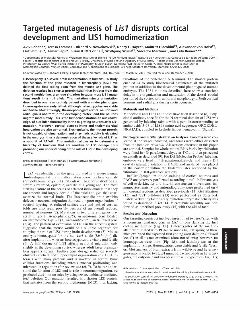
Results and DiscussionOur targeting construct involved insertion of two loxP sites, witha neomycin resistance gene in Lis1 introns flanking the firstcoding methionine (Fig. 1A). Heterozygotes of the loxP-neoallele were mated with PGK-Cre mice (16). Offspring of thesemice exhibited the expected first coding exon deletion (‘‘f loxedlocus’’) in all tissues examined (data not shown); however, nohomozygotes were born (Fig. 1B), and lethality was at theimplantation stage. Heterozygotes were viable and fertile. West-ern blot analysis of brain extracts from wild-type and heterozy-gous mice revealed two LIS1 immunoreactive bands in heterozy-gotes, but only one band was present in wild-type mice (Fig. 1D);
Abbreviations: En, embryonic day n; CP, cortical plate.
**To whom reprint requests should be addressed. E-mail: [email protected].
The publication costs of this article were defrayed in part by page charge payment. Thisarticle must therefore be hereby marked “advertisement” in accordance with 18 U.S.C.§1734 solely to indicate this fact.
www.pnas.orgycgiydoiy10.1073ypnas.101122598 PNAS u May 22, 2001 u vol. 98 u no. 11 u 6429–6434
NEU
ROBI
OLO
GY
therefore, the mice with the first coding exon deletions will bereferred to as Lis1ysLis1 mice. The shorter protein (sLIS1)present in the heterozygotes is likely to result from translationinitiation at the second methionine of LIS1 (Fig. 1C). Thisassumption was confirmed by specific antibodies recognizingonly the N-terminal domain (Fig. 1D). Quantitative analysisrevealed that heterozygous mice have higher total levels of LIS1than wild type because of the addition of expression from themutant allele (sLIS1). Our mutation is therefore different fromthe null allele of Lis1 reported previously (6).
Analysis of the central nervous system in Lis1ysLis1 embryosshowed that the parietal and occipital cortex, regions where thephenotype in lissencephaly patients is more pronounced, exhib-ited apparent alterations. LiS1ysLIS1 embryos at ages embry-
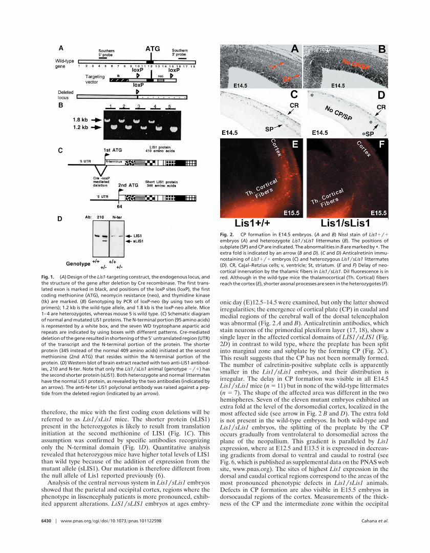
onic day (E)12.5–14.5 were examined, but only the latter showedirregularities; the emergence of cortical plate (CP) in caudal andmedial regions of the cerebral wall of the dorsal telencephalonwas abnormal (Fig. 2 A and B). Anticalretinin antibodies, whichstain neurons of the primordial plexiform layer (17, 18), show asingle layer in the affected cortical domains of LIS1ysLIS1 (Fig.2D) in contrast to wild type, where the preplate has been splitinto marginal zone and subplate by the forming CP (Fig. 2C).This result suggests that the CP has not been normally formed.The number of calretinin-positive subplate cells is apparentlysmaller in the Lis1ysLis1 embryos, and their distribution isirregular. The delay in CP formation was visible in all E14.5Lis1ysLis1 mice (n 5 11) but in none of the wild-type littermates(n 5 7). The shape of the affected area was different in the twohemispheres. Seven of the eleven mutant embryos exhibited anextra fold at the level of the dorsomedial cortex, localized in themost affected side (see arrow in Fig. 2 B and D). The extra foldis not present in the wild-type embryos. In both wild-type andLis1ysLis1 embryos, the splitting of the preplate by the CPoccurs gradually from ventrolateral to dorsomedial across theplane of the neopallium. This gradient is paralleled by Lis1expression, where at E12.5 and E13.5 it is expressed in decreas-ing gradients from dorsal to ventral and caudal to rostral (seeFig. 6, which is published as supplemental data on the PNAS website, www.pnas.org). The sites of highest Lis1 expression in thedorsal and caudal cortical regions correspond to the areas of themost pronounced phenotypic defects in Lis1ysLis1 animals.Defects in CP formation are also visible in E15.5 embryos indorsocaudal regions of the cortex. Measurements of the thick-ness of the CP and the intermediate zone within the occipital
Fig. 1. (A) Design of the Lis1-targeting construct, the endogenous locus, andthe structure of the gene after deletion by Cre recombinase. The first trans-lated exon is marked in black, and positions of the loxP sites (loxP), the firstcoding methionine (ATG), neomycin resistance (neo), and thymidine kinase(tk) are marked. (B) Genotyping by PCR of loxP-neo (by using two sets ofprimers); 1.2 kb is the wild-type allele, and 1.8 kb is the loxP-neo allele. Mice1–4 are heterozygotes, whereas mouse 5 is wild type. (C) Schematic diagramof normal and mutated LIS1 proteins. The N-terminal portion (95 amino acids)is represented by a white box, and the seven WD tryptophane aspartic acidrepeats are indicated by using boxes with different patterns. Cre-mediateddeletion of the gene resulted in shortening of the 59 untranslated region (UTR)of the transcript and the N-terminal portion of the protein. The shorterprotein (345 instead of the normal 409 amino acids) initiated at the secondmethionine (2nd ATG) that resides within the N-terminal portion of theprotein. (D) Western blot of brain extract reacted with two anti-LIS1 antibod-ies, 210 and N-ter. Note that only the Lis1ysLis1 animal (genotype 2y1) hasthe second shorter protein (sLIS1). Both heterozygote and normal littermateshave the normal LIS1 protein, as revealed by the two antibodies (indicated byan arrow). The anti-N-ter LIS1 polyclonal antibody was raised against a pep-tide from the deleted region (indicated by an arrow).
Fig. 2. CP formation in E14.5 embryos. (A and B) Nissl stain of Lis11y1embryos (A) and heterozygote Lis1ysLis1 littermates (B). The positions ofsubplate (SP) and CP are indicated. The abnormalities in B are marked by *. Theextra fold is indicated by an arrow (B and D). (C and D) Anticalretinin immu-nostaining of Lis11y1 embryos (C) and heterozygous Lis1ysLis1 littermates(D). CR, Cajal–Retzius cells; v, ventricle; St, striatum. (E and F) Delay of neo-cortical innervation by the thalamic fibers in Lis1ysLis1. DiI fluorescence is inred. Although in the wild-type mice the thalamocortical (Th. Cortical) fibersreach the cortex (E), shorter axonal processes are seen in the heterozygotes (F).
6430 u www.pnas.orgycgiydoiy10.1073ypnas.101122598 Cahana et al.
cortex revealed that the width of the CP (but not of theintermediate zone) is reduced significantly in mutant mice (seeFig. 7, supplemental data). Collectively, these data suggest thatalthough the CP does form in the occipital regions of Lis1ysLis1mice, the process is slowed or delayed.
It has been hypothesized that the maturation of neurons in theCP plays a crucial role in the invasion of layer 4 by thalamo-cortical afferent axons (19–22). The retarded development ofthe CP in Lis1ysLis1 embryos led us to speculate that axonaldevelopment might be altered as well. Indeed, injection of DiI(1,19-dioctadecyl-3,3,39,39-tetramethyl-indocarbocyanine per-chlorate) into the thalamus revealed clear labeling of thalamo-cortical axons in wild-type cortices at E14.5 (Fig. 2E), but littleor no labeling was present in Lis1ysLis1 embryos (Fig. 2F). Thisdifference persisted at E15.5; however, by E16.5 and E17.5, thelabeling patterns in heterozygotes and their wild-type littermateswere similar (data not shown). Additionally, the formation ofbarrels in the somatosensory cortex was normal (data notshown). The results were verified by immunostaining withcalretinin, a calcium-binding protein expressed in the developingdorsal thalamic nuclei and their axons (17, 18) (data not shown).The delay described here may result from intrinsic changes in theproperties of thalamocortical fibers andyor from cell autono-mous defects in the thalamus. It is also possible that there aredelays in the production of guidance cue(s) (or elimination ofrepellant molecules) by CP neurons (reviewed in ref. 23), or theretarded thalamocortical axon growth may directly affect corti-cal progenitors as well (23).
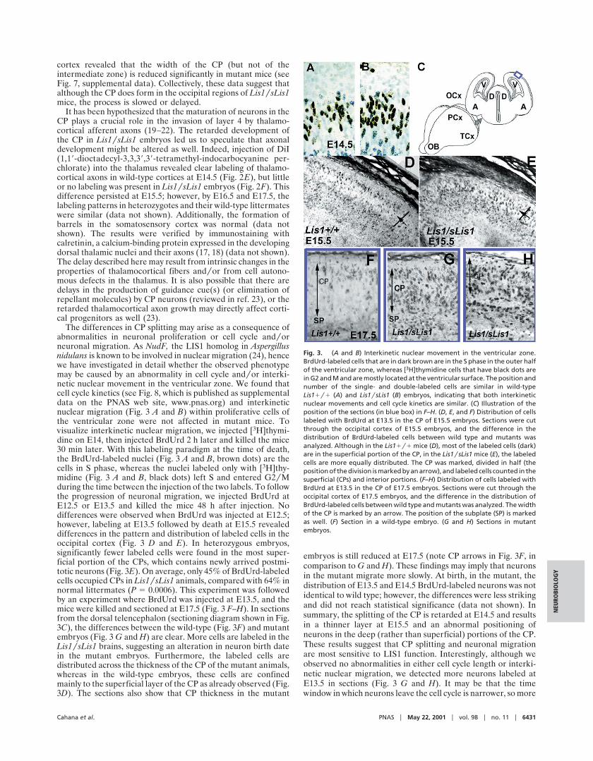
The differences in CP splitting may arise as a consequence ofabnormalities in neuronal proliferation or cell cycle andyorneuronal migration. As NudF, the LIS1 homolog in Aspergillusnidulans is known to be involved in nuclear migration (24), hencewe have investigated in detail whether the observed phenotypemay be caused by an abnormality in cell cycle andyor interki-netic nuclear movement in the ventricular zone. We found thatcell cycle kinetics (see Fig. 8, which is published as supplementaldata on the PNAS web site, www.pnas.org) and interkineticnuclear migration (Fig. 3 A and B) within proliferative cells ofthe ventricular zone were not affected in mutant mice. Tovisualize interkinetic nuclear migration, we injected [3H]thymi-dine on E14, then injected BrdUrd 2 h later and killed the mice30 min later. With this labeling paradigm at the time of death,the BrdUrd-labeled nuclei (Fig. 3 A and B, brown dots) are thecells in S phase, whereas the nuclei labeled only with [3H]thy-midine (Fig. 3 A and B, black dots) left S and entered G2yMduring the time between the injection of the two labels. To followthe progression of neuronal migration, we injected BrdUrd atE12.5 or E13.5 and killed the mice 48 h after injection. Nodifferences were observed when BrdUrd was injected at E12.5;however, labeling at E13.5 followed by death at E15.5 revealeddifferences in the pattern and distribution of labeled cells in theoccipital cortex (Fig. 3 D and E). In heterozygous embryos,significantly fewer labeled cells were found in the most super-ficial portion of the CPs, which contains newly arrived postmi-totic neurons (Fig. 3E). On average, only 45% of BrdUrd-labeledcells occupied CPs in Lis1ysLis1 animals, compared with 64% innormal littermates (P 5 0.0006). This experiment was followedby an experiment where BrdUrd was injected at E13.5, and themice were killed and sectioned at E17.5 (Fig. 3 F–H). In sectionsfrom the dorsal telencephalon (sectioning diagram shown in Fig.3C), the differences between the wild-type (Fig. 3F) and mutantembryos (Fig. 3 G and H) are clear. More cells are labeled in theLis1ysLis1 brains, suggesting an alteration in neuron birth datein the mutant embryos. Furthermore, the labeled cells aredistributed across the thickness of the CP of the mutant animals,whereas in the wild-type embryos, these cells are confinedmainly to the superficial layer of the CP as already observed (Fig.3D). The sections also show that CP thickness in the mutant
embryos is still reduced at E17.5 (note CP arrows in Fig. 3F, incomparison to G and H). These findings may imply that neuronsin the mutant migrate more slowly. At birth, in the mutant, thedistribution of E13.5 and E14.5 BrdUrd-labeled neurons was notidentical to wild type; however, the differences were less strikingand did not reach statistical significance (data not shown). Insummary, the splitting of the CP is retarded at E14.5 and resultsin a thinner layer at E15.5 and an abnormal positioning ofneurons in the deep (rather than superficial) portions of the CP.These results suggest that CP splitting and neuronal migrationare most sensitive to LIS1 function. Interestingly, although weobserved no abnormalities in either cell cycle length or interki-netic nuclear migration, we detected more neurons labeled atE13.5 in sections (Fig. 3 G and H). It may be that the timewindow in which neurons leave the cell cycle is narrower, so more
Fig. 3. (A and B) Interkinetic nuclear movement in the ventricular zone.BrdUrd-labeled cells that are in dark brown are in the S phase in the outer halfof the ventricular zone, whereas [3H]thymidine cells that have black dots arein G2 and M and are mostly located at the ventricular surface. The position andnumber of the single- and double-labeled cells are similar in wild-typeLis11y1 (A) and Lis1ysLis1 (B) embryos, indicating that both interkineticnuclear movements and cell cycle kinetics are similar. (C) Illustration of theposition of the sections (in blue box) in F–H. (D, E, and F) Distribution of cellslabeled with BrdUrd at E13.5 in the CP of E15.5 embryos. Sections were cutthrough the occipital cortex of E15.5 embryos, and the difference in thedistribution of BrdUrd-labeled cells between wild type and mutants wasanalyzed. Although in the Lis11y1 mice (D), most of the labeled cells (dark)are in the superficial portion of the CP, in the Lis1ysLis1 mice (E), the labeledcells are more equally distributed. The CP was marked, divided in half (theposition of the division is marked by an arrow), and labeled cells counted in thesuperficial (CPs) and interior portions. (F–H) Distribution of cells labeled withBrdUrd at E13.5 in the CP of E17.5 embryos. Sections were cut through theoccipital cortex of E17.5 embryos, and the difference in the distribution ofBrdUrd-labeled cells between wild type and mutants was analyzed. The widthof the CP is marked by an arrow. The position of the subplate (SP) is markedas well. (F) Section in a wild-type embryo. (G and H) Sections in mutantembryos.
Cahana et al. PNAS u May 22, 2001 u vol. 98 u no. 11 u 6431
NEU
ROBI
OLO
GY
do so within the time frame (25). This observation may berelevant to the given role of LIS1 in chromosome segregation(26). It is also possible that the introduced Lis1 mutation affectsneuronal proliferation only within an area limited to the ob-served phenotype.
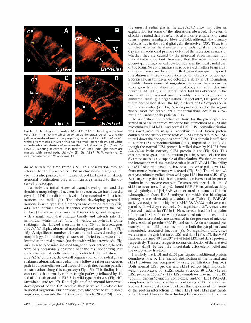
To study the initial stages of axonal development and thedendritic morphology of neurons in the cortex, we introduced acrystal of DiI into different levels of the cerebral wall to labelneurons and radial glia. The labeled developing pyramidalneurons in wild-type E14.5 embryos are oriented radially (Fig.4A), with normal apical dendrites directed toward the pialsurface (Fig. 4A, white arrow). Each soma is large and polygonal,with a single axon that emerges basally and extends into theprimordial white matter (Fig. 4A, yellow arrowhead). Quitestrikingly, the labeled neurons in the CP of heterozygousLis1ysLis1 display abnormal morphology and organization (Fig.4B). A significant number of neurons had altered multipolarmorphology. Interestingly, clusters of labeled cells were oftenlocated at the pial surface (marked with white arrowheads, Fig.4B). In wild-type mice, isolated tangentially oriented single cellswere only occasionally observed near the pia (not shown), butsuch clusters of cells were not detected. In addition, inLis1ysLis1 embryos, the overall organization of the radial glia isstrikingly abnormal: many glial fibers follow a rather curvaceouspath in dorsomedial cortical areas, although they remain parallelto each other along this trajectory (Fig. 4D). This finding is incontrast to the normally rather straight pathway followed by theradial glia observed at E15.5 in wild-type embryos (Fig. 4C,arrowhead, and ref. 27). Radial glia are fundamental for normaldevelopment of the CP, because they serve as a scaffold forneuronal migration. Furthermore, they also serve to route someingrowing axons into the CP (reviewed by refs. 28 and 29). Thus,
the unusual radial glia in the Lis1ysLis1 mice may offer anexplanation for some of the alterations observed. However, itshould be noted that in reeler, radial glia differentiate poorly andform a sparse misaligned fiber scaffold, although the primarydefect is not in the radial glial cells themselves (30). Thus, it isnot clear whether the abnormalities in radial glial cell morphol-ogy are an additional primary defect of the mutation in sLis1 orwhether they are caused by the neuronal abnormalities. It isundoubtedly important, however, that the most pronouncedphenotype during cortical development is in the most caudal partof the brain. No abnormalities were observed in other brain areasor organs; hence, we do not think that general nonspecific growthretardation is a likely explanation for the observed phenotype.Specifically, in this area, we detected a delay in CP formation,possibly slower neuronal migration, delay in thalamocorticalaxon growth, and abnormal morphology of radial glia andneurons. At E14.5, a unilateral extra fold was observed in thecortex of most mutant mice, possibly as a consequence ofabnormal radial glia organization. Importantly, this portion ofthe telencephalon shows the highest level of Lis1 expression inthe mouse cortex (see Fig. 6, www.pnas.org) and is the regionwhere most noticeable brain malformations occur in LIS1-mutated lissencephaly patients (31).
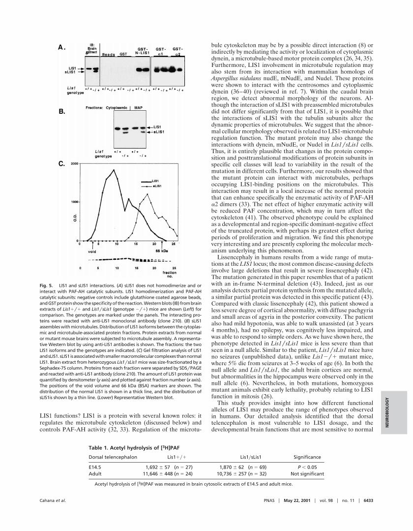
To understand the biochemical basis for the phenotypes ob-served in our mutant mice, we tested the interactions of sLIS1 andmicrotubules, PAH-AH, and normal LIS1. LIS1 homodimerizationwas investigated by using a recombinant GST fusion proteincontaining the first 95 amino acids of LIS1 (referred to as N-LIS1)to pull down the endogenous brain LIS1. This domain is sufficientto confer LIS1 homodimerization (O.R., unpublished data). Al-though the normal LIS1 protein is pulled down by N-LIS1 fromLis1ysLis1 brain extracts, sLIS1 protein is not (Fig. 5A). Thisexperiment suggests that the mutant protein, which lacks the first63 amino acids, is not capable of dimerization. We then examinedthe interaction with the catalytic subunits of PAF-AH. The abilityof GST fusion proteins of the bovine a1 and a2 to pull down LIS1from mouse brain extracts was tested (Fig. 5A). The a1 and a2catalytic subunits pulled down wild-type LIS1 but not sLIS1 (Fig.5A), suggesting that LIS1 homodimerization is essential for partic-ipation in the enzyme complex. To assess whether the failure ofsLIS1 to associate with a1ya2 altered PAF-AH enzymatic activity,acetyl hydrolysis of [3H]PAF was measured in extracts of dorsaltelencephalon from E14.5 embryos (in which the histologicalphenotype was observed) and adult mice (Table 1). PAF-AHactivity was significantly higher in E14.5 Lis1ysLis1 embryos com-pared with wild-type controls. No significant differences wereobserved in adult mice (Table 1). Next, we examined the interactionof the two LIS1 isoforms with preassembled microtubules. In thisassay, the microtubules are assembled in the presence of microtu-bule-associated proteins (MAPs) and taxol. As demonstrated pre-viously, normal LIS1 protein is found in both the cytoplasmic andmicrotubule-associated fractions (8). No significant differenceswere seen in the distribution of LIS1 and sLIS1 (Fig. 5B); the MAPfraction contained 40.7 and 37.3% of total LIS1 and sLIS1 proteins,respectively. This result suggests normal distribution of the mutatedprotein (sLIS1) between the microtubule cytoskeleton pellet andthe cytoplasmic fraction.
It is likely that LIS1 and sLIS1 participate in additional proteincomplexes in vivo. The fraction distribution of the normal andsLIS1 proteins was compared by using gel filtration (Fig. 5C).Both normal LIS1 protein and sLIS1 reflect high molecularweight complexes, but sLIS1 peaks at about 80 kDa, whereasLIS1 peaks at 150 kDa (12). LIS1 complexes may include LIS1tubulin, dyneinydynactin complexes, andyor LIS1-PAF-AHcomplexes, whereas complexes containing sLIS1 are not yetknown. However, it is obvious from this experiment that someof the protein interactions in which LIS1 and sLIS1 participateare different. How can these findings be associated with known
Fig. 4. DiI labeling of the cortex. (A and B) E14.5 DiI labeling of corticalcells. (Bar 5 1 mm.) The white arrow labels the apical dendrite, and theyellow arrowhead marks the projecting axon. Lis11y1 (A); Lis1ysLis1,white arrow marks a neuron that has ‘‘normal’’ morphology, and whitearrowheads mark clusters of neurons that look abnormal (B). (C and D)E15.5 DiI labeling of cortical cells. (Bar 5 25 mm.) Radial glia fibers aremarked with arrowheads. Lis11y1 (E), Lis1ysLis1 (F). V, ventricle; IZ,intermediate zone; CP*, abnormal CP.
6432 u www.pnas.orgycgiydoiy10.1073ypnas.101122598 Cahana et al.
LIS1 functions? LIS1 is a protein with several known roles: itregulates the microtubule cytoskeleton (discussed below) andcontrols PAF-AH activity (32, 33). Regulation of the microtu-
bule cytoskeleton may be by a possible direct interaction (8) orindirectly by mediating the activity or localization of cytoplasmicdynein, a microtubule-based motor protein complex (26, 34, 35).Furthermore, LIS1 involvement in microtubule regulation mayalso stem from its interaction with mammalian homologs ofAspergillus nidulans nudE, mNudE, and Nudel. These proteinswere shown to interact with the centrosomes and cytoplasmicdynein (36–40) (reviewed in ref. 7). Within the caudal brainregion, we detect abnormal morphology of the neurons. Al-though the interaction of sLIS1 with preassembled microtubulesdid not differ significantly from that of LIS1, it is possible thatthe interactions of sLIS1 with the tubulin subunits alter thedynamic properties of microtubules. We suggest that the abnor-mal cellular morphology observed is related to LIS1-microtubuleregulation function. The mutant protein may also change theinteractions with dynein, mNudE, or Nudel in Lis1ysLis1 cells.Thus, it is entirely plausible that changes in the protein compo-sition and posttranslational modifications of protein subunits inspecific cell classes will lead to variability in the result of themutation in different cells. Furthermore, our results showed thatthe mutant protein can interact with microtubules, perhapsoccupying LIS1-binding positions on the microtubules. Thisinteraction may result in a local increase of the normal proteinthat can enhance specifically the enzymatic activity of PAF-AHa2 dimers (33). The net effect of higher enzymatic activity willbe reduced PAF concentration, which may in turn affect thecytoskeleton (41). The observed phenotype could be explainedas a developmental and region-specific dominant-negative effectof the truncated protein, with perhaps its greatest effect duringperiods of proliferation and migration. We find this phenotypevery interesting and are presently exploring the molecular mech-anism underlying this phenomenon.
Lissencephaly in humans results from a wide range of muta-tions at the LIS1 locus; the most common disease-causing defectsinvolve large deletions that result in severe lissencephaly (42).The mutation generated in this paper resembles that of a patientwith an in-frame N-terminal deletion (43). Indeed, just as ouranalysis detects partial protein synthesis from the mutated allele,a similar partial protein was detected in this specific patient (43).Compared with classic lissencephaly (42), this patient showed aless severe degree of cortical abnormality, with diffuse pachgyriaand small areas of agyria in the posterior convexity. The patientalso had mild hypotonia, was able to walk unassisted (at 3 years4 months), had no epilepsy, was cognitively less impaired, andwas able to respond to simple orders. As we have shown here, thephenotype detected in Lis1ysLis1 mice is less severe than thatseen in a null allele. Similar to the patient, Lis1ysLis1 mice haveno seizures (unpublished data), unlike Lis12y1 mutant mice,where 5% die from seizures at 3–5 weeks of age (6). In both thenull allele and Lis1ysLis1, the adult brain cortices are normal,but abnormalities in the hippocampus were observed only in thenull allele (6). Nevertheless, in both mutations, homozygousmutant animals exhibit early lethality, probably relating to LIS1function in mitosis (26).
This study provides insight into how different functionalalleles of LIS1 may produce the range of phenotypes observedin humans. Our detailed analysis identified that the dorsaltelencephalon is most vulnerable to LIS1 dosage, and thedevelopmental brain functions that are most sensitive to normal
Fig. 5. LIS1 and sLIS1 interactions. (A) sLIS1 does not homodimerize and orinteract with PAF-AH catalytic subunits. LIS1 homodimerization and PAF-AHcatalytic subunits: negative controls include glutathione coated agarose beads,andGSTprotein showthespecificityof thereaction.Westernblots (IB) frombrainextracts of Lis11y1 and Lis1ysLis1 (genotype 2y1) mice are shown (Left) forcomparison. The genotypes are marked under the panels. The interacting pro-teins were reacted with anti-LIS1 monoclonal antibody (clone 210). (B) sLIS1assembles with microtubules. Distribution of LIS1 isoforms between the cytoplas-mic and microtubule-associated protein fractions. Protein extracts from normalor mutant mouse brains were subjected to microtubule assembly. A representa-tive Western blot by using anti-LIS1 antibodies is shown. The fractions: the twoLIS1 isoforms and the genotypes are indicated. (C) Gel filtration analysis of LIS1andsLIS1. sLIS1 isassociatedwithsmallermacromolecularcomplexes thannormalLIS1. Brain extract from heterozygous Lis1ysLis1 mice was size-fractionated by aSephadex-75 column. Proteins from each fraction were separated by SDSyPAGEand reacted with anti-LIS1 antibody (clone 210). The amount of LIS1 protein wasquantified by densitometer (y axis) and plotted against fraction number (x axis).The positions of the void volume and 66 kDa (BSA) markers are shown. Thedistribution of the normal LIS1 is shown in a thick line, and the distribution ofsLIS1is shown by a thin line. (Lower) Representative Western blot.
Table 1. Acetyl hydrolysis of [3H]PAF
Dorsal telencephalon Lis11y1 Lis1ysLis1 Significance
E14.5 1,692 6 57 (n 5 27) 1,870 6 62 (n 5 69) P , 0.05Adult 11,646 6 448 (n 5 24) 10,736 6 257 (n 5 32) Not significant
Acetyl hydrolysis of [3H]PAF was measured in brain cytosolic extracts of E14.5 and adult mice.
Cahana et al. PNAS u May 22, 2001 u vol. 98 u no. 11 u 6433
NEU
ROBI
OLO
GY
LIS1 functions include formation of the CP, neuronal migration,neuronal morphology, and radial glia morphology. As full un-derstanding of the molecular complexes formed by LIS1 and itsmutant alleles is achieved, the involvement of this gene incortical development will become clear, and the crucial steps itplays in human cortical development will be elucidated.
We thank Prof. Peter Lonai (Weizmann Institute, Rehovot, Israel) forCre mice, Prof. Marion Wassef for help in DiI injections, Dr. Ahuva
Kanishinsky, Tanya Burakuva, and Yehudit Hermesh for help with theanimals, and Monica Rodenas for technical assistance. This work wassupported in part by the Fritz Thyssen Stiftung Foundation, BinationalScience Foundation Grant No. 97–00014 (to O.R.), a grant from theSeneca Foundation of Murcia community, 708yCVy99 (to S.M.), a grantfrom the European Community, EC PL-960146 (to O.R., S.M., andW.W.), Human Frontier Science Program Organization Grant No.RG283199 9 (to O.R. and S.K.M.), and Volkswagen Stiftung (to O.R.and W.W.). O.R. is an Incumbent of the Aser Rothstein CareerDevelopment Chair in Genetic Diseases (Weizmann Institute).
1. Reiner, O., Carrozzo, R., Shen, Y., Whenert, M., Faustinella, F., Dobyns, W. B.,Caskey, C. T. & Ledbetter, D. H. (1993) Nature (London) 364, 717–721.
2. Reiner, O. (2000) Mol. Neurobiol. 20, 143–156.3. des Portes, V., Pinard, J. M., Billuart, P., Vinet, M. C., Koulakoff, A., Carrie,
A., Gelot, A., Dupuis, E., Motte, J., Berwald-Netter, Y., et al. (1998) Cell 92,51–61.
4. Gleeson, J. G., Allen, K. M., Fox, J. W., Lamperti, E. D., Berkovic, S., Scheffer,I., Cooper, E. C., Dobyns, W. B., Minnerath, S. R., Ross, M. E. & Walsh, C. A.(1998) Cell 92, 63–72.
5. Reiner, O., Albrecht, U., Gordon, M., Chianese, K. A., Wong, C., Sapir, T.,Siracusa, L. D., Buchberg, A. M., Caskey, C. T. & Eichele, G. (1995) J. Neurosci.15, 3730–3738.
6. Hirotsune, S., Fleck, M. W., Gambello, M. J., Bix, G. J., Chen, A., Clark, G. D.,Ledbetter, D. H., McBain, C. J. & Wynshaw-Boris, A. (1998) Nat. Genet. 19,333–339.
7. Reiner, O. (2000) Neuron 28, 633–636.8. Sapir, T., Elbaum, M. & Reiner, O. (1997) EMBO J. 16, 6977–6984.9. Henrique, D., Adam, J., Myat, A., Chitnis, A., Lewis, J. & Ish-Horowicz, D.
(1995) Nature (London) 375, 787–790.10. McConnell, S. K. & Kaznowski, C. E. (1991) Science 254, 282–285.11. Hayes, N. L. & Nowakowski, R. S. (2000) Dev. Neurosci. 22, 44–55.12. Sapir, T., Eisenstein, M., Burgess, H. A., Horesh, D., Cahana, A., Aoki, J.,
Hattori, M., Arai, H., Inoue, K. & Reiner, O. (1999) Eur. J. Biochem. 266,1011–1020.
13. Caspi, M., Atlas, R., Kantor, A., Sapir, T. & Reiner, O. (2000) Hum. Mol. Genet.9, 2205–2213.
14. Hattori, M., Arai, H. & Inoue, K. (1993) J. Biol. Chem. 268, 18748–18753.15. Collins, C. A. (1991) in Methods in Enzymology (Academic, New York), Vol.
196, pp. 246–253.16. Lallemand, Y., Luria, V., Haffner-Krausz, R. & Lonai, P. (1998) Transgenic
Res. 7, 105–112.17. Fonseca, M., del Rio, J. A., Martinez, A., Gomez, S. & Soriano, E. (1995)
J. Comp. Neurol. 361, 177–192.18. Frassoni, C., Arcelli, P., Selvaggio, M. & Spreafico, R. (1998) Neuroscience 83,
1203–1214.19. Ghosh, A. & Shatz, C. J. (1993) Development (Cambridge, U.K.) 117, 1031–1047.20. Ghosh, A., Antonini, A., McConnell, S. K. & Shatz, C. J. (1990) Nature
(London) 347, 179–181.21. Katz, L. C. & Shatz, C. J. (1996) Science 274, 1133–1138.22. Finney, E. M. & Shatz, C. J. (1998) J. Neurosci. 18, 8826–8838.
23. Kennedy, H. & Dehay, C. (1997) in Normal and Abnormal Development of theCortex, eds. Galaburda, A. M. & Christen, Y. (Springer, Berlin), pp. 25–56.
24. Xiang, X., Osmani, A. H., Osmani, S. A., Xin, M. & Morris, N. R. (1995) Mol.Biol. Cell 6, 297–310.
25. Takahashi, T., Nowakowski, R. S. & Caviness, V. S., Jr. (1996) J. Neurosci. 16,6183–6196.
26. Faulkner, N. E., Dujardin, D. L., Tai, C. Y., Vaughan, K. T., O’Connell, C. B.,Wang, Y. & Vallee, R. B. (2000) Nat. Cell Biol. 2, 784–791.
27. Edwards, M. A., Yamamoto, M. & Caviness, V. S. J. (1990) Neuroscience 36,121–144.
28. Super, H., Soriano, E. & Uylings, H. B. (1998) Brain Res. Brain Res. Rev. 27,40–64.
29. Rakic, P. (1995) Proc. Natl. Acad. Sci. USA 92, 11323–11327.30. Hunter-Schaedle, K. E. (1997) J. Neurobiol. 33, 459–472.31. Pilz, D. T., Matsumoto, N., Minnerath, S., Mills, P., Gleeson, J. G., Allen, K. M.,
Walsh, C. A., Barkovich, A. J., Dobyns, W. B., Ledbetter, D. H. & Ross, M. E.(1998) Hum. Mol. Genet. 7, 2029–2037.
32. Hattori, M., Adachi, H., Tsujimoto, M., Arai, N. & Inoue, K. (1994) Nature(London) 370, 216–218.
33. Manya, H., Aoki, J., Kato, H., Ishii, J., Hino, S., Arai, H. & Inoue, K. (1999)J. Biol. Chem. 274, 31827–31832.
34. Smith, D. S., Niethammer, M., Ayala, R., Zhou, Y., Gambello, M. J., Wynshaw-Boris, A. & Tsai, L. H. (2000) Nat. Cell Biol. 2, 767–775.
35. Liu, Z., Steward, R. & Luo, L. (2000) Nat. Cell Biol. 2, 776–783.36. Efimov, V. P. & Morris, N. R. (2000) J. Cell Biol. 150, 681–688.37. Feng, Y., Olson, E. C., Stukenberg, P. T., Flanagan, L. A., Kirschner, M. W.
& Walsh, C. A. (2000) Neuron 28, 665–679.38. Kitagawa, M., Umezu, M., Aoki, J., Koizumi, H., Arai, H. & Inoue, K. (2000)
FEBS Lett. 479, 57–62.39. Niethammer, M., Smith, D. S., Ayala, R., Peng, J., Ko, J., Lee, M.-S., Morabito,
M. & Tsai, L.-H. (2000) Neuron 28, 697–711.40. Sasaki, S., Shionoya, A., Ishida, M., Gambello, M., Yingling, J., Wynshaw-
Boris, A. & Hirotsune, S. (2000) Neuron 28, 681–696.41. Clark, G. D., McNeil, R. S., Bix, G. J. & Swann, J. W. (1995) NeuroReport 6,
2569–2575.42. Dobyns, W. B., Reiner, O., Carrozzo, R. & Ledbetter, D. H. (1993) J. Am. Med.
Assoc. 270, 2838–2842.43. Fogli, A., Guerrini, R., Moro, F., Fernandez-Alvarez, E., Livet, M. O., Renieri,
A., Cioni, M., Pilz, D. T., Veggiotti, P., Rossi, E., et al. (1999) Ann. Neurol. 45,154–161.
6434 u www.pnas.orgycgiydoiy10.1073ypnas.101122598 Cahana et al.
Related Documents


![Homodimerization of Ehd1 Is Required to Induce …...Homodimerization of Ehd1 Is Required to Induce Flowering in Rice1[OPEN] Lae-Hyeon Cho, Jinmi Yoon, Richa Pasriga, and Gynheung](https://static.cupdf.com/doc/110x72/5e6155d943d617346e72cbdc/homodimerization-of-ehd1-is-required-to-induce-homodimerization-of-ehd1-is-required.jpg)


