Targeted disruption of the neurofibromatosis type-1 gene leads to developmental abnormalities in heart and various neural crest-derived tissues Camilynn I. Brannan, Archibald S. Perkins, 1 Kristine S. Vogel, Nancy Ratner, 2 Michael L. Nordlund, 2 Susan W. Reid, Arthur M. Buchberg, a Nancy A. Jenkins, Luis F. Parada, and Neal G. Copeland Mammalian Genetics Laboratory, ABL-Basic Research Program, Frederick Cancer Research and Development Center, Frederick, Maryland 21702-1201 USA; ~Department of Pathology, Yale University School of Medicine, New Haven, Connecticut 06437 USA; 2Department of Anatomy and Cell Biology, University of Cincinnati College of Medicine, Cincinnati, Ohio 45267-0521 USA The neurofibromatosis {NF1) gene shows significant homology to mammalian GAP and is an important regulator of the ras signal transduction pathway. To study the function of NF1 in normal development and to try and develop a mouse model of NF1 disease, we have used gene targeting in ES cells to generate mice carrying a null mutation at the mouse Nfl locus. Although heterozygous mutant mice, aged up to 10 months, have not exhibited any obvious abnormalities, homozygous mutant embryos die in utero. Embryonic death is likely attributable to a severe malformation of the heart. Interestingly, mutant embryos also display hyperplasia of neural crest-derived sympathetic ganglia. These results identify new roles for NF1 in development and indicate that some of the abnormal growth phenomena observed in NF1 patients can be recapitulated in neurofibromin-deficient mice. [Key Words: Neurofibromatosis; NF1; Ras; ES cells; transgenic mice] Received February 8, 1994; revised version accepted March 25, 1994. Von Recklinghausen neurofibromatosis or neurofibro- matosis type 1 (NF1) affects i in 3500 humans, making it one of the most common inherited human diseases. NF1 is inherited as an autosomal dominant disease with the most common phenotypic manifestations resulting from abnormalities of neural crest-derived tissues (Riccardi 1981, 1991; Riccardi and Eichner 1986). These abnormal- ities include benign tumors of the peripheral nerves composed mainly of Schwann cells and fibroblasts (neu- rofibromas), tumors of the iris (Lisch nodules), and hy- perpigmentation of melanocytes (car6 au lait spots). In addition, NF1 patients are at an increased risk for spe- cific kinds of malignant disease, including neurofibrosa- rcoma, astrocytoma, pheochromocytoma and embryonal rhabdomyosarcoma (Bader 1986). Although the clinical manifestations of NF1 are trans- mitted in an autosomal dominant fashion, it has been hypothesized that NF1 mutations are recessive at the cell level. If true, NF1 would comply with the Knudson model devised to explain the inheritance of retinoblas- toma (Knudson 1971}, whereby one mutated and one 3Present address: Jefferson Cancer Institute, Department of Microbiol- ogy and Immunology, Philadelphia, Pennsylvania 19107-5541 USA. normal allele are inherited, but tumors do not arise until the normal allele acquires a mutation as well. This model would place NF1 in the class of tumor suppressor genes, which includes RB, WT1, P53, and possibly APC, whose encoded proteins control cell growth {Marshall 1991). Evidence that NF1 is a tumor suppressor gene is two- fold. First, the majority of germ-line NF1 mutations de- scribed thus far are predicted to produce no protein prod- uct (Gutmann and Collins 1993}. Second, clonally de- rived malignant neurofibrosarcomas express nearly no detectable NF1 protein (Basu et al. 1992; DeClue et al. 1992), and one neurofibrosarcoma from an NF1 patient contains a somatic 200-kb deletion that eliminates the 5' half of the normal NF1 allele. Therefore, both alleles are defective in this tumor (Legius et al. 1993). The NF1 gene was identified in 1990 by positional cloning (Viskochil et al. 1990; Wallace et al. 1990). The human NF1 gene spans >300 kb of genomic DNA and encodes an mRNA of 11-13 kb comprising -50 exons, which is capable of encoding a 2818-amino-acid protein (for review, see Gutman and Collins 1993). The NF1- encoded protein, neurofibromin, has extensive homol- ogy with two negative regulators of Ras in Saccharomy- GENES & DEVELOPMENT 8:1019-1029 © 1994 by Cold Spring Harbor Laboratory Press ISSN 0890-9369/94 $5.00 1019 Cold Spring Harbor Laboratory Press on September 5, 2016 - Published by genesdev.cshlp.org Downloaded from

Welcome message from author
This document is posted to help you gain knowledge. Please leave a comment to let me know what you think about it! Share it to your friends and learn new things together.
Transcript
Targeted disruption of the neurofibromatosis type-1 gene leads to developmental abnormalities in heart and various neural crest-derived tissues Camilynn I. Brannan, Archibald S. Perkins, 1 Kristine S. Vogel, Nancy Ratner, 2 Michael L. Nordlund, 2 Susan W. Reid, Arthur M. Buchberg, a Nancy A. Jenkins, Luis F. Parada, and Neal G. Copeland
Mammal ian Genetics Laboratory, ABL-Basic Research Program, Frederick Cancer Research and Development Center, Frederick, Maryland 21702-1201 USA; ~Department of Pathology, Yale University School of Medicine, New Haven, Connecticut 06437 USA; 2Department of Anatomy and Cell Biology, University of Cincinnati College of Medicine, Cincinnati, Ohio 45267-0521 USA
The neurofibromatosis {NF1) gene shows significant homology to mammalian GAP and is an important regulator of the ras signal transduction pathway. To study the function of NF1 in normal development and to try and develop a mouse model of NF1 disease, we have used gene targeting in ES cells to generate mice carrying a null mutation at the mouse Nfl locus. Although heterozygous mutant mice, aged up to 10 months, have not exhibited any obvious abnormalities, homozygous mutant embryos die in utero. Embryonic death is likely attributable to a severe malformation of the heart. Interestingly, mutant embryos also display hyperplasia of neural crest-derived sympathetic ganglia. These results identify new roles for NF1 in development and indicate that some of the abnormal growth phenomena observed in NF1 patients can be recapitulated in neurofibromin-deficient mice.
[Key Words: Neurofibromatosis; NF1; Ras; ES cells; transgenic mice]
Received February 8, 1994; revised version accepted March 25, 1994.
Von Recklinghausen neurofibromatosis or neurofibro- matosis type 1 (NF1) affects i in 3500 humans, making it one of the most common inherited human diseases. NF1 is inherited as an autosomal dominant disease with the most common phenotypic manifestations resulting from abnormalities of neural crest-derived tissues (Riccardi 1981, 1991; Riccardi and Eichner 1986). These abnormal- ities include benign tumors of the peripheral nerves composed mainly of Schwann cells and fibroblasts (neu- rofibromas), tumors of the iris (Lisch nodules), and hy- perpigmentation of melanocytes (car6 au lait spots). In addition, NF1 patients are at an increased risk for spe- cific kinds of malignant disease, including neurofibrosa- rcoma, astrocytoma, pheochromocytoma and embryonal rhabdomyosarcoma (Bader 1986).
Although the clinical manifestations of NF1 are trans- mitted in an autosomal dominant fashion, it has been hypothesized that NF1 mutations are recessive at the cell level. If true, NF1 would comply with the Knudson model devised to explain the inheritance of retinoblas- toma (Knudson 1971}, whereby one mutated and one
3Present address: Jefferson Cancer Institute, Department of Microbiol- ogy and Immunology, Philadelphia, Pennsylvania 19107-5541 USA.
normal allele are inherited, but tumors do not arise until the normal allele acquires a mutation as well. This model would place NF1 in the class of tumor suppressor genes, which includes RB, WT1, P53, and possibly APC, whose encoded proteins control cell growth {Marshall 1991).
Evidence that NF1 is a tumor suppressor gene is two- fold. First, the majority of germ-line NF1 mutations de- scribed thus far are predicted to produce no protein prod- uct (Gutmann and Collins 1993}. Second, clonally de- rived malignant neurofibrosarcomas express nearly no detectable NF1 protein (Basu et al. 1992; DeClue et al. 1992), and one neurofibrosarcoma from an NF1 patient contains a somatic 200-kb deletion that eliminates the 5' half of the normal NF1 allele. Therefore, both alleles are defective in this tumor (Legius et al. 1993).
The NF1 gene was identified in 1990 by positional cloning (Viskochil et al. 1990; Wallace et al. 1990). The human NF1 gene spans >300 kb of genomic DNA and encodes an mRNA of 11-13 kb comprising - 5 0 exons, which is capable of encoding a 2818-amino-acid protein (for review, see Gutman and Collins 1993). The NF1- encoded protein, neurofibromin, has extensive homol- ogy with two negative regulators of Ras in Saccharomy-
GENES & DEVELOPMENT 8:1019-1029 © 1994 by Cold Spring Harbor Laboratory Press ISSN 0890-9369/94 $5.00 1019
Cold Spring Harbor Laboratory Press on September 5, 2016 - Published by genesdev.cshlp.orgDownloaded from
Brannan et al.
ces cerevisiae, IRA1 and IRA2 (Ballester et al. 1990; Buchberg et al. 1990; Xu et al. 1990a). NF1 and IRA share homology with the domain of mammalian GAP that en- codes its GTPase-activating function. This GAP domain negatively regulates Ras by catalyzing the conversion of the active GTP-bound form of Ras to the inactive GDP- bound form (Hall 1990). The GAP-like domain of neu- rofibromin can complement the loss of the yeast RAS- binding protein IRA (Ballester et al. 1990; Xu et al. 1990b), and can interact with, and stimulate, the GTPase activity of Ras in vitro (Martin et al. 1990).
Ras is well known for its role in the control of cell proliferation but it can also induce morphological differ- entiation in PC12 cells (Bar-Sagi and Feramisco 1985; Noda et al. 1985). The biological response of cells to Ras-mediated signals is thought to reflect the intrinsic properties of a given cell type rather than an inherent property of the Ras protein itself. It also appears that NF1 has a complex role in signal transduction, depending on the cell type. Analysis of schwannoma cell lines from neurofibromatosis patients indicates that low levels of neurofibromin, and the subsequent reduction in GAP- like activity, are associated with an increase in Ras- GTP, resulting in the stimulated growth of these lines (Basu et al. 1992; DeClue et al. 1992). Normal GAP lev- els are found in schwannoma lines, indicating that neu- rofibromin is an important negative regulator of Ras function in schwannoma lines and is key to regulating cellular proliferation. In contrast, neuroblastoma and melanoma cell lines that lack neurofibromin do not show altered Ras-GTP levels (Johnson et al. 1993; The et al. 1993). This implies that the GAP-like activity of neu- rofibromin is not the sole regulator of Ras-GTP levels in these cell types and that its role may be independent of Ras, perhaps regulating a differentiation pathway.
The developmental expression pattern of NF1 protein has been analyzed in the rat (Daston and Ratner 1992) and the mouse (Huynh et al. 1994). Neurofibromin is uniformly distributed throughout embryonic days 8-11. At embryonic day 12, however, NF1 protein becomes enriched in a subset of tissues, including the heart, dor- sal root ganglia, liver, metanephros, stomach lining, and bronchial tubes (Huynh et al. 1994). By day 16, neurofi- bromin is enriched in the cortical plate, dorsal root and sympathetic ganglia, and the central nervous system (Daston and Ratner 1992). This neuronal restriction con- tinues into the adult where the protein is detectable only in the brain, spinal cord, peripheral nerve, and adrenal medulla (Daston et al. 1992; Golubic et al. 1992).
Two alternatively spliced NFI exons have been re- ported. One exon, exon 23a, is located within the GAP- related domain and encodes an additional 21 amino acids (Nishi et al. 1991). Using an antibody specific for this 21-amino-acid epitope, Huynh et al. (1994) demonstrated that this isoform is present at low levels from mouse embryonic day 8-11 but becomes highly enriched in the heart at day 12 before declining at embryonic day 13. In the adult mouse, this isoform is predominantly ex- pressed in the brain as detected by RNA reverse tran- scriptase-polymerase chain reactin (RT-PCR) analysis
(Nishi et al. 1991). The other alternatively spliced exon, exon 48a, is located near the carboxyl terminus of the protein and encodes an additional 18 amino acids. The expression pattern of this second isoform was analyzed by RNA RT-PCR in humans and found to be expressed at high levels in adult cardiac muscle, skeletal muscle, and bladder. High levels were also detected in fetal heart (Gutmann et al. 1993}. Additionally, this isoform was detected in muscle tissues from other vertebrate species, including mice. The expression profile of both alterna- tively spliced forms of NF1 includes embryonic and adult heart, suggesting a role for neurofibromin in nor- mal cardiac development.
Neurofibromin is highly conserved throughout evolu- tion: The mouse Nfl protein is >98% identical with the human protein (Bernards et al. 1993). This conservation suggests that a mouse model system might be useful for elucidating the role of NF1 in development and disease. To this end, we have used gene targeting in embryonic stem (ES) cells to generate mice that carry a null muta- tion at Nfl.
Results
Generation of mice with a mutated Nfl gene
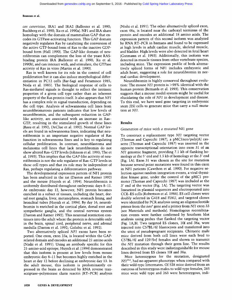
To construct a replacement type Nfl targeting vector (Thomas and Capecchi 1987), a pMClneo/poly(A) cas- sette (Thomas and Capecchi 1987) was inserted in the opposite transcriptional orientation into exon 31 of an Nfl genomic fragment, providing 7.1 kb of flanking ho- mology at the 5' end and 1.5 kb of homology at the 3' end (Fig. 1A). Exon 31 was chosen as the site for mutation because several point mutations were found in this exon in NF1 patients (Cawthon et al. 1990). For negative se- lection against random integration events, a viral thymi- dine kinase gene, under the control of the pMC1 pro- moter (Thomas and Capecchi 1987) was included at the 3' end of the vector (Fig. 1A). The targeting vector was linearized in plasmid sequences and electroporated into CCE-ES cells (Robertson et al. 1986). ES cell clones were doubly selected in G418 and FIAU, and targeted clones were identified by PCR analysis using an oligonucleotide primer from the neo r gene and a primer from Nfl exon 33 (see Materials and methods). Homologous recombina- tion events were further confirmed by Southern blot analysis using probes that flanked the targeting vector (Fig. 1A, B). Two targeted ES clones, 10f and 38a, were injected into C57BL/6J blastocysts and transferred into the uteri of pseudopregnant recipients. Chimeric male mice derived from both cell lines were each bred to C57BL/6J and 129/SvJ females and shown to transmit the Nfl mutation through their germ line. The results described in this study were indistinguishable for mouse lines derived from ES clones 10f and 38a.
Mice heterozygous for the mutation, designated Nfl ~cr, had no apparent phenotype when compared with their wild-type littermates. Of 528 mice derived from an outcross of heterozygous males to wild-type females, 265 mice were wild type and 263 were heterozygous, indi-
1020 GENES & DEVELOPMENT
Cold Spring Harbor Laboratory Press on September 5, 2016 - Published by genesdev.cshlp.orgDownloaded from
Disruption of Nfl
A exon: 28 29
,P U R,,P ,"ram Vmm,,R"
1Kb I I
,P U ~.P ,"-- ," • ,R., mm •
probe:
[
X
m N I
31 32 33 34
N H N P dm m ~m~m, R R i I • • • •
N R I < - ~ , > ,
P PR
~ Homologous integration
H N
I ' P PR
B C
Predicted fragment sizes
Allele ProbeA Probe B Probe C Pst I Pst 1 Eco R!
Wild-type (Kb) 11.8 3.2 1.6
Mutant (Kb) 11.0 11.0 2.6 1.6
B Probe
Enzyme
23.1 -
9 . 4 -
6 . 6 -
4 . 4 -
2 . 3 B
2 . 0 -
A B
P s t l P s t l
10f 38a 18b 10f 38a 18b
C
E c o R l
10f 38a 18b
23.1
9.4 6.6
4.4
2.3
2 . 0
Figure 1. Targeted disruption of the NfI locus. (A) Homologous recombination at the Nfl locus. The top line represents the structure of the region of the Nfl gene containing exons 28-34. Exons are shown as black boxes. The middle line represents the targeting vector in which the 1.1-kb pMClneo/poly(A) + cassette has been inserted into the NcoI site of exon 31 and a viral thymidine gene, under the control of the pMC1 promoter, which has been placed at the 3' end. Arrows indicate the direction of transcription of the neo ~ and tk genes. In the bottom line is the predicted structure of the locus following targeted integration of the replacement vector. Homologous recombination events were confirmed by probing Southern blots of ES cell clones with probes A, a 4-kb HindIII fragment; B, a 680-bp Neo PstI fragment; and C, a 1-kb PstI-HindIII fragment. The expected sizes of diagnostic restriction fragments are given in the box. (H) HindIII;/P} PstI; (R) EcoRI; {N) NcoI. (B) Southern blot analysis of ES cell clones transfected with the targeting vector. DNA from Neo r FIAU r ES clones were digested with PstI or EcoRI, blotted, and hybridized with either probes A, B, or C. The clones 10f and 38a contain bands diagnostic of homologous recombination at the NfI locus. Clone 18b represents a random integration event.
cating that there is no reduction in early survival of mice containing one copy of the N f l For allele. In addition, het- erozygotes, aged up to 10 months, have yet to display any abnormalit ies similar to those observed in h u m a n NF1 patients.
H o m o z y g o u s Nfl Fcr m i c e die in utero
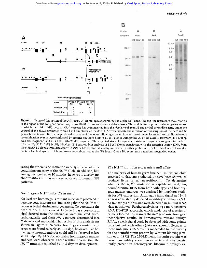
No liveborn homozygous mutan t mice were produced in heterozygous intercrosses, indicating that the N f l For mu- tation is lethal during embryogenesis. To determine the t ime of death, embryos at 11.5-14.5 days postcoi tum (dpc) derived from the intercross were analyzed histo- pathologically and their N f l genotype determined (see Materials and methods). The results of this analysis are shown in Figure 2. Necrotic homozygous mutan t em- bryos were found as early as 11.5 dpc; however, live ho- mozygous mu tan t embryos could still be observed as late as 13.5 dpc. By 14.5 dpc no viable homozygous mutan t embryos were observed. These results indicate that the Xf l For muta t ion is lethal by 14.5 days in development.
The Nfl For m u t a t i o n represents a nu l l allele
The majority of h u m a n germ-line NF1 mutat ions char- acterized to date are predicted, or have been shown, to produce little or no neurofibromin. To determine whether the N f l ~cr muta t ion is capable of producing neurofibromin, RNA from both wild-type and homozy- gous mutan t embryos was analyzed by Nor thern analy- sis for N f l expression. Although a faint signal at 11-13 kb was consistently detected in wild-type embryo RNA, no transcripts of this size were detected in mu tan t RNA (data not shown). Further analysis using a more sensitive RNA RT-PCR approach, which made use of a series of primers located upstream of the neo r gene insertion, gave inconclusive results. In homozygous m u t a n t embryo RNAs, a weak signal could be detected wi th some primer pairs but not wi th others (data not shown). Because of these ambiguous RNA results we decided to test directly for the neurofibromin protein by Western blotting (Dat- son et al. 1992). The 220-kD neurofibromin protein was present in wild-type embryo extracts and was consis- tently present in heterozygous l i t termate embryo ex-
GENES & DEVELOPMENT 1021
Cold Spring Harbor Laboratory Press on September 5, 2016 - Published by genesdev.cshlp.orgDownloaded from
Brannan et al.
7O
"-" 60
.E 50
"~ 40
= 30 O
"~ 20 O
O ' - 10
• +/+ [] +/m [] m/m [] necrotic
mR I I I
11.5 12.5 13.5 14.5
Embryonic Age (Days) Birth
/ / 0 H
Figure 2. Genotype and phenotype of Nfl For mutant embryos at varying gestational ages. Cumulative genotype data from analysis of 504 embryos derived from 69 litters were sorted by gestational age. The percentage of embryos of each genotype is plotted. Wild-type embryos (solid boxes); heterozygous animals (crosshatched boxes); homozygous mutant animals (open boxes). Overtly necrotic embryos are indicated by stippled boxes. Slashed x-axis indicates that this analysis did not include embryos beyond 14.5 days of gestation. The genotypes of live- born mice are included for comparison.
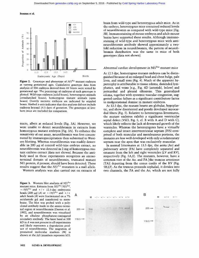
tracts, albeit at reduced levels (Fig. 3A). However, we were unable to detect neurofibromin in extracts from homozygous mutan t embryos (Fig. 3A). To enhance the sensit ivity of our assay, neurofibromin was first concen- trated by immunoprec ip i ta t ion then submit ted to West- ern blotting. Whereas neurofibromin was readily detect- able in 200 ~g of control wild-type embryo extract, no neurofibromin was detected in 2 mg of homozygous mu- tant embryo extract (data not shown). Because the anti- body used in these experiments recognizes an amino- terminal domain of neurofibromin, truncated mutan t Nf l protein, if present, should have been detected. These results suggest that the Nf l For mutat ion is a nul l allele.
Western analysis was also carried out on extracts of
brain from wild-type and heterozygous adult mice. As in the embryo, heterozygous mice contained reduced levels of neurofibromin as compared wi th wild-type mice (Fig. 3B). Immunosta in ing of mouse embryos and adult mouse brains have supported these results. Although immuno- staining of wild-type and heterozygous mice wi th anti- neurofibromin antibody showed approximately a two- fold reduction in neurofibromin, the pattern of neurofi- bromin distribution was the same in mice of both genotypes (data not shown).
Abnormal cardiac development in N f l Fc* m u t a n t mice
At 12.5 dpc, homozygous mutan t embryos can be distin- guished because of an enlarged head and chest bulge, pale liver, and small eyes (Fig. 4). Much of the apparent hy- pertrophy is attributable to tissue edema, distended lym- phatics, and veins [e.g., Fig. 6D (asterisk), below] and pericardial and pleural effusions. This generalized edema, together wi th systemic vascular congestion, sug- gested cardiac failure as a significant contributory factor to midgestational demise in mutan t embryos.
At 13.5 dpc, the mutan t hearts are globular, hypoplas- tic, and show disoriented and poorly developed myocar- dial fibers (Fig. 5). Relative to heterozygous li t termates, the mutan t embryos exhibit a significant ventricular septal defect (VSD; Fig. 5, cf. B wi th A and D wi th C), which likely reflects the lack of downward growth of the ventricles. Whereas the heterozygotes have a vir tual ly complete and intact interventricular septum (IVS) com- prised of both muscular and membranous portions, the mutants are less well developed wi th only a rudimentary septum near the apex that was exclusively muscular.
In normal l i t termates at 13.5 dpc, the aorta (Ao) and pulmonary artery (PA) have completely separated and emanate from the left and right ventricles (LV and RV), respectively (Fig. 5A, E). The mutants , however, have a common root of the Ao. and PA [the truncus arteriosus (TA)] departing from the conus cordis of the RV (Fig. 5B, I,F). As the truncus proceeds cephalad, it divides into two channels, the PA and the Ao, which are not fully
Figure 3. Westem blot analysis of Nflrcr mutant mice. Extracts from Nfl Vcr/Nflrcr, +/Nfl Fc~ and + / + 12.5-dpc embryonic heads (200 ~g)(A) or +/Nfl F~ and + / + adult heads (B) were fractionated on a 7% acrylamide gel and transferred to mem- brane. The blot was probed with a poly- clonal antibody made to the amino-termi- nal region of neurofibromin (Daston et al. 1992), and neurofibromin was visualized by an alkaline phosphatase-conjugated secondary antibody. The faint band at 200 kD in A was not present in all experiments and likely represents a degradation prod- uct of neurofibromin. The migration of prestained molecular markers (M) is shown at the left (numbers represent kD).
A
205 --",'-
116.5 '--~
B M - / - + 1 - + 1 - + 1 - +1+ + / + M + 1 + + 1 -
205
116.5 . . - -~
1022 GENES & DEVELOPMENT
Cold Spring Harbor Laboratory Press on September 5, 2016 - Published by genesdev.cshlp.orgDownloaded from
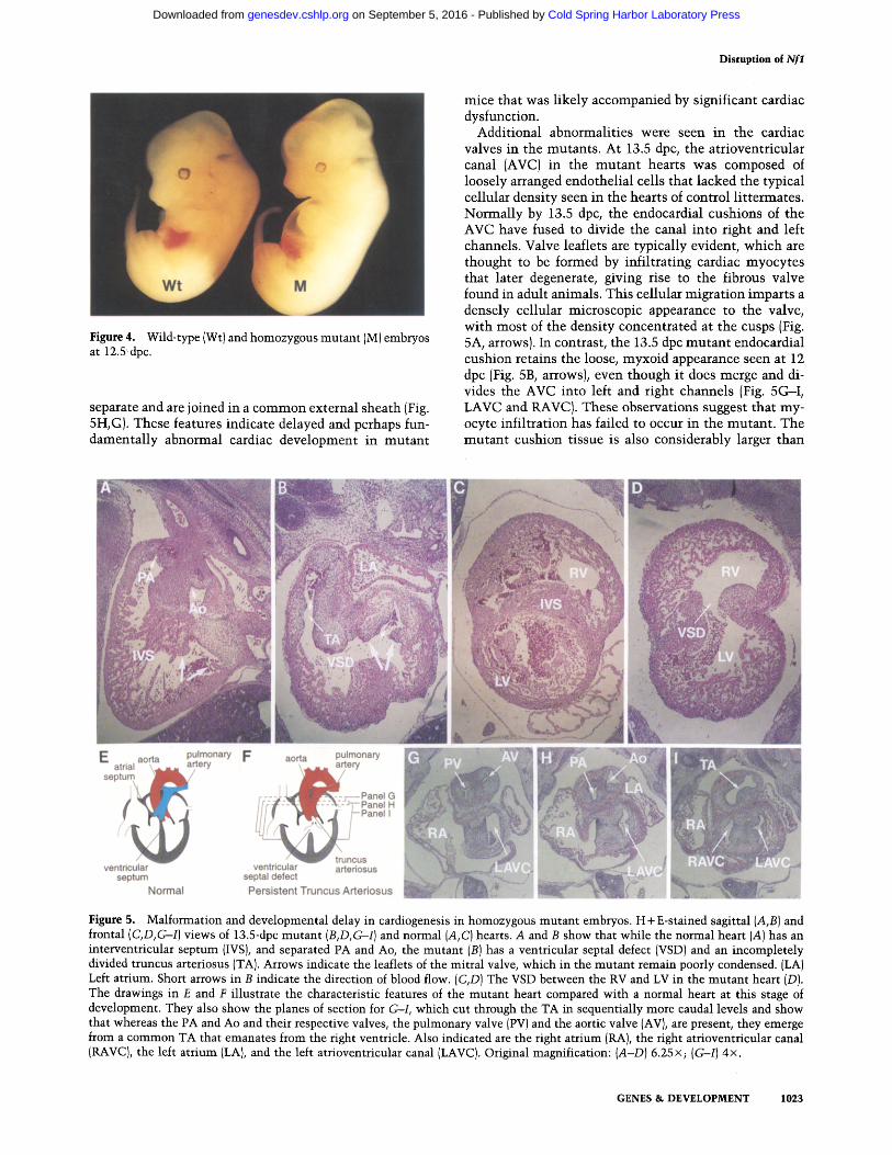
Figure 4. Wild-type (Wt) and homozygous mutant (M) embryos at 12.5 dpc.
separate and are joined in a common external sheath (Fig. 5H, G). These features indicate delayed and perhaps fun- damental ly abnormal cardiac development in mutan t
Disruption of Nfl
mice that was l ikely accompanied by significant cardiac dysfunction.
Additional abnormali t ies were seen in the cardiac valves in the mutants . At 13.5 dpc, the atrioventricular canal (AVC) in the mutan t hearts was composed of loosely arranged endothelial cells that lacked the typical cellular density seen in the hearts of control l i t termates. Normal ly by 13.5 dpc, the endocardial cushions of the AVC have fused to divide the canal into right and left channels. Valve leaflets are typically evident, which are thought to be formed by infi l trat ing cardiac myocytes that later degenerate, giving rise to the fibrous valve found in adult animals. This cellular migrat ion imparts a densely cellular microscopic appearance to the valve, wi th most of the density concentrated at the cusps (Fig. 5A, arrows). In contrast, the 13.5 dpc mutan t endocardial cushion retains the loose, myxoid appearance seen at 12 dpc (Fig. 5B, arrows), even though it does merge and di- vides the AVC into left and right channels (Fig. 5G--I, LAVC and RAVC). These observations suggest that my- ocyte infil tration has failed to occur in the mutant . The mutan t cushion tissue is also considerably larger than
E atrial aor septum
ventricular septum
Normal
pulmonary F ery
c
aorta pulmonary I ~ e r y
ventricular arteriosus septal defect Persistent Truncus Arteriosus
Figure 5. Malformation and developmental delay in cardiogenesis in homozygous mutant embryos. H + E-stained sagittal (A,B) and frontal (C,D,G-I) views of 13.5-dpc mutant (B,D,G--I) and normal {A, C) hearts. A and B show that while the normal heart (A) has an interventricular septum (IVS), and separated PA and Ao, the mutant (B) has a ventricular septal defect (VSD) and an incompletely divided truncus arteriosus (TA). Arrows indicate the leaflets of the mitral valve, which in the mutant remain poorly condensed. (LA) Left atrium. Short arrows in B indicate the direction of blood flow. (C,D) The VSD between the RV and LV in the mutant heart (D). The drawings in E and F illustrate the characteristic features of the mutant heart compared with a normal heart at this stage of development. They also show the planes of section for G-I, which cut through the TA in sequentially more caudal levels and show that whereas the PA and Ao and their respective valves, the pulmonary valve (PV) and the aortic valve (AV), are present, they emerge from a common TA that emanates from the right ventricle. Also indicated are the right atrium (RA), the right atrioventricular canal (RAVC), the left atrium (LA}, and the left atrioventricular canal (LAVC). Original magnification: {A-D) 6.25x; (G-I) 4x.
GENES & DEVELOPMENT 1023
Cold Spring Harbor Laboratory Press on September 5, 2016 - Published by genesdev.cshlp.orgDownloaded from
Brannan et al.
controls at 13.5 dpc (Fig. 5A, B), which may reflect con- tinued proliferation of this tissue or failure of the endo- thelial cells that comprise the structure to undergo apo- ptosis, or tissue edema.
Delay in renal, hepatic, and skeletal muscle development
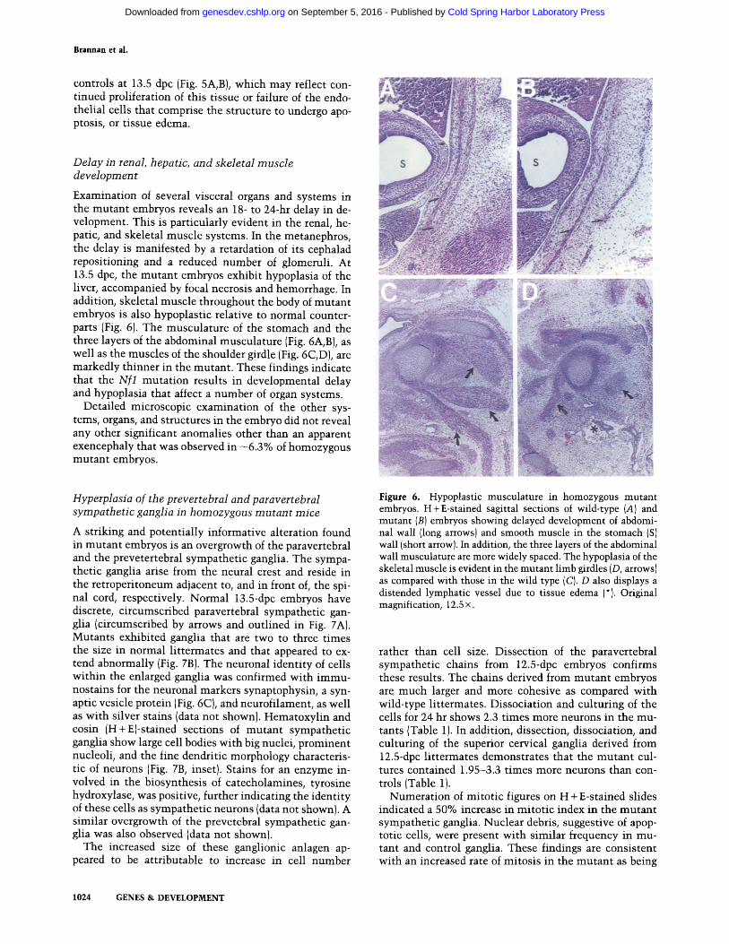
Examination of several visceral organs and systems in the mutant embryos reveals an 18- to 24-hr delay in de- velopment. This is particularly evident in the renal, he- patic, and skeletal muscle systems. In the metanephros, the delay is manifested by a retardation of its cephalad repositioning and a reduced number of glomeruli. At 13.5 dpc, the mutant embryos exhibit hypoplasia of the liver, accompanied by focal necrosis and hemorrhage. In addition, skeletal muscle throughout the body of mutant embryos is also hypoplastic relative to normal counter- parts (Fig. 6). The musculature of the stomach and the three layers of the abdominal musculature (Fig. 6A, B), as well as the muscles of the shoulder girdle (Fig. 6C, D), are markedly thinner in the mutant. These findings indicate that the Nfl mutation results in developmental delay and hypoplasia that affect a number of organ systems.
Detailed microscopic examination of the other sys- tems, organs, and structures in the embryo did not reveal any other significant anomalies other than an apparent exencephaly that was observed in ~6.3% of homozygous mutant embryos.
Hyperplasia of the prevertebral and paravertebral sympathetic ganglia in homozygous mutant mice
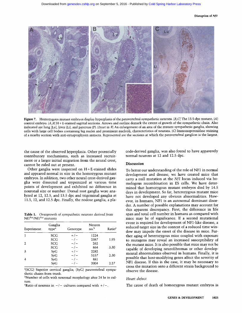
A striking and potentially informative alteration found in mutant embryos is an overgrowth of the paravertebral and the prevetertebral sympathetic ganglia. The sympa- thetic ganglia arise from the neural crest and reside in the retroperitoneum adjacent to, and in front of, the spi- nal cord, respectively. Normal 13.5-dpc embryos have discrete, circumscribed paravertebral sympathetic gan- glia (circumscribed by arrows and outlined in Fig. 7A). Mutants exhibited ganglia that are two to three times the size in normal littermates and that appeared to ex- tend abnormally (Fig. 7B). The neuronal identity of cells within the enlarged ganglia was confirmed with immu- nostains for the neuronal markers synaptophysin, a syn- aptic vesicle protein (Fig. 6C), and neurofilament, as well as with silver stains (data not shown). Hematoxylin and eosin (H+E)-stained sections of mutant sympathetic ganglia show large cell bodies with big nuclei, prominent nucleoli, and the fine dendritic morphology characteris- tic of neurons (Fig. 7B, inset). Stains for an enzyme in- volved in the biosynthesis of catecholamines, tyrosine hydroxylase, was positive, further indicating the identity of these cells as sympathetic neurons (data not shown). A similar overgrowth of the prevetebral sympathetic gan- glia was also observed (data not shown).
The increased size of these ganglionic anlagen ap- peared to be attributable to increase in cell number
!
Figure 6. Hypoplastic musculature in homozygous mutant embryos. H+E-stained sagittal sections of wild-type (A) and mutant (B) embryos showing delayed development of abdomi- nal wall (long arrows) and smooth muscle in the stomach (S) wall (short arrow). In addition, the three layers of the abdominal wall musculature are more widely spaced. The hypoplasia of the skeletal muscle is evident in the mutant limb girdles (D, arrows) as compared with those in the wild type (C). D also displays a distended lymphatic vessel due to tissue edema (*). Original magnification, 12.5 x.
rather than cell size. Dissection of the paravertebral sympathetic chains from 12.5-dpc embryos confirms these results. The chains derived from mutant embryos are, much larger and more cohesive as compared with wild-type littermates. Dissociation and culturing of the cells for 24 hr shows 2.3 times more neurons in the mu- tants (Table 1). In addition, dissection, dissociation, and culturing of the superior cervical ganglia derived from 12.5-dpc littermates demonstrates that the mutant cul- tures contained 1.95-3.3 times more neurons than con- trols (Table 1).
Numeration of mitotic figures on H + E-stained slides indicated a 50% increase in mitotic index in the mutant sympathetic ganglia. Nuclear debris, suggestive of apop- totic cells, were present with similar frequency in mu- tant and control ganglia. These findings are consistent with an increased rate of mitosis in the mutant as being
1024 GENES & DEVELOPMENT
Cold Spring Harbor Laboratory Press on September 5, 2016 - Published by genesdev.cshlp.orgDownloaded from
Disruption of Nfl
Figure 7. Homozygous mutant embryos display hyperplasia of the paravertebral sympathetic neurons. (B,C) The 13.5-dpc mutant; (A) control embryo. (A,B) H + E-stained sagittal sections. Arrows and outline demark the extent of growth of the sympathetic chain. Also indicated are lung {Lu), liver (Li), and pancreas (P). (Inset in B) An enlargement of an area of the mutant sympathetic ganglia, showing ceils with large cell bodies containing big nuclei and prominent nucleoli, characteristics of neurons. (C) Immunoperoxidase staining of a nearby section with anti-synaptophysin antisera. Represented are the sections at which the paravertebral ganglion is the largest.
the cause of the observed hyperplasia. Other potentially contributory mechanisms, such as increased recruit- ment or a larger init ial migrat ion from the neural crest, cannot be ruled out at present.
Other ganglia were inspected on H + E-stained slides and appeared normal in size in the homozygous mutan t embryos. In addition, two other neural crest-derived gan- glia were dissected and trypsinized at various t ime points of development and exhibited no difference in neuronal size or number: Dorsal root ganglia were ana- lyzed at 12, 12.5, and 13.5 dpc and tr igeminal ganglia at 11.5, 12, and 12.5 dpc. Finally, the nodose ganglia, a pla-
Table 1. Overgrowth of sympathetic neurons derived from NflFCr/NflFcr mutants
Ganglia Neuron Experiment type a Genotype no. b Ratio ¢
1 S C G + / - 1224
S C G / 2387 1.95
2 S C G + / - 262
S C G / 864 3 .30
3 S y G + / - 2282
S y G / 5157 2.30 4 S y G + / - 881
S y G / 2004 2.27
a(SCG) Superior cervical ganglia; (SyG) paravertebral sympa- thetic chains from trunk. bNumber of cells with neuronal morphology after 24 hr in cul- ture. CRatio of neurons in - / - cultures compared with + / - .
code-derived ganglia, was also found to have apparently normal neurons at 12 and 12.5 dpc.
Discussion
To better our understanding of the role of NF 1 in normal development and disease, we have created mice that carry a nul l muta t ion at the Nfl locus induced via ho- mologous recombinat ion in ES cells. We have deter- mined that homozygous mutan t embryos died by 14.5 days in development. So far, heterozygous mutan t mice have not developed any obvious abnormalit ies. How- ever, in humans, NF1 is an autosomal dominant disor- der. A number of possible explanations may account for this apparent discrepancy. First, the difference in life span and total cell number in humans as compared wi th mice may be of significance. If a second muta t ional event is required for development of NFl - l ike disease, a reduced target size in the context of a reduced t ime win- dow may impede the onset of the disease in mice. Fur- ther aging of heterozygous mice coupled wi th exposure to mutagens may reveal an increased susceptibil i ty of the mutan t mice. It is also possible that mice may not be capable of developing neurofibromas or other develop- menta l abnormali t ies observed in humans. Finally, it is possible that host-modifying genes affect the severity of NF1 disease. If this is the case, it may be necessary to cross the muta t ion onto a different strain background to observe the disease.
Heart defect
The cause of death of homozygous mutan t embryos is
GENES & DEVELOPMENT 1025
Cold Spring Harbor Laboratory Press on September 5, 2016 - Published by genesdev.cshlp.orgDownloaded from
Brannan et al.
likely attributable to cardiac dysfunction. We base this conclusion on the many abnormal morphologic features consistent with this interpretation, including systemic edema, dilatation of the venous and lymphatic systems, and severe abnormalities of the heart. Whereas cardiac dysfunction is an obvious cause for these findings, the small size of the dying embryos precludes functional as- sessment of cardiac function in vivo; therefore, addi- tional causes of hydrops, such as hypoproteinemia or re- nal dysfunction, must be considered. Although develop- mental delay could account for some aspects of the abnormal mutant hearts, a fundamental defect in cardiac development is also suggested by the histopathological results.
The severity of the malformations observed in the mu- tant embryo hearts indicate that neurofibromin has an important function in cardiac organogenesis and perhaps myocardial function. The abnormalities are multiple and complex and include overall hypoplasticity, and disori- ented and poorly developed myocardial fibers, as well as a ventricular septal defect attributable to insufficient ventricular growth, and abnormalities in valve leaflet formation. The severity and the timing of the cardiac defects is intruiging in light of new data regarding the expression profile of neurofibromin. For example, Huynh et al. (1994) have demonstrated recently that an isoform of NF1 containing 21 additional amino acids within the GAP homology domain shows dramatic changes in lev- els during development: Expression is low at 11 dpc, peaks in the heart at 12 dpc, and declines by 13 dpc. This peak of expression in the heart is at precisely the stage in embryogenesis where the major heart defects in Nfl-de- ficient mice are observed. In addition, an alternatively spliced isoform of NF1 located in the carboxy-terminal end of the protein has recently been detected in fetal cardiac muscle in addition to adult muscle (Gutmann et al. 1993). Although it has not yet been confirmed that a protein encoding this isoform is produced in vivo, it is clear that the fetal heart is home to the two rare isoforms of neurofibromin. This information, together with the severe heart defects in embryos lacking Nfl, strongly argues that neurofibromin is essential for normal cardiac development.
In addition to the above abnormalities, hearts derived from homozygous mutant embryos contain a common departure of Ao and PA from the RV and incomplete separation of these two outflow vessels. This defect, called persistent truncus arteriosus, is commonly seen in congenitally anomalous human hearts. Interestingly, persistent TA can be induced in the developing chick by ablation of neural crest cells from the level of somites 1-3 (Kirby et al. 1983). From chick-quail chimera studies it has been established that the neural crest contributes to the ectomesenchymal cells that are involved in divid- ing the TA into PA and Ao, as well as in the formation of the pulmonary and aortic valves (Kirby et al. 1983). The caudal extent of migration appears to be the conus cor- dis, so that neural crest-derived cells do not contribute to the formation of the valves in the AVC. It is unlikely that lack of neurofibromin results in a complete block of
neural crest migration into the heart, as in the mutant the truncus divides into two vessels, and the pulmonary and aortic valves are present. But the similarity of the phenotype observed in the mutant mice--common out- flow tract and ventricular septal defect--to that seen with ablation of neural crest cells from the level of somites 1-3 in the chick, suggests that neurofibromin might play an important role in the migration or func- tional capacity of these neural crest-derived cells.
Although cardiac abnormalities were observed in all homozygous mutant Nfl mouse embryos, only a small number of human patients with NF1 have been diag- nosed with congenital heart disease {Kaufman et al. 1972). This is most likely explained by the fact that hu- man NF1 patients carry only one germ-line copy of the NF1 mutation and most cells in the heart would thus be expected to be heterozygous for the mutation during this critical window of cardiac development in utero.
Renal, hepatic, and skeletal muscle defects
An 18- to 24-hr delay was evident in two organs, the liver and the kidney, as well as in the skeletal muscle system of homozygous mutant embryos. Again, this nicely re- flects the pattern of neurofibromin expression in these organs. At 12 dpc, in addition to strong staining in the heart, the liver and metanephros are enriched in neufi- bromin (Huynh et al. 1994). However, whereas the heart contains mainly the isoform with the extra 21 amino acids derived from exon 23a, the liver and presumptive kidney are enriched in the isoform without these amino acids (Huynh et al. 1994).
The poorly developed skeletal musculature of the mu- tant embryos that we observed in the stomach and the limb buds may similarly be attributable to the isoform derived from an Nfl mRNA containing the alternatively spliced exon, 48a. It has been demonstrated that this isoform is present at high levels in adult skeletal muscle (Gutmann et al. 1993), but it is not known whether it is present in fetal skeletal muscle.
NF1 as a regulator of growth
Hyperplasia of the prevertebral and paravertebral sympa- thetic ganglia is a consistent feature of mutant 13.5-dpc embryos. Because of similarities, if not identities, in cel- lular origin, phenotypic markers, and even certain mor- phologic features, these lesions are analogous to the ex- tra-adrenal pheochromocytomas that occur in patients with NF1. Although the demise of the mutant embryos before 14.5 dpc prevents the growth capacity and malig- nant potential of these lesions from being determined, this abnormal proliferation of neural crest-derived cells suggests an important role for the NF1 gene product in regulating growth in this cell type. The absence of hy- perplasia of other peripheral nerves in homozygous mu- tant mice at the time of death could be attributable to the amount of time needed for these lesions to become evident but more likely reflects an intrinsic difference in the role of Nfl in the different neuronal populations.
1026 GENES & DEVELOPMENT
Cold Spring Harbor Laboratory Press on September 5, 2016 - Published by genesdev.cshlp.orgDownloaded from
Disruption of Nfl
Does the hyperplas ia of sympa the t i c ganglia in the m u t a n t embryos impl ica te Nf l as a negat ive growth reg- u la tor in these cells? Sympathoadrena l precursors, after arising f rom the neura l crest, in i t i a te migra t ion to form the sympa the t i c chain on day 10 pc (in the rat) and con- ta in ca t echo lamine syn the t i c enzymes by 12.5 dpc (Co- chard et al. 1978). Tha t a two- to threefold difference in size of the ganglia exists so soon after its in i t ia l estab- l i s h m e n t in m u t a n t and wild- type embryos indicates a s ignif icant difference be tween m u t a n t and wild- type cells in rates of cell division, cell death, and /o r recruit- ment . The fact tha t nuc lear debris, suggestive of apop- tot ic cells, was equal ly present in m u t a n t and control ganglia is cons i s t en t w i t h the v iew tha t the hyperplas ia of sympa the t i c ganglia in m u t a n t embryos is no t the resul t of cel lular p ro tec t ion f rom programmed cell death. This in te rp re ta t ion is fur ther supported by the data f rom Wright et al. (1983), demons t r a t ing tha t na tu ra l ly occur- ring deve lopmenta l neu ron death does no t begin in the rat SCG un t i l after birth. A l though our data are mos t cons i s ten t w i t h an increased rate of mi tos is in the mu- tant, o ther con t r ibu to ry mechan i sms , such as increased r ec ru i tmen t or a larger in i t ia l migra t ion f rom the neura l crest m u s t be ruled out.
In summary , we have created mice tha t carry a nu l l m u t a t i o n at the Nf l locus, wh ich display some of the pathological features suggestive of h u m a n NF1 disease. Con t i nued s tudy of these mice may provide addi t ional ins ights in to our unders tand ing of the role of NF1 in no rma l deve lopmen t and disease, w h i c h wi l l be impor- tan t for designing efficient therapeut ic approaches to this impor t an t h u m a n genetic disease.
M a t er i a l s a n d m e t h o d s
NF1 homologous recombination construct
A 9.6-kb HindIII fragment was isolated from a C57BL/6 geno- mic phage using a partial NF1 eDNA as a probe {Buchberg et al. 1990). A 2-kb EcoRI-PstI fragment containing exons 31 and 32 was subcloned into pUC 18. The NcoI site in exon 31 was filled in and BamHI linkers were added. A 6.6-kb HindIII-EcoRI frag- ment containing Nfl exon 30 was subcloned 5' of the altered EcoRI-PstI fragment. The 1.1-kb BamHI Neo r gene from pMClneo/poly(A) (Thomas and Capecchi 1987} was subcloned into this BamHI site in the opposite transcriptional orientation. Finally, the 9.6-kb HindIII fragment containing exons 30--32 with exon 31 disrupted by neo r was subcloned into a plasmid containing pMC1Tk (Thomas and Capecchi 1987).
Electroporation and selection conditions
A total of 7x10⁷ CCE-ES cells (Robertson et al. 1986) were grown up on mitomycin C-treated G418-resistant mouse em- bryo fibroblast (MEF) feeder cells, trypsinized, and resuspended in 3.2 ml of PBS, to which 120 ~g of ClaI-digested replacement vector DNA was added. Four electroporations containing 0.8 ml each of this mixture were performed using a Bio-Rad Gene Pul- sar at 250 V and 500 ~F. The combined mixture was then ali- quoted onto eight 10-cm dishes, four that contained STO feeder cells and four that contained MEF feeder cells. Twenty-four hours later, the culture medium was changed to include G418 at 250 ~tg/ml total (GIBCOt either with or without 2 ~M FIAU.
Fresh medium was added every day until 5 days after the elec- troporation, the medium was changed to 300 ~g/ml of G418 for those cells on STO feeder cells and 200 ~g/ml of G418 for those cells on MEF feeder cells. The FIAU was removed and 1000 U/ml of leukemia inhibiting factor (LIF) (GIBCO) was added. Colonies were picked either 9 days (those on STO feeders} or 12 days (those on MEF feeders) after electroporation. Portions of individual colonies were placed onto wells of a 24-well dish containing MEF feeders, and the remainder of each colony was pooled with portions of five other colonies and processed as described previously (Laird et al. 1991). Eight of 39 pools for 6 clones were positive by a PCR screen used to identify homolo- gous recombination events. Individual PCR reactions identified the positive clones in each pool. Southern blot analysis was used to confirm that homologous recombination had occurred at both ends. Two positive clones were chosen for injection: 10f, selected on STO feeders and 38a, selected on MEF feeders. Both cell lines were subjected to a round of subcloning using 300 ~g/ml of G418 on MEF feeders. Prior to injection, the cells were culture without G418 in the medium.
PCR conditions
For the screening of G418-FIAU-resistant colonies, one-third of the final volume of processed cells (5 ~1} was added to 45 ~1 of PCR reaction mixture containing 5 ~1 of 10x reaction buffer (Perkin-Elmer Cetus), 400 ng of each oligonucleotide primer (NeoTkp, 5'-GCGTGTTCGAATTCGCCAATG-3'; NFexon 32, 5'-GAAGGACAGCATCAGCATG-3'), 0.5 units of Taq poly- merase (Perkin-Elmer Cetus), and a mixture of dATP, dCTP, dGTP, and dTTP each at a final concentration of 250 ~M. PCR was performed for 35 cycles using the following conditions: 94°C for 1 min, 55°C for 1 min, and 72°C for 2 min. Twenty microliters of the reaction was run out on an agarose gel, South- ern blotted and probed with an NF1 eDNA probe (Buchberg et al. 1990}.
To genotype the Nfl mice, a second set of Nfl oligonucleotide primers were used, both from exon 31 (NF31a, 5'-GTAT- TGAATTGAAGCACCTTTGTTTGG-3' and NF31b, 5'-CT- GCCCAAGGCTCCCCCAG-3'). PCR conditions were the same as above except for the following changes: 25-~1 reactions were used containing 400 ng of each oligonucleotide primer: NeoTkp, NF31a, and NF31b. The entire reaction mixture was electrophoresed through a 1% agarose gel. PCR amplification of DNA from wild-type animals resulted in a 194-bp fragment; from homozygous mutant animals, a 340-bp fragment; and from heterozygous animals, both a 194-bp and a 340-bp fragment.
Generation of chimeric mice
Chimeras were generated essentially as described (Bradley 1987). ES cells (10-15) were injected into the blastoceol cavity of C57BL/6 blastocyst at 3.5 dpc. Injected blastocysts were trans- ferred to the uterus of pseudopregnant recipients 2.5 days after mating with sterile males. Chimeras were identified by the ag- outi coat color. Male chimeras were mated with C57BL/6 fe- males. Offspring with agouti coat color were genotyped by tail clip. Chimeras that transmitted the Nfl mutation through their germ line were mated with either C57BL/6J or 129/Sv females to establish lines.
Southern blot analysis
DNA was isolated from ES cells and embryos as described (Laird et al. 1991). DNA from tails was isolated as described by Sira- cusa et al. (1987}. DNAs were digested to completion with an
GENES & DEVELOPMENT 1027
Cold Spring Harbor Laboratory Press on September 5, 2016 - Published by genesdev.cshlp.orgDownloaded from
Brannan et ai.
excess of restriction endonuclease under reaction conditions recommended by the manufacturer (New England Biolabs). The digested DNAs were electrophoresed through 0.8% agarose gels and transferred to Zetabind (Cuno, Inc.). Conditions for prehy- bridization and hybridization were as described by Jenkins et al. (1982). Washes were done at 0.2x SSC, 0.1% SDS, at 65°C. Films were autoradiographed at - 70°C using Kodak XAR film.
Anatomical, histological, and immunocytochemical analysis
Postimplantation embryos were recovered at various times of gestation. The morning that the vaginal plug was detected was considered as 0.5 dpc. Embryos were dissected and rinsed in PBS, and then fixed in 4% paraformaldehyde in PBS. Embryos were dehydrated and embedded in paraffin (Hogan et al. 1986). Embryos were sectioned at 4-5 ~m thickness, mounted on gel- atin-treated slides, and every fourth section was stained with H + E. Immunocytochemistry was done using unstained adja- cent sections: a 1:100 dilution of a rabbit anti-synaptophysin (Dako) antibody was used after a 3 min trypsin digest; a 1:1000 dilution of a rabbit anti-neurofilament antibody (Sigma); a 1: 1000 dilution of a rabbit anti-tyrosine hydroxylase antibody (Eu- gene Tech International, Inc.) was used after a 3-min trypsin digest; all were visualized after washing using the rabbit ABC Elite staining kit (Vectastain). Bodian staining was done accord- ing to Luna (1968). Photography was done using a Zeiss Axio- phot Microscope.
Western blotting and immunoprecipitations
Western blotting and immunoprecipitation followed by West- ern blotting were carried out as described previously (Daston et al. 1992) except that the extracts were fractionated in 7% SDS- PAGE gels (Bio-Rad) and transfer was onto PVDF membranes using a three pH transfer system as described by Millipore (Technote 036). Blotting and immunoprecipitation results were confirmed in three separate litters of embryos, and in three pairs of age- and sex-matched adult mice. Results were the same us- ing antibodies that recognize domains encoding amino acids 772-1085 and 2435-2745 of the neurofibromin sequence (no- menclature according to Marchuk et al. 1991 ). The specificity of these antibodies has been demonstrated previously (Daston and Ratner 1992; Daston et al. 1992). Immunostaining of fixed 30 mm frozen microtome sections of adult mouse cerebellum was carried out exactly as described in Nordlund et al. (1993).
Culturing of cells in vitro
Both superior cervical ganglia (SCG) were dissected from mu- tant and wild-type 12.5-dpc mice in Leibowitz-15 {L15) media containing 1 x penicillin/streptomycin (GIBCO)using watch- maker's forceps. Both paraverteral sympathetic chains (SyG) corresponding to 10 or 14 vertebral segments were dissected from mutant or wild-type 12.5-dpc embryos in the above media using electrolytically sharpened tungsten needles. Following trypsinization at 37°C for 30 min (SyG) or 20 rain (SCG) the ganglia were washed in culture medium (F14 with 10% heat- inactivated horse serum and 5% heat-inactivated fetal calf se- rum) and then gently triturated 8-10 times. The resulting sin- gle-cell suspension was plated onto 35-mm dishes that had been coated previously with polyornithine (0.5 mg/ml in 0.15 M bo- rate buffer at pH 8.6, ovemight) and then laminin (20 ~l/ml in PBS, 4--6 hr at 37°C) in 2 ml of culture medium. The survival and initial neurite outgrowth of 12.5-dpc SCG and SyG neurons are not influenced by or dependent on the neurotrophins NGF, BDNF, NT-3, or NT-5 (K.S. Vogel, unpubl.). All cells with a
neuronal morphology within a 5 x5-mm grid in the center of each dish were counted 18-24 hr later.
A c k n o w l e d g m e n t s
We thank Dr. Liz Robertson for the gift of CCE-ES cells, and Jan Flynn, Bryn Eagleson, and Lisa Secrest for excellent technical assistance. This work was supported in part by the National Cancer Institute, Department of Health and Human Services under contract NO1-CO-74101 with ABL. CTB. was supported by a postdoctoral fellowship from the National Neurofibroma- tosis Foundation. A.S.P. was supported by a grant from the James S. McDonnell Foundation; and N.R. by a grant from the National Institutes of Health (NS28840).
The publication costs of this article were defrayed in part by payment of page charges. This article must therefore be hereby marked "advertisement" in accordance with 18 USC section 1734 solely to indicate this fact.
R e f e r e n c e s
Bader, J.L. 1986. Neurofibromatosis and cancer. Ann. N.Y. Acad. Sci. 486: 56-65.
Ballester, R., D. Marchuk, M. Boguski, A. Saulino, R. Letcher, M. Wigler, and F.S. Collins. 1990. The NF1 locus encodes a protein functionally related to mammalian GAP and yeast IRA proteins. Cell 63: 851-859.
Bar-Sagi, D. and J. Feramisco. 1985. Microinjection of the ras oncogene protein into PC12 cells induces morphological dif- ferentiation. Cell 42: 841-848.
Basu, T.N., D.H. Gutmann, J.A. Fletcher, T.W. Glover, F.S. Col- lins, and J. Downward. 1992. Aberrant regulations of ras pro- teins in tumour cells from type 1 neurofibromatosis pa- tients. Nature 356: 713-715.
Bemards, A., A. Snijders, G.E. Hannigan, A.E. Murthy, and J.F. Gusella. 1993. Mouse neurofibromin type 1 cDNA sequence reveals high degree of conservation of both coding and non- coding mRNA segments. Hum. MoI. Genet. 2: 645--650.
Bradley, A. 1987. Production and analysis of chimeric mice. IRL Press, Oxford, England.
Buchberg, A.M., L.S. Cleveland, N.A. Jenkins, and N.G. Cope- land. 1990. Sequence homology shared by neurofibromatosis type-1 gene and IRA-1 and IRA-2 negative regulators of the RAS cyclic AMP pathway. Nature 347: 291-294.
Cawthon, R.M., R. Weiss, G. Xu, D. Viskochil, M. Culver, J. Stevens, M. Robertson, D. Dunn, R. Gesteland, P. O'Con- nell, and R. White. 1990. A major segment of the Neurofi- bromatosis type 1 gene: cDNA sequence, genomic structure, and point mutations. Cell 82: 193-201.
Cochard, P., M. Goldstein, and I.B. Black. 1978. Ontogenic ap- pearance and disappearance of tyrosine hydroxylase and cat- echolamines in the rat embryo. Proc. Natl. Acad. Sci. 75: 2986-2990.
Daston, M.M. and N. Ratner. 1992. Neurofibromin, a predom- inantly neuronal GTPase activating protein in the adult, is ubiquitously expressed during development. Dev. Dynamics 195: 216-226.
Daston, M.M., H. Scrable, M. Nordlund, A.K. Sturbaum, L.M. Nissen, and N. Ramer. 1992. The protein product of the neurofibromatosis type 1 gene is expressed at highest abun- dance in neurons, schwann cells, and oligodendrocytes. Neu- ron 8: 415-428.
DeClue, J.E., A.G. Papageorge, J.A. Fletcher, S.R. Diehl, N. Rat- her, W.V. Vass, and D.R. Lowy. 1992. Abnormal regulation of mammalian p21 ras contributes to malignant tumor
1028 GENES & DEVELOPMENT
Cold Spring Harbor Laboratory Press on September 5, 2016 - Published by genesdev.cshlp.orgDownloaded from
Disruption of Nfl
growth in von Recklinghausen (type 1) neurofibromatosis. Cell 69: 265-273.
Golubic, M., M. Roudebush, S. Dobrowski, A. Wolfman, and D.W. Stacey. 1992. Catalytic properties, tissue and intracel- lular distribution of neurofibromin. Oncogene 7: 2151- 2160.
Gutmann, D.H. and F.S. Collins. 1993. The neurofibromatosis type 1 gene and its protein product, neurofibromin. Neuron 10: 335-343.
Gutmann, D.H., L.B. Andersen, J.L. Cole, M. Swaroop, and F.S. Collins. 1993. An alternatively-spliced mRNA in the car- boxy terminus of the neurofibromatosis type 1 (NFI) gene is expressed in muscle. Hum. Mol. Genet. 2: 989-992.
Hall, A. 1990. ras and GAP-who's controlling whom? Cell 61: 921-923.
Hogan, B., F. Constantini, and E. Lacy. 1986. Manipulating the mouse embryo: A laboratory manual. Cold Spring Harbor Laboratory, Cold Spring Harbor, New York.
Huynh, D.P., T. Nechiporuk, and S.M. Pulst. 1994. Differential expression and tissue distribution of type I and type II neu- rofibromins during mouse fetal development. Dev. Biol. 161: 538-551.
Jenkins, N.A., N.G. Copeland, B.A. Taylor, and B.K. Lee. 1982. Organization, distribution, and stability of endogenous ecotropic murine leukemia virus DNA sequences in chro- mosomes of Mus musculus. J. Virol. 43: 26-36.
Johnson, M.R., A.T. Look, J.E. DeClue, M.B. Valentine, and D.R. Lowy. 1993. Inactivation of the NF1 gene in human mela- noma and neuroblastoma cell lines without impaired regu- lation of GTP-Ras. Proc. Natl. Acad. Sci. 90: 5539-5543.
Kaufman, R.L., A.F. Hartmann, and W.H. McAlister. 1972. Birth defects: Original article series 8: 92-95.
Kirby, M.L., T.F. Gale, and D.E. Stewart. 1983. Neural crest cells contribute to normal aorticopulmonary septation. Sci- ence 220: 1059-1061.
Knudson, A.G. 1971. Mutations and cancer: statistical study of retinoblastoma. Proc. Natl. Acad. Sci. 68: 820-823.
Laird, P.W., A. Zijderveld, K. Linders, M.A. Rudnicki, R. Jae- nisch, and A. Berns. 1991. Simplified mammalian DNA iso- lation procedure. Nucleic Acids Res. 19: 4293.
Legius, E., D.A. Marchuk, F.S. Collins, and T.W. Glover. 1993. Somatic deletion of the neurofibromatosis type 1 gene in a neurofibrosarcoma supports a tumor suppressor gene hy- pothesis. Nature Genet. 3: 122-126.
Luna, L.G. 1968. Manual of histologic staining methods of the Armed Forces Institute of Pathology, pp. 195-196. McGraw- Hill, New York.
Marchuk, D.A., A.M. Saulino, R. Tavakkol, M. Swaroop, M.R. Wallace, L.B. Andersen, A.L. Mitchell, D.H. Gutmann, M. Boguski, and F.S. Collins. 1991. eDNA cloning of the type 1 neurofibromatosis gene: Complete sequence of the NF1 gene product. Genomics 11: 931-940.
Marshall, C.J. 1991. Tumor suppressor genes. Cell 64: 313-326. Martin, G.A., D. Viskochil, G. Bollag, P.C. McCabe, W.J. Cro-
sier, H. Haubruck, L. Conroy, R. Clark, P. O'Connell, R.M. Cawthon, M.A. Innis, and F. McCormick. 1990. The GAP- related domain of the neurofibromatosis type i gene product interacts with ras p21. Cell 63: 84-3-849.
Nishi, T., P.S.Y. Lee, K. Oka, V.A. Levin, S. Tanase, Y. Morino, and H. Saya. 1991. Differential expression of two types of the neurofibromatosis type 1 (NF1) gene transcripts related to neuronal differentiation. Oncogene 6: 1555-1559.
Noda, M., M. Ko, A. Ogura, D.-G. Liu, T. Amano, T. Takano, and Y. Ikawa. 1985. Sarcoma viruses carrying ras oncogenes induce differentiation-associated properties in a neuronal cell line. Nature 318: 73-75.
Nordlund, M., X. Gu, M.T. Shipley, and N. Ratner. 1993. Neu- rofibromin is enriched in the endoplasm reticulum of CNS neurons. J. Neurosci. 13: 1588-1600.
Riccardi, V.M. 1981. Von Recklinghausen neurofibromatosis. N. Engl. J. Med. 305: 1617-1627.
Riccardi, V.M. 1991. Neurofibromatosis: Past, present and fu- ture. N. Engl. L Med. 324: 1283-1285.
Riccardi, V.M. and J.E. Eichner. 1986. Neurofibromatosis: Phe- notype, natural history and pathogenesis. Johns Hopkins University Press, Baltimore, MD.
Robertson, E., A. Bradley, M. Kuehn, and M. Evans. 1986. Germ- line transmission of genes introduced into cultured pluripo- tent cells by retroviral vector. Nature 323: 445--448.
Siracusa, L.D., L.B. Russell, N.A. Jenkins, and N.G. Copeland. 1987. Allelic variation within the Emv-15 locus defines ge- nomic sequences closely linked to the agouti locus on mouse chromosome 2. Genetics 117: 85-92.
The, I., A.E. Murthy, G.E. Hannigan, L.B. Jacoby, A.G. Menon, J.F. Gusella, and A. Bernards. 1993. Neurofibromatosis type 1 gene mutations in neuroblastoma. Nature Genet. 3: 62-66.
Thomas, K.R. and M.R. Capecchi. 1987. Site-directed mutagen- esis by gene targeting in mouse embryo-derived stem cells. Cell 51: 503-512.
Viskochil, D., A.M. Buchberg, G. Xu, R.M. Cawthon, J. Stevens, R.K. Wolff, M. Culver, J.C. Carey, N.G. Copeland, N.A. Jen- kins, R. White, and P. O'Connell. 1990. Deletions and a translocation interrupt a cloned gene at the neurofibroma- tosis type 1 locus. Cell 62: 187-192.
Wallace, M.R., D.A. Marchuk, L.B. Andersen, R. Letcher, H.M. Odeh, A.M. Saulino, J.W. Fountain, A. Brereton, J. Nichol- son, A.L. Mitchell, B.H. Brownstein, and F.S. Collins. 1990. Type 1 neurofibromatosis gene: Identification of a large tran- script disrupted in three NF1 patients. Science 249: 181- 186.
Wright, L.L., T.J. Cunningham, and A.J. Smolen. 1983. Devel- opmental neuron death in the rat superior cervical sympa- thetic ganglion: Cell counts and ultrastructure. J. Neurocy- tol. 12" 727-738.
Xu, G., B. Lin, K. Tanaka, D. Dunn, D. Wood, R. Gesteland, R. White, R. Weiss, and F. Tamanoi. 1990a. The catalytic do- main of the neurofibromatosis type 1 gene product stimu- lates rasGTPase and complements ira mutants of S. cerevi- siae. Cell 63: 835-841.
Xu, G., P. O'Connell, D. Viskochil, R. Cawthon, M. Robertson, M. Culver, D. Dunn, J. Stevens, R. Gesteland, R. White, and R. Weiss. 1990b. The neurofibromatosis type 1 gene encodes a protein related to GAP. Cell 62: 599-608.
GENES & DEVELOPMENT 1029
Cold Spring Harbor Laboratory Press on September 5, 2016 - Published by genesdev.cshlp.orgDownloaded from
10.1101/gad.8.9.1019Access the most recent version at doi: 1994 8: 1019-1029 Genes Dev.
C I Brannan, A S Perkins, K S Vogel, et al. tissues.
crest-deriveddevelopmental abnormalities in heart and various neural Targeted disruption of the neurofibromatosis type-1 gene leads to
References
http://genesdev.cshlp.org/content/8/9/1019.full.html#ref-list-1
This article cites 40 articles, 11 of which can be accessed free at:
ServiceEmail Alerting
click here.right corner of the article orReceive free email alerts when new articles cite this article - sign up in the box at the top
http://genesdev.cshlp.org/subscriptionsgo to: Genes & Development To subscribe to
Copyright © Cold Spring Harbor Laboratory Press
Cold Spring Harbor Laboratory Press on September 5, 2016 - Published by genesdev.cshlp.orgDownloaded from
Related Documents
![Cranial MR Imaging in Neurofibromatosis · bromatosis), neurofibromatosis II (bilateral acoustic neurofibromatosis), and other forms [5, 6]. Neuroradiology has traditionally played](https://static.cupdf.com/doc/110x72/5ed593375be95c6187174771/cranial-mr-imaging-in-bromatosis-neurofibromatosis-ii-bilateral-acoustic-neurofibromatosis.jpg)