Targeted CT/MR dual mode imaging of tumors using multifunctional dendrimer-entrapped gold nanoparticles Qian Chen a,1 , Kangan Li b, 1 , Shihui Wen c , Hui Liu c , Chen Peng a , Hongdong Cai a , Mingwu Shen c , Guixiang Zhang b, ** , Xiangyang Shi a, c, d, * a State Key Laboratory for Modification of Chemical Fibers and Polymer Materials, College of Materials Science and Engineering, Donghua University, Shanghai 201620, PR China b Department of Radiology, Shanghai First People’s Hospital, School of Medicine, Shanghai Jiaotong University, Shanghai 200080, PR China c College of Chemistry, Chemical Engineering and Biotechnology, Donghua University, Shanghai 201620, PR China d CQM-Centro de Química da Madeira, Universidade da Madeira, Campus da Penteada, 9000-390 Funchal, Portugal article info Article history: Received 22 January 2013 Accepted 3 March 2013 Available online 10 April 2013 Keywords: Dendrimers Gold nanoparticles Gadolinium Cancer cells CT imaging MR imaging abstract We report the synthesis and characterization of folic acid (FA)-modified multifunctional dendrimer- entrapped gold nanoparticles (Au DENPs) loaded with gadolinium (Gd) for targeted dual mode computed tomography (CT)/magnetic resonance (MR) imaging of tumors. In this work, amine-terminated generation 5 poly(amidoamine) dendrimers (G5.NH 2 ) modified with Gd(III) chelator, polyethylene glycol (PEG) monomethyl ether, and PEGylated FA were used as templates to entrap gold nanoparticles (AuNPs). Further chelation of Gd(III) ions and acetylation of the remaining dendrimer terminal amines led to the formation of multifunctional FA-targeted Au DENPs loaded with Gd(III) (GdeAu DENPs-FA). The formed GdeAu DENPs-FA probes were characterized via different techniques. We show that the GdeAu DENPs-FA probes with an Au NP core size of 4.0 nm are water dispersible, stable under different pH and temperature conditions, and cytocompatible in the given concentration range. With the co-existence of AuNPs and Gd(III) ions within the single multifunctional particles, GdeAu DENPs-FA displayed high X-ray attenuation intensity and reasonable r 1 relaxivity. These properties of the particles enabled them to be used as dual mode nanoprobes for targeted CT/MR imaging of cancer cells in vitro and xenograft tumor model in vivo via FA receptor-mediated active targeting pathway. The strategy to design multifunctional nanoprobes using the versatile dendrimer nanotechnology may be extended to design various dual mode or multimode imaging agents for accurate diagnosis of different types of cancer. Ó 2013 Elsevier Ltd. All rights reserved. 1. Introduction Recent advances in nanotechnology have provided a myriad of opportunities to prepare various nanomaterials for early-stage diagnosis of cancer via different imaging modalities [1e4]. Among many different imaging modalities, computed tomography (CT) imaging is one of the most convenient imaging/diagnostic tools in clinical field because of its wide availability, efficiency, and low cost [5e12]. CT imaging renders high spatial and density resolution, and provides valuable high-resolution 3-dimensional (3D) tomography information of the anatomic structure and functional information. On the other hand, magnetic resonance (MR) imaging is one of the most powerful non-invasive medical imaging techniques with good spatial resolution and high sensitivity, offering superior 3D detail and tomographic information of the soft-tissue contrast [13e18]. Due to the fact that each imaging modality has its own limitations, the combination of different imaging modalities would provide more comprehensive and accurate diagnostic information [1,2,4,19,20]. Therefore, it is necessary to develop various contrast agents for dual mode or multimode imaging applications. In particular, for dual mode CT/MR imaging applications, the developed nanoscale contrast agents should be integrated with both CT and MR imaging elements. Some contrast agents, such as gadolinium (Gd)-chelated Au nanoparticles (AuNPs) [21,22], water- soluble FePt NPs [23], Gd-G8 dendrimers [24], multifunctional Fe 3 O 4 /TaO x core/shell NPs [25], and polymer-stabilized lanthanide fluoride NPs [26] have been designed for dual mode CT/MR imag- ing. Although some of those contrast agents have been used to diagnose cancer, most of these studies have adopted a passive * Corresponding author. College of Chemistry, Chemical Engineering and Biotechnology, Donghua University, Shanghai 201620, PR China. Tel.: þ86 21 67792656; fax: þ86 21 67792306 804. ** Corresponding author. Tel.: þ86 21 63240090 4166; fax: þ86 21 63240825. E-mail addresses: [email protected] (G. Zhang), [email protected] (X. Shi). 1 Authors contributed equally to this work. Contents lists available at SciVerse ScienceDirect Biomaterials journal homepage: www.elsevier.com/locate/biomaterials 0142-9612/$ e see front matter Ó 2013 Elsevier Ltd. All rights reserved. http://dx.doi.org/10.1016/j.biomaterials.2013.03.009 Biomaterials 34 (2013) 5200e5209

Welcome message from author
This document is posted to help you gain knowledge. Please leave a comment to let me know what you think about it! Share it to your friends and learn new things together.
Transcript

at SciVerse ScienceDirect
Biomaterials 34 (2013) 5200e5209
Contents lists available
Biomaterials
journal homepage: www.elsevier .com/locate/biomateria ls
Targeted CT/MR dual mode imaging of tumors using multifunctionaldendrimer-entrapped gold nanoparticles
Qian Chen a,1, Kangan Li b,1, Shihui Wen c, Hui Liu c, Chen Peng a, Hongdong Cai a,Mingwu Shen c, Guixiang Zhang b,**, Xiangyang Shi a,c,d,*a State Key Laboratory for Modification of Chemical Fibers and Polymer Materials, College of Materials Science and Engineering, Donghua University,Shanghai 201620, PR ChinabDepartment of Radiology, Shanghai First People’s Hospital, School of Medicine, Shanghai Jiaotong University, Shanghai 200080, PR ChinacCollege of Chemistry, Chemical Engineering and Biotechnology, Donghua University, Shanghai 201620, PR ChinadCQM-Centro de Química da Madeira, Universidade da Madeira, Campus da Penteada, 9000-390 Funchal, Portugal
a r t i c l e i n f o
Article history:Received 22 January 2013Accepted 3 March 2013Available online 10 April 2013
Keywords:DendrimersGold nanoparticlesGadoliniumCancer cellsCT imagingMR imaging
* Corresponding author. College of Chemistry,Biotechnology, Donghua University, Shanghai 201667792656; fax: þ86 21 67792306 804.** Corresponding author. Tel.: þ86 21 63240090 416
E-mail addresses: [email protected] (G(X. Shi).
1 Authors contributed equally to this work.
0142-9612/$ e see front matter � 2013 Elsevier Ltd.http://dx.doi.org/10.1016/j.biomaterials.2013.03.009
a b s t r a c t
We report the synthesis and characterization of folic acid (FA)-modified multifunctional dendrimer-entrapped gold nanoparticles (Au DENPs) loaded with gadolinium (Gd) for targeted dual modecomputed tomography (CT)/magnetic resonance (MR) imaging of tumors. In this work, amine-terminatedgeneration 5 poly(amidoamine) dendrimers (G5.NH2) modified with Gd(III) chelator, polyethylene glycol(PEG) monomethyl ether, and PEGylated FAwere used as templates to entrap gold nanoparticles (AuNPs).Further chelation of Gd(III) ions and acetylation of the remaining dendrimer terminal amines led to theformation of multifunctional FA-targeted Au DENPs loaded with Gd(III) (GdeAu DENPs-FA). The formedGdeAu DENPs-FA probes were characterized via different techniques. We show that the GdeAu DENPs-FAprobes with an Au NP core size of 4.0 nm arewater dispersible, stable under different pH and temperatureconditions, and cytocompatible in the given concentration range. With the co-existence of AuNPs andGd(III) ions within the single multifunctional particles, GdeAu DENPs-FA displayed high X-ray attenuationintensity and reasonable r1 relaxivity. These properties of the particles enabled them to be used as dualmode nanoprobes for targeted CT/MR imaging of cancer cells in vitro and xenograft tumormodel in vivo viaFA receptor-mediated active targeting pathway. The strategy to design multifunctional nanoprobes usingthe versatile dendrimer nanotechnology may be extended to design various dual mode or multimodeimaging agents for accurate diagnosis of different types of cancer.
� 2013 Elsevier Ltd. All rights reserved.
1. Introduction
Recent advances in nanotechnology have provided a myriad ofopportunities to prepare various nanomaterials for early-stagediagnosis of cancer via different imaging modalities [1e4]. Amongmany different imaging modalities, computed tomography (CT)imaging is one of the most convenient imaging/diagnostic tools inclinical field because of its wide availability, efficiency, and low cost[5e12]. CT imaging renders high spatial and density resolution, andprovides valuable high-resolution 3-dimensional (3D) tomographyinformation of the anatomic structure and functional information.
Chemical Engineering and20, PR China. Tel.: þ86 21
6; fax: þ86 21 63240825.. Zhang), [email protected]
All rights reserved.
On the other hand, magnetic resonance (MR) imaging is one of themost powerful non-invasive medical imaging techniques with goodspatial resolution and high sensitivity, offering superior 3D detailand tomographic information of the soft-tissue contrast [13e18].Due to the fact that each imaging modality has its own limitations,the combination of different imaging modalities would providemore comprehensive and accurate diagnostic information[1,2,4,19,20]. Therefore, it is necessary to develop various contrastagents for dual mode or multimode imaging applications.
In particular, for dual mode CT/MR imaging applications, thedeveloped nanoscale contrast agents should be integrated withboth CT and MR imaging elements. Some contrast agents, such asgadolinium (Gd)-chelated Au nanoparticles (AuNPs) [21,22], water-soluble FePt NPs [23], Gd-G8 dendrimers [24], multifunctionalFe3O4/TaOx core/shell NPs [25], and polymer-stabilized lanthanidefluoride NPs [26] have been designed for dual mode CT/MR imag-ing. Although some of those contrast agents have been used todiagnose cancer, most of these studies have adopted a passive

Q. Chen et al. / Biomaterials 34 (2013) 5200e5209 5201
targeting strategy based on the well-known enhanced permeationretention (EPR) effect in the tumor tissues with leaky vasculatureand poor lymphatic drainage [27e30]. Therefore, for efficient andspecific imaging of tumors, it is essential to develop variousnanoprobes with targeting capability. The preparation of dualmode CT/MR contrast agents that can specifically target cancer cellsstill remains a great challenge.
Among many polymer-based materials used to synthesize NP-based contrast agents, dendrimers, a class of highly branched,monodispersed, synthetic macromolecules with internal cavity andabundant terminal functional groups [31] have been utilized asversatile templates or stabilizers to synthesize various inorganicNPs [6e9,30,32e37] or have been modified with iodinated smallmolecules on their surfaces [11] for CT imaging applications. On theother hand, by appropriately modifying dendrimer periphery withGd chelator or by assembling functional dendrimers onto the sur-face of magnetic NPs, the formed hybrid NPs can be used for MRimaging applications [14,16,24,38,39]. Furthermore, the uniqueproperties of dendrimers enable them to be used as an ideal plat-form to link targeting ligands, dyes, and drugs to form multifunc-tional NPs for targeting, imaging, and therapeutic treatment ofcancer [40e43]. Our previous work has shown that generation 5(G5) poly(amidoamine) (PAMAM) dendrimer-entrapped or -stabi-lized gold NPs can be covalently linked with targeting molecules(e.g., folic acid (FA) or RGD peptide) for selective targeting of cancercells [44e46]. These studies imply that dendrimer-based nano-technology may be used to design multifunctional nanoparticulatesystems with both CT and MR imaging elements for targeted dualmode CT/MR imaging of cancer.
In our previous work, we have shown that dendrimer-entrapped gold NPs (Au DENPs) are able to be assembled ontomagnetic Fe3O4 NPs for dual mode CT and T2 MR imaging appli-cations [47]. In another study [48], amine-terminated G5 PAMAMdendrimers (G5.NH2) modified with gadolinium (Gd) chelator andpolyethylene glycol (PEG) monomethyl ether (mPEG) were used astemplates to synthesize AuNPs. Further chelation of Gd(III) andacetylation of the remaining dendrimer terminal amine groups ledto the formation of multifunctional Gd(III)-loaded Au DENPs. Theformed NPs with both CT imaging element (AuNPs) and T1 MRimaging element (Gd(III) ions) are able to be used for dual mode CT/MR imaging of major organs of rats and mice. With the versatiledendrimer nanotechnology and our prior success to develop
Fig. 1. Schematic illustration of the preparation of the GdeAu DENPs-FA probes
dendrimer-based dual mode CT/MR contrast agents, it is expectedthat multifunctional dendrimer-based nanoprobes may be devel-oped for targeted dual mode CT/MR imaging of cancer.
In this present study, G5.NH2 dendrimers pre-modified with tar-geting ligands FA via a PEG linking strategy, Gd chelator, and mPEGwere used as templates to entrap AuNPs and to chelate Gd(III) ions.After acetylation of the remaining dendrimer terminal amines, FA-targeted Gd(III)-loaded Au DENPs (GdeAu DENPs-FA) were formed(Fig. 1). The formed GdeAu DENPs-FA probes were characterized via1H NMR spectroscopy, UV-Vis spectrometry, transmission electronmicroscopy (TEM), dynamic light scattering (DLS), and inductivelycoupled plasma-atomic emission spectroscopy (ICP-AES). Cytotox-icity assay and cell morphology observation were used to assess thecytocompatibility of the particles. The dual mode targeted CT/MRimaging performance was evaluated in vitro and in vivo using a hu-man epithelial carcinoma cell line (KB cells) and the KB xenograftedtumor model, respectively.
2. Experimental section
2.1. Materials
Ethylenediamine core amine-terminated G5.NH2 PAMAM dendrimerswith a polydispersity index less than 1.08 were purchased from Dendritech(Midland, MI). 2,20 ,200-(10-(2-(2,5-dioxopyrrolidin-1-yloxy)-2-oxoethyl)-1,4,7,10-tetraazacyclododecane-1,4,7-triyl)triacetic acid (DOTA-NHS) were purchased fromCheMatech (Dijon, France). PEG monomethyl ether with one end of carboxyl group(mPEG-COOH, Mw ¼ 2000) and a dual functional PEG with one end of amine groupand the other end of carboxyl group (NH2-PEG-COOH, Mw ¼ 2000) were fromShanghai Yanyi Biotechnology Corporation (Shanghai, China). 3-(4,5-dimethylthiazol-2-yl)-2,5-diphenyltetrazolium bromide (MTT) was acquired fromShanghai Sangon Biological Engineering Technology & Services Co., Ltd (China). FA,acetic anhydride, triethylamine, sodium hydroxide, 1-ethyl-3-(3-dimethylaminopropyl) carbodiimide hydrochloride (EDC), hydrochloric acid andall the other chemicals and solvents were purchased from Aldrich (St. Louis, MO) andused as received. KB cells were obtained from the Institute of Biochemistry and CellBiology (the Chinese Academy of Sciences, Shanghai, China). RPMI 1640 medium,fetal bovine serum (FBS), penicillin, and streptomycin were purchased from Hang-zhou Jinuo Biomedical Technology (Hangzhou, China). Water used in all experi-ments was purified using a Milli-Q Plus 185 water purification system (Millipore,Bedford, MA) with resistivity higher than 18 MU cm. Cellulose dialysis membranes(MWCO ¼ 14,000 or 1000) were acquired from Fisher.
2.2. Synthesis of FA-PEG-COOH
FA (29.01 mg, 65.72 mmol) were dissolved in dimethylsulfoxide (DMSO) solution(20mL), inwhich1Mequivalentof EDC (12.60mg, 65.72mmol)waspre-dissolved. After
. Ac2O and TEA represent acetic anhydride and triethylamine, respectively.

Q. Chen et al. / Biomaterials 34 (2013) 5200e52095202
vigorous magnetic stirring for 3 h, the g-carboxylic acid of FA was activated. Then,2 M equivalents of the activated FAwere dropwise added to the DMSO solution of NH2-PEG-COOH (65.73 mg, 32.87 mmol, 5 mL) with vigorous magnetic stirring for 3 d. Thereactionmixturewas extensively dialyzed against PBS (3 times, 2 L) andwater (3 times,2 L) for 3 d using a dialysis membrane with MWCO of 1000 to remove the excess re-actants and by-products, followed by lyophilization to obtain the product FA-PEG-COOH.
2.3. Synthesis of GdeAu DENPs-FA
Briefly, 10 M equivalents of DOTA-NHS (8.83 mg, 11.60 mmol) dissolved in DMSO(5 mL) were dropwise added to the DMSO solution of G5.NH2 dendrimers (30 mg,1.16 mmol, 15 mL) under vigorous magnetic stirring for 1 d to form raw product ofG5.NH2-DOTA. Then, 8 M equivalents of FA-PEG-COOH (22.49 mg, 9.28 mmol) pre-activated by EDC (17.79 mg, 92.80 mmol, 5 mL in DMSO) were dropwise added tothe above G5.NH2-DOTA solution under vigorous magnetic stirring for 3 d to formraw product of G5.NH2-DOTA-(PEG-FA). Finally, the raw product of G5.NH2-DOTA-(PEG-FA) was further modified with 15molar equivalents ofmPEG-COOH (34.80mg,17.4 mmol) that was pre-activated by EDC. The reaction mixture was vigorouslystirred for 3 d to get the raw product of G5.NH2-DOTA-(PEG-FA)-mPEG.
The procedure to synthesize GdeAu DENPs-FA is similar to that described in ourprevious reports with slight modifications [9,44]. Briefly, 200 molar equivalents ofHAuCl4.4H2O in aqueous solution (30 mg/mL, 3.18 mL) were added into the aqueoussolution of the above raw product of G5.NH2-DOTA-(PEG-FA)-mPEG under vigorousstirring. After 30 min, an icy cold aqueous NaBH4 solution (43.88 mg, 1.56 mmol,5 mL) with 5 times molar excess of the Au salt was added to the above mixturesolution under vigorous stirring for 2 h. The reaction mixture changed to be deep-red within a few seconds after addition of the NaBH4 solution, indicating the for-mation of AuNPs. Then, an aqueous Gd(NO3)3 solution (18.22 mg, 1 mL) with 35molar equivalents to the G5 dendrimer was dropwise added to the above mixturesolution under vigorous stirring for 1 d to form raw product of {(Au0)200-G5.NH2-DOTA(Gd)-(PEG-FA)-mPEG}.
The final acetylation stepwas used to convert the remaining dendrimer terminalamines to acetamide groups according to a procedure described in the literature[44]. Briefly, triethylamine (88.9 mL) were added to the above solution of the rawproduct of {(Au0)200-G5.NH2-DOTA(Gd)-(PEG-FA)-mPEG} under magnetic stirring.After 30 min, acetic anhydride (72.4 mL) with 5 M excess of the total primary aminesof G5 dendrimer was added to the above solution under vigorous stirring for 1 d. Thereaction mixture was extensively dialyzed against PBS (3 times, 2 L) and water (3times, 2 L) for 3 d to remove the excess reactants and by-products, followed bylyophilization to obtain the targeted GdeAu DENPs-FA product. For comparison, thenon-targeted product of {(Au0)200-G5.NHAc-DOTA(Gd)-mPEG} (GdeAu DENPs)were prepared according to our previous report but with dendrimer/Au salt molarratio of 1:200 [48]. The {(Au0)200-G5.NHAc-DOTA-(PEG-FA)-mPEG} (Au DENPs-FA)product without Gd loading and the G5.NHAc-DOTA(Gd)-(PEG-FA)-mPEG (G5-Gd-FA) dendrimers without the entrapment of AuNPs were prepared in a similarmanner. The intermediate products of G5.NH2-DOTA-(PEG-FA) and G5.NH2-DOTA-(PEG-FA)-mPEG were collected and purified to analyze the number of FA and mPEGmoieties attached onto each G5 dendrimer.
2.4. Characterization techniques
1HNMR spectrawere recorded on a Bruker DRX 400 nuclearmagnetic resonancespectrometer. Samples were dissolved in D2O before measurements. UV-Vis spectrawere collected using a Lambda 25 UV-Vis spectrophotometer (Perkin Elmer, USA).Sampleswere dissolved inwater before the experiments. TEMwas performed using aJEOL 2010F analytical electron microscope (JEOL, Japan) operating at 200 kV. TEMsamples were prepared by depositing a diluted particle suspension (1 mg/mL, 5 mL)onto a carbon-coated copper grid to allow the suspension air-dried before themeasurements. DLS measurements were performed using a Malvern Zetasizer NanoZSmodel ZEN3600 (Worcestershire, UK) equippedwith a standard 633nm laser. ICP-AES (Leeman Prodigy, USA) was used to analyze the composition of Au and Gd in themultifunctional NPs. CT scans were performed using a GE LightSpeed VCT imagingsystem (GEMedical Systems) with 100 kV, 80mA, and a slice thickness of 0.625mm.Solutions of GdeAu DENPs-FA (0.2 mL) with different Au concentrations were pre-pared in 2.0-mL Eppendorf tubes and placed in a self-designed scanning holder.Contrast enhancement was determined in Hounsfield units (HU) for each sample. T1relaxometry of GdeAu DENPs-FA was performed using a 3.0 T Signa HDxt super-conductor MR system (GE Medical Systems, Milwaukee, WI, USA) with a wristreceiver coil. GdeAu DENPs-FA probes were dissolved in PBS (1 mL) at differentconcentrations in 2-mL Eppendorf tubes. T1 relaxation time was measured using anSE/2D sequence. A total of four echoes were used with the following parameters:TR ¼ 300, 600, 900, 1200 ms, TE ¼ 10.7 ms, matrix ¼ 256 � 256, sectionthickness ¼ 2 mm, and FOV ¼ 12 cm. The T1 relaxivities (r1) were determined via alinear fit of the inverse relaxation time as a function of the Gd(III) concentration.
2.5. Cell culture
KB cells were continuously cultured in regular RPMI-1640 medium supple-mentedwith 10% heat-inactivated fetal bovine serum (FBS), 100 U/mL penicillin, and
100 mg/mL streptomycin in a 37 �C incubator with 5% CO2. The cells cultured inregular medium express high-level FA receptors (denoted as KB-HFAR), while thecells cultured in regular medium containing 2.5 mM free FA express low-level FAR(denoted as KB-LFAR).
2.6. In vitro cytotoxicity assay
The cytotoxicity of GdeAuDENPs-FAwasfirst evaluatedviaMTTcolorimetric assay.Briefly, 1 � 104 kB cells were seeded in each well of 96-well cell culture plates withregular RPMI-1640 medium containing 10% FBS, 100 U/mL penicillin, and 100 mg/mLstreptomycin. After overnight culture to bring the cells to about 80% confluence, themedium was replaced with RPMI-1640 medium containing GdeAu DENPs-FA with afinal Au concentration ranging from 0 to 200 mM. After 24 h incubation at 37 �C and 5%CO2, MTT (10 mL, 5 mg/mL) in PBS was added and the cells were incubated for another4 h. The medium was then carefully removed, and DMSO (200 mL) was added. Theabsorbance at a wavelength of 570 nm for each well was measured using a ThermoScientific Multiskan MK3 ELISA reader (Thermo Scientific, USA). Mean and standarddeviation for the triplicatewellswere reported. After treatmentwith theGdeAuDENPs-FA for 24 h, the cell morphology was observed using a Leica DM IL LED inverted phasecontrastmicroscope (Wetzlar, Germany)with amagnification of 200� for each sample.
2.7. In vitro targeted CT/MR imaging of cancer cells
Approximately 5 � 106 kB-HFAR cells or KB-LFAR cells were separately plated in5-mL cell culture flask overnight to bring the cells to about 80% confluence. Then themedium was replaced with 5 mL fresh medium containing GdeAu DENPs-FA([Au] ¼ 0, 10, 20, 40, and 80 mM, respectively). After incubation at 37 �C and 5%CO2 for 3 h, the cells were trypsinized, centrifuged, resuspended in 100 mL PBS, andplaced in 1.5-mL Eppendorf tubes. For CT imaging, the cell suspension in each tubewas placed in a self-designed scanning holder and then scanned using a GE Light-Speed VCT imaging system, operating at 100 kV, 80 mA, and a slice thickness of0.625 mm. The CT values were acquired on the sameworkstation using the softwaresupplied by the manufacturer. Each experiment was performed in triplicate.
For T1 MR imaging, the T1 relaxation time for each sample was measured using a3.0 T Signa HDxt superconducting magnetic resonance system (GE Medical Systems,Milwaukee, WI, USA) with a wrist receiver coil and a fast spin-echo (FSE) sequence(TR/TE ¼ 2000/81.9 ms, matrix ¼ 256 � 256, section thickness ¼ 2 mm, andFOV ¼ 80 � 80 mm). Region of interest (ROI) was selected to encompass cross-sections of respective tubes (ROI included roughly 30 voxels for each tube of cellsample). The mean T1 weighted signal intensity (Smean) was measured for each cellsample. Meanwhile, separate ROIs were also drawn outside the tube or within a re-gion void of cells to be used as background and the relative noise levels were esti-mated based upon the standard deviation of the background signal (NSD). Therelative signal-to-noise ratio (SNR¼ Smean/NSD)was then calculated for each sample.
2.8. In vitro cellular uptake assay
The cellular uptake of GdeAuDENPs-FA by target cells was evaluated by ICP-AES.In brief, both KB-HFAR and KB-LFAR cells were separately seeded in a 24-well plate ata density of 5 � 105 cells/well the day before the experiment to bring the cells toconfluence. Themediumwas replacedwith freshmedium containing GdeAuDENPs-FA ([Au] ¼ 10 mM and 40 mM, respectively) and the cells were then incubated at 37 �Cand 5% CO2. After 3 h, the cells were washed 3 times with PBS, lifted with trypsini-zation, and resuspended in PBS. The cell suspensions (100 mL) were counted. Theremaining cells were centrifuged to form pellets and lysed using an aqua regia so-lution (0.5 mL) to digest both the cells and the AuNPs. The digested cell sample wasthendilutedwith 1.3mL PBS before quantification by ICP-AES (LeemanProdigy, USA).
2.9. In vivo targeted dual mode CT/MR imaging of xenograft tumor model
Animal experiments were carried out according to protocols approved by theinstitutional committee for animal care and also in accordance with the policy of theNational Ministry of Health. Male 4- to 6-week-old BALB/c nude mice (Shanghai SlacLaboratory Animal Center, Shanghai, China) were subcutaneously injected with1 � 106 cells/mouse in the right side of their oxters. When the tumor nodulesreached a volume of 0.1e1 cm3 after approximately 3weeks post-injection, 200 mL ofGdeAu DENPs-FA ([Au]¼ 0.1 M) was delivered via the tail vein. For comparison, non-targeted GdeAu DENPs without FA ([Au] ¼ 0.1 M) with a similar dose were alsoadministrated. For CT imaging of the tumor model, the scans were performed beforeand 2 h, 6 h, and 24 h post-injection of the particles using amicro-CT imaging system(eXplore Locus, GE Healthcare, London, Ontario, Canada) with a tube voltage of80 kV, an electrical current of 450 mA, a slice thickness of 45 mm, a slice space of 0 mm,and scan field of view of 45mm� 80 mm. Images were reconstructed on amicro-CTimaging workstation (GEHC microView, GE Healthcare, London, Ontario, Canada)using the following parameters: voxel, 45 mm � 45 mm � 45 mm; display field ofview, 10 mm � 25 mm. CT values were acquired on the same workstation using thesoftware supplied by the manufacturer.
For MR imaging, a 3.0 T Signa HDxt superconducting clinical magnetic reso-nance system (GE Medical Systems, Milwaukee, WI, USA) was used with a custom-

Q. Chen et al. / Biomaterials 34 (2013) 5200e5209 5203
built rodent receiver coil (ChenguangMed Tech, Shanghai, China). At each time pointfor each animal, 2D spin-echo MR images were obtained with a 2 mm slice thick-ness, TR/TE 2000/81.9 ms, a 6 � 6 cm FOV, and a 256 � 160 matrix. MR images wereobtained both before and after administration of the particles at time points of 2 h,6 h, 24 h post-injection. The MR images were processed using the strategy describedabove for in vitro MR studies.
2.10. In vivo biodistribution study
The tumor-bearing BALB/c nude mice (20e22 g, Shanghai Slac Laboratory Ani-mal Center, Shanghai, China) were used to analyze the in vivo biodistribution of theGdeAu DENPs-FA probes. The mice were first anesthetized by intraperitoneal in-jection of pentobarbital sodium (40 mg/kg). Then, after intravenous injection of theGdeAu DENPs-FA probes (200 mL in PBS solution, [Au] ¼ 0.1 M) via the tail vein atdifferent time points (6, 24, and 96 h post-injection, respectively), the mice wereeuthanized and the heart, liver, spleen, lung and kidney organs and the tumor wereextracted and weighed. The organs were digested by aqua regia solution overnight.The Au uptake in different organs was quantified by ICP-AES.
2.11. Statistical analysis
One-wayANOVAstatistical analysiswas performed toevaluate the significanceofthe experimental data. 0.05 was selected as the significance level, and the data wereindicated with (*) for p < 0.05, (**) for p < 0.01, and (***) for p < 0.001, respectively.
3. Results and discussion
3.1. Synthesis and characterization of GdeAu DENPs-FA probes
Different from our previous study related to the synthesis ofGd(III)-loaded Au DENPs (GdeAu DENPs) [48], where only one typeof PEG moiety (mPEG-COOH) was modified onto the G5 dendrimer
Fig. 2. TEM image (a), size distribution histogram (b), high-resolution TEM image (c),
surface for templated Au NP synthesis, in this study mixed PEGmoieties with the same Mw of PEG segment (FA-PEG-COOH andmPEG-COOH) were sequentially conjugated onto the DOTA-modified G5 dendrimer surface via EDC coupling chemistry(Fig. 1). The FA moieties modified onto the dendrimer surface areexpected to enable the particles to specifically target cancer cellsoverexpressing FAR via ligandereceptor interaction. Similar to ourprevious study [48], approximately 8.8 DOTA moieties were able tobe conjugated onto each G5 dendrimer based on our NMR results.The linked DOTAmoieties can be used to chelate Gd(III) ions [49,50]for T1 MR imaging applications.
The PEG modification onto the dendrimer surface is able toexpand the dendrimer periphery, significantly increasing the Auloading per dendrimer for sensitive CT imaging applications andalso improving the biocompatibility of the final NPs [9]. Therefore,followed by FA-PEG-COOH modification, mPEG-COOH was modi-fied onto the dendrimer surface to increase the density of the PEGchain. The formed G5.NH2-DOTA-(PEG-FA) and G5.NH2-DOTA-(PEG-FA)-mPEG dendrimers were characterized via 1H NMRspectroscopy (Fig. S1, Supporting Information). By integrating therelated NMR peaks, we were able to estimate the number of FA-PEG-COOH and mPEG-COOH moieties onto each G5 dendrimerto be 5.2 and 7.1, respectively. The G5.NH2-DOTA-(PEG-FA)-mPEGdendrimers were then used as templates to entrap AuNPs andchelate Gd(III) ions. Final acetylation of the remaining dendrimerterminal amines led to the formation of the multifunctional GdeAu DENPs-FA probes.
and selected area electron diffraction pattern (d) of the GdeAu DENPs-FA probes.
Q. Chen et al. / Biomaterials 34 (2013) 5200e52095204
TEM was used to characterize the size and morphology of theGdeAu DENPs-FA (Fig. 2). It is clear that the formed NPs are nearlyspherical and the size of the NPs (4.0 � 1.2 nm) falls within arelatively narrow range (Fig. 2a, b). The crystalline nature of the AuDENPs was confirmed by high-resolution TEM image and selectedarea electron diffraction (SAED). The lattice structure of the parti-cles can be clearly seen (Fig. 2c), and the (111), (200), (220), and(311) rings in the SAED pattern (Fig. 2d) indicate the typical face-centered-cubic (fcc) crystal structure of AuNPs. The hydrody-namic size of the GdeAu DENPs-FA probes was measured to be170.8 nm via DLS (Fig. S2, Supporting Information), which is muchlarger than that measured by TEM. This can be ascribed to the factthat DLS measures the size of large aggregates of particles that mayconsist of many GdeAu DENPs-FA, while TEM just measures thediameter of single GdeAu DENPs-FA, in agreement with our pre-vious results [34]. It should be noted that the modification of FAmoieties onto the particles significantly increases the hydrody-namic size of the particles when compared to those without FA(89.4 nm) reported in our previous study [48]. This is likely due tothe fact that the presence of FA moieties may enhance the inter-particle interaction, resulting in the formation of larger aggregatedparticles.
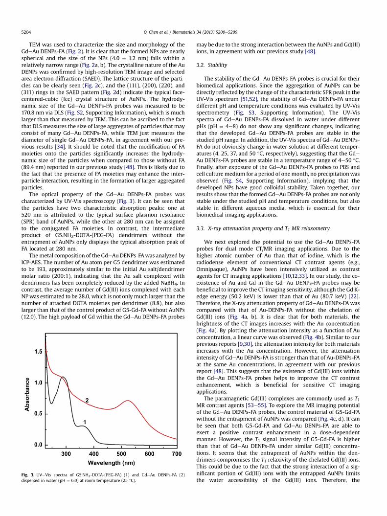
The optical property of the GdeAu DENPs-FA probes wascharacterized by UV-Vis spectroscopy (Fig. 3). It can be seen thatthe particles have two characteristic absorption peaks: one at520 nm is attributed to the typical surface plasmon resonance(SPR) band of AuNPs, while the other at 280 nm can be assignedto the conjugated FA moieties. In contrast, the intermediateproduct of G5.NH2-DOTA-(PEG-FA) dendrimers without theentrapment of AuNPs only displays the typical absorption peak ofFA located at 280 nm.
Themetal composition of the GdeAu DENPs-FAwas analyzed byICP-AES. The number of Au atom per G5 dendrimer was estimatedto be 193, approximately similar to the initial Au salt/dendrimermolar ratio (200:1), indicating that the Au salt complexed withdendrimers has been completely reduced by the added NaBH4. Incontrast, the average number of Gd(III) ions complexed with eachNPwas estimated to be 28.0, which is not only much larger than thenumber of attached DOTA moieties per dendrimer (8.8), but alsolarger than that of the control product of G5-Gd-FA without AuNPs(12.0). The high payload of Gd within the GdeAu DENPs-FA probes
Fig. 3. UVeVis spectra of G5.NH2-DOTA-(PEG-FA) (1) and GdeAu DENPs-FA (2)dispersed in water (pH ¼ 6.0) at room temperature (25 �C).
may be due to the strong interaction between the AuNPs and Gd(III)ions, in agreement with our previous study [48].
3.2. Stability
The stability of the GdeAu DENPs-FA probes is crucial for theirbiomedical applications. Since the aggregation of AuNPs can bedirectly reflected by the change of the characteristic SPR peak in theUV-Vis spectrum [51,52], the stability of GdeAu DENPs-FA underdifferent pH and temperature conditions was evaluated by UV-Visspectrometry (Fig. S3, Supporting Information). The UV-Visspectra of GdeAu DENPs-FA dissolved in water under differentpHs (pH ¼ 4e8) do not show any significant changes, indicatingthat the developed GdeAu DENPs-FA probes are stable in thestudied pH range. In addition, the UV-Vis spectra of GdeAu DENPs-FA do not obviously change in water solution at different temper-atures (4, 25, 37, and 50 �C, respectively), suggesting that the GdeAu DENPs-FA probes are stable in a temperature range of 4e50 �C.Finally, after exposure of the GdeAu DENPs-FA probes to PBS andcell culture medium for a period of onemonth, no precipitationwasobserved (Fig. S4, Supporting Information), implying that thedeveloped NPs have good colloidal stability. Taken together, ourresults show that the formed GdeAu DENPs-FA probes are not onlystable under the studied pH and temperature conditions, but alsostable in different aqueous media, which is essential for theirbiomedical imaging applications.
3.3. X-ray attenuation property and T1 MR relaxometry
We next explored the potential to use the GdeAu DENPs-FAprobes for dual mode CT/MR imaging applications. Due to thehigher atomic number of Au than that of iodine, which is theradiodense element of conventional CT contrast agents (e.g.,Omnipaque), AuNPs have been intensively utilized as contrastagents for CT imaging applications [10,12,33]. In our study, the co-existence of Au and Gd in the GdeAu DENPs-FA probes may bebeneficial to improve the CT imaging sensitivity, although the Gd K-edge energy (50.2 keV) is lower than that of Au (80.7 keV) [22].Therefore, the X-ray attenuation property of GdeAu DENPs-FA wascompared with that of Au-DENPs-FA without the chelation ofGd(III) ions (Fig. 4a, b). It is clear that for both materials, thebrightness of the CT images increases with the Au concentration(Fig. 4a). By plotting the attenuation intensity as a function of Auconcentration, a linear curve was observed (Fig. 4b). Similar to ourprevious reports [9,30], the attenuation intensity for both materialsincreases with the Au concentration. However, the attenuationintensity of GdeAu DENPs-FA is stronger than that of Au-DENPs-FAat the same Au concentrations, in agreement with our previousreport [48]. This suggests that the existence of Gd(III) ions withinthe GdeAu DENPs-FA probes helps to improve the CT contrastenhancement, which is beneficial for sensitive CT imagingapplications.
The paramagnetic Gd(III) complexes are commonly used as T1MR contrast agents [53e55]. To explore the MR imaging potentialof the GdeAu DENPs-FA probes, the control material of G5-Gd-FAwithout the entrapment of AuNPs was compared (Fig. 4c, d). It canbe seen that both G5-Gd-FA and GdeAu DENPs-FA are able toexert a positive contrast enhancement in a dose-dependentmanner. However, the T1 signal intensity of G5-Gd-FA is higherthan that of GdeAu DENPs-FA under similar Gd(III) concentra-tions. It seems that the entrapment of AuNPs within the den-drimers compromises the T1 relaxivity of the chelated Gd(III) ions.This could be due to the fact that the strong interaction of a sig-nificant portion of Gd(III) ions with the entrapped AuNPs limitsthe water accessibility of the Gd(III) ions. Therefore, the

Fig. 4. CT images (a) and X-ray attenuation (HU) (b) of GdeAu DENPs-FA (1) and AuDENPs-FA (2) at different Au concentrations. (c) and (d) show the T1 MR phantomimages and the linear fitting of the inverse T1 of GdeAu DENPs-FA (1) and G5-Gd-FA(3) as a function of Gd concentration.
Q. Chen et al. / Biomaterials 34 (2013) 5200e5209 5205
longitudinal r1 relaxivity of GdeAu DENPs-FA (3.139 mM�1s�1) was
lower than that of G5-Gd-FA without AuNPs (6.022 mM�1s�1).
However, by counting the dendrimer concentration (Fig. S5, Sup-porting Information), the relaxivity of GdeAu DENPs-FA(87.892 mM
�1s�1) is 1.22 times higher than that of G5-Gd-FA(72.264 mM
�1s�1), due to the higher concentration of Gd(III)complexed within the GdeAu DENPs-FA probes (28.0 Gd(III) ionsper dendrimer) than that complexed within G5-Gd-FA (12.0Gd(III) ions per dendrimer). Taken together with the enhanced CTimaging sensitivity and reasonable r1 relaxivity, the developed
Fig. 5. (a) MTT assay of the viability of KB cells treated with GdeAu DENPs-FA at different Auwith the GdeAu DENPs-FA probes with different Au concentrations for 3 h.
GdeAu DENPs-FA probes are expected to be used for dual modeCT/MR imaging applications.
3.4. Cytotoxicity assay
For biomedical imaging applications, it is crucial to test thecytocompatibility of the developed multifunctional nanoprobes.The cytotoxicity of the GdeAu DENPs-FA probes was accessed byMTT colorimetric assay of cell viability (Fig. 5a). It can be seen thatthe GdeAu DENPs-FA probes do not display apparent cytotoxicityat the Au concentration up to 200 mM after 24 h of incubation withKB cells. At the tested highest Au concentration (200 mM), the KB cellviability is still more than 80% without statistically significant dif-ference when compared to the PBS control (p > 0.05). Our resultsindicate that the developed GdeAu DENPs-FA probes are cyto-compatible at the Au concentration range of 0e200 mM. The cyto-compatibility of GdeAu DENPs-FA was further confirmed byobservation of the morphology of KB cells treated with the particlesat different Au concentrations for 24 h (Fig. S6, Supporting Infor-mation). It is clear that the KB cells treated with GdeAu DENPs-FAat the Au concentrations of 5e200 mM (Fig. S6 b-h) are quite healthyand display morphologies similar to those treated with PBS(Fig. S6a), although at the Au concentration up to 200 mM, a smallportion of cells became rounded and detached. The cellmorphology observation results corroborate the above MTT assaydata.
3.5. In vitro cellular uptake
For targeted dual mode CT/MR imaging, it is essential to inves-tigate the ability of the developed GdeAu DENPs-FA probes to bespecifically uptakenbycancer cells overexpressing FAR. ICP-AESwasused to quantify the Au uptake in both KB-HFAR and KB-LFAR cellsafter treatment with GdeAu DENPs-FA at different Au concentra-tions for 3 h (Fig. 5b). It is clear that at the studied Au concentrations,the Au uptake in KB-HFAR cells is significantly higher than that inKB-LFAR cells (p < 0.01). This suggests that the GdeAu DENPs-FAprobes can specifically target the FAR-expressing cancer cells viareceptor-mediated pathway, which is essential for them to be usedfor targeted imaging of FAR-expressing cancer cells.
3.6. In vitro targeted dual mode CT/MR imaging of cancer cells
We next explored the potential to use the GdeAu DENPs-FAprobes for targeted dual mode CT/MR imaging of cancer cells
concentrations for 24 h. (b) Cellular uptake of Au in KB-HFAR and KB-LFAR cells treated
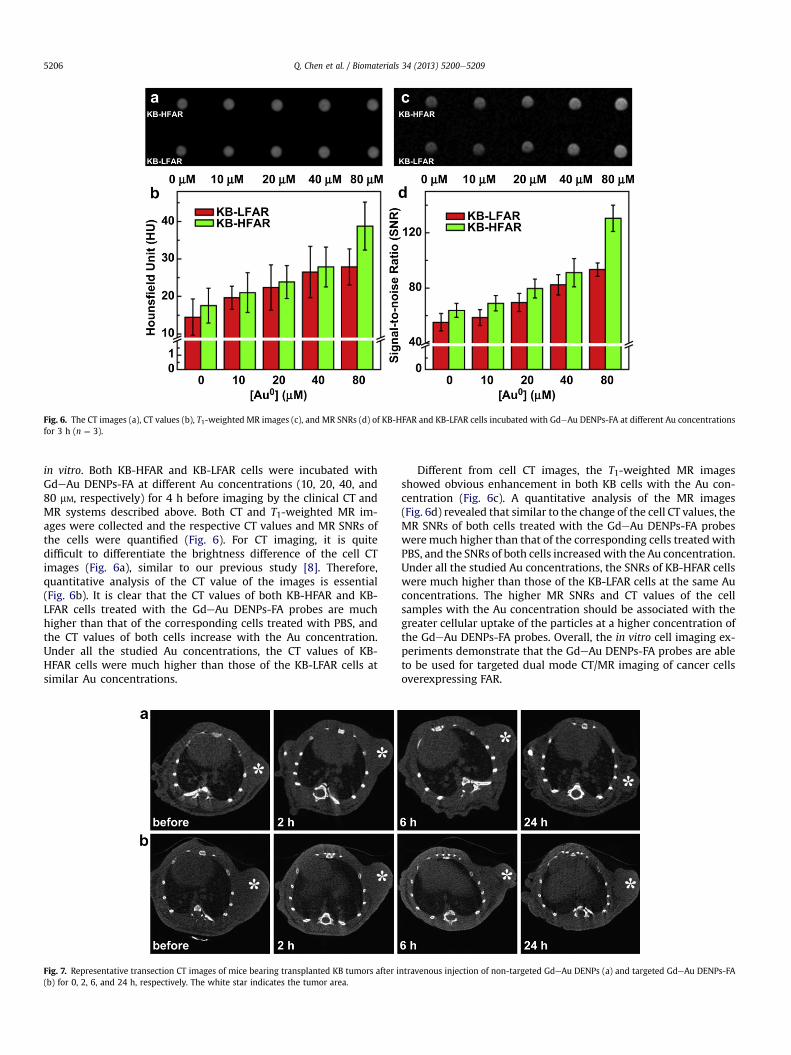
Fig. 6. The CT images (a), CT values (b), T1-weighted MR images (c), and MR SNRs (d) of KB-HFAR and KB-LFAR cells incubated with GdeAu DENPs-FA at different Au concentrationsfor 3 h (n ¼ 3).
Q. Chen et al. / Biomaterials 34 (2013) 5200e52095206
in vitro. Both KB-HFAR and KB-LFAR cells were incubated withGdeAu DENPs-FA at different Au concentrations (10, 20, 40, and80 mM, respectively) for 4 h before imaging by the clinical CT andMR systems described above. Both CT and T1-weighted MR im-ages were collected and the respective CT values and MR SNRs ofthe cells were quantified (Fig. 6). For CT imaging, it is quitedifficult to differentiate the brightness difference of the cell CTimages (Fig. 6a), similar to our previous study [8]. Therefore,quantitative analysis of the CT value of the images is essential(Fig. 6b). It is clear that the CT values of both KB-HFAR and KB-LFAR cells treated with the GdeAu DENPs-FA probes are muchhigher than that of the corresponding cells treated with PBS, andthe CT values of both cells increase with the Au concentration.Under all the studied Au concentrations, the CT values of KB-HFAR cells were much higher than those of the KB-LFAR cells atsimilar Au concentrations.
Fig. 7. Representative transection CT images of mice bearing transplanted KB tumors after i(b) for 0, 2, 6, and 24 h, respectively. The white star indicates the tumor area.
Different from cell CT images, the T1-weighted MR imagesshowed obvious enhancement in both KB cells with the Au con-centration (Fig. 6c). A quantitative analysis of the MR images(Fig. 6d) revealed that similar to the change of the cell CT values, theMR SNRs of both cells treated with the GdeAu DENPs-FA probesweremuch higher than that of the corresponding cells treated withPBS, and the SNRs of both cells increasedwith the Au concentration.Under all the studied Au concentrations, the SNRs of KB-HFAR cellswere much higher than those of the KB-LFAR cells at the same Auconcentrations. The higher MR SNRs and CT values of the cellsamples with the Au concentration should be associated with thegreater cellular uptake of the particles at a higher concentration ofthe GdeAu DENPs-FA probes. Overall, the in vitro cell imaging ex-periments demonstrate that the GdeAu DENPs-FA probes are ableto be used for targeted dual mode CT/MR imaging of cancer cellsoverexpressing FAR.
ntravenous injection of non-targeted GdeAu DENPs (a) and targeted GdeAu DENPs-FA
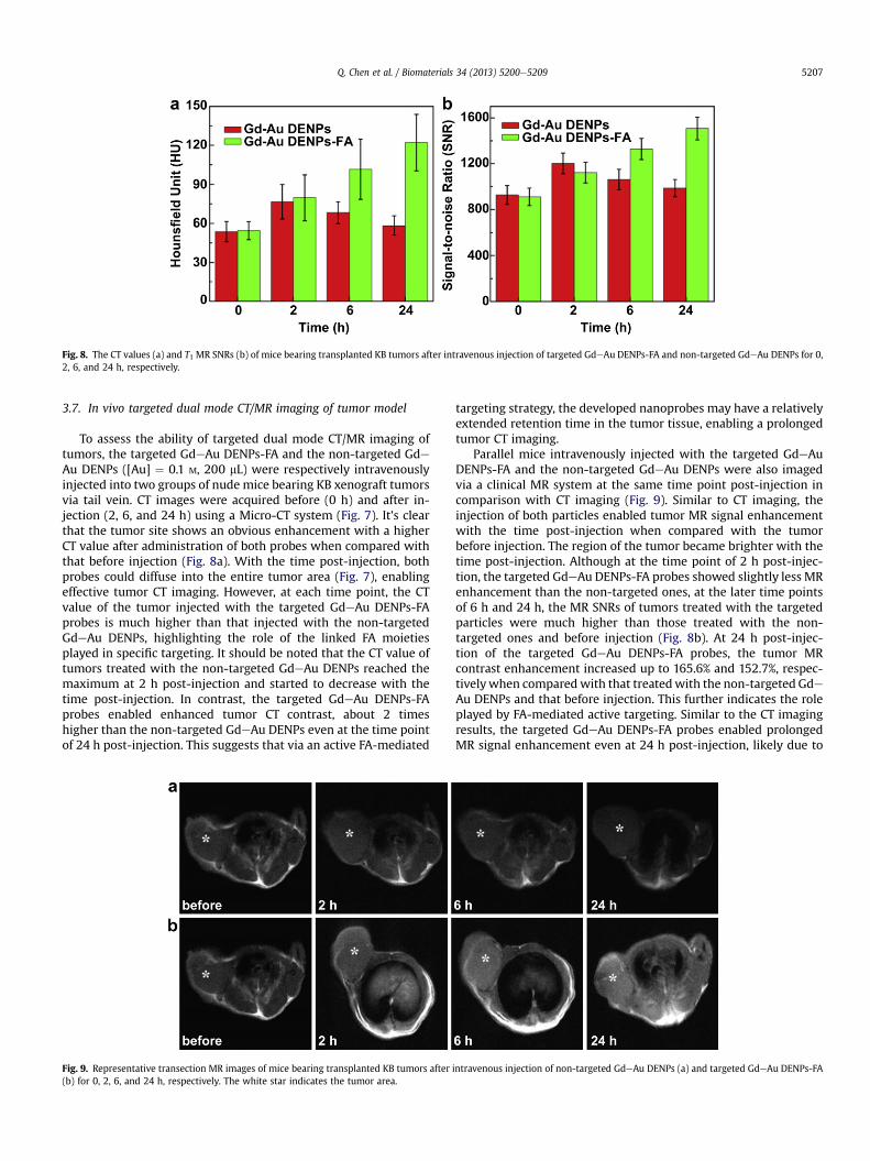
Fig. 8. The CT values (a) and T1 MR SNRs (b) of mice bearing transplanted KB tumors after intravenous injection of targeted GdeAu DENPs-FA and non-targeted GdeAu DENPs for 0,2, 6, and 24 h, respectively.
Q. Chen et al. / Biomaterials 34 (2013) 5200e5209 5207
3.7. In vivo targeted dual mode CT/MR imaging of tumor model
To assess the ability of targeted dual mode CT/MR imaging oftumors, the targeted GdeAu DENPs-FA and the non-targeted GdeAu DENPs ([Au] ¼ 0.1 M, 200 mL) were respectively intravenouslyinjected into two groups of nude mice bearing KB xenograft tumorsvia tail vein. CT images were acquired before (0 h) and after in-jection (2, 6, and 24 h) using a Micro-CT system (Fig. 7). It’s clearthat the tumor site shows an obvious enhancement with a higherCT value after administration of both probes when compared withthat before injection (Fig. 8a). With the time post-injection, bothprobes could diffuse into the entire tumor area (Fig. 7), enablingeffective tumor CT imaging. However, at each time point, the CTvalue of the tumor injected with the targeted GdeAu DENPs-FAprobes is much higher than that injected with the non-targetedGdeAu DENPs, highlighting the role of the linked FA moietiesplayed in specific targeting. It should be noted that the CT value oftumors treated with the non-targeted GdeAu DENPs reached themaximum at 2 h post-injection and started to decrease with thetime post-injection. In contrast, the targeted GdeAu DENPs-FAprobes enabled enhanced tumor CT contrast, about 2 timeshigher than the non-targeted GdeAu DENPs even at the time pointof 24 h post-injection. This suggests that via an active FA-mediated
Fig. 9. Representative transection MR images of mice bearing transplanted KB tumors after(b) for 0, 2, 6, and 24 h, respectively. The white star indicates the tumor area.
targeting strategy, the developed nanoprobes may have a relativelyextended retention time in the tumor tissue, enabling a prolongedtumor CT imaging.
Parallel mice intravenously injected with the targeted GdeAuDENPs-FA and the non-targeted GdeAu DENPs were also imagedvia a clinical MR system at the same time point post-injection incomparison with CT imaging (Fig. 9). Similar to CT imaging, theinjection of both particles enabled tumor MR signal enhancementwith the time post-injection when compared with the tumorbefore injection. The region of the tumor became brighter with thetime post-injection. Although at the time point of 2 h post-injec-tion, the targeted GdeAu DENPs-FA probes showed slightly less MRenhancement than the non-targeted ones, at the later time pointsof 6 h and 24 h, the MR SNRs of tumors treated with the targetedparticles were much higher than those treated with the non-targeted ones and before injection (Fig. 8b). At 24 h post-injec-tion of the targeted GdeAu DENPs-FA probes, the tumor MRcontrast enhancement increased up to 165.6% and 152.7%, respec-tivelywhen comparedwith that treatedwith the non-targeted GdeAu DENPs and that before injection. This further indicates the roleplayed by FA-mediated active targeting. Similar to the CT imagingresults, the targeted GdeAu DENPs-FA probes enabled prolongedMR signal enhancement even at 24 h post-injection, likely due to
intravenous injection of non-targeted GdeAu DENPs (a) and targeted GdeAu DENPs-FA

Fig. 10. Biodistribution of Au in the major organs of the mice including heart, liver,spleen, lung, kidney, and the tumor. The data were recorded from the whole organ orthe whole tumor at different time points post intravenous injection of GdeAu DENPs-FA (200 mL, [Au] ¼ 0.1 M).
Q. Chen et al. / Biomaterials 34 (2013) 5200e52095208
the extended retention time in the tumor tissue. Taken togetherwith the CT imaging results, our results clearly indicate that thedeveloped GdeAu DENPs-FA probes are able to be used for targeteddual mode CT/MR imaging of tumors overexpressing FAR.
3.8. In vivo biodistribution
The biodistribution of the developed multifunctional nanop-robes, in particular the Au uptake in the major organs includingheart, liver, spleen, lung, kidney, and tumor was analyzed by ICP-AES (Fig. 10). It is clear that at 6 and 24 h post-injection, liver andspleen have a significant Au uptake, with Au uptake of 315.9 mg/g(6 h) and 691.9 mg/g (24 h) in the liver, and 187.8 mg/g (6 h) and747.9 mg/g (24 h) in the spleen, respectively. The PEGylation of thenanoprobes seemed to be able to allow a portion of particles toescape from the recognition of reticuloendothelial system in theliver and spleen, and accumulated in the other organs such as heart,lung, and kidney [56,57]. With the prolonged circulation timebenefited from the PEGylation modification [48] and the FA-mediated active targeting, the GdeAu DENPs-FA probes were ableto gradually accumulate in the tumor area with Au uptake of 3.0and 18.4 mg/g at 6 h and 24 h post-injection, respectively, enablingeffectively dual mode CT/MR imaging. At 96 h post-injection, the Auuptake in all the major organs approached the level before injec-tion, suggesting that the developed nanoprobes are able to bemetabolized and cleared out from the body, which is very impor-tant for practical biomedical imaging applications.
4. Conclusion
In summary, we developed a dendrimer-based multifunctionalFA-targeted nanoprobe for dual mode CT/MR imaging of tumors.Via the versatile dendrimer nanotechnology, AuNPs and Gd(III) areable to be entrapped within the dendrimer interior and modifiedonto the dendrimer surfaces, respectively. The formed multifunc-tional nanoprobes are water soluble, colloidal stable, and non-cytotoxic in the given concentration range. With the PEGylationmodification and the FA-mediated targeting pathway, the devel-oped multifunctional nanoprobes are able to be used for dual modeCT/MR imaging of cancer cells and xenograft tumor model
overexpressing FAR in vitro and in vivo. Importantly, biodistributionstudies show that the developed multifunctional nanoprobes canbe cleared out from the body at 96 h post-injection. With theunique structural characteristics of dendrimers that can be linkedwith different targeting ligands, multifunctional nanoprobes withother targeting functionalities may be developed for targeted dualmode CT/MR imaging of different types of cancer.
Acknowledgment
This research is financially supported by the National NaturalScience Foundation of China (21273032 and 81101150), the Fun-dação para a Ciência e a Tecnologia (FCT) for funding through theproject PTDC/CTM-NAN/1748/2012, the Program for New CenturyExcellent Talents in University, State Education Ministry, and theFund of the Science and Technology Commission of Shanghai Mu-nicipality (11nm0506400 for X. S., 12520705500 for M. S., and11JC1410500 for G. Z.). X. S. gratefully acknowledges the FCT andSantander bank for the Invited Chair in Nanotechnology, and FCTthrough the Strategic Plan PEst-OE/QUI/UI0674/2011. K. L. thanksthe support from Natural Science Foundation of Shanghai, China(11ZR1429300), the Medical Guiding Program of Shanghai Scienceand Technology Committee (114119a0800), and Songjiang MedicalClimbing Program, Shanghai, China (2011PD04).
Appendix. Supplementary material
Supplementary data related to this article can be found online athttp://dx.doi.org/10.1016/j.biomaterials.2013.03.009.
References
[1] Huang W-Y, Davis JJ. Multimodality and nanoparticles in medical imaging.Dalton Trans 2011;40:6087e103.
[2] Kobayashi H, Longmire MR, Ogawa M, Choyke PL, Kawamoto S. Multiplexedimaging in cancer diagnosis: applications and future advances. Lancet Oncol2010;11:589e95.
[3] Reuveni T, Motiei M, Romman Z, Popovtzer A, Popovtzer R. Targeted goldnanoparticles enable molecular CT imaging of cancer: an in vivo study. Int JNanomed 2011;6:2859e64.
[4] Lee D-E, Koo H, Sun I-C, Ryu JH, Kim K, Kwon IC. Multifunctional nanoparticlesfor multimodal imaging and theragnosis. Chem Soc Rev 2012;41:2656e72.
[5] Rabin O, Perez JM, Grimm J, Wojtkiewicz G, Weissleder R. An X-ray computedtomography imaging agent based on long-circulating bismuth sulphidenanoparticles. Nat Mater 2006;5:118e22.
[6] Liu H, Wang H, Guo R, Cao X, Zhao J, Luo Y, et al. Size-controlled synthesis ofdendrimer-stabilized silver nanoparticles for X-ray computed tomographyimaging applications. Polym Chem 2010;1:1677e83.
[7] Liu H, Xu Y, Wen S, Zhu J, Zheng L, Shen M, et al. Facile hydrothermal synthesisof low generation dendrimer-stabilized gold nanoparticles for in vivocomputed tomography imaging applications. Polym Chem 2013;4:1788e95.
[8] Peng C, Li K, Cao X, Xiao T, HouW, Zheng L, et al. Facile formation of dendrimer-stabilized gold nanoparticles modified with diatrizoic acid for enhancedcomputed tomography imaging applications. Nanoscale 2012;4:6768e78.
[9] Peng C, Zheng L, Chen Q, Shen M, Guo R, Wang H, et al. PEGylated dendrimer-entrapped gold nanoparticles for in vivo blood pool and tumor imaging bycomputed tomography. Biomaterials 2012;33:1107e19.
[10] Popovtzer R, Agrawal A, Kotov NA, Popovtzer A, Balter J, Carey TE, et al.Targeted gold nanoparticles enable molecular CT imaging of cancer. Nano Lett2008;8:4593e6.
[11] Yordanov AT, Lodder AL, Woller EK, Cloninger MJ, Patronas N, Milenic D, et al.Novel iodinated dendritic nanoparticles for computed tomography (CT) im-aging. Nano Lett 2002;2:595e9.
[12] Kim D, Park S, Lee JH, Jeong YY, Jon S. Antibiofouling polymer-coated goldnanoparticles as a contrast agent for in vivo x-ray computed tomographyimaging. J Am Chem Soc 2007;129:7661e5.
[13] Debouttière PJ, Roux S, Vocanson F, Billotey C, Beuf O, Favre-Réguillon A, et al.Design of gold nanoparticles for magnetic resonance imaging. Adv FunctMater 2006;16:2330e9.
[14] Shi X, Wang SH, Swanson SD, Ge S, Cao Z, Van Antwerp ME, et al. Dendrimer-functionalized shell-crosslinked iron oxide nanoparticles for in-vivo magneticresonance imaging of tumors. Adv Mater 2008;20:1671e8.
[15] Song Y, Xu X, MacRenaris KW, Zhang XQ, Mirkin CA, Meade TJ. Multimodalgadolinium-enriched DNA-gold nanoparticle conjugates for cellular imaging.Angew Chem Int Ed 2009;48:9143e7.

Q. Chen et al. / Biomaterials 34 (2013) 5200e5209 5209
[16] Swanson SD, Kukowska-Latallo JF, Patri AK, Chen C, Ge S, Cao Z, et al. Targetedgadolinium-loaded dendrimer nanoparticles for tumor-specific magneticresonance contrast enhancement. Int J Nanomed 2008;3:201e10.
[17] Yang H, Zhuang Y, Hu H, Du X, Zhang C, Shi X, et al. Silica-coated manganeseoxide nanoparticles as a platform for targeted magnetic resonance and fluo-rescence imaging of cancer cells. Adv Funct Mater 2010;20:1733e41.
[18] Yang H, Zhuang Y, Sun Y, Dai A, Shi X, Wu D, et al. Targeted dual-contrast T1-and T2-weighted magnetic resonance imaging of tumors using multifunc-tional gadolinium-labeled superparamagnetic iron oxide nanoparticles. Bio-materials 2011;32:4584e93.
[19] Jennings LE, Long NJ. ‘Two is better than one’dprobes for dual-modalitymolecular imaging. Chem Commun 2009:3511e24.
[20] Pagel MD. The hope and hype of multimodality imaging contrast agents.Nanomedicine 2011;6:945e8.
[21] Park J-A, Kim H-K, Kim J-H, Jeong S-W, Jung J-C, Lee G-H, et al. Gold nano-particles functionalized by gadolinium-DTPA conjugate of cysteine as amultimodal bioimaging agent. Bioorg Med Chem Lett 2010;20:2287e91.
[22] Alric C, Taleb J, Duc GL, Mandon C, Billotey C, Meur-Herland AL, et al. Gado-linium chelate coated gold nanoparticles as contrast agents for both X-raycomputed tomography and magnetic resonance imaging. J Am Chem Soc2008;130:5908e15.
[23] Chou S-W, Shau Y-H, Wu P-C, Yang Y-S, Shieh D-B, Chen C-C. In vitro andin vivo studies of FePt nanoparticles for dual modal CT/MRI molecular im-aging. J Am Chem Soc 2010;132:13270e8.
[24] Regino CAS, Walbridge S, Bernardo M, Wong KJ, Johnson D, Lonser R, et al.A dual CT-MR dendrimer contrast agent as a surrogate marker for convection-enhanced delivery of intracerebral macromolecular therapeutic agents.Contrast Media Mol Imaging 2008;3:2e8.
[25] Lee N, Cho HR, Oh MH, Lee SH, Kim K, Kim BH, et al. Multifunctional Fe3O4/TaOx core/shell nanoparticles for simultaneous magnetic resonance imagingand X-ray computed tomography. J Am Chem Soc 2012;134:10309e12.
[26] Cheung ENM, Alvares RDA, Oakden W, Chaudhary R, Hill ML, Pichaandi J, et al.Polymer-stabilized lanthanide fluoride nanoparticle aggregates as contrastagents for magnetic resonance imaging and computed tomography. ChemMater 2010;22:4728e39.
[27] Maeda H. Tumor-selective delivery of macromolecular drugs via the EPR ef-fect: background and future prospects. Bioconjug Chem 2010;21:797e802.
[28] Barreto JA, O’MalleyW, Kubeil M, GrahamB, Stephan H, Spiccia L. Nanomaterials:applications in cancer imaging and therapy. Adv Mater 2011;23:H18e40.
[29] Minelli C, Lowe SB, Stevens MM. Engineering nanocomposite materials forcancer therapy. Small 2010;6:2336e57.
[30] Wang H, Zheng L, Peng C, Guo R, Shen M, Shi X, et al. Computed tomographyimaging of cancer cells using acetylated dendrimer-entrapped gold nano-particles. Biomaterials 2011;32:2979e88.
[31] Tomalia DA, Frechet JMJ. Dendrimers and other dendritic polymers. NewYork: John Wiley & Sons Ltd; 2001.
[32] Guo R, Wang H, Peng C, Shen M, Zheng L, Zhang G, et al. Enhanced X-rayattenuation property of dendrimer-entrapped gold nanoparticles complexedwith diatrizoic acid. J Mater Chem 2011;18:5120e7.
[33] Guo R, Wang H, Peng C, Shen MW, Pan MJ, Cao XY, et al. X-ray attenuationproperty of dendrimer-entrapped gold nanoparticles. J Phys Chem C2010;114:50e6.
[34] Liu H, Shen M, Zhao J, Guo R, Cao X, Zhang G, et al. Tunable synthesis andacetylation of dendrimer-entrapped or dendrimer-stabilized goldesilver alloynanoparticles. Colloid Surf B-Biointerfaces 2012;94:58e67.
[35] Peng C, Wang H, Guo R, Shen MW, Cao XY, Zhu MF, et al. Acetylation ofdendrimer-entrapped gold nanoparticles: synthesis, stability, and X-rayattenuation properties. J Appl Polym Sci 2011;119:1673e82.
[36] Shen M, Shi X. Dendrimer-based organic/inorganic hybrid nanoparticles inbiomedical applications. Nanoscale 2010;2:1596e610.
[37] Wang H, Zheng L, Peng C, Shen M, Shi X, Zhang G. Folic acid-modified den-drimer-entrapped gold nanoparticles as nanoprobes for targeted CT imagingof human lung adencarcinoma. Biomaterials 2013;34:470e80.
[38] Shi X, Thomas TP,Myc LA, Kotlyar A, Baker Jr JR. Synthesis, characterization, andintracellular uptake of carboxyl-terminated poly(amidoamine) dendrimer-stabilized iron oxide nanoparticles. Phys Chem Chem Phys 2007;9:5712e20.
[39] Wang SH, Shi X, Van Antwerp M, Cao Z, Swanson SD, Bi X, et al. Dendrimer-functionalized iron oxide nanoparticles for specific targeting and imaging ofcancer cells. Adv Funct Mater 2007;17:3043e50.
[40] Kukowska-Latallo JF, Candido KA, Cao Z, Nigavekar SS, Majoros IJ, Thomas TP,et al. Nanoparticle targeting of anticancer drug improves therapeutic responsein animal model of human epithelial cancer. Cancer Res 2005;65:5317e24.
[41] Majoros IJ, Myc A, Thomas T, Mehta CB, Baker JR. PAMAM dendrimer-basedmultifunctional conjugate for cancer therapy: synthesis, characterization,and functionality. Biomacromolecules 2006;7:572e9.
[42] Thomas TP, Majoros IJ, Kotlyar A, Kukowska-Latallo JF, Bielinska A, Myc A,et al. Targeting and inhibition of cell growth by an engineered dendriticnanodevice. J Med Chem 2005;48:3729e35.
[43] Nwe K, Bryant Jr LH, Brechbiel MW. Poly (amidoamine) dendrimer based MRIcontrast agents exhibiting enhanced relaxivities derived via metal preligationtechniques. Bioconjug Chem 2010;21:1014e7.
[44] Shi X, Wang S, Meshinchi S, Van Antwerp ME, Bi X, Lee I, et al. Dendrimer-entrapped gold nanoparticles as a platform for cancer-cell targeting and im-aging. Small 2007;3:1245e52.
[45] Shi X, Wang SH, Van Antwerp ME, Chen X, Baker Jr JR. Targeting and detectingcancer cells using spontaneously formed multifunctional dendrimer-stabilized gold nanoparticles. Analyst 2009;134:1373e9.
[46] Shukla R, Hill E, Shi X, Kim J, Muniz MC, Sun K, et al. Tumor microvasculaturetargeting with dendrimer-entrapped gold nanoparticles. Soft Matter 2008;4:2160e3.
[47] Cai H, Li K, Shen M, Wen S, Luo Y, Peng C, et al. Facile assembly of Fe3O4@Aunanocomposite particles for dual mode magnetic resonance and computedtomography imaging applications. J Mater Chem 2012;22:15110e20.
[48] Wen S, Li K, Cai H, Chen Q, Shen M, Huang Y, et al. Multifunctional dendrimer-entrapped gold nanoparticles for dual mode CT/MR imaging applications.Biomaterials 2013;34:1570e80.
[49] Taylor KML, Jin A, Lin W. Surfactant-assisted synthesis of nanoscale gadolin-ium metal-organic frameworks for potential multimodal imaging. AngewChem Int Ed 2008;120:7836e9.
[50] Cheng Z, Thorek DLJ, Tsourkas A. Gadolinium-conjugated dendrimer nano-clusters as a tumor-targeted T1 magnetic resonance imaging contrast agent.Angew Chem Int Ed 2010;49:346e50.
[51] Takeuchi Y, Ida T, Kimura K. Colloidal stability of gold nanoparticles in 2-propanol under laser irradiation. J Phys Chem B 1997;101:1322e7.
[52] Ghosh SK, Pal T. Interparticle coupling effect on the surface plasmon reso-nance of gold nanoparticles: from theory to applications. Chem Rev 2007;107:4797e862.
[53] Bridot J-L, Faure A-C, Laurent S, Rivière C, Billotey C, Hiba B, et al. Hybridgadolinium oxide nanoparticles: multimodal contrast agents for in vivo im-aging. J Am Chem Soc 2007;129:5076e84.
[54] Frullano L, Meade T. Multimodal MRI contrast agents. J Biol Inorg Chem2007;12:939e49.
[55] Hahn MA, Singh AK, Sharma P, Brown SC, Moudgil BM. Nanoparticles ascontrast agents for in-vivo bioimaging: current status and future perspectives.Anal Bioanal Chem 2011;399:3e27.
[56] Moghimi SM, Hunter AC, Murray JC. Nanomedicine: current status and futureprospects. FASEB J 2005;19:311e30.
[57] Liu Z, Cai W, He L, Nakayama N, Chen K, Sun X, et al. In vivo biodistributionand highly efficient tumour targeting of carbon nanotubes in mice. NatNanotechnol 2007;2:47e52.
Related Documents


