Target cell-specific modulation of neuronal activity by astrocytes A. S. Kozlov* † , M. C. Angulo*, E. Audinat, and S. Charpak Laboratory of Neurophysiology, Institut National de la Sante ´ et de la Recherche Me ´ dicale U603 and Centre National de la Recherche Scientifique FRE2500, Ecole Supe ´ rieure de Physique et de Chimie Industrielles, and Universite ´ Paris Descartes, 75006 Paris, France Communicated by A. James Hudspeth, The Rockefeller University, New York, NY, May 8, 2006 (received for review April 4, 2006) Interaction between astrocytes and neurons enriches the behavior of brain circuits. By releasing glutamate and ATP, astrocytes can directly excite neurons and modulate synaptic transmission. In the rat olfactory bulb, we demonstrate that the release of GABA by astrocytes causes long-lasting and synchronous inhibition of mitral and granule cells. In addition, astrocytes release glutamate, leading to a selective activation of granule-cell NMDA receptors. Thus, by releasing excitatory and inhibitory neurotransmitters, astrocytes exert a complex modulatory control on the olfactory network. glutamate GABA inhibition olfactory bulb synchronization A comprehensive description of brain circuits must include interactions between neurons and glial cells. Studies in different preparations have shown that glial cells communicate with neurons, respond to their activity, and affect neuronal behavior by releasing various neuroactive substances (for review, see refs. 1 and 2). Astrocytes, the predominant type of glial cells in the central nervous system, form a highly organized multi- cellular syncytium extending throughout the gray matter (3, 4). Calcium imaging in acute brain slices and in vivo has demon- strated that astrocytes can generate various patterns of activity, either local and spatially restricted to processes of individual cells or coordinated between adjacent cells and propagating across the brain tissue (5–12). Modeling indicates that coupling of astrocytic and neuronal activities may give rise to membrane potential instability and oscillations (13). Recently, excessive astrocytic release of excitatory neurotransmitters was shown to lead to hyperexcitability and seizures (14). Glutamate release by astrocytes has been well documented in both cultures and acute brain slices (for review see refs. 1 and 2). In hippocampal slices, astrocytes release glutamate onto pyra- midal neurons to evoke phasic and tonic currents mediated by NMDA receptors (15–18). In contrast, little is known regarding the role of astrocytes in neuronal inhibition. One study reported a potentiation of synaptic inhibition of the CA1 pyramidal neurons in the rat hippocampus, manifested as an increased frequency of inhibitory postsynaptic currents (19). This form of neuronal inhibition was proposed to depend on astrocytic cal- cium signaling and on activation of GABAergic interneurons, presumably by astrocytic glutamate. Another mechanism of neuronal inhibition was reported in the supraoptic nucleus: In response to a decrease of extracellular osmolarity within a physiological range, astrocytes were shown to release taurine and, thus, to activate neuronal glycine receptors (20). The mammalian olfactory bulb is a valuable system to explore the diversity of neuroglial interactions because it contains many different types of neurons and astrocytes and because most of its neuronal circuits are characterized in detail and their function is well understood (for a review, see ref. 21). In this study, by using acute slices of the rat olfactory bulb, we demonstrate direct and selective excitation and inhibition of neurons by astrocytes. By releasing GABA, astrocytes evoke hyperpolarizing inhibitory currents in mitral and granule cells. These currents may occur synchronously in adjacent neurons and can block neuronal firing for several hundred milliseconds. In addition, astrocytes release glutamate that activates NMDA receptors of granule cells. This target cell-specific modulation of neuronal activity adds to the complexity of neuro–glial interactions. Results Slow GABAergic Inhibition of Mitral Cells. Prolonged intracellular recording from mitral cells revealed the spontaneous occurrence of rare, long-lasting inhibitory events. These slow hyperpolar- izations, which could block the discharge of mitral cells (Fig. 1A), had a mean rise time of 47.2 4.2 ms (range: 7.6–165 ms; n 73 events; here and throughout the text, see figure legends for additional statistics). Corresponding slow outward currents (SOCs) were detected in voltage-clamp mode at a holding membrane potential of 50 mV, with an equilibrium potential for chloride ions (E Cl ) of 78 mV. Their reversal potential shifted with E Cl as expected for currents carried by chloride ions (Fig. 1B; n 7 cells). SOCs were sensitive neither to 10 M strychnine nor to 50 M (1,2,5,6-tetrahydropyridin-4-yl) meth- ylphosphinic acid (TPMPA), antagonists of glycine and GABA C receptors, respectively (n 4 for each drug). In particular, normalized frequency of SOCs was 98.4 2.3% in control and 71 17% in 50 M TPMPA (P 0.13 with ANOVA; for a detailed description of all data analysis, see Supporting Text, which is published as supporting information on the PNAS web site), which rules out the involvement of GABA C receptors. In contrast, the currents were completely blocked by a specific competitive antagonist of GABA A receptors gabazine (11 M; n 6; data not shown), and by a noncompetitive antagonist picrotoxin (Fig. 1C; n 7). Note that at a low concentration (2 M), gabazine similarly blocked synaptic currents and SOCs (89 9% and 92 9% inhibition; P 0.01 and 0.006, respectively; n 3). When used at 40 M, bicuculline, another competitive antagonist of GABA A receptors, completely blocked synaptic outward currents but only partially blocked spontaneous slow outward currents (Fig. 1D; n 5 cells). A complete block of spontaneous SOCs required higher concen- trations of bicuculline (100 M). These results indicate that SOCs differ from classical GABAergic synaptic currents and correspond to uncharacterized events involving GABA A recep- tors. In addition, all three antagonists of GABA A receptors also blocked a tonic outward current (shown for picrotoxin in Fig. 1C), as previously reported in the cerebellum and hippocampus (for review, see ref. 22). SOCs Occur in the Absence of Neuronal Vesicular Release. To further investigate the origin of SOCs, we found it necessary to establish a precise criterion enabling us to distinguish them from synaptic currents. To eliminate the latter, we preincubated the slices for Conflict of interest statement: No conflicts declared. Abbreviations: SOC, slow outward current; SIC, slow inward current; TTX, tetrodotoxin. *A.S.K. and M.C.A. contributed equally to this work. † To whom correspondence should be sent at the present address: Laboratory of Sensory Neuroscience, The Rockefeller University, 1230 York Avenue, New York, NY 10021. E-mail: [email protected]. © 2006 by The National Academy of Sciences of the USA 10058 –10063 PNAS June 27, 2006 vol. 103 no. 26 www.pnas.orgcgidoi10.1073pnas.0603741103

Welcome message from author
This document is posted to help you gain knowledge. Please leave a comment to let me know what you think about it! Share it to your friends and learn new things together.
Transcript
Target cell-specific modulation of neuronal activityby astrocytesA. S. Kozlov*†, M. C. Angulo*, E. Audinat, and S. Charpak
Laboratory of Neurophysiology, Institut National de la Sante et de la Recherche Medicale U603 and Centre National de la Recherche Scientifique FRE2500,Ecole Superieure de Physique et de Chimie Industrielles, and Universite Paris Descartes, 75006 Paris, France
Communicated by A. James Hudspeth, The Rockefeller University, New York, NY, May 8, 2006 (received for review April 4, 2006)
Interaction between astrocytes and neurons enriches the behaviorof brain circuits. By releasing glutamate and ATP, astrocytes candirectly excite neurons and modulate synaptic transmission. In therat olfactory bulb, we demonstrate that the release of GABA byastrocytes causes long-lasting and synchronous inhibition of mitraland granule cells. In addition, astrocytes release glutamate, leadingto a selective activation of granule-cell NMDA receptors. Thus, byreleasing excitatory and inhibitory neurotransmitters, astrocytesexert a complex modulatory control on the olfactory network.
glutamate � GABA � inhibition � olfactory bulb � synchronization
A comprehensive description of brain circuits must includeinteractions between neurons and glial cells. Studies in
different preparations have shown that glial cells communicatewith neurons, respond to their activity, and affect neuronalbehavior by releasing various neuroactive substances (for review,see refs. 1 and 2). Astrocytes, the predominant type of glial cellsin the central nervous system, form a highly organized multi-cellular syncytium extending throughout the gray matter (3, 4).Calcium imaging in acute brain slices and in vivo has demon-strated that astrocytes can generate various patterns of activity,either local and spatially restricted to processes of individual cellsor coordinated between adjacent cells and propagating acrossthe brain tissue (5–12). Modeling indicates that coupling ofastrocytic and neuronal activities may give rise to membranepotential instability and oscillations (13). Recently, excessiveastrocytic release of excitatory neurotransmitters was shown tolead to hyperexcitability and seizures (14).
Glutamate release by astrocytes has been well documented inboth cultures and acute brain slices (for review see refs. 1 and 2).In hippocampal slices, astrocytes release glutamate onto pyra-midal neurons to evoke phasic and tonic currents mediated byNMDA receptors (15–18). In contrast, little is known regardingthe role of astrocytes in neuronal inhibition. One study reporteda potentiation of synaptic inhibition of the CA1 pyramidalneurons in the rat hippocampus, manifested as an increasedfrequency of inhibitory postsynaptic currents (19). This form ofneuronal inhibition was proposed to depend on astrocytic cal-cium signaling and on activation of GABAergic interneurons,presumably by astrocytic glutamate. Another mechanism ofneuronal inhibition was reported in the supraoptic nucleus: Inresponse to a decrease of extracellular osmolarity within aphysiological range, astrocytes were shown to release taurineand, thus, to activate neuronal glycine receptors (20).
The mammalian olfactory bulb is a valuable system to explorethe diversity of neuroglial interactions because it contains manydifferent types of neurons and astrocytes and because most of itsneuronal circuits are characterized in detail and their function iswell understood (for a review, see ref. 21). In this study, by usingacute slices of the rat olfactory bulb, we demonstrate direct andselective excitation and inhibition of neurons by astrocytes. Byreleasing GABA, astrocytes evoke hyperpolarizing inhibitorycurrents in mitral and granule cells. These currents may occursynchronously in adjacent neurons and can block neuronal firingfor several hundred milliseconds. In addition, astrocytes release
glutamate that activates NMDA receptors of granule cells. Thistarget cell-specific modulation of neuronal activity adds to thecomplexity of neuro–glial interactions.
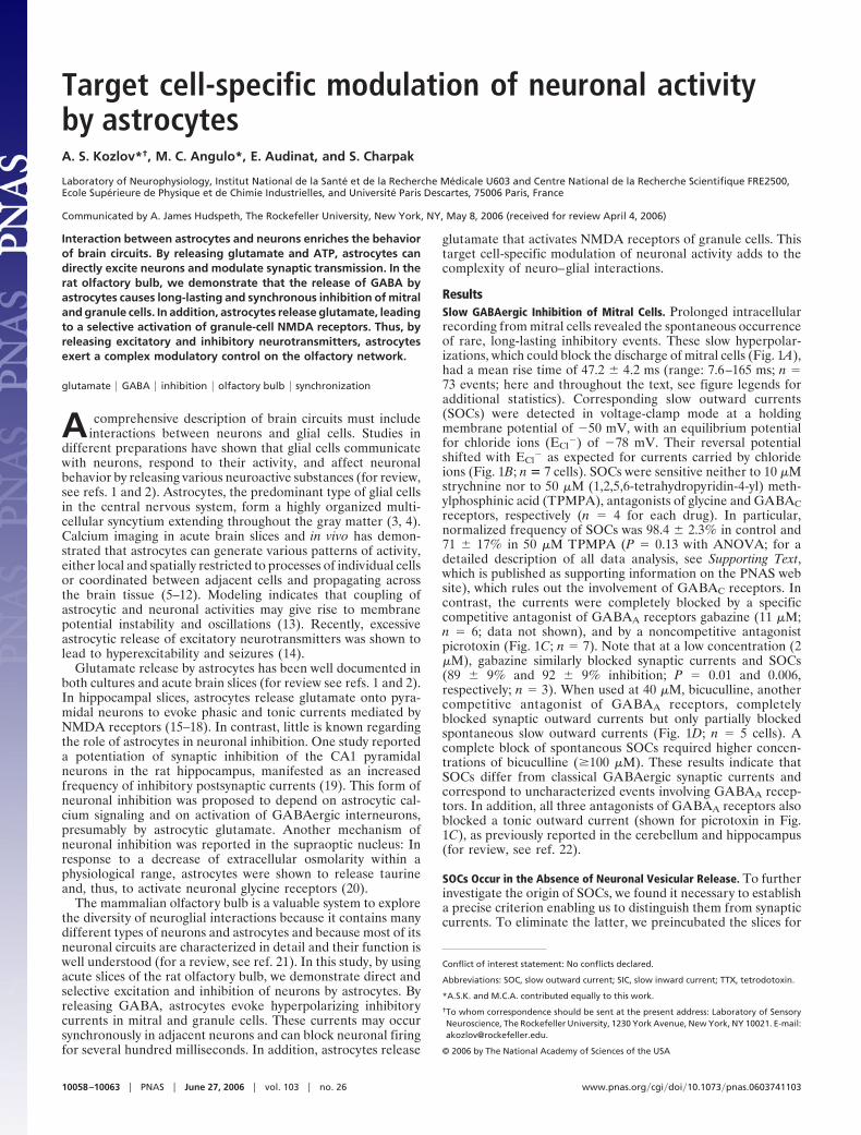
ResultsSlow GABAergic Inhibition of Mitral Cells. Prolonged intracellularrecording from mitral cells revealed the spontaneous occurrenceof rare, long-lasting inhibitory events. These slow hyperpolar-izations, which could block the discharge of mitral cells (Fig. 1A),had a mean rise time of 47.2 � 4.2 ms (range: 7.6–165 ms; n �73 events; here and throughout the text, see figure legends foradditional statistics). Corresponding slow outward currents(SOCs) were detected in voltage-clamp mode at a holdingmembrane potential of �50 mV, with an equilibrium potentialfor chloride ions (ECl
�) of �78 mV. Their reversal potentialshifted with ECl
� as expected for currents carried by chlorideions (Fig. 1B; n � 7 cells). SOCs were sensitive neither to 10 �Mstrychnine nor to 50 �M (1,2,5,6-tetrahydropyridin-4-yl) meth-ylphosphinic acid (TPMPA), antagonists of glycine and GABACreceptors, respectively (n � 4 for each drug). In particular,normalized frequency of SOCs was 98.4 � 2.3% in control and71 � 17% in 50 �M TPMPA (P � 0.13 with ANOVA; for adetailed description of all data analysis, see Supporting Text,which is published as supporting information on the PNAS website), which rules out the involvement of GABAC receptors. Incontrast, the currents were completely blocked by a specificcompetitive antagonist of GABAA receptors gabazine (11 �M;n � 6; data not shown), and by a noncompetitive antagonistpicrotoxin (Fig. 1C; n � 7). Note that at a low concentration (2�M), gabazine similarly blocked synaptic currents and SOCs(89 � 9% and 92 � 9% inhibition; P � 0.01 and 0.006,respectively; n � 3). When used at 40 �M, bicuculline, anothercompetitive antagonist of GABAA receptors, completelyblocked synaptic outward currents but only partially blockedspontaneous slow outward currents (Fig. 1D; n � 5 cells). Acomplete block of spontaneous SOCs required higher concen-trations of bicuculline (�100 �M). These results indicate thatSOCs differ from classical GABAergic synaptic currents andcorrespond to uncharacterized events involving GABAA recep-tors. In addition, all three antagonists of GABAA receptors alsoblocked a tonic outward current (shown for picrotoxin in Fig.1C), as previously reported in the cerebellum and hippocampus(for review, see ref. 22).
SOCs Occur in the Absence of Neuronal Vesicular Release. To furtherinvestigate the origin of SOCs, we found it necessary to establisha precise criterion enabling us to distinguish them from synapticcurrents. To eliminate the latter, we preincubated the slices for
Conflict of interest statement: No conflicts declared.
Abbreviations: SOC, slow outward current; SIC, slow inward current; TTX, tetrodotoxin.
*A.S.K. and M.C.A. contributed equally to this work.
†To whom correspondence should be sent at the present address: Laboratory of SensoryNeuroscience, The Rockefeller University, 1230 York Avenue, New York, NY 10021. E-mail:[email protected].
© 2006 by The National Academy of Sciences of the USA
10058–10063 � PNAS � June 27, 2006 � vol. 103 � no. 26 www.pnas.org�cgi�doi�10.1073�pnas.0603741103
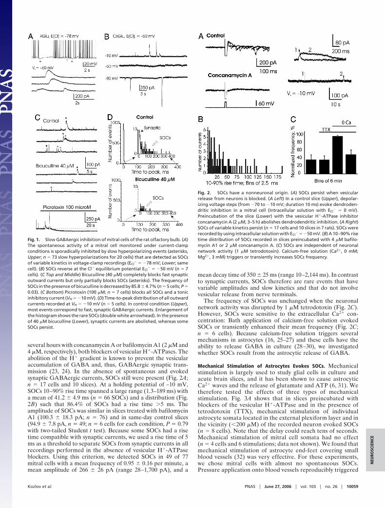
several hours with concanamycin A or bafilomycin A1 (2 �M and4 �M, respectively), both blockers of vesicular H�-ATPases. Theabolition of the H� gradient is known to prevent the vesicularaccumulation of GABA and, thus, GABAergic synaptic trans-mission (23, 24). In the absence of spontaneous and evokedsynaptic GABAergic currents, SOCs still were present (Fig. 2A;n � 17 cells and 10 slices). At a holding potential of –10 mV,SOCs 10–90% rise time spanned a large range (1.3–189 ms) witha mean of 41.2 � 4.9 ms (n � 66 SOCs) and a distribution (Fig.2B) such that 86.4% of SOCs had a rise time �5 ms. Theamplitude of SOCs was similar in slices treated with bafilomycinA1 (100.3 � 18.3 pA; n � 76) and in same-day control slices(94.9 � 7.8 pA, n � 49; n � 6 cells for each condition, P � 0.79with two-tailed Student t test). Because some SOCs had a risetime compatible with synaptic currents, we used a rise time of 5ms as a threshold to separate SOCs from synaptic currents in allrecordings performed in the absence of vesicular H�-ATPaseblockers. Using this criterion, we detected SOCs in 49 of 77mitral cells with a mean frequency of 0.95 � 0.16 per minute, amean amplitude of 266 � 26 pA (range 28–1,700 pA), and a
mean decay time of 350 � 25 ms (range 10–2,144 ms). In contrastto synaptic currents, SOCs therefore are rare events that havevariable amplitudes and slow kinetics and that do not involvevesicular release from nerve terminals.
The frequency of SOCs was unchanged when the neuronalnetwork activity was disrupted by 1 �M tetrodotoxin (Fig. 2C).However, SOCs were sensitive to the extracellular Ca2� con-centration: Bath application of calcium-free solution evokedSOCs or transiently enhanced their mean frequency (Fig. 2C;n � 6 cells). Because calcium-free solution triggers severalmechanisms in astrocytes (16, 25–27) and these cells have theability to release GABA in culture (28–30), we investigatedwhether SOCs result from the astrocytic release of GABA.
Mechanical Stimulation of Astrocytes Evokes SOCs. Mechanicalstimulation is largely used to study glial cells in culture andacute brain slices, and it has been shown to cause astrocyticCa2� waves and the release of glutamate and ATP (6, 31). Wetherefore tested the effects of three types of mechanicalstimulation. Fig. 3A shows that in slices preincubated withblockers of the vesicular H�-ATPase and in the presence oftetrodotoxin (TTX), mechanical stimulation of individualastrocyte somata located in the external plexiform layer and inthe vicinity (�200 �M) of the recorded neuron evoked SOCs(n � 8 cells). Note that the delay could reach tens of seconds.Mechanical stimulation of mitral cell somata had no effect(n � 4 cells and 6 stimulations; data not shown). We found thatmechanical stimulation of astrocyte end-feet covering smallblood vessels (32) was very effective. For these experiments,we chose mitral cells with almost no spontaneous SOCs.Pressure application onto blood vessels reproducibly triggered
Fig. 1. Slow GABAergic inhibition of mitral cells of the rat olfactory bulb. (A)The spontaneous activity of a mitral cell monitored under current-clampconditions is sporadically inhibited by slow hyperpolarizing events (asterisks,Upper; n � 73 slow hyperpolarizations for 20 cells) that are detected as SOCsof variable kinetics in voltage-clamp recordings (ECl
� � �78 mV; Lower; samecell). (B) SOCs reverse at the Cl� equilibrium potential ECl
� � �50 mV (n � 7cells). (C Top and Middle) Bicuculline (40 �M) completely blocks fast synapticoutward currents but only partially blocks SOCs (asterisks). The frequency ofSOCs in the presence of bicuculline is decreased by 85.8 � 4.7% (n � 5 cells; P �0.03). (C Bottom) Picrotoxin (100 �M; n � 7 cells) blocks all SOCs and a tonicinhibitory current (VH � �10 mV). (D) Time-to-peak distribution of all outwardcurrents recorded at VH � �10 mV (n � 5 cells). In control condition (Upper),most events correspond to fast, synaptic GABAergic currents. Enlargement ofthe histogram shows the rare SOCs (double white arrowhead). In the presenceof 40 �M bicuculline (Lower), synaptic currents are abolished, whereas someSOCs persist.
Fig. 2. SOCs have a nonneuronal origin. (A) SOCs persist when vesicularrelease from neurons is blocked. (A Left) In a control slice (Upper), depolar-izing voltage steps (from �70 to �10 mV; duration 10 ms) evoke dendroden-dritic inhibition in a mitral cell (intracellular solution with ECl
� � 0 mV).Preincubation of the slice (Lower) with the vesicular H�-ATPase inhibitorconcanamycin A (2 �M; 3–5 h) abolishes dendrodendritic inhibition. (A Right)SOCs of variable kinetics persist (n � 17 cells and 10 slices in 7 rats). SOCs wererecorded by using intracellular solution with ECl
� � �50 mV. (B) A 10–90% risetime distribution of SOCs recorded in slices preincubated with 4 �M bafilo-mycin A1 or 2 �M concanamycin A. (C) SOCs are independent of neuronalnetwork activity (1 �M tetrodotoxin). Calcium-free solution (Ca2�, 0 mM;Mg2�, 3 mM) triggers or transiently increases SOCs frequency.
Kozlov et al. PNAS � June 27, 2006 � vol. 103 � no. 26 � 10059
NEU
ROSC
IEN
CE
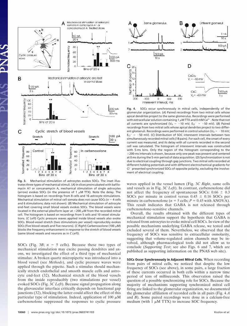
SOCs (Fig. 3B; n � 5 cells). Because these two types ofmechanical stimulation may excite passing dendrites and ax-ons, we investigated the effect of a third type of mechanicalstimulus: A broken quartz micropipette was introduced into ablood vessel (see Methods), and cyclic pressure waves wereapplied through the pipette. Such a stimulus should mechan-ically stretch endothelial and smooth muscle cells and astro-cyte end-feet (32). Mechanical stretch of the blood vesselsfrom the inside reproducibly (two stimulations per vessel)evoked SOCs (Fig. 3C Left). Because signal propagation alongthe gliovascular interface critically depends on functional gapjunctions (32), blocking the latter could affect the effect of thisparticular type of stimulation. Indeed, application of 100 �Mcarbenoxolone suppressed the responses to cyclic pressure
waves applied in the vessel lumen (Fig. 3C Right, same cellsand vessels as in Fig. 3C Left). In contrast, carbenoxolone didnot affect the frequency of spontaneous SOCs: 0.66 � 0.3SOCs per minute in control versus 0.45 � 0.16 SOCs perminute in carbenoxolone (n � 5 cells; P � 0.45 with ANOVA).This result indicates that GABA is not released throughcarbenoxolone-sensitive channels.
Overall, the results obtained with the different types ofmechanical stimulation support the hypothesis that GABA isreleased from astrocytes to evoke mitral cell SOCs. In view of thepossible mechanisms underlying GABA release, we tested andexcluded several of them. Nevertheless, we observed that thefrequency of SOCs was sensitive to extracellular osmolarity,suggesting that volume-regulated anion channels may be in-volved, although pharmacological tools did not allow us toconclude (Supporting Text; see also Figs. 6 and 7, which arepublished as supporting information on the PNAS web site).
SOCs Occur Synchronously in Adjacent Mitral Cells. When recordingfrom pairs of mitral cells, we noticed that despite the lowfrequency of SOCs (see above), in some pairs, a large fractionof these currents occurred in both cells within a narrow timeperiod of tens of milliseconds. This observation raised thequestion of a possible synchronizing role for SOCs. Because themajority of mechanisms supporting synchronized mitral cellfiring are linked to the glomerular organization, we documentedthe glomerular affiliation of recorded cells in all pairs (Fig. 4 Aand B). Some paired recordings were done in a calcium-freemedium (with 1 �M TTX) to increase SOC frequency.
Fig. 3. Mechanical stimulation of astrocytes evokes SOCs. The inset illus-trates three types of mechanical stimuli. (A) In slices preincubated with bafilo-mycin A1 or concanamycin A, mechanical stimulation of single astrocytes(arrow) evokes SOCs (in the presence of 1 �M TTX). Note the delay. Thehistogram is based on recordings from 8 cells and 16 astrocyte stimulations.Mechanical stimulation of mitral cell somata does not cause SOCs (n � 4 cellsand 6 stimulations; data not shown). (B) Mechanical stimulation of astrocyteend-feet covering small blood vessels evokes SOCs. The blood vessels werelocated in the external plexiform layer at �200 �M from the recorded mitralcell. The histogram is based on recordings from 5 cells and 10 vessel stimula-tions. (C Left) Cyclic pressure waves applied inside blood vessels also evokeSOCs. Blood-vessel stretch (two stimulations per vessel) reproducibly evokesSOCs (five blood vessels and five neurons). (C Right) Carbenoxolone (100 �M)blocks the frequency enhancement in response to the stretch of blood vessels(same blood vessels and neurons as in C Left).
Fig. 4. SOCs occur synchronously in mitral cells, independently of theglomerular organization. (A) Paired recordings from two mitral cells whoseapical dendrites project to the same glomerulus. Recordings were performedwith extracellular solution containing 1 �M TTX and 0 mM Ca2�. Note that notall currents are synchronized (VH � �10 mV; ECl
� � �50 mV). (B) Pairedrecordings from two mitral cells whose apical dendrites project to two differ-ent glomeruli. Recordings were performed in control solution (VH � �10 mV;ECl
� � �50 mV). (C) Distribution of SOC interevent intervals between twosimultaneously recorded mitral cells (18 pairs). For each cell, the onset of everycurrent was measured, and its delay with all currents recorded in the secondcell was calculated. The histogram of interevent intervals was constructedwith 5-ms bins. Only the region of the histogram corresponding to the�200-ms intervals is shown, because only one peak was present and centeredat 0 ms during the 5-min period of data acquisition. (D) Synchronization is notdue to electrical coupling through gap junctions. Two mitral cells recorded atdifferent holding potentials and with different electrochemical gradients forCl� presented synchronized SOCs of opposite polarity, excluding the involve-ment of electrical coupling.
10060 � www.pnas.org�cgi�doi�10.1073�pnas.0603741103 Kozlov et al.
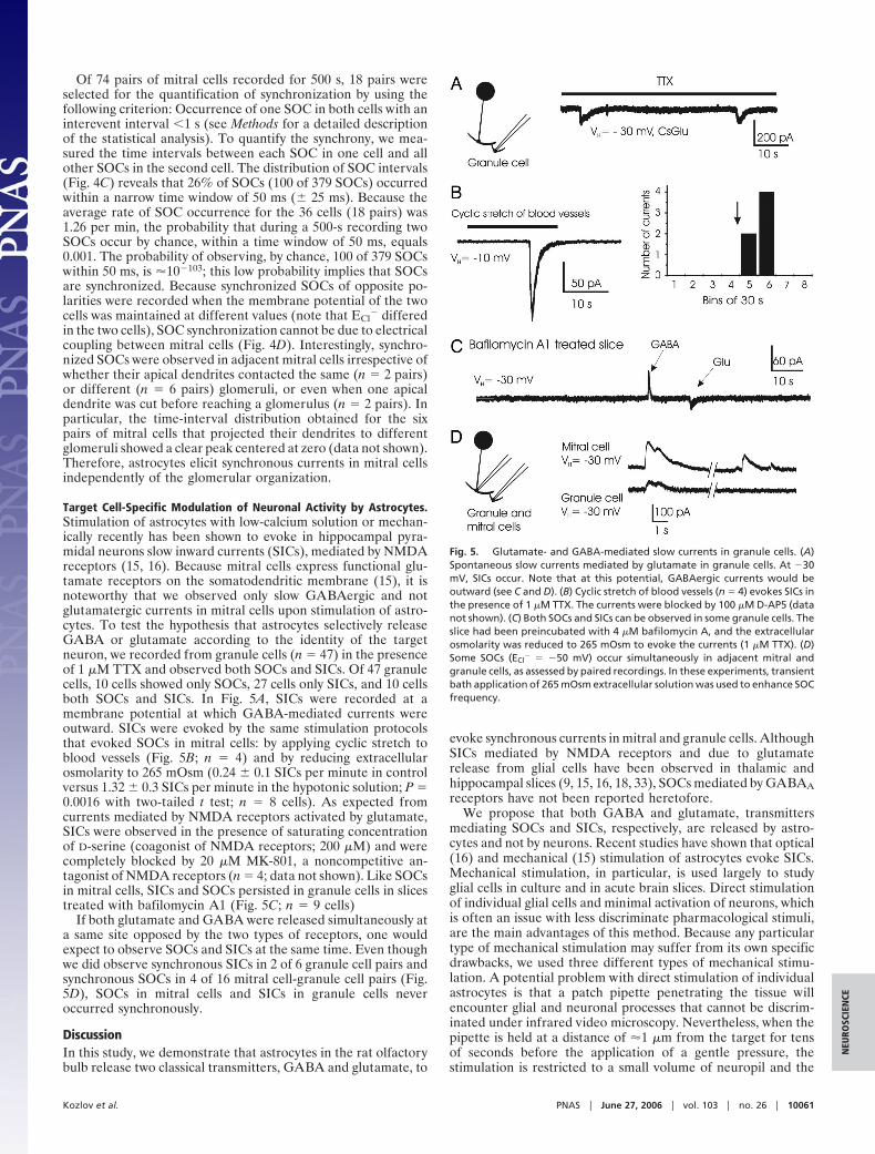
Of 74 pairs of mitral cells recorded for 500 s, 18 pairs wereselected for the quantification of synchronization by using thefollowing criterion: Occurrence of one SOC in both cells with aninterevent interval �1 s (see Methods for a detailed descriptionof the statistical analysis). To quantify the synchrony, we mea-sured the time intervals between each SOC in one cell and allother SOCs in the second cell. The distribution of SOC intervals(Fig. 4C) reveals that 26% of SOCs (100 of 379 SOCs) occurredwithin a narrow time window of 50 ms (� 25 ms). Because theaverage rate of SOC occurrence for the 36 cells (18 pairs) was1.26 per min, the probability that during a 500-s recording twoSOCs occur by chance, within a time window of 50 ms, equals0.001. The probability of observing, by chance, 100 of 379 SOCswithin 50 ms, is �10�103; this low probability implies that SOCsare synchronized. Because synchronized SOCs of opposite po-larities were recorded when the membrane potential of the twocells was maintained at different values (note that ECl
� differedin the two cells), SOC synchronization cannot be due to electricalcoupling between mitral cells (Fig. 4D). Interestingly, synchro-nized SOCs were observed in adjacent mitral cells irrespective ofwhether their apical dendrites contacted the same (n � 2 pairs)or different (n � 6 pairs) glomeruli, or even when one apicaldendrite was cut before reaching a glomerulus (n � 2 pairs). Inparticular, the time-interval distribution obtained for the sixpairs of mitral cells that projected their dendrites to differentglomeruli showed a clear peak centered at zero (data not shown).Therefore, astrocytes elicit synchronous currents in mitral cellsindependently of the glomerular organization.
Target Cell-Specific Modulation of Neuronal Activity by Astrocytes.Stimulation of astrocytes with low-calcium solution or mechan-ically recently has been shown to evoke in hippocampal pyra-midal neurons slow inward currents (SICs), mediated by NMDAreceptors (15, 16). Because mitral cells express functional glu-tamate receptors on the somatodendritic membrane (15), it isnoteworthy that we observed only slow GABAergic and notglutamatergic currents in mitral cells upon stimulation of astro-cytes. To test the hypothesis that astrocytes selectively releaseGABA or glutamate according to the identity of the targetneuron, we recorded from granule cells (n � 47) in the presenceof 1 �M TTX and observed both SOCs and SICs. Of 47 granulecells, 10 cells showed only SOCs, 27 cells only SICs, and 10 cellsboth SOCs and SICs. In Fig. 5A, SICs were recorded at amembrane potential at which GABA-mediated currents wereoutward. SICs were evoked by the same stimulation protocolsthat evoked SOCs in mitral cells: by applying cyclic stretch toblood vessels (Fig. 5B; n � 4) and by reducing extracellularosmolarity to 265 mOsm (0.24 � 0.1 SICs per minute in controlversus 1.32 � 0.3 SICs per minute in the hypotonic solution; P �0.0016 with two-tailed t test; n � 8 cells). As expected fromcurrents mediated by NMDA receptors activated by glutamate,SICs were observed in the presence of saturating concentrationof D-serine (coagonist of NMDA receptors; 200 �M) and werecompletely blocked by 20 �M MK-801, a noncompetitive an-tagonist of NMDA receptors (n � 4; data not shown). Like SOCsin mitral cells, SICs and SOCs persisted in granule cells in slicestreated with bafilomycin A1 (Fig. 5C; n � 9 cells)
If both glutamate and GABA were released simultaneously ata same site opposed by the two types of receptors, one wouldexpect to observe SOCs and SICs at the same time. Even thoughwe did observe synchronous SICs in 2 of 6 granule cell pairs andsynchronous SOCs in 4 of 16 mitral cell-granule cell pairs (Fig.5D), SOCs in mitral cells and SICs in granule cells neveroccurred synchronously.
DiscussionIn this study, we demonstrate that astrocytes in the rat olfactorybulb release two classical transmitters, GABA and glutamate, to
evoke synchronous currents in mitral and granule cells. AlthoughSICs mediated by NMDA receptors and due to glutamaterelease from glial cells have been observed in thalamic andhippocampal slices (9, 15, 16, 18, 33), SOCs mediated by GABAAreceptors have not been reported heretofore.
We propose that both GABA and glutamate, transmittersmediating SOCs and SICs, respectively, are released by astro-cytes and not by neurons. Recent studies have shown that optical(16) and mechanical (15) stimulation of astrocytes evoke SICs.Mechanical stimulation, in particular, is used largely to studyglial cells in culture and in acute brain slices. Direct stimulationof individual glial cells and minimal activation of neurons, whichis often an issue with less discriminate pharmacological stimuli,are the main advantages of this method. Because any particulartype of mechanical stimulation may suffer from its own specificdrawbacks, we used three different types of mechanical stimu-lation. A potential problem with direct stimulation of individualastrocytes is that a patch pipette penetrating the tissue willencounter glial and neuronal processes that cannot be discrim-inated under infrared video microscopy. Nevertheless, when thepipette is held at a distance of �1 �m from the target for tensof seconds before the application of a gentle pressure, thestimulation is restricted to a small volume of neuropil and the
Fig. 5. Glutamate- and GABA-mediated slow currents in granule cells. (A)Spontaneous slow currents mediated by glutamate in granule cells. At �30mV, SICs occur. Note that at this potential, GABAergic currents would beoutward (see C and D). (B) Cyclic stretch of blood vessels (n � 4) evokes SICs inthe presence of 1 �M TTX. The currents were blocked by 100 �M D-AP5 (datanot shown). (C) Both SOCs and SICs can be observed in some granule cells. Theslice had been preincubated with 4 �M bafilomycin A, and the extracellularosmolarity was reduced to 265 mOsm to evoke the currents (1 �M TTX). (D)Some SOCs (ECl
� � �50 mV) occur simultaneously in adjacent mitral andgranule cells, as assessed by paired recordings. In these experiments, transientbath application of 265 mOsm extracellular solution was used to enhance SOCfrequency.
Kozlov et al. PNAS � June 27, 2006 � vol. 103 � no. 26 � 10061
NEU
ROSC
IEN
CE
target and may be assumed to stimulate the target preferentially.Our second method of mechanical stimulation, the applicationof pressure to the vessel outer wall, is designed to activateastrocyte end-feet that cover �90% of the surface of small bloodvessels. This approach very efficiently triggered SOCs in theabsence of synaptic transmission (blocked by bafilomycin A1),supporting the involvement of astrocytes in the generation ofSOCs. Furthermore, vascular distension from the inside, apotent means of evoking SOCs, is very unlikely to stimulateneurons directly (it also was efficient in the presence of TTX).Indeed, when stretching blood vessels, we observed no activationof mitral cell firing. In addition, direct mechanical stimulation ofneurons did not evoke SOCs; likewise, SICs were not evoked bymechanical stimulation of neurons in the hippocampus (15).Because all three types of mechanical stimulation led to the sameresult, activation of SOCs, we conclude that GABA is releasedfrom astrocytes.
Our data raise the question of the source of GABA releasedby astrocytes. Although several studies have suggested thatGABA or GABA-like substances can be secreted from glial cellsin culture (28, 30, 34, 35), it is generally assumed that glial cellsdo not contain the enzymes necessary for the synthesis of GABAand that, if present, GABA is taken up from the extracellularmedium by GABA transporters. Our results suggest that GABAis most likely the transmitter involved, even though othertransmitters might substitute (36). Taurine would be a possiblecandidate because it is abundant in the olfactory bulb (37, 38)and induces a slow, ‘‘nondesensitizing’’ inhibition of mitral cellsduring a prolonged bath application (39). The same studyreported, however, that taurine does not affect granule cells. Incontrast, we observed SOCs in 40% of granule cells. Moreover,the requirement of high concentrations of bicuculline to blockthe SOCs and the absence of activation of glycine receptorspresent on mitral cells argue against the involvement of taurine.Finally, the kinetics of SOCs is slower in the presence ofnipecotic acid, a blocker of GABA transporters (SupportingText). We thus propose that the transmitter responsible for SOCsis GABA.
Do SOCs and SICs involve two types of astrocytes that wouldrelease either GABA or glutamate? Alternatively, can a singleastrocyte release both GABA and glutamate, possibly at thesame site, with an effect on neurons depending on the type ofneuronal receptors that face the release site? In the glomerularlayer, astrocytes respect the glomerular organization (40),surrounding a single glomerulus and extending processes thatwrap and isolate bundles of dendrites with dendrodendriticsynapses (41). Such compartmentalization should favor neu-ronal synchronization because of spillover of a transmitter,whether released by neurons or by glial cells. In the externalplexiform layer (40), an important feature of astrocytes is theregular spacing of their cell bodies and the spatially limitedoverlap of their processes. The presence of such astrocyticfunctional domains (42) suggests that it is likely that twodifferent types of astrocytes are responsible for SOCs andSICs. Indeed, SICs were not observed in mitral cells, whereastheir dendrites are covered by functional glutamate receptors(43), and synchronous SOCs, but not synchronous SOCs andSICs, were detected in paired recordings of mitral and granulecells. However, we cannot rule out the possibility that a singleastrocyte releases GABA and glutamate separately at differ-ent and spatially distant release sites.
SOCs obviously differ from classical GABAergic synapticcurrents. They occur at a low frequency, display slow andvariable kinetics, and require high concentrations of a compet-itive antagonist, bicuculline, to be blocked. These propertiescould result from an astrocytic ensheathment of dendrites thatcreates a barrier for diffusion and clearance of GABA, from therelease kinetics, or from the involvement of particular subtypes
of GABAA receptors. Because the synaptic sites and dendriticshafts of mitral cells are wrapped together but synaptic currentsand SOCs have different sensitivities to bicuculline, differentGABAA receptor subtypes may mediate synaptic currents andSOCs, as it is the case for the tonic GABAergic current (forreview, see ref. 22). However, gabazine had similar effects onsynaptic currents and SOCs, in contrast to its differential actionobserved on synaptic and tonic currents in the hippocampus (22).
It has been demonstrated that several intra- and interglomeru-lar mechanisms support synchronization of mitral cell activity, afiring behavior important in the processing of odors (44, 45). Wepropose that an additional type of mitral cell synchronization,mediated by astrocytes, operates in the olfactory bulb. It occursat a very low frequency, its time course is slow, it does not respectthe glomerular organization, and its physiological or patholog-ical relevance remains to be established in vivo.
MethodsSlice Preparation and Electrophysiology. Horizontal slices (300–400�m) from olfactory bulbs of 2- to 5-week-old Wistar rats were cutin ice-cold extracellular solution (for composition of all solutionsused in the study, see Supporting Text) and incubated either atroom temperature or at 34°C. Recordings were done withborosilicate patch pipettes with a resistance of 2–7 M�. Datawere acquired by using Axopatch 200A and 200B amplifiers andP-CLAMP8 software (Axon Instruments, Union City, CA). Analogsignals recorded in voltage clamp and current clamp modes werelow-pass filtered with a four-pole Bessel filter at 2 kHz and 5kHz, and sampled, respectively, at 10 kHz and 20 kHz. Seriesresistance was not compensated, and leak currents were notsubtracted. For recordings with intracellular solutions contain-ing gluconate as the major anion, all potentials were correctedfor a �10 mV junction potential.
Slice preincubation in the presence of the vesicular H�-ATPase inhibitors bafilomycin A1 or concanamycin A wascarried out at 34°C for 3–5 h. Control slices were taken from thesame animals and incubated at 34°C in the absence of drug.
Mechanical Stimulation of Astrocytes and Blood Vessels. A patchpipette containing the extracellular solution was positioned nearastrocytes and blood vessels located in the external plexiformlayer within 100–200 �M from the recorded neuron. A gentlepressure was transiently (�1 s) applied onto the astrocytemembrane or onto the blood vessel wall. Cyclic stretch of bloodvessels (arterioles and venules) was performed from the insideas follows. A quartz micropipette with a broken and sharp tip wasforced through the vessel wall, and after several minutes, a cyclicpressure wave (2 Hz) was applied for 20 s. The vessel was chosensuch that its penetration site was located several hundredmicrometers from the recorded neuron, and it or one of itseff luents was running near the neuron.
Statistics. To determine the statistical significance of the differ-ence between means, the two-tailed Student t test and theANOVA test were used; P � 0.05 was considered statisticallysignificant. P � 0.05 and 0.01 are indicated in the figures by singleand double asterisks, respectively. Averaged values are reportedas mean � SEM. For more information on data analysis andstatistics, see Supporting Text.
We thank M. Hanafi for technical assistance. A. J. Hudspeth madehelpful comments on the manuscript. A.S.K. was supported by Interna-tional Brain Research Organization and Ecole Superieure de Physiqueet de Chimie Industrielles. Support was provided by the Institut Nationalde la Sante et de la Recherche Medicale, the Ministere de l’EducationNationale de la Recherche et de la Technologie, the Centre National dela Recherche Scientifique, and Fondation pour la Recherche MedicaleGrant ICP20001222128.
10062 � www.pnas.org�cgi�doi�10.1073�pnas.0603741103 Kozlov et al.
1. Auld, D. S. & Robitaille, R. (2003) Neuron 40, 389–400.2. Volterra, A. & Meldolesi, J. (2005) Nat. Rev. Neurosci. 6, 626–640.3. Bushong, E. A., Martone, M. E., Jones, Y. Z. & Ellisman, M. H. (2002)
J. Neurosci. 22, 183–192.4. Ogata, K. & Kosaka, T. (2002) Neuroscience 113, 221–233.5. Nett, W. J., Oloff, S. H. & McCarthy, K. D. (2002) J. Neurophysiol. 87, 528–537.6. Newman, E. A. & Zahs, K. R. (1998) J. Neurosci. 18, 4022–4028.7. Aguado, F., Espinosa-Parrilla, J. F., Carmona, M. A. & Soriano, E. (2002)
J. Neurosci 22, 9430–9444.8. Pasti, L., Volterra, A., Pozzan, T. & Carmignoto, G. (1997) J. Neurosci. 17,
7817–7830.9. Parri, H. R., Gould, T. M. & Crunelli, V. (2001) Nat. Neurosci. 4, 803–812.
10. Hirase, H., Qian, L., Bartho, P. & Buzsaki, G. (2004) PLoS Biol. 2, E96.11. Basarsky, T. A., Duffy, S. N., Andrew, R. D. & MacVicar, B. A. (1998)
J. Neurosci. 18, 7189–7199.12. Grosche, J., Matyash, V., Moller, T., Verkhratsky, A., Reichenbach, A. &
Kettenmann, H. (1999) Nat. Neurosci. 2, 139–143.13. Nadkarni, S. & Jung, P. (2003) Phys. Rev. Lett. 91, 268101.14. Tian, G. F., Azmi, H., Takano, T., Xu, Q., Peng, W., Lin, J., Oberheim, N., Lou,
N., Wang, X., Zielke, H. R., et al. (2005) Nat. Med. 11, 973–981.15. Angulo, M. C., Kozlov, A. S., Charpak, S. & Audinat, E. (2004) J. Neurosci. 24,
6920–6927.16. Fellin, T., Pascual, O., Gobbo, S., Pozzan, T., Haydon, P. G. & Carmignoto, G.
(2004) Neuron 43, 729–743.17. Cavelier, P., Hamann, M., Rossi, D., Mobbs, P. & Attwell, D. (2005) Prog.
Biophys. Mol. Biol. 87, 3–16.18. Perea, G. & Araque, A. (2005) J. Neurosci. 25, 2192–2203.19. Kang, J., Jiang, L., Goldman, S. A. & Nedergaard, M. (1998) Nat. Neurosci. 1,
683–692.20. Deleuze, C., Duvoid, A. & Hussy, N. (1998) J. Physiol. (London) 507, 463–471.21. Shepherd, G. M., Chen, W. R. & Greer, C. A. (2004) in The Synaptic
Organization of the Brain, ed. Shepherd, G. M. (Oxford Univ. Press, New York),pp. 165–203.
22. Semyanov, A., Walker, M. C., Kullmann, D. M. & Silver, R. A. (2004) TrendsNeurosci. 27, 262–269.
23. Zhou, Q., Petersen, C. C. & Nicoll, R. A. (2000) J. Physiol. (London) 525,195–206.
24. Rossi, D. J., Hamann, M. & Attwell, D. (2003) J. Physiol. (London) 548,97–110.
25. Zanotti, S. & Charles, A. (1997) J. Neurochem. 69, 594–602.26. Ye, Z. C., Wyeth, M. S., Baltan-Tekkok, S. & Ransom, B. R. (2003) J. Neurosci.
23, 3588–3596.27. Duan, S., Anderson, C. M., Keung, E. C., Chen, Y., Chen, Y. & Swanson, R. A.
(2003) J. Neurosci. 23, 1320–1328.28. Liu, Q. Y., Schaffner, A. E., Chang, Y. H., Maric, D. & Barker, J. L. (2000)
J. Neurophysiol. 84, 1392–1403.29. Verderio, C., Bruzzone, S., Zocchi, E., Fedele, E., Schenk, U., De Flora, A. &
Matteoli, M. (2001) J. Neurochem. 78, 646–657.30. Jow, F., Chiu, D., Lim, H. K., Novak, T. & Lin, S. (2004) Neurochem. Int. 45,
273–283.31. Koizumi, S., Fujishita, K., Tsuda, M., Shigemoto-Mogami, Y. & Inoue, K.
(2003) Proc. Natl. Acad. Sci. USA 100, 11023–11028.32. Simard, M., Arcuino, G., Takano, T., Liu, Q. S. & Nedergaard, M. (2003)
J. Neurosci. 23, 9254–9262.33. Demarque, M., Represa, A., Becq, H., Khalilov, I., Ben Ari, Y. & Aniksztejn,
L. (2002) Neuron 36, 1051–1061.34. Neal, M. J. & Bowery, N. G. (1979) Brain Res. 167, 337–343.35. Wu, P. H., Durden, D. A. & Hertz, L. (1979) J. Neurochem. 32, 379–390.36. Pasantes-Morales, H., Murray, R. A., Sanchez-Olea, R. & Moran, J. (1994)
Am. J. Physiol. 266, C172–C178.37. Collins, G. G. (1974) Brain Res. 76, 447–459.38. Didier, A., Ottersen, O. P. & Storm-Mathisen, J. (1994) NeuroReport 6,
145–148.39. Belluzzi, O., Puopolo, M., Benedusi, M. & Kratskin, I. (2004) Neuroscience 124,
929–944.40. Bailey, M. S. & Shipley, M. T. (1993) J. Comp. Neurol. 328, 501–526.41. Kasowski, H. J., Kim, H. & Greer, C. A. (1999) J. Comp. Neurol. 407, 261–274.42. Nedergaard, M., Ransom, B. & Goldman, S. A. (2003) Trends Neurosci. 26,
523–530.43. Lowe, G. (2003) J. Neurophysiol. 90, 1737–1746.44. Stopfer, M., Bhagavan, S., Smith, B. H. & Laurent, G. (1997) Nature 390, 70–74.45. MacLeod, K., Backer, A. & Laurent, G. (1998) Nature 395, 693–698.
Kozlov et al. PNAS � June 27, 2006 � vol. 103 � no. 26 � 10063
NEU
ROSC
IEN
CE
Corrections
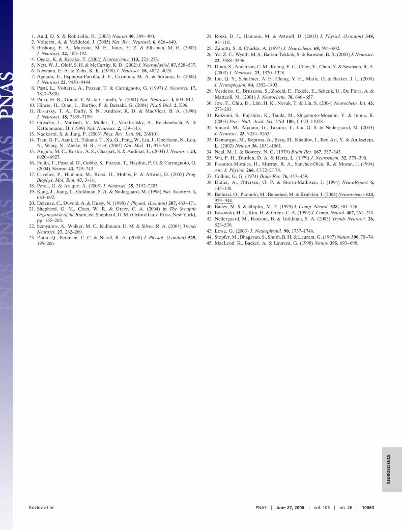
GENETICS. For the article ‘‘A Sanger�pyrosequencing hybridapproach for the generation of high-quality draft assemblies ofmarine microbial genomes,’’ by Susanne M. D. Goldberg, JustinJohnson, Dana Busam, Tamara Feldblyum, Steve Ferriera,Robert Friedman, Aaron Halpern, Hoda Khouri, Saul A. Krav-itz, Federico M. Lauro, Kelvin Li, Yu-Hui Rogers, Rob-ert Strausberg, Granger Sutton, Luke Tallon, Torsten Thomas,
Fig. 3. Decision tree for hybrid sequencing strategy. For organisms with a small genome size (�3 Mb) and�or a small number of gaps and�or high levels ofrepetitive structure inducing physical ends, we found 8� Sanger sequencing to be the most cost-effective approach. For organisms with a large genome size,many sequencing gaps, and�or hard stops, we found initial sequencing of 5.3� Sanger data followed by the addition of two 454 runs to be the most cost-effectiveapproach.
www.pnas.org�cgi�doi�10.1073�pnas.0607197103
Eli Venter, Marvin Frazier, and J. Craig Venter, which appearedin issue 30, July 25, 2006, of Proc Natl Acad Sci USA (103:11240–11245; first published July 13, 2006; 10.1073�pnas.0604351103),the authors note that in Fig. 3, the text in the lower green box,‘‘Reference Genome Unavailable,’’ should read ‘‘ReferenceGenome Available.’’ The corrected figure and its legend appearbelow. This error does not effect the conclusions of the article.
www.pnas.org PNAS � October 24, 2006 � vol. 103 � no. 43 � 16057–16058
CORR
ECTI
ON
S
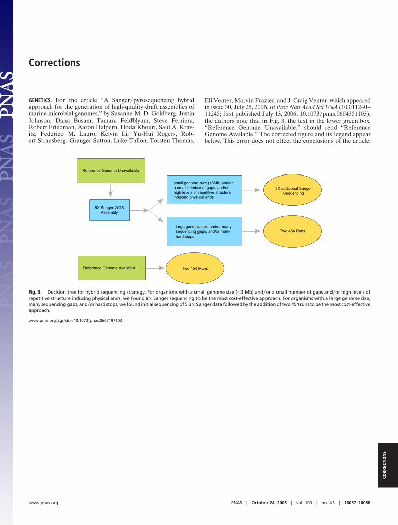
PHARMACOLOGY. For the article ‘‘RF9, a potent and selectiveneuropeptide FF receptor antagonist, prevents opioid-inducedtolerance associated with hyperalgesia,’’ by Frederic Simonin,Martine Schmitt, Jean-Paul Laulin, Emilie Laboureyras, Jack H.Jhamandas, David MacTavish, Audrey Matifas, CatherineMollereau, Patrick Laurent, Marc Parmentier, Brigitte L.Kieffer, Jean-Jacques Bourguignon, and Guy Simonnet, whichappeared in issue 2, January 10, 2006, of Proc Natl Acad Sci USA(103:466–471; first published January 3, 2006; 10.1073�pnas.0502090103), the authors note that in Fig. 1B, the position of thebond between the adamantane system and the rest of the RF9compound is incorrect. The corrected figure and its legend appearbelow. This error does not affect the conclusions of the article.
NEUROSCIENCE. For the article ‘‘Target cell-specific modulation ofneuronal activity by astrocytes,’’ by A. S. Kozlov, M. C. Angulo,E. Audinat, and S. Charpak, which appeared in issue 26, June 27,2006, of Proc Natl Acad Sci USA (103:10058–10063; first pub-lished June 16, 2006; 10.1073�pnas.0603741103), the authorsnote that some affiliation and correspondence information wasincorrect or incomplete as given. The correct addresses for theauthors’ laboratories are as follows: Institut National de la Santeet de la Recherche Medicale, U603, Paris, F-75006 France;Centre National de la Recherche Scientifique, FRE2500, Paris,F-75006 France; Laboratory of Neurophysiology, Ecole Su-perieure de Physique et de Chimie Industrielles, Paris, F-75005France; and Laboratory of Neurophysiology and New Micros-copies, Universite Paris Descartes, Paris, F-75006 France. Inaddition, S. Charpak should have been listed as one of thecorresponding authors. E-mail: [email protected].
www.pnas.org�cgi�doi�10.1073�pnas.0608114103
Fig. 1. Screening of RFamide derivatives on hNPFF2R. (A) hNPFF2R mem-branes were labeled with [125I]Tyr-NPFF, and three concentrations of RFamidederivatives were tested in competition experiments. Each concentration wastested in duplicate. Results for the reference and the six most active com-pounds are shown. Arrowheads indicate compounds that were selected forfurther characterization. (B) Structures of RF2, RF9, RF48, RF49, and BIBP3226.
www.pnas.org�cgi�doi�10.1073�pnas.0608112103
16058 � www.pnas.org
Related Documents


