ADVERTIMENT. Lʼaccés als continguts dʼaquesta tesi queda condicionat a lʼacceptació de les condicions dʼús establertes per la següent llicència Creative Commons: http://cat.creativecommons.org/?page_id=184 ADVERTENCIA. El acceso a los contenidos de esta tesis queda condicionado a la aceptación de las condiciones de uso establecidas por la siguiente licencia Creative Commons: http://es.creativecommons.org/blog/licencias/ WARNING. The access to the contents of this doctoral thesis it is limited to the acceptance of the use conditions set by the following Creative Commons license: https://creativecommons.org/licenses/?lang=en

Welcome message from author
This document is posted to help you gain knowledge. Please leave a comment to let me know what you think about it! Share it to your friends and learn new things together.
Transcript
ADVERTIMENT. Lʼaccés als continguts dʼaquesta tesi queda condicionat a lʼacceptació de les condicions dʼúsestablertes per la següent llicència Creative Commons: http://cat.creativecommons.org/?page_id=184
ADVERTENCIA. El acceso a los contenidos de esta tesis queda condicionado a la aceptación de las condiciones de usoestablecidas por la siguiente licencia Creative Commons: http://es.creativecommons.org/blog/licencias/
WARNING. The access to the contents of this doctoral thesis it is limited to the acceptance of the use conditions setby the following Creative Commons license: https://creativecommons.org/licenses/?lang=en
1
PhD DISSERTATION NEUROSCIENCE PHD PROGRAM
JUNE 2019
Taking control to cope with stress: Consequences of
Controllability on Behavior & Gene Expression
INÉS CORDÓN MORILLAS
AUTONOMOUS UNIVERSITY OF BARCELONA
Department of Cellular Biology, Physiology and Immunology
Animal Physiology Unit
School of Biosciences
Institute of Neuroscience
DIRECTORS:
ANTONIO ARMARIO GARCÍA, PhD
JAVIER CARRASCO TRANCASO, PhD

2

3
A mis abuelicos, Alfonso y Mª Pilar
4
Durante la realización de la presente Tesis Doctoral nuestro laboratorio ha recibido las siguientes becas y ayudas:
Ministerio de Ciencia e Innovación/Ministerio de Economía y Competitividad:
SAF2011-28313
SAF2014-53876-R
SAF2017-017-83430-R
RTC-2015-3898-1
Generalitat de Catalunya:
SGR2009-16
SGR2014-1020
SGR2017-457
Otras ayudas:
RD12/0028/0014, Red de Trastornos Adictivos, RETICS, Instituto de Salud Carlos III
CIBER en Salud Mental, CIBERSAM, Instituto de Salud Carlos III
2011/021, Plan Nacional sobre Drogas, Ministerio de Sanidad, Consumo y bienestar Social
5
ABSTRACT
Research in humans and animals have shown that individual differences and controllability over stress are critical factors to determine its main outcomes. Thus, exposure to controllable stress (CoS) buffers part of the detrimental effects of exposure to the same amount of uncontrollable stress (UnS). This buffering effect includes a protection (“immunization”) from the consequences of future exposure to UnS, highlighting the potential of prior experience with CoS as a therapeutic approach to treat stress-related disorders.
In the present work, adult rats were exposed to 2 or 5 sessions of an active avoidance (TWAA) task in a shuttle-box using footshock as the aversive stimulus. This group had the possibility of control over stress, whereas an additional group received the same amount of shocks regardless of its behavior (yoked, UnS). A stress-naïve group was also included. We studied: (i) whether 2 or 5 sessions of UnS is sufficient to induce long-lasting behavioral effects on activity, arousal and anxiety-like behaviors; (ii) whether exposure to the same amount of CoS buffers the negative long-term impact observed after exposure to UnS; (iii) whether individual differences in the performance in the TWAA task or more prolonged experience with it is necessary to better observe the benefits of controllability; and (iv) the possible contribution of the changes in the expression of genes related to synaptic plasticity and epigenetic regulation in brain areas critically involved in stress (e.g. prefrontal cortex, amygdala and hippocampal formation).
Two phenotypes arose in the TWAA task: some animals exhibited high levels of avoidance after 2 or 5 days, whereas others showed marked deficits in acquiring the avoidance response from the first day and did not rescue this deficit despite repeated experience with the task. These phenotypes differed in their latency to escape from the first trials, suggesting that how individuals confront and respond initially to the
6
aversive stimuli could determine how avoidance is acquired and performed.
Exposure to CoS or UnS did not induce a clear behavioral profile, although UnS decreased activity in new environments and enhanced fear conditioning during exposure to the shock context. Importantly, exposure to the same amount of CoS buffered both effects. Moreover, only animals showing high avoidance rates during prolonged exposure to the TWAA task showed this beneficial effect, suggesting that actual rather than potential control over stress is a critical factor. Results from a behavioral profiling identifying “affected” and “non-affected” subpopulations based on key parameters related to activity, anxiety-like and associative behaviors indicated that prolonged exposure to CoS reduced drastically the proportion of animals identified as “affected”, an effect not shown by exposure to UnS.
When gene expression (two experiments) was assessed between 2 and 13 days after stress, a decrease was observed in BDNF gene expression in CA1. Other changes, including TrkB or histone deacetylases were not consistent or complex depending on controllability, the brain area and the time elapse since stress. Therefore, we were unable to observe any clear and consistent biochemical signature of exposure to stress and the degree of control over stress.
In conclusion, the present results clearly indicate that, within the frameworks of studies about the influence of controllability in the consequences of stress, the characterization of individual differences in the performance in an active avoidance tasks can contribute to distinguish between the possibility of control and the actual control over stress. Such a distinction can help to better understand the differences in the behavioral and biochemical consequences of exposure to CoS versus UnS and the role of actual controllability.
7
CONTENTS Page
List of abbreviations………………………………………….9
Introduction……………………………………………………….13
1.! The concept of stress……………………………………………………………14 2.! HPA axis………………………………………………………………………...18 3.! Long-term effects of acute stress……………………………………………….22
3.1.! Post-Traumatic Stress Disorder………………………………………….22 3.2.! Animal models of PTSD………………………………………………….29
4.! Individual differences in psychopathology…………………………………….42 4.1.! Differential response to trauma & Biological risk factors……………….42 4.2.! Modelling variability associated to PTSD in animal models……………44 4.3.! The environmental epigenetics hypothesis………………………………46 4.4.! Influence of cognitive and emotional regulation
on vulnerability………………………...………………………...……….50 5.! Coping, Active Avoidance and Controllability………………………...............52
5.1.! Coping strategies………………………...………………………..............52 5.2.! Fear conditioning and Active Avoidance………………………………..54 5.3.! Controllability and Learned Helplessness……………………………….61
5.3.1.!Two-Way Avoidance task………………………………………….66 5.3.2.!Neuronal circuits of avoidance…………………………………….69 5.3.3.!Human studies on controllability………………………………….74
6.! Epigenetic regulation of long-term effects…………………………………….76 6.1.! Chromatic structure and post-translational modifications……………..77 6.2.! Epigenetic mechanisms in stress-related learning and memory………..82
6.2.1.!Epigenetics of memory……………………………………………..84 6.2.2.!Epigenetics of stress………………………………………………...89
6.3.! BDNF-TrkB: PTSD implications and epigenetic regulation……………91
8
Materials and Methods………………………………….....94
Results Chapter 1……………………………………………..132
Experiment 1
“Short and long-term behavioral effects of acute exposure to
controllable & uncontrollable stress”…………………………………………133
Experiment 2
“Short and long-term changes in gene expression induced
by acute exposure to controllable & uncontrollable stress”………………150
Results Chapter 2……………………………………………..162
Experiment 3
“Dynamics in the acquisition of control: behavioral and
biochemical long-lasting effects”…………………………………………..163
Discussion…………………………………………………………190
General conclusions…………………………………………219
References………………………………………………………..221
9
List of abbreviations
5-HT = Serotonin
5-hmC = 5-hidroxymethylcytosine
5-mC = 5-methylcytosine
ACC = Anterior Cingulate Cortex
ASR = Acoustic Startle Response
BLA = Basolateral Amygdala
CFC = Contextual Fear Conditioning
CR = Conditioned Response
CS = Conditioned Stimulus
CoS = Controllable-stress group
DA = Dopamine
DLS = Dorsolateral Striatum
DMS = Dorsomedial Striatum
DNMT = DNA-methyl Transferases
DRN = Dorsal Raphe Nucleus
EPM = Elevated-plus Maze
fMRI = Functional Magnetic Resonance Imaging
FS = Forced Swim
GAS = General Adaptation Syndrome
GC = Glucocorticoids
GR = Glucocorticoid Receptor
10
GRE = Glucocorticoid-responsive Elements
HA = High Avoiders
HAT = Histone Acetyl-Transferases
HDAC = Histone Deacetylases
HDACi = Histone Deacetylases Inhibitors
HDM = Histone Demethylases
HF = Hippocampal Formation
HMT = Histone Methyl-transferase
HPA = Hypothalamic-Pituitary-Adrenal Axis
IL = Infralimbic
IMO = Immobilization
ITI = Inter-trial Interval
IC = Intercrossing
LA = Low Avoider
LAm = Lateral Amygdala
LH = Learned Helplessness
ME = Median Eminence
MR = Mineralocorticoid Receptor
MWM = Morris Water Maze
MeCP2 = MBP methyl-CpG-binding protein 2
NR3C1 = Gene coding for GR
NR3C2 = Gene coding for MR
NTS = Nucleus Tractus Solitaris
11
OCD = Obsessive Compulsive Disorder
OF = Open Field
PAG = Periaqueductal Grey area
PFC = Prefrontal Cortex
PL = Prelimbic
PP1 = Protein Phosphatase-1
PPS = Predator-psychosocial Stress
PSS = Predator-scent Stress
PTM = Post-translational Modifications
PTSD = Post-traumatic Stress Disorder
PVN = Paraventricular Nucleus
SAM = Sympatho-adrenal Medullary system
SDS = Social Defeat Stress
SSDR = Species-specific Defense Reactions
TWAA = Two-way Active avoidance
U-HA = Yoked animals to HA
U-LA = Yoked animals to LA
US = Unconditioned Stimulus
UT = Underwater Trauma
UnS = Uncontrollable-stress group

12
13
INTRODUCTION
Stress is one of the key determinant factors affecting global health. Even though it was selected through evolution to equip living organisms with tools to survive in face of a danger, the way of life of modern society has turned this evolutionary asset into a dangerous weapon if not used under control by our own body.
Epidemiological studies warn about the unstoppable spread of mental disorders worldwide. Research on their etiology agree without a doubt to consider stress as a primary risk factor for their development (Caspi et al., 2003; Greenberg et al., 2014; McEwen, 2017; McEwen & Stellar, 1993; Pittenger & Duman, 2008).
Mental disorders are among the most prevalent classes of chronic diseases in the general population, with a lifetime prevalence in the 25-50% range of the population and with a 12-month prevalence in the range of 15–25% depending on case definitions and instruments used for the study (Baxter et al., 2013; Alonso et al., 2004; Kessler et al., 1994; Narrow et al., 2002; Steel et al., 2014). Mental disorders are among the most impairing of all chronic diseases (Kessler, et al., 2001; Wang et al., 2003). Furthermore, they present much earlier ages of onset ranging from the early teens to mid-twenties depending on the mental disorder (Kessler et al., 2005). Importantly, only a minority of those who meet criteria for a mental disorder report that they have received treatment (Alegrıa et al., 2000).
A call to action for global mental health is being predicated on the evidence of increasing prevalence and disability in mental disorders (Alonso et al., 2004; Baxter et al., 2013; Horton, 2007; Sayers, 2001; Steel et al., 2014; Whiteford et al., 2013). In a time in which people’s health is no longer exclusively judged in terms of mortality rates, disability plays a central role in determining the health status of a population. Health systems need to respond to this rising burden by implementing effective
14
prevention and treatment strategies, and for that aim it will be critical to support research to understand what stress is nowadays and how it contributes to the development of human psychopathologies.
The concept of stress
The concept of stress as it is known today has been under considerable debate from the first appearance of this concept in physiological and biomedical research by Hans Selye in the 1930s (Selye, 1936, 1950). “Stress” was first defined as the non-specific physiological response aiming at regaining homeostasis after being perturbed by an external change.
The term “homeostasis”, which means steady state (from Greek ὅμοιος, hómoios, "similar" and στάσις, stásis, "standing still"), was first coined by the American physiologist Walter Cannon in 1932. Cannon’s work was based on the concept developed earlier in the 1860s by Claude Bernard in which he defined what he called the “milieu intérieur” (Holmes, 1986):
“There are two general conditions necessary for knowledge of the mechanism of life: 1. The milieu; 2. The organism. Life does not inhere in the one or the other, it is in the reunion of the one and the other...Suppress the milieu or suppress the organism, and life will cease... The seat of life is in the living organism, in the living molecule”.
Walter Cannon deepened the idea of the importance for life endurance to maintain this “milieu” under a certain stability and described homeostasis as the physiological mechanisms that enable the organism to confront perturbations of these internal fluids by changes in the environment. He highlighted the importance of the sympatho-adrenal medullary system (SAM) as a main actor and described what is known as the “Fight or Flight” response as the behavioural expression of the mechanisms activated to maintain homeostasis in response to external threats (Goldstein & Kopin, 2007).
15
While Bernard and Cannon were building the foundations of regulatory physiology and adaptation, Selye was drawn to study the context of “maladaptation” or pathology. In 1936, Selye publishes in a short note what he calls the “General Adaptation Syndrome” (GAS) (Selye, 1936), a biological syndrome with three phases. First, an alarm reaction is activated comprising congestion of the adrenals, stomach ulcers, shrinkage of the thymus and lymphatic nodes. In a second stage if the threat continues, an adaptation process of resistance. This adaptation mechanisms include increased granules secretion in adrenal cortex, hemodilution, hyperchloremia, anabolism, etc. And lastly if strains continue, exhaustion. “Stress” is described as the non-specific body response to any external demand and the nature of the aversive event becomes largely irrelevant.
Cannon’s and Selye’s concepts of homeostasis and stress led to a misapprehension in the definition of the terms due to a lack of specificity with regard to its meaning referring to the internal state or the mechanisms involved in maintaining that internal state. Selye’s wrote in his book, Stress of Life:
“This lack of distinction between cause and effect was, I suppose, fostered by the fact that when I introduced the word stress into medicine in its present meaning, my English was not yet good enough for me to distinguish between the words stress and strain. It was not until several years later that the British Medical Journal called my attention to this fact, by the some- what sarcastic remark that according to Selye stress is its own cause. Actually, I should have called my phenomenon the strain reaction and that which causes it ‘stress,’ which would parallel the use of these terms in physics. However, by the time that this came to my attention, biological stress in my sense of the word was so generally accepted in various languages that I could not have redefined it. Hence, I was forced to create a neologism and introduce the word stressor, for the causative agent, into the English language, retaining stress for the resulting condition” (Selye, 1974).
Furthermore, an additional paradox occurred. In the next decades, numerous studies showed a dual and contrasting reality of stress. While

16
exposure to acute stress induces the activation of adaptive mechanisms able to restore homeostasis, exposure to persistent chronic stress leads to the development of pathologies and damaging outcomes (Brindley & Rolland, 1989). A general term that comprehend both meanings under the same concept seemed thus too unspecific. Intended to supplement and clarify the meanings of these terms, McEwen recently introduced the concept of “Allostasis” and “Allostatic load” (McEwen, 2000). Briefly, McEwen defines “Allostasis” as the process of adaptation to acute stress, involving the release of stress hormones to restore homeostasis in the fact of a challenge. On the other hand, “allostatic load” is described as “the price the body pays for being forced to adapt to adverse psychosocial or physical situations, and it represents either the presence of too much stress or the inefficient operation of the stress hormone response system, which must be turned on and then off again after the stressful situation is over” (McEwen, 2000).
The lack of specificity of the stress response has been a key matter to physiologists. It has been argued (Armario, 2006, 2015; Chrousos et al., 1988) that two kinds of physiological responses are elicited when homeostasis is challenged by a systemic stressor: (i) a specific response, not related to its stressful properties; and (ii) a non-specific response, common to all stressful stimuli (the stress response). Hence, the specific physiological response able to restore normal variation in homeostatic values should be distinguished from the non-specific emergency mechanisms activated when normal mechanisms fail to restore homeostatic valence.
Activation of the hypothalamic-pituitary-adrenal (HPA) and SAM are the two prototypical stress responses in all vertebrates. Adrenocorticotrophic hormone (ACTH; in the pituitary gland) and glucocorticoids (GCs; in the adrenal cortex) are released in response to the activation of the HPA axis by corticotropine-release hormone (CRH) neurons in the paraventricular nucleus (PVN). The main GCs are cortisol in most mammals and corticosterone in rodents (including rats and mice). On the other hand, activation of the SAM axis results in the increase of plasma levels of catecholamines (noradrenaline and adrenaline) as well as in cardiovascular and other vegetative changes.
17
Despite the long-standing acceptance of the interpretation of stress in terms of a threat to homeostasis in general, several authors have emphasized the ambiguity of this definition arguing that virtually all activities are directly or indirectly linked to the defense of homeostasis (Day, 2005; Levine, 2005; McEwen, 1998; Romero et al., 2009). A proper definition of stress is still a challenge for researchers. However, from our point of view, one of the most accurate definitions is the one proposed by Vigas (Vigas, 1980): “Stress is the response of the organism, evolved in the course of the phylogeny, to agents actually or symbolically endangering its integrity”.
Although initial studies by Selye were mostly centered on agents directly compromising homeostasis (physical stressors) (Selye, 1936), later authors like Levine and Vigas have emphasized the importance of psychological stressors (Levine, 1985, 2000). A psychological stressor is understood as a potential but not actual threat for the organism (i.e. predator odor). Indeed, several studies have shown that psychological factors like novelty, withholding a reward or the anticipation of punishment (rather than the punishment itself) lead to the most potent activation of the HPA and autonomic nervous system (ANS) (Bliss et al., 1956; Euler et al., 1959; Mason, 1975). A third category is comprised by stressors of mixed nature, physical and emotional (i.e. electric-shock, forced swim, immobilization). They share physiological characteristics from both type of stressors (Herman et al., 2003) but have a pattern of central nervous system (CNS) activation which strongly suggests that they are predominantly emotional (Pacak & Palkovits, 2001).
Activation of the HPA axis through the activation of CRH neurons in the PVN is the convergent point of the different pathways activated by stressful stimuli of different nature in the CNS (Herman et al., 2003). Areas with direct inputs to this region are in charge of relaying sensory information (i.e. visceral afferences, nociceptors and circumventricular organs). Corticosteroid responses are thus activated in response to homeostatic challenges through these direct inputs in a reactive manner. In contrast, indirect inputs coming from limbic areas are capable of activating CRH neurons in response to “anticipatory” signals predicting a physical challenge
18
but in the absence of an actual threat to homeostatic imbalance. The nucleus of the solitary tract (NTS), a major catecholamine source of the PVN, appears to be a common site for integration of HPA responses. While several studies indicate c-fos activation of this area in response to sensory and visceral inputs (e.g. visceral illness induced by lithium chloride, inflammatory challenge, hypoxia) (Lacroix & Rivest, 1997; Lamprecht & Dudai, 1995; Teppema et al., 1997), the NTS has been shown to be activated as well by numerous stressors of mixed reactive and anticipatory nature (Cullinan et al., 1995; Sawchenko et al., 2000). Hence, a comparison between internally generated and peripheral inputs with several limbic sources occur, which in turn regulate the release of corticosteroids from the adrenal cortex in a finely tuned manner.
HPA axis
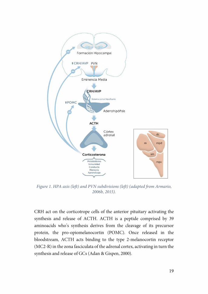
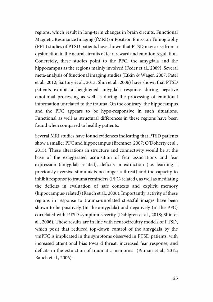
The PVN is the initial point of activation of the HPA axis (figure 1). It is comprised of the magnocellular (mPVN) and parvocellular (pPVN) divisions (Herman et al., 2003). Afferences from the periventricular and medial dorsal regions of the pPVN innervate the median eminence (ME) which is connected in turn to the anterior pituitary gland. CRH and arginine-vasopressin (AVP) are synthesized by the pPVN neurons in the medial dorsal division, transported via axons to the ME and finally released in the anterior pituitary gland. Other neuropeptides such are thyrotropin-releasing hormone (TRH), somatostatin or growth-hormone releasing hormone (GHRH) are synthesized in this region and regulate the release of other anterior pituitary hormones like the growth hormone, thyrotropin and prolactin (Swanson et al., 1986).
19
Figure 1. HPA axis (left) and PVN subdivisions (left) (adapted from Armario,
2006b, 2015).
CRH act on the corticotrope cells of the anterior pituitary activating the synthesis and release of ACTH. ACTH is a peptide comprised by 39 aminoacids who’s synthesis derives from the cleavage of its precursor protein, the pro-opiomelanocortin (POMC). Once released in the bloodstream, ACTH acts binding to the type 2-melanocortin receptor (MC2-R) in the zona fasciculata of the adrenal cortex, activating in turn the synthesis and release of GCs (Adan & Gispen, 2000).
20
GCs act at multiple levels in the body with the aim of reorganizing and optimizing energy expenditure in response to an immediate challenge. These effects include activation of glycogenolysis in the liver to release glucose in the bloodstream, proteo- and lipolysis, vasoconstriction, immunosuppression, inhibition of muscle and bone growth and suppression of the reproductive function (McEwen & Stellar, 1993; Munck et al., 1984; Sapolsky et al., 2000).
The molecular role that GCs play in inducing long-lasting behavioral changes appears to be highly complex. It involves genomic and non-genomic mechanisms (Reul & Kloet, 1985). First studies on GC described a mechanism of slow genomic modulation. The lipophilic nature of GCs enables them to freely cross the membrane and bind to two types of cytosolic receptors: mineralocorticoid receptor (MR; encoded by the gene Nr3c2) and glucocorticoid receptor (GR; encoded by the gene Nr3c1). Binding of the ligand induces the dimerization of the receptor and translocation to the nucleus, where it binds to transcription factors activating or inhibiting gene transcription by interacting with GC-response elements (GREs) located throughout the genome (Hollenberg et al., 1987; Mifsud & Reul, 2016). The non-genomic action of GCs was demonstrated much more recently. They involve the membrane-bound MRs and GRs, resulting in neurophysiological changes (Groeneweg et al., 2012), and the interaction of GRs with intracellular cascades such as the extracellular signal-regulated kinase mitogen-activated protein kinase (ERK/MAPK) pathway in the hippocampus, resulting in behavioral adaption through epigenetic and genomic changes (Gutierrez-Mecinas et al., 2011; Saunderson et al., 2016).
One of the main roles of GC is to regulate their own levels by means of retroinhibitory mechanisms (de Kloet et al., 1998; Sapolsky et al., 2000). Autoregulation of the GC release is mediated by their action upon several brain regions including the PVN and anterior pituitary gland, together with suprahypothalamic regions such as the medial prefrontal cortex (mPFC) and the hippocampal formation (HF).
21
Two mechanisms of GC autoregulation occur: (1) The “proactive” mode, in charge of maintaining basal levels of HPA activity and thus determining the threshold for activating the stress response. This process involves the function of the MR localized in higher brain regions; and (2) The “reactive” mode, which function is to suppress ACTH and GC levels induced by stress. In this case the mechanism is deployed by the action of the lower affinity GRs placed in parvocellullar neurons of the PVN and the pituitary gland. Due to this lower affinity, GRs but not MRs are capable of modulating its occupancy depending on the GCs released under a stressful event (de Kloet, 1993). GRs are also located in cortical regions, HF and ascending aminergic pathways, through which they exert as well modulatory influence on the activity of the HPA axis (de Kloet et al., 1998).
At the molecular level, negative feedback suppression by GCs occurs by inhibiting the expression of the pituitary POMC gene. Specifically, this gene presents a negative glucocorticoid response element (nGRE) to which three GRs bind (one dimer and one monomer), interfering with the binding on adjacent sites of transcription factors that activate POMC expression (Drouin et al., 1993).
Decades of research have gathered compelling evidence linking exposure to a history of stress to the dysfunction in the mechanisms regulating the CRH system and the HPA axis (Arborelius et al., 1999; Holsboer & Ising, 2008; Sanders & Nemeroff, 2016; Ströhle & Holsboer, 2003). This inability of the organism to adjust homeostasis in the face of stressful events seems to be at the core of the development of human psychopathology (de Carvalho Tofoli et al., 2011; Juster et al., 2010; McEwen, 1998a). Furthermore, evidence has shown that the development of psychopathology by exposure to stress depends on several characteristics of the stressor such as the specific type of the stressor (physical, emotional or mixed) (Armario et al., 2012; Daviu et al, 2012a) and the duration of the exposure (acute or chronic) (McEwen, 2004), as well as on situational factors such as the time in the development when it is suffered (childhood, adolescence or adulthood)(Masten & Garmezy, 1985; Salmon & Bryant, 2002) or the gender of the individual (Hankin & Abramson, 2001).
22
Long-term effects of acute stress
Post-traumatic stress disorder & Clinical Studies
Based on the solid evidence linking chronic exposure to stress to the development of anxiety and depression, the majority of studies on psychiatric disorders have been based on animal models of chronic stress. Nevertheless, interest in the study of the negative consequences of exposure to an acute stressful event has grown in the last decades. This new wave of interest comes to a great extent from an increased concern in understanding the etiology of Post-Traumatic Stress Disorder (PTSD), a psychopathology developed in humans after experiencing a traumatic event such as combat-exposure, sexual abuse or a natural disaster.
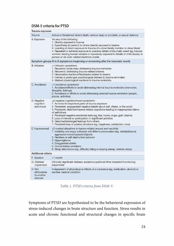
While acute symptoms of distress normally follow life-threatening circumstances, it is the endurance of the symptoms that typifies PTSD. PTSD was first included as a mental disorder in 1980 (American Psychiatric Association, 1980). It was first considered as an anxiety disorder until a recent reclassification as a “Trauma- and Stress Disorder” (American Psychiatric Association, 2013). Criteria for diagnostic of PTSD include the experience of a traumatic event, considered as so an experience of threat to death, serious injury or sexual assault. Its symptoms are grouped under four clusters: intrusion, avoidance, negative cognition and mood and arousal (table 1). In order to be diagnosed of PTSD, the subject has to experience the symptomatology for a minimum of a month and suffer a clear impairment in normal life.
Impairment in everyday life is caused by the re-experiencing of the trauma through intrusive memories about the event, flashbacks and recurrent nightmares. Subjects exhibit avoidance of places, sounds or any environmental reminder of the trauma, as well as a lack of interest in
23
social activities and deficits in declarative-memory related to key aspects of the event. Finally, arousal symptoms are marked by aggressive, reckless and self-destructive behaviors, sleep disturbances and hypervigilance.
Traumatic events are commonly experienced in the general population but most of these experiences do not lead to PTSD. Depending on the particular population studied and the methodology, PTSD lifetime prevalence has been shown to range from 1 to 14%. In the same manner, studies show that the subset of population developing PTSD after a traumatic experience variate between 20-30% (Cohen et al., 2004; Frans et al., 2005; Kessler et al., 1995). However, a common pattern found in the majority of studies is a 2:1 women to men ratio in PTSD rate, suggesting a higher vulnerability of women to develop the psychopathology (Frans et al., 2005; Perkonigg et al., 2000).
24
Table 1. PTSD criteria from DSM-V.
Symptoms of PTSD are hypothesized to be the behavioral expression of stress-induced changes in brain structure and function. Stress results in acute and chronic functional and structural changes in specific brain
25
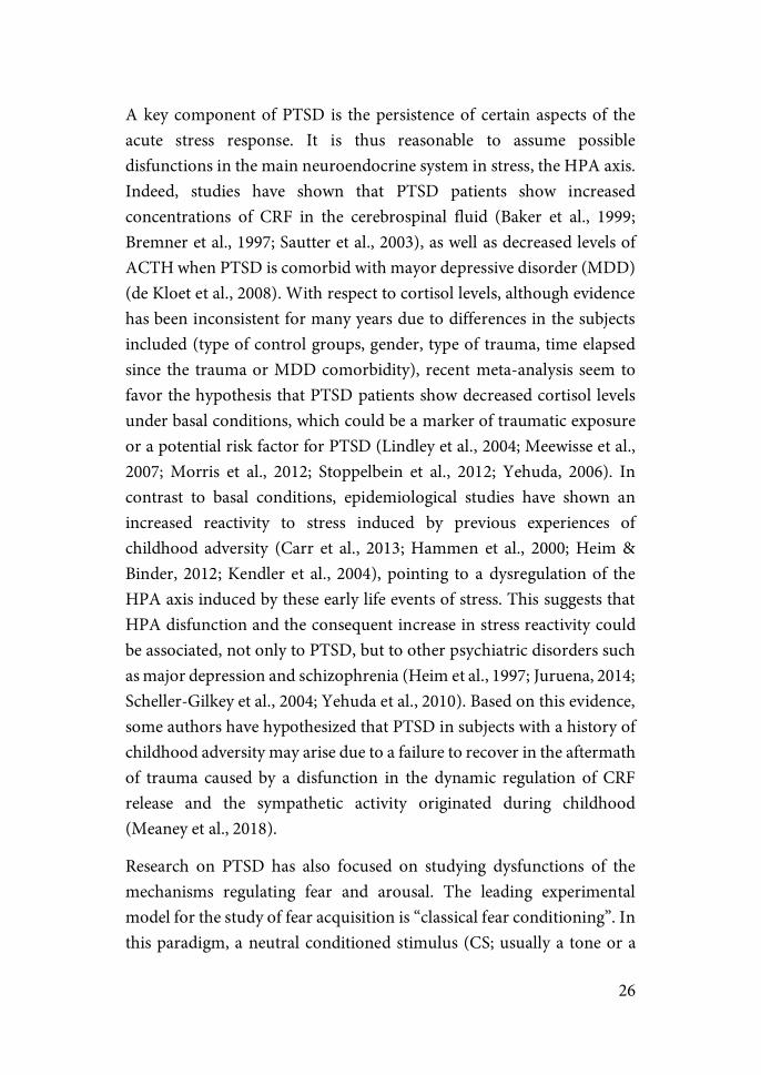
regions, which result in long-term changes in brain circuits. Functional Magnetic Resonance Imaging (fMRI) or Positron Emission Tomography (PET) studies of PTSD patients have shown that PTSD may arise from a dysfunction in the neural circuits of fear, reward and emotion regulation. Concretely, these studies point to the PFC, the amygdala and the hippocampus as the regions mainly involved (Feder et al., 2009). Several meta-analysis of functional imaging studies (Etkin & Wager, 2007; Patel et al., 2012; Sartory et al., 2013; Shin et al., 2006) have shown that PTSD patients exhibit a heightened amygdala response during negative emotional processing as well as during the processing of emotional information unrelated to the trauma. On the contrary, the hippocampus and the PFC appears to be hypo-responsive in such situations. Functional as well as structural differences in these regions have been found when compared to healthy patients.
Several MRI studies have found evidences indicating that PTSD patients show a smaller PFC and hippocampus (Bremner, 2007; O’Doherty et al., 2015). These alterations in structure and connectivity would be at the base of the exaggerated acquisition of fear associations and fear expression (amygdala-related), deficits in extinction (i.e. learning a previously aversive stimulus is no longer a threat) and the capacity to inhibit response to trauma reminders (PFC-related), as well as mediating the deficits in evaluation of safe contexts and explicit memory (hippocampus-related) (Rauch et al., 2006). Importantly, activity of these regions in response to trauma-unrelated stressful images have been shown to be positively (in the amygdala) and negatively (in the PFC) correlated with PTSD symptom severity (Dahlgren et al., 2018; Shin et al., 2006). These results are in line with neurocircuitry models of PTSD, which posit that reduced top-down control of the amygdala by the vmPFC is implicated in the symptoms observed in PTSD patients, with increased attentional bias toward threat, increased fear response, and deficits in the extinction of traumatic memories (Pitman et al., 2012; Rauch et al., 2006).
26
A key component of PTSD is the persistence of certain aspects of the acute stress response. It is thus reasonable to assume possible disfunctions in the main neuroendocrine system in stress, the HPA axis. Indeed, studies have shown that PTSD patients show increased concentrations of CRF in the cerebrospinal fluid (Baker et al., 1999; Bremner et al., 1997; Sautter et al., 2003), as well as decreased levels of ACTH when PTSD is comorbid with mayor depressive disorder (MDD) (de Kloet et al., 2008). With respect to cortisol levels, although evidence has been inconsistent for many years due to differences in the subjects included (type of control groups, gender, type of trauma, time elapsed since the trauma or MDD comorbidity), recent meta-analysis seem to favor the hypothesis that PTSD patients show decreased cortisol levels under basal conditions, which could be a marker of traumatic exposure or a potential risk factor for PTSD (Lindley et al., 2004; Meewisse et al., 2007; Morris et al., 2012; Stoppelbein et al., 2012; Yehuda, 2006). In contrast to basal conditions, epidemiological studies have shown an increased reactivity to stress induced by previous experiences of childhood adversity (Carr et al., 2013; Hammen et al., 2000; Heim & Binder, 2012; Kendler et al., 2004), pointing to a dysregulation of the HPA axis induced by these early life events of stress. This suggests that HPA disfunction and the consequent increase in stress reactivity could be associated, not only to PTSD, but to other psychiatric disorders such as major depression and schizophrenia (Heim et al., 1997; Juruena, 2014; Scheller-Gilkey et al., 2004; Yehuda et al., 2010). Based on this evidence, some authors have hypothesized that PTSD in subjects with a history of childhood adversity may arise due to a failure to recover in the aftermath of trauma caused by a disfunction in the dynamic regulation of CRF release and the sympathetic activity originated during childhood (Meaney et al., 2018).
Research on PTSD has also focused on studying dysfunctions of the mechanisms regulating fear and arousal. The leading experimental model for the study of fear acquisition is “classical fear conditioning”. In this paradigm, a neutral conditioned stimulus (CS; usually a tone or a

27
light) is paired with an aversive unconditioned stimulus (US), usually an electric shock. After repetitive pairings, presentation of the CS alone come to elicit fear responses (for more details see “Coping, Active Avoidance & Controllability” bellow)(LeDoux, 2000). Evidence supporting a dysfunction of these mechanisms in PTSD patients have been found. However, results are inconclusive on whether individuals with PTSD present an enhanced fear acquisition, expression or both (Fani et al., 2012; Orr et al., 2000). Clear evidence has been found indicating that PTSD patients are deficient at extinguishing a fear response, in other words, learning that a certain stimulus previously associated with an aversive outcome no longer poses a threat (Blechert et al., 2007; Fani et al., 2012; Norrholm et al., 2011). Importantly, it is not clearly stablished if extinction deficits are a consequence of trauma-exposure or a pre-exposure risk factor for developing the disease (Kremen, et al., 2012; Lommen et al., 2013; Orr et al., 2012). Nevertheless, studies with identical twins discordant for combat exposure found that only PTSD-affected twins showed defective fear extinction as measured by conditioned skin conductance response (Milad et al., 2008). Similarly, studies on sensitization as measured by heart-rate responses to loud tone stimuli, strongly suggest that heightened heart-rate reactivity in PTSD-affected twins is an acquired feature of the disorder (Orr et al., 2003). While these studies did not exclude a role for genetic predisposition (or previous life events) in PTSD, they showed that the environmental stimulation has a primary role in this disease. On the other hand, other human studies showed that individual differences in some of these parameters before trauma exposure may be predictors of severity of PTSD symptoms after trauma (Metzger et al., 2008; Orr et al., 2012).
With regards to arousal, PTSD patients react showing an exaggerated startle response upon presentation of arousing stimuli. Symptoms of hyperarousal have been interpreted as reflecting a general sensitization phenomenon (Horn et al., 2016; Pitman et al., 2012). Evidence suggesting the involvement of other processes such as fear generalization and an attentional bias towards trauma-related stimuli have been shown
28
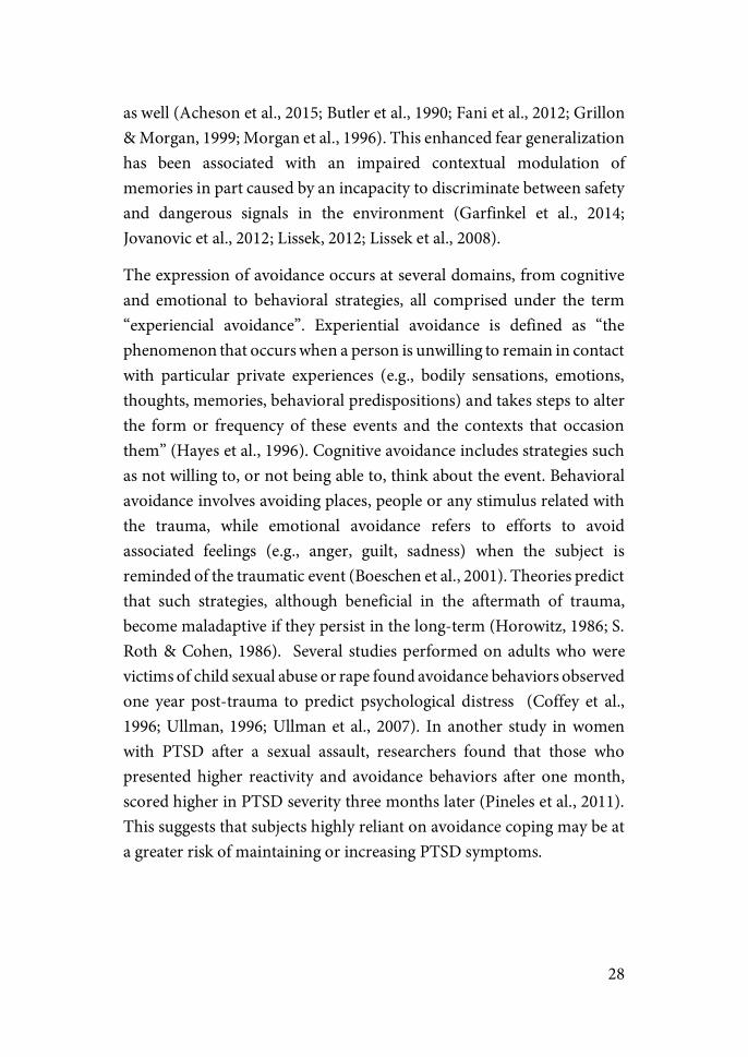
as well (Acheson et al., 2015; Butler et al., 1990; Fani et al., 2012; Grillon & Morgan, 1999; Morgan et al., 1996). This enhanced fear generalization has been associated with an impaired contextual modulation of memories in part caused by an incapacity to discriminate between safety and dangerous signals in the environment (Garfinkel et al., 2014; Jovanovic et al., 2012; Lissek, 2012; Lissek et al., 2008).
The expression of avoidance occurs at several domains, from cognitive and emotional to behavioral strategies, all comprised under the term “experiencial avoidance”. Experiential avoidance is defined as “the phenomenon that occurs when a person is unwilling to remain in contact with particular private experiences (e.g., bodily sensations, emotions, thoughts, memories, behavioral predispositions) and takes steps to alter the form or frequency of these events and the contexts that occasion them” (Hayes et al., 1996). Cognitive avoidance includes strategies such as not willing to, or not being able to, think about the event. Behavioral avoidance involves avoiding places, people or any stimulus related with the trauma, while emotional avoidance refers to efforts to avoid associated feelings (e.g., anger, guilt, sadness) when the subject is reminded of the traumatic event (Boeschen et al., 2001). Theories predict that such strategies, although beneficial in the aftermath of trauma, become maladaptive if they persist in the long-term (Horowitz, 1986; S. Roth & Cohen, 1986). Several studies performed on adults who were victims of child sexual abuse or rape found avoidance behaviors observed one year post-trauma to predict psychological distress (Coffey et al., 1996; Ullman, 1996; Ullman et al., 2007). In another study in women with PTSD after a sexual assault, researchers found that those who presented higher reactivity and avoidance behaviors after one month, scored higher in PTSD severity three months later (Pineles et al., 2011). This suggests that subjects highly reliant on avoidance coping may be at a greater risk of maintaining or increasing PTSD symptoms.

29
Animal models of PTSD
Clinical studies have yielded important findings regarding the etiology of PTSD. Nevertheless, due to the ethical limitations associated with human research, animal models are needed in order to decipher the specific neural mechanisms underlying the symptoms of this psychopathology.
The validity of animal models is assessed using a combination of criteria including: (1) Construct validity—is it accurate measuring what it intends to measure? (e.g. EPM as a measure of anxiety) (2) Face validity—does the model reproduce symptoms associated with the human syndrome? (i.e. analogy of symptoms) (3) Predictive validity— is the model thorough predicting features of its cure in human? (i.e. similarity in its treatment) (4) Discriminant validity—does the model differentiate between those with and without PTSD? (Belzung & Lemoine, 2011; Siegmund & Wotjak, 2006).
The peculiar nature of PTSD (i.e. that it is mostly triggered by a single traumatic event) has led researchers to suggest some essential criteria for a model of PTSD to have good translational value (Flandreau & Toth, 2017; Musazzi et al., 2017; Siegmund & Wotjak, 2006): (i) the stressor must be of severe intensity; (ii) a short duration of the protocol should be sufficient to induce PTSD-like symptoms; (iii) the intensity of the trauma should be positively correlated with the severity of the outcome; and (iv) significant interindividual variability should be observed in the outcomes.
Some of the most common animal models of PTSD include: inescapable shocks, immobilization, exposure to predator or its scent (i.e. cat), under-water trauma or single-prolonged stress (SPS) (e.g. serial exposure to multiple intense stressors). In order to demonstrate face validity, a series of behavioral tests measuring anxiety-like behaviors, arousal and deficits in fear memory are usually conducted several days post-stress.
30
With respect to its construct and predictive validity, measures of basal levels of hormones or in response to drugs are usually taken.
It is important to note that no particular model can individually be related to a single pathology, as no animal model can reproduce the entire psychopathology of a specific human disease (Armario et al., 2008; Bennett et al., 2016; Slattery & Cryan, 2017). In that manner, the objective in the use of animal models should be, not to model a specific human psychopathology, but to identify specific brain circuits subservient of specific features of the disease (i.e. ahnehodia, avoidance, deficits in explicit memory or cognitive impairment), features that may be common to other mental disorders (Hariri & Holmes, 2015; Insel et al., 2010).
Recent studies using models that fulfill the criteria mentioned above have shown that exposure to acute stressors can induce not only immediate, but also persistent changes in synaptic function, neuroarchitecture (dendritic morphology, synaptic spines), and behavior (cognitive and emotional functions). There is evidence from clinical studies pointing towards these changes as potential factors in the development of PTSD in humans, supporting the relevance of these animal models as a valid approximation to the study of PTSD. Of note, we refer as long-term effects as those that persist 3 or more days post-stress (Armario et al., 2008). This is an important remark as the majority of research on stress before the last decade described changes that vanish within 24-48h after stress termination, thus invalidating the protocol as an animal model of PTSD.
31
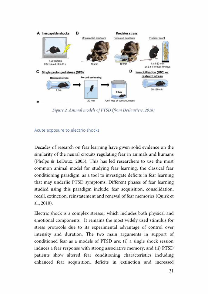
Figure 2. Animal models of PTSD (from Deslauriers, 2018).
Acute&exposure&to&electric.shocks&&
Decades of research on fear learning have given solid evidence on the similarity of the neural circuits regulating fear in animals and humans (Phelps & LeDoux, 2005). This has led researchers to use the most common animal model for studying fear learning, the classical fear conditioning paradigm, as a tool to investigate deficits in fear learning that may underlie PTSD symptoms. Different phases of fear learning studied using this paradigm include: fear acquisition, consolidation, recall, extinction, reinstatement and renewal of fear memories (Quirk et al., 2010).
Electric shock is a complex stressor which includes both physical and emotional components. It remains the most widely used stimulus for stress protocols due to its experimental advantage of control over intensity and duration. The two main arguments in support of conditioned fear as a models of PTSD are: (i) a single shock session induces a fear response with strong associative memory; and (ii) PTSD patients show altered fear conditioning characteristics including enhanced fear acquisition, deficits in extinction and increased
32
probability of relapse (Acheson et al., 2015; Norrholm et al., 2011). Most studies use unpredictable inescapable shocks. Exposure ranges from 1 to 10 shocks of 0.3-3 mA and 1 or 2 seconds in a session of 5 to 30 minutes duration. Specific CS, when used, are commonly a sound, light or odor.
Persistent changes at the behavioral level can include: hypoactivity in novel environments (Armario et al., 2008; Daviu et al., 2010; Van Dijken et al., 1992), enhanced startle response (Milde et al., 2003), avoidance, sleep disturbances (Philbert et al., 2011) and angioxenic effects measured in the open field (OF), light-dark test or elevated-plus maze (EPM), together with a decrease in social interaction (Bruijnzeel et al., 2001; Louvart et al., 2005). A recent study (Balázsfi et al., 2018) has shown anxiogenic and fear-generalization effects to last more than two weeks post-stress. Moreover, increased HPA reactivity was found in response to shock-exposure and the behavioral tests in accordance with previous studies (Daviu et al., 2012).
Restraint/Immobilization&(IMO)&
Both restraint and immobilization protocols involve limiting the free movement of the animal. While restraint is typically done by placing the animal in a plastic tube, IMO involves placing the animal on a board with its paws attached to guides with a plastic tape. Both have a duration ranging usually from 30 to 120 min but that can last until 6h.
The relevance of IMO as a PTSD model arises from the evidence showing that detrimental effects after exposure to IMO are among the most severe and persistent in rodent models (Armario et al., 1990; García et al., 2000; Marquez et al., 2002; Martí et al., 1994). Its effects on the HPA axis regulation have been extensively studied. Studies have shown that IMO induces a long-term desensitization of the HPA axis activity when repeatedly exposed to the same stressor (homotypic stressor).In contrast, sensitization occurs when subsequent exposures are with a novel stressor
33
(heterotypic stressor)(Armario et al., 2008; Armario et al., 2004; Belda et al, 2012; Belda et al., 2008). At the behavioral level it has been shown to induce long-term avoidance, enhanced startle, depression-like behaviors together with deficits in declarative memory in the Morris Water Maze (MWM) and fear extinction (Andero et al., 2013; Andero et al., 2012; Belda et al., 2008; Belda et al., 2004; Fuentes et al., 2014; Sawamura et al., 2016).
Studies using restraint as a stressor have reported an increase in anxiety-like behaviors in the EPM, depressive-like behaviors in the forced swim (FS) test as well as a decrease in glucose intake as long as 35 days post-stress. Deficits in fear extinction were also shown (Chauveau et al., 2012; Chu et al., 2016; Mitra et al., 2005). Extinction deficits are a consistent phenotype of PTSD and a construct with proven translation across animal and human studies.
Underwater&trauma&(UT)&&
Richter-Levin’s laboratory developed this model which consists of 40 s of forced swimming followed by a 20 s forced immersion with a metal net in a water maze (Richter-Levin, 1998). They highlight the additional ethological value of this model in comparison to electric-shocks, since the threat of drowning is a real threat in rodents’ life. They found that underwater stress caused an immediate and persistent anxiogenic-like effects in the EPM in comparison to rats that swim without submersion. This data was further confirmed and extended by other groups (Cohen et al., 2004). UT resulted in enhanced anxiety-like behaviors in the EPM and startle response 1, 7 and 30-day post-stress. Moreover, a long-term decrease in basal corticosterone levels was found when performed in adolescent rats (Moore et al., 2012). Additionally, enhanced contextual fear memory (CFC) together with anhedonic behaviors and altered limbic activity were detected one month after trauma (Ritov et al., 2016). Learning in the MWM was impaired three weeks after UT, results in line
34
with the evidence pointing to a detrimental effect of stress on cognitive tasks and deficits shown by PTSD patients (Richter-Levin, 1998).
Predator&psychosocial&stress&(PPS)&
In this model, animals are exposed to brief periods of immobility while confined in close proximity to a cat, in addition to daily social instability for a period of a month. This rat model of PTSD is based on the exposure to the animal to a situation of threat to survival, a lack of control, intrusive reminder of the traumatic experience and a lack of social support, which are also factors in PTSD in humans.
Three weeks after the second cat exposure, animals show heightened anxiety, exaggerated startle, impaired cognition and increased cardiovascular reactivity, all of which are commonly observed in people with PTSD (Roth et al., 2011; Zoladz, et al., 2008; Zoladz et al., 2012). Recent results have shown that these PTSD-like symptoms may persist as long as 4 months post-cat exposure (Zoladz et al., 2015).
Predator.scent&stress&(PSS)&
In contrast to the PPS, in this model rats are exposed to the scent of the predator by placing the rat in a well-soiled cat litter for 10 min. Control rats are exposed to unused litter for the same amount of time (Cohen and Zohar, 2004; Cohen et al., 2007).
Using this protocol, results show anxiogenic and hyperarousal effects one-week post-stress. Furthermore, changes in the sympathetic-adrenomedullary system are as well observed. Concretely an increase in heart rate and a decrease in heart-rate variability was found in animals exhibiting severe behavioral effects after stress-exposure (Koresh et al., 2016).
35
One of the features that Cohen highlights about this model is the variability in the responses of the rats to this experience. While some animals show extreme (PTSD-like) behavioral responses, others show minimal responses (resilient). This is related to the feature of PTSD that only a subset of the people exposed to the traumatic event will develop PTSD.
Single.prolonged&stress&(SPS)&
In this model, rats are exposed to three consecutive stressors: (1) 2 h of restraint; (2) 20 min of forced swim; and (3): 15 min post-forced swim, ether until anesthetized. Some modifications may include cold temperature (instead of 24ºC) (Hofford et al., 2018) or foot-shock instead of ether (Rice et al., 2018).
Persistent changes (at least one week post-stress) include: (i) exaggerated acoustic startle (Khan & Liberzon, 2004), anxiogenic effects and avoidance of trauma-related cues (Toledano & Gisquet-Verrier, 2014); (ii) enhanced negative feedback of the HPA axis (Liberzon et al., 1997; Liberzon et al., 1999) and (iii) deficits in extinction memories (Dayan Knox et al., 2012) and fear generalization to trauma-unrelated cues (Toledano & Gisquet-Verrier, 2014). All features observed in PTSD (Kohda et al., 2007; Quirk et al., 2006; Yehuda et al., 1993). Importantly, a recent study demonstrated a reduction of PTSD-like symptoms and extinction deficits when rats received vagal-nerve stimulation paired with the CS during extinction trials (Noble et al., 2017).
Other&models&
Even though it is typically considered a model of depression (Huhman, 2006; Huhman et al., 1992), Social Defeat stress (SDS) has been
36
interpreted by some authors as a model of PTSD. This is in view of the long-term anxiogenic effects caused by a single defeat session. The protocol consists on placing an “intruder” inside the homecage of a “resident” dominant animal, thus inducing the attack of the intruder. The number of daily encounters range from one to ten days. This protocol was shown to persistently cause in the intruder: decreased social exploration, anhedonia, enhanced startle response and anxiety-like behaviors on the EPM (Berton et al., 2006; Narayanan et al., 2011; Pulliam et al., 2010). Although most of the SDS models violate de criteria of a single traumatic exposure, authors argue that it may be a valid model for socially-induced or combat-related PTSD which typically involve several exposures. Moreover, SDS may be relevant for its comorbidity with depression-like behaviors which is in line with PTSD and depression comorbidity in humans.
Finally, mention that another interesting approach concerns the use of genetic models to study the neural mechanisms underlying putatively PTSD-symptoms. These models include rat or mouse strains that have been selected for high trait anxiety or marked extinction deficits (Holmes & Singewald, 2013; Landgraf & Wigger, 2002; Neumann et al., 2011).
For a more extensive review on the symptoms induced in animal models of PTSD view Flandreau (Flandreau & Toth, 2017).
Long-term Molecular & Cellular changes induced by acute-stress
The large majority of evidence linking stress to the development of psychiatric disorders comes from animal models of chronic stress. These studies have yielded a large body of evidence illustrating structural and functional changes induced by chronic stress. Changes in volume, density of dentrites and/or spines, as well as synaptic connectivity mainly in the PFC, amygdala and hippocampus have been characterized in
37
animal models of chronic stress (Duman & Aghajanian, 2012; McEwen et al., 2015; Popoli et al., 2011).
Only a few studies have investigated the effects of acute stress on neuroarchitecture. Interestingly, these studies have found that a single stressful event is also able to induce rapid morphological changes in the brain. A protocol of learned helplessness (LH) consisting of two sessions of foot-shocks showed a loss of synaptic spines in the hippocampus 1 and 7 days later (Hajszan et al., 2009). Another study showed that two acute social defeat episodes was sufficient to induce a decrease in CA3 apical dendrites and impairment of LTP 21 days after stress-exposure (Kole et al., 2004). In the PFC, exposure to one or three sessions of forced-swim (20-min/session) induce a retraction of apical branches in infralimbic (IL) dendrites concomitant to a deficit in fear extinction (Izquierdo et al., 2006).
Researchers have argued that these structural changes in neural morphology may be a consequence of a sustained increase in excitatory activity. Studies using acute inescapable footshocks found an increase in glutamate release measured from synaptosomes freshly purified from PFC immediately after the stress exposure. Moreover, this increase in glutamate release was found to be dependent on GC release and binding to its receptors, as treatment with MR or GR antagonists hindered this increase (Musazzi et al., 2010). The effect of GR/MR appears to involve rapid enhancement of the trafficking of glutamate synaptic vesicles into the readily releasable pool (RRP) (Treccani et al., 2014). Using the same stress protocol, researchers showed that acute stress not only enhances glutamate release, but also stimulates the formation of new excitatory synapsis, an effect that was blocked by treatment with desipramine (an antidepressant targetic the glutamatergic system) (Nava et al., 2015).
38
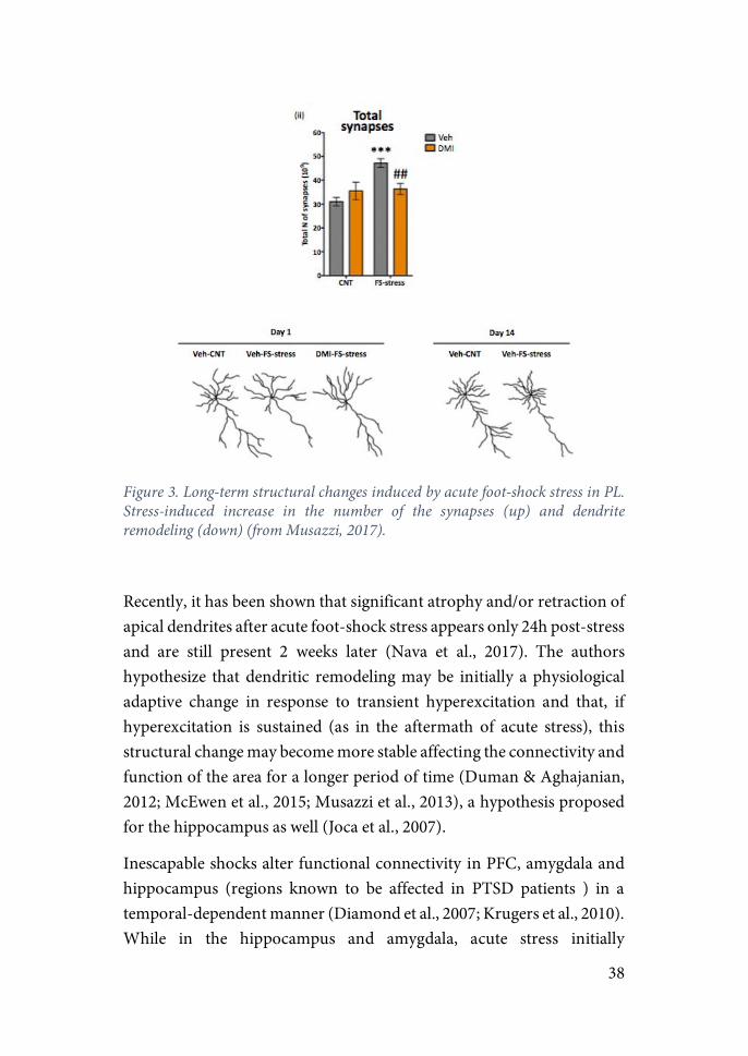
Figure 3. Long-term structural changes induced by acute foot-shock stress in PL. Stress-induced increase in the number of the synapses (up) and dendrite remodeling (down) (from Musazzi, 2017).
Recently, it has been shown that significant atrophy and/or retraction of apical dendrites after acute foot-shock stress appears only 24h post-stress and are still present 2 weeks later (Nava et al., 2017). The authors hypothesize that dendritic remodeling may be initially a physiological adaptive change in response to transient hyperexcitation and that, if hyperexcitation is sustained (as in the aftermath of acute stress), this structural change may become more stable affecting the connectivity and function of the area for a longer period of time (Duman & Aghajanian, 2012; McEwen et al., 2015; Musazzi et al., 2013), a hypothesis proposed for the hippocampus as well (Joca et al., 2007).
Inescapable shocks alter functional connectivity in PFC, amygdala and hippocampus (regions known to be affected in PTSD patients ) in a temporal-dependent manner (Diamond et al., 2007; Krugers et al., 2010). While in the hippocampus and amygdala, acute stress initially

39
potentiates LTP to later inhibit it for a longer period of time, in the PFC there is an immediate impairment of LTP that progressively recovered (figure 4). It has been hypothesized that in PTSD patients this recovery of LTP in PFC may be hindered, diminishing the inhibitory action of this region to the amygdala, thus explaining the enhancement in fear conditioning and deficits in extinction observed among PTSD patients. At the molecular level, although long-term changes were not evaluated, recent studies have shown time-dependent changes in glutamatergic receptor subunits in the PFC after acute stress (Bonini et al., 2016). Early and transient enhancement of AMPA-mediated currents occur, followed by a transient activation of NMDA receptors, processes that could be linked to subsequent functional changes in synapsis. Stress-induced changes in LTP has also been described in the hippocampus. Studies using SPS exposure have showed reduced synaptic plasticity in the hippocampus (Kohda et al., 2007). Studies have suggested that the SPS-induced deficits in extinction may be induced by an upregulation in GR expression in the hippocampus and PFC (Knox et al., 2012), as well as by a decrease in glutamate and glutamine levels (Knox et al., 2010).

40
Figure 4. Temporal dynamic model of stress effects on functional synaptic plasticity of the amygdala, hippocampus and prefrontal cortex (from Diamond2007).
A more radical effect involving apoptosis and inhibition of cell proliferation is produced in the hippocampus after exposure to acute and chronic stress. Studies in rodents, tree shrew and monkeys have shown that acute stress inhibits cell proliferation in the DG (Gould et al., 1998; Gould et al., 1997; Mirescu & Gould, 2006; Tanapat et al., 2001). These changes may be induced by a sustained increase in GC release, responsible as well for the induction of LTP and memory impairments (McEwen, 2008). Moreover, apoptosis has been also reported in the DG one week after exposure to SPS (Li et al., 2010).
In contrast to the loss of dendritic branches and spines observed in the hippocampus and PFC after exposure to stress, the opposite phenomenon is induced by stress in the amygdala. Chronic stress has been shown to increase dendritic branching and dendritic spine number within the amygdala together with a facilitation of LTP, changes shown to be more persistent than those observed in the hippocampus (Manzanares, 2005; Vyas et al., 2004; Vyas et al., 2006). Studies
41
comparing the effects of acute vs. chronic stress found that while chronic immobilization stress elicited an increase in spinogenesis one day post-stress in the basolateral amygdala (BLA), acute immobilization stress did not (Mitra et al., 2005). Strikingly, exposure to acute stress increased the number of spines 10 days later. Moreover, this gradual increase in dendritic spines was concomitant with a development of anxiety-like behaviors in the EPM 10 days post-stress. These results suggest that acute stress may induce long-term effects that are not evident in the aftermath of the exposure and require a maturation process.
Regarding excitatory activity induced by stress in the amygdala, studies have shown that exposure to acute stress, as in the PFC and the hippocampus, is capable of inducing an increase in glutamate release, although the temporal dynamic in BLA and CeA are different (Reznikov et al., 2007). Furthermore, treatment with the antidepressant tianeptine abolished the stress-induced increase in glutamate release in BLA but not in CeA. Interestingly, this antidepressant was also shown to inhibit stress-induced dendritic hypertrophy in BLA but not CeA, suggesting a region-specific action on neuroarchitecture of some antidepressants (McEwen & Chattarji, 2004).
Results on the influence of stress in glutamate levels have favored a major shift in the conceptual framework investigating neuropsychiatric disorders, from a monoamine-oriented hypothesis (which dominated pharmacological research for over half a century) to a neuroplasticity hypothesis, which posits that structural and functional changes in brain circuitry (largely in the glutamate system) mediate psychopathology and also therapeutic action. Although further studies on the characterization of long-term molecular and cellular changes induced by acute stress need to be undertaken, the results show that stress exposure restricted to minutes or hours are sufficient to induce long-term structural, functional and behavioral consequences that may contribute to explain how exposure to acute traumatic stress in humans results in psychopathologies such as PTSD.
42
Individual differences in psychopathology
Differential response to trauma & Biological risk factors
A core characteristic in the etiology of PTSD is that only a fraction of the subjects exposed to the same traumatic event will develop the disease. Epidemiological studies have estimated that the incidence of PTSD after a traumatic experience variate between 20-30% of the exposed population (Breslau et al., 1991; Cohen et al., 2004; Frans et al., 2005; Kessler et al., 1995). This fact suggests the existence of pre-trauma factors such as the genetic background or early-life stress experiences that contribute to inducing a vulnerable or resilient phenotype which may critically determine if the psychopathology is developed in the aftermath of trauma (Charney, 2004; Feder et al., 2009; Horn et al., 2016; Musazzi et al., 2017; Yehuda & LeDoux, 2007).
Studies using identical twins have estimated the heritability component of PTSD to be 25-35% (Kremen et al., 2012; Tambs et al., 2009). Although research aimed at identifying specific genes associated with PTSD has been inconclusive, several potential candidates have emerged. Some of the most compelling studies show a link between a gene variant and PTSD prevalence and fear extinction. One example is the gene encoding for the serotonin transporter, SLC6A4, associated with increased PTSD in high-trauma populations (Wang et al., 2011) and impaired extinction in healthy subjects (Hartley et al., 2012). Importantly, deleting this gene in mice reproduced impairments in extinction and neuromorphic changes in PFC and amygdala dendrites, giving translational validity to this model (Wellman et al., 2007). Another example of cross-species translational evidence is the case of ADCYAP1 and ADCYAPR1, genes encoding the neuropeptide pituitary adenylate cyclase-activating polypeptide (PACAP) and its receptor. They have been associated with fear learning and PTSD prevalence in women and an increase of the receptor mRNA in amygdala in fear learning in mice (Ressler et al.,
43
2011). Another relevant example of genetic influence are studies showing an association between certain genetic variants of CRF and CRF receptors and PTSD (Amstadter et al., 2011; Sanders & Nemeroff, 2016; S. White et al., 2013). Of note, some of them were shown to be specific for predicting PTSD among women (Wolf et al., 2013). A special attention has been given to FKBP5. This is a chaperone that takes part of the molecular complex that binds to GR and acts inhibiting the capacity for GR to access DNA binding sites. FKBP5 expression is reduced in PTSD, which could be linked to an increase in GR signaling, consistent with the idea of a heightened negative-feedback suppression of the HPA activity in PTSD. Multiple FKBP5 SNPs are associated with increased risk for PTSD (Boscarino et al., 2012; Koenen et al., 2005). Moreover, a genetic variation of FKBP5 in conjunction with childhood abuse has been shown to confer risk for adult PTSD (Binder et al., 2008; Koenen et al., 2005). NR3C1 gene codes for GR. A variant of the NR3C1 gene has been shown to be associated with HPA function and the risk of PTSD and the severity of symptoms (Hauer et al., 2011). Taken together, these findings suggest that alterations in GR signaling and HPA activity influence risk for PTSD.
Other pathways such as the one involving dopamine metabolism has also been shown to be involved. In particular, a polymorphism is which the aminoacid Val is substituted for a Met in the gene COMT coding for the enzyme cathecol-O-methyltransferase has been shown to be a risk factor of PTSD. COMT is the primary enzyme in charge of metabolizing dopamine in the PFC. The substitution of the aminoacid decreases its effectiveness, thus increasing the levels of dopamine in this region which has been linked to a variety of emotional and behavioral dysfunctions (Clark et al., 2013; Kolassa et al., 2010; Valente et al., 2011).
Mixed results have been reported on whether other parameters known to related to PTSD such as reduced hippocampal volume (Bonne et al., 2001; Gilbertson et al., 2002; Logue et al., 2018; McEwen et al., 2016), low cortisol levels and certain brain activity patterns are a pre-trauma risk factor for the development of PTSD or a consequence of trauma
44
exposure (Milad et al., 2009; Shin et al., 2011; Yehuda, 2006). A more direct link with the risk for PTSD has been found in GR. GR density in peripheral blood mononuclear cells prior to deployment was shown to be higher among participants with high level of post-deployment PTSD symptoms (van Zuiden et al., 2011; Van Zuiden et al., 2012). Furthermore, several studies including survivors of the 9/11 attacks and soldiers showed lower levels of FKBP5 mRNA in subjects with PTSD than those without PTSD (Sarapas et al., 2011; Yehuda et al., 2009).
On the contrary, evidence is clear on the association between childhood adversity and enhanced reactivity (Hammen et al., 2000; Kendler et al., 2004) with a the subsequent risk for PTSD (Bremner et al., 1993; Spatz Widom, 1999; Yehuda et al., 2001). Extensive research have pointed to dysregulation of the HPA axis as a possible mechanism implicated in this process, as exposure to early-life stress has been linked to altered HPA function (Carpenter et al., 2007; Heim et al., 2008). A wealth of studies have shown early-life stress to strongly shape responses to stress not only at the endocrine level, but at the neural and immune level as well, defining vulnerability to psychopathology (Nemeroff, 2016). The underlying mechanism has been suggested to be a process of stress sensitization in which individuals with a history of early-life adversity overreact to moderate stressful circumstances resulting in an increased risk for mood and anxiety disorders such as depression or PTSD (Breslau et al., 2008; Brewin et al., 2000; Hammen et al., 2000b; Heim et al., 2001; Kendler et al., 2004).
Modelling variability associated to PTSD in animal models
Clinical studies of PTSD employ stringent inclusion-criteria to identify PTSD patients, yet animal studies have included the entire exposed population as the study population for years. In view of these discrepancy, recent studies in animal models of PTSD have sought to use
45
individual variability in response to acute stress as a means to identify neurobiological risk factors in the development of psychopathology.
One of the first studies aiming to specifically study stress-outcome variability in animal models of PTSD was done by Cohen and colleagues (Cohen et al., 2004; Cohen et al., 2006; Cohen et al., 2005; Cohen et al., 2012). Using predator exposure as the stressor, response magnitude in behavioral tests conducted one week later (usually EPM and acoustic-startle response; ASR) was used to group animals into “maladapted” and “well adapted”. A cut-off behavioral criterion was stablished based on the behavior of the control group (percentiles or standard deviation depending of the study) and those animals scoring above or below those thresholds were assigned to “extreme behavioral response” or EBR (i.e. vulnerable phenotype) and “minimal behavioral response” or MBR (i.e. resilient phenotype). To maximize effect size, animals between EBR and MBR considered as “partial behavioral response” were not investigated. Using this method, studies have shown EBR rates in Sprague-Dawley rats as high as 90% one day post-stress, but as in human, acute-anxiety symptoms drops drastically in the following days and settles at around 25% (Cohen et al., 2003), which is in accordance with human epidemiological data (Kessler et al., 1995). Importantly, these symptoms last as long as 3 weeks post-stress giving face validity to the model (Cohen et al., 2004). Of note, EBR rates vary significantly between rat and mice strains, being 10 and 50% in Lewis and Fisher rats respectively (Cohen et al., 2006; Cohen et al., 2006; Goswami et al., 2010), and from 6 to 55% in the most widely used mice strains DBA/2J and C57Bl/6J, respectively (Cohen et al., 2008).
Using this criteria studies have reported EBR animals to show enhanced reactivity as measured by increased heart rate and sympathetic activity together with higher GC and ACTH levels (Cohen et al., 2003, 2005). At the molecular level, EBR exhibited reduced expression of hippocampal BDNF, synaptophysin and ERK pathway factors and elevated GR expression and PSD95 protein, all linked to plasticity processes. Interestingly, animals exhibiting extreme behavioral disruption ,but not
46
those with a resilient phenotype, presented extensive neuronal retraction in the hippocampus and dendrite proliferation in the amygdala (Cohen et al., 2014; Kozlovsky et al., 2007; Matar et al., 2013).
Stress-response variability has been reproduced in other stress procedures. Using underwater trauma, results have shown EBR animals to exhibit long-term anxiety and hyperarousal (Cohen et al., 2004). In another study, a procedure involving two days of unpredictable shocks was shown to induce symptoms of hyperarousal, hypervigilance, impairment in attention, engagement in risky behaviors and difficulty sleeping. Those classified as “PTSD-like phenotype” based on their response in the behavioral battery, showed attenuated stress-induced GC levels as well as increased levels of Crfr2 mRNA in the BNST. Interestingly, knocking-down the expression of Crfr2 through viral injections in this region attenuated susceptibility to PTSD-like behaviors (Lebow et al., 2012).
The environmental epigenetics hypothesis
PTSD has some particularities with respect to the rest of mental disorders (Kessler et al., 1995; Weiss, 1992): (i) characteristics of the stressors inducing the disease is clearly defined (i.e. type, duration, time of exposure), and yet, not all individuals exposed in the same conditions develop the psychopathology; and (ii) a single stress-exposure of short duration may be sufficient to induce life-time PTSD. These raises two main questions: (a) what differentiates the maladaptive stress response of some individuals from the pro-adaptive response of the majority?; and (b) how can a single, although traumatic, stressful event induce a disease that can last for years or decades? The answer to both questions may arise from the same regulating process: epigenetics. Epigenetic mechanisms refer to chemical modifications of DNA (without change in nucleotide sequence), histone proteins and RNA molecules that bind to DNA, which regulate transcription. The last decade has yielded a vast
47
number of studies pointing to the involvement of this regulating mechanism in the development of psychiatric disorders including PTSD (Bagot et al., 2014; McEwen et al., 2012; Meaney & Yehuda, 2018; Raabe & Spengler, 2013; Reul et al., 2015; Zovkic & Sweatt, 2013). These studies show that environmental factors such as parental care, a history of childhood adversity or exposure to traumatic events may embed persistently in the DNA through epigenetic modifications of genes mainly related to fear memory and HPA-activity regulation inducing vulnerability to psychopathology (Bock et al., 2014; Franklin et al., 2012; McEwen et al., 2015; Radley et al., 2011; Singh-Taylor et al., 2015). Three main epigenetic mechanisms have been extensively studied: DNA methylation, post-translational modification of histone tails and microRNAs (for more details see section “Epigenetic regulation of long-term effects” below). Here, I will focus on the first question: what defines the individual variability in stress susceptibility?
The first evidence of the influence of environmental factors on stress reactivity came from work in the lab of Michael Meaney, focusing on the influence of maternal care on the development of individual differences in stress reactivity in the rat. These experiments showed that rodents exposed to different levels of maternal care show behavioral alterations in the fear and endocrine response to novel environments in adulthood (Meaney, 2001). These studies showed persistent alterations in the expression of genes related to HPA function such as GR in brain areas regulating anxiety and HPA activity such as the PFC, hippocampus and hypothalamus. Depending on the level of maternal care of the pups, rodents in adulthood varied in life-long epigenetic alterations (DNA methylation and H3K9 acetylation) presented in the promoter regions of Gad67 in the PFC and GR in the hippocampus (Weaver et al., 2004; T.-Y. Zhang et al., 2010). Other groups studying the effects of early-life stress on adulthood have shown that other genes such as Avp in the hypothalamus and Bdnf in the PFC and hippocampus, are epigenetically regulated as well (Murgatroyd et al., 2009; Roth et al., 2009). Interestingly, recent studies have shown that changes may emerge long
48
after childhood. In a study where pups were subjected to either maltreatment or nurturing maternal during the first post-natal week, sex-specific differences in the Bdnf DNA methylation in the mPFC emerged during adolescence and adulthood in animals that had been exposed to maltreatment. In the case of Reelin, differences in its DNA methylation state showed a transient change in maltreated females, while the rest of the groups showed differences in Reelin methylation by adulthood. These data suggest that epigenetic changes may be in a large degree tissue-, loci-, and in some cases sex-specific, and that the temporal relationship between environmental adversity and epigenetic modulation may be more complex than firstly assumed.
In view of these results, attempts to study epigenetic regulation in humans have been conducted. However, the cell-type specificity of epigenetic modulation has limited to a high extent the possibility of analysis of relevant neural epigenetic markers. Nevertheless, some studies have been performed assessing epigenetic changes of tissue samples from suicide subjects. A study analyzing DNA methylation of a promoter region of Nr3c1 from suicide completers with and without a history of childhood abuse, found that those who had suffered childhood abuse presented higher levels of DNA methylation in this region, than those with no history of childhood adversity. Hypermethylation was associated with increased GR mRNA levels, and in-vitro analysis indicated that this increase in expression was associated with an inhibition of the binding of the transcription factor of Egr1 (i.e. NGFI-A, Zif268) (Labonte et al., 2012; McGowan et al., 2009).
Another way of studying epigenetic regulation in humans is to use peripheral tissues, mainly blood cells. Lymphocytes are well-known targets of glucocorticoids and immune profiles are known to be sensitive to alterations in GR abundance (Baschant & Tuckermann, 2010). One study found increased DNA methylation of several sites within the Gr1f promoter region in lymphocytes of adult subjects that had a history of childhood adversity (parental loss, childhood maltreatment, and lack of parental care) (Tyrka et al., 2012). These results highlight that epigenetic
49
alterations as a result of childhood adversity persist in peripheral tissues and can be detectable in mixed lymphocyte cell populations. A recent investigation using whole blood found that only individuals subjected to childhood abuse and a determined polymorphism of Fkbp5, showed lower levels of DNA methylation in an intronic region of the gene. These data demonstrate that epigenetic modifications take place in response to psychosocial environment early in life and persist throughout life even in cells with continuous cycles of cell division.
Studies analyzing the methylation state of DNA of the whole genome from hippocampi from suicide victims found clusters of hundreds of genes that were hyper- and hypo-methylated in those that had suffered childhood abuse compared to those who did not. Interestingly, enriched candidate pathways included neuroplasticity-related genes (Labonté et al., 2012). Similar results were found in a study of PTSD patients with and without a history of childhood adversity (Mehta et al., 2013). Other studies have shown similar epigenetic changes in the GR promoter region in the hippocampus between animal models of childhood maltreatment and suicide victims who had suffered from child abuse (McGowan et al., 2011; Suderman et al., 2012). Interestingly, a cluster of cell-adhesion molecules named protocadherin (PCDH) known to be involved in the regulation of neuronal morphology and synaptic plasticity has been found to present altered epigenetic modifications in response to early-life maltreatment in rodents and childhood adversity in humans (Suderman et al., 2012). It remains to be determined whether epigenetic changes in these genes are linked to the changes in neuroplasticity observed in response to differences in maternal care (Bagot et al., 2009).
One of the major caveats of these studies is the lack of cell-specificity in the analysis. Most of the studies analyse epigenetic changes from mixed cell populations potentially masking epigenetic differences in determined cell populations or group differences. A potential solution is the technique of cell sorting by fluorescence markers FACS. However, cell specificity determined by these markers is not absolute making it
50
difficult to ascertain if the necessary levels of specificity are attained. The ENCODE (Encyclopedia of DNA Elements) project is another helpful approach, in that it contains epigenetic and functional genomic signatures from a large number of cell types, offering a tool to bioinformatically distinguish different cell types from a mixed tissue population. Moreover, epigenetically invariant genomic regions can also be identified reducing the complexity in genome-wide analysis (Consortium, 2012; Houseman et al., 2012).
Despite evidence showing the link between early-life stress, PTSD and epigenetic modifications, critical questions need to be addressed including when precisely during development do epigenetic changes related to early adversity emerge, in what contexts, brain areas, and in which cell types.
Influence of cognitive and emotional regulation on vulnerability
Despite the extensive study on the biological and sociological risk factors in the development of PTSD, the sum of explained variability considering these factors is low. Two often cited meta-analysis indicate this explained variability to be lower than 20% (Brewin et al., 2000; Ozer et al., 2003). Thus, an important deal of the variance remains unexplained and additional predictive variables need to be identified.
Importantly, the results from Ozer’s group indicate that peritraumatic psychological processes, and not prior characteristics, are the strongest predictors of PSTD. Considering that the implication of psychological attributes in the development of PTSD has often been neglected, examination of the influence of such factors in predicting individual differences represents a potential way of increasing explained variability in PTSD disease. Among the few studies that have been conducted on this regard, coping style and controllability (or “locus of control”) are the factors that have gathered most of the attention.

51
Coping style is defined as the cognitive and behavioral efforts applied by an individual to manage internal or external demands (Folkman et al., 1986). Coping is a multi-dimensional concept in which different coping strategies have been conceived depending on the context of study (e.g. active, passive, avoidant, emotional). In the context of human psychology, two coping typologies conceived by Lazarus and Folkman are well-stablished (Folkman et al., 1986): (i) a problem-focused coping strategy that tries to solve the external source originating the stress response; and (ii) an emotion-focused strategy aimed at reducing internal distress. Whereas the problem-focused strategy focuses at reducing an external factor, the emotion-focused coping style aims to reduce the internal experienced distress through various mechanisms including reappraisal, selective attention, and avoidance (Karstoft et al., 2015). Studies have shown that whereas avoidance coping (an emotion-focused coping strategy) is related to higher levels of PTSD (Bryant & Harvey, 1995; Chang et al., 2003; Feder et al., 2016; Ménard & Arter, 2014; Mikulincer & Solomon, 1989), coping strategies centered on solving the problem are linked to lower risk of developing PTSD (Mikulincer & Solomon, 1989).
Locus of control (LOC) or controllability refers to the extent to which individuals feel they control events that affect them (Rotter, 1966). In other words, it assesses the degree to which individuals believe that events are controllable by their actions (internal LOC) or on the contrary, they depend on factors that are beyond their control (external LOC). Studies have shown that individuals that believe stressful events are not under their control have higher level of PTSD-symptoms (Casella & Motta, 1990; Kushner et al., 1993; McKeever et al., 2006; Solomon et al., 1988; Zhang et al, 2014). Internal LOC is thus expected to be a protective factor against PTSD, while an external LOC is a risk factor for the development of PTSD. However, it is not known whether the degree of controllability predicts the persistence of the symptoms (Olff et al., 2005).
52
Taken together, these results suggest that individual differences in the development of psychopathology may arise from the expression of different coping strategies to deal with stress and the degree to which individuals believe that events are controllable by their actions. Prospective studies on the predictive value of controllability and coping style in the aftermath of a severe stressful event may therefore yield potentially relevant results, and yet studies on this matter remain largely unexplored.
Coping, Active Avoidance & Controllability
The stress response can be viewed as a functional defense system that deploys a repertoire of innate behavioral responses crucial for survival of the individual and the species. Usually, this repertoire has been defined as the series of animal behaviors induced when facing a predator and include: flight, defensive fight, freezing, immobility, submissive posture, hypoalgesia, as well as autonomic reactions.
Importantly, when facing a threatening situation, it is the cognitive appraisal induced what determines the aversive character of a certain stimulus and not its physical characteristics per se (Olff et al., 1993; Weiss, 1968). Cognitive appraisal depends to a high extent on the ability of the organism to cope with the situation (Folkman et al., 1986). Thus, the coping strategy deployed in a certain situation will substantially determine the aversive impact of the stressor.
Coping Strategies
Studies in the taxonomy of behavior have defined four main categories: reflexes, reactions and actions and habits (Balleine & Dickinson, 1998; Lang & Davis, 2006; Yin & Knowlton, 2006). Reflexes are stimulus-

53
evoked behaviors genetically inherited that require the engagement of simple motor mechanisms (e.g. withdrawal of a paw when painful stimuli are applied). Reactions and reflexes share the characteristic of being unlearned innate responses (what ethologists call “innate patterns). Reactions however, require the involvement of a more generalized net of muscles and the processing of information in the CNS outside of the strictly sensory pathways (e.g. freezing) (Blanchard & Blanchard, 1971). While these reactions do not require a previous encounter with the inducing stimuli to be acquired, they can become under the control of previously neutral stimuli through a process of associative learning. In this case, responses are denominated as conditioned or conditional responses (Blanchard & Blanchard, 1969; Bolles, 1972; Fanselow, 1980). In contrast to reactions, actions are complex and flexible behaviors performed in order to obtain a goal or a reward. Furthermore, they are not elicited by the presentation of a stimulus, but a combination of external and internal stimuli including factors such as motivation and arousal (Balleine & Dickinson, 1998; Estes, 1948; P. F. Lovibond, 1983; Niv, Joel, & Dayan, 2006; Rescorla, 1968; Skinner, 1938). In this case the associative learning is created between a response (R) and an outcome (O). Actions can become inflexible and conducted in an automatic manner when it has been performed repeatedly for a long period of time. In this case, we say the action has become a habit (Hull, 1943; Killcross, 2014; Skinner, 1938; Thorndike, 1898). In contrast to actions that are driven by the outcome elicited by the response, habits are a next stage where the same action, with extensive training, becomes independent of the outcome produced. Distinction between one and the other can be evidenced by their sensitivity to outcome devaluation (Yin et al., 2004). While performance of a goal-directed action will decrease if the value of the reward is diminished (e.g. lever-pressing to access food if access to food is ad-libitum in homecage), after extended training the habitual behavior will still persist in conditions where outcome value has been reduced. When performed as a habit, a behavior becomes dependent on the stimuli

54
present around the response instead of the outcome and is thus referred as a stimulus-response (S-R) habit.
Based on this taxonomic definition of behavior, it has been described two distinct patterns of reaction to stressful situations or coping styles: proactive and reactive (Koolhaas et al., 1999). While reactive coping involves the engagement of reflexes and reactions, proactive coping is characterized by the implementation of complex actions and habits to obtain a determined goal or reward.
Reactions are behaviors genetically encoded in an organism’s repertoire of defensive behaviors (Hirsch & Bolles, 1980). In 1970, Bolles defined what he called “Species-specific defense reactions” (SSDRs) as a series of innate responses that are elicited by an organism when any sudden or new stimulus appears in the environment. He highlighted that these reflexive innate behaviors (e.g. running or flying away, freezing or fighting) are the main determinants of the survival of the individual, and minimized the importance of reinforcement learning in nature (Bolles, 1970).
Fear conditioning and Active Avoidance
During the last decades, research on instrumental learning has been mainly focused on the study of appetitive motivation (i.e. food, drugs or sex). However, first studies on instrumental behavior were focused on aversively motivated actions.
Such learning processes were originally studied by Pavlov who defined in 1927 what he called “defensive conditioning” (Pavlov, 1927). The paradigm used in his studies consisted on the presentation of a neutral (to be conditioned) stimulus (CS; e.g. light, tone, context), repeatedly paired with an innately-aversive unconditioned stimulus (US), usually an electric shock. With repeated pairings, the initial neutral stimulus turns to elicit a conditioned fear response (CR), thus becoming a
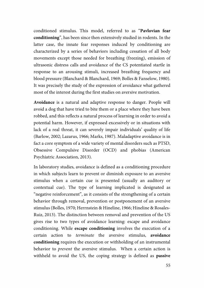
55
conditioned stimulus. This model, referred to as “Pavlovian fear conditioning”, has been since then extensively studied in rodents. In the latter case, the innate fear responses induced by conditioning are characterized by a series of behaviors including cessation of all body movements except those needed for breathing (freezing), emission of ultrasonic distress calls and avoidance of the CS potentiated startle in response to an arousing stimuli, increased breathing frequency and blood pressure (Blanchard & Blanchard, 1969; Bolles & Fanselow, 1980). It was precisely the study of the expression of avoidance what gathered most of the interest during the first studies on aversive motivation.
Avoidance is a natural and adaptive response to danger. People will avoid a dog that have tried to bite them or a place where they have been robbed, and this reflects a natural process of learning in order to avoid a potential harm. However, if expressed excessively or in situations with lack of a real threat, it can severely impair individuals’ quality of life (Barlow, 2002; Lazarus, 1966; Marks, 1987). Maladaptive avoidance is in fact a core symptom of a wide variety of mental disorders such as PTSD, Obsessive Compulsive Disorder (OCD) and phobias (American Psychiatric Association, 2013).
In laboratory studies, avoidance is defined as a conditioning procedure in which subjects learn to prevent or diminish exposure to an aversive stimulus when a certain cue is presented (usually an auditory or contextual cue). The type of learning implicated is designated as “negative reinforcement”, as it consists of the strengthening of a certain behavior through removal, prevention or postponement of an aversive stimulus (Bolles, 1970; Herrnstein & Hineline, 1966; Hineline & Rosales-Ruiz, 2013). The distinction between removal and prevention of the US gives rise to two types of avoidance learning: escape and avoidance conditioning. While escape conditioning involves the execution of a certain action to terminate the aversive stimulus, avoidance conditioning requires the execution or withholding of an instrumental behavior to prevent the aversive stimulus. When a certain action is withheld to avoid the US, the coping strategy is defined as passive
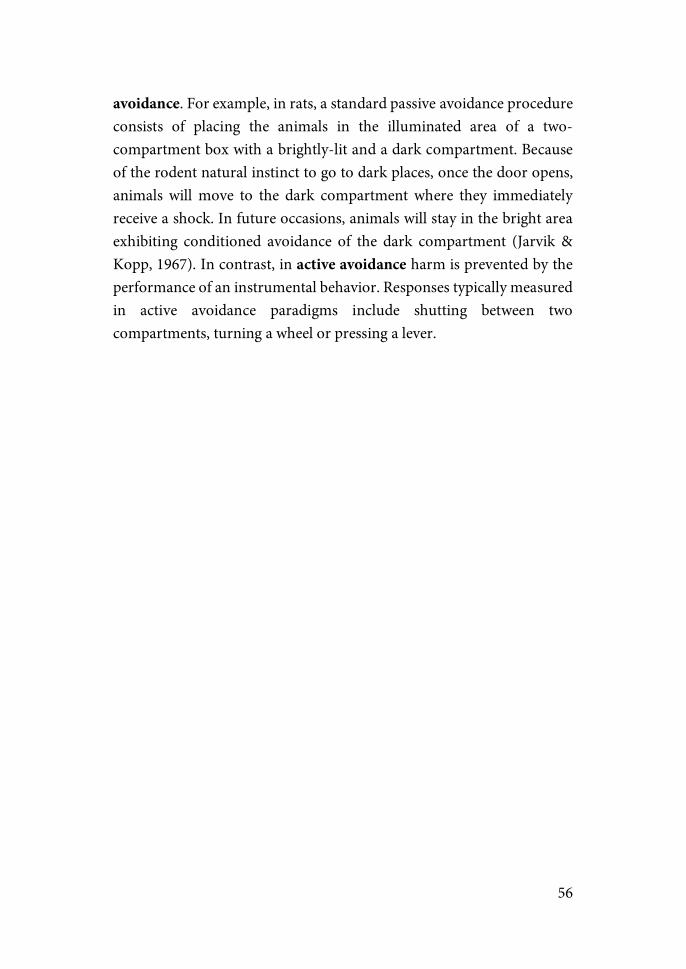
56
avoidance. For example, in rats, a standard passive avoidance procedure consists of placing the animals in the illuminated area of a two-compartment box with a brightly-lit and a dark compartment. Because of the rodent natural instinct to go to dark places, once the door opens, animals will move to the dark compartment where they immediately receive a shock. In future occasions, animals will stay in the bright area exhibiting conditioned avoidance of the dark compartment (Jarvik & Kopp, 1967). In contrast, in active avoidance harm is prevented by the performance of an instrumental behavior. Responses typically measured in active avoidance paradigms include shutting between two compartments, turning a wheel or pressing a lever.
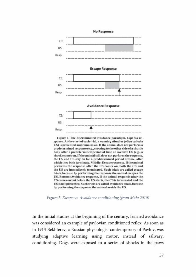
57
Figure 5. Escape vs. Avoidance conditioning (from Maia 2010)
In the initial studies at the beginning of the century, learned avoidance was considered an example of pavlovian conditioned reflex. As soon as in 1913 Bekhterev, a Russian physiologist contemporary of Pavlov, was studying adaptive learning using motor, instead of salivary, conditioning. Dogs were exposed to a series of shocks in the paws
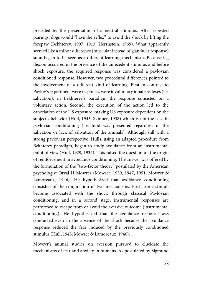
58
preceded by the presentation of a neutral stimulus. After repeated pairings, dogs would “have the reflex” to avoid the shock by lifting the forepaw (Bekhterev, 1907, 1913; Herrnstein, 1969). What apparently seemed like a minor difference (muscular instead of glandular response) soon began to be seen as a different learning mechanism. Because leg flexion occurred in the presence of the antecedent stimulus and before shock exposure, the acquired response was considered a pavlovian conditioned response. However, two procedural differences pointed to the involvement of a different kind of learning. First in contrast to Pavlov’s experiment were responses were involuntary innate reflexes (i.e. salivation), in Bekhterev’s paradigm the response consisted on a voluntary action. Second, the execution of the action led to the cancelation of the US exposure, making US exposure dependent on the subject’s behavior (Hull, 1943; Skinner, 1938) which is not the case in pavlovian conditioning (i.e. food was presented regardless of the salivation or lack of salivation of the animals). Although still with a strong pavlovian perspective, Hulls, using an adapted procedure from Bekhterev paradigm, began to study avoidance from an instrumental point of view (Hull, 1929, 1934). This raised the question on the origin of reinforcement in avoidance conditioning. The answer was offered by the formulation of the “two-factor theory” postulated by the American psychologist Orval H Mowrer (Mowrer, 1939, 1947, 1951; Mowrer & Lamoreaux, 1946). He hypothesized that avoidance conditioning consisted of the conjunction of two mechanisms. First, some stimuli become associated with the shock through classical Pavlovian conditioning, and in a second stage, instrumental responses are performed to escape from or avoid the aversive outcome (instrumental conditioning). He hypothesized that the avoidance response was conducted even in the absence of the shock because the avoidance response reduced the fear induced by the previously conditioned stimulus (Hull, 1943; Mowrer & Lamoreaux, 1946).
Mowrer’s animal studies on aversion pursued to elucidate the mechanisms of fear and anxiety in humans. As postulated by Sigmund
59
Freud (Freud, 1936), he accepted the idea that fear and anxiety were learned states. However, Mowrer studied and tried to explain the underlying mechanisms involved from the behaviorist stimulus-response principles. Mowrer thought that the Pavlovian CS induced a fearful state and that, if an instrumental behavior reduced exposure to this CS, this action would diminish CS-elicited fear. Thus, two links were stablished: CS-fear and fear-avoidance associations.
For the next decades, Mowrer’s ideas, together with the work of his colleague Miller (Miller, 1941, 1948), would not only define laboratory studies on avoidance behavior and its underlaying neural mechanisms, but also influence on the ideas about the nature and treatment of fear and anxiety in humans (Dollard & Miller, 1950; Stampfl & Levis, 1967).
Despite the success of the two-factor theory during the first decades, criticisms began to arouse due to experimental inconsistencies regarding the reinforcing value of fear reduction and the instrumentality of the behavior (i.e. actions were motivated by their consequences). One of the main arguments against the first point was extinction. Extinction involves the reduction of fear elicited by the CS when CS-US contingency is no longer present. As avoidance conditioning is acquired, exposure to the US diminishes. This implied that with learning, fear levels were hypothesized to be reduced. Hence, if the two-factor theory was correct, avoidance responses should cease. On the contrary, avoidance responses persisted and were resistant to extinction (Mineka, 1979). Present authors such as LeDoux have claimed that the two-factor theory could not be proven because it was based on a subjective state (fear) in rodents impossible to measure objectively (Ledoux et al., 2016). Furthermore, in humans, where it is possible to measure subjective states of fear together with behavioral and physiological parameters, poor correlations have been found (Lang, 1968; Zinbarg, 1998). To solve this problem, attempts were made to try to explain the phenomenon in terms other that fear reduction. One example is the “aversion theory”. This theory hypothesized that in view of the animal’s attempts to remove the CS, CS should be considered aversive. In that manner, CS inactivation would act

60
as the negative reinforcer and not fear reduction. However, the field was driven to a fear-based explanation and tried to measure fear as a psychological or physiological variable leading to inconclusive explanations (Bolles & Fanselow, 1980; Fanselow, 1994). These discrepancies regarding the identification of the reinforcer and the instrumentality of the paradigm, together with the lack of a clear picture of the neural circuits involved despite decades of research, led the study of avoidance conditioning to fell out of favor for decades.
Thanks to advances in the techniques for tracing neural circuits and identifying specific cellular populations, tremendous advances have been achieved independently in the field of pavlovian fear conditioning and instrumental learning. Improved knowledge about the neural circuits involved in each of the processes justifies the reemergence of interest in avoidance conditioning and new theories have been proposed. LeDoux has defined avoidance learning as a process involving three different types of learning that are sequentially acquired (Ledoux et al., 2016): (i) Pavlovian conditioning leading to the expression of defensive reactions, understood as species-specific behaviors (e.g. freezing); (ii) Defensive actions, which are flexible responses learned by the outcomes they produce (avoidance responses); and (iii) Defensive habits, inflexible responses acquired after extensive training. As commented above, habits are highly persistent and outcome insensitive, explaining the resistance to extinction, the main past criticism about the two-factor theory.
Other recent theories include informational factors such as expectancies and predictions to explain avoidance learning in associative terms. One example of this is the “negative occasion setter account” (De Houwer, 2009; De Houwer et al., 2005) which posits that avoidance responses act as a signal predicting that a CS is not going to be followed by an aversive event. Another example of a theory including informational factors is the “expectancy model” (Lovibond, 2006), which assumes that avoidance learning is acquired by the accumulation of explicit knowledge through higher-order processing of the stimulus contingencies. Lastly, some
61
authors still argue that pavlovian conditioning is sufficient to evoke avoidance responses (Krypotos, 2015; Krypotos et al., 2014).
Controllability and Learned Helplessness
Based on the previous studies in the field of avoidance, Solomon together with his students Maier and Seligman, performed a series of experiments in the early 1960s with the aim of understanding how acquisition of Pavlovian fear conditioning could interfere with the acquisition of a subsequent instrumental response. For that purpose, they performed a series of studies with dogs. While restrained in a hammock, dogs were first conditioned to an auditory cue that signaled the presentation of electric-shocks delivered through the paws. The next day, dogs were placed on a shuttle box with two compartments separated by a fence, in which they received shocks from an electrified ground floor. Animals had the possibility to escape from the shocks by shuttling to the adjacent chamber. They realized that animals that had been previously exposed to electric shocks exhibited a deficit of shuttlings in comparison to those animals exposed directly to the shuttle box, a phenomenon that they defined as “Learned Helpnessness” (LH) (Maier & Seligman, 1976). They postulated the idea that animals learned the absence of a response-positive outcome contingency (i.e. avoidance of the US) and thus, coped passively with the situation withholding any kind of active response. This state of helplessness generalized and was exhibited not only in situations with a lack of control over the occurrence of the stressor, but also in situations where subjects did have control (Maier, 1984).
Although the first studies were performed using dogs, soon works shifted to the use of rats as experimental subjects (Weiss, 1968). The most extensively used paradigm consisted of the “triadic design”: (i) Master group, in which animals have the possibility to escape from the shocks and represents the “controllable stress” group. Shocks, delivered in an unpredictable manner, can be stopped if a specific instrumental action is
62
performed; (ii) Yoked group, in which animals are exposed to inescapable shocks in that shock delivered is determined by the performance of the Master group and not by their own actions. Therefore, the total shock received is equal in number, duration and intensity in both groups, being the possibility of exerting control the only difference between the two groups; and (iii) Control or Naïve group, usually exposed to the same context for the same duration as the master and yoked group. However, in some studies control animals are kept in their homecages.
Two instrumental actions have been mainly used for the study of controllability. Most studies have been conducted in small cages were animals were slightly restrained while a wire delivering the shock was connected to their tail. The instrumental action determining the end of the shocks in the Master group consisted of turning with the nose a wheel placed in the front part of the cage. However, other studies on controllability have been conducted using the Two-Way Active Avoidance task (TWAA) where the instrumental response to escape or avoid a shock consits on shutting to the adjacent chamber in a two-sided box (see below for details). A thrid less common action involves the avoidance of the US by pressing a lever in a certain ratio.
Although research on the mechanisms underlying active avoidance conditioning continued contemporarly, Maier and Seligman focused their work on the specific role of controllability in the negative consequences of exposure to stress (Maier & Seligman, 2016). Their work with the “triadic design” yielded a fruitful amount of evidence indicating that exposure to inescapable shocks, but not the same amount of escapable shocks, induced a series of behavioral changes including deficits in active avoidance of shocks in a TWAA task (Amat et al., 2008; Short & Maier, 1993), enhanced fear conditioning to new stressors (Baratta et al., 2008; Short & Maier, 1993), decreased social interaction (Christianson et al., 2008; Christianson et al., 2009; Short & Maier, 1993) and a decreased expression of dominant behaviors (Amat et al., 2010; Rapaport & Maier, 1978; Williams, 1982). In contrast, other parameters

63
such as the reduction of food-intake and body weight gain (Dess, Minor, & Brewer, 1989), anxiety measured in the EPM (Grahn et al., 1995) or sucrose comsumption (Christianson et al., 2008) were not sensitive to controllabillity and were affected equally in the groups exposed to shocks.
Importantly, the duration of the effects variate in function of the way the shocks are delivered. Studies where shocks were delivered through the tail showed a decrease in the LH symptoms usually around 72h post-stress. These symptoms were maintained until 22 days post-stress if animals were repeatedly exposed to the context where they received the inescapable shocks (8 times, one every 48h)(Maier, 2001). In contrast to these short-term effects, effects such as the shuttling deficits in the TWAA task were shown to last for days and even weeks after a single session of inescapable shocks delivered through the paws (Martin, Soubrie, & Simon, 1986; Murua & Molina, 1991; Valentine, 2008). It is important to note that a critical change in both paradigms is that, when the shock was delivered in the paws, the context of induction and test of the LH was the same. In contrast when the shocks were delivered through the tail, the context of LH induction differed from the one where deficiencies in avoidance were tested. Therefore, it is reasonable to think that differences in the duration of the symptoms were due to contextual fear conditioning. In effect, there are studies showing that bilateral lesions of the BLA before re-exposure to the TWAA context disrupted not only contextual fear conditioning but also rescued the deficits in active avoidance, suggesting the existence of a link between both phenomena (Greenwood et al., 2010).
Although some of the most remarkable negative consequences of exposure to uncontrollable stress included deficits at the associative, motivational and emotional level, some theories pointed at non-cognitive mechanisms to try to explain this phenomenon. Concretely, Weiss performed a series of experiments where he attributed the biochemical and behavioral consequences observed to changes in noradrenergic activity (Weiss et al., 1975).
64
The LH phenomena can be explained from an objective as well as a subjective perspective. LH can be objectively assigned to situations where the probability of occurrence of an aversive event is equal whether an action takes place or not. However, Maier and Seligman also pointed out that LH has a subjective component in the way that a lack of action contingency is “detected” by the animal, and that this lack of action contingency is “expected” to be present in future aversive situations (Maier & Seligman, 2016).
The studies of Seligman and Maier described not only negative consequences of exposure to uncontrollable stress, but also described positive effects of exposure to controllable stress. They showed that dogs exposed to controllable stress in the TWAA task showed less negative consequences of a later session of inescapable shocks (Seligman & Maier, 1967), a phenomenon they defined as “immunization”. This phenomenon was later reproduced in experiments with rats (Maier, 2015). Some of the buffering effects of previous exposure to controllable stress included diminished levels of freezing and latency to escape when exposed to the TWAA (Amat et al., 2008; Baratta et al., 2008) and an increase in social interaction (Christianson et al., 2008).
Regarding the neural mechanisms, the dorsal raphe nucleus (DRN) has been identified as a central area driving the negative consequences of exposure to inescapable shocks (Maier et al., 2006). Studies measuring local release of 5-HT as well as neural activation (through measures of c-fos expression) has shown a stronger activation of DRN serotoninergic neurons in animals exposed to inescapable compared escapable shocks (Grahn et al., 1999; Maswood et al., 1998). Furthermore, pharmacological inactivation of this area before exposure to inescapable shocks prevented the induction of the negative behavioral outcomes normally derived from such an exposure (Maier et al., 1994).
The mPFC has been identified as another key area in the regulation of the consequences of exposure to stress. Several studies have shown the influence of this area over the activity of the DRN through direct and
65
indirect neural pathways. Specifically, it has been shown that the prelimbic area (PL) of the mPFC inhibits the serotoninergic DRN activity if a contingency between a determined action and the end of the US (in other words, exerting control over the stressor) is detected. This inhibition is conducted through the activation of GABAergic interneurons in the DRN (Amat et al., 2005; Christianson et al., 2009; Maier et al., 2006). Studies in which PL is inactivated with muscimol have shown that this area has to be activated during the exposure to escapable shocks to exert the buffering effects on the negative consequences of exposure to inescapable shocks.
Other studies have shown that the striatum also play a fundamental role in the detection of control. More precisely, using c-fos expression as a marker of activity and NMDA receptor antagonists to inactivate the area, evidence was gathered indicating that: (i) escapable shocks but not inescapable shocks activated the dorsomedial striatum (DMS); (ii) inactivation of the DMS before exposure to escapable shocks led the animals to behave in a social interaction test as if they had been exposed to inescapable shocks; and (iii) Blockade of DMS activity prevented the inhibition of 5-HT release normally induced by exposure to escapable shocks. The striatum has been extensively studied in the context of instrumental-reward learning. Decades of research, mainly by Balleine, have identified the functional specificity of this region in action execution (Balleine & Dickinson, 1998; Griffiths et al., 2014; Yin et al., 2004). Studies indicate that while PL-DMS circuits are involved in learning of a goal-directed behavior, an action that is performed in a habitual manner is mediated by the dorsolateral striatum (DLS). While the goal-directed behaviors are sensitive to response-outcome contingency, the habits are not and are instead driven by a stimulus-response contingency. Taken together these data suggest that in both scenarios (appetitive and aversive learning) the PL-DMS may mediate the detection of response-outcome contingencies. Importantly, PL neurons that project to the DRN and DMS are located in different regions of the PL (Gabbott et al., 2005) suggesting that the neuronal
66
population in charge of detecting the presence of control and the one in charge of using this information to inhibit reactions to the stressor may be distinct populations in PL.
Two-Way Avoidance Task
In this paradigm animals are placed in a box with two compartments called shuttle box. The response dictating the end of shocks is the “shuttling” or “intercrossing” of the animal from one side of the chamber to the other through a door communicating both compartments. Importantly, animals under controllable stress (master) have the possibility to exert control over the stressor performing two different instrumental responses: (i) Escape responses, in which the animals stops the US shuttling to the adjacent chamber during US presentation (i.e. once it has received shock to some extent); and (ii) Avoidance responses, when the animal shuttles during CS presentation and thus avoids any exposure to the shock. When the animal does not perform any response and receives the full amount of shock (usually a maximum of 10 seconds), the response is counted as a null response.
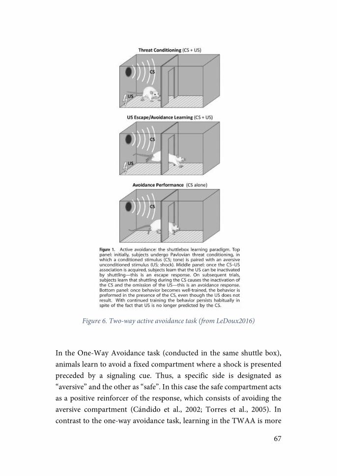
67
Figure 6. Two-way active avoidance task (from LeDoux2016)
In the One-Way Avoidance task (conducted in the same shuttle box), animals learn to avoid a fixed compartment where a shock is presented preceded by a signaling cue. Thus, a specific side is designated as “aversive” and the other as “safe”. In this case the safe compartment acts as a positive reinforcer of the response, which consists of avoiding the aversive compartment (Cándido et al., 2002; Torres et al., 2005). In contrast to the one-way avoidance task, learning in the TWAA is more
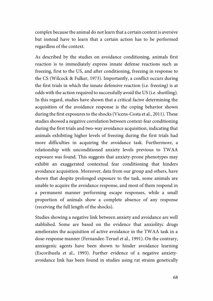
68
complex because the animal do not learn that a certain context is aversive but instead have to learn that a certain action has to be performed regardless of the context.
As described by the studies on avoidance conditioning, animals first reaction is to immediately express innate defense reactions such as freezing, first to the US, and after conditioning, freezing in response to the CS (Wilcock & Fulker, 1973). Importantly, a conflict occurs during the first trials in which the innate defensive reaction (i.e. freezing) is at odds with the action required to successfully avoid the US (i.e. shuttling). In this regard, studies have shown that a critical factor determining the acquisition of the avoidance response is the coping behavior shown during the first exposures to the shocks (Vicens-Costa et al., 2011). These studies showed a negative correlation between context-fear conditioning during the first trials and two-way avoidance acquisition, indicating that animals exhibiting higher levels of freezing during the first trials had more difficulties in acquiring the avoidance task. Furthermore, a relationship with unconditioned anxiety levels previous to TWAA exposure was found. This suggests that anxiety-prone phenotypes may exhibit an exaggerated contextual fear conditioning that hinders avoidance acquisition. Moreover, data from our group and others, have shown that despite prolonged exposure to the task, some animals are unable to acquire the avoidance response, and most of them respond in a permanent manner performing escape responses, while a small proportion of animals show a complete absence of any response (receiving the full length of the shocks).
Studies showing a negative link between anxiety and avoidance are well stablished. Some are based on the evidence that anxiolityc drugs ameliorates the acquisition of active avoidance in the TWAA task in a dose-response manner (Fernandez-Teruel et al., 1991). On the contrary, anxiogenic agents have been shown to hinder avoidance learning (Escorihuela et al., 1993). Further evidence of a negative anxiety-avoidance link has been found in studies using rat strains genetically
69
selected to show high and low anxiety-like phenotypes (Brush, 2003; Steimer & Driscoll, 2003).
Neural circuits of avoidance
Active avoidance comprises a complex learning process that involves both Pavlovian and instrumental components for regulating fear. Several decades of research using pharmacological drug infusions and lesion manipulations, as well as more recent optogenetic and transgenic technologies studies, have clearly identified the amygdala as the key site where CS-US associations are formed during fear conditioning, as well as a necessary site for the induction of the defensive reactions elicited by the CS (see for review LeDoux, 2003; Maren & Quirk, 2004; Tovote et al., 2015). Specifically, in the case of auditory fear conditioning, sensory information is received from the auditory cortex and thalamus through inputs to the lateral amygdala (LA), whereas in contextual fear conditioning information about the context enters the amygdala through the basal (B) and accesory basal (AB) nuclei (also known as basolateral and basomedial nuclei) from the ventral hippocampus. Information about the US and CS is integrated to form associative memories through processes involving hebbian plasticity in which cell firing in LA neurons is potentiated upon arrival of sensory information about the CS.
In addition to receiving and integrating sensory information, LA is in charge of controlling defensive reactions via direct and indirect (via BLA) connections to CeA, the major output of the amygdala. CeA then controls the behavioral and autonomic responses through long-range projections to the brainstem periaqueductal gray (PAG) and hypothalamus, respectively. Studies have shown that processes of synaptic plasticity also take place in CeA, and recent advances in genetic manipulation of concrete pathways have allowed to dissect the microcircuits within CeA and the efferents that control defensive behaviors (see for review Lüthi & Lüscher, 2014).
70
During avoidance acquisition, a conflict arises between the expression of these defensive reactions evoked by the CS and the instrumental response that need to be conducted to escape from and later on avoid the US. Hence, inhibitory inputs must reach the CeA to overcome the freezing response and execute the shuttling response. Indeed, several studies have shown that while lesioning LA and BA induces deficits in avoidance acquisition and increase the latency to avoid, lesions of the CeA had no effect or even recovered avoidance performance in animals showing deficits in avoidance acquisition (Choi et al., 2010; Lázaro-Muñoz et al., 2010). While LA-CeA circuits are involved in mediating fear reactions, LA-BA mediate fear-motivated actions (Amorapanth et al., 2000). Concretely, the role of BA have been suggested to be to use this CS-US association stored in LA for guiding actions through projections to the striatum (Robbins et al., 1989). Moreover, several studies have pointed at different subpopulations within CeA that may be critical for suppressing threat reactions (Ciocchi et al., 2010; Haubensak et al., 2010). A study showed that inhibition of a subpopulation of these neurons was involved in switching behavioral responses to the fear stimulus from passive to active responses, active responses understood as an increase in CS-induced exploratory activity (Gozzi et al., 2010). Although these studies did not assess for the role of these circuits in an active avoidance task, they suggest that activation of these inhibitory neurons in CeA could be driving the switch from escape responses to avoidance responses.
Some studies have shown a role of the PFC in the acquisition and execution of avoidance (Moscarello & LeDoux, 2013). In this study, in addition to replicating the lesioning effect of CeA on avoidance, the authors show that lesions of the IL but not PL in the PFC impaired active avoidance acquisition, enhanced freezing levels during the task and increased c-fos activation in CeA in response to the CS, pointing to an inhibitory role of IL towards CeA. These results, together with the evidence gathered from studies on fear extinction pointing at IL as the region in charge of inhibiting CS-induced outputs of CeA (Likhtik et al.,
71
2005; Likhtik et al., 2008; Milad & Quirk, 2002; Quirk et al., 2003), suggest that IL could be the input driving the difference in activation between these inhibitory CeA neurons, hence driving the differences between reactive (i.e. escapes) and active (i.e. avoidances) responses to threat. The excitatory nature of cortical neurons suggests that IL-CeA connections may be indirect in order to exert an inhibitory action on CeA. A group of GABAergic cells called intercalated cells (ITCs) placed between the BLA and CeA have been shown to be a candidate. They have been shown to be innervated by the IL and in turn to innervate CeA thus inhibiting CeA output (Amir et al., 2011; Pinard et al., 2012). Furthermore, they have been implicated in the regulation of fear responses and specifically fear extinction through the engagement of distinct subpopulations within ITCs (Busti et al., 2011). Recently, it has been shown that ITCs function not only as a relay from the PFC to CeA, but that they may play a critical role in regulating BLA information towards CeA as well, placing ITCs in a unique position to regulate fear-related responses (Asede et al., 2015). Interestingly, a recent study showed a possible direct connection between the PFC and the BA through long-range GABAergic neurons (Lee at al., 2014). Although the functional role of these direct PFC-BA projections remains to be determined, the same direct inhibitory connections were shown to innervate the NAcc eliciting aversive behaviors, suggesting these neurons as a potential pathway for direct top-down regulation of amygdala outputs and action selection from the PFC (Bravo-Rivera et al., 2015).
Upon arrival of the IL inputs into the amygdala, what is the pathway in charge of switching behavior from freezing to action when exposed to the CS? Evidence has pointed to a change in the balance of amygdala output from a predominant CeA-brainstem and hypothalamus output inducing freezing, to a BLA-AccSh output favoring the instrumental response (Ramirez et al., 2015). After three days of TWAA, those animals that had reached an 80% of avoidance responses showed increased c-fos activated neurons in the AccSh and decreased freezing levels compared

72
to yoked animals when exposed to a US-free session in the TWAA one day later. Furthermore, pharmacological inactivation of the AccSh or BA-AccSh circuit impaired avoidance performance and increased freezing levels, suggesting that this pathway may be necessary for the execution of the instrumental response in detriment of CeA outputs. Evidence showing that post-training BA lesions and inactivation attenuate CS-induced freezing in pavlovian as well as in active avoidance conditioning (Amano et al., 2011; Anglada-Figueroa & Quirk, 2005; Choi et al., 2010; Lázaro-Muñoz et al., 2010), the knowledge that BA send robust projections to both Cea and the NAcc (Pitkänen, Jolkkonen, & Kemppainen, 2000) and that several BLA subpopulations are engaged to regulate fear responses (Krabbe et al., 2018) put BA as a plausible candidate as the point of divergence between CS-evoked actions and reactions (Campese et al., 2015). Very interestingly, it has been shown that different subpopulations within BA target the PL or IL regions regulating fear conditioning and extinction respectively (Senn et al., 2014), indicating the presence of a down-top flow of information from the amygdala towards the PFC as well.
Besides this direct BA-PFC connection, studies suggest a possible indirect connection between the amygdala and the PFC through the striatum. Studies using electrolytic lesions showed that lesioning the caudate as well as the substancia nigra impaired avoidance acquisition (Mitcham & Thomas, 1972). Using more precise lesions within the striatum, Wendler et al. (Wendler et al., 2014) show that while neurotoxic lesions of the NAc-core and DSL delayed avoidance learning, animals with lesions on the DMS show impaired performance after more extensive training (from the forth session). This suggest that while the NAc and the DLS may play a role in avoidance acquisition, DMS may be necessary for avoidance expression once avoidance learning is consolidated. These results are in agreement with previous studies (Lázaro-Muñoz et al., 2010; Poremba & Gabriel, 1999) showing that execution of active avoidance becomes amygdala-independent with overtraining (understanding as such, 15 sessions of TWAA, 7 sessions
73
after reaching asymptotic performance) suggesting the involvement of other brain areas when the action is performed in a habitual manner. Darvas et. al (Darvas et al., 2011) show that whereas dopamine (DA) signaling in the amygdala and the striatum are necessary for acquisition and execution of active avoidance, DA signaling is only required in the striatum after extensive TWAA exposure (9 days 100 trials/day). This study is in line with previous studies showing the relevance of the dopaminergic system in avoidance conditioning (Boschen et al., 2011; Wietzikoski et al., 2012). Concretely these studies show that there is a DA release during the first training sessions of TWAA in the NAc, and that infusions of D1 or D2 dopamine receptors antagonist into the NAc impaired avoidance performance. Furthermore, it has been shown that the realease of DA in the striatum occur during the first sessions in trials where animals do not receive an expected shock (outcome better than expected), changes that did not occur in animals exposed to the same aversive stimuli in an unpredictable and inescapable manner, thus suggesting that DA release may function as a signal of positive prediction error reinforcing the acquisition of avoidance responses (Dombrowski et al., 2013). Interestingly, learning correlated with the level of DA release, and partial lesions of dopaminergic neurons from the substancia nigra impaired avoidance acquisition. These results are in line with another study showing an increase in DA in the NAc-core during the period previous to an avoidance response (Oleson et al., 2012). Importantly, previous to escape responses or during fear conditioning trials, a decrease in DA in this region was observed. This data suggests that whereas increased cue-evoked DA release in this region may induce active avoidance of aversive stimuli, reduced DA release may promote passive defensive behaviors when the aversive stimuli is unavoidable. In line with this hypothesis, studies have shown that stimulation of the VTA (Ilango et al., 2011), one of the main sources of DA, as well as optogenetic activation of DA neurons from this region facilitate avoidance acquisition, whereas inhibiting these neurons impaired it (Wenzel et al., 2018). However, manipulation of these neurons after overtraining had no effect, suggesting that while the dopaminergic system may play a key
74
role in avoidance acquisition its regulating role may vanish with prolonged exposure as the instrumental action is performed as a habit.
The majority of studies excluded “poor performers” making it difficult to know if the results obtained about the consequences of active avoidance apply as well to escape responses. However, some studies including this group have pointed towards a differential role of the dopaminergic system in mediating escape vs. avoidance responses. Darvas et al. showed that during acquisition escape responses, as avoidance responses, were performed only if DA signaling was intact in the amygdala and the striatum, suggesting that not only avoidance but also escape responses depend on both structures during acquisition (Darvas et al., 2011). These results are in line with studies using electrolytic lesions showing that lesioning the striatum impaired both, escape and avoidance responses (Kirkby & Kimble, 1968). Furthermore, the study from Oleson et al. showed that in contrast to the DA increase observed in NAcc core previous to an avoidance response, a decrease in DA is observed previous to the execution of an escape response (Oleson et al., 2012). These results suggest that the dopaminergic system may play a critical role in determining the final output from the amygdala between the BA-NAc pathway to promote defensive actions, or the Cea-Brainstem/Hypothalamus pathway to express defensive reactions.
Human studies on controllability
Animal studies on extinction have shown that animals learn to inhibit fear responses to previously fear-eliciting stimuli upon repeated exposure of this conditioned stimuli without the co-occurrence of the US. Based on these findings, during the last decades the most common treatment for PTSD and other anxiety disorders such as phobias and OCD, has been its homologous treatment in humans, exposure therapy. Despite its relative success in alleviating part of the symptoms in these pathologies, an important pitfall of exposure therapy (already observed

75
after extinction paradigms in animals) derives from the context-dependence of its effects to diminish fear responses. Indeed, phenomena such as the return of the extinguished threat response with the mere passage of time (“spontaneous recovery”) or the re-appearance of the symptoms outside of the context where the therapy is conducted (“renewal”), have highlighted the importance of identifying new treatments that induce more enduring means of fear-attenuation.
In this regard, animal studies suggest that exposure to aversive stimuli that can be behaviorally controlled by executing an instrumental response can persistently diminish learned threat responses (Cain & Ledoux, 2007; Ledoux et al., 2016; Maier, 2015). Importantly, fear responses are diminished even in new contexts where a new conditioned stimuli is presented and avoidance is no longer possible (Baratta et al., 2007; Cain & Ledoux, 2007), suggesting that control over aversive stimuli may have potential to promote resilience not only over previously acquired fear-responses but over new ones.
Although translation of these findings to humans has been scarce, a few studies have shown that the protective effects of aversive control observed in animal studies are also found in humans (Boeke et al., 2017; Hartley et al., 2014). Furthermore, it has been shown that, as in animal models, striatal and prefrontal regions may be implicated in mediating these effects (Boeke et al., 2017; Collins, Mendelsohn, Cain, & Schiller, 2014). Therapy based on training how to deal with stress through pro-active coping strategies and/or exposure to moderate levels of controllable stress have been proposed as plausible alternatives to exposure therapy (Diehl et al., 2010; Southwick et al., 2005). A study in humans showed that, as in animal models, two phenotypes arise depending on the ability to acquire the avoidance response (Collins et al., 2014). Furthermore, the study found individual differences in achieving successful avoidance depended on the functional synchronization of the PFC, the amygdala and the striatum. This suggests that, as in animal models, individual differences in neural connectivity of these regions may determine if an individual may be able to conduct active coping

76
strategies in stressful situations and thus benefit from therapies based on exposure to controllable stress.
Neuroimaging studies in humans have also supported a role of the striatum in learning to avoid aversive outcomes. Reduced fear arousal and amygdala activity was linked to increased activity in the striatum in participants who learned to avoid a US by terminating the presence of a fear-associated CS pressing a button (Delgado, 2009).
Epigenetic regulation of long-term effects
In the last decade, it has become increasingly clear that epigenetic regulation through changes in DNA methylation, chromatin structure and miRNA, play a central role in the induction of stable long-term behavioral changes. As mentioned above, epigenetic mechanisms in the context of PTSD have been suggested: (i) as a mechanism explaining vulnerability and resilience to stress (for more details see “The environmental epigenetics hypothesis”); and (ii) as a mechanism for explaining PTSD symptoms perdurance. Here I will focus on the second point.
One of the most relevant discoveries in the field of learning and memory in the past two decades is the evidence showing that changes in gene expression are necessary for experience to induce lasting functional and behavioral changes (Agranoff et al., 1967). Parallel over the last decade, epigenetic modifications have been shown to be a major actor in the regulation of persistent changes in gene transcription in the CNS (Borrelli et al., 2008b; Turner, 2002), and specifically in response to stress and the consequent development of psychiatric disorders (Tsankova et al., 2007).
One of the most extended hypothesis in the implication of epigenetics in the development of psychiatric disorders, and specifically PTSD, is that epigenetic molecular mechanisms underlie the formation and
77
stabilization of context- and cue-triggered fear conditioning based in the hippocampus and amygdala, as well as its extinction, highly dependent on the influence of the prefrontal cortex in the amygdala. In view of the dysfunction of these processes in the etiology of PTSD, it has been proposed that an aberrant regulation of epigenetic modifications in these processes may contribute to the development of the disease (Blouin et al., 2016; Kim et al., 2018; Kwapis & Wood, 2014; Trollope et al., 2012; Zovkic & Sweatt, 2013a). Moreover, pharmacological agents targeting epigenetic modulators such as HDAC inhibitors have been proposed as a new avenue of drug treatment for disorders involving cognitive and memory impairments, as well as anxiety disorders and depression (Gräff et al., 2014; Guan et al., 2009; Heinrichs et al., 2013; Tardito et al., 2013; Tsai & Graff, 2014; Whittle & Singewald, 2014a; Wilson et al., 2014).
Chromatin structure and post-translational modifications (PTMs)
Gene expression regulation is executed through dynamic modifications in the chromatin state, enabling or impeding access of the transcriptional machinery to DNA.
The nucleosome is the fundamental unit of chromatin structure. It is formed by 147 base pairs of DNA wrapped around an octamer composed by two copies of each of the core histone variants: H2A, H2B, H3 and H4 (Luger, 1997). Consecutive nucleosomes are connected by linker histones H1 and H5 to form the final block of chromatin (Ramakrishnan et al., 1993; Wolffe & Hayes, 1999). This nucleosomal structure of chromatin allows DNA to be tightly folded inside the nucleus (Felsenfeld & Groudine, 2003).
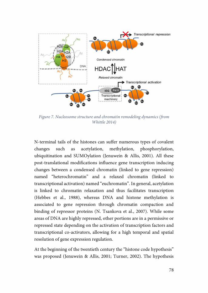
78
Figure 7. Nucleosome structure and chromatin remodeling dynamics (from
Whittle 2014)
N-terminal tails of the histones can suffer numerous types of covalent changes such as acetylation, methylation, phosphorylation, ubiquitination and SUMOylation (Jenuwein & Allis, 2001). All these post-translational modifications influence gene transcription inducing changes between a condensed chromatin (linked to gene repression) named “heterochromatin” and a relaxed chromatin (linked to transcriptional activation) named “euchromatin”. In general, acetylation is linked to chromatin relaxation and thus facilitates transcription (Hebbes et al., 1988), whereas DNA and histone methylation is associated to gene repression through chromatin compaction and binding of repressor proteins (N. Tsankova et al., 2007). While some areas of DNA are highly repressed, other portions are in a permissive or repressed state depending on the activation of transcription factors and transcriptional co-activators, allowing for a high temporal and spatial resolution of gene expression regulation.
At the beginning of the twentieth century the “histone code hypothesis” was proposed (Jenuwein & Allis, 2001; Turner, 2002). The hypothesis
79
predicted that covalent modifications on the histone tails function as a “code” that provides specific binding sites for effector proteins leading to the specific gene activation or repression of particular targets. Evidence was found indicating that epigenetic modifications of chromatin structure modulated functions such as imprinting phenomena, germ line silencing and stem cell divisions, indicating that overall chromosome stability, under epigenetic control, determined cell identity (Peters et al., 2002; Y. Wei et al., 1999). Although once thought to be an irreversible mechanism involved in stablishing cell fate, in the past decade a large amount of work has shown that dynamic changes in epigenetic marks plays a critical role in guiding behavior in response to different environmental stimuli (Borrelli et al., 2008).
Until now, one of the best-characterized covalent modifications is lysine acetylation. Lysine residues located at the N-terminal tail of histones are acetylated and deacetylated by the action of Histone acetyl-transferases (HATs) and Histone Deacetylases (HDACs) respectively. HATs and HDACs act as part of multiple protein complexes to execute this function (Hsieh & Gage, 2005). Apart from this chromatin-structure regulating role, acetylation and deacetylation has been shown to be a critical post-translational modification of non-histone proteins (Haggarty et al., 2003; Vaghefi & Neet, 2004). In a similar manner, due to its central role in transcription regulation of all kinds of proteins, aberrant regulation of these acetyl marks has been shown to be at the core of multiple disease states including cancer (Noonan et al., 2009; Witt et al., 2009), cardiovascular disease (Cao et al., 2011; Ordovás & Smith, 2010; Pons et al., 2009), asthma (North & Ellis, 2011; Su et al., 2009) and many others (Haberland et al., 2009).
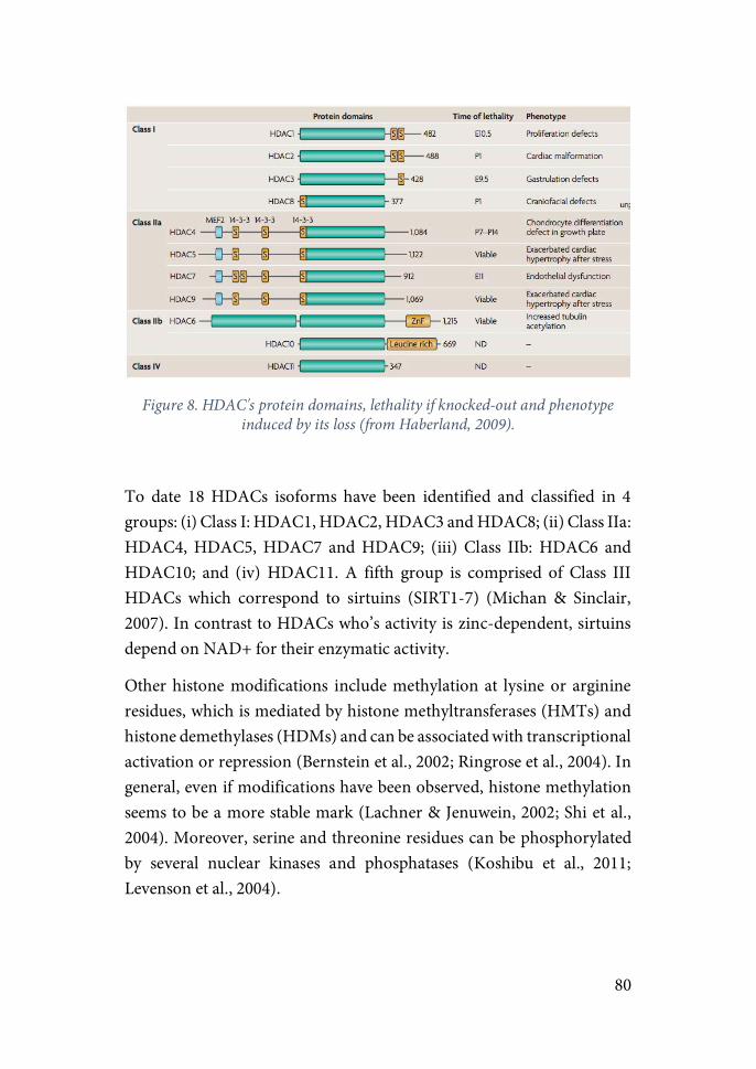
80
Figure 8. HDAC's protein domains, lethality if knocked-out and phenotype
induced by its loss (from Haberland, 2009).
To date 18 HDACs isoforms have been identified and classified in 4 groups: (i) Class I: HDAC1, HDAC2, HDAC3 and HDAC8; (ii) Class IIa: HDAC4, HDAC5, HDAC7 and HDAC9; (iii) Class IIb: HDAC6 and HDAC10; and (iv) HDAC11. A fifth group is comprised of Class III HDACs which correspond to sirtuins (SIRT1-7) (Michan & Sinclair, 2007). In contrast to HDACs who’s activity is zinc-dependent, sirtuins depend on NAD+ for their enzymatic activity.
Other histone modifications include methylation at lysine or arginine residues, which is mediated by histone methyltransferases (HMTs) and histone demethylases (HDMs) and can be associated with transcriptional activation or repression (Bernstein et al., 2002; Ringrose et al., 2004). In general, even if modifications have been observed, histone methylation seems to be a more stable mark (Lachner & Jenuwein, 2002; Shi et al., 2004). Moreover, serine and threonine residues can be phosphorylated by several nuclear kinases and phosphatases (Koshibu et al., 2011; Levenson et al., 2004).
81
DNA methylation involves the addition of a methyl group in cytosine adjacent to guanine residues called “CpG islands” to form 5-methylcytosine (5mC) (Klose & Bird, 2006). It is largely associated to induction of gene repression as they interfere with the binding of transcriptional factors to the DNA. DNA methylation is catalyzed by DNA methyltransferases (DNMTs) which consist of two categories: DNMT1, which copies DNA methylation pattern from the parental DNA strand to the new daughter strand during cell division (Santos et al., 2005); and DNMT3a and DNMT3b, in charge of adding novel methylation marks (Day & Sweatt, 2010). Due to its central role in embryonic and early-life development, germ line knockout animals die early during development (Li et al., 1992). Hence, initial studies focused on its role on disorders such as cancer and other developmental disorders, and it was not until recently that it was shown its role in transcription regulation, synaptic plasticity and memory formation in the adult brain (Barrett et al., 2008; Feng et al., 2010). In addition to impeding access to the transcriptional machinery, DNA methylation binds to methyl-binding proteins (MBPs) which in turn interact with repressor complexes such as HDACs to inhibit gene transcription (Nan et al., 1998; Wade et al., 1999). Of note, a mutation in the MBP methyl-CpG-binding protein 2 (MeCP2) is responsible for the Rett syndrome, a progressive developmental disorder and one of the most common causes of mental retardation in females (Amir et al., 1999). Recently a new modification similar to DNA methylation which involves the hydromethylation of cytosines to form 5-hydroxy-methylcytosine (5-hmC) was discovered. It is highly present in neurons compared to other tissues, it is not present in cancer cell lines, and in contrast to methylation it is associated with transcriptional activation (Kriaucionis & Heintz, 2009; Sérandour et al., 2012; Tahiliani et al., 2009). Although 5-hmC is known to be an intermediate step in DNA methylation (Guo et al., 2011) some evidence indicate it may have an unknown biological role (Spruijt et al., 2013).

82
Other general mechanisms of chromatin remodeling include nucleosome sliding, which involves the movement of the histone octamer along DNA (Felsenfeld & Groudine, 2003), or histone substitution and turnover, where canonical histones are changed with other histone variants or new identical variants (Hake et al., 2004).
Epigenetic mechanisms in stress-related learning and memory One of the most valuable skills for survival is the ability to learn from a harmful situation. It is critical for the individual to store in the long-term relevant information about cues in the environment to avoid, react faster or improve how to cope with a threat in future encounters. Thus, it is not surprising that studies during the last decades on learning and memory have pointed at mechanisms activated by stress as strengthening actors in the consolidation of memory (Buchanan & Lovallo, 2001; Roozendaal et al., 2009), and epigenetic mechanisms as main actors in the regulation of the changes in gene expression needed for these processes to take place and persist in time (Fischer et al., 2007; Graff et al., 2011; Jarome & Lubin, 2014; Levenson & Sweatt, 2005a; Sahar & Sassone-Corsi, 2012). It should be noted that studies on the field of learning and memory use behavioral challenges such as the MWM, FS and fear conditioning that represent a stressful situation for the rodent. Therefore, it is important to consider the major stress component of these tests when interpreting the results.
Fear learning have been modeled in animal studies through the use of the classical fear conditioning paradigm (Blumberger et al., 2018; Deslauriers et al., 2018; Flandreau & Toth, 2017; Schöner et al., 2017; Török et al., 2018). The results yielded by these studies have shown that gene expression changes in key regions in the regulation of the stress response such as the amygdala, PFC and hippocampus play a central role in the aberrant processes of structural and functional plasticity thought to underlie PTSD symptoms and fear memory dysfunction (Adamec et
83
al., 1998; Li et al., 2005; Mahan & Ressler, 2013). These results, together with the evidence gathered from the molecular processes involved in learning and memory, paved the way for the study of epigenetic regulation of gene expression in the context of fear memory and stress-related disorders.
While it is beyond the scope of the present work to provide a detailed review on memory formation, I outline the different stages involved to set the framework on fear memory and fear conditioning paradigms. Formation of an aversive memory involves four stages (Abel & Lattal, 2001): (i) Acquisition, which occurs as the animal learns the association between a context or a cue (CS) with an aversive stimulus (US); (ii) Consolidation, which can last from minutes to hours, involves the stabilization of the acquired memory and requires de novo synthesis of proteins (Nader, 2000). Contextual information about the event is processed in the hippocampus, while cue-associations are mainly dependent on the amygdala (Maren, 2001; Maren et al., 2013). Whereas recent contextual memories are transferred to the cortex for long-term storage (Frankland et al., 2004), remote cued-fear associations remain localized in the amygdala (Bergstrom, 2016); (iii) Retrieval, occurs when the animal is put back in the context where conditioning took place and involves the temporal destabilization of the memory trace; (iv) Reconsolidation, is the process of re-stabilization of the fear memory triggered by retrieval of the original memory. Importantly, studies have shown it may function as a window of intervention, allowing the incorporation of additional features to alter its fearful meaning (Lee, 2009; Nader, 2000; Nader & Hardt, 2009; Schiller et al., 2010).
A suppression of the consolidated memory can be obtained by a process of fear extinction. Fear extinction involves the acquisition of a new inhibitory learning through repeated presentation of the CS in the absence of the US. In this conditions the animal learns that CS no longer predicts the occurrence of the aversive stimulus and thus decreases the magnitude of the conditioned response (Lee et al., 2013; Myers & Davis, 2007; Quirk et al., 2006). Evidence that extinction involves the formation

84
of a new memory and not the erasure of the fear memory comes from studies showing that the original fear memory can reappear after extinction in three different conditions (Myers & Davis, 2007): (i) Spontaneous recovery, refers to the reappearance of the conditioned response (i.e. freezing) after extinction with the passage of time; (ii) Renewal, which consists in the return of the fear response when CS is presented outside of the extinction context; and (iii) Reinstatement, where the animal is re-exposed to the US with the subsequent recovery of the fear response.
One of the hallmark symptoms of PTSD is the perdurance of intense fear memories related to the traumatic event in the form of intrusive memories, flashbacks and nightmares. Moreover, recall of these fear memories can be triggered not only by cues related to the traumatic event, but also by non-threatening cues under safe conditions due to a process of pathological fear generalization (Acheson et al., 2015; Morey et al., 2015). These symptoms are hypothesized to reflect an over-consolidation and reconsolidation of the initial fear memory together with a dysfunction in fear extinction learning (Careaga et al., 2016). Therefore, pharmacological treatments and therapies of PTSD patients have focused on interfering with the reconsolidation of the memory to weaken or change the original traumatic memory, and on strengthening extinction learning (Koenigs & Grafman, 2009; Lattal & Wood, 2013; Zovkic & Sweatt, 2013a).
Epigenetic&of&fear&memory&&
The first evidence of dynamic changes in histone post-translational modifications in response to neural stimulation was shown in studies in Aplysia. The authors showed that neural stimulation induced a decrease in gene expression associated with LTD caused by deacetylation of the promoter regions of early-response genes in response to the recruitment of HDAC5 (Guan et al., 2002). Since then, a clear link has been shown by
85
numerous studies between the induction of LTD and LTP, memory formation and changes in histone epigenetic markers in mammals (Guan et al., 2002; Kerimoglu et al., 2013; Levenson et al., 2004; Levenson & Sweatt, 2005a).
The first studies on mammals investigating the involvement of epigenetic modifications in memory were focused on assessing if chromatin structure was changed in the hippocampus in the early stages of memory formation. In particular, Sweatt and colleagues showed that a transient increase in global acetylation of histone H3 in CA1 occurred 1h post-CFC and returned to basal levels by 24h. Interestingly, if acetylation levels were increased by injecting HDAC inhibitors memory recall persisted 24h post-CFC (Levenson et al., 2004). Since then , not only acetylation but methylation and phosphorylation of H3 and H4 in the hippocampus have been associated to memory formation in CFC (Chwang et al., 2007; Gupta et al., 2010; Kerimoglu et al., 2013; Levenson et al., 2004; Peleg et al., 2010).
The same group reported that in response to CFC animals showed an increase in the expression of de novo DNMTs 3a and 3b in CA1, concomitant to an increase in the methylation and decreased expression of the memory suppressor gene protein phosphatase 1 (PP1), as well as demethylation and increased expression of the plasticity gene reelin (Miller & Sweatt, 2007). Moreover, treatment with a DNMT inhibitor after training impaired fear memory and blocked the decrease in PP1 expression. These results are in line with other studies from the group suggesting that hippocampal DNA methylation together with histone acetylation are required for memory consolidation (Levenson et al., 2006; Lubin et al., 2008; Miller et al., 2008).
In addition to changes in the hippocampus during consolidation, memory recall has also been associated with changes in histone PTMs in CA1, changes that are hypothesized to be mediating the reconsolidation of the previously consolidated memories (Lubin & Sweatt, 2007).
86
As in the hippocampus, DNA methylation changes and histone PTMs have also been shown in the amygdala after auditory fear conditioning (Monsey et al., 2011). In particular, it was shown that auditory fear conditioning induces the acetylation of histone H3 and DNMT3A expression in an ERK-dependent manner in LA neurons. Moreover, whereas inhibition of acetylation by administration of TSA (an HDAC inhibitor; HDACi) enhanced fear memory 24h post-conditioning, inhibition of DNA methylation by administration of an DNMT inhibitor impaired it. However, the effect on fear memory was not present after one week. In another study the same group showed transient increases in the acetylation of H3 in the LA during retrieval of a fear memory. Moreover, DNMT inhibitors administered into the LA impaired memory reconsolidation, an effect that was reversed by pre-treatment with HDACi immediately after recall (Maddox & Schafe, 2011), suggesting that dynamic changes in epigenetic markers are necessary for both consolidation and reconsolidation of fear memory. However, the persistence of these epigenetic changes in the amygdala remains to be determined.
An extensive body of studies using lesions of different regions in different time-points after learning suggest that whereas short-term memory seem to rely on the hippocampus, maintenance of remote memory depend on cortical support (Frankland & Bontempi, 2005; Walters & Zovkic, 2015). In line with this hypothesis, changes in epigenetic modifications in the hippocampus have been shown to be mainly transient, while changes in the PFC seem to be more stable. One study showed persistent changes in cortical DNA methylation and expression 7 days and 30 days post-conditioning (3 shocks 1 sec) (Miller et al., 2010). Importantly, fear memory was impaired in animals by pharmacological inhibition of DNA methylation in the mPFC at 30 days.
Results from a seminal study investigating genome-wide changes in CA1 and the anterior cingulate cortex (ACC) 1h and 4 weeks post-CFC seem to favor this hypothesis (Halder et al., 2016). Although a robust increase in differentially-methylated regions (DMRs) was found in CA1 1h post-
87
CFC, almost no DNA methylation change could be detected at 4 weeks. Interestingly, 6 times more DMRs were found in the ACC than in CA1 1h post-CFC, among which a substantial part could still be detected in cortical neurons at 4 weeks. More impressive, when changes restricted to associative memory (animals exposed to the context vs. exposed to context and shock) were analyzed, DMRs were almost exclusively restricted to CA1 at 1h and the ACC at 4 weeks.
Importantly, the authors performed the analysis separating neuronal from non-neuronal cells. Results indicated that non-neuronal cells presented DMRs during memory consolidation in CA1 and ACC, but not during maintenance. These results highlight the importance of performing cell-specific analysis when studying epigenetic regulation. Furthermore, the authors show that DMRs are mainly found in intergenic (64%) and intronic (30%) regions, and that promoter regions represented only 1% of DMRs. Additionally, results show an increase of activity-related histone PTMs concomitant to a decrease of inactivity-related histone PTMs in CA1, results that would be masked in studies of global changes in PTMs. These results emphasize the relevance of genome-wide analysis as compared with restricted analysis of single-target gene promoters, and add weight to the theory that histone PTMs might not constitute a mnemonic substrate but instead may function as a priming mark, sensitizing neurons and non-neuronal cells for future activity (Edith et al., 2011; Halder et al., 2016; Lopez-Atalaya & Barco, 2014).
Interestingly, studies from our group are in agreement with this hypothesis. Expression of c-fos, a IEG, has been widely used to identify brain areas activated in response to stressors and drugs (Pacak & Palkovits, 2001). However, studies quantifying activity by c-fos and other IEGs expression are insensitive to the intensity of the stressor (Campeau et al., 1997; Ons et al., 2004). Using acute exposure to a novel environment (low intensity stressor) and 30 or 90 minutes of IMO (high intensity stressor), the authors showed that only a restricted set of activated neurons as measured by c-fos expressed pH3S10, a histone
88
phosphorylation mark related to enhanced gene expression. Furthermore, although changes in pH3S10 were not in general more sensitive to the intensity of the stressor than c-fos, they were more sensitive to discriminate the end of the stressor (Rotllant et al., 2013). In order to express such epigenetic changes neurons may receive additional or stronger synaptic signals as compared to those required for c-fos, suggesting that, although transient, expression of pH3S10 may be a molecular mark signaling those neurons most critically involved in the long-term impact of the stress in comparison to c-fos activation that may reflect to a high extent processes of general arousal.
Fear extinction has been shown to be largely dependent of the mPFC (Myers & Davis, 2007; Quirk et al., 2006). Several studies have shown that extinction learning is associated with increased levels of histone acetylation, and that enhancement of these PTMs by administration of HDACi promotes extinction of conditioned fear (Bredy et al., 2007; Lattal et al., 2007; Stafford et al., 2012). Concretely, acetylation increased in the hippocampus and PFC in response to extinction learning. Moreover, treatment with HDACi systemically, in the hippocampus or into the IL enhanced extinction learning for up to 14 days. In contrast, HDACi administered into the PL had no effect (Lattal et al., 2007; Stafford et al., 2012). As discussed earlier, the study of the mechanisms underlying extinction learning is of special interest in the context of PTSD, as PTSD patients exhibit an impairment in extinction learning, showing sustained levels of fear despite exposure to extinction training (Blechert et al., 2007; Fani et al., 2012; Norrholm et al., 2011). In this regard, administration of HDACi to treat PTSD have been suggested as a potential therapy (Lattal & Wood, 2013; Tsai & Graff, 2014; Whittle & Singewald, 2014b; Zovkic & Sweatt, 2013b).
Caution should be taken when administering HDACi as animals studies have shown that doses of HDACi such as sodium butyrate (NaBu) in the range used in experiments have been reported to increase the plasma levels of stress markers without prior exposure to stress, indicating that HDACi may act as pharmacological stressors (Gagliano et al., 2014).

89
Of note, even if we have limited the discussion to fear learning and memory, epigenetic regulation has been shown to be mediating learning and memory in fear-unrelated tasks such as novel object recognition and conditioned place preference (Featherstone & McDonald, 2004; Federman et al., 2014; Gräff & Tsai, 2013; Rogge et al., 2013).
Epigenetics&of&stress&&
Apart from studies focused on fear learning in CFC, other paradigms such as the FS have been extensively used to study epigenetic changes on pathways involved in adaptation to stress. As mentioned previously, animals exposed to FS exhibit an immobility response that can be observed 24h or even several weeks after the initial test. This response has been shown to be dependent on the action of GCs in the DG of the hippocampus (Kloet & Molendijk, 2016). The authors showed that an increase in histone H3 phosphorylation (H3S10p) in combination with the acetylation of lysine14 (K14ac) is involved in activating gene transcription in a net of sparsely distributed dentate granule neurons and that this process depended on the action of GRs on NMDA-Rs (Chandramohan et al., 2008; Chandramohan, Droste, & Reul, 2007; Gutierrez-Mecinas et al., 2011; Reul, 2014). In the same study, the authors found a decrease of DNA methylation in CpG islands of immediate-early genes such as Fos and Erg1 specifically in the dentate granule neurons but not in CA1.
In a recent study it has been shown that in the same FS protocol, GR expression itself is also decreased 15 minutes post-FS (Mifsud et al., 2017). This decrease is concomitant to an increase in the DG of DNMT3a expression and DNA methylation of Nr3c1 promoter, together with an increase in mir124a, a microRNA known to inhibit GR mRNA expression.

90
Global changes in DNA methylation in the hippocampus have been associated to PTSD-like phenotypes (7 days post-stress) in rats exposed to 10 minutes of predator-scent odor (Chertkow-Deutsher et al., 2010). Interestingly, methylation of a specific gene, Dlgpa2 (specific of the post-synaptic density zone in post-synaptic neurons), and the subsequent decrease in gene expression, was only found in animals showing anxiety-like behaviors but not in resilient animals.
Using a modified model of predator-scent odor exposure, an association was found in the amygdala between the methylation of Avp promoter, levels of GC and progesterone, and the coping strategy shown in response to the stressor (Bowen et al., 2014). In particular, only animals showing active coping behaviors in response to the stressor showed lower levels of GC and progesterone 3 days post-stress, as well as lower expression levels of Avp corresponding to a diminished methylation of its promoter. These results indicate that epigenetic regulation may be linked to intergroup variability in rodents, which recapitulates the variability in stress response observed in humans after exposure to a traumatic event. !!In a model of acute-restraint stress, recent genome-wide analysis of DNA methylation reported global decreases in DNA methylation in the hippocampus, cortex and periaqueductal grey 1h post-stress (Rodrigues et al., 2015). In line with these results, another study using the same protocol found a global increase in gene expression but in this case associated with increased levels of 5hmC in the hippocampus.
Other stress models such as chronic social stress have been shown to induce prolonged anxiety-like behavior (measured in the light-dark and EPM test 2 weeks post-stress) associated with a decrease in mRNA levels of DNMT3A and global DNA methylation levels in the mPFC (Elliott et al., 2016). Interestingly, knocking-down this gene in naïve animals reproduced enhanced anxiety-like behaviors similar to mice that had undergone chronic social defeat stress, while DNMT3a over-expression resulted in a reduction of anxiety-like levels.
91
These results show that even brief exposure to stress are capable of inducing global and region and time specific changes in gene expression that may underlie the induction of molecular changes responsible for the long-term behavioral consequences of acute-stress.
BDNF-TrkB: PTSD implications and epigenetic regulation
The capacity of neuronal circuits to undergo functional changes is a basic requirement for learning and behavioral adaptation. The dynamic remodeling underlying these changes depend on the capacity of environmental factors to influence gene expression and protein activation.
Brain-derived neurotrophin factor (BDNF) is the most studied neurothrophin involved in synaptic plasticity processes necessary for long-term learning and memory. BDNF protein is not only involved in the modulation of synaptic strength, but its gene is as well subject to experience-dependent epigenetic regulation.
BDNF is a member of the neurotrophins, a family of signaling proteins including nerve-growth factor (NGF), neurotrophic factor 3 (NT3) and NT4/5. Its major functions are its involvement in developmental programming of neuronal differentiation and survival () and synaptic and structural plasticity.
Rodent BDNF gene is formed by a minimum of nine 5’ non-coding exons each containing a unique promoter region, and a 3’- exon (IX), coding for the BDNF prepropeptide (Musumeci & Minichiello 2011). It is highly expressed in the amygdala, hippocampus and prefrontal cortex, and its expression and release are activity-dependent. Cells known to express BDNF are fibroblasts, astrocytes and different types of neurons. BDNF acts through two distinct receptor systems, its high-affinity tropomysin-related kinase B (TrkB) receptor and the low-affinity receptor p75 shared for all neurotrophins. Binding of BDNF to its
92
receptor induces its dimerization and leads to the activation of several signaling cascades through the intracellular kinase-binding domain including Ras/MAPK, PLC-g and PI3K/AKT pathways.
BDNF gene expression and activation of its high-affinity tropomyosin-related kinase B (TrkB) receptor are necessary in the amygdala, hippocampus and prefrontal cortex for the formation of emotional memories, including fear memories. Several studies implicate BDNF concretely in the enhancement of fear extinction. In view of the evidence pointing to an altered processing of extinction in PTSD patients, new lines of treatment target BDNF as a way to improve efficacy in anxiety disorders such as PTSD.
BDNF exon I and IV were shown to increase in the amygdala during cue-dependent fear conditioning (Rattiner etal. 2004b), while re-exposure to the contextual fear conditioning box causes increased exon IV mRNA in the hippocampus (Lubin etal. 2008). Interestingly, contextual fear extinction elicits an increase in BDNF exon I and IV mRNA in the mPFC (Bredy etal. 2007). Epigenetic regulation of BDNF has been reported to be crucial in these changes in gene expression (Musumeci & Minichiello 2011). This epigenetic regulation includes dynamic changes in histone acetylation state of the N-terminal lysine residues. Histone acetyltransferases (HATs) and histone deacetylases (HDACs) are in charge of mediating these processes. Studies of fear memory have shown that histone H3 acetylation and phosphoacetylation levels are increased and histone H4 acetylation levels decreased in promoter 4 in the hippocampus after contextual fear conditioning (Lubin etal. 2008; Takei etal. 2011). Regional differences might explain an increase in H4 acetylation in the promoter 4 within the PFC after contextual fear extinction (Bredy etal. 2007). In the lateral amygdala, levels of acetylated H3 are increased in response to cued fear conditioning (Maddox & Schafe 2011).
Very recently, a new DNA modification of adenosine, and not cytosine residues as reported until now, has been shown to be implicated in the
93
regulation of gene expression required for fear extinction (Xiang Li et al., 2019). Concretely, N6-methyl-2’-deoxyadenosine (m6dA) modifications were shown to be increased along promoter and coding regions of genes in activated neurons in the PFC in response to fear extinction. Interestingly, increases of this modification linked to expression activation were found in the promoter region of Bdnf-IV, adding evidence to the relevance of increases in the expression of this neurotrophic factor for the induction and consolidation of fear memory and specifically fear extinction.
94
MATERIALS AND METHODS
1. ANIMALS AND GENERAL PROCEDURE
This project consists of 3 experiments. Experiment 1 and 2 were conducted at the Universitat Autònoma of Barcelona, whereas experiment 3 was performed at the University of Haifa (Israel) in collaboration with Dr. Gal Richter-Levin’s group thanks to an FPI grant of the Spanish Ministry of Science.
Same materials and methods were used in both locations unless stated otherwise.
The aim of experiment 1 was to evaluate the behavioral effects of acute exposure to controllable and uncontrollable stress. For that purpose, animals were exposed to two sessions of TWAA in non-consecutive days. An elevated-plus maze, an open-field and an acoustic startle test were conducted two, five and seven days post-TWAA to assess for short and long-term effects on activity, arousal and anxiety-like parameters.
The objective of experiment 2 was to ascertain if differences in changes in mRNA levels of genes related to synaptic plasticity and epigenetic regulation could be involved in the induction and persistence of the different consequences of exposure to controllable vs. uncontrollable stress. With that aim, the same behavioral procedure conducted in experiment 1 was reproduced, this time performing gene expression analysis of genes related to the Bdnf-TrkB pathway and HDACs, two and five days post-TWAA in the main regions in charge of regulating the stress response.
Finally, the aim of experiment 3 was to evaluate: (i) if it is necessary to reach a certain level of controllability in order to obtain the stress buffering effects of controllable stress; (ii) if prolonged exposure after reaching this level of controllability is a critical factor for this learning to consolidate and exert an emotional buffering effect; (iii) if prolonged

95
exposures are necessary in order to detect long-term differences in the consequences of controllable vs. uncontrollable stress; (iv) if uncontrollable stress has a higher detrimental effect than short exposure to uncontrollable stress; and finally (v), if changes in synaptic plasticity and mRNA levels of the above-mentioned genes could be part of the molecular mechanisms underlying these phenomena. To answer these questions the experiment was designed as follows. Two groups of animals were exposed to short (2 days) or prolonged (5 days) TWAA, each group in turn divided into exposure to controllable stress (CoS), uncontrollable stress (UnS) or the context (Naïve). A behavioral battery of tests consisting of an OF, a social-interaction test, an EPM and an extinction test were conducted one-week post-TWAA to assess for the long-term behavioral effects. Finally, gene expression analysis was conducted in the hippocampus three days and one week respectively after the extinction test to evaluate for possible molecular correlates.
1.1.!ANIMALS
Experiment 1&2
50 and 44 male Sprague-Dawley rats provided by the breeding center of the Universitat Autònoma de Barcelona were used in experiment 1 and 2, respectively. Animals were housed in pairs in 42.5 x 26.5 x 18 cm plastic cages with sawdust bedding (Lignocel, Panlab S.L.U., Barcelona, Spain) in standard conditions of temperature (21 ± 1 °C) and a 12-h light/12-h dark schedule (lights on at 08.00 h). Food (SAFE-diet A04, Panlab S.L.U., Barcelona, Spain) and water were available ad libitum. Animals were at PND60 at the time of experimental procedures. All procedures were conducted during the light-phase (8:00-20:00).
The experimental protocol was approved by the Committee of Ethics of the Universitat Autònoma de Barcelona and the Generalitat of Catalunya, followed the “Principles of laboratory animal care” and was
96
carried out in accordance with the European Communities Council Directives (2010/63/EU) and the Spanish Legislation (RD 53/2013).
Experiment 3
112 male Sprague-Dawley rats were obtained from Harlan Laboratory (Jerusalem, Israel) at an approximate age of 60 PND (weight 275-300 g) in experiment 3. Animals were maintained in groups of 4 per cage (75x55x15cm) in temperature-controlled (23 ± 1°C) animal facilities on a 12:12 light- dark cycle (lights on 07:00-19:00 hours) with food (standard Purina rodent chow pellets) and water ad libitum. Animals were at PND60-70 at the time of experimental procedures in experiment 3.
Experiments 3 were in accordance with the NIH guidelines for the care and use of laboratory animals and were approved by the University of Haifa ethical committee (Ethical No. 230/11).
1.2. GENERAL PROCEDURE
Upon arrival to the animal facility animals were let undisturbed until the first handling session that began one day after and consisted of at least three sessions. Each session consisted of a repetition of all the transportation from the vivarium to the experimental room and the handling procedures that would be performed the day of the experiment. Specifically, animals were taken out of their homecages and put in 25x15x15 white individual plexiglass cages for transportation until the experimental room. Two animals from the same homecage were moved into two different cages. Once in the experimental room animals were let undisturbed for 30 seconds, taken out of the cages, put back again and moved to the adjacent room were blood samples are obtained. Once in
97
this room, general handling procedures including gentle body and tail massage while wrapped in a cloth were carried out. In the case of animals in experiment 1, blood sampling was done in the last handling session by the tail nick procedure (see below) to habituate animals to the procedure.
Animals were assigned semi-randomly to the different experimental groups to balance for weight, mother and date of birth in experiment 1; or weight in experiment 3 (no information about the mother and date of birth was provided by Harlan, Jerusalem).
2.! BLOOD SAMPLING PROCEDURE
Blood samples was obtained by “tail nick”. The “tail nick” procedure consists of making a 2-mm incision at the end of the tail veins and massaging the tail while collecting 300 μl of blood into ice-cold EDTA capillary tubes (Sarsted, Granollers, Spain). All the procedure is performed in a 2-minute period while the animal is gently wrapped with a cloth. Samples were kept at -4ºC until they were centrifuged for 25 minutes at 7100 rpm (Heraeus-Biofuge Primo Centrifuge, Thermo Scientific, Germany). The plasma obtained was split in two aliquots and stored at -20 ºC until hormone analysis were done.
This procedure is extensively used in our group and other labs (Belda et al., 2004; Vahl et al., 2005) because very actual basal levels of stress hormones are obtained if the procedure is done adequately. Blood sampling was done in a different room than the vivarium.
98
3.! STRESS PROCEDURE: TWO-WAY SHUTTLE AVOIDANCE TASK.
In active avoidance paradigms subjects learn to avoid an aversive stimulus (e.g. foot-shock) by initiating a specific locomotor response. The active avoidance task used in the present study was the Two-way active-avoidance task (TWAA). This task belongs to the paradigms used for the study of the “Learned Helplessness” phenomenon which consists of a triadic experimental design: Controllable stress, Uncontrollable stress and Naïve (for more details see “Coping, Active Avoidance and Controllability” in the introduction section). In the TWAA the specific locomotor response required by the animal to avoid the aversive stimulus consists of shuttling to an adjacent chamber in a shuttle-box. Concretely, the three experimental groups are:
1.! Controllable-stress group (CoS). Animals assigned to this group have the possibility to exert control over the stressor conducting two different behaviors: (1) Escape response – shuttling to the adjacent chamber during the US (e.g. shock), stopping it and thus reducing US duration; and (2) Avoidance response – shuttling to the other compartment during the presentation of a conditioned stimulus (e.g. light and/or tone), thus avoiding completely any exposure to the US. If the animal does not exhibit any of the previous responses, it will receive the full length of the US and would be accounted as a Null response.
2.! Uncontrollable-stress group (UnS). Animals in this group are exposed to the same stimuli (CS and US) as the CoS group but no instrumental behavior of the animal is contingent with shock avoidance or termination. Two setting configurations are possible: Yoked and Averaged CoS protocol. See details bellow.
3.! Naive group. Animals in this group are exposed to the shuttle-box for the same duration as CoS and UnS groups, but with no US or CS presentation.

99
3.1.! Apparatus and TWAA set-up
All experiments were done using shuttle-boxes provided by Panlab (LE916, Panlab S.L.U., Barcelona, Spain). These boxes (Fig. 9) consist of two equally sized compartments (25 x 25 x 25 cm) with two independent grid floors separated by a metallic wall with an 8 x 10 cm aperture between them. The cage contains a general sound generator and a visual stimulus (light) for each compartment. The shuttle-box is inside a dimly-lit, ventilated, sound-attenuating cage to avoid any external noise during the task.
Figure 9. Illustration of the shuttle-box apparatus used for the Two-way shuttle avoidance task.
Information about the position of the rat is transmitted through weight-sensitive electrifiable grids. Grids are connected to a shocker (LE100-26, Panlab S.L.U., Barcelona, Spain) and a controller device (LE900, Panlab S.L.U., Barcelona, Spain) which are in turn under the control of Packwin v1.2.23 or Shutavoid 1.8.02 software (Panlab S.L.U., Barcelona, Spain).
In experiment 1 and 2 the general set-up consisted of six shuttle-boxes. Two animals from each experimental group and from the same homecage were put per round. Stress procedure consisted of two TWAA
100
sessions on non-consecutive days. This number of sessions was chosen as a compromise between two factors: (1) duration (the shortest possible to be considered acute); and (2) reaching sufficient learning of the task to set a difference between controllable and uncontrollable stress.
In experiment 3, the TWAA set-up consisted of eight shuttle-boxes. Rounds of 3 to 6 animals from the same experimental group were conducted. Daily sessions during two or five consecutive days were administered depending on the group. Animals were never let alone in homecages before TWAA sessions to minimize possible distress before sessions.
3.2.! Procedure
Sessions were conducted during the morning (9:00 AM-13:00 PM) to reduce the influence of the circadian rhythm of stress hormones in the results. Every session began with 5 minutes of habituation. No stimuli were delivered during this period. Once finished, animals were exposed to 50 trials per session.
Controllable stress protocol
Every trial consisted on:
1.! Presentation of the conditioned stimuli (CS): A light (7W) concomitant with a tone (2400 Hz, 80 dB) 10 seconds (shorter if an avoidance response was performed).
2.! Presentation of the unconditioned stimuli (US) if no avoidance response had been performed: 0.7 mA electric-shock for a maximum of 10 seconds (shorter if an escape response was performed). There was no delay between CS termination and US initiation.
3.! Inter-trial interval (ITI): A random ITI was set, ranging from 5 to 55 seconds, with an average of 30 seconds.
101
Additional trials were added in the first session if animals in the CoS group performed an “avoidance” response during the first trials without being firstly exposed to the US (“False avoidance”). Trials with shock deficiencies due to system failures were also compensated so that all animals received 50 shocks per session.
Cages were thoroughly cleaned at the before the first session, between sessions as well as at the end of the last session of the day (ethanol 70% in experiment 1 and 2; water with soap in experiment 3).
Uncontrollable stress protocol
Two different protocols were used: “Yoked” in experiment 1 and 2, and “Average CoS” protocol in experiment 3.
-! “Yoked” configuration: In these setting conditions, a shuttle-box configured as “master” (CoS) is yoked to a second “slave” shuttle-box (UnS). Information of the performance of the master is sent to the slave shuttle-box in a manner that both cages deliver simultaneously the CS and US with a duration determined by the performance of the master animal. That way both animals, master and its respective yoked animal, receive the exact same amount of physical stressor but whereas the master group has control over the exposure and duration of the US, the yoked group does not. Slave animals were paired to the same master in both sessions.
-! “Average CoS” protocol: It consists of a protocol based on the average performance of CoS animals in previous studies. Blocks were designed so the duration of CS, US as well as the number of US presentations decreased with time reflecting what occurs as CoS animals learn the task. The exact protocol employed in experiment 3 is shown in table 2. Importantly, all UnS animals
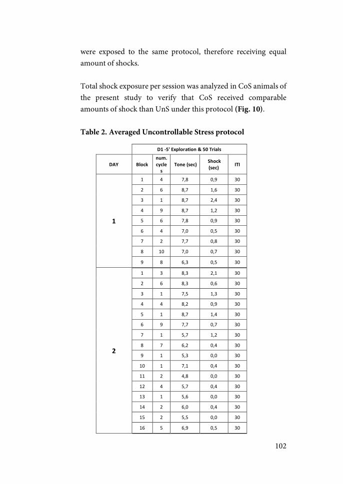
102
were exposed to the same protocol, therefore receiving equal amount of shocks. Total shock exposure per session was analyzed in CoS animals of the present study to verify that CoS received comparable amounts of shock than UnS under this protocol (Fig. 10). Table 2. Averaged Uncontrollable Stress protocol
D1#$5'#Exploration#2#Trials#
DAY# Block#num.#cycles#
#Tone#(sec)##Shock#(sec)#
ITI#
1#
1" 4" 7,8" 0,9" 30"
2" 6" 8,7" 1,6" 30"
3" 1" 8,7" 2,4" 30"
4" 9" 8,7" 1,2" 30"
5" 6" 7,8" 0,9" 30"
6" 4" 7,0" 0,5" 30"
7" 2" 7,7" 0,8" 30"
8" 10" 7,0" 0,7" 30"
9" 8" 6,3" 0,5" 30"
2#
1" 3" 8,3" 2,1" 30"
2" 6" 8,3" 0,6" 30"
3" 1" 7,5" 1,3" 30"
4" 4" 8,2" 0,9" 30"
5" 1" 8,7" 1,4" 30"
6" 9" 7,7" 0,7" 30"
7" 1" 5,7" 1,2" 30"
8" 7" 6,2" 0,4" 30"
9" 1" 5,3" 0,0" 30"
10" 1" 7,1" 0,4" 30"
11" 2" 4,8" 0,0" 30"
12" 4" 5,7" 0,4" 30"
13" 1" 5,6" 0,0" 30"
14" 2" 6,0" 0,4" 30"
15" 2" 5,5" 0,0" 30"
16" 5" 6,9" 0,5" 30"
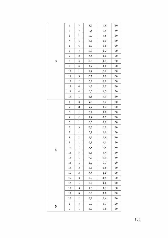
103
3#
1" 5" 8,2" 0,8" 30"
2" 4" 7,8" 1,3" 30"
3" 5" 7,0" 0,5" 30"
4" 1" 5,1" 0,0" 30"
5" 6" 6,2" 0,6" 30"
6" 4" 5,3" 0,2" 30"
7" 2" 4,4" 0,0" 30"
8" 4" 6,3" 0,4" 30"
9" 4" 4,2" 0,0" 30"
10" 1" 4,7" 1,7" 30"
11" 3" 5,1" 0,0" 30"
12" 2" 5,1" 2,0" 30"
13" 4" 4,8" 0,0" 30"
14" 4" 6,0" 0,3" 30"
15" 1" 5,8" 0,0" 30"
4#
1" 3" 7,8" 1,7" 30"
2" 8" 7,7" 0,7" 30"
3" 1" 5,4" 0,0" 30"
4" 2" 7,4" 0,9" 30"
5" 1" 6,0" 0,0" 30"
6" 3" 6,5" 1,2" 30"
7" 1" 5,2" 0,0" 30"
8" 2" 6,1" 0,6" 30"
9" 1" 5,8" 0,0" 30"
10" 1" 6,8" 0,9" 30"
11" 5" 6,3" 0,4" 30"
12" 1" 4,9" 0,0" 30"
13" 1" 8,0" 1,7" 30"
14" 2" 6,6" 0,8" 30"
15" 3" 4,4" 0,0" 30"
16" 3" 6,0" 0,5" 30"
17" 1" 5,0" 0,0" 30"
18" 3" 4,6" 0,3" 30"
19" 6" 3,9" 0,0" 30"
20" 2" 6,1" 0,4" 30"
5#1" 4" 7,9" 0,7" 30"
2" 1" 8,7" 1,6" 30"

104
3" 5" 8,4" 0,6" 30"
4" 1" 7,1" 1,7" 30"
5" 15" 6,9" 0,4" 30"
6" 4" 5,6" 0,0" 30"
7" 3" 6,6" 0,3" 30"
8" 2" 5,0" 0,0" 30"
9" 5" 6,3" 0,2" 30"
10" 3" 6,1" 0,0" 30"
11" 1" 6,5" 0,3" 30"
12" 3" 5,3" 0,0" 30"
13" 3" 7,0" 0,5" 30"
The protocol derives from the average performance of animals exposed to controllable stress in the same conditions in previous studies.
Figure 10. Total time of shock received by CoS and UnS animals in experiment 3. UnS group (red solid line) received the same amount of shock as the average in CoS animals (green line).
Day$1 Day$2$ Day$3 Day$4 Day$5$0
50
100
150TOTAL%SHOCK%RECEIVED
Time%(s)
Total%shock%%in%UnSMean%shock%exposure%in%CnS
105
3.3.! Video-recording
Performance of the animal was videotaped using cameras (KPC-S500P3, KT&C co.) placed 20 cm above the grid floor on the front door of the isolation chamber, one in each compartment of the shuttle box. Cameras were in turn connected to a video-recorder located outside of the isolation chamber.
3.4.! Data analysis
All variables measured during the habituation period of the TWAA were measured manually and in blind conditions, with no information about the experimental group of the animal. These variables included: time of freezing, scanning or grooming and number of intercrossings and rearings. The number of intercrossings was also measured automatically during the whole session using the Packwin v1.2.23 or the Shutavoid 1.8.02 software (Panlab S.L.U., Barcelona, Spain).
Freezing involved the absence of all movement, except for respiratory-related movements, while the animal was in the stereotyped crouching posture (Blanchard & Blanchard, 1969). Freezing is readily distinguished from resting by the crouching posture and by the fact that rats never lie down when they are freezing.
The time the animal performed lateral head scanning movements was measured and defined as scanning behavior.
Grooming was defined as a rapid wiping of the head usually with both forepaws. It involves a conjunction of licking, scratching and face-washing behaviors (Bolles, 1960).
Rearing behavior consists of the position where animals stand on both hind paws in a vertical upright posture. It is considered an orienting response, a means of sampling or scanning the environment or a marker
106
of environmental novelty, all related to exploratory behaviors (Lever et al., 2006; van Abeelen, 1970).
The number of times an animal moved from one compartment of the shuttle box to the contiguous one was counted and registered as the number of intercrossings. When measured manually, the detection of a change in the location a subject was counted when the four paws of the animal were placed in the adjacent chamber. Automatic measures with Packwin and Shutavoid software depend on detecting the weight of the animal in the adjacent chamber. No intercrossing was detected automatically unless all the body of the animal was on the other compartment of the TWAA, making automatic and manual measures equivalent.
4.!BEHAVIORAL TESTS
4.1.!OPEN FIELD
The open field task (OF) is a commonly used test developed by Calvin S. Hall to measure general activity and exploratory behavior in rodents (Hall & Ballachey, 1932). It approaches the conflict between the innate fear of rodents to open brightly areas versus their desire to explore new environments. It assesses both quantity (length) and quality (center vs. wall areas) of the exploratory behavior (Fig. 11). When anxious, the natural tendency of rodents is to stay close to the walls (thigmotaxis). In this context, the OF task has also been used to assess anxiety-like behaviors by measuring the degree to which the animal avoids the center area of the OF. However, the extent to which behavior in the open field measures anxiety is controversial (Ennaceur & Chazot, 2016).
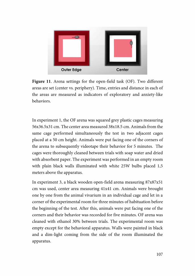
107
Figure 11. Arena settings for the open-field task (OF). Two different areas are set (center vs. periphery). Time, entries and distance in each of the areas are measured as indicators of exploratory and anxiety-like behaviors.
In experiment 1, the OF arena was squared grey plastic cages measuring 56x36.5x31 cm. The center area measured 38x18.5 cm. Animals from the same cage performed simultaneously the test in two adjacent cages placed at a 50 cm height. Animals were put facing one of the corners of the arena to subsequently videotape their behavior for 5 minutes. The cages were thoroughly cleaned between trials with soap water and dried with absorbent paper. The experiment was performed in an empty room with plain black walls illuminated with white 25W bulbs placed 1,5 meters above the apparatus.
In experiment 3, a black wooden open-field arena measuring 87x87x51 cm was used, center area measuring 41x41 cm. Animals were brought one by one from the animal vivarium in an individual cage and let in a corner of the experimental room for three minutes of habituation before the beginning of the test. After this, animals were put facing one of the corners and their behavior was recorded for five minutes. OF arena was cleaned with ethanol 30% between trials. The experimental room was empty except for the behavioral apparatus. Walls were painted in black and a dim-light coming from the side of the room illuminated the apparatus.
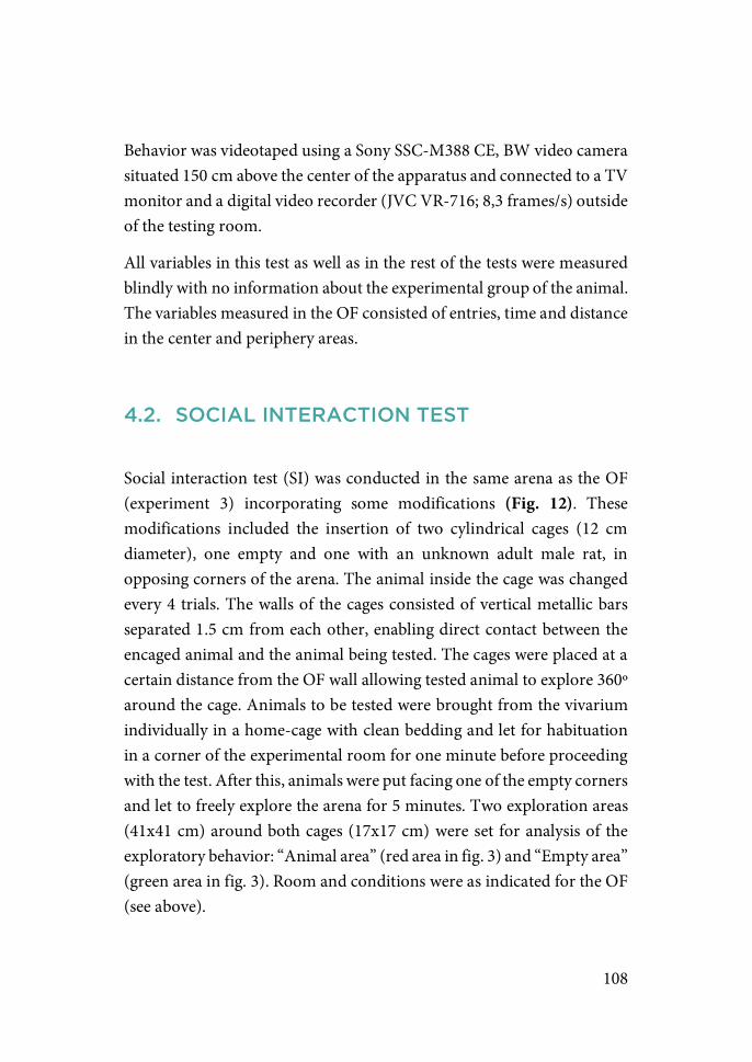
108
Behavior was videotaped using a Sony SSC-M388 CE, BW video camera situated 150 cm above the center of the apparatus and connected to a TV monitor and a digital video recorder (JVC VR-716; 8,3 frames/s) outside of the testing room.
All variables in this test as well as in the rest of the tests were measured blindly with no information about the experimental group of the animal. The variables measured in the OF consisted of entries, time and distance in the center and periphery areas.
4.2.! SOCIAL INTERACTION TEST
Social interaction test (SI) was conducted in the same arena as the OF (experiment 3) incorporating some modifications (Fig. 12). These modifications included the insertion of two cylindrical cages (12 cm diameter), one empty and one with an unknown adult male rat, in opposing corners of the arena. The animal inside the cage was changed every 4 trials. The walls of the cages consisted of vertical metallic bars separated 1.5 cm from each other, enabling direct contact between the encaged animal and the animal being tested. The cages were placed at a certain distance from the OF wall allowing tested animal to explore 360º around the cage. Animals to be tested were brought from the vivarium individually in a home-cage with clean bedding and let for habituation in a corner of the experimental room for one minute before proceeding with the test. After this, animals were put facing one of the empty corners and let to freely explore the arena for 5 minutes. Two exploration areas (41x41 cm) around both cages (17x17 cm) were set for analysis of the exploratory behavior: “Animal area” (red area in fig. 3) and “Empty area” (green area in fig. 3). Room and conditions were as indicated for the OF (see above).
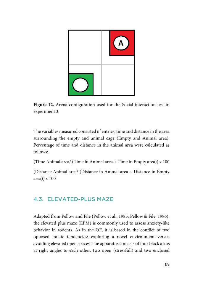
109
Figure 12. Arena configuration used for the Social interaction test in experiment 3.
The variables measured consisted of entries, time and distance in the area surrounding the empty and animal cage (Empty and Animal area). Percentage of time and distance in the animal area were calculated as follows:
(Time Animal area/ (Time in Animal area + Time in Empty area)) x 100
(Distance Animal area/ (Distance in Animal area + Distance in Empty area)) x 100
4.3.! ELEVATED-PLUS MAZE
Adapted from Pellow and File (Pellow et al., 1985; Pellow & File, 1986), the elevated plus maze (EPM) is commonly used to assess anxiety-like behavior in rodents. As in the OF, it is based in the conflict of two opposed innate tendencies: exploring a novel environment versus avoiding elevated open spaces. The apparatus consists of four black arms at right angles to each other, two open (stressfull) and two enclosed
110
(protecting) connected to a central square (10x10 cm) to form the shape of a plus sign (Fig. 13). The prototypical measures include time, distance and entries in the center area, closed and open arms, as well as distance travelled in the whole arena. The percentage of time spent in the open arms is considered as an indicator of anxiety-like behavior. Animals are considered as “anxious” when they spend most of the time in the protected areas of the enclosed arms, whereas animals spending most of the time in the open unprotected arms are considered non-anxious. Nevertheless, as in the case of the OF, the interpretation of the results in the EPM as reflective of anxiety-like behaviors is controversial (Armario & Nadal, 2013).
Figure 13. Illustration of the elevated-plus maze (EPM).
All the apparatus was elevated 50 cm (in experiment 1 and 2) or 70 cm (in experiment 3) above the floor. Each arm was 46 cm long and 10 cm wide. The two opposite enclosed arms had walls of 43 cm of height, whereas the other two open arms had a 0.7 cm ridge to provide an additional grip. Animals were positioned in the center area facing one of the closed arms to begin the test. Behavior was recorded for a period of 5 minutes and the apparatus was cleaned using a solution of soap water
111
(in experiment 1 and 2) or ethanol 30% (experiment 3) between trials. The EPM test was performed and behavior was videotaped in the same room and conditions as the OF.
The variables measured consisted of the number of entries, time and distance in the center, open (OA) and closed arms (CA) areas. Percentage of time in the open arms was calculated as follows:
(Time OA/ (Time OA + Time CA)) x 100
Position tracking system
Data from the OF, SI and EPM was collected using two automatic video tracking systems (Smart 2.5.19, Panlab in experiment 1; EthoVision XT8 video tracking system, Noldus in experiment 3).
Smart software tracks the animal position using a unique reference point based on three possible locations: nose, center and tail base. The tail-base reference point was used for analysis in this project. This option was chosen as this location yielded correlation values higher than 0.8 when compared with results obtained by manual measurement. Manual measures of change of area were counted if all paws crossed to the other area.
In the case of EthoVision, two reference points are employed. One located in the middle part of the body, and a second one in the posterior area of the body (beginning of the tail). A change of area was detected if both points crossed to the adjacent area.
In both cases a smoothing algorithm is applied to eliminate the noise of the tracks from the small movements of the animal (body wobble). The selected algorithm smoothed tracks by calculating the averaged path between two position points of the animal detected with a 1 second interval.

112
4.4.! ACOUSTIC STARTLE TEST
The acoustic startle response (ASR) is a rapid sensorimotor response elicited by a sudden and intense auditory stimulus. It can be modulated by a variety of changes in the perceptual or emotional state of the organism and can be enhanced by conditioned and unconditioned aversive events, making it useful for the study of anxiety-related behaviors (Koch, 1999).
Two identical San Diego Instruments standard startle chambers (SR-LAB) were used to measure ASR (figure 14). Each startle chamber was placed inside a sound-attenuating ventilated box (33×35×33 cm). A white light and a constant 60 dB background white noise was provided by a fan placed inside each chamber during the entire session. Every chamber had a stabilimeter consisting on a clear Plexiglas tube (10.2x20.5 cm) connected to an accelerometer converting any movement from the tube to a resultant voltage proportional to the velocity of the tube displacement. The analog output of the accelerometer was amplified, digitized and interfaced to a Windows computer.
Behavior was videotaped with a camera (KPC-S500P3, KT&C co.). Cameras were positioned 5 cm in front of the stabilimeter. In both cases, cameras were connected to a recorder and a TV monitor outside of the isolation chamber.
Startle responses were evoked by 50-ms bursts of white-noise at various intensities generated by a noise generator and delivered through high frequency speakers located 5 cm from the front of each stabilimeter containing the animal. Stimulus presentation and signal recording (1000 Hz) was controlled by the software package SR-Lab (San Diego Instruments, San Diego, CA, US) under the control of a Windows computer. Sound level measurements were made with a Brüel & Kjaer model 2240 sound-level meter with the microphone located in the front of the stabilimeter, which approximates the distance of the rat’s ear from the speaker during testing. The chambers were calibrated before the
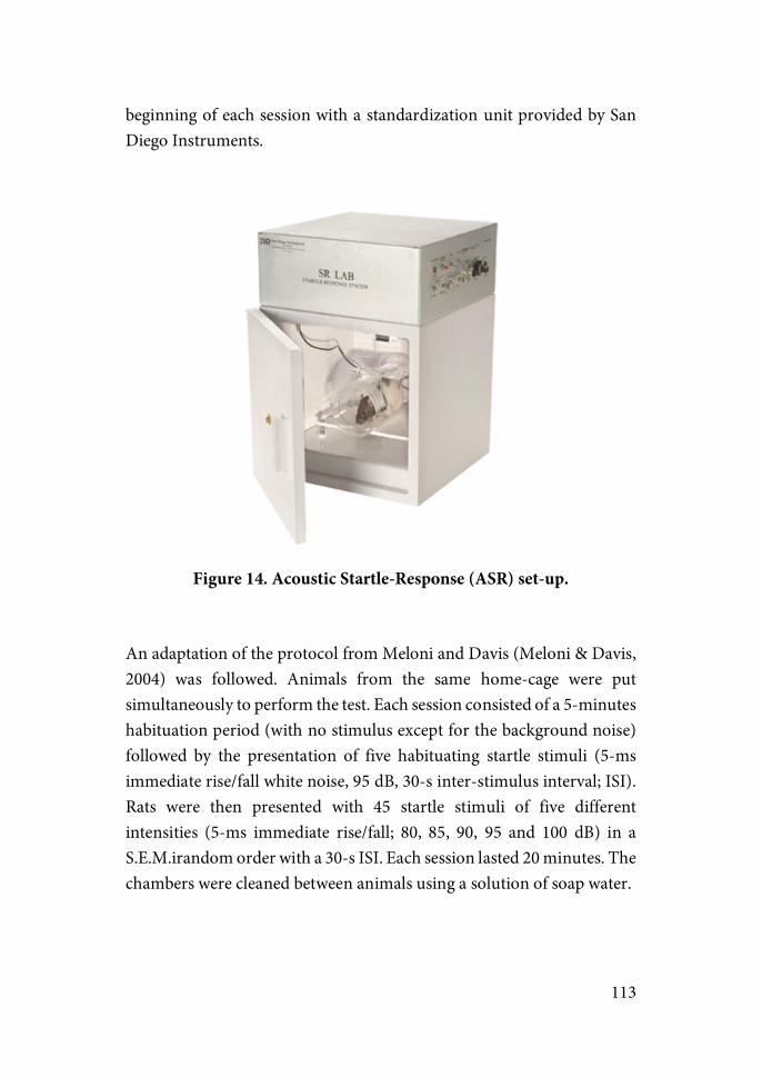
113
beginning of each session with a standardization unit provided by San Diego Instruments.
Figure 14. Acoustic Startle-Response (ASR) set-up.
An adaptation of the protocol from Meloni and Davis (Meloni & Davis, 2004) was followed. Animals from the same home-cage were put simultaneously to perform the test. Each session consisted of a 5-minutes habituation period (with no stimulus except for the background noise) followed by the presentation of five habituating startle stimuli (5-ms immediate rise/fall white noise, 95 dB, 30-s inter-stimulus interval; ISI). Rats were then presented with 45 startle stimuli of five different intensities (5-ms immediate rise/fall; 80, 85, 90, 95 and 100 dB) in a S.E.M.irandom order with a 30-s ISI. Each session lasted 20 minutes. The chambers were cleaned between animals using a solution of soap water.
114
Startle amplitude was defined as the peak accelerometer voltage that occurred during the first 200 ms after onset of the startle stimulus. In addition to startle measurements, baseline activity measurements were made by sampling cage movement (200 ms duration) 15 s after the onset of each startle stimulus.
4.5.! FEAR EXTINCTION TEST
In experiment 3, a fear extinction protocol was conducted 10 days after the end of the TWAA. It consisted of a re-exposure to the TWAA context with three different phases:
-! Pre-tone interval: Three minutes in the TWAA shuttle boxes with no additional stimuli. This period allows the measure of contextual fear memory as no auditory cue is presented during this phase.
-! Extinction interval: 10 tones of 10 seconds with a 30 seconds ITI. -! Post-tone interval: Three minutes with no additional stimuli to
assess the recovery response from the extinction test.
Time of freezing and grooming as well as the number of intercrossings and rearings were analyzed in each of the phases (for a detailed description on the behaviors see TWAA “Data analysis” above).
4.6.! BEHAVIOR ANALYSIS
4.6.1.!Assessment of overall behavioral effect
Overall effects were analyzed as a first approximation to behavioral characterization. Mean, standard deviation (SD) and standard error of
115
the mean (S.E.M.) of the parameters previously indicated were calculated for each experimental group.
4.6.2.!Behavioral profiling
A behavioral profiling approach was employed in experiment 3 to characterize individual differences in response to the stressor. Specifically, the aim was to characterize subjects “affected” by the stress exposure from those “not affected” from it. For that purpose, the behavioral profiling was conducted selecting, based on previous studies, a series of key behavioral parameters indicators of activity and anxiety-like behaviors. “Affected” and “Not-affected” cut-off values were established based on the normal distribution of those parameters in the naïve group. This method was first proposed by Cohen et al. (Cohen et al., 2003) and further developed by the group of Richter-Levin (Ardi et al., 2016; Ariel et al., 2017; Cohen et al., 2004; Horovitz et al., 2014; Ritov et al., 2016).
Specifically, cut-off thresholds for each parameter were determined based on the 20th-80th percentile of the values shown by the stress-naïve group. A list of the selected variables and its respective thresholds is shown in table 3. A subject was identified as “Affected” if values in at least 5 out of the 8 selected variables were out of this “normal behavior window” defined by the behavior shown in naïve group (see Fig.15).
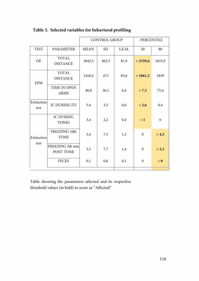
116
Table 3. Selected variables for behavioral profiling
CONTROL GROUP! PERCENTILE!
TEST! PARAMETER! MEAN! SD! S.E.M.! 20! 80!
OF!TOTAL
DISTANCE !3042,5! 463,5! 81,9! < 2559,6! 3433,9!
EPM!
TOTAL DISTANCE !
1418,4! 473! 83,6! < 1061,2! 1839!
TIME IN OPEN ARMS!
40,8! 36,1! 6,4! < 7,3! 75,4!
Extinction test!
IC DURING ITI! 5,4! 3,5! 0,6! < 2,6! 8,4!
Extinction test!!
IC DURING TONES!
3,4! 2,2! 0,4! < 1! 6!
FREEZING 10th TONE!
3,4! 7,5! 1,3! 0! > 4,5!
FREEZING 3th min POST-TONE!
3,3! 7,7! 1,4! 0! > 3,1!
FECES! 0,1! 0,6! 0,1! 0! > 0!
! ! ! ! ! !!
Table showing the parameters selected and its respective threshold values (in bold) to score as “Affected”
!
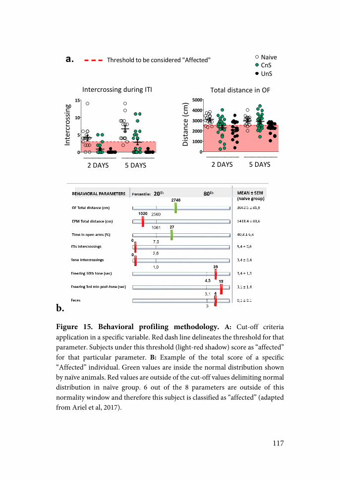
117
b. Figure 15. Behavioral profiling methodology. A: Cut-off criteria application in a specific variable. Red dash line delineates the threshold for that parameter. Subjects under this threshold (light-red shadow) score as “affected” for that particular parameter. B: Example of the total score of a specific “Affected” individual. Green values are inside the normal distribution shown by naïve animals. Red values are outside of the cut-off values delimiting normal distribution in naïve group. 6 out of the 8 parameters are outside of this normality window and therefore this subject is classified as “affected” (adapted from Ariel et al, 2017).
0
5
10
15
Intercrossing+during+ITI
2+DAYS 5+DAYS
Intercrossing
0
1000
2000
3000
4000
5000
Total+distance+in+OF
2+DAYS 5+DAYS
NaiveCnSUnS
Distance+(cm)
a. Threshold+to+be+considered+"Affected"
118
5.! ENDOCRINE ANALYSIS
5.1.! RADIOIMMUNO ASSAY
A blood sample was obtained after the end of the ASR test to assess for possible long-term changes in the reactivity of the HPA response. Double-antibody radioimmunoassays (RIA) were performed to determine ACTH and corticosterone plasma levels.
Levels of ACTH were assessed using the protocol kindly provided by Dr. W.C. Engeland (Department of Surgery, University of Minnesota, Minneapolis) and modified in our lab (García et al., 2000). Buffer consisted on 50 mM dibasic sodium phosphate (pH 7.2) containing 25 mM disodium EDTA, 0.1% Triton X-100 and 0.25% bovine albumin in MilliQ water. All procedures were done at 4ºC to avoid ACTH degradation. ACTH levels were determined using 125I-ACTH (PerkinElmer Life Science, Boston, US) as the tracer, rat synthetic ACTH 1-39 (Sigma, Barcelona, Spain) as the standard and an antibody raised against rat ACTH (Ab Rb 7) kindly provided by Dr. W.C. Engeland. To separate free fraction from the one bound, a second antibody was used (goat anti-rabbit IgG, Chemicon) diluted 1:32, containing non-immunized rabbit serum at 0.3% and polyethylene glycol 9%. After incubation samples were centrifuged at 4700 rpm (Sigma-Laboratory Centrifuges 6K15) for 30 minutes at 4ºC. The sensitivity was of 1 pg/tube in approximately 10 µl of blood volume depending on the concentration.
Corticosterone RIA was conducted using the protocol kindly provided by Dr. G.B. Makara (Institute of Experimental Medicine, Budapest, Hungría) where plasma corticosteroid binding globulin is inactivated by low pH. The buffer employed consisted on 0.2 M sodium phosphate (pH 7.4) dissolved in MilliQ water. Samples were incubated for 2 hours in 0.1M citric acid to release corticosterone from its transporter, the corticosteroid-binding globulin (CBG). Corticosterone levels were
119
determined using 125I-corticosteronecarboxi-methyloxime-tyrosine-methyl ester (MPBiomedicals, Irvine, CA, US) as the tracer, synthetic corticosterone (Sigma, Barcelona, Spain) as the standard, and an antibody raised in rabbits against corticosterone in buffer with 0.15% non-immunized rabbit serum kindly provided by Dr. G. Makara. The free-fraction was separated by means of a secondary antibody (goat anti-rabbit IgG, Chemicon) diluted 1:48 in buffer with polyethylene glycon at 7.5% in MilliQ water and 10 µl/tube of cow serum, followed by 30 minutes of centrifugation at 4700 rpm (4ºC).
All samples to be statistically compared were run in the same assay to avoid inter-assay variability. The intra-assay coefficient of variation was less than 8 % for ACTH and 6 % for corticosterone. The sensitivity was 1 ng/ml using 2 µl of sample.
The radioactivity present in the pellets was measured with a gamma counter (Wallac 1272 Clinigamma).
6.!MOLECULAR ANALYSIS: q-PCR
6.1.! EUTHANASIA AND BRAIN MICROPUNCH PROCEDURE
Animals from the same homecage were euthanized simultaneously to minimize distress before obtaining the brain samples. Brains from animals assigned to perform gene expression analysis were extracted after direct decapitation (2 and 7 days after TWAA in experiment 2; 3 days after extinction test in experiment 3). In experiment 2 decapitation was performed without anesthesia, whereas in experiment 3 a fast isofluorane anesthesia was induced before decapitation. Briefly, in this case animals were put in a camera saturated with isofluorane and as soon as the first signs of anesthesia appeared (eyes closed and loss of leg reflex
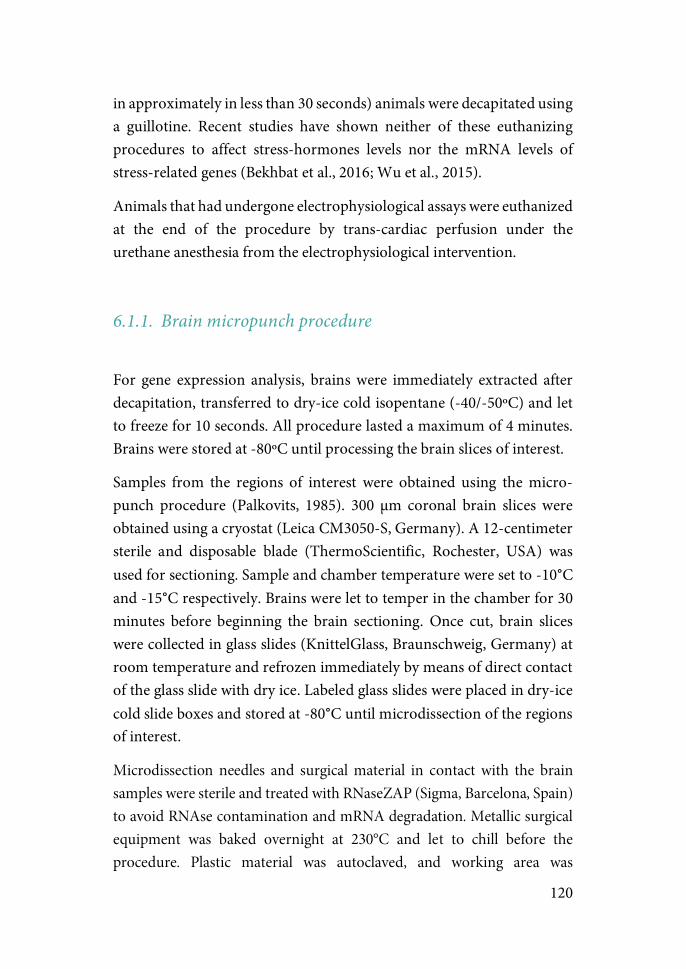
120
in approximately in less than 30 seconds) animals were decapitated using a guillotine. Recent studies have shown neither of these euthanizing procedures to affect stress-hormones levels nor the mRNA levels of stress-related genes (Bekhbat et al., 2016; Wu et al., 2015).
Animals that had undergone electrophysiological assays were euthanized at the end of the procedure by trans-cardiac perfusion under the urethane anesthesia from the electrophysiological intervention.
6.1.1.!Brain micropunch procedure
For gene expression analysis, brains were immediately extracted after decapitation, transferred to dry-ice cold isopentane (-40/-50ºC) and let to freeze for 10 seconds. All procedure lasted a maximum of 4 minutes. Brains were stored at -80ºC until processing the brain slices of interest.
Samples from the regions of interest were obtained using the micro-punch procedure (Palkovits, 1985). 300 µm coronal brain slices were obtained using a cryostat (Leica CM3050-S, Germany). A 12-centimeter sterile and disposable blade (ThermoScientific, Rochester, USA) was used for sectioning. Sample and chamber temperature were set to -10°C and -15°C respectively. Brains were let to temper in the chamber for 30 minutes before beginning the brain sectioning. Once cut, brain slices were collected in glass slides (KnittelGlass, Braunschweig, Germany) at room temperature and refrozen immediately by means of direct contact of the glass slide with dry ice. Labeled glass slides were placed in dry-ice cold slide boxes and stored at -80°C until microdissection of the regions of interest.
Microdissection needles and surgical material in contact with the brain samples were sterile and treated with RNaseZAP (Sigma, Barcelona, Spain) to avoid RNAse contamination and mRNA degradation. Metallic surgical equipment was baked overnight at 230°C and let to chill before the procedure. Plastic material was autoclaved, and working area was
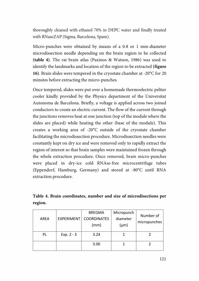
121
thoroughly cleaned with ethanol 70% in DEPC water and finally treated with RNaseZAP (Sigma, Barcelona, Spain).
Micro-punches were obtained by means of a 0.8 or 1 mm-diameter microdissection needle depending on the brain region to be collected (table 4). The rat brain atlas (Paxinos & Watson, 1986) was used to identify the landmarks and location of the region to be extracted (figure 16). Brain slides were tempered in the cryostate chamber at -20°C for 20 minutes before extracting the micro-punches.
Once tempered, slides were put over a homemade thermoelectric peltier cooler kindly provided by the Physics department of the Universitat Autonoma de Barcelona. Briefly, a voltage is applied across two joined conductors to create an electric current. The flow of the current through the junctions removes heat at one junction (top of the module where the slides are placed) while heating the other (base of the module). This creates a working area of -20°C outside of the cryostate chamber facilitating the microdissection procedure. Microdissection needles were constantly kept on dry ice and were removed only to rapidly extract the region of interest so that brain samples were maintained frozen through the whole extraction procedure. Once removed, brain micro-punches were placed in dry-ice cold RNAse-free microcentrifuge tubes (Eppendorf, Hamburg, Germany) and stored at -80°C until RNA extraction procedure.
Table 4. Brain coordinates, number and size of microdisections per region.
AREA" EXPERIMENT"BREGMA"
COORDINATES"
(mm)"
Micropunch"diameter"
(µm)""
Number"of"micropunches"
PL" Exp."2"S"3" 3.24" 1" 2"
"" "" 3.00" 1" 2"
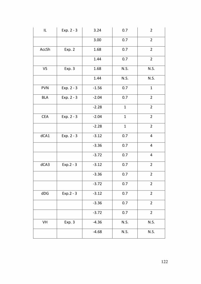
122
IL" Exp."2"S"3" 3.24" 0.7" 2"
"" "" 3.00" 0.7" 2"
AccSh" Exp."2" 1.68" 0.7" 2"
"" "" 1.44" 0.7" 2"
VS" Exp."3" 1.68" N.S." N.S."
"" "" 1.44" N.S." N.S."
PVN" Exp."2"S"3" S1.56" 0.7" 1"
BLA" Exp."2"S"3" S2.04" 0.7" 2"
"" "" S2.28" 1" 2"
CEA" Exp."2"S"3" S2.04" 1" 2"
"" "" S2.28" 1" 2"
dCA1" Exp."2"S"3" S3.12" 0.7" 4"
"" "" S3.36" 0.7" 4"
"" "" S3.72" 0.7" 4"
dCA3" Exp.2"S"3" S3.12" 0.7" 2"
"" "" S3.36" 0.7" 2"
"" "" S3.72" 0.7" 2"
dDG" Exp.2"S"3" S3.12" 0.7" 2"
"" "" S3.36" 0.7" 2"
"" "" S3.72" 0.7" 2"
VH" Exp."3" S4.36" N.S." N.S."
"" "" S4.68" N.S." N.S."
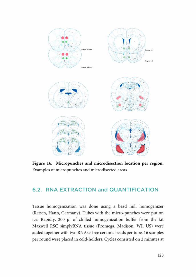
123
Figure 16. Micropunches and microdisection location per region. Examples of micropunches and microdisected areas
6.2.! RNA EXTRACTION and QUANTIFICATION
Tissue homogenization was done using a bead mill homogenizer (Retsch, Hann, Germany). Tubes with the micro-punches were put on ice. Rapidly, 200 µl of chilled homogenization buffer from the kit Maxwell RSC simplyRNA tissue (Promega, Madison, WI, US) were added together with two RNAse-free ceramic beads per tube. 16 samples per round were placed in cold-holders. Cycles consisted on 2 minutes at
124
30 Hz with 1 minute of rest between rounds. Usually one cycle was sufficient to yield a homogenous solution.
RNA was extracted using the automated Maxwell RSC Instrument (Promega, Madison, WI, US). RNA is extracted using paramagnetic particles which provide a mobile solid phase to optimize sample capture, washing and purification of nucleid acids. The samples are automatically processed through a series of washes until nucleic acids are eluted yielding samples of high purification.
A series of samples were selected to assess RNA integrity prior to the qPCR. RNA integrity was verified using Experion Eukaryote Total RNA StdSens Assay (BioRad, Hercules, CA, USA). RNA quality index (RQI, 1 being the most degraded RNA profile, 10 being the most intact RNA) ranged between 8.6 and 9.6, thus validating the maintenance of the RNA structure through the extraction procedure.
Once extracted, RNA was quantified by fluorimetry using the Quantifluor RNA system (Promega, Madison, WI, US).
6.3.! ONE-STEP QUANTITATIVE-PCR (qPCR)
One-step quantitative-PCR for mRNA analysis was performed using the iTaq Universal One-step RT-qPCR kit (Biorad, Hercules, CA, USA), a SYBR green-based protocol where retrotranscription of the RNA and amplification of the resulting cDNA occur directly in the qPCR plate. Instructions supplied by the manufacturer were thoroughly followed.
6.3.1.!Primer design and validation
Oligos were designed using the Primer3 algorithm (Taylor et al., 2010) and the Primer-blast tool (https://www.ncbi.nlm.nih.gov/tools/primer-
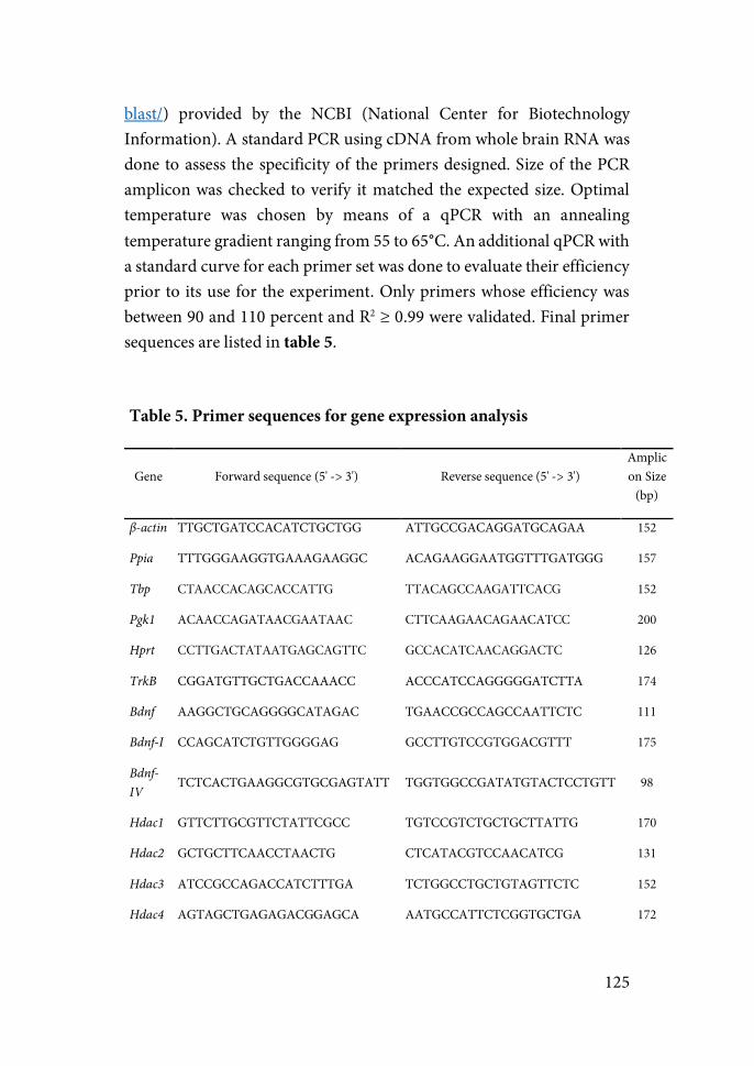
125
blast/) provided by the NCBI (National Center for Biotechnology Information). A standard PCR using cDNA from whole brain RNA was done to assess the specificity of the primers designed. Size of the PCR amplicon was checked to verify it matched the expected size. Optimal temperature was chosen by means of a qPCR with an annealing temperature gradient ranging from 55 to 65°C. An additional qPCR with a standard curve for each primer set was done to evaluate their efficiency prior to its use for the experiment. Only primers whose efficiency was between 90 and 110 percent and R2 ≥ 0.99 were validated. Final primer sequences are listed in table 5.
Table 5. Primer sequences for gene expression analysis
Gene Forward sequence (5' -> 3') Reverse sequence (5' -> 3') Amplicon Size
(bp)
β-actin TTGCTGATCCACATCTGCTGG ATTGCCGACAGGATGCAGAA 152
Ppia TTTGGGAAGGTGAAAGAAGGC ACAGAAGGAATGGTTTGATGGG 157
Tbp CTAACCACAGCACCATTG TTACAGCCAAGATTCACG 152
Pgk1 ACAACCAGATAACGAATAAC CTTCAAGAACAGAACATCC 200
Hprt CCTTGACTATAATGAGCAGTTC GCCACATCAACAGGACTC 126
TrkB CGGATGTTGCTGACCAAACC ACCCATCCAGGGGGATCTTA 174
Bdnf AAGGCTGCAGGGGCATAGAC TGAACCGCCAGCCAATTCTC 111
Bdnf-I CCAGCATCTGTTGGGGAG GCCTTGTCCGTGGACGTTT 175
Bdnf-IV TCTCACTGAAGGCGTGCGAGTATT TGGTGGCCGATATGTACTCCTGTT 98
Hdac1 GTTCTTGCGTTCTATTCGCC TGTCCGTCTGCTGCTTATTG 170
Hdac2 GCTGCTTCAACCTAACTG CTCATACGTCCAACATCG 131
Hdac3 ATCCGCCAGACCATCTTTGA TCTGGCCTGCTGTAGTTCTC 152
Hdac4 AGTAGCTGAGAGACGGAGCA AATGCCATTCTCGGTGCTGA 172
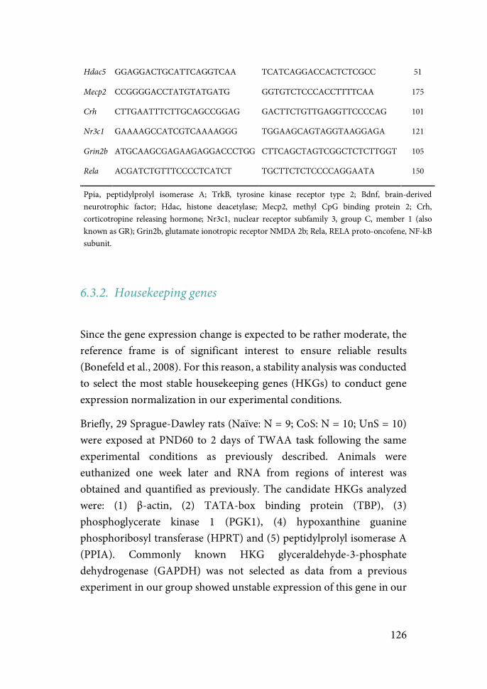
126
Hdac5 GGAGGACTGCATTCAGGTCAA TCATCAGGACCACTCTCGCC 51
Mecp2 CCGGGGACCTATGTATGATG GGTGTCTCCCACCTTTTCAA 175
Crh CTTGAATTTCTTGCAGCCGGAG GACTTCTGTTGAGGTTCCCCAG 101
Nr3c1 GAAAAGCCATCGTCAAAAGGG TGGAAGCAGTAGGTAAGGAGA 121
Grin2b ATGCAAGCGAGAAGAGGACCCTGG CTTCAGCTAGTCGGCTCTCTTGGT 105
Rela ACGATCTGTTTCCCCTCATCT TGCTTCTCTCCCCAGGAATA 150
Ppia, peptidylprolyl isomerase A; TrkB, tyrosine kinase receptor type 2; Bdnf, brain-derived neurotrophic factor; Hdac, histone deacetylase; Mecp2, methyl CpG binding protein 2; Crh, corticotropine releasing hormone; Nr3c1, nuclear receptor subfamily 3, group C, member 1 (also known as GR); Grin2b, glutamate ionotropic receptor NMDA 2b; Rela, RELA proto-oncofene, NF-kB subunit.
6.3.2.!Housekeeping genes
Since the gene expression change is expected to be rather moderate, the reference frame is of significant interest to ensure reliable results (Bonefeld et al., 2008). For this reason, a stability analysis was conducted to select the most stable housekeeping genes (HKGs) to conduct gene expression normalization in our experimental conditions.
Briefly, 29 Sprague-Dawley rats (Naïve: N = 9; CoS: N = 10; UnS = 10) were exposed at PND60 to 2 days of TWAA task following the same experimental conditions as previously described. Animals were euthanized one week later and RNA from regions of interest was obtained and quantified as previously. The candidate HKGs analyzed were: (1) β-actin, (2) TATA-box binding protein (TBP), (3) phosphoglycerate kinase 1 (PGK1), (4) hypoxanthine guanine phosphoribosyl transferase (HPRT) and (5) peptidylprolyl isomerase A (PPIA). Commonly known HKG glyceraldehyde-3-phosphate dehydrogenase (GAPDH) was not selected as data from a previous experiment in our group showed unstable expression of this gene in our
127
experimental paradigm. For primer sequences see table 1 (in ‘Primer design and validation’).
NormFinder software was used to perform the analysis (Andersen et al., 2004). Combined estimates of intra- and intergroup values are used by the software to calculate a stability value. NormFinder determines the optimal reference gene among the HKGs and additionally suggests the best combination of two HKGs, where the geometric average between these may be used for normalization.
Results of the analysis revealed β-actin to be the most stable HKG (stability value: 0.14), whereas the best combination of HKGs was for β-actin and PPIA (stability value: 0.11). Expression data from the experiment was thus obtained normalizing with both HKGs, β-actin and PPIA.
6.3.3.! Samples, controls and qPCR plate
Briefly, 384-well plates were designed containing: (1) All samples from the region to be analyzed; (2) 4-points stardard curve ranging from 100 to 0.1 ng of RNA; (3) Non-template controls (NTC), which controls for RNA contamination of the reagents and consists of wells where all reagents are added except for the RNA sample whose volume is replaced by nuclease-free water; (4) Non-retrostranscription controls (NRT), which serves as a control for possible DNA contamination of the samples. All reagents except for the retrotranscriptase are added; and (5) an inter-plate control (IP-CTR), a sample to be used as a general control of amplification in case repetitions of some samples should be made in a different plate. All samples were run in triplicates. The details of the PCR reactions are shown in table 6.
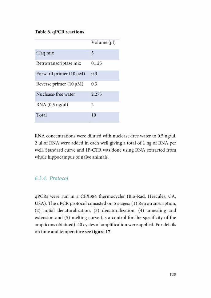
128
Table 6. qPCR reactions
Volume (µl)
iTaq mix 5
Retrotranscriptase mix 0.125
Forward primer (10 µM) 0.3
Reverse primer (10 µM) 0.3
Nuclease-free water 2.275
RNA (0.5 ng/µl) 2
Total 10
RNA concentrations were diluted with nuclease-free water to 0.5 ng/µl. 2 µl of RNA were added in each well giving a total of 1 ng of RNA per well. Standard curve and IP-CTR was done using RNA extracted from whole hippocampus of naïve animals.
6.3.4.!Protocol
qPCRs were run in a CFX384 thermocycler (Bio-Rad, Hercules, CA, USA). The qPCR protocol consisted on 5 stages: (1) Retrotranscription, (2) initial denaturalization, (3) denaturalization, (4) annealing and extension and (5) melting curve (as a control for the specificity of the amplicons obtained). 40 cycles of amplification were applied. For details on time and temperature see figure 17.
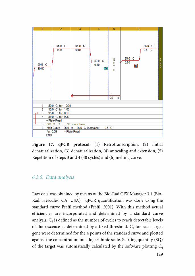
129
Figure 17. qPCR protocol: (1) Retrotranscription, (2) initial denaturalization, (3) denaturalization, (4) annealing and extension, (5) Repetition of steps 3 and 4 (40 cycles) and (6) melting curve.
6.3.5.!Data analysis
Raw data was obtained by means of the Bio-Rad CFX Manager 3.1 (Bio-Rad, Hercules, CA, USA). qPCR quantification was done using the standard curve Pfaffl method (Pfaffl, 2001). With this method actual efficiencies are incorporated and determined by a standard curve analysis. Cq is defined as the number of cycles to reach detectable levels of fluorescence as determined by a fixed threshold. Cq for each target gene were determined for the 4 points of the standard curve and plotted against the concentration on a logarithmic scale. Starting quantity (SQ) of the target was automatically calculated by the software plotting Cq
130
values of the unknown samples against the concentration on the corresponding standard curve. SQs are normalized by two factors: (i) HKG factor, which corrects for the small margin of error in measures of concentration and inherent deviation in volume pipetting; and (ii) by the expression of the control group, giving the difference in expression levels attributed to the experimental treatment.
7.! STATISTICAL ANALYSIS
Between-subject factors analysed include:
Effect of PHENOTYPE- refers to: (i) Among animals exposed to controllable stress, the effects derived from differences in the level of avoidance conditioning acquired and/or the different amount of total shock received due to this difference in learning (HA vs. LA); (ii) Among subjects exposed to uncontrollable stress in the yoked configuration (UnS-HA vs. UnS-LA), the effects derived from differences in the total shock received.
Effect of CONTROLLABILITY- refers to differences arising from exposure to controllable (HA-LA) vs. uncontrollable stress (UnS-HA vs. UnS-LA).
Effect of TIME POINT- refers to differences between values observed at 2 days vs. 7 days post-TWAA.
Effect of GROUP- refers to differences when comparing Naïve, HA, LA and UnS in experiment 3 where all animals exposed to uncontrollable stress receive the same amount of shock (equivalent to the averaged shock duration received by CoS subjects).
Effect of NUMBER OF SESSIONS (N.Sessions)- refers to differences observed when comparing animals exposed to 2-days vs. 5-days of TWAA.
131
Intra-subject factors include:
Effect of BLOCK- It refers to: (i) differences when comparing blocks of 10 trials in the TWAA; (ii) differences when comparing blocks of one minute in the extinction test.
Effect of DAYS- differences observed between values in different days.
Statistical analysis was done using the Statistical Package for Social Science software (IBM SPSS version 18 for Windows. Armonk, NY: IBM Corp.). Variance homogeneity by means of Levene’s (in single-point analysis) or Box’s T test (in repeated measures) was verified in all cases. If true, one or two-way analysis of variances (ANOVA) depending on the number of factors, were conducted. In case of non-fulfilment of variance homogeneity, a generalized linear model (GENLIN) was used for one-single point analysis (Mcculloch and Searle, 2001), whereas a generalized estimating equations model (GEE) was used to analyze repeated measures (Hardin and Hilbe, 2003). Maximum likelihood (ML) was used as the method of estimation. Within and between-subject factors used are indicated in each experiment. Pairwise comparisons were made when statistical interactions were found by means of post-hoc sequential Bonferroni corrections. Significant effects were determined by the Wald chi-square statistic. In all tests significance was established at p ≤ 0.05, although 0.05 < p < 0.06 were indicated as a tendency, and interactions where 0.05 < p < 0.08 were validated for conducting pairwise comparisons.
GENLIN and GEE models were chosen as the general statistical tools because of its higher flexibility compared to general linear models. The former models offer three main advantages: (1) they allow statistical analysis without homogeneity of variance, (2) missing data does not preclude the analysis and (3) you can choose between several types of distribution of your data (normal, binomial, Poisson, gamma or inverse-Gaussian).
Graphs were plotted using Graph-Pad Prism (version 5 for Windows, GraphPad Software, La Jolla, California, EEUU).
132
RESULTS
CHAPTER ONE: Long-term effects of acute exposure to TWAA

133
EXPERIMENT 1: SHORT AND LONG-TERM BEHAVIORAL EFFECTS OF ACUTE EXPOSURE TO CONTROLLABLE OR UNCONTROLLABLE STRESS
The aim of this study was to characterize the short and long-term behavioral consequences of acute exposure to controllable (CoS) or uncontrollable stress (UnS) in the context of a two-way active avoidance task in a shuttle box (TWAA). To this end, animals (n: naïve = 12; controllable stress or CoS = 16; yoked-uncontrollable stress or UnS = 16) went through an EPM, OF and an ASR test 2, 5 and 7 days respectively after the end of two sessions of TWAA (Fig. 18).
Animals in the CoS group could avoid or escape from the shock by shuttling to the adjacent chamber during the CS or US respectively. On the other hand, UnS animals were yoked to a CoS animal whose performance dictated the termination of the shock for both animals. The performance of the yoked animal did not determine in any way the appearance or termination of the shocks. Therefore, both groups received the same amount of physical stressor (e.g. electricfoot-shock), the perception of control being the only difference between both groups. Naïve animals were exposed to the TWAA chamber during the same time as CoS and UnS animals but with no shock presentation.
Figure 18. General design of the experiment. Animals were exposed to two non-consecutive days of TWAA of 50 trials per session. A battery of behavioral
134
tests consisting on an EPM, OF and an ASR test were conducted 2, 5 and 7 days respectively after the end of the TWAA task. A blood sample was taken at the end of ASR test to measure corticosterone and ACTH in response to this test.
Active avoidance acquisition: High and Low avoiders
Acquisition of active avoidance conditioning was analyzed in the CoS group. The number of avoidance, escape and failure responses was measured in blocks of 10 trials (Fig. 19). Results were analyzed by a repeated measures ANOVA with two within-subjects factors: Day and Block.
Escape responses was found in the 85.6 ± 14.6% of the trials from the first block. In the same block avoidance responses represented 4.4 ± 6.3% and increased until reaching 9.4 ± 13.4% at the end of the first session. By the end of the final session both responses reached similar levels (Escapes: 46.3%; Avoidances: 41.9%) with a SD of 40% in both parameters.
With respect to avoidance responses, results showed a significant effect of Day (F(1,15) = 13.4 p = 0.002), Block (F(4,60) = 5.4 p = 0.001) and Day x Block interaction (F(4,60) = 3.5 p = 0.012). No effect of Block was present on day 1 (F(4,60) = 1.2 p = 0.336), but it was found on day 2 (F(4,60) = 5.5 p = 0.001).
Regarding escape responses, analysis revealed a significant effect of Block (F(4,60) = 6.1 p = 0.000) and Day x Block interaction (F(4,60) = 4.6 p = 0.003). Effect of Block was present on day 2 (F(4,60) = 8.32 p = 0.000), but not on day 1 (F(4,60) = 0.4 p = 0.838).
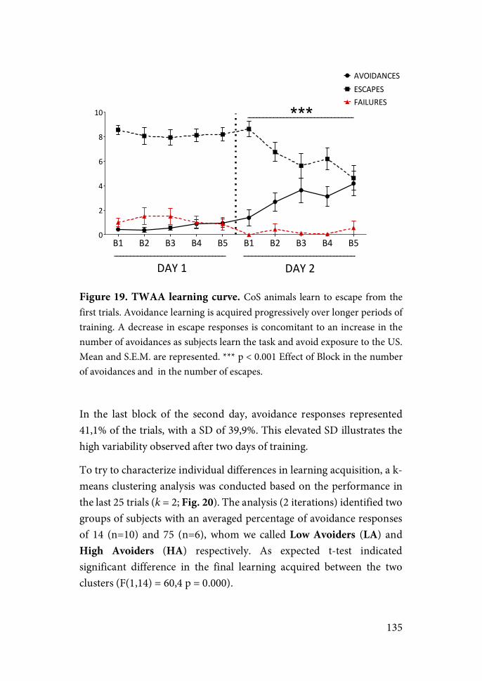
135
Figure 19. TWAA learning curve. CoS animals learn to escape from the first trials. Avoidance learning is acquired progressively over longer periods of training. A decrease in escape responses is concomitant to an increase in the number of avoidances as subjects learn the task and avoid exposure to the US. Mean and S.E.M. are represented. *** p < 0.001 Effect of Block in the number of avoidances and in the number of escapes.
In the last block of the second day, avoidance responses represented 41,1% of the trials, with a SD of 39,9%. This elevated SD illustrates the high variability observed after two days of training.
To try to characterize individual differences in learning acquisition, a k-means clustering analysis was conducted based on the performance in the last 25 trials (k = 2; Fig. 20). The analysis (2 iterations) identified two groups of subjects with an averaged percentage of avoidance responses of 14 (n=10) and 75 (n=6), whom we called Low Avoiders (LA) and High Avoiders (HA) respectively. As expected t-test indicated significant difference in the final learning acquired between the two clusters (F(1,14) = 60,4 p = 0.000).
B1 B2 B3 B4 B5 B1 B2 B3 B4 B50
2
4
6
8
10
AVOIDANCESESCAPESFAILURES
DAY91 DAY92
***
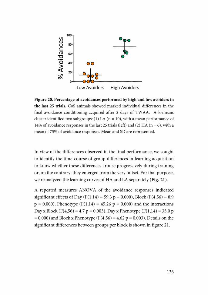
136
Figure 20. Percentage of avoidances performed by high and low avoiders in the last 25 trials. CoS animals showed marked individual differences in the final avoidance conditioning acquired after 2 days of TWAA. A k-means cluster identified two subgroups: (1) LA (n = 10), with a mean performance of 14% of avoidance responses in the last 25 trials (left) and (2) HA (n = 6), with a mean of 75% of avoidance responses. Mean and SD are represented.
In view of the differences observed in the final performance, we sought to identify the time-course of group differences in learning acquisition to know whether these differences arouse progressively during training or, on the contrary, they emerged from the very outset. For that purpose, we reanalyzed the learning curves of HA and LA separately (Fig. 21).
A repeated measures ANOVA of the avoidance responses indicated significant effects of Day (F(1,14) = 59.3 p = 0.000), Block (F(4,56) = 8.9 p = 0.000), Phenotype (F(1,14) = 45.26 p = 0.000) and the interactions Day x Block (F(4,56) = 4.7 p = 0.003), Day x Phenotype (F(1,14) = 33.0 p = 0.000) and Block x Phenotype (F(4,56) = 4.62 p = 0.003). Details on the significant differences between groups per block is shown in figure 21.
Low$Avoiders High$Avoiders0
20
40
60
80
100
%$Avoidances
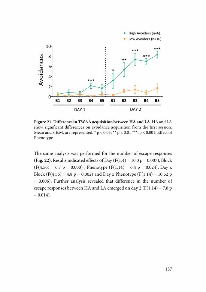
137
Figure 21. Difference in TWAA acquisition between HA and LA. HA and LA show significant differences on avoidance acquisition from the first session. Mean and S.E.M. are represented. * p < 0.05; ** p < 0.01 ***; p < 0.001: Effect of Phenotype.
The same analysis was performed for the number of escape responses (Fig. 22). Results indicated effects of Day (F(1,4) = 10.0 p = 0.007), Block (F(4,56) = 6.7 p = 0.000) , Phenotype (F(1,14) = 6.4 p = 0.024), Day x Block (F(4,56) = 4.8 p = 0.002) and Day x Phenotype (F(1,14) = 10.52 p = 0.006). Further analysis revealed that difference in the number of escape responses between HA and LA emerged on day 2 (F(1,14) = 7.8 p = 0.014).
B1 B2 B3 B4 B5 B1 B2 B3 B4 B50
2
4
6
8
10
High+Avoiders+(n=6)Low+Avoiders+(n=10)
DAY+1 DAY+2
***
*
*****
******
Avoida
nces
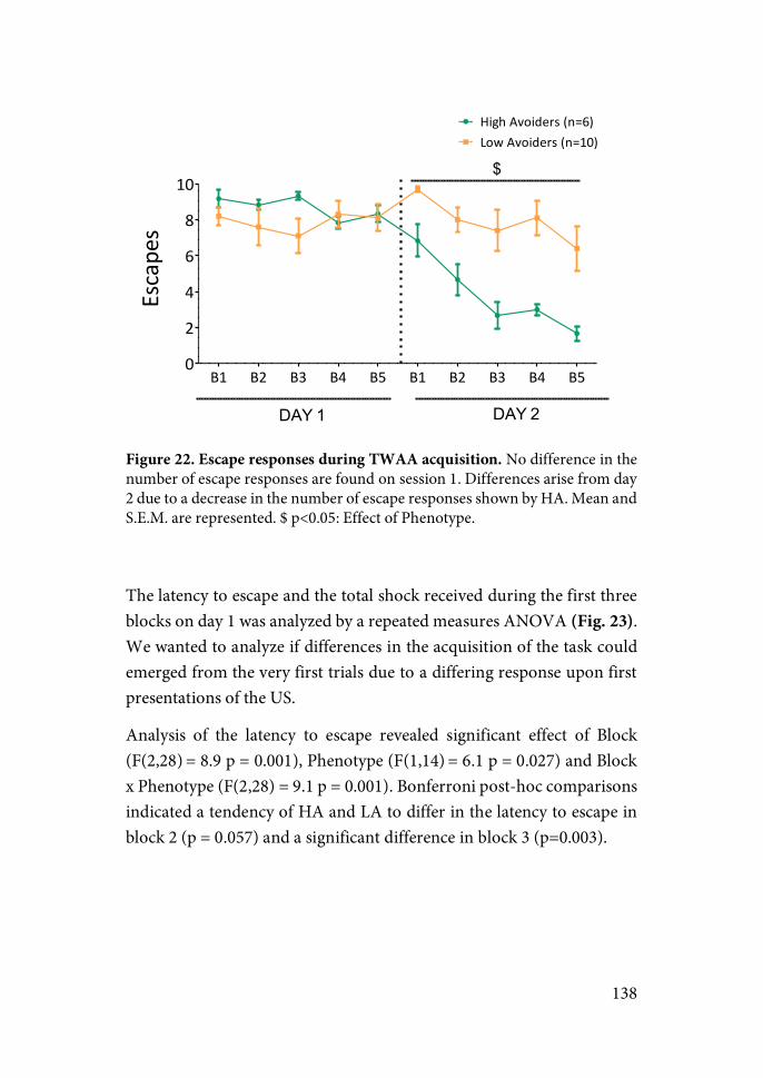
138
Figure 22. Escape responses during TWAA acquisition. No difference in the number of escape responses are found on session 1. Differences arise from day 2 due to a decrease in the number of escape responses shown by HA. Mean and S.E.M. are represented. $ p<0.05: Effect of Phenotype.
The latency to escape and the total shock received during the first three blocks on day 1 was analyzed by a repeated measures ANOVA (Fig. 23). We wanted to analyze if differences in the acquisition of the task could emerged from the very first trials due to a differing response upon first presentations of the US.
Analysis of the latency to escape revealed significant effect of Block (F(2,28) = 8.9 p = 0.001), Phenotype (F(1,14) = 6.1 p = 0.027) and Block x Phenotype (F(2,28) = 9.1 p = 0.001). Bonferroni post-hoc comparisons indicated a tendency of HA and LA to differ in the latency to escape in block 2 (p = 0.057) and a significant difference in block 3 (p=0.003).
B1 B2 B3 B4 B5 B1 B2 B3 B4 B50
2
4
6
8
10
High.Avoiders.(n=6)Low.Avoiders.(n=10)
DAY$1 DAY$2
$Escapes
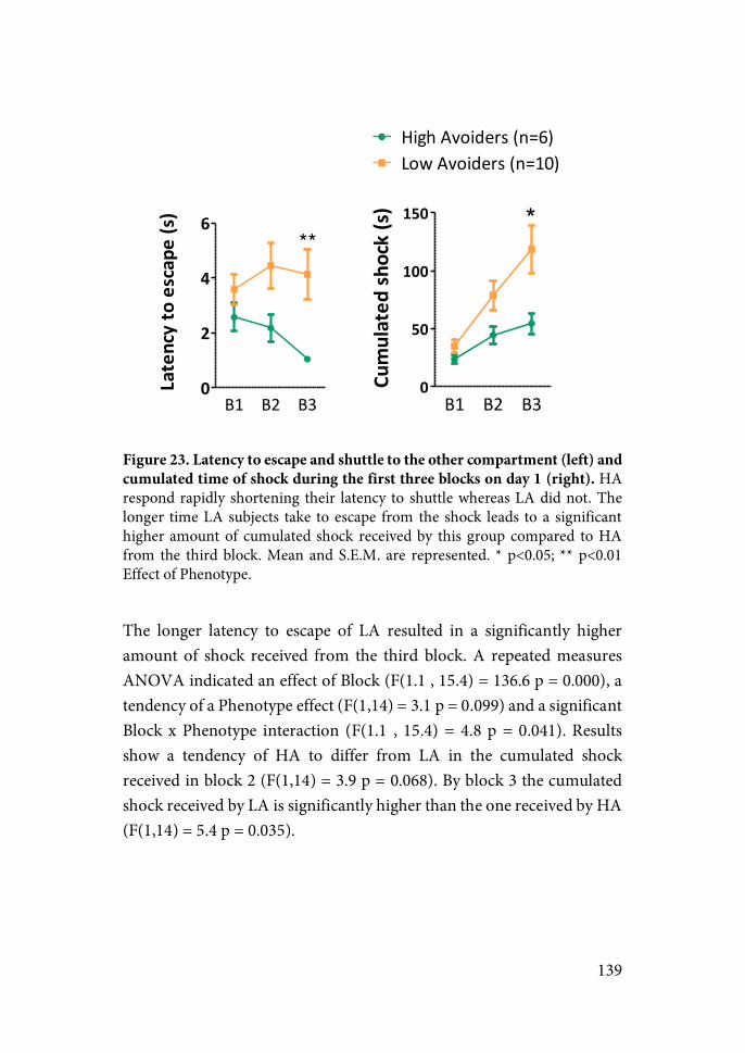
139
Figure 23. Latency to escape and shuttle to the other compartment (left) and cumulated time of shock during the first three blocks on day 1 (right). HA respond rapidly shortening their latency to shuttle whereas LA did not. The longer time LA subjects take to escape from the shock leads to a significant higher amount of cumulated shock received by this group compared to HA from the third block. Mean and S.E.M. are represented. * p<0.05; ** p<0.01 Effect of Phenotype.
The longer latency to escape of LA resulted in a significantly higher amount of shock received from the third block. A repeated measures ANOVA indicated an effect of Block (F(1.1 , 15.4) = 136.6 p = 0.000), a tendency of a Phenotype effect (F(1,14) = 3.1 p = 0.099) and a significant Block x Phenotype interaction (F(1.1 , 15.4) = 4.8 p = 0.041). Results show a tendency of HA to differ from LA in the cumulated shock received in block 2 (F(1,14) = 3.9 p = 0.068). By block 3 the cumulated shock received by LA is significantly higher than the one received by HA (F(1,14) = 5.4 p = 0.035).
B1 B2 B30
2
4
6**
Latency,to,escape,(s)
B1 B2 B30
50
100
150
High)Avoiders)(n=6)Low)Avoiders)(n=10)
*
Cumulated
,shock,(s)

140
Contextual fear memory is affected by controllability
Contextual fear memory (CFC) was assessed by measuring the freezing levels and the number of intercrossings (IC) shown during the habituation period of the second session (Fig 24).
Due to the yoked-protocol employed for the experiment and in view of the difference observed in the total-shock received by HA and LA, animals in the UnS group were also split into “UnS-HA” (U-HA; UnS animals yoked to HA) and “UnS-LA” animals (U-LA; UnS animals yoked paired with LA) accordingly.
Freezing levels during the habituation period of the second session day were analyzed by a two-way ANOVA with two main factors: Controllability (CoS vs. UnS) and Phenotype (HA/U-HA vs. LA/U-LA). Results indicated an effect of Controllability (F(1,28) = 6.3 p = 0.018), Phenotype (F(1,28) = 11.3 p = 0.002) and a tendency to an interaction (F(1,28) = 3.8 p = 0.062). Sequential bonferroni analysis showed a significant difference in contextual fear memory between HA and all the other groups (LA: p = 0.001; U-HA: p = 0.009; U-LA: p = 0.000).
The same pattern was observed when measuring the number of IC performed during the habituation period on day 2. GzLM results revealed a significant effect of Controllability (χ2(1) = 9.2 p = 0.002) and Phenotype (χ2(1) = 10.2 p = 0.001). A tendency to an interaction was also found (χ2(1) = 2.8 p = 0.096), therefore post-hoc analysis were conducted. Pairwise comparisons indicated significant differences between HA and all remaining groups (LA: p = 0.001; U-HA: p = 0.003; U-LA: p = 0.000).
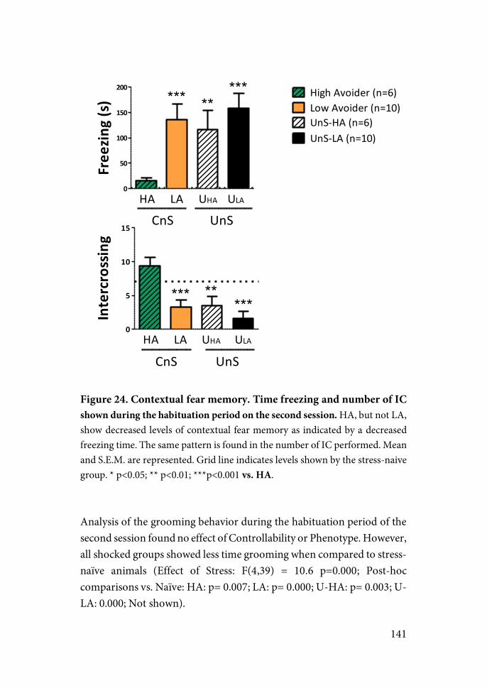
141
Figure 24. Contextual fear memory. Time freezing and number of IC shown during the habituation period on the second session. HA, but not LA, show decreased levels of contextual fear memory as indicated by a decreased freezing time. The same pattern is found in the number of IC performed. Mean and S.E.M. are represented. Grid line indicates levels shown by the stress-naive group. * p<0.05; ** p<0.01; ***p<0.001 vs. HA.
Analysis of the grooming behavior during the habituation period of the second session found no effect of Controllability or Phenotype. However, all shocked groups showed less time grooming when compared to stress-naïve animals (Effect of Stress: F(4,39) = 10.6 p=0.000; Post-hoc comparisons vs. Naïve: HA: p= 0.007; LA: p= 0.000; U-HA: p= 0.003; U-LA: 0.000; Not shown).
0
50
100
150
200***
CnSHA LA UHA ULA
*****
UnS
High+Avoider+(n=6)Low+Avoider+(n=10)UnS8HA+(n=6)UnS8LA+(n=10)
Free
zing,(s)
0
5
10
15
***
CnSHA LA UHA ULA
*****
UnS
Intercrossing

142
During inter-trial intervals (ITIs), the analysis of the number of IC was restricted to CoS animals due to a limitation in the Packwin software employed in the procedure, which prevented the appropriate acquisition of this variable in UnS animals. Analysis of the CoS groups by a repeated measures ANOVA (Fig. 25) indicated an effect of Day (F(1,14) = 12.2 p = 0.004), Phenotype and Day x Phenotype interaction (F(1,14) = 9.5 p = 0.008). While the number of IC was the same in the two groups on day 1 (p = 0.525), HA showed a higher IC than LA on day 2 (p = 0.012).
Figure 25. Number of intercrossings performed during ITIs. Differences between HA and LA in the number of IC performed during ITIs emerge on day 2. Mean and S.E.M. are represented. * p<0.05 Effect of Phenotype. The criteria for assessing individual differences in CoS subjects was based on the final learning acquired (i.e. last 25 trials of the last day), and differences on avoidance learning were only significant in the 4th block of the first day. Therefore, differences in contextual fear memory observed during the habituation period on the second day could be a consequence of the differing learning on day 1, but also a causal factor for the learning differences shown during the second (final) session. A significant negative correlation was found between the freezing shown during the habituation period on day 2 and the number of avoidance
Day$1 Day$20
10
20
30 High$Avoider$(n=11)Low$Avoider$(n=9)*
Intercrossings
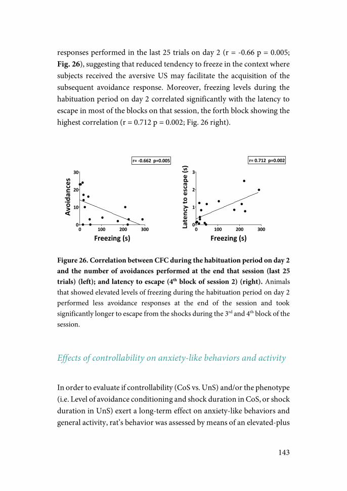
143
responses performed in the last 25 trials on day 2 (r = -0.66 p = 0.005; Fig. 26), suggesting that reduced tendency to freeze in the context where subjects received the aversive US may facilitate the acquisition of the subsequent avoidance response. Moreover, freezing levels during the habituation period on day 2 correlated significantly with the latency to escape in most of the blocks on that session, the forth block showing the highest correlation (r = 0.712 p = 0.002; Fig. 26 right).
Figure 26. Correlation between CFC during the habituation period on day 2 and the number of avoidances performed at the end that session (last 25 trials) (left); and latency to escape (4th block of session 2) (right). Animals that showed elevated levels of freezing during the habituation period on day 2 performed less avoidance responses at the end of the session and took significantly longer to escape from the shocks during the 3rd and 4th block of the session.
Effects of controllability on anxiety-like behaviors and activity
In order to evaluate if controllability (CoS vs. UnS) and/or the phenotype (i.e. Level of avoidance conditioning and shock duration in CoS, or shock duration in UnS) exert a long-term effect on anxiety-like behaviors and general activity, rat’s behavior was assessed by means of an elevated-plus
0 100 200 3000
10
20
30
r='(0.662''p=0.005
Freezing'(s)
Avoida
nces
0 100 200 3000
1
2
3
r='0.712''p=0.002
Freezing'(s)
Latency'to'escap
e'(s)

144
maze (EPM) and an open-field test (OF) 2 and 5 days respectively after the end of the TWAA task.
A two-way ANOVA with Controllability (CoS vs. UnS) and Phenotype (HA/U-HA vs. LA/U-LA) as main factors was run to analyze the EPM (Fig. 27). Regarding the percentage of time spent in the open arms, results revealed a significant effect of Phenotype (F(1,28) = 6.1 p = 0.020) but not of Controllability (F(1,28) = 1.8 p = 0.186). A similar pattern was found regarding the distance run on the open arms: significant effect of Phenotype (F(1,28) = 10.4 p = 0.003) but not of Controllability (F(1,28) = 2.2 p = 0.149). In the case of the distance traveled on the closed arms, results indicated a tendency of a Phenotype effect (F(1,28) = 4.1 p = 0.052) but no effect of Controllability or interaction (Not shown). Finally, analysis of the total distance traveled during the test revealed an effect of the Phenotype (F(1,28) = 11.2 p = 0.002) but not of the controllability of the stressor (F(1,28) = 2.15 p = 0.154). In sum, the main parameters analyzed in EPM showed an effect of Phenotype but not of Controllability, suggesting a relationship to the amount of shock received.
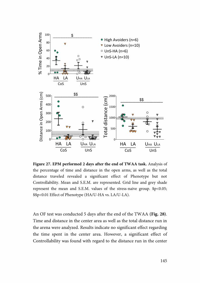
145
Figure 27. EPM performed 2 days after the end of TWAA task. Analysis of the percentage of time and distance in the open arms, as well as the total distance traveled revealed a significant effect of Phenotype but not Controllability. Mean and S.E.M. are represented. Grid line and grey shade represent the mean and S.E.M. values of the stress-naive group. $p<0.05; $$p<0.01 Effect of Phenotype (HA/U-HA vs. LA/U-LA).
An OF test was conducted 5 days after the end of the TWAA (Fig. 28). Time and distance in the center area as well as the total distance run in the arena were analyzed. Results indicate no significant effect regarding the time spent in the center area. However, a significant effect of Controllability was found with regard to the distance run in the center
0
20
40
60
80
100
CoS UnSHA LA UHA ULA
High2Avoiders2(n=6)
UnS;LA2(n=10)
Low2Avoiders2(n=10)UnS;HA2(n=6)
$%2Time2in2Open2Arms
0
100
200
300
400
500 $$
CoS UnSHA LA UHA ULA
Distance2in2Ope
n2Arms2(cm
)
0
500
1000
1500
2000$$
CoS UnSHA LA UHA ULA
Total2distance2(cm)
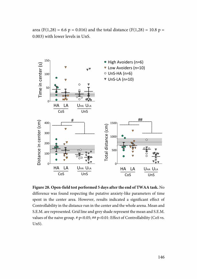
146
area (F(1,28) = 6.6 p = 0.016) and the total distance (F(1,28) = 10.8 p = 0.003) with lower levels in UnS.
Figure 28. Open-field test performed 5 days after the end of TWAA task. No difference was found respecting the putative anxiety-like parameters of time spent in the center area. However, results indicated a significant effect of Controllability in the distance run in the center and the whole arena. Mean and S.E.M. are represented. Grid line and grey shade represent the mean and S.E.M. values of the naive group. # p<0.05; ## p<0.01: Effect of Controllability (CoS vs. UnS).
0
50
100
150 High(Avoiders((n=6)
UnS7LA((n=10)
Low(Avoiders((n=10)UnS7HA((n=6)
CoS UnSHA LA UHA ULA
Time(in(ce
nter((s)
0
100
200
300
400#
CoS UnSHA LA UHA ULA
Distance(in(cen
ter((cm
)
0
500
1000
1500##
CoS UnSHA LA UHA ULA
Total(distance((cm)
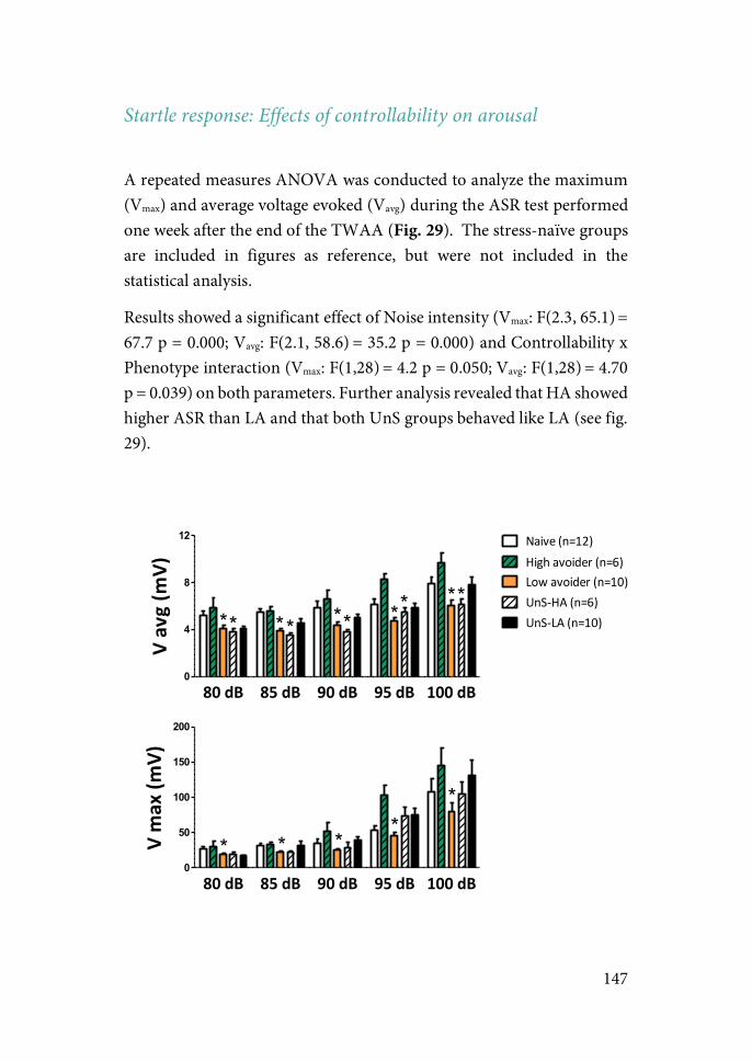
147
Startle response: Effects of controllability on arousal
A repeated measures ANOVA was conducted to analyze the maximum (Vmax) and average voltage evoked (Vavg) during the ASR test performed one week after the end of the TWAA (Fig. 29). The stress-naïve groups are included in figures as reference, but were not included in the statistical analysis.
Results showed a significant effect of Noise intensity (Vmax: F(2.3, 65.1) = 67.7 p = 0.000; Vavg: F(2.1, 58.6) = 35.2 p = 0.000) and Controllability x Phenotype interaction (Vmax: F(1,28) = 4.2 p = 0.050; Vavg: F(1,28) = 4.70 p = 0.039) on both parameters. Further analysis revealed that HA showed higher ASR than LA and that both UnS groups behaved like LA (see fig. 29).
80#dB 85#dB 90#dB 95#dB 100#dB0
4
8
12 Naive&(n=12)High&avoider&(n=6)Low&avoider&(n=10)UnS9HA&(n=6)UnS9LA&(n=10)* * *
* ******
V#avg#(m
V)
80#dB 85#dB 90#dB 95#dB 100#dB0
50
100
150
200
* * **
*
V#max#(m
V)
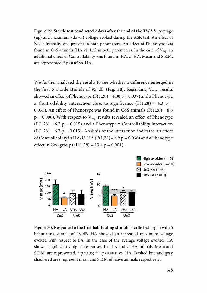
148
Figure 29. Startle test conducted 7 days after the end of the TWAA. Average (up) and maximum (down) voltage evoked during the ASR test. An effect of Noise intensity was present in both parameters. An effect of Phenotype was found in CoS animals (HA vs. LA) in both parameters. In the case of Vavg, an additional effect of Controllability was found in HA/U-HA. Mean and S.E.M. are represented. * p<0.05 vs. HA.
We further analyzed the results to see whether a difference emerged in the first 5 startle stimuli of 95 dB (Fig. 30). Regarding Vmax, results showed an effect of Phenotype (F(1,28) = 4.80 p = 0.037) and a Phenotype x Controllability interaction close to significance (F(1,28) = 4.0 p = 0.055). An effect of Phenotype was found in CoS animals (F(1,28) = 8.8 p = 0.006). With respect to Vavg, results revealed an effect of Phenotype (F(1,28) = 6.7 p = 0.015) and a Phenotype x Controllability interaction (F(1,28) = 6.7 p = 0.015). Analysis of the interaction indicated an effect of Controllability in HA/U-HA (F(1,28) = 4.9 p = 0.036) and a Phenotype effect in CoS groups (F(1,28) = 13.4 p = 0.001).
Figure 30. Response to the first habituating stimuli. Startle test began with 5 habituating stimuli of 95 dB. HA showed an increased maximum voltage evoked with respect to LA. In the case of the average voltage evoked, HA showed significantly higher responses than LA and U-HA animals. Mean and S.E.M. are represented. * p<0.05; *** p<0.001: vs. HA. Dashed line and gray shadowed area represent mean and S.E.M of naïve animals respectively.
0
50
100
150
200
250
*V&max&(m
V)
0
5
10
15
High%avoider%(n=6)Low%avoider%(n=10)UnS7HA%(n=6)UnS7LA%(n=10)
****
V&avg&(mV)
HA LA UHA ULA
CoS UnSHA LACoS UnS
UHA ULA
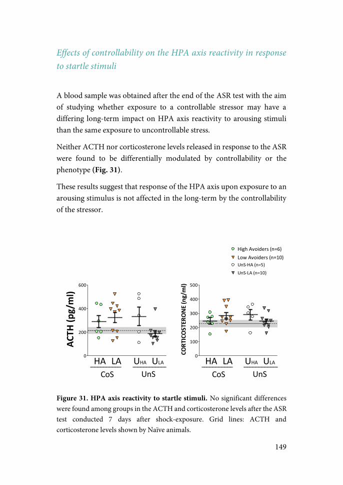
149
Effects of controllability on the HPA axis reactivity in response to startle stimuli
A blood sample was obtained after the end of the ASR test with the aim of studying whether exposure to a controllable stressor may have a differing long-term impact on HPA axis reactivity to arousing stimuli than the same exposure to uncontrollable stress.
Neither ACTH nor corticosterone levels released in response to the ASR were found to be differentially modulated by controllability or the phenotype (Fig. 31).
These results suggest that response of the HPA axis upon exposure to an arousing stimulus is not affected in the long-term by the controllability of the stressor.
Figure 31. HPA axis reactivity to startle stimuli. No significant differences were found among groups in the ACTH and corticosterone levels after the ASR test conducted 7 days after shock-exposure. Grid lines: ACTH and corticosterone levels shown by Naïve animals.
0
100
200
300
400
500
CoS UnSHA LA UHA ULA
High2Avoiders2(n=6)Low2Avoiders2(n=10)UnS=HA2(n=5)UnS=LA2(n=10)
CORT
ICOS
TERO
NE)(n
g/ml)
0
200
400
600
CoS UnSHA LA UHA ULA
ACTH
)(pg/ml)
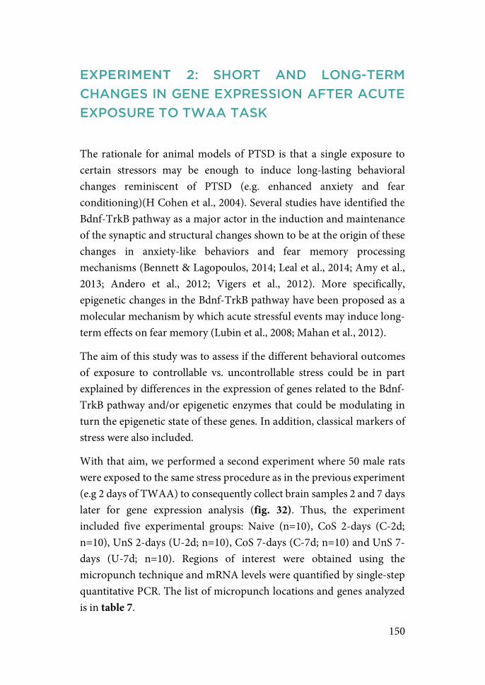
150
EXPERIMENT 2: SHORT AND LONG-TERM CHANGES IN GENE EXPRESSION AFTER ACUTE EXPOSURE TO TWAA TASK
The rationale for animal models of PTSD is that a single exposure to certain stressors may be enough to induce long-lasting behavioral changes reminiscent of PTSD (e.g. enhanced anxiety and fear conditioning)(H Cohen et al., 2004). Several studies have identified the Bdnf-TrkB pathway as a major actor in the induction and maintenance of the synaptic and structural changes shown to be at the origin of these changes in anxiety-like behaviors and fear memory processing mechanisms (Bennett & Lagopoulos, 2014; Leal et al., 2014; Amy et al., 2013; Andero et al., 2012; Vigers et al., 2012). More specifically, epigenetic changes in the Bdnf-TrkB pathway have been proposed as a molecular mechanism by which acute stressful events may induce long-term effects on fear memory (Lubin et al., 2008; Mahan et al., 2012).
The aim of this study was to assess if the different behavioral outcomes of exposure to controllable vs. uncontrollable stress could be in part explained by differences in the expression of genes related to the Bdnf-TrkB pathway and/or epigenetic enzymes that could be modulating in turn the epigenetic state of these genes. In addition, classical markers of stress were also included.
With that aim, we performed a second experiment where 50 male rats were exposed to the same stress procedure as in the previous experiment (e.g 2 days of TWAA) to consequently collect brain samples 2 and 7 days later for gene expression analysis (fig. 32). Thus, the experiment included five experimental groups: Naive (n=10), CoS 2-days (C-2d; n=10), UnS 2-days (U-2d; n=10), CoS 7-days (C-7d; n=10) and UnS 7-days (U-7d; n=10). Regions of interest were obtained using the micropunch technique and mRNA levels were quantified by single-step quantitative PCR. The list of micropunch locations and genes analyzed is in table 7.
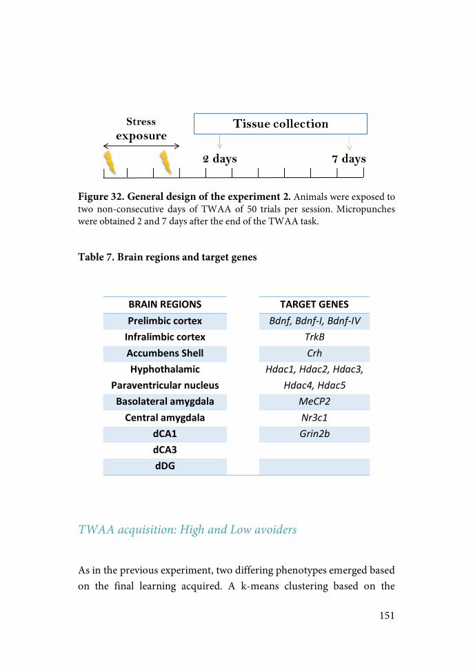
151
Figure 32. General design of the experiment 2. Animals were exposed to two non-consecutive days of TWAA of 50 trials per session. Micropunches were obtained 2 and 7 days after the end of the TWAA task.
Table 7. Brain regions and target genes
BRAIN#REGIONS#
#
TARGET#GENES#
Prelimbic#cortex# Bdnf,&Bdnf'I,&Bdnf'IV&Infralimbic#cortex# TrkB&Accumbens#Shell# Crh&Hyphothalamic#
Paraventricular#nucleus#
Hdac1,&Hdac2,&Hdac3,&Hdac4,&Hdac5&
Basolateral#amygdala# MeCP2&Central#amygdala# Nr3c1&
dCA1# Grin2b&dCA3# "
dDG# "
TWAA acquisition: High and Low avoiders
As in the previous experiment, two differing phenotypes emerged based on the final learning acquired. A k-means clustering based on the
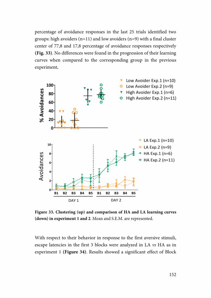
152
percentage of avoidance responses in the last 25 trials identified two groups: high avoiders (n=11) and low avoiders (n=9) with a final cluster center of 77,8 and 17,8 percentage of avoidance responses respectively (Fig. 33). No differences were found in the progression of their learning curves when compared to the corresponding group in the previous experiment.
Figure 33. Clustering (up) and comparison of HA and LA learning curves (down) in experiment 1 and 2. Mean and S.E.M. are represented.
With respect to their behavior in response to the first aversive stimuli, escape latencies in the first 3 blocks were analyzed in LA vs HA as in experiment 1 (Figure 34). Results showed a significant effect of Block
0
20
40
60
80
100Low$Avoider$Exp.1$(n=10)Low$Avoider$Exp.2$(n=9)High$Avoider$Exp.1$(n=6)High$Avoider$Exp.2$(n=11)
%(Avoidan
ces
B1 B2 B3 B4 B5 B1 B2 B3 B4 B50
2
4
6
8
10
HA#Exp.2#(n=11)
LA#Exp.2#(n=9)
DAY#1 DAY#2
HA#Exp.1#(n=6)
LA#Exp.1#(n=10)
Avoidances
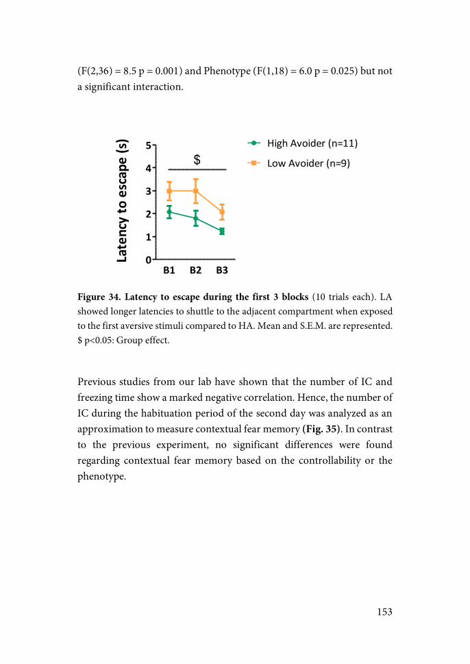
153
(F(2,36) = 8.5 p = 0.001) and Phenotype (F(1,18) = 6.0 p = 0.025) but not a significant interaction.
Figure 34. Latency to escape during the first 3 blocks (10 trials each). LA showed longer latencies to shuttle to the adjacent compartment when exposed to the first aversive stimuli compared to HA. Mean and S.E.M. are represented. $ p<0.05: Group effect.
Previous studies from our lab have shown that the number of IC and freezing time show a marked negative correlation. Hence, the number of IC during the habituation period of the second day was analyzed as an approximation to measure contextual fear memory (Fig. 35). In contrast to the previous experiment, no significant differences were found regarding contextual fear memory based on the controllability or the phenotype.
B1 B2 B30
1
2
3
4
5$
High%Avoider%(n=11)
Low%Avoider%(n=9)
Latency/to/escape/(s)
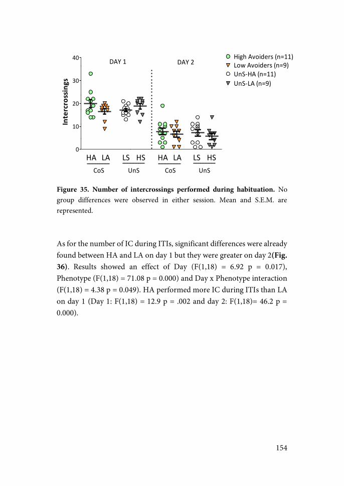
154
Figure 35. Number of intercrossings performed during habituation. No group differences were observed in either session. Mean and S.E.M. are represented.
As for the number of IC during ITIs, significant differences were already found between HA and LA on day 1 but they were greater on day 2(Fig. 36). Results showed an effect of Day (F(1,18) = 6.92 p = 0.017), Phenotype (F(1,18) = 71.08 p = 0.000) and Day x Phenotype interaction (F(1,18) = 4.38 p = 0.049). HA performed more IC during ITIs than LA on day 1 (Day 1: F(1,18) = 12.9 p = .002 and day 2: F(1,18)= 46.2 p = 0.000).
0
10
20
30
40
CoS UnS
HA LA LS HS
High1Avoiders1(n=11)
UnS:LA1(n=9)
Low1Avoiders1(n=9)UnS:HA1(n=11)
CoS UnS
HA LA LS HS
DAY11 DAY12Intercrossings
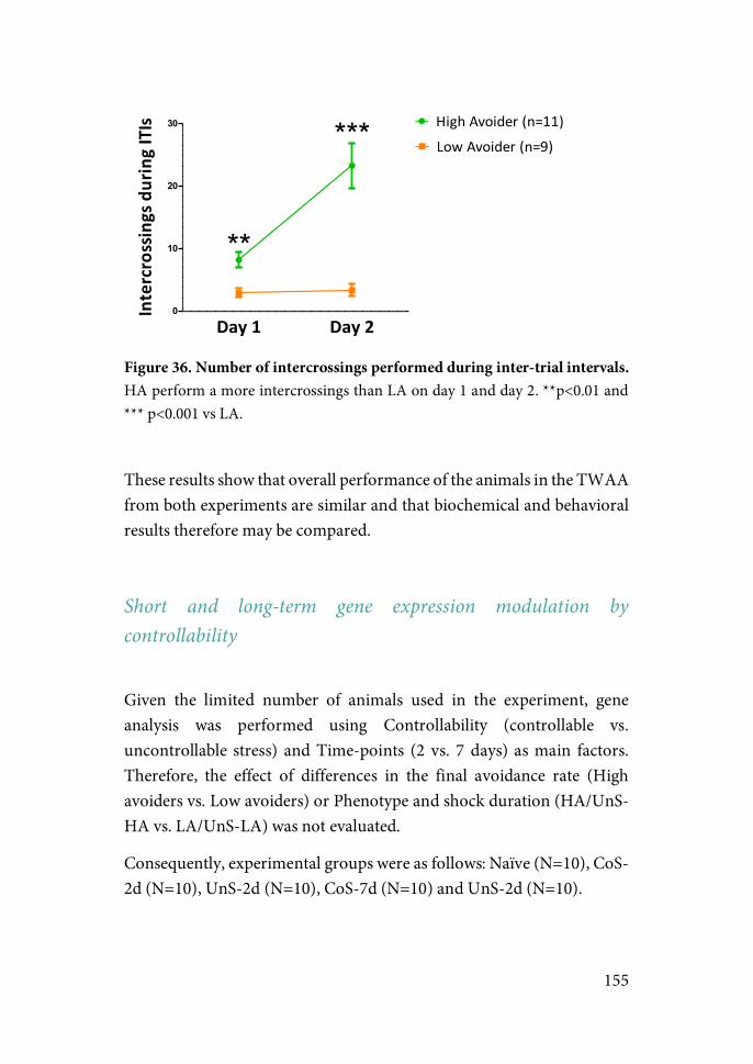
155
Figure 36. Number of intercrossings performed during inter-trial intervals. HA perform a more intercrossings than LA on day 1 and day 2. **p<0.01 and *** p<0.001 vs LA.
These results show that overall performance of the animals in the TWAA from both experiments are similar and that biochemical and behavioral results therefore may be compared.
Short and long-term gene expression modulation by controllability
Given the limited number of animals used in the experiment, gene analysis was performed using Controllability (controllable vs. uncontrollable stress) and Time-points (2 vs. 7 days) as main factors. Therefore, the effect of differences in the final avoidance rate (High avoiders vs. Low avoiders) or Phenotype and shock duration (HA/UnS-HA vs. LA/UnS-LA) was not evaluated.
Consequently, experimental groups were as follows: Naïve (N=10), CoS-2d (N=10), UnS-2d (N=10), CoS-7d (N=10) and UnS-2d (N=10).
Day$1 Day$20
10
20
30 High%Avoider%(n=11)Low%Avoider%(n=9)***
**
Intercrossings$during$ITIs
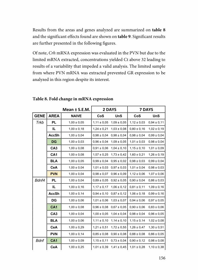
156
Results from the areas and genes analyzed are summarized on table 8 and the significant effects found are shown on table 9. Significant results are further presented in the following figures.
Of note, Crh mRNA expression was evaluated in the PVN but due to the limited mRNA extracted, concentrations yielded Ct above 32 leading to results of a variability that impeded a valid analysis. The limited sample from where PVN mRNA was extracted prevented GR expression to be analyzed in this region despite its interest.
Table 8. Fold change in mRNA expression
Mean#±#S.E.M.# 2#DAYS# 7#DAYS#GENE! AREA! NAIVE! CoS! UnS! CoS! UnS!
Trkb! PL! 1,00!±!0,05! 1,11!±!0,05! 1,09!±!0,05! 1,12!±!0,03! 0,94!±!0,11!
!! IL! 1,00!±!0,18! 1,24!±!0,21! 1,03!±!0,08! 0,80!±!0,16! 1,02!±!0,19!
!! AccSh! 1,00!±!0,04! 0,98!±!0,04! 0,98!±!0,04! 0,98!±!0,04! 0,99!±!0,04!
!! DG! 1,00!±!0,03! 0,96!±!0,04! 1,09!±!0,05! 1,01!±!0,03! 0,98!±!0,04!
!! CA3! 1,00!±!0,08! 0,91!±!0,06! 1,04!±!0,10! 1,15!±!0,10! 1,01!±!0,09!
!! CA1! 1,00!±!0,08! 1,57!±!0,25! 1,73!±!0,42! 1,60!±!0,21! 1,26!±!0,19!
!! BLA! 1,00!±!0,05! 0,99!±!0,04! 0,95!±!0,02! 0,98!±!0,03! 0,99!±!0,04!
!! CeA! 1,00!±!0,04! 1,01!±!0,03! 0,97!±!0,03! 1,01!±!0,04! 0,98!±!0,03!
!! PVN! 1,00!±!0,04! 0,98!±!0,07! 0,96!±!0,09! 1,12!±!0,06! 1,07!±!0,06!
Bdnf4! PL! 1,00!±!0,04! 0,89!±!0,05! 0,92!±!0,05! 0,90!±!0,04! 0,86!±!0,03!
"" IL! 1,00!±!0,16! 1,17!±!0,17! 1,06!±!0,12! 0,81!±!0,11! 1,09!±!0,16!
"" AccSh! 1,00!±!0,14! 0,94!±!0,10! 0,87!±!0,12! 1,06!±!0,18! 0,99!±!0,16!
"" DG! 1,00!±!0,06! 1,01!±!0,06! 1,03!±!0,07! 0,94!±!0,06! 0,97!±!0,05!
"" CA1! 1,00!±!0,08! 0,96!±!0,08! 0,67!±!0,05! 0,90!±!0,06! 0,83!±!0,06!
"" CA3! 1,00!±!0,04! 1,00!±!0,05! 1,04!±!0,04! 0,98!±!0,04! 0,96!±!0,05!
"" BLA! 1,00!±!0,06! 1,11!±!0,10! 1,14!±!0,10! 1,15!±!0,14! 1,02!±!0,08!
"" CeA! 1,00!±!0,29! 1,21!±!0,51! 1,72!±!0,55! 1,26!±!0,47! 1,30!±!0,51!
"" PVN! 1,00!±!0,14! 0,85!±!0,08! 0,90!±!0,08! 0,88!±!0,08! 0,86!±!0,05!
Bdnf! CA1! 1,00!±!0,09! 1,15!±!0,11! 0,73!±!0,04! 0,90!±!0,12! 0,98!±!0,08!
!! CeA! 1,00!±!0,25! 1,01!±!0,39! 1,41!±!0,45! 1,01!±!0,28! 1,10!±!0,38!
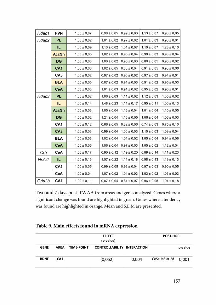
157
Hdac1! PVN! 1,00!±!0,07! 0,98!±!0,05! 0,99!±!0,03! 1,13!±!0,07! 0,98!±!0,05!
Hdac2! PL! 1,00!±!0,02! 1,01!±!0,02! 0,97!±!0,02! 1,01!±!0,03! 0,98!±!0,01!
!! IL! 1,00!±!0,09! 1,13!±!0,02! 1,01!±!0,07! 1,10!±!0,07! 1,28!±!0,10!
!! AccSh! 1,00!±!0,05! 1,02!±!0,03! 0,95!±!0,04! 0,90!±!0,03! 0,93!±!0,04!
!! DG! 1,00!±!0,03! 1,00!±!0,02! 0,96!±!0,03! 0,80!±!0,05! 0,90!±!0,02!
!! CA1! 1,00!±!0,08! 1,02!±!0,05! 0,83!±!0,04! 0,91!±!0,05! 0,93!±!0,06!
!! CA3! 1,00!±!0,02! 0,97!±!0,02! 0,96!±!0,02! 0,97!±!0,02! 0,94!±!0,01!
!! BLA!! 1,00!±!0,05! 0,87!±!0,02! 0,91!±!0,03! 0,91!±!0,02! 0,95!±!0,03!
!! CeA! 1,00!±!0,03! 1,01!±!0,03! 0,91!±!0,02! 0,95!±!0,02! 0,96!±!0,01!
Hdac3! PL! 1,00!±!0,02! 1,06!±!0,03! 1,11!±!0,02! 1,12!±!0,03! 1,05!±!0,02!
!! IL! 1,00!±!0,14! 1,48!±!0,23! 1,11!±!0,17! 0,95!±!0,11! 1,06!±!0,13!
!! AccSh! 1,00!±!0,03! 1,05!±!0,04! 1,16!±!0,04! 1,01!±!0,04! 1,10!±!0,05!
!! DG! 1,00!±!0,02! 1,21!±!0,04! 1,16!±!0,05! 1,06!±!0,04! 1,06!±!0,03!
!! CA1! 1,00!±!0,12! 0,66!±!0,05! 0,82!±!0,06! 0,74!±!0,03! 0,75!±!0,10!
!! CA3! 1,00!±!0,03! 0,99!±!0,04! 1,06!±!0,03! 1,10!±!0,03! 1,09!±!0,04!
!! BLA! 1,00!±!0,03! 1,02!±!0,04! 1,01!±!0,02! 1,05!±!0,04! 0,94!±!0,06!
!! CeA! 1,00!±!0,05! 1,06!±!0,04! 0,97!±!0,03! 1,05!±!0,02! 1,12!±!0,04!
Crh! CeA! 1,00!±!0,17! 0,90!±!0,12! 1,19!±!0,20! 0,89!±!0,14! 1,11!±!0,23!
Nr3c1! IL! 1,00!±!0,16! 1,57!±!0,22! 1,11!±!0,18! 0,98!±!0,13! 1,19!±!0,13!
!! CA1! 1,00!±!0,05! 0,99!±!0,05! 0,92!±!0,04! 0,97!±!0,03! 0,90!±!0,05!
"" CeA! 1,00!±!0,04! 1,07!±!0,02! 1,04!±!0,03! 1,03!±!0,02! 1,03!±!0,03!
Grin2b! CA1! 1,00!±!0,11! 0,87!±!0,04! 0,84!±!0,07! 0,96!±!0,05! 1,04!±!0,18!
Two and 7 days post-TWAA from areas and genes analyzed. Genes where a significant change was found are highlighted in green. Genes where a tendency was found are highlighted in orange. Mean and S.E.M are presented.
Table 9. Main effects found in mRNA expression
## ## EFFECT"(p$value)#
POST$HOC#
GENE## AREA# TIME$POINT# CONTROLLABILITY## INTERACTION# ## p$value#
BDNF## CA1# "" "(0,052)"" 0,004" CoS/UnS"at"2d" 0,001"
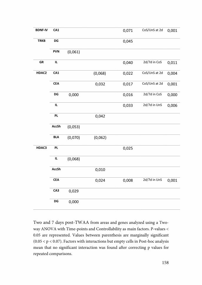
158
BDNF$IV# CA1# "" "" 0,071" CoS/UnS"at"2d" 0,001"
TRKB# DG# "" "" 0,045" "" ""
PVN# (0,061)" "" "" "" ""
GR# IL# "" "" 0,040" 2d/7d"in"CoS" 0,011"
HDAC2# CA1# "" (0,068)"" 0,022" CoS/UnS"at"2d" 0,004"
CEA# "" 0,032" 0,017" CoS/UnS"at"2d" 0,001"
DG# 0,000" "" 0,016" 2d/7d"in"CoS" 0,000"
IL# "" "" 0,033" 2d/7d"in"UnS" 0,006"
PL# "" 0,042" "" "" ""
AccSh# (0,053)" "" "" "" ""
BLA## (0,070)" (0,062)" "" "" ""
HDAC3# PL# "" "" 0,025" "" ""
IL# (0,068)" "" "" "" ""
AccSh# "" 0,010" "" "" ""
CEA# "" 0,024" 0,008" 2d/7d"in"UnS" 0,001"
CA3# 0,029" "" "" "" ""
DG# 0,000" "" "" "" ""
Two and 7 days post-TWAA from areas and genes analyzed using a Two-way ANOVA with Time-points and Controllability as main factors. P-values < 0.05 are represented. Values between parenthesis are marginally significant (0.05 < p < 0.07). Factors with interactions but empty cells in Post-hoc analysis mean that no significant interaction was found after correcting p values for repeated comparisons.
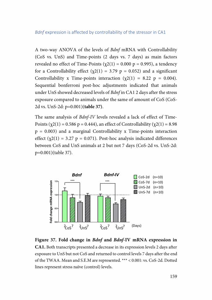
159
Bdnf%expression&is&affected&by&controllability&of&the&stressor&in&CA1&
A two-way ANOVA of the levels of Bdnf mRNA with Controllability (CoS vs. UnS) and Time-points (2 days vs. 7 days) as main factors revealed no effect of Time-Points (χ2(1) = 0.000 p = 0.995), a tendency for a Controllability effect (χ2(1) = 3.79 p = 0.052) and a significant Controllability x Time-points interaction (χ2(1) = 8.22 p = 0.004). Sequential bonferroni post-hoc adjustments indicated that animals under UnS showed decreased levels of Bdnf in CA1 2 days after the stress exposure compared to animals under the same of amount of CoS (CoS-2d vs. UnS-2d: p=0.001)(table 37).
The same analysis of Bdnf-IV levels revealed a lack of effect of Time-Points (χ2(1) = 0.586 p = 0.444), an effect of Controllability (χ2(1) = 8.98 p = 0.003) and a marginal Controllability x Time-points interaction effect (χ2(1) = 3.27 p = 0.071). Post-hoc analysis indicated differences between CoS and UnS animals at 2 but not 7 days (CoS-2d vs. UnS-2d: p=0.001)(table 37).
Figure 37. Fold change in Bdnf and Bdnf-IV mRNA expression in CA1. Both transcripts presented a decrease in its expression levels 2 days after exposure to UnS but not CoS and returned to control levels 7 days after the end of the TWAA. Mean and S.E.M are represented. *** < 0.001: vs. CoS-2d. Dotted lines represent stress naïve (control) levels.
0.0
0.5
1.0
1.5
CoS$2d''''(n=10)
UnS$2d''''(n=10)CoS$7d''''(n=10)
UnS$7d''''(n=10)
Bdnf Bdnf%IV*** ***
2 7 2 7CoS UnS (Days)2 7 2 7CoS UnS
Fold%ch
ange%m
RNA%expressio
n
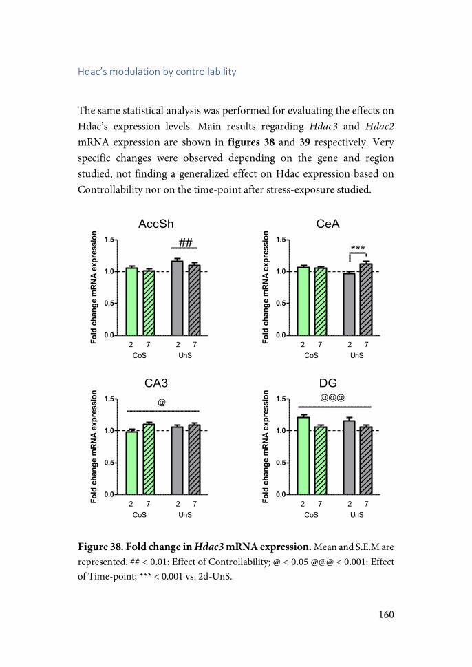
160
Hdac’s&modulation&by&controllability&&
The same statistical analysis was performed for evaluating the effects on Hdac’s expression levels. Main results regarding Hdac3 and Hdac2 mRNA expression are shown in figures 38 and 39 respectively. Very specific changes were observed depending on the gene and region studied, not finding a generalized effect on Hdac expression based on Controllability nor on the time-point after stress-exposure studied.
Figure 38. Fold change in Hdac3 mRNA expression. Mean and S.E.M are represented. ## < 0.01: Effect of Controllability; @ < 0.05 @@@ < 0.001: Effect of Time-point; *** < 0.001 vs. 2d-UnS.
AccSh
0.0
0.5
1.0
1.5
2 7 2 7CoS UnS
##
Fold)change)mRNA)expression
CeA
0.0
0.5
1.0
1.5
2 7 2 7
***
CoS UnS
Fold)change)mRNA)expression
CA3
0.0
0.5
1.0
1.5
2 7 2 7
@
CoS UnS
Fold)change)mRNA)expression
DG
0.0
0.5
1.0
1.5
2 7 2 7
@@@
CoS UnS
Fold)change)mRNA)expression
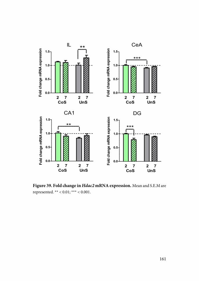
161
Figure 39. Fold change in Hdac2 mRNA expression. Mean and S.E.M are represented. ** < 0.01; *** < 0.001.
IL
0.0
0.5
1.0
1.5
2 7 2 7CoS UnS
**Fold/change/mRNA/expression
CeA
0.0
0.5
1.0
1.5
***
2 7 2 7CoS UnS
Fold/change/mRNA/expression
CA1
0.0
0.5
1.0
1.5
2 7 2 7CoS UnS
**
Fold/change/mRNA/expression
DG
0.0
0.5
1.0
1.5
2 7 2 7CoS UnS
***
Fold/change/mRNA/expression
162
CHAPTER TWO: Dynamics in the acquisition of control and behavioral and biochemical long-term effects
INTRODUCTION
Several studies have shown that exerting control over a stressor is a crucial factor in buffering some of the detrimental behavioral effects of stress. However, to our knowledge no study to date have examined the temporal dynamics involved in the acquisition of control. Furthermore, in view of the limited effect on anxiety-like behaviors observed in our experiment after acute exposure to controllable stress, we hypothesized that the buffering effects of controllability may depend on reaching a certain controllability level under which the detrimental effects of exposure to the stressor would be overweighed by the beneficial effects of control (a level not reached by acute exposure to a controllable stressor). If this was the case, the variability present in the acquisition of control would in part explain the variability observed in the behavioral outcomes of exposure to the same stressful event in our studies and in the literature.
In this regard, our working hypothesis is that despite being exposed for longer periods of time to a stressful event, prolonged exposure to a foot-shock active-avoidance task which involves control will enhance buffering the negative effects of stress compared to shorter or uncontrollable shock-exposure. On the other hand, because subjects under uncontrollable stress do not benefit from any advantage from prolonged exposures, individuals exposed for longer periods of time should show the detrimental effects of stress to a higher degree.
163
EXPERIMENT 3: RESULTS AND EXPERIMENTAL DESIGN
To test the stated hypothesis, we designed an experiment comparing the long-term outcome of controllability in a battery of behavioral tests 7 days after a short (2 days) or prolonged (5 days) exposure to the TWAA task (figure 40). Each of these groups was further divided into “Controllable Stress” (CoS), “Uncontrollable Stress” (UnS) and Naive animals depending on the type of control over the stressor they had during the task. Thus, the experiment consisted of six experimental groups: 2 Days-Naive (2-N; n=16), 2 Days-CoS (2-CoS; n=24), 2 Days-UnS (2-UnS; n=16), 5 Days-Naive (5-N; n=16), 5 Days-CoS (5-CoS; n=24) and 5 Days-UnS (5-UnS; n=16). The behavioral battery began 7 days after the end of the TWAA and consisted of open-field and social interaction tests on the first day, elevated-plus maze on the second day, and an extinction test on the third day.
Importantly, responses to trauma are very diverse. Even in the case of pathologies like PTSD where the characteristics of the stressor are well defined in nature and its temporal onset, epidemiological studies indicate that only about 20-30% of individuals exposed to the same stressful event will develop PTSD (Kessler et al., 1995). Despite clinical studies being based on stringent criteria for inclusion in the study population, animal models are largely based on the comparison of exposed vs. non-exposed animals regardless of individual variation in response. To overcome this methodological discrepancy, we mimicked the methodology used in human studies by conducting an individual profiling approach in which “affected” and “not-affected” animals were identified by measuring their response in a series of key behavioral variables compared to the response shown by the naive group. This behavioral profiling was based on the methodology suggested in previous works (Ardi et al., 2016; Cohen et al., 2004). Specifically, animals were classified as “affected” when their performance in 5 out of 8 behavioral

164
variables were outside of the normal behavior in the naive group as indicated by the 20th and 80th percentiles (see Methods for a detailed description of the selected variables and thresholds).
Finally, two procedures were conducted with the aim of analyzing part of the possible molecular mechanisms underlying the long-term differences observed after exposure to controllable vs. uncontrollable stress. Brain samples were collected from part of the subjects three days after the end of the behavioral tests to perform gene expression analysis.
Figure 40. Experimental design of experiment 3. A behavioral battery of tests was conducted one week after exposure to 2 or 5 days of TWAA: open-field and a social interaction test (first day), elevated-plus maze (second day) and extinction test (third day). Three days after the end on the behavioral battery brain samples from part of the animals were collected for expression analysis, while electrophysiological recordings were conducted on the rest of the animals one week after the extinction test.
High and Low avoiders: Differences in first reactions to US exposure
The number of avoidance responses was measured throughout the sessions. As a first approach, we wanted to assess: (a) whether pre-TWAA factors may influence how subjects react to the first US presentations; and (b) whether differing levels of avoidance conditioning and/or the controllability over the stressor may affect the long-term consequences of stress-exposure.
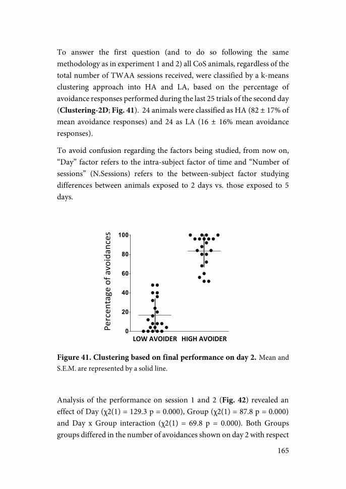
165
To answer the first question (and to do so following the same methodology as in experiment 1 and 2) all CoS animals, regardless of the total number of TWAA sessions received, were classified by a k-means clustering approach into HA and LA, based on the percentage of avoidance responses performed during the last 25 trials of the second day (Clustering-2D; Fig. 41). 24 animals were classified as HA (82 ± 17% of mean avoidance responses) and 24 as LA (16 ± 16% mean avoidance responses).
To avoid confusion regarding the factors being studied, from now on, “Day” factor refers to the intra-subject factor of time and “Number of sessions” (N.Sessions) refers to the between-subject factor studying differences between animals exposed to 2 days vs. those exposed to 5 days.
Figure 41. Clustering based on final performance on day 2. Mean and S.E.M. are represented by a solid line.
Analysis of the performance on session 1 and 2 (Fig. 42) revealed an effect of Day (χ2(1) = 129.3 p = 0.000), Group (χ2(1) = 87.8 p = 0.000) and Day x Group interaction (χ2(1) = 69.8 p = 0.000). Both Groups groups differed in the number of avoidances shown on day 2 with respect
LOW$AVOIDER HIGH$AVOIDER0
20
40
60
80
100
Percen
tage)of)a
voidan
ces
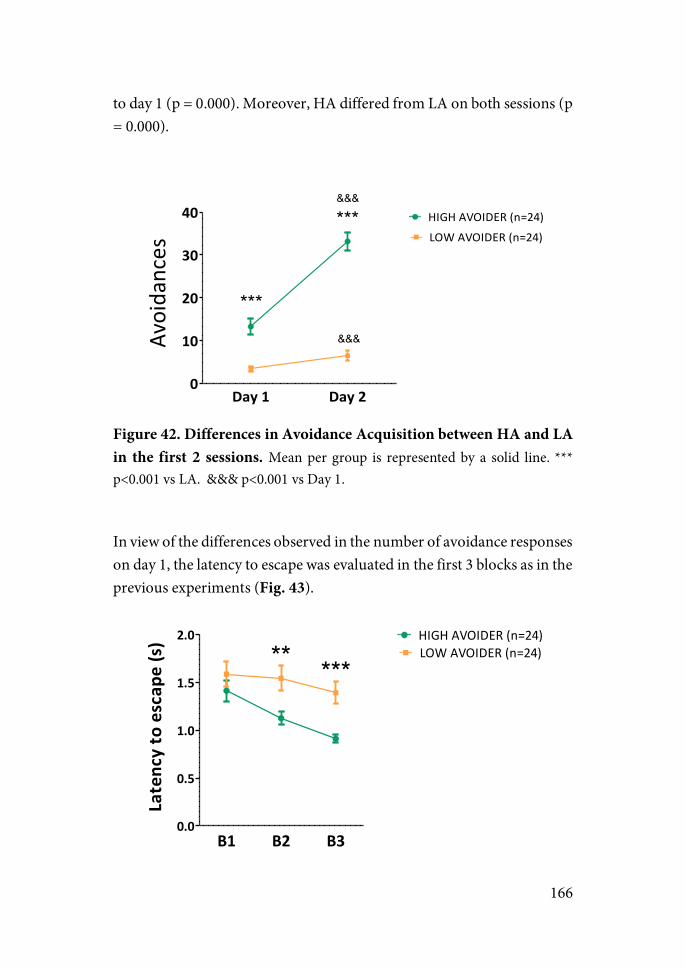
166
to day 1 (p = 0.000). Moreover, HA differed from LA on both sessions (p = 0.000).
Figure 42. Differences in Avoidance Acquisition between HA and LA in the first 2 sessions. Mean per group is represented by a solid line. *** p<0.001 vs LA. &&& p<0.001 vs Day 1.
In view of the differences observed in the number of avoidance responses on day 1, the latency to escape was evaluated in the first 3 blocks as in the previous experiments (Fig. 43).
Day$1 Day$20
10
20
30
40 HIGH$AVOIDER$(n=24)LOW$AVOIDER$(n=24)
***
***
Avoidances
&&&
&&&
B1 B2 B30.0
0.5
1.0
1.5
2.0 HIGH$AVOIDER$(n=24)LOW$AVOIDER$(n=24)** ***
Latency/to/escap
e/(s)
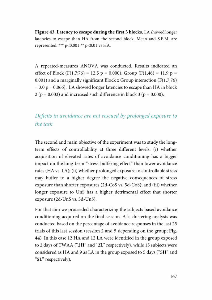
167
Figure 43. Latency to escape during the first 3 blocks. LA showed longer latencies to escape than HA from the second block. Mean and S.E.M. are represented. *** p<0.001 ** p<0.01 vs HA.
A repeated-measures ANOVA was conducted. Results indicated an effect of Block (F(1.7;76) = 12.5 p = 0.000), Group (F(1,46) = 11.9 p = 0.001) and a marginally significant Block x Group interaction (F(1.7;76) = 3.0 p = 0.066). LA showed longer latencies to escape than HA in block 2 (p = 0.003) and increased such difference in block 3 (p = 0.000).
Deficits in avoidance are not rescued by prolonged exposure to the task
The second and main objective of the experiment was to study the long-term effects of controllability at three different levels: (i) whether acquisition of elevated rates of avoidance conditioning has a bigger impact on the long-term “stress-buffering effect” than lower avoidance rates (HA vs. LA); (ii) whether prolonged exposure to controllable stress may buffer to a higher degree the negative consequences of stress exposure than shorter exposures (2d-CoS vs. 5d-CoS); and (iii) whether longer exposure to UnS has a higher detrimental effect that shorter exposure (2d-UnS vs. 5d-UnS).
For that aim we proceeded characterizing the subjects based avoidance conditioning acquired on the final session. A k-clustering analysis was conducted based on the percentage of avoidance responses in the last 25 trials of this last session (session 2 and 5 depending on the group; Fig. 44). In this case 12 HA and 12 LA were identified in the group exposed to 2 days of TWAA (“2H” and “2L” respectively), while 15 subjects were considered as HA and 9 as LA in the group exposed to 5 days (“5H” and “5L” respectively).
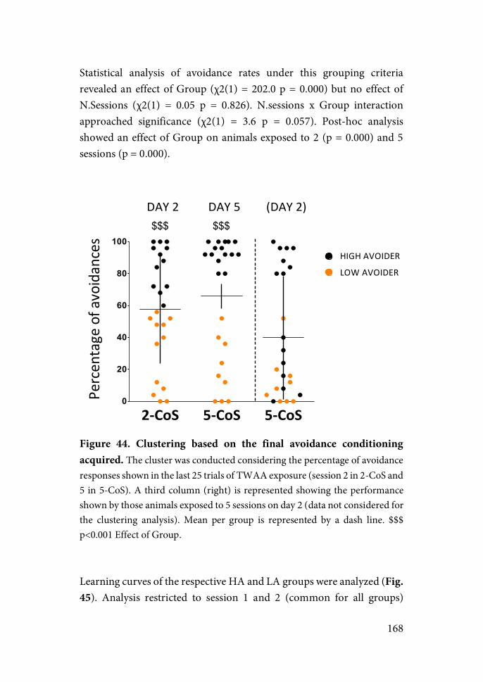
168
Statistical analysis of avoidance rates under this grouping criteria revealed an effect of Group (χ2(1) = 202.0 p = 0.000) but no effect of N.Sessions (χ2(1) = 0.05 p = 0.826). N.sessions x Group interaction approached significance (χ2(1) = 3.6 p = 0.057). Post-hoc analysis showed an effect of Group on animals exposed to 2 (p = 0.000) and 5 sessions (p = 0.000).
Figure 44. Clustering based on the final avoidance conditioning acquired. The cluster was conducted considering the percentage of avoidance responses shown in the last 25 trials of TWAA exposure (session 2 in 2-CoS and 5 in 5-CoS). A third column (right) is represented showing the performance shown by those animals exposed to 5 sessions on day 2 (data not considered for the clustering analysis). Mean per group is represented by a dash line. $$$ p<0.001 Effect of Group.
Learning curves of the respective HA and LA groups were analyzed (Fig. 45). Analysis restricted to session 1 and 2 (common for all groups)
2"CoS 5"CoS 5"CoS0
20
40
60
80
100
DAY$2 DAY$5 (DAY$2)
HIGH$AVOIDER
LOW$AVOIDER
$$$ $$$
Percen
tage$of$avoidances
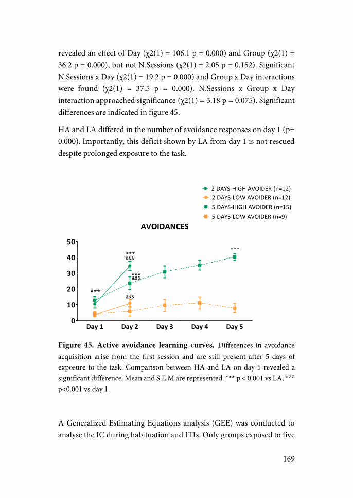
169
revealed an effect of Day (χ2(1) = 106.1 p = 0.000) and Group (χ2(1) = 36.2 p = 0.000), but not N.Sessions (χ2(1) = 2.05 p = 0.152). Significant N.Sessions x Day (χ2(1) = 19.2 p = 0.000) and Group x Day interactions were found (χ2(1) = 37.5 p = 0.000). N.Sessions x Group x Day interaction approached significance (χ2(1) = 3.18 p = 0.075). Significant differences are indicated in figure 45.
HA and LA differed in the number of avoidance responses on day 1 (p= 0.000). Importantly, this deficit shown by LA from day 1 is not rescued despite prolonged exposure to the task.
Figure 45. Active avoidance learning curves. Differences in avoidance acquisition arise from the first session and are still present after 5 days of exposure to the task. Comparison between HA and LA on day 5 revealed a significant difference. Mean and S.E.M are represented. *** p < 0.001 vs LA; &&& p<0.001 vs day 1.
A Generalized Estimating Equations analysis (GEE) was conducted to analyse the IC during habituation and ITIs. Only groups exposed to five
AVOIDANCES
Day,1 Day,2 Day,3 Day,4 Day,50
10
20
30
40
50
***
2"DAYS'HIGH"AVOIDER"(n=12)2"DAYS'LOW"AVOIDER"(n=12)5"DAYS'HIGH"AVOIDER"(n=15)5"DAYS'LOW"AVOIDER"(n=9)
&&&
&&&
&&&
***
***
***
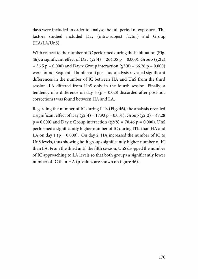
170
days were included in order to analyse the full period of exposure. The factors studied included Day (intra-subject factor) and Group (HA/LA/UnS).
With respect to the number of IC performed during the habituation (Fig. 46), a significant effect of Day (χ2(4) = 264.05 p = 0.000), Group (χ2(2) = 36.5 p = 0.000) and Day x Group interaction (χ2(8) = 66.26 p = 0.000) were found. Sequential bonferroni post-hoc analysis revealed significant differences in the number of IC between HA and UnS from the third session. LA differed from UnS only in the fourth session. Finally, a tendency of a difference on day 5 (p = 0.028 discarded after post-hoc corrections) was found between HA and LA.
Regarding the number of IC during ITIs (Fig. 46), the analysis revealed a significant effect of Day (χ2(4) = 17.93 p = 0.001), Group (χ2(2) = 47.28 p = 0.000) and Day x Group interaction (χ2(8) = 78.46 p = 0.000). UnS performed a significantly higher number of IC during ITIs than HA and LA on day 1 (p = 0.000). On day 2, HA increased the number of IC to UnS levels, thus showing both groups significantly higher number of IC than LA. From the third until the fifth session, UnS dropped the number of IC approaching to LA levels so that both groups a significantly lower number of IC than HA (p-values are shown on figure 46).
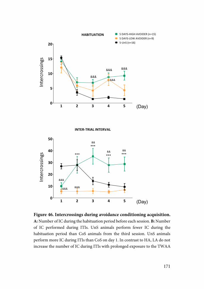
171
Figure 46. Intercrossings during avoidance conditioning acquisition. A: Number of IC during the habituation period before each session. B: Number of IC performed during ITIs. UnS animals perform fewer IC during the habituation period than CoS animals from the third session. UnS animals perform more IC during ITIs than CoS on day 1. In contrast to HA, LA do not increase the number of IC during ITIs with prolonged exposure to the TWAA
HABITUATION
1 2 3 4 50
5
10
15
20
5"DAYS'HIGH"AVOIDER"(n=15)5"DAYS'LOW"AVOIDER"(n=9)5'UnS"(n=16)
&&&
&&&
&&&
&&&
Intercrossings
INTER1TRIAL3INTERVAL
1 2 3 4 50
10
20
30
40
50
&&&
&&&
&&&
***
***
*** ***
&&
&& &&
Intercrossings
(Day)
(Day)
172
task. Mean and S.E.M are represented. *** p < 0.001: HA vs. LA; && p < 0.01 && p < 0.001: HA or LA vs. UnS.
Long-term behavioral effects of exposure to controllable vs. uncontrollable stress
One week after the end of the TWAA exposure, animals went through a behavioral battery of tests consisting of an OF and a social interaction test on the first day, an EPM on the second day and an extinction test on the third day. The aim was to assess not only whether exerting control over the stressor affects the long-term behavioral consequences of the stress exposure but also whether they may differ depending on the level of avoidance conditioning acquired.
A two-ANOVA of total distance in the OF (Fig. 47) indicated an effect of N.Sessions (χ2(1) = 4.73 p = 0.030) and Group (χ2(3) = 22.40 p = 0.000), but not interaction. Post-hoc analysis revealed that UnS, but not CoS animals, show a significant long-term decrease in their exploratory behavior compared to naïve animals (p= 0.000). In the case of the number of entries in the center area, again an effect of Group was found (F(3,104) = 3.11 p = 0.030). In that, only HA increased significantly the number of entries in the center area compared to naïve animals (p = 0.003). Analysis of the latency to enter the center area indicated an effect of Group (F(3,104) = 4.3 p = 0.007), HA taking significantly less time than naïve animals to enter the center area (p = 0.002). Lastly, an effect of Group was also found in the velocity shown in the periphery (χ2(3) = 24.13 p = 0.000; Not shown), in this case UnS exhibiting a decreased velocity while exploring the periphery than naïve animals (p= 0.000).

173
Figure 47. Open field test. Total distance (up), number of entries (bottom-left) and latency to enter the center area (bottom right) during the OF test performed 7 days after the end of the stress exposure. Only the UnS group showed a significant hypoactivity compared to naïve animals. Moreover, HA but not LA showed an increased number of entries and a decreased latency to enter the center area compared to naive group. Mean and S.E.M are represented. ** p < 0.01; *** p < 0.001 vs. Naïve group; @ p< 0.05 Effect of N.sessions.
On the same day a social interaction test was conducted on the same OF introducing two cages in opposing corners of the arena, one with an animal inside and one empty. Prolonged TWAA exposure (5 vs 2 days) increased the total distance run on the arena regardless of the type of stress-exposure (Effect of N.Sessions: χ2(3) = 5.79 p=0.017). Animals spent most of the time in the area surrounding the animal cage, those exposed to 5 instead of 2 days of TWAA showing a general decrease in
2N5N 2H5H 2L 5L 2U5U0
1000
2000
3000
4000
5000 2"days'Naive'(2N)'(N=16)
5"days'Naive'(5N)'(N=16)
2"days'High'Avoider'(2H)'(N=12)
5"days'High'Avoider'(5H)'(N=15)
2"days'Low'Avoider'(2L)'(N=12)
5"days'Low'Avoider'(5L)'(N=9)
2"days'UnS'(2U)'(N=16)
5"days'UnS'(5U)'(N=16)Total'distance'(cm)
2N5N 2H5H 2L 5L 2U5U0
5
10
15
20
25
Entries'in'center
2N5N 2H5H 2L 5L 2U5U0
100
200
300
400
Latency'to'cen
ter'(s)
***@
** **

174
its exploration time (Fig. 48; Effect of N.Sessions: χ2(3) = 7.51 p=0.006). Regarding the distance run in the animal zone, results found an effect of N.Sessions (F(1,104) = 12,19 p = 0.001), Group (F(3,104) = 0.56 p = 0.641) and N.Sessions x Group interaction (F(3,112) = 3.35 p = 0.022). Post-hoc analysis revealed that HA exposed to 5 days of TWAA (5H) ran a shorter distance in the animal area compared to HA exposed to 2 days (2H) (p=0.000).
Figure 48. Social Interaction test ran 7 days after the end of the TWAA and after the OF test. HA exposed to 5 days of controllable stress spent less time exploring the animal ring area compared to HA exposed to 2 days. 5 days of TWAA exposure decreased the time in animal zone and increased the total distance ran compared to 2-days exposure. @ p< 0.05 @@ p<0.01 Effect of N.Sessions; *** p< 0.001 vs. 2H.
An EPM was conducted 8 days after the end of the TWAA task (one day after the OF and SI tests)(Fig. 49). Analysis of variances of the total distance indicate an effect of N.Sessions (F(1,104) = 10.24 p = 0.002) but
2N5N 2H5H 2L 5L 2U5U0
1000
2000
3000
4000
5000 2"days'Naive'(2N)'(N=16)
5"days'Naive'(5N)'(N=16)
2"days'High'Avoider'(2H)'(N=12)
5"days'High'Avoider'(5H)'(N=15)
2"days'Low'Avoider'(2L)'(N=12)
5"days'Low'Avoider'(5L)'(N=9)
2"days'UnS'(2U)'(N=16)
5"days'UnS'(5U)'(N=16)Total'distance'(cm)
2N5N 2H5H 2L 5L 2U5U40
60
80
100
%'Distance'in''A
nimal'zo
ne
2N5N 2H5H 2L 5L 2U5U40
60
80
100
%'Tim
e'in'Animal'zo
ne ***
@
@@
A
B C
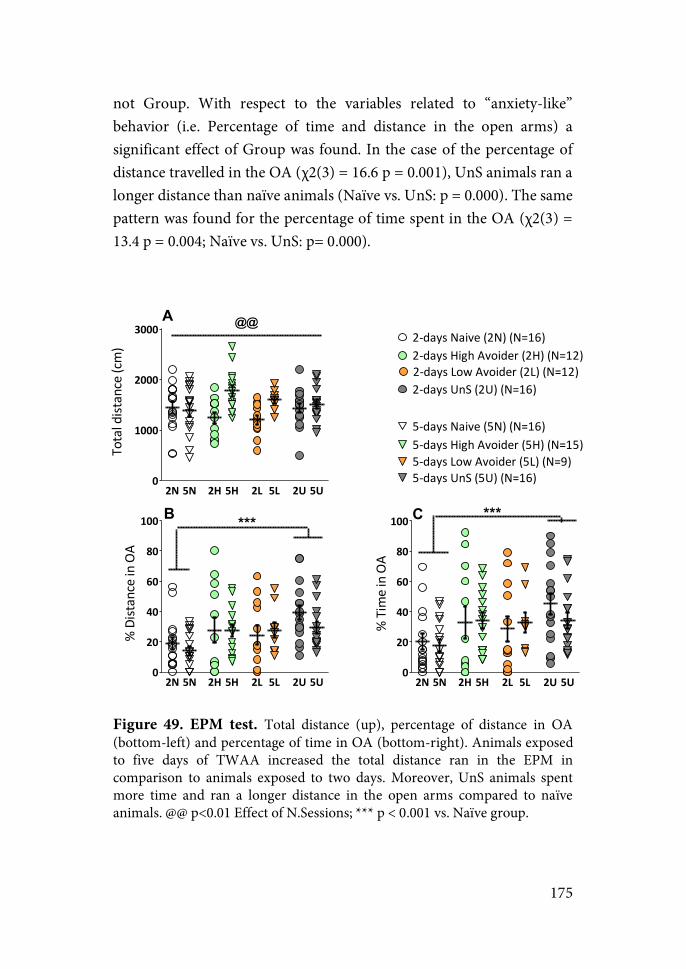
175
not Group. With respect to the variables related to “anxiety-like” behavior (i.e. Percentage of time and distance in the open arms) a significant effect of Group was found. In the case of the percentage of distance travelled in the OA (χ2(3) = 16.6 p = 0.001), UnS animals ran a longer distance than naïve animals (Naïve vs. UnS: p = 0.000). The same pattern was found for the percentage of time spent in the OA (χ2(3) = 13.4 p = 0.004; Naïve vs. UnS: p= 0.000).
Figure 49. EPM test. Total distance (up), percentage of distance in OA (bottom-left) and percentage of time in OA (bottom-right). Animals exposed to five days of TWAA increased the total distance ran in the EPM in comparison to animals exposed to two days. Moreover, UnS animals spent more time and ran a longer distance in the open arms compared to naïve animals. @@ p<0.01 Effect of N.Sessions; *** p < 0.001 vs. Naïve group.
2N 5N 2H 5H 2L 5L 2U 5U0
1000
2000
3000 2"days'Naive'(2N)'(N=16)
5"days'Naive'(5N)'(N=16)
2"days'High'Avoider'(2H)'(N=12)
5"days'High'Avoider'(5H)'(N=15)
2"days'Low'Avoider'(2L)'(N=12)
5"days'Low'Avoider'(5L)'(N=9)
2"days'UnS'(2U)'(N=16)
5"days'UnS'(5U)'(N=16)
Total'distance'(cm)
2N 5N 2H 5H 2L 5L 2U 5U0
20
40
60
80
100
%'Time'in'OA
*** ***
2N 5N 2H 5H 2L 5L 2U 5U0
20
40
60
80
100
%'Distance'in'OA
@@A
B C
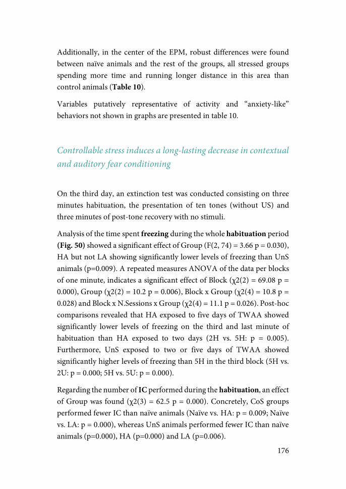
176
Additionally, in the center of the EPM, robust differences were found between naïve animals and the rest of the groups, all stressed groups spending more time and running longer distance in this area than control animals (Table 10).
Variables putatively representative of activity and “anxiety-like” behaviors not shown in graphs are presented in table 10.
Controllable stress induces a long-lasting decrease in contextual and auditory fear conditioning
On the third day, an extinction test was conducted consisting on three minutes habituation, the presentation of ten tones (without US) and three minutes of post-tone recovery with no stimuli.
Analysis of the time spent freezing during the whole habituation period (Fig. 50) showed a significant effect of Group (F(2, 74) = 3.66 p = 0.030), HA but not LA showing significantly lower levels of freezing than UnS animals (p=0.009). A repeated measures ANOVA of the data per blocks of one minute, indicates a significant effect of Block (χ2(2) = 69.08 p = 0.000), Group (χ2(2) = 10.2 p = 0.006), Block x Group (χ2(4) = 10.8 p = 0.028) and Block x N.Sessions x Group (χ2(4) = 11.1 p = 0.026). Post-hoc comparisons revealed that HA exposed to five days of TWAA showed significantly lower levels of freezing on the third and last minute of habituation than HA exposed to two days (2H vs. 5H: p = 0.005). Furthermore, UnS exposed to two or five days of TWAA showed significantly higher levels of freezing than 5H in the third block (5H vs. 2U: p = 0.000; 5H vs. 5U: p = 0.000).
Regarding the number of IC performed during the habituation, an effect of Group was found (χ2(3) = 62.5 p = 0.000). Concretely, CoS groups performed fewer IC than naïve animals (Naïve vs. HA: p = 0.009; Naïve vs. LA: p = 0.000), whereas UnS animals performed fewer IC than naïve animals (p=0.000), HA (p=0.000) and LA (p=0.006).
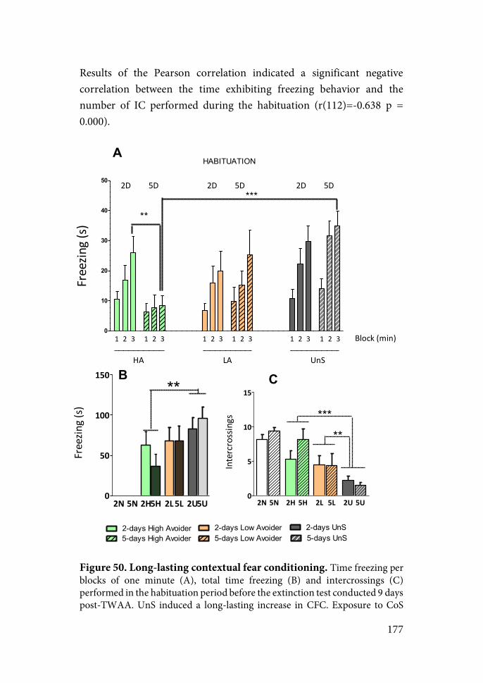
177
Results of the Pearson correlation indicated a significant negative correlation between the time exhibiting freezing behavior and the number of IC performed during the habituation (r(112)=-0.638 p = 0.000).
Figure 50. Long-lasting contextual fear conditioning. Time freezing per blocks of one minute (A), total time freezing (B) and intercrossings (C) performed in the habituation period before the extinction test conducted 9 days post-TWAA. UnS induced a long-lasting increase in CFC. Exposure to CoS
0
10
20
30
40
50
1 2 3 1 2 3 1 2 3 1 2 3 1 2 3 1 2 3
HA LA UnS
2D 5D 2D 5D 2D 5D
Block1(min)
***
**
HABITUATION
Freezin
g1(s)
2N 5N 2H 5H 2L 5L 2U 5U0
5
10
15
**
***
Intercrossings
A
B C
2+days0High0Avoider5+days0High0Avoider
2+days0Low0Avoider5+days0Low0Avoider
2+days0UnS5+days0UnS
2N 5N 2H5H 2L5L 2U5U0
50
100
150**
Freezin
g1(s)
178
blocked this effect induced by UnS in HA but not LA. Mean and S.E.M are represented. **p<0.01, *** p<0.001. Although not indicated in the graphs, all groups were statistically different from stress naïve animals. Analysis of the freezing shown during the whole tone-interval (Fig. 51) revealed a significant effect of Group (χ2(2) = 6.63 p=0.036) and N.sessions x Group interaction (χ2(2) = 7.47 p=0.024). Post-hoc Sequential bonferroni indicated HA exposed to 5 days freezed significantly less time than 5L (p= 0.002), 5S (p=0.002), 2H (p=0.001), 2L (p=0.010) and 2S (p=0.000).
Figure 51. Extinction test. Significantly lower freezing levels are shown by HA exposed to 5 days compared to the rest of the groups. Mean and S.E.M are represented. *p<0.05 5H vs 2L, ** p < 0.01 5H vs 5L, 5S and 2H; *** p < 0.001 5H vs 2L.
1 2 3 4 5 6 7 8 9 100
10
20
30
40
******
2&DAYS+HIGH+AVOIDER+(n=12)2&DAYS+LOW+AVOIDER+(n=12)2&DAYS+UnS+(n=16)
5&DAYS+HIGH+AVOIDER+(n=15)5+DAYS&LOW+AVOIDER+(n=9)5&DAYS+UnS+(n=16)
TONES
Freezin
g+(s)
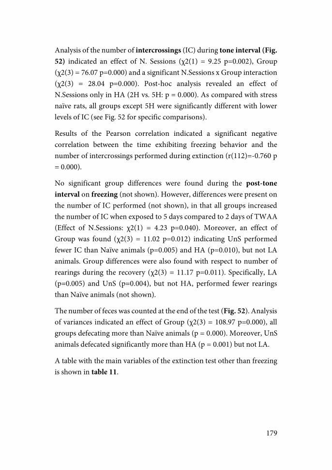
179
Analysis of the number of intercrossings (IC) during tone interval (Fig. 52) indicated an effect of N. Sessions (χ2(1) = 9.25 p=0.002), Group (χ2(3) = 76.07 p=0.000) and a significant N.Sessions x Group interaction (χ2(3) = 28.04 p=0.000). Post-hoc analysis revealed an effect of N.Sessions only in HA (2H vs. 5H: p = 0.000). As compared with stress naïve rats, all groups except 5H were significantly different with lower levels of IC (see Fig. 52 for specific comparisons).
Results of the Pearson correlation indicated a significant negative correlation between the time exhibiting freezing behavior and the number of intercrossings performed during extinction (r(112)=-0.760 p = 0.000).
No significant group differences were found during the post-tone interval on freezing (not shown). However, differences were present on the number of IC performed (not shown), in that all groups increased the number of IC when exposed to 5 days compared to 2 days of TWAA (Effect of N.Sessions: χ2(1) = 4.23 p=0.040). Moreover, an effect of Group was found (χ2(3) = 11.02 p=0.012) indicating UnS performed fewer IC than Naïve animals (p=0.005) and HA (p=0.010), but not LA animals. Group differences were also found with respect to number of rearings during the recovery (χ2(3) = 11.17 p=0.011). Specifically, LA (p=0.005) and UnS (p=0.004), but not HA, performed fewer rearings than Naïve animals (not shown).
The number of feces was counted at the end of the test (Fig. 52). Analysis of variances indicated an effect of Group (χ2(3) = 108.97 p=0.000), all groups defecating more than Naïve animals (p = 0.000). Moreover, UnS animals defecated significantly more than HA (p = 0.001) but not LA.
A table with the main variables of the extinction test other than freezing is shown in table 11.
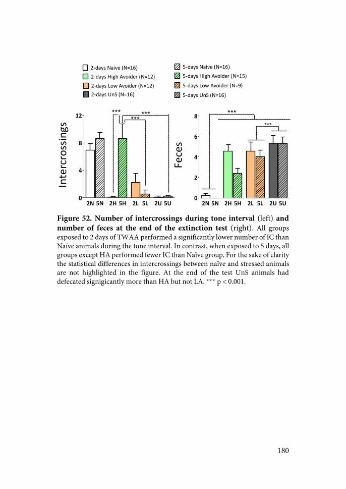
180
Figure 52. Number of intercrossings during tone interval (left) and number of feces at the end of the extinction test (right). All groups exposed to 2 days of TWAA performed a significantly lower number of IC than Naïve animals during the tone interval. In contrast, when exposed to 5 days, all groups except HA performed fewer IC than Naïve group. For the sake of clarity the statistical differences in intercrossings between naïve and stressed animals are not highlighted in the figure. At the end of the test UnS animals had defecated signigicantly more than HA but not LA. *** p < 0.001.
2N 5N 2H 5H 2L 5L 2U 5U0
4
8
12
2"days'Naive'(N=16) 5"days'Naive'(N=16)
2"days'High'Avoider'(N=12) 5"days'High'Avoider'(N=15)
2"days'Low'Avoider'(N=12) 5"days'Low'Avoider'(N=9)2"days'UnS'(N=16) 5"days'UnS'(N=16)
****** ***
Intercrossings
2N 5N 2H 5H 2L 5L 2U 5U0
2
4
6
8 ******
Feces
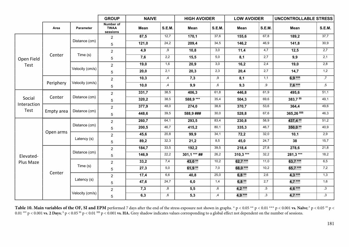
181
GROUP& NAIVE& HIGH&AVOIDER& LOW&AVOIDER& UNCONTROLLABLE&STRESS&
& Area& Parameter&Number&of&TWAA&sessions&
Mean& S.E.M.& Mean& S.E.M.& Mean& S.E.M.& Mean& S.E.M.&
Open%Field%Test%
Center%
Distance)(cm))2) 87,5& 12,7) 170,1& 37,6) 155,6) 67,8) 189,2) 37,7)
5) 121,0) 24,2) 209,4& 34,5) 146,2) 46,9) 141,8) 30,9)
Time)(s))2) 4,9) ,9) 10,8& 3,0) 11,4) 4,7) 12,5) 2,7)
5) 7,6) 2,2) 15,5& 5,0) 8,1) 2,7) 9,9) 2,1)
Velocity)(cm/s))2) 19,0) 1,6) 20,9& 3,0) 16,2) 2,4) 19,0) 2,8)
5) 20,0) 2,1) 20,3& 2,3) 20,4) 2,7) 14,7) 1,2)
Periphery% Velocity)(cm/s))2) 10,3) ,4) 7,3& ,9) 8,1) 1,1) 6,9&***) ,7)
5) 10,0) ,4) 9,9& ,6) 9,3) ,9) 7,6&***) ,5)
Social%Interaction%
Test%
Center% Distance)(cm))2) 331,7) 38,5) 406,3& 61,8) 446,8) 61,9) 495,6) 51,1)
5) 320,2) 38,5) 588,9&***& 35,4) 504,3) 69,6) 383,7&$$) 49,1)
Empty%area% Distance)(cm))2) 377,9) 48,0) 274,0& 38,6) 370,7) 53,6) 364,4) 49,6)
5) 448,6) 39,5) 588,9&###& 30,0) 528,8) 67,6) 365,26&$$$) 46,3)
Elevated:Plus%Maze%
Open%arms%Distance)(cm))
2) 260,7) 64,1) 293,5& 83,4) 230,8) 58,9) 437,4&**) 51,2)
5) 200,5) 46,7) 415,2& 80,1) 335,3) 46,7) 350,0&**) 40,9)
Latency)(s))2) 45,6) 20,8) 99,9& 34,1) 72,2) 32,0) 10,1) 2,9)
5) 89,2) 32,3) 21,2& 8,5) 45,0) 24,7) 38) 15,7)
Center%
Distance)(cm))2) 184,7) 33,5) 192,2& 39,5) 218,4) 27,8) 278,6) 21,8)
5) 146,9) 22,2) 301,1&***&##& 26,2) 310,3&***) 32,2) 281,3&***) 18,2)
Time)(s))2) 33,2) 7,4) 43,0&**& 10,2) 62,7&***) 11,0) 63,7&***) 6,5)
5) 27,3) 5,0) 61,9&**& 7,0) 68,0&***) 10,2) 65,7&***) 7,2)
Latency)(s))2) 17,4) 6,6) 40,8& 25,0) 6,8&**) 2,6) 4,3&***) 1,3)
5) 47,6) 24,7) 6,0& 1,4) 6,8&**) 2,7) 4,7&***) 1,6)
Velocity)(cm/s))2) 7,3) ,8) 5,5& ,6) 4,2&***) ,5) 4,6&***) ,3)
5) 6,3) ,6) 5,3& ,4) 4,9&***) ,3) 4,7&***) ,3)
Table 10. Main variables of the OF, SI and EPM performed 7 days after the end of the stress exposure not shown in graphs. * p < 0.05 ** p < 0.01 *** p < 0.001 vs. Naïve; # p < 0.05 ## p < 0.01 ### p < 0.001 vs. 2 Days; $ p < 0.05 $$ p < 0.01 $$$ p < 0.001 vs. HA. Grey shadow indicates values corresponding to a global effect not dependent on the number of sessions.
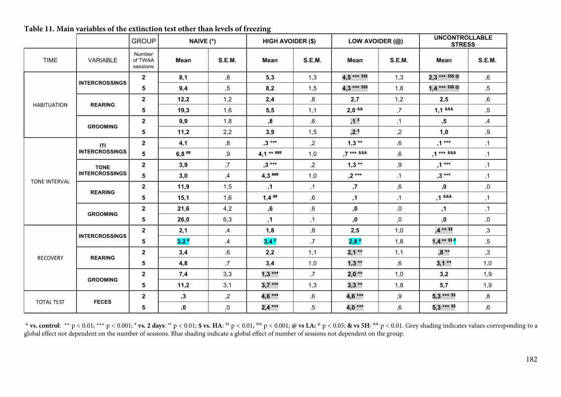
182
Table 11. Main variables of the extinction test other than levels of freezing
)) GROUP) NAIVE&(*)& HIGH&AVOIDER&($)& LOW&AVOIDER&(@)& UNCONTROLLABLE&STRESS&
TIME) VARIABLE)Number)of)TWAA)sessions)
Mean& S.E.M.& Mean& S.E.M.& Mean& S.E.M.& Mean& S.E.M.&
HABITUATION%
INTERCROSSINGS&2& 8,1) ,8) 5,3) 1,3) 4,5&***&$$$) 1,3) 2,3&***&$$$&@) ,6)
5& 9,4) ,5) 8,2) 1,5) 4,3&***&$$$) 1,8) 1,4&***&$$$&@) ,5)
REARING&2& 12,2) 1,2) 2,4) ,8) 2,7) 1,2) 2,5) ,6)
5& 19,3) 1,6) 5,5) 1,1) 2,0&&&) ,7) 1,1&&&&) ,5)
GROOMING&2& 9,9) 1,8) ,8) ,6) ,1&$) ,1) ,5) ,4)
5& 11,2) 2,2) 3,9) 1,5) ,2&$) ,2) 1,0) ,9)
TONE%INTERVAL%
ITI&INTERCROSSINGS&&
2& 4,1) ,8) ,3&***) ,2) 1,3&**) ,6) ,1&***) ,1)
5& 6,8&##) ,9) 4,1&**&###) 1,0) ,7&***&&&&) ,6) ,1&***&&&&) ,1)
TONE&INTERCROSSINGS&
2& 3,9) ,7) ,3&***) ,2) 1,3&**) ,9) ,1&***) ,1)
5& 3,0) ,4) 4,3&###) 1,0) ,2&***) ,1) ,3&***) ,1)
REARING&2& 11,9) 1,5) ,1) ,1) ,7) ,6) ,0) ,0)
5& 15,1) 1,6) 1,4&##) ,6) ,1) ,1) ,1&&&&) ,1)
GROOMING&2& 21,6) 4,2) ,6) ,6) ,0) ,0) ,1) ,1)
5& 26,0) 6,3) ,1) ,1) ,0) ,0) ,0) ,0)
RECOVERY%
INTERCROSSINGS&2& 2,1) ,4) 1,8) ,8) 2,5) 1,0) ,4&**&$$) ,3)
5& 3,2&#) ,4) 3,4&#) ,7) 2,8&#) 1,8) 1,4&**&$$&#&) ,5)
REARING&2& 3,4) ,6) 2,2) 1,1) 2,1&**) 1,1) ,8&**) ,3)
5& 4,8) ,7) 3,4) 1,0) 1,3&**) ,6) 3,1&**) 1,0)
GROOMING&2& 7,4) 3,3) 1,3&***) ,7) 2,0&**) 1,0) 3,2) 1,9)
5& 11,2) 3,1) 3,7&***) 1,3) 3,3&**) 1,8) 5,7) 1,9)
TOTAL%TEST% FECES&2& ,3) ,2) 4,6&***) ,6) 4,6&***) ,9) 5,3&***&$$) ,8)
5& ,0) ,0) 2,4&***) ,5) 4,0&***) ,6) 5,3&***&$$) ,6) * vs. control: ** p < 0.01; *** p < 0.001; # vs. 2 days: ## p < 0.01; $ vs. HA: $$ p < 0.01; $$$ p < 0.001; @ vs LA: @ p < 0.05; & vs 5H: && p < 0.01. Grey shading indicates values corresponding to a global effect not dependent on the number of sessions. Blue shading indicate a global effect of number of sessions not dependent on the group.
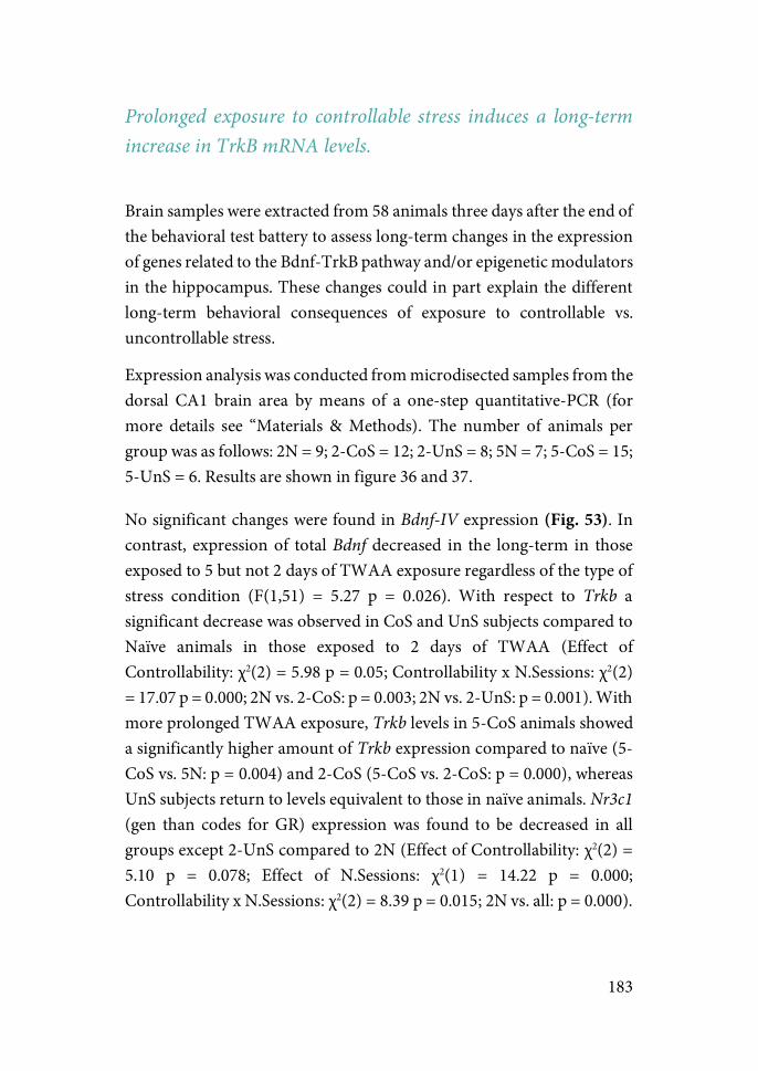
183
Prolonged exposure to controllable stress induces a long-term increase in TrkB mRNA levels.
Brain samples were extracted from 58 animals three days after the end of the behavioral test battery to assess long-term changes in the expression of genes related to the Bdnf-TrkB pathway and/or epigenetic modulators in the hippocampus. These changes could in part explain the different long-term behavioral consequences of exposure to controllable vs. uncontrollable stress.
Expression analysis was conducted from microdisected samples from the dorsal CA1 brain area by means of a one-step quantitative-PCR (for more details see “Materials & Methods). The number of animals per group was as follows: 2N = 9; 2-CoS = 12; 2-UnS = 8; 5N = 7; 5-CoS = 15; 5-UnS = 6. Results are shown in figure 36 and 37.
No significant changes were found in Bdnf-IV expression (Fig. 53). In contrast, expression of total Bdnf decreased in the long-term in those exposed to 5 but not 2 days of TWAA exposure regardless of the type of stress condition (F(1,51) = 5.27 p = 0.026). With respect to Trkb a significant decrease was observed in CoS and UnS subjects compared to Naïve animals in those exposed to 2 days of TWAA (Effect of Controllability: χ2(2) = 5.98 p = 0.05; Controllability x N.Sessions: χ2(2) = 17.07 p = 0.000; 2N vs. 2-CoS: p = 0.003; 2N vs. 2-UnS: p = 0.001). With more prolonged TWAA exposure, Trkb levels in 5-CoS animals showed a significantly higher amount of Trkb expression compared to naïve (5-CoS vs. 5N: p = 0.004) and 2-CoS (5-CoS vs. 2-CoS: p = 0.000), whereas UnS subjects return to levels equivalent to those in naïve animals. Nr3c1 (gen than codes for GR) expression was found to be decreased in all groups except 2-UnS compared to 2N (Effect of Controllability: χ2(2) = 5.10 p = 0.078; Effect of N.Sessions: χ2(1) = 14.22 p = 0.000; Controllability x N.Sessions: χ2(2) = 8.39 p = 0.015; 2N vs. all: p = 0.000).
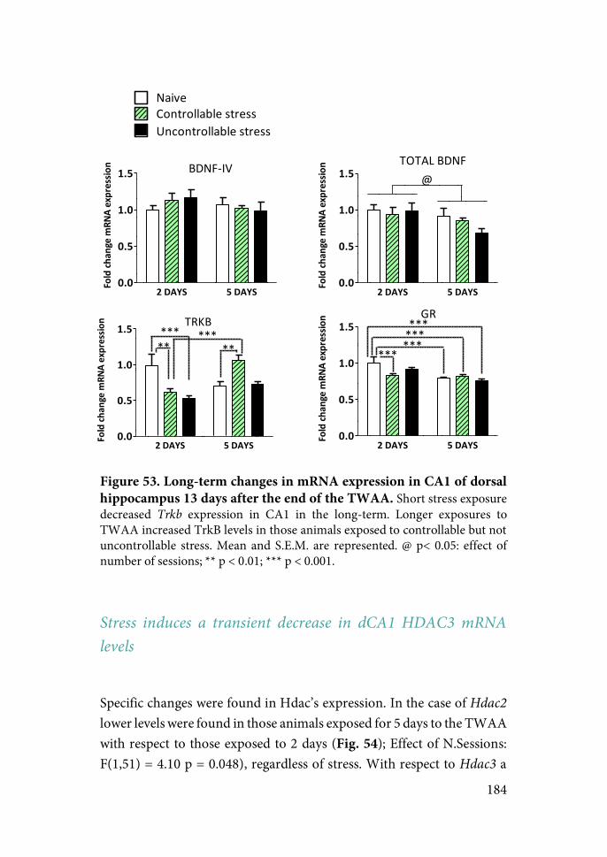
184
Figure 53. Long-term changes in mRNA expression in CA1 of dorsal hippocampus 13 days after the end of the TWAA. Short stress exposure decreased Trkb expression in CA1 in the long-term. Longer exposures to TWAA increased TrkB levels in those animals exposed to controllable but not uncontrollable stress. Mean and S.E.M. are represented. @ p< 0.05: effect of number of sessions; ** p < 0.01; *** p < 0.001.
Stress induces a transient decrease in dCA1 HDAC3 mRNA levels
Specific changes were found in Hdac’s expression. In the case of Hdac2 lower levels were found in those animals exposed for 5 days to the TWAA with respect to those exposed to 2 days (Fig. 54); Effect of N.Sessions: F(1,51) = 4.10 p = 0.048), regardless of stress. With respect to Hdac3 a
2"DAYS 5"DAYS0.0
0.5
1.0
1.5 BDNF%IV
Fold"ch
ange"m
RNA"expressio
n
2"DAYS 5"DAYS0.0
0.5
1.0
1.5TOTAL,BDNF
@
Fold"ch
ange"m
RNA"expressio
n
2"DAYS 5"DAYS0.0
0.5
1.0
1.5 TRKB
*****
Fold"ch
ange"m
RNA"expressio
n
*****
2"DAYS 5"DAYS0.0
0.5
1.0
1.5GR
***
*********
Fold"ch
ange"m
RNA"expressio
n
NaiveControllable,stressUncontrollable,stress
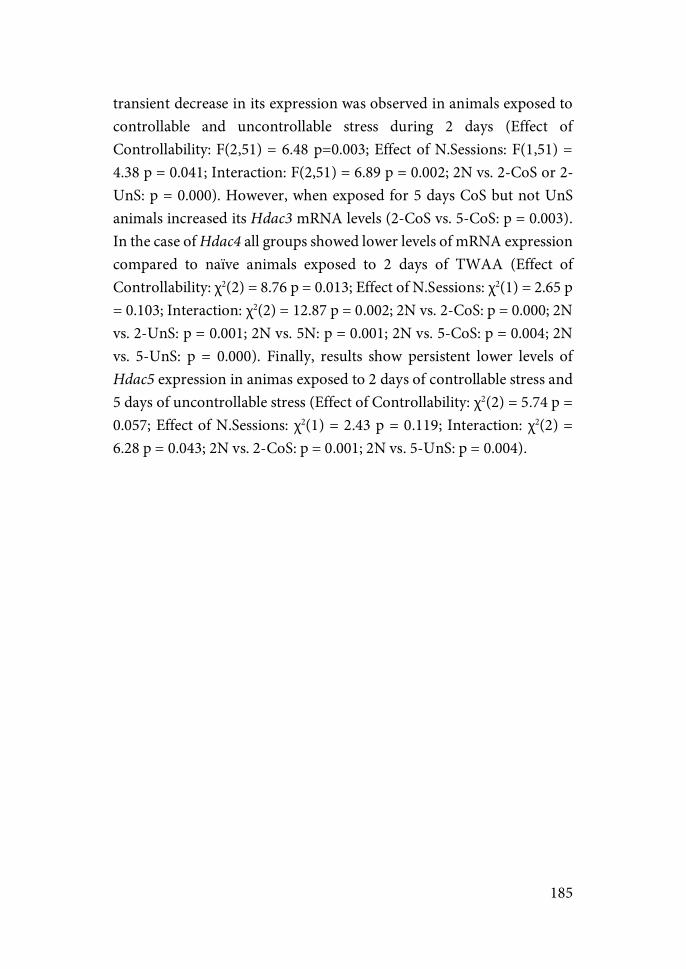
185
transient decrease in its expression was observed in animals exposed to controllable and uncontrollable stress during 2 days (Effect of Controllability: F(2,51) = 6.48 p=0.003; Effect of N.Sessions: F(1,51) = 4.38 p = 0.041; Interaction: F(2,51) = 6.89 p = 0.002; 2N vs. 2-CoS or 2-UnS: p = 0.000). However, when exposed for 5 days CoS but not UnS animals increased its Hdac3 mRNA levels (2-CoS vs. 5-CoS: p = 0.003). In the case of Hdac4 all groups showed lower levels of mRNA expression compared to naïve animals exposed to 2 days of TWAA (Effect of Controllability: χ2(2) = 8.76 p = 0.013; Effect of N.Sessions: χ2(1) = 2.65 p = 0.103; Interaction: χ2(2) = 12.87 p = 0.002; 2N vs. 2-CoS: p = 0.000; 2N vs. 2-UnS: p = 0.001; 2N vs. 5N: p = 0.001; 2N vs. 5-CoS: p = 0.004; 2N vs. 5-UnS: p = 0.000). Finally, results show persistent lower levels of Hdac5 expression in animas exposed to 2 days of controllable stress and 5 days of uncontrollable stress (Effect of Controllability: χ2(2) = 5.74 p = 0.057; Effect of N.Sessions: χ2(1) = 2.43 p = 0.119; Interaction: χ2(2) = 6.28 p = 0.043; 2N vs. 2-CoS: p = 0.001; 2N vs. 5-UnS: p = 0.004).
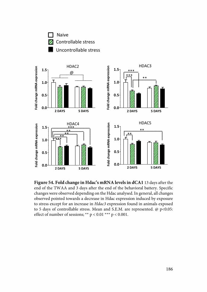
186
Figure 54. Fold change in Hdac’s mRNA levels in dCA1 13 days after the end of the TWAA and 3 days after the end of the behavioral battery. Specific changes were observed depending on the Hdac analysed. In general, all changes observed pointed towards a decrease in Hdac expression induced by exposure to stress except for an increase in Hdac3 expression found in animals exposed to 5 days of controllable stress. Mean and S.E.M. are represented. @ p<0.05: effect of number of sessions; ** p < 0.01 *** p < 0.001.
2"DAYS 5"DAYS0.0
0.5
1.0
1.5HDAC2@
Fold"ch
ange"m
RNA"expressio
n
2"DAYS 5"DAYS0.0
0.5
1.0
1.5HDAC3
****** **
Fold"ch
ange"m
RNA"expressio
n
2"DAYS 5"DAYS0.0
0.5
1.0
1.5HDAC4
**********
**
Fold"ch
ange"m
RNA"expressio
n
2"DAYS 5"DAYS0.0
0.5
1.0
1.5HDAC5
****
Fold"ch
ange"m
RNA"expressio
n
NaiveControllable6stressUncontrollable6stress
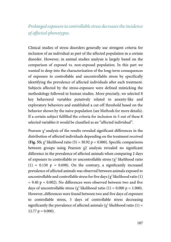
187
Prolonged exposure to controllable stress decreases the incidence of affected-phenotypes.
Clinical studies of stress disorders generally use stringent criteria for inclusion of an individual as part of the affected population in a certain disorder. However, in animal studies analysis is largely based on the comparison of exposed vs. non-exposed population. In this part we wanted to deep into the characterization of the long-term consequences of exposure to controllable and uncontrollable stress by specifically identifying the prevalence of affected individuals after each treatment. Subjects affected by the stress-exposure were defined mimicking the methodology followed in human studies. More precisely, we selected 8 key behavioral variables putatively related to anxiety-like and exploratory behaviors and established a cut-off threshold based on the behavior shown by the naive population (see Methods for more details). If a certain subject fulfilled the criteria for inclusion in 5 out of these 8 selected variables it would be classified as an “affected individual”.
Pearson χ2 analysis of the results revealed significant differences in the distribution of affected individuals depending on the treatment received (Fig. 55; χ2 likelihood ratio (5) = 30.92 p = 0.000). Specific comparisons between groups using Pearson χ2 analysis revealed no significant difference in the prevalence of affected animals when comparing 2 days of exposure to controllable or uncontrollable stress (χ2 likelihood ratio (1) = 0.150 p = 0.698). On the contrary, a significantly increased prevalence of affected animals was observed between animals exposed to uncontrollable and controllable stress for five days (χ2 likelihood ratio (1) = 9.40 p = 0.002). No differences were observed between two and five days of uncontrollable stress (χ2 likelihood ratio (1) = 0.000 p = 1.000). However, differences were found between two and five days of exposure to controllable stress, 5 days of controllable stress decreasing significantly the prevalence of affected animals (χ2 likelihood ratio (1) = 12.77 p = 0.000).
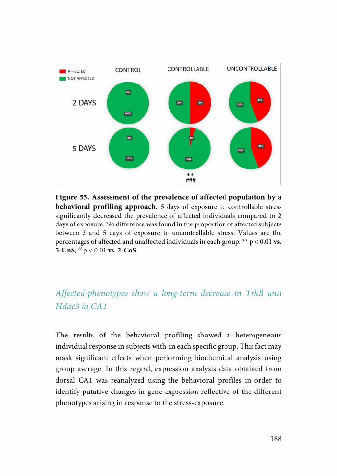
188
Figure 55. Assessment of the prevalence of affected population by a behavioral profiling approach. 5 days of exposure to controllable stress significantly decreased the prevalence of affected individuals compared to 2 days of exposure. No difference was found in the proportion of affected subjects between 2 and 5 days of exposure to uncontrollable stress. Values are the percentages of affected and unaffected individuals in each group. ** p < 0.01 vs. 5-UnS; ## p < 0.01 vs. 2-CoS.
Affected-phenotypes show a long-term decrease in TrkB and Hdac3 in CA1
The results of the behavioral profiling showed a heterogeneous individual response in subjects with-in each specific group. This fact may mask significant effects when performing biochemical analysis using group average. In this regard, expression analysis data obtained from dorsal CA1 was reanalyzed using the behavioral profiles in order to identify putative changes in gene expression reflective of the different phenotypes arising in response to the stress-exposure.
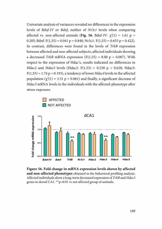
189
Univariate analysis of variances revealed no differences in the expression levels of Bdnf-IV or Bdnf, neither of Nr3c1 levels when comparing affected vs. non-affected animals (Fig. 56. Bdnf-IV: χ2(1) = 1.61 p = 0.205; Bdnf: F(1,55) = 0.041 p = 0.840; Nr3c1: F(1,55) = 0.653 p = 0.422). In contrast, differences were found in the levels of TrkB expression between affected and non-affected subjects, affected individuals showing a decreased TrkB mRNA expression (F(1,55) = 8.00 p = 0.007). With respect to the expression of Hdac’s, results indicated no differences in Hdac2 and Hdac5 levels (Hdac2: F(1,55) = 0.238 p = 0.628; Hdac5: F(1,55) = 1.74 p = 0.193), a tendency of lower Hdac4 levels in the affected population (χ2(1) = 3.51 p = 0.061) and finally, a significant decrease of Hdac3 mRNA levels in the individuals with the affected-phenotype after stress-exposure.
Figure 56. Fold change in mRNA expression levels shown by affected and non-affected phenotypes obtained in the behavioral profiling analysis. Affected individuals show a long-term decreased expression of TrkB and Hdac3 genes in dorsal CA1. **p<0.01 vs not affected group of animals.
0.0
0.5
1.0
1.5
NOT$AFFECTED$AFFECTED
dCA1
****
TrkB Hdac3 Hdac4Bdnf-IV Bdnf Nr3c1 Hdac2 Hdac5
Fold%ch
ange%m
RNA%expressio
n

190
DISCUSSION
The rationale for animal models of PTSD is that a punctual exposure to a stressor of elevated intensity may be sufficient to induce long-lasting behavioral sequelae reminiscent of the symptoms observed in PTSD patients (i.e. dysfunction in fear memory processing, anxiety regulation, maladaptive avoidance and hyperarousal). While the vast majority recover within the following month, 20-30% of the population exposed to the same stressful event will progress to develop PTSD, a variability reflected in some animal models of PTSD. Importantly, while factors such as determined genetic polymorphisms or a history of childhood adversity have been shown to partially explain this variability in humans in response to trauma, an extensive margin of variance remains unexplained even in studies controlling for these factors, suggesting the existence of further determinants contributing to the development of psychopathology after a traumatic experience. In this regard, several human and animal studies have pointed at psychological factors such as the controllability of the stressor as a critical component in determining the outcome of exposure to stress, concretely, exerting control over the stressor buffering part of the detrimental effects of exposure to the same amount of uncontrollable stress. Furthermore, even if exposed to the same traumatic experience, individual differences arise in the aftermath of trauma.
Within this framework, the present project was developed to: (i) determine whether acute exposure to uncontrollable stress in the context of an active avoidance task is sufficient to induce long-lasting behavioral effects on activity, arousal and anxiety-like behaviors; (ii) ascertain whether exposure to the same amount of controllable stress buffers the negative long-term impact observed after exposure to uncontrollable stress; (iii) determine if a certain level of experience of controllability is necessary to obtain the benefits of controllability to overcome the negative impact of exposure to stress and; and (iv) evaluate whether

191
changes in the expression of genes related to synaptic plasticity and epigenetic regulation in the main regions in charge of regulating the stress response (i.e PFC, amygdala and hippocampus) may be part of the molecular mechanisms driving the individual difference in the behavioral outcomes of exposure to controllable vs. uncontrollable stress.
Two-way Active Avoidance: Individual differences & Effects on fear-conditioning
Individual differences in avoidance acquisition
In our study animals were exposed to two or five days of an active avoidance task depending on the experiment and group. In all cases marked differences in the final avoidance learning were observed in CoS animals at the end of two days of TWAA and also after prolonged TWAA exposure. Two groups were clearly defined characterized by high avoidance rates (High Avoiders) or low avoidance rates (Low Avoiders) by the end of the task.
Individual differences in active avoidance acquisition have been consistently observed in past studies. While the majority of studies excluded animals not reaching a certain threshold of avoidance responses by the end of the task, a few have taken advantage of these differences to study differences in the neural circuitry regulating avoidance acquisition (Choi et al., 2010; Galatzer-Levy et al., 2014; Martinez et al., 2013). In our study, when possible, subpopulations based on the final rate of avoidance conditioning were identified and studied separately with the aim of avoiding masking the possible effects of controllability on the behavioral and biochemical consequences of stress-exposure due to the variability in the level of performance of the animals in the TWAA task. Notably, while most of the studies characterize two groups, Galatzer-Levy et al. (2014) identified under the same task (five

192
days of TWAA) four populations with different learning trajectories. This suggests that variability in avoidance acquisition may be even higher than that our study was able to characterize. Interestingly, high variability in avoidance acquisition has also been found in human studies, linked to differences in the neural activity of the same regions found to be implicated in avoidance conditioning in rodents (Collins et al., 2014).
When searching for the time-course of the appearance of individual differences, we found that differences between HA and LA emerge from the very first exposure to the aversive stimuli, and significant differences are found not only in the number of avoidance responses during the first day but also in the latency to escape during the first blocks. These data indicate that whereas HA responded to the first exposures to the US in a proactive way shortening rapidly their latency to shuttle, LA responded more passively exhibiting longer latencies to shuttle and escape from the US. Moreover, even at the end of the fifth session, LA keep performing mostly escape responses showing a profound deficit in avoidance acquisition. This suggests that the differing way in which individuals confront and respond to the first aversive stimuli determine the way the task is acquired and performed even after repeated experience with the situation.
Studies on the neurocircuitry implicated in regulating defense responses to threat have pointed towards the involvement of different pathways in the execution of defense reactions (escape) vs. defense actions (avoidance) (Campese et al., 2015). Importantly, in the process of acquiring avoidance conditioning, there is an initial phase in which defensive reactions evoked by the CS (i.e. freezing) are in direct conflict with the instrumental action needed to be executed to terminate (escape) or post-pone (avoidance) the US. Thus, in order to execute the motor action needed to escape or avoid the US (i.e. shuttling), inhibitory inputs must reach CeA in order to inhibit the defensive freezing reaction elicited by the CS. LeDoux’s lab have shown that while electrolytic and excitotoxic lesions of LA and BA nuclei performed after having learned

193
the task (5 days of TWAA) hindered avoidance conditioned responses and increased avoidance latency, the same lesions in CeA had no effect (Choi et al., 2010). In another study the authors showed that pre-TWAA lesions of LA and BA, but not CeA, impaired active avoidance acquisition as well (Lázaro-Muñoz, et al. 2010). Importantly, these lesions were performed in animals having reached more than 80% of avoidance responses in at least 7 days of training (corresponding to the average 80% of avoidance performance of HA in our study). Moreover, animals not reaching 20% of avoidance responses after 3 days of TWAA were designated as “poor performers” (in agreement with the average 20% of avoidance responses shown by LA in our study). Notably, in both studies electrolytic lesions of CeA in poor performers increased drastically the rate of avoidance until reaching close to 80% of avoidance responses after 5 days of TWAA.
Based on the knowledge about the neurocircuitry implicated in Pavlovian conditioning, and the role of this process as a first stage in acquisition of avoidance conditioning, the results suggest that lesions of LA may impede access to the associative CS-US learning stored in this region required for reinforcing and motivating the instrumental action of avoidance. In the other hand, BA would play an important role in the use of these LA-based CS-US association for producing actions through projection to striatal regions (Robbins et al. 1989), in line with previous studies showing that whereas LA and CeA mediate fear reactions, LA and BA mediate fear-motivated actions (Amorapanth et al. 2000). Furthermore, in agreement with our results showing a higher latency to escape in low avoiders from the first blocks, negative correlation rates were found in previous studies between freezing levels during the first five inter-trial intervals and avoidance performance during the session using different subtypes of genetically heterogeneous rats (Vicens-Costa et al. 2011). These results, together with evidence from our work of persistent deficits in avoidance performance in low avoiders even after prolonged exposure to the task, suggest that excessive CeA activity in low avoiders could interfere with acquisition and expression of avoidance,
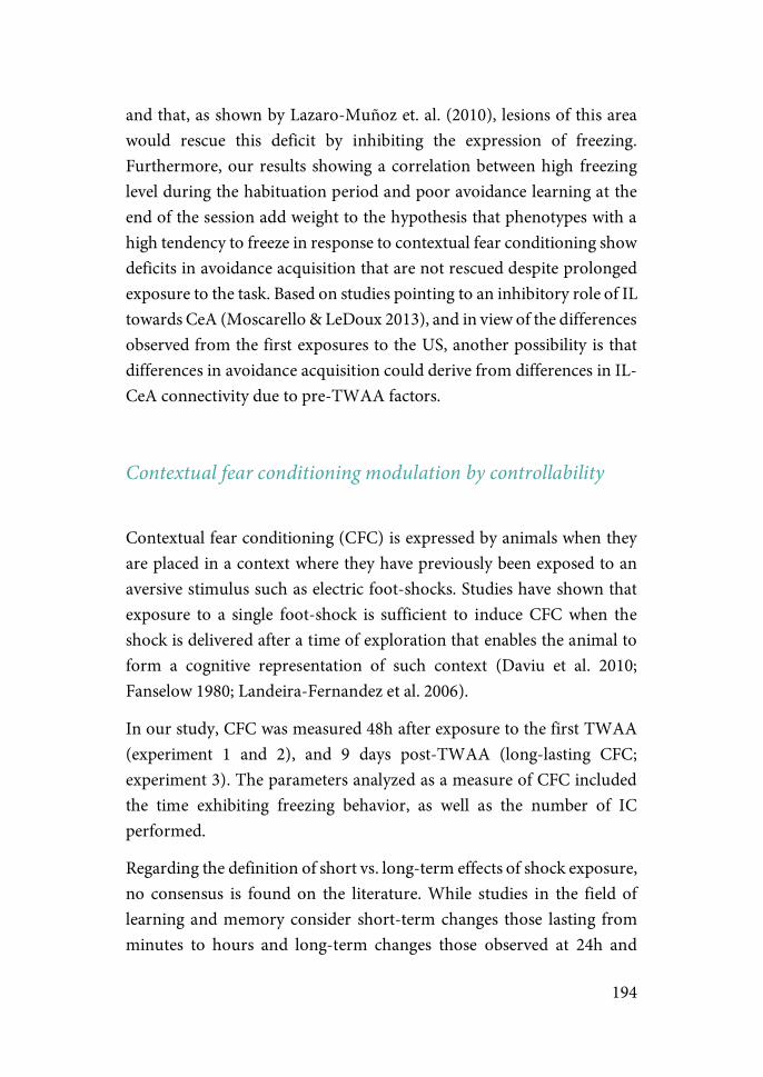
194
and that, as shown by Lazaro-Muñoz et. al. (2010), lesions of this area would rescue this deficit by inhibiting the expression of freezing. Furthermore, our results showing a correlation between high freezing level during the habituation period and poor avoidance learning at the end of the session add weight to the hypothesis that phenotypes with a high tendency to freeze in response to contextual fear conditioning show deficits in avoidance acquisition that are not rescued despite prolonged exposure to the task. Based on studies pointing to an inhibitory role of IL towards CeA (Moscarello & LeDoux 2013), and in view of the differences observed from the first exposures to the US, another possibility is that differences in avoidance acquisition could derive from differences in IL-CeA connectivity due to pre-TWAA factors.
Contextual fear conditioning modulation by controllability
Contextual fear conditioning (CFC) is expressed by animals when they are placed in a context where they have previously been exposed to an aversive stimulus such as electric foot-shocks. Studies have shown that exposure to a single foot-shock is sufficient to induce CFC when the shock is delivered after a time of exploration that enables the animal to form a cognitive representation of such context (Daviu et al. 2010; Fanselow 1980; Landeira-Fernandez et al. 2006).
In our study, CFC was measured 48h after exposure to the first TWAA (experiment 1 and 2), and 9 days post-TWAA (long-lasting CFC; experiment 3). The parameters analyzed as a measure of CFC included the time exhibiting freezing behavior, as well as the number of IC performed.
Regarding the definition of short vs. long-term effects of shock exposure, no consensus is found on the literature. While studies in the field of learning and memory consider short-term changes those lasting from minutes to hours and long-term changes those observed at 24h and
195
beyond (Goelet et al. 1986), studies on the field of stress usually refer to short-term effects those observed immediately after the last session of stress or in the following 24-48h, while long-term effects are considered those observed at least three days post-stress exposure (Armario et al. 2008). Within the context of the present work we will consider the latter timing definition of short and long-term changes, when referring to behaviors not directly related to fear conditioning.
Contextual*Fear*Conditioning**
Results from experiment 1 show that all groups except HA exhibit elevated levels of fear conditioning when exposed to the TWAA context at 48h. These results point to two main conclusions: (i) exerting control over the stressor induces a buffering effect on CFC (CoS vs. Uns); and (ii) in order for the possibility of control to buffer the negative impact of stress a certain level of controllability must be acquired (HA vs. LA). Results from experiment 2 are less clear. No significant effects on CFC conditioning were found due to controllability or to the different rate of avoidance conditioning acquired. Although they may seem to contradict results of the experiment 1, differences in the latency to escape and the number of avoidance responses in the last block on day 1 between HA and LA on both experiments may explain this difference. In experiment 2, HA and LA show less difference in their latency to escape between HA and LA than in experiment 1 (LA are not so slow to shuttle once they receive the shock as LA in experiment 1). This suggests that they may differ to a lesser extent in their tendency to freeze in response to aversive stimuli, a factor shown to critically influence avoidance acquisition (Vicens-Costa et al. 2011). In fact, smaller differences are also found between HA and LA in the avoidance responses at the end of session 1, which in turn may explain a more homogeneous response with respect to CFC 48h later in this experiment.
196
Our results showing a significant correlation between freezing during the habituation on day 2 and avoidance responses on that session are also in agreement with the results found by Vicens-Costa et al. (2011). Furthermore, they are in line with the hypothesis supported by the literature stating that an initial passive response (due to conditioned freezing) runs against the appearance of active escape/avoidance responses (Choi et al. 2010; Fadok et al. 2017; Lázaro-Muñoz et al. 2010; Martinez et al. 2013). The impact of CFC on avoidance acquisition is well stablished. Weiss et al. (1968) showed that exposure to eight foot-shocks induced marked deficits in later avoidance acquisition, and that decreasing freezing levels by extinction procedures previous to avoidance training rescued these deficits in poor performers, supporting the critical role of fear conditioning in the appropriate acquisition of avoidance.
With respect to the effects of controllable vs. uncontrollable stress, previous studies have shown how exerting control over the stressor buffers associative deficits shown by animals that have been exposed to the same amount of uncontrollable stress (Baratta et al. 2007; 2008). Results from experiment 1 are in line with these studies, as UnS groups show an enhanced CFC compared to HA 48h post-stress exposure. Due to the yoked procedure, latencies to escape in CoS group determine the total time of shock received by the UnS group. As with the differences in avoidance conditioning, the fact that HA and LA in experiment 2 show similar latencies to escape than HA in experiment 1, indicate that UnS animals in experiment 2 received similar amounts of shock than HA in experiment 1 (i.e. less than LA in experiment 1). This difference in the total time of shock could underlie the lack of a controllability effect in the second experiment. However, other studies using the same task showed a lack of effect of controllability 24h after a first TWAA session (Ilin et al. 2009) suggesting that further studies need to be undertaken to confirm the results from experiment 1 showing a differential short-term effect of controllability on fear conditioning.
197
Effect*of*prolonged*TWAA*exposure*on*fear*conditioning*
In experiment 3 we wanted to know how more prolonged exposure to controllable and uncontrollable shocks would affect the overall behavioral impact (see later) and fear conditioning. Our hypothesis was that the possible beneficial effects of controllability will increase with more prolonged experience of control in the TWAA task (2 versus 5 days). Two additional questions were posed with respect to fear conditioning: (i) how CFC is modified by more sessions of exposure to controllable stress; (ii) if the number of stress sessions differentially affect fear conditioning when assessed 10 days post-stress. ; and (iii), if yes, uncontrollable stress does induce a long-lasting increase in CFC, if exposure to the same amount of controllable stress is able to buffer this effect.
CFC was evaluated by the number of IC during the habituation period corresponding to each session. Although IC is an indirect measure of fear conditioning, negative correlations rates higher than 0.7 between the number of IC performed and the freezing time were found in our lab in previous experiments, validating the number of IC as an approximation to measure fear conditioning levels. However, measures of freezing during these periods would be valuable to confirm these results. Analysis of the IC performed during the habituation period indicate that differences associated with the controllability of the stressor emerged from the third session. In the case of differences between HA and LA, no significant differences were found even on session 5. Nevertheless, a tendency to lower fear conditioning levels in HA was present, suggesting that a more prolonged exposure to the task could eventually lead to significant differences in CFC between HA and LA as well.
These results are in line with previous studies (Lázaro-Muñoz et al. 2010; Martinez et al. 2013) where they show that CFC differences between HA and LA arise after prolonged exposure to the task (10th and 7th session respectively) due to a decrease in the freezing levels shown by HA that

198
does not occur in LA. This suggest that although LA exert a certain level of control by escaping from the US, this instrumental response in not sufficient to acquire the stress-buffering effects of controllability induced by the opportunity of voidance.
Differences in the engagement of the BA-NAc circuits could underlie such differences in CFC. While data suggest that HA are able to engage this pathway (Ramirez et al. 2015), in LA rats, the persistence of exposure to the US despite the execution of an instrumental response to escape from the shock may hinder the engagement of this pathway. In this study LA were removed so this remains to be elucidated. Importantly, inhibiting the NAcSh affected fear conditioning to the context of TWAA but not in the classical pavlovian fear conditioning paradigm, suggesting a concrete role of this region in regulating the freezing response based on the execution of instrumental responses. Another possibility that has been proposed (Cain & LeDoux 2008; Dombrowski et al. 2013) is that HA may have enhanced motivational control of actions and/or enhanced reward processing perhaps due to more efficient neural interactions between the amygdala and the NAc.
With respect to the impact of controllability on CFC, our results show that whereas animals exposed to controllable stress start reducing fear conditioning from the third session, animals exposed to uncontrollable stress keep exhibiting high levels of fear conditioning to the context after five TWAA sessions. Therefore, controllability is clearly mitigating CFC.
The need for repeated experience with the controllable stressor to exert buffering effects on the consequences of exposure to an aversive stimulus such as shock is in line with previous studies (Ilin & Richter-levin 2009). Results from Richter-Levin’s lab has shown that animals require prolonged exposures to controllable stress (usually six sessions) to decrease the negative consequences on fear conditioning, anxiety- and depression-like behaviors observed after exposure to the same amount of uncontrollable stress. Based on these results he has postulated the concept of “Operational vs. Emotional Control”. He argues that the
199
acquisition of control when exposed to a controllable stressor involves two separate phases. In a first step animals acquire the motor skills and learn the cognitive elements necessary to perform the instrumental response. After this phase, the animal would have acquired what he calls “Operational Control”, in which the animal knows how to perform the specific instrumental response needed to avoid the aversive stimuli, but because the training has been short, this action is not executed with ease and the animal still suffers from the negative consequences of the stressful situation. However, if the subject is repeatedly exposed to the controllable stressor, he argues that the animal will end up performing proficiently the instrumental action needed to stop the aversive stimulus, a phase in which this excellence in the performance would enable the animal to gain what he calls “Emotional Control”. Once this is reached, the animal is able to prevent the negative effects of the stressor associated with mood regulation. This hypothesis is in line with our results. The “operational control” phase would correspond to the first two sessions in which the animals have learned to avoid but still exhibit elevated levels of CFC. On the other hand, “emotional control” would appear from the third and following sessions, in which animals under controllable stress progressively decrease their levels of CFC.
Long9lasting*Fear*Conditioning*
In addition to evaluating the progression of fear conditioning as avoidance is progressively acquired, a second goal in experiment 3 was to assess whether the buffering effects of prolonged exposure to controllable versus uncontrollable stress on fear conditioning would persist over time. Thus, long-lasting effects on fear memory were assessed 10 days after stress-exposure in an extinction test. The test had three phases: habituation, tone interval and post-tone recovery. Freezing levels and the number of IC during the habituation were used as indicators of long-lasting contextual fear memory, whereas freezing
200
levels and the number of IC during the presentation of the tones were considered as a measure of long-lasting auditory fear memory.
The analysis of freezing during the habituation period of the extinction test performed 10 days after the last shock exposure indicated that exposure to uncontrollable stress induced a persistent increase in contextual fear memory, regardless of the number of sessions. Marked differences were observed in HA as compared with LA. An analysis of freezing time per blocks indicated that by the end of the last block significant differences arouse between HA exposed for 2 days compared to those exposed for 5 days, the latter exhibiting decreased freezing levels compared to the former. In contrast, LA showed similar levels of freezing as UnS, suggesting that reduced CFC after repeated experience of controllability is only achieved when the actual levels of control are high.
When the number of IC performed during the habituation period instead of the time spent freezing was considered, it was observed only a partially similar pattern. All stressed rats showed lower levels of IC than stress-naïve rats and these levels progressively reduced from HA to LA and then to UnS rats, which differ from both HA and LA rats. HA rats exposed to 5 sessions tended to have higher levels of IC than HA rats exposed to 2 sessions, whereas LA rats maintained the same level of IC after 2 or 5 sessions.
The different sensitivity of freezing and IC to detect group differences are not surprising as there is evidence for discrepancies between different measures in other fear conditioning procedures (e.g. Antoniadis and McDonald 1999; Gisquet-Verrier et al. 1999; Pisano et al. 2012). In addition, it is likely that learning that crossing to the other compartment reduced shock exposure might interfere with the purely reflection of IC as a measure of activity.
Results of the freezing levels showed during the extinction test revealed the same effect of controllability as during habituation. During exposure to the tones, only high avoiders exposed to 5 sessions showed decreased
201
freezing levels compared to all other groups, an effect further supported by differences in the number of IC performed.
Interestingly, no differences were found between short vs. prolonged exposure to uncontrollable stress, suggesting that prolonged exposures to uncontrollable stress would not lead to enhanced fear conditioning. It is likely that a plateau is soon reached in freezing to the uncontrollable shock context. Data about the contribution of shock intensity to fear conditioning are also controversial. Whereas some authors favor the idea that there is a positive relationship between intensity of shocks and conditioned freezing (Cordero et al. 1998), other studies have found that an increase in shock intensity leads, not to an increase in fear conditioning, but to enhanced fear generalization and fear sensitization (Baldi et al. 2004; Kamprath and Wotjak 2004). These results suggest that, in the case of inescapable shocks, the characteristics of the shock rather than the number of sessions of stress exposure have a higher influence on the long-lasting effects the stressor on CFC.
How prior exposure to incontrollable shocks affects further active escape or avoidance learning has been extensively studied by exposure to inescapable shocks previous to avoidance acquisition (Anisman & Waller, 1972; Brush & Levine, 1966; Steven F Maier & Watkins, 2005; Murua & Molina, 1991; Overmier & Seligman, 1967). Studies in which the inescapable shocks were administered in the same context than the avoidance test showed evidence of the induction of long-lasting escape deficits even after 22 days (Anisman & Waller, 1972; Steven F Maier & Watkins, 2005). In contrast to this, studies where these contexts were different showed differences that were not evident after 72h, suggesting a key role of context fear conditioning in the development of deficits in avoidance learning.
In contrast to the well-studied impact of prior exposure to inescapable shocks affects shock avoidance behavior, how prior experience of avoidance affects fear conditioning has been far less studied. In this regard, our results showing a long-lasting negative effect of exposure to
202
uncontrollable stress on fear conditioning, as well as a long-lasting positive effect of controllable stress, are in line with previous studies showing that animals exposed to controllable stress exhibit decreased levels of corticosterone and freezing in an extinction test performed two weeks post-TWAA (Hadad-Ophir et al., 2017; Horovitz et al., 2017; Ilin & Richter-levin, 2009). Our results are also in accordance with previous data from our laboratory in adolescent rats repeatedly exposed to a TWAA task or corresponding uncontrollable shocks, showing clearly reduced levels of freezing when exposed again during adulthood to the TWAA context (Sanchís et al., 2018). Beneficial effect of controllability on exploratory behaviors in new environments after one month has also been reported (Lucas et al., 2014).
Regarding the differential effect on fear conditioning of high and low avoiders, several studies have suggested that enhanced freezing and deficit in avoidance acquisition in the latter animals could be due to a higher CS-induced CeA activity as compared to high avoiders (Choi et al. 2010; Lázaro-Muñoz et al. 2010; Moscarello & LeDoux, 2013). However, results from a study by Martinez et al. (2013) are in contrast to this hypothesis. Twenty-four hours after seven Sidman active avoidance (Sidman-AA) sessions, neural activity was measured by c-fos expression in several brain areas including some amygdala subnuclei and the PFC when exposed to the context of avoidance conditioning 24h later. As expected, the authors showed decreased levels of freezing and increased number of shuttlings in high compared to low avoiders, together with higher number of c-fos activated neurons in the IL and medial CeA of high avoiders, in contrast to the stablished role of CeA in favoring CS-induced freezing behavior after pavlovian conditioning. Interestingly, several studies have point out to the existence of a different contribution of lateral versus medial subdivisions of the CeA to conditioned fear that could explain these results (Ciocchi et al. 2010; Haubensak et al. 2010).
203
Effects of controllability on behavior and HPA axis
Behavioral effects of exposure to controllable vs. uncontrollable stress were evaluated in two different experiments. In experiment 1 after exposure to 2 sessions of stress we evaluated behavior of rats in the EPM, the OF and the ASR, 2 or 5 days after the last stress exposure. In experiment 3, after 2,5 or 7 sessions of stress we evaluated animals in the OF, the social interaction test and the EPM, on days 7 and 8 after the last stress exposure.
When evaluating the influence of controllability and amount of shock received on behavior in the EPM, the results of the first experiment indicate that only the number of shocks affected behavior, regardless of controllability. More precisely, greater total activity in the apparatus and distance and time in the open arms was observed in those receiving less shocks (HA, U-HA), although uncontrollable stress also tended to reduce these variables. In fact when stress naïve rats were considered as the reference, only HA rats showed signs of reduced anxiety and greater number of shocks caused certain levels of hypoactivity. However, in the OF only the factor controllability was significant, UnS exposure resulting in reduced overall activity of rats.
In the other experiment where behavior was studied (experiment 3), one week was imposed between the last exposure to shock and testing. The comparison of data from the two experiments lack consistency when analyzed in detail. It is important to consider that the experiments were done in different laboratories and the origin of the animals as well as the conditions of the animal facilities were clearly different. Inconsistencies were evident in the EPM in that all stressed rats tended to show signs of reduced anxiety, the effect being significant after exposure to uncontrollable shocks. In this sense, Hadad-Ophir et al. (2016) using a similar design showed lower rates of anxiety-like behaviors in animals
204
exposed to controllable stress compared to naïve animals when tested in an EPM 2 weeks after exposure to six days of TWAA.
Results in the literature regarding the effects of shock exposure on behavior in the EPM are markedly controversial, with results showing increased, decreased and no change in the time spent in the open arms (e.g. Daviu et al., 2010; Grahn et al. 1995; Radulovic et al. 1998; Steenbergen et al. 1990; 1991). The reasons for the discrepancies are unclear. Some other studies have shown stressed animals to spend more time in the open arms than non-stressed animals. For instance, Dopfel et al. (2019) showed that animals exposed to predator-scent stress spent more time in the open arms of the EPM conducted one week post-stress, in contrast to other previous results with the same type of stressor or after direct exposure to predators (see Armario et al. 2008; Staples 2010 for review).
Animals spending more time in the closed arms have been generally interpreted by the literature as animals with high levels of anxiety-like behaviors. This interpretation derives from the view that “anxious” animals will prefer to stay protected in the closed arms than to take the risk of exploring an open area despite its natural instinct of exploring new environments. One possible explanation for the UnS group to spend more time in the open arms could come from a process of fear generalization due to the similarity of the context of the enclosed arms to the dark compartment in the shuttle where the animals received the shock. In fact, evidence for generalization of fear to other completely contexts after exposure to foot-shocks has been shown by previous studies (Daviu et al. 2010; Radulovic et al. 1998).
In the OF, uncontrollable but not controllable shocks caused hypoactivity, whereas the experience of control over shocks exerted a modest anxiolytic effect (distance and time spent in the center). Therefore, it appears that exposure to UnS consistently caused hypoactivity in OF and the effects of stress are mitigated by control. In contrast to the behavior in the EPM, more consistent data are reported

205
regarding activity in OF. Our results agree with results from previous studies from our lab and others where it is shown that fear conditioning is induced after exposure to even a single foot-shock and that this fear conditioning is responsible for inducing hypoactivity when later exposed to novel environments non-related to the original context of shock-exposure (Daviu et al. 2010; 2014; Radulovic et al. 1998; Van Dijken et al. 1992). Interestingly, our lab has demonstrated that this phenomenon is dependent on the development of fear conditioning (Daviu et al. 2010). This phenomenon has been designated as “cognitive generalization” by our lab to distinguish it from the generalization process that occur when exposed to similar contexts than the one of the stress exposure (Daviu et al. 2010; 2014). Thus, the increased fear conditioning levels shown by UnS animals during the TWAA compared to CoS animals would give weight to the hypothesis of a relation between increased fear conditioning and the facilitation of processes of fear generalization to other contexts.
The possible effect of shock exposure to cause hyperarousal was assessed by an ASR performed one-week post-TWAA. Results indicate a diminished response (as measured by the averaged voltage evoked) in LA as well as in UnS animals yoked to HA as compared with HA. Results of the maximum voltage evoked showed a diminished response in LA compared to HA. The response of the naïve animals suggests that this significant difference derives from an increased response in HA. We can speculate that during the TWAA, HA may have acquired a state of alert and focused attention towards environmental stimuli in order to respond quickly to the CS and avoid the US, and that this state of alert may generalize to different contexts. Another interesting possibility is that a pre-stress factor, such as a more sensitive startle response, is facilitating avoidance acquisition. In this regard, Vicens-Costa et al. (2011) observed a positive correlation between basal startle response (pre-TWAA) and avoidance rates in the subsequent exposure to the TWAA.
206
In the same rats tested for the ASR, the activation of the HPA axis was studied to show whether or not stress caused sensitization of this stress system. Prior stress did not alter the ACTH and corticosterone responses to the ASR test as compared with shock-naïve animals, and no influence of the number of shocks or controllability was observed. It has been previously reported that acute exposure to severe stressors, including foot-shocks, consistently resulted in sensitization of the HPA response to additional stressors (Belda et al., 2012; Belda et al., 2015; Belda et al., 2008; Johnson, 2002; O’Connor et al., 2003), particularly when the challenging stressor is of low intensity and short duration (Belda et al., 2016). However, this phenomenon is transient and can disappear in less than one week, although occasionally longer lasting sensitization has been reported (O’Connor et al., 2003). As the animals were tested in the ASR 7 days after shock exposure, it appears that under the present conditions HPA sensitization had already vanished. In any case, control over stress does not appear to affect the degree of sensitization of the HPA axis (O’Connor et al., 2003).
Individual variability in fear response
Individuals exposed to a traumatic experience show marked heterogeneity in their response (Galatzer-Levy & Bryant 2013; Yehuda & LeDoux 2007). Whereas some react exhibiting resilience to stressful situations, others show maladaptive behaviors and develop significant symptomatology related to stress and mood regulation. Despite this critical feature, animal models thought to give relevant information about the course and etiology of stress psychopathologies (e.g. PTSD models) typically use the mean response of the whole population, thus masking the difference in fear response among subpopulations.
In order to avoid masking these subpopulations in our study, a behavioral profiling was conducted in experiment 3. “Affected” and
207
“non-affected” populations were identified by comparison with the normal distribution of the behavior in naïve animals. More precisely, activity and anxiety-related parameters of the behavioral battery of tests conducted one-week post-stress were used for this characterization.
The results of this profiling approach revealed that whereas with acute exposure to the stressor the percentage of affected population was around 50 percent in both CoS and UnS groups, when the exposure is prolonged to five sessions a marked decrease in the affected population occurred in CoS but not UnS, which remained with the same percentage of affected individuals. These results give strong support to our working hypothesis that exposure to prolonged controllable stress exert a beneficial buffering effect over the consequences of exposure to uncontrollable stress.
Importantly, in contrast to the poor results obtained when studying the effect of controllability on single parameters in the behavioral tests, a clear effect is obtained when taking into consideration a combination of many of the factors involved. These results highlight the value of the profiling approach to explore the existence of subpopulations and detect effects that may be masked when studying mere mean values in the population.
In fact, in the last decade, researchers have begun to adopt this approach to characterize heterogeneous populations in animal studies yielding fruitful results on the molecular mechanisms involved in these differences. In the field of animal models of stress disorders, distinct populations have been identified based on factors such as the ability and rate to extinguish fear responses (Bush et al. 2007; Cowansage et al. 2013; Galatzer-Levy et al. 2013) or their ability to learn an instrumental response to avoid an aversive stimulus (Choi et al., 2010). Furthermore, using the same behavioral profiling approach of our study in underwater-trauma exposed rats, Ritov et al. (2016) identified three distinct phenotypes of stress response (anxious, anhedonic or exposed-unaffected), each of them characterized by the involvement of different
208
limbic regions and distinct activity patterns. Hence, such an approach shows promising value in the identification of neurobiological markers involved in the development of adaptive vs. maladaptive responses to stress exposure. Nevertheless, we still need more in deep characterization of the factors involved in the behavior of animals in the different tests to better distinguish between affected and not affected subjects.
Molecular mechanisms underlying the effects of controllability
Decades of research have highlighted the key role for active changes in neuronal connectivity as a controller of behavior and inductors of behavioral change (Kandel, 1976; Willows, 1971), including behaviors associated to threat responsivity (J.E. LeDoux & Phillips, 1992). Processes such as changes in post-synaptic receptor density or synaptic pruning together with dendrites remodeling have been shown to be associated with activity-dependent neural changes in synaptic strength and lately on behavior (Champagne et al. 2008; Lamprecht & LeDoux 2004; Schafe et al. 2001) . Regulation of gene transcription was found to be a bridge between experience and behavioral change, and epigenetic regulation a crucial mechanism in the induction of these changes in gene transcription underlying learning, memory and experience-dependent behavioral changes (J. J. Day & Sweatt, 2011; Levenson & Sweatt, 2005b; N. M. Tsankova et al., 2006; Zovkic & Sweatt, 2013a).
In the second experiment we studied the impact of two sessions of stress on a wide range of biochemical parameters in critical brain areas (PFC, amygdala, dorsal HF and PVN) when measured 2 or 7 days after stress. We focused on two main types of parameters: those related to BDNF and those related to epigenetics, particularly HDACs. In the third experiment, some variables were also measured 10 days after exposure to short vs. extensive TWAA training. These factors has been extensively
209
studied regarding stress and learning in the literature (Blugeot et al., 2011; Gray et al., 2013; Penney & Tsai, 2014; White & Wood, 2014a). However, despite the large number of studies showing a link between BDNF function and the regulation of fear and memory processes in animal models of stress and psychiatric disorders, and the evidence showing that exposure to controllable stress induces buffering effects on some of these processes occurring after exposure to uncontrollable stress, to our knowledge, only a few studies to date have assessed the differential effects of control over stress on BNDF-TrkB function and none has studied the impact on HDAC expression.
Changes in BDNF expression has been associated with processes of stress response regulation (Bath et al. 2013; Berton et al. 2006; Murakami et al. 2005), long-term memory (Radecki et al. 2005; Rosas-vidal et al. 2014; Schaaf et al. 2000; Takei et al. 2011) and diseases associated with malfunctions of memory and mood regulation (Andero & Ressler, 2012; Calabrese et al. 2009; Martinowich et al. 2007; Naert et al. 2011; Takei et al. 2011). The changes in BDNF associated with fear and memory regulation have been linked to epigenetic modulation mainly through changes in the methylation state of its promoter regions or post-translational histone modifications (Lubin et al. 2008; Mitchelmore & Gede, 2014; Règue-Guyon et al. 2018; Rodrigues et al. 2015; Tsankova et al. 2004).
In most parameters, no effect of stress exposure was observed. However, Bdnf and Bdnf-IV transcript decreased in CA1 after 2 days of exposure to UnS but not CoS, the effect disappearing after 7 days. No changes were observed in the other regions of the HF or in other brain areas studied. Similarly, expression of the TrkB receptor was not affected by stress. In contrast, results from the third experiment showed that two sessions of stress induced a long-lasting decrease in TrkB regardless of the controllability of the stressor and that more prolonged stress exposure persistently increased TrkB mRNA levels in CoS but not UnS animals compared to stress-naïve rats and CoS animals exposed only to two TWAA sessions.
210
Several studies have reported a stress-induced decrease in hippocampal Bdnf expression after acute and chronic stress (Rasmusson et al., 2002; Smith et al., 1995; Ueyama et al., 1997), and acute and chronic corticosterone treatment (Schaaf et al., 1997; 1998; 2000; Smith et al., 1995). Interestingly, a study reported specific changes in Bdnf expression only in animals identified as affected after predator-odor stress, in which a decrease in Bdnf mRNA levels was detected one week post-stress in DG and CA1, associated with an increase in its receptor TrkB in the same regions (Kozlovsky et al., 2007). Moreover, studies from the last decade have shown decreased levels of hippocampal Bdnf linked to epigenetic regulation. Tsankova et al. (2006) found that chronic social defeat decreased total Bdnf and Bdnf-IV mRNA levels in the hippocampus, concomitant to increases in the repressive histone methylation of its promoters. In agreement with these results, Roth et al. (2011) also reported a decrease in the expression of Bdnf-IV mRNA levels in DG and CA1 and DG, whereas an increase was observed in CA3, and no change in the PFC or amygdala. These results are at odds with previous work from the same authors, who found no changes in the hippocampus after early-life stress, but decreased levels of Bdnf-IV mRNA in the PFC (Roth et al., 2009). Furthermore, other studies have reported an increase in Bdnf expression in response to stress and fear-memory processes. Stress-induced increases in hippocampal Bdnf-IV mRNA and protein levels have also been reported in response to fear conditioning one-week after an SPS protocol (Takei et al., 2011). Other studies have reported a bidirectional change in Bdnf depending on the duration of the exposure: short exposure (15 min) to immobilization stress increased Bdnf mRNA levels in the hippocampus, whereas prolonged exposure (3h) decreased them (Marmigère et al., 2003). Similarly, Pizarro et al. (2004) showed decreased Bdnf mRNA in DG, CA3 and CA1 24 h after 10 min of social defeat but not at shorter times.
Some studies have reported changes in Bdnf expression after exposure to an avoidance task. Datta et al. (2008) showed increased levels of Bdnf mRNA in the hippocampus and amygdala immediately after a session of
211
TWAA. Interestingly, blocking PKA activity prevented this increase in CA3 after a session of TWAA and hindered avoidance consolidation 24h later (Datta et al. 2009). Moreover, studies analyzing hippocampal Bdnf expression in rat strains genetically selected for its avoidance expression, showed lower basal levels of Bdnf and TrkB in “Roman Low Avoidance” rats compared to “Roman High Avoidance” rats (Serra et al. 2017). In contrast, no differences in basal levels of Bdnf levels have been observed in strains bred for learned helplessness and non-learned helplessness and paradoxically, acute immobilization decreased expression only in the latter rats (Vollmayr et al. 2001).
Using exposure to electric shocks, any effect of UnS on HF or PFC BDNF protein was observed in mice either during the first 24 h or between 2 and 14 days later (Schulte-Herbruggen et al., 2006). In contrast, using tail-shock decreased BDNF expression in the HF but not in the PFC was observed in rats 24 and 72 h after exposure (Greenwood et al., 2007), although the effects were independent of controllability (Bland et al., 2007). Occasionally, decreased Bdnf gene expression or protein has been found to be more persistent (7 days after inescapable foot-shock exposure) in both the HF and the PFC (Li et al., 2010; Liu et al., 2012). In some studies, the results are difficult to interpret because BDNF expression was studied after exposure to inescapable shock combined with testing for LH making it difficult to distinguish between the impact of prior exposure to UnS and the impact of the escape tasks used to evaluate LH (e.g. Song et al., 2006; Clark et al., 2014). A contribution of individual differences in the consequences of inescapable shocks is possible as decreased Bdnf gene expression has been observed in mPFC and DG of rats which developed LH but not in those which were resilience, as compared with stress-naïve animals (Yang et al., 2015).
All these results illustrate the high variability observed in the modulation of Bdnf expression, deriving to a high extent from the dependence of the effects to the type of stressor, duration of exposure, time since stress exposure and brain region. Nevertheless, our results agree with the general trend showing a stress-induced decrease in hippocampal Bdnf
212
expression. The absence of a transient decrease in Bdnf expression in CoS animals may indicate the presence of a protective effect on the detrimental effects of stress-induced changes on synaptic plasticity, in part responsible for the behavioral consequences observed in animals exposed to uncontrollable stress but not those exposed to controllable stress. Specifically, in view of the known role of the BDNF-TrkB pathway in the modulation of fear conditioning and memory, we can speculate that the different fear conditioning levels observed in CoS compared to UnS animals could be associated with a different regulation of those genes linked to synaptic plasticity.
Effect of controllability on Hdac’s expression
During the last decade, a large amount of evidence has been gathered on the involvement of aberrant chromatin-mediated neuroplasticity in the development of psychopathologies (Bagot et al. 2014; Haggarty & Tsai 2011). Studies have shown epigenetic regulation to play a crucial role in the regulation of learning, memory and fear conditioning processes that could underlie their involvement in the development of anxiety and stress disorders (Hemstedt et al. 2017; Kwapis & Wood 2014; Lattal & Wood 2013; Levenson & Sweatt 2005; Zovkic & Sweatt 2013). In particular HDACs, enzymes in charge of repressing gene expression by removing acetyl groups from histones in specific promoter regions, have been shown to be a central player in the regulation of these processes (Bahari-Javan et al. 2012; McQuown et al. 2011; Morris et al. 2013; Penney & Tsai 2014; White & Wood 2014). In line with this evidence, a new therapeutic approach has been focused on the use of HDAC inhibitors as a treatment for psychiatric disorders such as PTSD and anxiety disorders (Fass et al. 2013; Kim et al. 2018; Matsumoto et al. 2013; Whittle & Singewald 2014; Wilson et al. 2014).
In view of this evidence we wanted to explore the differential effects of exposure to controllable vs. uncontrollable stress in the regulation of
213
these enzymes. We focused on HDAC2 and HDAC3, as there are studies showing the implication of HDAC3 on the molecular mechanisms involved in promoting the consolidation of long-lasting changes in memory (Janczura et al. 2018; Kwapis et al. 2017; McQuown et al. 2011; Mcquown & Wood, 2011; Shu et al. 2018) and studies showing HDAC2 to be linked to cognitive dysfunction, memory formation and extinction (Gräff et al. 2014; Guan et al. 2009; Krämer, 2009; Moonat et al. 2013; Morris et al. 2013; Mungenast & Tsai 2012; Schroeder et al. 2013; Singh et al. 2015).
We observed a heterogeneous response to stress across brain areas that was in some cases affected by controllability. For instance, UnS as compared with CoS increased Hdac2 gene expression 7 days after stress in IL, whereas 2 days after UnS stress resulted in a decrease in CA1 and CeA with respect to CoS condition. In the DG a delayed decrease was observed after 7 vs. 2 days of exposure, with no changes after UnS. Changes in Hdac2 where not related to changes in Hdac3. In the latter case, UnS increased the expression in the AcCSh whereas in the DG and CA3 changes were independent of controllability, showing a modest increase in the DG at 2 days after stress and in CA3 at 7 days after stress. The pattern of expression of the two enzymes clearly indicate the critical importance of following the time-course of the changes and the differential impact on the different brain areas.
To our knowledge, there is only one previous study specifically dealing with the influence of inescapable shocks on HDACs (Lebow et al., 2012) and no studies comparing CoS and UnS. In this study the focus was in the BNST and minor changes were observed. However, changes in the expression of some of these enzymes have been reported after other stress conditions. Chronic social defeat in mice did not alter the expression of HDACs (1, 2, 4, 5, 7 or 9) in the HF (Tsankova et al., 2006). However, chronic unpredictable stress increased HDAC5 protein in the HF the day after (Liu et al., 2014). Similarly, the procedure increased this enzyme in PFC and HF although measurements were done after superimposing acute swim (Li et al., 2017). In the NAc, chronic, but not acute social
214
defeat, decreased Hdac5 (Renthal et al., 2007), whereas increases in Hdac5 and Sirt2 were found in the PFC after the same chronic procedure (Erburu et al., 2015). Repeated restraint stress increased Hdac2 but no other HDACs in the PFC (Wei et al., 2016), whereas repeated exposure to FS for 4 days resulted in enhanced expression of Hdac1, Hdac2, Hdac4 and Hdac7 (but not Hdac6) in the HF of mice when studied two weeks after exposure (Sailaja et al., 2012), suggesting long-lasting consequences.
Epigenetic modulation has a highly specific nature and target not only specific neuronal and non-neuronal cell types (Halder et al., 2016), but also specific cell populations among all activated cells in response to a stimuli (Rotllant et al. 2013). Therefore, the technique used in the present study to dissect the tissue and extract the mRNA lacked the level of specificity necessary to evaluate changes in gene expression in specific neuronal populations. It is then likely that possible changes occurring in small cell population may be diluted by the lack of changes in the rest of the cells. Techniques such as laser microdissection of smaller tissue samples, as well as FACS, may be an alternative to perform this analysis with a higher resolution.
Comments*on*the*reproducibility*of*data*
Whereas the results obtained in the present thesis regarding behavior of animals in the TWAA tasks are reasonably consistent, the overall behavioral impact of controllable or incontrollable shocks has been inconsistent as it has been the impact on biochemical variables. Therefore, it appears that there are important factors that can contribute to reduce the replicability of experiments within and between laboratories. Although this is well-recognized in most reviews about specific topics, how we need to proceed to overcome this problem is unclear.

215
More obvious problems are related to the type of stressor and duration of exposure to it as well as when brain tissue after the final exposure to the stressors was obtained. Although all emotional or predominantly emotional stressors share certain characteristics, qualitative aspects can be important. Among them, the possibility of control over stress was a critical aim of the present thesis, but only a few laboratories have specifically approached to the influence of controllability in animal models and much remains to be known about this topic, particularly regarding repeated experience of controllability or uncontrollability. That acute and chronic exposure to stressors might have different consequences in a wide range of variables have been repeatedly reported and this concern extent to epigenetic markers. Finally, only in a reduced number of papers the time-course of the behavioral and biochemical changes is addressed. To complicate the picture, in some studies biochemical variables are studied after superimposing testing procedures or acute stressors, which might per se affect the results.
In our view, it would be important to repeat experiments within the same laboratory and conditions and then to engage collaborations with other laboratories to compare results. We can be at least aware of which results are consistent and which are not. Obviously, one source of variability is related to the genetic characteristics of the animals (outbred or inbred strains) and another with environmental factors related to the breeding of animals, including weaning conditions and weaning age, maintenance during adolescence and adulthood and details of the procedure. The importance of these details is frequently underestimated and details are not reported. The small size of the samples used is also an important concern. Whereas there is a pressure to reduce the number of animals in experimentation for ethical reasons and the cost of maintaining them in the animal facilities, this approach strongly increase the possibility to obtain non reproducible results. All the above concerns affects not only behavioral measures but also biochemical variables. In addition, biochemical results are very often difficult to compare because different brain areas are studied, and the experimental evidence strongly indicate

216
that biochemical and epigenetic changes are markedly different among the different areas.
Translating findings on avoidance and controllability to the clinic
Although there is clear evidence linking proactive coping in response to stress to protective effects against the development of psychopathology, inferences about the neural circuits and molecular mechanisms from animal studies of avoidance and controllability to humans are difficult. Animal studies of active avoidance have been used to study two markedly different processes: (i) The positive effect of pro-active coping in the face of a threat; and (ii) avoidance as a model to mimic the acquisition of maladaptive avoidance developed in psychopathologies such as PTSD, OCD or phobias. While in the first case, avoidance is considered as the adequate and beneficial response, the same instrumental response is studied as detrimental in the latter case. Moreover, in line with the first point of view, animals that acquire rapidly an avoidance response in response to a threat are considered “good performers” and animals that fail to acquire the avoidance response are considered “bad performers”. In contrast, in humans, those who respond pathologically to a traumatic experience are those exhibiting persistent avoidance conditioning promptly in the aftermath of trauma, while those who do not acquire avoidance behaviors are considered resilient. This discrepancy makes it difficult to deduce whether changes on neural activity or at the molecular level that lead to enhanced avoidance in animal models are related to advantageous phenotypes. That is, whether it implies to dealing proactively with stress, or on the contrary, they are a biological marker of a higher tendency to develop avoidance conditioning when exposed to a stressful event, and thus should be considered as a maladaptive condition that may be involved in facilitating the development of psychopathology in humans.
217
Few animal studies have explored the perspective of enhanced avoidance acquisition as a vulnerability factor to the development of anxiety disorders. Among them, several studies are focused on the study of the role of the hippocampus on avoidance acquisition. Based on the evidence showing that hippocampal damage results in facilitated avoidance learning (Black et al. 1977; Olton 1973; Olton & Isaacson 1968), together with the evidence from human studies linking a dysfunction of this region with anxiety disorders and PTSD, authors such as Pang et al. (2014) have argued that hippocampal dysfunction may enhance the rate of avoidance acquisition and the development of persistent avoidant responding, thereby resulting in a risk factor for the development of these disorders.
Based on studies on instrumental learning using appetitive reinforcement, some authors have argued that this discrepancy in the outcome of the same action may be due to differences in the time of exposure and level of proficiency in the execution of the instrumental response (LeDoux et al. 2016). While avoidance is seen as beneficial if acquired rapidly in response to threats and if performed in a goal-directed manner, with overtraining the action turns to be executed in a stimulus-response fashion where it becomes independent of the outcome. In other words, with overtraining avoidance is performed as a habit. The authors argue that the detrimental consequences of avoidance conditioning derive from this last stage and that in consequence, insights about the human molecular mechanisms potentially involved in maladaptive avoidance should be inferred once animals have reached this point.
Although it is true that this perspective on the study of avoidance has not been explored and valuable information about the performance of aversively-motivated behaviors in a habitual manner can be obtained, it is less clear whether the study of the neural pathways and molecular mechanisms involved in the performance of “habitual avoidance” in animal models will be the same as those mediating the pathological avoidance behaviors in humans or not. While the execution of avoidance
218
in both cases may be driven by the same neural circuits, the temporal dynamics in the development of “automatic avoidance” in animal studies (meaning as such the execution of avoidance in a stimulus-response manner, independent of the outcome) is at odds with the timing of its appearance in humans after exposure to trauma. In humans the execution of avoidance in a pathological automatic manner does not require overtraining of the instrumental response as required in animals models, and in some cases like in PTSD, it may be developed immediately in the aftermath of trauma with no further exposure to the fear-eliciting stimulus in any manner, suggesting that at least the mechanisms involved in the appearance of maladaptive avoidance cannot be the same in humans than in the mentioned animal models of habitual avoidance.
Further studies need to be undertaken to study the mechanisms involved in the execution of avoidance after extensive training and assess the hypothetical change from positive to negative long-term consequences of active avoidance as the action becomes habitual. If this was the case, manipulation of the neural circuits involved with the aim of obtaining the detrimental effects of avoidance on “good performers” without over-training could give valuable information about the possible neural pathways leading to maladaptive avoidance in humans. In this regard, studies showing that Wistar-Kyoto rats acquire avoidance faster and to a higher degree than Sprague-Dawley rats (Servatius et al. 2008), as well as its higher avoidance persistence during extinction (Jiao et al. 2011) suggest this rat strain as a valuable candidate to perform these studies.
Although the hypothesis of a dysfunction in the neural circuits mediating the execution of instrumental actions as a habit in the development of maladaptive avoidance remains to be tested, a study found that patients with OCD showed an enhanced tendency to develop avoidance habits using a shock-avoidance test (Gillan et al. 2014), suggesting that indeed, changes in the neural pathways mediating the habit formation of aversively-motivated actions may be related to the development of some psychopathologies.
219
General conclusions
1.! First reactions to an aversive event are critical to avoidance conditioning learning. Excessive or persistent expression of defensive reactions hinder avoidance acquisition.
2.! Deficits in avoidance acquisition are not rescued by prolonged exposure to controllable stress.
3.! Contextual fear memory is modulated by controllability. High rates of avoidance do not induce the same level of fear conditioning observed after exposure to the same amount of uncontrollable stress or low avoidance rates.
4.! Controllable stress buffers the long-lasting increase in fear conditioning induced by uncontrollable stress. Repeated experience of high rates of avoidance are required to exert this buffering effect.
5.! Some differences in fear conditioning to the context were observed not only on the basis of performance in the task in the controllable groups, but also between low avoiders (that can escape from shocks) and uncontrollable groups (that cannot escape), suggesting that active escape versus active avoidance tasks might exert different behavioral effects.
6.! Exposure to uncontrollable stress, but not to controllable stress,
induces hypoactivity in novel environments.
7.! Animals showing high rates of avoidance exhibit a higher startle response than low avoiders.
220
8.! HPA axis reactivity is not sensitized in the long-term by exposure to foot-shocks.
9.! Prolonged exposure to controllable stress induced a decrease in the proportion of animals with an affected phenotype in comparison to the percentage shown when exposed to short or prolonged uncontrollable stress.
10.!Evidence was found suggesting the modulation of mRNA expression of BDNF and TrkB expression by controllability.
11.!When classifying the rats in affected and not affected on the basis of
behavioral outcomes, affected animals showed persistent decreased levels of TrkB and HDAC3 gene expression in CA1.
12.! No reliable and clear biochemical signatures of exposure to controllable versus uncontrollable stress were observed. Whether biochemical parameters can consistently reflect individual differences remains to be elucidated.
221
References
Abel, T., & Lattal, K. M. (2001). Molecular mechanisms of memory acquisition, consolidation and retrieval. Current Opinion in Neurobiology, 11(2), 180–187.
Acheson, D. T., Geyer, M. A., Baker, D. G., Nievergelt, C. M., Yurgil, K., Risbrough, V. B., & Team, M. I. (2015). Conditioned fear and extinction learning performance and its association with psychiatric symptoms in active duty Marines. Psychoneuroendocrinology, 51, 495–505.
Adamec, R., Kent, P., Anisman, H., Shallow, T., & Merali, Z. (1998). Neural plasticity, neuropeptides and anxiety in animals implications for understanding and treating affective disorder following traumatic stress in humans. Neurosci Biobehav Rev, 23(2), 301–318.
Adan, R., & Gispen, W. H. (2000). Melanocortins and the brain: from effects via receptors to drug targets. European Journal of Pharmacology, 405(1–3), 13–24. Retrieved from
Agranoff, B. W., Davis, R. E., Casola, L., & Lim, R. (1967). Actinomycin D Blocks Formation of Memory of in Goldfish. Science, 158, 1600–1601.
Alegrıa, M., Kessler, R. C., Bijl, R., Lin, E., Heeringa, S. G., Takeuchi, D. T., & Kolody, B. (2000). Comparing data on mental health service use between countries. Unmet need in psychiatry: problems, resources, responses. Cambridge University Press, Cambridge.
Alonso, J., Angermeyer, M. C., Bernert, S., Bruffaerts, R., Brugha, T. S., Bryson, H., … Vollebergh, W. A. M. (2004). Use of mental health services in Europe: Results from the European Study of the Epidemiology of Mental Disorders (ESEMeD) project. Acta Psychiatrica Scandinavica, 109(SUPPL. 420), 47–54.
Amano, T., Duvarci, S., Popa, D., & Paré, D. (2011). The fear circuit revisited: contributions of the basal amygdala nuclei to conditioned fear. Journal of Neuroscience, 31(43), 15481–15489.
Amat, J., Aleksejev, R. M., Paul, E., Watkins, L. R., & Maier, S. F. (2010). Behavioral control over shock blocks behavioral and neurochemical effects of later social defeat. Neuroscience, 165(4), 1031–1038.
Amat, J., Baratta, M. V., Paul, E., Bland, S. T., Watkins, L. R., & Maier, S. F. (2005). Medial prefrontal cortex determines how stressor controllability affects behavior
222
and dorsal raphe nucleus. Nature Neuroscience, 8(3), 365–371.
Amat, J., Paul, E., Watkins, L. R., & Maier, S. F. (2008a). Activation of the ventral medial prefrontal cortex during an uncontrollable stressor reproduces both the immediate and long-term protective effects of behavioral control. Neuroscience, 154(4), 1178–1186.
Amat, J., Paul, E., Watkins, L. R., & Maier, S. F. (2008b). Activation of the ventral medial prefrontal cortex during an uncontrollable stressor reproduces both the immediate and long-term protective effects of behavioral control. Neuroscience.
Amir, A., Amano, T., & Pare, D. (2011). Physiological identification and infralimbic responsiveness of rat intercalated amygdala neurons. American Journal of Physiology-Heart and Circulatory Physiology.
Amir, R. E., Van Den Veyver, I. B., Wan, M., Tran, C. Q., Francke, U., & Zoghbi, H. Y. (1999). Rett syndrome is caused by mutations in X-linked MECP2, encoding methyl- CpG-binding protein 2. Nature Genetics, 23(2), 185–188.
Amorapanth, P., LeDoux, J. E., & Nader, K. (2000). Different lateral amygdala outputs mediate reactions and actions elicited by a fear-arousing stimulus. Nature Neuroscience, 3(1), 74.
Amstadter, A. B., Nugent, N. R., Yang, B. Z., Miller, A., Siburian, R., Moorjani, P., … Koenen, K. C. (2011). Corticotrophin-releasing hormone type 1 receptor gene (CRHR1) variants predict posttraumatic stress disorder onset and course in pediatric injury patients. Disease Markers, 30(2–3), 89–99.
Andero, R, & Ressler, K. J. (2012). Fear extinction and BDNF: translating animal models of PTSD to the clinic. Genes, Brain, and Behavior, 11(5), 503–512.
Andero, Raül, Brothers, S. P., Jovanovic, T., Chen, Y. T., Salah-Uddin, H., Cameron, M., … Ressler, K. J. (2013). Amygdala-dependent fear is regulated by Oprl1 in mice and humans with PTSD. Science Translational Medicine, 5(188).
Andero, Raül, Daviu, N., Escorihuela, R. M., Nadal, R., & Armario, A. (2012). 7,8-dihydroxyflavone, a TrkB receptor agonist, blocks long-term spatial memory impairment caused by immobilization stress in rats. Hippocampus, 22(3), 399–408.
Andersen, C. L., Jensen, J. L., & Ørntoft, T. F. (2004). Normalization of real-time quantitative reverse transcription-PCR data: A model-based variance estimation approach to identify genes suited for normalization, applied to bladder and colon cancer data sets. Cancer Research, 64(15), 5245–5250.
223
Anglada-Figueroa, D., & Quirk, G. J. (2005). Lesions of the basal amygdala block expression of conditioned fear but not extinction. Journal of Neuroscience, 25(42), 9680–9685.
Anisman, H., & Waller, T. G. (1972). Facilitative and disruptive effects of prior exposure to shock on subsequent avoidance performance. Journal of Comparative and Physiological Psychology, 78(1), 113.
Arborelius, L., Owens, M. J., Plotsky, P. M., & Nemeroff, C. B. (1999). The role of corticotropin-releasing factor in depression and anxiety disorders. Journal of Endocrinology, 160(1), 1–12.
Ardi, Z., Albrecht, A., Richter-Levin, A., Saha, R., & Richter-Levin, G. (2016). Behavioral profiling as a translational approach in an animal model of posttraumatic stress disorder. Neurobiology of Disease, 88, 139–147.
Ariel, L., Inbar, S., Edut, S., & Richter-levin, G. (2017). Fluoxetine treatment is effective in a rat model of childhood-induced post- traumatic stress disorder. Translational Psychiatry.
Armario, A, Escorihuela, R. M., & Nadal, R. (2008). Long-term neuroendocrine and behavioural effects of a single exposure to stress in adult animals. Neurosci Biobehav Rev, 32(6), 1121–1135.
Armario, Antonio. (2006). The hypothalamic-pituitary-adrenal axis: what can it tell us about stressors? CNS Neurol Disord Drug Targets, 5(5), 485–501.
Armario, Antonio. (2015). Studying Chronic Stress in Animals: Purposes, Models and Consequences. In Neuroendocrinology of Stress (pp. 143–168).
Armario, Antonio, Daviu, N., Muñoz-Abellán, C., Rabasa, C., Fuentes, S., Belda, X., Nadal, R. (2012). What can we know from pituitary-adrenal hormones about the nature and consequences of exposure to emotional stressors? Cellular and Molecular Neurobiology, 32(5), 749–758.
Armario, Antonio, Marti, J., & Gil, M. (1990). The serum glucose response to acute stress is sensitive to the intensity of the stressor and to habituation. Psychoneuroendocrinology, 15(5–6), 341–347.
Armario, Antonio, & Nadal, R. (2013). Individual differences and the characterization of animal models of psychopathology: a strong challenge and a good opportunity. Frontiers in Pharmacology, 4(November), 137.
Armario, Antonio, Vallès, A., Dal-Zotto, S., Márquez, C., & Belda, X. (2004). A single exposure to severe stressors causes long-term desensitisation of the physiological
224
response to the homotypic stressor. Stress, 7(3), 157–172.
Asede, D., Bosch, D., Ferraguti, F., Ehrlich, I., Asede, D., Bosch, D., … Ehrlich, I. (2015). Sensory Inputs to Intercalated Cells Provide Fear- Learning Modulated Inhibition to the Basolateral Article Sensory Inputs to Intercalated Cells Provide Fear-Learning Modulated Inhibition to the Basolateral Amygdala. Neuron, 86, 541–554.
Association, A. P. (1980). Diagnostic and Statistical Manual of Mental Disorders. DSM-III. American Psychiatric Association.
Association, A. P. (2013). Diagnostic and statistical manual of mental disorders (DSM-5®). American Psychiatric Pub.
Bagot, R. C., Labonté, B., Peña, C. J., & Nestler, E. J. (2014). Epigenetic signaling in psychiatric disorders: Stress and depression. Dialogues in Clinical Neuroscience, 16(3), 281–295.
Bagot, R. C., van Hasselt, F. N., Champagne, D. L., Meaney, M. J., Krugers, H. J., & Joëls, M. (2009). Maternal care determines rapid effects of stress mediators on synaptic plasticity in adult rat hippocampal dentate gyrus. Neurobiology of Learning and Memory, 92(3), 292–300.
Bahari-Javan, S., Maddalena, A., Kerimoglu, C., Wittnam, J., Held, T., Bahr, M., … Sananbenesi, F. (2012). HDAC1 Regulates Fear Extinction in Mice. Journal of Neuroscience, 32(15), 5062–5073.
Baker, D. G., West, S. A., Nicholson, W. E., Ekhator, N. N., Kasckow, J. W., Hill, K. K., … Geracioti Jr., T. D. (1999). Serial CSF corticotropin-releasing hormone levels and adrenocortical activity in combat veterans with posttraumatic stress disorder. Am J Psychiatry, 156(4), 585–588.
Balázsfi, D., Fodor, A., Török, B., Ferenczi, S., Kovács, K. J., Haller, J., & Zelena, D. (2018). Enhanced innate fear and altered stress axis regulation in VGluT3 knockout mice. Stress, 21(2), 151–161.
Baldi, E., Lorenzini, C. A., & Bucherelli, C. (2004). Footshock intensity and generalization in contextual and auditory-cued fear conditioning in the rat. Neurobiology of Learning and Memory, 81(3), 162–166.
Balleine, B. W., & Dickinson, A. (1998). Goal-directed instrumental action: contingency and incentive learning and their cortical substrates. Neuropharmacology, 37(4–5), 407–419.
Baratta, Michael V., Lucero, T. R., Amat, J., Watkins, L. R., & Maier, S. F. (2008). Role

225
of the ventral medial prefrontal cortex in mediating behavioral control-induced reduction of later conditioned fear. Learning and Memory, 15(2), 84–87.
Baratta, M V, Christianson, J. P., Gomez, D. M., Zarza, C. M., Amat, J., Masini, C. V, … Maier, S. F. (2007). Controllable versus uncontrollable stressors bi-directionally modulate conditioned but not innate fear. Neuroscience, 146(4), 1495–1503.
Barlow, D. H. (2002). Anxiety and its disorders.
Barrett, R. M., Wood, M. A., Barrett, R. M., & Wood, M. A. (2008). Beyond transcription factors!: The role of chromatin modifying enzymes in regulating transcription required for memory Beyond transcription factors!: The role of chromatin modifying enzymes in regulating transcription required for memory, 460–467.
Baschant, U., & Tuckermann, J. (2010). The role of the glucocorticoid receptor in inflammation and immunity. Journal of Steroid Biochemistry and Molecular Biology, 120(2–3), 69–75.
Bath, K. G., Schilit, A., & Lee, F. S. (2013). Stress effects on BDNF expression: Effects of age, sex, and form of stress. Neuroscience, 239, 149–156.
Baxter, A. J., Patton, G., Scott, K. M., Degenhardt, L., & Whiteford, H. A. (2013). Global Epidemiology of Mental Disorders: What Are We Missing? PLoS ONE, 8(6), 1–9.
Bekhbat, M., Merrill, L., Kelly, S. D., Lee, V. K., & Neigh, G. N. (2016). Brief anesthesia by isoflurane alters plasma corticosterone levels distinctly in male and female rats: Implications for tissue collection methods. Behavioural Brain Research, 305, 122–125.
Bekhterev, V. (1907). Objective Psychology. St. Petersburg: Soikin.
Bekhterev, V. (1913). La Psychologie Objective. Paris: Alcan.
Belda, X., Daviu, N., Nadal, R., & Armario, A. (2012). Acute stress-induced sensitization of the pituitary-adrenal response to heterotypic stressors: Independence of glucocorticoid release and activation of CRH1 receptors. Hormones and Behavior, 62(4), 515–524.
Belda, X., Fuentes, S., Daviu, N., Nadal, R., & Armario, A. (2015). Stress-induced sensitization: The hypothalamic-pituitary-adrenal axis and beyond. Stress, 18(3), 269–279.
Belda, X., Fuentes, S., Nadal, R., & Armario, A. (2008). A single exposure to

226
immobilization causes long-lasting pituitary-adrenal and behavioral sensitization to mild stressors. Hormones and Behavior, 54(5), 654–661.
Belda, X., Márquez, C., & Armario, A. (2004). Long-term effects of a single exposure to stress in adult rats on behavior and hypothalamic–pituitary–adrenal responsiveness: comparison of two outbred rat strains. Behavioural Brain Research, 154, 399–408.
Belda, X., Nadal, R., & Armario, A. (2016). Critical features of acute stress-induced cross-sensitization identified through the hypothalamic-pituitary-adrenal axis output. Scientific Reports, 6(March), 1–12.
Belzung, C., & Lemoine, M. (2011). Criteria of validity for animal models of psychiatric disorders: focus on anxiety disorders and depression. Biology of Mood & Anxiety Disorders, 1(1), 9.
Bennett, M. R., & Lagopoulos, J. (2014). Stress and trauma: BDNF control of dendritic-spine formation and regression. Progress in Neurobiology, 112, 80–99.
Bennett, M. R., Hatton, S. N., & Lagopoulos, J. (2016). Stress, trauma and PTSD: translational insights into the core synaptic circuitry and its modulation. Brain Structure and Function, 221(5), 2401–2426.
Bergstrom, H. C. (2016). The neurocircuitry of remote cued fear memory. Neuroscience and Biobehavioral Reviews, 71, 409–417.
Bernstein, B. E., Humphrey, E. L., Erlich, R. L., Schneider, R., Bouman, P., Liu, J. S., … Schreiber, S. L. (2002). Methylation of histone H3 Lys 4 in coding regions of active genes. Proceedings of the National Academy of Sciences, 99(13), 8695–8700.
Berton, O., McClung, C. a, Dileone, R. J., Krishnan, V., Renthal, W., Russo, S. J., … Nestler, E. J. (2006). Essential role of BDNF in the mesolimbic dopamine pathway in social defeat stress. Science (New York, N.Y.), 311(5762), 864–868.
Binder, E. B., Bradley, R. G., Liu, W., Epstein, M. P., Deveau, T. C., Mercer, K. B., … Hershenberg, R. (2008). Association of FKBP5 Polymorphisms and Childhood Abuse With Risk of Posttraumatic Stress Disorder Symptoms in Adults. Jama, 299(11), 1291–1305.
Black, A. H., Nadel, L., & O’Keefe, J. (1977). Hippocampal function in avoidance learning and punishment. Psychological Bulletin, 84(6), 1107–1129.
Blanchard, R. J., & Blanchard, D. C. (1969). Crouching as an index of fear. Journal of Comparative and Physiological Psychology, 67(3), 370–375.
Blanchard, R. J., & Blanchard, D. C. (1971). Defensive reactions in the albino rat.
227
Learning and Motivation, 2(4), 351–362.
Blechert, J., Michael, T., Vriends, N., Margraf, J., & Wilhelm, F. H. (2007). Fear conditioning in posttraumatic stress disorder: Evidence for delayed extinction of autonomic, experiential, and behavioural responses. Behaviour Research and Therapy, 45(9), 2019–2033.
Bliss, E., Migeon, C., Branch, C., & Samuels, L. (1956). Reaction of the adrenal cortex to emotional stress. Psychosomatic Medicine, 18(1), 56–76.
Blouin, A. M., Sillivan, S. E., Joseph, N. F., & Miller, C. A. (2016). The potential of epigenetics in stress-enhanced fear learning models of PTSD. Learning and Memory, 23(10), 576–586.
Blugeot, A., Rivat, C., Bouvier, E., Molet, J., Mouchard, A., Zeau, B., … Becker, C. (2011). Vulnerability to Depression: From Brain Neuroplasticity to Identification of Biomarkers. Journal of Neuroscience, 31(36), 12889–12899.
Blumberger, D. M., Vila-Rodriguez, F., Thorpe, K. E., Feffer, K., Noda, Y., Giacobbe, P., … Downar, J. (2018). Effectiveness of theta burst versus high-frequency repetitive transcranial magnetic stimulation in patients with depression (THREE-D): a randomised non-inferiority trial. The Lancet, 391(10131), 1683–1692.
Bock, J., Rether, K., Gröger, N., Xie, L., & Braun, K. (2014). Perinatal programming of emotional brain circuits: an integrative view from systems to molecules. Frontiers in Neuroscience, 8(February), 11.
Boeke, E. A., Moscarello, J. M., LeDoux, J. E., Phelps, E. A., & Hartley, C. A. (2017). Active Avoidance: Neural Mechanisms and Attenuation of Pavlovian Conditioned Responding. The Journal of Neuroscience, 37(18), 4808–4818.
Boeschen, L. E., Koss, M. P., Figueredo, A. J., & Coan, J. A. (2001). Experiential avoidance and post-traumatic stress disorder: A cognitive mediational model of rape recovery. Journal of Aggression, Maltreatment & Trauma, 4(2), 211–245.
Bolles, R. C. (1960). Grooming behavior in the rat. Journal of Comparative and Physiological Psychology, 53(3), 306–310.
Bolles, R. C. (1970). SPECIES-SPECIFIC DEFENSE REACTIONS AND AVOIDANCE LEARNING. Psychological Review, 77(1), 32–48.
Bolles, R. C. (1972). Reinforcement, expectancy, and learning. Psychological Review, 79(5), 394–409.
Bolles, R. C., & Fanselow, M. S. (1980). PDR - a multi-level model of fear and pain. Behavioral and Brain Sciences, 3(2), 315–323.
228
Bonefeld, B. E., Elfving, B., & Wegener, G. (2008). Reference Genes for Normalization: A Study of Rat Brain Tissue. Synapse (New York, N.Y.), 62, 302–309.
Bonini, D., Mora, C., Tornese, P., Sala, N., Filippini, A., La Via, L., … Barbon, A. (2016). Acute Footshock Stress Induces Time-Dependent Modifications of AMPA/NMDA Protein Expression and AMPA Phosphorylation. Neural Plasticity, 2016.
Bonne, O., Brandes, D., Gilboa, A., Gomori, J. M., Shenton, M. E., Pitman, R. K., & Shalev, A. Y. (2001). Longitudinal MRI study of hippocampal volume in trauma survivors with PTSD. American Journal of Psychiatry, 158(8), 1248–1251.
Borrelli, E., Nestler, E. J., Allis, C. D., & Sassone-Corsi, P. (2008a). Decoding the epigenetic language of neuronal plasticity. Neuron, 60(6), 961–974.
Borrelli, E., Nestler, E. J., Allis, C. D., & Sassone-Corsi, P. (2008b). Decoding the Epigenetic Language of Neuronal Plasticity. Neuron, 60(6), 961–974.
Boscarino, J., Erlich, Hoffman, & Zhang. (2012). Higher FKBP5, COMT, CHRNA5, and CRHR1 allele burdens are associated with PTSD and interact with trauma exposure: implications for neuropsychiatric research and treatment. Neuropsychiatric Disease and Treatment, 131.
Boschen, S. L., Wietzikoski, E. C., Winn, P., & Da Cunha, C. (2011). The role of nucleus accumbens and dorsolateral striatal D2 receptors in active avoidance conditioning. Neurobiology of Learning and Memory, 96(2), 254–262.
Bowen, M. T., Hari Dass, S. A., Booth, J., Suraev, A., Vyas, A., & McGregor, I. S. (2014). Active coping toward predatory stress is associated with lower corticosterone and progesterone plasma levels and decreased methylation in the medial amygdala vasopressin system. Hormones and Behavior, 66(3), 561–566.
Bravo-Rivera, C., Diehl, M. M., Roman-Ortiz, C., Rodriguez-Romaguera, J., Rosas-Vidal, L. E., Bravo-Rivera, H., … Do-Monte, F. H. (2015). Long-range GABAergic neurons in the prefrontal cortex modulate behavior: Fig. 1. Journal of Neurophysiology, 114(3), 1357–1359.
Bredy, T. W., Wu, H., Crego, C., Bredy, T. W., Wu, H., Crego, C., … Barad, M. (2007). Histone modifications around individual BDNF gene promoters in prefrontal cortex are associated with extinction of conditioned fear Histone modifications around individual BDNF gene promoters in prefrontal cortex are associated with extinction of conditio, 268–276.
Bremner, J. D. (2007). Neuroimaging in posttraumatic stress disorder and other stress-related disorders. Neuroimaging Clin N Am, 17(4), 523–538.
229
Bremner, J. D., Licinio, J., Darnell, A., Krystal, J. H., Owens, M. J., Southwick, S. M., … Charney, D. S. (1997). Elevated CSF corticotropin-releasing factor concentrations in posttraumatic stress disorder. Am J Psychiatry, 154(5), 624–629.
Bremner, J. D., Southwick, S. M., Johnson, D. R., Yehuda, R., & Charney, D. S. (1993). Childhood Physical Abuse and Combat-Related PTSD in Vietnam Veterans. American Journal of Psychiatry, 150(2), 235–239.
Breslau, N., Davis, G. C., Andreski, P., & Peterson, E. (1991). Traumatic Events and Posttraumatic Stress Disorder in an Urban Population of Young Adults. Archives of General Psychiatry, 48(3), 216–222.
Breslau, N., Peterson, E. L., & Schultz, L. R. (2008). A Second Look at Prior Trauma and the Posttraumatic Stress Disorder Effects of Subsequent Trauma. Archives of General Psychiatry, 65(4), 431.
Brewin, C. R., Andrews, B., & Valentine, J. D. (2000). Meta-analysis of risk factors for posttraumatic stress disorder in trauma-exposed adults. Journal of Consulting and Clinical Psychology, 68(5), 748–766.
Brindley, D. N., & Rolland, Y. (1989). Possible connections between stress, diabetes, obesity, hypertension and altered lipoprotein metabolism that may result in atherosclerosis. Clinical Science, 77, 453–461.
Bruijnzeel, A. W., Stam, R., & Wiegant, V. M. (2001). Effect of a benzodiazepine receptor agonist and corticotropin-releasing hormone receptor antagonists on long-term foot-shock-induced increase in defensive withdrawal behavior. Psychopharmacology (Berl), 158(2), 132–139.
Brush, F. R. (2003). Selection for Differences in Avoidance Learning: The Syracuse Strains Differ in Anxiety, Not Learning Ability. Behavior Genetics, 33(6), 677–696.
Brush, F. R., & Levine, S. (1966). Adrenocortical activity and avoidance learning as a function of time after fear conditioning. Physiology & Behavior, 1(3–4), 309–311.
Bryant, R. A., & Harvey, A. G. (1995). Avoidant coping style and post-traumatic stress following motor vehicle accidents. Behaviour Research and Therapy, 33(6), 631–635.
Buchanan, T. W., & Lovallo, W. R. (2001). Enhanced memory for emotional material following stress-level cortisol treatment in humans. Psychoneuroendocrinology, 26(3), 307–317.
230
Bush, D. E. A., Sotres‐Bayon, F., & LeDoux, J. E. (2007). Individual differences in fear: isolating fear reactivity and fear recovery phenotypes. Journal of Traumatic Stress: Official Publication of The International Society for Traumatic Stress Studies, 20(4), 413–422.
Busti, D., Geracitano, R., Whittle, N., Dalezios, Y., Man, M., Kaufmann, W., … Ferraguti, F. (2011). Different Fear States Engage Distinct Networks within the Intercalated Cell Clusters of the Amygdala. J Neurosci, 31(13), 5131–5144.
Butler, R. W., Braff, D. L., Rausch, J. L., Jenkins, M. A., Sprock, J., & Geyer, M. A. (1990). Physiological evidence of exaggerated startle response in a subgroup of vietnam veterans with combat-related PTSD. American Journal of Psychiatry, 147(10), 1308–1312.
Cain, C. K., & Ledoux, J. E. (2007). Escape From Fear!: A Detailed Behavioral Analysis of Two Atypical Responses Reinforced by CS Termination, 33(4), 451–463.
Cain, C. K., & LeDoux, J. E. (2008). Brain mechanisms of Pavlovian and instrumental aversive conditioning. Handbook of Behavioral Neuroscience, 17, 103–124.
Calabrese, F., Molteni, R., Racagni, G., & Riva, M. a. (2009). Neuronal plasticity: a link between stress and mood disorders. Psychoneuroendocrinology, 34 Suppl 1, S208-16.
Campeau, S., Falls, W. A., Cullinan, W. E., Helmreich, D. L., Davis, M., & Watson, S. J. (1997). Elicitation and reduction of fear: behavioural and neuroendocrine indices and brain induction of the immediate-early gene c-fos. Neuroscience, 78(4), 1087–1104.
Campese, V. D., Sears, R. M., Moscarello, J. M., Diaz-mataix, L., Cain, C. K., & Ledoux, J. E. (2015). The Neural Foundations of Reaction and Action in Aversive Motivation.
Cándido, A., Maldonado, A., Rodríguez, A., & Morales, A. (2002). Successive positive contrast in one-way avoidance learning. The Quarterly Journal of Experimental Psychology Section B, 55(2b), 171–184.
Cao, D. J., Wang, Z. V, Battiprolu, P. K., Jiang, N., Morales, C. R., & Kong, Y. (2011). Histone deacetylase ( HDAC ) inhibitors attenuate cardiac hypertrophy by suppressing autophagy, 75390(18).
Careaga, M. B. L., Girardi, C. E. N., & Suchecki, D. (2016). Understanding posttraumatic stress disorder through fear conditioning, extinction and reconsolidation. Neuroscience and Biobehavioral Reviews, 71, 48–57.
231
Carpenter, L. L., Carvalho, J. P., Tyrka, A. R., Wier, L. M., Mello, A. F., Mello, M. F., … Price, L. H. (2007). Decreased Adrenocorticotropic Hormone and Cortisol Responses to Stress in Healthy Adults Reporting Significant Childhood Maltreatment. Biological Psychiatry, 62(10), 1080–1087.
Carr, C. P., Martins, C. M. S., Stingel, A. M., Lemgruber, V. B., & Juruena, M. F. (2013). The role of early life stress in adult psychiatric disorders: A systematic review according to childhood trauma subtypes. Journal of Nervous and Mental Disease, 201(12), 1007–1020.
Casella, L., & Motta, R. W. (1990). Comparison of characteristics of Vietnam veterans with and without posttraumatic stress disorder. Psychological Reports, 67(2), 595–605.
Caspi, A., Sugden, K., Moffitt, T. E., Taylor, A., Craig, I. W., Harrington, H., … Braithwaite, A. (2003). Influence of life stress on depression: moderation by a polymorphism in the 5-HTT gene. Science, 301(5631), 386–389.
Champagne, D. L., Bagot, R. C., van Hasselt, F., Ramakers, G., Meaney, M. J., de Kloet, E. R., … Krugers, H. (2008). Maternal care and hippocampal plasticity: evidence for experience-dependent structural plasticity, altered synaptic functioning, and differential responsiveness to glucocorticoids and stress. The Journal of Neuroscience!: The Official Journal of the Society for Neuroscience, 28(23), 6037–6045.
Chandramohan, Y., Droste, S. K., Arthur, J. S. C., & Reul, J. M. H. M. (2008). The forced swimming-induced behavioural immobility response involves histone H3 phospho-acetylation and c-Fos induction in dentate gyrus granule neurons via activation of the N-methyl-D-aspartate/extracellular signal-regulated kinase/mitogen- and stress-a. European Journal of Neuroscience, 27(10), 2701–2713.
Chandramohan, Y., Droste, S. K., & Reul, J. M. H. M. (2007). Novelty stress induces phospho-acetylation of histone H3 in rat dentate gyrus granule neurons through coincident signalling via the N-methyl-D-aspartate receptor and the glucocorticoid receptor: Relevance for c-fos induction. Journal of Neurochemistry, 101(3), 815–828.
Chang, C. M., Lee, L. C., Connor, K. M., Davidson, J. R. T., Jeffries, K., & Lai, T. J. (2003). Posttraumatic distress and coping strategies among rescue workers after an earthquake. Journal of Nervous and Mental Disease, 191(6), 391–398.
Charney, D. S. (2004). Psychobiological Mechanisms of Resilience and Vulnerability!: Implications for Successful. Stress: The International Journal on the Biology of
232
Stress, II(3), 368–391.
Chauveau, F., Lange, M. D., Jüngling, K., Lesting, J., Seidenbecher, T., & Pape, H. C. (2012). Prevention of stress-impaired fear extinction through neuropeptide S action in the lateral amygdala. Neuropsychopharmacology, 37(7), 1588–1599.
Chertkow-Deutsher, Y., Cohen, H., Klein, E., & Ben-Shachar, D. (2010). DNA methylation in vulnerability to post-traumatic stress in rats: evidence for the role of the post-synaptic density protein Dlgap2. The International Journal of Neuropsychopharmacology / Official Scientific Journal of the Collegium Internationale Neuropsychopharmacologicum (CINP), 13(3), 347–359.
Choi, J.-S., Cain, C. K., & LeDoux, J. E. (2010). The role of amygdala nuclei in the expression of auditory signaled two-way active avoidance in rats. Learning & Memory, 17(3), 139–147.
Christianson, J. P., Paul, E. D., Irani, M., Thompson, B. M., Kubala, K. H., Yirmiya, R., … Maier, S. F. (2008). The role of prior stressor controllability and the dorsal raphé nucleus in sucrose preference and social exploration. Behavioural Brain Research, 193(1), 87–93.
Christianson, J. P., Thompson, B. M., Watkins, L. R., & Maier, S. F. (2009). Medial prefrontal cortical activation modulates the impact of controllable and uncontrollable stressor exposure on a social exploration test of anxiety in the rat. Stress, 12(5), 445–450.
Chrousos, G. P., Loriaux, D. L., & Gold, P. W. (1988). Introduction: The concept of stress and its historical development. In Mechanisms of physical and emotional stress (pp. 3–7). Springer.
Chu, X., Zhou, Y., Hu, Z., Lou, J., Song, W., Li, J., … Li, W. (2016). 24-Hour-Restraint Stress Induces Long-Term Depressive-Like Phenotypes in Mice. Scientific Reports, 6(September), 1–12.
Chwang, W. B., Arthur, J. S., Schumacher, A., & Sweatt, J. D. (2007). The nuclear kinase mitogen- and stress-activated protein kinase 1 regulates hippocampal chromatin remodeling in memory formation. The Journal of Neuroscience!: The Official Journal of the Society for Neuroscience, 27(46), 12732–12742.
Ciocchi, S., Herry, C., Grenier, F., Wolff, S. B. E., Letzkus, J. J., Vlachos, I., … Stadler, M. B. (2010). Encoding of conditioned fear in central amygdala inhibitory circuits. Nature, 468(7321), 277.
Clark, R., DeYoung, C. G., Sponheim, S. R., Bender, T. L., Polusny, M. A., Erbes, C. R., & Arbisi, P. A. (2013). Predicting post-traumatic stress disorder in veterans:
233
Interaction oftraumatic load with COMT gene variation. Journal of Psychiatric Research, 47(12), 1849–1856.
Coffey, P., Leitenberg, H., Henning, K., Turner, T., & Bennett, R. T. (1996). The relation between methods of coping during adulthood with a history of childhood sexual abuse and current psychological adjustment. Journal of Consulting and Clinical Psychology, 64(5), 1090–1093.
Cohen, H, Matar, M. A., Richter-Levin, G., & Zohar, J. (2006). The contribution of an animal model toward uncovering biological risk factors for PTSD. Ann N Y Acad Sci, 1071, 335–350.
Cohen, H, Zohar, J., Gidron, Y., Matar, M. A., Belkind, D., Loewenthal, U., … Kaplan, Z. (2006). Blunted HPA axis response to stress influences susceptibility to posttraumatic stress response in rats. Biol Psychiatry, 59(12), 1208–1218.
Cohen, H, Zohar, J., & Matar, M. (2003). The relevance of differential response to trauma in an animal model of posttraumatic stress disorder. Biol Psychiatry, 53(6), 463–473.
Cohen, H, Zohar, J., Matar, M. A., Kaplan, Z., & Geva, A. B. (2005). Unsupervised fuzzy clustering analysis supports behavioral cutoff criteria in an animal model of posttraumatic stress disorder. Biol Psychiatry, 58(8), 640–650.
Cohen, H, Zohar, J., Matar, M. A., Zeev, K., Loewenthal, U., & Richter-Levin, G. (2004). Setting apart the affected: the use of behavioral criteria in animal models of post traumatic stress disorder. Neuropsychopharmacology, 29(11), 1962–1970. Retrieved from
Cohen, Hagit, Geva, A. B., Matar, M. A., Zohar, J., & Kaplan, Z. (2008). Post-traumatic stress behavioural responses in inbred mouse strains: Can genetic predisposition explain phenotypic vulnerability? International Journal of Neuropsychopharmacology, 11(3), 331–349.
Cohen, Hagit, Kozlovsky, N., Alona, C., Matar, M. A., & Joseph, Z. (2012). Animal model for PTSD: From clinical concept to translational research. Neuropharmacology, 62(2), 715–724.
Cohen, Hagit, Kozlovsky, N., Matar, M. A., Zohar, J., & Kaplan, Z. (2014). Distinctive hippocampal and amygdalar cytoarchitectural changes underlie specific patterns of behavioral disruption following stress exposure in an animal model of PTSD. European Neuropsychopharmacology, 24(12), 1925–1944.
Cohen, J. A., Deblinger, E., Mannarino, A. P., & Steer, R. A. (2004). A multisite, randomized controlled trial for children with sexual abuse–related PTSD
234
symptoms. Journal of the American Academy of Child & Adolescent Psychiatry, 43(4), 393–402.
Collins, K. A., Mendelsohn, A., Cain, C. K., & Schiller, D. (2014). Taking action in the face of threat: neural synchronization predicts adaptive coping. Journal of Neuroscience, 34(44), 14733–14738.
Consortium, E. P. (2012). An integrated encyclopedia of DNA elements in the human genome. Nature, 489(7414), 57.
Cordero, M. I., Merino, J. J., & Sandi, C. (1998). Correlational relationship between shock intensity and corticosterone secretion on the establishment and subsequent expression of contextual fear conditioning. Behavioral Neuroscience, 112(4), 885–891.
Cowansage, K. K., Bush, D. E. A., Josselyn, S. A., Klann, E., & LeDoux, J. E. (2013). Basal variability in CREB phosphorylation predicts trait-like differences in amygdala-dependent memory. Proceedings of the National Academy of Sciences, 110(41), 16645–16650.
Cullinan, W. E., Herman, J. P., Battaglia, D. F., Akil, H., & Watson, S. J. (1995). Pattern and time course of immediate early gene expression in rat brain following acute stress. Neuroscience, 64(2), 477–505.
Dahlgren, M. K., Laifer, L. M., VanElzakker, M. B., Offringa, R., Hughes, K. C., Staples-Bradley, L. K., … Shin, L. M. (2018). Diminished medial prefrontal cortex activation during the recollection of stressful events is an acquired characteristic of PTSD. Psychological Medicine, 48(7), 1128–1138.
Darvas, M., Fadok, J. P., & Palmiter, R. D. (2011). Requirement of dopamine signaling in the amygdala and striatum for learning and maintenance of a conditioned avoidance response. Learning & Memory, 18(3), 136–143.
Datta, S., Siwek, D. F., & Huang, M. P. (2009). Improvement of two-way active avoidance memory requires protein kinase A activation and brain-derived neurotrophic factor expression in the dorsal hippocampus. Journal of Molecular Neuroscience, 38(3), 257–264.
Daviu, N., Andero, R., Armario, A., & Nadal, R. (2014). Sex differences in the behavioural and hypothalamic-pituitary-adrenal response to contextual fear conditioning in rats. Hormones and Behavior, 66(5), 713–723.
Daviu, N., Delgado-Morales, R., Nadal, R., & Armario, A. (2012a). Not all stressors are equal: behavioral and endocrine evidence for development of contextual fear conditioning after a single session of footshocks but not of immobilization.
235
Frontiers in Behavioral Neuroscience, 6(October), 1–14.
Daviu, N., Delgado-Morales, R., Nadal, R., & Armario, A. (2012b). Not all stressors are equal: behavioral and endocrine evidence for development of contextual fear conditioning after a single session of footshocks but not of immobilization. Frontiers in Behavioral Neuroscience, 6(October), 1–14.
Daviu, N., Fuentes, S., Nadal, R., & Armario, A. (2010). A single footshock causes long-lasting hypoactivity in unknown environments that is dependent on the development of contextual fear conditioning. Neurobiology of Learning and Memory, 94(2), 183–190.
Day, J. J., & Sweatt, J. D. (2010). DNA methylation and memory formation. Nature Neuroscience, 13(11), 1319–1323.
Day, J. J., & Sweatt, J. D. (2011). Epigenetic mechanisms in cognition. Neuron, 70(5), 813–829.
Day, T. A. (2005). Defining stress as a prelude to mapping its neurocircuitry: No help from allostasis. Progress in Neuro-Psychopharmacology and Biological Psychiatry, 29(8), 1195–1200.
de Carvalho Tofoli, S. M., Von Werne Baes, C., Martins, C. M. S., & Juruena, M. (2011). Early life stress, HPA axis, and depression. Psychology & Neuroscience, 4(2), 229.
De Houwer, J. (2009). The propositional approach to associative learning as an alternative for association formation models. Learning and Behavior, 37(1), 1–20.
De Houwer, J., Crombez, G., & Baeyens, F. (2005). Avoidance behavior can function as a negative occasion setter. Journal of Experimental Psychology: Animal Behavior Processes, 31(1), 101–106.
de Kloet, C., Vermetten, E., Lentjes, E., Geuze, E., van Pelt, J., Manuel, R., … Westenberg, H. (2008). Differences in the response to the combined DEX-CRH test between PTSD patients with and without co-morbid depressive disorder. Psychoneuroendocrinology, 33(3), 313–320.
De Kloet, E. R., & Molendijk, M. L. (2016). Coping with the Forced Swim Stressor: Towards Understanding an Adaptive Mechanism. Neural Plasticity, 2016.
de Kloet, E. Ronald, Oitzl, M. S., & Joëls, M. (1993). Functional implications of brain corticosteroid receptor diversity. Cellular and Molecular Neurobiology, 13(4), 433–455.
de Kloet, E. Ronald, Vreugdenhil, E., Oitzl, M. S., & Joels, M. (1998). Brain Corticosteroid Receptor Balance in Health and Disease. Endocrine Reviews, 19(3),
236
269–301.
Delgado, M. R. (2009). Avoiding negative outcomes: tracking the mechanisms of avoidance learning in humans during fear conditioning. Frontiers in Behavioral Neuroscience, 3(October), 1–9.
Deslauriers, J., Toth, M., Der-Avakian, A., & Risbrough, V. B. (2018). Current Status of Animal Models of Posttraumatic Stress Disorder: Behavioral and Biological Phenotypes, and Future Challenges in Improving Translation. Biological Psychiatry, 1–13.
Dess, N. K., Minor, T. R., & Brewer, J. (1989). Suppression of feeding and body weight by inescapable shock: Modulation by quinine adulteration, stress reinstatement, and controllability. Physiology and Behavior, 45(5), 975–983.
Diamond, D. M., Campbell, A. M., Park, C. R., Halonen, J., & Zoladz, P. R. (2007). The temporal dynamics model of emotional memory processing: A synthesis on the neurobiological basis of stress-induced amnesia, flashbulb and traumatic memories, and the Yerkes-Dodson law. Neural Plasticity, 2007.
Diehl, M., & Hay, E. L. (2010). Risk and Resilience Factors in Coping With Daily Stress in Adulthood: The Role of Age, Self-Concept Incoherence, and Personal Control. Developmental Psychology, 46(5), 1132–1146.
Dollard, J., & Miller, N. E. (1950). Personality and psychotherapy; an analysis in terms of learning, thinking, and culture. New York, NY, US: McGraw-Hill.
Dombrowski, P. A., Maia, T. V, Boschen, S. L., Bortolanza, M., Wendler, E., Schwarting, R. K. W., … Da, C. (2013). Evidence that conditioned avoidance responses are reinforced by positive prediction errors signaled by tonic striatal dopamine. Behavioural Brain Research, 241, 112–119.
Dopfel, D., Perez, P. D., Verbitsky, A., Bravo-Rivera, H., Ma, Y., Quirk, G. J., & Zhang, N. (2019). Individual variability in behavior and functional networks predicts vulnerability using a predator scent model of PTSD. BioRxiv, 438952.
Drouin, J., Sun, Y. L., Chamberland, M., Gauthier, Y., De Léan, a, Nemer, M., & Schmidt, T. J. (1993). Novel glucocorticoid receptor complex with DNA element of the hormone-repressed POMC gene. The EMBO Journal, 12(1), 145–156.
Duman, R. S., & Aghajanian, G. K. (2012). Synaptic dysfunction in depression: Potential therapeutic targets. Science, 338(6103), 68–72.
Edith, L., Oliviero L., G., Stéphanie, A.-C., Anne, H., Pierre, T., & Bruno, B. (2011). Early Tagging of Cortical Networks Is Required for the Formation of Enduring
237
Associative Memory. Science, 331(February), 924–929.
Elliott, E., Manashirov, S., Zwang, R., Gil, S., Tsoory, M., Shemesh, Y., & Chen, A. (2016). Dnmt3a in the Medial Prefrontal Cortex Regulates Anxiety-Like Behavior in Adult Mice. Journal of Neuroscience, 36(3), 730–740.
Ennaceur, A., & Chazot, P. L. (2016). Preclinical animal anxiety research - flaws and prejudices. Pharmacology Research & Perspectives, 4(2), e00223.
Erburu, M., Muñoz-Cobo, I., Domínguez-Andrés, J., Beltran, E., Suzuki, T., Mai, A., … Tordera, R. M. (2015). Chronic stress and antidepressant induced changes in Hdac5 and Sirt2 affect synaptic plasticity. European Neuropsychopharmacology, 25(11), 2036–2048.
Escorihuela, R. M., Fernandez-Teruel, A., Zapata, A., Nüñez, J. F., & Tobeńa, A. (1993). Flumazenil prevents the anxiolytic effects of diazepam, alprazolam and adinazolam on the early acquisition of two-way active avoidance. Pharmacological Research, 28(1), 53–58.
Estes, W. K. (1948). Discriminative conditioning. II. Effects of a Pavlovian conditioned stimulus upon a subsequently established operant response. Journal of Experimental Psychology, 38(2), 173–
Etkin, A., & Wager, T. D. (2007). Functional neuroimaging of anxiety: a meta-analysis of emotional processing in PTSD, social anxiety disorder, and specific phobia. American Journal of Psychiatry, 164(10), 1476–1488.
Euler, U. S., Gemzell, C. A., Levi, L., & Ström, G. (1959). Cortical and medullary adrenal activity in emotional stress. Acta Endocrinologica, 30(4), 567–573.
Fadok, J. P., Krabbe, S., Markovic, M., Courtin, J., Xu, C., Massi, L., … Lüthi, A. (2017). A competitive inhibitory circuit for selection of active and passive fear responses. Nature, 542(7639), 96–99.
Fani, N., Tone, E. B., Phifer, J., Norrholm, S. D., Bradley, B., Ressler, K. J., … Jovanovic, T. (2012). Attention bias toward threat is associated with exaggerated fear expression and impaired extinction in PTSD. Psychological Medicine, 42(3), 533–543.
Fanselow, M. S. (1980). Conditional and unconditional components of post-shock freezing. The Pavlovian Journal of Biological Science!: Official Journal of the Pavlovian, 15(4), 177–182.
Fanselow, M. S. (1994). Neural organization of the defensive behavior system responsible for fear. Psychonomic Bulletin & Review, 1(4), 429–438.
238
Fass, D. M., Reis, S. a, Ghosh, B., Hennig, K. M., Joseph, N. F., Zhao, W.-N., … Haggarty, S. J. (2013). Crebinostat: a novel cognitive enhancer that inhibits histone deacetylase activity and modulates chromatin-mediated neuroplasticity. Neuropharmacology, 64, 81–96.
Featherstone, R. E., & McDonald, R. J. (2004). Dorsal striatum and stimulus-response learning: lesions of the dorsolateral, but not dorsomedial, striatum impair acquisition of a stimulus-response-based instrumental discrimination task, while sparing conditioned place preference learning. Neuroscience, 124(1), 23–31.
Feder, A, Nestler, E. J., & Charney, D. S. (2009). Psychobiology and molecular genetics of resilience. Nat Rev Neurosci, 10(6), 446–457.
Feder, Adriana, Mota, N., Salim, R., Rodriguez, J., Singh, R., Schaffer, J., … Pietrzak, R. H. (2016). Risk, coping and PTSD symptom trajectories in World Trade Center responders. Journal of Psychiatric Research (Vol. 82). Elsevier Ltd.
Federman, N., Zalcman, G., de la Fuente, V., Fustiñana, M. S., & Romano, A. (2014). Epigenetic mechanisms and memory strength: A comparative study. Journal of Physiology Paris, 108(4–6), 278–285.
Felsenfeld, G., & Groudine, M. (2003). Controlling the double helix. Nature, 421(6921), 448.
Feng, J., Zhou, Y., Campbell, S. L., Le, T., Li, E., Sweatt, J. D., … Fan, G. (2010). Dnmt1 and Dnmt3a maintain DNA methylation and regulate synaptic function in adult forebrain neurons. Nature Neuroscience, 13(4), 423–430.
Fernandez-Teruel, A., Escorihuela, R. M., Núñez, J. F., Zapata, A., Boix, F., Salazar, W., & Tobeña, A. (1991). The early acquisition of two-way (shuttle-box) avoidance as an anxiety-mediated behavior: Psychopharmacological validation. Brain Research Bulletin, 26(1), 173–176.
Fischer, A., Sananbenesi, F., Wang, X., Dobbin, M., & Tsai, L. H. (2007). Recovery of learning and memory is associated with chromatin remodelling. Nature, 447(7141), 178–182.
Flandreau, E. I., & Toth, M. (2017). Animal models of PTSD: A critical review.
Folkman, S., Lazarus, R. S., Dunkel-Schetter, C., DeLongis, A., & Gruen, R. J. (1986). Dynamics of a Stressful Encounter: Cognitive Appraisal, Coping, and Encounter Outcomes. Journal of Personality and Social Psychology, 50(5), 992–1003.
Frankland, P. W., & Bontempi, B. (2005). The organization of recent and remote memories. Nature Reviews Neuroscience, 6(2), 119–130.

239
Frankland, P. W., Bontempi, B., Talton, L. E., Kaczmarek, L., & Silva, A. J. (2004). The involvement of the anterior cingulate cortex in remote contextual fear memory. Science, 304(5672), 881–883.
Franklin, T. B., Saab, B. J., & Mansuy, I. M. (2012). Neural mechanisms of stress resilience and vulnerability. Neuron, 75(5), 747–761.
Frans, Ö., Rimmö, P., Åberg, L., & Fredrikson, M. (2005). Trauma exposure and post‐traumatic stress disorder in the general population. Acta Psychiatrica Scandinavica, 111(4), 290–291.
Freud, S. (1936). The problem of anxiety. New York: Norton & Co.
Fuentes, S., Carrasco, J., Armario, A., & Nadal, R. (2014). Behavioral and neuroendocrine consequences of juvenile stress combined with adult immobilization in male rats. Hormones and Behavior, 66, 475–486.
Gabbott, P. L. A., Warner, T. A., Jays, P. R. L., Salway, P., & Busby, S. J. (2005). Prefrontal cortex in the rat: Projections to subcortical autonomic, motor, and limbic centers. Journal of Comparative Neurology, 492(2), 145–177.
Gagliano, H., Delgado-Morales, R., Sanz-Garcia, A., & Armario, A. (2014). High doses of the histone deacetylase inhibitor sodium butyrate trigger a stress-like response. Neuropharmacology, 79, 75–82.
Galatzer-Levy, Isaac R., & Bryant, R. A. (2013). 636,120 Ways to Have Posttraumatic Stress Disorder. Perspectives on Psychological Science, 8(6), 651–662.
Galatzer-Levy, Isaac R., Moscarello, J., Blessing, E. M., Klein, J., Cain, C. K., & LeDoux, J. E. (2014). Heterogeneity in signaled active avoidance learning: substantive and methodological relevance of diversity in instrumental defensive responses to threat cues. Frontiers in Systems Neuroscience, 8(September), 1–12.
Galatzer-Levy, Isaac Robert, Bonanno, G. A., Bush, D. E., & LeDoux, J. (2013). Heterogeneity in threat extinction learning: Substantive and methodological considerations for identifying individual difference in response to stress. Frontiers in Behavioral Neuroscience, 7, 55.
García, A., Martí, O., Vallès, A., Dal-Zotto, S., & Armario, A. (2000). Recovery of the Hypothalamic-Pituitary-Adrenal Response to Stress. Neuroendocrinology, 72(2), 114–125.
Garfinkel, S. N., Abelson, J. L., King, A. P., Sripada, R. K., Wang, X., Gaines, L. M., & Liberzon, I. (2014). Impaired Contextual Modulation of Memories in PTSD: An fMRI and Psychophysiological Study of Extinction Retention and Fear Renewal.
240
Journal of Neuroscience, 34(40), 13435–13443.
Gilbertson, M. W., Shenton, M. E., Ciszewski, A., Kasai, K., Lasko, N. B., Orr, S. P., & Pitman, R. K. (2002). Smaller hippocampal volume predicts pathologic vulnerability to psychological trauma. Nat Neurosci, 5(11), 1242–1247.
Gillan, C. M., Morein-Zamir, S., Urcelay, G. P., Sule, A., Voon, V., Apergis-Schoute, A. M., … Robbins, T. W. (2014). Enhanced avoidance habits in obsessive-compulsive disorder. Biological Psychiatry, 75(8), 631–638.
Goelet, P., Castellucci, V. F., Schacher, S., & Kandel, E. R. (1986). The long and the short of long–term memory—a molecular framework. Nature, 322(6078), 419.
Goldstein, D. S., & Kopin, I. J. (2007). Evolution of concepts of stress. Stress, 10(2), 109–120.
Goswami, S., Cascardi, M., Rodríguez-Sierra, O. E., Duvarci, S., & Paré, D. (2010). Impact of predatory threat on fear extinction in Lewis rats. Learning and Memory, 17(10), 494–501.
Gould, E., Tanapat, P., McEwen, B. S., Flügge, G., & Fuchs, E. (1998). Proliferation of granule cell precursors in the dentate gyrus of adult monkeys is diminished by stress. Proceedings of the National Academy of Sciences, 95(March), 3168–3171.
Gould, Elizabeth, McEwen, B. S., Tanapat, P., Galea, L. A. M., & Fuchs, E. (1997). Neurogenesis in the Dentate Gyrus of the Adult Tree Shrew Is Regulated by Psychosocial Stress and NMDA Receptor Activation. The Journal of Neuroscience, 17(7), 2492–2498.
Gozzi, A., Jain, A., Giovanelli, A., Bertollini, C., Crestan, V., Schwarz, A. J., … Bifone, A. (2010). A neural switch for active and passive fear. Neuron, 67(4), 656–666.
Gräff, J., Joseph, N. F., Horn, M. E., Samiei, A., Meng, J., Seo, J., … Tsai, L.-H. (2014). Epigenetic priming of memory updating during reconsolidation to attenuate remote fear memories. Cell, 156(1–2), 261–276.
Graff, J., Kim, D., Dobbin, M. M., & Tsai, L. (2011). Epigenetic Regulation of Gene Expression in Physiological and Pathological Brain Processes. Physiological Reviews, 603–649.
Gräff, J., & Tsai, L.-H. (2013). The potential of HDAC inhibitors as cognitive enhancers. Annual Review of Pharmacology and Toxicology, 53, 311–330.
Grahn, R E, Will, M. J., Hammack, S. E., Maswood, S., McQueen, M. B., Watkins, L. R., & Maier, S. F. (1999). Activation of serotonin-immunoreactive cells in the dorsal raphe nucleus in rats exposed to an uncontrollable stressor. Brain Research,
241
826(1), 35–43.
Grahn, Ruth E., Kalman, B. A., Brennan, F. X., Watkins, L. R., & Maier, S. F. (1995). The elevated plus-maze is not sensitive to the effect of stressor controllability in rats. Pharmacology, Biochemistry and Behavior, 52(3), 565–570.
Gray, J. D., Milner, T. a, & McEwen, B. S. (2013). Dynamic plasticity: the role of glucocorticoids, brain-derived neurotrophic factor and other trophic factors. Neuroscience, 239, 214–227.
Greenberg, M. S., Tanev, K., Marin, M. F., & Pitman, R. K. (2014). Stress, PTSD, and dementia. Alzheimer’s and Dementia, 10(3 SUPPL.), S155–S165.
Greenwood, B. N., Strong, P. V., & Fleshner, M. (2010). Lesions of the basolateral amygdala reverse the long-lasting interference with shuttle box escape produced by uncontrollable stress. Behav Brain Res, 211(1), 71–76.
Griffiths, K. R., Morris, R. W., & Balleine, B. W. (2014). Translational studies of goal-directed action as a framework for classifying deficits across psychiatric disorders. Frontiers in Systems Neuroscience, 8(May), 1–16.
Grillon, C., & Morgan, C. A. (1999). Fear-potentiated startle conditioning to explicit and contextual cues in Gulf War veterans with posttraumatic stress disorder. Journal of Abnormal Psychology, 108(1), 134–142.
Groeneweg, F. L., Karst, H., de Kloet, E. R., & Joëls, M. (2012). Mineralocorticoid and glucocorticoid receptors at the neuronal membrane, regulators of nongenomic corticosteroid signalling. Molecular and Cellular Endocrinology, 350(2), 299–309.
Guan, J.-S., Haggarty, S. J., Giacometti, E., Dannenberg, J.-H., Joseph, N., Gao, J., … Tsai, L.-H. (2009). HDAC2 negatively regulates memory formation and synaptic plasticity. Nature, 459(7243), 55–60.
Guan, J. S., Haggarty, S. J., Giacometti, E., Dannenberg, J. H., Joseph, N., Gao, J., … Tsai, L. H. (2009). HDAC2 negatively regulates memory formation and synaptic plasticity. Nature, 459(7243), 55–60.
Guan, Z., Giustetto, M., Lomvardas, S., Kim, J. H., Miniaci, M. C., Schwartz, J. H., … Kandel, E. R. (2002). Integration of long-term-memory-related synaptic plasticity involves bidirectional regulation of gene expression and chromatin structure. Cell, 111(4), 483–493.
Guo, J. U., Su, Y., Zhong, C., Ming, G. L., & Song, H. (2011). Hydroxylation of 5-methylcytosine by TET1 promotes active DNA demethylation in the adult brain. Cell, 145(3), 423–434.
242
Gupta, S., Kim, S. Y., Artis, S., Molfese, D. L., Schumacher, A., Sweatt, J. D., … Lubin, F. D. (2010). Histone methylation regulates memory formation. The Journal of Neuroscience!: The Official Journal of the Society for Neuroscience, 30(10), 3589–3599.
Gutierrez-Mecinas, M., Trollope, A. F., Collins, A., Morfett, H., Hesketh, S. A., Kersante, F., & Reul, J. M. H. M. (2011). Long-lasting behavioral responses to stress involve a direct interaction of glucocorticoid receptors with ERK1/2-MSK1-Elk-1 signaling. Proceedings of the National Academy of Sciences, 108(33), 13806–13811.
Haberland, M., Montgomery, R. L., & Olson, E. N. (2009). The many roles of histone deacetylases in development and physiology!: implications for disease and therapy, 10(December 2008).
Hadad-Ophir, O., Ardi, Z., Brande-Eilat, N., Kehat, O., Anunu, R., & Richter-Levin, G. (2017). Exposure to prolonged controllable or uncontrollable stress affects GABAergic function in sub-regions of the hippocampus and the amygdala. Neurobiology of Learning and Memory.
Haggarty, S. J., Koeller, K. M., Wong, J. C., Grozinger, C. M., & Schreiber, S. L. (2003). Domain-selective small-molecule inhibitor of tubulin deacetylation, 6.
Haggarty, S. J., & Tsai, L. H. (2011). Probing the role of HDACs and mechanisms of chromatin-mediated neuroplasticity. Neurobiology of Learning and Memory, 96(1), 41–52.
Hajszan, T., Dow, A., Warner-Schmidt, J. L., Szigeti-Buck, K., Sallam, N. L., Parducz, A., … Duman, R. S. (2009). Remodeling of Hippocampal Spine Synapses in the Rat Learned Helplessness Model of Depression. Biological Psychiatry, 65(5), 392–400.
Hake, S. B., Xiao, A., & Allis, C. D. (2004). Linking the epigenetic ‘language’of covalent histone modifications to cancer. British Journal of Cancer, 90(4), 761.
Halder, R., Hennion, M., Vidal, R. O., Shomroni, O., Rahman, R. U., Rajput, A., … Bonn, S. (2016). DNA methylation changes in plasticity genes accompany the formation and maintenance of memory. Nature Neuroscience, 19(1), 102–110.
Hall, C., & Ballachey, E. L. (1932). A study of the rat’s behavior in a field. A contribution to method in comparative psychology. University of California Publications in Psychology, 6, 1–12.
Hammen, C., Henry, R., & Daley, S. E. (2000a). Depression and sensitization to stressors among young women as a function of childhood adversity. Journal of
243
Consulting and Clinical Psychology, 68(5), 782.
Hammen, C., Henry, R., & Daley, S. E. (2000b). Depression and sensitization to stressors among young women as a function of childhood adversity. Journal of Consulting and Clinical Psychology, 68(5), 782–787.
Hankin, B. L., & Abramson, L. Y. (2001). Development of gender differences in depression: An elaborated cognitive vulnerability–transactional stress theory. Psychological Bulletin, 127(6), 773.
Hardin, J. M., & Hilbe, J. M. (2003). Generalized Estimating Equations. Champan and Hall/ CRC.
Hariri, A. R., & Holmes, A. (2015). Finding translation in stress research. Nature Neuroscience, 18(10), 1347–1352.
Hartley, C. A., McKenna, M. C., Salman, R., Holmes, A., Casey, B. J., Phelps, E. A., & Glatt, C. E. (2012). Serotonin transporter polyadenylation polymorphism modulates the retention of fear extinction memory. Proceedings of the National Academy of Sciences, 109(14), 5493–5498.
Hartley, Catherine A, Gorun, A., Reddan, M. C., Ramirez, F., & Phelps, E. A. (2014). Stressor controllability modulates fear extinction in humans. Neurobiology of Learning and Memory, 113, 149–156.
Haubensak, W., Kunwar, P. S., Cai, H., Ciocchi, S., Wall, N. R., Ponnusamy, R., … Callaway, E. M. (2010). Genetic dissection of an amygdala microcircuit that gates conditioned fear. Nature, 468(7321), 270.
Hauer, D., Weis, F., Papassotiropoulos, A., Schmoeckel, M., Beiras-Fernandez, A., Lieke, J., … Schelling, G. (2011). Relationship of a common polymorphism of the glucocorticoid receptor gene to traumatic memories and posttraumatic stress disorder in patients after intensive care therapy. Critical Care Medicine, 39(4), 643–650.
Hayes, S. C., Wilson, K. G., Gifford, E. V, Follette, V. M., & Strosahl, K. D. (1996). Experiential Avoidance and Behavioral Disorders A Functional Dimensional Approach to Diagnosis and Treatment Comparing Syndromal and Functional Classification. Journal of Consulting and Clinical Psychology, 64(6), 1152–1168.
Hebbes, T. R., Thorne, A. W., & Crane-Robinson, C. (1988). A direct link between core histone acetylation and transcriptionally active chromatin. The EMBO Journal, 7(5), 1395–1402.
Heim,C, & Nemeroff,C B. (2001). The role of childhood trauma in the neurobiology of
244
mood and anxiety disorders: preclinical and clinical studies. Biological Psychiatry, 49(12), 1023–1039.
Heim, C, Newport, D. J., Mletzko, T., Miller, A. H., & Nemeroff, C. B. (2008). The link between childhood trauma and depression: insights from HPA axis studies in humans. Psychoneuroendocrinology, 33(6), 693–710.
Heim, Christine, & Binder, E. B. (2012). Current research trends in early life stress and depression: Review of human studies on sensitive periods, gene-environment interactions, and epigenetics. Experimental Neurology, 233(1), 102–111.
Heim, Christine, Owens, M. J., Plotsky, P. M., & Nemeroff, C. B. (1997). Persistent changes in corticotropin-releasing factor systems due to early life stress: relationship to the pathophysiology of major depression and post-traumatic stress disorder. Psychopharmacology Bulletin, 33(2), 185.
Heinrichs, S. C., Leite-Morris, K. A., Rasmusson, A. M., & Kaplan, G. B. (2013). Repeated valproate treatment facilitates fear extinction under specific stimulus conditions. Neuroscience Letters, 552, 108–113.
Hemstedt, T. J., Lattal, K. M., & Wood, M. A. (2017). Reconsolidation and extinction: Using epigenetic signatures to challenge conventional wisdom. Neurobiology of Learning and Memory, 142, 55–65.
Herman, J. P., Figueiredo, H., Mueller, N. K., Ulrich-Lai, Y., Ostrander, M. M., Choi, D. C., & Cullinan, W. E. (2003). Central mechanisms of stress integration: hierarchical circuitry controlling hypothalamo-pituitary-adrenocortical responsiveness. Front Neuroendocrinol, 24(3), 151–180.
Herrnstein, R. J. (1969). Method and theory in the study of avoidance. Psychological Review, 76(1), 49–69.
Herrnstein, R. J., & Hineline, P. N. (1966). Negative reinforcement as shock-frequency reduction. Journal of the Experimental Analysis of Behavior, 9(4), 421–430.
Hineline, P. N., & Rosales-Ruiz, J. (2013). Behavior in relation to aversive events: Punishment and negative reinforcement. APA Handbook of Behavior Analysis, Vol. 1: Methods and Principles., 1, 483–512.
Hirsch, S. M., & Bolles, R. C. (1980). On the Ability of Prey to Recognize Predators. Zeitschrift Für Tierpsychologie, 54(1), 71–84.
Hofford, R. S., Prendergast, M. A., & Bardo, M. T. (2018). Modified single prolonged stress reduces cocaine self-administration during acquisition regardless of rearing environment. Behavioural Brain Research, 338, 143–152.
245
Hollenberg, S. M., Giguere, V., Segui, P., & Evans, R. M. (1987). Colocalization of DNA-binding and transcriptional activation functions in the human glucocorticoid receptor. Cell, 49(1), 39–46.
Holmes, A., & Singewald, N. (2013). Individual differences in recovery from traumatic fear. Trends in Neurosciences, 36(1), 23–31.
Holmes, L. (1986). Claude Bernard , The " Milieu Intérieur ", and Regulatory Physiology. History and Philosophy of the Life Sciences, 8(1), 3–25.
Holsboer, F., & Ising, M. (2008). Central CRH system in depression and anxiety--evidence from clinical studies with CRH1 receptor antagonists. European Journal of Pharmacology, 583(2–3), 350–357.
Horn, S. R., Charney, D. S., & Feder, A. (2016). Understanding resilience: New approaches for preventing and treating PTSD. Experimental Neurology, 284, 119–132.
Horovitz, O, Tsoory, M. M., & Yovell, Y. (2014). A rat model of pre-puberty ( Juvenile ) stress-induced predisposition to stress-related disorders!: Sex similarities and sex differences. The World Journal of Biological Psychiatry, 15, 36–48.
Horovitz, Omer, Richte, A., Xu, L., Jing, L., & Richter-levin, G. (2017). Periaqueductal Grey differential modulation of Nucleus Accumbens and Basolateral Amygdala plasticity under controllable and uncontrollable stress. Scientific Reports, (September 2016), 1–12.
Horowitz, M. J. (1986). Stress-Response Syndromes: A Review of Posttraumatic and Adjustment Disorders. Psychiatric Services, 37(3), 241–249.
Horton, R. (2007). Launching a new movement for mental health. The Lancet, 370(9590), 806.
Houseman, E. A., Accomando, W. P., Koestler, D. C., Christensen, B. C., Marsit, C. J., Nelson, H. H., … Kelsey, K. T. (2012). DNA methylation arrays as surrogate measures of cell mixture distribution. BMC Bioinformatics, 13(1).
Huhman, K. L. (2006). Social conflict models: can they inform us about human psychopathology? Hormones and Behavior, 50(4), 640–646.
Huhman, K. L., Moore, T. O., Mougey, E. H., & Meyerhoff, J. L. (1992). Hormonal responses to fighting in hamsters: separation of physical and psychological causes. Physiology & Behavior, 51(5), 1083–1086.
Hull, C. L. (1929). A functional interpretation of the conditioned reflex. Psychological Review, 36(6), 498.
246
Hull, C. L. (1934). Learning: II. The factor of the conditioned reflex.
Hull, C. L. (1943). Principles of behavior: An introduction to behavior theory.
Ilango, A., Shumake, J., Wetzel, W., Scheich, H., & Ohl, F. W. (2011). Effects of ventral tegmental area stimulation on the acquisition and long-term retention of active avoidance learning. Behavioural Brain Research, 225(2), 515–521.
Ilin, Y., & Richter-levin, G. (2009). ERK2 and CREB Activation in the Amygdala When an Event Is Remembered as ‘“ Fearful ”’ and Not When It Is Remembered as ‘“ Instructive .”’ J Neurosci Res, 87, 1823–1831.
Insel, T., Cuthbert, B., Garvey, M., Heinssen, R., Pine, D., Quinn, K., … Wang, P. (2010). Research Domain Criteria ( RDoC ): Toward a. American Journal of Psychiatry Online, (July), 748–751.
Investigators, Esem. 2000, Alonso, J., Angermeyer, M. C., Bernert, S., Bruffaerts, R., Brugha, T. S., … Demyttenaere, K. (2004). Prevalence of mental disorders in Europe: results from the European Study of the Epidemiology of Mental Disorders (ESEMeD) project. Acta Psychiatrica Scandinavica, 109, 21–27.
Izquierdo, A., Wellman, C. L., & Holmes, A. (2006). Brief uncontrollable stress causes dendritic retraction in infralimbic cortex and resistance to fear extinction in mice. Journal of Neuroscience, 26(21), 5733–5738.
Janczura, K. J., Volmar, C., Sartor, G. C., Rao, S. J., & Ricciardi, N. R. (2018). Inhibition of HDAC3 reverses Alzheimer ’ s disease- related pathologies in vitro and in the 3xTg-AD mouse model, (November).
Jarome, T. J., & Lubin, F. D. (2014). Neurobiology of Learning and Memory Epigenetic mechanisms of memory formation and reconsolidation. Neurobiology of Learning and Memory, 115, 116–127.
Jarvik, M. E., & Kopp, R. (1967). An Improved One-Trial Passive Avoidance Learning Situation. Psychological Reports, 21(1), 221–224.
Jenuwein, T., & Allis, C. D. (2001). Translating the Histone Code, 293(August), 1074–1081.
Jiao, X., Pang, K. C. H., Beck, K. D., Minor, T. R., & Servatius, R. J. (2011). Avoidance perseveration during extinction training in Wistar-Kyoto rats: An interaction of innate vulnerability and stressor intensity. Behavioural Brain Research, 221(1), 98–107.
Joca, S. R. L., Ferreira, F. R., & Guimarães, F. S. (2007). Modulation of stress consequences by hippocampal monoaminergic, glutamatergic and nitrergic
247
neurotransmitter systems. Stress, 10(3), 227–249.
Johnson, J. (2002). Prior Stressor Exposure Sensitizes LPS-Induced Cytokine Production. Brain, Behavior, and Immunity, 16(4), 461–476.
Jovanovic, T., Kazama, A., Bachevalier, J., & Davis, M. (2012). Impaired safety signal learning may be a biomarker of PTSD. Neuropharmacology, 62(2), 695–704.
Juruena, M. F. (2014). Early-life stress and HPA axis trigger recurrent adulthood depression. Epilepsy and Behavior, 38, 148–159.
Juster, R. P., McEwen, B. S., & Lupien, S. J. (2010). Allostatic load biomarkers of chronic stress and impact on health and cognition. Neuroscience and Biobehavioral Reviews, 35(1), 2–16.
K.S., K., J.W., K., & C.A., P. (2004). Childhood sexual abuse, stressful life events and risk for major depression in women. Psychological Medicine, 34(8), 1475–1482.
Kandel, E. R. (1976). Cellular basis of behavior: An introduction to behavioral neurobiology.
Karstoft, K. I., Armour, C., Elklit, A., & Solomon, Z. (2015). The role of locus of control and coping style in predicting longitudinal PTSD-trajectories after combat exposure. Journal of Anxiety Disorders, 32, 89–94.
Kendler, K. S., Kuhn, J. W., & Prescott, C. A. (2004). Childhood sexual abuse, stressful life events and risk for major depression in women. Psychological Medicine, 34(8), 1475–1482.
Kerimoglu, C., Agis-Balboa, R. C., Kranz, A., Stilling, R., Bahari-Javan, S., Benito-Garagorri, E., … Fischer, A. (2013). Histone-Methyltransferase MLL2 (KMT2B) Is Required for Memory Formation in Mice. Journal of Neuroscience, 33(8), 3452–3464.
Kessler, R. C., Berglund, P., Demler, O., Jin, R., Merikangas, K. R., & Walters, E. E. (2005). Lifetime prevalence and age-of-onset distributions of DSM-IV disorders in the National Comorbidity Survey Replication. Archives of General Psychiatry, 62(6), 593–602.
Kessler, R. C., Greenberg, P. E., Mickelson, K. D., Meneades, L. M., & Wang, P. S. (2001). The effects of chronic medical conditions on work loss and work cutback. Journal of Occupational and Environmental Medicine, 43(3), 218–225.
Kessler, R. C., McGonagle, K. A., Zhao, S., Nelson, C. B., Hughes, M., Eshleman, S., … Kendler, K. S. (1994). Lifetime and 12-month prevalence of DSM-III-R psychiatric disorders in the United States: results from the National Comorbidity
248
Survey. Archives of General Psychiatry, 51(1), 8–19.
Kessler, R. C., Sonnega, A., Bromet, E., Hughes, M., & Nelson, C. B. (1995). Posttraumatic stress disorder in the National Comorbidity Survey. Archives of General Psychiatry, 52(12), 1048–1060.
Khan, S., & Liberzon, I. (2004). Topiramate attenuates exaggerated acoustic startle in an animal model of PTSD. Psychopharmacology, 172(2), 225–229.
Killcross, S. (2014). Coordination of Actions and Habits in the Medial Prefrontal Cortex of Rats. Cerebral Cortex, 2(December), 400–408.
Kim, G. S., Smith, A. K., Nievergelt, C. M., & Uddin, M. (2018). Neuroepigenetics of Post-Traumatic Stress Disorder. Progress in Molecular Biology and Translational Science, 158, 227–253.
Kirkby, R. J., & Kimble, D. P. (1968). Avoidance and escape behavior following striatal lesions in the rat. Experimental Neurology, 20(2), 215–227.
Klose, R. J., & Bird, A. P. (2006). Genomic DNA methylation: The mark and its mediators. Trends in Biochemical Sciences, 31(2), 89–97.
Knox, D., Nault, T., Henderson, C., & Liberzon, I. (2012). Glucocorticoid receptors and extinction retention deficits in the single prolonged stress model. Neuroscience, 223, 163–173.
Knox, Dayan, George, S. A., Fitzpatrick, C. J., Rabinak, C. A., Maren, S., & Liberzon, I. (2012). Single prolonged stress disrupts retention of extinguished fear in rats. Learning and Memory, 19(2), 43–49.
Knox, Dayan, Perrine, S. a, George, S. a, Galloway, M. P., & Liberzon, I. (2010). Single prolonged stress decreases glutamate, glutamine, and creatine concentrations in the rat medial prefrontal cortex. Neuroscience Letters, 480(1), 16–20.
Koch, M. (1999). THE NEUROBIOLOGY OF STARTLE. Progress in Neurobiology, 59, 107–128.
Koenen, K. C., Saxe, G., Purcell, S., Smoller, J. W., Bartholomew, D., Miller, A., … Baldwin, C. (2005). Polymorphisms in FKBP5 are associated with peritraumatic dissociation in medically injured children. Molecular Psychiatry, 10(12), 1058–1059.
Koenigs, M., & Grafman, J. (2009). Posttraumatic stress disorder: the role of medial prefrontal cortex and amygdala. The Neuroscientist!: A Review Journal Bringing Neurobiology, Neurology and Psychiatry, 15(5), 540–548.
249
Kohda, K., Harada, K., Kato, K., Hoshino, A., Motohashi, J., Yamaji, T., … Kato, N. (2007). Glucocorticoid receptor activation is involved in producing abnormal phenotypes of single-prolonged stress rats: a putative post-traumatic stress disorder model. Neuroscience, 148(1), 22–33.
Kolassa, I. T., Kolassa, S., Ertl, V., Papassotiropoulos, A., & De Quervain, D. J. F. (2010). The Risk of Posttraumatic Stress Disorder After Trauma Depends on Traumatic Load and the Catechol-O-Methyltransferase Val158Met Polymorphism. Biological Psychiatry, 67(4), 304–308.
Kole, M. H., Costoli, T., Koolhaas, J. M., & Fuchs, E. (2004). Bidirectional shift in the cornu ammonis 3 pyramidal dendritic organization following brief stress. Neuroscience, 125(2), 337–347.
Koolhaas, J. M., Korte, S. M., De Boer, S. F., Van Der Vegt, B. J., Van Reenen, C. G., Hopster, H., … Blokhuis, H. J. (1999). Coping styles in animals: current status in behavior and stress-physiology. Neurosci Biobehav Rev, 23(7), 925–935.
Koresh, O., Kaplan, Z., Zohar, J., Matar, M. A., Geva, A. B., & Cohen, H. (2016). Distinctive cardiac autonomic dysfunction following stress exposure in both sexes in an animal model of PTSD. Behavioural Brain Research, 308, 128–142.
Koshibu, K., Gräff, J., & Mansuy, I. M. (2011). Nuclear protein phosphatase-1: An epigenetic regulator of fear memory and amygdala long-term potentiation. Neuroscience, 173, 30–36.
Kozlovsky, N., Matar, M. A., Kaplan, Z., Kotler, M., Zohar, J., & Cohen, H. (2007a). Long-term down-regulation of BDNF mRNA in rat hippocampal CA1 subregion correlates with PTSD-like behavioural stress response. International Journal of Neuropsychopharmacology, 10(6), 741–758.
Kozlovsky, N., Matar, M. a, Kaplan, Z., Kotler, M., Zohar, J., & Cohen, H. (2007b). Long-term down-regulation of BDNF mRNA in rat hippocampal CA1 subregion correlates with PTSD-like behavioural stress response. The International Journal of Neuropsychopharmacology / Official Scientific Journal of the Collegium Internationale Neuropsychopharmacologicum (CINP), 10(6), 741–758.
Krabbe, S., Gründemann, J., & Lüthi, A. (2018). Amygdala Inhibitory Circuits Regulate Associative Fear Conditioning. Biological Psychiatry, 83(10), 800–809.
Krämer, O. H. (2009). HDAC2: a critical factor in health and disease. Trends in Pharmacological Sciences, 30(12), 647–655.
Kremen, W. S., Koenen, K. C., Afari, N., & Lyons, M. J. (2012). Twin studies of posttraumatic stress disorder: Differentiating vulnerability factors from sequelae.
250
Neuropharmacology, 62(2), 647–653.
Kriaucionis, S., & Heintz, N. (2009). The nuclear DNA base 5-hydroxymethylcytosine is present in purkinje neurons and the brain. Science, 324(5929), 929–930.
Krugers, H. J., Hoogenraad, C. C., & Groc, L. (2010). Stress hormones and AMPA plasticity and memory. Nature Reviews Neuroscience, 11(10), 675–681.
Krypotos, A.-M. (2015). Avoidance learning: a review of theoretical models and recent developments. Frontiers in Behavioral Neuroscience, 9(July).
Krypotos, A. M., Effting, M., Arnaudova, I., Kindt, M., & Beckers, T. (2014). Avoided by association: Acquisition, extinction, and renewal of avoidance tendencies toward conditioned fear stimuli. Clinical Psychological Science, 2(3), 336–343.
Kushner, M. G., Riggs, D. S., Foa, E. B., & Miller, S. M. (1993). Perceived controllability and the development of posttraumatic stress disorder (PTSD) in crime victims. Behaviour Research and Therapy, 31(1), 105–110.
Kwapis, J. L., Alaghband, Y., López, A. J., White, A. O., Campbell, R. R., Dang, R. T., … Wood, M. A. (2017). Context and Auditory Fear are Differentially Regulated by HDAC3 Activity in the Lateral and Basal Subnuclei of the Amygdala. Neuropsychopharmacology, 42(6), 1284–1294.
Kwapis, J. L., & Wood, M. A. (2014). Epigenetic mechanisms in fear conditioning: Implications for treating post-traumatic stress disorder. Trends in Neurosciences, 37(12), 706–720.
Labonté, B., Suderman, M., Maussion, G., Navaro, L., Yerko, V., Mahar, I., … Meaney, M. J. (2012). Genome-wide epigenetic regulation by early-life trauma. Archives of General Psychiatry, 69(7), 722–731.
Labonte, B., Yerko, V., Gross, J., Mechawar, N., Meaney, M. J., Szyf, M., & Turecki, G. (2012). Differential glucocorticoid receptor exon 1 B, 1 C, and 1 H expression and methylation in suicide completers with a history of childhood abuse. Biological Psychiatry, 72(1), 41–48.
Lachner, M., & Jenuwein, T. (2002). The many faces of histone lysine methylation. Current Opinion in Cell Biology, 14(3), 286–298.
Lacroix, S., & Rivest, S. (1997). Functional Circuitry in the Brain of Immune-Challenged Rats: Partial Involvement of Prostaglandins. The Journal of Comparative Neurology, 387(January), 307–324.
Lamprecht, R., & Dudai, Y. (1995). Differential modulation of brain immediate early genes by intraperitoneal LiCl. Neuroreport: An International Journal for the Rapid

251
Communication of Research in Neuroscience.
Lamprecht, R., & LeDoux, J. (2004). Structural plasticity and memory. Nature Reviews Neuroscience, 5(1), 45–54.
Landeira-Fernandez, J., DeCola, J. P., Kim, J. J., & Fanselow, M. S. (2006). Immediate shock deficit in fear conditioning: Effects of shock manipulations. Behavioral Neuroscience, 120(4), 873–879.
Landgraf, R., & Wigger, A. (2002). High vs low anxiety-related behavior rats: an animal model of extremes in trait anxiety. Behavior Genetics, 32(5), 301–314.
Lang, P. J. (1968). Fear reduction and fear behavior: Problems in treating a construct. In Research in psychotherapy conference, 3rd, May-Jun, 1966, Chicago, IL, US. American Psychological Association.
Lang, P. J., & Davis, M. (2006). Emotion, motivation, and the brain: Reflex foundations in animal and human research. Progress in Brain Research, 156, 3–29.
Lattal, K. M., Barrett, R. M., & Wood, M. A. (2007). Systemic or Intrahippocampal Delivery of Histone Deacetylase Inhibitors Facilitates Fear Extinction. Behavioral Neuroscience, 121(5), 1125–1131.
Lattal, K. M., & Wood, M. A. (2013). Epigenetics and persistent memory: Implications for reconsolidation and silent extinction beyond the zero. Nature Neuroscience, 16(2), 124–129.
Lázaro-Muñoz, G., LeDoux, J. E., & Cain, C. K. (2010). Sidman instrumental avoidance initially depends on lateral and basal amygdala and is constrained by central amygdala-mediated Pavlovian processes. Biological Psychiatry, 67(12), 1120–1127.
Lazarus, R. S. (1966). Psychological stress and the coping process.
Leal, G., Comprido, D., & Duarte, C. B. (2014). BDNF-induced local protein synthesis and synaptic plasticity. Neuropharmacology, 76(PART C), 639–656.
Lebow, M., Neufeld-Cohen, A., Kuperman, Y., Tsoory, M., Gil, S., & Chen, A. (2012). Susceptibility to PTSD-Like Behavior Is Mediated by Corticotropin-Releasing Factor Receptor Type 2 Levels in the Bed Nucleus of the Stria Terminalis. Journal of Neuroscience, 32(20), 6906–6916.
LeDoux, J. (2003). The emotional brain, fear, and the amygdala. Cellular and Molecular Neurobiology, 23(4–5), 727–738.
Ledoux, J. E. J. E., Moscarello, J., Sears, R., & Campese, V. (2016). The birth, death and
252
resurrection of avoidance: a reconceptualization of a troubled paradigm. Molecular Psychiatry, 22(1), 24–36.
LeDoux, J.E., & Phillips, R. (1992). Differential contribution of amygdala and hippocampus to cued and contextual fear conditioning. Behavioral Neuroscience, 106(2), 274–285. Retrieved from
LeDoux, Joseph E. (2000). Emotion circuits in the brain. Annual Review of Neuroscience, 23(1), 155–184.
Lee, A. T., Vogt, D., Rubenstein, J. L., & Sohal, V. S. (2014). A Class of GABAergic Neurons in the Prefrontal Cortex Sends Long-Range Projections to the Nucleus Accumbens and Elicits Acute Avoidance Behavior. Journal of Neuroscience, 34(35), 11519–11525.
Lee, J. L. C. (2009). Reconsolidation: maintaining memory relevance. Trends in Neurosciences, 32(8), 413–420.
Lee, S., Kim, S., Kwon, O., Lee, J. H., & Kim, J. (2013). Inhibitory networks of the amygdala for emotional memory, 7(August), 1–10.
Levenson, J. M., O’Riordan, K. J., Brown, K. D., Trinh, M. A., Molfese, D. L., & Sweatt, J. D. (2004). Regulation of histone acetylation during memory formation in the hippocampus. Journal of Biological Chemistry, 279(39), 40545–40559.
Levenson, J. M., Roth, T. L., Lubin, F. D., Miller, C. A., Huang, I. C., Desai, P., … Sweatt, J. D. (2006). Evidence that DNA (cytosine-5) methyltransferase regulates synaptic plasticity in the hippocampus. Journal of Biological Chemistry, 281(23), 15763–15773.
Levenson, J. M., & Sweatt, J. D. (2005a). Epigenetic mechanisms in memory formation. Nature Reviews. Neuroscience, 6(2), 108–118.
Levenson, J. M., & Sweatt, J. D. (2005b). Epigenetic mechanisms in memory formation. Nature Reviews. Neuroscience, 6(2), 108–118.
Lever, C., Burton, S., & Ο’Keefe, J. (2006). Rearing on Hind Legs, Environmental Novelty, and the Hippocampal Formation. Reviews in the Neurosciences, 17(1–2).
Levine, S. (1985). A definition of stress? In Animal stress (pp. 51–69). Springer.
Levine, S. (2000). Influence of psychological variables on the activity of the hypothalamic-pituitary-adrenal axis. European Journal of Pharmacology, 405(1–3), 149–160.
Levine, S. (2005). Developmental determinants of sensitivity and resistance to stress.

253
Psychoneuroendocrinology, 30(10), 939–946.
Li, E., Bestor, T. H., & Jaenisch, R. (1992). Targeted mutation of the DNA methyltransferase gene results in embryonic lethality. Cell, 69(6), 915–926.
Li, H. Y., Jiang, Q. S., Fu, X. Y., Jiang, X. H., Zhou, Q. X., & Qiu, H. M. (2017). Abnormal modification of histone acetylation involved in depression-like behaviors of rats induced by chronically unpredicted stress. NeuroReport, 28(16), 1054–1060.
Li, Xiang, Zhao, Q., Wei, W., Lin, Q., Magnan, C., Emami, M. R., … Bredy, T. W. (2019). The DNA modification N6-methyl-2’- deoxyadenosine (m6dA) drives activity-induced gene expression and is required for fear extinction. Nature Neuroscience.
Li, Xiaoming, Han, F., Liu, D., & Shi, Y. (2010). Changes of Bax, Bcl-2 and apoptosis in hippocampus in the rat model of post-traumatic stress disorder. Neurological Research, 32(6), 579–586.
Li, Z., Zhou, Q., Li, L., Mao, R., Wang, M., Peng, W., … Cao, J. (2005). Effects of unconditioned and conditioned aversive stimuli in an intense fear conditioning paradigm on synaptic plasticity in the hippocampal CA1 area in vivo. Hippocampus, 15(6), 815–824. https://doi.org/10.1002/hipo.20104
Liberzon, I., Krstov, M., & Young, E. A. (1997). Stress-restress: effects on ACTH and fast feedback. Psychoneuroendocrinology, 22(6), 443–453.
Liberzon, I., Lopez, J. F., Flagel, S. B., Vazquez, D. M., & Young, E. A. (1999). Differential regulation of hippocampal glucocorticoid receptors mRNA and fast feedback: relevance to post-traumatic stress disorder. J Neuroendocrinol, 11(1), 11–17.
Likhtik, E., Pelletier, J. G., Paz, R., & Paré, D. (2005). Prefrontal control of the amygdala. Journal of Neuroscience, 25(32), 7429–7437.
Likhtik, E., Popa, D., Apergis-Schoute, J., Fidacaro, G. A., & Paré, D. (2008). Amygdala intercalated neurons are required for expression of fear extinction. Nature, 454(7204), 642.
Lindley, S. E., Carlson, E. B., & Benoit, M. (2004). Basal and dexamethasone suppressed salivary cortisol concentrations in a community sample of patients with posttraumatic stress disorder. Biological Psychiatry, 55(9), 940–945.
Lissek, S. (2012). Toward an account of clinical anxiety predicated on basic, neurally mapped mechanisms of Pavlovian fear-learning: The case for conditioned overgeneralization. Depression and Anxiety, 29(4), 257–263.
254
Lissek, S., Biggs, A. L., Rabin, S. J., Cornwell, B. R., Alvarez, R. P., Pine, D. S., & Grillon, C. (2008). Generalization of conditioned fear-potentiated startle in humans: Experimental validation and clinical relevance. Behaviour Research and Therapy, 46(5), 678–687.
Liu, D., Qiu, H. M., Fei, H. Z., Hu, X. Y., Xia, H. J., Wang, L. J., … Zhou, Q. X. (2014). Histone acetylation and expression of mono-aminergic transmitters synthetases involved in CUS-induced depressive rats. Experimental Biology and Medicine, 239(3), 330–336.
Logue, M. W., van Rooij, S. J. H., Dennis, E. L., Davis, S. L., Hayes, J. P., Stevens, J. S., … Morey, R. A. (2018). Smaller Hippocampal Volume in Posttraumatic Stress Disorder: A Multisite ENIGMA-PGC Study: Subcortical Volumetry Results From Posttraumatic Stress Disorder Consortia. Biological Psychiatry, 83(3), 244–253.
Lommen, M. J. J., Engelhard, I. M., Sijbrandij, M., van den Hout, M. A., & Hermans, D. (2013). Pre-trauma individual differences in extinction learning predict posttraumatic stress. Behaviour Research and Therapy, 51(2), 63–67.
Lopez-Atalaya, J. P., & Barco, A. (2014). Can changes in histone acetylation contribute to memory formation? Trends in Genetics, 30(12), 529–539.
Louvart, H., Maccari, S., Ducrocq, F., Thomas, P., & Darnaudéry, M. (2005). Long-term behavioural alterations in female rats after a single intense footshock followed by situational reminders. Psychoneuroendocrinology, 30(4), 316–324.
Lovibond, P. (2006). Fear and Avoidance: An Integrated Expectancy Model.
Lovibond, P. F. (1983). Facilitation of instrumental behavior by a Pavlovian appetitive conditioned stimulus. Journal of Experimental Psychology: Animal Behavior Processes, 9(3), 225.
Lubin, F. D., Roth, T. L., & Sweatt, J. D. (2008). Epigenetic Regulation of bdnf Gene Transcription in the Consolidation of Fear Memory. Journal of Neuroscience, 28(42), 10576–10586.
Lubin, Farah D., & Sweatt, J. D. (2007). The IκB Kinase Regulates Chromatin Structure during Reconsolidation of Conditioned Fear Memories. Neuron, 55(6), 942–957.
Lucas, M., Ilin, Y., Anunu, R., Kehat, O., Xu, L., Desmedt, A., & Richter-Levin, G. (2014). Long-term effects of controllability or the lack of it on coping abilities and stress resilience in the rat. Stress (Amsterdam, Netherlands), 17(5), 423–430.
Lüthi, A., & Lüscher, C. (2014). Pathological circuit function underlying addiction and

255
anxiety disorders. Nature Neuroscience, 17(12), 1635.
Maddox, S., & Schafe, G. (2011). Epigenetic alterations in the lateral amygdala are required for reconsolidation. Learn Mem, 18(9), 579–593.
Mahan, A. L., Mou, L., Shah, N., Hu, J.-H., Worley, P. F., & Ressler, K. J. (2012). Epigenetic Modulation of Homer1a Transcription Regulation in Amygdala and Hippocampus with Pavlovian Fear Conditioning. Journal of Neuroscience, 32(13), 4651–4659.
Mahan, Amy L, & Ressler, K. J. (2013). Fear Conditioning, Synpatic Plasticity, and the Amygdala: Implications for Posttraumatic Stress Disorder. Trends in Neuroscience, 35(1), 24–35.
Maier, S. F., Kalman, B. A., & Grahn, R. E. (1994). Chlordiazepoxide microinjected into the region of the dorsal raphe nucleus eliminates the interference with escape responding produced by inescapable shock whether administered before inescapable shock or escape testing. Behavioral Neuroscience, 108(1), 121–130.
Maier, S F. (1984). Learned helplessness and animal models of depression. Prog Neuropsychopharmacol Biol Psychiatry, 8(3), 435–446.
Maier, S F. (2001). Exposure to the stressor environment prevents the temporal dissipation of behavioral depression/learned helplessness. Biol Psychiatry, 49(9), 763–773.
Maier, Steven F. (2015). Behavioral control blunts reactions to contemporaneous and future adverse events: Medial prefrontal cortex plasticity and a corticostriatal network. Neurobiology of Stress, 1(1), 12–22.
Maier, Steven F., & Seligman, M. E. P. (2016). Learned helplessness at fifty: Insights from neuroscience. Psychological Review, 123(4), 349–367.
Maier, Steven F. (2015). Neurobiology of Stress Behavioral control blunts reactions to contemporaneous and future adverse events!: Medial prefrontal cortex plasticity and a corticostriatal network. Neurobiology of Stress, 1, 12–22.
Maier, Steven F, Amat, J., Baratta, M. V, Paul, E., & Watkins, L. R. (2006). Behavioral control, the medial prefrontal cortex, and resilience, 397–406.
Maier, Steven F, & Seligman, M. E. (1976). Learned helplessness: Theory and evidence. Journal of Experimental Psychology: General, 105(1), 3.
Maier, Steven F, & Watkins, L. R. (2005). Stressor controllability and learned helplessness: the roles of the dorsal raphe nucleus, serotonin, and corticotropin-releasing factor. Neuroscience and Biobehavioral Reviews, 29(4–5), 829–841.
256
Manzanares, P. A. R. (2005). Previous Stress Facilitates Fear Memory, Attenuates GABAergic Inhibition, and Increases Synaptic Plasticity in the Rat Basolateral Amygdala. Journal of Neuroscience, 25(38), 8725–8734.
Maren, S. (2001). Neurobiology of Pavlovian fear conditioning. Annual Review of Neuroscience, 24(1), 897–931.
Maren, S., Phan, K. L., & Liberzon, I. (2013). The contextual brain: implications for fear conditioning, extinction and psychopathology. Nature Reviews. Neuroscience, 14(6), 417–428.
Maren, S., & Quirk, G. J. (2004). Neuronal signalling of fear memory. Nature Reviews Neuroscience, 5(11), 844.
Marks, I. M. (1987). Fears, phobias, and rituals: Panic, anxiety, and their disorders. Oxford University Press on Demand.
Marmigère, F., Givalois, L., Rage, F., Arancibia, S., & Tapia-Arancibia, L. (2003). Rapid induction of BDNF expression in the hippocampus during immobilization stress challenge in adult rats. Hippocampus, 13(5), 646–655.
Marquez, C., Belda, X., & Armario, A. (2002). Post-stress recovery of pituitary-adrenal hormones and glucose, but not the response during exposure to the stressor, is a marker of stress intensity in highly stressful situations. Brain Res, 926(1–2), 181–185.
Martí, O., Martí, J., & Armario, A. (1994). Effects of chronic stress on food intake in rats: Influence of stressor intensity and duration of daily exposure. Physiology and Behavior, 55(4), 747–753.
Martin, P., Soubrie, P., & Simon, P. (1986). Shuttle-box deficits induced by inescapable shocks in rats: Reversal by the beta-adrenoreceptor stimulants clenbuterol and salbutamol. Pharmacology, Biochemistry and Behavior, 24(2), 177–181.
Martinez, R. C. R., Gupta, N., Lázaro-Muñoz, G., Sears, R. M., Kim, S., Moscarello, J. M., … Cain, C. K. (2013). Active vs. reactive threat responding is associated with differential c-Fos expression in specific regions of amygdala and prefrontal cortex. Learning & Memory, 20(8), 446–452.
Martinowich, K., Manji, H., & Lu, B. (2007). New insights into BDNF function in depression and anxiety. Nature Neuroscience, 10(9), 1089–1093.
Mason, J. W. (1975). A historical view of the stress field. Journal of Human Stress, 1(1), 6–12.
Masten, A. S., & Garmezy, N. (1985). Risk, vulnerability, and protective factors in
257
developmental psychopathology. In Advances in clinical child psychology (pp. 1–52). Springer.
Maswood, S., Barter, J. E., Watkins, L. R., & Maier, S. F. (1998). Exposure to inescapable but not escapable shock increases extracellular levels of 5-HT in the dorsal raphe nucleus of the rat. Brain Research, 783(1), 115–120.
Matar, M. A., Zohar, J., & Cohen, H. (2013). Translationally relevant modeling of PTSD in rodents. Cell and Tissue Research, 354(1), 127–139.
Matsumoto, Y., Morinobu, S., Yamamoto, S., Matsumoto, T., Takei, S., Fujita, Y., & Yamawaki, S. (2013). Vorinostat ameliorates impaired fear extinction possibly via the hippocampal NMDA-CaMKII pathway in an animal model of posttraumatic stress disorder. Psychopharmacology, 229(1), 51–62.
Mcculloch, C., & Searle, S. (2001). Generalized, linear and mixed models. Wiley and Sons.
McEwen, B. S. (1998a). Protective and damaging effects of stress mediators. New England Journal of Medicine, 338(3), 171–179.
McEwen, B. S. (1998b). Stress, Adaptation and Disease. Annals of the New York Academy of Sciences, (1), 33–44.
McEwen, B. S. (2000). Allostasis and Allostatic Load: Implications for Neuropsychopharmacology. Neuropsychopharmacology, 22(2), 108–124.
McEwen, B. S. (2004). Protection and damage from acute and chronic stress: allostasis and allostatic overload and relevance to the pathophysiology of psychiatric disorders. Annals of the New York Academy of Sciences, 1032(1), 1–7.
McEwen, B. S. (2008). Central effects of stress hormones in health and disease: Understanding the protective and damaging effects of stress and stress mediators. European Journal of Pharmacology, 583(2–3), 174–185.
McEwen, B. S. (2017). Neurobiological and Systemic Effects of Chronic Stress. Chronic Stress.
McEwen, B. S., Bowles, N. P., Gray, J. D., Hill, M. N., Hunter, R. G., Karatsoreos, I. N., & Nasca, C. (2015). Mechanisms of stress in the brain. Nature Neuroscience, 18(10), 1353–1363.
McEwen, B. S., & Chattarji, S. (2004). Molecular mechanisms of neuroplasticity and pharmacological implications: The example of tianeptine. European Neuropsychopharmacology, 14(SUPPL. 5), 497–502.
258
McEwen, B. S., Eiland, L., Hunter, R. G., & Miller, M. M. (2012). Stress and anxiety: Structural plasticity and epigenetic regulation as a consequence of stress. Neuropharmacology, 62(1), 3–12.
McEwen, B. S., Nasca, C., & Gray, J. D. (2016). Stress Effects on Neuronal Structure: Hippocampus, Amygdala, and Prefrontal Cortex. Neuropsychopharmacology, 41(1), 3–23.
McEwen, Bruce, & Stellar, E. (1993). Stress and the individual: mechanisms leading to disease. Archives of Internal Medicine, 153(18), 2093–2101.
McEwen, BS. (2000). The neurobiology of stress: from serendipity to clinical relevance. Brain Research, 886(1), 172–189.
McGowan, P. O., Sasaki, A., D’Alessio, A. C., Dymov, S., Labonté, B., Szyf, M., … Meaney, M. J. (2009). Epigenetic regulation of the glucocorticoid receptor in human brain associates with childhood abuse. Nature Neuroscience, 12(3), 342–348.
McGowan, P. O., Suderman, M., Sasaki, A., Huang, T. C. T., Hallett, M., Meaney, M. J., & Szyf, M. (2011). Broad epigenetic signature of maternal care in the brain of adult rats. PLoS ONE, 6(2).
McKeever, V. M., McWhirter, B. T., & Huff, M. E. (2006). Relationships between attribution style, child abuse history, and PTSD symptom severity in Vietnam veterans. Cognitive Therapy and Research, 30(2), 123–133. https://doi.org/10.1007/s10608-006-9018-9
McQuown, S. C., Barrett, R. M., Matheos, D. P., Post, R. J., Rogge, G. A., Alenghat, T., … Wood, M. A. (2011). HDAC3 Is a Critical Negative Regulator of Long-Term Memory Formation. Journal of Neuroscience, 31(2), 764–774.
Mcquown, S. C., & Wood, M. A. (2011). Neurobiology of Learning and Memory HDAC3 and the molecular brake pad hypothesis. Neurobiology of Learning and Memory, 96(1), 27–34.
McQuown, Susan C., & Wood, M. A. (2011). HDAC3 and the molecular brake pad hypothesis. Neurobiology of Learning and Memory, 96(1), 27–34.
Meaney, M J. (2001). Maternal care, gene expression, and the transmission of individual differences in stress reactivity across generations. Annu Rev Neurosci, 24, 1161–1192.
Meaney, Michael J, & Yehuda, R. (2018). Epigenetic Mechanisms and the Risk for PTSD. Post-Traumatic Stress Disorder. Oxford University Press.
259
Meewisse, M.-L., Reitsma, J. B., De Vries, G.-J., Gersons, B. P. R., & Olff, M. (2007). Cortisol and post-traumatic stress disorder in adults: systematic review and meta-analysis. The British Journal of Psychiatry, 191(5), 387–392.
Mehta, D., Klengel, T., Conneely, K. N., Smith, A. K., Altmann, A., Pace, T. W., … Binder, E. B. (2013). Childhood maltreatment is associated with distinct genomic and epigenetic profiles in posttraumatic stress disorder. Proceedings of the National Academy of Sciences, 110(20), 8302–8307.
Meloni, E. G., & Davis, M. (2004). The substantia nigra pars reticulata mediates the enhancement of startle by the dopamine D 1 receptor agonist SKF 82958 in rats. Psychopharmacology, 174, 228–236.
Ménard, K. S., & Arter, M. L. (2014). Stress, Coping, Alcohol Use, and Posttraumatic Stress Disorder Among an International Sample of Police Officers: Does Gender Matter? Police Quarterly, 17(4), 307–327.
Metzger, L. J., Carson, M. A., Lasko, N. B., Paulus, L. A., Orr, S. P., Pitman, R. K., & Yehuda, R. (2008). Basal and suppressed salivary cortisol in female Vietnam nurse veterans with and without PTSD. Psychiatry Research, 161(3), 330–335.
Michan, S., Sinclair, D., & Transferases, M. (2007). Sirtuins in mammals!: insights into their biological function, 13, 1–13.
Mifsud, K. R., & Reul, J. M. H. M. (2016). Acute stress enhances heterodimerization and binding of corticosteroid receptors at glucocorticoid target genes in the hippocampus. Proceedings of the National Academy of Sciences, 113(40), 11336–11341.
Mifsud, K. R., Saunderson, E. A., Spiers, H., Carter, S. D., Trollope, A. F., Mill, J., & Reul, J. M. H. M. (2017). Rapid Down-Regulation of Glucocorticoid Receptor Gene Expression in the Dentate Gyrus after Acute Stress in vivo: Role of DNA Methylation and MicroRNA Activity. Neuroendocrinology, 104(2), 157–169.
Mikulincer, M., & Solomon, Z. (1989). Causal attribution, coping strategies, and combat‐related post‐traumatic stress disorder. European Journal of Personality, 3(4), 269–284.
Milad, M. R., Orr, S. P., Lasko, N. B., Chang, Y., Rauch, S. L., & Pitman, R. K. (2008). Presence and acquired origin of reduced recall for fear extinction in PTSD: Results of a twin study. Journal of Psychiatric Research, 42(7), 515–520.
Milad, M. R., Pitman, R. K., Ellis, C. B., Gold, A. L., Shin, L. M., Lasko, N. B., … Rauch, S. L. (2009). Neurobiological basis of failure to recall extinction memory in posttraumatic stress disorder. Biological Psychiatry, 66(12), 1075–1082.
260
Milad, M. R., & Quirk, G. J. (2002). Neurons in medial prefrontal cortex signal memory for fear extinction. Nature, 420(6911), 70.
Milde, A. M., Sundberg, H., Røseth, A. G., & Murison, R. (2003). Proactive sensitizing effects of acute stress on acoustic startle responses and experimentally induced colitis in rats: Relationship to corticosterone. Stress, 6(1), 49–57.
Miller, C. A., Gavin, C. F., White, J. A., Parrish, R. R., Honasoge, A., Yancey, C. R., … Sweatt, J. D. (2010). Cortical DNA methylation maintains remote memory. Nature Neuroscience, 13(6), 664–666.
Miller, C. A., & Sweatt, J. D. (2007). Covalent Modification of DNA Regulates Memory Formation. Neuron, 53(6), 857–869.
Miller, C. a, Campbell, S. L., & Sweatt, J. D. (2008). DNA methylation and histone acetylation work in concert to regulate memory formation and synaptic plasticity. Neurobiology of Learning and Memory, 89(4), 599–603.
Miller, N. E. (1941). An experimental investigation of acquired drives. Psychol. Bull, 38, 534–535.
Miller, N. E. (1948). Studies of fear as an acquirable drive: I. Fear as motivation and fear-reduction as reinforcement in the learning of new responses. Journal of Experimental Psychology, 38(1), 89–101.
Mineka, S. (1979). The role of fear in theories of avoidance learning, flooding, and extinction. Psychological Bulletin, 86(5), 985.
Mirescu, C., & Gould, E. (2006). Stress and adult neurogenesis. Hippocampus, 16(3), 233–238.
Mitcham, J. C., & Thomas, R. K. (1972). Effects of substantia nigra and caudate nucleus lesions on avoidance learning in rats. Journal of Comparative and Physiological Psychology, 81(1), 101–107.
Mitchelmore, C., & Gede, L. (2014). Brain derived neurotrophic factor: Epigenetic regulation in psychiatric disorders. Brain Research, 1586, 162–172.
Mitra, R., Jadhav, S., McEwen, B. S., Vyas, A., & Chattarji, S. (2005). Stress duration modulates the spatiotemporal patterns of spine formation in the basolateral amygdala. Proc Natl Acad Sci U S A, 102(26), 9371–9376.
Monsey, M. S., Ota, K. T., Akingbade, I. F., Hong, E. S., & Schafe, G. E. (2011). Epigenetic alterations are critical for fear memory consolidation and synaptic plasticity in the lateral amygdala. PloS One, 6(5), e19958.
261
Moonat, S., Sakharkar, A. J., Zhang, H., Tang, L., & Pandey, S. C. (2013). Aberrant histone deacetylase2-mediated histone modifications and synaptic plasticity in the amygdala predisposes to anxiety and alcoholism. Biological Psychiatry, 73(8), 763–773.
Moore, N. L. T., Gauchan, S., & Genovese, R. F. (2012). Differential severity of anxiogenic effects resulting from a brief swim or underwater trauma in adolescent male rats. Pharmacology Biochemistry and Behavior, 102(2), 264–268.
Morey, R. A., Dunsmoor, J. E., Haswell, C. C., Brown, V. M., Vora, A., Weiner, J., … Szabo, S. T. (2015). Fear learning circuitry is biased toward generalization of fear associations in posttraumatic stress disorder. Translational Psychiatry, 5(12), e700-10.
Morgan, C. A., Grillon, C., Southwick, S. M., Davis, M., & Charney, D. S. (1996). Exaggerated acoustic startle reflex in Gulf War veterans with posttraumatic stress disorder. American Journal of Psychiatry, 153(1), 64–68.
Morris, M. C., Compas, B. E., & Garber, J. (2012). Relations among posttraumatic stress disorder, comorbid major depression, and HPA function: A systematic review and meta-analysis. Clinical Psychology Review, 32(4), 301–315.
Morris, M. J., Mahgoub, M., Na, E. S., Pranav, H., & Monteggia, L. M. (2013). Loss of Histone Deacetylase 2 Improves Working Memory and Accelerates Extinction Learning. Journal of Neuroscience, 33(15), 6401–6411.
Moscarello, J. M., & LeDoux, J. E. (2013). Active Avoidance Learning Requires Prefrontal Suppression of Amygdala-Mediated Defensive Reactions. Journal of Neuroscience, 33(9), 3815–3823.
Mowrer, O. H. (1939). A STIMULUS-RESPONSE ANALYSIS OF ANXIETY AND ITS ROLE AS A REINFORCING AGENT. Psychological Review, 46, 553–565.
Mowrer, O. H. (1947). On the dual nature of learning—a re-interpretation of "conditioning" and "prob. Harvard Educational Review, 17, 102–148.
Mowrer, O. H. (1951). Two-factor learning theory: Summary and comment. Psychological Review, 58(5), 350–354.
Mowrer, O. H., & Lamoreaux, R. R. (1946). Fear as an intervening variable in avoidance conditioning. Journal of Comparative Psychology, 39(1), 29–50.
Munck, A., Guyre, P. M., & Holbrook, N. J. (1984). Physiological Functions of Glucocorticoids in Stress and Their Relation to Pharmacological Actions.
262
Endocrine Reviews, 5(1), 25–44.
Mungenast, A. E., & Tsai, L.-H. (2012). Cognitive function in health and disease: the role of epigenetic mechanisms. Neuro-Degenerative Diseases, 10(1–4), 191–194.
Murakami, S., Imbe, H., Morikawa, Y., Kubo, C., & Senba, E. (2005). Chronic stress, as well as acute stress, reduces BDNF mRNA expression in the rat hippocampus but less robustly. Neuroscience Research, 53(2), 129–139.
Murgatroyd, C., Patchev, A. V., Wu, Y., Micale, V., Bockmühl, Y., Fischer, D., … Spengler, D. (2009). Dynamic DNA methylation programs persistent adverse effects of early-life stress. Nature Neuroscience, 12(12), 1559–1566.
Murua, V. S., & Molina, V. A. (1991). Antidepressants reduce inactivity during both inescapable shock administration and shuttle-box testing. Eur J Pharmacol, 204(2), 187–192.
Musazzi, L., Milanese, M., Farisello, P., Zappettini, S., Tardito, D., Barbiero, V. S., … Popoli, M. (2010). Acute stress increases depolarization-evoked glutamate release in the rat prefrontal/frontal cortex: the dampening action of antidepressants. PloS One, 5(1), e8566.
Musazzi, L., Tornese, P., Sala, N., & Popoli, M. (2017). Acute or Chronic? A Stressful Question. Trends in Neurosciences, 40(9), 525–535.
Musazzi, L., Treccani, G., Mallei, A., & Popoli, M. (2013). The action of antidepressants on the glutamate system: regulation of glutamate release and glutamate receptors. Biological Psychiatry, 73(12), 1180–1188.
Myers, K. M., & Davis, M. (2007). Mechanisms of fear extinction. Molecular Psychiatry, 12(2), 120.
Nader, K, Schafe, G. E., & LeDoux, J. E. (2000). The labile nature of consolidation theory. Nature Reviews. Neuroscience, 1(3), 216–219.
Nader, Karim, & Hardt, O. (2009). A single standard for memory: The case for reconsolidation. Nature Reviews Neuroscience, 10(3), 224–234.
Nader, Karim, Schafe, G. E., & Le Doux, J. E. (2000). Fear memories require protein synthesis in the amygdala for reconsolidation after retrieval. Nature, 406(6797), 722.
Naert, G., Ixart, G., Maurice, T., Tapia-Arancibia, L., & Givalois, L. (2011). Brain-derived neurotrophic factor and hypothalamic-pituitary-adrenal axis adaptation processes in a depressive-like state induced by chronic restraint stress. Molecular and Cellular Neuroscience, 46(1), 55–66.
263
Nan, X., Ng, H.-H., Johnson, C. A., Laherty, C. D., Turner, B. M., Eisenman, R. N., & Bird, A. (1998). Transcriptional repression by the methyl-CpG-binding protein MeCP2 involves a histone deacetylase complex. Nature, 393(6683), 386.
Narayanan, V., Heiming, R. S., Jansen, F., Lesting, J., Sachser, N., Pape, H. C., & Seidenbecher, T. (2011). Social defeat: Impact on fear extinction and Amygdala-prefrontal cortical theta synchrony in 5-HTT deficient mice. PLoS ONE, 6(7).
Narrow, W. E., Rae, D. S., Robins, L. N., & Regier, D. A. (2002). Revised Prevalence Estimates of Mental Disorders in the United States. Archives of General Psychiatry, 59(2), 115.
Nava, N., Treccani, G., Alabsi, A., Kaastrup Mueller, H., Elfving, B., Popoli, M., … Nyengaard, J. R. (2017). Temporal Dynamics of Acute Stress-Induced Dendritic Remodeling in Medial Prefrontal Cortex and the Protective Effect of Desipramine. Cerebral Cortex (New York, N.Y.!: 1991), 27(1), 694–705.
Nava, N., Treccani, G., Liebenberg, N., Chen, F., Popoli, M., Wegener, G., & Nyengaard, J. R. (2015). Chronic desipramine prevents acute stress-induced reorganization of medial prefrontal cortex architecture by blocking glutamate vesicle accumulation and excitatory synapse increase. International Journal of Neuropsychopharmacology, 18(3), pyu085.
Nemeroff, C. B. (2016). Paradise Lost: The Neurobiological and Clinical Consequences of Child Abuse and Neglect. Neuron, 89(5), 892–909.
Neumann, I. D., Wegener, G., Homberg, J. R., Cohen, H., Slattery, D. A., Zohar, J., … Mathé, A. (2011). Animal models of depression and anxiety: What do they tell us about human condition? Progress in Neuro-Psychopharmacology and Biological Psychiatry, 35(6), 1357–1375.
Niv, Y., Joel, D., & Dayan, P. (2006). A normative perspective on motivation. Trends in Cognitive Sciences, 10(8), 375–381.
Noble, L. J., Gonzalez, I. J., Meruva, V. B., Callahan, K. A., Belfort, B. D., Ramanathan, K. R., … McIntyre, C. K. (2017). Effects of vagus nerve stimulation on extinction of conditioned fear and post-traumatic stress disorder symptoms in rats. Translational Psychiatry, 7(8), e1217.
Noonan, E. J., Place, R. F., Pookot, D., Basak, S., Whitson, J. M., Hirata, H., … Dahiya, R. (2009). miR-449a targets HDAC-1 and induces growth arrest in prostate cancer, 28(14), 1714–1724.
Norrholm, S. D., Jovanovic, T., Olin, I. W., Sands, L. A., Karapanou, I., Bradley, B., & Ressler, K. J. (2011). Fear extinction in traumatized civilians with posttraumatic
264
stress disorder: Relation to symptom severity. Biological Psychiatry, 69(6), 556–563.
North, M. L., & Ellis, A. K. (2011). The role of epigenetics in the developmental origins of allergic disease. ANAI, 106(5), 355–361.
O’Connor, K. A., Johnson, J. D., Hammack, S. E., Brooks, L. M., Spencer, R. L., Watkins, L. R., & Maier, S. F. (2003). Inescapable shock induces resistance to the effects of dexamethasone. Psychoneuroendocrinology, 28(4), 481–500.
O’Doherty, D. C. M., Chitty, K. M., Saddiqui, S., Bennett, M. R., & Lagopoulos, J. (2015). A systematic review and meta-analysis of magnetic resonance imaging measurement of structural volumes in posttraumatic stress disorder. Psychiatry Research - Neuroimaging, 232(1), 1–33.
Oleson, E. B., Gentry, R. N., Chioma, V. C., & Cheer, J. F. (2012). Subsecond dopamine release in the nucleus accumbens predicts conditioned punishment and its successful avoidance. Journal of Neuroscience, 32(42), 14804–14808.
Olff, M., Brosschot, J. F., & Godaert, G. (1993). Coping styles and health. Personality and Individual Differences, 15(1), 81–90.
Olff, M., Langeland, W., & Gersons, B. P. R. (2005). The psychobiology of PTSD: Coping with trauma. Psychoneuroendocrinology, 30(10), 974–982.
Olton, D. S. (1973). Shock-motivated avoidance and the analysis of behavior. Psychological Bulletin, 79(4), 243.
Olton, D. S., & Isaacson, R. L. (1968). Hippocampal lesions and active avoidance. Physiology & Behavior, 3(5), 719–724.
Ons, S., Marti, O., & Armario, A. (2004). Stress-induced activation of the immediate early gene Arc (activity-regulated cytoskeleton-associated protein) is restricted to telencephalic areas in the rat brain: relationship to c-fos mRNA. J Neurochem, 89(5), 1111–1118.
Ordovás, J. M., & Smith, C. E. (2010). Epigenetics and cardiovascular disease. Nature Publishing Group, 7(9), 510–519.
Orr, S. P., Lasko, N. B., Macklin, M. L., Pineles, S. L., Chang, Y., & Pitman, R. K. (2012). Predicting post-trauma stress symptoms from pre-trauma psychophysiologic reactivity, personality traits and measures of psychopathology. Biology of Mood & Anxiety Disorders, 2(1), 8.
Orr, S. P., Metzger, L. J., Lasko, N. B., Macklin, M. L., Hu, F. B., Shalev, A. Y., & Pitman, R. K. (2003). Physiologic responses to sudden, loud tones in monozygotic twins
265
discordant for combat exposure: association with posttraumatic stress disorder. Archives of General Psychiatry, 60(3), 283–288.
Orr, S. P., Metzger, L. J., Lasko, N. B., Macklin, M. L., Peri, T., & Pitman, R. K. (2000). De novo conditioning in trauma-exposed individuals with and without posttraumatic stress disorder. Journal of Abnormal Psychology, 109(2), 290–298.
Overmier, J. B., & Seligman, M. E. (1967). Effects of inescapable shock upon subsequent escape and avoidance responding. Journal of Comparative and Physiological Psychology, 63(1), 28.
Ozer, E. J., Best, S. R., Lipsey, T. L., & Weiss, D. S. (2003). Predictors of posttraumatic stress disorder and symptoms in adults: A meta-analysis. Psychological Bulletin, 129(1), 52–73. https://doi.org/10.1037/0033-2909.129.1.52
Pacak, K., & Palkovits, M. (2001). Stressor Specificity of Central Neuroendocrine Responses!: Implications for Stress-Related Disorders. Endocrine Reviews, 22(4), 502–548.
Palkovits, M. (1985). Microdissection of Individual Brain Nuclei and Areas. In General Neurochemical Techniques. Torowa, NJ: Humana Press.
Patel, R., Spreng, R. N., Shin, L. M., & Girard, T. A. (2012). Neuroscience and Biobehavioral Reviews Neurocircuitry models of posttraumatic stress disorder and beyond!: A meta-analysis of functional neuroimaging studies. Neuroscience and Biobehavioral Reviews, 36(9), 2130–2142.
Pavlov, I. P. (1927). Conditional reflexes: an investigation of the physiological activity of the cerebral cortex. Oxford, England: Oxford Univ. Press.
Paxinos, G., & Watson, C. (1986). The rat brain in stereotaxis coordinates. New York: Academic Press.
Peleg, S., Sananbenesi, F., Zovoilis, A., Burkhardt, S., Bahari-Javan, S., Agis-Balboa, R. C., … Fischer, A. (2010). Altered histone acetylation is associated with age-dependent memory impairment in mice. Science, 328(5979), 753–756.
Pellow, S, Chopin, P., File, S. E., & Briley, M. (1985). Validation of open:closed arm entries in an elevated plus-maze as a measure of anxiety in the rat. J Neurosci Methods, 14(3), 149–167.
Pellow, Sharon, & File, S. E. (1986). Anxiolytic and anxiogenic drug effects on exploratory activity in an elevated plus-maze: A novel test of anxiety in the rat. Pharmacology Biochemistry and Behavior, 24(3), 525–529.
Penney, J., & Tsai, L.-H. (2014). Histone deacetylases in memory and cognition. Science
266
Signaling, 7(355), re12–re12.
Perkonigg, A., Kessler, R. C., Storz, S., & Wittchen, H. (2000). Traumatic events and post‐traumatic stress disorder in the community: prevalence, risk factors and comorbidity. Acta Psychiatrica Scandinavica, 101(1), 46–59.
Peters, A. H. F. M., Mermoud, J. E., Carroll, D. O., Pagani, M., Schweizer, D., Brockdorff, N., & Jenuwein, T. (2002). Histone H3 lysine 9 methylation is an epigenetic imprint of facultative heterochromatin, 30(january), 77–80.
Pfaffl, M. W. (2001). A new mathematical model for relative quantification in real-time RT–PCR. Nucleic Acids Research, 29(9), e45.
Phelps, E. a, & LeDoux, J. E. (2005). Contributions of the amygdala to emotion processing: from animal models to human behavior. Neuron, 48(2), 175–187.
Philbert, J., Pichat, P., Beeské, S., Decobert, M., Belzung, C., & Griebel, G. (2011). Acute inescapable stress exposure induces long-term sleep disturbances and avoidance behavior: A mouse model of post-traumatic stress disorder (PTSD). Behavioural Brain Research, 221(1), 149–154.
Pinard, C. R., Mascagni, F., & McDonald, A. J. (2012). Medial prefrontal cortical innervation of the intercalated nuclear region of the amygdala. Neuroscience, 205, 112–124.
Pineles, S. L., Mostoufi, S. M., Ready, C. B., Street, A. E., Griffin, M. G., & Resick, P. A. (2011). Trauma reactivity, avoidant coping, and PTSD symptoms: A moderating relationship? Journal of Abnormal Psychology, 120(1), 240–246.
Pitkänen, A., Jolkkonen, E., & Kemppainen, S. (2000). Anatomic heterogeneity of the rat amygdaloid complex. Folia Morphologica, 59(1), 1–24.
Pitman, R. K., Rasmusson, A. M., Koenen, K. C., Shin, L. M., Orr, S. P., Gilbertson, M. W., … Liberzon, I. (2012). Biological studies of post-traumatic stress disorder. Nature Reviews. Neuroscience, 13(11), 769–787.
Pittenger, C., & Duman, R. S. (2008). Stress, depression, and neuroplasticity: A convergence of mechanisms. Neuropsychopharmacology, 33(1), 88–109.
Pons, D., Vries, F. R. De, Elsen, P. J. Van Den, Heijmans, B. T., Quax, P. H. A., & Jukema, J. W. (2009). Epigenetic histone acetylation modifiers in vascular remodelling!: new targets for therapy in cardiovascular disease, 266–277.
Popoli, M., Yan, Z., Mcewen, B. S., & Sanacora, G. (2011). The stressed synapse!: the impact of stress and glucocorticoids on glutamate transmission, (November).
267
Poremba, A., & Gabriel, M. (1999). Amygdala neurons mediate acquisition but not maintenance of instrumental avoidance behavior in rabbits. Journal of Neuroscience, 19(21), 9635–9641.
Pulliam, J. V. K., Dawaghreh, A. M., Alema-Mensah, E., & Plotsky, P. M. (2010). Social defeat stress produces prolonged alterations in acoustic startle and body weight gain in male Long Evans rats. Journal of Psychiatric Research, 44(2), 106–111.
Quirk, G. J., Garcia, R., & González-Lima, F. (2006). Prefrontal Mechanisms in Extinction of Conditioned Fear. Biological Psychiatry, 60(4), 337–343.
Quirk, G. J., Likhtik, E., Pelletier, J. G., & Paré, D. (2003). Stimulation of medial prefrontal cortex decreases the responsiveness of central amygdala output neurons. Journal of Neuroscience, 23(25), 8800–8807.
Quirk, G. J., Paré, D., Richardson, R., Herry, C., Monfils, M. H., Schiller, D., & Vicentic, A. (2010). Erasing fear memories with extinction training. J Neurosci, 30(45), 14993–14997.
Raabe, F. J., & Spengler, D. (2013). Epigenetic Risk Factors in PTSD and Depression. Frontiers in Psychiatry, 4(August), 80.
Radecki, D. T., Brown, L. M., Martinez, J., & Teyler, T. J. (2005). BDNF protects against stress-induced impairments in spatial learning and memory and LTP. Hippocampus, 15(2), 246–253.
Radley, J. J., Kabbaj, M., Jacobson, L., Heydendael, W., Yehuda, R., & Herman, J. P. (2011). Stress risk factors and stress-related pathology: neuroplasticity, epigenetics and endophenotypes. Stress (Amsterdam, Netherlands), 14(5), 481–497.
Radulovic, J., Kammermeier, J., & Spiess, J. (1998). Generalization of fear responses in C57BL/6N mice subjected to one-trial foreground contextual fear conditioning. Behavioural Brain Research, 95(2), 179–189.
Ramakrishnan, V., Finch, J. T., Graziano, V., Lee, P. L., & Sweet, R. M. (1993). Crystal structure of globular domain of histone H5 and its implications for nucleosome binding. Nature, 362(6417), 219–223.
Ramirez, F., Moscarello, J. M., LeDoux, J. E., & Sears, R. M. (2015). Active Avoidance Requires a Serial Basal Amygdala to Nucleus Accumbens Shell Circuit. Journal of Neuroscience, 35(8), 3470–3477.
Rapaport, P. M., & Maier, S. F. (1978). Inescapable shock and food-competition dominance in rats. Animal Learning & Behavior, 6(2), 160–165.

268
Rasmusson, A. M., Shi, L., & Duman, R. (2002). Downregulation of BDNF mRNA in the hippocampal dentate gyrus after re-exposure to cues previously associated with footshock. Neuropsychopharmacology!: Official Publication of the American College of Neuropsychopharmacology, 27(2), 133–142.
Rauch, S. L., Shin, L. M., & Phelps, E. a. (2006). Neurocircuitry models of posttraumatic stress disorder and extinction: human neuroimaging research--past, present, and future. Biological Psychiatry, 60(4), 376–382.
Raül Andero and Kerry J Ressler. (2012). Fear extinction and BDNF: Translating animal models of PTSD, 11(5), 503–512.
Règue-Guyon, M., Lanfumey, L., & Mongeau, R. (2018). Neuroepigenetics of Neurotrophin Signaling: Neurobiology of Anxiety and Affective Disorders. Progress in Molecular Biology and Translational Science, 158, 159–193.
Renthal, W., Maze, I., Krishnan, V., Covington, H. E., Xiao, G., Kumar, A., … Nestler, E. J. (2007). Histone deacetylase 5 epigenetically controls behavioral adaptations to chronic emotional stimuli. Neuron, 56(3), 517–529.
Rescorla, R. (1968). Pavlovian conditioned fear in Sidman avoidance learning. J Comp Physiol Psychol, 65(1), 55–60.
Ressler, K. J., Mercer, K. B., Bradley, B., Jovanovic, T., Mahan, A., Kerley, K., … May, V. (2011). Post-traumatic stress disorder is associated with PACAP and the PAC1 receptor. Nature, 470(7335), 492–497.
Reul, J., & Kloet, E. R. de. (1985). Two receptor systems for corticosterone in rat brain: microdistribution and differential occupation. Endocrinology, 117(6), 2505–2511.
Reul, J. M. H. M. (2014). Making memories of stressful events: A journey along epigenetic, gene transcription, and signaling pathways. Frontiers in Psychiatry, 5, 5.
Reul, J. M. H. M., Collins, A., Saliba, R. S., Mifsud, K. R., Carter, S. D., Gutierrez-Mecinas, M., … Linthorst, A. C. E. (2015). Glucocorticoids, epigenetic control and stress resilience. Neurobiology of Stress, 1(1), 44–59.
Reznikov, L. R., Grillo, C. A., Piroli, G. G., Pasumarthi, R. K., Reagan, L. P., & Fadel, J. (2007). Acute stress-mediated increases in extracellular glutamate levels in the rat amygdala: Differential effects of antidepressant treatment. European Journal of Neuroscience, 25(10), 3109–3114.
Rice, O. V., Ashby, C. R., Dixon, C., Laurenzo, W., Hayden, J., Song, R., … Gardner, E. L. (2018). Selective dopamine D3receptor antagonism significantly attenuates

269
stress-induced immobility in a rat model of post-traumatic stress disorder. Synapse, 72(8).
Richmond, R. K., Sargent, D. F., Richmond, T. J., Luger, K., & Ma, A. W. (1997). Crystal structure of the nucleosome ˚ resolution core particle at 2 . 8 A, 7, 251–260.
Richter-Levin, G. (1998). Acute and long-term behavioral correlates of underwater trauma--potential relevance to stress and post-stress syndromes. Psychiatry Research, 79(1), 73–83. Retrieved from
Ringrose, L., Ehret, H., & Paro, R. (2004). Distinct contributions of histone H3 lysine 9 and 27 methylation to locus-specific stability of Polycomb complexes. Molecular Cell, 16(4), 641–653.
Ritov, G., Boltyansky, B., & Richter-Levin, G. (2016). A novel approach to PTSD modeling in rats reveals alternating patterns of limbic activity in different types of stress reaction. Molecular Psychiatry, 21(5), 630–641.
Robbins, T. W., Cador, M., Taylor, J. R., & Everitt, B. J. (1989). Limbic-striatal interactions in reward-related processes. Neuroscience & Biobehavioral Reviews, 13(2–3), 155–162.
Rodrigues, G. M., Toffoli, L. V., Manfredo, M. H., Francis-Oliveira, J., Silva, A. S., Raquel, H. A., … Gomes, M. V. (2015). Acute stress affects the global DNA methylation profile in rat brain: Modulation by physical exercise. Behavioural Brain Research, 279, 123–128.
Rogge, G. A., Singh, H., Dang, R., & Wood, M. A. (2013). HDAC3 Is a Negative Regulator of Cocaine-Context-Associated Memory Formation. Journal of Neuroscience, 33(15), 6623–6632.
Romero, L. M., Dickens, M. J., & Cyr, N. E. (2009). The reactive scope model - A new model integrating homeostasis, allostasis, and stress. Hormones and Behavior, 55(3), 375–389.
Roozendaal, B., McEwen, B. S., & Chattarji, S. (2009). Stress, memory and the amygdala. Nature Reviews Neuroscience, 10(6), 423.
Rosas-vidal, L. E., Do-monte, F. H., Sotres-bayon, F., & Quirk, G. J. (2014). Hippocampal – Prefrontal BDNF and Memory for Fear Extinction. Neuropsychopharmacology, 39(9), 2161–2169.
Roth, S., & Cohen, L. J. (1986). Approach, avoidance, and coping with stress. American Psychologist, 41(7), 813.
Roth, T. L., Lubin, F. D., Funk, A. J., & Sweatt, J. D. (2009). Lasting epigenetic influence
270
of early-life adversity on the BDNF gene. Biological Psychiatry, 65(9), 760–769.
Roth, T. L., Zoladz, P. R., Sweatt, J. D., & Diamond, D. M. (2011). Epigenetic modification of hippocampal Bdnf DNA in adult rats in an animal model of post-traumatic stress disorder. Journal of Psychiatric Research, 45(7), 919–926.
Rotllant, D., Pastor-Ciurana, J., & Armario, A. (2013). Stress-induced brain histone H3 phosphorylation: Contribution of the intensity of stressors and length of exposure. Journal of Neurochemistry, 125(4), 599–609.
Rotter, J. B. (1966). Generalized expectancies for internal versus external control of reinforcement. Psychological Monographs: General and Applied, 80(1), 1.
Sahar, S., & Sassone-Corsi, P. (2012). Circadian rhythms and memory formation: regulation by chromatin remodeling. Frontiers in Molecular Neuroscience, 5(March), 1–4.
Sailaja, B. S., Cohen-Carmon, D., Zimmerman, G., Soreq, H., & Meshorer, E. (2012). Stress-induced epigenetic transcriptional memory of acetylcholinesterase by HDAC4. Proceedings of the National Academy of Sciences, 109(52), E3687–E3695.
Salmon, K., & Bryant, R. A. (2002). Posttraumatic stress disorder in children: The influence of developmental factors. Clinical Psychology Review, 22(2), 163–188.
Sanchís, M., Armario, A., & Nadal, R. (2018). Caracterización del impacto diferencial de la exposición al estrés crónico controlable e incontrolable en ratas adolescentes.Tesis doctoral.
Sanders, J., & Nemeroff, C. (2016). The CRF System as a Therapeutic Target for Neuropsychiatric Disorders. Trends in Pharmacological Sciences, 37(12), 1045–1054.
Santos, K. F., Mazzola, T. N., & Carvalho, H. F. (2005). The prima donna of epigenetics: The regulation of gene expression by DNA methylation. Brazilian Journal of Medical and Biological Research, 38(10), 1531–1541.
Sapolsky, R. M., Romero, L. M., & Munck, A. U. (2000). How Do Glucocorticoids Influence Stress Responses!? Preparative Actions. Endocr Rev, 21(April), 55–89.
Sarapas, C., Cai, G., Bierer, L. M., Golier, J. A., Galea, S., Ising, M., … Yehuda, R. (2011). Genetic markers for PTSD risk and resilience among survivors of the World Trade Center attacks. Disease Markers, 30(2–3), 101–110.
Sartory, G., Cwik, J., Knuppertz, H., Schürholt, B., Lebens, M., Seitz, R. J., & Schulze, R. (2013). In search of the trauma memory: a meta-analysis of functional neuroimaging studies of symptom provocation in posttraumatic stress disorder
271
(PTSD). PloS One, 8(3), e58150.
Saunderson, E. A., Spiers, H., Mifsud, K. R., Gutierrez-Mecinas, M., Trollope, A. F., Shaikh, A., … Reul, J. M. H. M. (2016). Stress-induced gene expression and behavior are controlled by DNA methylation and methyl donor availability in the dentate gyrus. Proceedings of the National Academy of Sciences, 113(17), 4830–4835.
Sautter, F. J., Bissette, G., Wiley, J., Manguno-Mire, G., Schoenbachler, B., Myers, L., … Malaspina, D. (2003). Corticotropin-releasing factor in posttraumatic stress disorder (PTSD) with secondary psychotic symptoms, nonpsychotic PTSD, and healthy control subjects. Biol Psychiatry, 54(12), 1382–1388.
Sawamura, T., Klengel, T., Armario, A., Jovanovic, T., Norrholm, S. D., Ressler, K. J., & Andero, R. (2016). Dexamethasone Treatment Leads to Enhanced Fear Extinction and Dynamic Fkbp5 Regulation in Amygdala. Neuropsychopharmacology, 41(3), 832–846.
Sawchenko, P. E., Li, H. Y., & Ericsson, A. (2000). Circuits and mechanisms governing hypothalamic responses to stress: a tale of two paradigms. Prog Brain Res, 122, 61–78.
Sayers, J. (2001). The world health report 2001-Mental health: new understanding, new hope. Bulletin of the World Health Organization, 79, 1085.
Schaaf, M. J., de Jong, J., de Kloet, E. R., & Vreugdenhil, E. (1998). Downregulation of BDNF mRNA and protein in the rat hippocampus by corticosterone. Brain Research, 813(1), 112–120.
Schaaf, M. J., Hoetelmans, R. W., de Kloet, E. R., & Vreugdenhil, E. (1997). Corticosterone regulates expression of BDNF and trkB but not NT-3 and trkC mRNA in the rat hippocampus. J Neurosci Res, 48(4), 334–341.
Schaaf, M. J. M., De Kloet, E. R., & Vreugdenhil, E. (2000). Corticosterone effects on bdnf expression in the hippocampus implications for memory formation. Stress, 3(3), 201–208.
Schafe, G. E., Nader, K., Blair, H. T., & LeDoux, J. E. (2001). Memory consolidation of Pavlovian fear conditioning: a cellular and molecular perspective. Trends in Neurosciences, 24(9), 540–546.
Scheller-Gilkey, G., Moynes, K., Cooper, I., Kant, C., & Miller, A. H. (2004). Early life stress and PTSD symptoms in patients with comorbid schizophrenia and substance abuse. Schizophrenia Research, 69(2–3), 167–174.
272
Schiller, D., Monfils, M.-H., Raio, C. M., Johnson, D. C., Ledoux, J. E., & Phelps, E. a. (2010). Preventing the return of fear in humans using reconsolidation update mechanisms. Nature, 463(7277), 49–53.
Schöner, J., Heinz, A., Endres, M., Gertz, K., & Kronenberg, G. (2017). Post‐traumatic stress disorder and beyond: an overview of rodent stress models. Journal of Cellular and Molecular Medicine, 21(10), 2248–2256.
Schroeder, F. A., Lewis, M. C., Fass, D. M., Wagner, F. F., Zhang, Y. L., Hennig, K. M., … Petryshen, T. L. (2013). A Selective HDAC 1/2 Inhibitor Modulates Chromatin and Gene Expression in Brain and Alters Mouse Behavior in Two Mood-Related Tests. PLoS ONE, 8(8).
Seligman, M. E., & Maier, S. F. (1967). Failure to escape traumatic shock. J Exp Psychol, 74(1), 1–9.
Selye, H. (1936). A Syndrome produced by Diverse Nocuous Agents. Nature, 138, 32.
Selye, H. (1950). Stress and the General Adaptation Syndrome. British Medical Journal, 17, 1383–1392.
Selye, H. (1974). Stress without distress. New York.
Senn, V., Wolff, S. B. E., Herry, C., Grenier, F., Ehrlich, I., Gründemann, J., … Lüthi, A. (2014). Long-range connectivity defines behavioral specificity of amygdala neurons. Neuron, 81(2), 428–437.
Sérandour, A. A., Avner, S., Oger, F., Bizot, M., Percevault, F., Lucchetti-Miganeh, C., … Salbert, G. (2012). Dynamic hydroxymethylation of deoxyribonucleic acid marks differentiation-associated enhancers. Nucleic Acids Research, 40(17), 8255–8265.
Serra, M. P., Poddighe, L., Boi, M., Sanna, F., Piludu, M. A., Corda, M. G., … Quartu, M. (2017). Expression of BDNF and trkB in the hippocampus of a rat genetic model of vulnerability ( Roman low- avoidance ) and resistance ( Roman high- avoidance ) to stress- induced depression. Brain and Behavior, (May), 1–13.
Servatius, R. J., Jiao, X., Beck, K. D., Pang, K. C. H., & Minor, T. R. (2008). Rapid avoidance acquisition in Wistar-Kyoto rats. Behavioural Brain Research, 192(2), 191–197.
Shi, Y., Lan, F., Matson, C., Mulligan, P., Whetstine, J. R., Cole, P. A., … Shi, Y. (2004). Histone Demethylation Mediated by the Nuclear Amine Oxidase Homolog LSD1 instance, histone H3 K9 (H3-K9) methylation is associ-ated with heterochromatin formation (Nakayama et al and also. The Sidney Kimmel
273
Comprehensive Cancer Center, 119, 941–953.
Shin, L M, Rauch, S. L., & Pitman, R. K. (2006). Amygdala, medial prefrontal cortex, and hippocampal function in PTSD. Ann N Y Acad Sci, 1071, 67–79.
Shin, Lisa M, Bush, G., Milad, M. R., Lasko, N. B., Brohawn, K. H., Hughes, K. C., … Orr, S. P. (2011). Exaggerated activation of dorsal anterior cingulate cortex during cognitive interference: a monozygotic twin study of posttraumatic stress disorder. American Journal of Psychiatry, 168(9), 979–985.
Shin, Lisa M, Rauch, S. L., & Pitman, R. K. (2006). Amygdala, medial prefrontal cortex, and hippocampal function in PTSD. Annals of the New York Academy of Sciences, 1071(1), 67–79.
Short, K. R., & Maier, S. F. (1993). Stressor controllability, social interaction, and benzodiazepine systems. Pharmacology, Biochemistry and Behavior, 45(4), 827–835.
Shu, G., Kramár, E. A., López, A. J., Huynh, G., Wood, M. A., & Kwapis, J. L. (2018). Deleting HDAC3 rescues long-term memory impairments induced by disruption of the neuronspecific chromatin remodeling subunit BAF53b. Learning and Memory, 25(3), 109–114.
Siegmund, A., & Wotjak, C. T. (2006). Toward an animal model of posttraumatic stress disorder. Annals of the New York Academy of Sciences, 1071, 324–334.
Singh-Taylor, A., Korosi, A., Molet, J., Gunn, B. G., & Baram, T. Z. (2015). Synaptic rewiring of stress-sensitive neurons by early-life experience: A mechanism for resilience? Neurobiology of Stress, 1(1), 109–115.
Singh, P., Konar, A., Kumar, A., Srivas, S., & Thakur, M. K. (2015). Hippocampal chromatin-modifying enzymes are pivotal for scopolamine-induced synaptic plasticity gene expression changes and memory impairment. Journal of Neurochemistry, 134(4), 642–651.
Skinner, B. F. (1938). The behavior of organisms: An experimental analysis. BF Skinner Foundation.
Slattery, D. A., & Cryan, J. F. (2017). Modelling depression in animals: at the interface of reward and stress pathways. Psychopharmacology, 234(9–10), 1451–1465.
Solomon, Z., Mikulincer, M., & Avitzur, E. (1988). Coping, Locus of Control, Social Support, and Combat-Related Posttraumatic Stress Disorder: A Prospective Study. Journal of Personality and Social Psychology, 55(2), 279–285.
Southwick, S. M., Vythilingam, M., & Charney, D. S. (2005). The Psychobiology of
274
Depression and Resilience to Stress: Implications for Prevention and Treatment. Annual Review of Clinical Psychology, 1(1), 255–291.
Spatz Widom, C. (1999). Posttraumatic Stress Disorder in Abused and Neglected Children Grown Up. American Journal of Psychiatry, 156(8), 1223–1229.
Spruijt, C. G., Gnerlich, F., Smits, A. H., Pfaffeneder, T., Jansen, P. W. T. C., Bauer, C., … Vermeulen, M. (2013). Dynamic readers for 5-(Hydroxy)methylcytosine and its oxidized derivatives. Cell, 152(5), 1146–1159.
Stafford, J. M., Raybuck, J., Ryabinin, A., & Lattal, K. M. (2012). Increasing histone acetylation in the hippocampus-infralimbic network enhances fear extinction. Biol Psychiatry, 72(1), 25–33.
Stampfl, T. G., & Levis, D. J. (1967). Essentials of Implosive Therapy: a Learning-Theory-Based Psychodynamic Behavioral Therapy. Journal of Abnormal Psychology, 72(6), 496–503.
Steel, Z., Marnane, C., Iranpour, C., Chey, T., Jackson, J. W., Patel, V., & Silove, D. (2014). The global prevalence of common mental disorders: A systematic review and meta-analysis 1980-2013. International Journal of Epidemiology, 43(2), 476–493.
Steimer, T., & Driscoll, P. (2003). Divergent stress responses and coping styles in psychogenetically selected Roman high-(RHA) and low-(RLA) avoidance rats: behavioural, neuroendocrine and developmental aspects. Stress, 6(2), 87–100.
Stoppelbein, L., Greening, L., & Fite, P. (2012). The role of cortisol in PTSD among women exposed to a trauma-related stressor. Journal of Anxiety Disorders, 26(2), 352–358.
Ströhle, A., & Holsboer, F. (2003). Stress responsive neurohormones in depression and anxiety. Pharmacopsychiatry, 36(S 3), 207–214.
Su, R., Becker, A. B., Kozyrskyj, A. L., & Hayglass, K. T. (2009). Altered epigenetic regulation and increasing severity of bronchial hyperresponsiveness in atopic asthmatic children. Journal of Allergy and Clinical Immunology, 124(5), 1116–1118.
Suderman, M., McGowan, P. O., Sasaki, A., Huang, T. C. T., Hallett, M. T., Meaney, M. J., … Szyf, M. (2012). Conserved epigenetic sensitivity to early life experience in the rat and human hippocampus. Proceedings of the National Academy of Sciences, 109(Supplement_2), 17266–17272.
Swanson, L. W., Sawchenko, P. E., & Lind, R. W. (1986). Regulation of multiple peptides
275
in CRF parvocellular neurosecretory neurons: Implications for the stress response. Progress in Brain Research, 68(C), 169–190.
Tahiliani, M., Koh, K. P., Shen, Y., Pastor, W. A., Bandukwala, H., Brudno, Y., … Rao, A. (2009). Conversion of 5-methylcytosine to 5-hydroxymethylcytosine in mammalian DNA by MLL partner TET1. Science, 324(5929), 930–935.
Takei, S., Morinobu, S., Yamamoto, S., Fuchikami, M., Matsumoto, T., & Yamawaki, S. (2011). Enhanced hippocampal BDNF/TrkB signaling in response to fear conditioning in an animal model of posttraumatic stress disorder. Journal of Psychiatric Research, 45(4), 460–468.
Tambs, K., Czajkowsky, N., Røysamb, E., Neale, M. C., Reichbom-Kjennerud, T., Aggen, S. H., … Kendler, K. S. (2009). Structure of genetic and environmental risk factors for dimensional representations of DSM-IV anxiety disorders. British Journal of Psychiatry, 195(4), 301–307.
Tanapat, P., Hastings, N. B., Rydel, T. A., Galea, L. A. M., & Gould, E. (2001). Exposure to fox odor inhibits cell proliferation in the hippocampus of adult rats via an adrenal hormone-dependent mechanism. Journal of Comparative Neurology, 437(4), 496–504.
Tardito, D., Mallei, A., & Popoli, M. (2013). Lost in translation. New unexplored avenues for neuropsychopharmacology: epigenetics and microRNAs. Expert Opinion on Investigational Drugs, 22(2), 217–233.
Taylor, S., Wakem, M., Dijkman, G., Alsarraj, M., & Nguyen, M. (2010). A practical approach to RT-qPCR-Publishing data that conform to the MIQE guidelines. Methods, 50(4), S1–S5.
Teppema, L. J., Veening, J. G., Kranenburg, A., Dahan, A., Berkenbosch, A., & Olievier, C. (1997). Expression of c-fos in the rat brainstem after exposure to hypoxia and to normoxic and hyperoxic hypercapnia. Journal of Comparative Neurology, 388(2), 169–190.
Thorndike, E. (1898). Animal Intelligence: An Experimental Study of the Associative Processes in Animals. Psychological Review, 5(5), 551–553.
Toledano, D., & Gisquet-Verrier, P. (2014). Only susceptible rats exposed to a model of PTSD exhibit reactivity to trauma-related cues and other symptoms: An effect abolished by a single amphetamine injection. Behavioural Brain Research, 272, 165–174.
Török, B., Sipos, E., Pivac, N., & Zelena, D. (2018). Modelling posttraumatic stress disorders in animals. Progress in Neuro-Psychopharmacology and Biological
276
Psychiatry, #pagerange#.
Torres, C., Cándido, A., Escarabajal, M. D., De La Torre, L., Maldonado, A., Tobeña, A., & Fernández-Teruel, A. (2005). Successive negative contrast in one-way avoidance learning in female roman rats. Physiology and Behavior, 85(4), 377–382.
Tovote, P., Fadok, J. P., & Lüthi, A. (2015). Neuronal circuits for fear and anxiety. Nature Reviews Neuroscience.
Treccani, G., Musazzi, L., Perego, C., Milanese, M., Nava, N., Bonifacino, T., … Popoli, M. (2014). Stress and corticosterone increase the readily releasable pool of glutamate vesicles in synaptic terminals of prefrontal and frontal cortex. Molecular Psychiatry, 19(4), 433–443.
Trollope, A. F., Gutièrrez-Mecinas, M., Mifsud, K. R., Collins, A., Saunderson, E. a, & Reul, J. M. H. M. (2012). Stress, epigenetic control of gene expression and memory formation. Experimental Neurology, 233(1), 3–11.
Tsai, L.-H., & Graff, J. (2014). On the resilience of remote traumatic memories against exposure therapy-mediated attenuation. EMBO Reports, 15(8), 853–861.
Tsankova, N. M., Berton, O., Renthal, W., Kumar, A., Neve, R. L., & Nestler, E. J. (2006). Sustained hippocampal chromatin regulation in a mouse model of depression and antidepressant action. Nature Neuroscience, 9(4), 519–525.
Tsankova, N. M., Kumar, A., & Nestler, E. J. (2004). Histone modifications at gene promoter regions in rat hippocampus after acute and chronic electroconvulsive seizures. The Journal of Neuroscience!: The Official Journal of the Society for Neuroscience, 24(24), 5603–5610.
Tsankova, N., Renthal, W., Kumar, A., & Nestler, E. J. (2007). Epigenetic regulation in psychiatric disorders. Nature Reviews Neuroscience, 8(5), 355–367.
Turner, B. M. (2002). Cellular memory and the histone code. Cell, 111(3), 285–291.
Tyrka, A. R., Price, L. H., Marsit, C., Walters, O. C., & Carpenter, L. L. (2012). Childhood adversity and epigenetic modulation of the leukocyte glucocorticoid receptor: Preliminary findings in healthy adults. PLoS ONE, 7(1).
Ueyama, T., Kawai, Y., Nemoto, K., Sekimoto, M., Toné, S., & Senba, E. (1997). Immobilization stress reduced the expression of neurotrophins and their receptors in the rat brain. Neuroscience Research, 28(2), 103–110.
Ullman, S. E. (1996). Social reactions, coping strategies, and self-blame attributions in adjustment to sexual assault. Psychology of Women Quarterly, 20(4), 505–526.
277
Ullman, S. E., Townsend, S. M., Filipas, H. H., & Starzynski, L. L. (2007). Structural models of the relations of assault severity, social support, avoidance coping, self-blame, and PTSD among sexual assault survivors. Psychology of Women Quarterly, 31(1), 23–37.
Vaghefi, H., & Neet, K. E. (2004). Deacetylation of p53 after nerve growth factor treatment in PC12 cells as a post-translational modification mechanism of neurotrophin-induced tumor suppressor activation, 8078–8087.
Vahl, T. P., Ulrich-Lai, Y. M., Ostrander, M. M., Dolgas, C. M., Elfers, E. E., Seeley, R. J., … Os-Trander, M. M. (2005). Comparative analysis of ACTH and corticosterone sampling methods in rats. Am J Physiol Endocrinol Metab, 289, 823–828.
Valente, N. L. M., Vallada, H., Cordeiro, Q., Bressan, R. A., Andreoli, S. B., Mari, J. J., & Mello, M. F. (2011). Catechol-O-methyltransferase (COMT) val158met polymorphism as a risk factor for PTSD after urban violence. Journal of Molecular Neuroscience, 43(3), 516–523.
Valentine, G. W. (2008). Differential effects of chronic antidepressant treatment on shuttle box escape deficits induced by uncontrollable stress. Psychopharmacology, 200(4), 585–596.
van Abeelen, J. H. F. (1970). Genetics of rearing behavior in mice. Behavior Genetics, 1(1), 71–76.
Van Dijken, H. H., Van der Heyden, J. A., Mos, J., & Tilders, F. J. (1992). Inescapable footshocks induce progressive and long-lasting behavioural changes in male rats. Physiol Behav, 51(4), 787–794.
Van Zuiden, M., Geuze, E., Willemen, H. L. D. M., Vermetten, E., Maas, M., Amarouchi, K., … Heijnen, C. J. (2012). Glucocorticoid receptor pathway components predict posttraumatic stress disorder symptom development: A prospective study. Biological Psychiatry, 71(4), 309–316.
van Zuiden, M., Geuze, E., Willemen, H. L. D. M., Vermetten, E., Maas, M., Heijnen, C. J., & Kavelaars, A. (2011). Pre-existing high glucocorticoid receptor number predicting development of posttraumatic stress symptoms after military deployment. The American Journal of Psychiatry, 168(1), 89–96.
Vicens-Costa, E., Martínez-Membrives, E., López-Aumatell, R., Guitart-Masip, M., Cañete, T., Blázquez, G., … Fernández-Teruel, A. (2011). Two-way avoidance acquisition is negatively related to conditioned freezing and positively associated with startle reactions: A dissection of anxiety and fear in genetically
278
heterogeneous rats. Physiology and Behavior, 103(2), 148–156.
Vigas, M. (1980). Contribution to the understanding of the stress concept. Catecholamines and Stress: Recent Advances, 572–582.
Vigers, a J., Amin, D. S., Talley-Farnham, T., Gorski, J. a, Xu, B., & Jones, K. R. (2012). Sustained expression of brain-derived neurotrophic factor is required for maintenance of dendritic spines and normal behavior. Neuroscience, 212, 1–18.
Vollmayr, B., Faust, H., Lewicka, S., & Henn, F. A. (2001). BDNF decrease in response to stress is not seen in rats bred for learned helplessness. Molecular Psychiatry, 6(4), 358–358.
Vyas, a, Pillai, a G., & Chattarji, S. (2004). Recovery after chronic stress fails to reverse amygdaloid neuronal hypertrophy and enhanced anxiety-like behavior. Neuroscience, 128(4), 667–673.
Vyas, A., Jadhav, S., & Chattarji, S. (2006). Prolonged behavioral stress enhances synaptic connectivity in the basolateral amygdala. Neuroscience, 143(2), 387–393.
Wade, P. A., Gegonne, A., Jones, P. L., Ballestar, E., Aubry, F., & Wolffe, A. P. (1999). Mi-2 complex couples DNA methylation to chromatin remodelling and histone deacetylation. Nature Genetics, 23(1), 62.
Walters, B. J., & Zovkic, I. B. (2015). Building up and knocking down: An emerging role for epigenetics and proteasomal degradation in systems consolidation. Neuroscience, 300, 39–52.
Wang, P. S., Beck, A., Berglund, P., Leutzinger, J. A., Pronk, N., Richling, D., … Kessler, R. C. (2003). Chronic Medical Conditions and Work Performance in the Health and Work Performance Questionnaire Calibration Surveys. Journal of Occupational and Environmental Medicine, 45(12), 1303–1311.
Wang, Z., Baker, D. G., Harrer, J., Hamner, M., Price, M., & Amstadter, A. (2011). The relationship between combat-related posttraumatic stress disorder and the 5-HTTLPR/rs25531 polymorphism. Depression and Anxiety, 28(12), 1067–1073.
Weaver, I. C. G., Cervoni, N., Champagne, F. A., D’Alessio, A. C., Sharma, S., Seckl, J. R., … Meaney, M. J. (2004). Epigenetic programming by maternal behavior. Nature Neuroscience, 7(8), 847.
Wei, J., Xiong, Z., Lee, J. B., Cheng, J., Duffney, L. J., Matas, E., & Yan, Z. (2016). Histone Modification of Nedd4 Ubiquitin Ligase Controls the Loss of AMPA Receptors and Cognitive Impairment Induced by Repeated Stress. Journal of Neuroscience, 36(7), 2119–2130.

279
Wei, Y., Yu, L., Bowen, J., Gorovsky, M. A., Allis, C. D., & York, N. (1999). Phosphorylation of Histone H3 Is Required for Proper Chromosome Condensation and Segregation, 97, 99–109.
Weiss, D. S. (1992). The Prevalence of Lifetime and Partial PTSD in Vietnam Theater Veterans. Journal of Traumatic Stress, 5(3), 365–376.
Weiss, J Michael. (1968). EFFECTS OF COPING RESPONSES ON STRESS. Journal of Comparative and Physiological Psychology, 65(2), 251–260.
Weiss, Jay M, Glazer, H. I., Pohorecky, L. A., Brick, J., & Miller, N. E. (1975). Effects of chronic exposure to stressors on avoidance-escape behavior and on brain norepinephrine. Psychosomatic Medicine, 37(6), 522–534.
Wellman, C. L., Izquierdo, A., Garrett, J. E., Martin, K. P., Carroll, J., Millstein, R., … Holmes, A. (2007). Impaired Stress-Coping and Fear Extinction and Abnormal Corticolimbic Morphology in Serotonin Transporter Knock-Out Mice. Journal of Neuroscience, 27(3), 684–691.
Wendler, E., Gaspar, J. C. C., Ferreira, T. L., Barbiero, J. K., Andreatini, R., Vital, M. A. B. F., … Da, C. (2014). The roles of the nucleus accumbens core , dorsomedial striatum , and dorsolateral striatum in learning!: Performance and extinction of Pavlovian fear-conditioned responses and instrumental avoidance responses. Neurobiology of Learning and Memory, 109, 27–36.
Wenzel, J. M., Oleson, E. B., Gove, W. N., Patel, S., Lupica, C. R., Cheer, J. F., … Patel, S. (2018). Phasic Dopamine Signals in the Nucleus Accumbens that Cause Active Avoidance Require Endocannabinoid Mobilization in the Midbrain Article Phasic Dopamine Signals in the Nucleus Accumbens that Cause Active Avoidance Require Endocannabinoid Mobilization in . Current Biology, 1–13.
White, A. O., & Wood, M. A. (2014a). Does stress remove the HDAC brakes for the formation and persistence of long-term memory? Neurobiology of Learning and Memory, 112, 61–67.
White, A. O., & Wood, M. A. (2014b). Neurobiology of Learning and Memory Does stress remove the HDAC brakes for the formation and persistence of long-term memory!? Neurobiology of Learning and Memory, 112, 61–67.
White, S., Acierno, R., Ruggiero, K. J., Koenen, K. C., Kilpatrick, D. G., Galea, S., … Amstadter, A. B. (2013). Association of CRHR1 variants and posttraumatic stress symptoms in hurricane exposed adults. Journal of Anxiety Disorders, 27(7), 678–683.
Whiteford, H. A., Degenhardt, L., Rehm, J., Baxter, A. J., Ferrari, A. J., Erskine, H. E.,
280
… Vos, T. (2013). Global burden of disease attributable to mental and substance use disorders: Findings from the Global Burden of Disease Study 2010. The Lancet, 382(9904), 1575–1586.
Whittle, N., & Singewald, N. (2014a). HDAC inhibitors as cognitive enhancers in fear, anxiety and trauma therapy: where do we stand? Biochemical Society Transactions, 42(2), 569–581.
Whittle, N., & Singewald, N. (2014b). HDAC inhibitors as cognitive enhancers in fear , anxiety and trauma therapy!: where do we stand!?, 569–581.
Wietzikoski, E. C., Boschen, S. L., Miyoshi, E., Bortolanza, M., Dos Santos, L. M., Frank, M., … Da Cunha, C. (2012). Roles of D1-like dopamine receptors in the nucleus accumbens and dorsolateral striatum in conditioned avoidance responses. Psychopharmacology, 219(1), 159–169.
Wilcock, J., & Fulker, D. W. (1973). Avoidance learning in rats: Genetic evidence for two distinct behavioral processes in the shuttle box. Journal of Comparative and Physiological Psychology, 82(2), 247–253.
Williams, J. L. (1982). Influence of shock controllability by dominant rats on subsequent attack and defensive behaviors toward colony intruders. Animal Learning & Behavior, 10(3), 305–313.
Willows, A. O. D. (1971). Giant brain cells in mollusks. Scientific American, 224(2), 68–75.
Wilson, C. B., McLaughlin, L. D., Ebenezer, P. J., Nair, A. R., & Francis, J. (2014). Valproic acid effects in the hippocampus and prefrontal cortex in an animal model of post-traumatic stress disorder. Behavioural Brain Research, 268, 72–80.
Witt, O., Deubzer, H. E., Milde, T., & Oehme, I. (2009). Mini review HDAC family!: What are the cancer relevant targets!? Cancer Letters, 277(1), 8–21.
Wolf, E. J., Mitchell, K. S., Logue, M. W., Baldwin, C. T., Reardon, A. F., Humphries, D. E., & Miller, M. W. (2013). Corticotropin releasing hormone receptor 2 (CRHR-2) gene is associated with decreased risk and severity of posttraumatic stress disorder in women. Depression and Anxiety, 30(12), 1161–1169.
Wolffe, A. P., & Hayes, J. J. (1999). Chromatin disruption and modification. Nucleic Acids Research, 27(3), 711–720.
Wu, X. Y., Hu, Y. T., Guo, L., Lu, J., Zhu, Q. Bin, Yu, E., … Bao, A. M. (2015). Effect of pentobarbital and isoflurane on acute stress response in rat. Physiology and Behavior, 145, 118–121.
281
Yehuda, R. (2006). Advances in understanding neuroendocrine alterations in PTSD and their therapeutic implications. Ann N Y Acad Sci, 1071, 137–166.
Yehuda, R, Southwick, S. M., Krystal, J. H., Bremner, D., Charney, D. S., & Mason, J. W. (1993). Enhanced suppression of cortisol following dexamethasone administration in posttraumatic stress disorder. Am J Psychiatry, 150(1), 83–86.
Yehuda, Rachel, Cai, G., Golier, J. A., Sarapas, C., Galea, S., Ising, M., … Buxbaum, J. D. (2009). Gene Expression Patterns Associated with Posttraumatic Stress Disorder Following Exposure to the World Trade Center Attacks. Biological Psychiatry, 66(7), 708–711.
Yehuda, Rachel, Flory, J. D., Pratchett, L. C., Buxbaum, J., Ising, M., & Holsboer, F. (2010). Putative biological mechanisms for the association between early life adversity and the subsequent development of PTSD. Psychopharmacology, 212(3), 405–417.
Yehuda, Rachel, Halligan, S. L., & Grossman, R. (2001). Childhood trauma and risk for PTSD: Relationship to intergenerational effects of trauma, parental PTSD, and cortisol excretion. Development and Psychopathology, 13(3), 733–753.
Yehuda, Rachel, & LeDoux, J. (2007a). Response variation following trauma: a translational neuroscience approach to understanding PTSD. Neuron, 56(1), 19–32.
Yehuda, Rachel, & LeDoux, J. (2007b). Response Variation following Trauma: A Translational Neuroscience Approach to Understanding PTSD. Neuron, 56(1), 19–32.
Yin, H. H., & Knowlton, B. J. (2006). The role of the basal ganglia in habit formation. Nature Reviews. Neuroscience, 7(6), 464–476.
Yin, H. H., Knowlton, B. J., & Balleine, B. W. (2004). Lesions of dorsolateral striatum preserve outcome expectancy but disrupt habit formation in instrumental learning. European Journal of Neuroscience, 19(1), 181–189.
Zhang, T.-Y., Hellstrom, I. C., Bagot, R. C., Wen, X., Diorio, J., & Meaney, M. J. (2010). Maternal care and DNA methylation of a glutamic acid decarboxylase 1 promoter in rat hippocampus. Journal of Neuroscience, 30(39), 13130–13137.
Zhang, W., Liu, H., Jiang, X., Wu, D., & Tian, Y. (2014). A longitudinal study of posttraumatic stress disorder symptoms and its relationship with coping skill and locus of control in adolescents after an earthquake in China. PLoS ONE, 9(2), 1–7.

282
Zinbarg, R. E. (1998). Concordance and synchrony in measures of anxiety and panic reconsidered: A hierarchical model of anxiety and panic. Behavior Therapy, 29(2), 301–323.
Zoladz, P. R., Conrad, C. D., Fleshner, M., & Diamond, D. M. (2008). Acute episodes of predator exposure in conjunction with chronic social instability as an animal model of post-traumatic stress disorder. Stress, 11(4), 259–281.
Zoladz, P. R., Fleshner, M., & Diamond, D. M. (2012). Psychosocial animal model of PTSD produces a long-lasting traumatic memory, an increase in general anxiety and PTSD-like glucocorticoid abnormalities. Psychoneuroendocrinology, 37(9), 1531–1545.
Zoladz, P. R., Park, C. R., Fleshner, M., & Diamond, D. M. (2015). Psychosocial predator-based animal model of PTSD produces physiological and behavioral sequelae and a traumatic memory four months following stress onset. Physiology and Behavior, 147, 183–192.
Zovkic, I. B., & Sweatt, J. D. (2013a). Epigenetic mechanisms in learned fear: implications for PTSD. Neuropsychopharmacology!: Official Publication of the American College of Neuropsychopharmacology, 38(1), 77–93.
Zovkic, I. B., & Sweatt, J. D. (2013b). Epigenetic mechanisms in learned fear: Implications for PTSD. Neuropsychopharmacology, 38(1), 77–93.
Related Documents