Green Chemistry PAPER Cite this: Green Chem., 2021, 23, 2778 Received 13th January 2021, Accepted 16th March 2021 DOI: 10.1039/d1gc00145k rsc.li/greenchem Introduction Tailorable cellulose II nanocrystals (CNC II) prepared in mildly acidic lithium bromide trihydrate (MALBTH)† Ning Li, ‡ a Huiyang Bian, b,c J. Y. Zhu, a,c Peter N. Ciesielski d and * a Xuejun Pan Preparing cellulose II nanocrystals (CNC II) requires a polymorph transformation of natural cellulose I feedstocks. The transformation is usually achieved via a process such as mercerization or dissolution– regeneration. This study demonstrated a new method to prepare CNC II directly from bleached kraft pulp (BKP, a commercially available cellulose I feedstock) in a mildly acidic lithium bromide trihydrate (MALBTH) system, a concentrated (∼61 wt%) solution of LiBr in water with a very low concentration (2.5 mM) of sulfuric acid. First, the BKP was treated in the MALBTH system to generate a cellulose II hydro- lysis solid residue (CHR) with a yield of 64–86%, during which the selective hydrolysis of disordered cell- ulose and the polymorph transformation were completed simultaneously. Then, subsequent oxidation of the CHR by ammonium persulfate (APS, 0.1–0.6 M) resulted in the CNC II with high yield (up to 62%), high crystallinity (over 90%), rich surface carboxyl group (0.3–1.2 mmol g −1 cellulose), excellent colloidal stability (up to −59 mV zeta potential), and high thermal stability. The CNC II had a tunable length (26–57 nm), determined by the conditions of the MALBTH hydrolysis and the APS oxidation, but similar lateral dimension (8–10 nm). The characterization of the CHR by wide-angle X-ray diffraction and Fourier transform infrared spectroscopy verified the polymorphic transformation from cellulose I to II during the MALBTH treatment. The swelling of the BKP in the MALBTH enabled cellulose crystallites to slide and reassemble, which completed the rearrangement of cellulose chains from parallel to anti-parallel confor- mation ( polymorph transformation from cellulose I to II). This study provided an efficient and green method to produce cellulose II nanocrystals with controllable aspect ratios via the simultaneous hydro- lysis and polymorph transformation of cellulose I feedstocks. Harnessing cellulose (the most abundant biopolymer, ∼1.5 × 10 12 ton per year) has been constantly pursued 1–3 for merits of renewability, sustainability, biodegradability, and non- toxicity. 4,5 Traditional cellulosic feedstocks (e.g., cotton and a Department of Biological Systems Engineering, University of Wisconsin-Madison, Madison, WI 53706, USA. E-mail: [email protected]; Fax: +1-608-2621228; Tel: +1-608-2624951 b Jiangsu Co-Innovation Center of Efficient Processing and Utilization of Forest Resources, Nanjing Forestry University, Nanjing, JS 210037, China c Forest Products Laboratory, U.S. Forest Service, U.S. Department of Agriculture, Madison, WI 53726, USA d Bioscience Center, National Renewable Energy Laboratory, Golden, CO 80401, USA † Electronic supplementary information (ESI) available. See DOI: 10.1039/ d1gc00145k ‡ Present address: State Key Laboratory of Catalysis (SKLC), Dalian National Laboratory for Clean Energy (DNL), Dalian Institute of Chemical Physics (DICP), Chinese Academy of Sciences, Dalian 116023, China. wood logs), composed of hierarchical fiber units in tens of microns, are either utilized to produce textiles and construc- tion materials, or processed and purified after chemical and mechanical treatments into individual cellulosic fibers for manufacturing paper products and cellulose derivatives. Alternatively, the cellulose fibers can be further downsized to isolate the elementary nanocellulose particles (exhibiting at least one nanoscale dimension), rendering bonus performance by cellulosic materials (such as improved optical transparency, high surface area, colloidal stability, enhanced surface reactiv- ity, mechanical strength, and barrier property). 3,4,6,7 Enormous efforts have been made to exploit the potential of nanocellu- lose for biomedical engineering, polymer reinforcement, environmental treatment, energy harvesting/storage, food packaging, etc. 8–10 Depending on the specific applications, cellulose nanomaterials can be either dispersed as 1-dimen- sional individual particles for their excellent interfacial pro- perties and surface chemical reactivity, casted into 2-dimen- sional films for cellulose’s flexibility and strength, incorpor- 2778 | Green Chem. , 2021, 23, 2778–2791 This journal is © The Royal Society of Chemistry 2021

Welcome message from author
This document is posted to help you gain knowledge. Please leave a comment to let me know what you think about it! Share it to your friends and learn new things together.
Transcript
Green Chemistry
PAPER
Cite this: Green Chem., 2021, 23, 2778
Received 13th January 2021, Accepted 16th March 2021
DOI: 10.1039/d1gc00145k
rsc.li/greenchem
Introduction
Tailorable cellulose II nanocrystals (CNC II) prepared in mildly acidic lithium bromide trihydrate (MALBTH)†
Ning Li, ‡a Huiyang Bian, b,c J. Y. Zhu, a,c Peter N. Ciesielski d and *aXuejun Pan
Preparing cellulose II nanocrystals (CNC II) requires a polymorph transformation of natural cellulose I
feedstocks. The transformation is usually achieved via a process such as mercerization or dissolution–
regeneration. This study demonstrated a new method to prepare CNC II directly from bleached kraft pulp
(BKP, a commercially available cellulose I feedstock) in a mildly acidic lithium bromide trihydrate
(MALBTH) system, a concentrated (∼61 wt%) solution of LiBr in water with a very low concentration
(2.5 mM) of sulfuric acid. First, the BKP was treated in the MALBTH system to generate a cellulose II hydro-
lysis solid residue (CHR) with a yield of 64–86%, during which the selective hydrolysis of disordered cell-
ulose and the polymorph transformation were completed simultaneously. Then, subsequent oxidation of
the CHR by ammonium persulfate (APS, 0.1–0.6 M) resulted in the CNC II with high yield (up to 62%),
high crystallinity (over 90%), rich surface carboxyl group (0.3–1.2 mmol g−1 cellulose), excellent colloidal
stability (up to −59 mV zeta potential), and high thermal stability. The CNC II had a tunable length
(26–57 nm), determined by the conditions of the MALBTH hydrolysis and the APS oxidation, but similar
lateral dimension (8–10 nm). The characterization of the CHR by wide-angle X-ray diffraction and Fourier
transform infrared spectroscopy verified the polymorphic transformation from cellulose I to II during the
MALBTH treatment. The swelling of the BKP in the MALBTH enabled cellulose crystallites to slide and
reassemble, which completed the rearrangement of cellulose chains from parallel to anti-parallel confor-
mation (polymorph transformation from cellulose I to II). This study provided an efficient and green
method to produce cellulose II nanocrystals with controllable aspect ratios via the simultaneous hydro-
lysis and polymorph transformation of cellulose I feedstocks.
Harnessing cellulose (the most abundant biopolymer, ∼1.5 × 10¹² ton per year) has been constantly pursued1–3 for merits of renewability, sustainability, biodegradability, and non-toxicity.4,5 Traditional cellulosic feedstocks (e.g., cotton and
aDepartment of Biological Systems Engineering, University of Wisconsin-Madison,
Madison, WI 53706, USA. E-mail: [email protected]; Fax: +1-608-2621228;
Tel: +1-608-2624951 bJiangsu Co-Innovation Center of Efficient Processing and Utilization of Forest
Resources, Nanjing Forestry University, Nanjing, JS 210037, China cForest Products Laboratory, U.S. Forest Service, U.S. Department of Agriculture,
Madison, WI 53726, USA dBioscience Center, National Renewable Energy Laboratory, Golden, CO 80401, USA
† Electronic supplementary information (ESI) available. See DOI: 10.1039/ d1gc00145k
‡ Present address: State Key Laboratory of Catalysis (SKLC), Dalian National Laboratory for Clean Energy (DNL), Dalian Institute of Chemical Physics (DICP), Chinese Academy of Sciences, Dalian 116023, China.
wood logs), composed of hierarchical fiber units in tens of microns, are either utilized to produce textiles and construc-tion materials, or processed and purified after chemical and mechanical treatments into individual cellulosic fibers for manufacturing paper products and cellulose derivatives. Alternatively, the cellulose fibers can be further downsized to isolate the elementary nanocellulose particles (exhibiting at least one nanoscale dimension), rendering bonus performance by cellulosic materials (such as improved optical transparency, high surface area, colloidal stability, enhanced surface reactiv-ity, mechanical strength, and barrier property).3,4,6,7 Enormous efforts have been made to exploit the potential of nanocellu-lose for biomedical engineering, polymer reinforcement, environmental treatment, energy harvesting/storage, food packaging, etc.8–10 Depending on the specific applications, cellulose nanomaterials can be either dispersed as 1-dimen-sional individual particles for their excellent interfacial pro-perties and surface chemical reactivity, casted into 2-dimen-sional films for cellulose’s flexibility and strength, incorpor-
2778 | Green Chem., 2021, 23, 2778–2791 This journal is © The Royal Society of Chemistry 2021

Green Chemistry Paper
ated into polymer matrices as a strengthening agent, or molded into 3-dimensional hydrogels and aerogels for its porosity and mechanical properties.8
Producing a cellulose nanocrystal (CNC) usually involves acidic hydrolysis of the cellulosic feedstocks to rupture and remove the disordered regions of cellulose. The hydrolysis con-ditions, especially the acid type and concentration, play a criti-cal role in the CNC preparation. Concentrated sulfuric acid (e.g., 64%) has been extensively employed due to its excellent ability to swell cellulose fibers and cause selective hydrolysis of disordered cellulose.11–13 To improve the dispersibility of cell-ulose nanoparticles in an aqueous solution, the surface of the nanoparticles needs to be charged, usually negatively, via oxidation,14,15 carboxylation,16 and sulfation reactions, with the latter occurring naturally during sulfuric acid hydrolysis. Other concentrated strong acids, such as hydrochloric acid (6 M), phosphoric acids (10.7 M), as well as concentrated weak acids, such as oxalic acid (50–70%) and maleic acid, have also been used for the preparation of cellulose nanocrystals.16–18
The CNC produced from cellulose fibers through acid hydrolysis described above retains the cellulose I polymorph of native cellulose, which usually has a high aspect ratio (30–100) and is excellent to produce strong and flexible films and reinforced composite materials. The CNC with a low aspect ratio, as well-dispersed 1D particles, could be beneficial in applications such as Pickering emulsifiers, drug/catalyst car-riers, and dispersants due to the high interfacial surface cover-
19–21age as well as abundant surface functional groups. However, the low-aspect-ratio CNC was rarely available and difficult to prepare in cellulose I form because the recalcitrant cellulose I crystallites are difficult to deconstruct below their persistence length using mechanical disintegration.22
A feasible route to produce the CNCs with small and tunable aspect ratios and particle sizes is to artificially modify (reduce) the size of cellulose crystallites during the polymorph trans-formation from cellulose I (paralleled chain conformation) to cellulose II (anti-paralleled chain conformation) by tuning the treatment conditions. The polymorph transformation can be accomplished via either the mercerization treatment using con-centrated sodium hydroxide or dissolution/regeneration pro-cesses using a cellulose solvent such as N-methylmorpholine N-oxide (NMMO) or ionic liquids before acid hydrolysis.23–25
These processes require multiple operations and/or involve costly and toxic/caustic solvents that have poor compatibility with the subsequent operations of cellulose hydrolysis. It was reported that concentrated H2SO4 (66%) showed a pseudo-mer-cerization performance in polymorph transformation, but the treatment conditions fell in a narrow range and varied among different laboratories.21,26 Recently, lithium bromide molten salt hydrate (LiBr·3H2O, a LiBr solution in water at a concen-tration of ∼61%) has been identified as an effective solvent for cellulose swelling and dissolution.27–31 The LBTH is also a green and recyclable solvent without government-regulated environmental hazards and risks.31 Herein, adopting the LiBr trihydrate system, a facile process was designed to prepare a cellulose II nanocrystal (CNC II) from a commercial bleached
kraft pulp (BKP, cellulose I polymorph). The proposed mildly acidic lithium bromide trihydrate (MALBTH) could achieve selective hydrolysis of disordered cellulose and polymorph transformation of cellulose simultaneously under swelling con-ditions. The cellulose II nanocrystal (CNC II) was then modified by diluted ammonia persulfate for easy mechanical disinte-gration. The resultant CNC II had tunable aspect ratios and surface changes that were dependent on the hydrolysis and oxi-dation conditions.
Experimental Cellulose feedstock
A commercial bleached kraft pulp (BKP) from eucalyptus was used as feedstock. The pulp board was immersed in deionized (DI) water overnight and mechanically disintegrated into indi-vidual fibers. The fiber slurry was concentrated to ∼10 wt% and then lyophilized for downstream treatments. The chemical composition of the BKP included glucan (86.7 ± 0.4%) and xylan (11.4 ± 0.2%). The average degree of polymerization (DP) of the BKP was 603, determined using the method described below.
Mildly acidic lithium bromide trihydrate (MALBTH) treatment of cellulose
The BKP was suspended in a 61% LiBr solution (a solid-to-liquid ratio 1 : 10, w/v) in a 40 mL glass vial with a sealed cap and a magnetic stir-bar at 100 °C for 45 min to let cellulose fibers fully swell. Then, an acid (H2SO4, 2.5 mM in the LiBr solution) was added to cause the mild hydrolysis of cellulose at 100 °C for 10–60 min. The hydrolysis was quenched by dilution with DI water to yield regenerated ivory cellulose hydrolysis solid residue (CHR). The mixture was centrifuged at 4500 rpm for 25 min at 4 °C and then washed three times with DI water. The suspension (approximately at 1 wt% CHR) was transferred to a sealed dialysis tube (12 000 Da molecular weight cutoff ) and immersed in a large amount of DI water for 72 h. The yield of CHR was gravimetrically determined based on the initial cellulose content in BKP. The monosaccharides released from BKP during the hydrolysis were quantified using high-performance anion–exchange chromatography (HPAEC) on an ICS-3000 system (Dionex, Sunnyvale, CA) equipped with a pulsed amperometric detector and a 250 mm × 4 mm (length × inner diameter) CarboPac PA1 column (Thermo Scientific, Sunnyvale, CA) at 30 °C. The eluent was fed at a flow rate of 0.7 mL min−1, according to the following gradient: 0–25 min, 100% water; 25.1–35 min, 30% water and 70% 0.1 M NaOH; and 35.1–40 min, 100% water. Post-column eluent of 0.5 M NaOH at a flow rate of 0.3 mL min−1 was used to ensure base-line stability and detector sensitivity.32
Preparation of oxidized CNC II (ox-CNC II) by APS oxidation
The ox-CNC II was prepared by oxidizing the CHR with ammonium persulfate (APS, 0.1–0.6 M) at 60 °C for 6–24 h. After APS oxidation, the cellulose residue was collected by cen-
This journal is © The Royal Society of Chemistry 2021 Green Chem., 2021, 23, 2778–2791 | 2779
Paper Green Chemistry
trifugation and further washed with DI water. When the super-natant turned turbid after centrifugation, the whole mixture was transferred to a sealed dialysis tube in DI water and dia-lyzed for 72 h. The ox-CNC II was then disintegrated using an ultra-sonicator (Sonics Vibra Cell, Newton, CT) at 80% ampli-tude. The yield of ox-CNC (in the supernatant after centrifu-gation) was measured gravimetrically and calculated based on the cellulose content in the original BKP.
Wide-angle X-ray diffraction (WAXD) measurement
WAXD measurement was performed using an X-ray diffract-ometer (Bruker D8 Discover diffractometer) with Cu-Kα micro X-ray (wavelength 1.5418 Å) and a Vantec 500 area detector. The sample was compressed to a flat cellulose pad (thickness: ∼1 mm) and analyzed in a step-scan mode with the 2θ angle ranging from 5° to 55°. The Segal crystallinity index (CrI) was calculated using the experimental diffraction patterns after background subtraction following eqn (1).29
Icþa IaCrIð%Þ ¼ 100 ð1Þ Icþa
where Ic+a is the intensity corresponding to the (200) peak of cellulose Iβ at 2θ of 22.7° or the (020) peak of cellulose II at 2θ of 21.8°; Ia is the intensity corresponding to the disordered peaks of cellulose Iβ at 2θ of 18° or cellulose II at 2θ of 16°.
The diffraction pattern was deconvoluted using the Origin 2016 software (OriginLab Corp.) by Gaussian function. The CrI′ was calculated from the percentage of the areas assigned to crystalline peaks to the areas of all the peaks.
The average size of cellulose crystallites (d, nm) perpendicu-lar to the corresponding lattice plane of the diffraction peak was estimated by the Scherrer equation (eqn (2)).30
Kλd ¼ ð2Þ
β cos θ
where K denotes the Scherrer constant (0.9); λ denotes the radi-ation wavelength of the X-ray (0.15418 nm); β denotes the full width at half maximum (FWHM) of the diffraction peak in radians; and θ denotes the Bragg angle of the diffraction peak.
Diffraction simulation
The simulated diffraction patterns of the ideal cellulose Iβ and cellulose II crystallites were obtained using the Mercury 3.9 program (The Cambridge Crystallographic Data Centre, UK).33
The coordinates of the asymmetric crystal units of both cell-ulose polymorphs were adopted from French. 34 The input FWHM was set to be 1.0° (2θ, 0.0174 radians).
Polarized optical microscope (POM)
The morphology of the wetted BKP and CHR samples was monitored using a Motic microscope equipped with two crossed polarizers in reflection mode. The images were recorded via a Q-imaging G3-go camera.
Transmission electron microscopy (TEM)
The dimensions (both length and width) of ox-CNC II were characterized using a Tecnai G2 TF12 TEM (FEI, Hillsboro, OR) with a four mega-pixel GatanUltra Scan 1000 camera. A drop of diluted suspension (0.04 wt% the ox-CNC II in water) was gently loaded on a freshly glow-discharged carbon-coated (5–6 nm in thickness) copper grid (VWR, 300 mesh). After 5 min, the excess liquid was blotted away, and the grid was then covered with 5 µL of 1% aqueous uranyl acetate (negative staining reagent, Sigma-Aldrich) for 2 min. After removing the extra solution, the sample was dried under vacuum before the morphology imaging.
Atomic force microscopy (AFM)
The thickness of ox-CNC II was determined using an AFM Workshop system (Signal Hill, CA). Diluted samples (0.005%) were dripped on freshly peeled mica slices and air-dried over-night at room temperature. AFM scanning was operated in a tapping mode with a resonance frequency in the range of 160–225 kHz and height topographies were analyzed using the Gwyddion imaging analysis software (Department of Nanometrol, Czech Metrology Institute, Czech Republic).
Scanning electron microscopy (SEM)
Morphology of BKP fibers and hydrolysis residues was observed by field emission scanning electron microscopy (FE-SEM, Leo Co., Oberkochen, Germany). To prepare SEM samples, a drop of the cellulose suspension after solvent exchange by t-butanol was placed on a clean aluminum foil. Dried under vacuum, the foil was firmly attached on an alumi-num mount by conductive tape and coated with a thin layer of Au. The SEM images were recorded by using an in-lens detec-tor at 3.0 kV accelerating voltage and 4–5 mm working distance.
Dynamic light scattering (DLS) and zeta potential analyses
The ox-CNC II suspension (0.1 wt%) was measured using a DLS analyzer (Nanobrook Omni, Holtsville, NY) at a 90° scat-tering angle. The resultant hydrodynamic diameter was an average of 5 continuous measures. It provided a rough esti-mation of nanoparticle size since scattering analysis using the Stokes–Einstein equation.
The interface zeta potential of the ox-CNC II was deter-mined using a phase analysis light scattering (PALS) potential analyzer (NanoBrook, Holtsville, NY) and fitted to the Smoluchowski model. The zeta potential value was read after the accumulation of 30 data cycles and the result was an average of triplicate measures.
Carboxyl group content
Electrical conductivity titration was performed to determine the COOH content of the ox-CNC II. The ox-CNC II suspension (50 mg in dry weight) was mixed with 10 mL of 0.01 M HCl for 5 min and then titrated against 0.01 M standard NaOH. The consumption of NaOH (mL) by weak carboxylic acid was
2780 | Green Chem., 2021, 23, 2778–2791 This journal is © The Royal Society of Chemistry 2021
Green Chemistry Paper
obtained from the resultant titration curves (Fig. S1†). Then the carboxyl content (Xc, mmol g−1) was calculated following eqn (3).
c ð V2 V1Þ Xc ¼ ð3Þ m
where c (mol L−1) is the concentration of the standard NaOH solution; V1 and V2 (mL) are the volumes of the standard NaOH solution at the inflection points of the titration curve; and m (g), is the oven-dry weight of the ox-CNC II.
Degree of polymerization (DP)
DP of cellulose was estimated by a capillary viscometer method following the TAPPI T230 om-08 procedure. Cellulose samples (0.1 g) were dispersed in 10 mL DI water and subsequently dis-solved in 20 mL of 0.5 M cupriethylenediamine (CED) for 30 min. The kinematic viscosity of the solutions equilibrated to 25.0 °C was measured using a Cannon–Fenske capillary visc-ometer to yield the corresponding intrinsic viscosities ([η]c, mL g−1). The DP value was calculated from eqn (4).35
DP0:905 ¼ 0:75½η ð 4Þ where the constants of 0.905 and 0.75 are from the empirical values for the polymer–solvent system. It should be mentioned that eqn (4) is empirical and averaged, so the DP value from the viscosity measurement may not reflect the DP of individual cellulose chains.
Attenuated total reflectance (ATR) – Fourier transform infrared (FTIR) spectroscopic analysis
The CHR and the ox-CNC II samples were analyzed by ATR-FTIR spectroscopy (PerkinElmer Spectrum 100, Hopkinton, MA). Each measurement was recorded by 64 scans at 4 cm−1 resolution.
Hydrogen–deuterium exchange
Hydrogen–deuterium exchange (a facile approach to probe the accessibility of cellulose to water) was conducted in the MALBTH system. Under the conditions analogous to CHR preparation, BKP fibers (10%, w/v loading) were either fully swelled in LiBr·3D2O at 100 °C for 60 min or partially hydro-lyzed in LiBr·3D2O (containing 2.5 mM H2SO4, deuterated MALBTH) at 100 °C for 30 min. After immersion in an ice water bath for 10 min, the mixture underwent a 10-fold dilution with D2O and was subsequently washed with either D2O or H2O. In the experimental control, BKP was treated in D2O at 100 °C for 60 min. All samples were dried in an isother-mal oven at 105 °C for 12 h, cooled in a moisture-free desicca-tor, and immediately analyzed using ATR-FTIR with minimal exposure to ambient moisture. The baseline-correction, peak deconvolution, and integration were processed using the Origin 2016 software (OriginLab Corp.)
Thermogravimetric analysis (TGA)
The thermal stability was determined by a Q500 thermo-gravimetric analyzer (TA Instruments, Wilmington, DE).
Cellulose samples (4.0 mg) were heated from 30 to 600 °C at a rate of 10 °C min−1 under a flow of nitrogen at 20 mL min−1.
Results and discussion Controlled hydrolysis of BKP in the MALBTH
The BKP in this study was industrially manufactured from hardwood (eucalyptus) by kraft pulping. Lignin had been extensively removed during the pulping and bleaching. The BKP fibers are composed dominantly of cellulose (∼87%) along with a small amount of hemicellulose (∼11%), as indi-cated by the composition analysis (the Klason method). In our previous study,36 prompt dissolution and hydrolysis of cell-ulose in acidic lithium bromide trihydrate (ALBTH) was described, when sufficient acid concentration and temperature were provided. With these observations, it was hypothesized that if cellulose fibers were swelled in the LiBr solution at a mild temperature (e.g., 100 °C) but remained in a solid-state (undissolved), and then acid was introduced at a very low con-centration (e.g., 5 mM H+) to selectively hydrolyze disordered cellulose and hemicelluloses under the swelling conditions, it would be possible to prepare CNC II from the BKP via simul-taneous hydrolysis of disordered cellulose and polymorph transformation in the mildly acidic lithium bromide trihydrate (MALBTH) system.
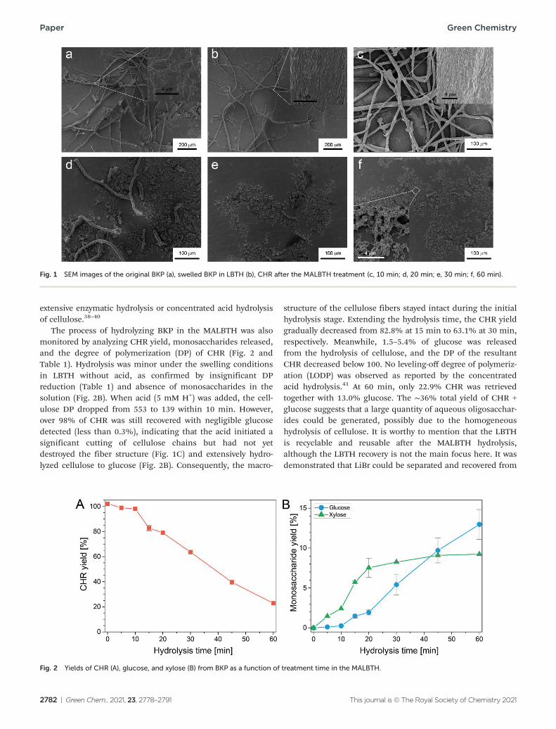
The macroscopic structures and morphology of the BKP during the treatment in the MALBTH were observed using SEM (Fig. 1, dry samples) and POM (Fig. S2,† wet samples), respectively. The original BKP consisted mainly of cellulose fiber cells with a small number of parenchyma cells (Fig. 1a). After the pretreatment in lithium bromide trihydrate (LBTH) without acid, the parenchyma cells were almost invisible. These thin-walled cells were likely destroyed and dissolved in the LBTH. The cellulose fibers remained mostly intact in the length dimension (Fig. 1b and S2B†). Compared with the smooth surface of the original BKP, a wrinkled surface was observed after the LBTH pretreatment (amplified insets in Fig. 1a and b), which was presumably caused by the drying-induced shrinkage of the swelled fibers when preparing the SEM sample. Similar surface topology was also observed during the mercerization process.37 In the first 10 min of the MLBTH treatment, macroscopic structures of the fibers were mostly preserved, and the surface morphology resembled that carried over from the LBTH pretreatment, though fractures or tiny holes appeared, possibly due to acidic corrosion. This observation was indicative of cellulose hydrolysis inside the fiber cell wall under the swelling conditions. Extending the treatment time in the MALBTH, the BKP fibers were remark-ably cut along the length dimension. The average length of the fiber fragments in average was shorter than 500 and 100 μm after 20 and 30 min, respectively. The cellulose particles from the fiber destruction, especially those after extensive treatment in the MALBTH, showed nano-scale porous structures (the inset of Fig. 1f), which were distinct from those isolated by
This journal is © The Royal Society of Chemistry 2021 Green Chem., 2021, 23, 2778–2791 | 2781
Paper Green Chemistry
Fig. 1 SEM images of the original BKP (a), swelled BKP in LBTH (b), CHR after the MALBTH treatment (c, 10 min; d, 20 min; e, 30 min; f, 60 min).
extensive enzymatic hydrolysis or concentrated acid hydrolysis of cellulose.38–40
The process of hydrolyzing BKP in the MALBTH was also monitored by analyzing CHR yield, monosaccharides released, and the degree of polymerization (DP) of CHR (Fig. 2 and Table 1). Hydrolysis was minor under the swelling conditions in LBTH without acid, as confirmed by insignificant DP reduction (Table 1) and absence of monosaccharides in the solution (Fig. 2B). When acid (5 mM H+) was added, the cell-ulose DP dropped from 553 to 139 within 10 min. However, over 98% of CHR was still recovered with negligible glucose detected (less than 0.3%), indicating that the acid initiated a significant cutting of cellulose chains but had not yet destroyed the fiber structure (Fig. 1C) and extensively hydro-lyzed cellulose to glucose (Fig. 2B). Consequently, the macro-
structure of the cellulose fibers stayed intact during the initial hydrolysis stage. Extending the hydrolysis time, the CHR yield gradually decreased from 82.8% at 15 min to 63.1% at 30 min, respectively. Meanwhile, 1.5–5.4% of glucose was released from the hydrolysis of cellulose, and the DP of the resultant CHR decreased below 100. No leveling-off degree of polymeriz-ation (LODP) was observed as reported by the concentrated acid hydrolysis.41 At 60 min, only 22.9% CHR was retrieved together with 13.0% glucose. The ∼36% total yield of CHR + glucose suggests that a large quantity of aqueous oligosacchar-ides could be generated, possibly due to the homogeneous hydrolysis of cellulose. It is worthy to mention that the LBTH is recyclable and reusable after the MALBTH hydrolysis, although the LBTH recovery is not the main focus here. It was demonstrated that LiBr could be separated and recovered from
Fig. 2 Yields of CHR (A), glucose, and xylose (B) from BKP as a function of treatment time in the MALBTH.
2782 | Green Chem., 2021, 23, 2778–2791 This journal is © The Royal Society of Chemistry 2021

Green Chemistry Paper
Table 1 Effect of the treatment time in the MALBTH on crystallinity, crystalline dimension, and DP of the CHR from the BKP
Crystallite size (nm)
Sample CrI (%) (1−10) (110) (200)/(020) DP
BKP 75.1 5.8 3.0 5.6 603 0 min 62.9 5.7 2.8 3.4 553 5 min 72.3 5.8 2.7 2.9 208 10 min 73.2 6.2 2.7 3.1 139 15 min 75.3 6.0 3.2 4.1 79 20 min 79.2 6.4 3.4 4.2 67 30 min 82.2 9.3 5.2 4.2 41 60 min 84.9 9.4 4.2 5.4 45
the hydrolysate using different technologies, such as ion exclu-sion or exchange chromatography, solvent extraction, and selective crystallization in anti-solvents.31
To elucidate the unique hydrolysis behavior of MALBTH, a hydrogen–deuterium exchange experiment was conducted and the accessibility of BKP fibers under the swelling conditions was investigated. Since hydronium ions are readily generated in an aqueous solvent, protons dissociated from a strong acid are assumably as accessible as water molecules in the catalytic hydrolysis of cellulose. In other words, any sites on/in a cell-
ulose microfibril where water can access should be equally accessible to protons. Thus, the region of cellulose accessible to water also represents the acid hydrolysable part. Because the hydrogen of hydroxyls exchanges readily with the deuterium in deuterium oxide, the hydroxyls on the accessible cellulose will be labeled via the hydrogen exchange with D2O molecules in the mildly acidic lithium bromide trideuterate (MALBTD), which could thus be detected by FTIR. As shown in Fig. 3, the absorption peaks in the wavenumber ranges of
−1 −1 −13200–3600 cm , 2800–3000 cm , and 2400–2600 cm were assigned to the vibrational stretching of O–H, C–H, and O–D in cellulose, respectively.42,43 Compared with the CHR pre-pared in the MALBTH, those prepared in the MALBTD showed strong vibrational signals of the O–D (Fig. 3A–D and S3†), indi-cating occurrence of the hydrogen–deuterium exchange between D2O and O–H of cellulose. After washing the CHR pre-pared in MALBTD with H2O, which could transform the surface O–D back to O–H, the O–D vibrational signals were still detectable, indicating that a certain region of cellulose was inaccessible to H2O after the exchange. The preserved O–D represented the portion of cellulose (hydroxyls) that was exclu-sively accessible in MALBTD, but not in water, as water mole-cules are only capable of entering the disordered region of cell-ulose.42 Therefore, we deduce that the crystalline portion of
Fig. 3 FTIR spectra of cellulose samples from the hydrogen–deuterium exchange experiment. ((A) Partially hydrolyzed in LiBr·3D2O for 30 min and washed with H2O; (B) partially hydrolyzed in LiBr·3D2O for 30 min and washed with D2O; (C) swelled in LiBr·3D2O for 45 min and washed with H2O; (D) swelled in LiBr·3D2O for 45 min and washed with D2O; (E) Swelled in D2O for 45 min and washed with D2O.) Note: All the treatments were at 100 °C. A and B were subjected to a swelling process in LiBr·3H2O and LiBr·3D2O, respectively before the acid (2.5 mM H2SO4) was added. The FTIR spectra were baseline-corrected, normalized and integrated from 3000–2700 cm−1 for the C–H vibrational signal and from 2700–2350 cm−1 for the O–D vibrational signal. The Y-axis denotes absorption.
This journal is © The Royal Society of Chemistry 2021 Green Chem., 2021, 23, 2778–2791 | 2783
Paper Green Chemistry
the cellulose preserved the O–D after the MALBTD treatment. This observation provided direct evidence for our assumption that the H+/water in the MALBTH could penetrate inside the crystalline region of cellulose under the swelling conditions, contributing to the enhanced hydrolysis of cellulose.
As shown in Fig. S3 in the ESI,† hydroxyls (OHs) at C2, C3, and C6 positions of cellulose II were responsible for the three peaks at 3470, 3402, and 3269 cm−1, and deuteroxyls (ODs) at C2, C3, C6 positions of cellulose II were responsible for the three peaks at 2583, 2551, and 2474 cm−1.40 The relative inten-sities of the O–D vibrational signals are shown in Fig. 3 using the C–H vibrational signal as a reference. Under the mild hydrolysis conditions in the MALBTD, the relative intensities of O–D (I(O–D)r) were 0.447 (counting the hydroxyls in the crys-talline region, Fig. 3A) and 0.917 (counting all the accessible hydroxyls, Fig. 3B). The relative intensity of O–D responsible
for the disordered and surface cellulose was calculated to be 0.470. Under the swelling conditions in deuterated LBTH without acid, the relative intensities of O–D (I(O–D)r) were 0.573 (for the disordered and surface cellulose) and 0.364 (for the crystalline cellulose). The decreased O–D intensity of the dis-ordered and surface cellulose after hydrolysis in the MLABTD indicated that disordered cellulose was preferentially removed by the selective hydrolysis in the system.
Polymorph transformation of cellulose in the MALBTH and proposed mechanism
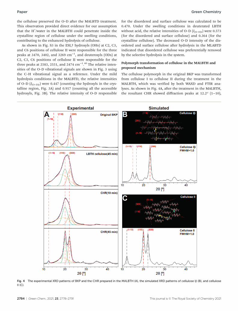
The cellulose polymorph in the original BKP was transformed from cellulose I to cellulose II during the treatment in the MALBTH, which was verified by both WAXD and FTIR ana-lyses. As shown in Fig. 4A, after the treatment in the MALBTH, the resultant CHR showed diffraction peaks at 12.2° (1−10),
Fig. 4 The experimental XRD patterns of BKP and the CHR prepared in the MALBTH (A), the simulated XRD patterns of cellulose Iβ (B), and cellulose II (C).
2784 | Green Chem., 2021, 23, 2778–2791 This journal is © The Royal Society of Chemistry 2021
Green Chemistry Paper
20.0° (110), and 22.1° (020), which were characteristic for cell-ulose II crystallites, while the untreated BKP, which is composed of cellulose Iβ crystallites, showed diffraction peaks at 14.8° (1−10), 16.7° (110), and 22.6° (200). Based on the .cif files pro-vided by French et al.,34 the ideal XRD patterns were simulated using the generally accepted cellulose Iβ and cellulose II lattice units by the Mercury software, as shown in Fig. 4B and C. The experimental XRD patterns of the original BKP and the CHR prepared in the MALBTH were in perfect agreement with the simulation using ideal cellulose Iβ and cellulose II crystallites, respectively. The results confirmed that the polymorph trans-formation of cellulose occurred during the MALBTH treatment. Swelling BKP in LBTH without acid resulted in an XRD pattern distinct from cellulose I but consistent with cellulose II (LBTH cellulose in Fig. 4A), suggesting that polymorph transformation could be initiated under the swelling conditions in the LBTH and completed in the MALBTH. In particular, the diffraction pattern of the CHR after 45 min (Fig. 4A) fit well with those of ideal cellulose II (Fig. 4C) due to the removal of disordered cell-ulose. The LBTH or MALBTH treatment was more efficient at transforming cellulose polymorph, compared to other cellulose swelling solvents such as concentrated sulfuric acid and [BMIM] Cl.11,25 As far as we are aware, this is the first report of poly-morph transformation achieved by swelling cellulose in an aqueous solvent other than corrosive concentrated sodium hydroxide or sulfuric acid.
As shown in Table 1, swelling BKP in the LBTH without acid reduced the crystallinity from 75.1% of original BKP to 62.9% of swelled cellulose II fibers. Reduction in crystallinity is presumably due to the generation of disordered cellulose during polymorph transformation. During the controlled hydrolysis in the MALBTH, the crystallinity of CHR gradually increased with hydrolysis time from 72.3% at 5 min to 90.9% at 45 min because of the removal of disordered cellulose. This confirmed that crystalline cellulose was more recalcitrant to hydrolysis than disordered cellulose. The crystal size corres-ponding to the three major crystalline planes [(1−10), (110), and (020)] of cellulose II increased by 50–100% when extend-ing the hydrolysis time from 5 min to 45–60 min.
The transformation from cellulose I to cellulose II was also verified from the FTIR spectra of BKP before and after the treatment in LBTH and MALBTH (Fig. S4†). The absorption bands at 1429, 1105, and 1053 cm−1, which are characteristic for cellulose I (e.g., BKP), disappeared after the treatments in LBTH and MALBTH, verifying again that the polymorph trans-formation of cellulose was initiated and mostly completed in the LBTH. The vibrational frequency of CH2 symmetric
−1 −1bending shifted to 1418 cm from 1429 cm in the CHR spectrum, consistent with that of the cellulose II crystallites in lyocell fibers,44 which is additional evidence of the polymorph transformation from cellulose I to cellulose II in the MALBTH treatment. Furthermore, the enhanced intensity of the vibrational bands at 1368 and 1263 cm−1 in the CHR spectrum was consistent with the XRD results above that cellulose II crystallites accumulate as a consequence of polymorph trans-formation and subsequent hydrolysis of disordered cellulose.
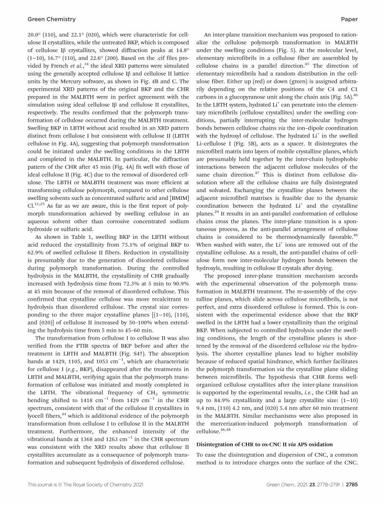
An inter-plane transition mechanism was proposed to ration-alize the cellulose polymorph transformation in MALBTH under the swelling conditions (Fig. 5). At the molecular level, elementary microfibrils in a cellulose fiber are assembled by cellulose chains in a parallel direction.45 The direction of elementary microfibrils had a random distribution in the cell-ulose fiber. Either up (red) or down (green) is assigned arbitra-rily depending on the relative positions of the C4 and C1 carbons in a glucopyranose unit along the chain axis (Fig. 5A).46
In the LBTH system, hydrated Li+ can penetrate into the elemen-tary microfibrils (cellulose crystallites) under the swelling con-ditions, partially interrupting the inter-molecular hydrogen bonds between cellulose chains via the ion–dipole coordination with the hydroxyl of cellulose. The hydrated Li+ in the swelled Li–cellulose I (Fig. 5B), acts as a spacer. It disintegrates the microfibril matrix into layers of mobile crystalline planes, which are presumably held together by the inter-chain hydrophobic interactions between the adjacent cellulose molecules of the same chain direction.47 This is distinct from cellulose dis-solution where all the cellulose chains are fully disintegrated and solvated. Exchanging the crystalline planes between the adjacent microfibril matrixes is feasible due to the dynamic coordination between the hydrated Li+ and the crystalline planes.29 It results in an anti-parallel conformation of cellulose chains cross the planes. The inter-plane transition is a spon-taneous process, as the anti-parallel arrangement of cellulose chains is considered to be thermodynamically favorable.46
When washed with water, the Li+ ions are removed out of the crystalline cellulose. As a result, the anti-parallel chains of cell-ulose form new inter-molecular hydrogen bonds between the hydroxyls, resulting in cellulose II crystals after drying.
The proposed inter-plane transition mechanism accords with the experimental observation of the polymorph trans-formation in MALBTH treatment. The re-assembly of the crys-talline planes, which slide across cellulose microfibrils, is not perfect, and extra disordered cellulose is formed. This is con-sistent with the experimental evidence above that the BKP swelled in the LBTH had a lower crystallinity than the original BKP. When subjected to controlled hydrolysis under the swell-ing conditions, the length of the crystalline planes is shor-tened by the removal of the disordered cellulose via the hydro-lysis. The shorter crystalline planes lead to higher mobility because of reduced spatial hindrance, which further facilitates the polymorph transformation via the crystalline plane sliding between microfibrils. The hypothesis that CHR forms well-organized cellulose crystallites after the inter-plane transition is supported by the experimental results, i.e., the CHR had an up to 84.9% crystallinity and a large crystallite size: (1−10) 9.4 nm, (110) 4.2 nm, and (020) 5.4 nm after 60 min treatment in the MALBTH. Similar mechanisms were also proposed in the mercerization-induced polymorph transformation of cellulose.46,48
Disintegration of CHR to ox-CNC II via APS oxidation
To ease the disintegration and dispersion of CNC, a common method is to introduce charges onto the surface of the CNC.
This journal is © The Royal Society of Chemistry 2021 Green Chem., 2021, 23, 2778–2791 | 2785

Paper Green Chemistry
Fig. 5 Schematic illustration of the polymorph transformation from cellulose I to cellulose II under swelling conditions in the MALBTH. (A) Assignment of cellulose chain orientation; (B) Proposed mechanism of polymorph transformation.
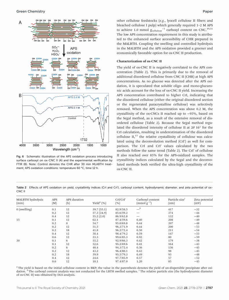
In the present study, ammonium persulfate (APS) was used as an oxidizing reagent to introduce carboxyl groups to the surface of the CNC II (CHR) by partial oxidation, which led to the oxi-dized cellulose II nanocrystals (ox-CNC II) with a negative surface charge. As an alternative to TEMPO reagents, APS can oxidize surface hydroxyls of cellulose nanocrystals to carboxyl groups but has lower chronic toxicity and cost than TEMPO.14,49
At elevated temperatures, persulfate (S2O82−) can slowly decom-
pose to SO4 −, HSO4
− , and H2O2. The HSO4 − provides an acidic
environment for further removal of disordered cellulose, and the SO4
− free radical and H2O2 contribute to the oxidation of surface hydroxyl to the carboxyl (Fig. 6A). The cellulose residues produced by the swelling alone in the LBTH and the swelling and controlled hydrolysis in the MALBTH were both subjected to the APS oxidation. Starting from the CHR prepared from 15 min treatment in the MALBTH, the maximum yield of the ox-CNC II was 62.1% after the APS oxidation (Table 2). From the swelled BKP in LBTH and the CHR after extensive treatment (30 min) in the MALBTH, the yield of the ox-CNC II was lower under the same APS oxidation conditions (0.1 M APS). The swelling of BKP in the LBTH without acid increased the percen-tage of overall disordered cellulose, which impeded the release of the high-crystallinity ox-CNC II. As a result, the non-dispersi-ble precipitates with large particle sizes were up to 55.1%, as shown in Fig. S5 in the ESI.† On the other hand, extending hydrolysis in the MALBTH resulted in a significant loss of CHR yield (over 40%), which in turn impaired the final yield of the
ox-CNC II calculated based on the initial cellulose content in BKP. The yield of the ox-CNC (23.3%–62.1%, based on BKP, varying with the APS concentration) was comparable to that of the CNC produced by concentrated sulfuric acid (28.0%–75.6%, varying with the sulfuric acid concentration), but lower than that of those produced by TEMPO oxidation (over 90%).13,50 The APS oxidation did not affect cellulose polymorph or cause poly-morph transformation, as the ox-CNC II maintained the same polymorph as CNC II (Fig. S6†).
The characteristic peak of the CvO bond at 1732 cm−1 in the FTIR spectra (Fig. 6B) confirmed that carboxyl groups were introduced after the APS oxidation, which oxidized the surface hydroxyl of cellulose into carboxyl. The peak intensity increased with the APS concentration. A semi-quantitative ana-lysis of the carboxyl content has been reported utilizing FTIR spectra,49 but the more precise electrical conductivity titration method was used to quantitate the carboxyl groups. As shown in Table 2, the carboxyl content increased from 0.4 mmol gcellulose
−1 (0.1 M APS) to 1.2 mmol gcellulose −1 (0.6 M APS) with
the increased APS concentration, indicating concentrated APS greatly enhanced the surface oxidation of cellulose. Introducing carboxyl by APS oxidation is analogous to that by TEMPO oxidation which resulted in 1.1–1.7 mmol gcellulose
−1.48,49 For comparison, the CNC prepared by concen-trated H2SO4 was less functionalized (∼0.2 mmol gcellulose
−1
sulfate).50 It is worth noting that the concentration of APS used in this study was significantly lower than that used for
2786 | Green Chem., 2021, 23, 2778–2791 This journal is © The Royal Society of Chemistry 2021
Green Chemistry Paper
Fig. 6 Schematic illustration of the APS oxidation process introducing surface carboxyl on ox-CNC II (A) and the experimental verification by FTIR (B). Note: Control denotes the CHR after 30 min MLABTH treat-ment; APS oxidation conditions: temperature 60 °C, time 12 h.
other cellulose feedstocks (e.g., lyocell cellulose II fibers and bleached cellulose I pulp) which generally required 1–2 M APS to achieve 1.0 mmol gcellulsoe
−1 carboxyl content on CNC.49,51
The low APS concentration requirement in this study is attribu-ted to the enhanced surface accessibility of CHR prepared in the MALBTH. Coupling the swelling and controlled hydrolysis in the MALBTH and the APS oxidation provided a greener and economically favorable option for ox-CNC II production.
Characterization of ox-CNC II
The yield of ox-CNC II is negatively correlated to the APS con-centration (Table 2). This is primarily due to the removal of additional disordered cellulose from CNC II (CHR) at high APS concentrations. As no glucose was detected after the APS oxi-dation, it is speculated that soluble oligo- and mono-glucuro-nic acids account for the loss of ox-CNC II yield. Increasing the APS concentration contributed to higher CrI, indicating that the disordered cellulose (either the original disordered section or the regenerated paracrystalline cellulose) was selectively removed. When the APS concentration was above 0.2 M, the crystallinity of the ox-CNCs II reached up to ∼95%, based on the Segal method, as a result of the extensive removal of dis-ordered cellulose (Table 2). Because the Segal method regu-lated the disordered intensity of cellulose II at 2θ 16° for the CrI calculation, resulting in underestimation of the disordered cellulose II,15 the relative crystallinity of cellulose was calcu-lated using the deconvolution method (CrI′) as well for com-parison. The CrI and CrI′ values calculated by the two methods follow the same trend (Table 2). The CrI′ of cellulose II also reached over 85% for the APS-oxidized samples. The crystallinity indices calculated by the Segal and the deconvo-luted methods both verified the ultra-high crystallinity of the ox-CNC II.
Table 2 Effects of APS oxidation on yield, crystallinity indices (CrI and CrI’), carboxyl content, hydrodynamic diameter, and zeta potential of ox-CNC II
MALBTH hydrolysis APS APS duration CrI/CrI′ Carboxyl content Particle sizec Zeta potential (min) (M) (h) Yielda (%) (%) (mmol g−1) (nm) (mV)
0 (swelling) 0.1 12 39.7 (55.1) 82.9/58.5 — b 417 −32 0.2 12 57.3 (24.9) 85.6/59.2 — 374 −44 0.4 12 55.2 (2.0) 86.9/62.8 — 132 −40
15 0.1 12 62.1 87.4/59.6 0.40 208 −49 0.2 8 54.7 95.0/69.8 0.44 207 −48 0.2 12 51.5 96.3/71.9 0.44 200 −53 0.2 18 41.8 96.2/72.4 0.50 211 −54 0.4 12 38.4 98.4/79.2 0.59 147 −53 0.6 12 23.3 99.6/85.3 0.92 80 −59
30 0.1 6 55.2 92.9/66.3 0.42 179 −38 0.1 12 52.6 95.2/69.6 0.41 164 −43 0.2 6 48.4 94.3/72.4 0.36 136 −45 0.2 12 43.2 96.4/80.1 0.43 98 −45 0.2 18 39.9 95.5/79.3 0.49 93 −48 0.4 12 24.6 97.7/85.9 0.57 57 −52 0.6 12 10.1 97.4/87.0 1.20 85 −45
a The yield is based on the initial cellulose content in BKP; the value in the parenthesis denotes the yield of un-dispersible precipitate after oxi-dation. b The carboxyl content analysis was not conducted for the LBTH swelled samples. c The relative particle size (the hydrodynamic diameter of ox-CNC II) was obtained by DLS analysis.
This journal is © The Royal Society of Chemistry 2021 Green Chem., 2021, 23, 2778–2791 | 2787
Paper Green Chemistry
The hydrodynamic diameter of the ox-CNC II was estimated by DLS analysis for comparing the relative size among the samples prepared under different APS oxidation conditions. It must be noted that the diameter value, as fitted by isotropic particles, does not represent the real dimension of the rod-like nanoparticles.48 For the CHR prepared after treatment in the MALBTH for 15 min, the hydrodynamic diameters of the resul-tant ox-CNC II decreased from 208 to 80 nm when increasing the APS concentration from 0.1 to 0.6 M, as shown in Table 2. The effect of the APS oxidation duration was insignificant on the relative size of the ox-CNC II, especially after 12 h. However, extending the hydrolysis in the MALBTH could reduce the hydrodynamic diameter of the ox-CNC II to smaller than 60 nm after the oxidation with 0.4 M APS.
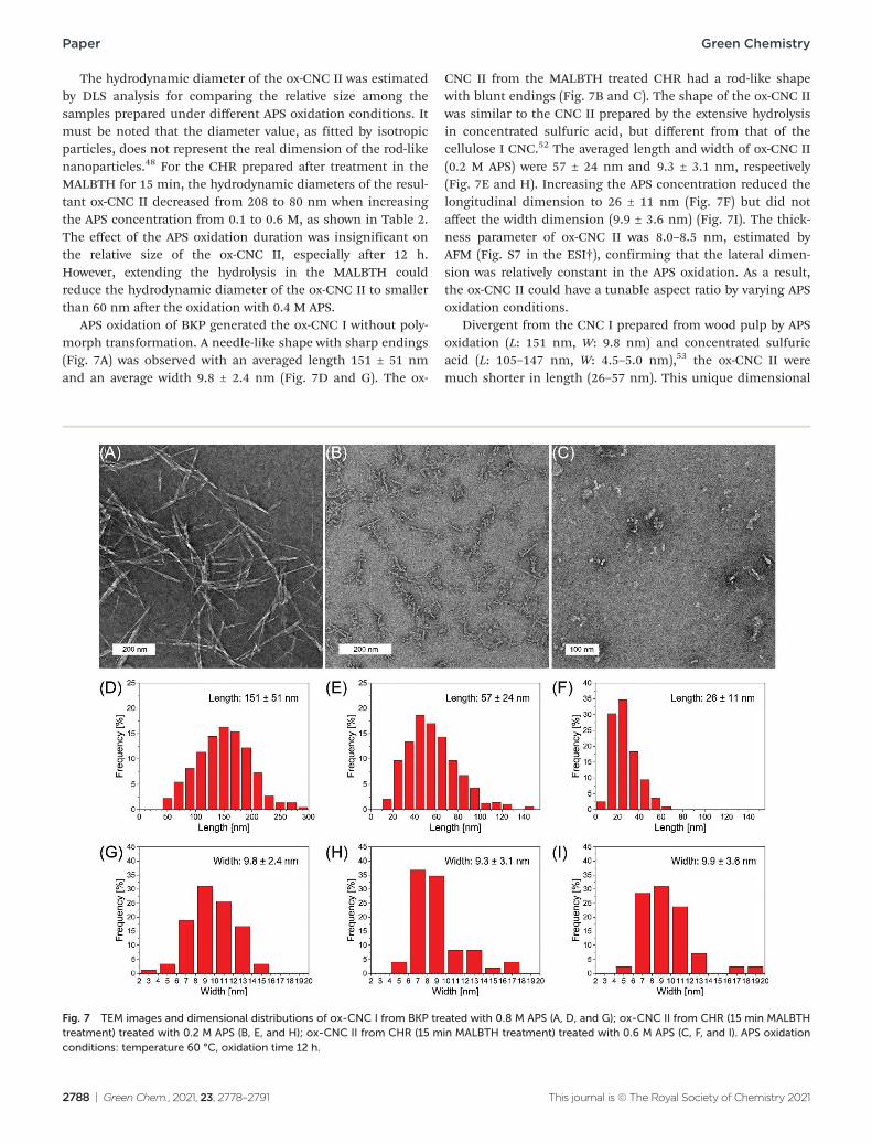
APS oxidation of BKP generated the ox-CNC I without poly-morph transformation. A needle-like shape with sharp endings (Fig. 7A) was observed with an averaged length 151 ± 51 nm and an average width 9.8 ± 2.4 nm (Fig. 7D and G). The ox-
CNC II from the MALBTH treated CHR had a rod-like shape with blunt endings (Fig. 7B and C). The shape of the ox-CNC II was similar to the CNC II prepared by the extensive hydrolysis in concentrated sulfuric acid, but different from that of the cellulose I CNC.52 The averaged length and width of ox-CNC II (0.2 M APS) were 57 ± 24 nm and 9.3 ± 3.1 nm, respectively (Fig. 7E and H). Increasing the APS concentration reduced the longitudinal dimension to 26 ± 11 nm (Fig. 7F) but did not affect the width dimension (9.9 ± 3.6 nm) (Fig. 7I). The thick-ness parameter of ox-CNC II was 8.0–8.5 nm, estimated by AFM (Fig. S7 in the ESI†), confirming that the lateral dimen-sion was relatively constant in the APS oxidation. As a result, the ox-CNC II could have a tunable aspect ratio by varying APS oxidation conditions.
Divergent from the CNC I prepared from wood pulp by APS oxidation (L: 151 nm, W: 9.8 nm) and concentrated sulfuric acid (L: 105–147 nm, W: 4.5–5.0 nm),53 the ox-CNC II were much shorter in length (26–57 nm). This unique dimensional
Fig. 7 TEM images and dimensional distributions of ox-CNC I from BKP treated with 0.8 M APS (A, D, and G); ox-CNC II from CHR (15 min MALBTH treatment) treated with 0.2 M APS (B, E, and H); ox-CNC II from CHR (15 min MALBTH treatment) treated with 0.6 M APS (C, F, and I). APS oxidation conditions: temperature 60 °C, oxidation time 12 h.
2788 | Green Chem., 2021, 23, 2778–2791 This journal is © The Royal Society of Chemistry 2021

Green Chemistry Paper
Fig. 8 TGA (A) and the first derivative curves (B) of various cellulose samples.
feature was ascribed to the polymorph transformation under the swelling conditions in the MALBTH treatment. The poly-morph transformation of BKP in MALBTH involved the re-assembly of cellulose crystallites via an inter-plane transition. Extending the hydrolysis time generated larger crystallites with a lower DP (Table 1). It differed from the cellulose hydrolysis without polymorph transformation in which the crystallite size was relatively constant.54 Besides, the APS oxidation of CHR did not change the width dimension of ox-CNC II (Fig. 7). The results above suggest that the CHR had the fringed-micellar structure in which additional disordered cellulose was gener-ated from the inter-plane transition process, contributing to the tailorable aspect ratio of ox-CNC II. For comparison, the CNC II prepared in an ionic liquid ([BMIM]Cl) via a dis-solution and regeneration process had irregular and mixed shape and size, including rod-shape CNC II (L: 112 nm, W: 12 nm) and sphere-shape CNC II (118 nm in diameter).25
The ox-CNC II exhibited high zeta potential ranging from −42.8 to −59.0 mV, depending on the carboxyl content and the particle size of the ox-CNC II prepared under varied oxidation conditions (Table 2). The zeta potential was relevant to surface charge density which was calculated using the dimensions determined by TEM and AFM, considering ox-CNC II in a rod shape and cellulose density 1.6 g cm−3. Full dissociation of carboxyl was assumed at the neutral pH. The surface charge density of ox-CNC II increased from 0.87 e− nm−2 (0.2 M APS) to 1.72 e− nm−2 (0.6 M APS), while the average crystallite size was relatively stable (6.2 nm and 6.8 nm, respectively). It differed from the TEMPO-oxidized CNC which showed the decreased carboxylate density with the crystalline size.55 At 0.6 M APS, the surface charge density of ox-CNC II was compar-able to that of CNC by the TEMPO (∼1.7 group per nm2) and higher than that by concentrated H2SO4 (0.29–0.38 e−
−2).55,56nm The high zeta potential (absolute value) would grant ox-CNC II excellent colloidal stability in water. The colloid suspension (0.5–1.0 wt%) of the ox-CNC II prepared under varied APS oxidation conditions was found to be stable
for up to 6 months, as shown in Fig. S8 in the ESI.† In con-trast, flocculation was inevitable for traditional CNC after weeks of storage.57
The thermal stability of cellulose samples with different cellulose polymorphs was evaluated using TGA (Fig. 8). The ox-CNC II from the MALBTH treated CHR had the major pyrolytic degradation peak at 338 °C which was slightly lower than those of original BKP (355 °C) and CHR (345 °C). The slight decrease in thermal stability was ascribed to the reduced mole-cular weight of the ox-CNC II caused by the hydrolysis in the MALBTH and the introduced carboxyl groups by the APS oxi-dation. Compared with the ox-CNC II, the ox-CNC I without the MALBTH treatment had lower stability at temperatures above 300 °C. This is consistent with the hypothesis that thermodynamically, cellulose II is more resistant to thermal degradation than cellulose I.58 Traditional CNC I prepared by the controlled hydrolysis in 64% H2SO4 displayed a downward shift in its major degradation peak (255 °C), indicating a sig-nificant decrease in thermal stability. The results above con-firmed the ox-CNC II derived from the MALBTH CHR had improved thermal stability.
Conclusions
The simultaneous hydrolysis and polymorph transformation of cellulose I fibers (BKP) in the MALBTH followed by the APS oxidation was successfully demonstrated for preparing cell-ulose II nanocrystals. The hydrated lithium ions (Li+) in the MALBTH swelled the cellulose fibers via disrupting the inter-molecular hydrogen bonds of cellulose. The removal of the dis-ordered cellulose by selective hydrolysis in the MALBTH under the swelling conditions resulted in well-organized crystallites. Meanwhile, it was proved that the lithium ions were able to penetrate inside the cellulose crystallites under the swelling condition using the hydrogen–deuterium exchange experi-ment, which led to the sliding and reassembling of the crystal-
This journal is © The Royal Society of Chemistry 2021 Green Chem., 2021, 23, 2778–2791 | 2789
Paper Green Chemistry
line planes of cellulose and thereby caused the polymorph transformation from parallel-oriented cellulose I to anti-paral-lel-oriented cellulose II. The APS oxidation at low APS concen-trations (0.1–0.6 M) introduced the surface charges (0.3–1.2 mmol COOH per gcellulose) and therefore facilitated the disintegration of the cellulose nanocrystals. The yield of the ox-CNC II was up to 62%. The resultant ox-CNC II featured ultra-high crystallinity (above 90%), excellent dispersibility and colloidal stability, and good thermal stability. Depending on the conditions of the MALBTH hydrolysis and APS oxidation, the length of the ox-CNC II was tunable (26–57 nm) with a rela-tively constant lateral dimension (8–10 nm). This study pro-vides a caustic-chemical-free method to produce tunable cell-ulose II nanocrystals by the simultaneous hydrolysis and poly-morph transformation of cellulose I in the MALBTH system.
Author contributions
NL and XP conceived the idea and designed the research. NL conducted most of the experiments, and HB completed the AFM analysis. TEM imaging was performed by PNC and NL. NL, HB, XP, JZ, and PNC analyzed the data. NL and XP drafted the manuscript. All authors reviewed the manuscript and suggested improvements.
Conflicts of interest
All authors declare no conflict of interest.
Acknowledgements
This work was supported by the National Science Foundation (NSF) (CBET 1159561) and the U.S. Department of Agriculture (USDA) National Institute of Food and Agriculture, McIntire Stennis grant (WIS01996) to XP. NL is thankful to China Scholarship Council (CSC) for partially supporting his Ph.D. study at the University of Wisconsin–Madison.
References
1 D. Klemm, B. Heublein, H. P. Fink and A. Bohn, Angew. Chem., Int. Ed., 2005, 44, 3358–3393.
2 H. Zhu, W. Luo, P. N. Ciesielski, Z. Fang, J. Y. Zhu, G. Henriksson, M. E. Himmel and L. Hu, Chem. Rev., 2016, 116, 9305–9374.
3 O. M. Vanderfleet and E. D. Cranston, Nat. Rev. Mater., 2020, 1–21.
4 D. Trache, M. H. Hussin, M. M. Haafiz and V. K. Thakur, Nanoscale, 2017, 9, 1763–1786.
5 S. Wang, A. Lu and L. Zhang, Prog. Polym. Sci., 2016, 53, 169–206.
6 A. Dufresne, Mater. Today, 2013, 16, 220–227.
7 B. L. Tardy, S. Yokota, M. Ago, W. Xiang, T. Kondo, R. Bordes and O. J. Rojas, Curr. Opin. Colloid Interface Sci., 2017, 29, 57–67.
8 N. Grishkewich, N. Mohammed, J. Tang and K. C. Tam, Curr. Opin. Colloid Interface Sci., 2017, 29, 32–45.
9 X. Wang, C. Yao, F. Wang and Z. Li, Small, 2017, 13, 1702240.
10 C. Miao and W. Y. Hamad, Curr. Opin. Solid State Mater. Sci., 2019, 23, 100761.
11 D. Bondeson, A. Mathew and K. Oksman, Cellulose, 2006, 13, 171–180.
12 W. Y. Hamad and T. Q. Hu, Can. J. Chem. Eng., 2010, 88, 392–402.
13 L. Chen, Q. Wang, K. Hirth, C. Baez, U. P. Agarwal and J. Zhu, Cellulose, 2015, 22, 1753–1762.
14 A. C. Leung, S. Hrapovic, E. Lam, Y. Liu, K. B. Male, K. A. Mahmoud and J. H. Luong, Small, 2011, 7, 302– 305.
15 S. Nam, A. D. French, B. D. Condon and M. Concha, Carbohydr. Polym., 2016, 135, 1–9.
16 L. Chen, J. Zhu, C. Baez, P. Kitin and T. Elder, Green Chem., 2016, 18, 3835–3843.
17 H. Yu, Z. Qin, B. Liang, N. Liu, Z. Zhou and L. Chen, J. Mater. Chem. A, 2013, 1, 3938–3944.
18 S. Camarero Espinosa, T. Kuhnt, E. J. Foster and C. Weder, Biomacromolecules, 2013, 14, 1223–1230.
19 I. Kalashnikova, H. Bizot, P. Bertoncini, B. Cathala and I. Capron, Soft Matter, 2013, 9, 952–959.
20 I. Capron, O. J. Rojas and R. Bordes, Curr. Opin. Colloid Interface Sci., 2017, 29, 83–95.
21 J. M. González-Domínguez, A. Ansón-Casaos, L. Grasa, L. Abenia, A. Salvador, E. Colom, J. E. Mesonero, J. E. García-Bordejé, A. M. Benito and W. K. Maser, Biomacromolecules, 2019, 20, 3147–3160.
22 Y. Qin, X. Qiu and J. Zhu, Sci. Rep., 2016, 6, 35602. 23 M. Hirota, N. Tamura, T. Saito and A. Isogai, Cellulose,
2012, 19, 435–442. 24 M. Beaumont, T. Nypelö, J. König, R. Zirbs, M. Opietnik,
A. Potthast and T. Rosenau, Green Chem., 2016, 18, 1465– 1468.
25 J. Han, C. Zhou, A. D. French, G. Han and Q. Wu, Carbohydr. Polym., 2013, 94, 773–781.
26 G. Sebe, F. Ham-Pichavant, E. Ibarboure, A. L. Koffi and P. Tingaut, Biomacromolecules, 2012, 13, 570–578.
27 Y.-J. Yang, J.-M. Shin, T. H. Kang, S. Kimura, M. Wada and U.-J. Kim, Cellulose, 2014, 21, 1175–1181.
28 X. Zhang, N. Xiao, H. Wang, C. Liu and X. Pan, Polymers, 2018, 10, 614.
29 Y. Liao, Z. Pang and X. Pan, ACS Sustainable Chem. Eng., 2019, 7, 17723–17736.
30 L. Zhang, Y. Liao, Y. C. Wang, S. Zhang, W. Yang, X. Pan and Z. L. Wang, Adv. Funct. Mater., 2020, 2001763.
31 X. Pan and L. Shuai, Saccharification of lignocellulosic biomass, US Patent 9187790B2, 2015.
32 L. Shuai, Q. Yang, J. Zhu, F. Lu, P. Weimer, J. Ralph and X. Pan, Bioresour. Technol., 2010, 101, 3106–3114.
2790 | Green Chem., 2021, 23, 2778–2791 This journal is © The Royal Society of Chemistry 2021

Green Chemistry Paper
33 C. F. Macrae, I. J. Bruno, J. A. Chisholm, P. R. Edgington, P. McCabe, E. Pidcock, L. Rodriguez-Monge, R. Taylor, J. V. Streek and P. A. Wood, J. Appl. Crystallogr., 2008, 41, 466–470.
34 A. D. French, Cellulose, 2014, 21, 885–896. 35 W. P. F. Neto, J.-L. Putaux, M. Mariano, Y. Ogawa,
H. Otaguro, D. Pasquini and A. Dufresne, RSC Adv., 2016, 6, 76017–76027.
36 N. Li, X. Pan and J. Alexander, Green Chem., 2016, 18, 5367– 5376.
37 H. Wang, D. Li, H. Yano and K. Abe, Cellulose, 2014, 21, 1505–1515.
38 W. Wang, M. D. Mozuch, R. C. Sabo, P. Kersten, J. Zhu and Y. Jin, Cellulose, 2015, 22, 351–361.
39 A. A. Oun and J.-W. Rhim, Carbohydr. Polym., 2016, 150, 187–200.
40 Y.-H. P. Zhang, J. Cui, L. R. Lynd and L. R. Kuang, Biomacromolecules, 2006, 7, 644–648.
41 Y. Nishiyama, U.-J. Kim, D.-Y. Kim, K. S. Katsumata, R. P. May and P. Langan, Biomacromolecules, 2003, 4, 1013–1017.
42 E. L. Lindh and L. Salmén, Cellulose, 2017, 24, 21–33. 43 J. Fan, M. De Bruyn, V. L. Budarin, M. J. Gronnow,
P. S. Shuttleworth, S. Breeden, D. J. Macquarrie and J. H. Clark, J. Am. Chem. Soc., 2013, 135, 11728–11731.
44 F. Carrillo, X. Colom, J. Sunol and J. Saurina, Eur. Polym. J., 2004, 40, 2229–2234.
45 Y. Nishiyama, P. Langan and H. Chanzy, J. Am. Chem. Soc., 2002, 124, 9074–9082.
46 T. Okano and A. Sarko, J. Appl. Polym. Sci., 1985, 30, 325– 332.
47 B. Lindman, B. Medronho, L. Alves, C. Costa, H. Edlund and M. Norgren, Phys. Chem. Chem. Phys., 2017, 19, 23704– 23718.
48 T. Okano and A. Sarko, J. Appl. Polym. Sci., 1984, 29, 4175– 4182.
49 M. Cheng, Z. Qin, Y. Liu, Y. Qin, T. Li, L. Chen and M. Zhu, J. Mater. Chem. A, 2014, 2, 251–258.
50 A. Rattaz, S. P. Mishra, B. Chabot and C. Daneault, Cellulose, 2011, 18, 585–593.
51 K. Zhang, P. Sun, H. Liu, S. Shang, J. Song and D. Wang, Carbohydr. Polym., 2016, 138, 237–243.
52 W. P. Flauzino Neto, J.-L. Putaux, M. Mariano, Y. Ogawa, H. Otaguro, D. Pasquini and A. Dufresne, RSC Adv., 2016, 6, 76017–76027.
53 S. Beck-Candanedo, M. Roman and D. G. Gray, Biomacromolecules, 2005, 6, 1048–1054.
54 C. Driemeier and J. Bragatto, J. Phys. Chem. B, 2013, 117, 415–421.
55 Y. Okita, T. Saito and A. Isogai, Biomacromolecules, 2010, 11, 1696–1700.
56 S. Beck-Candanedo, M. Roman and D. G. Gray, Biomacromolecules, 2005, 6, 1048–1054.
57 J. Lazko, T. Sénéchal, N. Landercy, L. Dangreau, J.-M. Raquez and P. Dubois, Cellulose, 2014, 21, 4195–4207.
58 Y. Yue, C. Zhou, A. D. French, G. Xia, G. Han, Q. Wang and Q. Wu, Cellulose, 2012, 19, 1173–1187.
This journal is © The Royal Society of Chemistry 2021 Green Chem., 2021, 23, 2778–2791 | 2791
Related Documents