Tailor-made three-dimensional hybrid scaffolds for cell cultures This article has been downloaded from IOPscience. Please scroll down to see the full text article. 2011 Biomed. Mater. 6 045008 (http://iopscience.iop.org/1748-605X/6/4/045008) Download details: IP Address: 139.91.179.22 The article was downloaded on 11/07/2011 at 09:01 Please note that terms and conditions apply. View the table of contents for this issue, or go to the journal homepage for more Home Search Collections Journals About Contact us My IOPscience

Welcome message from author
This document is posted to help you gain knowledge. Please leave a comment to let me know what you think about it! Share it to your friends and learn new things together.
Transcript

Tailor-made three-dimensional hybrid scaffolds for cell cultures
This article has been downloaded from IOPscience. Please scroll down to see the full text article.
2011 Biomed. Mater. 6 045008
(http://iopscience.iop.org/1748-605X/6/4/045008)
Download details:
IP Address: 139.91.179.22
The article was downloaded on 11/07/2011 at 09:01
Please note that terms and conditions apply.
View the table of contents for this issue, or go to the journal homepage for more
Home Search Collections Journals About Contact us My IOPscience
IOP PUBLISHING BIOMEDICAL MATERIALS
Biomed. Mater. 6 (2011) 045008 (16pp) doi:10.1088/1748-6041/6/4/045008
Tailor-made three-dimensional hybridscaffolds for cell culturesStylianos Psycharakis1,2, Androniki Tosca2, Vasileia Melissinaki1,3,Anastasia Giakoumaki1,4 and Anthi Ranella1,5
1 Institute of Electronic Structure and Laser, Foundation for Research and Technology Hellas,PO Box 1527, 711 10 Heraklion, Crete, Greece2 Department of Medicine, University of Crete, 710 03 Heraklion, Crete, Greece3 Department of Physics, University of Crete, 710 03 Heraklion, Crete, Greece4 Department of Materials Science and Technology, University of Crete, 710 03 Heraklion, Crete, Greece
E-mail: [email protected]
Received 8 February 2011Accepted for publication 6 June 2011Published 8 July 2011Online at stacks.iop.org/BMM/6/045008
AbstractThe construction of the ideal three-dimensional scaffold for cell culture is one of the mostintriguing topics in tissue engineering. It has been shown that cells can be cultured on mostorganic biomimetic materials, which now are losing popularity in favour of novel, hybridsystems. In this study, a series of photosensitive sol–gel hybrid materials, based onsilicon–zirconium and silicon–titanium oxides, have been investigated for their suitability inthree-dimensional scaffold fabrication. These materials can be structured by two-photonpolymerization, a laser-based technique allowing the fabrication of micrometre-size structureswith submicron resolution. The work presented here examined the effect of theorganic/inorganic composition of the materials on cell behaviour and the establishment of a‘cell-culture friendly’ environment. This is vital for cell adhesion, growth and differentiation,as the organic part of the material provides the soft matrix for cell growth, whereas theinorganic component gives the mechanical stability and rigidity of the three-dimensionalstructures. In addition, the use of femtosecond laser structuring permits the fabrication of awide range of mechanically stable scaffolds of different sizes and shapes to be tested in termsof cell viability, proliferation and orientation.
(Some figures in this article are in colour only in the electronic version)
1. Introduction
Tissue engineering is an area of research that combinescells with synthesis and engineering of novel materials todevelop substitutes for the repair, regeneration or replacementof biological functions. Previous studies have shown thatextrinsic signals deriving from the extracellular matrix (ECM)play an essential role in major cell functions, includingproliferation, differentiation, migration or release of activemediators [17, 34]. Biomimetically driven studies haveindicated that the surface chemistry [9, 10, 43] and topography[12, 15, 44] of the scaffold notably influence biologicalactivities [37].
5 Author to whom any correspondence should be addressed.
The use of new alloys, ceramics and polymers, developedto mimic the physical properties of the tissue surroundingthe implanted site, has significantly contributed to theimprovement of biomaterials used as scaffolds in tissueengineering [3, 23]. Many natural materials represent hybridstructures, consisting of inorganic and organic components.The inorganic part provides mechanical strength, whilethe organic part ensures bonding between the inorganicbuilding blocks and/or soft tissue. The latest generationof bioactive biomaterials succeeds in generating specificbiological responses from the host [19, 36]. Thus, the sol–gelprocess has been shown to trigger cell differentiation and tissueproduction depending on the combination of bulk and surfaceproperties, such as topography, chemical composition andwettability [20, 47].
1748-6041/11/045008+16$33.00 1 © 2011 IOP Publishing Ltd Printed in the UK

Biomed. Mater. 6 (2011) 045008 S Psycharakis et al
Lately there have been serious scientific attempts tounderstand cell–cell and cell–matrix interactions in the naturalenvironment. In tissues, cells are attached to the dynamicthree-dimensional ECMs that provide mechanical support,certain morphology, cellular signalling and migration routeoffering instructive processes in micro- and nanoscales thatare important for their survival [8, 16, 33]. For this reason, thefabrication of the proper three-dimensional scaffold in orderto achieve the complexity of the ECM needs to follow a three-dimensional model with submicron resolution.
Additional studies show that for the same degree ofroughness, a change in the surface energy can switchbehaviour from cell-phobic to cell-philic and vice versa,while such a transition is always accompanied by a similarsharp transition of surface wettability [35]. Although itwas initially believed that cell adhesion is favoured onmoderate hydrophilic substrates, recently it was shown thatoptimal cell spreading occurred on low-roughness substrates,independently of hydrophilicity [38]. These findings suggestthat cell attachment is primarily influenced by surfacetopography [24–26, 35].
Great interest has been lately shown in the developmentof novel bioactive biomaterials that promote cell proliferationin vivo [30], which in a porous form could be applied in cellularscaffolds allowing in vitro as well as in vivo specific cellspreading, with potentially controlled adhesion and productionof ECM [2], suitable for in vitro-generated transplants.In this respect, the development of new biomaterials andmethodologies for the fabrication of 3D micro-/nanostructuresfulfilling the requirements for growth of the different cell typesand tissues is necessary [7, 28, 42]. The use of hybridmaterials allows straightforward preparation, modificationand processing, while ensuring post-processing chemicalinertness and mechanical stability [32, 40]. Due to theorganic/inorganic hybrid composition, such structures couldprovide not only mechanical support for growing cells butalso the appropriate shape and directionality for tissue damagehealing [11].
The aim of this study is to prove that hybrid bioactivescaffolds can mimic the ECM of tissue and be used asexperimental models for the study of the interactions betweenliving cells and the matrix. This goal can be accomplishedby using the tunable hybrid materials and applying the two-photon polymerization (2PP) technique for the fabrication ofscaffolds with nanometre resolution. The significance of thismethod is that it could provide accommodation in accordancewith every single cell of the artificial tissue. However, the long-term scope of the development of such biomimetic scaffoldsis to apply them in the repair, regeneration or replacementof nonfunctional tissues and especially in wound and ulcerhealing.
In this study, mouse NIH/3T3 fibroblasts were usedas typical representatives of connective tissue, thoroughlyinvestigated by many laboratories and easy to manipulate.This type of cell has been successfully used as asubstrate on scaffolds to support keratinocyte culturesas dermal equivalents for ulcer healing, hemopoiesis, aswell as osteogenic differentiation of bone marrow stromalcells [17, 27, 34].
2. Materials and methods
2.1. Synthesis of hybrid materials
Methacryloxypropyl trimethoxysilane (MAPTMS, 99%,Polysciences Inc.) and methacrylic acid (MAA, >98%,Sigma-Aldrich) were used as the organic photopolymerizablemonomers. Zirconium n-propoxide (Zr(OCH2CH2CH3)4,ZPO, 70% in propanol, Sigma-Aldrich), titanium (IV)isopropoxide (Ti[OCH(CH3)2]4, TIP, 97%, Sigma-Aldrich)and the alkoxysilane groups of MAPTMS served as theinorganic network forming moieties.
The difference in reactivity of the trimethoxysilane aswell as the zirconium and titanium propoxide groups requireda three-step procedure for the successful formation of thecombined inorganic network: MAPTMS was hydrolyzed first,followed by the addition of the zirconium or titanium complexwith MAA before the final hydrolysis of the mixture (tables 1and 2).
The hydrolysis of MAPTMS was performed using HClsolution (0.01 M) at a 1:0.75 ratio. Since MAPTMS isnot miscible with water, the hydrolysis was carried out ina heterogeneous phase. Strong complexing ligands haveoften been used in order to control the relative hydrolysisand condensation reaction rates of non-silicate metal alkoxideprecursors. MAA has been employed as such a ligand and wasshown to be covalently bound to the zirconium or titaniumatom through its two oxygen atoms, while a third bond isshared between the two C–O bonds in order to respect thecarbon valence. In the present synthesis, different MAA to Zror Ti molar ratios were prepared by the drop-wise addition ofMAA to the ZPO or TIP solution [32, 40]. The metal–MAAmolar ratio varied between 1:3, 1:4, 1:8 and 1:12. Highermetal concentrations needed longer time for the mixture toreach homogenization.
The partially hydrolyzed MAPTMS was thereafter slowlyadded to the zirconium or titanium complex. In all cases,the molar ratio of MAPTMS to the zirconium or titaniumcomplex was constant at 4:1 (table 3). The resulting materialsdisplayed an increasing organic content as the metal:MAAratio decreased.
Finally, the photoinitiator Irgacure 365 (Sigma–Aldrich)at a 2% w/w concentration with respect to the final materialwas added to each mixture.
2.2. Thin film preparation
The samples were prepared by spin-coating or drop-castingonto glass substrates. Spin-coating was performed using Spin150 (APT GmpH) at a speed of 3000 rpm for 3 min. Theresultant films were dried on a hotplate at 100◦C for 1 hand then photopolymerized using UV radiation for 12 h. Toremove any unpolymerized material, the films were developedfor 1 h in 4-methyl-2-pentanone and rinsed with isopropanol.
2.3. 3D scaffold fabrications
The procedure for the fabrication of 3D microstructures bymultiphoton polymerization has been described before [29]. In
2

Biomed. Mater. 6 (2011) 045008 S Psycharakis et al
Table 1. Composition of titanium-based hybrid material.
(a)
(b)
(c)
(d)
(e)
this study, the light source used was a Ti sapphire femtosecondlaser operating at 800 nm with a repetition rate of 75 MHz(Femtolasers Fusion). The laser apparatus and the fabricationprocess have been described in detail elsewhere [7, 13]. Thestructures were fabricated in a layer-by-layer fashion with thelast layer being the one on the surface of the coverslip; this wasdone to avoid illuminating through the already polymerizedregion. After the completion of the fabrication process,the sample was developed for 1 h in 4-methyl-2-pentanoneand rinsed with isopropanol. Using the 2PP technique, 3Dcubic and porous scaffolds were fabricated. Cubes of side
dimensions 10, 20, 25 and 50 μm were manufactured atspacing of 10, 20, 25 and 50 μm respectively (figure 5(a)). Theroughness ratio for all four different in size cubic areas wascalculated to be 1.63. As far as porous scaffolds are concerned,two different design patterns were used. Firstly, cubic porousscaffolds were fabricated with their side varying between 20,30 and 50 μm (figure 7(a)). The second porous patternconsisted of layers of crossed one-dimensional rods, with astacking sequence that repeats itself every four layers and eachpore had a square side of 50 μm on every level that finally led to25 μm throughout the whole structure (figure 8(a)). The
3

Biomed. Mater. 6 (2011) 045008 S Psycharakis et al
Table 2. Composition of Zirconium based hybrid material.
(a)
(b)
(c)
(d )
(e)
porosity of each scaffold was estimated using the ratio:porosity = VV /VT , where VV is the volume of void spaceand VT is the total or bulk volume of material.
2.4. Cell cultures
Swiss mouse NIH/3T3 fibroblasts were obtained fromAmerican Type Culture Collection and maintained inDulbecco’s modified Eagle’s medium (DMEM) supplemented
with 10% foetal bovine serum (FBS), and 1% antibioticsolution (Gentamycin, GIBCO, Invitrogen, Karlsruhe,Germany). Confluent cell layers were detached using 0.05%trypsin/EDTA (GIBCO, Invitrogen, Karlsruhe, Germany).Cell suspensions (1 ml) at the concentration of 105 cells ml−1
were transferred into 24-well plates containing the previouslysterilized (using UV light) structured surfaces and cultured at37 ◦C in a 5% CO2 atmosphere for 1, 3 or 7 days. Cells were
4

Biomed. Mater. 6 (2011) 045008 S Psycharakis et al
Table 3. Composition of the materials prepared.
Molar ratio
Zirconium Titanium (IV)Samples n-propoxide isopropoxide MAA MAPTMS
Ti 1:3 1 3 4Ti 1:4 1 4 4Ti 1:8 1 8 4Ti 1:12 1 12 4Zr 1:3 1 3 4Zr 1:4 1 4 4Zr 1:8 1 8 4Zr 1:12 1 12 4
used at passage 4 to 6. All experiments have been repeated atleast three times.
2.5. Cell viability assay
Cell viability was evaluated using the Live–Dead Cell StainingKit (BioVision). The full assay protocol has been describedin previous studies [35]. Healthy cells have been stainedonly green and dead cells have been stained yellow-red. Inaddition, the number of fibroblasts per mm2 (cell density) wascalculated for all the different thin films and three-dimensionalscaffolds. The experiments were repeated at least three timesfor each type of surface and the mean number of live cells wascalculated.
2.6. Immunocytochemical staining
For the double staining of actin cytoskeleton and focaladhesion complexes, a staining kit (Chemicon InternationalInc., Temecula, CA, USA) was used. After the cell fixation andpermeabilization, the non-specific binding sites were blockedand the actin filaments and focal adhesion complexes werestained. The monomeric cyanine nucleic acid stain, TO-PRO3 (Invitrogen), was used for nuclear staining. The detailedexperimental protocol has been presented elsewhere [35].The samples were then washed with PBS and stored in 10%glycerol in PBS in dark. The cells were imaged using a ‘ZeissAxiosKop 2 plus’ laser scanning confocal microscope.
2.7. Scanning electron microscopy
The morphologies of fibroblast cells seeded on flat andpatterned surfaces were observed by SEM. After culturetermination, the cells were washed with 0.1 M sodiumcacodylate buffer (SCB) and incubated with SCB for 15 min.After fixation with 2% glutaraldehyde and 2% formaldehydein 1% SCB fixation buffer, for 1 h at 4 ◦C, all surfaceswere washed twice (15 min each time) with 1% SCB at4 ◦C, dehydrated through a graded ethanol series (from 10to 100%) and incubated for 15 min on 100% dry ethanol. Thesamples were coated with a 15 nm gold layer for electronmicroscopy examination. SEM was performed on a JEOL7000 field emission scanning electron microscope operatingat an acceleration voltage of 15 kV.
2.8. Statistical analysis
All data are expressed as mean ± standard deviation. Student’st-test was used to compare the significance levels (p < 0.001)between control and test values.
3. Results
3.1. Selection of the bioactive material based on cell viabilityand proliferation of NIH/3T3 cells
The bioactive materials produced in this study were initiallyevaluated as to their ability to support cell viability andproliferation of fibroblast cells. Thin films of the titanium-and zirconium-based composites (table 3) were spin coatedonto 15 mm diameter glass slides, placed in 24-well plates andcovered with the cell suspension (see section 2). Cell viabilitywas assessed after 1, 3 and 7 days of culture. Using a cell-permeable green fluorescent dye, healthy cells stained greenand were shown to develop a complete network of cytoplasmicinteractions (figures 1 and 2: II, IV, VI). Dead cells stained withPI (red) could only rarely be detected on either the titanium- orthe zirconium-based materials. Therefore, murine fibroblastsseemed to nicely tolerate both hybrid films tested, showingsignificant levels of viability and development of cell–cellinteractions. Scanning electron microscopy (figures 1 and 2:I, III, V) as well as optical microscopy analysis (figures 1and 2: II, IV, VI) made the visualization of spreading andproliferation of NIH/3T3 cells on the hybrid films possible.
The results shown here clearly demonstrate that thecomposition (organic/inorganic ratio) of the material affectedcell growth. The calculated cell densities (number of cells persurface area) on the hybrid films and on control glass surfacesshowed that although both the zirconium- and the titanium-based materials exhibited similar behaviour, cell growthwas inversely proportional to the MAA content of the film.Comparison was firstly made within groups of the same metal-based materials (figure 3). The material with the smallest metalconcentration (metal:MAA = 1:3) had the bigger mean valueand was compared to the others including the control surfacesusing t-test on all days of culture. Results presented in figure 3demonstrated that the concentration of cells on metal: MAA =1:3 was statistically bigger (p < 0.001) in correlation to thecontrol surfaces and the metal:MAA concentrations of 1:8and 1:12. However there was no statistical difference (p >
0.05) within the groups 1:3 and 1:4. For all the time intervalstested, higher cell densities could be detected in the Zr:MAA,Ti:MAA, 1:3 and 1:4, films suggesting that these substratesprovide a favourable environment for cell proliferation. Thecriterion of the choice of the concentration of metal:MAA =1:3 instead of metal:MAA = 1:4 was the better performance ofthe samples during the polymerization process for applicationin the subsequent experiments and the 3D scaffold fabrication.Zirconium:MAA = 1:3 and titanium:MAA = 1:3 were alsocompared with no important statistical difference (p > 0.05).
5

Biomed. Mater. 6 (2011) 045008 S Psycharakis et al
(a) (b) (c) (d ) (e)
(e)
(e)
(e)
(e)
(e)
(d )
(d )
(d )
(d )
(d )
(c)
(c)
(c)
(c)
(c)
(b)
(b)
(b)
(b)
(b)
(a)
(a)
(a)
(a)
(a)
Figure 1. Fibroblast cell growth on titanium-based films. NIH/3T3 cells were cultured as described in section 2 and processed for electronand confocal microscopy imaging. Scanning electron microscopy analysis (I, III, V) of NIH/3T3 cells cultured on control surfaces (a) andthe titanium-based films (b)–(e), after 1 (I), 3 (III) and 7 (V) days of culture. Fluorescence microscopy images (II, IV, VI) of live (green)cultured on control surfaces (a) and the titanium-based films (b)–(e), after 1 (I), 3 (III) and 7 (V) days of culture. Dead (orange–red) cellshave not been detected. The indicated line on each figure is 50 μm.
3.2. Selection of the bioactive material based on thedevelopment of cell–cell interactions
Since the proposed bioactive materials are meant to hostcellular layers to be used as substrates mimicking theproperties of the ECM, it is essential to study the morphologyof cell spreading, the material/cell interaction as well as thedevelopment of functional cell–cell connectivity. Adherentcells show great sensitivity to the nanoscale topography, usingfilopodia as sensors for tracking their environment and toadhere [1]. Therefore, the ability levels of filopodia/materialinteractions could dictate the degree of ‘fertility’ of theinvestigated hybrid materials vis-a-vis the cell type studied.In this study, immunochemical staining techniques detectedthe cytoskeleton and the development of focal adhesion
complexes. In murine fibroblasts, vinculin is a membrane-cytoskeletal protein in the focal adhesion plaques, involvedin the linkage of integrin adhesion molecules to the actincytoskeleton [4]. This protein is associated with cell–celland cell–matrix junctions, where it is thought to functionas one of several interacting proteins involved in anchoringF-actin to the cell membrane. Thus, the detection of vinculinwould demonstrate the adherence of the cells on the studiedsurfaces. Therefore, fibroblasts were cultured for 5 days onspin-coated thin films of the hybrid materials and processed toimmunofluorescence experiments using a specific monoclonalantibody coupled to FITC. Confocal microscopy analysisdetected vinculin at the edges of filopodia interacting withthe titanium- and zirconium-based films (figures 4(a) and(b) respectively). This membrane expression of vinculin
6

Biomed. Mater. 6 (2011) 045008 S Psycharakis et al
(a) (b) (c) (d ) (e)
(e)
(e)
(e)
(e)
(e)
(d )
(d )
(d )
(d )
(d )
(c)
(c)
(c)
(c)
(c)
(b)
(b)
(b)
(b)
(b)
(a)
(a)
(a)
(a)
(a)
Figure 2. Fibroblast cell growth on zirconium-based films. NIH/3T3 cells were cultured as described in section 2 and processed forelectron and confocal microscopy imaging. Scanning electron microscopy analysis (I, III, V) of NIH/3T3 cells cultured on control surfaces(a) and the zirconium-based films (b)–(e), after 1 (I), 3 (III) and 7 (V) days of culture. Fluorescence microscopy images (II, IV, VI) of live(green) cells cultured on control surfaces (a) and the zirconium-based films (b)–(e), after 1 (I), 3 (III) and 7 (V) days of culture. Dead(orange–red) cells have not been detected. The indicated line on each figure is 50 μm.
suggested the existence of strong cell–surface interactions,demonstrating that fibroblasts adhere strongly and can besafely cultured on the proposed hybrid materials.
Furthermore, cytoskeleton morphology was studied byanalysing actin distribution in the cultured cells. Actinfilaments, composed of two intertwined chains, are mostlyconcentrated just beneath the cell membrane and areresponsible for tension resistance, maintenance of cellularshape and formation of cytoplasmatic protuberances such aspseudopodia and microvilli [18, 39, 45]. In the experimentspresented here, the use of phalloidin toxin allowed stainingof actin and following up of cell growth and interactionwith surfaces. The detection of F-actin distribution in thecytoplasm allowed us to precisely study the morphologyof each individual cell and appreciate the quality of cell
communication and cell–surface adhesion on the titanium- andzirconium-based films (figures 4(a) and (b)). The formation ofa complete cytoskeleton showed that fibroblasts fully interactwith the underlying hybrid film. Confocal microscopy analysisdemonstrated the development of a complete microfilamentnetwork indicating that the fibroblasts interacted strongly withthe hybrid surfaces (figure 4).
3.3. Deposition of bioactive material on cubic scaffolds:study of their ability to support NIH/3T3 cell growth
The 3D scaffolds were fabricated by using two-photonpolymerization techniques [32, 40]. 3D cubes werechosen as a simple arbitrary model to increase the culturesurface, and their size and spacing was varied systematically
7

Biomed. Mater. 6 (2011) 045008 S Psycharakis et al
Figure 3. Quantification of cells growing on the different surfaces. Cell densities (number of cells per mm2) on the different surfaces on thefirst, third, fifth and seventh day of culture are shown. Glass surfaces were used as a control. All experiments were performed in triplicateand the cell density values represent mean values ± SD.
to investigate the effect of spatial distribution on cellproliferation (figure 5(a)). As demonstrated above, the optimalorganic/inorganic composition of the hybrid materials wasTi:MAA = 1:3 and Zr:MAA = 1:3, which were thereafterused for the construction of all 3D scaffolds. The fabricatedstructures were placed next to each other in order to maintainthe culture conditions constant and minimize the variationsin cell density or other environmental factors. Cubes withvaried side dimensions, fabricated as described in section 2,were covered with the cell suspension and cultured for 3 days.Upon culture termination, the cells were dehydrated, fixedand visualized using scanning electron microscopy techniques.Thus, it was shown that micro-cubes as well as smallercubes using Ti- and Zr-based hybrids (figures 5(b) and (c)respectively) increased the culture surface without obstructingcell proliferation and adherence. At higher magnification, it
can be seen that fibroblasts use their filopodia to tightly attachto the side surfaces of the cubes and form communicationbridges between neighbouring structures, taking thus fulladvantage of the increased culture surface (figures 5(b)and (c)).
Quantification of the results (figure 6) has shown thatmicro-cubes can be used to increase culture surface withoutobstructing cell proliferation. Cubic surfaces of different sizeswere compared for the third and the seventh day of cultivation.On day 3, comparison between the cubic scaffold of25 μm side dimension (bigger mean value) and the other three-dimensional scaffolds proved to be of no statistical significance(p > 0.05). On day 5, comparison between the cubic scaffoldof 50 μm side dimension (bigger mean value) and the otherstructures had no statistical difference (p > 0.05). Theseresults indicate that the number of cells attached to the cubic
8

Biomed. Mater. 6 (2011) 045008 S Psycharakis et al
(Ia) (Ib) (Ic)
(I Ia) (I I b) (I I c)
Figure 4. Detection of vinculin and actin in fibroblasts cultured on bioactive materials using confocal microscopy analysis. Confocalmicroscopy images show the distribution of vinculin (green) and actin (red) in fibroblasts cultured for 3 days on the titanium-based films(I) and the zirconium-based films (II). Double stained images are also shown (Ic, IIc). Nuclei were stained (blue) with TO-PRO-3 dye. Theindicated line on the lower left on every image is 10 μm.
scaffolds showed no statistically important differences amongthe various cube sizes tested (figure 6).
Based on the observed cell spreading and morphology,this type of scaffold provides a great supportive matrix wherecubes, by increasing the available surface area, can host largernumber of cells. Such scaffolds could be used as implantsin more solid tissues such as bone and cartilage as well as incultures requiring cell growth at different height levels like incases of dermal equivalents [41, 48]. However, the existenceof pores on every tissue of the human body, even the most solidones, led to the construction and study of porous scaffolds.
3.4. Deposition of the bioactive material on porous scaffolds:study of their ability to support NIH/3T3 cell growth
Since porosity is a common feature of most tissues, theconstruction of 3D porous scaffolds is an intriguing andpromising technology. The fabrication of such scaffoldsrequired the study of theoretical models that could comfortablyaccommodate a cell culture. Three different sizes were usedfor the fabrication of such scaffolds. The pores’ sides were 20,30 and 50 μm and the porosity of each construction was 0.625,0.755 and 0.873, respectively, while in all cases zirconium-and titanium-based hybrid materials were used (figure 7(a)).
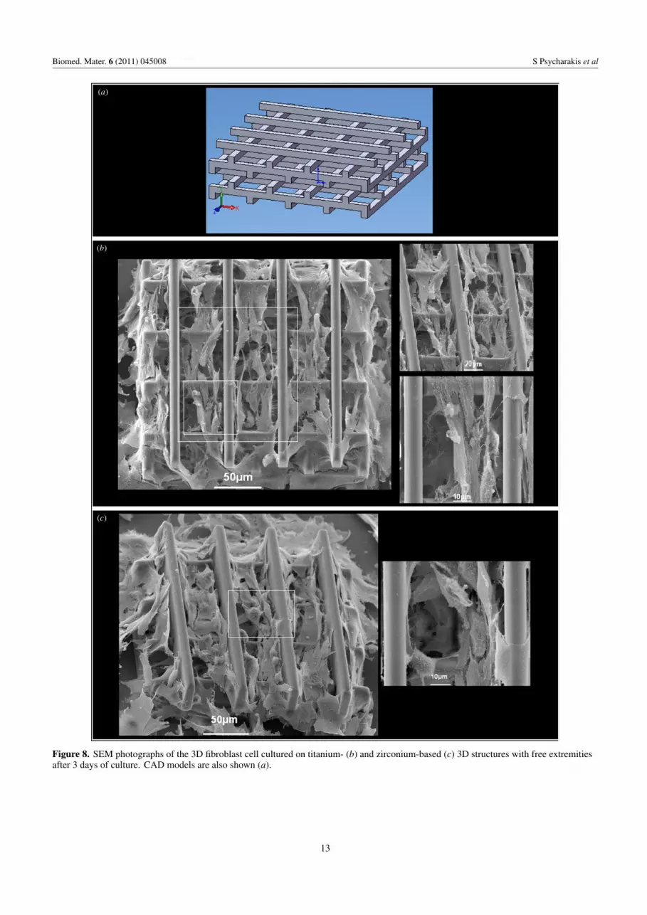
Following a similar culture protocol, the cells were visualizedusing scanning electron microscopy techniques. In this case,fibroblasts migrated successfully on the three-dimensionalscaffolds on both zirconium- and titanium-based hybridmaterials (figures 7(b) and (c)), independently of their size.Bigger porous scaffolds were compared (figure 9) with averageand small porous scaffolds with no statistical difference(p > 0.001). Bigger pores facilitated the formation of 3Dcultures in the inner part of the structure, which can be veryuseful for culturing more than one type of cell. Completecell spreading with cells developing filopodia and lamelipodiafacilitating cell–substrate and cell–cell interactions could beobserved (figure 7).
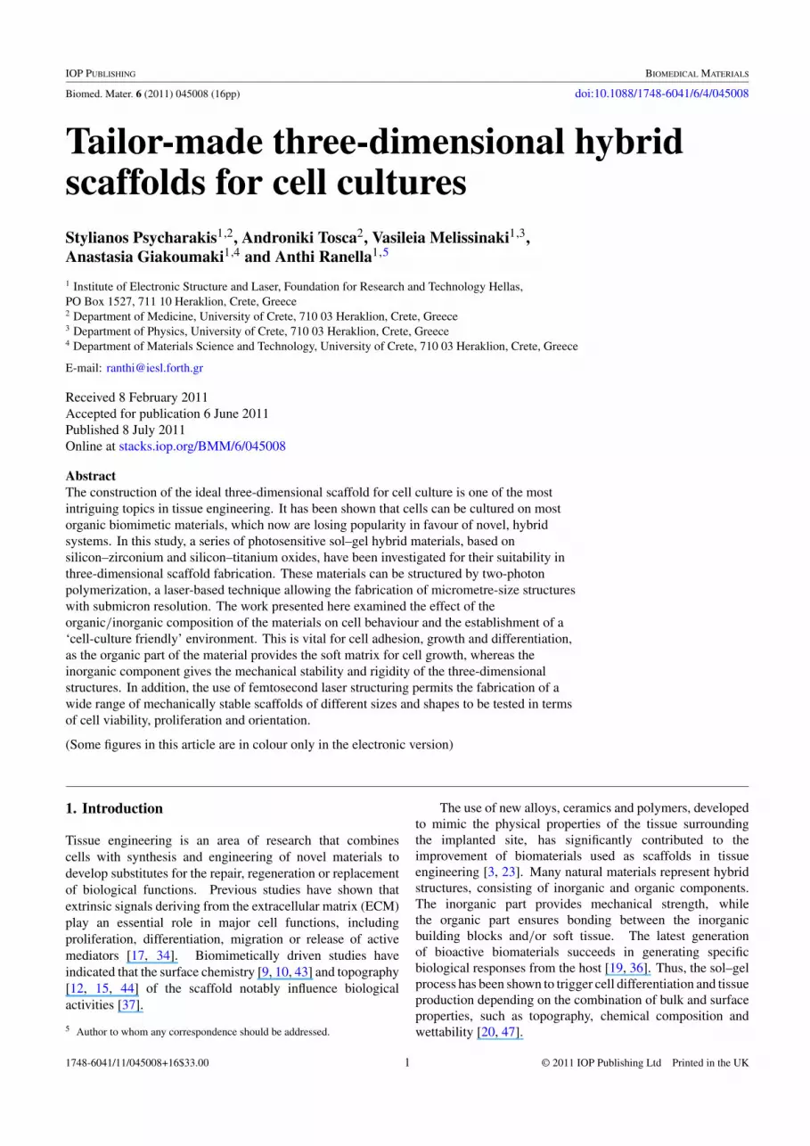
Furthermore, woodpile-shaped structures, consisting oflayers of crossed one-dimensional rods, with a stackingsequence that repeated itself every four layers, wereconstructed (figure 8(a)). The porosity of this type ofstructure increased to 0.895. In this case, fibroblastswere bound to the free edges of the scaffold, which wereused as anchors for migration in the adhesion points ofthe 3D structures (figures 8(b) and (c)). Similar to theresults discussed above, cells were fully developed insidethe structures, forming filopodia to facilitate adherence on
9

Biomed. Mater. 6 (2011) 045008 S Psycharakis et al
(a)
(b)
(c)
Figure 5. High-magnification SEM images of fibroblast cells cultured on the titanium- (b) and zirconium-based (c) microcubes after 3 daysof culture. Computer-aided design (CAD) models are also shown (a).
the hybrid surfaces and sense the environmental morphologyfor expansion (figures 8(b) and (c)).
Woodpile structures (higher mean value) were comparedwith bigger porous scaffolds for the third and the seventhday of cultivation, and differences in cell density proved tobe statistically significant (p < 0.001). Quantification of thecells after extracting them from the fabrications showed thatwoodpile-shaped structures could accommodate the highest
number of cells per volume, while porous structures displayingsmaller pores could accommodate the least (figure 9).
4. Discussion
The use of technologically innovative materials for thefabrication of scaffolds to be used in tissue culture technologyrequires the thorough testing of viability and proliferation of
10

Biomed. Mater. 6 (2011) 045008 S Psycharakis et al
Figure 6. Cell density on cubic surfaces (number of cells per mm2). The number of fibroblasts per surface unit seemed to be independent ofthe dimensions of the cubes (x axis) or the type of hybrid material used. All experiments were carried out in triplicate and total cell valuesare the calculated mean values.
the cells interacting with the material. In addition, specializedtechniques are needed to evaluate the chemical and mechanicalproperties of the scaffolds allowing cell attachment and growth[21, 22]. An important feature as to the type and the propertiesof the candidate materials is dictated by the type of cells to beapplied on the bioactive structures.
In this study, hybrid materials based on zirconium andtitanium were evaluated by cultivating murine fibroblastson them. The existence of organic and inorganic parts inthese hybrid mixtures contributed to the optimum tuning ofthe properties of the three-dimensional scaffolds that werefabricated to host the fibroblasts. In order to examine thesurvival of the cells on the substrate, there have been a seriesof experiments testing cell viability on various concentrationsof the initial mixture of the materials. Results demonstratedthat the concentration (organic/inorganic ratio) of the mixturesignificantly affected cell growth. Quantification of cellpopulation (number of cells per surface area) on hybridand control surfaces proved that cells showed preference forsurfaces with higher metal concentration (metal:MAA = 1:3and 1:4), thus denoting a preference for more stablefabrications. In parallel, cell morphology was investigatedby staining the cytoskeleton, the focal adhesion points and thenucleus. The study of the morphology proved that cells weretightly attached to the substrate and were forming junctionsfor cell–cell and cell–substrate communication.
Secondly, zirconium- and titanium-based hybrid materials(metal:MAA = 1:3) were used for the fabrication of cubic
constructions. These special constructions offer biggercultivation surface in comparison with two-dimensionalcultures on the same terrain size. Fibroblasts successfullyproliferated on the cubes and formed communication bridgesto enhance cell interactions. Furthermore, porous scaffoldswere fabricated in order to investigate the growth of three-dimensional cell cultures inside the fabrication. Porousconstructions of different sizes and shapes were fabricatedin order to tune cell adherence and proliferation in accordancewith the properties of the scaffold. Cells proved to bemore familiar with woodpile-shaped structures that had biggerporosity and free extremities that were used as anchors formigration in the adhesion points of the 3D structures. On allof the three-dimensional porous scaffolds, cells were fullydeveloped inside the structures and they formed filopodiain order to adhere to the hybrid surfaces and to sense theenvironmental morphology for proliferation. This resultimplies that the morphology of the structures can assistcell proliferation in three-dimensional fabrications. Resultsseem to be independent of the type of hybrid material used.However, depending on the desired tissue equivalent, morecomplex fabrications, with a variety of pores, could be moreconvenient.
Porous scaffolds can mimic structures of the body andcan accommodate a wide variety of different cell lines[14, 31]. The colonization of such structures by fibroblastswhich could form ECM equivalents would be useful in ulcerhealing. Furthermore, in combination with a superficial
11

Biomed. Mater. 6 (2011) 045008 S Psycharakis et al
(a)
(b)
(c)
Figure 7. SEM images of the 3D fibroblast cell cultured on titanium- (b) and zirconium-based (c) porous scaffolds after 3 days of culture.CAD models are also shown (a).
12
Biomed. Mater. 6 (2011) 045008 S Psycharakis et al
(a)
(b)
(c)
Figure 8. SEM photographs of the 3D fibroblast cell cultured on titanium- (b) and zirconium-based (c) 3D structures with free extremitiesafter 3 days of culture. CAD models are also shown (a).
13
Biomed. Mater. 6 (2011) 045008 S Psycharakis et al
Figure 9. Cell growth quantification on porous scaffolds. Counting of total number of cells (cells mm−3) cultured in porous scaffoldsproved that woodpile-shaped scaffolds accommodate more cells in the same days of culture. All experiments were carried out in triplicateand total cell values are the calculated mean values.
culture of keratinocytes, it would be possible to manufacturedermal equivalents. The development of bone marrow stromalequivalents and their colonization by stem cells could be veryuseful in hemopoiesis and osteogenic differentiation.
5. Conclusion
The synthesis and processing of biomaterials leading tothe generation of fully functional organs require a deepunderstanding of the interactions between living tissue and thematerials [5]. Therefore, understanding the intricacy of theinterface between living tissue and biomaterial is an importanttask to achieve [6, 46].
In this scientific work, our goal is to develop steadyfabrications that would not degrade and remain stable fora long period of time. This type of bioscaffold could beused in the regeneration of tissues that need controllablemechanical properties (e.g. orthopaedic prosthetics) or longtime healing (e.g. ulcers). For this reason, we are usingthese special hybrid materials that are biocompatible but notbiodegradable.
This work systematically investigated the interactionsbetween titanium- or zirconium-based hybrid materials andfibroblast cells, which were shown to grow in a fullyfunctional mode retaining the expected morphological featuresin substrates with a 1:3 metal:MAA molar ratio. The useof the two-photon polymerization technique in the scaffoldfabrication allowed the fabrication of 3D scaffolds of tuneable
shape and size for use in tissue engineering. Laser processingpresents distinct advantages, compared to alternate ways ofmaterial processing, since (a) it can be applied in the processingof a wide range of materials, (b) it is rapid, easily adaptableand scalable through parallel processing, (c) it does not requirecontact and therefore the selection of parameters allowingmanipulation of sensitive biological molecules and living cellscan be used without loss of activity, and (d) it possesseshigh resolution to the nanoscale features. Most important,it allows the unique possibility for dual micro- and nano-dimensions scale, which is crucial for tissue engineeringapplications [42].
Cell attachment was shown to be further enhanced andfacilitated by the proper choice of the shape and size of 3Dscaffolds. Thus, in cubic structures the smaller cube volumefacilitated cell proliferation as it was easier for the cells toexplore the whole surface of the scaffolds, while smallerspacing allowed better cell communication with neighbouringcubes. In the porous cubic scaffolds, cells preferred the largerstructures that allowed cell migration inside the pores. Finally,woodpile-type fabrications, by offering more cell adhesionpoints on the free extremities, facilitated cell migration withinthe scaffold.
The herein described hybrid materials, in combinationwith the ability for submicron three-dimensional scaffoldingusing 2PP, provide new perspectives in bioengineeringapplications, where scaffolds could mimic the realextracellular matrix and provide a cell-friendly environment
14

Biomed. Mater. 6 (2011) 045008 S Psycharakis et al
for fully guided specific cell cultures comprising one or moredifferent cell types.
Acknowledgments
This work was performed at the Ultraviolet Laser Facilityoperating at IESL-FORTH under the European Commission‘Improving Human Research Potential’ program (RII3-CT-2003-506350). We are grateful to Mrs Aleka Manousakiand Alexandra Siakouli-Galanopoulou for expert technicalassistance with the scanning electron microscopy.
References
[1] Andersson A S, Backhed F, von Euler A, Richter-Dahlfors A,Sutherland D and Kasemo B 2003 Nanoscale featuresinfluence epithelial cell morphology and cytokineproduction Biomaterials 24 3427–36
[2] Bonfield W 2006 Designing porous scaffolds for tissueengineering Phil. Trans. R. Soc. A 364 227–32
[3] Burg K J L, Porter S and Kellam J F 2000 Biomaterialdevelopments for bone tissue engineering Biomaterials21 2347–59
[4] Burridge K, Fath K, Kelly T, Nuckolls G and Turner C 1988Focal adhesions—transmembrane junctions between theextracellular-matrix and the cytoskeleton Annu. Rev. CellBiol. 4 487–525
[5] Chen S S, Fitzgerald W, Zimmerberg J, Kleinman H Kand Margolis L 2007 Cell–cell and cell–extracellular matrixinteractions regulate embryonic stem cell differentiationStem Cells 25 553–61
[6] Chevallay B and Herbage D 2000 Collagen-based biomaterialsas 3D scaffold for cell cultures: applications for tissueengineering and gene therapy Med. Biol. Eng. Comput.38 211–8
[7] Claeyssens F et al 2009 Three-dimensional biodegradablestructures fabricated by two-photon polymerizationLangmuir 25 3219–23
[8] Cukierman E, Pankov R, Stevens D R and Yamada K M 2001Taking cell–matrix adhesions to the third dimension Science294 1708–12
[9] Cyster L A, Parker K G, Parker T L and Grant D M 2003 Theeffect of surface chemistry and nanotopography of titaniumnitride (TiN) films on 3T3-L1 fibroblasts J. Biomed. Mater.Res. A 67 138–47
[10] Cyster L A, Parker K G, Parker T L and Grant D M 2004 Theeffect of surface chemistry and nanotopography of titaniumnitride (TiN) films on primary hippocampal neuronesBiomaterials 25 97–107
[11] Dinca V, Kasotakis E, Catherine J, Mourka A, Ranella A,Ovsianikov A, Chichkov B N, Farsari M, Mitraki Aand Fotakis C 2008 Directed three-dimensional patterningof self-assembled peptide fibrils Nano Lett. 8 538–43
[12] Falconnet D, Csucs G, Grandin H M and Textor M 2006Surface engineering approaches to micropattern surfaces forcell-based assays Biomaterials 27 3044–63
[13] Farsari M, Filippidis G and Fotakis C 2005 Fabrication ofthree-dimensional structures by three-photonpolymerization Opt. Lett. 30 3180–2
[14] Feng B, Jinkang Z, Zhen W, Jianxi L, Jiang C, Jian L, GuolinM and Xin D 2011 The effect of pore size on tissueingrowth and neovascularization in porous bioceramics ofcontrolled architecture in vivo Biomed. Mater. 6 015007
[15] Flemming R G, Murphy C J, Abrams G A, Goodman S Land Nealey P F 1999 Effects of synthetic micro- andnano-structured surfaces on cell behavior Biomaterials20 573–88
[16] Friedl P, Zanker K S and Brocker E B 1998 Cell migrationstrategies in 3D extracellular matrix: differences inmorphology, cell matrix interactions, and integrin functionMicrosc. Res. Tech. 43 369–78
[17] Gomes M E, Sikavitsas V I, Behravesh E, Reis R L and MikosA G 2003 Effect of flow perfusion on the osteogenicdifferentiation of bone marrow stromal cells cultured onstarch-based three-dimensional scaffolds J. Biomed. Mater.Res. A 67 87–95
[18] Hansen J C, Lim J Y, Xu L C, Siedlecki C A, Mauger D Tand Donahue H J 2007 Effect of surface nanoscaletopography on elastic modulus of individual osteoblasticcells as determined by atomic force microscopy J. Biomech.40 2865–71
[19] Hench L L and Polak J M 2002 Third-generation biomedicalmaterials Science 295 1014
[20] Hubbell J A 1999 Bioactive biomaterials Curr. Opin.Biotechnol. 10 123–9
[21] Kapoor A, Caporali E H G, Kenis P J A and Stewart M C 2010Microtopographically patterned surfaces promote thealignment of tenocytes and extracellular collagen ActaBiomater. 6 2580–9
[22] Karageorgiou V and Kaplan D 2005 Porosity of 3Dbiomaterial scaffolds and osteogenesis Biomaterials26 5474–91
[23] Katti K S 2004 Biomaterials in total joint replacementColloids Surf. B 39 133–42
[24] Khang D, Lu J, Yao C, Haberstroh K M and Webster T J 2008The role of nanometer and sub-micron surface features onvascular and bone cell adhesion on titanium Biomaterials29 970–83
[25] Lim J Y, Dreiss A D, Zhou Z Y, Hansen J C, Siedlecki C A,Hengstebeck R W, Cheng J, Winograd N and Donahue H J2007 The regulation of integrin-mediated osteoblast focaladhesion and focal adhesion kinase expression by nanoscaletopography Biomaterials 28 1787–97
[26] Lim J Y, Shaughnessy M C, Zhou Z Y, Noh H, Vogler E Aand Donahue H J 2008 Surface energy effects on osteoblastspatial growth and mineralization Biomaterials 29 1776–84
[27] Ma K, Chan C K, Liao S, Hwang W Y K, Feng Qand Ramakrishna S 2008 Electrospun nanofiber scaffoldsfor rapid and rich capture of bone marrow-derivedhematopoietic stem cells Biomaterials 29 2096–103
[28] Malinauskas M et al 2010 3d artificial polymeric scaffolds forstem cell growth fabricated by femtosecond laserLithuanian J. Phys. 50 75–82
[29] Meisel D C, Deubel M, Hermatschweiler M, Busch K,Koch W, von Freymann G, Blanco A, Enkrich Cand Wegener M 2004 Three-dimensional photonic crystalsFunct. Nanomater. Optoelectron. Other Appl. 99–100 55–64
[30] Mieszawska A J and Kaplan D L 2010 Smartbiomaterials—regulating cell behavior through signalingmolecules BMC Biol. 8 59
[31] Mittal A, Negi P, Garkhal K, Verma S and Kumar N 2010Integration of porosity and bio-functionalization to form a3D scaffold: cell culture studies and in vitro degradationBiomed. Mater. 5 045001
[32] Ovsianikov A et al 2008 Ultra-low shrinkage hybridphotosensitive material for two-photon polymerizationmicrofabrication ACS Nano 2 2257–62
[33] Pedersen J A and Swartz M A 2005 Mechanobiology in thethird dimension Ann. Biomed. Eng. 33 1469–90
[34] Prunieras M, Regnier M and Woodley D 1983 Methods forcultivation of keratinocytes with an air–liquid interfaceJ. Invest. Dermatol. 81 S28–33
[35] Ranella A, Barberoglou M, Bakogianni S, Fotakis Cand Stratakis E 2010 Tuning cell adhesion by controllingthe roughness and wettability of 3D micro/nano siliconstructures Acta Biomater. 6 2711–20
15

Biomed. Mater. 6 (2011) 045008 S Psycharakis et al
[36] Ratner B D and Bryant S J 2004 Biomaterials: where we havebeen and where we are going Annu. Rev. Biomed. Eng.6 41–75
[37] Reichert J, Brukner S, Bartelt H and Jandt K D 2007 Tuningcell adhesion on PTFE surfaces by laser inducedmicrostructures Adv. Eng. Mater. 9 1104–13
[38] Rosales-Leal J I, Rodriguez-Valverde M A, Mazzaglia G,Ramon-Torregrosa P J, Diaz-Rodriguez L,Garcia-Martinez O, Vallecillo-Capilla M, Ruiz Cand Cabrerizo-Vilchez M A 2010 Effect of roughness,wettability and morphology of engineered titanium surfaceson osteoblast-like cell adhesion Colloids Surf. A 365 222–9
[39] Rotsch C and Radmacher M 2000 Drug-induced changes ofcytoskeletal structure and mechanics in fibroblasts: anatomic force microscopy study Biophys. J. 78 520–35
[40] Sakellari I, Gaidukeviciute A, Giakoumaki A, Gray D,Fotakis C, Farsari M, Vamvakaki M, Reinhardt C,Ovsianikov A and Chichkov B N 2010 Two-photonpolymerization of titanium-containing sol–gel compositesfor three-dimensional structure fabrication Appl. Phys. A100 359–64
[41] Shikinami Y, Okazaki K, Saito M, Okuno M, Hasegawa S,Tamura J, Fujibayashi S and Nakamura T 2006 Bioactiveand bioresorbable cellular cubic-composite scaffolds for usein bone reconstruction J. R. Soc. Interface 3 805–21
[42] Stratakis E, Ranella A, Farsari M and Fotakis C 2009Laser-based micro/nanoengineering for biologicalapplications Prog. Quantum Electron. 33 127–63
[43] Suh J Y, Jang B C, Zhu X L, Ong J L and Kim K 2003 Effectof hydrothermally treated anodic oxide films on osteoblastattachment and proliferation Biomaterials 24 347–55
[44] Teixeira A I, Nealey P F and Murphy C J 2004 Responses ofhuman keratocytes to micro- and nanostructured substratesJ. Biomed. Mater. Res. A 71 369–76
[45] Trickey W R, Vail T P and Guilak F 2004 The role of thecytoskeleton in the viscoelastic properties of humanarticular chondrocytes J. Orthop. Res. 22 131–9
[46] Wang Y Z, Kim U J, Blasioli D J, Kim H J and Kaplan D L2005 In vitro cartilage tissue engineering with 3D porousaqueous-derived silk scaffolds and mesenchymal stem cellsBiomaterials 26 7082–94
[47] Xynos I D, Edgar A J, Buttery L D K, Hench L L andPolak J M 2001 Gene-expression profiling of humanosteoblasts following treatment with the ionic products ofBioglass (R) 45S5 dissolution J. Biomed. Mater. Res.55 151–7
[48] Yang B, Yin Z H, Cao J L, Shi Z L, Zhang Z T, Song H X,Liu F Q and Caterson B 2010 In vitro cartilage tissueengineering using cancellous bone matrix gelatin as abiodegradable scaffold Biomed. Mater.5 045003
16
Related Documents