UNIVERSIDADE FEDERAL DE SANTA MARIA CENTRO DE CIÊNCIAS RURAIS PROGRAMA DE PÓS-GRADUAÇÃO EM ZOOTECNIA Taida Juliana Adorian FIBRAS FUNCIONAIS DA LINHAÇA E SEUS IMPACTOS NA NUTRIÇÃO DE JUNDIÁS Santa Maria, RS 2018

Welcome message from author
This document is posted to help you gain knowledge. Please leave a comment to let me know what you think about it! Share it to your friends and learn new things together.
Transcript

3
UNIVERSIDADE FEDERAL DE SANTA MARIA
CENTRO DE CIÊNCIAS RURAIS
PROGRAMA DE PÓS-GRADUAÇÃO EM ZOOTECNIA
Taida Juliana Adorian
FIBRAS FUNCIONAIS DA LINHAÇA E SEUS IMPACTOS NA
NUTRIÇÃO DE JUNDIÁS
Santa Maria, RS
2018

4

5
Taida Juliana Adorian
FIBRAS FUNCIONAIS DA LINHAÇA E SEUS IMPACTOS NA NUTRIÇÃO DE
JUNDIÁS
Tese apresentada ao Curso de Doutorado do
Programa de Graduação em Zootecnia, da
Universidade Federal de Santa Maria (UFSM,
RS), como requisito parcial para obtenção do
título de Doutora em Zootecnia.
Orientadora: Profª Drª Leila Picolli da Silva
Santa Maria, RS
2018
6
______________________________________________________________
© 2018
Todos os direitos autorais reservados a Taida Juliana adorian. A reprodução de partes ou do
todo deste trabalho só poderá ser feita mediante a citação da fonte.
Endereço: Avenida Roraima, n. 1000, Bairro Camobi, Santa Maria, RS. CEP: 97105-900
Fone (55) 3220 8365; E-mail: [email protected]

7

8
9
AGRADECIMENTOS
Primeiramente agradeço à Universidade Federal de Santa Maria por me proporcionar um
ensino de qualidade, da graduação ao doutorado.
À professora Dra. Leila Picolli da Silva, por me orientar e ensinar durante todos esses anos
de convivência.
À professora Dra. Naglezi Lovatto, pela coorientação, amizade e apoio, mesmo quando em
licença maternidade.
À Dra. Fernanda Goulart, pela ajuda incondicional durante toda minha formação, pelas
trocas de ideia, incentivo e amizade.
À Pati e Dina, pela ajuda antes, durante e depois do experimento, pela amizade,
companheirismo e por todos esses anos de convivência.
Ao Bruno e Marina, meu agradecimento por não medirem esforços para me auxiliar no que
necessário, pela parceria e amizade.
À Ana Betine por sempre estar disposta a ajudar, seja nos artigos, análises ou biometrias.
Agradeço imensamente por tudo.
À Carol pelo auxílio com as análises, biometrias e pela amizade de sempre.
Agradeço de coração aos demais colegas do Laboratório de Piscicultura que acompanharam
minha trajetória, sempre dispostos a ajudar; também agradeço a companhia,
mates, almoços, conversas, biometrias, análises...
Ao Professor Roger Wagner e Mariane Fagundes, meu muito obrigada por nos auxiliarem
com a técnica de AGCC, pela paciência e excelência com que trataram nosso trabalho.
À Luiza Loebens, pelo auxílio com as análises histológicas.
Ao professor Ayrton Martins e Giovani Pedroso pela determinação dos monossacarídeos.
Ao Silvino, por toda ajuda com o uso dos novos equipamentos e auxílio nas análises.
Ao secretário do PPGZ, Marcos, pela dedicação com que desenvolve o seu trabalho e
disponibilidade em nos auxiliar sempre que necessário.
À Capes pela bolsa de doutorado concedida.
À Giovelli & Cia pela doação da linhaça utilizada nesta pesquisa.
Meu muito obrigada!

10

11
“A tarefa não é tanto ver aquilo que ninguém viu,
mas pensar o que ninguém ainda pensou,
sobre aquilo que todo mundo vê.”
Arthur Schopenhauer

12

13
RESUMO
FIBRAS FUNCIONAIS DA LINHAÇA E SEUS IMPACTOS NA NUTRIÇÃO DE
JUNDIÁS
AUTORA: Taida Juliana Adorian
ORIENTADORA: Leila Picolli da Silva
Este estudo objetivou avaliar a ação prebiótica de fibras funcionais de linhaça com distintas
proporções de fibra alimentar solúvel e insolúvel e seus impactos na nutrição e saúde de juvenis de
jundiás (6,43g). Para isso, foram concentradas as frações solúvel e insolúvel de fibra da linhaça, a
partir da utilização de técnicas físicas e químicas de concentração. Estas frações foram combinadas em
diferentes proporções (1:0,5, 1:1, 1:2 e 1:4 fibra solúvel: insolúvel) para obtenção de fibras funcionais,
que foram adicionadas a dietas e avaliadas em um ensaio biológico com juvenis de jundiá. O ensaio
biológico teve duração de 45 dias e foi realizado em sistema de recirculação de água, composto por 20
tanques (290L), biofiltros e reservatório de água. Neste período os peixes foram alimentados até a
saciedade aparente, três vezes ao dia. Ao final do período os peixes foram submetidos a jejum de 18
horas e biometria para coleta de dados de peso, comprimento, coleta de sangue, tecidos (fígado e trato
digestivo), muco e digesta para determinação de parâmetros de desempenho, composição e deposição
corporal, metabólitos plasmáticos, hepáticos, enzimas digestivas, indicadores imunológicos, histologia
intestinal e produção de ácidos graxos de cadeia curta. Após a biometria final os peixes foram
mantidos nas unidades experimentais por mais cinco dias e ao final deste período, submetidos a
estresse agudo, com posterior coleta de sangue e muco para determinação de metabólitos e indicadores
imunológicos. O delineamento experimental utilizado foi o inteiramente casualizado, composto cinco
tratamentos e quatro repetições (600 peixes). Os resultados obtidos foram submetidos à teste de
normalidade, seguido por análise de variância, sendo as médias comparadas pelo teste de Tukey ao
nível de 5% de significância. As dietas com as proporções 1:2 e 1:4 proporcionaram maior ganho de
peso, taxa de crescimento específico e deposição de proteína bruta corporal aos peixes, proteínas totais
circulantes e globulinas, assim como o teor de mucoproteína, imunoglobulinas totais e pH do muco
cutâneo. Já os níveis de cortisol e o pH intestinal foram mais baixos nestes tratamentos. A dieta 1:0,5
alterou a atividade de tripsina no intestino dos jundiás e juntamente com a dieta 1:4 proporcionou
maior altura das vilosidades intestinais. Enquanto que altura total da vilosidade foi superior para os
peixes que receberam fibra de linhaça na dieta, independente da proporção, o inverso foi observado
para a espessura da camada muscular. Independente da proporção na dieta, o consumo de fibra de
linhaça aumentou as imunoglobulinas totais no plasma e a atividade da fosfatase alcalina no plasma e
muco cutâneo. A produção de ácido acético intestinal foi superior nos peixes alimentados com a dieta
1: 2, enquanto que de ácido butírico com a dieta 1:4 e ácido propiônico com a dieta controle. A dieta
controle levou a menor contagem de células caliciformes. Após o estresse agudo, os peixes
alimentados com as dietas contendo as proporções de fibra solúvel: insolúvel 1: 2 e 1: 4 apresentaram
maior teor de proteína total, globulina e atividade de fosfatase alcalina do plasma, além de maior teor
de mucoproteína no muco cutâneo dos peixes. Em conclusão, os resultados indicam que a fibra de
linhaça tem ação prebiótica imunoestimulante para juvenis de jundiá, sendo que as proporções de 1:2 e
1:4 de fibra solúvel: insolúvel otimizam o sistema imune e a produção de ácidos graxos de cadeia
curta, com reflexos positivos sobre o desempenho dos peixes. Além disso, nessas proporções ela ainda
age como mitigadora de estresse.
Palavras-chave: Fibra alimentar. Linhaça. Prebiótico. Rhamdia quelen.

14

15
ABSTRACT
FUNCTIONAL FIBERS OF LINSEED AND ITS IMPACTS ON NUTRITION OF
SILVER CATFISH
AUTHOR: Taida Juliana Adorian
ADVISOR: Leila Picolli da Silva
This study aimed to evaluate the prebiotic action of functional linseed fibers with different proportions
of soluble and insoluble dietary fiber and its impact on the nutrition and health of juveniles silver
catfish (6.43g). For this, the soluble and insoluble fractions of linseed fiber were concentrated, using
the use of physical and chemical concentration techniques. These fractions were combined in different
proportions (1:0.5, 1:1, 1:2 and 1:4 soluble: insoluble fibre) to obtain functional fibers, which were
added to diets and evaluated in a biological test with juveniles silver catfish. The biological assay
lasted 45 days and was performed in a water recirculation system, composed of 20 tanks (290L),
biofilters and water reservoir. At this time the fish were fed to apparent satiety three times a day. At
the end of the period the fish were submitted to a 18 hour fast and biometry for data collection of
weight, length, blood collection, tissues (liver and digestive tract), mucus and digesta for
determination of performance parameters, composition and body deposition, plasma metabolites,
hepatic enzymes, digestive enzymes, immunological indicators, intestinal histology and production of
short chain fatty acids. After the final biometry the fish were kept in the experimental units for another
five days and at the end of this period, submitted to acute stress, with subsequent collection of blood
and mucus for determination of metabolites and immunological indicators. The experimental design
was a completely randomized design, consisting of five treatments and four replications (600 fish).
The results were submitted to the normality test, followed by analysis of variance, and the means were
compared by the Tukey test at the 5% level of significance. Diets 1:2 and 1:4 provided greater weight
gain, specific growth rate and crude protein deposition in fish, total circulating proteins and globulins,
as well as mucoprotein content, total immunoglobulins and cutaneous mucus pH. Cortisol levels and
intestinal pH were lower in these treatments. The 1:0.5 diet altered the trypsin activity in the silver
catfish intestine and together with the 1:4 diet provided higher intestinal villi height. While total villus
height was higher for the fish that received linseed fiber in the diet, regardless of the proportion, the
inverse was observed for the thickness of the muscle layer. Regardless of dietary ratio, linseed fiber
intake increased total plasma immunoglobulins and plasma alkaline phosphatase activity and
cutaneous mucus. The production of intestinal acetic acid was higher in the fish fed with the 1:2 diet,
whereas of the butyric acid with the 1:4 diet and propionic acid with the control diet. The control diet
led to lower counts of goblet cells. After acute stress, the fish fed the diets containing soluble:
insoluble fiber ratios 1:2 and 1:4 presented higher total protein, globulin and plasma alkaline
phosphatase activity, as well as a higher mucoprotein content in the mucus of fish. In conclusion, the
results indicate that linseed fiber has an immunostimulating prebiotic action for silver catfish
juveniles, and the 1:2 and 1:4 ratios of soluble: insoluble fiber optimize the immune system and the
production of short-chain fatty acids, with positive reflexes on fish performance. Moreover, in these
proportions it still acts as a stress reliever.
Keywords: Dietary fiber. Linseed. Prebiotic. Rhamdia quelen.

16

17
LISTA DE TABELAS
ARTIGO I
Tabela 1 - Dietary formulations and proximate composition of the experimental diets (g/k)
................................................................................................................................................. 36
Tabela 2 - Performance parameters of Rhamdia quelen fed with different ratio soluble:
insoluble linseed fiber in the diet ....................................................................... 37
Tabela 3 – Corporal composition (g/kg) and body deposition of protein and fat (g) of juvenile
Rhamdia quelen .................................................................................................. 38
Tabela 4 – Corporal yield and digestive index (g/kg) of juvenile silver catfish (Rhamdia
quelen) ................................................................................................................ 39
Tabela 5 – Activity of digestive enzymes of juvenile Rhamdia quelen receiving the
experimental diets ................................................................................................. 40
Tabela 6 – Intestinal histology of juvenile Rhamdia quelen fed with different ratio soluble:
insoluble linseed fiber in the diet .......................................................................... 41
Tabela 7 - Hepatic metabolites of juvenile Rhamdia quelen receiving the experimental diets
................................................................................................................................................. 42
ARTIGO II
Tabela 1 – Dietary formulations and proximate composition of the experimental diets (g/kg)
................................................................................................................................................. 67
Tabela 2 – Plasma parameters of juvenile Rhamdia quelen receiving the experimental diets
................................................................................................................................................. 68
Tabela 3 – Skin mucus parameters of juvenile Rhamdia quelen fed with different ratio
soluble: insoluble linseed fiber in the diet .......................................................... 69
Tabela 4 – pH and concentration of short-chain fatty acids (μmol/g) in gut contents of
Rhamdia quelen .................................................................................................. 70
Tabela 5 – Effect of different proportions of soluble and insoluble fiber on intestinal goblet
cell counts (cells/g) in silver catfish ..................................................................... 71
Tabela 6 – Parameters of performance and survival of Rhamdia quelen receiving the
experimental diets ................................................................................................. 72

18
ARTIGO III
Tabela 1 – Dietary formulations and proximate composition of the experimental diets (g/kg)
.................................................................................................................................................. 91
Tabela 2 – Plasma parameters of juvenile Rhamdia quelen receiving the experimental diets
.................................................................................................................................................. 92
Tabela 3 – Skin mucus parameters of juvenile Rhamdia quelen fed with different ratio
soluble: insoluble linseed fiber in the diet............................................................. 93

19
SUMÁRIO
1 INTRODUÇÃO ............................................................................................................. 21
1.1 OBJETIVOS .................................................................................................................... 23
1.1.1 Objetivo geral ................................................................................................................. 23
1.1.2 Objetivos específicos ...................................................................................................... 23
2 ARTIGO I ...................................................................................................................... 25
ABSTRACT .................................................................................................................... 27
INTRODUCTION .......................................................................................................... 28
MATERIAL AND METHODS ...................................................................................... 29
RESULTS ....................................................................................................................... 34
DISCUSSION ................................................................................................................. 36
CONCLUSION ............................................................................................................... 40
ACKNOWLEDGMENTS .............................................................................................. 26
REFERENCES ................................................................................................................ 40
3 ARTIGO II .................................................................................................................... 54
ABSTRACT .................................................................................................................... 56
INTRODUCTION .......................................................................................................... 57
MATERIAL AND METHODS ...................................................................................... 58
RESULTS ....................................................................................................................... 64
DISCUSSION ................................................................................................................. 66
CONCLUSION ............................................................................................................... 70
ACKNOWLEDGMENTS .............................................................................................. 58
REFERENCES ................................................................................................................ 71
4 ARTIGO III ................................................................................................................... 84
ABSTRACT .................................................................................................................... 86
INTRODUCTION .......................................................................................................... 86
MATERIAL AND METHODS ...................................................................................... 87
RESULTS ...................................................................................................................... 91
DISCUSSION ................................................................................................................. 91
CONCLUSION ............................................................................................................... 94
ACKNOWLEDGMENTS .............................................................................................. 84
REFERENCES ................................................................................................................ 95
5 DISCUSSÃO GERAL ................................................................................................. 105
6 CONCLUSÃO GERAL .............................................................................................. 109
REFERÊNCIAS BIBLIOGRÁFICAS ...................................................................... 110
20
APÊNDICE A – Fracionamento da linhaça e obtenção de ingredientes ricos em
proteína e fibra: alternativas para a alimentação animal....................................... 113
ANEXO A – Normas da revista Animal Feed Science and Technology ................ 140
ANEXO B – Normas da revista Aquaculture Research ......................................... 146

21
1 INTRODUÇÃO
As estatísticas mostram que a aquicultura tem crescido em ritmo acelerado em todo o
mundo, destacando a importância da atividade na produção de proteína de origem animal. De
acordo com a FAO, em 2017 a produção de pescado foi 43% superior a carne suína, sendo
que deste total, quase metade foi proveniente da aquicultura, demonstrando um crescimento
no cultivo mundial de 60% entre 2007 e 2017 (ANUÁRIO PeixeBR, 2018; FAO, 2017). No
Brasil, o setor aquícola também tem crescido substancialmente, com aumento de 8% na
produção somente em 2017 (691.700 toneladas produzidas no ano de referência). Tal
crescimento foi alavancado pelos estados da região Sul do País (Paraná, Santa Catarina e Rio
Grande do Sul), que juntos contribuíram com mais de 178.000 toneladas no ano.
Com este crescimento da atividade e a intensificação do cultivo objetivando alcançar
altos índices de produtividade, os peixes acabam sendo expostos com maior frequência a
situações estressantes. Altas densidades de estocagem, variações na qualidade da água,
manejos frequentes inerentes a atividade, reduzem a resposta imune dos animais, tornando-os
mais susceptíveis a doenças e, consequentemente, aumentando a mortalidade e diminuindo a
viabilidade econômica do cultivo de peixes (URBINATI; CARNEIRO, 2004).
Para reduzir estes impactos, uma prática comum por anos foi o uso de antibióticos.
Porém, devido a restrição ao uso destas moléculas, seja por promoverem resistência em
microorganismos patogênicos, pelo acúmulo residual sobre o produto animal ou pela
contaminação ambiental, buscam-se alternativas racionais, eficientes e ambientalmente
seguras para substituição destes produtos (CYRINO et al., 2010), motivando os estudos com
aplicação de moléculas orgânicas, assim como aditivos alternativos para uso zootécnico.
Como opção, destaca-se a suplementação das dietas com prebióticos, os quais são
carboidratos seletivamente fermentáveis que permitem modificações na composição e/ou
atividade da microbiota intestinal, resultando em melhorias na saúde e desempenho dos
animais (ROBERFROID, 2007). Grande parte dos prebióticos comercialmente disponíveis
são frações isoladas e parcialmente hidrolisadas (oligossacarídios), provenientes da fibra
alimentar, porém estudos tem demonstrado que o uso de concentrados de fibra alimentar tem
efeitos similares aqueles proporcionados por prebióticos comerciais usuais (ADORIAN et al.
2015; ADORIAN et al. 2016; GOULART et al. 2017; MOMBACH, 2015). Embora em início
de desenvolvimento conceitual e tecnológico, estes estudos demonstram que a manipulação
do teor e das proporções das frações de fibra alimentar nas dietas, resulta em efeitos positivos
para os peixes, com maior racionalidade produtiva e ambiental.

22
A fibra alimentar é classificada de acordo com a sua solubilidade em água, em solúvel
ou insolúvel (WENZEL, 2012). Na prática, ambas as frações da fibra alimentar são partes da
dieta, porém seus efeitos dependem da variação de seus teores individuais, da predominância
de uma fração em relação a outra, sua composição química e organização estrutural
(MACAGNAN et al., 2016; MORRE et al., 1998).
Ao chegar no intestino, tanto a fibra solúvel quanto a insolúvel servem como substrato
para fermentação microbiana (WENZEL, 2012). Nesse ambiente a fibra se depara com grande
atividade bioquímica de bactérias, sendo que as espécies sacarolíticas ali presentes, participam
de forma intensa da sua quebra e fermentação (FERREIRA, 2012). Nesse processo são
gerados alguns produtos, como os ácidos graxos de cadeia curta (AGCC) acetato, butirato e
propionato, bem como, ocorrerá liberação gradual dos compostos fenólicos ligados a fibra, os
quais são parcialmente absorvidos pelas células epiteliais do intestino (FERREIRA, 2012;
QUIRÓS-SAUCEDA et al., 2014; WENZEL, 2012). Além da ação antioxidante que previne
danos em lipídios, proteínas e ácidos nucleicos, conservando a fluidez, permeabilidade e
integridade celular (BARRERA, 2012; REPETTO et al., 2012; ZHANG et al., 2008), os
compostos fenólicos também apresentam atividade anti-inflamatória, que inibe a produção de
citoquinas, evitando doenças imunológicas resultantes da inflamação (LIU; LIN, 2013;
VERES, 2012).
Como espécie com potencial de cultivo no Sul do Brasil, destaca-se o jundiá (Rhamdia
quelen). Porém existem várias lacunas relacionadas a sua produção que precisam ser
elucidadas para que a espécie se torne competitiva. Dentre as linhas de pesquisa que merecem
atenção, estão as exigências nutricionais, ingredientes alternativos e sistemas de cultivos.
Além disso, o desenvolvimento de aditivos alimentares para a espécie com foco principal na
proteção e promoção da saúde é uma tendência que deve continuar crescendo nos próximos
anos (VALLADÃO et al., 2018).
Trabalhos com jundiás demonstram que a adição de fibras alimentares concentradas
nas dietas desta espécie, exercem ação efetivamente prebiótica, uma vez que otimizam o
desempenho, metabolismo e sistema imunológico dos peixes (ADORIAN et al., 2015;
ADORIAN et al., 2016; GOULART et al., 2017). Dentre as fontes de fibras testadas, os
resultados de maior impacto foram obtidos com a adição de fibra de linhaça (Linum
uistatissimum L.). A linhaça é reconhecida como uma fonte rica em fibra alimentar, que
apresenta boa proporção de fibras solúveis e insolúveis (GALVÃO et al., 2008). A fibra
solúvel, também conhecida como mucilagem, é composta por monossacarídeos como a

23
ramnose, galactose, frutose, xilose e arabinose. Já a fibra insolúvel, por celulose (monômeros
de glicose) e lignina (álcoois aromáticos) (RAY et al., 2013; SHIM et al., 2014).
De acordo com Goulart et al. (2013), o farelo de linhaça in natura é uma fonte
alternativa de proteína para fabricação de rações para jundiás. Segundo os autores, os bons
resultados obtidos estão relacionados a presença da fibra solúvel, a qual pode ter exercido
efeito prebiótico, refletindo de forma desejável no desempenho animal. Outras evidências da
ação pebiótica da fibra de linhaça foram demonstradas por Goulart et al. (2017), ao
suplementar mucilagem de linhaça em dietas para mesma espécie, a qual proporcionou maior
ganho de peso e conversão alimentar. O que reforça essa ideia são os resultados de Adorian et
al. (2015) e Adorian et al. (2016), onde peixes que receberam fibra de linhaça na dieta
(solúvel + insolúvel) tiveram resultados iguais ou superiores ao que receberam dieta com
prebiótico comercial (Actigen®).
Porém, as proporções de fibra solúvel e insolúvel ideais para otimizar tais resultados
ainda não são conclusivas. Dessa forma, é perceptível a necessidade de aprofundar as
pesquisas neste viés, focando na obtenção de fibras funcionais de linhaça, com inclusão de
distintas proporções das frações solúvel e insolúvel.
1.1 OBJETIVOS
1.1.1 Objetivo geral
Avaliar a ação prebiótica de fibras funcionais de linhaça com distintas proporções de
fibra alimentar solúvel e insolúvel e seus impactos na nutrição e saúde de juvenis de jundiás.
1.1.2 Objetivos específicos
- Concentrar a fibra alimentar contida na linhaça para desenvolvimento de fibras
funcionais com potencial prebiótico;
- Combinar e avaliar o potencial prebiótico das distintas proporções de fibra solúvel e
insolúvel de linhaça (1:0,5; 1:1; 1:2; 1:4) em dietas para juvenis de jundiá, sobre os
parâmetros de desempenho, metabólicos e imunológicos;
24
- Avaliar a resistência ao estresse de jundiás alimentados com distintas proporções de
fibra solúvel e insolúvel de linhaça em dietas.
O presente estudo foi desenvolvido em duas fases. A primeira consistiu na obtenção
das frações solúvel e insolúvel de fibra de linhaça e análise de sua composição química e
propriedades físico-químicas. Na segunda fase, as frações foram combinadas em quatro
distintas proporções de fibra solúvel: insolúvel (1:0,5, 1:1, 1:2 e 1:4), adicionadas a dietas
para jundiás e avaliadas em ensaio biológico. Os resultados estão apresentados na forma de
artigos científicos, onde o artigo I corresponde a avaliação das distintas proporções de fibra
solúvel: insolúvel sobre o desempenho zootécnico, qualidade corporal, metabolismo e
morfometria intestinal. No artigo II, avaliou-se o efeito das combinações sobre os parâmetros
imunológicos e de crescimento. Enquanto que no artigo III, a ação imunoestimulante das
fibras solúvel e insolúvel de linhaça foi avaliada em jundiás submetidos a estresse agudo.
É apresentado ainda um artigo no apêndice A, que corresponde a primeira fase do
estudo, onde realizou-se a obtenção e caracterização química e de proriedades físico-químicas
das frações solúvel e insolúvel de fibra de linhaça, assim como, de um concentrado proteico,
avaliado em outra tese pertencente ao mesmo projeto do nosso grupo de pesquisa
(“Alternativas de nutrientes e compostos bioativos: estudo do fracionamento da linhaça para
nutrição de peixes”, registrado no CEUA pelo nº 8015120816).

25
2 ARTIGO I
O artigo científico intitulado “Functional linseed fibers and their impacts on silver
catfish nutrition” foi submetido para a revista Animal Feed Science and Technology e está
formatado segundo as normas descritas no Guia dos Autores (Anexo A).

26
Functional linseed fibers and their impacts on silver catfish nutrition 1
2
Taida Juliana Adoriana*, Patrícia Inês Mombacha, Dirleise Pianessoa, Bruno Bianch Loureiroa, 3
Joziane Limaa, Thaís Soaresa, Luiza Loebensb, Leila Picolli da Silvaa 4
5
aDepartment of Animal Science, Federal University of Santa Maria, Santa Maria, Rio Grande 6
do Sul. AV. Roraima nº 1000, Cidade Universitária, Bairro Camobi, Santa Maria – RS, 7
Brazil. CEP: 97105-900. 8
9
bDepartment of Ecology and Evolution, Federal University of Santa Maria, Santa Maria, Rio 10
Grande do Sul. AV. Roraima nº 1000, Cidade Universitária, Bairro Camobi, Santa Maria – 11
RS, Brazil. CEP: 97105-900. 12
13
14
*Corresponding author. Tel. 55 (55) 3220-8365; Fax: 55 (55) 3220-82 40; E-mails: 15
[email protected]; [email protected] 16
17
18

27
Abstract 19
This study was conducted with the objective of evaluating the combination of different ratios 20
of soluble and insoluble linseed fiber on the zootechnical performance, body quality and 21
intestinal morphometry of young silver catfish. For this, the soluble and insoluble fractions of 22
linseed fiber were concentrated separately and combined in four ratios (1:0.5, 1:1, 1:2, 1:4), 23
which were added to silver catfish (6.43 ± 0.12 g) diets and evaluated in a bioassay, along 24
with a control diet (without the addition of linseed fiber). After 45 days receiving the 25
experimental diets, the animals were fasted and anesthetized in order to perform a biometry to 26
collect data and tissues for further analysis. The experimental design was completely 27
randomized, with five treatments and four replications. Data were submitted to analysis of 28
variance and the means were compared by Tukey’s test (P <0.05). Diets 1:2 and 1:4 provided 29
higher weight gain, specific growth rate and crude protein deposition to the fish, whereas only 30
the 1:4 diet reflected higher crude body protein. The 1:0.5 diet altered the trypsin activity in 31
the intestine of silver catfish and, together with the 1:4 diet, it provided higher intestinal villus 32
height. While the total villus height was greater for the fish that received linseed fiber in their 33
diet, regardless of the proportion, the opposite was observed for the muscle layer thickness. 34
Body yield, somatic and digestive parameters, chymotrypsin activity and glucose, glycogen 35
and liver protein were not altered, regardless of the experimental diets. In conclusion, the 36
results indicate that linseed fiber acts effectively as a growth promoter in silver catfish diets, 37
with the use of 1:2 and 1:4 ratios optimizing its prebiotic action on the animal organism. 38
Keywords: Rhamdia quelen, soluble fiber, insoluble fiber, Linum usitatissimum, prebiotic 39
Abbreviations: AFC, apparent feed conversion; CF, condition factor; CY, corporal yield; DSI, 40
digestive somatic index; FBF, final body fat; FW, final weight; FBP, final body protein; HSI, 41
hepatosomatic index; IBF, initial body fat; IBP, initial body protein; IQ, intestinal quotient; 42
IW, initial weight; SCFA, short-chain fatty acids; SE, standard error; SGR, specific growth 43
28
rate; S:IF, soluble: insoluble fiber; TCA, trichloroacetic acid; UFSM, University of Santa 44
Maria; VFI, visceral fat index. 45
46
1. Introduction 47
Food fiber consists of a complex and heterogeneous set of non-starch polysaccharides, 48
oligosaccharides and minor compounds, which are resistant to the enzymatic digestion in the 49
digestive tract of animals and which can, to varying degrees, be degraded and fermented into 50
short chain fatty acids by intestinal microbiota (Buttriss and Stokes, 2008; Macagnan et al., 51
2016). Fibers are classified according to their solubility, as soluble or insoluble, and the 52
relations between these fractions, their composition, organizational structure, physico-53
chemical characteristics and presence of bioactive compounds associated to the matrix, are 54
determinant for their functional properties (Westenbrink et al., 2013, Macagnan et al., 2016). 55
In practice, both fractions are found in diets, but the effects on the digestive and metabolic 56
processes depend on both solubility variations and the chemical ratios and interactions 57
between fractions (Van Soest et al., 1991; Morre et al., 1998; Silva and Walter, 2012). 58
In order to enhance the functional benefits, many authors suggest the application of 59
dietary fiber hydrolysis techniques to obtain oligosaccharides, which are used as prebiotic 60
agents in diets (Gullón et al., 2011; Chen et al., 2013; Gómez et al., 2014). However, in fish 61
nutrition, studies have shown that the use of non-hydrolysed food fiber concentrates (linseed, 62
brewer's yeast and citrus pulp) has equivalent or greater effects than consolidated commercial 63
prebiotics, optimizing the immune system and acting as a growth promoter (Adorian et al., 64
2015; Mombach, 2015; Adorian et al., 2016; Goulart et al., 2017). This demonstrates that the 65
functional agents for fish can be obtained with simpler and lower cost technology than 66
oligosaccharides that make up the vast majority of commercial prebiotics. 67

29
For linseed (Linum usitatissimum L.), total dietary fiber concentration techniques were 68
applied and the resulting fibrous concentrates were successfully tested on fish nutrition 69
(Adorian et al., 2015; Adorian et al., 2016, Goulart et al., 2018). It is possible to believe, 70
however, that there is still scope to optimize the results of these studies, through the direct 71
application of different ratios of soluble and insoluble fibers, which can be extracted from 72
isolated fractions and independently combined in fish diets. In this contex, this study was 73
conducted to evaluate the combination of different soluble and insoluble linseed fiber ratios 74
(1:0.5, 1:1, 1:2, 1:4) on the growth performance, body quality and intestinal morphometry of 75
silver catfish (Rhamdia quelen). 76
77
2. Material and methods 78
The study was conducted at the Laboratory of Fish Farming of the Department of Animal 79
Science of the Federal University of Santa Maria (UFSM), Rio Grande do Sul, Brazil 80
(Latitude: 29º 41’ 03’’ S; Longitude: 53º 48’ 25’’ W), after being approved by the Ethics 81
Committee on Animal Trials of this University, under the process number 8015120816. 82
83
2.1 Preparation of functional fibers 84
Linseed fiber was obtained in two distinct stages. In the first stage, soluble fiber of 85
linseed (mucilage) was obtained by soaking the whole grain in water at a concentration of 86
10% w/v, maintaining the reaction between 60 ºC and 80 ºC under constant stirring for 150 87
min. Subsequently, the soluble fiber was separated from the grains by sieving, followed by 88
addition of ethanol, for the precipitation of this fraction following the method described by 89
Goulart et al. (2013). The resulting soluble fiber of this process was dried in an air circulating 90
oven at 55ºC for 48 hours and ground in a micro-grinder (Marconi, model MA-630/1) to 91
obtain particles smaller than 590 μm, representing the Linseed soluble fiber. 92

30
In the second stage, the insoluble fiber contained in the linseed was extracted. The 93
demucilaged grain was defatted with hexane at a ratio of 1:2 (w/v), performing for 30 min 94
washes. After defatted, the protein content of the residue was reduced by dispersion in 95
distilled water at room temperature at the ratio of 1:30 (w/v), sifted and dried in an air 96
circulating oven at 55 ºC for 24 h. The Linseed insoluble fiber obtained in this stage was 97
ground in a micro-grinder (Marconi, model MA-630/1) to obtain particles smaller than 590 98
μm. 99
100
2.2 Experimental diets 101
Five experimental diets (Table 1) were formulated to achieve the nutritional 102
requirements of juvenile silver catfish, according to Meyer and Fracalossi (2004). The 103
experiment comprised the following treatments: Addition of functional fibers in the diet in 104
proportions of 1:0.5, 1:1, 1:2, 1:4 of soluble: insoluble fiber (S:IF) and control diet (without 105
addition of fiber). The diets were produced in the Laboratory of Fisheries of UFSM. The dry 106
ingredients were weighed and manually homogenized, then water was added and pelleting 107
with matrix of 3 mm in diameter. They were dried in an oven with forced air circulation for 108
24 h at a temperature of 55 ºC. After drying, the diets were milled and selected according to 109
the fish ingestion capacity. Diets were stored under a temperature of −20 ºC throughout the 110
experimental period. The diets composition and physicochemical properties were determined 111
based on analyses of crude protein (method 960.52), total, insoluble and soluble dietary fibers 112
(method 991.43) (AOAC, 1995), fat (Bligh and Dyer, 1959), hydration capacity and fat 113
binding capacity (Wang and Kinsella, 1976), copper binding (McBurney, 1983) and phenolic 114
compounds (Waterhouse, 2003). 115
116

31
2.3 Animals and feed 117
Six-hundred juveniles of silver catfish with an average initial weight of 6.43 ± 0.12 g 118
were distributed randomly in 20 polypropylene tanks with 290 liters capacity (30 animals per 119
experimental unit). Each tank had individual water inlet and outlet, arranged in a water 120
recirculation system comprised of a decanter, two mechanical and biological filtering and a 121
water reservoir with a capacity for 2000 liters, equipped with a heating system. During the 122
experimental period, the fish were fed with the experimental diet until apparent satiation three 123
times a day (9:00, 13:00 and 17:00 o’clock) for 45 days. 124
125
2.4 Water quality 126
Prior to the first and last meals (8:00 and 15:00 o’clock), fecal residues were removed 127
from the tanks by siphoning twice a day. During the experimental period, the water quality 128
parameters were monitored using colorimetric kits and maintained as follows: morning 129
temperature of 23.33 ± 1.71ºC; afternoon temperature of 24.90 ± 1.37ºC; pH: 7.45 ± 0.20; 130
alkalinity: 37.25 ± 4.95 mg CaCO3/L; hardness: 36.75 ± 11.25 mg CaCO3/L; total ammonia: 131
0.28 ± 0.10 mg L−1; nitrite: 0.02 ± 0.14 mg L−1 and oxygen: 7.75 ± 0.88 mg L−1. 132
133
2.5 Data collection and performance evaluation 134
In the early and late experimental period, a biometric assessment was performed to 135
collect data from the animals, which had fasted for 18 h and were anesthetized with 136
Benzocaine (100 mg/L), to estimate the following: individual weight gain (g); total length 137
(cm); specific growth rate (SGR): [(ln (final weight) − ln (initial weight))/days] × 100,where: 138
ln= Neperian logarithm; condition factor (CF): weight/(total length) 3 × 100; apparent feed 139
conversion (AFC): feed intake/weight gain and consumption (g). The daily feed intake (g) 140
32
was recorded to calculate the total feed intake estimated per experimental unit at the end of 141
the experiment. 142
143
2.6 Corporal composition and nutrient deposition 144
For the analysis of proximate corporal composition, eight animals per treatment were 145
used. Crude protein was determined by the micro-Kjeldahl method (method 960.52) using the 146
N x 6.25 factor, and the moisture content and ash content were determined according to 147
AOAC (1995). Fat was extracted and quantified according to the method described by Bligh 148
and Dyer (1959). 149
The nutrients deposition was calculated according to the following equations: 150
- Body deposition protein (g): [FW × (% FBP/100)] – [IW × (% IBP/100)]; 151
- Body deposition fat (g): [FW × (% FBF/100)] – [IW × (% IBF/100)]; 152
Where: FW = final weight; IW = initial weight; IBP = initial body protein; FBP = final body 153
protein; IBF = initial body fat; FBF = final body fat. 154
155
2.7 Corporal yield and digestive index 156
For the analysis of the somatic parameters, eight animals per treatment were 157
euthanized by benzocaine overdose (10%, 250 mg/L) (AVMA, 2013). This fish were used for 158
determining the digestive somatic index (DSI): (weight of the digestive tract/weight of the 159
whole fish) × 100; hepatosomatic index (HSI): (weight of the liver/weight of the whole fish) × 160
100; visceral fat index (VFI): (weight of visceral fat/whole weight) × 100; intestinal quotient 161
(IQ): length of the digestive tract/total fish length; and corporal yield (CY): ((eviscerated 162
weight with head and gills)/(whole weight)) × 100. Subsequently, the intestine and liver of 163
these fish were used for determination of digestive enzymes and hepatic metabolites. 164
165

33
2.8 Analysis of digestive enzymes 166
Eight fish per treatment were used to determine the activity of trypsin and 167
chymotrypsin enzymes. The intestines collected were homogenized in a buffer solution 168
(10mM phosphate/20mM Tris). The samples were then centrifuged, and the supernatants were 169
used in the assays as enzyme source for determining intestine trypsin and chymotrypsin 170
enzymes. To determine the trypsin enzyme activity, TAME (α-ρ-toluenesulphonyl- L-arginin 171
e methyl ester hydrochloride) was used as substrate. The intestine extracts were incubated for 172
two minutes in a 2-ml buffer solution of Tris/CaCl2, pH 8.1. For determining chymotrypsin, 173
the substrate used was BTEE (benzoyl-L-tyrosine ethyl ester). The extracts were incubated for 174
two minutes in a 2-ml buffer solution of Tris/CaCl2 (2 ml), pH of 7.8. The trypsin activity was 175
expressed in µmol of hydrolyzed TAME/minute/mg of protein, and the chymotrypsin activity 176
in µmol of BTEE/minute/mg protein. Readings were taken in a spectrophotometer, 177
absorbance of 247 and 256 nm respectively, following the methodology described by 178
Hummel (1959). 179
180
2.9 Histological parameters 181
Anterior intestine was collected (four fish/ treatment) and prepared for light 182
microscopy. Histological samples were fixed in 10% formalin and preserved in 70% ethanol 183
and subjected to the histological routine, following the method described by Gressler et al. 184
(2016). The material was sent to go through the histological routine for dehydration in 185
increasing ethanol series (70%–99% alcohol) and embedded in methacrylate glycol resin 186
(Technovit 7100). From this material, slits of 2 µm were obtained from rotary microtome 187
(LEICA RM2245) to subsequent coloration with hematoxylin-eosin. For morphological 188
examination, the slides were observed and documented in light microscopy (ZEISS PrimoStar 189
with AxioCam ERc5s) and analyzed through the software ZEN LITE (Carl Zeiss). At each 190

34
repetition villus height, total villus height, epithelium thickness and muscle layer thickness 191
were estimated using Image J® software. The slides were thoroughly examined in order to 192
determine the presence of histopathological alterations. 193
194
2.10 Hepatic metabolites 195
Hepatic metabolites were determined in the liver samples (50 mg), which were heated 196
to 100 ºC with KOH to estimate the protein content according to the technique described by 197
Bradford (1976). In an aliquot of this extract, ethanol was added to hydrolyze and precipitate 198
glycogen, and after centrifugation at 1000g for 10 min, the glucose content was determined 199
(Park and Johnson, 1949). The liver samples (50 mg) were homogenized in 10% 200
trichloroacetic acid (TCA) and centrifuged (1000g, 10 min), and the supernatant was used for 201
glucose quantification (Park and Johnson, 1949). 202
203
2.11 Statistical analysis 204
Initially, the data were analyzed for outlier identification. The experimental design was 205
completely randomized with five treatments and four replications. The data were subjected to 206
analysis of variance and means were compared by Tukey’s test. Differences were considered 207
significant at the level of P<0.05 208
209
3. Results 210
3.1 Performance parameters 211
Fish performance was significantly influenced by the tested soluble and insoluble fiber 212
ratios (Table 2). Diets with 1:2 and 1:4 S:IF diets given greater weight gain (P= 0.041) and 213
specific growth rate (P= 0.048) in animals when compared to other treatments tested. The 214
total length was also higher (P= 0.015) for fish fed a ratio of 1:2 in the diet, but not different 215

35
from animals fed the diet containing ratio of 1:0.5, 1:1 and 1:4. Condition factor, food 216
consumption and apparent feed conversion were not influenced by the diets tested (P>0.05). 217
218
3.2 Corporal composition and nutrient deposition 219
Corporal composition and nutrient deposition were influenced by the diets tested 220
(Table 3). Diets with ratio of 1:4 S:IF provided higher corporal crude protein (P= 0.041) for 221
fish, when compared to the control diet. The same diet provided greater corporal dry matter 222
(P= 0.023) than fish fed with the diet containing ratio of 1:0,5. Diets with ratio of 1:2 and 1:4 223
caused greater deposition of crude protein in the body (P= 0.003), compared to the other 224
treatments. There was no significant difference in corporal fat, ash and fat deposition 225
(P>0.05). 226
227
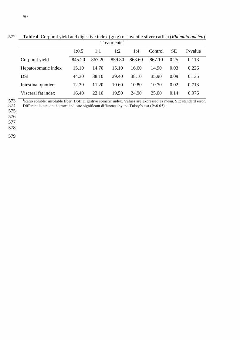
3.3 Corporal yield and digestive index 228
Diets containing different proportions of soluble and insoluble fiber no influenced 229
significantly in corporal yield, somatic and digestive parameters (P>0.05) (Table 4). 230
231
3.4 Digestive enzymes 232
Diets containing different proportions of soluble and insoluble fiber no influenced 233
significantly chymotrypsin activity (P>0.05) (Table 5). However, trypsin activity was higher 234
for fish fed with ratio of 1:0,5 S:IF in diet (P= 0.007). Fish fed with diet containing ratio of 235
1:2 and 1:4 showed lower trypsin activity. 236
237
3.5 Histological parameters 238
Linseed fiber ratios significantly influenced the development of the silver catfish 239
intestine. Villus height was higher for fish that received fiber in their diet (P<0.001), 240

36
regardless of the ratio. The opposite was observed for the muscular layer thickness (P<0.001), 241
which was superior for the fish fed on the control diet. The total villus height was higher for 242
the fish fed on the 1:0.5 and 1:4 S:IF diets (P= 0.003), not differing significantly from the 1:1 243
and 1:2 diets. On the other hand, the epithelium thickness was lower in fish fed on the 1:2 244
diet, differing only from those fed on the 1:0.5 diet (P= 0.020). 245
246
3.6 Hepatic metabolites 247
Diets containing different proportions of soluble and insoluble fiber no influenced 248
significantly (P>0.05) in the levels of glucose, glycogen and protein in fish liver (Table 7). 249
250
4. Discussion 251
The results obtained in this study present a new perspective for the use of dietary fiber 252
in fish nutrition. The simple inclusion of 10% of dietary fiber from linseed, without protein-253
energy changes or constitutional ingredients in the diet, promoted a mean increase of 28.5% 254
in the weight gain of the animals compared to the control diet (Table 2). Among the tested 255
soluble: insoluble fiber ratios, 1:2 and 1:4 promoted higher specific weight gain and growth 256
rate, without affecting the consumption and feed conversion of fish (Table 2), truly acting as 257
growth promoters. 258
In recent years, studies have shown that sensible dietary fiber inclusions optimize the 259
immune system and animal production, with an emphasis on the prebiotic action (Cerezuela et 260
al., 2013; Yarahmadi et al., 2014; Adorian et. al., 2015; Adorian et al., 2016; Goulart et al., 261
2017,). While the incorporation of more refined substances such as scFOS, XOS and GOS do 262
not present growth effects for several species of fish (Grisdale-Helland, et al., 2008; 263
Buentello, et al., 2010; Burr, et al. 2010; Hoseinifar, et al., 2014; Guerreiro, et al., 2015; 264
Guerreiro, et al., 2015; Hoseinifar et al., 2016; Guerreiro et al., 2018). These results 265

37
demonstrate the clear need for a change in perspectives on this food fraction in fish nutrition, 266
which can no longer be seen as a diluent of energy and antinutrient, but rather as a fraction 267
that deserves to be studied in detail, in order to express its functional effects the animal health 268
and production. 269
The positive effects of linseed fiber consumption on fish are possibly reflective of the 270
stimulus it exerts on the intestinal microbiota, similar to that reported for humans (Wenzel, 271
2012; Merrifield and Ringø, 2014). Since dietary fiber is resistant to the enzymatic digestion 272
and reaches the intestine while still being intact, it acts as a substrate for microbial 273
fermentation. In this fermentation process, short-chain fatty acids (SCFA) are produced; they 274
enter several metabolic pathways, generating energy and releasing bioactive compounds 275
bound to fiber (Ferreira, 2012; Wenzel, 2012; Quirós-Sauceda et al., 2014; Ríos-Covián et al., 276
2016; Celi et al., 2017). 277
This release of bioactive compounds may have contributed to the higher performance 278
of the fish that received the 1:2 and 1:4 S:IF in diets, because they have higher phenolic 279
compound (Table 1) contents, which follow the physiological processes that are common to 280
fiber, producing a synergic effect in the gastrointestinal tract (Goñi et al., 2009), promoting an 281
antioxidant environment and the maintenance of the intestinal integrity (Saura-Calixto, 2011; 282
Quirós-Sauceda et al., 2014). This fact shows that the functional effects of fiber are not only 283
related to their ratios, but also to characteristics that are intrinsic to their source of origin. 284
However, it is important to highlight that the use of diets with a higher degree of fiber 285
solubility (1:0.5 and 1:1) do not lead to significant differences in animal performance, 286
compared to the control diet (Table 2); this indicates that silver catfish tolerate high levels of 287
soluble fiber in their diet (51.9-68.3 g/kg). However, under these conditions, the prebiotic 288
action of linseed fiber appears to be inhibited. 289
Considering the above demonstrated aspects, it is clear that linseed fiber is a functional 290
38
ingredient, with the ability to improve performance when properly administered. Evidence of 291
its functional role had already been reported for juvenile silver catfish, where the 292
administration of soluble linseed fiber (mucilage) provided greater weight gain and feed 293
conversion (Goulart et al., 2017), similarly to what occurred with juvenile Nile tilapias 294
(Oreochromis niloticus) (Mombach, 2015). These results demonstrate that the formulation of 295
diets can be manipulated in order to balance the amount of dietary fiber, in order to obtain 296
positive results from its presence. However, it is important to emphasize that these authors 297
only evaluate food fiber concentrates from isolated fractions (soluble), without considering 298
that the combination of different ratios of soluble and insoluble fiber could boost their action. 299
Our results show that the effects of linseed fiber are not only limited to improvements 300
in the performance of the animals, since their supplementation in diets leads to positive 301
changes in the body composition of fish and in the pattern of nutrient deposition in the body. 302
This is clear from the higher crude protein content (1:4) and protein deposition (1:2 and 1:4) 303
provided by diets (Table 3). These fiber ratios may have stimulated the production of SCFA 304
by the intestinal microbiota, providing an additional amount of energy for animal metabolism. 305
This may reflect in improvements in the mucosal morphology, increasing intestinal villus and 306
absorptive area, and avoiding possible infections by opportunistic microorganisms (Topping, 307
1996; Park and Floch, 2007). Thus, the energy saved by the reduction of cell turnover can be 308
destined to protein deposition (Merrifield et al., 2010; Ferreira, 2012). These results 309
demonstrate that, in spite of being less efficient compared to glucose metabolism, potentially 310
fermentable fibers can contribute to nutrient deposition. 311
It is worth highlighting that the supplementation of linseed fiber at the tested ratios did 312
not cause physiological and metabolic changes in silver catfish (Table 4 and 7). However, the 313
higher hydration capacity of the 1:0.5 S:IF in diet (Table 1), may have caused an increase in 314
the viscosity of the digesta, to the point of hindering the enzyme-substrate interaction 315

39
(Easwood, 1992; Sinha et al., 2011). In an attempt to compensate for this situation, digestive 316
metabolism may have increased the secretion and activity of trypsin (Table 5) which, during a 317
culture cycle, could reflect on adaptations of the gastrointestinal tract. 318
The functional effect of linseed fiber is also evidenced by the positive changes in the 319
intestinal histological parameters of the silver catfish (Table 6). These results show that the 320
consumption of this fiber stimulates the development of intestinal villi, providing a greater 321
absorptive area, which may have contributed to the better performance and nutritional 322
deposition observed in the fish that received the 1:2 and 1:4 diets. In addition, larger villi 323
reduce the susceptibility of fish to diseases caused by intestinal pathogens (Brumano and 324
Gattás, 2009; Ferreira, 2012). Goulart et al. (2017) report similar results when supplementing 325
soluble fiber of linseed and β-Glican + Mananas in diets. The authors also point out that the 326
higher the villi height, the better the digestion and absorption of nutrients, reflecting greater 327
zootechnical performance, as occurred in this study. 328
The greater thickness of the intestinal epithelium of silver catfish fed on the 1:0.5 diet 329
corroborates the idea that its greater hydration capacity hinders the absorption of nutrients by 330
fish, which occurs not only because it has effects on the viscosity of the digesta, but also 331
according to intestinal histological changes, since the greater thickness of the epithelium 332
demands greater metabolic efforts for the absorption of the nutrients. However, the lower 333
thickness of the muscle layer resulting from the consumption of linseed fiber diets is directly 334
related to the higher villus height, which as well as increasing the absorptive area, has a 335
protective function (Ferreira, 2012). As the control diet provided less development of the villi, 336
there was a need to thicken the muscular layer, in order to maintain its physiological role in 337
protecting against the invasion of pathogens, since this layer consists of a dense network of 338
macrophages (Bauer, 2008). 339
Finally, it is important to consider that each fiber source has its peculiarities and it is 340

40
essential to study them more thoroughlly and to establish the correct levels and ratios of 341
inclusion, since its beneficial effects can be easily compromised by their excess in the diet, 342
whereas, when balanced, they may improve animal performance and the functionality of the 343
gastrointestinal tract (Celi et al., 2017). 344
345
5. Conclusion 346
These results allow concluding that linseed fiber acts effectively as a growth promoter 347
in silver catfish diets, and the use of the 1:2 and 1:4 ratios of soluble: insoluble linseed fiber 348
optimizes its prebiotic action in the animal organism. However, it is necessary to conduct 349
further studies in the area, which allow understanding the action of each fiber fraction, as well 350
as its effects on immunological parameters. 351
352
Acknowledgements 353
The authors would like to thank the National Council for Technological Development (CNPq) 354
for granting a research productivity scholarship (Leila Picolli da Silva) – Process number 355
307757/2015-3; to the Coordenação de Aperfeiçoamento de Pessoal de Nível Superior - 356
Brasil (CAPES) - Finance Code 001 by granting a doctorate scholarship (Taida Juliana 357
Adorian) and to Giovelli & Cia Ltda for the linseed courtesy provided. 358
359
This research did not receive any specific grant from funding agencies in the public, 360
commercial, or not-for-profit sectors. 361
362
References 363
Adorian, T.J., Mombach, P.I, Goulart, F.R., Loureiro, B.B., Pianesso, D., Silva, L.P., 2015. 364
Dietary fiber in the nutrition of silver catfish: Prebiotic or antinutrient? Anim. Feed. Sci. 365
Technol. 209, 167–173. https://doi.org/10.1016/j.anifeedsci.2015.07.017 366
41
Adorian, T.J., Goulart, F.R., Mombach, P.I., Lovatto, N.M., Dalcin, M. Molinari, M., 367
Lazzari, R., Silva, L.P. 2016. Effect of different dietary fiber concentrates on the 368
metabolism and indirect immune response in silver catfish. Anim. Feed. Sci. Technol. 369
215, 124–132. https://doi.org/10.1016/j.anifeedsci.2016.03.001 370
AOAC, 1995. Official Methods of Analysis. Association of Official Analytical Chemists, 371
Washington, DC. 372
AVMA. Guidelines for the Euthanasia of Animals: 2013 Edition. Association American 373
Veterinary Medical, 2013. Disponível em: 374
<https://www.avma.org/KB/Policies/Documents/euthanasia.pdf 375
Bauer, A.J. 2008. Mentation on the immunological modulation of gastrointestinal motility. 376
Neurogast. Mot. 20, 81–90. 10.1111 / j.1365-2982.2008.01105.x. 377
Bligh, E.G., Dyer, W.J., 1959. Rapid method of total lipid extraction and purification. J. 378
Biochem. Physiol. 37, 911–917. 379
Bradford, M. 1976. A Rapid and Sensitive Method for the Quantitation of Microgram 380
Quantities of Protein Utilizing the Principle of Protein-Dye Binding. Anal. Biochemi., 381
72, 248-254. 382
Brumano, G., Gattás, G. 2009. Alternativas ao uso de antibióticos como promotores de 383
crescimento em rações de aves e suínos. Ver. Elet. Nut., 6, 856-875. 384
https://www.revistas.ufg.br/vet/article/view/5886/4765 385
Buentello, J.A., Neill, W.H., Gatlin, D.M., 2010. Effects of dietary prebiotics on the growth, 386
feed efficiency and non-specific immunity of juvenile red drum Sciaenops ocellatus fed 387
soybean-based diets. Aquacul. Res. 41, 411–418. https://doi.org/10.1111/j.1365-388
2109.2009.02178.x 389
Burr, G., Hume, M., Ricke, S., Nisbet, D., Gatlin, D., 2010. In Vitro and In Vivo Evaluation 390
of the prebiotics GroBiotic®-A, inulin, mannanoligosaccharide, and 391
galactooligosaccharide on the digestive microbiota and performance of hybrid striped 392
bass (Morone chrysops × Morone saxatilis). Microb. Ecol., 59, 187–198. 10.1007 / 393
s00248-009-9597-6. 394
Buttriss, J.L., Stokes, C.S., 2008. Dietary fibre and health: an overview. Nutr. Bull. 33, 186–395
200. https://doi.org/10.1111/j.1467-3010.2008.00705.x 396
Celi, P., Cowieson, A.J., Fru-Nji, F., Steinert, R.E., Kluenter, A.-M.,Verlhac, V., 2017. 397
Gastrointestinal functionality in animal nutrition and health: New opportunities for 398
sustainable animal production. Anim. Feed. Sci. Technol., 234, 88–100. 399
https://doi.org/10.1016/j.anifeedsci.2017.09.012 400

42
Cerezuela, R., Fumanal, M., Tapia-Paniagua, S.T., Meseguer, J., Moriñigo, M.A., Esteban, 401
M.A., 2013. Changes in intestinal morphology and microbiotacaused by dietary 402
administration of inulin and Bacillus subtilis in gilthead sea bream (Sparus aurata L.) 403
specimens. Fish Shellfish Immunol. 34,1063–1070. 404
http://dx.doi.org/10.1016/j.fsi.2013.01.015 405
Chen, J., Liang, R.H., Liu, W., Li, T., Liu, C.M., Wu, S.S., Wang, Z.J., 2013. Pectic-406
oligosaccharides prepared by dynamic high-pressure microfluidization and their in vitro 407
fermentation properties. Carbohydr. Polym., 91, 175–182. 408
10.1016/j.carbpol.2012.08.021 409
Davidson, M.H., McDonald, A., 1998. Fiber: Forms and function. Nutrition Research, 18, 410
617–624. 411
Easwood, M.A., 1992. The physiological effect of dietary fiber: and update. Annu. Rev. Nutr, 412
12, 19-35. 413
Elleuch, M., Bedigian, D., Roiseux, O., Besbes, S., Blecker, C.,Attia, H., 2011. Dietary fibre 414
and fibre-rich by-products of food processing: Characterisation, technological 415
functionality and commercial applications: A review. Food Chem., 124, 411–421. 416
https://doi.org/10.1016/j.foodchem.2010.06.077 417
Ferreira, C.L.L., 2012. Prébióticos e Probióticos – Atualização e Prospecção. Rio de Janeiro: 418
Editora Rubio, 226p. 419
Gomez, B., Gullón, B., Remoroza, C., Schols, H.A., Parajó, J.C., Alonso, J.L., 2014. 420
Purification, Characterization, and Prebiotic Properties of Pectic Oligosaccharides from 421
Orange Peel Wastes. J. Agric. Food Chem., 62, 9769−9782. 10.1021 / jf503475b 422
Goñi, I., 2009. Towards an updated methodology for measurement of dietary fiber, including 423
associated polyphenols, in food and beverages. Food Res. Int., 42, 840–846. 424
10.1016/j.foodres.2009.03.010 425
Goulart, F.R., Speroni, C.S., Lovatto, N. M., Loureiro, B.B., Corrêia, V., Radünz Neto, J., 426
Silva, L.P., 2013. Atividade de enzimas digestivas e parâmetros de crescimento de 427
juvenis de jundiá (Rhamdia quelen) alimentados com farelo de linhaça in natura e 428
demucilada. Semina: Cien. Agr., 34, 3069-3080. doi.org/10.5433/1679-429
0359.2013v34n6p3069 430
Goulart, F.R., Silva, L.P., Loureiro, B.B., Adorian, T.J., Mombach, P.I., Petkowicz, C.L.O., 431
2017. Effects of Dietary Fiber Concentrates on growth performance and digestive 432
enzyme activities of jundiá (Rhamdia quelen). Aquacult. Nut., 23, 358-366. 433
doi.org/10.1111/anu.12400 434

43
Goulart, F.R., Adorian, T.J., Lovatto, N.M., Loureiro, B.B., Pianesso, D., Barcellos, L.G., 435
Koakoski, G., Silva, L.P., 2018. Effect of supplementation of dietary fibre concentrates 436
on biochemical parameters, stress response, immune response and skin mucus of jundiá 437
(Rhamdia quelen). 24, 375-382. doi.org/10.1111/anu.12568 438
Gressler, L.T., Sutili, F.J., Loebens, L., Saccol, E.M.H., Pês, T.S., Parody, T.V., Costa, S.T., 439
Pavanato, M.A., Baldisserotto, B., 2016. Histological and antioxidant responses in 440
Rhamdia quelen sedated with propofol. Aquacult. Res., 47, 2297-2306. 441
doi.org/10.1111/are.12682. 442
Grisdale-Helland, B., Helland, S. J., Gatlin, D. M. III., 2008. The effects of dietary 443
supplementation with mannanoligosaccharide, fructooligosaccharide or 444
galactooligosaccharide on the growth and feed utilization of Atlantic salmon (Salmo 445
salar). Aquaculture, 283, 163–167. doi.org/10.1016/j.aquaculture.2008.07.012 446
Guerreiro, I., Enes, P., Oliva-Teles, A., 2015. Effects of short chain fructooligosaccharides 447
(scFOS) and rearing temperature on growth performance and hepatic intermediary 448
metabolism in gilthead sea bream (Sparus aurata) juveniles. Fish Physiol. Biochem., 449
41, 1333–1344. doi.org/10.1007 / s10695-015-0089-y. 450
Guerreiro, I., Oliva-Teles, A., Enes, P., 2015. Improved glucose and lipid metabolism in 451
European sea bass (Dicentrarchus labrax) fed short-chain fructooligosaccharides and 452
xylooligosaccharides. Aquaculture, 441,57–63. 453
doi.org/10.1016/j.aquaculture.2015.02.015 454
Guerreiro, I., Serra, C. R., Pousão-Ferreira, P., Oliva-Teles, A., Enes, P., 2018. Prebiotics 455
effect on growth performance, hepatic intermediary metabolism, gut microbiota and 456
digestive enzymes of white sea bream (Diplodus sargus). Aquacul. Nut., 24, 153–163. 457
doi.org/10.1111/anu.12543 458
Gullón, B., Gullón, P., Sanz, Y., Alonso, J.L., Parajó, J.C., 2011. Prebiotic potential of a 459
refined product containing pectic oligosaccharides. Food Sci. Technol., 44, 1687-1696. 460
doi.org/10.1016/j.lwt.2011.03.006 461
Hoseinifar, S. H., Eshaghzadeh, H., Vahabzadeh, H., Mana, N. P. 2016. Modulation of 462
growth performances, survival, digestive enzyme activities and intestinal microbiota in 463
common carp (Cyprinus carpio) larvae using short chain fructooligosaccharide. 464
Aquacult. Res., 47, 3246–3253. doi.org/10.1111/are.12777 465
Hoseinifar, S. H., Sharifian, M., Vesaghi, M. J., Khalili, M., Esteban, M. Á., 2014. The effects 466
of dietary xylooligosaccharide on mucosal parameters, intestinal microbiota and 467
44
morphology and growth performance of Caspian whitefish (Rutilus frisii kutum) fry. 468
Fish Shel. Immunol., 39, 231–236. doi.org/10.1016/j.fsi.2014.05.009 469
Hummel, B.C.W., 1959. A modified spectrophotometric determination of chymotrypsin, 470
trypsin and thrombin. Can. J. Biochem. Physiol. 37, 1393–1399. 471
Jobling, M., 1983. A short reviewand citric of methodology used in fish growth and nutrition 472
studies. J. Fish Biol., 23, 685-703. doi.org/10.1111/j.1095-8649.1983.tb02946.x 473
Macagnan, F.T. , Silva, L.P., Hecktheuer, L.H., 2016. Dietary fibre: The scientific search for 474
an ideal definition and methodology of analysis, and its physiological importance as a 475
carrier of bioactive compounds. Food Res. Int., 85, 144–154. 476
doi.org/10.1016/j.foodres.2016.04.032 477
McBurney, M.I., Van Soest, P.J., Chase, L.E., 1983 Cation exchange capacity and buffering 478
capacity of neutral-detergent fibres. J. Sci. Food Agric., 34, 910-16. doi. org/ 479
10.1002/jsfa.2740340903 480
Meyer, G., Fracalossi, D.M., 2004. Protein requirement of jundia fingerlings. Rhamdia 481
quelen, at two dietary energy concentrations. Aquaculture, 240, 331–343. 482
doi.org/10.1016/j.aquaculture.2004.01.034. 483
Merrifield, D.L., Dimitroglou, A., Foey, A., Davies, S.J., Baker, R.T.M., Bøgwald, J., 484
Castex, M., Ringø, E., 2010. The current status and future focus of probiotic and 485
prebiotic applications for salmonids. Aquaculture, 302, 1-18. 486
doi.org/10.1016/j.aquaculture.2010.02.007 487
Merrifield, D., Ringø, E., 2014. Aquaculture Nutrition. John Wiley & Sons Ltd, Chichester, 488
UK, 500pp. 489
Mombach, P. I., 2015. Novos prebióticos na nutrição de Tilápia do Nilo. 81p. Dissertação 490
(Mestrado em Zootecnia) – Universidade Federal de Santa Maria, Santa Maria, 2015. 491
Morre, M.A., Park, C.B., Tsuda, H., 1998. Soluble and insoluble fiber influences on cancer 492
development. Crit. Rev. Oncol. Hematol., 27, 229-242. 493
Park, J., Floch, M.H., 2007. Prebiotics, probiotics, and dietary fiber in gastrointestinal 494
disease. Gastroenterol. Clin. North Am., 36, 47-63. 10.1016/j.gtc.2007.03.001 495
Park, J.T., Johnson, M.J., 1949. A submicro determination of glucose. J. Biol. Chem., 181, 496
149-151. 497
Quirós-Sauceda, A.E., Palafox-Carlos, H., Sáyago-Ayerdi, S.G., Ayala-Zavala, J.F., Bello-498
Pérez, L.A., Alvarez-Parrilla, E., Rosa, L.A., González-Córdova, A.F., González-499
Aguilar, G.A., 2014. Dietary fiber and phenolic compounds as functional ingredients: 500

45
Interaction and possible effect after ingestion. Food Funct., 5, 1063–1072. 10.1039 / 501
c4fo00073k 502
Ríos-Covián, D., Ruas-Madiedo, P., Margolles, A., Gueimonde, M., Reyes-Gavilán, C.G., 503
Salazar, N., 2016. Intestinal Short Chain Fatty Acids and their Link with Diet and 504
Human Health. Front. Microbiol., 7, 1-9. 10.3389 / fmicb.2016.00185 505
Roehrig, K.L., 1988. The physiological effects of dietary fiber. Food Hydrocol., 2, 506
1–18. 507
Saura-Calixto, F., 2011. Dietary Fiber as a Carrier of Dietary Antioxidants: An Essential 508
Physiological Function. J.Agricult. Food Chem., 59, 43–49. 509
Schneeman, B.O., 1987. Soluble and insoluble fibre-different physiological responses. Food 510
Technol., 41, 81–82. 511
Sinha, A.K., Kumar, K., Makkar, H.P.S., Boeck, G.D., Becker, K., 2011. Non-starch 512
polysaccharides and their role in fish nutrition – A review. Food Chem., 127, 1409–513
1426. doi.org/10.1016/j.foodchem.2011.02.042 514
Silva, L.P., Walter, M., 2012. Insoluble dietary fiber. In: Leo, M.L., Nollet, F.T. (Eds.), 515
Handbook of Analysis of Active Compounds in Functional Foods, 1. CRC Press Inc, 516
London, pp. 545–557. 517
Topping, D.L., 1996. Short-chain fatty acids produced by intestinal bacteria. Asia Pac. J. Clin. 518
Nut., 5, 15-19. 519
Tripathy, M.K., Mishra, A.S., 2007. Glucosinolates in animal nutrition: a review. Anim. Feed 520
Sci. Technol. 132, 1–27,http://dx.doi.org/10.1016/j.anifeedsci.2006.03.003 521
Van Soest, P.J., Robertson, J.B., Lewis, B.A., 1991. Methods for dietary fiber, neutral 522
detergent fiber, and non starch polysaccharides in relation to animal nutrition. J. Dairy 523
Sci., 74, 3583-3597. 524
Wang, J.C., Kinsella, J.E., 1976. Functional properties of novel proteins: alfalfa leaf proteins. 525
J. Food Sci. 41, 286–292, doi.org/10.1111/j.1365-2621.1976.tb00602.x. 526
Waterhouse, A.L. 2003. Determination of total phenolics. In: Current Protocols in Food 527
Analytical Chemistry, R. E. Wrolstad, Ed., units I, pp. I1.1.1–I1.1.8, John Wiley & 528
Sons, New York, NY, USA. 529
Wenzel, G.E., 2012. Carboidratos nutracêuticos e/ou prebióticos. São Leopoldo, RS: Ed. 530
Unisinos. 361p. 531
Westenbrink, S., Brunt, K., Kamp, J-W., 2013. Dietary fibre: Challenges in production and 532
use of food composition data. Food Chem., 140, 562–567. 533
doi.org/10.1016/j.foodchem.2012.09.029 534

46
Wilson, R.P., 1995. Lipid nutrition of finfish. Nutrition and utilization technology. In: Lim, 535
C., Sessa, D.J. (Eds.), Nutrition and Utilization Technology in Aquaculture, 1. AOSC 536
Press, Champaign, IL, pp. 74–81. http://www.ruforum.org/system/files/Kangombe.pdf 537
Yarahmadi, P., Miandare, M.K., Farahmand, H., Mirvaghefi, A., Hoseinifar, S.H., 2014. 538
Dietary fermentable fiber upregulated immune related genes expression, increased 539
innate immune response and resistance of rainbow trout (Oncorhynchus mykiss) against 540
Aeromonas hydrophila. Fish Shell. Fish Immunol. 41, 326–331. 541
doi.org/10.1016/j.fsi.2014.09.007 542
543
544

47
Table 1. Dietary formulations and proximate composition of the experimental diets (g/kg) 545
Treatments1
Ingredients 1:0.5 1:1 1:2 1:4 Control
Fish meal2 582.00 577.00 571.00 567.00 621.00
Maize starch 100.00 100.00 100.00 100.00 100.00
Linseed soluble fiber 93.70 64.80 35.80 13.80
Linseed insoluble fiber 43.20 73.00 102.80 125.50
Microcrystalline cellulose 105.70
NaCl 5.00 5.00 5.00 5.00 5.00
Soybean oil 50.00 46.00 42.00 39.00 54.00
Vitamin and mineral mixture3 30.00 30.00 30.00 30.00 30.00
BHT4 0.10 0.10 0.10 0.10 0.10
Inert5 96.00 104.10 113.30 119.60 84.20
Total 1000 1000 1000 1000 1000
Analyzed nutrient
Crude protein 381.40 382.80 382.40 383.40 377.80
Calculated energy (MJ/kg)6 13.41 13.42 13.42 13.43 13.41
Lipids 116.30 115.20 116.50 116.50 119.00
Total dietary fiber 102.90 103.90 103.10 103.30 103.50
Soluble fiber 68.30 51.90 35.00 21.30 02.70
Insoluble fiber 34.60 52.00 68.10 82.00 100.80
Physicochemical properties7
Hydration capacity 2.40 1.79 1.30 1.43 1.51
Fat binding capacity 0.94 0.91 0.97 0.96 1.05
Copper binding capacity 10.80 10.96 10.52 11.02 10.70
Phenolic compounds (mg EAG/g)8 55.77 68.80 77.80 86.21 1Ratio soluble: insoluble fiber. 546 2Waste flour tilapia/Copisces-Paraná/ Brazil. 547 3Composition (kg): folic acid 997.50 mg; pantothenic acid 9975.00 mg; biotin 159.60 mg; cobalt 39.90 mg; 548 copper 2800.00 mg; etoxiquin 24.78 g; iron 19.62 g; iodine 120.00 mg; manganese 5200.00 mg; niacin 19.95 g; 549 selenium 119.70 mg; zinc 28.00 g; vit.A 1995000 UI; vit. B1 4987.50 mg; vit. B12 5985,00 mg; vit. B2 550 4987.50g; vit. B6 4987.50 mg; vit. C 70.00 g; vit. D3 198000.05 UI; vit. E 19950.00 UI; vit. K 997.50 mg. 551 4Butyl hydroxy toluene (BHT). 552 5Sand. 553 6Digestible energy calculated according to ingredient analysis = [(crude protein × 5640 kcal/kg × 0.9) + (fat × 554 9510 kcal/kg × 0.85) + (Carbohydrates soluble in neutral detergent × 4110 kcal/kg ×0.50)] (Jobling, 1983). 555 7Hydration capacity: g water/g sample; Fat binding capacity: g fat/g sample; Copper binding: mg Cu/ g sample. 556
8Calculated 557
558
559
560

48
Table 2. Performance parameters of Rhamdia quelen fed with different ratio soluble: 561
insoluble linseed fiber in the diet 562
Treatments1
1:0.5 1:1 1:2 1:4 Control SE P-
value
Weight gain (g) 31.23ab 27.77b 33.12a 33.88a 24.44b 1.22 0.041
Total length (cm) 15.78ab 15.23ab 15.70a 15.55ab 15.06b 0.06 0.015
Condition factor 0.94 0.93 0.97 0.96 0.94 0.01 0.407
Specific growth rate (%/day) 4.45ab 4.36ab 4.65a 4.56a 4.13b 0.06 0.048
Consumption (g) 982.56 936.56 1088.31 1173.49 889.51 28.33 0.066
Apparent feed conversion 1.06 1.12 1.08 1.11 1.21 0.02 0.229
1Ratio soluble: insoluble fiber. Values are expressed as mean. SE: standard error. Different letters on the rows indicate 563
significant difference by the Tukey’s test (P<0.05). 564
565
566

49
Table 3. Corporal composition (g/kg) and body deposition of protein and fat (g) of juvenile 567
Rhamdia quelen 568
Treatments1
1:0.5 1:1 1:2 1:4 Control SE P-value
Crude protein 156.50ab 157.10ab 157.80ab 160.30a 152.20b 0.29 0.041
Fat 87.20 88.80 89.30 94.80 93.60 0.17 0.235
Dry matter 243.00b 253.30ab 253.20ab 264.06a 252.60ab 0.19 0.023
Ash 28.80 26.50 29.60 27.70 25.70 0.07 0.476
Body deposition (g)
Protein 4.30b 4.59ab 5.21a 4.95a 4.03b 0.12 0.003
Fat 2.42 2.33 2.62 2.70 2.21 0.07 0.274
1Ratio soluble: insoluble fiber. Values are expressed as mean. SE: standard error. Different letters on the rows 569 indicate significant difference by the Tukey’s test (P<0.05). 570 571
50
Table 4. Corporal yield and digestive index (g/kg) of juvenile silver catfish (Rhamdia quelen) 572
Treatments1
1:0.5 1:1 1:2 1:4 Control SE P-value
Corporal yield 845.20 867.20 859.80 863.60 867.10 0.25 0.113
Hepatosomatic index 15.10 14.70 15.10 16.60 14.90 0.03 0.226
DSI 44.30 38.10 39.40 38.10 35.90 0.09 0.135
Intestinal quotient 12.30 11.20 10.60 10.80 10.70 0.02 0.713
Visceral fat index 16.40 22.10 19.50 24.90 25.00 0.14 0.976
1Ratio soluble: insoluble fiber. DSI: Digestive somatic index. Values are expressed as mean. SE: standard error. 573 Different letters on the rows indicate significant difference by the Tukey’s test (P<0.05). 574 575
576
577
578
579
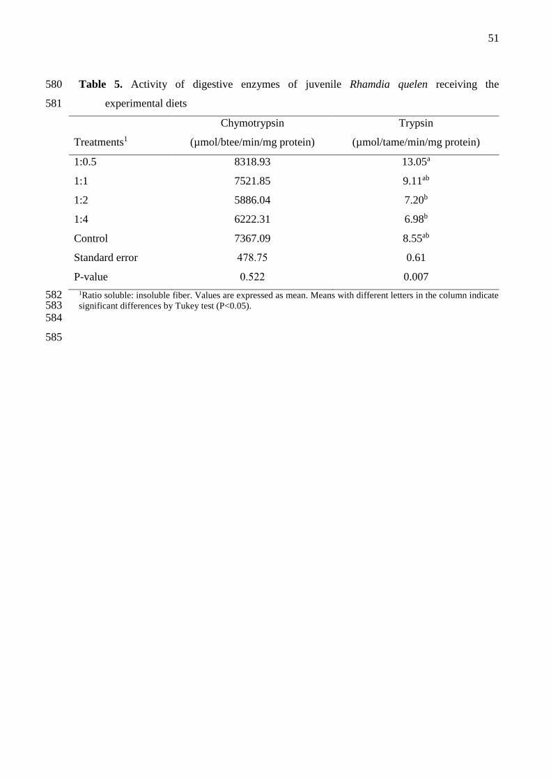
51
Table 5. Activity of digestive enzymes of juvenile Rhamdia quelen receiving the 580
experimental diets 581
Chymotrypsin Trypsin
Treatments1 (µmol/btee/min/mg protein) (µmol/tame/min/mg protein)
1:0.5 8318.93 13.05a
1:1 7521.85 9.11ab
1:2 5886.04 7.20b
1:4 6222.31 6.98b
Control 7367.09 8.55ab
Standard error 478.75 0.61
P-value 0.522 0.007
1Ratio soluble: insoluble fiber. Values are expressed as mean. Means with different letters in the column indicate 582 significant differences by Tukey test (P<0.05). 583 584
585

52
Table 6. Intestinal histology of juvenile Rhamdia quelen fed with different ratio soluble: 586
insoluble linseed fiber in the diet 587
Treatments1
1:0.5 1:1 1:2 1:4 Control SE P-
value
Total villus height 802.04a 691.19ab 736.54ab 802.07a 631.84b 16.57 0.003
Villus height 694.91a 677.92a 647.02a 708.32a 539.87b 13.12 <0.001
Epithelium thickness 101.92a 86.72ab 83.45b 96.80ab 95.58ab 2.01 0.020
Muscle layer thickness 49.02b 47.49b 41.42b 46.85b 58.82a 1.05 <0.001
1Ratio soluble: insoluble fiber. Total villus height, villus height, epithelium thickness and muscle layer thickness: 588 µm. Values are expressed as mean. SE: standard error. Different letters on the rows indicate significant 589 difference by the Tukey’s test (P<0.05). 590 591 592
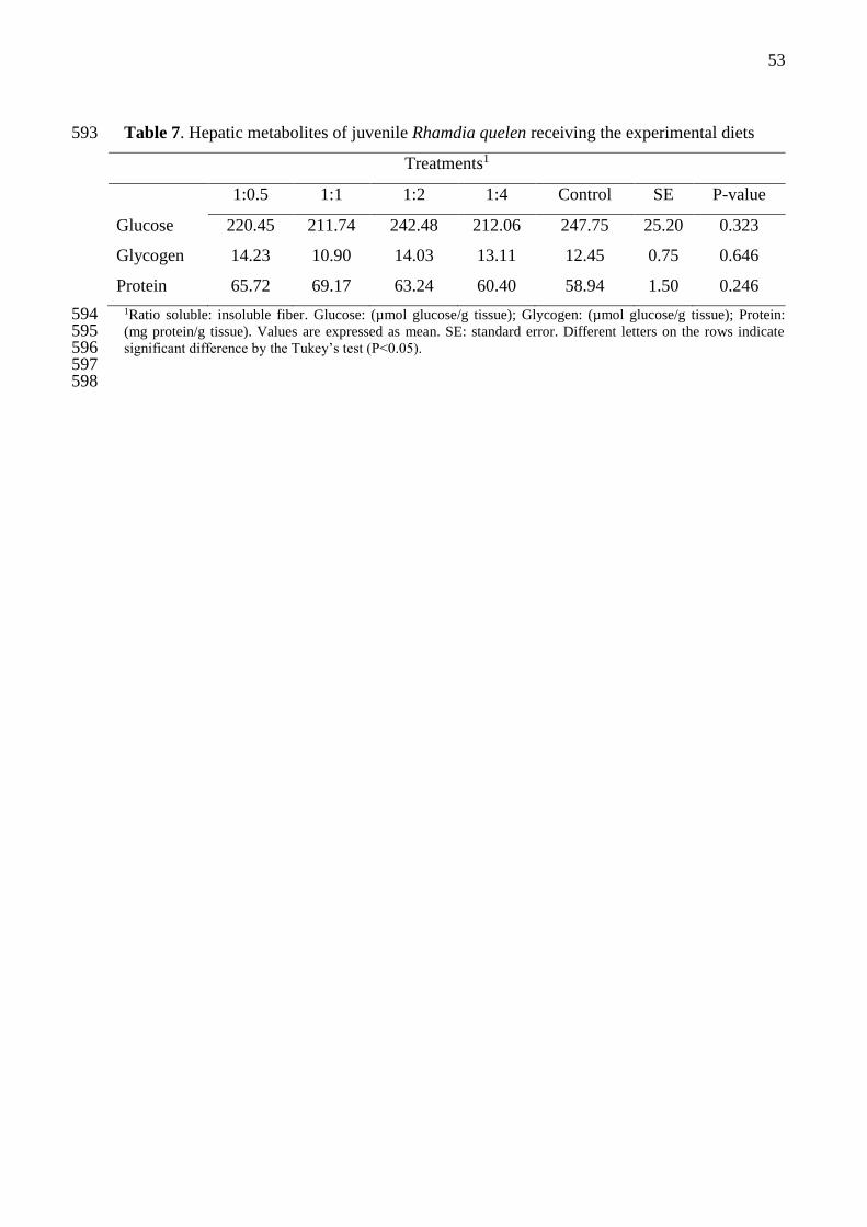
53
Table 7. Hepatic metabolites of juvenile Rhamdia quelen receiving the experimental diets 593
Treatments1
1:0.5 1:1 1:2 1:4 Control SE P-value
Glucose 220.45 211.74 242.48 212.06 247.75 25.20 0.323
Glycogen 14.23 10.90 14.03 13.11 12.45 0.75 0.646
Protein 65.72 69.17 63.24 60.40 58.94 1.50 0.246
1Ratio soluble: insoluble fiber. Glucose: (µmol glucose/g tissue); Glycogen: (µmol glucose/g tissue); Protein: 594 (mg protein/g tissue). Values are expressed as mean. SE: standard error. Different letters on the rows indicate 595 significant difference by the Tukey’s test (P<0.05). 596 597 598

54
3 ARTIGO II
O artigo científico intitulado “Functional linseed fibers in improves the immune
functions and performances of juveniles of silver catfish” foi submetido para a revista Animal
Feed Science and Technology e está formatado segundo as normas descritas no Guia dos
Autores (Anexo A).
55
Functiona linseed l fibers in improves the immune functions and performances of juveniles of 1
silver catfish 2
3
Taida Juliana Adoriana, Patrícia Inês Mombacha, Mariane Bittencourt Fagundesb, Roger 4
Wagnerb, Dirleise Pianessoa, Yuri Bohnenberger Tellesc, Marina Osmari Dalcina, Leila Picolli 5
da Silvaa 6
7
aDepartment of Animal Science, Federal University of Santa Maria, Santa Maria, Rio Grande 8
do Sul. AV. Roraima nº 1000, Cidade Universitária, Bairro Camobi, Santa Maria – RS, 9
Brazil. CEP: 97105-900. 10
11
bDepartment of Food and Science Technology, Federal University of Santa Maria, Santa 12
Maria, Rio Grande do Sul. AV. Roraima nº 1000, Cidade Universitária, Bairro Camobi, 13
Santa Maria – RS, Brazil. CEP: 97105-900. 14
15
cLaboratory of Systematics, Entomology and Biogeography, Federal University of Santa 16
Maria, Santa Maria, Rio Grande do Sul. AV. Roraima nº 1000, Cidade Universitária, Bairro 17
Camobi, Santa Maria – RS, Brazil. CEP: 97105-900. 18
19
20
21
*Corresponding author. Tel. 55 (55) 3220-8365; Fax: 55 (55) 3220-82 40; E-mails: 22
[email protected]; [email protected] 23
24
25

56
Abstract 26
This study was conducted to evaluate the prebiotic action of distinct linseed functional 27
fibers in the diets of juvenile silver catfish, under immunological and growth parameters. For 28
this, soluble and insoluble fractions of linseed fiber were concentrated separately and 29
combined in four ratios (1:0.5, 1:1, 1:2, 1:4), which were added to silver catfish diets (6.43 ± 30
0.12 g) and evaluated in a bioassay, along with a control diet (without the addition of linseed 31
fiber). After 45 days receiving the experimental diets, the animals were submitted to biometry 32
for data collection and samples for further analysis. The experimental design was completely 33
randomized, with five treatments and four replications; data were submitted to analysis of 34
variance and the means were compared by Tukey’s test (P <0.05). Total circulating proteins 35
and globulins were higher in the plasma of fish fed on diets 1:2 and 1:4, as well as 36
mucoprotein content, total immunoglobulins and cutaneous mucus pH. Cortisol levels and 37
intestinal pH were lower in these treatments. Regardless of the dietary ratio, the linseed fiber 38
intake increased total plasma immunoglobulins and plasma alkaline phosphatase activity and 39
cutaneous mucus. The production of intestinal acetic acid was higher in fish fed on the 1:2 40
diet, whereas the production of butyric acid was higher with the 1:4 diet and the propionic 41
acid with the control diet. The control diet led to lower counts of goblet cells. Fish 42
performance was higher for the group that received the 1:2 and 1:4 diets. In conclusion, the 43
results indicate that linseed fiber has an immunostimulating action for juvenile silver catfish; 44
ratios of 1:2 and 1:4 soluble: insoluble fiber optimize the immune system and the production 45
of SCFA, with positive effects on fish performance. 46
Keywords: SCFA, Rhamdia quelen, dietary fiber, Linum usitatissimum, prebiotic. 47
Abbreviations: DWG, daily weight gain; SCFA, short-chain fatty acids; SGR, specific growth 48
rate; SE, standard error; S:IF, soluble:insoluble fiber; IgT, total immunoglobulin; UFSM, 49
University of Santa Maria; 50

57
1. Introduction 51
Fiber is a food constituent with distinct functional properties in the animal organism, 52
which are intrinsically associated with the proportions of its water-soluble or -insoluble 53
fractions (Macagnan et al., 2016). The effects of its consumption are directly related to the 54
fermentability of the fiber by the intestinal microbiota, as well as by the bioactive compounds 55
associated with it, which promote improvements in the gastrointestinal environment, 56
impacting on the health and performance of animals (Giuntini and Menezes, 2011; Saura-57
Calixto, 2011). 58
The short chain fatty acids (SCFA) resulting from fiber fermentation are absorbed and 59
metabolically used as an energy source, positively influencing metabolic and physiological 60
processes (Guillon and Champ, 2000; Ferreira, 2012). They further promote luminal pH 61
reduction, avoiding infections by opportunistic microorganisms, potentiate the immune 62
system and provide maintenance of the mucosal integrity (Radecki and Yokoyama, 1991; 63
Park and Floch, 2007; Ferreira, 2012). 64
The intensive production of fish for commercial interests constantly exposes them to 65
stressful conditions and unfavorable environments, which increase the susceptibility to a 66
diversity of pathogens (Yokoyama et al., 2005). Therefor, the use of substances that modulate 67
the immune system and improve immunocompetence may promote the activation of defense 68
mechanisms and increase resistance and tolerance to unfavorable conditions, avoiding 69
deleterious effects for fish and reflecting improvements in their performance, as occurs with 70
prebiotic supplementation for different animal species (Silva and Nörnberg, 2003; Yokoyama 71
et al., 2005; Saurabh and Sahoo, 2008; Merrifield et al., 2010; Ringø et al., 2010; Freitas, et 72
al., 2014). 73

58
In this context, this study aims at evaluating the prebiotic action of different soluble 74
and insoluble linseed fiber ratios (1:0.5, 1:1, 1:2, 1:4) in juvenile silver catfish (Rhamdia 75
quelen) diets under immunological parameters and growth. 76
77
2. Material and methods 78
The study was conducted at the Laboratory of Fish Farming of the Department of 79
Animal Science of the Federal University of Santa Maria (UFSM), Rio Grande do Sul, Brazil 80
(Latitude: 29º 41’ 03’’ S; Longitude: 53º 48’ 25’’ W), after being approved by the Ethics 81
Committee on Animal Trials of this University, under the process number 8015120816. 82
83
2.1 Preparation of functional fibers 84
Linseed fiber is obtained in two distinct stages. In the first stage, soluble fiber of 85
linseed (mucilage) was obtained by soaking the whole grain in water at a concentration of 86
10% w/v, maintaining the reaction between 60◦C and 80◦C under constant stirring for 150 87
min. Subsequently, the soluble fiber was separated from the grains by sieving, followed by 88
addition of ethanol, for the precipitation of this fraction following the method described by 89
Goulart et al. (2013). The resulting soluble fiber of this process was dried in an air circulating 90
oven at 55ºC for 48 hours and ground in a micro-grinder (Marconi, model MA-630/1) to 91
obtain particles smaller than 590μm, representing the Linseed soluble fiber. 92
In the second stage, the insoluble fiber contained in the linseed was extracted. The 93
demucilaged grain was defatted with hexane at a ratio of 1:2 (w/v), performing for 30 min 94
washes. After defatted, the protein content of the residue was reduced by dispersion in 95
distilled water at room temperature at the ratio of 1:30 (w/v),sifted and dried in an air 96
circulating oven at 55 ºC for 24 h. The Linseed insoluble fiber obtained in this stage was 97

59
ground in a micro-grinder (Marconi, model MA-630/1) to obtain particles smaller than 98
590μm. 99
100
2.2 Experimental diets 101
Five experimental diets (Table 1) were formulated to achieve the nutritional 102
requirements of juvenile silver catfish, according to Meyer and Fracalossi (2004). The 103
experiment comprised the following treatments: Addition of functional fibers in the diet in 104
proportions of 1:0.5, 1:1, 1:2, 1:4 of soluble: insoluble fiber (S:IF) and control diet (without 105
addition of fiber). The diets were produced in the Laboratory of Fisheries of UFSM. The dry 106
ingredients were weighed and manually homogenized, then water was added and pelleting 107
with matrix of 3 mm in diameter. They were dried in an oven with forced air circulation for 108
24 h at a temperature of 55 ºC. After drying, the diets were milled and selected according to 109
the fish ingestion capacity. Diets were stored under a temperature of −20 ºC throughout the 110
experimental period. The diets composition and physicochemical properties were determined 111
based on analyses of crude protein (method 960.52), total, insoluble and soluble dietary fibers 112
(method 991.43) were determined according to the methodologies described by AOAC 113
(1995), fat (Bligh and Dyer, 1959), hydration capacity and fat binding capacity (Wang and 114
Kinsella, 1976), copper binding (McBurney, 1983) and phenolic compounds (Waterhouse, 115
2003). 116
117
2.3 Animals and feed 118
Six-hundred juveniles of silver catfish with an average initial weight of 6.43 ± 0.12 g 119
were distributed randomly in 20 polypropylene tanks with 290 liters capacity (30 animals per 120
experimental unit). Each tank had individual water inlet and outlet, arranged in a water 121
recirculation system comprised of a decanter, two mechanical and biological filtering and a 122

60
water reservoir with a capacity for 2000 liters, equipped with a heating system. During the 123
experimental period, the fish were fed with the experimental diet until apparent satiation three 124
times a day (9:00, 13:00 and 17:00 o’clock) for 45 days. 125
126
2.4 Water quality 127
Prior to the first and last meals (8:00 and 15:00 o’clock), fecal residues were removed 128
from the tanks by siphoning twice a day. During the experimental period, the water quality 129
parameters were monitored using colorimetric kits and maintained as follows: morning 130
temperature of 23.33 ± 1.71ºC; afternoon temperature of 24.90 ± 1.37ºC; pH: 7.45 ± 0.20; 131
alkalinity: 37.25 ± 4.95 mg CaCO3/L; hardness: 36.75 ± 11.25 mg CaCO3/L; total ammonia: 132
0.28 ± 0.10 mg L−1; nitrite: 0.02 ± 0.14 mg L−1 and oxygen: 7.75 ± 0.88 mg L−1. 133
134
2.5 Plasma analyzes 135
Blood samples were collected randomly (eight fish/treatment) by tail vein puncture 136
using heparinized syringes. The samples were placed in microcentrifuge tubes for 137
centrifuging (1000g, 10 min at room temperature). The plasma was stored under refrigerated 138
(- 8 ºC) to determine the concentrations of total proteins (g/dL), albumin (g/dL), globulin 139
(g/dL)= total protein−albumin), glucose (mg/gL), triglycerides (mg/dL) and cholesterol 140
(mg/gL). These tests were carried out in automation system (Labmax 100) using commercial 141
kits Labtest®. The activity of alkaline phosphatase was carried out using commercial kit 142
Doles®. 143
Total immunoglobulin (IgT) levels were measured using the method described by 144
Hoseinifar et al. (2015). Briefly, total protein content was measured using commercial kits for 145
total circulating proteins (g/dL) Labtest®. Thereafter, the immunoglobulin molecules 146
precipitated down using a 12% solution of polyethylene glycol (Sigma®). The difference in 147
61
protein contents prior and after immunoglobulin molecules precipitation is considered as the 148
IgT content. 149
The concentration of cortisol in fish plasma was determined by enzyme immunoassay 150
for ELISA, using commercial kit DBC®. The test principle follows a typical scenario of 151
competitive binding between an unlabeled antigen and an enzyme labeled with antigen. The 152
assay was performed on a 96-well microplates and the absorbance read on Plate Reader 153
(Eppendorf, AF2200) at 450 nm. 154
155
2.6 Skin mucus analyzes 156
Fish skin mucus samples were collected randomly (eight fish/treatment) using the 157
methods of Ross et al. (2000) and Palaksha et al. (2008), with modifications. The fish were 158
transferred to polyethylene bags containing 10 mL of 50 mM NaCl and these were gently 159
shaken (manually) for 60 seconds to release the mucus. The bags were placed on ice to 160
euthanize the fish by hypothermia. After euthanasia was observed, skin mucus was collected 161
by soft scraping of the dorsolateral surface, avoiding contamination with urinary-genital and 162
intestinal excretions. The mucus samples were transferred to amber glass tubes, homogenized 163
and stored (-20 ° C) for further analysis. 164
The levels of mucoprotein (glycoprotein) were determined using a Bioclin® 165
commercial kit. The principle of this methodology is the protein precipitation in a solution of 166
perchloricacid, resulting in a glycoprotein fraction denominated seromucoid and/or 167
mucoproteins. These are, then, precipitated in the filtrate with phosphotungstic acid and 168
subsequently dissolved and dosed by means of the tyrosine content. 169
Skin mucus total immunoglobulin levels were measured using the method described 170
by Hoseinifar et al. (2015). Briefly, mucus total protein content was measured according to 171
the technique described by Bradford (1976). Thereafter, the immunoglobulin molecules 172
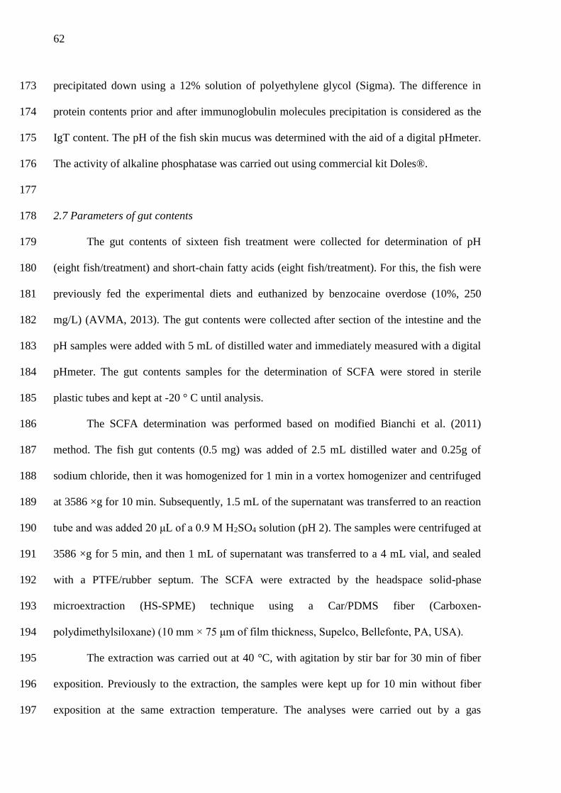
62
precipitated down using a 12% solution of polyethylene glycol (Sigma). The difference in 173
protein contents prior and after immunoglobulin molecules precipitation is considered as the 174
IgT content. The pH of the fish skin mucus was determined with the aid of a digital pHmeter. 175
The activity of alkaline phosphatase was carried out using commercial kit Doles®. 176
177
2.7 Parameters of gut contents 178
The gut contents of sixteen fish treatment were collected for determination of pH 179
(eight fish/treatment) and short-chain fatty acids (eight fish/treatment). For this, the fish were 180
previously fed the experimental diets and euthanized by benzocaine overdose (10%, 250 181
mg/L) (AVMA, 2013). The gut contents were collected after section of the intestine and the 182
pH samples were added with 5 mL of distilled water and immediately measured with a digital 183
pHmeter. The gut contents samples for the determination of SCFA were stored in sterile 184
plastic tubes and kept at -20 ° C until analysis. 185
The SCFA determination was performed based on modified Bianchi et al. (2011) 186
method. The fish gut contents (0.5 mg) was added of 2.5 mL distilled water and 0.25g of 187
sodium chloride, then it was homogenized for 1 min in a vortex homogenizer and centrifuged 188
at 3586 ×g for 10 min. Subsequently, 1.5 mL of the supernatant was transferred to an reaction 189
tube and was added 20 μL of a 0.9 M H2SO4 solution (pH 2). The samples were centrifuged at 190
3586 ×g for 5 min, and then 1 mL of supernatant was transferred to a 4 mL vial, and sealed 191
with a PTFE/rubber septum. The SCFA were extracted by the headspace solid-phase 192
microextraction (HS-SPME) technique using a Car/PDMS fiber (Carboxen-193
polydimethylsiloxane) (10 mm × 75 μm of film thickness, Supelco, Bellefonte, PA, USA). 194
The extraction was carried out at 40 °C, with agitation by stir bar for 30 min of fiber 195
exposition. Previously to the extraction, the samples were kept up for 10 min without fiber 196
exposition at the same extraction temperature. The analyses were carried out by a gas 197

63
chromatography equipped with a flame ionization detector (GC-FID), Varian Star 3400CX 198
(CA, USA). The SPME fiber was desorbed in a split/split less operated in a split less mode 199
with a period of 1.30 min at 230 °C. The carrier gas used was hydrogen, under a constant 200
pressure of 10 psi. The separation was made in a ZB-WAX Plus column (Chrompack, USA) 201
of 30 m × 0.25 mm i.d. × 0.25 μm film thickness). The oven temperature was programmed at 202
an initial temperature of 50 °C maintained for 1 min, and then increased to 110 °C at a rate of 203
5 °C min-1, after that, the temperature increased to 250 °C at 15 °C min-1 and was maintained 204
for 10 min. The detector temperature was held at 230 °C. The SCFA identification was 205
achieved by the comparison of the SCFA, acetic acid, butyric acid and propionic acid with 206
their authentic standards retention times (Sigma Aldrich). The quantification was performed 207
by a five-point external calibration curve. 208
209
2.8 Goblet cell counts 210
Anterior intestine was collected (four fish/ treatment) and prepared for light 211
microscopy. Histological samples were fixed in 10% formalin and preserved in 70% ethanol 212
and subjected to the histological routine, following the method described by Gressler et al. 213
(2016). The material was sent to go through the histological routine for dehydration in 214
increasing ethanol series (70%–99% alcohol) and embedded in methacrylate glycol resin 215
(Technovit 7100). From this material, slits of 2 µm were obtained from rotary microtome 216
(LEICA RM2245) to subsequent coloration with hematoxylin-eosin. For morphological 217
examination, the slides were observed and documented in light microscopy (ZEISS PrimoStar 218
with AxioCam ERc5s) and analyzed through the software ZEN LITE (Carl Zeiss). Goblet 219
cells were counted in 500 µm of villus and the results expressed in µm. The slides were 220
thoroughly examined in order to determine the presence of histopathological alterations. 221
222
64
2.9 Performance 223
In the early and late experimental period, a biometric assessment was performed to 224
collect data from the animals, which had fasted for 18 h and were anesthetized with 225
Benzocaine (100 mg/L), to estimate the following: biomass (g): final biomass - initial 226
biomass; daily weight gain(g): average weight gain/ 45 days; and fish survival (%). The daily 227
feed intake (g) was recorded to calculate the total feed intake estimated per experimental unit 228
at the end of the experiment. 229
230
2.10 Statistical analysis 231
Initially, the data were analyzed for outlier identification. The experimental design was 232
completely randomized with five treatments and four replications. The data were subjected to 233
analysis of variance and means were compared by Tukey’s test. Differences were considered 234
significant at the level of P<0.05. 235
236
3. Results 237
3.1 Plasma parameters 238
Plasma parameters were significantly influenced by the functional fibers tested (Table 239
2). The total circulating proteins (P= 0.020) and globulins (P<0.001) were higher in the 240
plasma of fish fed diets containing ratio 1:2 and 1:4 of S:IF, while cortisol (P= 0.005) had 241
reductions in treatments. Diets with functional fibers showed higher content of total 242
immunoglobulins (P<0.001) and alkaline phosphatase activity (P= 0.005) in plasma. 243
244
3.2 Skin mucus parameters 245
Skin mucus parameters were significantly influenced by the functional fibers tested 246
(Table 3). The mucoprotein (P= 0.046), total immunoglobulins (P= 0.043) e pH (P= 0.004) 247

65
were higher in skin mucus of fish fed diets containing ratio 1:2 and 1:4 of S:IF. The skin 248
mucus protein was superior (P= 0.012) in fish fed diet containing ratio 1:4, not differing 249
significantly only from fish fed 1:2 diet. Diets containing functional fibers resulted in 250
increased (P= 0.005) alkaline phosphatase activity in skin mucus, compared to control diet. 251
252
3.3 Parameters of gut contents 253
The gut content of silver catfish was significantly influenced by the fiber ratios 254
consumed in the diet (Table 4). The pH of the gut content of fish fed on diets 1:2 and 1:4 of 255
S:IF was significantly lower (P= 0.003). Acetic acid production was higher in the gut content 256
of fish fed on the 1:2 diet (P= 0.043), and it was not significantly different from those fed on 257
the 1:1 and 1:4 diets of S:IF. On the other hand, the production of butyric acid was higher for 258
fish fed on the 1:4 diet (P= 0.002). The production of propionic acid was higher in the gut 259
content of fish fed on the control diet (P= 0.048). 260
261
3.4 Goblet cell counts 262
Distinct counts of intestinal goblet cells were found in fish fed with the experimental 263
diets (Table 5). Diets with ratio of 1:0,5 S:IF provided higher intestinal goblet cell counts, not 264
differing from those given the 1: 2 diet. Fish fed with the control diet present lower goblet cell 265
counts (P>0.001). 266
267
3.5 Performance parameters 268
Diets with ratio of 1:2 and 1:4 S:IF provided higher biomass (P= 0.014) and daily weight 269
gain (P= 0.027) in fish when compared to the other treatments tested (Table 6). Feed intake 270
and fish survival were not influenced by the diets tested. 271
272
66
3. Discussion 273
Up to now, there are no studies evaluating the effects of different ratios of soluble and 274
insoluble dietary fiber on fish diets. These results show that linseed fiber supplementation in 275
1:2 and 1:4 ratios truly acts as a prebiotic, stimulating the immune system, SCFA production, 276
and juvenile silver catfish performance. 277
This is clear from the fact that fish fed on diets with 1:2 and 1:4 S:IF had higher total 278
protein contents and globulin levels in their plasma. These fiber ratios may have stimulated 279
the production of protective proteins in plasma, such as globulins, lysozyme, complementary 280
proteins and other peptides, with proven immune action and bactericidal activity (Alexander 281
and Ingram, 1992; Maqsood et al., 2009; Misra et al., 2009). Similar results were reported by 282
Adorian et al. (2016) when evaluating different dietary fiber concentrates in diets for the same 283
species. 284
As well as promoting the activation of plasma immune functions, diets containing 1:1, 285
1:2 and 1:4 of S:IF promote the reduction of plasma cortisol levels, which is the main 286
indicator of stress for fish (Urbinati et al., 2014). This result indicates a higher tolerance to 287
adverse culture conditions, as well as higher immunocompetence, since high levels of cortisol 288
lead to the depression of the immune system, with reflexes on growth (Mommsen et al., 1999; 289
Wendelaar Bonga, 2011; Urbinati et al., 2014). 290
Regardless of the dietary ratio, linseed fiber promotes higher levels of total 291
immunoglobulins and of alkaline phosphatase in plasma, indicating a real immunostimulatory 292
action. This is because immunoglobulins are involved in the systemic immunity of fish, and 293
IgM (more common in the plasma of teleosts) promotes activation of the complement system 294
that smooths and opsonizes pathogens, acting as mediator in the agglutination for 295
phagocytosis and the removal of pathogens (Hatten et al., 2001; Zhao et al., 2008; Ye et al., 296
2013; Mashoof and Criscitiello, 2016). 297

67
In the same way, the greater activity of alkaline phosphatase indicates improvements 298
in the immune system, since it is a hydrolase that has a protective role, with the capacity to 299
dephosphorylate certain molecules, removing phosphate groups (Calhau et al., 1999; Mota et 300
al. 2008; Ghahderijani et al., 2015). These results are in agreement with those of Goulart et al. 301
(2017), which highlight the prebiotic ability of soluble linseed fibers to provide increased IgM 302
in silver catfish plasma, indicating an increase in the immune function. Studies by Yarahmadi 303
et al. (2014) also demonstrate the immunostimulatory action of dietary fibers, reporting higher 304
lysozyme activity and expression of the immunological genes of rainbow trouts 305
(Oncorhynchus mykiss). 306
Skin mucus presents a large number of immunological substances, serving as an 307
indicator of the prebiotic action of ingredients used in fish nutrition (Esteban, 2012; Nigam et 308
al., 2012; Guardiola et al., 2014ab; Guardiola et al., 2015). Our results demonstrate that the 309
use of linseed fiber in the diet promotes positive changes in the skin mucus components of 310
silver catfish, strengthening the first defense line against pathogens. The production of 311
mucoprotein was stimulated by ratios 1:2 and 1:4 of S:IF, giving greater adhesive and 312
viscoelastic action. The main action mode of mucoproteins is the uptake of foreign particles, 313
which are removed by the continuous secretion of mucus by goblet cells (Roussel and 314
Delmotte, 2004; Lang et al., 2007; Esteban, 2012). 315
The higher production of immunoglobulins in the skin mucus resulting from the 316
consumption of 1:2 and 1:4 diets indicates a higher mucus capacity to eliminate pathogens, 317
avoiding colonization. However, the lower pH observed in these treatments hinders the 318
invasion by opportunistic pathogens, which usually require a neutral to alkaline environment 319
(Balebona et al., 1995; Zhang et al., 2010; Gonçalves et al., 2016). In addition, a higher pH 320
promotes the deterioration of biologically active mucus molecules, such as lysozyme, 321

68
reducing the antimicrobial activity, leading to impaired immune responses (Al-Arifa, et al., 322
2011). 323
Silver catfish fed with 1:4 also have a higher protein content in their mucus, indicating 324
a positive influence on the production of the different substances that compose this fraction. 325
Among them, lysozyme, antimicrobial peptides, protease enzymes and lectins stand out; they 326
show a lytic activity against many bacteria, preventing the colonization by pathogens, 327
cleaving proteins and interacting with the superficial structures of pathogens, resulting in the 328
increase of phagocytosis (Subramanian et al., 2007; Saurabh and Sahoo, 2008; Esteban, 2012; 329
Najafian and Babji, 2012; Gomez et al., 2013; Beck and Peatman, 2015). Regardless of the 330
ratios, linseed fiber promotes an increase in alkaline phosphatase, which is an 331
immunologically active enzyme, acting as an antimicrobial agent and in the regeneration of 332
the skin at the early stages of healing, under stress and parasitic infection (Bates et al., 2007; 333
Beck and Peatman, 2015). 334
The results observed in this study demonstrate that linseed fiber acts effectively as an 335
immunostimulant of the plasma and skin mucus functions of silver catfish, and can be used as 336
a prebiotic in diets for the species. It is suggested that these results are a consequence of the 337
higher fermentative production of acetic and butyric acid, which was reflected on the 338
reduction of intestinal pH (Table 4). Our results show that the fermentability profile is altered 339
by the fiber ratios contained in the diet; higher ratios of insoluble fiber (1:2 and 1:4) reflect a 340
higher production of acetic and butyric acid. However, it is important to observe that, 341
regardless of the ratios of linseed fiber, acetic acid was produced in greater abundance, 342
followed by propionic and butyric acid, similarly to what was reported by Ding et al. (2015) 343
when studying the in vitro fermentability profile of linseed fiber for pigs. 344
The higher production of acetic and butyric acid may also have contributed to the 345
better performance of fish fed on diets 1:2 and 1:4 (Table 6), which presented a daily weight 346

69
gain 28% higher than those fed on the control diet (same nutritional density as the test diets). 347
It is known that the SCFA generated in the intestinal fermentation are used as a source of 348
maintenance energy, which optimizes the use of the nutrients ingested in the diets for growth 349
functions, making the body energetically more efficient for muscle production (Fukuda et al., 350
2011; Koh et al., 2016). This is highlighted by different authors, who show that dietary fiber 351
has a growth promoting and nutritional deposition effect, similar to those provided by 352
consolidated commercial prebiotics (Adorian et al., 2015; Mombach, 2015; Adorian et al., 353
2016; Goulart et al., 2017). 354
The abundance of propionic acid in the gut content of fish fed on the control diet is 355
probably related to the absence of fermentable fibrous compounds, which reflects in the use of 356
other components as a source of energy by the microbiota. Studies with species of the same 357
food habit demonstrate that corn starch is not fully digested by the fish and its residues reach 358
the posterior intestine, stimulating the development of specific bacteria that generate 359
propionic acid (Heinitzet et al., 1996; Van Soest et al., 1991). Propionic acid inhibits 360
lipogenesis, which is not desired in fish at this stage, as it may compromise animal 361
development (Morrison and Preston, 2016). 362
Intrinsic characteristics of linseed fiber may have contributed to the fermentability 363
profile of the diets and their reflexes on fish performance. This is because, among the 364
monosaccharides found in linseed fiber, xylose, galactose and arabinose oligomers are found 365
in larger amounts (Goulart et al., 2017). These monosaccharides are responsible for promoting 366
the growth of beneficial bifidobacteria that contribute to the growth of the animal and act 367
directly on some populations of pathogenic bacteria through competitive exclusion (Ringo et 368
al., 2010; Freitas et al., 2014). 369
In addition to the aforementioned effects, the consumption of linseed fiber also 370
contributed to intestinal homeostasis. The higher number of goblet cells in the intestinal 371
70
epithelium of the fish that received the 1:0.5, 1:2 and 1:4 diets, respectively, support the idea 372
that linseed fiber has an immunostimulatory action, since goblet cells are responsible for the 373
production of intestinal mucus, composed mainly of mucins that bind to membranes and 374
provide an additional layer of defense to protect epithelial cells (Lang et al., 2007). Moreover, 375
they create a viscous gel that hinders microbial penetration by protecting and lubricating the 376
lining of the intestine (Junqueira and Carneiro, 2013). 377
In addition to mucins, other important substances are found in the intestinal mucus, 378
including innate and adaptive immune factors, such as immunoglobulins. Among the 379
immunoglobilins produced by fish, IgT is strategically designed to help teleosts to maintain 380
homeostasis with the microbiota, since it excludes unwanted luminal bacteria, avoiding their 381
colonization (Zhang et al., 2010; Gonçalves et al., 2016; Salinas et al., 2011). 382
383
4. Conclusion 384
Our results allow concluding that linseed fiber has an immunostimulating action for 385
juvenile silver catfish, with the ratios of 1:2 and 1:4 soluble: insoluble fiber optimizing the 386
immune system and the production of SCFA, with positive effects on the performance of fish. 387
More studies need to be conducted with this source of fiber in order to accurately determine 388
its action mode. 389
390
Acknowledgements 391
The authors would like to thank the National Council for Technological Development (CNPq) 392
for granting a research productivity scholarship (Leila Picolli da Silva) – Process number 393
307757/2015-3; to the Coordenação de Aperfeiçoamento de Pessoal de Nível Superior - 394
Brasil (CAPES) - Finance Code 001 by granting a doctorate scholarship (Taida Juliana 395
Adorian) and to Giovelli & Cia Ltda for the linseed courtesy provided. 396

71
This research did not receive any specific grant from funding agencies in the public, 397
commercial, or not-for-profit sectors. 398
399
References 400
Adorian, T.J., Mombach, P.I, Goulart, F.R., Loureiro, B.B., Pianesso, D., Silva, L.P., 2015. 401
Dietary fiber in the nutrition of silver catfish: Prebiotic or antinutrient? Anim. Feed. Sci. 402
Technol., 209, 167–173. https://doi.org/10.1016/j.anifeedsci.2015.07.017 403
Adorian, T.J., Goulart, F.R., Mombach, P.I., Lovatto, N.M., Dalcin, M. Molinari, M., 404
Lazzari, R., Silva, L.P. 2016. Effect of different dietary fiber concentrates on the 405
metabolism and indirect immune response in silver catfish. Anim. Feed. Sci. Technol. 406
215, 124–132. https://doi.org/10.1016/j.anifeedsci.2016.03.001 407
Al-Arifa, N.M., Mughal, S., Hanif, A., Batool, A., 2011. Effect of alkaline pH on bioactive 408
molecules of epidermal mucus from Labeo rohita (Rahu). Turk J Biochem., 36, 29–34. 409
http://www.turkjbiochem.com/2011/029-034.pdf 410
Alexander, J.B., Ingram, G.A., 1992. Non cellular non-specific defense mechanisms of fish. 411
Ann. Rev. Fish Dis., 2, 249–279. doi.org/10.1016/0959-8030(92)90066-7 412
AOAC, 1995. Official Methods of Analysis. Association of Official Analytical Chemists, 413
Washington, DC. 414
AVMA. Guidelines for the Euthanasia of Animals: 2013 Edition. Association American 415
Veterinary Medical, 2013. Disponível em: 416
<https://www.avma.org/KB/Policies/Documents/euthanasia.pdf 417
Balebona, M.C., Moriñigo, M.A., Faris, A., Krovacek, K., Månsson, I., Bordas, M.A., 418
Borrego, J.J., 1995. Influence of salinity and pH on the adhesion of pathogenic Vibrio 419
strains to Sparus aurata skin mucus. Aquaculture, 132, 113-120.doi.org/10.1016/0044-420
8486(94)00376-Y 421
Bates, J.M., Akerlund, J., Mittge, E., Guillemin, K., 2007. Intestinal alkaline phosphatase 422
detoxifies lipopolysaccharide and prevents inflammation in zebrafish in response to the 423
gut microbiota. Cell Host Mic., 2, 371–382. 10.1016 / j.chom.2007.10.010 424
Beck, B.H., Peatman, E., 2015. Mucosal Health in Aquaculture. Academic Press, 408pp. 425
Bianchi, F., Dall’Asta, M., Del Rio, D., Mangia, A., Musci, M., Scazzina, F., 2011. 426
Development of a headspace solid-phase microextraction gas chromatography–mass 427
72
spectrometric method for the determination of short-chain fatty acids from intestinal 428
fermentation. Food Chem., 129, 200–205. doi.org/10.1016/j.foodchem.2011.04.022 429
Bligh, E.G., Dyer, W.J., 1959. Rapid method of total lipid extraction and purification. J. 430
Biochem. Physiol. 37, 911–917. 431
Bradford, M., 1976. A Rapid and Sensitive Method for the Quantitation of Microgram 432
Quantities of Protein Utilizing the Principle of Protein-Dye Binding. Anal. Biochem., 433
72, 248-254. 434
Calhau, C., Hipolito-Reis, C., Azevedo, I., 1999. Alkaline phosphatase and exchange 435
surfaces. Clin. Biochem., 32, 153–154. 436
Esteban, M.A., 2012. An overview of the immunological defenses in fish skin. ISRN 437
Immunol., 29, 1-29. doi.org/10.5402/2012/853470 438
Ferreira, C.L.L., 2012. Prébióticos e Probióticos – Atualização e Prospecção. Rio de Janeiro: 439
Editora Rubio, 226p. 440
Freitas, E.R., Rabello, C.B., Watanabe, P.H., 2014. Probióticos e prebióticos na nutrição de 441
monogátricos. In: Sakomura, N.K., Silva, J.H.V., Costa, F.G.P., Fernandes, J.B.K. 442
Hauschild, L. Nutrição de não ruminantes. 1. ed. Jaboticabal: Funep, 2014. p. 487-510. 443
Fukuda, S., Toh, H., Hase, K., Oshima, K., Nakanishi, Y., Yoshimura, K., Tobe, T., Clarke, 444
J.M., Topping, D.L., Suzuki, T., Taylor, T.D., Itoh, K., Kikuchi, J., Morita, H., Hattori, 445
M., Ohno H., 2011. Bifidobacteria can protect from enteropathogenic infection through 446
production of acetate. Nature, 469, 543–547. 10.1038/nature09646 447
Ding, H.G., Cui, S.W., Goff, H.D., Gong, J., 2015. Short-chain fatty acid profiles from 448
flaxseed dietary fibres after in vitro fermentation of pig colonic digesta: Structure–449
function relationship. Bioac. Carb. Diet. Fib, 6, 62–68. 10.1016/j.bcdf.2015.09.006 450
Giuntini, E.B., Menezes, E.W. 2011. Fibra alimentar. Série de Publicações ILSI Brasil - 451
Funções Plenamente Reconhecidas de Nutrientes, São Paulo, 18, 23 p. 452
Gomez, D., Sunyer, J.O., Salinas, I., 2013. The mucosal immune system of fish: the evolution 453
of tolerating commensals while fighting pathogens. Fish Shellfish Immunol., 35, 1729-454
1739. doi.org/10.1016/j.fsi.2013.09.032 455
Gonçalves, J.L., Yaochite, J.N.U., Queiroz, C.A.A., Câmara, C.C., Oriá, R.B., 2016. Bases do 456
Sistema Imunológico Associado à Mucosa Intestinal. In: Oriá, R.B., Brito, G.A.C. 457
Sistema Digestório: Integração Básico-Clínica. Blucher Open Acess, 369-388pp. 458
459
Goulart, F.R., Speroni, C.S., Lovatto, N. M., Loureiro, B.B., Corrêia, V., Radünz Neto, J., 460
Silva, L.P., 2013. Atividade de enzimas digestivas e parâmetros de crescimento de 461
73
juvenis de jundiá (Rhamdia quelen) alimentados com farelo de linhaça in natura e 462
demucilada. Semina: Cien. Agr., 34, 3069-3080. doi.org/10.5433/1679-463
0359.2013v34n6p3069 464
Goulart, F.R., Silva, L.P., Loureiro, B.B., Adorian, T.J., Mombach, P.I., Petkowicz, C.L.O., 465
2017. Effects of Dietary Fiber Concentrates on growth performance and digestive 466
enzyme activities of jundiá (Rhamdia quelen). Aquacult. Nut., 23, 358-366. 467
doi.org/10.1111/anu.12400 468
Gressler, L.T., Sutili, F.J., Loebens, L., Saccol, E.M.H., Pês, T.S., Parody, T.V., Costa, S.T., 469
Pavanato, M.A., Baldisserotto, B., 2016. Histological and antioxidant responses in 470
Rhamdia quelen sedated with propofol. Aquacult. Res., 47, 2297-2306. 471
doi.org/10.1111/are.12682. 472
Guardiola, F.A., Cuesta, A., Abellán, E., Meseguer, J., Esteban, M.A., 2014a. Comparative 473
analysis of the humoral immunity of skin mucus from several marine teleost fish. Fish 474
Shellfish Immunol., 40, 24-31. doi.org/10.1016/j.fsi.2014.06.018 475
Guardiola, F.A., Cuesta, A., Arizcun, M., Meseguer, J., Esteban, M.A., 2014b. Comparative 476
skin mucus and serum humoral defence mechanisms in the teleost gilthead seabream 477
(Sparus aurata). Fish Shellfish Immunol., 36, 545–551. 478
doi.org/10.1016/j.fsi.2014.01.001 479
Guardiola, F.A.,Cuartero, M., Del Mar Collado-González M., Arizcún M., Díaz Baños, F.G., 480
Meseguer, J., Cuesta, A., Esteban, M.A., 2015. Description and comparative study of 481
physicochemical parameters of the teleost fish skin mucus. Biorheology, 52, 247–256. 482
10.3233/BIR-15052 483
Guillon, F., Champ, M., 2000. Structural and physical properties of dietary fibres, and 484
consequences of processing on human physiology. Food Res. Interat., 33, 233-245. 485
doi.org/10.1016/S0963-9969(00)00038-7 486
Hatten, F., Um, F., Hordvik, I., Endresen, C., 2001. Presence of IgM in cutaneous mucus, but 487
not in gut mucus of Atlantic salmon, Salmo salar. Serum IgM is rapidly degraded when 488
added to gut mucus. Fish Shellfish Immunol., 11, 257–268. 10.1006 / fsim.2000.0313 489
Heinitz, M.C., Lemme, A., Schulz, C., 2016. Measurement of digestibility in agastric fish 490
based on stripping method – apparent nutrient, energy and amino acid digestibilities of 491
common feed ingredients for carp diets (Cyprinus carpio). Aquacult. Nut., 22, 1065–492
1078. 10.1111/anu.12324 493
Hoseinifar, S.H., Khalili, M., Rufchaei, R., Raeisi, M., Attar, M., Cordero, H., Esteban, M.Á., 494
2015. Effects of date palm fruit extracts on skin mucosal immunity, immune related 495
74
genes expression and growth performance of common carp (Cyprinus carpio) fry. Fish 496
Shellfish Immunol., 47, 706-711. doi: 10.1016/j.fsi.2015.09.046. 497
Jobling, M., 1983. A short reviewand citric of methodology used in fish growth and nutrition 498
studies. J. Fish Biol., 23, 685-703. doi.org/10.1111/j.1095-8649.1983.tb02946.x 499
Junqueira, L.C., Carneiro, J., 2013. Histologia básica. 12.ed. Rio de Janeiro: Guanabara 500
Koogan, 2013. 556p. 501
Koh, A., De Vadder, F., Kovatcheva-Datchary, P. , Barreira, F., 2016. From Dietary Fiber to 502
Host Physiology: Short-Chain fatty acids as key bacterial metabolites. Cell, 165, 1332- 503
1341. 10.1016 / j.cell.2016.05.041 504
Lang, T., Hansson, G.C., Samuelsson, T., 2007. Gel-forming mucins appeared early in 505
metazoan evolution. Proceedings of the National Academy of Sciences of the United 506
States of America, 104, 16209–16214. 507
Macagnan, F.T. , Silva, L.P., Hecktheuer, L.H., 2016. Dietary fibre: The scientific search for 508
an ideal definition and methodology of analysis, and its physiological importance as a 509
carrier of bioactive compounds. Food Res. Int., 85, 144–154. 510
doi.org/10.1016/j.foodres.2016.04.032 511
Maqsood, S., Samoon, M.H., Singh, P., 2009. Immunomodulatory and growth promoting 512
effect of dietary levamisole in Cyprinus carpio fingerlings against the challenge of 513
Aeromonas hydrophila. Turkish J. Fish. Aquatic Sci., 9,111-120. 514
Mashoof, S., Criscitiello, M.F., 2016. Fish Immunoglobulins. Biology, 5, 1-23. 515
10.3390/biology5040045 516
McBurney, M.I., Van Soest, P.J., Chase, L.E., 1983 Cation exchange capacity and buffering 517
capacity of neutral-detergent fibres. J. Sci. Food Agric., 34, 910-16. doi.org/ 518
10.1002/jsfa.2740340903 519
Meyer, G., Fracalossi, D.M., 2004. Protein requirement of jundia fingerlings. Rhamdia 520
quelen, at two dietary energy concentrations. Aquaculture, 240, 331–343. 521
doi.org/10.1016/j.aquaculture.2004.01.034. 522
Merrifield, D.L., Dimitroglou, A., Foey, A., Davies, S.J., Baker, R.T.M., Bøgwald, J., 523
Castex, M., Ringø, E., 2010. The current status and future focus of probiotic and 524
prebiotic applications for salmonids. Aquaculture, 302, 1-18. 525
doi.org/10.1016/j.aquaculture.2010.02.007 526
Misra, C.K., Das, B.K., Mukherjee, S.C., 2009. Immune response, growth and survival of 527
Labeo rohita fingerlings fed with levamisole supplemented diets for longer duration. 528
Aquacult. Nut., 15, 356-365. doi.org/10.1111/j.1365-2095.2008.00600.x 529

75
Mombach, P. I., 2015. Novos prebióticos na nutrição de Tilápia do Nilo. 81p. Dissertação 530
(Mestrado em Zootecnia) – Universidade Federal de Santa Maria, Santa Maria, 2015. 531
Mommsen, T.P., Vijayan, M.M., Moon, T.W., 1999. Cortisol in teleosts: Dynamics, 532
mechanisms of action, and metabolic regulation. Rev. Fish Biol. Fish. ,9, 211-268. 533
Mota, A., Silva, P., Neves, D., Lemos, C., Calhau, C., Torres, D., Martel, F., Fraga, H., 534
Ribeiro, L., Alçada, M.N., Pinho, M.J., Negrão, M.R., Pedrosa, R., Guerreiro, S., 535
Guimarães, J.T., Azevedo, I., Martins, M.J., 2008. Characterization of rat heart alkaline 536
phosphatase isoenzymes and modulation of activity. Braz. J. Med. Biol. Res., 41, 600-537
609. 538
Morrison, D.J. Preston, T., 2016. Formation of short chain fatty acids by the gut microbiota 539
and their impact on human metabolism.Gut Microb., 7, 189-200. 10.1080 / 540
19490976.2015.1134082 541
Najafian, L., Babji, A.S., 2012. A review of fish-derived antioxidant and antimicrobial 542
peptides:vtheir production, assessment, and applications. Peptides, 33, 178–185. 543
10.1016/j.peptides.2011.11.013. 544
Nigam, A.K., Kumari, U., Mittal, S., Mittal, A.K., 2012. Comparative analysis of innate 545
immune parameters of the skin mucous secretions from certain freshwater teleosts, 546
inhabiting different ecological niches. Fish Physiol. Biochem., 38, 1245–1256. 547
10.1007/s10695-012-9613-5. 548
Palaksha, K.J., Shin, G.W., Kim, Y.R., Jung, T.S., 2008. Evaluation of non-specific immune 549
components from the skin mucus of olive flounder (Paralichthys olivaceus). Fish 550
Shellfish Immunol., 24, 479-488. 10.1016/j.fsi.2008.01.005. 551
Park, J., Floch, M.H., 2007. Prebiotics, probiotics, and dietary fiber in gastrointestinal 552
disease. Gastroenterol. Clin. North Am., 36, 47-63. 10.1016/j.gtc.2007.03.001 553
Radecki, S.V., Yokoyama, M.T., 1991. Intestinal bacteria and their influence on swine 554
nutrition. In: Miller, E.R., Duane, E.U., Lewis, A.J. Swine nutrition. Boston: 555
Butterworth- Heinemann, p.439-447. 556
Ringø, E., Olsen, R.E., Gifstad, P.A.R.A., Dalmo, R.A., Amlund, H., Hemre, G.I., Bakke, 557
A.M., 2010. Prebiotics in aquaculture: a review. Aquacult. Nut., 16, 117–136. 558
doi.org/10.1111/j.1365-2095.2009.00731.x 559
Ross, N.W., Firth, K.J., Wang, A., Burka, J.F., Johnson, S.C., 2000. Changes in hydrolytic 560
enzyme activities of naïve Atlantic salmon Salmo salar skin mucus due to infection 561
with the salmon louse Lepeophtheirus salmonis and cortisol implantation., Dis. Aquat. 562
Organ., 41, 43–51. 10.3354 / dao041043 563

76
Roussel, P., Delmotte, P., 2004. The diversity of epithelial secreted mucins. Cur. Org. Chem., 564
8, 413–437. doi.org/10.2174/1385272043485846 565
Salinas, I., Zhang, Y.A., Sunyer, J.O., 2011. Mucosal immunoglobulins and B cells of teleost 566
fish. Develop.Comp. Immunol., 35, 1346–1365. doi.org/10.1016/j.dci.2011.11.009 567
Saurabh, S., Sahoo, P.K., 2008. Lysozyme: an important defence molecule of fish innate 568
immune system. Aquacult. Res., 39, 223-239. doi.org/10.1111/j.1365-569
2109.2007.01883.x 570
Saura-Calixto, F. 2011. Dietary Fiber as a Carrier of Dietary Antioxidants: An Essential 571
Physiological Function. J.Agr. Food Chem., 59, 43–49. 10.1021/jf1036596 572
Silva, L.P., Nörnberg, J. L., 2003. Prebióticos na nutrição de não ruminantes. Cien. Rur., 33, 573
983-990. 574
Subramanian, S., Mackinnon, S.L., Ross, N.W., 2007. A comparative study on innate immune 575
parameters in the epidermal mucus of various fish species. Comp. Biochem. Physiol. 576
Part B: Biochem. Mol. Biol., 148, 256–263. 10.1016 / j.cbpb.2007.06.003 577
Urbinati, E.C., Zanuzzo, F.S., Biller-Takahashi, J. D., 2014. Estresse e sistema imune em 578
peixes. In: Baldisserotto, B., Cyrino, J.E.P., Urbinati, E.C. Biologia e fisiologia de 579
peixes neotropicais de água doce. Jaboticabal: FUNESP; UNESP, 87-105 p. 580
Van Soest, P.J., Robertson, J.B., Lewis, B.A., 1991. Methods for dietary fiber, neutral 581
detergent fiber, and non starch polysaccharides in relation to animal nutrition. J. Dairy 582
Sci., 74, 3583-3597. 583
Wang, J.C., Kinsella, J.E., 1976. Functional properties of novel proteins: alfalfa leaf proteins. 584
J. Food Sci. 41, 286–292, doi.org/10.1111/j.1365-2621.1976.tb00602.x. 585
Waterhouse, A.L., 2003. Determination of total phenolics. In: Current Protocols in Food 586
Analytical Chemistry, R. E. Wrolstad, Ed., units I, pp. I1.1.1–I1.1.8, John Wiley & 587
Sons, New York, NY, USA. 588
Wendelaar Bonga, S.E., 2011. Hormone Response to Stress. In: Farrel, A.P., Cech, J.J., 589
Richards, J.G., Stevens, E.D., (Ed.). Encyclopedia of Fish Physiology: from genome to 590
environment. Elsevier Academic Press Inc, UK, p.1515-1523. 591
Yarahmadi, P., Miandare, M.K., Farahmand, H., Mirvaghefi, A., Hoseinifar, S.H., 2014. 592
Dietary fermentable fiber upregulated immune related genes expression, increased 593
innate immune response and resistance of rainbow trout (Oncorhynchus mykiss) against 594
Aeromonas hydrophila. Fish Shell. Fish Immunol. 41, 326–331. 595
doi.org/10.1016/j.fsi.2014.09.007 596

77
Ye, J., Kaattari, I.M., Ma, C., Kaattari, S., 2013.The teleost humoral immune response. Fish 597
Shellfish Immunol., 35, 1719–1728. doi.org/10.1016/j.fsi.2013.10.015 598
Yokoyama, S., Koshio, S., Takakura, N., Oshida, K., Ishikawa, M., Gallardo-Cigarroa, F.J., 599
Teshima, S., 2005. Dietary bovine lactoferrin enhances tolerance to high temperature 600
stress in Japanese flounder Paralichthys olivaceus. Aquaculture, 249, 367–373. 601
doi.org/10.1016/j.aquaculture.2005.03.024 602
Zhang, Y.A., Salinas, I., Li, J., Parra, D., Bjork, S., Xu, Z., LaPatra, S.E., Bartolomeu, J., 603
Sunyer, J.O., 2010. IgT, a primitive immunoglobulin class specialized in mucosal 604
immunity. Nature Immunol.,11, 827–835. 10.1038 / ni.1913 605
Zhao, X., Findly, R. C., Dickerson, H.W., 2008. Cutaneous antibody-secreting cells and B 606
cells in a teleost fish. Develop.Comp. Immunol., 32, 500–508. 607
608
609

78
Table 1. Dietary formulations and proximate composition of the experimental diets (g/kg) 610
Treatments1
Ingredients 1:0.5 1:1 1:2 1:4 Control
Fish meal2 582.00 577.00 571.00 567.00 621.00
Maize starch 100.00 100.00 100.00 100.00 100.00
Linseed soluble fiber 93.70 64.80 35.80 13.80
Linseed insoluble fiber 43.20 73.00 102.80 125.50
Microcrystalline cellulose 105.70
NaCl 5.00 5.00 5.00 5.00 5.00
Soybean oil 50.00 46.00 42.00 39.00 54.00
Vitamin and mineral mixture3 30.00 30.00 30.00 30.00 30.00
BHT4 0.10 0.10 0.10 0.10 0.10
Inert5 96.00 104.10 113.30 119.60 84.20
Total 1000 1000 1000 1000 1000
Analyzed nutrient
Crude protein 381.40 382.80 382.40 383.40 377.80
Calculated energy (MJ/kg)6 13.41 13.42 13.42 13.43 13.41
Lipids 116.30 115.20 116.50 116.50 119.00
Total dietary fiber 102.90 103.90 103.10 103.30 103.50
Soluble fiber 68.30 51.90 35.00 21.30 02.70
Insoluble fiber 34.60 52.00 68.10 82.00 100.80
Physicochemical properties7
Hydration capacity 2.40 1.79 1.30 1.43 1.51
Fat binding capacity 0.94 0.91 0.97 0.96 1.05
Copper binding capacity 10.80 10.96 10.52 11.02 10.70
Phenolic compounds (mg EAG/g)8 55.77 68.80 77.80 86.21 1Ratio soluble: insoluble fiber. 611 2Waste flour tilapia/Copisces-Paraná/ Brazil. 612 3Composition (kg): folicacid 997.50 mg; pantothenic acid 9975.00 mg; biotin 159.60 mg; cobalt 39.90 mg; 613 copper 2800.00 mg; etoxiquin 24.78 g; iron19.62 g; iodine 120.00 mg; manganese 5200.00 mg; niacin 19.95 g; 614 selenium 119.70 mg; zinc 28.00 g; vit.A 1995000 UI; vit. B1 4987.50 mg; vit. B12 5985,00 mg; vit. B2 615 4987.50g; vit. B6 4987.50 mg; vit. C 70.00 g; vit. D3 198000.05 UI; vit. E 19950.00 UI; vit. K 997.50 mg. 616 4Butylhydroxytoluene (BHT). 617 5Sand. 618 6Digestible energy calculated according to ingredient analysis = [(crude protein × 5640 kcal/kg × 0.9) + (fat × 619 9510 kcal/kg × 0.85) + (Carbohydrates soluble in neutral detergent × 4110 kcal/kg ×0.50)] (Jobling, 1983). 620 7Hydrationcapacity: g water/g sample; Fat binding capacity: g fat/g sample; Copper binding: mg Cu/ g sample. 621
8Calculated 622
623
624
625
626
627

79
Table 2. Plasma parameters of juvenile Rhamdia quelen receiving the experimental diets 628
Treatments1
1:0.5 1:1 1:2 1:4 Control SE P-value
Total proteins 3.23ab 3.18b 3.70a 3.71a 3.09b 0.07 0.020
Albumin 0.54 0.72 0.68 0.67 0.71 0.02 0.210
Globulin 2.55b 2.91ab 3.05a 3.21a 2.53b 0.06 <0.001
Glucose 51.83 52.87 57.28 54.42 51.71 2.20 0.938
Triglycerides 553.57 661.67 618.83 658.33 653.49 20.83 0.216
Cholesterol 131.14 184.50 157.12 188.14 178.71 7.08 0.510
Alkaline phosphatase 21.85a 21.77a 22.78a 22.23a 18.85b 0.35 0.008
IgT 1.98a 2.18a 2.14a 2.10a 1.45b 0.05 <0.001
Cortisol 17.98ab 12.43b 13.85b 12.68b 19.10a 0.78 0.005
1Ratio soluble: insoluble fiber. Total proteins (g/dL); Albumin (g/dL); Globulin (g/dL); Glucose (mg/gL); 629 Triglycerides (mg/dL); Cholesterol (mg/gL); Alkaline phosphatase (U.I/L); IgT: Total immunoglobulin (mg/dL) 630 and Cortisol (µg/dL). Values are expressed as mean.SE: standard error. Different letters on the rows indicate 631 significant difference by the Tukey’s test (P<0.05). 632 633
634
635
636
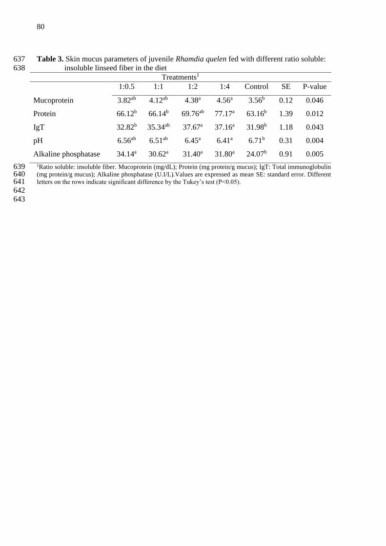
80
Table 3. Skin mucus parameters of juvenile Rhamdia quelen fed with different ratio soluble: 637
insoluble linseed fiber in the diet 638
Treatments1
1:0.5 1:1 1:2 1:4 Control SE P-value
Mucoprotein 3.82ab 4.12ab 4.38a 4.56a 3.56b 0.12 0.046
Protein 66.12b 66.14b 69.76ab 77.17a 63.16b 1.39 0.012
IgT 32.82b 35.34ab 37.67a 37.16a 31.98b 1.18 0.043
pH 6.56ab 6.51ab 6.45a 6.41a 6.71b 0.31 0.004
Alkaline phosphatase 34.14a 30.62a 31.40a 31.80a 24.07b 0.91 0.005
1Ratio soluble: insoluble fiber. Mucoprotein (mg/dL); Protein (mg protein/g mucus); IgT: Total immunoglobulin 639 (mg protein/g mucus); Alkaline phosphatase (U.I/L).Values are expressed as mean. SE: standard error. Different 640 letters on the rows indicate significant difference by the Tukey’s test (P<0.05). 641 642 643

81
Table 4. pH and concentration of short-chain fatty acids (μmol/g) in gut contents of Rhamdia 644
quelen 645
Treatments1
1:0.5 1:1 1:2 1:4 Control SE P-value
pH 7.37ab 7.36ab 7.29b 7.21b 7.45a 0.10 0.003
Short-chain fatty acids (µmol/g)
Acetic acid 4.75c 6.85ab 7.32a 6.37ab 5.73b 1.07 0.043
Butyric acid 0.02c 0.02c 0.04ab 0.06a 0.03bc 0.01 0.002
Propionic acid 0.11b 0.09b 0.11b 0.10b 0.20a 0.02 0.048
Total SCFA 4.88 6.96 7.47 6.60 5.96
1Ratio soluble: insoluble fiber. Values are expressed as mean.SE: standard error. Different letters on the rows 646 indicate significant difference by the Tukey’s test (P<0.05). 647 648
649

82
Table 5. Effect of different proportions of soluble and insoluble fiber on intestinal goblet cell 650
counts (cells/g) in silver catfish 651
Treatments1
1:0.5 1:1 1:2 1:4 Control EP P
Goblet cell counts 26.50a 18.25bc 23.50ab 19.50b 12.66c 0.58 <0.001
1Ratio soluble: insoluble fiber. Goblet cell counts in 500 µm. Values are expressed as mean.SE: standard error. 652 Different letters on the rows indicate significant difference by the Tukey’s test (P<0.05). 653 654 655
656
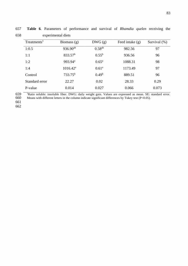
83
Table 6. Parameters of performance and survival of Rhamdia quelen receiving the 657
experimental diets 658
Treatments1 Biomass (g) DWG (g) Feed intake (g) Survival (%)
1:0.5 936.90ab 0.58ab 982.56 97
1:1 833.57b 0.55b 936.56 96
1:2 993.94a 0.65a 1088.31 98
1:4 1016.42a 0.61a 1173.49 97
Control 733.75b 0.49b 889.51 96
Standard error 22.27 0.02 28.33 0.29
P-value 0.014 0.027 0.066 0.073
1Ratio soluble: insoluble fiber. DWG: daily weight gain. Values are expressed as mean. SE: standard error. 659 Means with different letters in the column indicate significant differences by Tukey test (P<0.05). 660 661
662

84
4 ARTIGO III
O artigo científico intitulado “Functional linseed fibers enhance the immune functions
of silver catfish in response to acute stress” foi submetido para a revista Aquaculture Research
e está formatado segundo as normas descritas no Guia dos Autores (Anexo B).

85
Functional linseed fibers enhance the immune functions of silver catfish in response to acute 1
stress 2
3
4
Linseed fiber improves immunity of fish under stress 5
6
7
Taida Juliana Adorian1, Patrícia Inês Mombach1, Dirleise Pianesso1, Bruno Bianch Loureiro1, 8
Naglezi de Menezes Lovatto1, Fernanda Rodrigues Goulart1, Yuri Bohnenberger Telles2, 9
Mariana Macedo1, Leila Picolli da Silva1 10
11
1Department of Animal Science, Federal University of Santa Maria, Santa Maria, Rio Grande 12
do Sul. AV. Roraima nº1000, Cidade Universitária, Bairro Camobi, Santa Maria – RS, Brazil. 13
CEP: 97105-900. 14
15
2Laboratory of Systematics, Entomology and Biogeography, Federal University of Santa 16
Maria, Santa Maria, Rio Grande do Sul. AV. Roraima nº1000, Cidade Universitária, Bairro 17
Camobi, Santa Maria – RS, Brazil. CEP: 97105-900. 18
19
Corresponding author: Taida Juliana Adorian, Phone: 55 (55) 3220-8365, Fax: 55 (55) 3220-20
8240, E-mail: [email protected]; [email protected] 21
ORCID: https://orcid.org/0000-0002-8217-0067 22
23
24
25

86
Abstract 26
This study was conducted to evaluate the immunostimulating activity of diets supplemented 27
with different rations of soluble and insoluble linseed fiber to Rhamdia quelen under hypoxia-28
induced acute stress. For this reason, soluble and insoluble fractions of linseed fiber were 29
concentrated separately and combined into four ratios (1:0.5; 1:1; 1:2; 1:4), which were added 30
to the diets of silver catfish (6.43 ± 0.12 g) and evaluated in a biological assay, along with a 31
control diet (without addition of linseed fiber). After being fed the experimental diets for 45 32
days, specimens of silver catfish were submitted to hypoxia-induced acute stress. They were 33
kept out of water for 60 seconds. Immediately afterwards, blood and cutaneous mucus were 34
collected for subsequent determination of immunological indicators and stress. The 35
experimental design was completely randomized with five treatments and four replications. 36
The data underwent analysis of variance and the means were compared by Tukey’s test (P 37
<0.05). The fish fed diets containing the 1:2 and 1:4 soluble: insoluble fiber ratios, showed 38
higher total protein content, globulin and plasma alkaline phosphatase activity, in addition to 39
higher mucoprotein content in the cutaneous mucus of the fish. Regardless of their ratio in the 40
diet, linseed fiber provided higher plasma levels of total immunoglobulins and reduction of 41
cortisol levels. The 1:1, 1:2 and 1:4 diets led to higher levels of total immunoglobulins and 42
alkaline phosphatase activity in cutaneous mucus. The results indicate that linseed fiber has a 43
stress-reduction and immunostimulant effect on silver catfish, and the 1:2 or 1:4 soluble: 44
insoluble fiber ratios provided greater stimulation of the target immunological indicators. 45
Keywords: Rhamdia quelen, dietary fiber, immunostimulant, stress. 46
47
1. Introduction 48
49
Fish farming is an ever-expanding business. It produces animal protein on a large scale 50
and at a fast pace. However, intensive fish farming causes great stress to animals, i.e., 51
osmoregulatory, metabolic and immunologic disorders, induced by the combined action of 52
cortisol and catecholamines (Wedemeyer, Barton & McLeay, 1990; Mommsen, Vijayan & 53
Moon, 1999; Urbinati, Zanuzzo & Biller-Takahashi, 2014). These disorders reduce immune 54
responses, which results in infectious diseases that inhibit the effective development of fish 55
farming (Plant & Lapatra, 2011). 56
In this scenario, maintaining the health of cultivated species is essential for 57
sustainable growth of the industry. Management of functional food supplements has been a 58

87
sustainable approach to minimize the use of chemicals in aquaculture (Guardiola, Cuesta & 59
Esteban, 2016). 60
The use of immunostimulants has been considered as an environment-friendly method 61
to prevent diseases in farming systems (Carbone & Faggio, 2016). Among stress reduction 62
techniques, the use of prebiotics stands out as a promising alternative to minimize stress in 63
intensive farming. Normally, commercial prebiotics are concentrated medium-chain 64
oligosaccharides, obtained by partial hydrolysis of non-starch polysaccharides (NSPs) present 65
in plant dietary fiber. 66
Concentrated plant fiber also showed beneficial effects on the health of different 67
animal species (Yarahmadi, Miandare, Farahmand, Mirvaghefi & Hoseinifar, 2014; Adorian 68
et al., 2015; Mombach, 2015; Adorian et al., 2016; Goulart et al., 2017). However, the use of 69
plat fiber as a food supplement is still controversial because it may possibly increase the 70
viscosity of the digesta, which undermines the digestibility of the diets. 71
Previous studies conducted by our research group have shown that the sources and 72
solubility of fibers cause different effects on animal metabolism and growth (Adorian et al., 73
2015; Adorian et al., 2016; Goulart et al., 2017). Among the researched fiber sources, linseed 74
fiber has demonstrated excellent prebiotic functionality (Adorian et al., 2015; Adorian et al., 75
2016). However, its ideal degree of solubility for dietary inclusion is not yet known, and its 76
immunomodulating effects in stressful situations are little explored. 77
Therefore, the present study was conducted to evaluate the stress-reducing effect of 78
diets supplemented with different ratios of soluble and insoluble linseed fiber on silver catfish 79
(Rhamdia quelen) under acute stress. 80
81
2. Material and methods 82
The study was conducted at the Laboratory of Fish Farming Department of Animal 83
Science, Federal University of Santa Maria (UFSM), Rio Grande do Sul, Brazil (Latitude: 29º 84
41’ 03’’ S; Longitude: 53º 48’ 25’’ W), after being approved by the Ethics Committee on 85
Animal Experiments of this University, under protocol number 8015120816. 86
87
2.1 Preparation of functional fibers 88
Linseed fiber was obtained in two distinct stages. In the first stage, soluble fiber of 89
linseed (mucilage) was obtained by soaking the whole grain in water at a concentration of 90
10% w/v, maintaining the reaction between 60 ºC and 80 ºC under constant stirring for 150 91
min. Subsequently, the soluble fiber was separated from the grains by sieving, followed by 92

88
addition of ethanol for precipitation of this fraction, following the method described by 93
Goulart et al. (2013). The resulting soluble fiber of this process was dried in an air circulating 94
oven at 55ºC for 48 hours and ground in a micro-grinder (Marconi, model MA-630/1) to 95
obtain particles smaller than 590 μm, representing the Linseed soluble fiber. 96
In the second stage, the insoluble fiber contained in the linseed was extracted. The 97
demucilaged grain was defatted with hexane at a 1:2 (w/v) ratio in a 30 min wash. After 98
defatting, the protein content of the residue was reduced by dispersion in distilled water at 99
room temperature at a 1:30 (w/v) ratio, sifted and dried in an air circulating oven at 55 ºC for 100
24 h. The linseed insoluble fiber obtained in this stage was ground in a micro-grinder 101
(Marconi, model MA-630/1) to obtain particles smaller than 590 μm. 102
2.2 Experimental diets 103
Five experimental diets (Table 1) were formulated to achieve the nutritional 104
requirements of juvenile silver catfish, according to Meyer and Fracalossi (2004). The 105
experiment consisted of the following treatments: Addition of functional fibers in the diet in 106
the following soluble: insoluble fiber ratios (S:IF): 1:0.5; 1:1; 1:2; 1:4 and control diet 107
(without addition of fiber). The diets were produced in the Laboratory of Fisheries, UFSM. 108
The dry ingredients were weighed and manually homogenized, then water was added and 109
pelleted with a matrix of 3 mm in diameter. They were dried in a forced air circulation oven 110
for 24 h at a temperature of 55 ºC. After drying, the diets were milled and selected according 111
to fish ingestion capacity. Diets were stored under a temperature of −20 ºC throughout the 112
experimental period. The composition and physicochemical properties of the diets were 113
determined, based on analyses of crude protein (method 960.52), while total, insoluble and 114
soluble dietary fibers (method 991.43) were determined according to the methodologies 115
described by AOAC (1995); fat (Bligh & Dyer, 1959), hydration capacity and fat binding 116
capacity (Wang & Kinsella, 1976), copper binding (McBurney, 1983) and phenolic 117
compounds (Waterhouse, 2003) were also determined. 118
119
2.3 Animals and feed 120
Six hundred silver catfish juveniles with average initial weight of 6.43 ± 0.12 g were 121
distributed randomly into 20 polypropylene tanks with 290 liter capacity (30 animals per 122
experimental unit). Each tank had individual water inlet and outlet, arranged in a water 123
recirculation system comprised of a decanter, two mechanical and biological filters and a 124
water reservoir with a 2000 liter capacity, equipped with a heating system. During the 125

89
experimental period, the fish were fed with the experimental diet until apparent satiation three 126
times a day (9:00, 13:00 and 17:00 o’clock) for 45 days. 127
128
2.4 Water quality 129
Prior to the first and last meals (8:00 and 15:00 o’clock), fecal residues were removed 130
from the tanks by siphoning twice a day. During the experimental period, water quality 131
parameters were monitored by using colorimetric kits and maintained as follows: morning 132
temperature of 23.33 ± 1.71ºC; afternoon temperature of 24.90 ± 1.37ºC; pH: 7.45 ± 0.20; 133
alkalinity: 37.25 ± 4.95 mg CaCO3/L; hardness: 36.75 ± 11.25 mg CaCO3/L; total ammonia: 134
0.28 ± 0.10 mg L−1; nitrite: 0.02 ± 0.14 mg L−1 and oxygen: 7.75 ± 0.88 mg L−1. 135
136
2.5 Stress 137
After being fed the experimental diets for 45 days, specimens of silver catfish were 138
submitted to hypoxia-induced acute stress, according to the methodology described by 139
Barcellos, Kreutza & Quevedo (2006). Such methodology consisted of catching fish from 140
each tank with the aid of a dip net and removing them from the water, keeping them under 141
hypoxia for 60 seconds. Immediately after stress, blood and mucus were collected from the 142
fish for subsequent analysis. 143
144
2.6 Plasma collection and analysis 145
Blood samples were collected randomly (eight fish/treatment) by tail vein puncture 146
using heparinized syringes. The samples were placed in micro-centrifuge tubes and 147
centrifuged (1000g, 10 min). Plasma was stored under refrigeration (- 8 ºC) to determine the 148
concentrations of total circulating proteins (g/dL), albumin (g/dL), globulin (g/dL)= total 149
protein−albumin), glucose (mg/gL), triglycerides (mg/dL) and cholesterol (mg/gL). These 150
tests were carried out in an automation system (Labmax 100), using Labtest® commercial kits. 151
Alkaline phosphatase activity was determined by using a Doles® commercial kit. 152
Total immunoglobulin (IgT) levels were measured by using the method described by 153
Hoseinifar et al. (2015). Briefly, total protein content was measured by using Labtest® 154
commercial kits for total circulating proteins (g/dL). Thereafter, the immunoglobulin 155
molecules precipitated down by using a 12% solution of polyethylene glycol (Sigma®). The 156
difference in protein contents prior and after immunoglobulin molecule precipitation is 157
considered as IgT content. 158

90
Cortisol concentration in fish plasma was determined by enzyme immunoassay for 159
ELISA, using a DBC® commercial kit. The test principle follows a typical scenario of 160
competitive binding between an unlabeled antigen and an enzyme-labeled antigen. The assay 161
was performed on a 96-well microplate while absorbance was read on a PlateReader 162
(Eppendorf, AF2200) at 450 nm. 163
164
2.7 Skin mucus collection and analysis 165
Fish skin mucus samples were collected randomly (eight fish/treatment) by using the 166
methods of Ross, Firth, Wang, Burka & Johnson (2000) and Palaksha, Shin, Kim & Jung 167
(2008), with modifications. The fish were transferred to polyethylene bags containing 10 mL 168
of 50 mMNaCl and they were gently shaken (manually) for 60 seconds to release the mucus. 169
The bags were placed on ice to euthanize the fish by hypothermia. After occurrence of 170
euthanasia, skin mucus was collected by soft scraping of the dorsolateral surface, avoiding 171
contamination with urinary-genital and intestinal excretions. The mucus samples were 172
transferred to amber glass tubes, homogenized and stored (-20 ° C) for further analysis. 173
The levels of mucoprotein (glycoprotein) were determined with a Bioclin® commercial 174
kit. The principle of this methodology is protein precipitation in a perchloric acid solution, 175
which results in a glycoprotein fraction referred to as seromucoid and/or mucoproteins. Then, 176
they are precipitated in the filtrate with phosphotungstic acid and subsequently dissolved and 177
dosed by means of tyrosine content. 178
Total immunoglobulin levels in skin mucus were measured with the method described 179
by Hoseinifar et al. (2015). Briefly, mucus total protein content was measured according to 180
the technique described by Bradford (1976). Thereafter, the immunoglobulin molecules 181
precipitated down by using a 12% solution of polyethylene glycol (Sigma®). The difference in 182
protein contents prior and after immunoglobulin molecules precipitation was considered as 183
IgT content. The pH of the fish skin mucus was determined with the aid of a digital pHmeter. 184
Alkaline phosphatase activity was determined with a Doles® commercial kit. 185
186
2.8 Statistical analysis 187
Initially, the data were analyzed for outlier identification. The experimental design 188
was completely randomized with five treatments and four replications. The data were 189
subjected to analysis of variance and means were compared by Tukey’s test. Differences were 190
considered significant at the level of P<0.05. 191
192

91
3. Results 193
3.1 Plasma parameters 194
Diets with 1:2 and 1:4 S:IF ratios provided higher total protein content (P= 0.012) and 195
plasma globulin (P= 0.045) in fish when compared to the other treatments (Table 2). Plasma 196
alkaline phosphatase activity was significantly influenced by the functional fibers tested (P= 197
0.030). Fish fed diets containing 1:2 and 1:4 S:IF showed higher alkaline phosphatase activity 198
than the other treatments. Albumin, glucose, triglycerides and cholesterol content of the 199
plasma was not influenced by the functional fibers tested (Table 2). Total immunoglobulin 200
content was higher in the plasma of fish fed diets containing functional linseed fiber (P= 201
0.002) when compared to fish fed the control diet (Table 2). Plasma cortisol level of fish was 202
significantly higher in fish fed the control diet (P= 0.039) (Figure 1). 203
204
3.2 Skin mucus parameters 205
Mucoprotein, total immunoglobulins and alkaline phosphatase activity of fish skin 206
mucus were influenced by the consumption of functional fibers in the diets (Table 3). 207
Mucoprotein was higher (P= 0.007) in the skin mucus of fish fed diets containing 1:2 and 1:4 208
S:IF. Total immunoglobulins and alkaline phosphatase activity of fish skin mucus were higher 209
(P= 0.039) in fish fed diets containing 1:1, 1:2 and 1:4 S:IF (Table 3). Protein content and pH 210
of the fish skin mucus was not influenced by the functional fibers (P>0.05) (Table 3). 211
212
4. Discussion 213
Aquaculture production based on intensive farming systems allows an increase in fish 214
yield per area, and greater control of food and animal health (Lima et al. 2006). However, 215
routine activities of these systems, even when properly executed, submit the fish to stressful 216
situations, thus affecting their immune response and causing damage to production. 217
Therefore, knowledge about immunological responses triggered by acute stress favors the 218
development of dietary strategies that mitigate the adverse effects of stress on the health of 219
farmed fish. Our results showed that when adding fibers from an adequate source (linseed) to 220
isonutritive diets, a simple change in the ratios of soluble and insoluble fractions promotes 221
significant immunological gains for silver catfish juveniles. This is indicative that this group 222
of indigestible components should receive greater attention in aquaculture. 223
When adding linseed fiber to the diets of silver catfish, we found that the 1:2 and 1:4 224
S:IF ratios produced a positive response from the immune system, increasing plasma total 225
protein levels, which is positively related to several immunological components (globulins, 226

92
lysozyme, complement and other peptides (Alexander & Ingram, 1992; Misra, Das & 227
Mukherjee, 2009; Maqsood, Samoon & Singh, 2009; Choudhury et al., 2005; Jha et al., 228
2007). Additionally, alkaline phosphatase, whose activity is protective for fish as a result of 229
its antimicrobial effect (Ghahderijani, Hajimoradloo, Ghorbani & Roohi, 2015) was also 230
increased. These responses show that fish fed those diets responded effectively to the stress 231
stimuli by adapting their immune system to prevent damage to their body. 232
Results showed that dietary supplementation with linseed fiber had an 233
immunomodulatory effect on silver catfish in situations of acute stress (diets 1:2 e 1:4 S:IF) 234
thus increasing plasma total immunoglobulin content, one of the main components of innate 235
immunity of fish (Hatten, Fredriksen, Hordvik & Endresen 2001; Zhao, Findly & Dickerson, 236
2008; Mashoof & Criscitiello, 2016; Ye,, Kaattari, Ma & Kaattari, 2013). 237
As found in the present study, other studies also reported that the acute stress phase 238
promotes the mobilization of substances and defense cells and the distribution of the different 239
cell types, as necessary (Dhabhar, 2002). However, if stress lasts longer, there will be a 240
reduction in the number of circulating cells, which affects their migration and permanence in 241
the affected organs (Tort, 2011), thus reducing the resistance of fish to diseases and increasing 242
infection by opportunistic pathogens. In this case, dietary supplementation with linseed fiber 243
can be used strategically, giving animals better conditions of defense in stressful situations. 244
The results also showed that the consumption of linseed fiber, regardless of solubility, 245
had a repressive effect on plasma cortisol levels. It should be noted that plasma glucose did 246
not follow the same cortisol response profile, suggesting that under acute stress conditions, 247
the initial changes in glucose levels are dependent on catecholamine activity and, later, on 248
cortisol activity (Wendelaar Bonga, 1997; Mommsen et al., 1999; Wendelaar Bonga, 2011; 249
Urbinati et al., 2014). 250
It is not totally clear yet how linseed fiber acts upon the immune system. It is believed 251
that by promoting the development of gut microbiota, immunomodulating products arising 252
from fermentation of fibers (lipopolysaccharides, peptidoglycans and lipoteichoic acids and 253
short-chain fatty acids) are produced in greater quantity and act more intensely on the immune 254
response of animals (Macfarlane & Cummings, 1999; Park & Floch, 2007; Ferreira, 2012). In 255
the digestive tract, fermentation of fiber causes reduction of luminal pH, creating a hostile 256
environment for harmful microorganisms, reducing the pathogenic load of fish and 257
potentiating the immune system, similarly to prebiotics (Burr, Gatlin & Ricke, 2005; Nayak, 258
2010; Merrifield & Ringø, 2014; Radecki & Yokoyama, 1991). Additionally, linseed fiber is 259
rich in phenolic compounds with antioxidant capacity (flavonoids, tannins and polyphenols) 260

93
that assist in maintaining gut integrity (Saura-Calixto, 2011) and act synergistically in some 261
physiological responses. In the fermentation of fiber by digestive microbiota, there is also a 262
gradual release of phenolic compounds in the lumen, which are absorbed by epithelial cells of 263
the intestine, eliminating free radicals (Goñi, 2009). In the case of linseed fiber, phenolic 264
compounds are concentrated mainly in the insoluble fraction, which explains their different 265
concentrations in the diets (Table 1) and suggests best synergetic interactions with other fiber 266
fractions. 267
The effects of stress on mucosal surfaces of the fish are little known. Research is 268
mostly restricted to studies that evaluate stress caused by water quality, transportation, heavy 269
metal contamination, density, anesthetic agent and air exposure (Vatsos, Kotzamanis, Henry, 270
Angelidis & Alexis, 2010; Tacchi et al., 2015; Guardiola et al., 2015; Guardiola et al., 2016). 271
Furthermore, the majority of studies focused on evaluating only the increased release of 272
mucus but not the differences in mucus composition (Guardiola et al., 2016; Vatsos et al., 273
2010; Shephard, 1994). Our results show that in situations of acute stress, cutaneous mucus 274
composition is also changed, but it can be modulated beneficially through supply of linseed 275
fiber in diets (1:1, 1:2 and 1:4 S:IF), thus reinforcing the idea of its stress-reducing and health-276
promoting effects. 277
Similarly to what was found in the plasma, the increased concentration of total 278
immunoglobulins and increased alkaline phosphatase activity found in cutaneous mucus of 279
fish (1:1, 1:2 and 1:4 S:IF) indicate that linseed fiber stimulates the secretion of immune 280
substances in fish submitted to acute stress because immunoglobulins present in cutaneous 281
mucus act in host defense against superficial infections, and they have bactericidal activity 282
(Beck & Peatman, 2015; Magnadottir, 2006). In turn, alkaline phosphatase has regenerative 283
activity on the skin and acts as an antimicrobial agent, as a result of its hydrolytic capacity 284
(Bates, Akerlund, Mittge & Guillemin, 2007; Beck & Peatman, 2015; Ross et al. 2000). 285
Similarly to our work, previous studies showed an increase in immunoglobulin levels 286
of cutaneous mucus after administration of immunostimulants in the diet (Sheikhzadeh, 287
Pashaki, Nofouzi, Heidarieh & Tayefi-Nasrabadi, 2012; Sheikhzadeh et al. 2012). In addition 288
to the positive responses in IgT and alkaline phosphatase, the increased mucoprotein 289
production suggests that there was an increase in the thickness of the defense layer, protecting 290
the epithelial cells and preventing the entry of pathogenic agents (Esteban, 2012; Lang, 291
Hansson & Samuelsson, 2007; Roussel & Delmotte, 2004). 292
As the fish received the experimental diets for a period of 45 days prior to being 293
submitted to stress, their immunity may have been influenced by linseed fiber, hence they 294
94
were more able to cope with the situation of stress. It is suggested that while the fish 295
consumed linseed fiber, their immune system was modulated by fermentable substances and 296
by phenolic compounds present in the dietary fiber. 297
In addition to the previously known antioxidant action that prevents damage to lipids, 298
proteins and nucleic acids, thus preserving fluidity, permeability and cellular integrity 299
(Barrera, 2012; Repetto, Semprine & Boveris, 2012; Zhang, Seeram, Lee, Feng & Heber, 300
2008), phenolic compounds also have anti-inflammatory activity, which inhibits the 301
production of cytokines, avoiding immunological diseases which arise from inflammation 302
(Liu & Lin, 2013; Veres, 2012). 303
Considering the above-mentioned findings, it is suggested that the phenolic 304
compounds present in the linseed fiber concentrate have synergistic effects on the 305
immunomodulating response attributed to the fermentative events of NSPs. 306
It should be noted that the immune system of fish is influenced directly and indirectly 307
by nutrients ingested in food; therefore, the manipulation of the diets by including substances 308
with immunomodulatory potential is extremely important to increase productivity, since it 309
allows better response of animals to stressors present in the course of the productive cycle 310
(Menezes et al., 2006; Pezzato, Barros, Fracalossi, & Cyrino, 2004). 311
312
5. Conclusion 313
Our results showed supplementation with linseed fibers had positive effects on the 314
immune system of silver catfish. When degree of fiber solubility was manipulated, the 1:2 and 315
1:4 S:IF ratios offered greater stimuli to plasma and immunological indicators of cutaneous 316
mucus. 317
318
Acknowledgements 319
The authors would like to thank the National Council for Technological Development (CNPq) 320
for granting a research productivity scholarship (Leila Picolli da Silva) – Process number 321
307757/2015-3; to the Coordenação de Aperfeiçoamento de Pessoal de Nível Superior - 322
Brasil (CAPES) - Finance Code 001 by granting a doctorate scholarship (Taida Juliana 323
Adorian) and to Giovelli & Cia Ltda for the linseed courtesy provided. 324

95
References 325
Adorian, TJ., Goulart, FR., Mombach, PI., Lovatto, NM., Dalcin, M., Molinari, M., Lazzari, 326
R. & Silva, LP. (2016). Effect of different dietary fiber concentrates on the metabolism and 327
indirect immune response in silver catfish. Animal Feed Science and Technology, 215, 124–328
132. Doi: 10.1016/j.anifeedsci.2016.03.001 329
330
Adorian, TJ., Mombach, PI., Goulart, FR., Loureiro, BB., Pianesso, D. & Silva, LP. (2015). 331
Dietary fiber in the nutrition of silver catfish: Prebiotic or antinutrient? Animal Feed Science 332
and Technology, 209, 167–173. doi.org/10.1016/j.anifeedsci.2015.07.017 333
334
Alexander, JB. & Ingram, GA. (1992). Noncellularnon-specific defense mechanisms of fish. 335
Annual Revision of Fish Disease, 2, 249–279. doi: 10.1016/0959-8030(92)90066-7 336
337
AOAC (1995). Official Methods of Analysis. Association of Official Analytical Chemists, 338
Washington, DC. 339
340
Barcellos, LJG., Kreutza, LC. & Quevedo, RM. (2006). Previous chronic stress does not alter 341
the cortisol response to an additional acute stressor in jundiá (Rhamdia quelen, Quoy and 342
Gaimard) fingerlings. Aquaculture, 253, 317– 321. doi:10.1016/j.aquaculture.2005.05.035 343
344
Barrera, G. (2012). Oxidative stress and lipid peroxidation products in cancer progression and 345
therapy. ISRN Oncology, 1-21. 346
347
Bates, JM., Akerlund, J., Mittge, E. & Guillemin, K. (2007). Intestinal alkaline phosphatase 348
detoxifies lipopolysaccharide and prevents inflammation in zebrafish in response to the gut 349
microbiota. Cell Host & Microbe, 2, 371–382. doi: 10.1016/j.chom.2007.10.010 350
351
Beck, BH. & Peatman, E. (2015). Mucosal Health in Aquaculture. Academic Press, 408pp. 352
https://www.sciencedirect.com/science/book/9780124171862 353
354
Bligh, EG. & Dyer, WJ. (1959). Rapid method of total lipid extraction and purification. J. 355
Biochem. Physiol. 37, 911–917. 356
357
Bradford, M. (1976). A Rapid and Sensitive Method for the Quantitation of Microgram 358
Quantities of Protein Utilizing the Principle of Protein-Dye Binding. Analytical Biochemistry, 359
72, 248-254. 360
361
Burr, G., Gatlin, D. & Ricke, S. (2005). Microbial ecology of the gastrointestinaltract of fish 362
and the potential application of prebiotics and probiotics in Finfish aquaculture.Journal of 363
World Aquaculture Society, 36, 425–435. Doi: 10.1111/j.1749-7345.2005.tb00390.x 364
365
Carbone, D. & Faggio, C. (2016) Importance of prebiotics in aquaculture as 366
immunostimulants. Effects on immune system of Sparus aurata and Dicentrarchus labrax, 367
Fish and Shellfish Immunology, 54, 172–178. Doi:10.1016/j.fsi.2016.04.011 368
369
Castro-Osses, D., Carrera-Naipil, C., Gallardo-Escárate, C. & Gonçalves, AT. (2017). 370
Functional diets modulate the acute phase protein response in Oncorhynchus mykiss subjected 371
to chronic stress and challenged with Vibrio anguillarum. Fish and Shellfish Immunology, 66, 372
62-70. Doi: 10.1016/j.fsi.2017.05.001. 373
374

96
Choudhury, D., Pal, AK., Sahu, NP., Kumar, S., Das, SS. & Mukherjee, S.C. (2005). Dietary 375
yeast RNA supplementation reduces mortality by Aeromonas hydrophila in rohu 376
(Labeorohita L.) juvenilles. Fish Shellfish Immunology, 19, 281–291, doi: 377
0.1016/j.fsi.2005.01.004. 378
379
Dhabhar, FS. (2002). Stress-induced augmentation of immune function—The role of stress 380
hormones, leukocyte trafficking, and cytokines. Brain, Behavior, and Immunity, 16, 785-798. 381
382
Duan, Y., Zhang, Y., Dong, H., Wang, Y., Zhang, J. (2017). Effect of the dietary probiotic 383
Clostridium butyricum on growth, intestine antioxidant capacity and resistance to high 384
temperature stress in kurumashrimp Marsupenaeus japonicas. Journal of Thermal Biology, 385
66, 93–100. Doi:10.1016 / j.jtherbio.2017.04.004. 386
387
Esteban, MA. (2012). An overview of the immunological defenses in fish skin. ISRN 388
Immunolology, 29, 1-29. https://www.hindawi.com/journals/isrn/2012/853470/ 389
390
Ferreira, CLL. (2012). Prébióticos e Probióticos – Atualização e Prospecção. Rio de Janeiro: 391
Editora Rubio, 226p. 392
393
Ghahderijani, MS., Hajimoradloo, A., Ghorbani, R. & Roohi, Z. (2015). The effects of 394
garlic-supplemented diets on skin mucosal immune responses, stress resistance and growth 395
performance of the caspian roach (Rutilusrutilus) fry. Fish and Shellfish Immunology, 49, 79-396
83. Doi:10.1016 / j.fsi.2015.12.021 397
398
Goñi, I. (2009). Towards an updated methodology for measurement of dietary fiber, including 399
associated polyphenols, in food and beverages. Food Research International, 42 840–846. 400
Doi: 10.1016/j.foodres.2009.03.010 401
402
Goulart, FR., Silva, LP., Loureiro, BB., Adorian, TJ., Mombach, PI. & Petkowicz, CLO. 403
(2017). Effects of Dietary Fiber Concentrates on growth performance and digestive enzyme 404
activities of jundiá (Rhamdia quelen). Aquaculture Nutrition, 23, 358-366. Doi: 405
10.1111/anu.12400 406
407
Goulart, FR., Speroni, CS., Lovatto, NM., Loureiro, BB., Corrêia, V., Radünz Neto, J. & 408
Silva, LP. (2013). Atividade de enzimas digestivas e parâmetros de crescimento de juvenis de 409
jundiá (Rhamdia quelen) alimentados com farelo de linhaça in natura e demucilada. Semina: 410
Ciências Agrárias, 34, 3069-3080. 411
412
Guardiola, FA., Cuesta, A. & Esteban, ME. (2016). Using skin mucus to evaluate stress in 413
gilthead seabream (Sparus aurata L.). Fish and Shellfish Immunology, 59, 323-330. Doi: 414
10.1016/j.fsi.2016.11.005 415
416
Guardiola, FA., Dioguardi, M., Parisi, MG., Trapani, MR., Meseguer, J., Cuesta, A., 417
Cammarata, M. & Esteban, MA. (2015). Evaluation of waterborne exposure to heavy metals 418
in innate immune defences present on skin mucus of gilthead seabream (Sparus aurata). Fish 419
and Shellfish Immunology, 45, 112–123. doi:10.1016 / j.fsi.2015.02.010. 420
421
Guardiola, FA.,Cuesta, A., Arizcun, M., Meseguer, J. & Esteban, MA. (2014). 422
Comparativeskin mucus and serum humoral defence mechanisms in the teleost gilthead 423

97
seabream (Sparus aurata). Fish and Shellfish Immunology, 36, 545–551. Doi: 424
10.1016/j.fsi.2014.01.001 425
426
Hatten, F., Fredriksen, A., Hordvik, I. & Endresen, C. (2001). Presence of IgM in cutaneous 427
mucus, but not in gut mucus of Atlantic salmon, Salmo salar. Serum IgM is rapidly degraded 428
when added to gut mucus. Fish and Shellfish Immunology, 11, 257–268. Doi: 429
10.1006/fsim.2000.0313 430
431
Hoseinifar, SH., Khalili, M., Rufchaei, R., Raeisi, M., Attar, M., Cordero, H. & Esteban, MA. 432
(2015). Effects of date palm fruit extracts on skin mucosal immunity, immune related genes 433
expression and growth performance of common carp (Cyprinus carpio) fry. Fish and Shellfish 434
Immunology, 47, 706-711. Doi:10.1016/j.fsi.2015.09.046 435
436
Jha, AK., Pal, AK., Sahu, NP., Kumar, S. & Mukherjee, SC. (2007). Haemato-immunological 437
responses to dietary yeast RNA, omega-3 fatty acid and beta-carotene in Catla catla juveniles. 438
Fish and Shellfish Immunology, 23, 917–927, doi: 10.1016/j.fsi.2007.01.011 439
440
Jia, R., Liu, B-L., Feng, W-R., Han, C., Huang, B. & Lei J-L. (2016). Stress and immune 441
responses in skin of turbot (Scophthalmus maximus) under different stocking densities. Fish 442
and Shellfish Immunology, 55, 131-139. Doi: 10.1016/j.fsi.2016.05.032. 443
444
Jobling, M. (1983). A short reviewand citric of methodology used in fish growth and nutrition 445
studies. Journal of Fish Biology, 23, 685-703. Doi: 10.1111/j.1095-8649.1983.tb02946.x 446
447
Lang, T., Hansson, G.C. & Samuelsson, T. (2007). Gel-forming mucins appeared early in 448
metazoan evolution. Proceedings of the National Academy of Sciences of the United States of 449
America, 104, 16209–16214. Doi: 10.1073 / pnas.0705984104 450
451
Lima, LC., Ribeiro, LP., Leite, RC. & Melo, DC. (2006). Stress in fishes. Revista Brasileira 452
de Reprodução Animal, 30, 113-117. 453
454
Liu, C-J & Lin, J-Y. (2013). Anti-inflammatory effects of phenolic extracts from strawberry 455
and mulberry fruits on cytokine secretion profiles using mouse primary splenocytes and 456
peritoneal macrophages. International Immunopharmacology, 16, 165- 170. 457
458
Macfarlane, GT. & Cummings, JH. (1999). Probiotics and prebiotics: can regulating the 459
activities of intestinal bacteria benefit health? BMJ, London, 18, 999-1003. 460
461
Magnadottir, B., Audunsdottir, SS., Bragason, BT., Gisladottir, B., Jonsson, Z.O. & 462
Gudmundsdottir, S. (2006). The acute phase response of Atlantic cod (Gadus morhua): 463
Humoral and cellular responses. Fish and Shellfish Immunology, 30, 1124-1130. Doi: 464
10.1016 / j.fsi.2011.02.010. 465
466
Maqsood, S., Samoon, MH. & Singh, P. (2009). Immunomodulatory and growth promoting 467
effect of dietary levamisole in Cyprinuscarpio fingerlings against the challenge of Aeromonas 468
hydrophila.Turkish Journal of Fisheries and Aquatic Sciences, 9, 111-120. 469
http://www.trjfas.org/uploads/pdf_737.pdf 470
471
Mashoof, S. & Criscitiello, MF. (2016). Fish Immunoglobulins. Biology, 5, 1-23. Doi: 472
10.3390 / biology5040045 473

98
474
McBurney, MI., Van Soest, PJ. & Chase, LE. (1983). Cation exchange capacity and buffering 475
capacity of neutral-detergent fibres. Journal of the Science of Food and Agriculture. 34, 910-476
16. Doi: 10.1002/jsfa.2740340903 477
478
Menezes, GC., Tavares-Dias, M., Ono, EA., Andrade, JIA., Brasil, EM., Roubach, 479
R.Urbinati, EC., Marcon, JL. & Affonso, EG. (2006). The influence of dietary vitamin C and 480
E supplementation on the physiological response of pirarucu, Arapaima gigas, in net culture. 481
Comparative Biochemistry and Physiology, 145, 274-279. Doi: 10.1016 / j.cbpa.2006.06.035 482
483
Merrifield, D. & Ringø, E. (2014). Aquaculture Nutrition: gut health, probiotics and 484
prebiotics. John Wiley & Sons Ltd, Chichester, UK, 500pp. 485
486
Meyer, G. & Fracalossi, DM. (2004). Protein requirement of jundia fingerlings Rhamdia 487
quelen, at two dietary energy concentrations. Aquaculture, 240, 331–343. Doi: 488
10.1016/j.aquaculture.2004.01.034 489
490
Misra, CK., Das, BK. & Mukherjee, SC. (2009). Immune response, growth and survival of 491
Labeorohita fingerlings fed with levamisole supplemented diets for longer duration. 492
Aquaculture Nutrition, 15, 356-365. Doi: 10.1111/j.1365-2095.2008.00600.x 493
494
Mombach, PI. (2015) Novos prebióticos na nutrição de Tilápia do Nilo. (2015) 81p. 495
Dissertação (Mestrado em Zootecnia) – Universidade Federal de Santa Maria, Santa Maria. 496
497
Mommsen, TP.,Vijayan, MM. & Moon, TW. (1999). Cortisol in teleosts: Dynamics, 498
mechanisms of action, and metabolic regulation. Reviews in Fish Biology and Fisheries, 9, 499
211-268. Doi:10.1023/A:1008924418720 500
501
Nayak, SK. (2010). Role of gastrointestinal microbiota in fish. Aquaculture Research, 41, 502
1553–1573. Doi: 10.1111/j.1365-2109.2010.02546.x 503
504
Palaksha, KJ., Shin, GW., Kim, YR. & Jung, TS. (2008). Evaluation of non-specific immune 505
components from the skin mucus of olive flounder (Paralichthy solivaceus). Fish and 506
Shellfish Immunology, 24, 479-488. Doi: 10.1016 / j.fsi.2008.01.005 507
508
Plant, KP. & LaPatra, SE., (2011). Advances in fish vaccine delivery. Developmental and 509
Comparative Immunology, 35, 1256-1262. Doi: 10.1016/j.dci.2011.03.007 510
511
Park, J. & Floch, MH. (2007). Prebiotics, probiotics, and dietary fiber in gastrointestinal 512
disease. Gastroenterology Clinics of North America, 36, 47-63. Doi: 10.1016 / 513
j.gtc.2007.03.001 514
515
Pezzato, LE., Barros, MM., Fracalossi, DM. & Cyrino, JEP. (2004). Nutrição de peixes. In: 516
Cyrino, JEP.,Urbinati, EC.(Ed.). Tópicos especiais em piscicultura de água doce tropical 517
intensiva. São Paulo: TecArt, 5, 75-169. 518
519
Radecki, SV. & Yokoyama, MT. (1991) .Intestinal bacteria and their influence on swine 520
nutrition. In: Miller, ER., Duane, EU., Lewis, AJ. Swine nutrition. Boston: Butterworth- 521
Heinemann, p.439-447. 522
523

99
Repetto, M. Semprine, J. & Boveris, A. (2012). Lipid peroxidation: chemical mechanism, 524
biological implications and analytical determination. In: Marisa Repetto, Jimena Semprine 525
and Alberto Boveris (2012). 526
527
Ross, NW., Firth, KJ., Wang, A., Burka, JF. & Johnson, SC. (2000). Changes in 528
hydrolyticenzyme activities of naïve Atlantic salmon Salmo salar skin mucus due to 529
infection with the salmon louse Lepeophtheirus salmonis and cortisol implantation. 530
Diseases of Aquatic Organisms, 43-51. Doi: 10.3354 / dao041043 531
532
Roussel, P. & Delmotte, P. (2004). The diversity of epithelial secreted mucins. Current 533
Organic Chemistry, 8, 413–437. doi: 10.2174/1385272043485846 534
535
Saura-Calixto, F. (2011). Dietary Fiber as a Carrier of Dietary Antioxidants: An Essential 536
Physiological Function. Journal of Agricultural and Food Chemistry, 59, 43–49. 537
Doi:10.1021/jf1036596 538
539
Sheikhzadeh, N., Pashaki, AK., Nofouzi, K., Heidarieh, M. & Tayefi-Nasrabadi, H., (2012). 540
Effects of dietary Ergosan on cutaneous mucosal immune response in rainbow 541
trout (Oncorhynchus mykiss). Fish and Shellfish Immunology, 32, 407-410. Doi: 542
10.1016/j.fsi.2011.11.028 543
544
Sheikhzadeh, N., Heidarieh, M., Pashaki, AK., Nofouzi, K., Farshbafi, MA. & Akbari, M., 545
(2012). Hilyses®, fermented Saccharomyces cerevisiae, enhances the growth performanceand 546
skin non-specific immune parameters in rainbow trout (Oncorhynchus 547
mykiss). Fish and Shellfish Immunology, 32, 1083-1097. Doi: 10.1016/j.fsi.2012.03.003 548
549
Shephard, KL. (1994). Functions for fish mucus. Reviews in Fish Biology and Fisheries. 4, 550
401– 429. 551
552
Tacchi, L. Lowrey, L., Musharrafieh, R., Crossey, K., Larragoite, ET. & Salinas, I., (2015). 553
Effects of transportation stress and addition of salt to transport water on the skin mucosal 554
homeostasis of rainbow trout (Oncorhynchus mykiss), Aquaculture, 435, 120–127. Doi: 555
10.1016/j.aquaculture.2014.09.027 556
557
Tort, L. (2011). Stress and immune modulation in fish. Developmental and Comparative 558
Immunology, 35, 1366-1375. Doi: 10.1016/j.dci.2011.07.002 559
560
Urbinati, EC., Zanuzzo, FS. & Biller-Takahashi, JD. (2014). Estresse e sistema imune em 561
peixes. In: Baldisserotto, B.,Cyrino, JEP., Urbinati, EC. Biologia e fisiologia de peixes 562
neotropicais de água doce. Jaboticabal: FUNESP; UNESP, 87-105 p. 563
564
Vatsos, IN., Kotzamanis, Y., Henry, M. Angelidis P. & Alexis, M. (2010). Monitoring stress 565
in fish by applying image analysis to their skin mucous cells. European Journal of 566
Histochemistry, 54, 107–111. 567
568
Veres, B. (2012). Anti-Inflammatory Role of Natural Polyphenols and Their Degradation 569
Products. In: Severe Sepsis and Septic Shock - Understanding a Serious Killer, Dr Ricardo 570
Fernandez (Ed.). 571
572

100
Wang, JC. & Kinsella, JE. (1976). Functional properties of novel proteins: alfalfa leaf 573
proteins. Journal Food Science, 41, 286–292. Doi: 10.1111/j.1365-2621.1976.tb00602.x. 574
575
Waterhouse, AL. (2003). Determination of total phenolics. In: Current Protocols in Food 576
Analytical Chemistry, R. E. Wrolstad, Ed., units I, pp. I1.1.1–I1.1.8, John Wiley & Sons, New 577
York, NY, USA. 578
579
WendelaarBonga, SE. (2011). Hormone Response to Stress. In: Farrel, A.P.Cech, J.J., 580
Richards, J.G., Stevens, E.D. (Ed.). Encyclopedia of Fish Physiology: from genome to 581
environment. Elsevier Academic Press Inc, UK, p.1515- 1523. 582
583
WendelaarBonga, SE. (1997). The stress response in fish. Physiological Reviews, 77, 591-584
625. 585
586
Wedemeyer, GA., Barton, B. & McLeay, D. (1990). Stress and acclimation. In: Schereck C, 587
Moyle P. (Ed.). Methods for fish biology. Bethesda, MD: American Fisheries Society, 451-588
489. 589
590
Yarahmadi, P., Miandare, MK., Farahmand, H., Mirvaghefi, A. & Hoseinifar, SH. (2014). 591
Dietary fermentable fiber upregulated immune related genes expression, increased innate 592
immune response and resistance of rainbow trout (Oncorhynchus mykiss) against Aeromonas 593
hydrophila. Fish and Shellfish Immunology, 41, 326–331. Doi: 10.1016/j.fsi.2014.09.007 594
595
Ye, J. Kaattari, IM., Ma, C. & Kaattari, S. (2013). The teleost humoral immune response. Fish 596
and Shellfish Immunology, 35, 1719–1728. Doi: 10.1016/j.fsi.2013.10.015 597
598
Yokoyama, S., Koshio, S., Takakura, N., Oshida, K., Ishikawa, M., Gallardo-Cigarroa, FJ. & 599
Teshima, S. (2005). Dietary bovine lactoferrin enhances tolerance to high temperature stress 600
in Japanese flounder Paralichthys olivaceus. Aquaculture. 249, 367–373. Doi: 601
10.1016/j.aquaculture.2005.03.024 602
603
Zhang, YA., Salinas, I., Li, J., Parra, D., Bjork, S., Xu, Z., LaPatra, SE., Bartolomeu, J. & 604
Sunyer, JO. (2010). IgT, a primitive immunoglobulin class specialized in mucosal immunity. 605
Nature Immunology, 11, 827–835. Doi: 10.1038 / ni.1913. 606
607
Zhang, Y., Seeram NP., Lee R., Feng L. & Heber D. (2008). Isolation and identification of 608
strawberry phenolics with antioxidant and human cancer cell antiproliferative 609
properties. Journal of Agricultural and Food Chemistry, 56, 670-675. 610
611
Zhao, X., Findly, RC. & Dickerson, HW. (2008). Cutaneous antibody-secreting cells and B 612
cells in a teleost fish. Developmental and Comparative Immunology, 32, 500–508. 613
Doi:10.1016/j.dci.2007.08.009 614
615
616
617
618
619
620
621
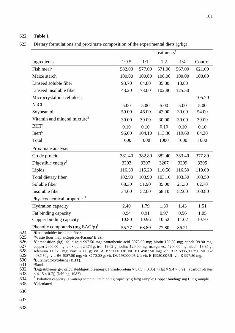
101
Table 1 622
Dietary formulations and proximate composition of the experimental diets (g/kg) 623
Treatments1
Ingredients 1:0.5 1:1 1:2 1:4 Control
Fish meal2 582.00 577.00 571.00 567.00 621.00
Maize starch 100.00 100.00 100.00 100.00 100.00
Linseed soluble fiber 93.70 64.80 35.80 13.80
Linseed insoluble fiber 43.20 73.00 102.80 125.50
Microcrystalline cellulose 105.70
NaCl 5.00 5.00 5.00 5.00 5.00
Soybean oil 50.00 46.00 42.00 39.00 54.00
Vitamin and mineral mixture3 30.00 30.00 30.00 30.00 30.00
BHT4 0.10 0.10 0.10 0.10 0.10
Inert5 96.00 104.10 113.30 119.60 84.20
Total 1000 1000 1000 1000 1000
Proximate analysis
Crude protein 381.40 382.80 382.40 383.40 377.80
Digestible energy6 3203 3207 3207 3209 3205
Lipids 116.30 115.20 116.50 116.50 119.00
Total dietary fiber 102.90 103.90 103.10 103.30 103.50
Soluble fiber 68.30 51.90 35.00 21.30 02.70
Insoluble fiber 34.60 52.00 68.10 82.00 100.80
Physicochemical properties7
Hydration capacity 2.40 1.79 1.30 1.43 1.51
Fat binding capacity 0.94 0.91 0.97 0.96 1.05
Copper binding capacity 10.80 10.96 10.52 11.02 10.70
Phenolic compounds (mg EAG/g)8 55.77 68.80 77.80 86.21 1Ratio soluble: insoluble fiber. 624 2Waste flour tilapia/Copisces-Paraná/ Brazil. 625 3Composition (kg): folic acid 997.50 mg; pantothenic acid 9975.00 mg; biotin 159.60 mg; cobalt 39.90 mg; 626 copper 2800.00 mg; etoxiquin 24.78 g; iron 19.62 g; iodine 120.00 mg; manganese 5200.00 mg; niacin 19.95 g; 627 selenium 119.70 mg; zinc 28.00 g; vit. A 1995000 UI; vit. B1 4987.50 mg; vit. B12 5985,00 mg; vit. B2 628 4987.50g; vit. B6 4987.50 mg; vit. C 70.00 g; vit. D3 198000.05 UI; vit. E 19950.00 UI; vit. K 997.50 mg. 629 4Butylhydroxytoluene (BHT). 630 5Sand. 631 6Digestibleenergy: calculateddigestibleenergy: [(crudeprotein × 5.65 × 0.85) + (fat × 9.4 × 0.9) + (carbohydrates 632 × 4.15 × 0.7)] (Jobling, 1983). 633 7Hydration capacity: g water/g sample; Fat binding capacity: g fat/g sample; Copper binding: mg Cu/ g sample. 634
8Calculated 635
636
637
638

102
Table 2 639
Plasma parameters of juvenile Rhamdia quelen receiving the experimental diets 640
Treatments1
1:0.5 1:1 1:2 1:4 Control SE P-value
Total proteins 3.95b 3.97b 4.38a 4.42a 3.92b 0.06 0.012
Albumin 0.74 0.71 0.85 0.96 0.69 0.04 0.202
Globulin 2.92b 3.20ab 3.43a 3.35a 3.05b 0.09 0.045
Glucose 57.25 52.75 54.37 48.94 48.62 1.89 0.350
Triglycerides 623.50 788.37 705.87 588.64 623.37 32.78 0.312
Cholesterol 167.50 159.12 175.87 168.00 184.50 6.06 0.748
Alkaline phosphatase 17.48b 18.87ab 20.85a 20.67a 17.27b 0.41 0.030
IgT 2.89ab 2.79ab 3.41a 3.34a 2.63b 0.06 0.002
1Ratio soluble: insoluble fiber. Total proteins (g/dL); Albumin (g/dL);Globulin (g/dL): total protein−albumin 641 (g/dL); Glucose (mg/gL);Triglycerides (mg/dL); Cholesterol (mg/gL);Alkaline phosphatase (U.I/L); IgT: Total 642 immunoglobulin (mg/dL) and Cortisol (µg/dL).SE: standard error. Different letters on the rows indicate 643 significant difference by the Tukey’s test (P<0.05). 644 645 646 647 648
649

103
Table 3 650
Skin mucus parameters of juvenile Rhamdia quelen fed with different ratio soluble: insoluble 651
linseed fiber in the diet 652
Treatments1
1:0.5 1:1 1:2 1:4 Control SE P-value
Mucoprotein 3.64ab 3.49ab 3.95a 4.04a 3.18b 0.09 0.007
Protein 58.31 64.32 60.64 65.73 57.33 1.79 0.144
IgT 35.43ab 38.81a 39.06a 42.30a 27.58b 1.81 0.018
pH 6.65 6.70 6.70 6.71 6.69 0.02 0.408
Alkaline phosphatase 31.52b 35.66a 37.61a 35.70a 33.07b 1.24 0.039
1Ratio soluble: insoluble fiber. Mucoprotein (mg/dL); Protein (mg protein/g mucus); IgT: Total immunoglobulin 653 (mg protein/g mucus); Alkaline phosphatase (U.I/L);SE: standard error. Different letters on the rows indicate 654 significant difference by the Tukey’s test (P<0.05). 655 656
657
658
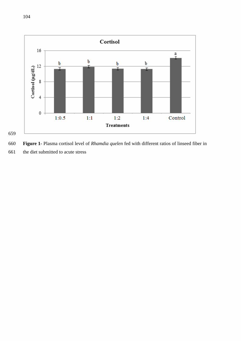
104
659
Figure 1- Plasma cortisol level of Rhamdia quelen fed with different ratios of linseed fiber in 660
the diet submitted to acute stress 661

105
5 DISCUSSÃO GERAL
A possibilidade de incorporação de diferentes proporções de fibra alimentar
concentrada em dietas para peixes é de grande relevância econômica e científica, uma vez que
busca a produção sustentável de proteína de alto valor biológico para consumo humano, sem a
utilização de antibióticos, os quais podem promover o surgimento de cepas de
microrganismos resistentes e deixar resíduos na carne e no ambiente. Para aplicação na
nutrição humana, muitos autores sugerem a utilização de técnicas de hidrólise da fibra
alimentar, para sua atuação efetiva como prebióticos (CHEN et al., 2013; GÓMEZ et al.,
2014; GULLÓN et al., 2011; OLANO-MARTIN et al., 2002).. Segundo estes autores, embora
fibras de alto peso molecular expressem atividade prebiótica, as de menor massa molar, como
os oligossacarídeos, produzem fermentação intestinal mais seletiva.
Porém, na nutrição de peixes estudos mostram que a utilização de substâncias menos
refinadas, como concentrados de fibra alimentar obtidos de distintas fontes, tem capacidade
de otimizar o sistema imune dos animais, além de promover o crescimento e deposição
nutricional. Os autores destacam ainda, que essas fibras proporcionam efeitos equivalentes ou
superiores a prebióticos comerciais consolidados (ADORIAN et al., 2015; ADORIAN et al.
2016; GOULART et al. 2017; MOMBACH, 2015).
Em função disso, as técnicas de concentração da fibra utilizadas nesta tese objetivaram
o fracionamento dos nutrientes contidos no grão da linhaça (fibra alimentar, proteína e
lipídios) para posterior concentração das frações insolúvel e solúvel de fibra. A eficiência da
concentração foi observada pelo valor relativo de fibra alimentar obtido para as frações
solúvel (67,56%) e insolúvel (63,07%) (Apêndice A, Tabelas 2). A fração solúvel da fibra de
linhaça apresentou em sua composição monossacarídica maior abundância de
xilose>glicose>ácido galacturônico>arabinose, enquanto que, a fração insolúvel
glicose>xilose>ácido galacturônico, não sendo encontradas quantidades detectáveis de
arabinose. É importante destacar que nas amostras analisadas não foram encontradas
galactose, ramnose, manose e frutose (Apêndice A, Tabelas 4).
Em relação as propriedades físico-químicas, a fração solúvel da linhaça apresentou
capacidade de hidratação 11,5 vezes maior quando comparada a fração insolúvel (Apêndice
A, Tabela 5). Este comportamento já era esperado, em função da natureza química da fibra
solúvel, que apresenta estrutura altamente ramificada e com grande quantidade de grupos
hidrofílicos (STEPHEN; CUMMINGS, 1979; VANDEROOF, 1998). A capacidade de
ligação a gordura foi semelhante entre as frações, sendo encontradas 1,30 e 1,65 g óleo/ g na

106
fração solúvel e na insolúvel, respectivamente (Apêndice A, Tabela 5). A capacidade de
ligação ao cobre da fração insolúvel da linhaça foi de 10,58 mg Cu/g de amostra (Apêndice A,
Tabela 5), porém na fração solúvel a quantificação não foi possível devido a problemas
metodológicos. Os resultados obtidos para os compostos fenólicos demonstram que as
técnicas utilizadas para concentração das distintas frações de fibra de linhaça concentram
esses compostos na fração insolúvel (Apêndice A, Tabela 5).
Após a analise das características químicas e físico-químicas, as frações de fibra de
linhaça foram adicionadas a dietas para juvenis de jundiá, nas proporções 1:0,5, 1:1, 1:2 e 1:4
de fibra solúvel: insolúvel (FS:FI), de modo a fechar a formulação com inclusão de 10% de
fibra alimentar total, além de um tratamento controle sem adição de fibra de linhaça. Como
consequência das proporções de fibra solúvel: insolúvel, as dietas apresentaram diferenças
principalmente quanto a capacidade de hidratação e ao teor de compostos fenólicos (Artigo I,
Tabela 1).
Os resultados obtidos no ensaio biológico mostraram que a suplementação das dietas
com as proporções 1:2 e 1:4 de FS:FI estimularam o crescimento dos peixes e a deposição de
proteína bruta corporal (Artigo I, Tabelas 2 e 3), com impacto positivo sobre diferentes
parâmetros imunológicos (Artigo II, Tabelas 2 e 3). Destacamos que o presente estudo traz o
primeiro relato sobre parâmetros imunes do muco de jundiás e demonstra que a composição
do mesmo responde a inclusão de substâncias com ação prebiótica as dietas. As mesmas
dietas são associadas ainda a redução nos níveis de cortisol plasmáticos dos jundiás, do pH da
digesta intestinal (Artigo II, Tabela 2 e 3) e na atividade de tripsina (Artigo I, Tabela 5). Essa
redução na atividade de tripsina não causou prejuízos para os peixes, visto que não refletiu em
alterações no desempenho zootécnico e parâmetros metabólicos (Artigo I, Tabelas 2 e 7). É
importante ressaltar que a dieta com 1:0,5 FS:FI, que ocasionou maior atividade de tripsina
(Artigo I, Tabela 7), também é a que apresentou a maior capacidade de hidratação (Artigo I,
Tabela 1). Possivelmente esta característica da dieta tenha influenciado a viscosidade da
digesta, dificultando a interação enzima-substrato e como forma de compensar, o
metabolismo digestivo pode ter elevado a secreção e atividade da enzima (EASWOOD, 1992;
SINHA et al., 2011).
Independente da proporção na dieta, o consumo de fibra de linhaça pelos jundiás
promoveu aumento nas imunoglobulinas totais do plasma e na atividade da fosfatase alcalina
do plasma e muco cutâneo (Artigo II, Tabelas 2 e 3), além de refletir em mudanças
histológicas intestinais, como maior altura de vilosidade e contagem de células caliciformes e
menor espessura da camada muscular (Artigo I, Tabela 6; Artigo II, Tabela 5). Estes

107
acréscimos são desejáveis, pois alterações na morfologia intestinal, como vilos mais curtos e
criptas mais profundas, estão associados à maior susceptibilidade de doenças provocadas por
patógenos intestinais (BRUMANO; GATTÁS, 2009; FERREIRA, 2012). Além disso, quanto
maior a altura das vilosidades intestinais melhor será a digestão e absorção de nutrientes,
refletindo em efeitos positivos sobre desempenho zootécnico, como ocorreu no presente
estudo (GOULART et al., 2018). Estes efeitos podem estar atrelados a composição
monossacarídica das frações solúvel e insolúvel da fibra de linhaça, que reflete em diferente
combinação de monossacarídeos nas dietas. Compostos estes que são responsáveis por
promover o crescimento de bactérias benéficas que impactam tanto a nível imunológico,
quanto para o crescimento do animal (RINGO et al., 2010).
As alterações a nível intestinal também podem estar relacionadas a presença dos
compostos fenólicos associados a fibra, principalmente na fração insolúvel (Apêndice A,
Tabela 5). Os compostos fenólicos possuem reconhecida ação antioxidante, e quando estão
bioacessíveis na região proximal do intestino, podem ser prontamente absorvidos pela
mucosa. Aqueles associados a fibra alimentar passam inalterados pelo trato digestório
superior, sendo liberados no intestino em decorrência da fermentação microbiana da fibra,
promovendo um ambiente antioxidante a nível intestinal (SAURA-CALIXTO, 2011).
Outro resultado que merece destaque é o perfil de fermentabilidade intestinal dos
jundiás. Não há na literatura pesquisas que caracterizem a produção de ácidos graxos de
cadeia curta (AGCC) a nível intestinal para a espécie. No presente estudo, foi observado que
as diferentes proporções de fibra solúvel: insolúvel consumidas pelos peixes afetam
decisivamente as quantidades de AGCC produzidos, apesar de não mudar o perfil de
fermentabilidade (acético>propiônico>butírico). A produção de ácido acético foi superior na
digesta dos peixes que receberam a dieta com 1:2 de FS:FI, de ácido butírico para os que
receberam a dieta com 1:4 de FS:FI, enquanto que a produção de ácido propiônico foi
superior na digesta dos peixes que receberam a dieta controle (Artigo II, Tabela 4).
Essas diferenças nas quantidades de AGCC produzidos pelos peixes são reflexos do
estímulo que a suplementação de fibras exerce sobre a microbiota intestinal. Como foi
observado, as dietas com maior proporção de fibra insolúvel resultaram em maior produção
de AGCC, o que pode estar relacionado a maior capacidade das bactérias intestinais em
degradar os compostos que formam a matriz insolúvel da parede celular, sendo que a
intensidade dessa degradação depende da composição e características físico-químicas da
fibra, além de particularidades da microbiota intestinal (VAN SOEST, 1994). Outra
possibilidade, é que esta microbiota tenha priorizado a degradação da fração insolúvel da fibra

108
de linhaça em detrimento a fração solúvel, havendo assim, um excedente de fibra solúvel, que
com sua alta capacidade de hidratação acabou tendo efeito negativo sobre a absorção de
nutrientes, o que explicaria o desempenho inferior dos peixes alimentados com dietas
contendo maiores proporções da fração solúvel de fibra de linhaça. Porém é importante
salientar, que mesmo não tendo proporcionado os melhores restados, as dietas com maior
proporção de fibra solúvel promoveram respostas semelhantes a dieta controle.
Além dos efeitos mencionados, o ensaio de estresse agudo por hipóxia mostrou que a
suplementação de 1:2 e 1:4 de FS:FI de linhaça as dietas possibilita uma melhor resposta ao
estresse pelos peixes, com aumento de indicadores imunológicos plasmáticos e do muco
cutâneo (Artigo III, Tabelas 2 e 3). Esses resultados confirmam a eficiência das respectivas
proporções de fibra e demonstram sua ação mitigadora de estresse para jundiás. É importante
destacar que, independente de sua proporção na dieta, a fibra de linhaça proporcionou maiores
teores plasmáticos de imunoglobulinas totais e redução dos níveis de cortisol (Artigo III,
Tabela 2; Figura 3). Esta observação permite inferir que a fibra de linhaça impulsiona a
função imunológica e aumenta a tolerância a condições desfavoráveis de manejo durante o
ciclo de cultivo dos jundiás (CASTRO-OSSES et al., 2017; YOKOYAMA et al., 2005).
Sabe-se que o sistema imunológico é influenciado direta e indiretamente pelos
nutrientes ingeridos na alimentação, portanto, a adequação de seus níveis na formulação das
dietas é de extrema importância, visando um caminho economicamente promissor para o
aumento da produtividade em sistemas intensivos de criação de peixes (MENEZES et al.,
2006; PEZZATO et al., 2004). Apesar da fibra alimentar não ter uma função verdadeiramente
nutricional, nossos resultados mostram que sua presença na dieta, em quantidade e proporções
equilibradas, refletem em vários benefícios para o cultivo de jundiás, agindo como promotor
de crescimento, imunoestimulante e mitigador de estresse. Embora não se possa afirmar que
tais efeitos sejam específicos e duradouros, tem-se como vantagem a possibilidade do seu uso
na alimentação como estratégia promotora de saúde e bem estar animal.
109
6 CONCLUSÃO GERAL
Os resultados deste estudo permitem concluir que a fibra de linhaça tem ação
funcional, agindo efetivamente como prebiótico, uma vez que estimula o desempenho,
sistema imune e age como mitigadora de estresse para jundiás. Dentre as proporções de fibra
testadas, a adição de 1:2 e 1:4 de fibra solúvel: insolúvel as dietas, proporcionaram os
melhores resultados para os parâmetros avaliados. Porém, são necessários mais pesquisas a
cerca da função da fibra de linhaça, seu modo de ação e análises minuciosas de seus
componentes, para orientar sua utilização de forma racional.

110
REFERÊNCIAS
Anuário PeixeBR da Piscicultura 2018. PeixeBR: Associação Brasileira de Piscicultura. 71p.,
2018. Disponível em: <https://www.peixebr.com.br/anuario-peixebr-2018/>. Acesso em: 30
setembro de 2018.
ADORIAN, T.J. et al. Dietary fiber in the nutrition of silver catfish: Prebiotic or antinutrient?
Animal Feed Science and Technology, v. 209, p.167–173, 2015.
ADORIAN, T. J. et al. Effect of different dietary fiber concentrates on themetabolism and
indirect immune response in silver catfish. Animal Feed Science and Technology, v. 215, p.
124–132, 2016.
BARRERA, G. Oxidative stress and lipid peroxidation products in cancer progression and
therapy. ISRN Oncology, p.1-21, 2012.
BRUMANO, G.; GATTÁS, G. Alternativas ao uso de antibióticos como promotores de
crescimento em rações de aves e suínos. Revista Eletrônica Nutritime, v. 6, p. 856-
875, 2009.
CASTRO-OSSES, D. et al. Functional diets modulate the acute phase protein response in
Oncorhynchus mykiss subjected to chronic stress and challenged with Vibrio anguillarum.
Fish and Shellfish Immunology, v.66, p. 62-70, 2017.
CHEN, J. et al. Pectic-oligosaccharides prepared by dynamic high-pressure microfluidization
and their in vitro fermentation properties. Carbohydrate Polymers, v. 91, p. 175–182, 2013.
CYRINO, J.E.P. et al. A piscicultura e o ambiente – o uso de alimentos ambientalmente
corretos em piscicultura. Revista Brasileira de Medicina Veterinária e Zootecnia, v.39,
p.68-87, 2010.
EASWOOD, M.A. The physiological effect of dietary fiber: and update. Annual Review of
Nutrition, v. 12, p.19-35, 1992.
FAO, Food and Agriculture Organization of the United Nations. FaoStat. 2017. Disponível
em: <http://www.fao.org/faostat/en/#data/QC>. Acesso em: 15 set. 2018.
FERREIRA, C.L.L. Prébióticos e Probióticos – Atualização e Prospecção. Rio de Janeiro:
Editora Rubio, 2012. 226p.
GALVÃO, E. L. et. al. Avaliação do potencial antioxidante e extração subcrítica
do óleo de linhaça. Ciência e Tecnologia de Alimentos, v. 28, n. 3, p. 551-557, 2008.
GOMEZ, B. et al. Purification, Characterization, and Prebiotic Properties of Pectic
Oligosaccharides from Orange Peel Wastes. Journal of Agricultural and Food Chemistry, v.
62, p.9769−9782, 2014.

111
GOULART, F. R. et al. Atividade de enzimas digestivas e parâmetros de crescimento de
juvenis de jundiá (Rhamdia quelen) alimentados com farelo de linhaça in natura e
demucilada. Semina: Ciências Agrárias, Londrina, v. 34, n. 6, p. 3069-3080, 2013.
GOULART, F.R et al. Effects of Dietary Fiber Concentrates on growth performance and
digestive enzyme activities of jundiá (Rhamdia quelen). Aquaculture Nutrition, v. 23, p.
358-366, 2017.
GOULART, F.R. et al. Effect of dietary fiber concentrates on growth performance, gut
morphology and hepatic metabolic intermediates in jundiá (Rhamdia quelen). Arquivo
Brasileiro de Medicina Veterinária e Zootecnia, v.70, p.1633-1640, 2018.
GULLÓN, B. et al. Prebiotic potential of a refined product containing pectic oligosaccharides.
Food Science and Technology, v. 44, p. 1687-1696, 2011.
LIU, C-J.; LIN, J-Y. Anti-inflammatory effects of phenolic extracts from strawberry and
mulberry fruits on cytokine secretion profiles using mouse primary splenocytes and peritoneal
macrophages.International Immunopharmacology, v. 16, p.165- 170, 2013.
MACAGNAN, F. T.; SILVA, L. P.; HECKTHEUER, L. H. Dietary fibre: The scientific
search for an ideal definition and methodology of analysis, and its physiological importance
as a carrier of bioactive compounds. Food Research International, v. 85 p. 144–154, 2016.
MENEZES, G. C. et al. The influence of dietary vitamin C and E supplementation on the
physiological response of pirarucu, Arapaima gigas, in net culture. Comparative
Biochemistry and Physiology, v. 145, p. 274-279, 2006.
MOMBACH, P. I. Novos prebióticos na nutrição de Tilápia do Nilo. 2015. 81p.
Dissertação (Mestrado em Zootecnia) – Universidade Federal de Santa Maria, Santa Maria,
2015.
MORRE, M. A.; PARK, C. B.; TSUDA, H. Soluble and insoluble fiber influences on cancer
development. Critical Reviews in Oncology/Hematology, v. 27, p.229-242, 1998.
OLANO-MARTIN, E.; GIBSON, G. R.; RASTALL, R. A. Comparison of the in vitro
bifidogenic properties of pectins and pectic-oligosaccharides. Journal of Applied
Microbiology, v. 93, p. 505–511, 2002.
PEZZATO, L. E. et al. Nutrição de peixes. In: CYRINO, J. E. P., URBINATI, E. C.(Ed.).
Tópicos especiais em piscicultura de água doce tropical intensiva. São Paulo: TecArt, cap. 5,
p.75-169, 2004.
QUIRÓS-SAUCEDA, A. E. et al. Dietary fiber and phenolic compounds as functional
ingredients: Interaction and possible effect after ingestion. Food & Function, v.5, p. 1063–
1072, 2014.
RAY, S. et al. Characterization of mucilage polysaccharides, arabinogalactan proteins and
cell-wall hemicellulosic polysaccharides isolated from flax seed meal: a wealth of structural
moieties. Carbohydrate polymers, v. 93, p. 651-660, 2013.
112
REPETTO, M. et al. Lipid peroxidation: chemical mechanism, biological implications and
analytical determination. In: Marisa Repetto, Jimena Semprine and Alberto Boveris, 2012.
RINGO, E. et al. Prebiotics in aquaculture: a review. Aquaculture Nutrition, v. 16, p. 117–
136, 2010.
ROBERFROID, M.B. Prebiotics: the concept revisited. The Journal of Nutrition, v.137,
p.830-837, 2007.
SAURA-CALIXTO, F. Dietary Fiber as a Carrier of Dietary Antioxidants: An Essential
Physiological Function. Journal of Agricultural and Food Chemistry, v. 59, p. 43–49,
2011.
SHIM, Y. Y. et al. Flaxseed (Linum usitatissimum L.) bioactive compounds and peptide
nomenclature: A review. Trends in Food Science & Technology, v. 38, p.5-20, 2014.
SINHA, A. K. et al. Non-starch polysaccharides and their role in fish nutrition – A review.
Food Chemistry, v. 127, p. 1409–1426, 2011.
STEPHEN, A. M.; CUMMINGS, J. H. Water-holding by dietary fibre in vitro and its
relationship to faecal output in man. Gut, v. 20, p.722-729, 1979.
URBINATI, E. C.; CARNEIRO, P. C. F. Práticas de manejo e estresse dos peixes em
piscicultura. In: CYRINO, J. E. P.;URBINATI, E. C.et al.(Ed.). Tópicos especiais em
piscicultura de água doce tropical intensiva. São Paulo: TecArt, 2004. cap. 6, p.171-194.
VALLADÃO, G. M. R.; GALLANI, S. U.; PILARSKI, F. South American fish for
continental aquaculture. Reviews in Aquaculture, v. 10, p. 351-369, 2018.
VANDEROOF, J.A. Immunonutrition: The role of carbohydrates. Nutrition Research, v. 14,
p.595-598, 1998.
VAN SOEST, P. J. Nutritional ecology of the ruminant. 2. ed. Ithaka: Cornell University
Press, 1994. 476p.
VERES, B. Anti-Inflammatory Role of Natural Polyphenols and Their Degradation Products.
In: Severe Sepsis and Septic Shock - Understanding a Serious Killer, Dr Ricardo Fernandez
(Ed.), 2012.
WENZEL, G.E. Carboidratos nutracêuticos e/ou prebióticos. São Leopoldo, RS: Ed.
Unisinos, 2012. 361p.
YOKOYAMA, A. et al. The menin tumor suppressor protein is an essential oncogenic
cofactor for MLL-associated leukemogenesis. Cell, v. 123, p. 207-218, 2005.
ZHANG, Y. et al. Isolation and identification of strawberry phenolics with antioxidant and
human cancer cell antiproliferative properties. Journal of Agricultural and Food
Chemistry, v. 56, p. 670-675, 2008.

113
APÊNDICE A – Fracionamento da linhaça e obtenção de ingredientes ricos em proteína
e fibra: alternativas para a alimentação animal
RESUMO
O fracionamento da linhaça foi realizado em escala laboratorial com o objetivo de obter
frações concentradas em proteína e fibra. Para obtenção do concentrado proteico de linhaça
(CPL) foram testados três métodos (pH ácido, alcalino e isoelétrico). O método de pH
isoelétrico foi mais eficiente (P<0,05) para elevar o conteúdo proteico e também que
proporcionou maior rendimento. Nas frações obtidas foi avaliada a composição química,
matéria seca, cinzas, lipídios, proteína bruta, fibra alimentar total, solúvel e insolúvel e os
minerais cálcio e fósforo. O perfil de aminoácidos foi determinado no farelo de linhaça e no
CPL e, nas frações solúvel e insolúvel da fibra e linhaça in natura, foi avaliado o perfil de
monossacarídeos. Os compostos fenólicos totais e as propriedades físico-químicas
(capacidades de hidratação, ligação a gordura e ao cobre) também foram avaliados. Os
resultados indicaram que o método de concentração proteica por pH isoelétrico melhorou o
perfil aminoacídico e a digestibilidade in vitro do CPL em relação ao farelo original. As
frações da linhaça apresentaram excelentes propriedades físico-químicas, podendo ser
aplicáveis com diferentes finalidades na alimentação animal.
Palavras chave: Compostos fenólicos. Concentrado proteico. Fibra alimentar. Perfil
aminoácidico.

114
1. INTRODUÇÃO
De acordo com a FAO (2014), até 2050 a demanda mundial de alimentos terá
aumento de 70% a fim de atender as necessidades de ingestão básicas de quase dez bilhões
de pessoas. Obviamente, o aumento produtivo exponencial das commodities agropecuárias
alcançados nas últimas décadas não será suficiente para atender satisfatoriamente as
demandas futuras, uma vez que o potencial produtivo das distintas espécies é passível de
estagnação e a mobilização de terras para a produção é limitada e tem apresentado intensos
sinais de degradação ao longo do tempo. Segundo o Diretor Geral da FAO, José Graziano
da Silva, uma mudança de paradigma é necessária para substituir o modelo agropecuário
dos últimos 40 anos, a fim de tornar os sistemas produtivos mais inteligentes e eficientes
(ONUBR, 2015), garantindo a sustentabilidade futura quanto à produção racional de
alimentos em larga escala.
Muitas culturas vegetais, embora amplamente adaptadas para cultivo, têm uso
restrito na nutrição de animais monogástricos devido aos seus fatores antinutricionais, o
que causa subutilização tanto da matéria-prima inicial (grão), como de seus subprodutos de
processamento (farelos). Embora com elevados teores de óleos e proteínas, a linhaça
(Linum uistatissimum L.) e seus subprodutos estão entre os diversos ingredientes de uso
restrito para arraçoamento animal (SOLTAN et al., 2008; TOMM, 2006), devido aos seus
elevados teores de mucilagem e de compostos fenólicos, que reduzem expressivamente o
aproveitamento e desempenho de peixes (BERGAMIN et al., 2011; HASAN et al., 1997)
aves e suínos (VRIES, et al., 2012). Contraditoriamente, estes mesmos fatores são
apontados como pró-nutricionais para a saúde humana, com ação efetiva na promoção da
microbiota intestinal benéfica e com efeito antioxidante a nível celular e metabólico
(HALL; TULBEK; XU, 2006). Efeitos estes que também são desejáveis para expressão na
criação dos animais e nos produtos derivados, desde que seus fatores desencadeantes não
estejam em excesso a ponto de prejudicar o desempenho zootécnico. Neste cenário,
podemos sugerir que o problema do uso da linhaça na nutrição animal não está
necessariamente relacionado aos seus aspectos qualitativos, mas sim, a escassez de
tecnologias racionais e sustentáveis, que garantam a utilização dessa matéria-prima com
máxima eficiência nutricional e ambiental.
Sabe-se que a qualidade nutricional dos produtos vegetais pode ser elevada com a
aplicação de técnicas químicas, físicas e enzimáticas que melhoram seu valor nutricional e
sua digestibilidade (YUE; ZHOU, 2008) podendo dar origem a novos produtos, com ações

115
nutricional e aditiva potencializadas. No caso da linhaça, é possível aplicar tecnologias para
a separação das fibras para uso como agentes prebióticos, bem como, concentrar seu
conteúdo proteico, obtendo-se novos ingredientes de aplicação direcionada na nutrição
animal e ausentes dos efeitos antinutricionais relatados para a fonte in natura (DENG et al.,
2006).
Considerando o exposto, o objetivo do presente estudo foi desenvolver e avaliar os
produtos, concentrado proteico e fibras solúvel e insolúvel, obtidos a partir do fracionamento
da linhaça, a fim de utilizá-los como ingredientes na nutrição animal.
2. MATERIAIS E MÉTODOS
2.1 FRACIONAMENTO DA LINHAÇA
Os grãos de linhaça marrom (Linum uistatissimum L.) foram fornecidos pela empresa
Giovelli Ltda (Guarani das Missões, RS, Brazil). O fracionamento da matéria-prima foi
realizado conforme as etapas descritas na Figura 1. A fração solúvel (mucilagem) foi obtida
seguindo metodologia descrita por Goulart et al. (2013). Inicialmente, os grãos inteiros foram
imersos em água (10%, peso/volume) aquecida (60 a 80ºC), sob agitação constante por 150
minutos. Após, a solução aquosa foi separada dos grãos por filtração (±185 µm) e a fibra
solúvel foi precipitada em meio etanólico (75%). Por fim, a fração solúvel obtida e os grãos
demucilados foram secos em estufa de circulação de ar (MA035; Marconi, Brasil) (55°C/24
horas), moídos em micro moinho (MA-630; Marconi, Brasil) e acondicionados sob
congelamento (-18°C).
O farelo demucilado foi desengordurado com lavagens sucessivas de hexano na
proporção de 1:2 (peso/volume) e seco em estufa com circulação de ar (MA035; Marconi,
Brasil) (55°C/24 h), para evaporação total do solvente. Este produto (farelo demucilado e
desengordurado) foi utilizado para obtenção da fração insolúvel da fibra e do concentrado
proteico de linhaça (CPL), através de metodologias propostas por Smith et al. (1946) e
Lovatto et al. (2017).
O processo de extração da fibra insolúvel foi realizado através de dispersão do farelo
em meio aquoso, utilizando um triturador de facas (LIQ789, Cadence, Brasil) (potência de
400W) por três vezes, a temperatura ambiente, em uma proporção final peso:volume de 1:30,
por 3 minutos. Após cada dispersão, a amostra foi filtrada em peneira (140 μm) e a fração
sólida resultante da última dispersão, correspondente à fração insolúvel, foi seca em estufa

116
com circulação de ar a 55°C por 24 horas. O sobrenadante foi homogeneizado e utilizado em
diferentes métodos de precipitação proteica.
No presente estudo, foram testados três métodos para concentrar a fração proteica do
farelo de linhaça, seguindo metodologia e modificações propostas por Lovatto et al. (2017):
- pH isoelétrico: a concentração proteica foi realizada com aumento do pH da amostra líquida
para 9,0 com NaOH 1N e, após 30 minutos, redução para 4,5 com HCl 1 N (SMITH et al.,
1946).
- pH ácido: ajustou-se o pH da amostra líquida para 4,5 com HCl 1 N (MODESTI et al.,
2007).
- pH alcalino: aumento do pH da amostra líquida para 9,0 com NaOH 1N (MODESTI et al.,
2007).
As medidas de pH foram realizadas com pHmetro de bancada digital (MPA 210-P,
Servilab, Brasil). Após os processos de concentração, as amostras foram deixadas em repouso
(overnight) a 8°C, para decantação da fração proteica. Em seguida, o sobrenadante foi
descartado, e o precipitado, correspondente ao concentrado proteico de linhaça, foi
centrifugado a 3500 rpm por 10 minutos, seco a 55°C por 24 horas em estufa com circulação
de ar (MA035; Marconi, Brasil), moído em micro moinho (MA-630, Marconi, Brasil) e
armazenado sob congelamento (-18°C).
2.2 CONTEÚDOS PROTEICO E LIPÍDICO E RENDIMENTO DE EXTRAÇÃO
Os concentrados proteicos foram analisados quanto ao teor de proteína bruta, através
da determinação de nitrogênio total pelo método de Kjeldahl (nº 960.52) (AOAC, 1995) e teor
lipídico, seguindo metodologia proposta por Bligh e Dyer (1959). O rendimento (R) foi
calculado levando-se em consideração a quantidade obtida, em gramas, de CPL após a
secagem em relação à quantidade de amostra inicial, seguindo a equação:
R (%) = massa inicial (g) x massa concentrado proteico após secagem (g)
100
2.3 CARACTERIZAÇÃO QUÍMICA
As amostras linhaça in natura, farelo de linhaça demucilado e desengordurado, CPL
com maior rendimento e teor proteico, fração solúvel e fração insolúvel da fibra, foram
avaliadas quanto à composição centesimal: matéria seca (método 925.45b), cinzas (método

117
923.03), proteína bruta (método 960.52), fibra alimentar total, solúvel e insolúvel (método
991.43) (AOAC, 1995) e lipídios (Bligh e Dyer, 1959). O conteúdo de cálcio e fósforo foi
analisado de acordo com metodologia proposta por Tedesco et al. (1995). A análise incluiu as
etapas de digestão dos minerais e quantificação por espectrofotometria de absorção atômica
(cálcio) e na região visível (fósforo).
O perfil de aminoácidos presentes no farelo de linhaça demucilado e desengordurado e
no CPL foi determinado através de cromatografia líquida de alta eficiência (CLAE) em fase
reversa com detecção UV a 254 nm (P4000-Thermo Fisher Scientific, Waltham, MA). A
extração foi realizada com HCl 6N por 24 horas e a derivatização com fenilisotiocianato
(WHITE et al., 1986).
O perfil de monossacarídeos foi analisado nas amostras linhaça in natura, fração
solúvel e fração insolúvel da fibra, utilizando cromatografia líquida de alta eficiência (CLAE)
(Shimadzu), com detector de índice de refração (DIR). A separação foi realizada em coluna
AMinexHPX-87H, seguindo metodologia descrita por Sluiter et al. (2008). Para tal,
utilizaram-se padrões de ácido galacturônico, arabinose, frutose, galactose, glicose, ramnose e
xilose.
2.4 DIGESTIBILIDADE IN VITRO
A digestibilidade in vitro do farelo de linhaça demucilado e desengordurado e do CPL
foi determinada de acordo com metodologia descrita por Mauron (1973), com modificações
propostas por Dias et al. (2010). A digestão das amostras foi realizada com adição das
enzimas pepsina (1: 10 000, Nuclear) e pancreatina (Sigma, São Paulo, Brasil). A
digestibilidade resulta das interações do nitrogênio total presente na amostra, do nitrogênio
digerido, do nitrogênio produzido pela autodigestão das enzimas e do nitrogênio solúvel
originalmente contido na amostra.
2.5 COMPOSTOS FENÓLICOS E PROPRIEDADES FÍSICO-QUÍMICAS
As amostras foram submetidas à extração, sequencial, com solução metanólica
acidificada (50:50, volume/volume, pH 2,0) e acetônica (70:30, volume/volume). A
quantificação dos compostos fenólicos totais foi realizada através do método de Folin–
Ciocalteu (WATERHOUSE, 2003), sendo os resultados expressos em mg equivalentes de
ácido gálico (EAG) por 100 g de amostra.

118
As capacidades de hidratação e ligação a gordura foram determinadas de acordo com
Wang e Kinsella (1976). Às amostras foram adicionados água ou óleo e, após
homogeneização, permaneceram em repouso, a temperatura ambiente, por 24 horas. Em
seguida, foram centrifugadas (1300 x g/ 20 min), sendo o sobrenadante descartado. Os
resultados foram expressos em g de água/óleo absorvidos em um grama de amostra seca. A
capacidade de ligação ao cobre foi estimada de acordo com a metodologia de McBurney et al.
(1983).
2.5 DELINEAMENTO EXPERIMENTAL E ANÁLISE ESTATÍSTICA
Para os dados obtidos nos três métodos de concentração proteica utilizou-se um
delineamento experimental casualizado. Os resultados foram submetidos à análise de
variância (ANOVA) e as médias foram comparadas pelo teste de Tukey a 5% de
significância. Para os demais dados são apresentadas as médias ± desvio padrão.
3. RESULTADOS
3.1 MÉTODOS DE CONCENTRAÇÃO PROTEICA
O método de pH isoelétrico foi mais eficiente (P<0,05) para elevar o teor proteico e
reduzir o conteúdo lipídico, bem como, proporcionou maior rentabilidade extrativa numérica
que os demais métodos testados (Tabela 1). O método de concentração por pH ácido mostrou-
se pouco eficiente, devido ao baixo rendimento de extração. A concentração por pH alcalino
revelou-se ineficiente para concentração e rentabilidade proteica.
3.2 COMPOSIÇÃO QUÍMICA
A composição química das frações obtidas a partir da linhaça está apresentada na
Tabela 2. O fracionamento em fibras solúvel e insolúvel proporcionou notória redução da
proteína bruta, 42% e 29,4%, respectivamente, em comparação com a linhaça in natura. A
fração solúvel, rica neste constituinte, apresentou reduzido teor proteico (13,20%). Na fração
insolúvel, o conteúdo proteico encontrado foi de 16,07%. Já no CPL, houve aumento de 60%
no teor de proteínas em relação ao farelo demucilado e desengordurado, utilizado para sua
obtenção.
119
Houve drástica redução (97,51%) no teor lipídico da fração solúvel em comparação
com a linhaça in natura, que passou de 34,11% para 0,85%. A fração insolúvel também teve
expressiva redução de lipídios (79,27%). Da mesma forma, o processo utilizado para obtenção
do CPL permitiu reduzir 31% do conteúdo lipídico (Tabela 2).
A matéria seca da linhaça in natura foi superior às demais frações analisadas,
enquanto que a matéria mineral foi superior na fração insolúvel (Tabela 2). Os teores de
cálcio das frações solúvel e insolúvel apresentaram-se iguais (1,58%). Entretanto, o CPL teve
menor concentração deste mineral (Tabela 2). As frações linhaça in natura, farelo de linhaça e
CPL apresentaram teores equivalentes de fósforo total (0,58, 0,59 e 0,57%, respectivamente).
Na fração insolúvel foram obtidos valores superiores à fração solúvel (Tabela 2).
O fracionamento da fibra da linhaça mostrou-se extremamente eficiente, visto que a
fibra solúvel da fração solúvel correspondeu a 67,56%. Na amostra inicial (linhaça in natura),
esta era de 17,3%, indicando incremento de 290,52% (Tabela 2). Da mesma forma, observou-
se aumento (220,21%) no teor de fibra insolúvel da fração insolúvel, em comparação à
linhaça in natura, de 19,1% para 63,07%. No CPL constatou-se redução no teor de fibra
alimentar total, de 50,6% (farelo de linhaça) para 29,1% (Tabela 2).
Os resultados do aminograma (Tabela 3) do farelo de linhaça demucilado e
desengordurado e do CPL revelaram a superioridade do concentrado em relação aos níveis
dos aminoácidos analisados. Para a nutrição de animais monogástricos, os aminoácidos lisina
e metionina+cistina são limitantes. Desta forma, nossos resultados mostraram-se relevantes,
visto que houve aumento na concentração dos respectivos aminoácidos, de 60% e 83%, no
CPL em relação ao farelo. Este acréscimo na composição aminoacídica e a redução no teor de
fibra alimentar total (Tabela 2) culminaram na melhora da qualidade proteica, refletida na
maior digestibilidade in vitro da proteína do CPL (88,98%) em relação ao farelo de linhaça
(75,69%).
Nas amostras analisadas, não foram encontradas quantidades observáveis de galactose,
ramnose, manose e frutose. Na linhaça in natura observou-se maior concentração de glicose,
seguida de xilose, ácido galacturônico e arabinose. Os mesmos monossacarídeos foram
identificados na fração insolúvel, com exceção da arabinose. Na fração solúvel foram
encontrados maiores teores de xilose, seguido por glicose, ácido galacturônico e arabinose. O
percentual total de monossacarídeos foi maior na fração solúvel (82,60%) do que na fração
insolúvel (64,24%).

120
3.3 COMPOSTOS FENÓLICOS E PROPRIEDADES FÍSICO-QUÍMICAS
A separação da fibra da linhaça concentrou os compostos fenólicos na fração insolúvel
(Tabela 5). Nesta porção, o teor encontrado foi de 654,7 mg EAG/100g, enquanto que na
fração solúvel foi 293,4 mg EAG/100g, conteúdo inferior ao contido na linhaça in natura.
Além disso, observou-se que o método de concentração proteica por pH isoelétrico foi eficaz
na redução do conteúdo de compostos fenólicos em, aproximadamente, 24% em relação ao
farelo (Tabela 5).
Em relação às propriedades físico-químicas, houve uma redução de 49% na
capacidade de hidratação do farelo de linhaça demucilado e desengordurado, comparando-se
com a linhaça in natura. A concentração proteica proporcionou aumento de 13% nesta
propriedade em relação ao farelo (Tabela 5). A capacidade de hidratação da fração solúvel da
fibra de linhaça foi de 43,53g água/g, o que representa 11,48 vezes a mais do que a
quantidade encontrada na fração insolúvel (3,79 g água/g) (Tabela 5).
Os resultados obtidos para capacidade de ligação a gordura variaram de 1,16 g óleo/ g
na linhaça in natura, para 1,30 e 1,65 g óleo/ g na fração solúvel e na fração insolúvel,
respectivamente (Tabela 5). A menor capacidade de ligação a gordura foi observada no CPL
(0,74 g óleo/ g), que apresentou redução de 59% em relação ao farelo (Tabela 5).
Quanto à capacidade de ligação ao cobre, esta passou de 10,28 mg Cu/g na linhaça in
natura para 10,58mg Cu/g na fração insolúvel. Não foi possível realizar a análise na fração
solúvel, devido a problemas metodológicos possivelmente causados por reações químicas
entre os regentes utilizados e o excesso de hidratação da mesma. A concentração proteica
provocou acréscimo de 13% na propriedade de ligação ao cobre, que passou de 10,10 mg
Cu/g (farelo de linhaça) para 11,39 mg Cu/g (CPL).
4. DISCUSSÃO
4.1 CARACTERIZAÇÃO QUÍMICA
A linhaça (Linum usitatissimum L.) é uma das culturas mais antigas produzidas no
mundo. Nativa do Oeste Asiático e do Mediterrâneo, ela é cultivada há cerca de 4000 anos,
sendo utilizada como fonte de óleo, linho e alimento. Para o consumo humano, destaca-se
pelo seu alto conteúdo de ômega-3 (533 mg/g), conferindo-lhe propriedades funcionais
(SHIM et al., 2014; TURNER et al., 2014; MARTIN et al., 2006). Além disso, tem sido

121
adicionada à alimentação animal, na forma de farelo e óleo, para melhorar o desempenho
produtivo e saúde dos animais, assim como para proporcionar o enriquecimento nutricional de
leite, ovos e carne (TURATTI, 2001). Porém, os nutrientes e compostos encontrados na
linhaça ainda são pouco explorados na nutrição animal, fazendo dela uma matéria-prima
subutilizada.
Em média, a linhaça apresenta 20% de proteína bruta, 41% de lipídios, 28% de fibra
alimentar, 92,3% de matéria seca e 3,4% de matéria mineral (SHIM et al., 2014). Composição
semelhante à encontrada no presente estudo para a linhaça in natura, que apresentou 22,76%
de proteína bruta, 34,11% de lipídios, 34,6% de fibra alimentar total (17,3% solúvel e 19,1%
insolúvel), 97,29% de matéria seca e 3,09% de matéria mineral. Segundo Shim et al. (2014),
essas variações na composição são decorrentes da cultivar de linhaça, de características
geográficas como o tipo de solo, características climáticas, entre outros.
Nossos resultados demonstraram que as técnicas adotadas para o fracionamento da
linhaça foram adequadas para a obtenção de um concentrado proteico e das frações solúvel e
insolúvel da fibra. O que fica claro ao observarmos que a proteína bruta da linhaça in natura
(22,76%) foi efetivamente concentrada no CPL (53,24%), restando baixas concentrações
deste nutriente nas frações solúvel e insolúvel da fibra (13,20% e 16,15%, respectivamente).
Os maiores teores de matéria seca encontrados para a linhaça in natura , provavelmente esteja
relacionado ao seu teor lipídico (34,11%), visto que esta amostra não foi submetida a nenhum
tipo de processamento. É importante destacar que para a obtenção do farelo de linhaça, foram
realizadas lavagens com hexano, a fim de reduzir o teor lipídico desse ingrediente. Assim,
consequentemente, houve redução deste componente nas frações obtidas a partir do farelo,
sendo esta mais pronunciada na fração solúvel da fibra, que apresentou 0,85% de lipídios. A
matéria mineral apresentou pequena variação (2,44-4,16%) entre as frações provenientes da
linhaça, reflexo tanto dos níveis de cálcio e fósforo das mesmas, quanto de outros minerais
presentes (não avaliados).
As diferenças encontradas na eficiência da extração proteica podem ser decorrentes
das propriedades dos radicais das estruturas químicas primárias dos aminoácidos que
compõem os ingredientes utilizados na concentração proteica (LOVATTO et al., 2017). O
método que baseou-se no pH isoelétrico foi mais eficaz para extrair e concentrar a proteína da
linhaça devido a maioria dos aminoácidos presentes possuírem pontos isoelétricos entre 4,5 e
6,5 (SGARBIERI, 1996), tornando-a apropriada para esta finalidade. O aumento no conteúdo
lipídico promovido pelo método de concentração por pH alcalino se deve as interações
122
lipídico-proteicas e formação de lipoproteínas hidrofóbicas (ARAÚJO, 2008; LOVATTO et
al., 2017).
A composição aminoacídica das fontes vegetais pode variar de acordo como tipo de
cultivar, procedimentos de controle das culturas, pragas e processos industriais
(ANDRIGUETTO, 1988; TAVERNARI, 2010). Segundo Linden e Lorient (1996) as técnicas
para obtenção de concentrados proteicos podem modificar o perfil aminoacídico da matéria-
prima, e diminuir a concentração de antinutrientes. Os teores de aminoácidos aumentaram no
CPL, em parte devido à redução no teor de fibras deste produto proveniente do
fracionamento. As fontes vegetais são normalmente deficientes em lisina e metionina+cistina,
dois aminoácidos limitantes na alimentação de animais monogástricos. O aumento nos níveis
desses aminoácidos no CPL confirmam que o método pH isoelétrico foi eficiente, não apenas
para aumentar o conteúdo proteico, mas principalmente para melhorar a qualidade proteica do
farelo de linhaça.
A maior digestibilidade in vitro observada no CPL destaca a superioridade deste
ingrediente em relação à fonte original. Essas características qualitativas são fundamentais
para elevar a inclusão de fontes vegetais nas dietas de animais monogástricos visto que os
farelos vegetais apresentam desvantagens, como menor concentração proteica, presença de
elementos antinutricionais e carboidratos de estrutura complexa que reduzem a digestibilidade
do alimento (GUILLAUME et al., 2001).
De forma geral, o excesso de fibra na dieta é considerado um ponto negativo, pois,
diminui a digestibilidade dos nutrientes e aumenta a produção de resíduo fecal, contribuindo
para a poluição do ambiente (NRC, 1993). Entretanto, ingredientes vegetais íntegros (como
farelos e tortas) constituem fontes de fibras, proteínas e lipídios e podem ou não ter sucesso na
nutrição animal. De acordo com Fedeniuk e Biliaderis (1994), a fibra solúvel encontrada na
linhaça, além de proporcionar aumento no tempo de retenção do alimento no estômago,
diminui o seu consumo. Uma vez que apresenta ótima capacidade de retenção de água,
provoca aumento da viscosidade e consequentemente, reduz a digestibilidade dos nutrientes.
Entretanto, Adorian et al. (2015; 2016) relataram ação funcional a nível imunológico e
produtivo, em peixes alimentados com dietas contendo concentrado de fibra alimentar de
linhaça (fração solúvel+insolúvel). Resultado semelhante ao que foi relatado por Goulart et al.
(2017) ao suplementar apenas a fração solúvel da linhaça em dietas para jundiás (Rhamdia
quelen). Esses relatos permitem questionar se são as fibras que apresentam efeito adverso no
desempenho animal ou se o prejudicial é sua adição em excesso nas dietas.

123
Os teores de fibra alimentar encontrados neste estudo confirmam a eficiência das
técnicas adotadas, visto que houve redução no CPL (29,1%) e concentração nas frações
solúvel e insolúvel (73,65% e 71,61%, respectivamente). É possível observar que da fibra
alimentar remanescente no CPL, 21,2% constituiu-se da porção solúvel. Esta, possivelmente
seja oriunda da parte interna do grão ou residual da extração da fibra solúvel do grão de
linhaça, apontando alguma falha no processamento. Independente de sua origem, esta
continua presente no CPL provavelmente em virtude da técnica utilizada para sua
concentração, que inclui a dispersão do farelo em água. Porém, este fato não prejudicou a
concentração das frações solúvel (FS) e insolúvel (FI) da fibra de linhaça, sendo que ambas
foram superiores a 60% (67,56% FS e 63,07% FI, respectivamente).
Para análise de monossacarídeos foram selecionadas apenas amostras de linhaça in
natura e as frações solúvel e insolúvel da fibra. Estas amostras apresentaram baixa
variabilidade de monossacarídeos, com predominância de glicose, xilose, ácido galacturônico,
e arabinose. Como a fibra alimentar total da linhaça in natura mostrou-se inferior às frações
solúvel e insolúvel, por consequência, apresentou menor percentual de monossacarídeos totais
(47,10%), tendo em maior abundância glicose (24,28%). Na fração solúvel observou-se um
percentual elevado de monossacarídeos totais (82,60%), com predomínio de xilose (41,17%).
Já na fração insolúvel o teor de monossacarídios totais foi de 64,24%, com maior percentual
de glicose (42,60%) e ausência de arabinose.
Estudos avaliando a composição de monossacarídeos da fibra solúvel da linhaça foram
realizados por diversos autores, que relataram maior variabilidade em sua composição, com
presença de ácido urônico (11,1%), arabinose (15,5-20,0%), frutose (8,4%), fucose (3%),
galactose (11,7-17,1%), glicose (6,9%), ramnose (11-25,3%) e xilose (29,1-35,4%)
(GOULART et al., 2017; RAY et al., 2013; SHIM et al., 2014;). Dentre os monossacarídeos
presentes na fibra solúvel da linhaça, oligômeros de xilose são encontrados em maiores
quantidades (GOULART et al., 2017). Até o momento, não foram encontrados na literatura
estudos avaliando a composição monossacarídica da fibra insolúvel da linhaça. Desta forma,
os resultados apresentados em nosso estudo são inéditos. De acordo com Ringo et al. (2010)
monossacarídeos como os presentes em ambas as frações, são responsáveis por promover o
crescimento de bifidobactérias benéficas que contribuem para o aumento do crescimento do
animal. Além disso, há a possibilidade de atuação direta sobre algumas populações de
bactérias patogênicas, por meio de exclusão competitiva (FREITAS et al., 2014). Deste modo,
é possível utilizar ambas as frações de fibra da linhaça como prebiótico em dietas para

124
monogástricos. Porém, estudos in vivo devem ser conduzidos para estabelecer as quantidades
a serem adicionadas nas dietas das diferentes espécies.
4.2 COMPOSTOS FENÓLICOS E PROPRIEDADES FÍSICO-QUÍMICAS
A linhaça é sabidamente rica em compostos fenólicos, grupo que inclui várias
substâncias com capacidade antioxidante, como flavonoides e taninos (GALVÃO et al., 2008;
GOÑI et al., 2009). Porém, não há relatos na literatura dos teores presentes nas diferentes
frações deste ingrediente. Deste modo, nossos resultados são originais, pois mostram que o
fracionamento da linhaça através das técnicas utilizadas permite produzir um CPL (459,30 mg
EAG/100g) e uma fração insolúvel de fibra (654,70 mg EAG/100g) ricos em compostos
fenólicos, agregando valor aos produtos obtidos. Estes compostos possuem diferentes pesos
moleculares e podem estar livres ou ligados à parede celular. É importante destacar que a
fibra alimentar e os compostos fenólicos ligados a ela seguem processos fisiológicos comuns,
produzindo efeito sinérgico no trato gastrintestinal (GOÑI et al., 2009). Evidências científicas
apontam que os compostos fenólicos associados à fibra constituem em torno de 50% dos
antioxidantes dietéticos totais (MACAGNAN et al., 2016).
A presença de compostos fenólicos associados à fibra alimentar exercem efeitos sobre
as suas propriedades físico-químicas e fisiológicas. Um exemplo é a sua ação na manutenção
da integridade intestinal, visto que alguns compostos são bioacessíveis na região proximal do
intestino, podendo ser prontamente absorvidos pela mucosa intestinal (SAURA-CALIXTO,
2011). Outros, por sua vez, passam inalterados através do trato gastrintestinal superior em
associação com as fibras, atingindo o cólon, onde podem ser fermentados por ação das
enzimas bacterianas. Estes últimos tornam-se substrato fermentável para a microflora
bacteriana, promovendo um ambiente antioxidante a nível intestinal (SAURA-CALIXTO,
2011). Devido ao potencial antioxidante os compostos fenólicos também podem ser
importantes no processo de armazenamento de ingredientes e rações, principalmente em
produtos com alto teor lipídico (SILVA; SILVA, 1999).
Alguns compostos fenólicos, como os taninos, principalmente os condensados, são
considerados fatores antinutricionais na alimentação animal, pois podem combinar-se com
proteínas e formar complexos que inibem proteases digestivas e enzimas amilolíticas e
lipolíticas reduzindo a digestibilidade proteica de leguminosas e cereais (FRANCIS et al.,
2001; SILVA e SILVA, 1999). Fato este que não foi observado em nosso estudo, já que a
digestibilidade do CPL foi superior a da matéria-prima inicial (Tabela 2). Provavelmente, no
125
processo de fracionamento da linhaça a maior proporção de taninos condensados (pouco
digestíveis) permaneceu ligada a fibra alimentar, não interferindo na digestibilidade proteica
do CPL.
As propriedades físico-químicas da linhaça e suas frações demonstraram que a
capacidade de hidratação da linhaça in natura (5,31 g água/g) foi reduzida no farelo de
linhaça (2,69 g água/g), no CPL (3,04 g água/g) e na fração insolúvel da fibra (3,79 g água/g),
enquanto na fração solúvel foi elevada, atingindo 43,53 g água/g. A maior porcentagem dos
aminoácidos polares (lisina, arginina, histidina, serina, treonina, ácido aspártico e glutâmico)
encontradas no CPL (28,7%) em comparação ao farelo (17,5%) pode explicar o aumento da
propriedade de hidratação deste ingrediente. Esse resultado corrobora com os achados de
Lovatto et al. (2017), que utilizando o pH isoelétrico também observaram maior capacidade
de hidratação no concentrado proteico em relação ao farelo original.
Vários pesquisadores têm mostrado interesse em estudar a capacidade de hidratação de
ingredientes utilizados na alimentação animal, devido a sua correlação com o aumento da
viscosidade do alimento a nível de trato gastrointestinal, influenciando tempo de trânsito
intestinal, desenvolvimento de órgãos, consumo de alimento e sensação de saciedade
(ARROYO et al., 2012; GIGER-REVERDIN, 2000; JIMÉNEZ-MORENO et al., 2011;
SERENA; BACH KNUDSEN, 2007). Brachet et al. (2015) estudaram a capacidade de
hidratação de vinte e quatro matérias-primas utilizadas em dietas para não ruminantes, entre
elas trigo, milho, cevada, farelo de soja e farelo de trigo. Os resultados obtidos pelos autores
supracitados variaram de 0,54-5,60 g água/g amostra, sendo similar à variabilidade
encontrada no presente estudo para as frações de linhaça, com exceção da fração solúvel da
fibra. Os autores destacaram a baixa capacidade de hidratação dos cereais e a alta capacidade
dos subprodutos (BRACHET et al., 2015).
É importante destacar que uma capacidade de hidratação excessivamente alta origina
reduções na digestão e absorção de aminoácidos, carboidratos, minerais e outros nutrientes,
com consequente queda na produtividade (TEJEDOR et al., 2001). Em função disso, autores
têm proposto tal parâmetro como ferramenta para melhorar os modelos de caracterização de
alimentos, devendo inclusive ser utilizado como um novo critério na formulação de dietas
(BRACHET et al., 2015; GOUS, 2014). Glencross et al. (2007) destacam que as
características físico-químicas dos ingredientes influenciam a qualidade tecnológica e
digestibilidade dos alimentos para organismos aquáticos.
A capacidade de ligação a gordura (CLG) apresentou pouca variabilidade entre as
amostras estudadas (0,74-1,81 g óleo/g), sendo a maior capacidade observada no farelo de

126
linhaça e a menor no CPL. A menor CLG do CPL pode ser explicada pelo fato da absorção de
óleo variar conforme o número de grupos hidrofóbicos (aminoácidos apolares) expostos na
proteína (DENCH et al., 1981), os quais estão geralmente localizados internamente,
dificultando a capacidade de ligarem-se com a gordura (LOVATTO et al., 2017). Ingredientes
com alta capacidade de ligação a gordura podem causar redução na absorção lipídica a nível
intestinal, impactando negativamente no desenvolvimento animal, principalmente nas fases
iniciais. Por outro lado, para animais em manutenção, este parâmetro pode ter impacto
positivo, visto que evita o acúmulo de gordura e o aumento dos níveis plasmáticos de
colesterol e triglicerídeos. Assim, considerar a capacidade de ligação a gordura dos
ingredientes utilizados na formulação das dietas, auxilia no ajuste das mesmas a diferentes
fases. Além disso, as capacidades de hidratação e ligação a gordura das rações têm ação sobre
a dureza, estabilidade na água, flutuabilidade e tempo de armazenamento (DRAGANOVIC et
al. 2011; LOVATTO et al., 2017).
Quanto à capacidade de ligação ao cobre, esta foi maior no CPL, possivelmente em
decorrência de ligações químicas entre os grupamentos proteicos reativos e os íons de cobre.
No entanto, a concentração proteica foi realizada no ponto isoelétrico, predominando a
igualdade entre cargas positivas e negativas da proteína. Ingredientes alimentares com alta
capacidade de ligação ao cobre normalmente possuem forte capacidade de ligação iônica com
demais elementos minerais, fazendo com que as dietas interfiram negativamente a sua
absorção (ARRUDA et al., 2003). É importante destacar que a fração solúvel da fibra de
linhaça não teve a propriedade de ligação ao cobre mensurada, mesmo após várias tentativas
de adaptação da técnica. Este problema metodológico possivelmente seja reflexo da alta
capacidade de hidratação desta fração, que em contato com os reagentes utilizados na técnica,
criaram uma solução extremamente viscosa.
As propriedades físico-químicas de ingredientes são pouco exploradas na nutrição
animal, porém podem impactar a nível metabólico e fisiológico no organismo, podendo
refletir positiva ou negativamente na produção animal. A carência destas informações para os
ingredientes convencionalmente utilizados na formulação de dietas para monogástricos limita
sua utilização. No entanto, quando se trata do desenvolvimento de novos produtos estas
análises se tornam imprescindíveis.
Com este estudo foi possível obter três distintas frações a partir da linhaça: uma
composta majoritariamente por proteína (CPL) e duas compostas principalmente por fibras
(frações solúvel e insolúvel). A composição química do CPL revelou características
satisfatórias em relação à proteína, devido ao maior aporte proteico, perfil de aminoácidos e

127
digestibilidade em comparação ao farelo de linhaça, possibilitando o seu uso em substituição
a fontes proteicas de origem animal e vegetais utilizadas na nutrição animal. Além disso, sua
concentração de compostos fenólicos deve ser melhor estudada e caracterizada, uma vez que
podem apresentar capacidade antioxidante. As frações de fibra solúvel e insolúvel podem ser
utilizadas como ingredientes com ação funcional, especialmente prebiótica e antioxidante. A
inclusão destes produtos com diferentes finalidades na alimentação animal ainda precisa ser
avaliada a fim de determinar níveis e aceitação, principalmente com relação à fração solúvel,
a fim de evitar efeitos negativos ao desempenho animal.
Agradecimentos
Ao Conselho Nacional de Desenvolvimento Científico e Tecnológico (CNPq) pela bolsa de
pesquisa (Leila Picolli da Silva), à Coordenação de Aperfeiçoamento de Pessoal de Nível
Superior (CAPES) pelas bolsas de estudos de Doutoramento das alunas Dirleise Pianesso e
Taida Juliana Adorian e a empresa Giovelli Alimentos (Guarani das Missões, RS, Brasil) pela
doação das sementes de linhaça.
128
REFERÊNCIAS
ADORIAN, T.J. et al. Dietary fiber in the nutrition of silver catfish: Prebiotic or antinutrient?
Animal Feed Science and Technology, v. 209, p.167–173, 2015.
ADORIAN, T. J. et al. Effect of different dietary fiber concentrates on the metabolism and
indirect immune response in silver catfish. Animal Feed Science and Technology, v. 215, p.
124-132, 2016.
ANDRIGUETTO, J. M. Nutrição Animal. 4ª ed., São Paulo, Nobel, 395 p., 1988.
AOAC. Association of official analytical chemists. Official Methods of Analyses of the
AOAC International.16 ed. Supplement 1998. Washington: AOAC, 1995.1018 p.
ARAÚJO, J. M. A. Química de Alimentos – Teoria e Prática. 4ª ed., Viçosa: editora, UFV,
2008, 478 p.
ARROYO, J., et al. Effects of presentation and type of cereals (corn or sorghum) on
performance of geese. Poultry Science, v. 91, p. 2063-2071,2012.
ARRUDA A.M.V., et al. Importância da fibra na nutrição de coelhos. Semina: Ciências
Agrárias, v. 24, p. 181-190, 2003.
BERGAMIN, G.T. et al. Fontes protéicas vegetais na alimentação da carpa húngara. Ciência
Rural, v.41, p.1660-1666, 2011.
BERGLUND, D.; ZOLLINGER, R.K. Flax production in North Dakota. North Dakota State
University: Extension service. Fargo: NDSU, 2007.
BHATTY, R. S. Further Compositional Analyses of Flax: Mucilage, Trypsin Inhibitors and
Hydrocyanic Acid. JAOCS, v. 70, n. 9, 1993.
BLIGH, E. G.; DYER, W. J. Rapid method of total lipid extraction and purification.
Canadian. Journal of Biochemistry and Physiology, v.37, p.911-917, 1959.
BRACHET, M., et al. Hydration capacity: a new criterion for feed formulation. Animal Feed
Science and Technology, v. 209, p. 174-185, 2015.

129
DENCH, J.E.; RIVAS, R.N.; CAYGIL, J.C. Selected functional properties of sesame (Sesame
indicum L) flour and two protein isolates. Journal of the Science of Food and Agriculture,
v. 32, p. 557-564, 1981.
DENG, J. et al. Effects of replacing fish meal with soy protein concentrate on feed intake and
growth of juvenile Japanese flounder, Paralichthys olivaceus. Aquaculture, v. 258, p. 503-
513, 2006.
DRAGANOVIC, V. et al. Assessment of the effects of fish meal, wheat gluten, soy protein
concentrate and feed moisture on extruder system parameters and the technical quality of fish
feed. Animal Feed Science and Technology, v. 165, p. 238-250, 2011.
FAO. Organização das Nações Unidas para Agricultura e Alimentação. FAO statistical
yearbook 2014. Food and Agriculture Organization of the United Nations
Regional Office for Asia and the Pacific, Bangkok, 2014, 195 pp.
FEDENIUK, R.W.; BILIADERIS, C.G. Composition and physicochemical properties of
linseed (Linumu sitatissimum L.) mucilage. Journal of Agriculture and Food Chemistry,
v.42, p. 240-247, 1994.
FRANCIS, G.; MAKKAR, H.P.S.; BECKER, K. Antinutritional factors present in plant-
derived alternate fish feed ingredients and their effects in fish. Aquaculture, 199, 197-227,
2001
FREITAS, E. R.; RABELLO, C. B.; WATANABE, P. H. Probióticos e prebióticos na
nutrição de monogátricos. In: SAKOMURA, N. K.; SILVA, J. H. V.; COSTA, F. G. P.;
FERNANDES, J. B. K.; HAUSCHILD, L. Nutrição de não ruminantes. 1. ed. Jaboticabal:
Funep, 2014. p. 487-510.
GALVÃO, E. L. et. al. Avaliação do potencial antioxidante e extração subcrítica do óleo de
linhaça. Ciência e Tecnologia de Alimentos, v. 28, n. 3, p. 551-557, 2008.
GIGER-REVERDIN, S. Characterisation of feed stuffs for ruminants using some physical
parameters. Animal Feed Science and Technology, v. 86, p. 53-69. 2000.
GLENCROSS, B. et al. Evaluation of the influence of drying process on the nutritional value
of lupin protein concentrates when fed to rainbow trout (Oncorhynchus mykiss).
Aquaculture, v.265, p. 218-229, 2007.
GOÑI, I. Towards an updated methodology for measurement of dietary fiber, including
associated polyphenols, in food and beverages. Food Research International, v.42, p. 840–
846, 2009.
GOULART, F. R. et al. Atividade de enzimas digestivas e parâmetros de crescimento de
juvenis de jundiá (Rhamdia quelen) alimentados com farelo de linhaça in natura e
demucilada.Semina: Ciências Agrárias, v. 34, n. 6, p. 3069-3080, 2013.

130
GOULART, F.R et al. Effects of Dietary Fiber Concentrates on growth performance and
digestive enzyme activities of jundiá (Rhamdia quelen). Aquaculture Nutrition, v. 23, p.
358-366, 2017.
GOUS, R.M. Modeling as a research tool in poultry science. Poultry Science, v. 93, p. 1-7,
2014.
GUILLAUME, J.; KAUSHIK, P.B.; MÉTAILLER, R. Nutrition and feeding of fish and
crustaceans. Praxis Publishing LTD, Chichester, UK, 2001. 408p.
HALL, C.; TULBEK, M. C.; XU, Y. Flaxseed. Advances in Food and Nutrition Research,
v. 51, p. 1-97, 2006.
HASAN, M.R.; MACINTOSH, D.J.; JAUNCEY, K. Evaluation of some plant ingredients as
dietary protein sources for common carp (Cyprinus carpio) fry. Aquaculture, v. 151, p. 55-
70. 1997
JIMÉNEZ-MORENO, E. et al.Effects of increasing levels of pea hulls in the diet on
productive performance, development of the gastrointestinal tract, and nutrient retention of
broilers from one to eighteen days of age. Animal Feed Science and Technology, v. 168, p.
100-112, 2011.
KRAUSE, J.-P.; SCHULTZ, M.; DUDEK, S. Effect of extraction conditions on composition,
surface activity and rheological properties of protein isolates from flaxseed (Linum
usitatissimum L.). Journal of the Science of Food and Agriculture, v. 82, p. 970–976, 2002.
LINDEN, G.; LORIENT, D. BioquímicaAgroindustrial. Editoria Acribia S/A, 1996, 380p.
LOVATTO, N. M. et al. Crambe (Crambe abyssinica) and sunflower (Helianthus annuus)
protein concentrates: production methods and nutritional properties for use in fish feed. Anais
da Academia Brasileira de Ciências, v. 89 (3 Suppl.), p. 2495-2504, 2017.
MACAGNAN, F. T.; SILVA, L. P.; HECKTHEUER, L. H. Dietary fibre: The scientific
search for an ideal definition and methodology of analysis, and its physiological importance
as a carrier of bioactive compounds. Food Research International, v. 85 p. 144–154, 2016.
MARTIN, C.A. et al. Ácidos graxos poliinsaturados ômega-3 eômega-6: importância e
ocorrência em alimentos. Revista de Nutrição, Campinas, v. 19, p. 761-770, 2006.
MARTÍNEZ-FLORES, H.E. et al. . Functional characteristics of protein flaxseed concentrate
obtained applying a response surface methodology. Journal of Food Science, v. 71, p. 495–
498, 2006.
MCBURNEY, M. I.; VAN SOEST, P. J.; CHASE, L.E. Cation exchange capacity and
buffering capacity of neutral-detergent fibres. Journal of the Science of Food and
Agriculture, v. 34, p. 910-916, 1983.
MODESTI, C. F. et al. Caracterização de concentrado proteico de folhas de mandioca obtido
por precipitação com calor e ácido. Ciência e Tecnologia de Alimentos, v. 27, n. 3, p. 464-

131
469, 2007.
MOMBACH, P. I. Novos prebióticos na nutrição de Tilápia do Nilo. 2015. 81p.
Dissertação (Mestrado em Zootecnia) – Universidade Federal de Santa Maria, Santa Maria,
2015.
MUELLER, K.; EISNER, P.; KIRCHHOFF, E. Simplified fractionation process for linseed
meal by alkaline extraction – Functional properties of protein and fibre fractions. Journal of
Food Engineering, v. 99, p. 49–54, 2010a.
MUELLER, K. et al. . Functionalproperties and chemical composition of fractionated brown
and yellow linseed meal (Linum usitatissimum L.). Journal of Food Engineering, v. 98, p.
453– 460, 2010b.
PIANESSO, D. et al. Substituição do farelo de soja pelo farelo de linhaça em dietas para a
piava (Leporinus obtusidens). Semina: Ciências Agrárias, v. 34, n. 1, p. 419-430, 2013.
NRC. National Research Council.Nutrient requirements of fish. Washington, D.C.:
National Academy Press, 1993. 124p.
ONUBR. Aumento na produção agrícola mundial não é sinônimo de fim da fome, afirma
FAO, 23 de fevereiro de 2015a. Disponível em: https://nacoesunidas.org/aumento-na-
producao-agricola-mundial-nao-e-sinonimo-de-fim-da-fome-afirma-fao/
RAY, S. et al. Characterization of mucilage polysaccharides, arabinogalactan proteins and
cell-wall hemicellulosic polysaccharides isolated from flax seed meal: a wealth of structural
moieties. Carbohydrate polymers, v. 93, p. 651-660, 2013.
RINGO, E. et al. Prebiotics in aquaculture: a review. Aquaculture Nutrition, v. 16, p. 117–
136, 2010.
SANTOS, E. L. et al. Digestibilidade de ingredientes alternativos para tilápia do Nilo
(Oreochromis niloticus): revisão. Revista Brasileira de Engenharia da Pesca, v.3, n. 2,
2008
SANZ, A.; GARCIA-GALLEGO, M. HIGUERA, M. Protein nutrition in fish: protein/energy
ratio and alternative protein sources to fish meal. Journal of Physiology and Biochemistry,
v.56, n.3, p.275-282, 2000.
SAURA-CALIXTO, F. Dietary Fiber as a Carrier of Dietary Antioxidants: An Essential
Physiological Function. Journal of Agricultural and Food Chemistry, v. 59, p. 43–49,
2011.
SERENA, A.; BACH KNUDSEN, K.E. Chemical and physicochemical characterization of
coproducts from the vegetable food and agro industries. Animal Feed Science and
Technology, . v. 139, p. 109-124. 2007.

132
SGARBIERI, V. C. Proteínas em alimentos proteicos: propriedades, degradações,
modificações. São Paulo: Varela, 1996, 517 p.
SHIM, Y. Y. et al. Flaxseed (Linum usitatissimum L.) bioactive compounds and peptide
nomenclature: A review. Trends in Food Science & Technology, v. 38, p.5-20, 2014.
SMITH, A.K.; JOHNSON, V.L.; BECKEL, A.C. Linseed proteins alkali dispersion and acid
precipitation. Journal of Industrial and Engineering Chemistry, v. 38, p. 353-356, 1946.
SOLTAN, M.A. et al. Effect of replacing fish meal by a mixture of different plant protein
sources in the Nile tilapia (Oreochromis niloticus) diets. Global Veterinaria, v.37, n.4,
p.157-164, 2008.
SONCIN, M. R. S. P. et al. Digestibilidade aparente, crescimento folicular e concentração de
metabólitos sanguíneos de éguas recebendo concentrado com semente de linhaça integral
(Linum usitatissimum L.).Acta Scientiarum Animal Sciences, v.31, n.2, p.191-197, 2009.
SILVA, M.R.; SILVA, M.A.A.P. Aspectos nutricionais de fitatos e taninos. Revista de
Nutrição, Campinas, v.12, p.5-19, 1999.
SLUITER, A., et al. Determination of Sugars, Byproducts, and Degradation Products in
Liquid Fraction Process Samples. National Renewable Energy Laboratory, Technical
Report NREL/TP-510-42623, 2008.
TAVERNARI, F. C. et al. Avaliação nutricional e energética do farelo de girassol para aves.
Arquivo Brassileiro de Medicina Veterinária e Zootecnia, v. 62, n1, p. 172-177, 2010.
TEDESCO, M. J. et al. Análises de solo, plantas e outros materiais. 2a Ed. Porto Alegre:
Departamento de solos, UFRGS, 1995, 174p (Boletim Técnico, 5).
TEJEDOR A.A. et al. Efeito da Adição de Enzimas em Dietas de Frangos de Corte à Base de
Milho e Farelo de Soja sobre a Digestibilidade Ileal de Nutrientes. Revista Brasileira de
Zootecnia, v. 30, p. 809-816. 2001.
TOMM, G.O. Indicações para o cultivo de linho no Rio Grande do Sul. Guarani
das Missões: Giovelli, 2006. 40p.
TURATTI, J. M. A importância dos ovos numa dieta saudável. Óleos e Grãos, v. 9, p. 22- 24,
2001.
TURNER, T. D. et al. Flaxseed fed pork: n-3 fatty acid enrichment and contribution to
dietary recommendations. Meat Science, v. 96, p. 541-547, 2014.
VRIES, S. Improving digestive utilization of fiber-rich feedstuffs in pigs and poultry by
processing and enzyme technologies: A review. Animal Feed Science and Technology,
v.178, p.123-138, 2012.
WANG, J. C.; KINSELLA, J. E. Functional properties of novel proteins: alfalfa leaf proteins.
Journal Food Science, v.41, p. 286-292, 1976.

133
WATERHOUSE A. L. Determination of total phenolics. In: Current Protocols in Food
Analytical Chemistry, R. E. Wrolstad, Ed., units I, pp. 111–118, John Wiley & Sons, New
York, NY, USA, 2003.
WHITE, J.A.; HART, R.J.; FRY.J.C. An evaluation of the Waters Pico-Tag system for the
amino-acid-analysis of food materials. Journal of Automatic Chemistry, v. 8, p.170–177,
1986.
YUE, Y.R.; ZHOU, Q.C. Effect of replacing soybean meal with cottonseed meal on growth,
feed utilization, and hematological indexes for juvenile hybrid tilapia, Oreochromis
niloticus×O.aureus. Aquaculture, v. 284, p.185-189, 2008.

134
Ajuste pH 9 com
NaOH 1M
Descanso 30 min (T° ambiente)
Ajustar o pH 4,5 com HCl 1M
Descarte sobrenadante
Filtrar amostra em peneira (140 µm)
Hexano (1:2)
Quatro lavagens de 30 minutos
Figura 1 – Etapas do fracionamento da linhaça para obtenção do Concentrado Proteico de
Linhaça (CPL) e das frações solúvel e insolúvel
Fração líquida 1
Amostras
Homogeneizadas
Amostra alcalinizada
Amostra acidificada
Overnight (8°C)
Amostra precipitada centrifugada
(3500 rpm/5 min)
Secar em estufa (55°C)
CPL
FARELO DEMUCILADO
Imersão em água destilada 10% peso/volume
Agitação a 60-80ºC por 150 minutos
Dispersão proteína em água destilada (1:10)
Amostra agitada por 3 minutos em liquidificador
Filtrar amostra em peneira (140 µm)
FARELO DESENGORDURADO
Amostra solubilizada
FRAÇÃO INSOLÚVEL
Dispersão proteína em água destilada (1:10)
Agitação por 3 minutos em liquidificador
Dispersão proteína em água destilada (1:10)
Agitação por 3 minutos em liquidificador
Fração retida na peneira
Fração líquida 2
Fração retida na peneira
Fração líquida 3
Fração retida na peneira
Secar em estufa (55°C)
Filtrar
LINHAÇA IN NATURA
Secar em estufa
(55°C)
FRAÇÃO SOLÚVEL
Desengordurar
Precipitação com etanol

135
Tabela 1- Composição proteica, lipídica e rendimento de extração do concentrado proteico de
linhaça, utilizando diferentes métodos de concentração: pH isoelétrico, pH ácido e
pH alcalino
Conteúdo Método de concentração proteica
pH isoelétrico pH ácido pH alcalino
% da matéria in natura
Proteína Bruta 53,24±0,29ª 42,85 ± 3,72ab 33,13±0,175c
Lipídios 12,75±0,11b 25,54±0,48a 13,06±0,15b
Rendimento 44,71 37,00 18,70
Fonte: Elaborada pelas autoras. Médias ± desvio padrão. Letras distintas na linha diferem estatisticamente pelo
teste de Tukey (P < 0,05).

136
Tabela 2- Composição nutricional da linhaça in natura, farelo de linhaça1, concentrado proteico
de linhaça e frações solúvel e insolúvel de fibra
Conteúdo Linhaça
in natura
Farelo
de linhaça1 CPL
Fração
solúvel
Fração
insolúvel
% da matéria in natura
Proteína bruta 22,76±0,37 33,24±0,06 53,24±0,58 13,20±0,80 16,15±1,14
Lipídios 34,11±0,41 18,45±0,13 12,75±0,18 0,85±0,02 7,07±0,13
Fibra alimentar total 36,40±2,71 50,60±3,78 29,10±5,43 73,65±3,18 71,61±0,76
Solúvel 17,30±0,14 22,30±2,12 21,20±5,25 67,56±16,33 8,54±3,92
Insolúvel 19,10±1,28 28,30±4,24 7,88±0,17 6,09±3,25 63,07±6,16
Matéria seca 97,29±0,01 93,68±0,10 93,62±0,04 91,25±0,13 93,62±0,31
Matéria mineral 3,09±0,01 3,89±0,02 2,44±0,04 2,48±0,04 4,16±0,05
Cálcio2 1,80 1,48 1,32 1,58 1,58
Fósforo 0,58±0,02 0,59±0,03 0,57±0,07 0,20±0,01 0,41±0,03
Digestibilidade proteica
in vitro
NA 75,69±3,54 88,98±1,65 NA NA
Fonte: Elaborada pelas autoras. 1demucilado e desengordurado. CPL: concentrado proteico de linhaça. NA: não
analisado. 2
Sem desvio padrão.

137
Tabela 3- Composição aminoacídica do farelo de linhaça (demucilado e desengordurado) e do
concentrado proteico de linhaça
Aminoácidos (%)1 Farelo de linhaça1 CPL
Essenciais
Arginina 3,45 5,90
Fenilalanina 1,70 2,97
Histidina 0,50 1,00
Isoleucina 1,54 2,63
Leucina 1,98 3,34
Lisina 1,41 2,25
Metionina+cistina 0,54 0,99
Treonina 1,11 1,87
Triptofano2 NA NA
Valina 1,74 3,02
Não essenciais
Ácido aspártico 2,96 4,84
Ácido glutâmico 6,51 10,33
Alanina 1,47 2,52
Glicina 1,80 2,83
Prolina 1,20 1,98
Serina 1,52 2,51
Tirosina 0,92 1,34
Fonte: Elaborada pelas autoras. 1Determinados por Cromatografia Líquida de Alta Eficiência (HPLC)
no Laboratório de Fontes Proteicas (LaFoP) da UNICAMP, Campinas, SP. CPL: concentrado proteico
de linhaça.2NA: não analisado.

138
Tabela 4 - Composição monossacarídica (%) da linhaça in natura e frações solúvel e
insolúvel da fibra
Monossacarídeos (%)
Linhaça in natura Fração solúvel Fração insolúvel
Ácido Galacturônico 10,51 14,96 8,56
Arabinose 2,87 8,05 0
Glicose 24,28 18,5 42,60
Xilose 12,31 41,17 13,09
Total 47,10 82,68 64,24
Fonte: Elaborada pelas autoras. Não foram encontradas quantidades observáveis, em nenhuma das amostras, de
galactose, ramnose, manose e frutose.

139
Tabela 5 – Propriedades físico-químicas e compostos fenólicos das frações da linhaça
Propriedades físico-químicas
Linhaça in
natura Farelo linhaça1 CPL
Fração
solúvel
Fração
insolúvel
CH (g água/g) 5,31±0,12 2,69±0,11 3,04±0,08 43,53±2,35 3,79±0,16
CLG (g óleo/g) 1,16±0,06 1,81±0,09 0,74±0,02 1,30±0,05 1,65±0,01
CLC (mg Cu/g) 10,28±1,06 10,10±1,41 11,39±0,02 - 10,58±0,41
Compostos fenólicos (mg de EAG/100 g)
375,5±12,24 600,30±159,31 459,3±9,87 293,4±128,01 654,7±177,40
Fonte: Elaborada pelas autoras. 1demucilado e desengordurado. CPL: concentrado proteico de linhaça. CH:
capacidade de hidratação. CLG: capacidade de ligação a gordura. CLC: capacidade de ligação ao cobre. EAG:
equivalentes de ácido gálico.

140
ANEXO A – Normas da revista Animal Feed Science and Technology
Types of article
1. Original Research Papers (Regular Papers)
2. Review Articles
3. Short Communications
4. Book Reviews
Original Research Papers should report the results of original research. The material should not have been
previously published elsewhere, except in a preliminary form.
Review Articles should cover subjects falling within the scope of the journal which are of active current interest.
A Short Communication is a concise but complete description of a limited investigation, which will not be
included in a later paper. Short Communications should be as completely documented, both by reference to the
literature and description of the experimental procedures employed, as a regular paper. They should not occupy
more than six printed pages (about 12 manuscript pages, including figures, tables and references).
Book Reviews will be included in the journal on a range of relevant books which are not more than two years old.
Book reviews will be solicited by the Book Review Editor. Unsolicited reviews will not usually be accepted, but
suggestions for appropriate books for review may be sent to the Book Review Editor:
Professor G. Flachowsky, Federal Research Centre of Agriculture, Institute of Animal Nutrition, Bundesallee 50,
D-38116 Braunschweig, Germany
Contact details for submission: For queries concerning the submission process or journal procedures please
visit the Elsevier Support Center. Authors can determine the status of their manuscript within the review
procedure using Elsevier Editorial System.
Submission checklist: You can use this list to carry out a final check of your submission before you send it to
the journal for review. Please check the relevant section in this Guide for Authors for more details.
Ensure that the following items are present: One author has been designated as the corresponding author with
contact details: E-mail address; Full postal address; All necessary files have been uploaded:
Manuscript: Include keywords; All figures (include relevant captions); All tables (including titles, description,
footnotes); Ensure all figure and table citations in the text match the files provided; Indicate clearly if color
should be used for any figures in print
Graphical Abstracts/Highlights files (where applicable), Supplemental files (where applicable)
Further considerations: Manuscript has been 'spell checked' and 'grammar checked'; All references mentioned in
the Reference List are cited in the text, and vice versa; Permission has been obtained for use of copyrighted
material from other sources (including the Internet); A competing interests statement is provided, even if the
authors have no competing interests to declare; Journal policies detailed in this guide have been reviewed;
Referee suggestions and contact details provided, based on journal requirements. For further information, visit
our Support Center.
Ethics in publishing: Please see our information pages on Ethics in publishing and Ethical guidelines for
journal publication.
Studies in humans and animals: If the work involves the use of human subjects, the author should ensure that
the work described has been carried out in accordance with The Code of Ethics of the World Medical
Association (Declaration of Helsinki) for experiments involving humans. The manuscript should be in line with
the Recommendations for the Conduct, Reporting, Editing and Publication of Scholarly Work in Medical
Journals and aim for the inclusion of representative human populations (sex, age and ethnicity) as per those
recommendations. The terms sex and gendershould be used correctly.
Declaration of interest: All authors must disclose any financial and personal relationships with other people or
organizations that could inappropriately influence (bias) their work. Examples of potential competing interests
include employment, consultancies, stock ownership, honoraria, paid expert testimony, patent
applications/registrations, and grants or other funding. Authors must disclose any interests in two places: 1. A
summary declaration of interest statement in the title page file (if double-blind) or the manuscript file (if single-
blind). If there are no interests to declare then please state this: 'Declarations of interest: none'. This summary
statement will be ultimately published if the article is accepted. 2. Detailed disclosures as part of a separate
Declaration of Interest form, which forms part of the journal's official records. It is important for potential
interests to be declared in both places and that the information matches. More information.
Submission declaration and verification: Submission of an article implies that the work described has not been
published previously (except in the form of an abstract, a published lecture or academic thesis, see 'Multiple,
redundant or concurrent publication' for more information), that it is not under consideration for publication
elsewhere, that its publication is approved by all authors and tacitly or explicitly by the responsible authorities
where the work was carried out, and that, if accepted, it will not be published elsewhere in the same form, in

141
English or in any other language, including electronically without the written consent of the copyright-holder. To
verify originality, your article may be checked by the originality detection service Crossref Similarity Check.
Preprints: Please note that preprints can be shared anywhere at any time, in line with Elsevier's sharing policy.
Sharing your preprints e.g. on a preprint server will not count as prior publication (see 'Multiple, redundant or
concurrent publication' for more information).
Use of inclusive language: Inclusive language acknowledges diversity, conveys respect to all people, is
sensitive to differences, and promotes equal opportunities. Articles should make no assumptions about the
beliefs or commitments of any reader, should contain nothing which might imply that one individual is superior
to another on the grounds of race, sex, culture or any other characteristic, and should use inclusive language
throughout. Authors should ensure that writing is free from bias, for instance by using 'he or she', 'his/her' instead
of 'he' or 'his', and by making use of job titles that are free of stereotyping (e.g. 'chairperson' instead of 'chairman'
and 'flight attendant' instead of 'stewardess').
Changes to authorship: Authors are expected to consider carefully the list and order of
authors beforesubmitting their manuscript and provide the definitive list of authors at the time of the original
submission. Any addition, deletion or rearrangement of author names in the authorship list should be made
only before the manuscript has been accepted and only if approved by the journal Editor. To request such a
change, the Editor must receive the following from the corresponding author: (a) the reason for the change in
author list and (b) written confirmation (e-mail, letter) from all authors that they agree with the addition, removal
or rearrangement. In the case of addition or removal of authors, this includes confirmation from the author being
added or removed. Only in exceptional circumstances will the Editor consider the addition, deletion or
rearrangement of authors after the manuscript has been accepted. While the Editor considers the request,
publication of the manuscript will be suspended. If the manuscript has already been published in an online issue,
any requests approved by the Editor will result in a corrigendum.
Copyright: Upon acceptance of an article, authors will be asked to complete a 'Journal Publishing Agreement'
(see more information on this). An e-mail will be sent to the corresponding author confirming receipt of the
manuscript together with a 'Journal Publishing Agreement' form or a link to the online version of this agreement.
Subscribers may reproduce tables of contents or prepare lists of articles including abstracts for internal
circulation within their institutions. Permission of the Publisher is required for resale or distribution outside the
institution and for all other derivative works, including compilations and translations. If excerpts from other
copyrighted works are included, the author(s) must obtain written permission from the copyright owners and
credit the source(s) in the article. Elsevier has preprinted forms for use by authors in these cases.
For gold open access articles: Upon acceptance of an article, authors will be asked to complete an 'Exclusive
License Agreement' (more information). Permitted third party reuse of gold open access articles is determined by
the author's choice of user license.
Author rights: As an author you (or your employer or institution) have certain rights to reuse your work. More
information.
Elsevier Researcher Academy: Researcher Academy is a free e-learning platform designed to support early and
mid-career researchers throughout their research journey. The "Learn" environment at Researcher Academy
offers several interactive modules, webinars, downloadable guides and resources to guide you through the
process of writing for research and going through peer review. Feel free to use these free resources to improve
your submission and navigate the publication process with ease.
Language (usage and editing services): Please write your text in good English (American or British usage is
accepted, but not a mixture of these). Authors who feel their English language manuscript may require editing to
eliminate possible grammatical or spelling errors and to conform to correct scientific English may wish to use
the English Language Editing service available from Elsevier's WebShop.
Submission: Our online submission system guides you stepwise through the process of entering your article
details and uploading your files. The system converts your article files to a single PDF file used in the peer-
review process. Editable files (e.g., Word, LaTeX) are required to typeset your article for final publication. All
correspondence, including notification of the Editor's decision and requests for revision, is sent by e-mail.
Poorly written and/or presented manuscripts (relative to the journal's guidelines) may be returned to authors for
upgrading by the editorial office, prior to a review for scientific merit.
Before preparing their manuscript, it is suggested that authors examine the editorial by the Editors-in-Chief
in Vol. 134/3-4, which outlines several practices and strategies of manuscript preparation that the Editors-in-
Chief have found to be successful. This editorial also outlines practices that can lead to difficulties with
reviewers and/or rejection of the manuscript for publication. There is also an example of an Animal Feed
Science and Technology manuscript available on the journal website
at http://www.elsevier.com/locate/anifeedsci.
Submit your article: Please submit your article via https://www.evise.com/profile/api/navigate/ANIFEE.

142
Referees: Please submit the names and institutional e-mail addresses of several potential referees. For more
details, visit our Support site. Note that the editor retains the sole right to decide whether or not the suggested
reviewers are used.
Peer review: This journal operates a single blind review process. All contributions will be initially assessed by
the editor for suitability for the journal. Papers deemed suitable are then typically sent to a minimum of two
independent expert reviewers to assess the scientific quality of the paper. The Editor is responsible for the final
decision regarding acceptance or rejection of articles. The Editor's decision is final. More information on types
of peer review.Use past tense for current findings, and the present tense for "truths" and hypotheses.
Article Structure: Manuscripts should have numbered lines, with wide margins and double spacing
throughout, i.e. also for abstracts, footnotes and references. Every page of the manuscript, including the title
page, references, tables, etc., should be numbered continuously. However, in the text no reference should be
made to page numbers; if necessary, one may refer to sections. Avoid excessive usage of italics to emphasize
part of the text.
Introduction: State the objectives of the work and provide an adequate background, avoiding a detailed
literature survey or a summary of the results.
Material and methods: Provide sufficient details to allow the work to be reproduced by an independent
researcher. Methods that are already published should be summarized, and indicated by a reference. If quoting
directly from a previously published method, use quotation marks and also cite the source. Any modifications to
existing methods should also be described. If reference is made to AOAC, ISO or similar analytical procedure(s),
the specific procedure identification number(s) must be cited. A number of references for neutral and acid
detergent fibre (NDF, ADF) assays exist, and an alternative reference to the now out-of-print USDA Agriculture
Handbook 379 must be used. There are many options for NDF and ADF assays (e.g. sodium sulfite, alpha
amylase, residual ash), which must be specified in the text. For more details see the editorial in Vol. 118/3-4.
While expressions of NDF and ADF inclusive of residual ash will continue to be acceptable (i.e., the terms
aNDF, NDF and ADF above), the Editors-in-Chief highly recommend reporting all fibre values, including
digestibilities, on an OM basis. Silica is partially soluble in ND, is quantitatively recovered in AD, and so may
contribute to the 'fibre' values and to subsequent digestibility coefficients.
Reporting 'hemicellulose' values as the difference between NDF and ADF is generally only acceptable if the
analyses have been sequential on the same sample. Crude fibre (CF), nitrogen-free extract (NFE) and total
digestible nutrients (TDN) are not acceptable terms for describing feeds and should only be referred to in a
historical context.
Results: Results should be clear and concise.
Discussion: This should explore the significance of the results of the work, not repeat them. Avoid extensive
citations and discussion of published literature. Combined 'Results and Discussion' sections are only acceptable
for 'Short Communications', except under compelling circumstances.
Conclusions: The main conclusions of the study may be presented in a short Conclusions section, which may
stand alone or form a subsection of a Discussion or Results and Discussion section.
Highlights: highlights are mandatory for this journal. They consist of a short collection of bullet points that
convey the core findings of the article and should be submitted in a separate editable file in the online
submission system. Please use 'Highlights' in the file name and include 3 to 5 bullet points (maximum 85
characters, including spaces, per bullet point). You can view example Highlights on our information site.
Keywords: Immediately after the abstract, provide a maximum of 6 keywords, using American spelling and
avoiding general and plural terms and multiple concepts (avoid, for example, 'and', 'of'). Be sparing with
abbreviations: only abbreviations firmly established in the field may be eligible. These keywords will be used for
indexing purposes.
Abbreviations: Define abbreviations that are not standard in this field in a footnote to be placed on the first page
of the article. Such abbreviations that are unavoidable in the abstract must be defined at their first mention there,
as well as in the footnote. Ensure consistency of abbreviations throughout the article.
Acknowledgements: Collate acknowledgements in a separate section at the end of the article before the
references and do not, therefore, include them on the title page, as a footnote to the title or otherwise. List here
those individuals who provided help during the research (e.g., providing language help, writing assistance or
proof reading the article, etc.).
Formatting of funding sources: List funding sources in this standard way to facilitate compliance to funder's
requirements: Funding: This work was supported by the National Institutes of Health [grant numbers xxxx,
yyyy]; the Bill & Melinda Gates Foundation, Seattle, WA [grant number zzzz]; and the United States Institutes
of Peace [grant number aaaa].
Nomenclature and units: Follow internationally accepted rules and conventions: use the international system of
units (SI). If other quantities are mentioned, give their equivalent in SI. You are urged to consult IUB:
Biochemical Nomenclature and Related Documents for further information. Authors and Editors are, by general
agreement, obliged to accept the rules governing biological nomenclature, as laid down in the International Code
143
of Botanical Nomenclature, the International Code of Nomenclature of Bacteria, and the International Code of
Zoological Nomenclature. All biotica (crops, plants, insects, birds, mammals, etc.) should be identified by their
scientific names when the English term is first used, with the exception of common domestic animals. All
biocides and other organic compounds must be identified by their Geneva names when first used in the text.
Active ingredients of all formulations should be likewise identified. SI or SI-derived units should be used
throughout (e.g. MJ and not Kcal for energy concentrations). Concentrations should be expressed on a 'per kg'
basis (w/w); however, w/v, v/v, mol/mol or M may be accepted depending on the circumstances. In addition,
'units' and 'equivalents' are acceptable. Normality should be avoided, as it may be ambiguous for certain acids. If
analytical standards have been used, they should be specified by name (e.g. yeast RNA) and form (e.g. lactose
monohydrate). Percents should only be used when describing a relative increase or decrease in a response.
Proportions should be maximum 1.0 or ≤1.0. For more details see the editorial in Vol. 118/3-4.
Percent is only used to indicate relative changes. For composition, both w/w (often solids composition g/kg) and
w/v (e.g. g/L), v/v (e.g. m/L), mol/mol or M can be accepted depending on the circumstances. Specify units (e.g.
g/L) and never as percent.
Math formulae: Please submit math equations as editable text and not as images. Present simple formulae in line
with normal text where possible and use the solidus (/) instead of a horizontal line for small fractional terms, e.g.,
X/Y. In principle, variables are to be presented in italics. Powers of e are often more conveniently denoted by
exp. Number consecutively any equations that have to be displayed separately from the text (if referred to
explicitly in the text). If differences between treatments are statistically significant, this should be indicated by
adding the actual 'P' value obtained. If 0.10 > P > 0.05, then differences can be considered to suggest a trend, or
tendency, to a difference, but the actual 'P' value should be stated. Further information on this issue can be found
in Animal Feed Science and Technology Vol. 129/1-2.
Spaces should be used between all values and units, except for the following: Between the value and degrees or
percent. In equations around * and /. In probability expressions (P<0.05). When probability values are given, the
'P' should be a capital letter.
Artwork Electronic artwork: General points: Make sure you use uniform lettering and sizing of your original
artwork. Embed the used fonts if the application provides that option. Aim to use the following fonts in your
illustrations: Arial, Courier, Times New Roman, Symbol, or use fonts that look similar. Number the illustrations
according to their sequence in the text. Use a logical naming convention for your artwork files. Provide
captions to illustrations separately. Size the illustrations close to the desired dimensions of the published
version. Submit each illustration as a separate file. A detailed guide on electronic artwork is available.
You are urged to visit this site; some excerpts from the detailed information are given here. Format: If your electronic artwork is created in a Microsoft Office application (Word, PowerPoint, Excel) then
please supply 'as is' in the native document format. Regardless of the application used other than Microsoft
Office, when your electronic artwork is finalized, please 'Save as' or convert the images to one of the following
formats (note the resolution requirements for line drawings, halftones, and line/halftone combinations given
below): EPS (or PDF): Vector drawings, embed all used fonts. TIFF (or JPEG): Color or grayscale photographs
(halftones), keep to a minimum of 300 dpi. TIFF (or JPEG): Bitmapped (pure black & white pixels) line
drawings, keep to a minimum of 1000 dpi. TIFF (or JPEG): Combinations bitmapped line/half-tone (color or
grayscale), keep to a minimum of 500 dpi.
Please do not: Supply files that are optimized for screen use (e.g., GIF, BMP, PICT, WPG); these typically
have a low number of pixels and limited set of colors; Supply files that are too low in resolution; Submit
graphics that are disproportionately large for the content. All data in figures should have a measure of variation
either on the plot (e.g., error bars), in the figure legend itself, or by reference to a table with measures of
variation in the figure legend. Explanations should be given in the figure legend(s). Drawn text in the figures
should be kept to a minimum. If a scale is given, use bar scales (instead of numerical scales) that must be
changed with reduction.
Color artwork: Please make sure that artwork files are in an acceptable format (TIFF (or JPEG), EPS (or PDF),
or MS Office files) and with the correct resolution. If, together with your accepted article, you submit usable
color figures then Elsevier will ensure, at no additional charge, that these figures will appear in color online (e.g.,
ScienceDirect and other sites) regardless of whether or not these illustrations are reproduced in color in the
printed version. For color reproduction in print, you will receive information regarding the costs from
Elsevier after receipt of your accepted article. Please indicate your preference for color: in print or online
only. Further information on the preparation of electronic artwork.
Tables: Please submit tables as editable text and not as images. Tables can be placed either next to the relevant
text in the article, or on separate page(s) at the end. Number tables consecutively in accordance with their
appearance in the text and place any table notes below the table body. Be sparing in the use of tables and ensure
that the data presented in them do not duplicate results described elsewhere in the article. Please avoid using
vertical rules and shading in table cells.
144
References: All publications cited in the text should be presented in a list of references following the text of the
manuscript. The manuscript should be carefully checked to ensure that the spelling of authors' names and dates
are exactly the same in the text as in the reference list. The accuracy of the references is the responsibility of the
author(s).
Reference links: Increased discoverability of research and high quality peer review are ensured by online links
to the sources cited. In order to allow us to create links to abstracting and indexing services, such as Scopus,
CrossRef and PubMed, please ensure that data provided in the references are correct. Please note that incorrect
surnames, journal/book titles, publication year and pagination may prevent link creation. When copying
references, please be careful as they may already contain errors. Use of the DOI is highly encouraged.
A DOI is guaranteed never to change, so you can use it as a permanent link to any electronic article. An example
of a citation using DOI for an article not yet in an issue is: VanDecar J.C., Russo R.M., James D.E., Ambeh
W.B., Franke M. (2003). Aseismic continuation of the Lesser Antilles slab beneath northeastern Venezuela.
Journal of Geophysical Research, https://doi.org/10.1029/2001JB000884. Please note the format of such
citations should be in the same style as all other references in the paper.
Web references: As a minimum, the full URL should be given and the date when the reference was last
accessed. Any further information, if known (DOI, author names, dates, reference to a source publication, etc.),
should also be given. Web references can be listed separately (e.g., after the reference list) under a different
heading if desired, or can be included in the reference list.
Data references: This journal encourages you to cite underlying or relevant datasets in your manuscript by citing
them in your text and including a data reference in your Reference List. Data references should include the
following elements: author name(s), dataset title, data repository, version (where available), year, and global
persistent identifier. Add [dataset] immediately before the reference so we can properly identify it as a data
reference. The [dataset] identifier will not appear in your published article.
Reference management software: Most Elsevier journals have their reference template available in many of the
most popular reference management software products. These include all products that support Citation Style
Language styles, such as Mendeley and Zotero, as well as EndNote. Using the word processor plug-ins from
these products, authors only need to select the appropriate journal template when preparing their article, after
which citations and bibliographies will be automatically formatted in the journal's style. If no template is yet
available for this journal, please follow the format of the sample references and citations as shown in this Guide.
If you use reference management software, please ensure that you remove all field codes before submitting the
electronic manuscript. More information on how to remove field codes.
Reference formatting: There are no strict requirements on reference formatting at submission. References can be
in any style or format as long as the style is consistent. Where applicable, author(s) name(s), journal title/book
title, chapter title/article title, year of publication, volume number/book chapter and the article number or
pagination must be present. Use of DOI is highly encouraged. The reference style used by the journal will be
applied to the accepted article by Elsevier at the proof stage. Note that missing data will be highlighted at proof
stage for the author to correct. If you do wish to format the references yourself they should be arranged
according to the following examples:
Reference style: Text: All citations in the text should refer to:
1. Single author: the author's name (without initials, unless there is ambiguity) and the year of publication;
2. Two authors: both authors' names and the year of publication;
3. Three or more authors: first author's name followed by 'et al.' and the year of publication.
Citations may be made directly (or parenthetically). Groups of references can be listed either first alphabetically,
then chronologically, or vice versa.
Examples: 'as demonstrated (Allan, 2000a, 2000b, 1999; Allan and Jones, 1999)…. Or, as demonstrated (Jones,
1999; Allan, 2000)… Kramer et al. (2010) have recently shown …'
List: References should be arranged first alphabetically and then further sorted chronologically if necessary.
More than one reference from the same author(s) in the same year must be identified by the letters 'a', 'b', 'c', etc.,
placed after the year of publication.
Journal abbreviations source: Journal names should be abbreviated according to the List of Title Word
Abbreviations.
Video: Elsevier accepts video material and animation sequences to support and enhance your scientific research.
Authors who have video or animation files that they wish to submit with their article are strongly encouraged to
include links to these within the body of the article. This can be done in the same way as a figure or table by
referring to the video or animation content and noting in the body text where it should be placed. All submitted
files should be properly labeled so that they directly relate to the video file's content. . In order to ensure that
your video or animation material is directly usable, please provide the file in one of our recommended file
formats with a preferred maximum size of 150 MB per file, 1 GB in total. Video and animation files supplied
will be published online in the electronic version of your article in Elsevier Web products,
including ScienceDirect. Please supply 'stills' with your files: you can choose any frame from the video or

145
animation or make a separate image. These will be used instead of standard icons and will personalize the link to
your video data. For more detailed instructions please visit our video instruction pages. Note: since video and
animation cannot be embedded in the print version of the journal, please provide text for both the electronic and
the print version for the portions of the article that refer to this content.
Data visualization: Include interactive data visualizations in your publication and let your readers interact and
engage more closely with your research. Follow the instructions hereto find out about available data
visualization options and how to include them with your article.
Supplementary material: Supplementary material such as applications, images and sound clips, can be
published with your article to enhance it. Submitted supplementary items are published exactly as they are
received (Excel or PowerPoint files will appear as such online). Please submit your material together with the
article and supply a concise, descriptive caption for each supplementary file. If you wish to make changes to
supplementary material during any stage of the process, please make sure to provide an updated file. Do not
annotate any corrections on a previous version. Please switch off the 'Track Changes' option in Microsoft Office
files as these will appear in the published version.
Research data: This journal encourages and enables you to share data that supports your research publication
where appropriate, and enables you to interlink the data with your published articles. Research data refers to the
results of observations or experimentation that validate research findings. To facilitate reproducibility and data
reuse, this journal also encourages you to share your software, code, models, algorithms, protocols, methods and
other useful materials related to the project.
Below are a number of ways in which you can associate data with your article or make a statement about the
availability of your data when submitting your manuscript. If you are sharing data in one of these ways, you are
encouraged to cite the data in your manuscript and reference list. Please refer to the "References" section for
more information about data citation. For more information on depositing, sharing and using research data and
other relevant research materials, visit the research data page.
Data linking: If you have made your research data available in a data repository, you can link your article
directly to the dataset. Elsevier collaborates with a number of repositories to link articles on ScienceDirect with
relevant repositories, giving readers access to underlying data that gives them a better understanding of the
research described.
There are different ways to link your datasets to your article. When available, you can directly link your dataset
to your article by providing the relevant information in the submission system. For more information, visit
the database linking page.
For supported data repositories a repository banner will automatically appear next to your published article on
ScienceDirect.
In addition, you can link to relevant data or entities through identifiers within the text of your manuscript, using
the following format: Database: xxxx (e.g., TAIR: AT1G01020; CCDC: 734053; PDB: 1XFN).
Mendeley Data: This journal supports Mendeley Data, enabling you to deposit any research data (including raw
and processed data, video, code, software, algorithms, protocols, and methods) associated with your manuscript
in a free-to-use, open access repository. During the submission process, after uploading your manuscript, you
will have the opportunity to upload your relevant datasets directly to Mendeley Data.
For more information, visit the Mendeley Data for journals page.
Data statement: To foster transparency, we encourage you to state the availability of your data in your
submission. This may be a requirement of your funding body or institution. If your data is unavailable to access
or unsuitable to post, you will have the opportunity to indicate why during the submission process, for example
by stating that the research data is confidential. The statement will appear with your published article on
ScienceDirect. For more information, visit the Data Statement page.
Additional Information: Authors should use the 'Track Changes' option when revising their manuscripts, so
that any changes made to the original submission are easily visible to the Editors. Those revised manuscripts
upon which the changes are not clear may be returned to the author.
Specific comments made in the Author Comments in response to referees' comments must be organised clearly.
For example, use the same numbering system as the referee, or use 2 columns of which one states the comment
and the other the response.
Online proof correction: Corresponding authors will receive an e-mail with a link to our online proofing
system, allowing annotation and correction of proofs online. The environment is similar to MS Word: in addition
to editing text, you can also comment on figures/tables and answer questions from the Copy Editor. Web-based
proofing provides a faster and less error-prone process by allowing you to directly type your corrections,
eliminating the potential introduction of errors.
If preferred, you can still choose to annotate and upload your edits on the PDF version. All instructions for
proofing will be given in the e-mail we send to authors, including alternative methods to the online version and
PDF.

146
ANEXO B – Normas da revista Aquaculture Research
1. SUBMISSION: Authors should kindly note that submission implies that the content has not been published
or submitted for publication elsewhere except as a brief abstract in the proceedings of a scientific meeting or
symposium. Once the submission materials have been prepared in accordance with the Author Guidelines,
manuscripts should be submitted online at http://mc.manuscriptcentral.com/are. The submission system will prompt authors to use an ORCID iD (a unique author identifier) to help distinguish
their work from that of other researchers. Click here to find out more.
For help with submissions, please contact: [email protected].
Data Protection: By submitting a manuscript to or reviewing for this publication, your name, email address, and
affiliation, and other contact details the publication might require, will be used for the regular operations of the
publication, including, when necessary, sharing with the publisher (Wiley) and partners for production and
publication. The publication and the publisher recognize the importance of protecting the personal information
collected from users in the operation of these services, and have practices in place to ensure that steps are taken
to maintain the security, integrity, and privacy of the personal data collected and processed. You can learn more
at https://authorservices.wiley.com/statements/data-protection-policy.html.
2. AIMS AND SCOPE: International in perspective, Aquaculture Research is published 12 times a year and
specifically addresses research and reference needs of all working and studying within the many varied areas of
aquaculture. The Journal regularly publishes papers on applied or scientific research relevant to freshwater,
brackish, and marine aquaculture. It covers all aquatic organisms, floristic and faunistic, related directly or
indirectly to human consumption. The journal also includes review articles, short communications and technical
papers. Young scientists are particularly encouraged to submit short communications based on their own
research.
3. MANUSCRIPT CATEGORIES AND REQUIREMENTS Original Articles: Generally original articles are based upon hypothesis-driven research describing a single
study or several related studies constituting a single project. Descriptive studies are allowed providing that they
include novel information and/or scholarly insight that contributes to advancement of the state of information on
a given scientific topic.
Review Articles: Review articles are welcome and should contain not only an up-to-date review of scientific
literature but also substantial scholarly interpretation of extant published literature. Compilations of scientific
literature without interpretation leading to new insights or recommendations for new research directions will be
returned to the author without review.
Short Communications: These should differ from full papers on the basis of scope or completeness, rather than
quality of research. They may report significant new data arising from problems with narrow, well defined
limits, or important findings that warrant rapid publication before broader studies are complete. Their text should
neither exceed 1500 words (approximately six pages of typescript) excluding keywords, tables and references,
nor be divided up into conventional sections. An abstract will be required on submission, but this is for
informing potential reviewers and will not be part of the Short Communication. When submitting Short
Communications, authors should make it clear that their work is to be treated as such.
4. PREPARING THE SUBMISSION Cover Letters: Cover letters are not mandatory; however, they may be supplied at the author’s discretion.
Parts of the Manuscript: The manuscript should be submitted in separate files: main text file; figures.
Main Text File: Line numbering should be included, with numbering to continue from the first line to the end of
the text (reference list). Line numbers should be continuous throughout the manuscript and not start again on
each page. The text file should be presented in the following order: i. A short informative title containing the
major key words. The title should not contain abbreviations (see Wiley's best practice SEO tips); ii. A short
running title of less than 40 characters; iii. The full names of the authors; iv. The author's institutional affiliations
where the work was conducted, with a footnote for the author’s present address if different from where the work
was conducted; v. Abstract and keywords; vi. Main text; vii. Acknowledgements; viii. References; ix. Tables
(each table complete with title and footnotes); x. Figure legends; xi. Appendices (if relevant).
Acknowledgements: Contributions from anyone who does not meet the criteria for authorship should be listed,
with permission from the contributor, in an Acknowledgments section. Financial and material support should
also be mentioned. Thanks to anonymous reviewers are not appropriate.
Conflict of Interest Statement: Authors will be asked to provide a conflict of interest statement during the
submission process. For details on what to include in this section, see the ‘Conflict of Interest’ section in
the Editorial Policies and Ethical Considerations section below. Submitting authors should ensure they liaise
with all co-authors to confirm agreement with the final statement.
Abstract: Please provide an abstract of no more than 200 words containing the major keywords.
Keywords: Please provide between 4-6 keywords.
147
References: References should be prepared according to the Publication Manual of the American Psychological
Association (6th edition). This means in text citations should follow the author-date method whereby the author's
last name and the year of publication for the source should appear in the text, for example, (Jones, 1998). The
use of et al is determined by the number of authors and whether it is the first time a reference has been cited in
the paper: articles with one or two authors include all names in every in-text citation; articles with three, four, or
five authors include all names in the first in-text citation but are abbreviated to the first author name plus et al.
upon subsequent citations; articles with six or more authors are abbreviated to the first author name plus et al. for
all in-text citations.
The complete reference list should appear alphabetically by name at the end of the paper. A sample of the most
common entries in reference lists appears below. Please note that a DOI should be provided for all references
where available. For more information about APA referencing style, please refer to the APA FAQ. Please note
that for journal articles, issue numbers are not included unless each issue in the volume begins with page one.
Tables: Tables should be self-contained and complement, not duplicate, information contained in the text. They
should be supplied as editable files, not pasted as images. Legends should be concise but comprehensive – the
table, legend, and footnotes must be understandable without reference to the text. All abbreviations must be
defined in footnotes. Footnote symbols: †, ‡, §, ¶, should be used (in that order) and *, **, *** should be
reserved for P-values. Statistical measures such as SD or SEM should be identified in the headings.
Figure Legends: Legends should be concise but comprehensive – the figure and its legend must be
understandable without reference to the text. Include definitions of any symbols used and define/explain all
abbreviations and units of measurement.
Figures: It is important that figures are supplied in accepted file formats and meet basic resolution
requirements. Click here for the basic figure requirements for figures submitted with manuscripts for initial
peer review, as well as the more detailed post-acceptance figure requirements.
Figures submitted in colour may be reproduced in colour online free of charge. Please note, however, that it is
preferable that line figures (e.g. graphs and charts) are supplied in black and white so that they are legible if
printed by a reader in black and white. If an author would prefer to have figures printed in colour in hard copies
of the journal, a fee will be charged by the Publisher (please click here for further details).
Guidelines for Cover Submissions: If you would like to send suggestions for artwork related to your
manuscript to be considered to appear on the cover of the journal, please follow these general guidelines.
Additional Files Appendices: Appendices will be published after the references. For submission they should be supplied as
separate files but referred to in the text.
Supporting: Information: Supporting information is information that is not essential to the article, but provides
greater depth and background. It is hosted online and appears without editing or typesetting. It may include
tables, figures, videos, datasets, etc. Click here for Wiley’s FAQs on supporting information.
Note: if data, scripts, or other artefacts used to generate the analyses presented in the paper are available via a
publicly available data repository, authors should include a reference to the location of the material within their
paper.
General Style Points: The following points provide general advice on formatting and style.
Resource Identification Initiative: Aquaculture Research is supportive of authors wishing to add Research
Resource Identifiers (RRIDs) for critical reagents and tools. More information can be found here: Resource
Identification Initiative
Wiley Author Resources: Manuscript Preparation Tips: Wiley has a range of resources for authors preparing
manuscripts for submission available here. In particular, authors may benefit from referring to Wiley’s best
practice tips on Writing for Search Engine Optimization.
Editing, Translation, and Formatting Support: Wiley Editing Services can greatly improve the chances of a
manuscript being accepted. Offering expert help in English language editing, translation, manuscript formatting,
and figure preparation, Wiley Editing Services ensures that the manuscript is ready for submission.
5. EDITORIAL POLICIES AND ETHICAL CONSIDERATIONS Editorial Review and Acceptance: The acceptance criteria for all papers are the quality and originality of the
research and its significance to journal readership. Except where otherwise stated, manuscripts are single-blind
peer reviewed. Papers will only be sent to review if the Editor-in-Chief determines that the paper meets the
appropriate quality and relevance requirements.
Wiley's policy on confidentiality of the review process is available here.
Data Storage and Documentation: Aquaculture Research encourages data sharing wherever possible, unless
this is prevented by ethical, privacy, or confidentiality matters. Authors publishing in the journal are therefore
encouraged to make their data, scripts, and other artefacts used to generate the analyses presented in the paper
available via a publicly available data repository; however, this is not mandatory. If the study includes original
data, at least one author must confirm that he or she had full access to all the data in the study and takes
responsibility for the integrity of the data and the accuracy of the data analysis.
148
Animal Studies: A statement indicating that the protocol and procedures employed were ethically reviewed and
approved, as well as the name of the body giving approval, must be included in the Methods section of the
manuscript. Authors are encouraged to adhere to animal research reporting standards, for example the ARRIVE
reporting guidelines for reporting study design and statistical analysis; experimental procedures; experimental
animals and housing and husbandry. Authors should also state whether experiments were performed in
accordance with relevant institutional and national guidelines for the care and use of laboratory animals:
Species Names: Upon its first use in the title, abstract, and text, the common name of a species should be
followed by the scientific name (genus, species, and authority with correct use of parentheses; date of species
description is not required) in parentheses. For well-known species, however, scientific names may be omitted
from article titles. If no common name exists in English, only the scientific name should be used. For further
information see American Fisheries Society Special Publication No. 20, A List of Common and Scientific Names
of Fishes from the United States and Canada.
Conflict of Interest: The journal requires that all authors disclose any potential sources of conflict of interest.
Any interest or relationship, financial or otherwise that might be perceived as influencing an author's objectivity
is considered a potential source of conflict of interest. These must be disclosed when directly relevant or directly
related to the work that the authors describe in their manuscript. Potential sources of conflict of interest include,
but are not limited to: patent or stock ownership, membership of a company board of directors, membership of
an advisory board or committee for a company, and consultancy for or receipt of speaker's fees from a company.
The existence of a conflict of interest does not preclude publication. If the authors have no conflict of interest to
declare, they must also state this at submission. It is the responsibility of the corresponding author to review this
policy with all authors and collectively to disclose with the submission ALL pertinent commercial and other
relationships.
Funding: Authors should list all funding sources in the Acknowledgments section. Authors are responsible for
the accuracy of their funder designation. If in doubt, please check the Open Funder Registry for the correct
nomenclature: https://www.crossref.org/services/funder-registry/
Authorship: The list of authors should accurately illustrate who contributed to the work and how. All those
listed as authors should qualify for authorship according to the following criteria:
1. Have made substantial contributions to conception and design, or acquisition of data, or analysis and
interpretation of data;
2. Been involved in drafting the manuscript or revising it critically for important intellectual content;
3. Given final approval of the version to be published. Each author should have participated sufficiently in
the work to take public responsibility for appropriate portions of the content; and
4. Agreed to be accountable for all aspects of the work in ensuring that questions related to the accuracy or
integrity of any part of the work are appropriately investigated and resolved.
Contributions from anyone who does not meet the criteria for authorship should be listed, with permission from
the contributor, in an Acknowledgments section (for example, to recognize contributions from people who
provided technical help, collation of data, writing assistance, acquisition of funding, or a department chairperson
who provided general support). Prior to submitting the article all authors should agree on the order in which their
names will be listed in the manuscript.
Additional Authorship Options: Joint first or senior authorship: In the case of joint first authorship, a footnote
should be added to the author listing, e.g. ‘X and Y should be considered joint first author’ or ‘X and Y should
be considered joint senior author.’
ORCID: As part of the journal’s commitment to supporting authors at every step of the publishing process, the
journal requires the submitting author (only) to provide an ORCID iD when submitting a manuscript. This takes
around 2 minutes to complete. Find more information here.
Publication Ethics: This journal is a member of the Committee on Publication Ethics (COPE). Note this journal
uses iThenticate’s CrossCheck software to detect instances of overlapping and similar text in submitted
manuscripts. Read the Top 10 Publishing Ethics Tips for Authors here. Wiley’s Publication Ethics Guidelines
can be found at authorservices.wiley.com/ethics-guidelines/index.html.
6. AUTHOR LICENSING: If a paper is accepted for publication, the author identified as the formal
corresponding author will receive an email prompting them to log in to Author Services, where via the Wiley
Author Licensing Service (WALS) they will be required to complete a copyright license agreement on behalf of
all authors of the paper. Authors may choose to publish under the terms of the journal’s standard copyright
agreement, or OnlineOpen under the terms of a Creative Commons License.
General information regarding licensing and copyright is available here. To review the Creative Commons
License options offered under OnlineOpen, please click here. (Note that certain funders mandate a particular
type of CC license be used; to check this please click here.)
Self-Archiving Definitions and Policies: Note that the journal’s standard copyright agreement allows for self-
archiving of different versions of the article under specific conditions. Please click here for more detailed
information about self-archiving definitions and policies.
149
Open Access fees: Authors who choose to publish using OnlineOpen will be charged a fee. A list of Article
Publication Charges for Wiley journals is available here.
Funder Open Access: Please click here for more information on Wiley’s compliance with specific Funder Open
Access Policies.
7. PUBLICATION PROCESS AFTER ACCEPTANCE Accepted Article Received in Production: When an accepted article is received by Wiley’s production team,
the corresponding author will receive an email asking them to login or register with Wiley Author Services. The
author will be asked to sign a publication license at this point.
Proofs: Once the paper is typeset, the author will receive an email notification with the URL to download a PDF
typeset page proof, as well as associated forms and full instructions on how to correct and return the file.
Please note that the author is responsible for all statements made in their work, including changes made during
the editorial process – authors should check proofs carefully. Note that proofs should be returned within 48 hours
from receipt of first proof.
Publication Charges: Colour figures. Colour figures may be published online free of charge; however, the
journal charges for publishing figures in colour in print. If the author supplies colour figures, they will be sent a
Colour Work Agreement once the accepted paper moves to the production process. If the Colour Work
Agreement is not returned by the specified date, figures will be converted to black and white for print
publication.
Early View: The journal offers rapid publication via Wiley’s Early View service. Early View (Online Version of
Record) articles are published on Wiley Online Library before inclusion in an issue. Note there may be a delay
after corrections are received before the article appears online, as Editors also need to review proofs. Once the
article is published on Early View, no further changes to the article are possible. The Early View article is fully
citable and carries an online publication date and DOI for citations.
8. POST PUBLICATION Access and Sharing: When the article is published online: The author receives an email alert (if requested);
The link to the published article can be shared through social media; The author will have free access to the
paper (after accepting the Terms & Conditions of use, they can view the article); The corresponding author and
co-authors can nominate up to ten colleagues to receive a publication alert and free online access to the article.
Promoting the Article: To find out how to best promote an article, click here.
Measuring the Impact of an Article: Wiley also helps authors measure the impact of their research through
specialist partnerships with Kudos and Altmetric.
Video Abstracts: Bring your research to life by creating a video abstract for your article! Wiley partners with
Research Square to offer a service of professionally produced video abstracts. Learn more about video abstracts
at www.wileyauthors.com/videoabstracts and purchase one for your article
at https://www.researchsquare.com/wiley/ or through your Author Services Dashboard. If you have any
questions, please direct them to
9. EDITORIAL OFFICE CONTACT DETAILS [email protected]
Author Guidelines updated October 2018
Related Documents