Metadata of the chapter that will be visualized online Chapter Title Taeniosis and Cysticercosis Copyright Year 2014 Copyright Holder Springer-Verlag Wien Author Family Name Ferrer Particle Given Name Elizabeth Suffix Division Instituto de Investigaciones Biomédicas “Dr. Francisco J. Triana Alonso” (BIOMED) and Departamento de Parasitología Organization Universidad de Carabobo Sede Aragua Address Maracay, Venezuela Corresponding Author Family Name Gárate Particle Given Name Teresa Suffix Organization Instituto de Salud Carlos III, Centro Nacional de Microbiología, Servicio de Parasitología Address Majadahonda, Madrid, España Abstract Taeniosis and cysticercosis are zoonotic diseases produced by Taenia saginata and Taenia solium. The adult tapeworms are parasites of human intestine and show a wide geographical distribution. Taenia asiatica, another tapeworm species, was described in Southeast Asia. The larval stages of these cestodes (metacestodes or cysticerci) cause cysticercosis; T. saginata causes bovine cysticercosis, T. asiatica larvae develop in the pig viscera, and T. solium is able to produce cysticercosis in both pig and man. When the parasite larva invades, the central nervous system (CNS) can provoke neurocysticercosis (NCC), one of the most frequent parasitic infections of human CNS. These diseases continue to cause health problems and livestock industry losses in areas where the parasites are endemic and also in non-endemic regions as a consequence of travel and migrations. There are few symptoms associated with taeniosis; in contrast, NCC (pleomorphic pathology) could be a life-threatening disease, depending on the location, number, stage of cysticerci, and the host immune response. Diagnosis of taeniosis is generally achieved by stool microscopic examinations, and the detection of cysticercosis is generally performed by neuroimaging and immunoassays. Both conventional coprological techniques and immunological assays show limitations, and new diagnosis tools have been developed, more specific and sensitive, such as specific

Welcome message from author
This document is posted to help you gain knowledge. Please leave a comment to let me know what you think about it! Share it to your friends and learn new things together.
Transcript
Metadata of the chapter that will be visualized online
Chapter Title Taeniosis and CysticercosisCopyright Year 2014Copyright Holder Springer-Verlag WienAuthor Family Name Ferrer
ParticleGiven Name ElizabethSuffixDivision Instituto de Investigaciones
Biomédicas “Dr. Francisco J. TrianaAlonso” (BIOMED) and Departamentode Parasitología
Organization Universidad de Carabobo Sede AraguaAddress Maracay, Venezuela
Corresponding Author Family Name GárateParticleGiven Name TeresaSuffixOrganization Instituto de Salud Carlos III, Centro
Nacional de Microbiología, Servicio deParasitología
Address Majadahonda, Madrid, EspañaAbstract Taeniosis and cysticercosis are zoonotic diseases produced by Taenia
saginata and Taenia solium. The adult tapeworms are parasites ofhuman intestine and show a wide geographical distribution. Taeniaasiatica, another tapeworm species, was described in Southeast Asia.The larval stages of these cestodes (metacestodes or cysticerci) causecysticercosis; T. saginata causes bovine cysticercosis, T. asiatica larvaedevelop in the pig viscera, and T. solium is able to produce cysticercosisin both pig and man. When the parasite larva invades, the centralnervous system (CNS) can provoke neurocysticercosis (NCC), one ofthe most frequent parasitic infections of human CNS. These diseasescontinue to cause health problems and livestock industry losses inareas where the parasites are endemic and also in non-endemic regionsas a consequence of travel and migrations. There are few symptomsassociated with taeniosis; in contrast, NCC (pleomorphic pathology)could be a life-threatening disease, depending on the location, number,stage of cysticerci, and the host immune response. Diagnosis oftaeniosis is generally achieved by stool microscopic examinations, andthe detection of cysticercosis is generally performed by neuroimagingand immunoassays. Both conventional coprological techniques andimmunological assays show limitations, and new diagnosis toolshave been developed, more specific and sensitive, such as specific

monoclonal antibodies, recombinant antigens, synthetic peptides, andPCR. Considering the clinical impact, veterinary problems, andeconomic losses derived from taeniosis/cysticercosis, control programshave been implemented. In addition, several vaccine candidates havebeen characterized that could complement the control measures.
1Chapter 7
2Taeniosis AU1and Cysticercosis
3Elizabeth Ferrer and Teresa Garate
4Abstract Taeniosis and cysticercosis are zoonotic diseases produced by Taenia5saginata and Taenia solium. The adult tapeworms are parasites of human intestine
6and show a wide geographical distribution. Taenia asiatica, another tapeworm
7species, was described in Southeast Asia. The larval stages of these cestodes
8(metacestodes or cysticerci) cause cysticercosis; T. saginata causes bovine cysti-
9cercosis, T. asiatica larvae develop in the pig viscera, and T. solium is able to
10produce cysticercosis in both pig and man. When the parasite larva invades, the
11central nervous system (CNS) can provoke neurocysticercosis (NCC), one of the
12most frequent parasitic infections of human CNS. These diseases continue to cause
13health problems and livestock industry losses in areas where the parasites are
14endemic and also in non-endemic regions as a consequence of travel and migra-
15tions. There are few symptoms associated with taeniosis; in contrast, NCC (pleo-
16morphic pathology) AU2could be a life-threatening disease, depending on the location,
17number, stage of cysticerci, and the host immune response. Diagnosis of taeniosis is
18generally achieved by stool microscopic examinations, and the detection of cysti-
19cercosis is generally performed by neuroimaging and immunoassays. Both conven-
20tional coprological techniques and immunological assays show limitations, and
21new diagnosis tools AU3have been developed, more specific and sensitive, such as
22specific monoclonal antibodies, recombinant antigens, synthetic peptides, and PCR.
23Considering the clinical impact, veterinary problems, and economic losses derived
24from taeniosis/cysticercosis, control programs have been implemented. In addition,
25several vaccine candidates have been characterized that could complement the
26control measures.
E. Ferrer
Instituto de Investigaciones Biomedicas “Dr. Francisco J. Triana Alonso” (BIOMED) and
Departamento de Parasitologıa, Universidad de Carabobo Sede Aragua, Maracay, Venezuela
T. Garate (*)
Instituto de Salud Carlos III, Centro Nacional de Microbiologıa, Servicio de Parasitologıa,
Majadahonda, Madrid, Espana
e-mail: [email protected]
F. Bruschi (ed.), Helminth Infections and their Impact on Global Public Health,DOI 10.1007/978-3-7091-1782-8_7, © Springer-Verlag Wien 2014
27 7.1 Introduction
28 Taeniosis is a human intestinal parasitic infection caused by Taenia saginata and
29 Taenia solium adult stages. Human being is the only definitive host for both
30 taeniids. Cysticercosis occurs as a result of the development of parasitic larval
31 stages (cysticerci or metacestodes) in the intermediate host tissues. The cysticerci
32 of T. saginata (Cysticercus bovis) cause bovine cysticercosis, and the cysticerci of
33 T. solium (Cysticercus cellulosae) provoke porcine and human cysticercosis, since
34 man can also accidentally act as an intermediate host of T. solium ( AU4White 1997).
35 Taenia asiatica is the last species found to be a human-infecting tapeworm,
36 although at the beginning there were controversies about its taxonomic status that
37 were finally overcome. The larvae of T. asiatica (Cysticercus viscerotropica) infect38 pig liver and other viscera. The new taeniid shows characteristic geographical
39 distribution, epidemiology, genomics, and immunodiagnosis (Eom and Rim
40 1993; Eom et al. 2009).
41 7.2 The Agent
42 Regarding morphology, the cestode life cycle includes three distinct stages: adult,
43 egg, and larva (metacestode or cysticercus). The adult tapeworm is flat, ribbon
44 shaped, and hermaphrodite; T. solium is 2–4 meters (m) in length, although it can
45 reach up to 8 m, while T. saginata has an average size of 5 m and can measure up to
46 16 m. The adult consists of three parts: scolex, neck, and body or strobila. The
47 scolex is a “head” at the anterior end, with globular form and 1 mm average
48 diameter; the scolex of T. solium has four suckers, a rostellum, and a double
49 crown of hooks (22–32); in contrast T. saginata scolex also presents four suckers
50 but does not have either hooks or rostellum. These organs are used to maintain the
51 position of the parasite in the host gut (Naquira 1999).
52 The neck is short, 5–10 mm, and slender and contains germinal cells that
53 apparently are responsible for giving rise to proglottids (strobilation). The strobila
54 is large, measuring several meters and consisting of hundreds of proglottids or
55 segments; AU5proglottids are classified according to reproductive system development
56 as immature, mature, and gravid. Mature proglottids are slightly wider than long,
57 while immature proglottids are narrower (6 mm). Mature proglottids have genital
58 organs consisting of about 150–400 testes, ovary, and a genital pore. Gravid pro-
59 glottids are longer than wide, arranged in the last fifth of the worm, and have eggs in
60 lateral uterine branches. T. solium and T. saginata differ in the number of primary
61 lateral uterine branches: T. solium contains 7–15 lateral branches and T. saginata62 15–30 lateral branches. Proglottids of T. asiatica are similar to those of T. saginata63 and possess more than 15 primary uterine branches (Pawlowski 2002).
E. Ferrer and T. Garate
64 AU6Eggs pass out with feces of taeniosis carrierAU7
s, in either gravid or free proglottids.
65Importantly, T. saginata proglottids are able to migrate out of the anus. The eggs are
66spherical, 30–50 μm, with an outer embryophore that is very thick and riddled,
67which protects the hexacanth embryo (oncosphere), and are fully embryonated
68when eliminated. The egg morphologies of human taeniids are identical, making
69diagnosis of species impossible on this character alone (Pawlowski 2002).
70The cysticercus or metacestode is the larval stage of this type of taeniids. The
71cysticerci of T. solium (C. cellulosae) are rounded or oval vesicles, 6–15 mm in
72diameter, whitish, and fluid filled, with an invaginated scolex (hooks and four
73suckers) which can be seen as a small eccentric and solid granule. Occasionally,
74racemose cysticercus could develop, being a large, irregular, fluid-filled, and
75lobulated vesicle, similar to a bunch of grapes. The cysticerci of T. saginata76(C. bovis) are akin to the T. solium vesicular cysticercus, although the scolex
77does not show the double row of hooks (Naquira 1999).
78Finally, T. asiatica life stages are similar to T. saginata, although the adult
79tapeworm is smaller with a narrower scolex, which has 4 suckers and a rostellum,
80and gravid proglottids with many uterine twigs. The metacestodes are also smaller
81and covered by wart-like formations; some of them show rudimentary hooklets
82(Eom and Rim 1993; Eom et al. 2009).
83Eggs, or gravid proglottids, are passed with feces of taeniosis carriers; the eggs
84can survive for days to months in the environment, soil, and water. Cattle
85(T. saginata) and pigs (T. solium and T. asiatica) become infected by ingesting
86eggs or gravid proglottids. In the animal intestine, the oncospheres hatch, activate,
87invade the intestinal wall, and migrate to the tissues and some organs, where they
88develop into cysticerci. A cysticercus can survive for several years in the animal.
89Humans become infected by the ingestion of raw or undercooked infected meat,
90such as pork (T. solium/T. asiatica) and cattle (T. saginata). In the human intestine,
91the cysticercus becomes an adult tapeworm in 2 months; the adult can survive for
92years and transmit the infection by eggs. In contrast to T. saginata, T. solium eggs
93are able to infect human, invade the intestinal wall, and migrate to striated muscles,
94as well as the brain, liver, eye, and other tissues, where they develop into cysticerci.
95In humans, T. solium metacestodes can cause serious illness if they localize in the
96brain (Pawlowski 2002).
977.3 Epidemiology of Infection
98Taeniosis and cysticercosis are endemic in some countries of Latin America, Asia,
99and Africa, especially rural areas, where socioeconomic status, sanitary conditions,
100and meat inspection infrastructure are insufficient. Importantly, human cysticerco-
101sis is identified as one of the most frequent parasitic diseases of the central nervous
102system (CNS), being considered to be related with the late-onset epilepsy cases in
103endemic regions (Carpio et al. 1998; Del Brutto and Garcia 2013). The World
104Health Organization (WHO) has estimated that neurocysticercosis (NCC) accounts
7 Taeniosis and Cysticercosis
105 for over 50,000 deaths per year and, for active epilepsy, many times this number of
106 deaths, because more than 80 % of the world’s 50 million people who are affected
107 by epilepsy live in low-income and lower-middle-income countries, many of which
108 are endemic for T. solium infections AU8. NCC is of great economic relevance, resulting
109 from the cost of medical treatment, lost working days, as well as reduction in
110 livestock industry profits. Very few studies have been conducted to evaluate the
111 burden of NCC; therefore, the disability-adjusted life year (DALY) has not been yet
112 estimated (Bhattarai et al. 2012). Regarding cattle and porcine cysticercosis, they
113 are overlapped in many countries and cause costly condemnations and important
114 economic losses as mentioned above (Murrell 1991).
115 The situation is different in high-resource countries such as the United States and
116 Europe. In the United States, human cysticercosis has always been predominantly
117 an imported disease. However, near 100 autochthonous cases have been reported
118 (Sorvillo et al. 2011). In Europe, cysticercosis was practically controlled during the
119 last century, but a significant increase has been detected in association with
120 immigration in the last two decades (Zammarchi et al. 2013). Most of the imported
121 cysticercosis patients have been diagnosed in Spain, France, Italy, and United
122 Kingdom (Roca et al. 2003; Zammarchi et al. 2013). European autochthonous
123 cases of human and porcine/cattle cysticercosis are infrequent (Allepuz
124 et al. 2012; Fabiani and Bruschi 2013).
125 In Asia, the prevalence of the disease is variable according to different risk
126 factors. So it is almost absent in countries like Japan and Singapore, with high
127 standards of living, and in Islamic countries (Rajshekhar et al. 2003), while in
128 others it is mainly endemic: India (Raghava et al. 2010), China (Ikejima
129 et al. 2005), Indonesia (Wandra et al. 2011), and Vietnam (Trung et al. 2013).
130 T. saginata, T. asiatica, and T. solium are overlapped in some Asiatic countries and,
131 importantly, the Asiatic T. solium genotype differs from the African-American
132 genotype (Ito et al. 2003; Sato et al. 2011).
133 In Africa, taeniosis and cysticercosis have been described in almost all regions
134 except for the Muslim areas (Phiri et al. 2003; Mwape et al. 2013). In recent years,
135 the number of studies on taeniosis/cysticercosis has increased in Africa, although
136 most large-scale control programs have been carried out in Latin America and Asia
137 (Assana et al. 2013).
138 In Latin America, several epidemiological studies demonstrated the relevance of
139 the disease in Mexico (Fleury et al. 2010), Peru (Garcıa et al. 2003), and Brazil
140 (Ishida et al. 2011). Other authors confirmed the significance of taeniosis/cysticer-
141 cosis in Guatemala (Garcıa-Noval et al. 1996), Honduras (Sanchez et al. 1999),
142 Bolivia (Carrique-Mas et al. 2001), Ecuador (Rodrıguez-Hidalgo et al. 2006),
143 Colombia (Sanzon et al. 2002), and Venezuela (Ferrer et al. 2003a; Cortez
144 et al. 2010); more recently, a systematic review and meta-analysis of the literature
145 found a consistent association between epilepsy and NCC in Latin American
146 countries (Bruno et al. 2013).
147 Finally, it should be mentioned that taeniosis/cysticercosis complex is consid-
148 ered a neglected tropical disease (NTD) by the WHO, because it is not adequately
149 addressed nationally and internationally in many endemic countries and affects the
E. Ferrer and T. Garate
150poorest populations, being not perceived as a significant burden on public health
151(WHO 2011).
1527.4 The Host Response to the Parasite
153There are few studies on the immune response in taeniosis, most of them about
154antibodies detection. However, as taeniosis carriers can also suffer cysticercosis, it
155is difficult to determine whether the antibodies detected could be due to adult or
156cysticerci (Correa and Medina 1999). Recently, T. solium taeniosis experimental
157models in hamsters, gerbils, and chinchillas have been established, being chin-
158chillas the most successful experimental definitive model since tapeworms with
159gravid proglottids were obtained (Flisser et al. 2010); new investigations on
160taeniosis immunology will be developed soon. By contrast, many studies have
161been made on cysticercosis to determine the mechanisms of immune response
162directed against the cysticerci of T. solium. This response has been evaluated in
163murine models (mouse/T. crassiceps), pigs, and humans and will be reviewed
164below.
1657.4.1 Innate Immunity
166In general, the innate immunity components are largely unknown. The Toll-like
167receptors (TLRs) appear to be important in recognizing these parasites and the
168induction of inflammatory responses. Dendritic cell (DC) unresponsiveness against
169parasite excretory/secretory (E/S) antigens could suggest that these antigens are
170immunosuppressive. In addition, the characteristic metacestode antigens could
171include pathogen-associated molecular patterns (PAMPs), mostly glycans in nature
172(Terrazas et al. 2012).
1737.4.2 Adaptive Immunity
1747.4.2.1 Humoral Responses
175The hu AU9moral response in NCC patients has been mainly studied as a tool for
176immunodiagnosis. Most infected individuals produce antibodies of different spec-
177ificities that appear at different periods of infection in response to changes in
178antigen release during parasite development (Flisser et al. 1980; Dorny
179et al. 2003). Anticysticerci IgG antibodies have been detected in serum, CSF, and
180saliva, along the infection; also, IgM, IgA, and IgE antibodies have been identified,
181but they are less common and present during inactive stages ( AU10Bueno et al. 2000). In
7 Taeniosis and Cysticercosis
182 general, antibodies against this parasite seem to be poorly effective in clearing
183 parasite, and only activated oncospheres showed susceptibility to specific antibody
184 attack mediated by complement system (Molinari et al. 1993b).
185 7.4.2.2 Cellular Responses
186 In general, along the different infection stages and types, a well-defined Th1 or Th2
187 profile is not clearly associated with NCC, and a more mixed Th1/Th2 response
188 seems to be the most commonly observed result (Restrepo et al. 2001; Toenjes and
189 Kuhn 2003; Terrazas et al. 2012).
190 Cysticerci can cause asymptomatic infection in the host and persist for many
191 years without triggering an inflammatory response. Histological studies in pigs and
192 humans have shown viable cysticerci without, or with, a slight inflammatory
193 reaction (Carpio 2002). This situation has been associated with the prevalence of
194 a Th2 response with high levels of IL-4, IL-5, IL-13, and anti-Taenia-specific IgG4195 (Terrazas et al. 2012). In contrast, symptomatic NCC is significantly associated
196 with the development of granulomas and degenerating cysts that are important
197 components of the neuropathology leading to neurological symptoms; the initiation
198 of granulomas has been related with a robust Th1 response; degenerating or dead
199 parasites trigger an intense antigen-specific cellular proliferation and a strong
200 proinflammatory response (TNF-alpha, IL-12, IL-18, IFN-gamma) (Terrazas
201 et al. 2012).
202 Although there are some controversies about inhibition of T-cell proliferation
203 in vitro and in NCC patients, it has been demonstrated that the parasite has
204 developed several immunomodulatory activities to evade the immune response
205 mechanisms. Immunomodulation through the production of alternately activated
206 macrophages (AAMs) that lead to the production of downregulatory cytokines and
207 activation of the alternative arginase 1 pathway has also been suggested as an
208 immunoprotective mechanism of the parasite (Rodrıguez-Sosa et al. 2006). It has
209 been reported that the parasite produces substances that modulate the complement
210 activation or block inflammation response (White et al. 1992). Sulfated polysac-
211 charides, teniastatin, and paramyosin interfere with complement system activation,
212 in addition to blocking other immunological responses ( AU11Leid et al. 1987; Laclette
213 et al. 1992). Also, a metacestode factor (MF), secreted by the parasite, inhibits
214 inflammation, cytokine production, and lymphocyte proliferation induced in vitro
215 (Arechavaleta et al. 1998). Furthermore, cysticerci are able to secrete molecules to
216 induce apoptosis in immune cells, like cysteine proteases and annexin B1, or
217 produce cysteine proteases (cathepsin L-like) that break down IgG, or use anti-
218 oxidative enzymes to protect them from oxidative damages (Terrazas et al. 2012).
219 Moreover, the parasite surface can adsorb host molecules (antibodies, complement
220 units) and mimic the host repertoire (White et al. 1992; AU12Spolski et al. 2002). Many
221 of these mechanisms could support the local immunosuppression observed in the
222 disease (Terrazas et al. 2012). All these evidences show that T. soliummetacestodes
223 influence on the host immune response to ensure their survival (Toenjes and Kuhn
E. Ferrer and T. Garate
2242003). In the porcine model, apoptotic cells were observed in the inflammatory
225infiltrate, but not when the parasite was dead (Sikasunge et al. 2008).
226In humans, some studies have noted variations in the immune response
227according to age and gender of patient, being stronger in females (Kelvin
228et al. 2009). It is hypothesized that population genetics (HLA polymorphisms)
229and parasite genotypes could be involved in the disease progression (Pal
230et al. 2000).
2317.5 Immunopathological Processes
232Several studies indicate that the symptom severity in NCC is associated with the
233intensity of the immune response, so symptomatic parenchymal disease occurs at
234the time of larval degeneration or death by cysticidal therapy. At first there is an
235asymptomatic period in which the immune response seems unable to resolve the
236infection, followed by a chronic hypersensitivity reaction, being inflammation the
237main response associated with the NCC pathology (Carpio et al. 2013).
238Moreover, several studies showed that the specific humoral response is effective
239against oncosphere (Flisser et al. 1979; AU13Molinari et al. 1993a, b; Correa and Medina
2401999; Carpio 2002), but when the cysticercus is developed, it produces mechanisms
241(bioactive molecules) to evade the immune response, through apoptosis induction,
242antibodies breakdown, and complement system inactivation (Flisser et al. 1979;
243Terrazas 2008; Terrazas et al. 2012), as mentioned above.
244Also, cysticerci can cause asymptomatic infection and persist for many years in
245the host without triggering an inflammatory response. Histological studies in pigs
246and humans have shown viable cysticerci without or with a slight inflammatory
247reaction, so it is believed that parasite secretion molecules have immunomodulatory
248activities (Carpio 2002; Terrazas 2008; Terrazas et al. 2012).
249Several studies have revealed a general immunosuppression in patients with
250cysticercosis (Chavarria et al. 2006). However, it was also published that NCC
251patients had active peripheral immune response, both cellular and humoral,
252although in vitro studies showed reduced lymphocyte proliferation by parasite
253antigens, suggesting that this effect could be responsible for immunosuppression
254in the vicinity of cysts (Restrepo et al. 2001). This hypothesis is supported by a
255study in the T. crassiceps/mouse model, which observed T-cell anergy in the
256parasite vicinity (Villa and Kuhn 1996). Anyhow, other authors found a local
257active, and systemic, immune response in symptomatic and drug-treated patients
258(Alvarez et al. 2002; Pal et al. 2000).
7 Taeniosis and Cysticercosis
259 7.6 Clinical Manifestations
260 7.6.1 Taeniosis
261 Taeniosis is usually asymptomatic; minimal lesions could be developed in the
262 intestinal mucosa. Although some clinicians described abdominal pain, loss of
263 weight, nausea, constipation, or diarrhea during taeniosis, others do not recognize
264 specific symptoms associated with the infection. It is relevant to consider that
265 T. solium taeniosis patients can also suffer from cysticercosis, in which the differ-
266 ential diagnosis of both infections before drug treatment is crucial (Bustos
267 et al. 2012).
268 7.6.2 Cysticercosis
269 Most infections are asymptomatic. The symptomatic cases include several clinical
270 forms depending on environmental factors, the individual (genetic background,
271 age, gender), and the parasite (Pal et al. 2000; Chavarria et al. 2006). The disease
272 prognosis is related with number, size, type, condition, and site of metacestodes, as
273 well as immunological responses to cysticercosis (Sotelo 2011). AU14T. solium cysti-
274 cerci can invade the CNS, eye, skeletal muscle, and subcutaneous tissues, muscle
275 and subcutaneous locations being the most benign forms of cysticercosis while
276 ocular cysticercosis and NCC the most serious conditions. Subcutaneous forms are
277 much more common in China than in Latin America or India, and serious NCC
278 forms are more common in Latin America than in India.
279 Ocular cysticercosis can cause vision damage and produce blindness (Martınez
280 et al. 1999). NCC may lead to death and shows wide variations of clinical mani-
281 festations that are a consequence of inflammation around a cyst(s), space occupa-
282 tion, and impedance to the flow of CSF, less commonly meningeal or vascular
283 inflammation (Pal et al. 2000). Seizures (most common symptom), nausea, ataxia,
284 confusion, hydrocephalus, vasculitis, stroke, altered mental state, paresthesia, head-
285 aches, and neurocognitive deficits have been reported (Del Brutto et al. 1996; Del
286 Brutto and Garcia 2013).
287 NCC can be classified according to different criteria: (i) Cysticerci location.
288 Parenchymal: parasites are in the brain parenchyma and it is the most common
289 form. Extraparenchymal: the parasite occurs primarily in the subarachnoid space,
290 ventricles, or spinal cord. (ii) Viability of cysticerci. Active: viable cysticerci.
291 Transitional: degenerating cysticerci. Inactive: calcified cysticerci ( AU15Carpio 1999,
292 2002). (iii) Parenchymal lesions evolution states by neuroimaging techniques.
293 Viable or vesicular form: small single or multiple lesions, hypodense rounded
294 images, with a hyperdense nodule inside (scolex), lacking surrounding edema.
295 Colloidal: lesions are surrounded by edema, represents the acute encephalitis
296 phase in which the host’s immune system is reacting against the parasite.
E. Ferrer and T. Garate
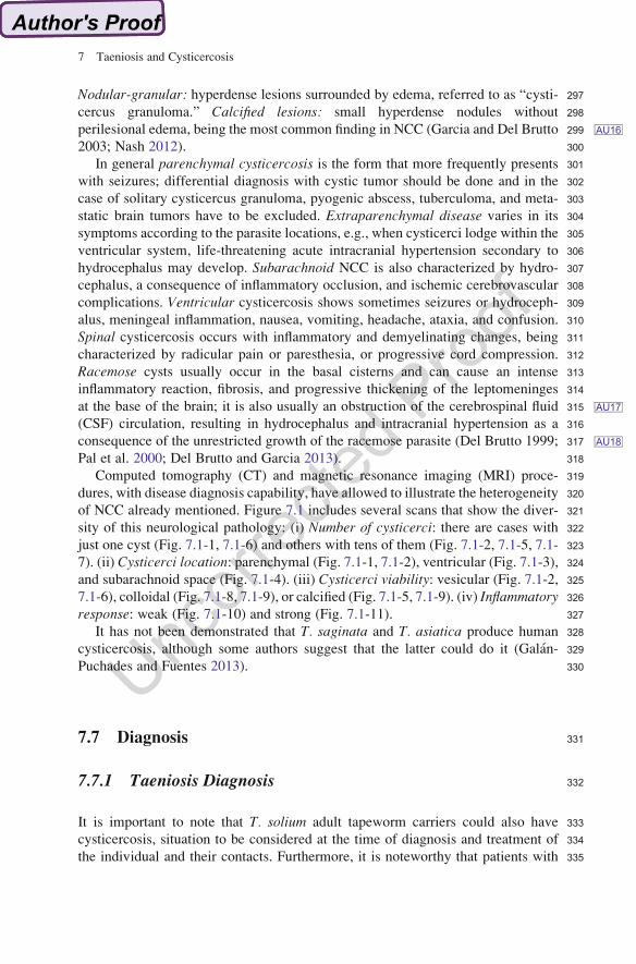
297Nodular-granular: hyperdense lesions surrounded by edema, referred to as “cysti-
298cercus granuloma.” Calcified lesions: small hyperdense nodules without
299perilesional edema, being the most common finding in NCC ( AU16Garcia and Del Brutto
3002003; Nash 2012).
301In general parenchymal cysticercosis is the form that more frequently presents
302with seizures; differential diagnosis with cystic tumor should be done and in the
303case of solitary cysticercus granuloma, pyogenic abscess, tuberculoma, and meta-
304static brain tumors have to be excluded. Extraparenchymal disease varies in its
305symptoms according to the parasite locations, e.g., when cysticerci lodge within the
306ventricular system, life-threatening acute intracranial hypertension secondary to
307hydrocephalus may develop. Subarachnoid NCC is also characterized by hydro-
308cephalus, a consequence of inflammatory occlusion, and ischemic cerebrovascular
309complications. Ventricular cysticercosis shows sometimes seizures or hydroceph-
310alus, meningeal inflammation, nausea, vomiting, headache, ataxia, and confusion.
311Spinal cysticercosis occurs with inflammatory and demyelinating changes, being
312characterized by radicular pain or paresthesia, or progressive cord compression.
313Racemose cysts usually occur in the basal cisterns and can cause an intense
314inflammatory reaction, fibrosis, and progressive thickening of the leptomeninges
315at the base of the brain; it is also usually an obstruction AU17of the cerebrospinal fluid
316(CSF) circulation, resulting in hydrocephalus and intracranial hypertension as a
317consequence of the unrestricted growth of the racemose parasite ( AU18Del Brutto 1999;
318Pal et al. 2000; Del Brutto and Garcia 2013).
319Computed tomography (CT) and magnetic resonance imaging (MRI) proce-
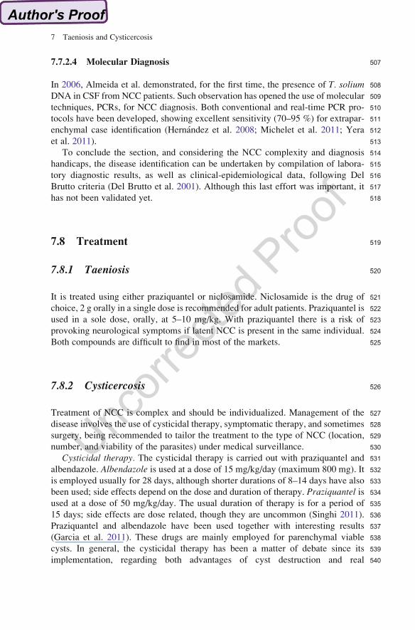
320dures, with disease diagnosis capability, have allowed to illustrate the heterogeneity
321of NCC already mentioned. Figure 7.1 includes several scans that show the diver-
322sity of this neurological pathology: (i) Number of cysticerci: there are cases with
323just one cyst (Fig. 7.1-1, 7.1-6) and others with tens of them (Fig. 7.1-2, 7.1-5, 7.1-
3247). (ii) Cysticerci location: parenchymal (Fig. 7.1-1, 7.1-2), ventricular (Fig. 7.1-3),
325and subarachnoid space (Fig. 7.1-4). (iii) Cysticerci viability: vesicular (Fig. 7.1-2,3267.1-6), colloidal (Fig. 7.1-8, 7.1-9), or calcified (Fig. 7.1-5, 7.1-9). (iv) Inflammatory327response: weak (Fig. 7.1-10) and strong (Fig. 7.1-11).
328It has not been demonstrated that T. saginata and T. asiatica produce human
329cysticercosis, although some authors suggest that the latter could do it (Galan-
330Puchades and Fuentes 2013).
3317.7 Diagnosis
3327.7.1 Taeniosis Diagnosis
333It is important to note that T. solium adult tapeworm carriers could also have
334cysticercosis, situation to be considered at the time of diagnosis and treatment of
335the individual and their contacts. Furthermore, it is noteworthy that patients with
7 Taeniosis and Cysticercosis
336 T. saginata taeniosis are the source of cattle cysticercosis and those with T. solium337 are the origin of both human and porcine cysticercosis (Allan et al. 2003).
338 7.7.1.1 Parasitological Diagnosis
339 It is based on the finding and morphological differentiation of gravid proglottids.
340 Proglottids with less than 15 uterine branches correspond to T. solium, while more
341 than 15 are characteristic of T. saginata. Often proglottids are deformed, their
342 species-specific morphological identification being very difficult (Mayta
343 et al. 2000; Guezala et al. 2009). The microscopic observation of eggs in stools
344 can only indicate taeniosis, but not determine whether the disease is caused by
345 T. solium or T. saginata, since taeniid eggs are morphologically indistinguishable.
346 These methods show low sensitivity (Allan et al. 2003; Guezala et al. 2009).
1 2 3 4
5 6 78
9 10 11 12
Fig. 7.1 Heterogeneity of NCC by MRI and CT scans (Images provided by Dr. Agnes Fleury,
Instituto de Investigaciones Biomedicas, UNAM, Instituto Nacional de Neurologıa y
Neurociencias, SS, Mexico City, Mexico)
E. Ferrer and T. Garate
3477.7.1.2 Immunodiagnosis
348Coproantigen detection was developed by the use of polyclonal antibodies (against
349adult crude extracts) in antigen-capture assays. The assay exhibited good sensitivity
350(100 %–85 %) AU19and specificity to detect taeniosis carriers although they could not
351distinguish between T. solium and T. saginata infections (Allan et al. 1990). It has
352been used to carry out epidemiological studies, determine the prevalence of
353taeniosis, and evaluate the efficacy of drug mass treatment campaigns in endemic
354regions (Bustos et al. 2012).
355Also, rabbit polyclonal antibodies were prepared using E/S or surface antigens of
356adult tapeworms (Machnicka et al. 1996), but they did not allow the species-specific
357taeniid diagnosis. More recently, an ELISA to detect T. solium-specific358coproantigens has been developed through a hybrid system, first antibody against
359T. solium adult crude extract and second antibody anti-E/S adult antigens (Guezala
360et al. 2009).
361There are methods that employ monoclonal antibodies to identify T. solium eggs,
362with excellent sensitivity and specificity (Montenegro et al. 1996), or coproantigens
363(Praet et al. 2013).
364On the other hand, various techniques have been developed for the antibody
365detection in sera of individuals with taeniosis. Of these, EITB assay with T. solium366adult E/S yielded 95 % sensitivity and 100 % specificity (Wilkins et al. 1999). Two
367E/S antigens were cloned, TSES38 and TSES33, expressed in baculovirus system,
368and they showed excellent sensitivities and specificities in EITB (Levine
369et al. 2004, 2007). TSES33, or ES33, was used in a magnetic immunochroma-
370tographic test to identify T. solium taeniosis carriers with very good results (Handali
371et al. 2010). Also, an immunoblot has been developed to specifically diagnose
372T. asiatica-taeniosis (Jeon and Eom 2009). Finally, it is important to note that the
373antibody levels do not decrease after drug treatment (Allan et al. 2003).
3747.7.1.3 Molecular Diagnosis
375Several molecular targets have been cloned, characterized and used for the molec-
376ular diagnosis of human taeniids. Ribosomal, mitochondrial, repetitive DNA
377sequences have been used in the development of different PCR (polymerase378chain reaction) protocols (Harrison et al. 1990; Gottstein et al. 1991; Zarlenga
379et al. 1991; Bowles and McManus 1994). Multiplex PCRs, PCR-RFLP (PCR-380restriction fragment length polymorphism), and PCR sequencing have allowed
381the species-specific identification of T solium, T. saginata, and T. asiatica (Mayta
382et al. 2000; Gonzalez et al. 2000, 2010; Jeon et al. 2009, 2011; Jeon and Eom 2013).
383Also, some molecular markers have permitted the differentiation between
384T. saginata and T. asiatica and the distinction of two genotypes within T. solium,385African-American genotype and Asian genotype (Gasser et al. 1999; Hancock
386et al. 2001; Yamasaki et al. 2004; AU20Ito et al. 2003b; Jeon et al. 2009, 2011; Sato
7 Taeniosis and Cysticercosis
387 et al. 2011; Jeon and Eom 2013). In addition, PCR loop-mediated isothermal388 amplification protocol (LAMP) has been established for the differential diagnosis
389 of human taeniids (Nkouawa et al. 2010), and it is a really simple technique. Some
390 molecular protocols have been used with infected stools, showing excellent sensi-
391 tivity and specificity.
392 7.7.2 Cysticercosis Diagnosis
393 7.7.2.1 Parasitological Diagnosis
394 In NCC, parasitological diagnosis (direct visualization of parasite/lesion) is gener-
395 ally carried out during autopsies (postmortem) and by biopsies (Schantz
396 et al. 1992). In ocular cysticercosis, the ophthalmologic examination is really
397 useful. When cysticerci are located in muscle or subcutaneous tissue, palpation
398 examination, biopsies, and fine needle aspiration cytology are employed, although
399 differential diagnosis from other pathogens should be undertaken (Handa
400 et al. 2008).
401 7.7.2.2 Neuroimaging
402 Computed axial tomography (CT) and magnetic resonance imaging (MRI) are
403 crucial tools for NCC diagnosis, since they allow to know the number, size,
404 evolutionary stage, and location of lesions, as well as the inflammation response
405 (Garcıa and Del Brutto 2003). It is relevant to carry out a differential diagnosis from
406 other neurological disorders and to consider that both neuroimaging cost and
407 technical complexity hamper their use in some endemic areas (Del Brutto
408 et al. 1996). Intramuscular cysticerci can also be identified by ultrasonography
409 and MRI, detecting even a solitary intramuscular cysticercus (Tripathy et al. 2012).
410 Recently, new imaging techniques have improved the detection of scolex and
411 visualization of cysts in the extraparenchymal spaces (Carpio et al. 2013).
412 7.7.2.3 Immunodiagnosis
413 The immunodiagnostic techniques include detection of antibodies, as well as of
414 antigens, in human serum and CSF samples. They are really important for NCC
415 diagnosis (Del Brutto et al. 1996). These techniques are also used for cattle and
416 porcine cysticercosis diagnosis.
417 Regarding antigen detection, Harrison et al. (1989) developed an antigen-
418 capture immunodiagnostic system based on the use of HP10 monoclonal antibody
419 (HP10-Ag-ELISA), specific for a repetitive glyco-residue secreted by T. saginata420 and other taeniid metacestodes. The assay has been used with both serum and CSF
E. Ferrer and T. Garate
421samples, showing the best sensitivity when patients have several alive cysticerci
422and severe NCC (Correa et al. 1989; Garcıa et al. 1998, 2002; Ferrer et al. 2003a;
423Fleury et al. 2007, 2013). Importantly, the Ag-ELISA permits the NCC treatment
424monitoring. Other authors have also prepared monoclonal antibodies (Wang
425et al. 1992; AU21Brandt et al. 1992) and rabbit polyclonal antibodies (Pardini
426et al. 2001; Parija and Rajesh Reddy 2006) for circulating antigen immunodiagno-
427sis. Van Kerckhoven et al. (1998) used B158 monoclonal antibody, an anti-
428T. saginata reagent, in a new Ag-ELISA for cysticercosis detection. The system
429has been used for cattle and porcine diagnosis, seroepidemiological surveys in
430endemic regions, and NCC detection and differentiation between active and inac-
431tive NCC (Dorny et al. 2003; Nguekam et al. 2003; Mwape et al. 2013). Recently,
432these authors have confirmed the utility of urine samples for cysticercosis diagnosis
433(Castillo et al. 2009; Mwape et al. 2011).
434In relation to NCC immunodiagnosis by antibody detection in serum, CSF,
435saliva, and urine samples, many methods and techniques have been used. Probably,
436they are the first choice for a routine microbiology laboratory, although it is worthy
437to consider that antibody detection indicates parasite exposure but not always active
438infection and works better for active NCC diagnosis than for inactive NCC
439(Harrison et al. 1989; Garcıa et al. 2001).
440T. solium crude antigens, complete extract or vesicular fluid, have been used
441during many years ( AU22Diwan et al. 1982;AU23
Larralde et al. 1986). These techniques show
442a poor specificity, mainly due to cross-reactions with related helminth infections
443(Gottstein et al. 1987; AU24Pammenter et al. 1992).
444Also, heterologous antigenic extracts were employed. T. saginata (Harrison and
445Parkhouse 1989; Oliveira et al. 2010), T. crassiceps (Larralde et al. 1990; Espındola446et al. 2002; Suzuki et al. 2007), and T. hydatigena (Hayunga et al. 1991) have been447used for detection of human cysticercosis.
448More recently, purified antigens were introduced as diagnostic tools. Antigen B
449(AgB or paramyosin) and glycoproteins have been studied for NCC immunodiag-
450nosis (Flisser et al. 1980, AU251986; Laclette et al. 1992; Tsang et al. 1989). Based on a
451lentil-lectin chromatography, metacestode glycoproteins were purified and used to
452diagnose NCC in either EITB or ELISA protocols, with serum or CSF samples.
453Although glycoproteins-EITB has 100 % specificity and an overall sensitivity of
45498 %, major problems are that approximately 30 % of patients with a single brain
455parasite, or calcified lesions, may test negative (Wilson et al. 1991) and that the
456T. solium genotypes showed distinct glycoprotein patterns (Sato et al. 2006). Also,
457it is important to note the technique is very expensive, hampering its use in endemic
458areas (Villota et al. 2003; Suzuki and Rossi 2011).The EITB was recognized as the
459gold standard for NCC immunodiagnosis by Pan American Health Organization
460(PAHO) (Greene et al. 2000). Also, purified T. solium E/S antigens have been
461employed in ELISA and FAST-ELISA with very promising results in antibody
462detection of NCC (Ng and Ko 1994; Sahu et al. 2009; Atluri et al. 2009). From the
463metacestode secretion proteins, those that have low molecular weight, and espe-
464cially glycoproteins, have showed the best performances in NCC diagnosis assays.
465Thus, 14 and 18 kDa antigens (Espındola et al. 2002; Molinari et al. 2002; Sahu
7 Taeniosis and Cysticercosis
466 et al. 2009) and 8–30 kDa protein fraction (Gottstein et al. 1987; Yang et al. 1998;
467 Park et al. 2000; Atluri et al. 2009; Jeon and Eom 2009) have been described as the
468 best candidates to prepare an antibody-detection system for NCC detection.
469 Although these systems have worked properly, some difficulties (biochemical
470 purification, big parasite amounts, reproducibility) restrict their uses.
471 Recombinant antigens: Biotechnological approaches have been used to solve the472 scarcity of T. solium parasitic material for the preparation and purification of
473 diagnostic antigen candidates. The cloning and expression of T. soliummetacestode
474 genes relevant for diagnosis have allowed circumventing the limitations mentioned.
475 Many genes have been studied during the last decades. Paramyosin, sHSP AU26,
476 TSA18, F18, 50 kDa glycoprotein, TsAg5, and other molecules were cloned and
477 expressed in prokaryotic and eukaryotic systems and evaluated with collections of
478 serum and CSF samples. The recombinant products have been checked in ELISA
479 and Western blot, with good sensitivity and specificity for NCC diagnosis
480 (Vazquez-Talavera et al. 2001; Ferrer et al. 2003b, 2005a, 2007a; AU27Montero
481 et al. 2003; AU28Hancock et al. 2004). Even though most of them worked better with
482 active NCC samples, TSA18 expressed in baculovirus system showed the best
483 sensitivity (60 %) for inactive NCC immunodetection. Also, some recombinant
484 antigens have been used for animal cysticercosis identification.
485 Regarding recombinant products, one of the most promising NCC diagnostic
486 antigens is the 8 kDa family. Their members are metacestode excretory/secretory
487 glycoproteins (65–90 amino acid residues and 7–12 kDa), which invoke strong
488 specific antibody reactions in the infected individuals, and appear to be expressed as
489 variant arrays, with both sequence heterogeneity and homology in clusters of small
490 domains that determine epitope differences among them (Ferrer et al. 2012). The
491 10 kDa molecule (cysticercosis diagnosis antigen, CyDA) (Chung et al. 1999);
492 NC-3 (8 kDa)/NC-9 (13 kDa) antigens (Hubert et al. 1999); the glycoproteins TS14
493 (14 kDa) and TS18 (18 kDa) (Greene et al. 2000); Ag1, Ag1V1, Ag2, Ag2V1, and
494 chimeric Ag1V1-Ag2 molecules (Sako et al. 2000; Sato et al. 2011); and Ts8B1,
495 Ts8B2, and Ts8B3 (Ferrer et al. 2007b) recombinant antigens showed excellent
496 sensitivity and specificity in NCC diagnosis AU29.
497 Synthetic peptides: Synthetic peptides, derived from the cloned molecules, have
498 been prepared as tools to be used in cysticercosis diagnosis. They have been
499 employed in ELISA and Western blot, with good results in some cases, although
500 the diagnostic properties of recombinant antigens were not improved.
501 Based on the Taenia 8 kDa family, sTS14 and sTS18 peptides, corresponding to
502 TS14 and TS18 antigens, have been synthesized, showing excellent specificity but
503 poor sensitivity (Greene et al. 2000). Also, peptides derived from T. saginata504 oncosphere molecules have been used for cysticercosis diagnosis and human and
505 animal disease, with similar results to the ones reported above (Fleury et al. 2003;
506 Ferrer et al. 2005b).
E. Ferrer and T. Garate
5077.7.2.4 Molecular Diagnosis
508In 2006, Almeida et al. demonstrated, for the first time, the presence of T. solium509DNA in CSF from NCC patients. Such observation has opened the use of molecular
510techniques, PCRs, for NCC diagnosis. Both conventional and real-time PCR pro-
511tocols have been developed, showing excellent sensitivity (70–95 %) for extrapar-
512enchymal case identification (Hernandez et al. 2008; Michelet et al. 2011; Yera
513et al. 2011).
514To conclude the section, and considering the NCC complexity and diagnosis
515handicaps, the disease identification can be undertaken by compilation of labora-
516tory diagnostic results, as well as clinical-epidemiological data, following Del
517Brutto criteria (Del Brutto et al. 2001). Although this last effort was important, it
518has not been validated yet.
5197.8 Treatment
5207.8.1 Taeniosis
521It is treated using either praziquantel or niclosamide. Niclosamide is the drug of
522choice, 2 g orally in a single dose is recommended for adult patients. Praziquantel is
523used in a sole dose, orally, at 5–10 mg/kg. With praziquantel there is a risk of
524provoking neurological symptoms if latent NCC is present in the same individual.
525Both compounds are difficult to find in most of the markets.
5267.8.2 Cysticercosis
527Treatment of NCC is complex and should be individualized. Management of the
528disease involves the use of cysticidal therapy, symptomatic therapy, and sometimes
529surgery, being recommended to tailor the treatment to the type of NCC (location,
530number, and viability of the parasites) under medical surveillance.
531Cysticidal therapy. The cysticidal therapy is carried out with praziquantel and
532albendazole. Albendazole is used at a dose of 15 mg/kg/day (maximum 800 mg). It
533is employed usually for 28 days, although shorter durations of 8–14 days have also
534been used; side effects depend on the dose and duration of therapy. Praziquantel is535used at a dose of 50 mg/kg/day. The usual duration of therapy is for a period of
53615 days; side effects are dose related, though they are uncommon (Singhi 2011).
537Praziquantel and albendazole have been used together with interesting results
538(Garcia et al. 2011). These drugs are mainly employed for parenchymal viable
539cysts. In general, the cysticidal therapy has been a matter of debate since its
540implementation, regarding both advantages of cyst destruction and real
7 Taeniosis and Cysticercosis
541 improvement of the clinical outcome. Most publications have reported “reduction
542 of the number of lesions” to measure anthelmintic drug effectiveness, which is
543 misleading; possibly, the evaluation of cyst disappearance could be a more appro-
544 priate approximation.
545 Summarizing, based on double-blind, placebo-controlled trials, and comparing
546 the effect of albendazole and praziquantel, it is generally accepted, with few
547 discrepancies, that both drugs are effective in destroying viable cysts, while their
548 use in cases with enhancing lesions has been debated as these lesions are considered
549 to represent degenerating cysts, many of which resolve spontaneously (Carpio
550 et al. 2008; Thussu et al. 2008; Chaurasia et al. 2010). In relation to the role of
551 cysticidal therapy in control seizures secondary to NCC, also there are some
552 controversial results; some authors found an improved seizure control in adults
553 with vesicular lesions, as well as enhancing lesions, with the use of cysticidal drugs
554 (Garcia et al. 2004; Del Brutto et al. 2006), while others did not find any significant
555 improvement (Carpio et al. 2008; Abba et al. 2010). In conclusion, cysticidal
556 therapy seems to be effective in reducing the number of lesions, but its role in
557 improving long-term seizure control needs further larger studies.
558 Cysticidal drugs have also been found to be effective in the treatment of some
559 extraparenchymal NCC cases and even for giant cysts (Proano et al. 2001) although
560 these NCC types need to be managed with exceptional caution. However, cysticidal
561 therapy should not be used in cases with markedly elevated intracranial pressure
562 and in ophthalmic (intraocular) NCC or massive infections; steroids alone are used
563 in these situations. Also, cysticidal therapy is of no use for calcified lesion(s). To
564 sum up, there is no universally agreed single protocol for the treatment of NCC, and
565 consensus guidelines recommend an individualized approach (Nash et al. 2006).
566 Symptomatic therapy. Seizures usually respond very well to first-line
567 antiepileptic drugs (AEDs). The recurrence rate after AED withdrawal is low in
568 NCC cases with single lesion. Recurrence of seizures after AED withdrawal is
569 correlated with the presence of multiple lesions prior to starting cysticidal therapy
570 and persistence or calcification of lesions after therapy (Talukdar et al. 2002; Goel
571 et al. 2010).
572 Corticosteroids. Oral corticosteroids are administrated generally a couple of
573 days before and a few days along with anticysticercal therapy so as to prevent
574 any adverse reactions that may occur due to the host inflammatory response.
575 Usually oral prednisolone, 1–2 mg/kg, is used; intravenous dexamethasone may
576 be used if there are features of raised intracranial pressure. In cases with dissem-
577 inated lesions and extensive cerebral edema, steroids may be required for a
578 prolonged period (Singhi 2011).
579 Surgery. Surgical intervention is required in some cases, particularly in intra-
580 ventricular and subarachnoid NCC. A ventriculoperitoneal shunt is needed for
581 hydrocephalus; simultaneous use of steroids and albendazole and recurrent courses
582 of steroids reduce the risk of frequent obstructions. Endoscopic removal of cysts is
583 the least invasive and is therefore the procedure of choice. Excision of giant cysts
584 that fail to respond to medical therapy may be required (Goel et al. 2008; Suri
585 et al. 2008; Singhi 2011).
E. Ferrer and T. Garate
5867.9 Prognosis
587Striated muscle and subcutaneous cysticercosis have good prognosis. Ocular cys-
588ticercosis could end in blindness if the eye parasite is not diagnosed and cysticidal
589treatment is used for a concomitant NCC (Sundar et al. 2010). NCC cases with
590single lesions generally have a good prognosis, seizures are usually well controlled,
591and lesions disappear within 6 months in over 60 % of cases. Patients with multiple
592lesions and those with calcifications often have frequent seizure recurrences.
593Cysticercus encephalitis and extraparenchymal NCC have a cautious prognosis
594(Singhi 2011).
5957.10 Prevention and Control
596Cysticercosis is an NTD that occurs in communities with low socioeconomic
597conditions and poor sanitation-hygienic practices. Globally, it can be prevented
598through improvements in health and education standards, treatment of T. solium599carriers, improved pig-rearing management, as well as by treatment of infected
600animals (Flisser et al. 2003, 2004; Engels et al. 2003; Sarti and Rajshekhar 2003;
601Gonzalez et al. 2003; Xiao et al. 2013). More importantly, it is possible to consider
602its eradication taking into account that human tapeworm carrier is the unique
603definitive host and the sole source of infection for intermediate hosts, domestic
604animals are the main intermediate hosts, wild reservoirs are not important, and
605intervention tools for control are available (Gilman et al. 2012). Thus, potential
606intervention measures should include the following criteria:
607Educational programs. Educational designs for endemic rural areas, taking into
608account culture and idiosyncrasy of the population. The designs will include basic
609and proper hygiene and sanitation measures, teaching the parasite biology and
610epidemiology, and apprising of taeniosis/cysticercosis symptoms and the ways to
611interrupt transmission, among other information.
612Drug treatment of taeniosis carriers. To decrease the source of infection, finding613and treating tapeworm-infected individuals would be the intervention of choice.
614Once a Taenia carrier is identified, careful treatment and follow-up can ensure the
615cure of the patient and thus close the transmission. So far, mass treatment programs
616of the population to eliminate tapeworms with the use of niclosamide or with
617praziquantel (Sarti and Rajshekhar 2003) have been successful and have temporar-
618ily reduced the disease transmission, but the effect has not been sustainable.
619Pig management. Slaughterhouse control is suggested as a key control compo-
620nent, but it is relevant to avoid the development and establishment of illegal
621markets for infected pork. Also, pig corralling is really important, although this
622option is opposed to the main reason of raising pigs in endemic regions as they roam
623free and do not need to be fed by their owners. Pig treatment with oxfendazole is
624another alternative (Gonzalez et al. 2012).
7 Taeniosis and Cysticercosis
625 Pig vaccination. Vaccination has been proposed as a possibility to control the
626 transmission of cysticercosis. Recently, the advances made in the vaccine area are
627 really promising to be applied to the control of cysticercosis. In fact, several
628 intervention programs have already included the use of vaccines to interrupt the
629 parasite transmission, among other measures (Assana et al. 2013; Xiao et al. 2013).
630 There are vaccines based on the use of crude extracts, recombinant antigens,
631 peptides, and naked DNA.
632 Pioneer vaccination studies in cattle were carried out with both crude and E/S
633 antigens from T. saginata and T. hydatigena (Rickard et al. 1981). AU30Since then,
634 different parasite extracts prepared from T. solium oncospheres, as well as
635 metacestodes, have been used in porcine vaccination trials, and different protection
636 levels have been reported (Molinari et al. 1993a, 1997; Pathak and Gaur 1990;
637 Verastegui et al. 2002). Also, this kind of assays has also been developed in the
638 mouse/T. crassiceps model (Valdez et al. 1994; Sciutto et al. 1995), obtaining
639 similar protection levels with both T. solium and T. crassiceps antigenic extracts.640 Also recombinant antigens have been used as vaccines. For T. saginata/cattle641 system, most of these molecules have been derived from both surface and secreted
642 components of the infective oncosphere (Benıtez et al. 1996; Lightowlers
643 et al. 1996; Bonay et al. 2002; Harrison et al. 2005). Antigens related to the taeniid
644 45 W protective gene family (Johnson et al. 1989; Lightowlers et al.1996; Gauci
645 and Lightowlers 2003; Gonzalez et al. 2005) and T. saginata TSA9 and TSA18
646 (syn. HP6) recombinant proteins were used in cattle immunization assays, yielding
647 the TSA18 candidate excellent results (Benıtez et al. 1996; Lightowlers et al. 1996).
648 It is interesting to note that all these purified recombinant proteins were demon-
649 strated to function as adhesion molecules, a property which is probably pertinent to
650 their potential as vaccines, as is the case for the HP6 molecule of T. saginata651 (Harrison and Parkhouse 1989; Benıtez et al. 1996; Bonay et al. 2002; Harrison
652 et al. 2005). AU31In T. solium/porcine system, the genes homologous to the ones
653 described above (Tsol18, Tsol45-1A, TSOL45-1B, TSOL16), and others
654 (paramyosin, fatty acid-binding proteins (FABPs), KETc1, KETc4, KETc7,
655 KETc11, KETc12), have been used in vaccination assays (Manoutcharian
656 et al. 1996; Vazquez-Talavera et al. 2001; Gauci et al. 2012). AU32Of all molecules,
657 Tsol18 plasmid construction, expressed in Escherichia coli and purified, is the
658 candidate that yielded best results, almost 100 % protection, and is being used as
659 a vaccine in the control programs organized in different endemic regions (Flisser
660 et al. 2004; Gauci et al. 2012; Assana et al. 2013).
661 Regarding peptides as vaccination tools, the most used have been KETc1,
662 KETc12, KETc7, GK1, GK2, and GK3 (Manoutcharian et al. 2004; Toledo
663 et al. 2001). Later, the combination GK1, KETc1, and KETc12, called S3Pvac,
664 has been extensively employed in vaccination assays in T. solium/porcine,665 T. crassiceps/mouse, and T. pisiformis/rabbit models. In pigs, the S3Pvac produced
666 till 98.7 % protection and showed therapeutic properties (de Aluja et al. 2005;
667 AU33Rassy et al. 2010; Sciutto et al. 2013a, 2013b). Finally, naked DNA vaccination has
668 also been used in experimental studies; T. saginata and T. solium cDNAs already
669 mentioned (KETc7, paramyosin, Tso 18, others) have been employed with
E. Ferrer and T. Garate
670promising protection (Manoutcharian et al. 1998; Cruz-Revilla et al. 2000; Rosas
671et al. 2002).
672 AU34References
673Abba K, Ramaratnam S, Ranganathan LN (2010) Anthelmintics for people with neurocysti-
674cercosis. Cochrane Database Syst Rev 17(3), CD000215. doi:10.1002.1465.8.8
675Allan JC, Avila G, Garcıa-Noval J et al (1990) Immunodiagnosis of taeniasis by coproantigen
676detection. Parasitology 101:473–477
677Allan JC,Wilkins P, Tsang V et al (2003) Immunodiagnostic tools for taeniasis. Acta Trop 87:87–93
678Allepuz A, Gabriel S, Dorny P et al (2012) Comparison of bovine cysticercosis prevalence
679detected by antigen ELISA and visual inspection in the North East of Spain. Res Vet Sci
68092:393–395
681Almeida CR, Ojopi EP, Nunes CM et al (2006) Taenia solium DNA is present in the cerebrospinal
682fluid of neurocysticercosis patients and can be used for diagnosis. Eur Arch Psychiatry Clin
683Neurosci 256:307–310
684Alvarez JI, Colegial CH, Castano CA et al (2002) The human nervous tissue in proximity to
685granulomatous lesions induced by Taenia solium metacestodes displays an active response. J
686Neuroimmunol 127:139–144
687Arechavaleta F, Molinari JL, Tato P (1998) A Taenia solium metacestode factor nonspecifically
688inhibits cytokine production. Parasitol Res 84:117–122
689Assana E, Lightowlers MW, Zoli AP et al (2013) Taenia solium taeniosis/cysticercosis in Africa:
690risk factors, epidemiology and prospects for control using vaccination. Vet Parasitol 195:14–23
691Atluri SR, Singhi P, Khandelwal N (2009) Neurocysticercosis immunodiagnosis using Taenia692solium cysticerci crude soluble extract, excretory secretory and lower molecular mass antigens
693in serum and urine samples of Indian children. Acta Trop 110:22–27
694Bhattarai R, Budke CM, Carabin H (2012) Estimating the non-monetary burden of neurocysti-
695cercosis in Mexico. PLoS Negl Trop Dis 6:e1521
696Benıtez L, Garate T, Harrison L et al (1996) Cloning and sequencing of the gene encoding the
697principal 18-kDa secreted antigen of activated oncospheres of Taenia saginata. Mol Biochem
698Parasitol 78:265–268
699Bonay P, Gonzalez LM, Benıtez L et al (2002) Genomic and functional characterisation of a
700secreted antigen of Taenia saginata oncospheres. Mol Biochem Parasitol 121:269–273
701Bowles JR, McManus DP (1994) Genetic characterization of the Asian Taenia, a newly described
702taeniid cestode of humans. Am J Trop Med Hyg 50:33–44
703Brandt JR, Geerts S, De Deken R et al (1992) A monoclonal antibody-based ELISA for the
704detection of circulating excretory-secretory antigens in Taenia saginata cysticercosis. Int J
705Parasitol 22:471–477
706Bruno E, Bartoloni A, Zammarchi L et al (2013) Epilepsy and neurocysticercosis in Latin
707America: a systematic review and meta-analysis. PLoS Negl Trop Dis 31:e2480
708Bustos JA, Rodriguez S, Jimenez JA (2012) Cysticercosis Working Group in Peru. Detection of
709Taenia solium taeniasis coproantigen is an early indicator of treatment failure for taeniasis.
710Clin Vaccine Immunol 19:570–573
711Carpio A, Escobar A, Hauser W (1998) Cysticercosis and epilepsy: a critical review. Epilepsia
71239:1025–1040
713Carpio A (2002) Neurocysticercosis: an update. Lancet Infect Dis 2:751–762
714Carpio A, Kelvin EA, Bagiella E et al (2008) Effects of albendazole treatment on neurocysti-
715cercosis: a randomised controlled trial. J Neurol Neurosurg Psychiatry 79:1050–1055
716Carpio A, Fleury A, Hauser WA (2013) Neurocysticercosis: five new things. Neurol Clin Pract
7173:118–125
7 Taeniosis and Cysticercosis
718 Carrique-Mas J, Iihoshi N, Widdowson MA et al (2001) An epidemiological study of Taenia719 solium cysticercosis in a rural population in the Bolivian Chaco. Acta Trop 80:229–235
720 Castillo Y, Rodriguez S, Garcıa HH et al (2009) Urine antigen detection for the diagnosis of
721 human neurocysticercosis. Am J Trop Med Hyg 80:379–383
722 Chaurasia RN, Garg RK, Agarwall A et al (2010) Three day albendazole therapy in patients with a
723 solitary cysticercus granuloma: a randomized double blind placebo controlled study. Southeast
724 Asian J Trop Med Public Health 41:517–525
725 Chavarria A, Fleury A, Bobes RJ et al (2006) A depressed peripheral cellular immune response is
726 related to symptomatic neurocysticercosis. Microbes Infect 8:1082–1089
727 Chung JY, Bahk YY, Huh S et al (1999) A recombinant 10 kDa protein of Taenia solium728 metacestodes specific to active neurocysticercosis. J Infect Dis 180:1307–1315
729 Correa D, Medina E (1999) Host-parasite immune relationship in Taenia solium taeniosis and
730 cysticercosis. In: Garcıa HH, Martinez M (eds) Taenia solium taeniasis/cysticercosis. Editorial
731 Universo, Peru, pp 83–96
732 Correa D, Sandoval MA, Harrison LJ et al (1989) Human neurocysticercosis: comparison of
733 enzyme immunoassay capture techniques based on monoclonal and polyclonal antibodies for
734 the detection of parasite products in cerebrospinal fluid. Trans R Soc Trop Med Hyg 83:814–
735 816
736 Cortez MM, Boggio G, Guerra M et al (2010) Evidence that active transmission of porcine
737 cysticercosis occurs in Venezuela. Trop Anim Health Prod 42:531–537
738 Cruz-Revilla C, Rosas G, Fragoso G et al (2000) Taenia crassiceps cysticercosis: protective effect739 and immune response elicited by DNA immunization. J Parasitol 86:67–74
740 de Aluja A, Villalobos N, Nava G et al (2005) Therapeutic capacity of the synthetic peptide-based
741 vaccine against Taenia solium cysticercosis in pigs. Vaccine 23:4062–4069
742 Del Brutto OH, Wadia NH, Dumas M et al (1996) Proposal of diagnostic criteria for human
743 neurocysticercosis. J Neurol Sci 42:1–6
744 Del Brutto OH, Rajshekhar V, White AC Jr et al (2001) Proposed diagnostic criteria for neurocys-
745 ticercosis. Neurology 57:177–183
746 Del Brutto OH, Roos KL, Coffey CS et al (2006) Meta-analysis: cysticidal drugs for neurocysti-
747 cercosis: albendazole and praziquantel. Ann Intern Med 145:43–51
748 Del Brutto OH, Garcia HH (2013) Neurocysticercosis. Handb Clin Neurol 114:313–325
749 Diehl Rodriquez R, Crestani DN, Dworzecki Soares JO et al (2012) L. Bruns’syndrome and
750 racemose neurocysticercosis: a case report. Rev Soc Bras Med Trop 45:269–271
751 Dorny P, Brandt J, Zoli A et al (2003) Immunodiagnostic tools for human and porcine cysticer-
752 cosis. Acta Trop 87:79–86
753 EomKS, RimHJ (1993) Morphologic descriptions of Taenia asiatica sp. Korean J Parasitol 31:1–6754 Eom KS, Jeon HK, Rim HJ (2009) Geographical distribution of Taenia asiatica and related
755 species. Korean J Parasitol 47(Suppl):S115–124
756 Engels D, Urbani C, Belotto A et al (2003) The control of human (neuro)cysticercosis: which way
757 forward? Acta Trop 87:177–182
758 Espındola NM, Vaz AJ, Pardini AX et al (2002) Excretory/secretory antigens (ES) from in vitro759 cultures of Taenia crassiceps cysticerci, and use of an anti-ES monoclonal antibody for antigen
760 detection in samples of cerebrospinal fluid from patients with neurocysticercosis. Ann Trop
761 Med Parasitol 96:361–368
762 Fabiani S, Bruschi F (2013) Neurocysticercosis in Europe: still a public health concern not only for
763 imported cases. Acta Trop 128:18–26
764 Ferrer E, Cabrera Z, Rojas G et al (2003a) Evidence for high seroprevalence of Taenia solium765 cysticercosis in individuals from three rural communities in Venezuela. Trans R Soc Trop Med
766 Hyg 97:522–526
767 Ferrer E, Moyano E, Benıtez L et al (2003b) Cloning and characterization of Taenia saginata768 paramyosin cDNA. Parasitol Res 91:60–67
E. Ferrer and T. Garate
769Ferrer E, Gonzalez LM, Foster-Cuevas M et al (2005a) Taenia solium: characterization of a small
770heat shock protein (Tsol-sHSP35.6) and its possible relevance to the diagnosis and pathogen-
771esis of neurocysticercosis. Exp Parasitol 110:1–11
772Ferrer E, Cortez MM, Cabrera Z et al (2005b) Oncospheral peptide-based ELISAs as potential
773seroepidemiological tools for Taenia solium cysticercosis/neurocysticercosis in Venezuela.
774Trans R Soc Trop Med Hyg 99:568–576
775Ferrer E, Gonzalez LM, Martınez-Escribano JA et al (2007a) Evaluation of recombinant
776HP6-Tsag, an 18 kDa Taenia saginata oncospheral adhesion protein, for the diagnosis of
777cysticercosis. Parasitol Res 101:517–525
778Ferrer E, Bonay P, Foster M et al (2007b) Molecular cloning and characterisation of Ts8B1, Ts8B2
779and Ts8B3, tree new members of the Taenia solium 8 kDa diagnostic antigen family. Mol
780Biochem Parasitol 152:90–100
781Ferrer E, Sanchez J, Milano A et al (2012) Diagnostic epitope variability within Taenia solium7828 kDa antigen family: implications for cysticercosis immunodetection. Exp Parasitol 130:78–85
783Fleury A, Beltran C, Ferrer E et al (2003) Application of synthetic peptides to the diagnosis of
784neurocysticercosis. Trop Med Int Health 8:1124–1130
785Fleury A, Hernandez M, Avila M et al (2007) Detection of HP10 antigen in serum for diagnosis
786and follow-up of subarachnoidal and intraventricular human neurocysticercosis. J Neurol
787Neurosurg Psychiatry 78:970–974
788Fleury A, Moreno Garcıa J, Valdez Aguerrebere P et al (2010) Neurocysticercosis, a persisting
789health problem in Mexico. PLoS Negl Trop Dis 4:e805
790Fleury A, Garcia E, Hernandez M, Carrillo R, Govezensky T, Fragoso G, Sciutto E, Harrison LJ,
791Parkhouse RM (2013) Neurocysticercosis: HP10 antigen detection is useful for the follow-up
792of the severe patients. PLoS Negl Trop Dis 7:e2096
793Flisser A, Perez-Montfort R, Larralde C (1979) The immunology of human and animal cysticer-
794cosis: a review. Bull World Health Organ 57:839–856
795Flisser A, Woodhouse E, Larralde C (1980) Human cysticercosis: antigens, antibodies and
796non-responders. Clin Exp Immunol 39:27–37
797Flisser A, Sarti E, Lightowlers M et al (2003) Neurocysticercosis: regional status, epidemiology,
798impact and control measures in the Americas. Acta Trop 87:43–51
799Flisser A, Gauci CG, Zoli A et al (2004) Induction of protection against porcine cysticercosis by
800vaccination with recombinant oncosphere antigens. Infect Immun 72:5292–5297
801Flisser A, Avila G, Maravilla P et al (2010) Taenia solium: current understanding of laboratory
802animal models of taeniosis. Parasitology 137:347–357
803Galan-Puchades MT, Fuentes MV (2013) Taenia asiatica: the most neglected human Taenia and
804the possibility of cysticercosis. Korean J Parasitol 51:51–54
805Garcıa HH, Harrison LJ, Parkhouse RM et al (1998) A specific antigen-detection ELISA for the
806diagnosis of human neurocysticercosis. The cysticercosis working group in Peru. Trans R Soc
807Trop Med Hyg 92:411–414
808Garcıa HH, Gonzalez AE, Gilman RH et al (2001) Short report: transient antibody response in
809Taenia solium infection in field conditions-a major contributor to high seroprevalence. Am J
810Trop Med Hyg 65:31–32
811Garcıa HH, Gonzalez AE, Gilman RH et al (2002) Circulating parasite antigen in patients with
812hydrocephalus secondary to neurocysticercosis. Am J Trop Med Hyg 66:427–430
813Garcıa HH, Gilman RH, Gonzalez AE et al (2003) Hyperendemic human and porcine Taenia814solium infection in Peru. Am J Trop Med Hyg 68:268–275
815Garcıa H, Del Brutto O (2003) Imaging findings in neurocysticercosis. Acta Trop 87:71–78
816Garcıa-Noval J, Allan JC, Fletes C et al (1996) Epidemiology of Taenia solium taeniasis and
817cysticercosis in two rural Guatemalan communities. Am J Trop Med Hyg 55:282–289
818Garcia HH, Pretell EJ, Gilman RH, Martinez SM (2004) A trial of antiparasitic treatment to reduce
819the rate of seizures due to cerebral cysticercosis. N Engl J Med 350:249–258
820Garcia HH, Gonzalez AE, Gilman RH (2011) Cysticercosis of the central nervous system: how
821should it be managed? Curr Opin Infect Dis 24:423–427
7 Taeniosis and Cysticercosis
822 Gasser RB, Zhu X, Woods W (1999) Genotyping Taenia tapeworms by single-strand conforma-
823 tion polymorphism of mitochondrial DNA. Electrophoresis 20:2834–2837
824 Gauci C, Lightowlers MW (2003) Molecular cloning of genes encoding oncosphere proteins
825 reveals conservation of modular protein structure in cestode antigens. Mol Biochem Parasitol
826 127:193–198
827 Gauci CG, Jayashi CM, Gonzalez AE et al (2012) Protection of pigs against Taenia solium828 cysticercosis by immunization with novel recombinant antigens. Vaccine 30:3824–3828
829 Gilman RH, Gonzalez AE, Llanos-Zavalaga F et al (2012) Prevention and control of Taenia830 solium taeniasis/cysticercosis in Peru. Pathog Glob Health 106:312–318
831 Goel RK, Ahmad FU, Vellimana AK et al (2008) Endoscopic management of intraventricular
832 neurocysticercosis. J Clin Neurosci 15:1096–1101
833 Goel D, Mittal M, Bansal KK et al (2010) Natural history of solitary cerebral cysticercosis cases
834 after albendazole therapy: a longitudinal follow-up study from India. Acta Neurol Scand
835 121:204–208
836 Gonzalez A, Garcıa HH, Gilman R et al (2003) Control of Taenia solium. Acta Trop 87:103–109
837 Gonzalez A, Gauci C, Barber D et al (2005) Vaccination of pigs to control human neurocysti-
838 cercosis. Am J Trop Med Hyg 72:837–839
839 Gonzalez AE, Bustos JA, Jimenez JA et al (2012) Efficacy of diverse antiparasitic treatments for
840 cysticercosis in the pig model. Am J Trop Med Hyg 87:292–296
841 Gonzalez LM, Montero E, Harrison LJS et al (2000) Differential diagnosis of Taenia saginata and842 Taenia solium infection by PCR. J Clin Microbiol 38:737–744
843 Gonzalez LM, Bailo B, Ferrer E et al (2010) Characterization of the Taenia spp HDP2 sequence
844 and development of a novel PCR-based assay for discrimination of Taenia saginata from
845 Taenia asiatica. Parasit Vectors 3:51846 Gottstein B, Zini D, Schantz PM (1987) Species-specific immunodiagnosis of Taenia solium847 cysticercosis by ELISA and immunoblotting. Trop Med Parasitol 38:299–303
848 Gottstein B, Deplazes P, Tanner I et al (1991) Diagnostic identification of Taenia saginatawith the849 polymerase chain reaction. Trans R Soc Trop Med Hyg 85:248–249
850 Greene RM, Hancock K, Wilkins PP et al (2000) Taenia solium: molecular cloning and serologic
851 evaluation of 14 and 18 kDa related, diagnostic antigens. J Parasitol 86:1001–1007
852 Guezala MC, Rodriguez S, Zamora H et al (2009) Development of a species-specific coproantigen
853 ELISA for human Taenia solium taeniasis. Am J Trop Med Hyg 81:433–437
854 Hancock K, Broughel DE, Moura IN et al (2001) Sequence variation in the cytochrome oxidase I,
855 internal transcribed spacer 1, and Ts14 diagnostic antigen sequences of Taenia solium isolates
856 from South and Central America, India, and Asia. Int J Parasitol 31:1601–1617
857 Hancock K, Pattabhi S, Greene RM et al (2004) Characterization and cloning of GP50, a Taenia858 solium antigen diagnostic for cysticercosis. Mol Biochem Parasitol 133:115–124
859 Handa U, Garg S, Mohan H (2008) Fine needle aspiration in the diagnosis of subcutaneous
860 cysticercosis. Diagn Cytopathol 36:183–187
861 Handali S, Klarman M, Gaspard AN et al (2010) Development and evaluation of a magnetic
862 immunochromatographic test to detect Taenia solium, which causes taeniasis and neurocysti-
863 cercosis in humans. Clin Vaccine Immunol 17:631–637
864 Harrison LJS, Parkhouse RME (1989) Taenia saginata and Taenia solium: reciprocal models.
865 Acta Leiden 57:143–152
866 Harrison LJS, Joshua GW, Wright SH et al (1989) Specific detection of circulating surface/
867 secreted glycoproteins of viable cysticerci in Taenia saginata cysticercosis. Parasite Immunol
868 11:351–370
869 Harrison LJS, Delgado J, Parkhouse RME (1990) Differential diagnosis of Taenia saginata and
870 Taenia solium with DNA probes. Parasitology 100:459–461
871 Harrison L, Garate T, Bryce D et al (2005) Ag-ELISA and PCR for monitoring the vaccination of
872 cattle against Taenia saginata cysticercosis using an oncospheral adhesion protein (HP6) with
873 surface and secreted localization. Trop Anim Health Prod 37:103–120
E. Ferrer and T. Garate
874Hayunga EG, Sumner MP, Rhoads ML et al (1991) Development of a serologic assay for
875cysticercosis, using an antigen isolated from Taenia spp. cyst fluid. Am J Vet Res 52:462–470
876Hernandez M, Gonzalez LM, Fleury A et al (2008) Neurocysticercosis: detection of Taenia solium877DNA in human cerebrospinal fluid using a semi-nested PCR based on HDP2. Ann Trop Med
878Parasitol 102:317–323
879Hubert K, Andriantsimahavandy A, Michault A et al (1999) Serological diagnosis of human
880cysticercosis by use of recombinant antigens from Taenia solium cysticerci. Clin Diagn Lab
881Immunol 6:479–482
882Ikejima T, Piao ZX, Sako Y et al (2005) Evaluation of clinical and serological data from Taenia883solium cysticercosis patients in eastern Inner Mongolia Autonomous Region, China. Trans R
884Soc Trop Med Hyg 99:625–630
885Ishida MM, Almeida MS, Espındola NM et al (2011) Seroepidemiological study of human
886cysticercosis with blood samples collected on filter paper, in Lages, State of Santa Catarina,
887Brazil, 2004–2005. Rev Soc Bras Med Trop 44:339–343
888Ito A, Yamasaki H, Nakao M et al (2003) Multiple genotypes of Taenia solium-ramifications for
889diagnosis, treatment and control. Acta Trop 87:95–101
890Jeon HK, Eom KS (2009) Immunoblot patterns of Taenia asiatica taeniasis. Korean J Parasitol
89147:73–77
892Jeon HK, Chai JY, Kong Y et al (2009) Differential diagnosis of Taenia asiatica using multiplex
893PCR. Exp Parasitol 121:151–156
894Jeon HK, Yong TS, Sohn WM et al (2011) Molecular identification of Taenia tapeworms by Cox1
895gene in Koh Kong, Cambodia. Korean J Parasitol 49:195–197
896Jeon HK, Eom KS (2013) Molecular approaches to Taenia asiatica. Korean J Parasitol 51:1–8
897Johnson KS, Harrison GB, Lightowlers MW et al (1989) Vaccination against ovine cysticercosis
898using a defined recombinant antigen. Nature 338:585–587
899Kelvin EA, Carpio A, Bagiella E et al (2009) The association of host age and gender with
900inflammation around neurocysticercosis cysts. Ann Trop Med Parasitol 103:487–499
901Laclette JP, Shoemaker CB, Richter D et al (1992) Paramyosin inhibits complement C1. J
902Immunol 148:124–128
903Larralde C, Sotelo J, Montoya RM et al (1990) Immunodiagnosis of human cysticercosis in
904cerebrospinal fluid. Antigens from murine Taenia crassiceps cysticerci effectively substitute
905those from porcine Taenia solium. Arch Pathol Lab Med 114:926–928
906Levine M, Calderon J, Wilkins PP et al (2004) Characterization, cloning, and expression of two
907diagnostic antigens for Taenia solium tapeworm infection. J Parasitol 90:631–638
908Levine MZ, Lewis MM, Rodriquez S et al (2007) Development of an enzyme-linked immunoelec-
909trotransfer blot (EITB) assay using two baculovirus expressed recombinant antigens for
910diagnosis of Taenia solium taeniasis. J Parasitol 93:409–417
911Lightowlers M, Rolfe R, Gauci C (1996) Taenia saginata: vaccination against cysticercosis in
912cattle with recombinant oncosphere antigens. Exp Parasitol 84:330–338
913Machnicka B, Dziemian E, Zwierz C (1996) Detection of Taenia saginata antigens in faeces by
914ELISA. Appl Parasitol 37:106–110
915Manoutcharian K, Rosas G, Hernandez M et al (1996) Cysticercosis: identification and cloning of
916protective recombinant antigens. J Parasitol 82:250–254
917Manoutcharian K, Terrazas LI, Gevorkian G et al (1998) Protection against murine cysticercosis
918using cDNA expression library immunization. Immunol Lett 62:131–136
919Manoutcharian K, Diaz-Orea A, Gevorkian G et al (2004) Recombinant bacteriophage-based
920multiepitope vaccine against Taenia solium pig cysticercosis. Vet Immunol Immunopathol
92199:11–24
922Martınez M, Martınez J, Padilla C et al (1999) Clinical aspects and unsolved questions in
923neurocysticercosis. In: Garcıa HH, Martinez M (eds) Taenia solium taeniasis/cysticercosis.
924Editorial Universo, Peru, pp 149–162
7 Taeniosis and Cysticercosis
925 Mayta H, Talley A, Gilman RH et al (2000) Differentiating Taenia solium and Taenia saginata926 infections by simple hematoxylin-eosin staining and PCR-restriction enzyme analysis. J Clin
927 Microbiol 38:133–137
928 Mellau BL, Nonga HE, Karimuribo ED (2011) Slaughter stock abattoir survey of carcasses and
929 organ/offal condemnations in Arusha region, northern Tanzania. Trop Anim Health Prod
930 43:857–864
931 Michelet L, Fleury A, Sciutto E et al (2011) Human neurocysticercosis: comparison of different
932 diagnostic tests using cerebrospinal fluid. J Clin Microbiol 49:195–200
933 Molinari JL, Soto R, Tato P et al (1993a) Immunization against porcine cysticercosis in an
934 endemic area in Mexico: a field and laboratory study. Am J Trop Med Hyg 49:502–512
935 Molinari JL, Tato P, Lara-Aguilera R et al (1993b) Effects of serum from neurocysticercosis
936 patients on the structure and viability of Taenia solium oncospheres. J Parasitol 79:124–127
937 Molinari JL, Rodriguez D, Tato P et al (1997) Field trial for reducing porcine Taenia solium938 cysticercosis in Mexico by systematic vaccination of pig. Vet Parasitol 69:55–63
939 Molinari JL, Garcıa-Mendoza E, de la Garza Y et al (2002) Discrimination between active and
940 inactive neurocysticercosis by metacestode excretory/secretory antigens of Taenia solium in an
941 enzyme-linked immunosorbent assay. Am J Trop Med Hyg 66:777–781
942 Montenegro TC, Miranda EA, Gilman R (1996) Production of monoclonal antibodies for the
943 identification of the eggs of Taenia solium. Ann Trop Med Parasitol 90:145–155
944 Montero E, Gonzalez LM, Harrison LJ et al (2003) Taenia solium cDNA sequence encoding a
945 putative immunodiagnostic antigen for human cysticercosis. J Chromatogr B Analyt Technol
946 Biomed Life Sci 786:255–269
947 Murrell KD (1991) Economic losses resulting from food-borne parasitic zoonoses. Southeast
948 Asian J Trop Med Public Health 22(Suppl):S377–381
949 Mwape KE, Praet N, Benitez-Ortiz W et al (2011) Field evaluation of urine antigen detection for
950 diagnosis of Taenia solium cysticercosis. Trans R Soc Trop Med Hyg 105:574–578
951 Mwape KE, Phiri IK, Praet N et al (2013) The incidence of human cysticercosis in a rural
952 community of Eastern Zambia. PLoS Negl Trop Dis 7:e2142
953 Nakao M, Okamoto M, Sako Y (2002) A phylogenetic hypothesis for the distribution of two
954 genotypes of the pig tapeworm Taenia solium worldwide. Parasitology 124:657–662
955 Naquira C (1999) Taenia solium: biological cycle and characteristics. In: Garcıa HH, Martinez M
956 (eds) Taenia solium taeniasis/cysticercosis. Editorial Universo, Peru, pp 7–14
957 Nash TE, Singh G, White AC et al (2006) Treatment of neurocysticercosis: current status and
958 future research needs. Neurology 67:1120–1127
959 Nash T (2012) Edema surrounding calcified intracranial cysticerci: clinical manifestations, natural
960 history, and treatment. Pathog Glob Health 106:275–279
961 Ng TF, Ko RC (1994) Serodiagnosis of cysticercosis: specificity of different antigens and enzyme-
962 linked immunosorbent assays. Trans R Soc Trop Med Hyg 88:421–422
963 Nguekam JP, Zoli AP, Ongolo-Zogo P et al (2003) Follow-up of neurocysticercosis patients after
964 treatment using an antigen detection ELISA. Parasite 10:65–68
965 Nkouawa A, Sako Y, Li T et al (2010) Evaluation of a loop-mediated isothermal amplification
966 method using fecal specimens for differential detection of Taenia species from humans. J Clin
967 Microbiol 48:3350–3352
968 Oliveira HB, Machado GA, Mineo JR et al (2010) Taenia saginatametacestode antigenic fractions
969 without affinity to concanavalin A are an important source of specific antigens for the diagnosis
970 of human neurocysticercosis. Clin Vaccine Immunol 17:638–44
971 Pal DK, Carpio A, Sander JW (2000) Neurocysticercosis and epilepsy in developing countries. J
972 Neurol Neurosurg Psychiatry 68:137–143
973 Pardini AX, Vaz AJ, Dos Ramos Machado L et al (2001) Cysticercus antigens in cerebrospinal
974 fluid samples from patients with neurocysticercosis. J Clin Microbiol 39:3368–3372
975 Parija SC, Rajesh Reddy S (2006) Co-agglutination test for cysticercus antigen detection in the
976 serum for the diagnosis of cysticercosis. Trop Doct 36:144–147
E. Ferrer and T. Garate
977Park SK, Yun DH, Chung JY et al (2000) The 10 kDa protein of Taenia solium metacestodes
978shows genus specific antigenicity. Korean J Parasitol 38:191–194
979Pathak KM, Gaur SMS (1990) Immunization of pigs with culture antigens of Taenia solium. Vet980Parasitol 34:353–356
981Pawlowski Z (2002) Taenia solium: basic biology and transmission. In: Singh G, Prabhakar S
982(eds) Taenia solium cysticercosis from basic to clinical science. CAB International, London, pp
9831–14
984Phiri I, Ngowi H, Afonso S et al (2003) The emergence of Taenia solium cysticercosis in Eastern
985and Southern Africa as serious agricultural problem and public health risk. Acta Trop 87:13–23
986Praet N, Verweij JJ, Mwape KE et al (2013) Bayesian modelling to estimate the test characteristics
987of coprology, coproantigen ELISA and a novel real-time PCR for the diagnosis of taeniasis.
988Trop Med Int Health 18:608–614
989Proano JV, Madrazo I, Avelar F et al (2001) Medical treatment for neurocysticercosis character-
990ized by giant subarachnoid cysts. N Engl J Med 345:879–885
991Raghava MV, Prabhakaran V, Jayaraman T et al (2010) Detecting spatial clusters of Taenia solium992infections in a rural block in South India. Trans R Soc Trop Med Hyg 104:601–612
993Rajshekhar V, Joshi D, Doanh N et al (2003) Taenia solium taeniosis/cysticercosis in Asia:
994epidemiology, impact and issues. Acta Trop 87:53–60
995Rassy D, Bobes RJ, Rosas G et al (2010) Characterization of S3Pvac anti-cysticercosis vaccine
996components: implications for the development of an anti-cestodiasis vaccine. PLoS One 5:
997e11287
998Restrepo BI, Alvarez JI, Castano JA et al (2001) Brain granulomas in neurocysticercosis patients
999are associated with a Th1 and Th2 profile. Infect Immun 69:4554–4560
1000Rickard MD, Arundel JH, Adolph AJ (1981) A preliminary field trial to evaluate the use of
1001immunisation for the control of naturally acquired Taenia saginata infection in cattle. Res Vet1002Sci 30:104–148
1003Roca C, Gascon J, Font B et al (2003) Neurocysticercosis and population movements: analysis of
100423 imported cases in Spain. Eur J Clin Microbiol Infect Dis 22:382–384
1005Rodrıguez-Hidalgo R, Benitez-Ortiz W, Praet N et al (2006) Taeniasis-cysticercosis in Southern
1006Ecuador: assessment of infection status using multiple laboratory diagnostic tools. Mem Inst
1007Oswaldo Cruz 101:779–782
1008Rodrıguez-Sosa M, Rivera-Montoya I, Espinoza A et al (2006) Acute cysticercosis favours rapid
1009and more severe lesions caused by Leishmania major and Leishmania mexicana infection, a
1010role for alternatively activated macrophages. Cell Immunol 242:61–71
1011Rosas G, Fragoso G, Garate T et al (2002) Protective immunity against Taenia crassiceps murine
1012cysticercosis induced by DNA vaccination with a Taenia saginata tegument antigen. Microbes
1013Infect 4:1417–1426
1014Sahu PS, Parija SC, Narayan SK, Kumar D et al (2009) Evaluation of an IgG-ELISA strategy using
1015Taenia solium metacestode somatic and excretory-secretory antigens for diagnosis of
1016neurocysticercosis revealing biological stage of the larvae. Acta Trop 110:38–45
1017Sako Y, Nakao M, Ikejima T et al (2000) Molecular characterization and diagnostic value of
1018Taenia solium low-molecular-weight antigens genes. J Clin Microbiol 38:4439–4444
1019Sanchez A, Lindback J, Schantz P et al (1999) A population-based, case-control study of Taenia1020solium taeniasis and cysticercosis. Ann Trop Med Parasitol 93:247–258
1021Sato MO, Sako Y, Nakao M et al (2011) A possible nuclear DNA marker to differentiate the two
1022geographic genotypes of Taenia solium tapeworms. Parasitol Int 60:108–110
1023Sanzon F, Osorio A, Morales J et al (2002) Serological screening for cysticercosis in mentally
1024altered individuals. Trop Med Int Health 7:532–538
1025Sarti E, Rajshekhar V (2003) Measures for the prevention and control of Taenia solium taeniosis
1026and cysticercosis. Acta Trop 87:137–143
1027Sato MO, Sako Y, Nakao M et al (2006) Evaluation of purified Taenia solium glycoproteins and
1028recombinant antigens in the serologic detection of human and swine cysticercosis. J Infect Dis
1029194:1783–1790
7 Taeniosis and Cysticercosis
1030 Schantz P, Moore A, Munoz J et al (1992) Neurocysticercosis in an Orthodox Jewish community
1031 in New York City. N Engl J Med 327:692–695
1032 Sciutto E, de Aluja A, Fragoso G et al (1995) Immunization of pigs against Taenia solium1033 cysticercosis: factors related to effective protection. Vet Parasitol 60:53–67
1034 Sciutto E, Fragoso G, Hernandez M et al (2013a) Development of the S3Pvac vaccine against
1035 porcine Taenia solium cysticercosis: a historical review. J Parasitol 99:686–692
1036 Sciutto E, Fragoso G, Hernandez M et al (2013b) Development of the S3Pvac vaccine against
1037 murine Taenia crassiceps cysticercosis: a historical review. J Parasitol 99:693–7021038 Sikasunge CS, Phiri IK, Johansen MV et al (2008) Host-cell apoptosis in Taenia solium-induced1039 brain granulomas in naturally infected pigs. Parasitology 135:1237–1242
1040 Singhi P (2011) Neurocysticercosis. Ther Adv Neurol Disord 4:67–81
1041 Sorvillo F, Wilkins P, Shafir S et al (2011) Public health implications of cysticercosis acquired in
1042 the United States. Emerg Infect Dis 17:1–6
1043 Sotelo J (2011) Clinical manifestations, diagnosis, and treatment of neurocysticercosis. Curr
1044 Neurol Neurosci Rep 11:529–535
1045 Sundar U, Chawla V, Lakkas Y et al (2010) Monocular blindness during therapy for cerebral
1046 neurocysticercosis. J Assoc Physicians India 58:570–572
1047 Suri A, Goel RK, Ahmad FU et al (2008) Transventricular, transaqueductal scope-in-scope
1048 endoscopic excision of fourth ventricular neurocysticercosis: a series of 13 cases and a review.
1049 J Neurosurg Pediatr 1:35–39
1050 Suzuki LA, Arruda GC, Quagliato EM et al (2007) Evaluation of Taenia solium and Taenia1051 crassiceps cysticercal antigens for immunodiagnosis of neurocysticercosis using ELISA on
1052 cerebrospinal fluid samples. Rev Soc Bras Med Trop 40:152–155
1053 Suzuki LA, Rossi CL (2011) Evaluation of two Taenia solium cysticercal antigenic preparations
1054 (vesicular fluid and a glycoprotein fraction with affinity for lentil lectin) for the immunodiag-
1055 nosis of neurocysticercosis by enzyme-linked immunosorbent assay (ELISA). Arq
1056 Neuropsiquiatr 69:470–474
1057 Talukdar B, Saxena A, Popli VK et al (2002) Neurocysticercosis in children: clinical character-
1058 istics and outcome. Ann Trop Paediatr 22:333–339
1059 Terrazas LI (2008) The complex role of pro- and anti-inflammatory cytokines in cysticercosis:
1060 immunological lessons from experimental and natural hosts. Curr Top Med Chem 8:383–392
1061 Terrazas CA, Rodriguez-Sosa M, Terrazas LI (2012) Cestoda: tapeworm infection. In: Lamb TJ
1062 (ed) Immunity to parasitic infection. Wiley, Hovoken, NJ, pp 307–322
1063 Thussu A, Chattopadhyay A, Sawhney IM et al (2008) Albendazole therapy for single small
1064 enhancing CT lesions (SSECTL) in the brain in epilepsy. J Neurol Neurosurg Psychiatry
1065 79:272–275
1066 Toenjes SA, Kuhn RE (2003) The initial immune response during experimental cysticercosis is of
1067 the mixed Th1/Th2 type. Parasitol Res 89:407–413
1068 Toledo A, Fragoso G, Rosas G et al (2001) Two epitopes shared by Taenia crassiceps and Taenia1069 solium confer protection against murine T. crassiceps cysticercosis along with a prominent T1
1070 response. Infect Immun 69:1766–1773
1071 Tripathy SK, Sen RK, Akkina N (2012) Role of ultrasonography and magnetic resonance imaging
1072 in the diagnosis of intramuscular cysticercosis. Skeletal Radiol 41:1061–1066
1073 Trung DD, Praet N, Cam TD et al (2013) Assessing the burden of human cysticercosis in Vietnam.
1074 Trop Med Int Health 18:352–356
1075 Tsang V, Brand JA, Boyer AE (1989) An enzyme-linked immunoelectrotransfer blot assay and
1076 glycoprotein antigens for diagnosing human cysticercosis (Taenia solium). J Infect Dis
1077 159:50–59
1078 Valdez F, Hernandez M, Govezensky T et al (1994) Immunization against Taenia crassiceps1079 cysticercosis: identification of the most promising antigens in the induction of protective
1080 immunity. J Parasitol 80:931–936
1081 Van Kerckhoven I, Vansteenkiste W, Claes M et al (1998) Improved detection of circulating
1082 antigen in cattle infected with Taenia saginata metacestodes. Vet Parasitol 76:269–274
E. Ferrer and T. Garate
1083Vazquez-Talavera J, Solis C, Terrazas L et al (2001) Characterization and protective potential of
1084the immune response to Taenia solium paramyosin in a murine model of cysticercosis. Infect
1085Immun 69:5412–5416
1086Verastegui M, Gilman R, Gonzalez A et al (2002) Taenia solium oncosphere antigens induce
1087immunity in pigs against experimental cysticercosis. Vet Parasitol 108:49–62
1088Villa OF, Kuhn RE (1996) Mice infected with the larvae of Taenia crassiceps exhibit a Th2-like1089immune response with concomitant anergy and downregulation of Th1-associated phenomena.
1090Parasitology 112:561–570
1091Villota GE, Gomez DI, Volcy M et al (2003) Similar diagnostic performance for neurocysti-
1092cercosis of three glycoprotein preparations from Taenia solium metacestodes. Am J Trop Med
1093Hyg 68:276–280
1094Wandra T, Sudewi AA, Swastika IK et al (2011) Taeniasis/cysticercosis in Bali, Indonesia.
1095Southeast Asian J Trop Med Public Health 42:793–802
1096Wang CY, Zhang HH, Ge LY (1992) AMab-based ELISA for detecting circulating antigen in CSF
1097of patients with neurocysticercosis. Hybridoma 11:825–827
1098White AC Jr, Tato P, Molinari JL (1992) Host-parasite interactions in Taenia solium cysticercosis.
1099Infect Agents Dis 1:185–193
1100WHO (2011) Report of the WHO expert consultation on foodborne trematode infections and
1101taeniasis/cysticercosis. World Health Organization, Geneva
1102Wilkins PP, Allan JC, Verastegui M et al (1999) Development of a serologic assay to detect Taenia1103solium taeniasis. Am J Trop Med Hyg 60:199–204
1104Wilson M, Bryan RT, Fried JA et al (1991) Clinical evaluation of the cysticercosis enzyme-linked
1105immunoelectrotransfer blot in patients with neurocysticercosis. J Infect Dis 164:1007–1009
1106Xiao N, Yao JW, Ding W et al (2013) Priorities for research and control of cestode zoonoses in
1107Asia. Infect Dis Poverty 2:16
1108Yamasaki H, Allan J, Sato M et al (2004) DNA differential diagnosis of taeniasis and cysticercosis
1109by multiplex PCR. J Clin Microbiol 42:548–553
1110Yang HJ, Chung JY, Yun DH et al (1998) Immunoblot analysis of a 10 kDa antigen in cyst fluid of
1111Taenia solium metacestodes. Parasite Immunol 20:483–488
1112Yera H, Dupont D, Houze S et al (2011) Confirmation and follow-up of neurocysticercosis by real-
1113time PCR in cerebrospinal fluid samples of patients living in France. J Clin Microbiol 49:4338–
11144340
1115Zammarchi L, Strohmeyer M, Bartalesi F, COHEMI Project Study Group et al (2013) Epidemi-
1116ology and management of cysticercosis and Taenia solium taeniasis in Europe, systematic
1117review 1990–2011. PLoS One 8:e69537
1118Zarlenga DS, McManus DP, Fan PC et al (1991) Characterization and detection of a newly
1119described Asian taeniid using cloned ribosomal DNA fragments and sequence amplification
1120by the polymerase chain reaction. Exp Parasitol 72:174–183
7 Taeniosis and Cysticercosis
Author QueriesChapter No.: 7 311462_1_En
Query Refs. Details Required Author’s response
AU1 Please check if all occurrencesof “taeniosis” should be changedto “taeniasis”.
AU2 Please check if “NCC (pleomorphicpathology)” is okay as edited.
AU3 Please check if “diagnosis tools”should be changed “diagnostictools”.
AU4 “White 1997” is cited in text but notgiven in the reference list. Pleaseprovide details in the list or delete thecitation from the text.
AU5 Please check if edit to sentences t a r t i n g “ p r o g l o t t i d s a r eclassified. . .” is okay.
AU6 Please check if edit to the latter partof sentence starting “Eggs passout. . .” is okay.
AU7 Please check if all occurrences of“taeniosis carriers” should be chan-ged to “Taenia carriers”.
AU8 Please check if edit to sentencestarting “The World Health. . .” isokay.
AU9 Please check hierarchy of the head-ing levels are appropriate as set.
AU10 “Bueno et al. 2000” is cited in textbut not given in the reference list.Please provide details in the list ordelete the citation from the text.
AU11 “Leid et al. 1987” is cited in text butnot given in the reference list. Pleaseprovide details in the list or delete thecitation from the text.
AU12 “Spolski et al. 2002” is cited in textbut not given in the reference list.Please provide details in the list ordelete the citation from the text.
AU13 The citation “Molinari et al. 1983a”(original) has been changed to “Mo-linari et al. 1993a”. Please check ifappropriate.
AU14 Please check if edit to sentencestarting “T. solium cysticerci can. . .”is okay.
AU15 “Carpio 1999” is cited in text but notgiven in the reference list. Pleaseprovide details in the list or delete thecitation from the text.
AU16 “Garcia and Del Brutto 2003” is citedin text but not given in the referencelist. Please provide details in the listor delete the citation from the text.
AU17 Please check if “it is also usually anobstruction” is okay as edited.
AU18 “Del Brutto 1999” is cited in text butnot given in the reference list. Pleaseprovide details in the list or delete thecitation from the text.
AU19 Please check “(100%–85%)” forcorrectness.
AU20 “Ito et al. 2003b” is cited in text butnot given in the reference list. Pleaseprovide details in the list or delete thecitation from the text.
AU21 The citation “Brant et al. 1992” (ori-ginal) has been changed to “Brandtet al. 1992”. Please check if appro-priate.
AU22 “Diwan et al. 1982” is cited in textbut not given in the reference list.Please provide details in the list ordelete the citation from the text.
AU23 “Larralde et al. 1986” is cited in textbut not given in the reference list.Please provide details in the list ordelete the citation from the text.
AU24 “Pammenter et al. 1992” is cited intext but not given in the referencelist. Please provide details in the listor delete the citation from the text.
AU25 “Flisser et al. 1986” is cited in textbut not given in the reference list.Please provide details in the list ordelete the citation from the text.
AU26 The term “a sHSP” has been changedto “sHSP”. Please check if okay.
AU27 The citation “Montero et al. 2003b”(original) has been changed to “Mon-tero et al. 2003”. Please check ifappropriate.
AU28 The citation “Hancok et al. 2004”(original) has been changed to “Han-cock et al. 2004”. Please check ifappropriate.
AU29 Please check if edit to sentencestarting “The 10 kDa. . .” is okay.
AU30 Please check if edit to sentencestarting “Since then, different. . .” isokay.
AU31 Please check if edit to sentencestarting “In T. solium/porcine. . .” isokay.
AU32 Please check if edit to sentencestarting “Of all molecules. . .” isokay.
AU33 The citation “Rassi et al. 2010” (ori-ginal) has been changed to “Rassyet al. 2010”. Please check if appro-priate.
AU34 References “Diehl Rodriquezet al. 2012; Mellau et al. 2011; Nakaoet al. 2002” are not cited in text butgiven in the reference list. Pleasecheck.
Related Documents