1 ĐẶT VẤN ĐỀ Tỉ lệ sai khớp cắn loại III trong các quần thể châu Á là 12,58% – 26,67%, trong đó 63% - 75% là do xương hàm và 1/3 trường hợp cần phẫu thuật. Phẫu thuật chẻ dọc cành cao xương hàm dưới (XHD), được thực hiện nhiều vì diện tiếp xúc xương rộng, có thể cố định xương cứng chắc giúp lành thương nhanh. Tuy nhiên, quá trình cố định xương cứng chắc dễ làm di lệch vị trí lồi cầu, dẫn đến sai khớp cắn và có thể gây ra loạn năng thái dương hàm (TDH). Hậu quả này đôi khi không thể sửa chữa bằng chỉnh hình răng sau phẫu thuật Để duy trì đúng vị trí lồi cầu trong phẫu thuật, nhiều phương pháp định vị đã ra đời và áp dụng gần 40 năm qua, từ những phương tiện đơn giản đến những thiết bị chuyên biệt hóa với ứng dụng khoa học công nghệ. Phương pháp định vị của Luhr có hiệu quả định vị lồi cầu theo ba chiều không gian mà ngày nay vẫn còn sử dụng. Tại Việt Nam chưa có tác giả nào nghiên cứu về lĩnh vực này. Vì vậy, dựa theo nguyên lý của Luhr, chúng tôi tiến hành nghiên cứu định vị lồi cầu bằng nẹp thẳng và máng nhai ở tương quan tâm với mục tiêu: 1. Mô tả đặc điểm lâm sàng, Xquang bệnh nhân sai khớp cắn loại III (*) có chỉ định phẫu thuật chẻ dọc cành cao XHD. 2. Theo dõi, đánh giá kết quả lâm sàng, Xquang và cảm nhận của bệnh nhân 12 tháng sau phẫu thuật chẻ dọc cành cao XHD có sử dụng khí cụ định vị lồi cầu. ( (*) Trong phạm vi luận án này, sai khớp cắn loại III được hiểu là lệch lạc xương hàm hạng III theo phân loại của Steiner) 1. Tính cấp thiết của đề tài: Phẫu thuật chẻ dọc cành cao XHD để điều trị nhô hàm dưới được hiện nhiều ở các cơ sở chuyên khoa và không chuyên khoa Răng hàm mặt. Đối với kỹ thuật này, việc duy trì vị trí lồi cầu giống như trước phẫu thuật đóng vai trò quan trọng để bảo đảm kết quả xương – khớp cắn ổn định, tránh những ảnh huởng có hại cho khớp TDH do thầy thuốc gây ra. Chính vì những lý do này cần có một nghiên cứu ứng dụng phương pháp định vị lồi cầu hữu hiệu, đơn giản, dễ thực hiện và đánh giá kết quả của kỹ thuật đó.

Welcome message from author
This document is posted to help you gain knowledge. Please leave a comment to let me know what you think about it! Share it to your friends and learn new things together.
Transcript

1
ĐẶT VẤN ĐỀ
Tỉ lệ sai khớp cắn loại III trong các quần thể châu Á là 12,58% –
26,67%, trong đó 63% - 75% là do xương hàm và 1/3 trường hợp cần
phẫu thuật.
Phẫu thuật chẻ dọc cành cao xương hàm dưới (XHD), được thực
hiện nhiều vì diện tiếp xúc xương rộng, có thể cố định xương cứng
chắc giúp lành thương nhanh. Tuy nhiên, quá trình cố định xương
cứng chắc dễ làm di lệch vị trí lồi cầu, dẫn đến sai khớp cắn và có thể
gây ra loạn năng thái dương hàm (TDH). Hậu quả này đôi khi không
thể sửa chữa bằng chỉnh hình răng sau phẫu thuật
Để duy trì đúng vị trí lồi cầu trong phẫu thuật, nhiều phương pháp định vị đã ra đời và áp dụng gần 40 năm qua, từ những phương tiện đơn giản đến những thiết bị chuyên biệt hóa với ứng dụng khoa học công nghệ. Phương pháp định vị của Luhr có hiệu quả định vị lồi cầu theo ba chiều không gian mà ngày nay vẫn còn sử dụng. Tại Việt Nam chưa có tác giả nào nghiên cứu về lĩnh vực này. Vì vậy, dựa theo nguyên lý của Luhr, chúng tôi tiến hành nghiên cứu định vị lồi cầu bằng nẹp thẳng và máng nhai ở tương quan tâm với mục tiêu:
1. Mô tả đặc điểm lâm sàng, Xquang bệnh nhân sai khớp cắn
loại III (*) có chỉ định phẫu thuật chẻ dọc cành cao XHD.
2. Theo dõi, đánh giá kết quả lâm sàng, Xquang và cảm nhận
của bệnh nhân 12 tháng sau phẫu thuật chẻ dọc cành cao XHD có sử
dụng khí cụ định vị lồi cầu.
((*)Trong phạm vi luận án này, sai khớp cắn loại III được hiểu là
lệch lạc xương hàm hạng III theo phân loại của Steiner)
1. Tính cấp thiết của đề tài:
Phẫu thuật chẻ dọc cành cao XHD để điều trị nhô hàm dưới
được hiện nhiều ở các cơ sở chuyên khoa và không chuyên khoa
Răng hàm mặt. Đối với kỹ thuật này, việc duy trì vị trí lồi cầu giống
như trước phẫu thuật đóng vai trò quan trọng để bảo đảm kết quả xương
– khớp cắn ổn định, tránh những ảnh huởng có hại cho khớp TDH do
thầy thuốc gây ra. Chính vì những lý do này cần có một nghiên cứu
ứng dụng phương pháp định vị lồi cầu hữu hiệu, đơn giản, dễ thực hiện
và đánh giá kết quả của kỹ thuật đó.
2
2. Những đóng góp mới của luận án:
Phẫu thuật chẻ dọc cành cao có sử dụng khí cụ định vị lồi cầu
bằng nẹp thẳng và máng nhai ở tương quan trung tâm là phẫu thuật
an toàn, đạt được mục tiêu điều trị, cho kết quả lâm sàng tốt về thẩm
mỹ và chức năng, duy trì kết quả ổn định ở các thời điểm theo dõi.
Phương pháp này có thể áp dụng có hiệu quả để điều trị sai khớp cắn
loại III, đặc biệt ở bệnh nhân có tiền sử loạn năng khớp TDH hay bất
cân xứng mặt trầm trọng.
Ngoài ra, phương pháp của chúng tôi cho phép phẫu thuật viên
tập trung kỹ thuật phẫu thuật, không bận tâm quá nhiều vào vị trí lồi
cầu và có thể tiên lượng trước kết quả phẫu thuật, dễ dàng triển khai
thực hiện tại tất cả các cơ sở phẫu thuật chỉnh hàm.
3. Bố cục luận án:
Luận án gồm 140 trang. Ngoài phần đặt vấn đề (2 trang), phần kết
luận (2 trang) và phần kiến nghị (1 trang); còn có 4 chương bao gồm:
chương 1: tổng quan 47 trang, chương 2: đối tượng và phương pháp
nghiên cứu 25 trang, chương 3: kết quả nghiên cứu 32 trang, chương 4:
bàn luận: 31 trang. Luận án gồm 40 bảng, 56 hình, 7 biểu đồ, 191 tài liệu
tham khảo (Tiếng Việt: 11.Tiếng Anh: 180).
Chương 1: TỔNG QUAN TÀI LIỆU
1.1. Sai khớp cắn loại III
Tại Việt Nam, tỉ lệ sai khớp cắn loại III của nhóm đối tượng từ
17 – 27 tuổi là 21,7% 1.1.1. Nguyên nhân
- Di truyền (nguyên phát): liên quan đến sự hình thành và phát triển của xương sọ mặt. Sự xuất hiện thêm cá thể mới trong gia đình chịu ảnh hưởng của cha là 31%, mẹ là 18%, cả cha và mẹ là 40%, anh chị em ruột là 13%. Có 3 gen gây ra nhô hàm dưới.
- Môi trường (thứ phát): thiếu răng cửa hàm trên, cản trở khớp cắn, thói quen xấu đưa hàm dưới ra trước. Mất thăng bằng giữa cơ môi má và lưỡi (lưỡi lớn). Khe hở môi – hàm ếch. Xáo trộn nội tiết. 1.1.2. Phân loại
- Theo Steiner dựa vào góc ANB (là hiệu SNA và SNB) để đánh giá sự khác biệt theo chiều trước sau giữa nền XHT và XHD. Giá trị trung bình của góc ANB = 2o. Nếu ANB< 0 o : xương hàm hạng III
- Có ba hình thái sai khớp cắn loại III: do xương hàm dưới quá phát, do xương hàm trên kém phát triển, do kết hợp cả hai.
3
1.1.3. Nguyên tắc điều trị
1.1.3.1. Không phẫu thuật
- Thay đổi hướng tăng trưởng.
- Chỉnh hình răng ngụy trang: thành công khi:
Trục răng cửa hàm trên – SN: 107,36 ˚ ± 6,93
Trục răng cửa hàm dưới - mặt phẳng hàm dưới: 89,05˚ ± 7,79
Độ cắn chìa (mm): 2,11 ± 2,12; cắn trùm: 1,1 ± 2,15; Wits: -7,16± 2,81
Góc SNA(o)= 79,56 ± 3,54; SNB = 80,1 ± 4,11; ANB = - 0,46 ± 2,74
Góc Holdaway là 7,2 ˚ đến 12 ˚
1.1.3.2. Phẫu thuật chỉnh hàm
Trường hợp lệch lạc trầm trọng, bệnh nhân ngừng tăng trưởng.
Phương thức điều trị kinh điển: chỉnh răng trước-phẫu thuật sau.
1.2. Kỹ thuật chẻ dọc cành cao XHD
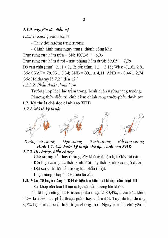
1.2.1. Mô tả kỹ thuật
Đường cắt xương Đục xương Tách xương Kết hợp xương
Hình 1.1. Các bước kỹ thuật chẻ dọc cành cao XHD
1.2.2. Di chứng, biến chứng
- Chẻ xương xấu hay đường gãy không thuận lợi. Gãy lồi cầu.
- Rối loạn cảm giác thần kinh, đứt dây thần kinh xương ổ dưới.
- Đặt sai vị trí lồi cầu trong lúc phẫu thuật.
- Loạn năng khớp TDH, tiêu lồi cầu.
1.3. Vấn đề loạn năng TDH ở bệnh nhân sai khớp cắn loại III
- Sai khớp cắn loại III tạo ra lực tải bất thường lên khớp.
-Tỉ lệ loạn năng TDH trước phẫu thuật là 39,4%, thoái hóa khớp
TDH là 20%; sau phẫu thuật: giảm hay chấm dứt. Tuy nhiên, khoảng
3,7% bệnh nhân xuất hiện triệu chứng mới. Nguyên nhân chủ yếu là
4
do đặt sai vị trí lồi cầu trong lúc phẫu thuật. Đây là mối quan tâm lớn
đối với các bác sĩ phẫu thuật hơn là ổn định xương và khớp cắn.
1.3.1. Những nguyên nhân làm thay đổi vị trí lồi cầu
- Gây mê: lồi cầu rớt khỏi hõm khớp theo hướng thẳng 2,43 mm.
- Phương pháp cố định xương.
- Mức độ và hướng di chuyển xương.
- Phương pháp cắt XHD.
1.3.2. Tầm quan trọng của định vị lồi cầu XHD trong phẫu thuật
- Bảo đảm kết quả phẫu thuật ổn định: nhóm không dùng khí cụ
có 40% trường hợp lồi cầu di lệch 2 – 4mm và 2 – 4o, nhóm có khí cụ
không có trường hợp nào di lệch trên 2 mm và 2o.
- Giảm ảnh hưởng có hại cho khớp TDH: đặt lồi cầu đúng trong
hõm khớp sẽ ít bị lực xoắn nên tránh được ảnh hưởng có hại lên khớp
TDH. Bệnh nhân với tiền sử loạn năng TDH có khả năng đáp ứng
của khớp kém hơn người bình thường. Do đó, nên dùng khí cụ định
để lồi cầu chịu sức căng sinh học ít nhất.
- Cải thiện chức năng nhai.
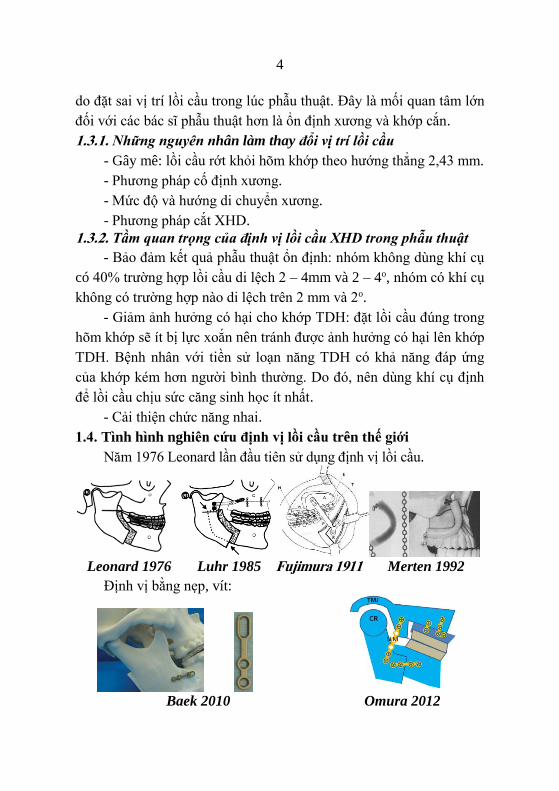
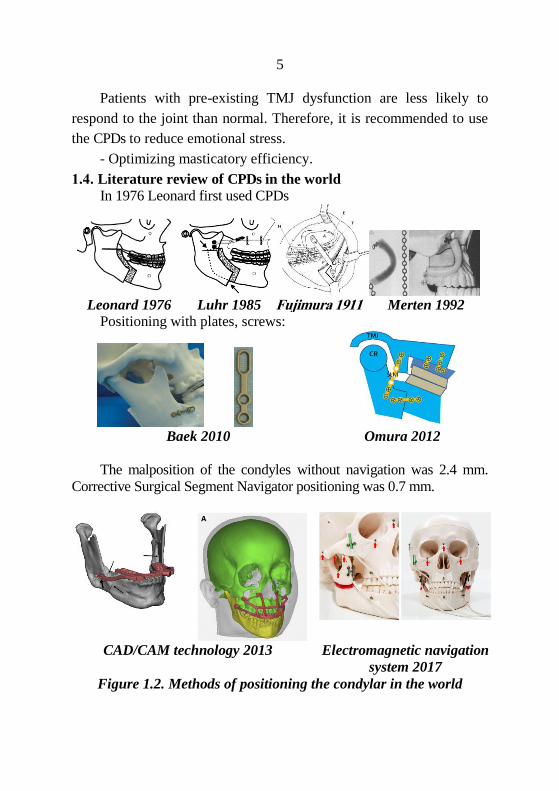
1.4. Tình hình nghiên cứu định vị lồi cầu trên thế giới
Năm 1976 Leonard lần đầu tiên sử dụng định vị lồi cầu.
Leonard 1976 Luhr 1985 Fujimura 1911 Merten 1992
Định vị bằng nẹp, vít:
Baek 2010 Omura 2012
5
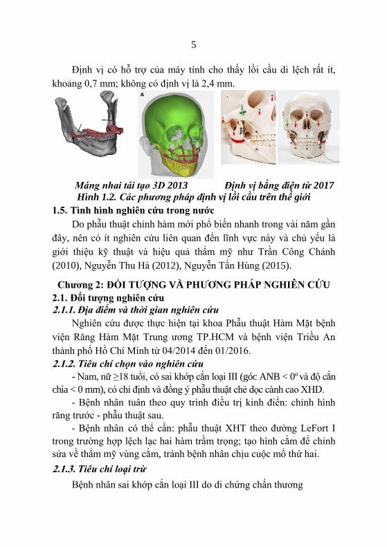
Định vị có hỗ trợ của máy tính cho thấy lồi cầu di lệch rất ít,
khoảng 0,7 mm; không có định vị là 2,4 mm.
Máng nhai tái tạo 3D 2013 Định vị bằng điện từ 2017
Hình 1.2. Các phương pháp định vị lồi cầu trên thế giới
1.5. Tình hình nghiên cứu trong nước
Do phẫu thuật chỉnh hàm mới phổ biến nhanh trong vài năm gần
đây, nên có ít nghiên cứu liên quan đến lĩnh vực này và chủ yếu là
giới thiệu kỹ thuật và hiệu quả thẩm mỹ như Trần Công Chánh
(2010), Nguyễn Thu Hà (2012), Nguyễn Tấn Hùng (2015).
Chương 2: ĐỐI TƯỢNG VÀ PHƯƠNG PHÁP NGHIÊN CỨU
2.1. Đối tượng nghiên cứu
2.1.1. Địa điểm và thời gian nghiên cứu
Nghiên cứu được thực hiện tại khoa Phẫu thuật Hàm Mặt bệnh
viện Răng Hàm Mặt Trung ương TP.HCM và bệnh viện Triều An
thành phố Hồ Chí Minh từ 04/2014 đến 01/2016.
2.1.2. Tiêu chí chọn vào nghiên cứu
- Nam, nữ ≥18 tuổi, có sai khớp cắn loại III (góc ANB < 0o và độ cắn
chìa < 0 mm), có chỉ định và đồng ý phẫu thuật chẻ dọc cành cao XHD.
- Bệnh nhân tuân theo quy trình điều trị kinh điển: chỉnh hình
răng trước - phẫu thuật sau.
- Bệnh nhân có thể cần: phẫu thuật XHT theo đường LeFort I
trong trường hợp lệch lạc hai hàm trầm trọng; tạo hình cằm để chỉnh
sửa về thẩm mỹ vùng cằm, tránh bệnh nhân chịu cuộc mổ thứ hai.
2.1.3. Tiêu chí loại trừ
Bệnh nhân sai khớp cắn loại III do di chứng chấn thương
6
2.2. Phương pháp nghiên cứu
- Thiết kế nghiên cứu: can thiệp lâm sàng không đối chứng.
- Cỡ mẫu tối thiểu 36 trường hợp. Thực tế đã phẫu thuật 36 bệnh nhân.
2.2.1. Biến số nghiên cứu
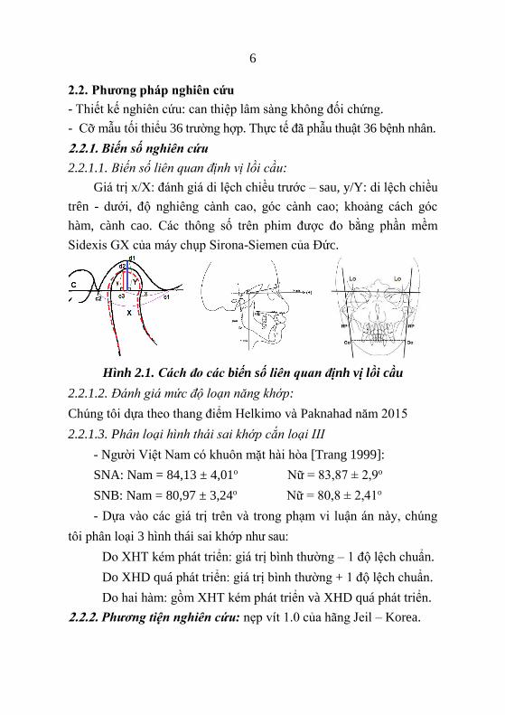
2.2.1.1. Biến số liên quan định vị lồi cầu:
Giá trị x/X: đánh giá di lệch chiều trước – sau, y/Y: di lệch chiều
trên - dưới, độ nghiêng cành cao, góc cành cao; khoảng cách góc
hàm, cành cao. Các thông số trên phim được đo bằng phần mềm
Sidexis GX của máy chụp Sirona-Siemen của Đức.
Hình 2.1. Cách đo các biến số liên quan định vị lồi cầu
2.2.1.2. Đánh giá mức độ loạn năng khớp:
Chúng tôi dựa theo thang điểm Helkimo và Paknahad năm 2015
2.2.1.3. Phân loại hình thái sai khớp cắn loại III
- Người Việt Nam có khuôn mặt hài hòa [Trang 1999]:
SNA: Nam = 84,13 ± 4,01o Nữ = 83,87 ± 2,9o
SNB: Nam = 80,97 ± 3,24o Nữ = 80,8 ± 2,41o
- Dựa vào các giá trị trên và trong phạm vi luận án này, chúng
tôi phân loại 3 hình thái sai khớp như sau:
Do XHT kém phát triển: giá trị bình thường – 1 độ lệch chuẩn.
Do XHD quá phát triển: giá trị bình thường + 1 độ lệch chuẩn.
Do hai hàm: gồm XHT kém phát triển và XHD quá phát triển.
2.2.2. Phương tiện nghiên cứu: nẹp vít 1.0 của hãng Jeil – Korea.

7
2.2.3. Các bước tiến hành nghiên cứu
SƠ ĐỒ NGHIÊN CỨU
Bước 1: Khám bệnh
Bước 2: Kế hoạch PT
- Vẽ nét phim sọ: PT 2 hàm để xác
định sự di chuyển XHT
Bước 3: Làm máng PT Máng nhai TQTT: định vị lồi cầu
Máng nhai sau cùng
Máng nhai chuyển tiếp (PT 2 hàm):
- Vô giá khớp ở thích ứng
- Cưa và di chuyển mẫu hàm trên
như kế hoạch vẽ nét phim sọ
Trong phẫu thuật
- Đặt XHD vào vị trí mới với máng
nhai sau cùng. Cố định liên hàm
- Đặt lại nẹp – vít định vị lồi cầu sao
cho khớp với lỗ nhớ vít
- Kết hợp xương XHD
- Tháo nẹp – vít định vị
Bước 3: Định vị lại
vị trí lồi cầu
Bước 1: Định vị lồi cầu
Bước 2: Cắt xương
Trước phẫu thuật
- Cố định hai hàm: máng nhai tương
quan trung tâm
- Rạch niêm mạc, đặt nẹp - vít định
vị từ mặt ngoài cành cao đến trụ
hàm - gò má, mỗi đầu nẹp: 2 vít
- Tháo nẹp và máng nhai.
- Hình dạng nẹp và lỗ nhớ vít giúp
ghi lại vị trí lồi cầu
Sau phẫu thuật - Cố định hàm 2-3 tuần

8
2.2.4. Thời điểm đánh giá: 30 ngày trước PT (T0), 2-3 tuần PT (T1),
1 tháng sau PT (T2), 3 tháng sau PT (T3), 6 tháng sau PT (T4) và 12
tháng sau PT (T3).
2.2.5. Xử lý và phân tích số liệu
Số liệu được thu thập theo bệnh án mẫu. Số liệu được lưu trữ
và xử lý trên phần mềm SPSS 16.0 và Microsoft Excel 2013.
- Phép kiểm định T hai mẫu độc lập, T bắt cặp, Chi 2, Chi 2 Mc-
Nemar.
Chương 3: KẾT QUẢ
Sau khi phẫu thuật 36 bệnh nhân sai khớp cắn loại III bằng phẫu
thuật chẻ dọc cành cao XHD có sử dụng khí cụ định vị lồi cầu, theo
dõi 12 tháng, chúng tôi có các kết quả sau:
3.1. Đặc điểm lâm sàng và Xquang của đối tượng nghiên cứu
3.1.1. Tuổi và giới
Tuổi trung bình 22 tuổi (19 - 40 tuổi). Tỉ lệ nữ/nam: 1,6/1.
3.1.2. Lý do phẫu thuật
Thẩm mỹ: 19 (52,8%), chức năng: 7 (19,4%), thẩm mỹ và chức
năng: 10 (27,8%). Các lý do chức năng: dễ trật khớp TDH, đau khớp
khi vận động hàm, không cắn được vùng răng trước, tiếng kêu khớp.
3.1.3. Phân loại hình thái sai khớp cắn loại III
- Sai khớp cắn loại III do XHD quá triển: 19,4%; XHT kém phát
triển: 13,9%; hai hàm: 66,7%.
- Phân bố theo nguyên nhân:
Bảng 3.1. Phân bố hình thái sai khớp cắn loại III theo nguyên nhân
Hình thái Di truyền
Khe hở môi
-vòm miệng Tổng số
n % n % n %
XHT kém phát triển 4 12,1 1 33,3 5 13,9
XHD quá phát triển 7 21,2 0 0 7 19,4
Do hai hàm 22 66,7 2 66,7 24 66,7
Tổng số 33 100,0 3 100,0 36 100,0
p 0,446
Sai khớp cắn loại III nguyên nhân di truyền có hình thái lệch
lạc do hai hàm cao hơn hình thái một hàm.
9
- Phân bố hình thái sai khớp cắn loại III theo đặc điểm bất cân xứng:
không liên quan với mức độ bất cân xứng vùng cằm ≥ 4 mm.
3.1.4. Thời gian chỉnh hình răng trước phẫu thuật
Trung bình 31 tháng (5 – 127 tháng).
3.1.5. Triệu chứng khớp thái dương hàm trước phẫu thuật
Bảng 3.2. Phân bố triệu chứng TDH theo hình thái sai khớp cắn
Triệu chứng Do XHT
n (%)
Do XHD n
(%)
Do 2 hàm n
(%)
Tổng cộng
Bình thường 2
(40,0) 3
(42,9) 12
(50,0) 17
(47,2)
Nhẹ 3
(60,0) 4
(57,1) 10
(41,7) 17
(47,2)
Nặng 0 0 2
(8,3) 2
(5,6)
Tổng cộng 5
(100,0) 7
(100,0) 24
(100,0) 36
(100,0) P(1) 0,907
Đa số bệnh nhân không hoặc có dấu chứng nhẹ. Hai trường hợp
nặng là bệnh nhân lệch lạc hai hàm với triệu chứng dễ bị trật khớp.
3.1.6. Đặc điểm khớp cắn vùng răng cửa
Độ cắn chìa trung bình là – 5,5 mm (13 mm; -1,5 mm).
Độ cắn phủ có thể âm, bằng 0 hay cắn hở. Có 16 trường hợp cắn
hở, độ cắn hở trung bình 2 mm (1 mm; - 5 mm).
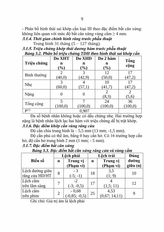
3.1.7. Đặc điểm bất cân xứng
Bảng 3.3. Đặc điểm bất cân xứng răng cửa và vùng cằm
Biến số
Lệch phải Lệch trái Đúng
đường
giữa (n) n Trung vị
(Phạm vi)
n Trung vị
(Phạm vi)
Lệch đường giữa
răng cửa HD/HT 8
- 3
(-5; -1) 18
3,5
(1; 9) 10
Lệch cằm
trên lâm sàng 7
-2
(-3; -0,5) 17
4
(1,5; 11) 12
Lệch cằm
trên phim 2
- 0,68
(-0,85; -0,5) 25
4,53
(0,67; 14,11) 9
Ghi chú: Giá trị âm là lệch phải
10
Về bất cân xứng, hàm dưới lệch trái nhiều hơn lệch phải. Số lượng
hàm dưới lệch trái trên phim nhiều hơn trên lâm sàng, kiểm định tính
đồng nhất cho thấy khác biệt không có ý nghĩa (Kappa = 0,38).
- Nghiêng mặt phẳng nhai:18 trường hợp, trung bình 1,81mm (0,5 –
6mm).
3.1.8. Đặc điểm X-quang của đối tượng nghiên cứu
Bảng 3.4. Đặc điểm X-quang trước phẫu thuật
Góc
xương hàm
�̅�
(mm) ĐLC
Trục răng
cửa �̅�
(mm) ĐLC
SNA 81,45 3,52 U1 – mặt phẳng
khẩu cái 120,52 6,99
SNB 86,02 4,82 U1 – đường NA 30,21 6,48
ANB -4,57 3,11 L1 – mặt phẳng
hàm dưới 87,08 6,47
Mặt phẳng
nhai-SN 14,62 5,93 L1 – đường NB 28,08 6,36
Mặt phẳng
hàm dưới - SN 34,74 5,87 U1 – L1 126,46 10,16
Tổng số 36 Tổng số 36
Sau giai đoạn chỉnh hình răng trước phẫu thuật, các góc trục
răng cửa chưa giải bù trử đủ.
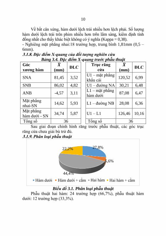
3.1.9. Phân loại phẫu thuật
Biểu đồ 3.1. Phân loại phẫu thuật
Phẫu thuật hai hàm: 24 trường hợp (66,7%), phẫu thuật hàm
dưới: 12 trường hợp (33,3%).
27,8%
5,6%
44,4%
22,2%
Hàm dưới Hàm dưới + cằm Hai hàm Hai hàm + cằm

11
Bảng 3.5. Phân bố loại phẫu thuật theo độ cắn chìa, góc ANB
Loại phẫu thuật n Độ cắn chìa
�̅� (mm) ± ĐLC
Góc ANB
�̅� (o) ± ĐLC
Hàm dưới tính chung 12 -2,92 ± 1,00 - 2,52 ± 2,25
Hai hàm tính chung 24 -7,67 ± 2,90 - 5,59 ± 3,00
p 36 < 0,001 0,004
Những trường hợp phẫu thuật hai hàm có độ cắn chìa và góc
ANB khác biệt có ý nghĩa so với phẫu thuật một hàm.
3.2. Kết quả phẫu thuật có sử dụng khí cụ định vị lồi cầu
3.2.1. Lâm sàng
- Thời gian phẫu thuật: hàm dưới là 150,50 ± 22,92 phút, hai hàm
là 258,50 ± 30,99 phút.
- Tai biến, biến chứng:
Không có trường hợp nào đứt dây thần kinh.
Thời gian rối loạn cảm giác thần kinh:
Môi dưới: 5,06 ± 3,23 tuần; vùng cằm: 6, 92 ± 3,84 tuần.
Môi trên: 5,33 ± 3,67 tuần; răng hàm trên: 6,25 ± 3,30 tuần.
- Thay đổi khớp cắn: sau phẫu thuật, độ cắn chìa, cắn trùm: 2,0 mm.
- Triệu chứng khớp thái dương hàm:
Biểu đồ 3.2. Loạn năng khớp TDH trước và sau phẫu thuật
Sau khi tháo cố định hàm, biên độ há miệng giảm và phục hồi
dần sau vài tháng. Sau 12 tháng, biên độ há miệng trở về bình thường
như trước phẫu thuật. Không có trường hợp nào xuất hiện mới.
0
20
40
T0 T3 T4 T5
n
Không dấu chứng Nhẹ Nặng
12
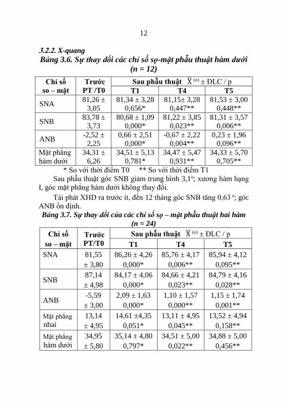
3.2.2. X-quang
Bảng 3.6. Sự thay đổi các chỉ số sọ-mặt phẫu thuật hàm dưới
(n = 12)
Chỉ số
so – mặt
Trước
PT /T0
Sau phẫu thuật X̅ (o) ± ĐLC / p
T1 T4 T5
SNA 81,26 ±
3,05
81,34 ± 3,28
0,656*
81,15± 3,28
0,447**
81,53 ± 3,00
0,448**
SNB 83,78 ±
3,73
80,68 ± 1,09
0,000*
81,22 ± 3,85
0,023**
81,31 ± 3,57
0,006**
ANB -2,52 ±
2,25
0,66 ± 2,51
0,000*
-0,67 ± 2,22
0,004**
0,23 ± 1,96
0,096**
Mặt phẳng
hàm dưới
34,31 ±
6,26
34,51 ± 5,13
0,781*
34,47 ± 5,47
0,931**
34,33 ± 5,70
0,705**
* So với thời điểm T0 ** So với thời điểm T1
Sau phẫu thuật góc SNB giảm trung bình 3,1o; xương hàm hạng
I, góc mặt phẳng hàm dưới không thay đổi.
Tái phát XHD ra trước ít, đến 12 tháng góc SNB tăng 0,63 o; góc
ANB ổn định.
Bảng 3.7. Sự thay đổi của các chỉ số sọ – mặt phẫu thuật hai hàm
(n = 24)
Chỉ số
so – mặt
Trước
PT/T0
Sau phẫu thuật X̅ (o) ± ĐLC / p
T1 T4 T5
SNA 81,55
± 3,80
86,26 ± 4,26
0,000*
85,76 ± 4,17
0,006**
85,94 ± 4,12
0,095**
SNB 87,14
± 4,98
84,17 ± 4,06
0,000*
84,66 ± 4,21
0,023**
84,79 ± 4,16
0,028**
ANB -5,59
± 3,00
2,09 ± 1,63
0,000*
1,10 ± 1,57
0,000**
1,15 ± 1,74
0,001**
Mặt phẳng nhai
13,14
± 4,95
14,61 ±4,35
0,051*
13,11 ± 4,95
0,045**
13,52 ± 4,94
0,158**
Mặt phẳng hàm dưới
34,95
± 5,80
35,14 ± 4,80
0,797*
34,51 ± 5,00
0,022**
34,88 ± 5,00
0,456**

13
Sau phẫu thuật, góc SNA tăng trung bình 4,71o; góc SNB giảm
trung bình 2,97o; xương hàm hạng I (p < 0,001). Góc mặt phẳng nhai
và mặt phẳng hàm dưới không thay đổi.
Sau 12 tháng, góc ANB giảm 0,94 o; xương hàm hạng I.
Bảng 3.8.Sự thay đổi của điểm B cho phẫu thuật hàm dưới (n = 12)
Điểm
B
Trước phẫu
thuật /T0
Sau phẫu thuật X̅ (mm) ± ĐLC / p
T1 T4 T5
Bx 63,17 ± 6,47 58,51 ± 6,76
0,000*
59,31 ± 6,68
0,107**
59,43 ± 6,31
0,050**
By 90,39 ± 4,86 89,67 ± 4,75
0,060*
88,42 ± 4,34
0,000**
89,03 ± 4,94
0,029**
Sau phẫu thuật: XHD (điểm B) di chuyển ra sau trung bình là
4,77 mm, di chuyển lên trên trung bình 0,72 mm.
Sau 12 tháng, điểm B tái phát lên trên 0,64 mm.
Bảng 3.9. Sự thay đổi của điểm A, B cho phẫu thuật hai hàm
(n = 24)
Điểm
A, B
Trước
PT/T0
Sau phẫu thuật X̅ (mm) ± ĐLC / p
T1 T4 T5
Ax 61,07
± 6,36
66,39 ± 6,86
0,000*
65,75 ± 6,74
0,039**
65, 45 ± 7,00
0,010**
Ay 52,70
± 4,03
53,33 ± 4,19
0,050*
53,09 ± 4,52
0,334**
53,28 ± 4,15
0,797**
Bx 69,85
± 10,41
64,57 ± 9,14
0,000*
65,80 ± 9,31
0,000**
65,63± 9,43
0,022**
By 90,81
± 6,93
90,57 ± 5,90
0,677*
89,67 ± 6,67
0,075**
89,76 ± 6,46
0,047** Sau phẫu thuật: XHT (điểm A) di chuyển trung bình ra trước là
5,32 mm, di chuyển xuống dưới 0,63 mm. XHD (điểm B) di chuyển
trung bình ra sau 5,28 mm; lên trên 0,24 mm.
Sau 12 tháng, điểm A gần như không thay đổi, vị trí XHD tại
điểm B tái phát ra trước và lên trên lần lượt là 0,17mm và 0,81mm so
với thời điểm T1.

14
Bảng 3.10. Sự thay đổi của lồi cầu, điểm Gonion và
độ nghiêng cành cao
Vị trí Trước
PT/T0
Sau phẫu thuật X̅ ± ĐLC / p
T1 T4 T5
Lồi cầu
(mm)
Cx 0,55
± 0,06
0,55 ± 0,05
0,878*
0,54 ± 0,05
0,483**
0,54 ± 0,05
0,496**
Cy 0,74
± 0,06
0,73 ± 0,05
0,720*
0,73 ± 0,03
0,735**
0,73 ± 0,04
0,559**
Gonion
(mm)
Gox -2,23
± 8,44
-3,49 ± 7,87
0,014*
-2,93 ± 7,49
0,037**
-2,71 ± 7,64
0,005**
Goy 75,16
± 7,05
75,15 ± 6,95
0,975*
74,47 ± 7,20
0,023**
74,52 ± 7,01
0,004**
Độ nghiêng
cành cao (o)
80,40
± 7,30
81,37 ± 7,17
0,059*
81,14 ± 7,06
0,233**
80,74 ± 7,27
0,022**
Sau phẫu thuật, lồi cầu thay đổi không có ý nghĩa. Điểm
Gonion di chuyển ra sau 1,26 mm. Sau 12 tháng, trở về gần với vị trí
trước phẫu thuật. Độ nghiêng cành cao: tăng ít tương ứng với thay
đổi điểm Gonion lùi sau, tức mảnh gần xoay theo chiều kim đồng hồ
0,97o; nhưng khác biệt không có ý nghĩa.
Bảng 3.11. Sự thay đổi điểm Gonion so với trục Y
trước và sau phẫu thuật
Điểm Go Trước PT/T0
n (%)
Sau phẫu thuật n (%)
T1 T4 T5
Trước trục Y
(giá trị +)
15
(41,67)
12
(33,33)
12
(33,33)
12
(33,33)
Sau trục Y
(giá trị -)
21
(58,33)
24
(66,67)
24
(66,67)
24
(66,67)
p(1) 0,375 0,999 0,999 (1) Kiểm định Chi 2 Mc - Nemar
Sự thay đổi vị trí điểm Gonion quanh trục Y không đáng kể.

15
Bảng 3.12. Sự thay đổi của góc cành cao, khoảng cách cành cao và
góc hàm trên phim sọ thẳng
Vị trí Trước
PT/T0
Sau phẫu thuật
T1 T4 T5
Góc
cành
cao (o)
Phải
80,69
± 3,55
82,08 ± 4,00
0,005*
81,85 ± 4,57
0,540**
81,89 ± 3,81
0,324**
Trái
82,42
± 3,77
83,11 ± 4,37
0,080*
83,09 ± 4,09
0,918**
83,41 ± 4,62
0,214**
Khoảng cách
cành cao (mm)
103,99
± 5,00
105,30 ± 4,82
0,002*
105,22 ± 5,51
0,791**
104,79 ± 4,90
0,092**
Khoảng cách
góc hàm (mm)
92,91
± 6,57
94,71 ± 6,02
0,000
94,77 ± 6,09
0,794
94,66 ± 5,90
0,788
Sau phẫu thuật góc cành cao bên phải tăng 1,39o; khoảng cách
cành cao, góc hàm tăng lần lượt là 1,31 mm và 1,8 mm và ổn định ở
thời điểm 6 tháng, 12 tháng.
3.2.3. Đánh giá mức độ hài lòng về thẩm mỹ và chức năng của
bệnh nhân sau phẫu thuật
Tất cả bệnh nhân hài lòng với kết quả phẫu thuật về chức năng
và thẫm mỹ. Đa số bệnh nhân cho điểm số ở mức "rất hài lòng".
Chương 4: BÀN LUẬN
4.1. Đặc điểm chung của đối tượng nghiên cứu
4.1.1. Tuổi và giới tính
Trong nghiên cứu của chúng tôi, tuổi trung bình là 22 tuổi (19 –
40 tuổi), tương đương các nghiên cứu khác. Tuy nhiên có nghiên cứu
với tuổi nhỏ nhất là 14, lớn nhất là 55,5 tuổi.
Về giới tính, tỉ lệ nữ/nam là 1,6/1 (22/14). Tỉ lệ nữ cao như đa số
các nghiên cứu khác.
4.1.2. Lý do phẫu thuật
Lý do thúc đẩy bệnh nhân phẫu thuật về thẩm mỹ là 52,8%. Nữ
giới có nhu cầu thẩm mỹ cao hơn nam giới (p = 0,01). Một số nghiên
cứu gần đây cho thấy lý do về chức năng cao. Silva 2016 cho thấy
nhu cầu về thẩm mỹ lẫn chức năng là 66%. B.Farahani 2016 khảo sát

16
về “chỉ số của nhu cầu điều trị chức năng” trên 103 bệnh nhân phẫu
thuật chỉnh hàm nói chung, cho thấy mức “rất cần” ở bệnh nhân sai
khớp cắn loại III là 95,6%.
4.1.3. Phân loại hình thái sai khớp cắn loại III
Trong nghiên cứu của chúng tôi, sai khớp cắn loại III do hai
hàm là 66,7%; trong đó do di truyền là 66,7% trường hợp. Những
bệnh nhân này, theo người nhà cho biết lúc nhỏ bệnh nhân có khuôn
mặt phát triển bình thường, khi đến tuổi dậy thì, gia đình mới nhận ra
sự phát triển quá mức của XHD. Thời điểm này bệnh nhân đã qua
đỉnh tăng trưởng của xương hàm và phần lớn các trường hợp không
thể chỉnh hình răng mặt ngụy trang được. Những trường hợp sai khớp
cắn thứ phát do khe hở môi – vòm miệng thường do thiểu sản tầng
mặt giữa.
Về bất cân xứng, trong nghiên cứu của chúng tôi đa số là hàm
dưới lệch trái, mức độ lệch trung bình là 4mm. Tỉ lệ bất cân xứng
hàm - mặt khoảng 21% - 85%, trong đó 48% các trường hợp là sai
khớp cắn loại III và mức độ mất cân xứng 4 mm có ý nghĩa trong
chẩn đoán và điều trị bất cân xứng.
4.1.4. Thời gian chỉnh hình răng mặt trước phẫu thuật
Trong nghiên cứu của chúng tôi, thời gian chỉnh hình răng mặt
trước phẫu thuật trung bình 31 tháng (5 – 127 tháng). Trường hợp
lâu nhất 127 tháng (trên 10 năm) ở bệnh nhân có khe hở môi vòm
miệng hai bên. Mặc dù bệnh nhân này được chỉnh hình răng mặt
từ nhỏ trong giai đoạn xương còn tăng trưởng, nhưng phải chờ đến
tuổi trưởng thành để phẫu thuật.
Theo y văn, thời gian chỉnh hình răng mặt trước phẫu thuật là 2
-12 tháng tùy theo mức độ răng lệch lạc. Gần đây, có nhiều nghiên
cứu đi theo hướng tiếp cận phẫu thuật trước - chỉnh hình sau. Điều
này, về tâm lý bệnh nhân hài lòng hơn vì cải thiện thẩm mỹ sớm,
nhưng bất lợi là khó tiên đoán được vị trí xương hàm tối ưu với vị trí
răng chưa được giải bù trừ. Những trường hợp không chỉnh hình răng
mặt trước phẫu thuật, kết quả kém ổn định do xuất hiện những điểm
chạm sớm ngay sau phẫu thuật khiến hàm dưới sẽ trượt đến vị trí cân
bằng hơn gây ra sai khớp cắn. Tái phát theo chiều ngang ở bệnh nhân
có chỉnh hình răng mặt trung bình 0,9 - 1,6 mm; không có chỉnh hình
răng mặt là 2,0 - 2,4 mm; tỉ lệ tái phát trên 3 mm nhóm phẫu thuật
17
kinh điển là 15, 8%; nhóm phẫu thuật trước chỉnh hình răng mặt sau
là 39,1%.
Nhiều nghiên cứu cho thấy phẫu thuật trước - chỉnh hình răng
mặt sau chỉ nên áp dụng trong những trường hợp lệch lạc mức độ
trung bình, chen chúc nhẹ không cần nhổ răng, bênh nhân không có
tiền sử loạn năng khớp TDH,... Những trường hợp nặng nên theo
phương pháp kinh điển.
4.2. Kết quả phẫu thuật chẻ dọc cành cao có định vị lồi cầu trên
bệnh nhân nghiên cứu
4.2.1. Loại phẫu thuật
Trong nghiên cứu của chúng tôi, phẫu thuật hai hàm chiếm 24
trường hợp (66,7%), phẫu thuật hàm dưới 12 trường hợp (33,3%),
trong đó có 10 trường hợp (27,8%) tạo hình cằm. Chúng tôi chỉ định
trường hợp phẫu thuật một hàm hay hay hàm phụ thuộc vào độ cắn
chìa và góc ANB. Có sự khác biệt có ý nghĩa về độ cắn chìa
(p<0,001) và góc ANB (p<0,005) giữa phẫu thuật một hàm và hai
hàm.
Hầu hết các dị dạng răng mặt có thể điều trị bằng ba phẫu thuật
chính, bao gồm phẫu thuật XHT theo đường LeFort I, chẻ dọc cành
cao XHD và tạo hình cằm. Đối với bất cân xứng, sau khi đặt vị trí
mảnh xa vào đúng khớp cắn, sẽ xuất hiện nhiều vướng cộm xương
giữa mảnh gần – mảnh xa. Điều này dễ làm di lệch lồi cầu. Vì vậy,
một số tác giả đề nghị nhiều biến thể cho điều trị bất cân xứng.
Trong nghiên cứu của chúng tôi sử dụng định vị lồi cầu kết
hợp với loại bỏ cản trở xương cho thấy có hiệu quả điều trị các
trường hợp bất cân xứng theo ba chiều không gian.
4.2.2. Về thời gian phẫu thuật
Thời gian phẫu thuật có áp dụng khí cụ định vị lồi cầu của
chúng tôi trung bình hàm dưới là 150,50 phút, hai hàm là 258,50
phút. Nghiên cứu Panula 2001 thì thời gian phẫu thuật hàm dưới là
130 phút, hai hàm là 249 phút. Điều này cho thấy kỹ thuật định vị lồi
cầu của chúng tôi tuy làm tăng thời gian phẫu thuật ít nhưng không
đáng kể so với lợi ích mà nó đem lại.
4.2.3. Về tai biến, biến chứng
- Biến chứng thần kinh:
Thời gian rối loạn cảm giác: trong nghiên cứu của chúng tôi là
5,06 ± 3,23 tuần. Rối loạn thần kinh cảm giác là biến chứng vốn có

18
sau phẫu thuật chẻ dọc cành cao XHD. Các yếu tố ảnh hưởng thời
gian rối loạn thần kinh cảm giác mức độ chấn thương thần kinh. Theo
Osburne (2007) có ba mức độ chấn thương thần kinh là liệt thần kinh
nhẹ không thoái hóa ngoại vi (anapraxia), đứt sợi trục thần kinh
(axonotmesis) và đứt dây thần kinh (neurotmesis). Ngoài ra còn tùy
thuộc vào giải phẫu dây thần kinh, cảm nhận của bệnh nhân.
Đứt dây thần kinh: trong nghiên cứu của chúng tôi không có
trường hợp nào, có thể do chúng tôi không đục xương mạnh, chủ yếu
là dùng cây tách xương. Tỉ lệ đứt dây thần là 1,5%; bệnh nhân biết
mất cảm giác và ảnh hưởng sinh hoạt là 7,4%.
- Chẻ xương xấu hay đường gãy xương không thuận lợi:
Trong nghiên cứu chúng tôi, có một trường hợp gãy vỏ xương
phía má, mảnh gãy nhỏ, chỉ cần tăng chiều dài nẹp cố định. Theo y
văn, tỉ lệ chẻ xương xấu là 2,3% - 3,9%. Sự hiện diện của răng hàm
thứ ba, đường cắt xương không cắt qua hết bờ dưới XHD có thể làm
tăng nguy cơ chẻ xương xấu.
- Biến chứng tiêu lồi cầu:
Trong nghiên cứu của chúng tôi, theo dõi đến 12 tháng, chưa ghi
nhận biến chứng này, có thể do nghiên cứu chúng tôi có sử dụng khí cụ
định vị lồi cầu, nên không gây ra tải lực quá mức lên khớp TDH.
Nguyên nhân của tiêu lồi cầu có thể do thay đổi của lực sinh
cơ học lên khớp TDH. Tỉ lệ tiêu lồi cầu sau phẫu thuật khoảng 7,5%
(224/2994 trường hợp).
4.2.4. Triệu chứng khớp TDH
Nghiên cứu của chúng tôi sử dụng định vị lồi cầu nên lồi cầu ít
thay đổi vị trí. Kết quả cho thấy, trước phẫu thuật có 52,8 % trường
hợp có triệu chứng loạn năng TDH. sau phẫu thuật còn 22% với triệu
chứng là thỉnh thoảng có tiếng kêu khi há lớn, không đau, không có
trường hợp nào mới mắc bệnh.
Sau phẫu thuật chỉnh hàm, khớp cắn cân bằng và vững ổn hơn
nên có lợi cho khớp TDH, vì vậy giảm đáng kể các dấu chứng loạn
năng khớp có trước phẫu thuật. Tuy nhiên vẫn có tỉ lệ mắc mới
khoảng 3,7%. Một trong những nguyên nhân làm gia tăng triệu
chứng khớp TDH sau phẫu thuật là thay đổi vị trí lồi cầu, gây ra xáo
trộn nội khớp. Cố định xương cứng chắc có thể làm sai vị trí lồi cầu.
Vì vậy, để giảm thiểu thay đổi vị trí lồi cầu, nhiều tác giả đề nghị sử
dụng khí cụ định vị lồi cầu, đặc biệt khi kết hợp xương cứng chắc.
19
4.2.5. Đặc điểm khớp cắn
Trước phẫu thuật: độ cắn chìa trung bình -5,5mm (13; -1,5
mm). Sau khi tháo cố định hàm, độ cắn chìa, cắn trùm trung bình 1 – 2
mm. Theo dõi 12 tháng sau phẫu thuật, khớp cắn ổn định. Trong nghiên
cứu chúng tôi có sử dụng khí cụ định vị nên hạn chế được lồi cầu di lệch
quá mức, vì vậy không gây ra tái phát xương và khớp cắn.
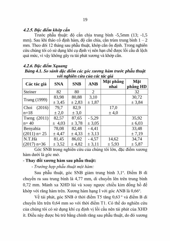
4.2.6. Đặc điểm Xquang
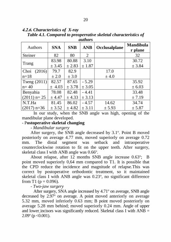
Bảng 4.1. So sánh đặc điểm các góc xương hàm trước phẫu thuật
với nghiên cứu của các tác giả
Các tác giả SNA SNB ANB Mặt phẳng
nhai
Mặt
phẳng HD
Steiner 82 80 2 32
Trang (1999) 83,98
± 3,45
80,88
± 2,83
3,10
± 1,87
30,72
± 3,84
Choi (2016)
n=18
79,7
± 2,0
82,9
± 3,0
17,0
± 4,0
Tseng (2011)
n= 40
82,57
± 4,03
87,65
± 3,78
- 5,29
± 3,05
35,92
± 6,03
Benyahia
(2011) n= 25
78,08
± 4,47
82,48
± 4,33
- 4,41
± 3,13
33,48
± 7,19
N.T.Hà
(2017) n=36
81,45
± 3,52
86,02
± 4,82
- 4,57
± 3,11
14,62
± 5,93
34,74
± 5,87
Góc SNB trong nghiên cứu của chúng tôi lớn, đặc điểm xương
hàm dưới là góc mở.
- Thay đổi xương hàm sau phẫu thuật:
- Trường hợp phẫu thuật một hàm:
Sau phẫu thuật, góc SNB giảm trung bình 3,1o. Điểm B di
chuyển ra sau trung bình là 4,77 mm, di chuyển lên trên trung bình
0,72 mm. Mảnh xa XHD lùi và xoay ngược chiều kim đồng hồ để
khớp với răng hàm trên. Xương hàm hạng I với góc ANB là 0,66o.
Về tái phát, góc SNB ở thời điểm T5 tăng 0,63 o và điểm B di
chuyển lên trên 0,64 mm so với thời điểm T1. Có thể do nghiên cứu
của chúng tôi có sử dụng khí cụ định vị lồi cầu nên tái phát của XHD
ít. Điều này được bù trừ bằng chỉnh răng sau phẫu thuật, do đó xương

20
hàm duy trì hạng I với góc ANB là 0,23o, không khác biệt so với thời
điểm T1 (p = 0,096).
- Trường hợp phẫu thuật hai hàm:
Sau phẫu thuật, góc SNA tăng trung bình 4,71o, góc SNB giảm
trung bình 2,97o. Điểm A trung bình di chuyển ra trước là 5,32 mm;
di chuyển xuống dưới 0,63 mm. Điểm B trung bình di chuyển ra sau
5,28 mm, lên trên 0,24 mm. Góc trục răng cửa hàm trên, hàm dưới
giảm đáng kể. Xương hàm hàm hạng I với góc ANB = 2,09 o (p<
0,001).
Về tái phát, góc SNB tăng trung bình 0,49 – 0,62o, nên góc
ANB giảm trung bình 0,94o. Sau 12 tháng, Điểm A tái phát ra sau
0,94 mm điểm B ra trước 1,06 mm, lên trên 0,81 mm so với thời
điểm T1.
Nghiên cứu chúng tôi có sử dụng khí cụ định vị lồi cầu, do đó
trong phẫu thuật hai hàm có tái phát ít và được bù trù bằng tăng góc
trục răng cửa hàm trên nên duy trì kết quả ổn định xương hàm hạng I.
Bảng 4.2. So sánh thay đổi các chỉ số sọ mặt phẫu thuật hai hàm
sau 6 – 12 tháng của các tác giả
Tác giả SNA SNB L1-
HD Ax Ay Bx By
Kor (2014)
n = 15
0,18
± 0,74
0,24
± 0,65
0,39
± 1,89
0,41
± 0,75
-0,05
± 0,81
0,81
± 1,34
0,14
± 1,13
Park (2016)
n = 29
0,14
± 0,73
1,01
± 0,74
0,15
± 0,57
-0,19
± 1,20
1,96
± 1,15
-1,14
± 1,10
Seeberger
(2013) n = 22
-0,54
± 2,82
Paeng (2012)
n = 15
- 0,57
± 1,46
0,34
± 1,01
- 0,79
± 1,85
0,76 ±
1,94
N.T.Hà (2017)
n = 36 - 0,50 0,49 0,75 - 0,1 0,31 0,8 -1,25
Park cho lồi cầu tự điều chỉnh trong giai đoạn cố định hàm, nên
mức độ tái phát cao hơn. Seeberger dùng CBCT để định vị lồi cầu
trong phòng mổ, cho thấy tái phát rất ít.Tái phát trong phẫu thuật
chỉnh hàm là điều không thể tránh khỏi vì có rất nhiều yếu tố ảnh

21
hưởng đến kết quả. Thay đổi vị trí lồi cầu là nguyên nhân được đề
cập nhất. Nhiều nghiên cứu đã đưa ra các phương pháp để giảm thiểu
di lệch lồi cầu. Tuy nhiên, mọi phương pháp đều có tái phát ít nhiều.
Nếu những thay đổi này ít và được điều chỉnh bởi những thay đổi của
khớp cắn và đáp ứng sinh lý của khớp sau phẫu thuật thì kết quả phẫu
thuật sẽ ổn định.
Nghiên cứu của chúng tôi cho thấy lồi cầu trước và sau phẫu
thuật ở các thời điểm như sau:
- Trên phim sọ nghiêng, sau phẫu thuật điểm Gonion di chuyển
ra sau 1,26 mm. Sau 12 tháng, tái phát ra trước theo chiều ngang
0,78mm và lên trên 0,63 mm so với T1.
Độ nghiêng cành cao thay đổi không đáng kể sau phẫu thuật (p
= 0,059), có nghĩa mảnh gần xoay ngược chiều kim đồng hồ 0,97o.
Sau 12 tháng tái phát ngược chiều kim đồng hồ là 0,63o nhưng không
ảnh hưởng đến kết quả phẫu thuật.
-Trên phim sọ thẳng, độ rộng góc hàm và cành cao thay đổi, có
nghĩa lồi cầu có xoay vô trong theo mặt phẳng trán. Sau phẫu thuật góc
cành cao bên phải tăng 1,39o; khoảng cách cành cao, góc hàm tăng lần
lượt là 1,31 mm và 1,8 mm và ổn định ở thời điểm 6 - 12 tháng.
Nghiên cứu của Park (2016) không có dùng định vị nên góc
mảnh gần thay đổi nhiều sau phẫu thuật là -2,59 ± 1,09o; sau 6 tháng là
-2,13± 0,99o so với T1. Nghiên cứu Seeberger (2013) dùng CBCT định
vị trong phòng mổ nên góc cành cao tăng ít là 0,64o. Nghiên cứu Ko
(2009) góc hàm hai bên giảm 3,4mm.
Nhìn chung, những thay đổi nhỏ trong nghiên cứu của chúng tôi
tương tự như các tác giả khác và không ảnh hưởng đến sự vững ổn.
Phẫu thuật chỉnh hàm là các quá trình phá vở mối tương quan nền
sọ – lồi cầu – răng và sau phẫu thuật các đơn vị này cần phải đặt lại vị trí
thích hợp khi sắp xếp các răng hàm trên – hàm dưới khớp với nhau
(Bethge 2015). Vấn đề này dẫn đến nhiều báo cáo tầm quan trọng của
vị trí lồi cầu trong hõm khớp. Tuy nhiên, sự khác biệt về vị trí lồi cầu
sau khi phẫu thuật chỉnh hàm và ảnh hưởng chức năng khớp TDH là
vẫn còn tranh cãi (Catherine 2016). Nghiên cứu của Seeberger 2013

22
cho thấy có 1/22 bệnh nhân di lệch khoảng gian lồi cầu vượt quá 1
mm và cần phải kết hợp xương lại. Do xuất hiện một vài trường hợp
biến chứng mà nhiều nghiên cứu đồng thuận chung là nên duy trì vị
trí lồi cầu trước phẫu thuật để bảo đảm khớp cắn ổn định lâu dài,
cũng như khớp TDH khỏe mạnh, không đau và chức năng nhai khỏe
mạnh. Từ đó, nhiều phương pháp và khí cụ được đề xuất nhằm mục
đích này.
Trong điều kiện tại Việt Nam, phương pháp chúng tôi là một giải
pháp thực tế trước mắt có hiệu quả trong kiểm soát vị trí lồi cầu trong
phẫu thuật chỉnh hàm. Xét về khả năng ứng dụng lâm sàng, phương
pháp của chúng tôi dễ dàng áp dụng, cho phép phẫu thuật viên tập trung
kỹ thuật phẫu thuật hơn, không bận tâm quá nhiều vào vị trí lồi cầu, nhất
là những trường hợp bất cân xứng hay bệnh nhân loạn năng TDH.
4.3. Đánh giá chức năng nhai và thẩm mỹ sau phẫu thuật
Về chức năng
Trong nghiên cứu của chúng tôi, sau phẫu thuật các răng tiếp
xúc nhau nhiều hơn nên ăn nhai tốt hơn, cải thiện hiệu quả hệ thống
nhai, làm giảm trình trạng loạn năng khớp. Điều này chứng tỏ rằng
phẫu thuật chỉnh hàm không chỉ đơn thuần là hình thức phẫu thuật
thẩm mỹ mà còn là một phương pháp điều trị những bệnh nhân có
lệch lạc chức năng nhai trầm trọng.
Về thẩm mỹ
Trong nghiên cứu của chúng tôi, 83,3% bệnh nhân “hoàn toàn
hai lòng”, 6 bệnh nhân chúng tôi xếp vào mức “hài lòng” vì còn bất
cân xứng nhẹ vùng cằm và bờ dưới XHD, nhưng hệnh nhân hài lòng
với kết quả thẩm mỹ nên không có nhu cầu phẫu thuật thêm.
Qua các câu hỏi gián tiếp phản ánh sự hài lòng của bệnh nhân.
Kết quả cho thấy đa số bệnh nhân hài lòng với kết quả phẫu thuật so
với những khó chịu của phẫu thuật mang lại và sẵn sàng khuyên
người có lệch lạc răng mặt nên phẫu thuật chiếm 92,7%.

23
KẾT LUẬN
Nghiên cứu và tiến hành phẫu thuật chẻ dọc cành cao XHD để
điều trị bệnh nhân sai khớp cắn loại III có sử dụng khí cụ định vị lồi
cầu trên 36 bệnh nhân (*) cho phép rút ra một số kết luận sau đây:
1. Về đặc điểm lâm sàng, X-quang trước phẫu thuật
Tuổi trung bình bệnh nhân là 22. Tỉ lệ nữ/nam là 1,6/1. Lý do
phẫu thuật về thẩm mỹ là 52,8%.
Hình thái sai khớp cắn loại III do XHT kém phát triển kèm XHD
quá triển là 66,7%. Độ cắn chìa trung bình -5,5 mm. Đặc điểm nổi
bật bất cân xứng trên lâm sàng là đa số XHD và cằm lệch trái (trung
bình 4mm). Triệu chứng loạn năng TDH nhẹ là 47,2%; nặng là 5,6%.
Về X-quang, góc ANB trung bình -4,57 o; góc mặt phẳng hàm
dưới là góc mở, trung bình 34,74o.
Thời gian chỉnh hình răng mặt trước phẫu thuật trên hai năm là
66,7%. Sau giai đoạn này, trục răng cửa hàm trên - đường NA còn
chìa (30,21o) so với giá trị bình thường theo Steiner (22o).
2. Theo dõi, đánh giá kết quả lâm sàng, X-quang và cảm
nhận của bệnh nhân 12 tháng sau phẫu thuật
Phẫu thuật hai hàm là 66,7%. Có tái phát ít ở những thời điểm
theo dõi. Sau 12 tháng, xương hàm duy trì hạng I với góc ANB bằng
0,23o (phẫu thuật hàm dưới); 1,15o (phẫu thuật hai hàm) và độ cắn
chìa trung bình 2 mm.
Loạn năng TDH sau phẫu thuật giảm còn 22,2%, không có
trường hợp nào nặng hay mới xuất hiện.
Tất cả bệnh nhân hài lòng về cải thiện chức năng nhai và thẩm mỹ.
Về hiệu quả của khí cụ định vị lồi:
- Trên phim sọ nghiêng, sau phẫu thuật vị trí lồi cầu thay đổi
không có ý nghĩa; mảnh gần có xoay ngược chiều kim đồng hồ trung
bình 0,97o. Sau 12 tháng đã trở về vị trí như trước phẫu thuật; tương
quan XHD – nền sọ tăng 0,63o (p>0,05).

24
- Trên phim sọ thẳng, sau phẫu thuật có tăng khoảng cách góc
hàm và cành cao lần lượt là 1,8 mm và 1,31 mm. Sau 12 tháng sự
thay đổi không có ý nghĩa (p>0,05).
Phẫu thuật chẻ dọc cành cao có sử dụng khí cụ định vị lồi cầu
bằng nẹp thẳng và máng nhai ở tương quan trung tâm là phẫu thuật
an toàn, đạt được mục tiêu điều trị, cho kết quả lâm sàng tốt, cho
phép phẫu thuật viên tập trung kỹ thuật phẫu thuật hơn, không bận
tâm quá nhiều vào vị trí lồi cầu và có thể tiên lượng trước kết quả
phẫu thuật.
Phương pháp này có thể áp dụng có hiệu quả để điều trị sai khớp
cắn loại III, đặc biệt trong những trường hợp bệnh nhân có tiền sử
loạn năng khớp TDH hay bất cân xứng mặt trầm trọng.
KIẾN NGHỊ
1/ Phương pháp định vị lồi cầu bằng nẹp thẳng và máng nhai ở
tương quan trung tâm được trình bày trong luận án này là một cách
tiếp cận đơn giản, chi phí thấp, có hiệu quả, có thể giới thiệu và ứng
dụng rộng rãi nhất là đối với bệnh nhân có tiền sử loạn năng khớp
TDH, bất cân xứng trầm trọng, bệnh nhân bị cắt đọan xương hàm mà
phần lồi cầu còn lại không thể xác định khớp cắn.
2/ Bệnh nhân với lý do phẫu thuật là loạn năng hàm-sọ và rối
loạn chức năng hệ thống nhai nên được hưởng bảo hiểm y tế.
3/ Quan niệm về phẫu thuật trước - chỉnh hình răng sau cũng cần
được tính đến cho những bệnh nhân sai khớp cắn nhẹ để cải thiện
thẩm mỹ sớm.
4/ Cần tiếp tục nghiên cứu về các phương tiện chẩn đoán và lập
kế hoạch điều trị bằng phần mềm thích hợp, máng nhai phẫu thuật tái
tạo bằng CAD/CAM, định vị bằng siêu âm, điện từ.
1
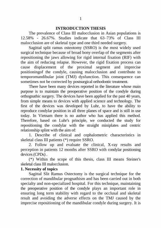
INTRODUCTION THESIS
The prevalence of Class III malocclusion in Asian populations is
12.58% - 26.67%. Studies indicate that 63–73% of Class III
malocclusion are of skeletal type and one third needed surgery.
Sagittal split ramus osteotomy (SSRO) is the most widely used
surgical technique because of broad bony overlap of the segments after
repositioning the jaws allowing for rigid internal fixation (RIF) with
the aim of reducing relapse. However, the rigid fixation process can
cause displacement of the proximal segment and imprecise
positioningof the condyle, causing malocclusion and contribute to
temporomandibular joint (TMJ) dysfunction. This consequence can
sometimes not be corrected by postsurgical orthodontic treatment.
There have been many devices reported in the literature whose main
purpose is to maintain the preoperative position of the condyle during
orthognathic surgery. The devices have been applied for the past 40 years,
from simple means to devices with applied science and technology. The
first of the devices was developed by Luhr, to have the ability to
reproduce condylar position in all three planes of space and is still in use
today. In Vietnam there is no author who has applied this method.
Therefore, based on Luhr's principle, we conducted the study for
repositioning the condylar with the straight miniplates and centric
relationship splint with the aim of:
1. Describe of clinical and cephalometric characteristics in
skeletal class III patients (*) require SSRO.
2. Follow up and evaluate the clinical, X-ray results and
perception in patients 12 months after SSRO with condylar positioning
devices (CPDs) .
(*) Within the scope of this thesis, class III means Steiner's
skeletal class III malocclusion.
1. Necessity of topics
Sagittal Slit Ramus Ostectomy is the surgical technique for the
correction of mandibular prognathism and has been carried out in both
specialty and non-specialized hospital. For this technique, maintaining
the preoperative position of the condyle plays an important role in
ensuring long term stability with regard to the occlusal and skeletal
result and avoiding the adverse effects on the TMJ caused by the
imprecise repositioning of the mandibular condyle during surgery. It is
2
for these reasons that a study should be conducted to assess a method
of for precise repositioning the condylar fragment that is simple and
effective.
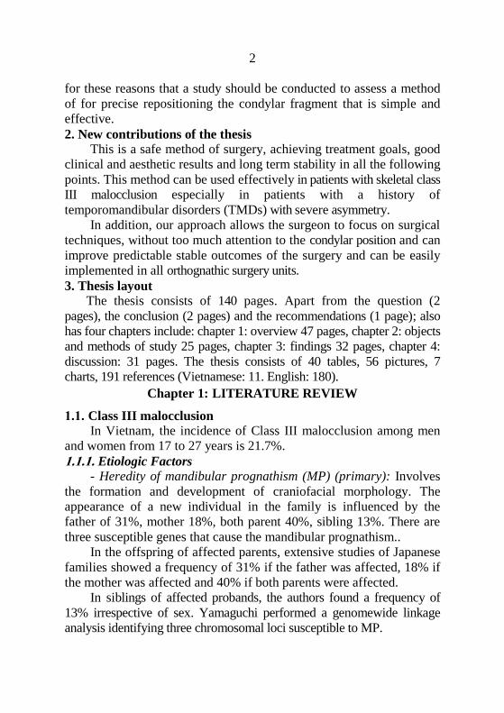
2. New contributions of the thesis
This is a safe method of surgery, achieving treatment goals, good
clinical and aesthetic results and long term stability in all the following
points. This method can be used effectively in patients with skeletal class
III malocclusion especially in patients with a history of
temporomandibular disorders (TMDs) with severe asymmetry.
In addition, our approach allows the surgeon to focus on surgical
techniques, without too much attention to the condylar position and can
improve predictable stable outcomes of the surgery and can be easily
implemented in all orthognathic surgery units.
3. Thesis layout
The thesis consists of 140 pages. Apart from the question (2
pages), the conclusion (2 pages) and the recommendations (1 page); also
has four chapters include: chapter 1: overview 47 pages, chapter 2: objects
and methods of study 25 pages, chapter 3: findings 32 pages, chapter 4:
discussion: 31 pages. The thesis consists of 40 tables, 56 pictures, 7
charts, 191 references (Vietnamese: 11. English: 180).
Chapter 1: LITERATURE REVIEW
1.1. Class III malocclusion
In Vietnam, the incidence of Class III malocclusion among men
and women from 17 to 27 years is 21.7%.
1.1.1. Etiologic Factors
- Heredity of mandibular prognathism (MP) (primary): Involves
the formation and development of craniofacial morphology. The
appearance of a new individual in the family is influenced by the
father of 31%, mother 18%, both parent 40%, sibling 13%. There are
three susceptible genes that cause the mandibular prognathism..
In the offspring of affected parents, extensive studies of Japanese
families showed a frequency of 31% if the father was affected, 18% if
the mother was affected and 40% if both parents were affected.
In siblings of affected probands, the authors found a frequency of
13% irrespective of sex. Yamaguchi performed a genomewide linkage
analysis identifying three chromosomal loci susceptible to MP.

3
- Environmental factors associated with MP (secondary): missing
upper incisors, occlusal interference, habit of protruding the mandible,
loss of balance between cheeks and tongue (enlarged tongue), patients
with cleft lip and palate, endocrine disturbance.
1.1.2. Morphological classification
According to Steiner, based on the ANB angle (SNA and SNB) to
evaluate the sagittal jaws relationship (the disharmony between the
mandible and maxilla). The normal average value of ANB = 2o. By
definition if ANB <0 o: skeletal class III malocclusion.
There are three morphological classification of class III
malformations: mandibular protrusion, maxillary retrusion or a
combination of the two.
1.1.3. Treatment principle
1.1.3.1. Non surgical treatment
- Changing direction of growth.
- Orthodontic camouflage: range can be successfully camouflage:
U1 - SN is 107.36 o ± 6.93
L1 - the mandibular plane is 89.05o ± 7.79
Overjet (mm): 2.11 ± 2.12; Overbite: 1.1 ± 2.15; Wits: -7.16 ± 2.81
SNA angle (o) = 79.56 ± 3.54; SNB = 80.1 ± 4.11; ANB = - 0.46 ± 2.74
The Holdaway angle is 7.2 ˚ to 12 ˚
1.1.3.2. Orthognathic Surgery
Severe patients and nongrowing patients.
Conventional approach: presurgical orthodontic therapy first to
level and align the arches followed by orthognathic surgery. This is
usually followed by a brief period of postsurgical orthodontics to fine
tune the occlusion and parallel the roots.
1.2. Mandibular sagittal split ramus osteotomy
1.2.1. Surgical technique
Bone cuts Osteotomes Separate bone Fixation
Figure 1.1. The technical steps of sagittal split ramus osteotomy

4
1.2.2. Sequelae and complications
- Poor splits, condylar fractures.
- Neurosensory disturbances, neurovascular bundle transection.
- Malposition of the proximal conduylar segment.
- TMDs, condylar resorption.
1.3. TMDs in class III malocclusion patients
- Class III malocclusion may cause abnormal overloading of
the joint.
- The prevalence of TMDs before surgery is 39.4%, degenerative
joint disease is 20%; After surgery: the TMD may be reduced, alleviated or
worsened. However, about 3.7% of patients develop new symptoms. The
main reason is due to displacement of the condyle from surgery. This is of
great concern to surgeons and my contribute to occlusal and skeletal
relapse.
1.3.1. Factors causing the change in condylar position
- General anesthesia: the condyles had dropped vertical
displacement 2.43 mm from the glenoid fossa.
- Various fixation methods.
- the direction and magnitude of movement.
- Technique for correcting mandibular prognathism
1.3.2. The importance of the repositioning the condylar during
surgery
- To improve the stability of the surgical result: changes in condyle
position between 2 - 4 mm and 2° - 4° were observed in 40% patients
treated without the devices, variations in condyle position of more than 2
mm or 2° were not observed in patients treated with CPDs.
- Reducing adverse and/or potentially adverse effects upon the TMJ
by condyles in the fossa with a proper anatomic relationship, minimizing
condylar torque, thus reducing the adverse effects on the TMJ.
5
Patients with pre-existing TMJ dysfunction are less likely to
respond to the joint than normal. Therefore, it is recommended to use
the CPDs to reduce emotional stress.
- Optimizing masticatory efficiency.
1.4. Literature review of CPDs in the world
In 1976 Leonard first used CPDs
Leonard 1976 Luhr 1985 Fujimura 1911 Merten 1992
Positioning with plates, screws:
Baek 2010 Omura 2012
The malposition of the condyles without navigation was 2.4 mm.
Corrective Surgical Segment Navigator positioning was 0.7 mm.
CAD/CAM technology 2013 Electromagnetic navigation
system 2017
Figure 1.2. Methods of positioning the condylar in the world

6
1.5. Literature review of CPDs in Vietnam
Orthognathic surgery was popular fast in the past few years;
therefore, there are studies related to this field and mainly the
introduction of techniques and aesthetic effects such as Tran Cong
Chanh (2010), Nguyen Thu Ha (2012), Nguyen Tan Hung (2015).
Chapter 2: MATERIALS AND METHODS
2.1. Location and time
The study was conducted at Department of Oro-
Maxillofacial Surgery and Stomatology of NHOS in Ho Chi Minh
City and Trieu An Hospital in Ho Chi Minh City from April 2014 to
January 2016.
2.2. Study subjects
2.2.1. Inclusion criteria
- Male and female aged ≥18 years, with Class III malocclusion
(ANB <0o angle and Overjet<0 mm.), required and agreed to SSRO.
- Patients followed the Conventional approach: first orthodontic -
after surgery
- Patients may need: Le Fort I osteotomies in case of severe
malformation; genioplasty to correct the aesthetic of the chin, avoiding
second surgery.
2.2.2. Exclusion criteria
Patients with Class III malocclusion due to traumatic sequelae.
2.3. Methodology
- Study design: uncontrolled clinical intervention.
- N minimum of 36 patients. In fact, 36 patients underwent surgery.
2.3.1. Study variables
2.3.1.1. Variables of repositioning the condylar fragment:
x/X value: assess anteroposterior position of mandibular condyle at
glenoid fossa in terms of proportion, y/Y value: assess superior-inferior
position of mandibular condyle at glenoid fossa in terms of proportion,
ramus inclination, ramus angle, intergonial width, ramus width.

7
Parameters were measured by the Sidexis GX software of the Sirona-
Siemen from Germany.
2.3.1.2. Assessment the TMDs:
We based Helkimo and Paknahad's clinical dysfunction index.
2.3.1.3. Classification of class III malocclusion
- The Vietnamese have a harmonious face [Ms. Trang 1999]:
SNA: Male = 84.13 ± 4.01o Female = 83.87 ± 2.9o
SNB: Male = 80.97 ± 3.24o Female = 80.8 ± 2.41o
- Based on the above values and within the scope of this thesis, we
classify three morphological classification of class III malocclusion as
follows:
A prognathic mandible: normal value + 1 standard deviation.
A hypoplastic maxila: normal value - 1 standard deviation.
Two jaws: A prognathic mandible and A hypoplastic maxila.
2.3.2. Study facilities
Jeil-Korea’s straight miniplates screw system.
8
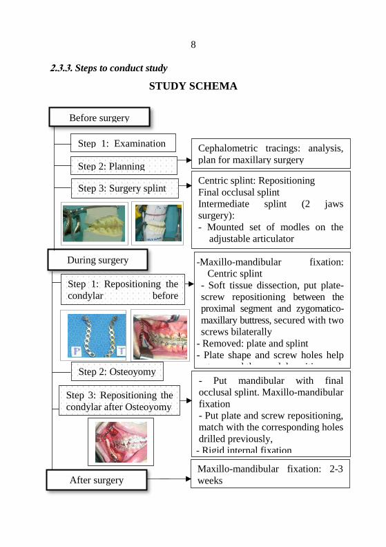
2.3.3. Steps to conduct study
STUDY SCHEMA
Step 1: Examination
Step 2: Planning
Cephalometric tracings: analysis,
plan for maxillary surgery
Centric splint: Repositioning
Final occlusal splint
Intermediate splint (2 jaws
surgery):
- Mounted set of modles on the
adjustable articulator
- Model surgery During surgery
- Put mandibular with final
occlusal splint. Maxillo-mandibular
fixation
- Put plate and screw repositioning,
match with the corresponding holes
drilled previously,
- Rigid internal fixation
Step 3: Repositioning the
condylar after Osteoyomy
Step 2: Osteoyomy
Before surgery
-Maxillo-mandibular fixation:
Centric splint
- Soft tissue dissection, put plate-
screw repositioning between the
proximal segment and zygomatico-
maxillary buttress, secured with two
screws bilaterally
- Removed: plate and splint
- Plate shape and screw holes help
to record the condyl position
Maxillo-mandibular fixation: 2-3
weeks
(MMF) 2-3 tuần
Step 3: Surgery splint
After surgery
Step 1: Repositioning the
condylar before
osteoyomy
9
2.3.4. Follow-up check: 30 days before surgery (T0), 2-3 weeks after
surgery (T1), 1 months after surgery (T2), 3 months after surgery (T3),
6 months after surgery (T4) and 12 months after surgery (T3).
2.3.5. Data analysis
- Data were collected according to the sample medical records.
Data is stored and processed on SPSS 16.0 software and Excel 2013.
- Independent samples T test, paired sample T test, Chi 2, Chi 2
Mc-Nemar.
Chapter 3: RESULTS
We have done study on 36 patients with SSRO with CPDs, the
follow-up period 12 months, we have the following results:
3.1. Clinical and X-ray characteristics of study subjects
3.1.1. Age and gender
Average age: 22, range: 19 - 40 years old. The proportion of female
/ male: 1.6 / 1.
3.1.2. Motivation for surgery
Aesthetic: 19 (52.8%), function: 7 (19.4%), aesthetic and function:
10 (27.8%). Functional reasons: easy to lock of the joint, pain on
movement, do not bite the front teeth, TMJ sounds.
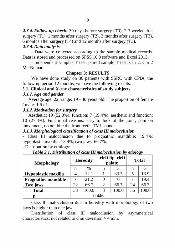
3.1.3. Morphological classification of class III malocclusion
- Class III malocclusion due to prognathic mandible: 19.4%;
hypoplastic maxilla: 13.9%; two jaws: 66.7%.
- Distribution by etiology:
Table 3.1. Distribution of class III malocclusion by etiology
Morphology Heredity
cleft lip–cleft
palate Total
n % n % n %
Hypoplastic maxilla 4 12.1 1 33.3 5 13.9
Prognathic mandible 7 21.2 0 0 7 19.4
Two jaws 22 66.7 2 66.7 24 66.7
Total 33 100.0 3 100.0 36 100.0
p 0.446
Class III malocclusion due to heredity with morphology of two
jaws is higher than one jaw.
Distribution of class III malocclusion by asymmetrical
characteristics: not related to chin deviation ≥ 4 mm.

10
3.1.4. Duration of presurgical orthodontics
Average 31 months (5 - 127 months).
3.1.5. Signs and symptoms of TMDs before surgery
Table 3.2. Distribution of TMDs by malocclusal morphology
Signs and
symptoms
Hypoplastic
maxilla
n (%)
Prognathic
mandible n
(%)
Two jaws
n (%) Total
Normal 2
(40.0)
3
(42.9)
12
(50.0)
17
(47.2)
Mild 3
(60.0)
4
(57.1)
10
(41.7)
17
(47.2)
Severe 0 0 2
(8.3)
2
(5.6)
Total 5
(100.0)
7
(100.0)
24
(100.0)
36
(100.0)
p(1) 0.907
Most patients have no signs or mild signs. Two severe cases have
the disharmony of two-jaws with joint locking.
3.1.6. Characteristics of the incisors occlusion
The average overjet was - 5.5 mm (13 mm; -1.5 mm).
The overbitel can be negative, zero or open bite. There are 16
cases of open bite, the average open bite 2mm (1 mm; - 5 mm).
3.1.7. Characteristics of facial asymmetry
Table 3.3. Characteristics of asymmetry of incisors and chin
Parameters
Right-side
dominance
Left-side
dominance Correct
(n) n Mean
(Range)
n Mean
(Range)
Deviation of
midline L1/U1 8
- 3
(-5; -1) 18
3.5
(1; 9) 10
Deviation of chin
in clinical 7
-2
(-3; -0.5) 17
4
(1.5; 11) 12
Deviation of chin
in film 2
- 0.68
(-0.85; -0.5) 25
4.53
(0.67; 14.11) 9
Note: Negative value was right-side dominance
11
Asymmetrical, left-side dominance more than the right. The number of
left-side dominance in the film was higher than one in the clinical, and
the test showed no significant difference (Kappa = 0.38).
- Occlusal plane cant: 18 cases, average: 1.81mm (0.5 - 6mm)
3.1.8. X-ray characteristics of study subjects
Table 3.4. Preoperrative X-ray characteristics
Angular
measurements
�̅�
(mm) SD
Incisor
measurements �̅�
(mm) SD
SNA 81.45 3.52 U1 – palatal
plane 120.52 6.99
SNB 86.02 4.82 U1 – NA line 30.21 6.48
ANB -4.57 3.11 L1- Mandibular
plane 87.08 6.47
Occlusal plane
- SN 14.62 5.93 L1 – NB line 28.08 6.36
Mandibular
plane - SN 34.74 5.87 U1 – L1 126.46 10.16
Total 36 Total 36
After the orthodontic period before surgery, the angles of the
incisors are not adequately compensated.
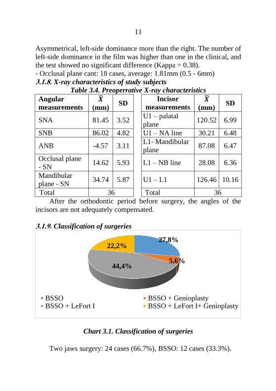
3.1.9. Classification of surgeries
Chart 3.1. Classification of surgeries
Two jaws surgery: 24 cases (66.7%), BSSO: 12 cases (33.3%).
27,8%
5,6%44,4%
22,2%
BSSO BSSO + GenioplastyBSSO + LeFort I BSSO + LeFort I+ Genioplasty

12
Table 3.5. Classification of surgeries by overjet, ANB angle
Surgeries n Overjet
�̅� (mm) ± ĐLC
ANB Angle
�̅� (o) ± ĐLC
Total BSSO 12 -2.92 ± 1.00 - 2.52 ± 2.25
Total BSSO + LeFort I 24 -7.67 ± 2.90 - 5.59 ± 3.00
p 36 < 0.001 0.004
Overjet, ANB angle of two jaws surgery cases have statistically
significant difference more than mandibular surgeries.
3.2. The results of the surgeries with CPDs
3.2.1. Clinical
- Duration of operation: mandibular surgery lasted 150.50 ± 22.92
minutes, two jaws surgery lasted 258.50 ± 30.99 minutes.
- Sequelae and complications:
There was no neurovascular bundle transection.
Duration of neurosensory disturbances:
Lower lip: 5.06 ± 3.23 weeks; chin: 6, 92 ± 3.84 weeks.
Upper lip: 5.33 ± 3.67 weeks; maxillary teeth: 6.25 ± 3.30
weeks.
- Occlusion: after surgery, overbite, overjet: 2.0 mm.
- Symptoms of temporomandibular joint:
Chart 3.2. TMDs before and after surgery
After removing intermaxillary fixation, patient were still limited
to opening mouth and recovered after a few months. After 12 months,
the opening mouth was as normal as before surgery. No new TMDs
case.
0
20
40
T1 T3 T4 T5
n
Normal Mild Servere
13
3.2.2. X-ray
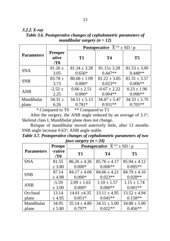
Table 3.6. Postoperative changes of cephalometric parameters of
mandibular surgery (n = 12)
Parameters
Preoper
ative
/T0
Postoperative X̅ (o) ± SD / p
T1 T4 T5
SNA 81.26 ±
3.05
81.34 ± 3.28
0.656*
81.15± 3.28
0.447**
81.53 ± 3.00
0.448**
SNB 83.78 ±
3.73
80.68 ± 1.09
0.000*
81.22 ± 3.85
0.023**
81.31 ± 3.57
0.006**
ANB -2.52 ±
2.25
0.66 ± 2.51
0.000*
-0.67 ± 2.22
0.004**
0.23 ± 1.96
0.096**
Mandibular
plane
34.31 ±
6.26
34.51 ± 5.13
0.781*
34.47 ± 5.47
0.931**
34.33 ± 5.70
0.705**
* Compared to T0 ** Compared to T1
After the surgery, the ANB angle reduced by an average of 3.1o;
Skeletal class I, Mandibular plane does not change.
Relapse of mandibular moved anteriorly little, after 12 months
SNB angle increase 0.63o; ANB angle stable.
Table 3.7. Postoperative changes of cephalometric parameters of two
jaws surgery (n = 24)
Parameters
Preope
-rative
/T0
Postoperative X̅ (o) ± SD / p
T1 T4 T5
SNA 81.55
± 3.80
86.26 ± 4.26
0.000*
85.76 ± 4.17
0.006**
85.94 ± 4.12
0.095**
SNB 87.14
± 4.98
84.17 ± 4.06
0.000*
84.66 ± 4.21
0.023**
84.79 ± 4.16
0.028**
ANB -5.59
± 3.00
2.09 ± 1.63
0.000*
1.10 ± 1.57
0.000**
1.15 ± 1.74
0.001**
Occlusal
plane
13.14
± 4.95
14.61 ±4.35
0.051*
13.11 ± 4.95
0.045**
13.52 ± 4.94
0.158**
Mandibular
plane
34.95
± 5.80
35.14 ± 4.80
0.797*
34.51 ± 5.00
0.022**
34.88 ± 5.00
0.456**

14
After surgery, SNA angle increased by 4.71o on average, SNB
angle decreased by 2.97o on average; Skeletal class I (p <0.001).
Occlusal plane and mandibular plane did not change.
After 12 months, ANB angle decreased 0.94o; Skeletal class I.
Table 3.8. Postoperative changes of B point of mandibular surgery
(n = 12)
B
point
Preoperative
/T0
Postoperative X̅ (mm) ± SD / p
T1 T4 T5
Bx 63.17 ± 6.47 58.51 ± 6.76
0.000*
59.31 ± 6.68
0.107**
59.43 ± 6.31
0.050**
By 90.39 ± 4.86 89.67 ± 4.75
0.060*
88.42 ± 4.34
0.000**
89.03 ± 4.94
0.029**
After surgery: mandibular (B point) moved posteriorly on
average 4.77 mm, moved superiorly on average 0.72 mm.
After 12 months, B point relapsed superiorly 0.64 mm.
Table 3.9. Postoperative changes of A, B points of two jaws surgery
(n = 24)
A, B
points
Preoperative
/T0
Postoperative X̅ (mm) ± SD / p
T1 T4 T5
Ax 61.07
± 6.36
66.39 ± 6.86
0.000*
65.75 ± 6.74
0.039**
65. 45 ± 7.00
0.010**
Ay 52.70
± 4.03
53.33 ± 4.19
0.050*
53.09 ± 4.52
0.334**
53.28 ± 4.15
0.797**
Bx 69.85
± 10.41
64.57 ± 9.14
0.000*
65.80 ± 9.31
0.000**
65.63± 9.43
0.022**
By 90.81
± 6.93
90.57 ± 5.90
0.677*
89.67 ± 6.67
0.075**
89.76 ± 6.46
0.047**
After surgery: Maxilla (A point) moved anteriorly on average
5.32 mm, moved inferiorly 0.63 mm. Mandibular (B point) moved
posteriorly on average 5.28 mm behind; moved superiorly 0.24 mm.
After 12 months, A point was almost unchanged, B point
relapsed anteriorly and superiorly 0.17mm and 0.81mm respectively,
Compared to T1.
15
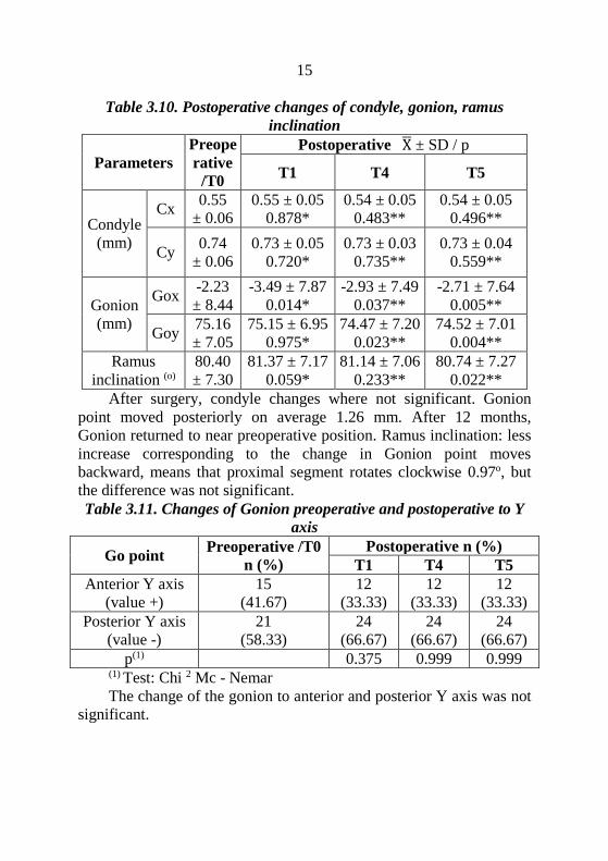
Table 3.10. Postoperative changes of condyle, gonion, ramus
inclination
Parameters
Preope
rative
/T0
Postoperative X̅ ± SD / p
T1 T4 T5
Condyle
(mm)
Cx 0.55
± 0.06
0.55 ± 0.05
0.878*
0.54 ± 0.05
0.483**
0.54 ± 0.05
0.496**
Cy 0.74
± 0.06
0.73 ± 0.05
0.720*
0.73 ± 0.03
0.735**
0.73 ± 0.04
0.559**
Gonion
(mm)
Gox -2.23
± 8.44
-3.49 ± 7.87
0.014*
-2.93 ± 7.49
0.037**
-2.71 ± 7.64
0.005**
Goy 75.16
± 7.05
75.15 ± 6.95
0.975*
74.47 ± 7.20
0.023**
74.52 ± 7.01
0.004**
Ramus
inclination (o)
80.40
± 7.30
81.37 ± 7.17
0.059*
81.14 ± 7.06
0.233**
80.74 ± 7.27
0.022**
After surgery, condyle changes where not significant. Gonion
point moved posteriorly on average 1.26 mm. After 12 months,
Gonion returned to near preoperative position. Ramus inclination: less
increase corresponding to the change in Gonion point moves
backward, means that proximal segment rotates clockwise 0.97o, but
the difference was not significant.
Table 3.11. Changes of Gonion preoperative and postoperative to Y
axis
Go point Preoperative /T0
n (%)
Postoperative n (%)
T1 T4 T5
Anterior Y axis
(value +)
15
(41.67)
12
(33.33)
12
(33.33)
12
(33.33)
Posterior Y axis
(value -)
21
(58.33)
24
(66.67)
24
(66.67)
24
(66.67)
p(1) 0.375 0.999 0.999 (1) Test: Chi 2 Mc - Nemar
The change of the gonion to anterior and posterior Y axis was not
significant.
16
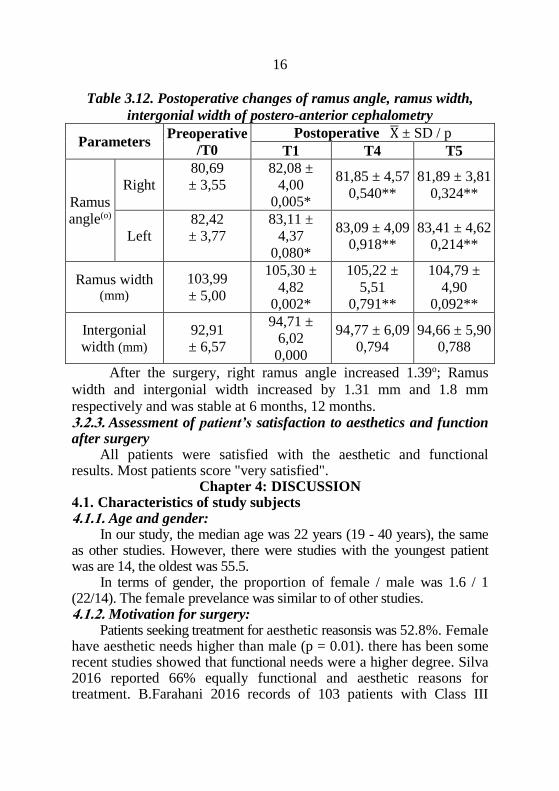
Table 3.12. Postoperative changes of ramus angle, ramus width,
intergonial width of postero-anterior cephalometry
Parameters Preoperative
/T0
Postoperative X̅ ± SD / p
T1 T4 T5
Ramus
angle(o)
Right
80,69
± 3,55
82,08 ±
4,00
0,005*
81,85 ± 4,57
0,540**
81,89 ± 3,81
0,324**
Left
82,42
± 3,77
83,11 ±
4,37
0,080*
83,09 ± 4,09
0,918**
83,41 ± 4,62
0,214**
Ramus width (mm)
103,99
± 5,00
105,30 ±
4,82
0,002*
105,22 ±
5,51
0,791**
104,79 ±
4,90
0,092**
Intergonial
width (mm)
92,91
± 6,57
94,71 ±
6,02
0,000
94,77 ± 6,09
0,794
94,66 ± 5,90
0,788
After the surgery, right ramus angle increased 1.39o; Ramus
width and intergonial width increased by 1.31 mm and 1.8 mm
respectively and was stable at 6 months, 12 months. 3.2.3. Assessment of patient’s satisfaction to aesthetics and function after surgery
All patients were satisfied with the aesthetic and functional results. Most patients score "very satisfied".
Chapter 4: DISCUSSION 4.1. Characteristics of study subjects 4.1.1. Age and gender:
In our study, the median age was 22 years (19 - 40 years), the same as other studies. However, there were studies with the youngest patient was are 14, the oldest was 55.5.
In terms of gender, the proportion of female / male was 1.6 / 1 (22/14). The female prevelance was similar to of other studies. 4.1.2. Motivation for surgery:
Patients seeking treatment for aesthetic reasonsis was 52.8%. Female have aesthetic needs higher than male (p = 0.01). there has been some recent studies showed that functional needs were a higher degree. Silva 2016 reported 66% equally functional and aesthetic reasons for treatment. B.Farahani 2016 records of 103 patients with Class III

17
sagittal skeletal patterns had higher percentages of grade “Very great need for treatment”; 95,6%. 4.1.3. Morphology of class III malocclusion
In our study, class III malocclusion due to two jaws were 66.7%; Of which 66.7% of cases were hereditary. These were cases in which their relatives said that they had normal face in the childhood, the family recognized the mandibular excess during adolescence. At the time of surgery, the patients had stopped growing and surgery was the only way to correct the malocclusion, rather than compensate for and camouflage the mandibular deformity. Patients with cleft lips and palates were usually due to the deficiency in the mailla.
About asymmetry, in our study. The majority of the patients had left-side dominance, mean 4mm. Facial asymmetry was concomitantly observed in 21 to 85% of patients and in 48% of skeletal Class III cases. The 4 mm difference in chin was significant in asymmetric diagnosis and treatment. 4.1.4. The duration of preoperative orthodontic treatment
In our study, the average duration of preoperative orthodontic treatment was 31 months (5 - 127 months). The longest case was 127 months (over 10 years) in cleft patients. Although the patient was orthodontic treatment in the childhood, she must wait until adulthood for surgery.
According to literature, duration of preoperative orthodontic treatment was 2 to 12 months depending on the degree of tooth misalignment. Recently, performing mandibular setback surgery in patients with skeletal Class III malocclusion without preoperative orthodontic treatment was proposed. This approach, in terms of patients' psychology, was more satisfactory as it improves early aesthetics, but disadvantage was that optimal positioning of the mandible was not possible due to uncorrected dental compensations such as proclination of the maxillary incisors and retroclination of the mandibular incisors. SSRO without presurgical orthodontic treatment was less stable than conventional orthognathic surgery as mandibular shift to a more balanced position. Before performing a surgery-first approach, skeletal stability needs to be considered. Horizontal relapses of patients with preoperative orthodontic treatment was 0.9 - 1.6 mm, without preoperative orthodontic treatment was 2.0 - 2.4 mm. Patients with a relapse greater than 3 mm comprised 39.1% of the surgery-first group compared with 15.8% of the conventional surgery group.
18
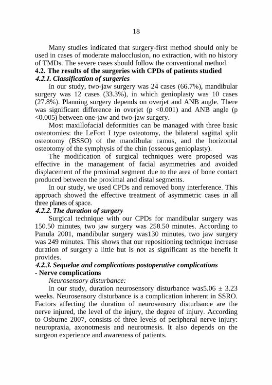
Many studies indicated that surgery-first method should only be
used in cases of moderate malocclusion, no extraction, with no history of TMDs. The severe cases should follow the conventional method. 4.2. The results of the surgeries with CPDs of patients studied 4.2.1. Classification of surgeries
In our study, two-jaw surgery was 24 cases (66.7%), mandibular surgery was 12 cases (33.3%), in which genioplasty was 10 cases (27.8%). Planning surgery depends on overjet and ANB angle. There was significant difference in overjet (p <0.001) and ANB angle (p <0.005) between one-jaw and two-jaw surgery.
Most maxillofacial deformities can be managed with three basic osteotomies: the LeFort I type osteotomy, the bilateral sagittal split osteotomy (BSSO) of the mandibular ramus, and the horizontal osteotomy of the symphysis of the chin (osseous genioplasty).
The modification of surgical techniques were proposed was effective in the management of facial asymmetries and avoided displacement of the proximal segment due to the area of bone contact produced between the proximal and distal segments.
In our study, we used CPDs and removed bony interference. This approach showed the effective treatment of asymmetric cases in all three planes of space. 4.2.2. The duration of surgery
Surgical technique with our CPDs for mandibular surgery was 150.50 minutes, two jaw surgery was 258.50 minutes. According to Panula 2001, mandibular surgery was130 minutes, two jaw surgery was 249 minutes. This shows that our repositioning technique increase duration of surgery a little but is not as significant as the benefit it provides. 4.2.3. Sequelae and complications postoperative complications - Nerve complications
Neurosensory disturbance: In our study, duration neurosensory disturbance was5.06 ± 3.23
weeks. Neurosensory disturbance is a complication inherent in SSRO. Factors affecting the duration of neurosensory disturbance are the nerve injured, the level of the injury, the degree of injury. According to Osburne 2007, consists of three levels of peripheral nerve injury: neuropraxia, axonotmesis and neurotmesis. It also depends on the surgeon experience and awareness of patients.

19
Neurovascular bundle transection: There are no cases in our study. Maybe the use of separators and
splitters, without chisels leads to a lower incidence of persistent postoperative hypoesthesia after SSRO. According to literature study, complete transection of the inferior alveolar nerve was 1.5%. Patients with postoperative hypoesthesia and affective activities were 7.4%. - Poor splits
In our study, there was a case of bad split on lateral cortical bone, little broken piece, just increased the length of the bone plate.
The rate of bad splits was 2.3% - 3.9%. The presence of impacted third molars during surgery and incomplete inferior border osteotomy can increase the risk of a bad split. - Condylar resorption
In our study, we did not encounter any case of condylar resorption with a follow-up 12 months after orthognathic surgery. The reason was because our study were CPDs that can not lead to TMJ overloading. The cause of condylar resorption may be due to changes in biomechanical loading on the TMJ. Postoperative condylar resorption was 7.5% (224/2994 cases).
The cause of convex capillaries may be due to changes in mechanical force on the temporomandibular joint. Postoperative prolapse rate was about 7.5% (224/2994 cases). 4.2.4. Signs and symptoms of temporomandibular joint
Our study there was no change in condylar position from preoperative to postoperative using the CPDs. The results showed that preoperative the incidence of TMDs was 52.8%, and postoperative TMDs was 22%. The symptoms occasionally were TMJ sounds when opening widely or or yawning, but there was no pain. There were no new onset cases of TMD.
After orthognathic surgery, the occlusion reach a state of equilibrium, so it is beneficial for TMJ, thus significantly reducing symptoms of preoperative TMDs. However, there is still a 3.7% incidence of new onset TMD after surgery. One of the main causes of postoperative TMDs is imprecise condyle position from surgery, that may result in TMJ internal derangements. Rigid internal fixation may altered condylar positioning. Therefore, many authors recommend the use CPDs for SSRO, especially when rigid internal fixation is used. 4.2.5. Characteristics of occlusion
Before surgery: overjet -5.5 mm (13; -1.5 mm). After removed intermaxillary fixation, overbite and overjet was 1 - 2 mm. Follow up 12 months after surgery, occlusion was stable. In our study we used CPDing occlusal/skeletal relapse.
20
4.2.6. Characteristics of X-ray
Table 4.1. Compared to preoperrative skeletal characteristics of authors
Authors SNA SNB ANB Occlusalplane Mandibula
r plane
Steiner 82 80 2 32
Trang 83.98
± 3.45
80.88
± 2.83
3.10
± 1.87
30.72
± 3.84
Choi (2016)
n=18
79.7
± 2.0
82.9
± 3.0
17.0
± 4.0
Tseng (2011)
n= 40
82.57
± 4.03
87.65
± 3.78
- 5.29
± 3.05
35.92
± 6.03
Benyahia
(2011) n= 25
78.08
± 4.47
82.48
± 4.33
- 4.41
± 3.13
33.48
± 7.19
N.T.Ha
(2017) n=36
81.45
± 3.52
86.02
± 4.82
- 4.57
± 3.11
14.62
± 5.93
34.74
± 5.87
In our study, when the SNB angle was high, opening of the mandibular plane developed. - Postoperative skeletal changing
- Mandibular surgery After surgery, the SNB angle decreased by 3.1o. Point B moved
posteriorly on average 4.77 mm, moved superiorly on average 0.72 mm. The distal segment was setback and intraoperative counterclockwise rotation to fit on the upper teeth. After surgery, skeletal class I with ANB angle was 0.66o.
About relapse, after 12 months SNB angle increase 0.63o; B point moved superiorly 0,64 mm compared to T1. It is possible that the CPD reduce the incidence and magnitude of relapse.This was correct by postoperative orthodontic treatment, so it maintained skeletal class I with ANB angle was 0.23o, no significant difference from T1 (p = 0.096).
- Two-jaw surgery After surgery, SNA angle increased by 4.71o on average, SNB angle
decreased by 2.97o on average. A point moved anteriorly on average 5.32 mm, moved inferiorly 0.63 mm; B point moved posteriorly on average 5.28 mm behind; moved superiorly 0.24 mm. Angle of upper and lower incisors was significantly reduced. Skeletal class I with ANB = 2.09o (p <0.001).
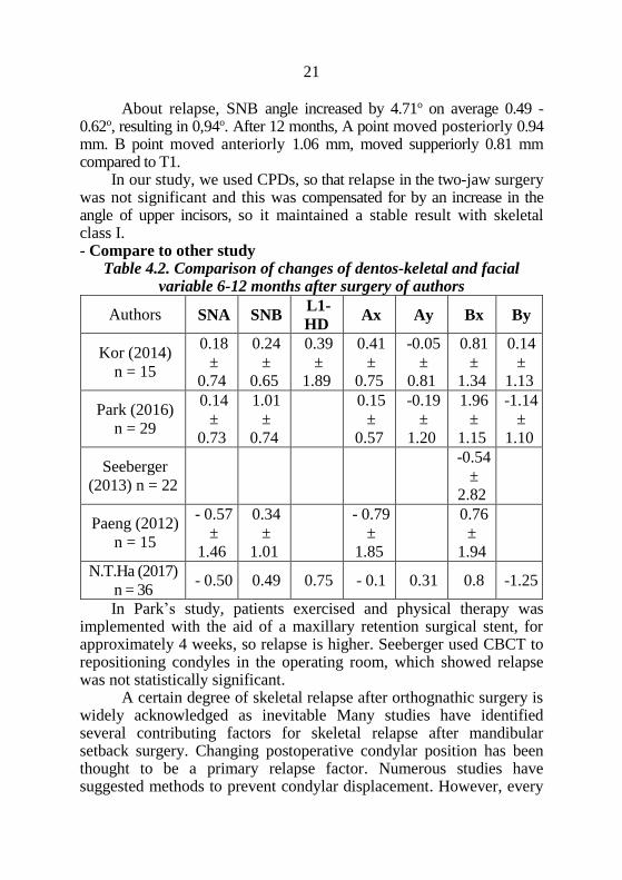
21
About relapse, SNB angle increased by 4.71o on average 0.49 -
0.62o, resulting in 0,94o. After 12 months, A point moved posteriorly 0.94 mm. B point moved anteriorly 1.06 mm, moved supperiorly 0.81 mm compared to T1.
In our study, we used CPDs, so that relapse in the two-jaw surgery was not significant and this was compensated for by an increase in the angle of upper incisors, so it maintained a stable result with skeletal class I. - Compare to other study
Table 4.2. Comparison of changes of dentos-keletal and facial variable 6-12 months after surgery of authors
Authors SNA SNB L1-
HD Ax Ay Bx By
Kor (2014)
n = 15
0.18
±
0.74
0.24
±
0.65
0.39
±
1.89
0.41
±
0.75
-0.05
±
0.81
0.81
±
1.34
0.14
±
1.13
Park (2016)
n = 29
0.14
±
0.73
1.01
±
0.74
0.15
±
0.57
-0.19
±
1.20
1.96
±
1.15
-1.14
±
1.10
Seeberger
(2013) n = 22
-0.54
±
2.82
Paeng (2012)
n = 15
- 0.57
±
1.46
0.34
±
1.01
- 0.79
±
1.85
0.76
±
1.94
N.T.Ha (2017)
n = 36 - 0.50 0.49 0.75 - 0.1 0.31 0.8 -1.25
In Park’s study, patients exercised and physical therapy was implemented with the aid of a maxillary retention surgical stent, for approximately 4 weeks, so relapse is higher. Seeberger used CBCT to repositioning condyles in the operating room, which showed relapse was not statistically significant.
A certain degree of skeletal relapse after orthognathic surgery is widely acknowledged as inevitable Many studies have identified several contributing factors for skeletal relapse after mandibular setback surgery. Changing postoperative condylar position has been thought to be a primary relapse factor. Numerous studies have suggested methods to prevent condylar displacement. However, every

22
method has relapse more or less. If these changes are less and are adjusted by changes in occlusion and the physiological responses of the TMJ after surgery, the results of the surgery will be stable.
Our study showed that pre- and post-operative condylar position changes as follows:
- On lateral cephalograms, after surgery, Gonion points moved posteriorly 1.26 mm. After 12 months, horizontal relapse is 0.78mm and 0.63mm compared with T1.
Ramus inclination did not significantly change after surgery (p = 0.059), which mean that intraoperative clockwise rotation of the proximal segment was 0.97o. After 12 months, counterclockwise rotation relapse was 0.63o but did not affect the results of surgery.
- On posteroanterior cephalograms, intergonial width, ramus width changed, which mean that the condyle inwardly rotate on coronal plane. After surgery, right ramus angle increased by 1.39o, ramus width, intergonial width increased by 1.31 mm and 1.8 mm, respectively, were stable at 6-12 months.
Park’s study in 2016 did not used CPDs, so proximal segment angle was -2.59 ± 1.09o. After 6 months, -2.13 ± 0.99o versus T1. The Seeberger’s study in 2013 used CBCT for repositioning the condylar fragment in the operating room so ramus angle increased little by 0.64o. Ko’s study in 2009, ramus width decreased by 3.4 mm.
In general, the small changes in our study resembled that of other authors and did not affect stability.
In orthognathic surgery, all procedures for the repositioning of the mandible that disrupt the teeth-condyle-cranial base relationship require proper positioning when repositioning (Bethge 2015). This problem has led to many reports of the importance of condye position. However, postoperative condylar position change and its influence TMDs are still controversial (Catherine 2016). Seeberger’s study in 2013 reported that 1 in 22 patients intraoperative revision was indicated and performed because the intercondylar distance exceeded our limit of 1 mm. Due to the occurrence of several complications, The main consensus remains condyles should be positioned inside the fossa and positioned to maintain a long-term stable occlusal result, as well as healthy TMJs, absence of pain and adequate function. Since then, many methods and devices have been proposed for this purpose.
In Vietnam, our method is an immediate practical solution that is effective in controlling the condyle repositioning in orthognathic
23
surgery. In terms of clinical applicability, our method is easy to apply, allowing the surgeon to focus on surgical techniques, not to worry too much on the condyle position, especially in cases of aymmetrical or TMDs. 4.3. Evaluation of function and aesthetics after surgery
- Function: In our study, after surgery, the teeth contact with each other more, so chewing better, improving the masticatory system, reduce TMDs. This proves that orthognathic surgery is not only a aesthetic surgery but also a approach for patients with TMDs.
- Aesthetics: In our study, 83.3% of patients were "completely satisfied", 6 patients were classified as "satisfied" because of mild asymmetry of the chin. There is no need for additional surgery in any of the patients regardless of the degree of satisfaction.
Questions of patient satisfaction showed that the 92.7% of patients were satisfied with surgerical and would recommend that facial disharmony people undergo surgical correction in spite of the short term postoperative discomfort.
CONCLUSIONS
Studying and conducting BSSO for treatment of 36 patients with skeletal class III using CPDs (*) allows the following conclusions:
1. Clinical and X-ray characteristics of before surgery The average age was twenty-two. The ratio of female/male was
1.6/1. The motivation for surgery was 52.8% facial aesthetics. Morphological classification of class III malocclusion due to maxillary
deficiency and mandiblar excess: 66.7%. Overject was -5.5 mm. The most prominent characteristic of clinical asymmetry is left-side dominance (4mm on average). Mild TMDs were 47.2%, seriously was 5.6%.
On X-ray, ANB average -4.57°. the mandibular plane angle was large, average 34.74o.
Duration of preoperative orthodontic treatment over two years was 66.7%. After this period, maxillary incisors was more protrusive than (30.21o) the normal value under Steiner (22o).
2. Following, evaluation of clinical and X-ray results and perception of patients 12 months after surgery
Two-jaw surgery was 66.7%. There was less relapse at follow-up timings. After 12 months, the skeletal was maintained class I with ANB angle was 0.23o (mandibular surgery); 1.15o (two-jaw surgery) and overject average 2mm.
24
The postoperative TMDs reduced to 22.2%, no worsening or new cases. All patients were satisfied with improved function and aesthetics. About the effectiveness of CPDs: - On lateral cephalograms, after surgery, the displacement of condyle
was not significant, the proximal segment was counterclockwise rotation on average 0.97o. After 12 months returned to position as before surgery. SNB angle increased by 0.63o (p> 0.05).
- On posteroanterior cephalograms, after surgery, intergonial width and ramus width increased 1.8 mm and 1.31 mm, respectively. After 12 months, the change was not significant (p> 0.05).
BSSO with CPDs by straight miniplates screw system and centric relation splint was safe, achieved treatment goals, and provided good clinical outcomes. The surgeon is able concentrate on surgical techniques, not worry as much about the condyle position leading to a more predictable surgical outcome.
This approach can be used effectively to treat skeletal class III cases, especially in patients with a preexisting of TMDs or severe asymmetry.
PROPOSALS
1 / The method of CPDs by straight miniplates screw system and centric relation splint presented in this thesis is a simple, low cost, and effective technique that can be used by any surgeon performing sagittal ramus oseotomies. This is especially uselful in patients with a history of TMDs, severe asymmetry, and in patients having mandibular resection without teeth to locate occlusion.
2/ Patients requiring orthognathic surgery and with clinical signs of craniomandibular and masticatory system disorder should be entitled to medical insurance.
3/ The concept of surgery first should also be considered for patients with mild malocclusion to improve aesthetics early.
4/ Future studies need to continue in the field of orthognathic surgery with appropriate software for diagnosis and treatment planning, occlusal splint fabrication using CAD/CAM technology, ultrasonic assited repositioning and electromagnetic navigation system.
Related Documents

![[CK]-Động học cắn khớp](https://static.cupdf.com/doc/110x72/55721388497959fc0b927bcd/ck-dong-hoc-can-khop.jpg)


