The Development of a Mouse Model of Ovarian Endosalpingiosis Sarah K. Bristol-Gould, Christina G. Hutten, Charles Sturgis, Signe M. Kilen, Kelly E. Mayo, and Teresa K. Woodruff Department of Neurobiology and Physiology (S.K.B.-G., T.K.W.), Department of Biochemistry, Molecular Biology and Cell Biology (K.E.M.), and Center for Reproductive Science (C.G.H., S.M.K., K.E.M., T.K.W.), Northwestern University, and Evanston Northwestern Hospital (C.S.), Evanston, Illinois 60208; and Department of Medicine (T.K.W.) Feinberg School of Medicine, Northwestern University, and Robert H. Lurie Comprehensive Cancer Center of Northwestern University (T.K.W.), Chicago, Illinois 60611 Pelvic pain is a common presenting ailment in women often linked to ovulation, endometriosis, early pregnancy, ovarian cancer, and cysts. Clear differential diagnosis for each con- dition caused by these varied etiologies is difficult and may slow the delivery of therapy that, in the case of ovarian cancer, could be fatal. Ovarian endosalpingiosis, a pelvic condition typified by the presence of cystic glandular structures lined by benign tubal/salpingeal epithelium, is also associated with pelvic pain in women. The exact cellular antecedents of these epithelial lined cystic structures are not known, nor is there a known link to ovarian cancer. A mouse model of ovarian endosalpingiosis has been developed by directing a dominant- negative version of the TGF- transcription factor, Smad2, to the ovary using the Mu ¨ llerian-inhibiting substance promoter (MIS-Smad2-dn). Female mice develop an ovarian endosalp- ingeal phenotype as early as 3 months of age. Importantly, cysts continuous with the ovarian surface epithelial have been identified, indicating that these cyst cells may be derived from the highly plastic ovarian surface epithelial cell layer. A second transgenic mouse model that causes loss of activin action (inhibin -subunit transgenic mice) develops similar cystic structures, supporting a TGF-/activin/Smad2 depen- dence in the onset of this disease. (Endocrinology 146: 5228 –5236, 2005) E NDOSALPINGIOSIS is a histopathologically recog- nized and not infrequently encountered condition identified in extratubal pelvic tissues in both pre- and post- menopausal women (1). It may be associated with pelvic pain and is characterized by the presence of extrasalpingeal cystic spaces lined by cells that phenocopy a tubal-type epithelium. Human ovarian endosalpingiosis is most commonly diag- nosed as an incidental finding when tissues are surgically resected for other reasons. Other generally involved sites where endosalpingiotic lesions are found include the uterine serosa, omentum, urinary bladder, bowel wall, and pelvic lymph nodes (1– 4). Neither the origin nor the transformative potential of these ectopic cells are understood; however, in many cases of endosalpingiosis additional pelvic pathologies are present (5). Case reports have found endosalpingiosis to be associated with ovarian surface papillary tumors, adeno- carcinoma of the fallopian tubes, and endometriosis (5– 8). In short of an animal model of endosalpingiosis coupled with the inability to track its course and lack of clinical correlation, there has been limited progress in understand- ing the condition and its implications to the health of affected women. One of the prevailing views of the onset of ovarian cancer is the transformation of cells that comprise the surface epithelium into neoplastic cysts, possibly caused by the process of ovulation (9 –12). The TGF- superfamily of ligands, particularly TGF- and activin, have been implicated in the regulation of ovulation; therefore, we targeted the sig- naling pathway used by these two ligands in developing a mouse model that might generate early stage ovarian surface epithelial (OSE)-derived neoplasms. TGF- is known to be im- portant in the regulation of the OSE, whereas activin functions in the control of wound repair and scar formation (13–16). Both ligands use Smad2 and Smad3 as common transcriptional co- regulators; therefore, we directed a Smad2 dominant-negative (Smad2-dn) transgene to the ovary. The development of animal models of complex disease, especially those that present in a sexually dimorphic manner, has progressed significantly in the past 2 yr. Mouse models of endometriosis and endometrioid ovarian cancer have been developed using targeted genetic deletion of the Pten and K-ras genes in the OSE (17). Advances have been made in BRCA1 and BRCA2 mouse models that phenocopy breast and ovarian cancer, and Smad7 up-regulation appears to be important to the TGF--resistant endometrial cancers (18, 19). Our model targeted the ovary to reduce the TGF-/ activin signal transduction pathway. These studies suggest that the origin of the endosalpingiotic lesions are from the Mu ¨ llerian epithelium and provide additional evidence that this cell type has a variety of endpoints to which it develops based on the genetic background and the microenvironment in which the cell exists. First Published Online September 1, 2005 Abbreviations: CL, Corpora lutea; FITC, fluorescein isothiocyanate; MIS, Mu ¨ llerian-inhibiting substance; MOF, multioocytic follicles; OSE, ovarian surface epithelium; Smad2-dn, Smad2 dominant-negative. Endocrinology is published monthly by The Endocrine Society (http:// www.endo-society.org), the foremost professional society serving the endocrine community. 0013-7227/05/$15.00/0 Endocrinology 146(12):5228 –5236 Printed in U.S.A. Copyright © 2005 by The Endocrine Society doi: 10.1210/en.2005-0697 5228 on May 19, 2006 endo.endojournals.org Downloaded from

Welcome message from author
This document is posted to help you gain knowledge. Please leave a comment to let me know what you think about it! Share it to your friends and learn new things together.
Transcript

The Development of a Mouse Model ofOvarian Endosalpingiosis
Sarah K. Bristol-Gould, Christina G. Hutten, Charles Sturgis, Signe M. Kilen, Kelly E. Mayo, andTeresa K. Woodruff
Department of Neurobiology and Physiology (S.K.B.-G., T.K.W.), Department of Biochemistry, Molecular Biology and CellBiology (K.E.M.), and Center for Reproductive Science (C.G.H., S.M.K., K.E.M., T.K.W.), Northwestern University, andEvanston Northwestern Hospital (C.S.), Evanston, Illinois 60208; and Department of Medicine (T.K.W.) Feinberg School ofMedicine, Northwestern University, and Robert H. Lurie Comprehensive Cancer Center of Northwestern University (T.K.W.),Chicago, Illinois 60611
Pelvic pain is a common presenting ailment in women oftenlinked to ovulation, endometriosis, early pregnancy, ovariancancer, and cysts. Clear differential diagnosis for each con-dition caused by these varied etiologies is difficult and mayslow the delivery of therapy that, in the case of ovarian cancer,could be fatal. Ovarian endosalpingiosis, a pelvic conditiontypified by the presence of cystic glandular structures linedby benign tubal/salpingeal epithelium, is also associated withpelvic pain in women. The exact cellular antecedents of theseepithelial lined cystic structures are not known, nor is therea known link to ovarian cancer. A mouse model of ovarianendosalpingiosis has been developed by directing a dominant-
negative version of the TGF-! transcription factor, Smad2, tothe ovary using the Mullerian-inhibiting substance promoter(MIS-Smad2-dn). Female mice develop an ovarian endosalp-ingeal phenotype as early as 3 months of age. Importantly,cysts continuous with the ovarian surface epithelial havebeen identified, indicating that these cyst cells may be derivedfrom the highly plastic ovarian surface epithelial cell layer. Asecond transgenic mouse model that causes loss of activinaction (inhibin "-subunit transgenic mice) develops similarcystic structures, supporting a TGF-!/activin/Smad2 depen-dence in the onset of this disease. (Endocrinology 146:5228–5236, 2005)
ENDOSALPINGIOSIS is a histopathologically recog-nized and not infrequently encountered condition
identified in extratubal pelvic tissues in both pre- and post-menopausal women (1). It may be associated with pelvic painand is characterized by the presence of extrasalpingeal cysticspaces lined by cells that phenocopy a tubal-type epithelium.Human ovarian endosalpingiosis is most commonly diag-nosed as an incidental finding when tissues are surgicallyresected for other reasons. Other generally involved siteswhere endosalpingiotic lesions are found include the uterineserosa, omentum, urinary bladder, bowel wall, and pelviclymph nodes (1–4). Neither the origin nor the transformativepotential of these ectopic cells are understood; however, inmany cases of endosalpingiosis additional pelvic pathologiesare present (5). Case reports have found endosalpingiosis tobe associated with ovarian surface papillary tumors, adeno-carcinoma of the fallopian tubes, and endometriosis (5– 8).In short of an animal model of endosalpingiosis coupledwith the inability to track its course and lack of clinicalcorrelation, there has been limited progress in understand-ing the condition and its implications to the health ofaffected women.
One of the prevailing views of the onset of ovarian
cancer is the transformation of cells that comprise thesurface epithelium into neoplastic cysts, possibly causedby the process of ovulation (9 –12). The TGF-! superfamilyof ligands, particularly TGF-! and activin, have been implicatedin the regulation of ovulation; therefore, we targeted the sig-naling pathway used by these two ligands in developing amouse model that might generate early stage ovarian surfaceepithelial (OSE)-derived neoplasms. TGF-! is known to be im-portant in the regulation of the OSE, whereas activin functionsin the control of wound repair and scar formation (13–16). Bothligands use Smad2 and Smad3 as common transcriptional co-regulators; therefore, we directed a Smad2 dominant-negative(Smad2-dn) transgene to the ovary.
The development of animal models of complex disease,especially those that present in a sexually dimorphic manner,has progressed significantly in the past 2 yr. Mouse modelsof endometriosis and endometrioid ovarian cancer have beendeveloped using targeted genetic deletion of the Pten andK-ras genes in the OSE (17). Advances have been made inBRCA1 and BRCA2 mouse models that phenocopy breastand ovarian cancer, and Smad7 up-regulation appears to beimportant to the TGF-!-resistant endometrial cancers (18,19). Our model targeted the ovary to reduce the TGF-!/activin signal transduction pathway. These studies suggestthat the origin of the endosalpingiotic lesions are from theMullerian epithelium and provide additional evidence thatthis cell type has a variety of endpoints to which it developsbased on the genetic background and the microenvironmentin which the cell exists.
First Published Online September 1, 2005Abbreviations: CL, Corpora lutea; FITC, fluorescein isothiocyanate;
MIS, Mullerian-inhibiting substance; MOF, multioocytic follicles; OSE,ovarian surface epithelium; Smad2-dn, Smad2 dominant-negative.Endocrinology is published monthly by The Endocrine Society (http://www.endo-society.org), the foremost professional society serving theendocrine community.
0013-7227/05/$15.00/0 Endocrinology 146(12):5228–5236Printed in U.S.A. Copyright © 2005 by The Endocrine Society
doi: 10.1210/en.2005-0697
5228
on May 19, 2006 endo.endojournals.orgDownloaded from

Materials and MethodsGeneration of transgenic animals
The genetic cassette used for creating the transgenic mice consists ofa mouse minimal Mullerian-inhibiting substance (MIS) promoter (!180bp), an epitope tag (Flag), a C-terminal truncation of the human Smad2
gene (dominant negative), and a human GH polyadenylation sequence(Fig. 1A). The DNA insert was digested free of vector sequence, purifiedby gel purification, and sent to the Northwestern University Transgenicand Targeted Mutagenesis Core Facility (CMIER) under the direction ofDr. Lynn Doglio, for pronuclear injection of one-cell stage mouse em-bryos on a CD-1 background. Injected embryos were reimplanted into
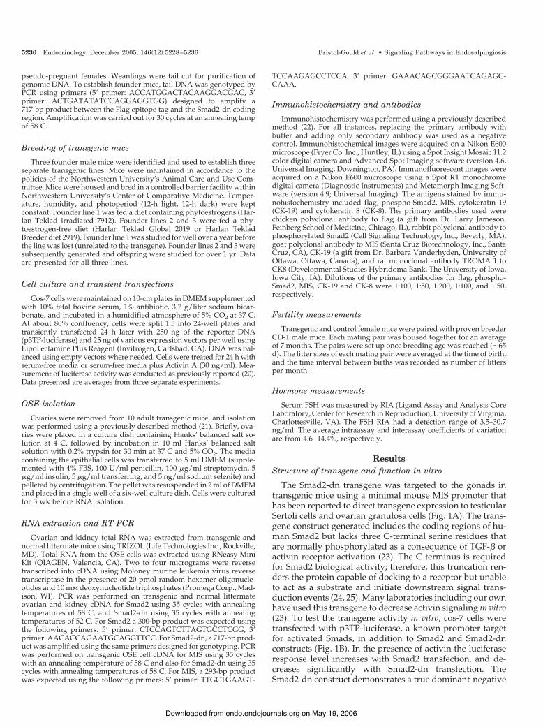
FIG. 1. Transgene construct actions in vitro and in vivo. A, The transgene construct includes a 180-bp promoter region of the mouse MIS genefused to a C-terminally truncated human Smad2 protein by the flag epitope tag. The series of primers used for RT-PCR analysis and genotypingare designated above the map. B, Transfection of Smad2-dn into Cos-7 cells indicates that this protein induces a dominant-negative effect incells at basal levels and in the presence of activin. Shown are mean values from three separate experiments " SEM. Statistical analysis wasperformed using a t test with variance indicated by SEM (P # 0.01). C–G, Expression of Smad2-dn transgene mRNA in mouse tissues. Wholeovary, kidney fragment, and isolated ovarian OSE RNA from transgenic (T) and normal littermate (N) mice were subjected to RT-PCR methodsdescribed in Materials and Methods. C, A 717-bp product shown in the T lanes indicates the presence of the transgene in transgenic ovaries,but not in normal littermate ovaries (N). D, The transgene is also not expressed in transgenic (T) or normal littermate (N) kidneys. E and F,A 300-bp product using primers for normal Smad2 shows expression in ovaries and kidneys regardless of genotype. G, Amplified Flag cDNAin transgenic ovaries (T) and in isolated transgenic OSE cells (OSE). Amplified MIS cDNA in normal littermate ovaries (N) and in isolatedtransgenic OSE cells (OSE). Water controls run throughout the PCR process are indicated with an (H). H–M, Transgene localization in ovariantissue. H, Normal littermate ovary depicting no production of flag protein in the follicles or the OSE. I, Transgenic ovary indicating flag proteinimmunolocalization in granulosa cells of a secondary follicle, a B/C primordial follicle (red arrow), and in the OSE. J, MIS protein located ingranulosa cells of a small antral follicle and the OSE (red arrowheads) in normal littermate mice. The inset shows additional MIS proteinproduction in the preantral follicle population. K, MIS located in the OSE (red arrowheads) and follicles of transgenic mice. B/C primordial follicleis indicated by the red arrow. L–M, Overlay images of phosphorylated Smad2 indicated by FITC, and nuclear stain indicated by 4$,6-diamidino-2-phenylindole. L, Normal littermate ovary indicating strong phospho-Smad2 protein production in follicles, and high levels in the OSE (redarrowheads). M, Transgenic ovary depicting decreased phospho-Smad2 protein in the ovarian follicles, and little phospho-Smad2 in the OSE(red asterisks). H–J were photographed at %400 magnification, K–M were photographed at %200 magnification using bright-field andfluorescence microscopy, respectively.
Bristol-Gould et al. • Signaling Pathways in Endosalpingiosis Endocrinology, December 2005, 146(12):5228–5236 5229
on May 19, 2006 endo.endojournals.orgDownloaded from
pseudo-pregnant females. Weanlings were tail cut for purification ofgenomic DNA. To establish founder mice, tail DNA was genotyped byPCR using primers (5$ primer: ACCATGGACTACAAGGACGAC, 3$primer: ACTGATATATCCAGGAGGTGG) designed to amplify a717-bp product between the Flag epitope tag and the Smad2-dn codingregion. Amplification was carried out for 30 cycles at an annealing tempof 58 C.
Breeding of transgenic mice
Three founder male mice were identified and used to establish threeseparate transgenic lines. Mice were maintained in accordance to thepolicies of the Northwestern University’s Animal Care and Use Com-mittee. Mice were housed and bred in a controlled barrier facility withinNorthwestern University’s Center of Comparative Medicine. Temper-ature, humidity, and photoperiod (12-h light, 12-h dark) were keptconstant. Founder line 1 was fed a diet containing phytoestrogens (Har-lan Teklad irradiated 7912). Founder lines 2 and 3 were fed a phy-toestrogen-free diet (Harlan Teklad Global 2019 or Harlan TekladBreeder diet 2919). Founder line 1 was studied for well over a year beforethe line was lost (unrelated to the transgene). Founder lines 2 and 3 weresubsequently generated and offspring were studied for over 1 yr. Dataare presented for all three lines.
Cell culture and transient transfections
Cos-7 cells were maintained on 10-cm plates in DMEM supplementedwith 10% fetal bovine serum, 1% antibiotic, 3.7 g/liter sodium bicar-bonate, and incubated in a humidified atmosphere of 5% CO2 at 37 C.At about 80% confluency, cells were split 1:5 into 24-well plates andtransiently transfected 24 h later with 250 ng of the reporter DNA(p3TP-luciferase) and 25 ng of various expression vectors per well usingLipoFectamine Plus Reagent (Invitrogen, Carlsbad, CA). DNA was bal-anced using empty vectors where needed. Cells were treated for 24 h withserum-free media or serum-free media plus Activin A (30 ng/ml). Mea-surement of luciferase activity was conducted as previously reported (20).Data presented are averages from three separate experiments.
OSE isolation
Ovaries were removed from 10 adult transgenic mice, and isolationwas performed using a previously described method (21). Briefly, ova-ries were placed in a culture dish containing Hanks’ balanced salt so-lution at 4 C, followed by incubation in 10 ml Hanks’ balanced saltsolution with 0.2% trypsin for 30 min at 37 C and 5% CO2. The mediacontaining the epithelial cells was transferred to 5 ml DMEM (supple-mented with 4% FBS, 100 U/ml penicillin, 100 "g/ml streptomycin, 5"g/ml insulin, 5 "g/ml transferring, and 5 ng/ml sodium selenite) andpelleted by centrifugation. The pellet was resuspended in 2 ml of DMEMand placed in a single well of a six-well culture dish. Cells were culturedfor 3 wk before RNA isolation.
RNA extraction and RT-PCR
Ovarian and kidney total RNA was extracted from transgenic andnormal littermate mice using TRIZOL (Life Technologies Inc., Rockville,MD). Total RNA from the OSE cells was extracted using RNeasy MiniKit (QIAGEN, Valencia, CA). Two to four micrograms were reversetranscribed into cDNA using Moloney murine leukemia virus reversetranscriptase in the presence of 20 pmol random hexamer oligonucle-otides and 10 mm deoxynucleotide triphosphates (Promega Corp., Mad-ison, WI). PCR was performed on transgenic and normal littermateovarian and kidney cDNA for Smad2 using 35 cycles with annealingtemperatures of 58 C, and Smad2-dn using 35 cycles with annealingtemperatures of 52 C. For Smad2 a 300-bp product was expected usingthe following primers: 5$ primer: CTCCAGTCTTAGTGCCTCGG, 3$primer: AACACCAGAATGCAGGTTCC. For Smad2-dn, a 717-bp prod-uct was amplified using the same primers designed for genotyping. PCRwas performed on transgenic OSE cell cDNA for MIS using 35 cycleswith an annealing temperature of 58 C and also for Smad2-dn using 35cycles with annealing temperatures of 58 C. For MIS, a 293-bp productwas expected using the following primers: 5$ primer: TTGCTGAAGT-
TCCAAGAGCCTCCA, 3$ primer: GAAACAGCGGGAATCAGAGC-CAAA.
Immunohistochemistry and antibodies
Immunohistochemistry was performed using a previously describedmethod (22). For all instances, replacing the primary antibody withbuffer and adding only secondary antibody was used as a negativecontrol. Immunohistochemical images were acquired on a Nikon E600microscope (Fryer Co. Inc., Huntley, IL) using a Spot Insight Mosaic 11.2color digital camera and Advanced Spot Imaging software (version 4.6,Universal Imaging, Downington, PA). Immunofluorescent images wereacquired on a Nikon E600 microscope using a Spot RT monochromedigital camera (Diagnostic Instruments) and Metamorph Imaging Soft-ware (version 4.9; Universal Imaging). The antigens stained by immu-nohistochemistry included flag, phospho-Smad2, MIS, cytokeratin 19(CK-19) and cytokeratin 8 (CK-8). The primary antibodies used werechicken polyclonal antibody to flag (a gift from Dr. Larry Jameson,Feinberg School of Medicine, Chicago, IL), rabbit polyclonal antibody tophosphorylated Smad2 (Cell Signaling Technology, Inc., Beverly, MA),goat polyclonal antibody to MIS (Santa Cruz Biotechnology, Inc., SantaCruz, CA), CK-19 (a gift from Dr. Barbara Vanderhyden, University ofOttawa, Ottawa, Canada), and rat monoclonal antibody TROMA 1 toCK8 (Developmental Studies Hybridoma Bank, The University of Iowa,Iowa City, IA). Dilutions of the primary antibodies for flag, phospho-Smad2, MIS, CK-19 and CK-8 were 1:100, 1:50, 1:200, 1:100, and 1:50,respectively.
Fertility measurements
Transgenic and control female mice were paired with proven breederCD-1 male mice. Each mating pair was housed together for an averageof 7 months. The pairs were set up once breeding age was reached (&65d). The litter sizes of each mating pair were averaged at the time of birth,and the time interval between births was recorded as number of littersper month.
Hormone measurements
Serum FSH was measured by RIA (Ligand Assay and Analysis CoreLaboratory, Center for Research in Reproduction, University of Virginia,Charlottesville, VA). The FSH RIA had a detection range of 3.5–30.7ng/ml. The average intraassay and interassay coefficients of variationare from 4.6–14.4%, respectively.
ResultsStructure of transgene and function in vitro
The Smad2-dn transgene was targeted to the gonads intransgenic mice using a minimal mouse MIS promoter thathas been reported to direct transgene expression to testicularSertoli cells and ovarian granulosa cells (Fig. 1A). The trans-gene construct generated includes the coding regions of hu-man Smad2 but lacks three C-terminal serine residues thatare normally phosphorylated as a consequence of TGF-! oractivin receptor activation (23). The C terminus is requiredfor Smad2 biological activity; therefore, this truncation ren-ders the protein capable of docking to a receptor but unableto act as a substrate and initiate downstream signal trans-duction events (24, 25). Many laboratories including our ownhave used this transgene to decrease activin signaling in vitro(23). To test the transgene activity in vitro, cos-7 cells weretransfected with p3TP-luciferase, a known promoter targetfor activated Smads, in addition to Smad2 and Smad2-dnconstructs (Fig. 1B). In the presence of activin the luciferaseresponse level increases with Smad2 transfection, and de-creases significantly with Smad2-dn transfection. TheSmad2-dn construct demonstrates a true dominant-negative
5230 Endocrinology, December 2005, 146(12):5228–5236 Bristol-Gould et al. • Signaling Pathways in Endosalpingiosis
on May 19, 2006 endo.endojournals.orgDownloaded from

effect in these cells at basal levels and in the presence ofactivin.
Transgene expression in vivo
The tissue distribution of the transgene was examinedusing specific primers and RT-PCR amplification of RNAisolated from whole ovary and kidney fragments of trans-genic and normal littermate animals in addition to the OSEcells of transgenic ovaries (Fig. 1, C–G). The transgene(717-bp product) was expressed in transgenic ovaries but notin transgenic mouse kidneys, nor in either tissue isolatedfrom normal littermates (Fig. 1, C–D). The endogenousmouse Smad2 gene (300-bp product), was detected in alltissues regardless of genotype (Fig. 1, E and F). To verifycell-specific expression, the transgene (Flag-Smad2-dn) andMIS were also amplified from transgenic OSE cell RNA (Fig.1G). We further analyzed the chimeric flag-Smad2-dn pro-tein by immunolocalization in ovaries of both genotypes. Theflag epitope was not detected in any cell types within normallittermate mouse ovarian tissue (Fig. 1H). Flag protein wasdetected in ovarian follicles of transgenic mice as early as theB/C primordial follicle (transitory primordial follicle con-taining squamous and cuboidal granulosa cells) stage, inaddition to being localized within portions of OSE (Fig. 1I).MIS protein was detected in granulosa cells of preantral andsmall antral follicles in mice of both genotypes. MIS was alsodetected in portions of the OSE in normal littermate andtransgenic ovaries (Fig. 1, J and K). Because the transgene wasexpected to block endogenous Smad2 activity, the presenceand cellular location of phosphorylated Smad2 was immu-nolocalized to cells within the ovaries. Nuclear Smad2 wasdetected in the granulosa cells of all follicles regardless ofgenotype; however, levels were slightly decreased in nearlyhalf the transgenic ovaries analyzed [decreased fluoresceinisothiocyanate (FITC) stain in granulosa cells in Fig. 1M].Conversely, phospho-Smad2 was not detected in the major-ity of OSE cells in transgenic mice, whereas normal littermateanimals had robust levels of nuclear Smad2 in the OSE (Fig.1, L and M). We quantified the amount of fluorescence inthese panels by counting positively (FITC labeled) stainedOSE cells. In the transgenic animals, 28% of OSE cells stainedfor phosphorylated Smad2, whereas 79% of OSE cells stainedfor phosphorylated Smad2 protein in normal littermate mice.Together, these RNA and protein data suggest that our trans-gene is targeted to the ovarian granulosa cells and the OSEbut may have the most impact on the OSE cell populationbased on subsequent observations.
Fertility studies and serum FSH levels
Smad2-dn female mice were subfertile with a decreasedlitter size and breeding frequency in comparison to controlbreeding animals. Control CD-1 breeding mice had an av-erage pup number at birth of 16.9, and a breeding frequencyof 1.2 litters per month (n ' 3). The transgenic breeding micehad an average pup number at birth of 11.0, and a breedingfrequency of 1.0 litter per month (n ' 6). In two of sixtransgenic breeder females, there was a delay in first preg-nancy by 2 months of setting up the mating pairs (the otherfour breeders became pregnant shortly after the males were
introduced), followed by a complete cessation of breedingafter only two gestations. Considering the transgene is ex-pressed in the granulosa cells of developing follicles, theremight be an affect to normal folliculogenesis in our trans-genic animals that subsequently alters fertility. Quantifica-tion of follicle populations and possible differences are on-going. Endosalpingiosis does not normally present as adisease that causes infertility. However, because the condi-tion is often incidentally found and because there is no di-agnosis of the syndrome, it is not yet known whether thereis a direct link between the fertility issues in our mice and theeventual development of endosalpingiosis.
There were fewer corpora lutea (CL) in Smad2-dn trans-genic mouse ovaries compared with ovaries from normallittermates. When CL were counted in serial sectioned ova-ries from animals ranging in age from 5–6 months, the av-erage number present in normal littermate animals was sig-nificantly higher (P # 0.003) then that present in transgenicanimals. Normal littermate mice had an average of 23.6 CL(n ' 3), whereas transgenic animals had an average of 16.4CL (n ' 4). Additionally, serum FSH levels were measuredand no significant difference between 3-month-old trans-genic and normal littermate mice was found (data notshown). Therefore, suggesting that our transgene is onlyhaving a local ovarian affect.
Ovarian pathologies I: stroma, crypts, and invaginations
As the ovary ages it becomes increasing irregular in shape.However, many of the ovarian phenotypes that are com-monly associated with middle and old age begin to appearin our transgenic animals by 3 months of age, and continuallyworsen. Stromal disorganization (classified as a stroma oc-cupied with holes instead of densely packed with folliclesand CL) was apparent in the medullary regions in mosttransgenic ovaries at all ages examined. Cryptic structuresand epithelial invaginations began to emerge anywhere from3–5 months in our animals. Crypts tend to be located near thehilus, whereas the invaginations resided near the outsideedge of the ovary and invaginate inwards. The cells lining thecrypts and invaginations display many characteristics of ep-ithelial cells: columnar shape, basally oriented nucleus, andciliation (Fig. 2, A and B). These cells may originate from theovarian surface epithelium or they may come from the ep-ithelial cells that line the rete ovarii.
Ovarian pathologies II: lesions, inclusions, and cysts
During postovulatory repair in the mouse and human, itis commonly thought that displaced OSE cells either revertto an epithelial cell phenotype or go through an epithelial tomesenchymal transition. If incomplete transition takes place,cells can be incorporated into the stroma near the site ofovulation and likely form inclusion cysts (10). Different typesand conformations of serous, benign, cystic structures/le-sions were commonly found in Smad2-dn mice around 8–12months of age, but we have also identified cyst formation asearly as 3 months (Fig. 3). Most transgenic ovaries containedmultiple small lesions/inclusion cysts representative of clas-sic ovarian endosalpingiosis seen in humans. Whereas othertransgenic ovaries possessed one large cyst that was either
Bristol-Gould et al. • Signaling Pathways in Endosalpingiosis Endocrinology, December 2005, 146(12):5228–5236 5231
on May 19, 2006 endo.endojournals.orgDownloaded from

fluid filled (in resemblance to those found in the inhibin-#transgenic mice) or contained a mass of cells, the origin ofwhich is not known (Fig. 3, D–F). Although cyst types weredifferent in gross appearance, microscopic evaluation, num-ber and location, the cells that line the cysts were epithelialin origin based on histomorphology.
The single layer of epithelium lining all the cysts is similarto a tubal-type epithelium. Three basic cell types are present
in various numbers that resemble the three cell types foundin the normal fallopian tube in women (oviduct in themouse): pale focally ciliated cells, secretory or vacuolatedcells, and dark intercalated cells with prominent basal nuclei(Fig. 4). The cysts often exhibit irregular contours and occa-sional intraluminal stromal papillae. All of these character-istics associate with an endosalpingiosis phenotype more sothan a serous cystadenoma phenotype.
FIG. 2. Ovarian crypts and invaginations in transgenic mouse ovaries. A, Crypt-like structures found throughout the ovarian stroma (usuallynear the hilus) in many transgenic mouse ovaries. Cells are cuboidal in shape, have a basally oriented nucleus, and contain cilia (black arrow).Inset is a %200 magnification of A that was photographed at %600 magnification. B, Invaginations of the ovarian surface frequently seen inyoung transgenic animals photographed at %600 magnification. Cells are cuboidal in shape, ciliated (black arrow), and tend to layer upon eachother in certain areas. Inset is a %200 magnified image of where these invaginations are usually found. Black asterisks mark the edge of theovary.
FIG. 3. Ovarian histology of transgenic mice. Ovaries were fixed in 4% paraformaldehyde, embedded in paraffin, sectioned, and stained withhematoxylin and eosin or eosin only. Detection of cysts begins at 3.5 months and ranges in age to 11 months old. A, 5-"m section of an ovaryfrom a 5-month-old normal littermate mouse showing normal histology. B, A 5-"m section of an ovary from a 3.5-month-old transgenic mouseshowing a large cyst near the hilus of the ovary, continuous with the OSE, and filled with cells. C, A 5-"m section of an ovary from a 5-month-oldtransgenic mouse showing a more centrally located cyst. This image is taken in the beginning of the cyst, subsequent serial sections show thecyst increasing dramatically in size and stretching all the way to the edge of the ovary. D, A 5-"m section of an ovary from a 9-month-oldtransgenic mouse showing a developed cyst containing a cellular mass within. This cyst is located in the outer portion of the ovary (this sectionis not cut through the center of the ovary). E, A 5-"m section of an ovary from a 9-month-old transgenic mouse representing the largest typeof cyst found. F, A 5-"m section of an ovary from an 11-month-old transgenic mouse with another very large fluid filled cyst. These fluid filledcysts are very similar to those seen in the inhibin-# subunit transgenic mice. All images were photographed at %100 magnification.
5232 Endocrinology, December 2005, 146(12):5228–5236 Bristol-Gould et al. • Signaling Pathways in Endosalpingiosis
on May 19, 2006 endo.endojournals.orgDownloaded from

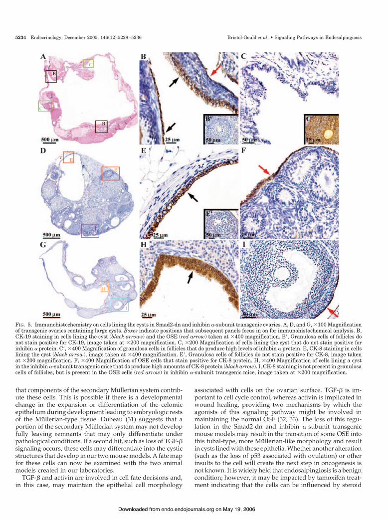
To distinguish and verify the cell type lining the cysts, westained sections for epithelial cell markers CK-19 and CK-8,and a granulosa cell marker inhibin #-subunit. CK-19 wasdetected in those cells that line the cysts and in OSE cells;however, CK-19 was not detected in the granulosa cells offollicles (Fig. 5, B and B$). As expected, the inhibin #-subunitprotein was detected in the granulosa cells of follicles but wasnot detected in the cells lining the cyst, confirming that thecyst cells are not granulosa cells (Fig. 5, C and C$). CK-8 wasalso detected in cells lining cysts formed in Smad2-dn ovariesand was not detected in follicular granulosa cells confirmingthe CK-19 results (Fig. 5, E and E$). Furthermore, CK-8 pro-tein was detected in the OSE (Fig. 5F).
A second animal model generated to block the activinsignal transduction system is the inhibin #-subunit trans-genic mouse (26). Importantly, the inhibin #-subunit trans-gene is driven by a metallothionein-I promoter and is there-fore ubiquitously expressed. However, similar to theSmad2-dn mice, the inhibin #-subunit transgenic females aresubfertile and develop ovarian cysts (26). The cysts are alsolined by tubal-type cells that stain positively for CK-8,whereas the granulosa cells fail to stain with this marker (Fig.5, H and I).
Ovarian phenotype III: multioocytic follicles (MOFs)
The final phenotype encountered in both the Smad2-dnand the inhibin #-subunit transgenic mice is the formation ofMOFs (26). Although present at a low frequency, severaltypes of MOFs appeared in founder line 1 transgenic animals.The most commonly detected MOFs were two oocytes perfollicle. MOFs in the Smad2-dn mice appear in two config-urations based on the encapsulated oocytes. Oocytes of someMOFs may possibly retain their embryonic connections (Fig.6B). Although more commonly, MOFs were detected that
have independent separated oocytes (Fig. 6, C and D). Thesomatic cells in the latter type of MOF have completelysurrounded each individual oocyte, but a basement mem-brane has not. Interestingly, MOFs were not detected inSmad2-dn founder lines 2 and 3 kept on the phytoestrogen-free diet; however, when these transgenic animals wereswitched back to the original phytoestrogen-containing food,the MOFs reappeared in these lines (data not shown).
Discussion
Endosalpingiosis can be diagnosed, but its onset, progres-sion, and implications for risk of other health complicationsis not completely understood. Two animal models that con-verge on the TGF-!/activin signal transduction pathway anddevelop this condition can now be studied to provide an-swers to these important questions. The histogenesis of en-dosalpingiosis is debatable because the origins of the cellslining the cysts are not known for certain. The first prospectis that the cells are ovarian surface epithelia. The surfaceepithelium of the ovary is a single, simple epithelial layer ofcells that undergoes dramatic reorganization during eachovulatory cycle. By unknown mechanisms, the OSE maybecome trapped within the stroma and instead of assuminga mesenchymal phenotype these cells undergo metaplasia tobecome Mullerian-like epithelia (27). These tubal-type epi-thelia may therefore make up the lining of the inclusion cyststhat are often assumed to be the precursors of neoplasticprogression (27, 28).
The OSE and extraovarian mesothelium are both derivedembryonically from the celomic epithelium and exist in asimilar environment. However, in the adult, the two differ-entiated cell populations express the epithelial differentia-tion marker CA125 (also the tumor marker for ovarian andMullerian duct-derived neoplasms) differently (29). CA125 isexpressed in adult oviductal, endometrial, and endocervicalepithelia, but not the OSE (10, 30). The OSE is a fairly un-differentiated cell and it is thought that the accumulation ofmutations in specific genes during ovulation can cause theOSE to differentiate into the serous, mucinous, or endomet-riod lineages associated with ovarian cancer. These variedcell fates suggest that the adult OSE may not be fully dif-ferentiated and that it possesses an intrinsic plasticity orpleuripotential resembling its mesodermal embryonic pre-cursor more than the other celomic epithelial derivatives(oviduct, endometrium, cervix) (10). Given that the OSEnever acquired CA125 or lost it early in development, unlikethe other celomic derivatives, suggests that this cell layermight be less committed to a particular phenotype in theadult. OSE transformation or differentiation probably alsodepends on the genetic and, very likely, the hormonal milieu.
The cell origin discrepancy arises due to the fact that thecommon subtypes of ovarian cancer (serous, mucinous, andendometroid) resemble epithelia that are not ovarian-likeand are not normally present in the ovary itself (31). Hence,the second potential origin of cells lining the endosalpingio-tic cysts maybe structures that are embryologically derivedfrom the Mullerian ducts themselves (oviduct, uterus, en-dometrium, rete ovarii) as opposed to the OSE. Consideringthe serous appearance of our cysts, it is a formal possibility
FIG. 4. Comparison of cells lining the cysts vs. cells lining the oviduct.A, %1000 Magnification of cells within the oviduct. B, %1000 Mag-nification of cyst cellular lining in a transgenic ovary. Below panelsA and B are enlarged representations of areas containing the threetypes of cells of interest. Black arrows indicate ciliated cells, redarrows indicate secretory cells, and blue arrows indicate intercalatedor peg cells.
Bristol-Gould et al. • Signaling Pathways in Endosalpingiosis Endocrinology, December 2005, 146(12):5228–5236 5233
on May 19, 2006 endo.endojournals.orgDownloaded from
that components of the secondary Mullerian system contrib-ute these cells. This is possible if there is a developmentalchange in the expansion or differentiation of the celomicepithelium during development leading to embryologic restsof the Mullerian-type tissue. Dubeau (31) suggests that aportion of the secondary Mullerian system may not developfully leaving remnants that may only differentiate underpathological conditions. If a second hit, such as loss of TGF-!signaling occurs, these cells may differentiate into the cysticstructures that develop in our two mouse models. A fate mapfor these cells can now be examined with the two animalmodels created in our laboratories.
TGF-! and activin are involved in cell fate decisions and,in this case, may maintain the epithelial cell morphology
associated with cells on the ovarian surface. TGF-! is im-portant to cell cycle control, whereas activin is implicated inwound healing, providing two mechanisms by which theagonists of this signaling pathway might be involved inmaintaining the normal OSE (32, 33). The loss of this regu-lation in the Smad2-dn and inhibin #-subunit transgenicmouse models may result in the transition of some OSE intothis tubal-type, more Mullerian-like morphology and resultin cysts lined with these epithelia. Whether another alteration(such as the loss of p53 associated with ovulation) or otherinsults to the cell will create the next step in oncogenesis isnot known. It is widely held that endosalpingiosis is a benigncondition; however, it may be impacted by tamoxifen treat-ment indicating that the cells can be influenced by steroid
FIG. 5. Immunohistochemistry on cells lining the cysts in Smad2-dn and inhibin #-subunit transgenic ovaries. A, D, and G, %100 Magnificationof transgenic ovaries containing large cysts. Boxes indicate positions that subsequent panels focus in on for immunohistochemical analysis. B,CK-19 staining in cells lining the cyst (black arrows) and the OSE (red arrow) taken at %400 magnification. B$, Granulosa cells of follicles donot stain positive for CK-19, image taken at %200 magnification. C, %200 Magnification of cells lining the cyst that do not stain positive forinhibin # protein. C$, %400 Magnification of granulosa cells in follicles that do produce high levels of inhibin # protein. E, CK-8 staining in cellslining the cyst (black arrow), image taken at %400 magnification. E$, Granulosa cells of follicles do not stain positive for CK-8, image takenat %200 magnification. F, %400 Magnification of OSE cells that stain positive for CK-8 protein. H, %400 Magnification of cells lining a cystin the inhibin #-subunit transgenic mice that do produce high amounts of CK-8 protein (black arrow). I, CK-8 staining is not present in granulosacells of follicles, but is present in the OSE cells (red arrow) in inhibin #-subunit transgenic mice, image taken at %200 magnification.
5234 Endocrinology, December 2005, 146(12):5228–5236 Bristol-Gould et al. • Signaling Pathways in Endosalpingiosis
on May 19, 2006 endo.endojournals.orgDownloaded from

background (34). A link between endosalpingiosis and ovar-ian cancer or other gynecological cancers has not been es-tablished but warrants further investigation.
Chodankar et al. (35) used the Cre-lox system to inactivateBRCA1 in granulosa cells by using the FSH receptor pro-moter as the driver. However, the inactivation of the BRCA1gene in the granulosa cells surprisingly led to the develop-ment of cystic structures lined by cells of epithelial morphol-ogy, not granulosa cells. They hypothesize that the granulosacells act at a distance to control Mullerian epithelial tumor-igenesis via a mechanism regulated by BRCA1. Signalingbetween granulosa cells and the OSE must occur given therequirement of ovulation at an explicit site on the ovariansurface. Because the transgene in our mice is expressed inboth granulosa and OSE cells, we are unable to rule out thepotential of defective signaling from the granulosa cells con-tributing to the cyst phenotype we observe. It is possible tospeculate that the transgene directly functions in the gran-ulosa cell population that then indirectly impacts the OSE orrete ovarii at a distance. How pathologies of the OSE areinfluenced by the granulosa cell compartment is an intrigu-ing area of future investigation.
In general, the ovaries of the young transgenic mice seemto develop morphologies we would commonly associatewith old age. The irregular shape, crypts, invaginations, in-clusions, and cysts have historically been observed in wild-type mice close to 1 yr of age. Therefore, the ovarian age doesnot match the chronological age of the mouse. This is true ofmany idiopathic infertility patients, women who enter meno-pause early in reproductive life, and many PCOS patients(36–39). In each of these cases, the women are said to haveovaries that are older than their chronological age. An ex-ception to this is a study which found PCOS patients enteredmenopause at older ages than normal women therefore sug-gesting these women’s ovarian age to be younger than chro-nological age (40).
The two mouse models share a second phenotype, thedevelopment of MOFs. A mechanism by which individualfollicles become encapsulated by granulosa cells has not beendetermined; however, it is likely that estrogen plays an im-portant role in the process and notably there may be signif-icant cross talk. We are currently investigating the effect ofphytoestrogen containing food on the formation of thesestructures as this could be a confounding factor in whetheror not MOFs form in our transgenic founder mice lines.Overall, the inappropriate encapsulation of follicles in theSmad2-dn and inhibin #-subunit transgenic mouse modelssuggests that the TGF-!/activin signal transduction path-way may somehow be involved. Neonatal exposure to es-trogens results in an increased incidence of MOFs, whichsuggests a link between the steroid hormone and peptidehormone control of follicle development (41). We hypothe-size that MOFs form through the absence of an imposedsignal to identify the outside border of the follicle thus re-sulting in groups of oocytes retaining intracellular connec-tions formed during development that subsequently are notbroken throughout germ line cyst breakdown. The basementmembrane and somatic cells that usually encapsulate oneoocyte per follicle somehow are misregulated and do notform the normal follicle borders. It is also unknown whetherthe MOFs we detect ovulate. If so, they potentially cause moredamage to the ovarian surface epithelium, and therefore couldincrease the probability of cyst formation in adult life.
Generation of animal models for gender-specific diseasesare crucial in revealing important new mechanisms by whichgenes work in a male or female environment. Males from theSmad2-dn and inhibin #-subunit transgenic backgrounds donot have any known phenotypes that directly impact fertility.Therefore, the ability to manipulate specific cellular func-tions in a sexually dimorphic manner provides insight intothe hormonal and genetic requirements for cellular function.The origins of many diseases of the female reproductive tract
FIG. 6. MOF phenotype. Hemotoxylin and eosin-stained sections of normal littermate and transgenicmouse ovaries. A, Two adjacent individual primary fol-licles in a normal littermate mouse ovary. B, Smad2-dnMOF that appears to retain oocyte connections and/orcontains a granulosa cell invasion defect. C and D, MOFspossibly formed by a different mechanism then that seenin B. The oocytes here are completely surrounded bygranulosa cells. Sections were photographed at %400magnification, except C, which is %200 magnified usingbright-field microscopy.
Bristol-Gould et al. • Signaling Pathways in Endosalpingiosis Endocrinology, December 2005, 146(12):5228–5236 5235
on May 19, 2006 endo.endojournals.orgDownloaded from

are unknown and consequently difficult to treat. Pelvic painis a frequent symptom with multiple etiologies. Therefore, itis necessary to continue to develop new diagnostics andexpand treatment options to better understand the endosal-pingiosis condition and intervene to improve the lives ofwomen.
Acknowledgments
We would like to thank the Northwestern University Transgenic andTargeted Mutagenesis Core Facility (CMIER) for creating our foundertransgenic lines. Andrew Lisowski in the P01 core facility for sectioningall tissue analyzed in this study. Jessica Kroger for her initial analysis ofthe Smad2-dn ovarian tissue. Dr. Joanna Burdette for her assistance withthe OSE isolation protocol and overall experimental advice.
Received June 10, 2005. Accepted August 25, 2005.Address all correspondence and requests for reprints to: Teresa K.
Woodruff, Ph.D., Professor, Northwestern University, Department ofNeurobiology, O. T. Hogan 4-150, 2205 Tech Drive, Evanston, Illinois60208. E-mail: [email protected].
This work was supported by Core services of University of VirginiaCenter for Research in Reproduction Ligand Assay and Analysis CoreNational Institute of Child Health and Human Development (NICHD)(Specialized Cooperative Centers Program in Reproduction Research)Grant U54-HD28934. National Institutes of Health (NIH)/NICHD Hor-mone Signals that Regulate Ovarian Differentiation, P01 HD021921.S.K.B.-G. received funding from the NIH/National Cancer Institute Train-ing Program in Oncogenesis and Development Biology, T32 CA080621.
References1. Clement PB, Seidman JD, Russell P, Kurman RJ 2002 Blaustein’s pathology
of the female genital tract. 5th ed. New York: Springer2. Zinsser KR, Wheeler JE 1982 Endosalpingiosis in the omentum: a study of
autopsy and surgical material. Am J Surg Pathol 6:109–1173. McCaughey WT, Schryer MJ, Lin XS, Al-Jabi M 1986 Extraovarian pelvic
serous tumor with marked calcification. Arch Pathol Lab Med 110:78–804. Sneige N, Fernandez T, Copeland LJ, Katz RL 1986 Mullerian inclusions in
peritoneal washings. Potential source of error in cytologic diagnosis. ActaCytol 30:271–276
5. Heinig J, Gottschalk I, Cirkel U, Diallo R 2002 Endosalpingiosis-an under-estimated cause of chronic pelvic pain or an accidental finding? A retrospectivestudy of 16 cases. Eur J Obstet Gynecol Reprod Biol 103:75–78
6. Ryuko K, Miura H, Abu-Musa A, Iwanari O, Kitao M 1992 Endosalpingiosisin association with ovarian surface papillary tumor of borderline malignancy.Gynecol Oncol 46:107–110
7. Yeung HH, Bannatyne P, Russell P 1983 Adenocarcinoma of the fallopiantubes: a clinicopathological study of eight cases. Pathology 15:279–286
8. deHoop TA, Mira J, Thomas MA 1997 Endosalpingiosis and chronic pelvicpain. J Reprod Med 42:613–616
9. Fathalla MF 1971 Incessant ovulation—a factor in ovarian neoplasia? Lancet2:163
10. Auersperg N, Wong AS, Choi KC, Kang SK, Leung PC 2001 Ovarian surfaceepithelium: biology, endocrinology, and pathology. Endocr Rev 22:255–288
11. Scully RE 1995 Pathology of ovarian cancer precursors. J Cell Biochem Suppl23:208–218
12. Tan OL, Hurst PR, Fleming JS 2005 Location of inclusion cysts in mouseovaries in relation to age, pregnancy, and total ovulation number: implicationsfor ovarian cancer? J Pathol 205:483–490
13. Berchuck A, Rodriguez G, Olt G, Whitaker R, Boente MP, Arrick BA, Clarke-Pearson DL, Bast Jr RC 1992 Regulation of growth of normal ovarian epithelialcells and ovarian cancer cell lines by transforming growth factor-!. Am J ObstetGynecol 166:676–684
14. Nilsson EE, Skinner MK 2002 Role of transforming growth factor beta inovarian surface epithelium biology and ovarian cancer. Reprod Biomed Online5:254–258
15. Munz B, Smola H, Engelhardt F, Bleuel K, Brauchle M, Lein I, Evans LW,Huylebroeck D, Balling R, Werner S 1999 Overexpression of activin A in theskin of transgenic mice reveals new activities of activin in epidermal mor-phogenesis, dermal fibrosis and wound repair. EMBO J 18:5205–5215
16. Wankell M, Munz B, Hubner G, Hans W, Wolf E, Goppelt A, Werner S 2001
Impaired wound healing in transgenic mice overexpressing the activin an-tagonist follistatin in the epidermis. EMBO J 20:5361–5372
17. Dinulescu DM, Ince TA, Quade BJ, Shafer SA, Crowley D, Jacks T 2005 Roleof K-ras and Pten in the development of mouse models of endometriosis andendometrioid ovarian cancer. Nat Med 11:63–70
18. Moynahan ME 2002 The cancer connection: BRCA1 and BRCA2 tumor sup-pression in mice and humans. Oncogene 21:8994–9007
19. Dowdy SC, Mariani A, Reinholz MM, Keeney GL, Spelsberg TC, PodratzKC, Janknecht R 2005 Overexpression of the TGF-! antagonist Smad7 inendometrial cancer. Gynecol Oncol 96:368–373
20. Suszko MI, Lo DJ, Suh H, Camper SA, Woodruff TK 2003 Regulation of therat follicle-stimulating hormone !-subunit promoter by activin. Mol Endocri-nol 17:318–332
21. Roby KF, Taylor CC, Sweetwood JP, Cheng Y, Pace JL, Tawfik O, PersonsDL, Smith PG, Terranova PF 2000 Development of a syngeneic mouse modelfor events related to ovarian cancer. Carcinogenesis 21:585–591
22. Bristol SK, Woodruff TK 2004 Follicle-restricted compartmentalization oftransforming growth factor ! superfamily ligands in the feline ovary. BiolReprod 70:846–859
23. Chen W, Woodruff TK, Mayo KE 2000 Activin A-induced HepG2 liver cellapoptosis: involvement of activin receptors and smad proteins. Endocrinology141:1263–1272
24. Abdollah S, Macias-Silva M, Tsukazaki T, Hayashi H, Attisano L, Wrana JL1997 T!RI phosphorylation of Smad2 on Ser465 and Ser467 is required forSmad2-Smad4 complex formation and signaling. J Biol Chem 272:27678–27685
25. Souchelnytskyi S, Tamaki K, Engstrom U, Wernstedt C, ten Dijke P, HeldinCH 1997 Phosphorylation of Ser465 and Ser467 in the C terminus of Smad2mediates interaction with Smad4 and is required for transforming growthfactor-beta signaling. J Biol Chem 272:28107–28115
26. McMullen ML, Cho BN, Yates CJ, Mayo KE 2001 Gonadal pathologies intransgenic mice expressing the rat inhibin #-subunit. Endocrinology 142:5005–5014
27. Resta L, Russo S, Colucci GA, Prat J 1993 Morphologic precursors of ovarianepithelial tumors. Obstet Gynecol 82:181–186
28. Dietl J, Marzusch K 1993 Ovarian surface epithelium and human ovariancancer. Gynecol Obstet Invest 35:129–135
29. Jacobs I, Bast Jr RC 1989 The CA 125 tumour-associated antigen: a review ofthe literature. Hum Reprod 4:1–12
30. Kabawat SE, Bast Jr RC, Bhan AK, Welch WR, Knapp RC, Colvin RB 1983Tissue distribution of a coelomic-epithelium-related antigen recognized by themonoclonal antibody OC125. Int J Gynecol Pathol 2:275–285
31. Dubeau L 1999 The cell of origin of ovarian epithelial tumors and the ovariansurface epithelium dogma: does the emperor have no clothes? Gynecol Oncol72:437–442
32. Hu X, Zuckerman KS 2001 Transforming growth factor: signal transductionpathways, cell cycle mediation, and effects on hematopoiesis. J HematotherStem Cell Res 10:67–74
33. Massague J 2000 How cells read TGF-! signals. Nat Rev Mol Cell Biol 1:169–178
34. McCluggage WG, Weir PE 2000 Paraovarian cystic endosalpingiosis in asso-ciation with tamoxifen therapy. J Clin Pathol 53:161–162
35. Chodankar R, Kwang S, Sangiorgi F, Hong H, Yen HY, Deng C, Pike MC,Shuler CF, Maxson R, Dubeau L 2005 Cell-nonautonomous induction ofovarian and uterine serous cystadenomas in mice lacking a functional Brca1in ovarian granulosa cells. Curr Biol 15:561–565
36. Jain T, Klein NA, Lee DM, Sluss PM, Soules MR 2003 Endocrine assessmentof relative reproductive age in normal eumenorrheic younger and olderwomen across multiple cycles. Am J Obstet Gynecol 189:1080–1084
37. Santoro N, Isaac B, Neal-Perry G, Adel T, Weingart L, Nussbaum A, ThakurS, Jinnai H, Khosla N, Barad D 2003 Impaired folliculogenesis and ovulationin older reproductive aged women. J Clin Endocrinol Metab 88:5502–5509
38. Elting MW, Kwee J, Korsen TJ, Rekers-Mombarg LT, Schoemaker J 2003Aging women with polycystic ovary syndrome who achieve regular menstrualcycles have a smaller follicle cohort than those who continue to have irregularcycles. Fertil Steril 79:1154–1160
39. Klein NA, Battaglia DE, Fujimoto VY, Davis GS, Bremner WJ, Soules MR1996 Reproductive aging: accelerated ovarian follicular development associ-ated with a monotropic follicle-stimulating hormone rise in normal olderwomen. J Clin Endocrinol Metab 81:1038–1045
40. Dahlgren E, Johansson S, Lindstedt G, Knutsson F, Oden A, Janson PO,Mattson LA, Crona N, Lundberg PA 1992 Women with polycystic ovarysyndrome wedge resected in 1956 to 1965: a long-term follow-up focusing onnatural history and circulating hormones. Fertil Steril 57:505–513
41. Jefferson WN, Couse JF, Padilla-Banks E, Korach KS, Newbold RR 2002Neonatal exposure to genistein induces estrogen receptor (ER)# expressionand multioocyte follicles in the maturing mouse ovary: evidence for ER!-mediated and nonestrogenic actions. Biol Reprod 67:1285–1296
Endocrinology is published monthly by The Endocrine Society (http://www.endo-society.org), the foremost professional society serving theendocrine community.
5236 Endocrinology, December 2005, 146(12):5228–5236 Bristol-Gould et al. • Signaling Pathways in Endosalpingiosis
on May 19, 2006 endo.endojournals.orgDownloaded from
Related Documents