© The Author 2013. Published by Oxford University Press on behalf of the UK Environmental Mutagen Society. All rights reserved. For permissions, please e-mail: [email protected]. doi:10.1093/mutage/ges080 T-cell-specific deletion of Mof blocks their differentiation and results in genomic instability in mice Arun Gupta 1,2 , Clayton R. Hunt 1,2 , Raj K. Pandita 2,3 , Juhee Pae 4 , K. Komal 5 , Mayank Singh 1 , Jerry W. Shay 3 , Rakesh Kumar 1,2 , Kiyoshi Ariizumi 4 , Nobuo Horikoshi 1 , Walter N. Hittelman 6 , Chandan Guha 7 , Thomas Ludwig 8 and Tej K. Pandita 1,2, * 1 Department of Radiation Oncology, UT Southwestern Medical Center, Dallas, TX 75229, USA, 2 Washington University School of Medicine, St Louis, MO 63108, USA, 3 Department of Cell Biology, UT Southwestern Medical Center, Dallas, TX 75229, USA, 4 Department of Dermatology, UT Southwestern Medical Center, Dallas, TX 75229, USA, 5 All India Institute of Medical Sciences, Delhi, India, 6 Department of Experimental Therapeutics, MD Anderson Cancer Center, Houston, TX 77030, USA, 7 Department of Radiation Oncology, Albert Einstein College of Medicine, Bronx, NY 10461, USA, 8 Institute for Cancer Genetics, Columbia University, New York, NY 32001, USA and 9 Department of Molecular and Cellular Biochemistry, Ohio State University Wexner Medical Center, Columbus, OH 43210, USA *To whom correspondence should be addressed. Department of Radiation Oncology, UT Southwestern Medical Center, 2201 Inwood Road, Dallas, TX 75390, USA. Tel: 1-214-648-1918; Fax: 1-214-648-8995; Email: [email protected] Received on October 20, 2012; revised on December 11, 2012; accepted on December 12, 2012 Ataxia telangiectasia patients develop lymphoid malignan- cies of both B- and T-cell origin. Similarly, ataxia telangiec- tasia mutated (Atm)-deficient mice exhibit severe defects in T-cell maturation and eventually develop thymomas. The function of ATM is known to be influenced by the mamma- lian orthologue of the Drosophila MOF (males absent on the first) gene. Here, we report the effect of T-cell-specific abla- tion of the mouse Mof (Mof) gene on leucocyte trafficking and survival. Conditional Mof Flox/Flox (Mof F/F ) mice express- ing Cre recombinase under control of the T-cell-specific Lck proximal promoter (Mof F/F /Lck-Cre + ) display a marked reduction in thymus size compared with Mof F/F /Lck- Cre – mice. In contrast, the spleen size of Mof F/F /Lck-Cre + mice was increased compared with control Mof F/F /Lck- Cre – mice. The thymus of Mof F/F /Lck-Cre + mice contained significantly reduced T cells, whereas thymic B cells were elevated. Within the T-cell population, CD4 + CD8 + double- positive T-cell levels were reduced, whereas the immature CD4 – CD8 – double-negative (DN) population was elevated. Defective T-cell differentiation is also evident as an increased DN3 (CD44 – CD25 + ) population, the cell stage during which T-cell receptor rearrangement takes place. The differ- entiation defect in T cells and reduced thymus size were not rescued in a p53-deficient background. Splenic B-cell distributions were similar between Mof F/F /Lck-Cre + and Mof F/F /Lck-Cre – mice except for an elevation of the κ light- chain population, suggestive of an abnormal clonal expan- sion. T cells from Mof F/F /Lck-Cre + mice did not respond to phytohaemagglutinin (PHA) stimulation, whereas LPS- stimulated B cells from Mof F/F /Lck-Cre + mice demonstrated spontaneous genomic instability. Mice with T-cell-specific loss of MOF had shorter lifespans and decreased survival following irradiation than did Mof F/F /Lck-Cre – mice. These observations suggest that Mof plays a critical role in T-cell differentiation and that depletion of Mof in T cells reduces T-cell numbers and, by an undefined mechanism, induces genomic instability in B cells through bystander mecha- nism. As a result, these mice have a shorter lifespan and reduced survival after irradiation. Introduction Males absent on the first (MOF) was initially discovered as a dosage compensation gene in Drosophila. MOF belongs to the MYST family of acetyltransferases and is a histone acetyltransferase (HAT) that acetylates chromatin specifically at histone H4 lysine 16 (H4K16). Depletion of MOF in Drosophila (1), as well as in human and mouse cells, results in the loss of acetylation at H4K16 (2–6), suggesting that the highly conserved MOF protein may be the major HAT acting on histone H4 at K16. MOF has been associated with acute myeloid leukaemia (AML) and transcriptional silencing in Saccharomyces cerevisiae (SAS2 and YBF2/SAS3). MOF also interacts with the human immunodeficiency virus Tat- interactive protein (TIP60) (7–9). We previously reported a higher frequency of residual DNA double-strand breaks and chromosome aberrations in cells expressing a HAT-dead human MOF after cellular exposure to ionising radiation (IR) (6,10). Additional studies indicate MOF plays a critical role in oogenesis, oncogenesis, DNA damage repair and survival of post-mitotic Purkinje cells (2,5,10). Chromosomal translocations that alter the activity of chromatin-modifying enzymes are repeatedly found associated with different forms of leukaemia indicating the importance of epigenetic regulation in haematopoiesis. One of these chromatin-modifying enzymes, the monocytic leukaemia zinc finger (MOZ or recently renamed as KAT6a3) protein, was first identified through positional cloning of a t(8;16)(p11;p13) translocation of the CREB- binding protein gene in AML (11). MOF affects ATM function, the gene responsible for the disease ataxia telangiectasia (A-T). Patients with mutated ATM have ~5-fold increased risk of developing leukaemia or lymphoblastic lymphomas and ~10% of A-T patients develop lymphoid malignancies (12,13) of either B-cell or T-cell ori- gin (14). Similarly, Atm-deficient mice exhibit severe defects in T-cell maturation and develop thymomas (15). As MOF regulates ATM function, we sought to determine the effect of Mof depletion on T-cell maturation. Since global Mof abla- tion results in early embryonic lethality (5), we employed a conditional murine system in which the Lck proximal pro- moter drives T-cell-specific expression of Cre recombinase (Mof F/F /Lck-Cre + mice) to determine the role of Mof in T-cell development. Materials and methods Generation of T-cell-specific Mof-deficient mice The details for generation of targeting vectors for the Mof locus used for an in vivo deletion of the Mof gene in mice and the conditional Mof allele were described recently (5,10). W9.5 ES cells were electroporated with the construct 263 Mutagenesis vol. 28 no. 3 pp. 263–270, 2013 Advance Access publication 5 February 2013 at LibrarySerials DeptUT Southwestern Medical Center on June 28, 2013 http://mutage.oxfordjournals.org/ Downloaded from

Welcome message from author
This document is posted to help you gain knowledge. Please leave a comment to let me know what you think about it! Share it to your friends and learn new things together.
Transcript

© The Author 2013. Published by Oxford University Press on behalf of the UK Environmental Mutagen Society. All rights reserved. For permissions, please e-mail: [email protected].
doi:10.1093/mutage/ges080
T-cell-specific deletion of Mof blocks their differentiation and results in genomic instability in mice
Arun Gupta1,2, Clayton R. Hunt1,2, Raj K. Pandita2,3, Juhee Pae4, K. Komal5, Mayank Singh1, Jerry W. Shay3, Rakesh Kumar1,2, Kiyoshi Ariizumi4, Nobuo Horikoshi1, Walter N. Hittelman6, Chandan Guha7, Thomas Ludwig8 and Tej K. Pandita1,2,*1Department of Radiation Oncology, UT Southwestern Medical Center, Dallas, TX 75229, USA, 2Washington University School of Medicine, St Louis, MO 63108, USA, 3Department of Cell Biology, UT Southwestern Medical Center, Dallas, TX 75229, USA, 4Department of Dermatology, UT Southwestern Medical Center, Dallas, TX 75229, USA, 5All India Institute of Medical Sciences, Delhi, India, 6Department of Experimental Therapeutics, MD Anderson Cancer Center, Houston, TX 77030, USA, 7Department of Radiation Oncology, Albert Einstein College of Medicine, Bronx, NY 10461, USA, 8Institute for Cancer Genetics, Columbia University, New York, NY 32001, USA and 9Department of Molecular and Cellular Biochemistry, Ohio State University Wexner Medical Center, Columbus, OH 43210, USA
*To whom correspondence should be addressed. Department of Radiation Oncology, UT Southwestern Medical Center, 2201 Inwood Road, Dallas, TX 75390, USA. Tel: 1-214-648-1918; Fax: 1-214-648-8995; Email: [email protected]
Received on October 20, 2012; revised on December 11, 2012; accepted on December 12, 2012
Ataxia telangiectasia patients develop lymphoid malignan-cies of both B- and T-cell origin. Similarly, ataxia telangiec-tasia mutated (Atm)-deficient mice exhibit severe defects in T-cell maturation and eventually develop thymomas. The function of ATM is known to be influenced by the mamma-lian orthologue of the Drosophila MOF (males absent on the first) gene. Here, we report the effect of T-cell-specific abla-tion of the mouse Mof (Mof) gene on leucocyte trafficking and survival. Conditional Mof Flox/Flox (Mof F/F) mice express-ing Cre recombinase under control of the T-cell-specific Lck proximal promoter (Mof F/F/Lck-Cre+) display a marked reduction in thymus size compared with Mof F/F/Lck-Cre– mice. In contrast, the spleen size of Mof F/F/Lck-Cre+ mice was increased compared with control Mof F/F/Lck-Cre– mice. The thymus of Mof F/F/Lck-Cre+ mice contained significantly reduced T cells, whereas thymic B cells were elevated. Within the T-cell population, CD4+CD8+ double-positive T-cell levels were reduced, whereas the immature CD4–CD8– double-negative (DN) population was elevated. Defective T-cell differentiation is also evident as an increased DN3 (CD44–CD25+) population, the cell stage during which T-cell receptor rearrangement takes place. The differ-entiation defect in T cells and reduced thymus size were not rescued in a p53-deficient background. Splenic B-cell distributions were similar between Mof F/F/Lck-Cre+ and Mof F/F/Lck-Cre– mice except for an elevation of the κ light-chain population, suggestive of an abnormal clonal expan-sion. T cells from Mof F/F/Lck-Cre+ mice did not respond to phytohaemagglutinin (PHA) stimulation, whereas LPS-stimulated B cells from Mof F/F/Lck-Cre+ mice demonstrated spontaneous genomic instability. Mice with T-cell-specific loss of MOF had shorter lifespans and decreased survival following irradiation than did Mof F/F/Lck-Cre– mice. These observations suggest that Mof plays a critical role in T-cell
differentiation and that depletion of Mof in T cells reduces T-cell numbers and, by an undefined mechanism, induces genomic instability in B cells through bystander mecha-nism. As a result, these mice have a shorter lifespan and reduced survival after irradiation.
Introduction
Males absent on the first (MOF) was initially discovered as a dosage compensation gene in Drosophila. MOF belongs to the MYST family of acetyltransferases and is a histone acetyltransferase (HAT) that acetylates chromatin specifically at histone H4 lysine 16 (H4K16). Depletion of MOF in Drosophila (1), as well as in human and mouse cells, results in the loss of acetylation at H4K16 (2–6), suggesting that the highly conserved MOF protein may be the major HAT acting on histone H4 at K16. MOF has been associated with acute myeloid leukaemia (AML) and transcriptional silencing in Saccharomyces cerevisiae (SAS2 and YBF2/SAS3). MOF also interacts with the human immunodeficiency virus Tat-interactive protein (TIP60) (7–9). We previously reported a higher frequency of residual DNA double-strand breaks and chromosome aberrations in cells expressing a HAT-dead human MOF after cellular exposure to ionising radiation (IR) (6,10). Additional studies indicate MOF plays a critical role in oogenesis, oncogenesis, DNA damage repair and survival of post-mitotic Purkinje cells (2,5,10). Chromosomal translocations that alter the activity of chromatin-modifying enzymes are repeatedly found associated with different forms of leukaemia indicating the importance of epigenetic regulation in haematopoiesis. One of these chromatin-modifying enzymes, the monocytic leukaemia zinc finger (MOZ or recently renamed as KAT6a3) protein, was first identified through positional cloning of a t(8;16)(p11;p13) translocation of the CREB-binding protein gene in AML (11).
MOF affects ATM function, the gene responsible for the disease ataxia telangiectasia (A-T). Patients with mutated ATM have ~5-fold increased risk of developing leukaemia or lymphoblastic lymphomas and ~10% of A-T patients develop lymphoid malignancies (12,13) of either B-cell or T-cell ori-gin (14). Similarly, Atm-deficient mice exhibit severe defects in T-cell maturation and develop thymomas (15). As MOF regulates ATM function, we sought to determine the effect of Mof depletion on T-cell maturation. Since global Mof abla-tion results in early embryonic lethality (5), we employed a conditional murine system in which the Lck proximal pro-moter drives T-cell-specific expression of Cre recombinase (MofF/F/Lck-Cre+ mice) to determine the role of Mof in T-cell development.
Materials and methods
Generation of T-cell-specific Mof-deficient miceThe details for generation of targeting vectors for the Mof locus used for an in vivo deletion of the Mof gene in mice and the conditional Mof allele were described recently (5,10). W9.5 ES cells were electroporated with the construct
263
Mutagenesis vol. 28 no. 3 pp. 263–270, 2013Advance Access publication 5 February 2013
at LibrarySerials D
eptUT
Southwestern M
edical Center on June 28, 2013
http://mutage.oxfordjournals.org/
Dow
nloaded from

A. Gupta et al.
to generate Mof flox/+ cells and the details for generation of Mof flox/+/Rosa26creERT2/+ and Mof Dflox/flox/Rosa26creERT2/+ ES cell clones have been described (5,10).
To inactivate Mof specifically in T cells, conditional Mof flox/flox (Mof F/F) mice were crossed with mice expressing Cre recombinase under the control of the Lck proximal promoter (16) to generate MofF/F/Lck-Cre+ mice. Genotyping of mice was performed as described previously (5,10). Animal care and treat-ments were performed in accordance with the National Institutes of Health guidelines at Washington University School of Medicine, St Louis, MO and Columbia University, New York.
Leucocyte isolation and flow cytometric analysisFor leucocyte isolation and flow cytometric analysis, cell suspensions pre-pared from fresh thymus and spleen were negatively enriched for T cells through magnetic bead depletion of B cells, NK cells, dendritic cells, granu-locytes, monocytes/macrophases and erythrocytes. For immunophenotyping, T cells were stained with specific primary-conjugated antibodies and analysed by Dickinson FACSCalibur flow. CD4–CD8– double-negative (DN) thymo-cyte subset purification was performed by staining unfractionated thymocytes with a fluorescein isothiocyanate (FITC)-labelled lineage-specific antibodies for CD4 and CD8. Spleen cells from Mofflox/flox/Lck-Cre+ mice and Mofflox/flox/Lck-Cre– mice were depleted of T cells, then cultured in RPMI 1640 medium supplemented with 10% fetal calf serum in the presence of 4 pg/ml of lipopol-ysaccharide (LPS) to stimulate B cells. T cells were stimulated with phyto-haemagglutinin (PHA). After 48 or 72 h of culture, colcemid was added and metaphases were prepared and analysed as described previously (17,18). To determine whether metaphases are from first (I), second (II) or third (III) cell cycle post-LPS stimulation, cells were incubated with BrdU and cell cycle (I, II, III) was determined as described previously (17). Metaphase bone mar-row cells were prepared from mice 4 h after administering colcemid. Telomere fluorescence in situ hybridization (FISH) was performed as described previ-ously (19–21).
Micronuclei analysis and ratio of normochromatic to polychromatic erythrocytesFrequency of micronucleus and the ratio of normochromatic to polychromatic erythrocytes were determined by previously described procedures (18,21,22). Briefly, bone marrow smears from the age-matched MofF/F/Lck-Cre+ and MofF/F/Lck-Cre– mice with and without treatment of mitomycin C were prepared, and the stained smears were examined to determine the incidence of micronucleated cells in 1800 polychromatic erythrocytes and the ratio of normochromatic to polychromatic erythrocytes for each animal, which were repeated three times.
Statistical analysisData are expressed as the means ± standard deviations from three to four exper-iments. Statistical comparison of means was performed by the Student’s t-test.
Results and discussion
Most of the A-T patients suffer from immune defects and MOF influences ATM function (6,23,24). Global Mof inactivation results in early embryonic lethality in mice (5). Therefore, we generated mice with T-cell-specific Mof deletion by breeding conditional Mof Flox/Flox (Mof F/F) mice (10) with transgenic mice expressing Cre recombinase under control of the Lck proximal promoter to generate Mof F/F/Lck-Cre+ mice. This approach provided a well-defined system for determining the role of Mof in leucocyte biology through Cre-mediated deletion in developing T cells (16). Lck is a non-receptor protein tyrosine kinase required for signal transduction via the T-cell antigen receptor and the Lck proximal promoter is activated at the DN1 (CD25–CD44+) to DN2 (CD25+CD44+) T-cell lineage stage. The production and ‘education’ of T cells, which are critical for the adaptive immune system, occur in the thymus, which provides an inductive environment for the development of T lymphocytes from haematopoietic progenitor cells.
T-cell-specific ablation of Mof had a major effect on the thymus and spleen (Figure 1A–D), MofF/F/Lck-Cre+ mice had consistently smaller thymi (approximately half of the size rel-ative to their body weight) than those of MofF/F/Lck-Cre– mice and the differences observed (at either 3 or 12 weeks of age)
are statistically significant (Figure 1B and D). In contrast, spleen size in MofF/F/Lck-Cre+ mice is consistently larger relative to body weight compared with Mof F/F/Lck-Cre– mice (Figure 1B and D) with differences being more pronounced at 12 weeks of age. The size reduction of the thymus was not p53-dependent since MofF/F/Lck-Cre+ mice generated in a p53-null background mice still displayed the reduced thy-mus size phenotype seen in MofF/F/Lck-Cre+ mice (Figure 1E and F). Irrespective of p53 status, the ratio of thymus size is reduced and spleen increased in 12-week-old compared with 3-week-old MofF/F/Lck-Cre+ mice. A reduction in thymus size has also been observed in mice with inactivated MOZ, another MYST family member, where both B and T cells were affected (25). Here, we observe that thymus size is affected by T-cell-specific inactivation of Mof. It is known that thymus function is eventually taken over by the spleen in adults so the increase in spleen size seen here might be due to defective T-cell development, which results in spleen hypertrophy in MofF/F/Lck-Cre+ mice (Figure 1).
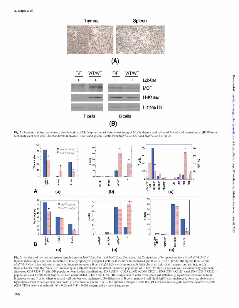
Immunostaining with Mof antibody of thymus and spleen tis-sue sections prepared from Mof F/F/Lck-Cre– mice detected pos-itive staining for Mof in both tissues (Figure 2A). Furthermore, when T and B cells from thymus and spleen of Mof F/F/Lck-Cre+ and Mof F/F/Lck-Cre– mice were enriched and examined for Mof and H4K16ac levels, both were significantly reduced in T cells from Mof F/F/Lck-Cre+ mice compared with T cells from Mof F/F/Lck-Cre– mice (Figure 2B). However, no reduction in Mof or H4K16ac levels was observed in B cells of MofF/F/Lck-Cre+ mice (Figure 2B).
To investigate potential immune system abnormalities, we performed flow cytometry analysis of thymocytes and spleno-cytes from MofF/F/Lck-Cre+ and MofF/F/Lck-Cre– mice. Fewer cells were collected from thymi of MofF/F/Lck-Cre+ mice in comparison with controls, which is consistent with the smaller organ size in MofF/F/Lck-Cre+ mice (Figure 1). Thymocytes from 12-week-old MofF/F/Lck-Cre+ mice had a significant reduction in total T-cell population compared with MofF/F/Lck-Cre– mice (Figure 3A(a)). T cells from MofF/F/Lck-Cre+ mice displayed a reduction of mature T cells (CD4+CD8+) and an accumulation of immature DN T cells (CD4–CD8–) (Figure 3A(c)). Further analysis revealed that T-cell development was retarded after Mof gene disruption at the DN3 (CD44–CD25+) stage, where T-cell receptor rearrangements are processed for β-receptor selection, and following DN4 stage (CD44–CD25–). Defective T-cell receptor rearrangement is a phenotype observed in A-T patients as well as in Atm-deficient mice (15,26,27), thus, Mof expression appears to be critical for T-cell development. Reduced T-cell populations were also observed in spleen (Figure 3B(a)) especially in the cytotoxic T-cell (CD4–CD8+) population (Figure 3B(c)).
While the T-cell population decreased, the thymic B-cell population was increased in MofF/F/Lck-Cre+ mice (Figure 3A (a)). A similar B-cell increase was observed as a result of apoptosis in the thymus (28), suggesting the decreased thy-mus size in MofF/F/Lck-Cre+ mice could be due, at least in part, to apoptotic elimination of immature T cells. However, when we examined MofF/F/Lck-Cre+ mice in a p53-null back-ground (Figure 4), there was no normalisation of thymus and spleen size (Figure 1E and F), nor did p53 inactivation affect the altered T- and B-cell distribution observed in Mof-deficient mice (Figure 4). In thymus, total T cells were reduced with increasing immature CD4–CD8– DN and reducing CD4+CD8+ double-positive populations (Figure 4A(b)), whereas thymic B
264
at LibrarySerials D
eptUT
Southwestern M
edical Center on June 28, 2013
http://mutage.oxfordjournals.org/
Dow
nloaded from

T-cell-specific deletion of Mof
cells were increased (Figure 4A(a)). In spleen, the number of mature B cells was unchanged (Figure 4B(a)), whereas total T cells were reduced (Figure 4B(b)). In sum, p53 does not play a significant role in the reduced thymus size or defective T-cell development seen in Mof-deficient mice.
Precursor B and T cells are produced in the bone marrow. To determine whether inactivation of Mof in T cells has any effect on bone marrow erythrocytes, we compared the number of micronucleated cells per 1800 polychromatic erythrocytes and the ratio of normochromatic to polychromatic erythrocytes in MofF/F/Lck-Cre+ and MofF/F/Lck-Cre– mice with or without treatment of mitomycin C. No significant differences were found between the two genotypes (Table I). This is reasonable since the Lck proximal promoter driving Cre recombinase is activated in thymus, thus T-cell precursors in bone marrow are likely express Mof gene.
To determine the cause for the T-cell differentiation blockage in MofF/F/Lck-Cre+ mice, thymocytes were examined for genomic integrity. We have recently shown that genomic integrity as well as the DNA damage response can be influenced by H4K16ac lev-els (2) as the H4K16ac modification structurally constrains the formation of higher-order chromatin (29), perhaps by inducing an open chromatin configuration that is more readily accessible to transcription as well as DNA repair proteins. H4K16ac is also critical for protein–protein interactions (29), and reduced levels
of H4K16ac correlate with a defective DNA damage response (2). Since thymus or blood T cells from MofF/F/Lck-Cre+ mice responded very poorly to PHA treatment for blastoid formation (data not shown), we collected thymocytes to examine nuclear morphology. Interestingly, a significant fraction (>14%) of T cells from MofF/F/Lck-Cre+ mice displayed chromatin blebbing and disintegration of the nucleus (Figure 5A and B), similar to the situation observed in MOF-depleted HeLa cells (4) as well as to the chromatin condensation observed in Mof-deleted post-mitotic neuronal Purkinje cells (10). These observations suggest that failure to maintain genomic integrity could be the major cause for defective T-cell development.
Precursor T cells mature in the thymus and helper T cells are known to release factors such as lymphokines that enhance B-cell blastoid formation as well as differentiation (30,31). As shown in Figures 1 and 3, depletion of Mof in T cells results in spleen enlargement and possibly abnormal clonal expansion of B cells. To determine the effect of Mof depletion on T cells, we compared the status of T-cell blastoid transformation in MofF/F/Lck-Cre+ and MofF/F/Lck-Cre– mice. PHA treatment produced minimal blastoid formation or metaphases in T cells from the thymus or spleen of MofF/F/Lck-Cre+ mice, whereas both blastoid and metaphases were seen in the cells from the MofF/F/Lck-Cre– mouse thymus or spleen, suggesting that Mof is important for mitogen-induced replication of T cells (data not shown).
Fig. 1. Effect of T-cell-specific Mof inactivation on thymus and spleen size. (A) Thymus and spleen from 3-week-old MofF/F/Lck-Cre+ and MofF/F/Lck-Cre– mice. (B) Histogram of the weight ratios of thymus and spleen to mouse body weight. The differences in thymus as well as spleen size in MofF/F/Lck-Cre+ and MofF/F/Lck-Cre– mice are statistically significant. (C) Thymus and spleen of 12-week-old MofF/F/Lck-Cre+ and MofF/F/Lck-Cre– mice. (D) Histogram showing the ratio of thymus and spleen to body weight. The thymus size is relatively smaller and spleen size is larger. The differences in thymus as well as spleen size in MofF/F/Lck-Cre+ and MofF/F/Lck-Cre– mice are statistically significant. (E and F) Thymus and spleen size in p53-null background of MofF/F/Lck-Cre+ and MofF/F/Lck-Cre– mice (E: 3 weeks old and F: 12 weeks old). *P < 0.05 and **P < 0.001 determined by the chi-square test.
265
at LibrarySerials D
eptUT
Southwestern M
edical Center on June 28, 2013
http://mutage.oxfordjournals.org/
Dow
nloaded from
A. Gupta et al.
Fig. 2. Immunostaining and western blot detection of Mof expression. (A) Immunostaining of Mof in thymus and spleen of 3-week-old control mice. (B) Western blot analysis of Mof and H4K16ac levels in thymus T cells and spleen B cells from MofF/F/Lck-Cre+ and MofF/F/Lck-Cre– mice.
Fig. 3. Analysis of thymus and spleen lymphocytes in MofF/F/Lck-Cre+ and MofF/F/Lck-Cre– mice. (A) Comparison of lymphocytes from (a) MofF/F/Lck-Cre+ thymus indicating a significant reduction in total lymphocytes and pan T cells (CD3+CD5+) but increased pan B cells (B220+) levels; (b) thymic B cells from MofF/F/Lck-Cre+ mice indicate a significant increase in mature B cells (IgM+IgD+) with an unusually high clonal (κ light-chain) expansion ratio (κ); and (c) thymic T cells from MofF/F/Lck-Cre+ indicating an early developmental defect, increased population of CD4–CD8– (DN) T cells as well as statistically significant decreased CD4+CD8+ T cells. DN population was further classified into DN1 (CD44+CD25–), DN2 (CD44+CD25+), DN3 (CD44–CD25+) and DN4 (CD44–CD25–) populations and T cells from MofF/F/Lck-Cre+ accumulated in DN3 and DN4. (B) Comparison of cells from spleen (a) statistically significant reduction in total lymphocytes and T cells, whereas total B-cell number was unchanged; (b) difference in B cells, mature B cell (IgM+IgD+) was unchanged; however, abnormal κ light-chain clonal expansion was observed; (c) difference in splenic T cells, the number of helper T cells (CD4+CD8–) was unchanged; however, cytotoxic T-cells (CD4–CD8+) level was reduced. *P < 0.05 and **P < 0.001 determined by the chi-square test.
266
at LibrarySerials D
eptUT
Southwestern M
edical Center on June 28, 2013
http://mutage.oxfordjournals.org/
Dow
nloaded from

T-cell-specific deletion of Mof
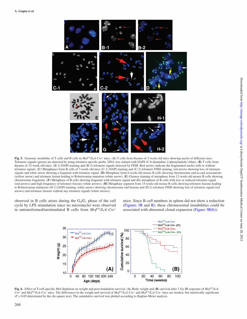
Next, to determine whether T-cell-specific inactivation of Mof also affects B-cell proliferation, splenocytes and whole white blood cells were treated with LPS, to specifically stimulate B cells. About ~7% of metaphases in LPS-treated splenic B cells of MofF/F/Lck-Cre+ mice (3 weeks old) had chromosomes with loss of telomere signals, as detected by telomere-specific FISH (Figure 5C, red arrows) and chromosome fragments (Figure 5C, white arrow). No such corresponding chromosomal defects were observed in MofF/F/Lck-Cre– mice. Also, ~8% of metaphases from spleen-derived B cells from MofF/F/Lck-Cre+ mice displayed chromosome end-to-end association
(Figure 5D, yellow arrows) not seen in MofF/F/Lck-Cre– mice. Similar chromosome end-to-end associations have been reported previously in cells derived from A-T patients (19,32). In addition, B cells from spleens of older (12 or 15 weeks old) MofF/F/Lck-Cre+ mice also displayed a high frequency of chromosome fragments, loss of telomere signals and telomere fusions resulting in Robertsonian mutations (Figure 5E–H).
The mechanistic basis for B-cell genomic instability in mice with T-cell-specific Mof depletion was further examined by determining where in the cell cycle phase-specific chromo-somal aberrations could occur through a bystander mechanism (33–35). B cells stimulated with LPS were labelled with BrdU and metaphase cells collected after 48 h of incubation. Cell cycle phase-specific chromosome aberrations were ascertained based on the frequency of chromosomal and chromatid-type aberrations observed at metaphase. G1-specific aberrations detected at metaphase are mostly of the chromosomal type and include a high frequency of dicentrics (21,36,37). S-phase-type aberrations detected at metaphase are chromosomes and chro-matid-type aberrations along with tri- or quadriradials, whereas G2-phase-type aberrations are only chromatid type. After stim-ulation, only chromosome-type aberrations were observed at first metaphase as determined by Fluorescence Plus Giemsa technique as described previously (17). The possibility of miss-ing chromatid aberrations arising during S phase was mini-mal as no metaphase cells with both chromatid DNA strands labelled with BrdU were detected by the described procedure (17,18). These results suggest that the genomic instability
Fig. 4. Effect of p53-null status on thymic and splenic cell populations in MofF/F/Lck-Cre+ mice. (A) Comparison of cells from thymus (a) B cells and (b) T cells present no significant differences in cell types between p53-null and wild-type background of MofF/F/Lck-Cre+ mice; however, significant differences were observed between MofF/F/Lck-Cre+ and MofF/F/Lck-Cre– mice as described in Figure 3. (B) Comparison of cells from spleen. (a) B cells and (b) T cells present no significant differences in cell types of p53-null and wild-type background MofF/F/Lck-Cre+ mice; however, some differences were observed between MofF/F/Lck-Cre+ and MofF/F/Lck-Cre– mice as described in Figure 3. *P < 0.05 and **P < 0.001 determined by the chi-square test.
Table I. Number of micronucleated polychromatic erythrocytes and the ratio of normochromatic to polychromatic erythrocytes in bone marrow smears after intraperitoneal administration of mitomycin C
Genotype Treatment Number of micronuleated cells/1800 polychromatic erythrocytes/mouse
Ratio of normochromatic to polychromatic erythrocytes
Mitomycin C (mg/kg body weight)
Mean Range Mean Range
MofF/F/Lck-Cre–
0 1.20 0–3.90 1.62 0.51–3.405 80.30 35–127 22.50 5.10–51.60
MofF/F/Lck-Cre+
0 1.28 0–4.00 1.68 0.54–3.515 82.32 34–130 23.70 5.21–53.28
267
at LibrarySerials D
eptUT
Southwestern M
edical Center on June 28, 2013
http://mutage.oxfordjournals.org/
Dow
nloaded from
A. Gupta et al.
observed in B cells arises during the G0/G1 phase of the cell cycle by LPS stimulation since no micronuclei were observed in untransformed/unstimulated B cells from MofF/F/Lck-Cre+
mice. Since B-cell numbers in spleen did not show a reduction (Figures 3B and B), these chromosomal instabilities could be associated with abnormal clonal expansion (Figure 3B(b)).
Fig. 5. Genomic instability of T cells and B cells in MofF/F/Lck-Cre+ mice. (A) T cells from thymus of 3-week-old mice showing nuclei of different sizes. Telomere signals (green) are detected by using telomere-specific probe. DNA was stained with DAPI (4′,6-diamidino-2-phenylindole) (blue). (B) T cells from thymus of 12-week-old mice. (B-1) DAPI staining and (B-2) telomere signals detected by FISH. Red arrows indicate the fragmented nuclei with or without telomere signals. (C) Metaphases from B cells of 3-week-old mice (C-1) DAPI staining and (C-2) telomere FISH staining, red arrows showing loss of telomere signals and white arrow showing a fragment with telomere signal. (D) Metaphase from 6-week-old mouse B cells showing chromosome end-to-end associations (yellow arrow) and telomere fusion leading to Robertsonian mutation (white arrow). (E) Giemsa staining of metaphase from 12-week-old mouse B cells showing chromosome fragments. (F) Metaphase of B cells showing fragment with telomere signal and (G) metaphase of B cells with loss or reduced telomere signal (red arrows) and high frequency of telomere fusions (white arrows). (H) Metaphase segment from 15-week-old mouse B cells showing telomere fusions leading to Robertsonian mutations (H-1) DAPI staining, white arrows showing chromosome end fusions and (H-2) telomere FISH showing loss of telomere signal (red arrows) and telomere fusions without any telomere signals (white arrows).
Fig. 6. Effect of T-cell-specific Mof depletion on weight and post-irradiation survival. (A) Body weight and (B) survival after 3 Gy IR exposure of MofF/F/Lck-Cre+ and MofF/F/Lck-Cre– mice. The differences in the weight and survival of MofF/F/Lck-Cre+ and MofF/F/Lck-Cre– mice are modest, but statistically significant (P < 0.05 determined by the chi-square test). The cumulative survival was plotted according to Kaplan–Meier analysis.
268
at LibrarySerials D
eptUT
Southwestern M
edical Center on June 28, 2013
http://mutage.oxfordjournals.org/
Dow
nloaded from

T-cell-specific deletion of Mof
To determine whether the genomic instability seen in B cells from spleen or thymus could be originating from bone marrow precursor B cells, we analysed metaphases in bone marrow cells from MofF/F/Lck-Cre+ and MofF/F/Lck-Cre– mice and found no difference in genomic stability, suggesting that Mof depletion in T cells seen in the thymus had no effect on the precursor cells.
We next determined whether the immunological alterna-tions observed in MofF/F/Lck-Cre+ mouse impacted growth by comparing the body weight of MofF/F/Lck-Cre+ and MofF/F/Lck-Cre– mice. A significant decrease in body weight of MofF/F/Lck-Cre+ mice was observed compared with MofF/F/Lck-Cre– mice (Figure 6A). In addition, MofF/F/Lck-Cre+ mice had a shortened lifespan compared with MofF/F/Lck-Cre– mice (Figure 6B). Since MofF/F/Lck-Cre+ mice display genomic instability of lym-phocytes, we examined whether the mice were radiosensitive. Mice (4 weeks of age) were treated with 3 Gy of IR and survival monitored for up to 100 weeks. Long-term survival of irradi-ated MofF/F/Lck-Cre+ mice was significantly decreased com-pared with MofF/F/Lck-Cre– mice (Figure 6B). Taken together, the results indicate that mice with Mof-deficient T cells have compromised immune systems and manifest growth retarda-tion as well as increasing radiation sensitivity.
We report here that T-cell-specific deletion of the Mof gene results in a smaller thymus and larger spleen and a blockage in T-cell differentiation at the stage during which T-cell recep-tor rearrangement normally occurs, which may correlate with genomic instability. Interestingly, loss of Mof in T cells also induces a genomic instability phenotype in B cells. Mice with Mof-null T cells had a reduced body weight as well as a reduced lifespan and demonstrated higher sensitivity to irradiation. This is similar to the situation in severe combined immunodeficient mice where a germ-line mutation in the DNA-PKcs gene blocks T- and B-cell development and there is elevated sensitivity to radiation (38). Since MOF influences ATM functions in response to DNA damage (6,39), the results suggest the mice have a defect in Atm-signalling pathway in T cells and thus demonstrate T-cell development defects similar to those observed in A-T patients and Atm-null mice. In addition, mice with T-cell-specific MOF inactivation display high genomic instability in B cells providing a valuable means for understanding the role of chromatin-mod-ifying factors, such as MOF, in the development of leukaemias and lymphomas. T cells are known to be important for B-cell functions; however, what factor(s) that are released by T cells induce genomic instability in B cells is not known. The identity of such factor(s) responsible for the bystander effects remains an interesting question to be answered.
Funding
National Institutes of Health/National Cancer Institute (R01CA123232, R01CA129537, R01CA154320, U19A1091175, R13CA130756, P01CA97403 and R01CA137023).
Acknowledgements
We thank members of the T.K.P. laboratory and K. Choi for helpful discussions and suggestions.
Author contributions: A.G., C.R.H., T.L. and T.K.P. designed research; A.G., J.P., R.K., C.R.H. and R.K.P. performed research; T.L., W.N.H. and C.G. con-tributed new reagents/analytic tools; J.W.S., J.W.S., W.N.H., K.K. and T.K.P. analysed data; and A.G., C.R.H., C.G., J.W.S., T.L., K.A., N.H. and T.K.P. wrote the paper.
Conflict of interest statement: None declared.
References
1. Bhadra, M. P., Horikoshi, N., Pushpavallipvalli, S. N. et al. (2012) The role of MOF in the ionizing radiation response is conserved in Drosophila melanogaster. Chromosoma, 121, 79–90.
2. Sharma, G. G., So, S., Gupta, A. et al. (2010) MOF and histone H4 acetyla-tion at lysine 16 are critical for DNA damage response and double-strand break repair. Mol. Cell. Biol., 30, 3582–3595.
3. Smith, A. T., Tucker-Samaras, S. D., Fairlamb, A. H. and Sullivan, W. J., Jr. (2005) MYST family histone acetyltransferases in the protozoan parasite Toxoplasma gondii. Eukaryot. Cell, 4, 2057–2065.
4. Taipale, M., Rea, S., Richter, K., Vilar, A., Lichter, P., Imhof, A. and Akhtar, A. (2005) hMOF histone acetyltransferase is required for histone H4 lysine 16 acetylation in mammalian cells. Mol. Cell. Biol., 25, 6798–6810.
5. Gupta, A., Guerin-Peyrou, T. G., Sharma, G. G. et al. (2008) The mam-malian ortholog of Drosophila MOF that acetylates histone H4 lysine 16 is essential for embryogenesis and oncogenesis. Mol. Cell. Biol., 28, 397–409.
6. Gupta, A., Sharma, G. G., Young, C. S. et al. (2005) Involvement of human MOF in ATM function. Mol. Cell. Biol., 25, 5292–5305.
7. Hilfiker, A., Hilfiker-Kleiner, D., Pannuti, A. and Lucchesi, J. C. (1997) mof, a putative acetyl transferase gene related to the Tip60 and MOZ human genes and to the SAS genes of yeast, is required for dosage com-pensation in Drosophila. EMBO J., 16, 2054–2060.
8. Sterner, D. E. and Berger, S. L. (2000) Acetylation of histones and tran-scription-related factors. Microbiol. Mol. Biol. Rev., 64, 435–459.
9. Suka, N., Luo, K. and Grunstein, M. (2002) Sir2p and Sas2p opposingly regulate acetylation of yeast histone H4 lysine16 and spreading of hetero-chromatin. Nat. Genet., 32, 378–383.
10. Kumar, R., Hunt, C. R., Gupta, A. et al. (2011) Purkinje cell-specific males absent on the first (mMof) gene deletion results in an ataxia-telangiectasia-like neurological phenotype and backward walking in mice. Proc. Natl Acad. Sci. USA, 108, 3636–3641.
11. Borrow, J., Stanton, V. P., Jr, Andresen, J. M. et al. (1996) The translocation t(8;16)(p11;p13) of acute myeloid leukaemia fuses a putative acetyltrans-ferase to the CREB-binding protein. Nat. Genet., 14, 33–41.
12. Shiloh, Y. (1995) Ataxia-telangiectasia: closer to unraveling the mystery. Eur. J. Hum. Genet., 3, 116–138.
13. Bundey, S. (1994) Clinical and genetic features of ataxia-telangiectasia. Int. J. Radiat. Biol., 66, S23–S29.
14. Vorechovsky, I., Luo, L., Lindblom, A., Negrini, M., Webster, A. D., Croce, C. M. and Hammarstrom, L. (1996) ATM mutations in cancer families. Cancer Res., 56, 4130–4133.
15. Elson, A., Wang, Y., Daugherty, C. J., Morton, C. C., Zhou, F., Campos-Torres, J. and Leder, P. (1996) Pleiotropic defects in ataxia-telangiectasia protein-deficient mice. Proc. Natl Acad. Sci. USA, 93, 13084–13089.
16. Gu, H., Marth, J. D., Orban, P. C., Mossmann, H. and Rajewsky, K. (1994) Deletion of a DNA polymerase beta gene segment in T cells using cell type-specific gene targeting. Science, 265, 103–106.
17. Pandita, T. K. (1983) Effect of temperature variation on sister chromatid exchange frequency in cultured human lymphocytes. Hum. Genet., 63, 189–190.
18. Pandita, T. K. (1988) Assessment of the mutagenic potential of a fungicide Bavistin using multiple assays. Mutat. Res., 204, 627–643.
19. Pandita, T. K., Pathak, S. and Geard, C. R. (1995) Chromosome end asso-ciations, telomeres and telomerase activity in ataxia telangiectasia cells. Cytogenet. Cell Genet., 71, 86–93.
20. Pandita, T. K. (2006) Role of mammalian Rad9 in genomic stability and ionizing radiation response. Cell Cycle, 5, 1289–1291.
21. Hunt, C. R., Dix, D. J., Sharma, G. G., Pandita, R. K., Gupta, A., Funk, M. and Pandita, T. K. (2004) Genomic instability and enhanced radio-sensitivity in Hsp70.1- and Hsp70.3-deficient mice. Mol. Cell. Biol., 24, 899–911.
22. Pandita, T. K. (1986) Evaluation of Thimet 10-G for mutagenicity by 4 dif-ferent genetic systems. Mutat. Res., 171, 131–138.
23. Pandita, T. K. (2003) A multifaceted role for ATM in genome maintenance. Expert Rev. Mol. Med., 5, 1–21.
24. Meyn, M. S. (1995) Ataxia-telangiectasia and cellular responses to DNA damage. Cancer Res., 55, 5991–6001.
25. Perez-Campo, F. M., Borrow, J., Kouskoff, V. and Lacaud, G. (2009) The histone acetyl transferase activity of monocytic leukemia zinc finger is critical for the proliferation of hematopoietic precursors. Blood, 113, 4866–4874.
26. Liyanage, M., Weaver, Z., Barlow, C., Coleman, A., Pankratz, D. G., Anderson, S., Wynshaw-Boris, A. and Ried, T. (2000) Abnormal rearrange-ment within the alpha/delta T-cell receptor locus in lymphomas from Atm-deficient mice. Blood, 96, 1940–1946.
269
at LibrarySerials D
eptUT
Southwestern M
edical Center on June 28, 2013
http://mutage.oxfordjournals.org/
Dow
nloaded from

A. Gupta et al.
27. Vacchio, M. S., Olaru, A., Livak, F. and Hodes, R. J. (2007) ATM defi-ciency impairs thymocyte maturation because of defective resolution of T cell receptor alpha locus coding end breaks. Proc. Natl Acad. Sci. USA, 104, 6323–6328.
28. Oka, S., Mori, N., Matsuyama, S., Takamori, Y. and Kubo, K. (2000) Presence of B220 within thymocytes and its expression on the cell surface during apoptosis. Immunology, 100, 417–423.
29. Shogren-Knaak, M., Ishii, H., Sun, J. M., Pazin, M. J., Davie, J. R. and Peterson, C. L. (2006) Histone H4-K16 acetylation controls chromatin structure and protein interactions. Science, 311, 844–847.
30. Howard, M., Farrar, J., Hilfiker, M., Johnson, B., Takatsu, K., Hamaoka, T. and Paul, W. E. (1982) Identification of a T cell-derived b cell growth factor distinct from interleukin 2. J. Exp. Med., 155, 914–923.
31. Coffman, R. L., Seymour, B. W., Lebman, D. A. et al. (1988) The role of helper T cell products in mouse B cell differentiation and isotype regula-tion. Immunol. Rev., 102, 5–28.
32. Pandita, T. K. (2002) ATM function and telomere stability. Oncogene, 21, 611–618.
33. Nagar, S., Smith, L. E. and Morgan, W. F. (2003) Characterization of a novel epigenetic effect of ionizing radiation: the death-inducing effect. Cancer Res., 63, 324–328.
34. Sowa, M. B., Goetz, W., Baulch, J. E. et al. (2010) Lack of evidence for low-LET radiation induced bystander response in normal human fibroblasts and colon carcinoma cells. Int. J. Radiat. Biol., 86, 102–113.
35. Morgan, W. F., Hartmann, A., Limoli, C. L., Nagar, S. and Ponnaiya, B. (2002) Bystander effects in radiation-induced genomic instability. Mutat. Res., 504, 91–100.
36. Pandita, R. K., Sharma, G. G., Laszlo, A. et al. (2006) Mammalian Rad9 plays a role in telomere stability, S- and G2-phase-specific cell survival, and homologous recombinational repair. Mol. Cell. Biol., 26, 1850–1864.
37. Sharma, G. G., Hwang, K. K., Pandita, R. K. et al. (2003) Human heterochromatin protein 1 isoforms HP1(Hsalpha) and HP1(Hsbeta) interfere with hTERT-telomere interactions and correlate with changes in cell growth and response to ionizing radiation. Mol. Cell. Biol., 23, 8363–8376.
38. Biedermann, K. A., Sun, J. R., Giaccia, A. J., Tosto, L. M. and Brown, J. M. (1991) scid mutation in mice confers hypersensitivity to ionizing radiation and a deficiency in DNA double-strand break repair. Proc. Natl Acad. Sci. USA, 88, 1394–1397.
39. Pandita, T. K. and Richardson, C. (2009) Chromatin remodeling finds its place in the DNA double-strand break response. Nucleic Acids Res., 37, 1363–1377.
270
at LibrarySerials D
eptUT
Southwestern M
edical Center on June 28, 2013
http://mutage.oxfordjournals.org/
Dow
nloaded from
Related Documents