Biology of Blood and Marrow Transplantation 7:454-466 (2001) 9 2001 American Society for Blood and Marrow Transplantation T-Cell Recovery in Adults and Children Following Umbilical Cord Blood Transplantation Andreas K. Klein, Dhavalkumar D. Patel, Maria E. Gooding, Gregory D. Sempowski, Benny J. Chen, Congxiao Liu, aToanneKurtzberg, Barton E Haynes, Nelson.7. Chao Departments of Medicine and Pediatrics and the Human Vaccine Institute, Duke University Medical Center, Durham, North Carolina Correspondence and reprint requests: Andreas Klein, MD, Tufts University School of Medicine, New England Medical Center, 750 Washington St, Box 245, Boston, MA 02111; 617-636-2520; fax: 617-636-4627 (e-maih aHein2@life span.org). Received April 3, 2001; accepted July 9, 2001 ABSTRACT .T-cell reeonstitution following allogeneic stem cell transplantation may involve thymic education of donor-derived precursors or peripheral expansion of mature T cells transferred in the graft. T ceil-receptor excision circles (sjTRECs) are generated within the thymus and identify new thymic emigrants and those that have not divided. We measured quantitative and qualitative immunologic reconstitution and sjTREC levels in adult and pediatric recipi- ents of umbilical cord blood transplants (UCBTs). sjTRECs were detected at normal levels in all children, starting 12 months after transplantation, sjTRECs were not detected until 18 months after transplantation in adults, and then only at a 3-fold lower level than expected for age. We used complementarity-determining region 3 (CDR3) spectratyping to measure changes in T cell-receptor diversity occurring with restoration of thymic function. T-cell repertoires were skewed in adults and children at 12 to 18 months after transplantation but recovered to near- normal diversity at 2 to 3 years post-UCBT. T-cell repertoires appeared more diverse earlier in children (at 1 to 2 years post-UCBT) than in adults (at 3 to 4 years post-UCBT). We conclude that early T-cell recovery after UCBT occurs primarily through peripheral expansion of adoptively transferred donor T cells and results in skewing of the T-cell repertoire. The reappearance of sjTREC-containing cells after UCBT is associated with increasing numbers of phenotypically naive T cells, improved mitogen and recall antigen responses, and diversification of the T-cell repertoire. The delay in central T-cell recovery in adults relative to children may be due to differences in thymic function resulting from age-related atrophy, graft-versus-host disease, or the pharmacologic effects of pro- phylaxis and treatment of graft-versus-host disease. KEY WORDS Immune reconstitution stem cell transplantation * Umbilical cord blood * Thymus | T-cell repertoire | Allogeneic INTRODUCTION Quantitative and qualitative immunologic reconstitu- tion following allogeneic bone marrow transplantation (BMT) has been described extensively [1-11], yet the mech- anisms by which lymphocytes recover and repopulate the immune system remain controversial. Following T-cell depletion through high-dose chemotherapy, stem cell transplantation (SCT), or human immunodeficiency virus (HIV) infection, restoration of the circulating T-cell pool may occur through central (thymus-dependent) or periph- eral (thymus-independent) mechanisms, or both [12]. Bone marrow-derived precursor cells are selected in the thymus and can be released into the circulation as naive T cells. T cells already in the circulation can expand through a process that requires antigen recognition [13]. Experiments in euthymic and thymectomized mice demonstrate T-cell recovery via both mechanisms, with a bias toward central T-cell education as long as the thymus is present [14]. Extrathymic T-cell maturation occurring in intestinal epithelium has been described [15] but does not appear to play a measurable role in recovery of the circulating pool. T-cell reconstitution following T cell-depleted allograft is prolonged [8,10,11] and is believed to occur primarily through peripheral expansion in adults, because the natural involution of the thymus with age limits the capacity to edu- cate new T cells [16-18]. Expansion of naive and memory 454

Welcome message from author
This document is posted to help you gain knowledge. Please leave a comment to let me know what you think about it! Share it to your friends and learn new things together.
Transcript
Biology of Blood and Marrow Transplantation 7:454-466 (2001) �9 2001 American Society for Blood and Marrow Transplantation
T-Cell Recovery in Adults and Children Following Umbilical Cord Blood Transplantation
Andreas K. Klein, Dhavalkumar D. Patel, Maria E. Gooding, Gregory D. Sempowski, Benny J. Chen, Congxiao Liu, aToanne Kurtzberg, Barton E Haynes, Nelson.7. Chao
Departments of Medicine and Pediatrics and the Human Vaccine Institute, Duke University Medical Center, Durham, North Carolina
Correspondence and reprint requests: Andreas Klein, MD, Tufts University School of Medicine, New England Medical Center, 750 Washington St, Box 245, Boston, MA 02111; 617-636-2520; fax: 617-636-4627 (e-maih aHein2@life span.org).
Received April 3, 2001; accepted July 9, 2001
ABSTRACT .T-cell reeonstitution following allogeneic stem cell transplantation may involve thymic education of donor-derived precursors or peripheral expansion of mature T cells transferred in the graft. T ceil-receptor excision circles (sjTRECs) are generated within the thymus and identify new thymic emigrants and those that have not divided. We measured quantitative and qualitative immunologic reconstitution and sjTREC levels in adult and pediatric recipi- ents of umbilical cord blood transplants (UCBTs). sjTRECs were detected at normal levels in all children, starting 12 months after transplantation, sjTRECs were not detected until 18 months after transplantation in adults, and then only at a 3-fold lower level than expected for age. We used complementarity-determining region 3 (CDR3) spectratyping to measure changes in T cell-receptor diversity occurring with restoration of thymic function. T-cell repertoires were skewed in adults and children at 12 to 18 months after transplantation but recovered to near- normal diversity at 2 to 3 years post-UCBT. T-cell repertoires appeared more diverse earlier in children (at 1 to 2 years post-UCBT) than in adults (at 3 to 4 years post-UCBT). We conclude that early T-cell recovery after UCBT occurs primarily through peripheral expansion of adoptively transferred donor T cells and results in skewing of the T-cell repertoire. The reappearance of sjTREC-containing cells after UCBT is associated with increasing numbers of phenotypically naive T cells, improved mitogen and recall antigen responses, and diversification of the T-cell repertoire. The delay in central T-cell recovery in adults relative to children may be due to differences in thymic function resulting from age-related atrophy, graft-versus-host disease, or the pharmacologic effects of pro- phylaxis and treatment of graft-versus-host disease.
KEY WORDS Immune reconstitution stem cell transplantation
* Umbilical cord blood * Thymus | T-cell repertoire | Allogeneic
INTRODUCTION Quantitative and qualitative immunologic reconstitu-
tion following allogeneic bone marrow transplantation (BMT) has been described extensively [1-11], yet the mech- anisms by which lymphocytes recover and repopulate the immune system remain controversial. Following T-cell depletion through high-dose chemotherapy, stem cell transplantation (SCT), or human immunodeficiency virus (HIV) infection, restoration of the circulating T-cell pool may occur through central (thymus-dependent) or periph- eral (thymus-independent) mechanisms, or both [12]. Bone marrow-derived precursor cells are selected in the thymus and can be released into the circulation as naive T cells.
T cells already in the circulation can expand through a process that requires antigen recognition [13]. Experiments in euthymic and thymectomized mice demonstrate T-cell recovery via both mechanisms, with a bias toward central T-cell education as long as the thymus is present [14]. Extrathymic T-cell maturat ion occurring in intestinal epithelium has been described [15] but does not appear to play a measurable role in recovery of the circulating pool.
T-cell reconstitution following T cell-depleted allograft is prolonged [8,10,11] and is believed to occur primarily through peripheral expansion in adults, because the natural involution of the thymus with age limits the capacity to edu- cate new T cells [16-18]. Expansion of naive and memory
454
T-Cell Recovery After UCBT
cells in the circulating pool requires recognition of specific antigen. Because the number of antigens encountered in the body after transplantation is limited, only a selected subset of available T cells will be expanded, and the resulting reper- toire within the circulating pool will be skewed [19]. T cell- replete allografts do not appear to be under the same pressure to expand, and maintain a more diverse repertoire of antigen specificities [6,20]. Infusion of mature donor lymphocytes after transplantation can restore repertoire diversity [7,21]. However, repertoires may become more skewed if graft- versus-host disease (GVHD) develops as a result [22].
Umbilical cord blood (UCB) is a potential source of hematopoietic stem cells for allogeneic BMT candidates who have no related donor. Experience in the pediatric popula- tion demonstrates that UCB is more tolerant of HLA mis- match than other stem cell sources, resulting in a reduced risk of GVHD [23]. At the same time, published series of both adult and pediatric patients have shown, by qualitative and quantitative measures, a substantial delay in the reconsti- tution of cellular immunity and a high risk of infection fol- lowing UCB transplantation (UCBT) [24,25] compared with that from other hematopoietic cell sources [3,4,8-11]. Cord blood T cells appear functionally immature: they have been shown to expand slowly in response to antigen stimulation, demonstrate a higher threshold for cytokine stimulation, and possess a lower effective cytotoxicity relative to adult-donor T-cell controls [26-28]. In adults, immune reconstitution through both central and peripheral mechanisms may be impaired following UCBT. Age-related thymic atrophy may preclude central T-cell education. At the same time, func- tionally immature cord blood T cells may be deficient in their ability to expand in response to antigen and may not be able to restore the circulating pool.
This study was undertaken to determine the mecha- nisms by which the circulating T-cell pool is reconstituted after UCBT in adults and children. Quantitative and quali- tative recovery of the lymphocyte compartments as defined by surface antigen expression (CD4/8, CD45RA/RO, CD56, CD19) and proliferative responses to mitogen and specific antigen were measured prospectively. Unfortu- nately, CD45RA expression does not unequivocally identify naive T cells, because they can expand extrathymically with- out stimulation [29], and memory (CD45RO +) cells may revert back to the naive phenotype [30]. A recently devel- oped polymerase chain reaction (PCR)-based assay quanti- fies the number of a[3 T cells bearing a circular DNA epi- some, the T cell-receptor (TCR) excision circle (sjTREC), which results from TCR gene rearrangement exclusively within the thymus [31]. As a T cell divides, it replicates the recombined T C R gene but not the episomal sjTREC. Thus, an increasing concentration of sjTREC DNA in the peripheral blood of transplant recipients indicates addition of new, thymus-educated T cells from the progenitor pool. Naive T-cell counts and proliferative responses to mitogen and specific antigen recovered earlier in children (less than 2 years post-UCBT) than in adults (at least 3 years post- UCBT). Quantitative and qualitative immunologic recovery followed restoration of sjTREC levels.
UCBT is complicated by a high rate of infection. To fur- ther define the quality of immunologic potential after trans- plantation, we examined TCR diversity before and after
recovery of thymic function. TCR specificity results from variability at the complementarity determining regions (CDRs), which in turn results from random recombinations of single variable (V), joining (l), and constant (C) gene seg- ments. Further variability results from nucleotide insertions and deletions at the splice between gene segments. CDR3 spectratyping uses oligonucleotide primers specific for each of 25 V gene segments and 1 C gene segment in the TCR[3 chain to amplify and detect the results of gene rearrange- ments that make up CDR3 [32]. The incorporation of indi- vidual V[3, J[3, and C[3 segments and variation in the V-J-C recombinations determine the TCR repertoire.
METHODS Patients
A total of 39 adult patients without a suitable HLA- matched related or unrelated bone marrow donor underwent mismatched unrelated UCBT between February 1995 and August 1999 at Duke University Medical Center. Twenty-one patients survived more than 100 days, 3 of whom demon- strated autologous recovery. The remaining 18 patients were considered in this analysis. Patient characteristics are summa- rized in Table t. Eight fully engrafted pediatric patients, 1 to 6 years posttransplantation, returned for follow-up between June 1999 and February 2000 and were chosen at random for analysis with the sjTREC and CDR3 spectratype assays. These patients served as the pediatric cohort for comparative purposes. All patients were treated following a single phase 1 protocol approved by the Duke University Medical Center Institutional Review Board. Informed consent was obtained from each patient or guardian.
Conditioning The 18 adult patients with complete donor engraftment
and greater than 100-day survival received chemotherapy consisting of melphalan 180 mg/m 2 over 3 days, melphalan 135 mg/m 2 over 3 days, or cyclophosphamide 120 mg/kg over 2 days. Eight of the adults received total body irradia- tion (TBI) given as 1350 cGy in 9 fractions of 150 cGy each over 5 days, with attenuated dose to the lungs. A further 8 of the adults were unable to receive TBI and were given busul- fan 16 mg/kg over 4 days with an adjusted dose based on serum drug levels. Finally, 2 adult patients with Fanconi anemia received thoraco-abdominal irradiation (500 cGy) instead of TBI. All 18 adult patients received antithymocyte globulin (ATG) at a dose of 75 or 90 mg/m 2 over 3 days.
The 8 pediatric patients received conditioning chemo- therapy with melphalan 180 mg/m 2 over 3 days, melphalan 135 mg/m 2 over 3 days, or cyclophosphamide 120 mg/kg over 2 days. Three of the pediatric patients received fraction- ated TBI regimens (1350 cGy given in 9 fractions of 150 cGy each over 5 days, with attenuated lung dose). The remaining 5 pediatric patients could not receive TBI and were given busulfan 16 mg/kg over 4 days with an adjusted dose based on serum drug levels. As in the adult cohort, all pediatric patients received ATG at a dose of 75 or 90 mg/m 2 over 3 days.
GVHD Prophylaxis All patients (adult and pediatric) received cyclosporin A
(CYA) for GVHD prophylaxis. Two adult patients received
BB & M T 455
A.K. Klein et aL
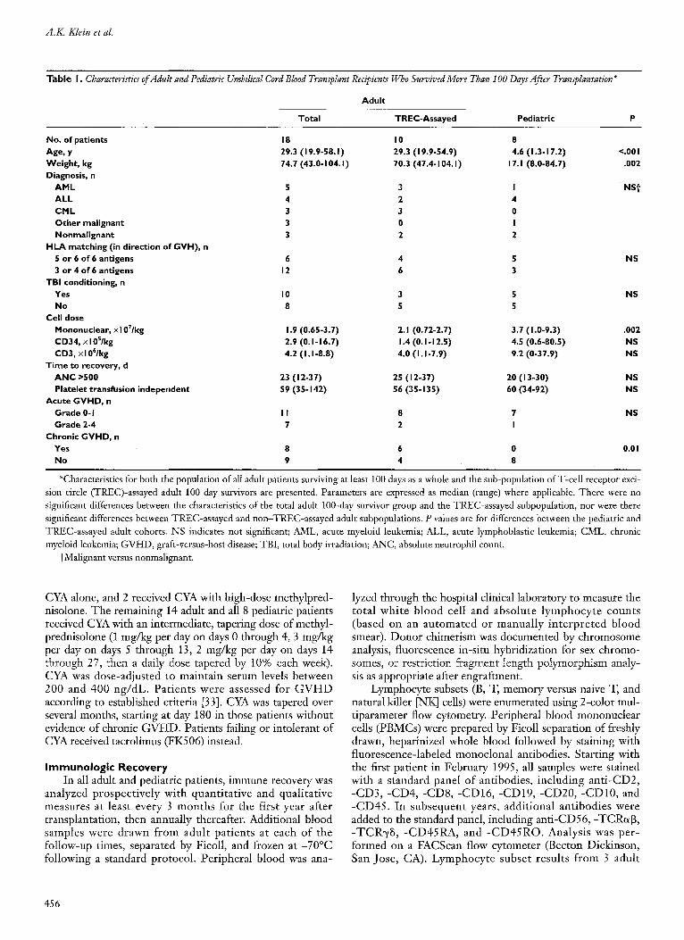
Table I. Characteriytics of Adult and Pediatric Umbilical Cord Blood Transplant Recipients Who Survived More Than I O0 Days After Transplantation*
Adul t
Total TREC-Assayed Pediatric
No. of patients 18 I 0 8 Age, y 29.3 ( I 9.9-58. I ) 29.3 ( I 9.9-54.9) 4.6 ( I .3-17.2) <.00 I Weight , kg 74.7 (43.0-104. I ) 70.3(47.4-104. I ) 17. I (8.0-84.7) .002 Diagnosis, n
A M L 5 3 I NS t ALL 4 2 4
C M L 3 3 0 O t h e r malignant 3 0 I Nonmalignant 3 2 2
H L A matching (in direction of G V H ) , n 5 or 6 of 6 antigens 6 4 5 NS 3 or 4 of 6 antigens 12 6 3
TBI conditioning, n Yes I 0 3 5 NS No 8 5 5
Cell dose Mononuclear, xl0T/kg 1.9 (0.65-3.7) 2.1 (0.72-2.7) 3.7 ( I .0-9.3) .002 CD34, • I 0S/kg 2.9 (0. I - 16.7) 1.4 (0. I - 12.5) 4.5 (0.6-80.5) NS CD3, • 4.2 ( I . I -8.8) 4.0 ( I . I -7.9) 9.2 (0-37.9) NS
T ime to recovery, d A N C >500 23 ( I 2-37) 25 ( I 2-37) 20 ( I 3-30) NS Platelet transfusion independent 59 (35-142) 56 (35-135) 60 (34-92) NS
Acute GVFID, n Grade 0-1 I I 8 7 NS Grade 2-4 7 2 I
Chronic G V H D , n Yes 8 6 0 0.0 I No 9 4 8
*Characteristics for both the population of all adult patients surviving at least 100 days as a whole and the sub-population ofT-cell receptor exci- sion circle (TREC)-assayed adult 100 day survivors are presented. Parameters are expressed as median (range) where applicable. There were no significant differences between the characteristics of the total adult 100-day survivor group and the TREC-assayed subpopulation, nor were there significant differences between TREC-assayed and non-TREC-assayed adult suhpopulations. P values are for differences between the pediatric and TREC-assayed adult cohorts. NS indicates not significant; AML, acute myeloid leukemia; ALL, acute lymphoblastic leukemia; CML, chronic myeloid leukemia; GVHD, graft-versus-host disease; TBI, total body irradiation; ANC, absolute neutrophil count.
tMalignant versus nonmalignant.
CYA alone, and 2 received CYA with high-dose methylpred- nisolone. The remaining 14 adult and all 8 pediatric patients received CYA with an intermediate, tapering dose of methyl- prednisolone (1 mg/kg per day on days 0 through 4, 3 mg/kg per day on days 5 through 13, 2 mg/kg per day on days 14 through 27, then a daily dose tapered by 10% each week). CYA was dose-adjusted to maintain serum levels between 200 and 400 ng/dL. Patients were assessed for G V H D according to established criteria [33]. CYA was tapered over several months, starting at day 180 in those patients without evidence of chronic GVHD. Patients failing or intolerant of CYA received tacrolimus (FKS06) instead.
I m m u n o l o g i c R e c o v e r y In all adult and pediatric patients, immune recovery was
analyzed prospectively with quantitative and qualitative measures at least every 3 months for the first year after transplantation, then annually thereafter. Additional blood samples were drawn from adult patients at each of the follow-up times, separated by Ficoll, and frozen at -70~ following a standard protocol. Peripheral blood was ana-
lyzed through the hospital clinical laboratory to measure the total white blood cell and absolute lymphocyte counts (based on an automated or manually interpreted blood smear). Donor chimerism was documented by chromosome analysis, fluorescence in-situ hybridization for sex chromo- somes, or restriction fragment length polymorphism analy- sis as appropriate after engraftment.
Lymphocyte subsets (B, T, memory versus naive T, and natural killer [NK] cells) were enumerated using 2-color mul- tiparameter flow cytometry. Peripheral blood mononuclear cells (PBMCs) were prepared by Ficoll separation of fleshly drawn, heparinized whole blood followed by staining with fluorescence-labeled monoclonal antibodies. Starting with the first patient in February 1995, all samples were stained with a standard panel of antibodies, including anti-CD2, -CD3, -CD4, -CD8, -CD 16, -CD19, -CD20, -CD10, and -CD45. In subsequent years, additional antibodies were added to the standard panel, including anti-CD56, - T C R ~ , -TCR~8, -CD45RA, and -CD45RO. Analysis was per- formed on a FACScan flow cytometer (Becton Dickinson, San Jose, CA). Lymphocyte subset results from 3 adult
456
T-Cell Recovery After UCBT
patients could not be interpreted because the absolute lym- phocyte count was not recorded.
Lymphocyte function was measured as the proliferative response of PBMCs to the plant mitogen phytohemagglutinin (PHA) and the recall antigen tetanus, following a method pre- viously described [34]. Briefly, PBMCs were isolated from freshly drawn, heparinized whole blood by Ficoll separation and diluted to a concentration of 1 x 10⁶/mE. One hundred microliters (1 • 105 PBMCs) was incubated with PHA at 3 dif- ferent concentrations for 3 to 6 days and with tetanus toxoid at 4 different concentrations for 6 days. 3H-labeled thymidine was added and further incubated for at least 6 hours. [3H]thymidine uptake into proliferating PBMCs was meas- ured by liquid scintillation counter after harvesting. The best responses to mitogen and antigen were recorded. Results were compared to unstimulated [3H]thymidine-incubated patient control PBMCs (background) and normal volunteer donor PBMCs incubated simultaneously.
sjTREC Measurement sjTREC assays were performed retrospectively on all
available banked samples. The concentration of sjTREC DNA in PBMCs was measured by quantitative-competitive (QC)-PCR following a method previously described [35]. Briefly, DNA was isolated from 2 to 10 million PBMCs using the Life Technologies (Rockville, MD) Trizol reagent protocol. DNA was resuspended in 100 gL of 8 mmol NaOH; the resulting concentration of DNA was deter- mined by spectrophotometry. DNA (1 gg) was amplified for 30 cycles using a 60~ annealing temperature and extension at 72~ for 30 seconds in a 50-gL PCR reaction mix con- taining lx PCR buffer (Platinum 7~q; Life Technologies), 1.8 mmol/L MgC12, 200 gmol/L dNTPs, sjTREC Primer A (250 nmol/L), sjTREC Primer B (250 nmol/L), 2.5 gCi [~32p]dCTP, 0.5 BL Platinum Taq Polymerase (Life Tech- nologies), and 5000, 1000, 500, or 100 molecules of sjTREC standard (60 base pairs shorter than the target sjTREC sequence). PCR products were separated on 6% or 8% polyacrylamide gels. The gels were dried and bands visual- ized on a PhosphorImager screen (Molecular Dynamics, Sunnyvale, CA) and quantified using ImageQuant (Amer- sham Pharmacia Biotech, Piscataway, NJ) software. In this assay, the limit of detection was > 100 sjTRECs/Bg DNA.
CDR3 Spectratyping The CDR3 spectratype assay was performed retrospec-
tively on banked samples. Each spectratype analysis was per- formed on a peripheral blood sample that had also been assayed for sjTRECs.
CDR3 spectratyping was performed as previously described, with oligonucleotide primers specific for each of 23 V[3 gene segments and the C[31 segment [32]. Total RNA was extracted from PBMCs using the RNEasy Mini Kit (Qiagen, Valencia, CA) or Trizol reagent (Life Tech- nologies) and used for first-strand complementary DNA (cDNA) synthesis using M-MLV [Moloney murine leukemia virus] Reverse Transcriptase (Life Technologies) with a random hexamer primer (Promega, Madison, WI). cDNA was amplified for 40 cycles by denaturing at 94~ for 25 seconds, annealing at 60~ for 45 seconds, and extending at 72~ for 40 seconds in a 25-gL PCR reaction
mix containing lx PCR Buffer C (Invitrogen, Carlsbad, CA), 2.5 gmol/L MgC12, 250 gmol/L dNTPs, 2.5 gmol/L V[3 primer, 100 Bmol/L C[31 primer, and 0.125 U Ampli- Taq DNA polymerase (Applied Biosystems, Foster City, CA). The reaction was completed with a final extension at 72~ for 5 minutes, cDNA derived from cultured Jurkat E6- 1 (human T-cell leukemia) cells was amplified with the V[38 primer in a separate reaction mixture as a positive control.
The resulting amplified PCR products were elongated over 5 cycles in a run-off reaction with a 5'-6 carboxyfluo- rescein (6-FAM)-labeled nested C13t primer and separated on a denaturing gel. CDR3 size and fluorescence intensity were measured using an ABI 377 XL Sequencer, and results were analyzed with ABI GeneScan software (version 3.1; Applied Biosystems). The number of peaks was counted within each V[3 family, and the total fluorescence intensity under each peak was calculated across each V[3 family.
Statistical Analysis Where applicable, results were compared (adult versus
pediatric) to determine significance using the Wilcoxon signed-rank test (for continuous data) or the Fisher's exact test (for nominal data).
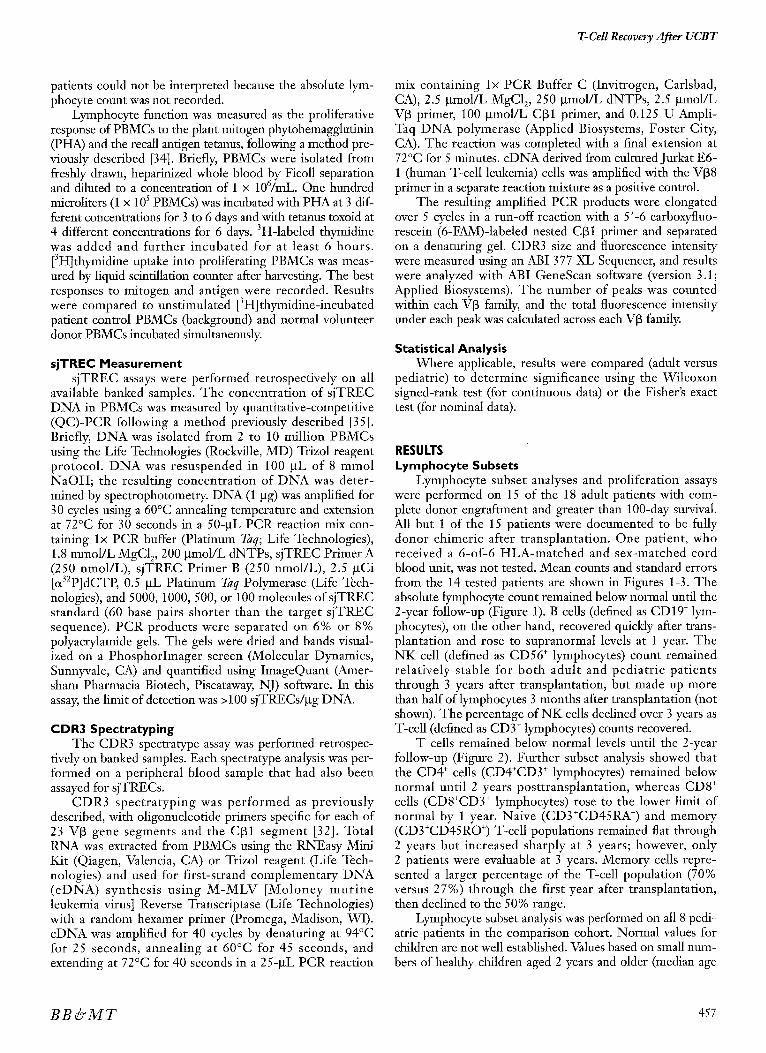
RESULTS Lymphocyte Subsets
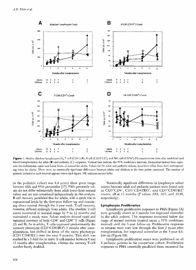
Lymphocyte subset analyses and proliferation assays were performed on 15 of the 18 adult patients with com- plete donor engraftment and greater than 100-day survival. All but 1 of the 15 patients were documented to be fully donor chimeric after transplantation. One patient, who received a 6-of-6 HLA-matched and sex-matched cord blood unit, was not tested. Mean counts and standard errors from the 14 tested patients are shown in Figures 1-3. The absolute lymphocyte count remained below normal until the 2-year follow-up (Figure 1). B cells (defined as CD19 + lym- phocytes), on the other hand, recovered quickly after trans- plantation and rose to supranormal levels at 1 year. The NK cell (defined as CD56 + lymphocytes) count remained relatively stable for both adult and pediatric patients through 3 years after transplantation, but made up more than half of lymphoeytes 3 months after transplantation (not shown). The percentage of NK cells declined over 3 years as T-cell (defined as CD3 + lymphocytes) counts recovered.
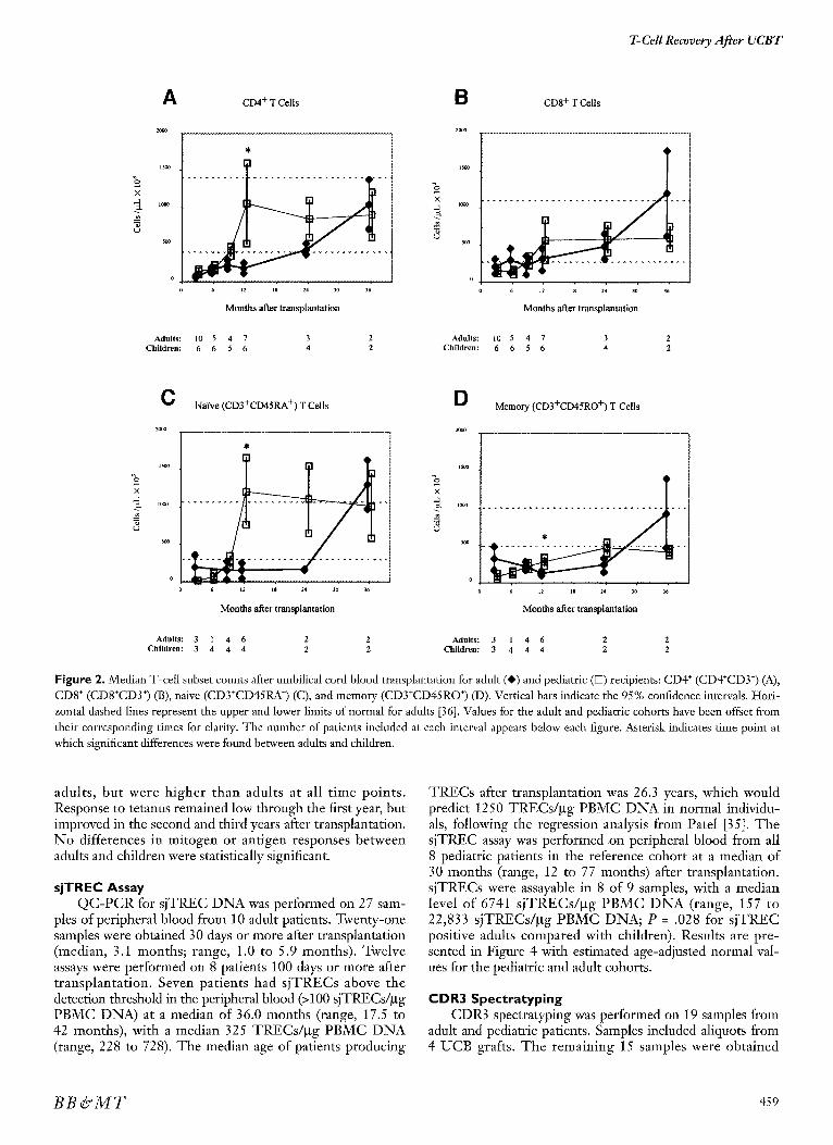
T cells remained below normal levels until the 2-year follow-up (Figure 2). Further subset analysis showed that the CD4 + cells (CD4+CD3 + lymphocytes) remained below normal until 2 years posttransplantation, whereas CD8 + cells (CD8+CD3 + lymphocytes) rose to the lower limit of normal by 1 year. Naive (CD3+CD45RA § and memory (CD3+CD45RO +) T-cell populations remained flat through 2 years but increased sharply at 3 years; however, only 2 patients were evaluable at 3 years. Memory cells repre- sented a larger percentage of the T-cell population (70% versus 27%) through the first year after transplantation, then declined to the 50% range.
Lymphocyte subset analysis was performed on all 8 pedi- atric patients in the comparison cohort. Normal values for children are not well established. Values based on small num- bers of healthy children aged 2 years and older (median age
B B & M T 457
A.K. Klein et al.
% X
A
Adults: Children:
Absolute Lymphocyte Count
i d ,; 21 ,~ ,~
Months after transplantation
10 5 4 7 3 2 6 6 5 6 4 2
%
X
B sso~
3000
2~
2~0
1500
io~o
5c~
o
B-Cel l ( C D 1 9 +) Count
6 ~2 15 24 Jo 36
Months after transplantation
Adults: 9 5 4 7 3 2 Children: 6 6 5 6 4 2
%
X
-6 (J
C 3500
3000
25oo
20C0
I000
SOO
0
T-Cell (CD3 +) Count
% x
6 12 1~ 24 30 16
Months after transplantation
10 5 4 7 3 2 6 6 5 6 4 2
D 700
600
m0
4O0
300
2~o
tco
o
Adults: Adults: Children: Children:
NK-Cell (CD56 +) Count
6 L2 ~$ 24 30 36
Months after transplantation
9 5 4 7 3 2 4 5 5 6 3 2
F i g u r e I. Median absolute lymphocyte (A), T cell (CD3 +) (B), B cell (CD19 +) (C), and NK-cell (CD56 +) (D) counts versus time after umbilical cord blood transplantation for adult (0) and pediatric ([B) recipients. Vertical bars indicate the 95% confdence intervals. Horizontal dashed lines repre- sent the institutional upper and lower limits of normal for adults. Values for the adult and pediatric cohorts have been offset from their correspond- ing times for clarity. There were no statistically significant differences between adults and children at the time points examined. The number of patients included at each interval appears below each figure. NK indicates natural killer.
in the pediatr ic cohor t was 4.6 years) show great range between fifth and 95th percentiles [37]. Fifth percentile val- ues do not differ substantially from adult lower-limit normal values and are not considered independently in this analysis. B-cell recovery paralleled that for adults, with a quick rise to supranormal levels by the first-year fol low-up and remain- ing above normal through the 3-year mark. T-cell recovery, however, differed strikingly from adults. The absolute T-cell count recovered to normal range by 9 to 12 months and maintained a steady state. Subset analysis showed rapid and sustained recovery of both CD4 + and CD8 § T cells (Figure 2A and B). As in adults, T cells expressed predominantly the memory phenotype (CD3+CD45RO +) 3 months after trans- planta t ion, but shif ted in favor of the naive p h e n o t y p e (CD3+CD45RA +) over the next 9 months. This shift was marked by a 5-fold rise in naive T-cell number between 9 and 12 months after transplantation, whereas the memory T-cell number barely doubled.
Statistically significant differences in lymphocyte subset counts between adult and pediatric patients were found only in C D 3 + C D 4 +, C D 3 + C D 4 5 R O +, and C D 3 + C D 4 5 R A + counts, all at 12 months (P values .014, .023, and .0149, respectively).
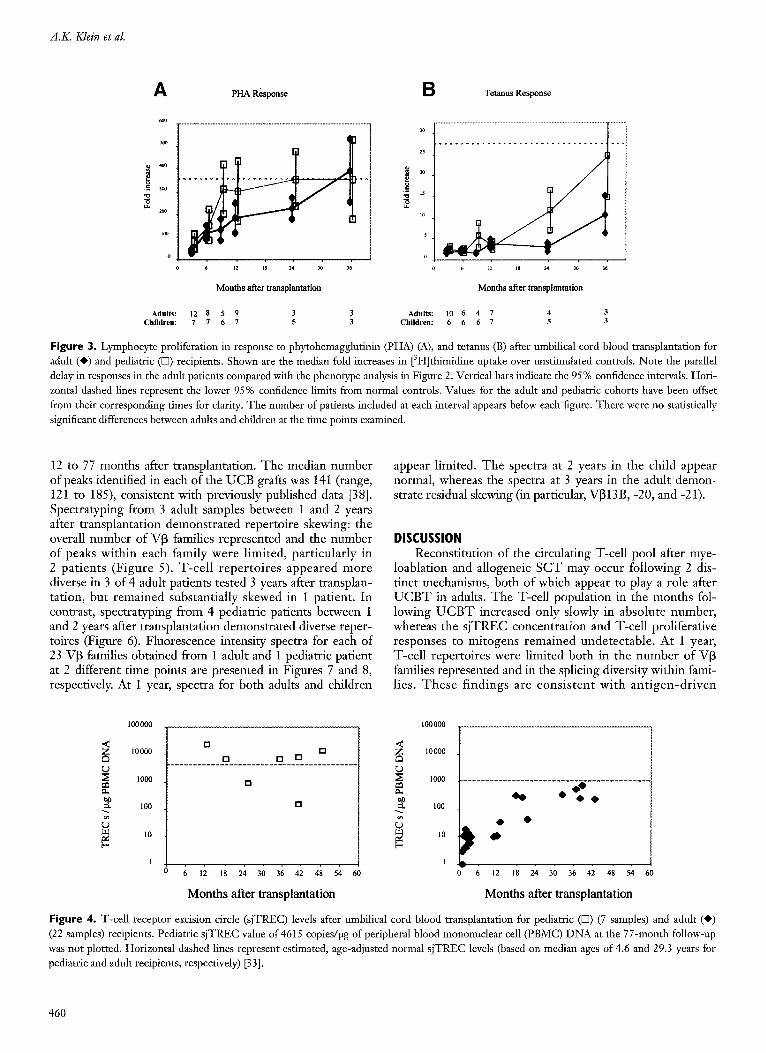
Lymphocyte Proliferation Lymphocyte proliferative responses to P H A (Figure 3A)
were generally absent at 3 months but improved thereafter in the adult cohort . T h e responses remained below the range of normal controls (control mean _+ 95% confidence interval) until the 3-year follow-up. Proliferative responses to te tanus were very low through the first 2 years after transplantation, but improved somewhat at the 3-year fol- low-up (Figure 3B).
Lymphocyte proliferation assays were performed on all 8 pediatric patients in the comparison cohort. Proliferative responses to P H A essentially paralleled those measured for
458
T-Cell Recovery AJ~er UCBT
% •
A C D 4 + T Cel ls
* l
ii + 6 12 18 24 30 36
M o n t h s after transplantat ion
B C D 8 + T Cel ls
5oo
0 0 6 12 IS 24 30 36
Months after transplantat ion
Adults: 10 5 4 7 3 2 Adults: l0 5 4 7 3 2 Children: 6 6 5 6 4 2 Children: 6 6 5 6 4 2
X
O 2OOO
N a i v e ( C D 3 + C D 4 5 R A +) T Cel ls
6 12 18 24 30 36
M o n t h s alter transplantat ion
%
D 2O0O
•
Memory ( C D 3 + C D 4 5 R O +) T Cel ls
e t2 is 24 30 36
Months after transplantat ion
Adults: 3 1 4 6 2 2 Adults: 3 1 4 6 2 2 Children: 3 4 4 4 2 2 Children: 3 4 4 4 2 2
Figure 2. Median T-celt subset counts after umbilical cord blood transplantation for adult (@) and pediatric ([Z) recipients: CD4 § (CD4+CD3 +) (A), CD8 + (CD8+CD3 +) (B), naive (CD3+CD45RA +) (C), and memory (CD3+CD45RO +) (D). Vertical bars indicate the 95% confidence intervals. Hori- zontal dashed lines represent the upper and lower limits of normal for adults [36]. Values for the adult and pediatric cohorts have been offset from their corresponding times for clarity. The number of patients included at each interval appears below each figure. Asterisk indicates time point at which significant differences were found between adults and children.
adults, but were higher than adults at all t ime points. Response to tetanus remained low through the first year, but improved in the second and third years after transplantation. No differences in mitogen or antigen responses between adults and children were statistically significant.
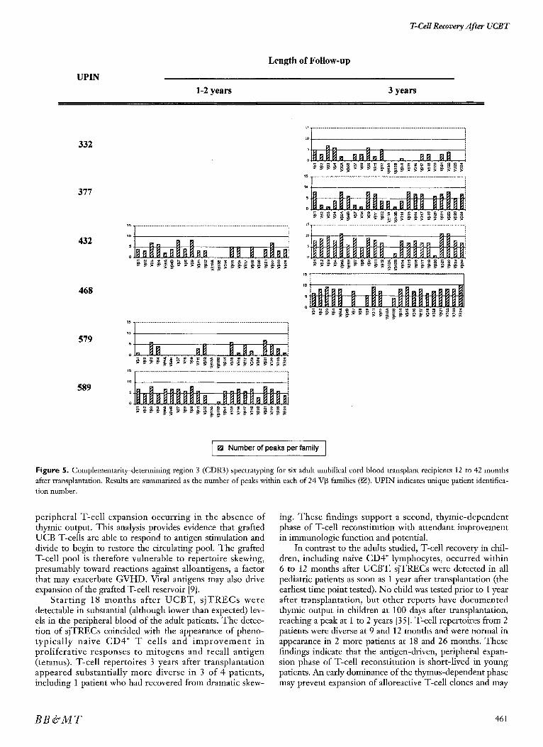
sjTREC Assay QC-PCR for sjTREC DNA was performed on 27 sam-
ples of peripheral blood from 10 adult patients. Twenty-one samples were obtained 30 days or more after transplantation (median, 3.1 months; range, 1.0 to 5.9 months). Twelve assays were performed on 8 patients 100 days or more after transplantation. Seven patients had s jTRECs above the detection threshold in the peripheral blood (>100 sjTRECs/btg PBMC DNA) at a median of 36.0 months (range, 17.5 to 42 months), with a median 325 TRECs/btg PBMC DNA (range, 228 to 728). The median age of patients producing
TRECs after transplantation was 26.3 years, which would predict 1250 TRECs/btg PBMC DNA in normal individu- als, following the regression analysis from Patel [35]. The sjTREC assay was performed on peripheral blood from all 8 pediatric patients in the reference cohort at a median of 30 months (range, 12 to 77 months) after transplantation. sjTRECs were assayable in 8 of 9 samples, with a median level of 6741 sjTRECs/btg PBMC D N A (range, 157 to 22,833 sjTRECs/btg PBMC DNA; P = .028 for s jTREC positive adults compared with children). Results are pre- sented in Figure 4 with estimated age-adjusted normal val- ues for the pediatric and adult cohorts.
CDR3 Spectratyping CDR3 spectratyping was performed on 19 samples from
adult and pediatric patients. Samples included aliquots from 4 UCB grafts. The remaining 15 samples were obtained
BB & M T 459
A.K. Klein et al.
A PHA Response B T e t a n u s R e s p o n s e
600
~0
300
200
0
6 12 18 24 30 36
30
25
20
15
10
5
0
. . . . . . . . . . . . . . . . . . . . . . . . . . . . . . . . . i
Months after transplantation Months after transplantation
Adults: 12 8 5 9 3 3 Adu l t s : 10 6 4 7 4 3 Children: 7 7 6 7 5 3 C h i l d r e n : 6 6 6 7 5 3
Figure 3. Lymphocyte proliferation in response to phytohemagglutinin (PHA) (A), and tetanus (B) after umbilical cord blood transplantation for adult (O) and pediatric (D) recipients. Shown are the median fold increases in [3H]thimidine uptake over unstimulated controls. Note the parallel delay in responses in the adult patients compared with the phenotype analysis in Figure 2. Vertical bars indicate the 95% confidence intervals. Hori- zontal dashed lines represent the lower 95% confidence limits from normal controls. Values for the adult and pediatric cohorts have been offset from their corresponding times for clarity. The number of patients included at each interval appears below each figure. There were no statistically significant differences between adults and children at the time points examined.
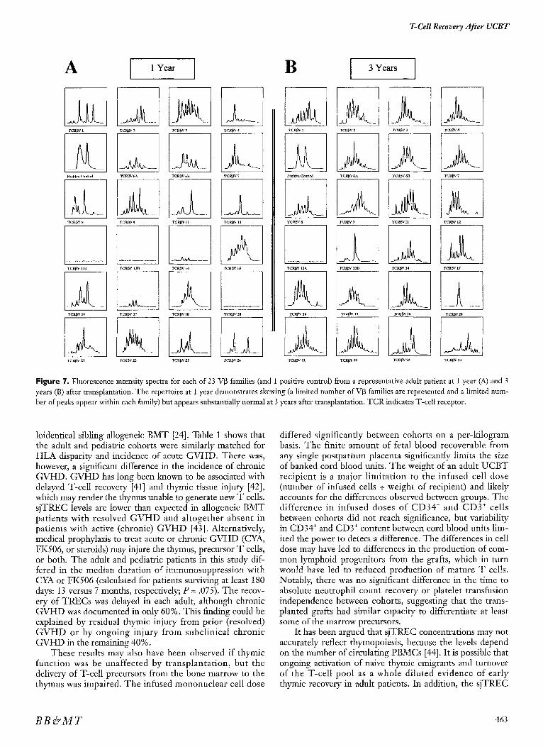
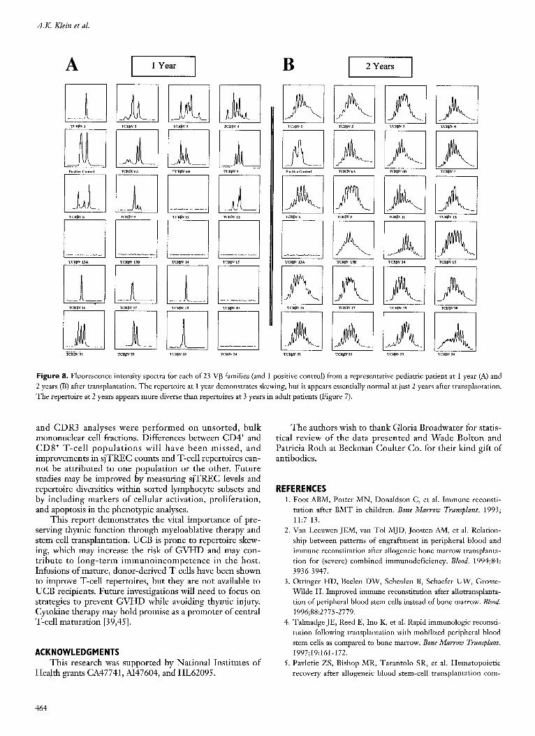
12 to 77 months after transplantation. The median number of peaks identified in each of the UCB grafts was 141 (range, 121 to 185), consistent with previously published data [38]. Spectratyping from- 3 adult samples between 1 and 2 years after transplantation demonstrated repertoire skewing: the overall number of VI3 families represented and the number of peaks within each family were limited, part icularly in 2 pat ients (Figure 5). T-cel l reper to i res appeared more diverse in 3 of 4 adult patients tested 3 years after transplan- tation, but remained substantially skewed in 1 patient. In contrast, spectratyping from 4 pediatric patients between 1 and 2 years after transplantation demonstrated diverse reper- toires (Figure 6). Fluorescence intensity spectra for each of 23 V[3 families obtained from 1 adult and 1 pediatric patient at 2 different time points are presented in Figures 7 and 8, respectively. At 1 year, spectra for both adults and children
appear limited. T h e spectra at 2 years in the child appear normal, whereas the spectra at 3 years in the adult demon- strate residual skewing (in particular, V]313 B, -20, and -21).
DISCUSSION Reconstitution of the circulating T-cell pool after mye-
loablation and allogeneic S C T may occur following 2 dis- tinct mechanisms, both of which appear to play a role after U C B T in adults. The T-cell population in the months fol- lowing U C B T increased only slowly in absolute number, whereas the s jTREC concentration and T-cell proliferative responses to mitogens remained undetectable. At 1 year, T-cell repertoires were limited both in the number of VI3 families represented and in the splicing diversity within fami- lies. T h e s e f indings are cons is tent wi th an t igen-d r iven
100000
< t ~ 1 0 0 0 0
1ooo
~ 1o [.-,
1
rn El
[] [] []
[ ]
[ ]
6 12 18 24 30 36 42 48 54 60
100000
< 10000
L) 1000
e~ e~
"4 100
L) [--
1
[ [
E
v . . . . . . . . . I 6 12 18 24 30 36 42 48 54 60
M o n t h s after t ransp lanta t ion M o n t h s after t ransp lanta t ion
F igure 4. T-cell receptor excision circle (sjTREC) levels after umbilical cord blood transplantation for pediatric (D) (7 samples) and adult (0) (22 samples) recipients. Pediatric sjTREC value of 4615 copies/pg of peripheral blood mononuclear cell (PBMC) DNA at the 77-month follow-up was not plotted. Horizontal dashed lines represent estimated, age-adjusted normal sjTREC levels (based on median ages of 4.6 and 29.3 years for pediatric and adult recipients, respectively) [33].
460
T-Cell Recovery After UCBT
UPIN
Length of Follow-up
1-2 years 3 years
332
377
432
468
579
589
o
~ 5 . . . . . . . . . . . . . . . . . . . . . . . . . . . . . . . . . . . . . . . . . . . . . . . . . . . . . . . . . . . . . . . . . . . . . . . . . . . . . . . . . . . . . . . . . . . . . . . . . . . . . . . . . . . . . . . . . . . . . . . . . . . . . . . . . . . .
o . . [1.11. e c e e ~ e e e e = ~ e e e e e e g ee
e~ge .eeeeg~eeee .eeeee e ~
L5 ...................................................................................................................................... i
5 ;
0
. e e a s e eeeegeg
o
~5 . . . . . . . . . . . . . . . . . . . . . . .
o
[ m Number of peaks per family [
Figure 5. Complementarity-determining region 3 (CDR3) spectraty-ping for six adult umbilical cord blood transplant recipients 12 to 42 months after transplantation. Results are summarized as the number of peaks within each of 24 V[3 families (~1). UPIN indicates unique patient identifica- tion number.
peripheral T-cell expansion occurring in the absence of thymic output. This analysis provides evidence that grafted UCB T-cells are able to respond to antigen stimulation and divide to begin to restore the circulating pool. The grafted T-cell pool is therefore vulnerable to repertoire skewing, presumably toward reactions against alloantigens, a factor that may exacerbate GVHD. Viral antigens may also drive expansion of the grafted T-ceU reservoir [9].
Start ing 18 months after UCBT, s jTRECs were detectable in substantial (although lower than expected) lev- els in the peripheral blood of the adult patients. The detec- tion of sjTRECs coincided with the appearance of pheno- typical ly naive CD4 + T cells and improvement in proliferative responses to mitogens and recall antigen (tetanus). T-cell repertoires 3 years after transplantation appeared substantially more diverse in 3 of 4 patients, including 1 patient who had recovered from dramatic skew-
ing. These findings support a second, thymic-dependent phase of T-celt reconstitution with attendant improvement in immunologic function and potential.
In contrast to the adults studied, T-cell recovery in chil- dren, including naive CD4 § lymphocytes, occurred within 6 to 12 months after UCBT. sjTRECs were detected in all pediatric patients as soon as 1 year after transplantation (the earliest time point tested). No child was tested prior to 1 year after transplantation, but other reports have documented thymic output in children at 100 days after transplantation, reaching a peak at 1 to 2 years [35]. T-cell repertoires from 2 patients were diverse at 9 and 12 months and were normal in appearance in 2 more patients at 18 and 26 months. These findings indicate that the antigen-driven, peripheral expan- sion phase of T-cell reconstitution is short-lived in young patients. An early dominance of the thymus-dependent phase may prevent expansion of alloreactive T-cell clones and may
B B & M T 461
A.K. Klein et al.
UPIN
Lengths of Follow-up
1-2 years 3 years
124
317
490
499
532
577
15
!
15
10
15
15
0 . . . . . .
Number of peaks per family t
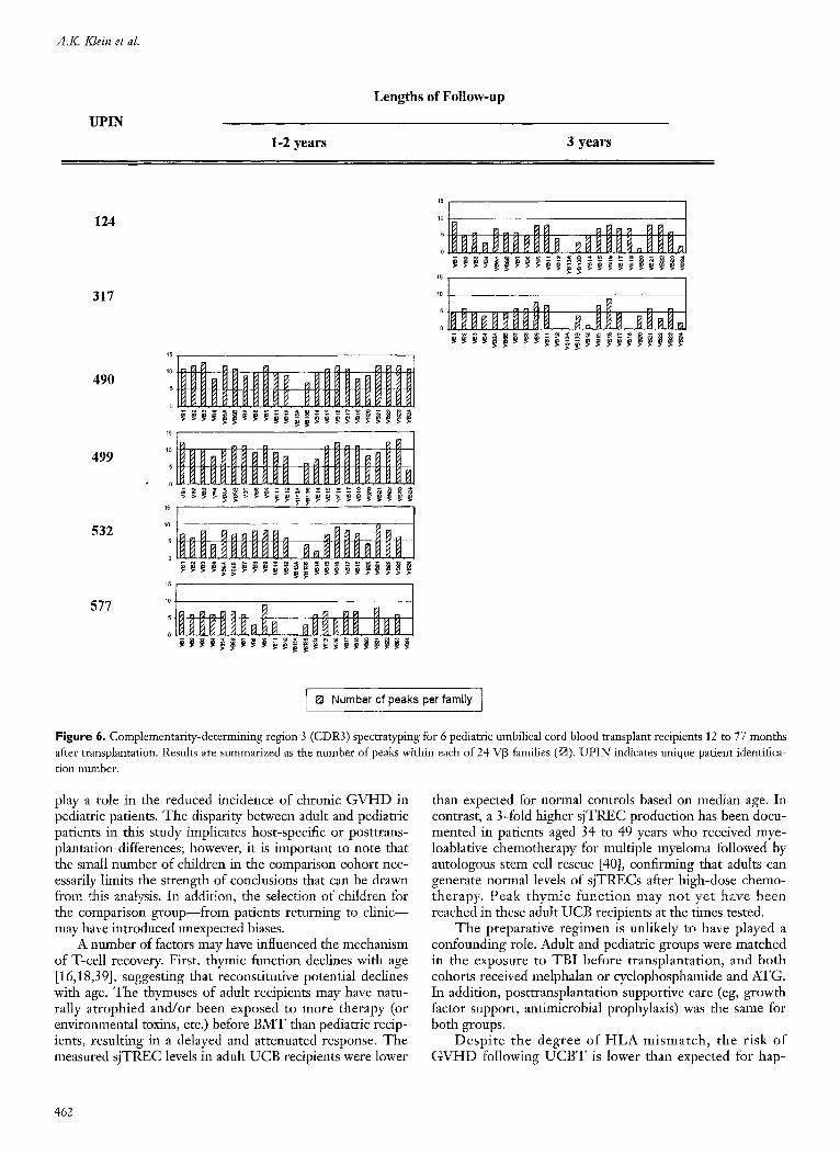
Figure 6. Complementarity-determining region 3 (CDR3) spectratyping for 6 pediatric umbilical cord blood transplant recipients 12 to 77 months after transplantation. Results are summarized as the number of peaks within each of 24 VI3 families ([]). UPIN indicates unique patient identifica- tion number.
play a role in the reduced incidence of chronic GVHD in pediatric patients. The disparity between adult and pediatric patients in this study implicates host-specific or posttrans- plantation differences; however, it is important to note that the small number of children in the comparison cohort nec- essarily limits the strength of conclusions that can be drawn from this analysis. In addition, the selection of children for the comparison group--from patients returning to clinic-- may have introduced unexpected biases.
A number of factors may have influenced the mechanism of T-cell recovery. First, thymic function declines with age [16,18,39], suggesting that reconstitutive potential declines with age. The thymuses of adult recipients may have natu- rally atrophied and/or been exposed to more therapy (or environmental toxins, etc.) before BMT than pediatric recip- ients, resulting in a delayed and attenuated response. The measured sjTREC levels in adult UCB recipients were lower
than expected for normal controls based on median age. In contrast, a 3-fold higher sjTREC production has been docu- mented in patients aged 34 to 49 years who received mye- loablative chemotherapy for multiple myeloma followed by autologous stem cell rescne [40], confirming that adults can generate normal levels of sjTRECs after high-dose chemo- therapy. Peak thymic function may not yet have been reached in these adult UCB recipients at the times tested.
The preparative regimen is unlikely to have played a confounding role. Adult and pediatric groups were matched in the exposure to TBI before transplantation, and both cohorts received melphalan or cyclophosphamide and ATG. In addition, posttransplantation supportive care (eg, growth factor support, antimicrobial prophylaxis) was the same for both groups.
Despite the degree of HLA mismatch, the risk of GVHD following UCBT is lower than expected for hap-
462
T-Cell Recovery After UCBT
A
r c ~ v t
Positive Control
TCR~V8
TCR[~V 13A
~CRI~V 16
"1CRI~V 21
TCRI~V 2
TCR~V6A
TCR f,v 9
TCR~V 13B
TCUf~V 17
TeRry 2~
1 Year
"1 CR~V 3
TCR[~V 6B
_. ~ _ _ .
r c R ~ v n
TCR~V 14
la Tcl~l~v I s
'rctll~v 23
TCR~V 4
a_ TCR~V 7
TCR[~V 12
'rCR~V 1~
TCRI3V 20
2a_ TCRI3V 24
B
I'CR~V 1
9o$1ti~,e Colllrol
r c R ~ v s
T C]R~'V 13A la TCI~V 16
"teRrY 21
3 Years
TCR[3V 2
'rCRl3V 6A
TCR~V 9
TCR~V 1313
TCRDV 17
TCR~V22
TCR~V3
T C R ~ V ~
TCR~V II
TCR~V 14
TCRf~V 18
TCR~V 23
TCR~V 4
TCRI~V 7
TCKf~V lZ
TCRgV l.g
'rcR[~V 20
TCR[~V 24
Figure 7. Fluorescence intensity spectra for each of 23 V~ families (and 1 posifve control) from a representative adult panent at 1 year (A) and 3 years (B) after transplantation. The repertoire at 1 year demonstrates skewing (a limited number of V[3 families are represented and a limited num- ber of peaks appear within each family) but appears substantially normal at 3 years after transplantation. TCR indicates T-cell receptor.
loidentical sibling allogeneic BMT [24]. Table 1 shows that the adult and pediatric cohorts were similarly matched for HLA disparity and incidence of acute GVHD. There was, however, a significant difference in the incidence of chronic GVHD. G V H D has long been known to be associated with delayed T-cell recovery [41] and thymic tissue injury [42], which may render the thymus unable to generate new T cells. sjTREC levels are lower than expected in allogeneic BMT patients with resolved G V H D and altogether absent in patients with active (chronic) G V H D [43]. Alternatively, medical prophylaxis to treat acute or chronic G V H D (CYA, FK506, or steroids) may injure the thymus, precursor T cells, or both. The adult and pediatric patients in this study dif- fered in the median duration of immunosuppression with CYA or FK506 (calculated for patients surviving at least 180 days: 13 versus 7 months, respectively; P = .075). The recov- ery of TRECs was delayed in each adult, although chronic GVHD was documented in only 60%. This finding could be explained by residual thymic injury from prior (resolved) G V H D or by ongoing injury f rom subclinical chronic GVHD in the remaining 40%.
These results may also have been observed if thymic function was be unaffected by transplantation, but the delivery of T-cell precursors from the bone marrow to the thymus was impaired. The infused mononuclear cell dose
differed significantly between cohorts on a per-kilogram basis. The finite amount of fetal blood recoverable from any single postpartum placenta significantly limits the size of banked cord blood units. The weight of an adult U C B T recipient is a major l imitat ion to the infused cell dose (number of infused cells + weight of recipient) and likely accounts for the differences observed between groups. The difference in infused doses of CD34 § and CD3 + cells between cohorts did not reach significance, but variability in CD34 + and CD3 + content between cord blood units lim- ited the power to detect a difference. The differences in cell dose may have led to differences in the production of com- mon lymphoid progenitors from the grafts, which in turn would have led to reduced production of mature T cells. Notably, there was no significant difference in the time to absolute neutrophil count recovery or platelet transfusion independence between cohorts, suggesting that the trans- planted grafts had similar capacity to differentiate at least some of the marrow precursors.
It has been argued that sjTREC concentrations may not accurately reflect thymopoiesis, because the levels depend on the number of circulating PBMCs [44]. It is possible that ongoing activation of naive thymic emigrants and turnover of the T-cell pool as a whole diluted evidence of early thymic recovery in adult patients. In addition, the sjTREC
B B & M T 463
A.K. Klein et al.
A
TCR~V 1
Positive Control
'rCR~V 8
I TCR[~V 13A
TCR]~V 16
I
TCR~V 2
f A TCR~V 6A
A_ TCR~V 9
1 Year
TCR~V 3
TC'~V aB
TCII~V 11
A TCR~V 4
al TCR[3V 7
TCRI3V lab TCR~3V 14
A .... 1 TCR~V 17 "r CR~,V 18
TeRI3V22 TClt~V 2a
B
TCR ~,V 12
TCRI3V 1.r
TCR]~V 20
TCR[3V 24
2 Years I
TCR~V 1 TCR~V 2
Positive Control TCR~V 6A
IA
A TCI~V 3
TCR~V 6]8
a . TCRJ~V 8 "r CR~V 9
/% TCR~3V 13A
IA TCRI~V 16
A TCR~V 21
.A TCRI3V 4
IA'
TCR[3V 1318
TCRI~V 17
A TCR~V 22
TCR~V 11
TCRI~V 14
A TCR~3V 18
A Telt~V 23
TCR~V 7
TCR[BV 12
A TCR~V IS
A TCR~V20
AI TCRffr 21 TCR~V 24
Figure 8. Fluorescence intensity spectra for each of 23 V[3 families (and 1 positive control) from a representative pediatric patient at 1 year (A) and 2 years (B) after transplantation. The repertoire at 1 year demonstrates skewing, but it appears essentially normal at just 2 years after transplantation. The repertoire at 2 years appears more diverse than repertoires at 3 years in adult patients (Figure 7).
and CDR3 analyses were pe r fo rmed on unsorted, bulk mononuclear cell fractions. Differences between CD4 § and C D 8 § T-ce l l p o p u l a t i o n s will have been missed, and improvements in s jTREC counts and T-cell repertoires can- not be attributed to one population or the other. Future studies may be improved by measuring s jTREC levels and repertoire diversities within sorted lymphocyte subsets and by including markers of cellular activation, proliferation, and apoptosis in the phenotypic analyses.
This report demonstrates the vital importance of pre- serving thymic function through myeloablative therapy and stem cell transplantation. UCB is prone to repertoire skew- ing, which may increase the risk of G V H D and may con- tribute to long- t e rm immuno incompe tence in the host. Infusions of mature, donor-derived T cells have been shown to improve T-cell repertoires, but they are not available to UCB recipients. Future investigations will need to focus on strategies to prevent G V H D while avoiding thymic injury. Cytoldne therapy may hold promise as a promoter of central T-cell maturation [39,45].
ACKNOWLEDGMENTS This research was supported by National Institutes of
Health grants CA47741, AI47604, and HL62095.
The authors wish to thank Gloria Broadwater for statis- tical review of the data presented and Wade Bolton and Patricia Roth at Beckman Coulter Co. for their kind gift of antibodies.
REFERENCES 1. Foot ABM, Potter MN, Donaldson C, et al. Immune reconsti-
tution after BMT in children. Bone Marrow Transplant. 1993; 11:7-13.
2. Van Leeuwen JEM, van Tol MJD, Joosten AM, et al. Relation- ship between patterns of engraftment in peripheral blood and immune reconstitution after allogeneic bone marrow transplanta- tion for (severe) combined immunodeficiency. Blood. 1994;84: 3936-3947.
3. Ottinger HD, Beelen DW, Scheulen B, Schaefer UW, Grosse- Wilde H. Improved immune reconstitution after allotransplanta- tion of peripheral blood stem cells instead of bone marrow. Blood. 1996;88:2775-2779.
4. Talmadge JE, Reed E, Ino K, et al. Rapid immunologic reconsti- tution following transplantation with mobilized peripheral blood stem cells as compared to bone marrow. Bone Marrow Transplant. 1997;19:161-172.
5. Pavletic ZS, Bishop MR, Tarantolo SR, et al. Hematopoietic recovery after allogeneic blood stem-cell transplantation com-
464
T-Cell Recovery After UCBT
pared with bone marrow transplantation in patients with hemato- logic malignancies. J Clin Oncol. 1997; 15:1608-1616.
6. Godthelp BC, van Tol MJD, Vossen JM, van den Elsen PJ. T-cell immune reconstitution in pediatric leukemia patients after allo- geneic bone marrow transplantation with T-cell-depleted or unmanipulated grafts: evaluation of overall and antigen-specific T-cell repertoires. Blood. 1999;94:4358-4369.
7. Small TN, Papadopoulos EB, Boulad F, et al. Comparison of immune reeonstitution after unrelated and related T-cell- depleted bone marrow transplantation: effect of patient age and donor leukocyte infusions. Blood. 1999;93:467-480.
8. Kook H, Goldman F, Padley D, et al. Reconstruction of the immune system after unrelated or partially matched T-cell- depleted bone nlarrow transplantation in children: itumunophe- notypic analysis and factors affecting the speed of recovery. Blood. 1996;88:1089-1097.
9. Bchringer D, Bertz H, Schmoor C, et al. Quantitative lymphocyte subset reconstitution after allogeneic hematopoietic transplanta- tion from matched related donors with CD34 § selected PBPC grafts, unselected PBPC grafts or BM grafts. Bone Marrow Trans- plant. 1999;24:295-302.
10. Lamb LS, Gee AP, Henslee-Downey PJ, et al. Phenotypic and functional reconstitution of peripheral blood lymphocytes follow- ing T ceil-depleted bone marrow transplantation from partially mismatched related donors. Bone Marrow Transplant. 1998;21: 461-471.
11. Davidson GM, Novitzky N, Kline A, et al. Immune reconstitution after allogeneic bone marrow transplantation depleted of T cells. Transplantation. 2000;69:1341-1347.
12. Mackall CL, Gress RE. Pathways of T-cell regeneration in mice and humans: implications for bone marrow transplantation and immunotherapy. ImmunolRev. 1997;157:61-72.
13. Mackall CL, Bare CV, Granger LA, et al. Thymic-independent T cell regeneration occurs via antigen-driven expansion of peripheral T cells resulting in a repertoire that is limited in diver- sity and prone to skewing. J Immunol. 1996; 156:4609-4616.
14. Mackall C, Granger L, Sheard MA, Cepeda R, Gress RE. T-cell regeneration after bone marrow transplantation: differential CD45 isoform expression on rhymic-derived versus thymic-inde- pendent progeny. Blood. 1993;82:2585-2594.
15. Howie D, Spencer J, DeLord D, et al. Extrathymic T cell differ- entiat ion in the human intestine early in life. J Immunol. 1998;161:5862-5872.
16. Mackall CL, Fleisher TA, Brown MR, et al. Age, thymopoiesis, and CD4 + T-lymphocyte regeneration after intensive chemo- therapy. NEnglJMed. 1995;332:143-149.
17. Douek DC, McFarland RD, Keiser PH, et al. Changes in thymic function with age and during the treatment of HIV infection. Nature. 1998;396:690-695.
18. Haynes BF, Markert ML, Sempowski GD, Patel DD, Hale LP. The role of the thymus in immune reconstitution in aging, bone marrow transplantation, and H1V-1 infection, dnnu Rev lmmunol. 2000;18:529-560.
19. Roux E, Dumont-Girard F, Starabinski M, et al. Recovery of immune reactivity after T-cell-depleted bone marrow transplanta- tion depends on thymic activity. Blood. 2000;96:2299-2303.
20. Hirokawa M, Horiuchi T, Kitabayashi A, et al. Delayed recovery of CDR3 complexity of the T-cell receptor-[3 chain in recipients of allogeneic bone marrow transplants who had virus-associated interstitial pneumonia: monitor of T-cell function by CDR3 spec- tratyping. J Allerg;y Clin Iramunol. 2000;106:$32-$39.
21. Verfueth S, Peggs t4, Vyas P, et al. Longitudinal monitoring of imm~me reconstimtion by CDR3 size spectratyping after T-cell- depleted allogeneic bone marrow transplant and the effect of donor lymphocyte infusions on T-cell repertoire. Blood. 2000;95: 3990-3995.
22. Orsini E, Alyea EP, Schlossman R, et al. Changes in T-cell recep- tor repertoire associated with graft-versus-tumor effect and graft- versus-host disease in patients with relapsed multiple myeloma after donor lymphocyte infusion. Bone Marrow Transplant. 2000;25:623-632.
23. KurtzbergJ, Laughlin M, Graham ML, et al. Placental blood as a source of hematopoiedc stem cells for transplantation into unre- lated recipients. NEnglJMed. 1996;335:157-166.
24. Rubinstein P, Carrier C, Scaradavou A, et al. Outcomes among 562 recipients of placental-blood transplants from unrelated donors. NEnglJ Med. 1998;339:1565-1577.
25. Laughlin MJ, Barker J, Bambach K, et al. Hematopoietic engraft- ment and survival in adult recipients of umbilical-cord blood from unrelated donors. NEnglJMed. 2001;344:1815-1822.
26. Han P, Hodge G, Story C, Xu X. Phenotypic analysis of func- tional T-lymphocyte subtypes and natural killer cells in human cord blood: relevance to umbilical cord blood transplantation. Br J Haematol. 1995;89:733-740.
27. Barbey C, Irion O, Helg C, et al. Characterisation of the cyto- toxic alloresponse of cord blood. Bone Marrow Transplant. 1998;22 (snppt 1):$26-$30.
28. Risdon G, Gaddy J, Stehman FB, Broxmeyer HE. Proliferative and cytotoxic responses of human cord blood T lymphocytes fol- lowing allogeneic stimulation. Cell Immunol. 1994; 154:14 24.
29. Haynes BF, Hale LP, Weinhold KJ, et al. Analysis of the adult thymus in reconstitution of T lymphocytes in HIV-1 infection. .,7 Clin Invest. 1999;103:453-460.
30. Picker L, Treer J, Ferguson-Darnell B, Collins P, Buck D, Ter- stappen L. Control of lymphocyte recirculation in man. y Immunol. 1993;150:1105-1121.
31. Bogue M, Roth DB. Mechanism of V(D)J recombination. Curr Opin Iramunol. 1996;8:175-180.
32. Oksenberg JR, Wang L, Yen J. The immunoscope approach for the analysis of T cell repertoires. In: Oksenberg JR, Ed. The Anti- gen T Cell Receptor: Selected Protocols and Applications. New York, NY: Chapman & Hall; 1996:287-325.
33. Przepiorka D, Weisdorf D, Martin P, et al. 1994 Consensus Con- ference on Acute GVHD Grading. Bone Marrow Transplant. 1995;15:825-828.
34. Buckley RH, Schiff SE, Sampson HA, et al. Development of immunity in human severe primary T cell deficiency following haploidentical bone marrow stem cell transplantation. J Immunol. 1986;136:2398-2407.
35. Patel DD, Gooding ME, Parrott RE, Curtis KM, Haynes BF, Buckley RH. Thymic function after hematopoietic stem-cell transplantation for the treatment of severe combined immunode- ficiency. N EnglJ Med. 2000;342:1325-1332.
36. de Vries E, de Bruin-Versteeg S, Comans-Bitter WM, et al. Lon- gitudinal survey of lymphocyte subpopulations in the first year of life. Pediatr Res. 2000;47:528-537.
37. Comans-Bitter WM, de Groot R, van den Beemd, et al. Immuno- phenotyping of blood lymphocytes in childhood. J Pediatr. 1997;130:388-93.
38. Garderet L, Dulphy N, Douay C, et al. The umbilical cord blood ~[~ T-cell repertoire: characteristics of a polyclonal and naive but completely formed repertoire. Blood. 1998;91:340-346.
BB & M T 465
A.K. Klein et al.
39. Sempowski GD, Hale LP, Sundy JS, et al. Leukemia inhibitory factor, oneostatin M, IL-6, and stem cell factor mRNA expression in human thymus increases with age and is associated with thymic atrophy. J Immunol. 2000; 164:2180-2187.
40. Douek CD, Vescio RA, Betts MR, et al. Assessment of thymic output in adults after haematopoietic stem-cell transplantation and prediction of T-cell reconstitution. Lancet. 2000;355: 1875-1881.
41. Van Den Brink MRM, Moore E, Ferrara JLM, Burakoff SJ. Graft-versus-host-disease-associated thymic damage results in the appearance of T cell clones with anti-host reactivity. Transplanta- tion. 2000;69:446-449.
42. Seddik M, Seemayer TA, Lapp WS. The graft-versus-host reac-
tion and immune function, 1: T helper cell immunodeficiency associated with graft-versus-host-induced thymic epithelial cell damage. Transplantation. 1984;37:281-285.
43. Weinberg K, Blazar BR, Wagner JE, et al. Factors affecting thymic function after allogeneic hematopoietic stem cell trans- plantation. Blood. 2001;97:1458-1466.
44. Hazenberg MD, Otto SA, Cohen Stewart JWT, et al. Increased cell division but not thymic dysfunction rapidly affects the T-cell receptor excision circle content of the naive T-cell population in HIV-1 infection. NatureMed. 2000;6:1036-1042.
45. Mackall CL, Fry TJ, Bare C, et al. IL-7 increases both thymic- dependent and thymic-independent T-cell regeneration after bone marrow transplantation. Blood. 2001 ;97:1491-1497.
466
Related Documents


