T Cell Numbers Relate to Bone Involvement in Gaucher Disease Submitted 04/09/99; revised 04/15/99 (communicated by Ernest Beutler, M.D. 04/19/99) Lucia Lacerda 1 , Fernando A. Arosa 2 , Rosa Lacerda 3 , Jose ´ Cabeda 3,4 , Grac ¸a Porto 3,4 , Olga Amaral 1 , Ana Fortuna 5 , Rui Pinto 1 , Pedro Oliveira 6 , Christine E. McLaren 7 , Clara Sa ´ Miranda 1 , Maria de Sousa 2,3 ABSTRACT: The major elements of bone pathology in Gaucher disease are a failure of osteoclast and osteoblast function, resulting in osteopenia and also osteonecrosis. T lymphocytes have recently been found to be involved in the regulation of osteoblast/osteoclast activity in vitro. In the present report the peripheral blood T major lymphocyte subsets were investigated in a group of genotyped type 1 Gaucher disease patients. A total of 31 patients were studied: 21 non-splenectomized (5 N370S homozygotes) and 10 splenectomized (of whom 1 was a N370S homozygote). The results show that non-splenectomized patients present a decrease in absolute numbers of peripheral blood T lymphocytes, specially the CD4 1 T subset. However, when patients were analyzed with respect to the presence of bone disease, the number of CD8 1 T lymphocytes was found to be statistically significantly lower in patients presenting bone involvement. Furthermore, lower numbers of CD8 1 T lymphocytes were significantly correlated with higher levels of plasma tartrate resistant acid phosphatase (TRAP) activity, a putative marker of osteoclast cell activity. These in vivo findings are in agreement with the results reached in vitro by others. They provide an additional marker of disease severity in Gaucher disease. In the group of genotyped Gaucher disease patients, the majority of the N370S homozygous patients presented a clinically milder phenotype, including the absence of bone involvement, confirming earlier reports predicting that a number of these patients may remain undiagnosed. Collectively the homozygosity for the N370S mutation and normal T cell numbers may provide additional markers for the clinical heterogeneity of Gaucher disease. r 1999 Academic Press Keywords: Gaucher’s disease, chitotriosidase, tartrate resistant acid phosphatase, CD8 positive T lymphocytes and bone pathology INTRODUCTION The most prevalent lysosomal storage disease, Gaucher disease, is an autosomal recessive hereditary disorder of glycosphingolipid metabolism, characterized by accumula- tion of glucosylceramide in cells due to the deficient activity of the lysosomal enzyme glucocerebrosidase (E.C.3.2.1.45). The occurrence of type 1 Gaucher disease (McKusick 230800), the non-neuronophatic form, has an estimated incidence of about 1 in 40.000–200.000 births. A higher prevalence is observed among Ashkenazi Jews in which the incidence is 1 in 625–1.500 (1). Type 1 Gaucher disease is a multi-system disease associated with striking variation in its clinical onset, severity and course. Moreover, genetic variation in the glucocerebrosidase locus has not provided a reliable prediction of clinical phenotype or prognosis of the disease. In fact, the determination of the frequency of the prevalent N370S mutated allele in the Portu- guese population indicated that the majority of Gaucher disease patients homozygous for this mutation remained undiagnosed (2). Although glucocerebrosidase is present in almost all cell types, the accumulation of glucosylceramide is charac- teristically observed in macrophages, as the result of their role in the turnover of red blood cells and consequently on the degradation of membrane glycolipids. The occurrence of these lipid loaded macrophages (Gaucher cells) in tissues 1 Department of Genetics Neurobiology, Institute for Molecular and Cell Biology of Porto University, Porto, Portugal. 2 Department of Pathology and Molecular Immunology, Institute for Molecular and Cell Biology of Porto University, Porto, Portugal. 3 Department of Immunology, Abel Salazar Institute for Biomedical Sciences, University of Porto, Porto, Portugal. 4 Department of Hematology, St. Anto ´ nio General Hospital, Porto, Porto, Portugal. 5 Clinical Department, Jacinto de Magalha ˜es Institute of Medical Genetics, Porto, Portugal. 6 Department of Production and Systems, University of Minho, Braga, Portugal. 7 Division of Epidemiology, Department of Medicine, University of California, Irvine, CA 92697-7550, USA. Reprint request to: Prof. Maria de Sousa, Departamento de Patologia e Imunologia Molecular, Instituto de Biologia Molecular e Celular da Universidade do Porto, Rua do Campo Alegre, 823, 4150 Porto, Portugal, telephone 351 2 6074900 (ext: 367), fax 351 2 6099157, e-mail: [email protected]. Blood Cells, Molecules, and Diseases (1999) 25(8) April 30: 130–138 L. Lacerda, et al. Article ID bcmd.1999.0237, available online at http://www.idealibrary.com on 1079-9796/99 $30.00 Copyright r 1999 by Academic Press All rights of reproduction in any form reserved 130

Welcome message from author
This document is posted to help you gain knowledge. Please leave a comment to let me know what you think about it! Share it to your friends and learn new things together.
Transcript
t function,gulation of
wereudied: 21The resultses, speciallynumber ofermore,nt acidhegroup of
ally milderse patientsy provide
Blood Cells, Molecules, and Diseases (1999) 25(8) April 30: 130–138 L. Lacerda, et al.Article ID bcmd.1999.0237, available online at http://www.idealibrary.com on
T Cell Numbers Relate to Bone Involvement in Gaucher DiseaseSubmitted 04/09/99; revised 04/15/99(communicated by Ernest Beutler, M.D. 04/19/99)
Lucia Lacerda1, Fernando A. Arosa2, Rosa Lacerda3, Jose´ Cabeda3,4, Graca Porto3,4, Olga Amaral1,Ana Fortuna5, Rui Pinto1, Pedro Oliveira6, Christine E. McLaren7, Clara Sa´ Miranda1, Maria de Sousa2,3
ABSTRACT: The major elements of bone pathology in Gaucher disease are a failure of osteoclast and osteoblasresulting in osteopenia and also osteonecrosis. T lymphocytes have recently been found to be involved in the reosteoblast/osteoclast activityin vitro. In the present report the peripheral blood T major lymphocyte subsetsinvestigated in a group of genotyped type 1 Gaucher disease patients. A total of 31 patients were stnon-splenectomized (5 N370S homozygotes) and 10 splenectomized (of whom 1 was a N370S homozygote).show that non-splenectomized patients present a decrease in absolute numbers of peripheral blood T lymphocytthe CD41 T subset. However, when patients were analyzed with respect to the presence of bone disease, theCD81 T lymphocytes was found to be statistically significantly lower in patients presenting bone involvement. Furthlower numbers of CD81 T lymphocytes were significantly correlated with higher levels of plasma tartrate resistaphosphatase (TRAP) activity, a putative marker of osteoclast cell activity. Thesein vivofindings are in agreement with tresults reachedin vitro by others. They provide an additional marker of disease severity in Gaucher disease. In thegenotyped Gaucher disease patients, the majority of the N370S homozygous patients presented a clinicphenotype, including the absence of bone involvement, confirming earlier reports predicting that a number of themay remain undiagnosed. Collectively the homozygosity for the N370S mutation and normal T cell numbers maadditional markers for the clinical heterogeneity of Gaucher disease.r 1999 Academic Press
Keywords: Gaucher’s disease, chitotriosidase, tartrate resistant acid phosphatase, CD8 positive T lymphocytes and bone pathology
ucherer ofula-
f). The), theout 1rved.500
ociaterse.
s hasor
f theortu-seaseosed
t allrac-theiry once ofsues
sida
INTRODUCTION
The most prevalent lysosomal storage disease, Gadisease, is an autosomal recessive hereditary disordglycosphingolipid metabolism, characterized by accumtion of glucosylceramide in cells due to thedeficient activity othe lysosomal enzyme glucocerebrosidase (E.C.3.2.1.45occurrence of type 1 Gaucher disease (McKusick 230800non-neuronophatic form, has an estimated incidence of abin 40.000–200.000 births. A higher prevalence is obseamongAshkenazi Jews in which the incidence is 1 in 625–1(1). Type 1 Gaucher disease is a multi-system disease asswith striking variation in its clinical onset, severity and cou
1 Department of Genetics Neurobiology, Institute for Molecular and Ce2 Department of Pathology and Molecular Immunology, Institute for M3 Department of Immunology, Abel Salazar Institute for Biomedical Sc4 Department of Hematology, St. Anto´nio General Hospital, Porto, Porto,5 Clinical Department, Jacinto de Magalha˜es Institute of Medical Genetic6 Department of Production and Systems, University of Minho, Braga,7 Division of Epidemiology, Department of Medicine, University of CalReprint request to: Prof. Maria de Sousa, Departamento de Patologia
Porto, Rua do Campo Alegre, 823, 4150 Porto, Portugal, telephone 351 2 60130
d
Moreover, genetic variation in the glucocerebrosidase locunot provided a reliable prediction of clinical phenotypeprognosis of the disease. In fact, the determination ofrequency of the prevalent N370S mutated allele in the Pguese population indicated that the majority of Gaucher dipatients homozygous for this mutation remained undiagn(2).
Although glucocerebrosidase is present in almoscell types, the accumulation of glucosylceramide is chateristically observed in macrophages, as the result ofrole in the turnover of red blood cells and consequentlthe degradation of membrane glycolipids. The occurrenthese lipid loaded macrophages (Gaucher cells) in tis
gy of Porto University, Porto, Portugal.r and Cell Biology of Porto University, Porto, Portugal., University of Porto, Porto, Portugal.al.
o, Portugal.al.Irvine, CA 92697-7550, USA.ologia Molecular, Instituto de Biologia Molecular e Celular da Univerde do
ll BiolooleculaiencesPortug
s, PortPortug
ifornia,e Imun
74900 (ext: 367), fax 351 2 6099157, e-mail: [email protected].
1079-9796/99 $30.00Copyrightr 1999 byAcademic Press
All rights of reproduction in any form reserved
tope-d innmentpoi-he. Inum-sibly
tationtioned
prov-ano-
rienceehindtionose-
ins inonesedrts ofinntia-fileify ifiatediosi-
er ofralland(13),15),
asey of(16).
ibedn re-
s, 11
f theomi-
enec-d in
w isifera-
byllingtingeticThe1), to), to
lapseonestalnotts at
d onhichegaly,
bone
en-lglu-manrmi-acro-g the1).
entlyex-
ineseted).tion,ry 2
to thed 35and
doseand
, 18,
fromwasatepH
L. Lacerda, et al. Blood Cells, Molecules, and Diseases (1999) 25(8) April 30: 130–138
Article ID bcmd.1999.0237, available online at http://www.idealibrary.com on
and organs, underlies the most common signs of pancynia, organomegaly and skeletal deterioration (reviewe3). Infiltration of normal bone marrow by lipid-ladeGaucher cells results in apparently progressive displaceof the fat-rich marrow with a consequent shift in haematoetic activity from proximal to more distal sites (4). Tpathophysiology is however not clearly understoodparticular, the balance between the impact of altered nbers and increased activity of osteoclasts and the possuppressed osteoblast activity (5). Enzyme supplementherapy (6) which has as its principal aim the correcand/or prevention of ongoing formation of the lipid loadmacrophages, has proven to be safe and effective in iming the haematological parameters and reducing the orgmegaly in Gaucher disease patients. The general expeis however that the skeletal response to therapy lags ball other symptoms (7). In fact, although tissue distribustudies with tracer doses of radio-iodinated mannterminated enzyme show avid uptake in the marrowproportion to the putative cellular pool of macrophagethis organ (8),directb-glucosidase measurements in bmarrow aspirates showed very little activity of the infuenzyme in the marrow macrophages (9). Recent repostudiesin vitro point to the involvement of T lymphocytesbone homeostasis via the regulation of osteoclast differetion (10, 11). In the present study, the T lymphocyte prowas studied in Gaucher disease patients in order to verabnormalities in the immune system could also be assocto the clinical expression of the disease. Plasma chitotrdase activity, a human chitinase and a putative markmacrophage activation (12) which may reflect the oveGaucher cell accumulation in the body (reviewed in 14),the tartrate resistant acid phosphatase (TRAP) activitywhich may constitute a marker of osteoclastic activity (were also studied.
PATIENTS AND METHODS
Glucocerebrosidase Activity
The biochemical diagnosis of the Gaucher disepatients was done by determining the residual activitglucocerebrosidase in peripheral blood total leukocytes
Genotype Analysis
Patients were genotyped using previously descrmethods. Most of their genotypes have already beeported (17, 18).
Clinical Evaluation
The patients’ present ages range from 10 to 62 year
of them being male and 20 women. In general all non-131
splenectomized patients suffered from enlargement oliver and spleen with subsequent pancytopenia and abdnal pain. Ten patients had been subjected to total spltomy which, with the exception of one patient, resultethe correction of anaemia and thrombocytopenia.
The accumulation of Gaucher cells in bone marroassociated with varying degrees of necrosis, fibrous proltion and resorption of the bony trabeculae, followederosion of the endosteal surface of the cortex and modedeformities. Bone pathology was evaluated by treaclinicians by several methods including X-ray, magnresonance imaging (MRI) and bone tomodensitometry.lesions detected range from diffuse osteoporosis (stagemedullary expansion (stage 2), to osteolysis (stage 3necrosis/sclerosis (stage 4), to destruction and col(stage 5) (19). One of the earliest clinical signs of binvolvement is the Erlenmeyer flask deformity of the difemur and the proximal tibia (stage 2) which althoughpathognomonic of the disease, is seen in most patienpresentation.
For each patient, the clinical severity was calculatethe basis of the Zimran severity score index (SSI), wtakes into account the age of onset, the degree of organomliver function tests, clinical signs of liver disease, cytopenia,involvement and other organ involvement (20).
Patients submitted to intravenously administeredzyme supplementation therapy started treatment with acerase, human glucocerebrosidase purified from huplacenta and enzymatically deglycosylated to reveal tenal mannose residues, which are recognized by the mphages membrane mannose-specific receptors, allowintargeting and intracellular trafficking of the enzyme (2Presently all treated patients are receiving the recavailable imiglucerase (human glucocerebrosidasepressed as the recombinant product in engineered Chhamster ovary cells and enzymatically deglycosylaWith respect to the dosage and frequency of administraall the presented five patients started with 60 U/kg/eveweeks. Treatment regimens were changed accordingpatient’s clinical response, as follows: Patient 19, 33 anreduced the receiving dose to 30U after respectively 3, 69 months of treatment. Patient 33 returned to the initialafter the 12th month of treatment. Patients 19, 41, 35, 331 changed to imiglucerase at respectively the 14, 1827th and the 33rd month of treatment.
All studies were done with the patients’consent.
Tartrate Resistant Acid Phosphatase(TRAP) Activity
TRAP activity was measured in plasma separatedheparinised peripheral blood (22). Acidified plasmaincubated with 5 mmol/l 4-methylumbelliferyl-phosphsubstrate, prepared in 0.1 mol/l sodium citrate buffer,
6.0, with 0.02 mol/l tartaric acid.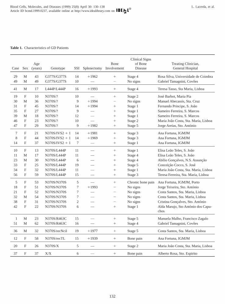
ra
Blood Cells, Molecules, and Diseases (1999) 25(8) April 30: 130–138 L. Lacerda, et al.Article ID bcmd.1999.0237, available online at http://www.idealibrary.com on
Table 1. Characteristics of GD Patients
Case SexAge
(years) Genotype SSI SplenectomyIn
29 M 43 G377S/G377S 14 1196249 M 49 G377S/G377S 10 —
41 M 17 L444P/L444P 16 11993
19 F 10 N370S/? 10 —30 M 36 N370S/? 9 1199431 F 45 N370S/? 14 1199435 F 27 N370S/? 9 —39 M 18 N370S/? 12 —46 F 23 N370S/? 10 —47 F 29 N370S/? 9 11982
7 F 21 N370S/IVS21 1 14 119818 F 44 N370S/IVS21 1 14 11969
14 F 37 N370S/IVS21 1 7 —
10 F 13 N370S/L444P 11 —11 M 17 N370S/L444P 11 —23 M 30 N370S/L444P 6 —33 F 25 N370S/L444P 19 —34 F 32 N370S/L444P 11 —56 F 59 N370S/L444P 15 —
5 F 53 N370S/N370S 5 —18 F 51 N370S/N370S 7 1199321 F 52 N370S/N370S 7 —32 M 54 N370S/N370S 7 —38 F 31 N370S/N370S 2 —42 F 22 N370S/N370S 6 —
1 M 23 N370S/R463C 15 —51 M 62 N370S/R463C 16 —
36 M 32 N370S/recNcil 19 11977
12 F 58 N370S/recTL 15 11939
20 F 26 N370S/X 5 —
37 F 37 X/X 6 —
Bonevolvement
Clinical Signsof BoneDisease
Treating Clinician,General Hospital
1 Stage 4 Rosa Silva, Universidade de Coimb2 No signs Gabriel Tamagnini, Covo˜es
1 Stage 4 Teresa Tasso, Sta Maria, Lisboa
1 Stage 2 Jose´ Barbot, Maria Pia2 No signs Manuel Abecassis, Sta. Cruz1 Stage 1 Fernando Principe, S. Joa˜o1 Stage 1 Sameiro Ferreira, S. Marcos1 Stage 1 Sameiro Ferreira, S. Marcos1 Stage 2 Maria Joa˜o Costa, Sta. Maria, Lisboa1 Stage 5 Jorge Areias, Sto. Anto´nio
1 Stage 3 Ana Fortuna, IGMJM1 Stage 3 Ana Fortuna, IGMJM1 Stage 1 Ana Fortuna, IGMJM
1 Stage 1 Elisa Lea˜o Teles, S. Joa˜o1 Stage 4 Elisa Lea˜o Teles, S. Joa˜o1 Stage 4 Abilio Gonc¸alves, N.S. Assunc¸ao1 Stage 5 Conceic¸ao Cocco, S. Jose´1 Stage 1 Maria Joa˜o Costa, Sta. Maria, Lisboa1 Stage 3 Teresa Ferreira, Sta. Maria, Lisboa
1 Chronic bone pain Ana Fortuna, IGMJM, Porto2 No signs Jorge Teixeira, Sto. Anto´nio2 No signs Costa Santos, Sta. Maria, Lisboa2 No signs Costa Santos, Sta. Maria, Lisboa2 No signs Cristina Gonc¸alves, Sto. Anto´nio1 Stage 1 Alda Marujo, Sto Anto´nio dos Capu-
chos
1 Stage 5 Manuela Malho, Francisco Zagalo1 Stage 4 Gabriel Tamagnini, Covo˜es
1 Stage 5 Costa Santos, Sta. Maria, Lisboa
1 Bone pain Ana Fortuna, IGMJM
1 Stage 2 Maria Joa˜o Costa, Sta. Maria, Lisboa
1 Bone pain Alberto Rosa, Sto. Espirito
132
epa-was
ol/l
JS
ellsE)
TS).
ord-was
ment370Skal–ces
hreeusedD4ofivided
ent.d toD4tic
l ofPSS
aly-ocyteosepple-lysiserehe4
ty.bles,r didAll
wereundsean
ange
izedsingath-
type,oneed asiciand by(pre-370S,ant
zy-able
in atheverityician
e
tesizedenia
sytosis
ratelyau-
eased).
an’sease
L. Lacerda, et al. Blood Cells, Molecules, and Diseases (1999) 25(8) April 30: 130–138
Article ID bcmd.1999.0237, available online at http://www.idealibrary.com on
Chitotriosidase Activity
Chitotriosidase activity was measured in plasma srated from heparinised blood (12). Acidified plasmaincubated with 0.0260 mmol/l 4-methylumbelliferyl-b-D-N,N8,N9-triacetylchitotriose substrate prepared in 0.1mcitrate–phosphate buffer, pH 5.2.
Hematological Parameters
Whole blood cell counts were done in a Coulterautomatic cell counter.
Freshly collected peripheral blood mononuclear cwere stained with CD4-FITC (or PE), CD8-FITC (or Pand CD3-FITC monoclonal antibodies from DAKOPATand analyzed in a FACscan as described previously (23
Statistical Analysis
Subjects were subdivided into three categories accing to genotype. The chi-square test for independenceused to examine the association between bone involve(present, absent) and genotype subgroup (NS370S/NSNS370S/L444, and other). The nonparametric KrusWallis analysis of variance was applied to test for differenin the average Zimran severity score index in the tgenotype subgroups. The Kruskal–Wallis test was alsoto compare the average number of peripheral blood C1
and CD81 T lymphocytes found in controls with thatnon-splenectomized Gaucher disease patients subdaccording to presence or absence of bone involvemSpearman rank correlation coefficients were calculatetest for a linear association between the number of C1
and CD81 T lymphocytes and plasma TRAP enzymaactivity. All statistical tests were performed at the levesignificance of 0.05. These data were analyzed using S(Statistical Program for the Social Sciences).
We also performed longitudinal patient-specific anses to detect changes in the mean number of T lymphcells and mean chitotriosidase and TRAP activity in thpatients treated with alglucerase/imiglucerase enzyme sumentation therapy for at least 24 months. For the anafrom a single patient, multiple regression models wformed in a hierarchical step-down fashion (24). Tresponse variables were percent of initial activity for CD1
cells, CD81 cells, chitotriosidase activity and TRAP activiTo model the mean change in each response variaindicator variables represented whether a change did, onot, occur during each specified month of therapy.possible subsets of indicator variables (i.e. months)considered using the method known as Leaps and Bo(25). Thus, models with one or more change(s) in the mresponse were compared to that of the null model (no ch
in the mean response during therapy). A minimum risk133
,
criteria was applied for model selection (26). Specialprograms for these longitudinal analyses were written uthe statistical programming language S-PLUS 4.5 (MSoft, Inc., Seattle, Washington).
RESULTS
Patients’ characteristics including age, sex, genoclinical presentation with respect to splenectomy, binvolvement, and the overall disease severity, presentseverity score (SSI) on the basis of the respective clinassessment, are shown in Table 1. When analyzegenotype, the association between bone involvementsent, absent) and genotype subgroup (NS370S/NSNS370S/L444, and other) was statistically signific(p5.013). A lower proportion of N370S/N370S homogous patients was found to present bone involvement (T1).
As can be observed in Figure 1, this is reflectedcomparatively milder overall clinical presentation ofN370S homozygotes, as assessed by the Zimran sescore index (SSI) on the basis of the respective clinassessment (Figure 1).
Peripheral Blood CD41 and CD81 T LymphocytSubpopulations
The study of the peripheral blood total leukocyshowed that the large majority of non-splenectomGaucher disease patients presented with leucop(mean6 sd; 3 9956 1 721 cells3 106/l, n 5 20) whereathe splenectomized ones presented in general a leucoc(13 2406 4 901, n5 10, p, 0.01).
Non-splenectomized patients were analyzed sepaand when compared to controls, with one exception, Gcher disease patients were found to present decrnumbers of the CD41 T lymphocyte subset (Figure 2
Figure 1. Clinical severity (as assessed by the Zimrseverity score index) of 31 genotyped Gaucher dis
patients.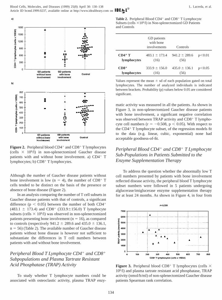
hout
ce or
ts incant4
edd
easet toeen
t
be
n intientstion
ols fithad
e
w Tent
cyteoingerapyom
ase
RAPase
talateddered
Blood Cells, Molecules, and Diseases (1999) 25(8) April 30: 130–138 L. Lacerda, et al.Article ID bcmd.1999.0237, available online at http://www.idealibrary.com on
Although the number of Gaucher disease patients witbone involvement is low (n5 4), the number of CD81 Tcells tended to be distinct on the basis of the presenabsence of bone disease (Figure 2).
In an analysis comparing the number of T cell subseGaucher disease patients with that of controls, a signifidifference (p, 0.05) between the number of both CD1
(483.1 6 173.4) and CD81 (333.96156.0) T lymphocytesubsets (cells3 106/l) was observed in non-splenectomizpatients presenting bone involvement (n5 16), as compareto controls (respectively 941.26 289.6 and 435.06 136.1,n 5 56) (Table 2). The available number of Gaucher dispatients without bone disease is however not sufficiensubstantiate the differences in T cell numbers betwpatients with and without bone involvement.
Peripheral Blood T Lymphocyte CD41 and CD81
Subpopulations and Plasma Tartrate ResistanAcid Phosphatase (TRAP) Activity
To study whether T lymphocyte numbers could
Figure 2. Peripheral blood CD41 and CD81 T lymphocytes(cells 3 106/l) in non-splenectomized Gaucher disepatients with and without bone involvement. a) CD41 Tlymphocytes; b) CD81 T lymphocytes.
associated with osteoclastic activity, plasma TRAP enzy-
134
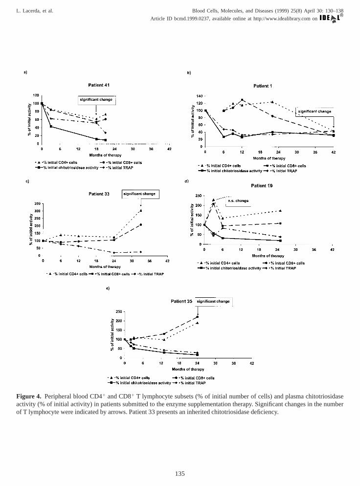
matic activity was measured in all the patients. As showFigure 3, in non-splenectomized Gaucher disease pawith bone involvement, a significant negative correlawas observed between TRAP activity and CD81 T lympho-cyte cell numbers (r5 20.508, p, 0.05). With respect tthe CD41 T lymphocyte subset, of the regression modeto the data (e.g. linear, cubic, exponential) noneacceptable goodness-of-fit.
Peripheral Blood CD41 and CD81 T LymphocytSub-Populations in Patients Submitted to theEnzyme Supplementation Therapy
To address the question whether the abnormally locell numbers presented by patients with bone involvemreflected disease activity, the peripheral blood T lymphosubset numbers were followed in 5 patients undergalglucerase/imiglucerase enzyme supplementation thfor at least 24 months. As shown in Figure 4, in four fr
Figure 3. Peripheral blood CD81 T lymphocytes (cells3106/l) and plasma tartrate resistant acid phosphatase, Tactivity (nmol/h/ml) of non-splenectomized Gaucher dise
Table 2. Peripheral Blood CD41 and CD81 T LymphocyteSubsets (cells3106/l) in Non-splenectomized GD Patientsand Controls
GD patientswith bone
involvements Controls
CD41 T 483.16 173.4 941.26 289.6 p,0.01lymphocytes (16) (56)
CD81 333.96 156.0 435.06 136.1 p,0.05lymphocytes (16) (56)
Values represent the mean6 sd of each population gated on tolymphocytes. The number of analyzed individuals is indicbetween brackets. Probability (p) values below 0.05 are consisignificant.
patients Spearman rank correlation.
idasenumber
L. Lacerda, et al. Blood Cells, Molecules, and Diseases (1999) 25(8) April 30: 130–138
Article ID bcmd.1999.0237, available online at http://www.idealibrary.com on
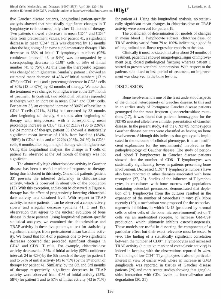
Figure 4. Peripheral blood CD41 and CD81 T lymphocyte subsets (% of initial number of cells) and plasma chitotriosactivity (% of initial activity) in patients submitted to the enzyme supplementation therapy. Significant changes in the
of T lymphocyte were indicated by arrows. Patient 33 presents an inherited chitotriosidase deficiency.135
cificin Tapy.
cantthsThis5%y a
len
d an3 to
that
ded
ne inthsof
ean).ally4%,8ase.
ofnot
hererapytientaseionre 4,osi-AP
ively19),one
ecificand
allyctiv-
antes insencent 1
thsAP,
isti-AP
ges, orfit
.s ofove-nt 1
to theove-
pectsandtientsuta-thechergousbone
pli-uffi-theriph-ientssoneve
boneho-onseple-the
clas-al
n TSFsis.of a
edoned) is
arM3easeglio-
and
Blood Cells, Molecules, and Diseases (1999) 25(8) April 30: 130–138 L. Lacerda, et al.Article ID bcmd.1999.0237, available online at http://www.idealibrary.com on
five Gaucher disease patients, longitudinal patient-speanalysis showed that statistically significant changeslymphocyte cells occurred only after 12 months of therTwo patients showed a decrease in mean CD41 and CD81
cells from pretreatment values. For patient 41, a signifidecrease in mean CD41 cells was observed by 18 monafter the beginning of enzyme supplementation therapy.decrease to 68% of initial T lymphocyte numbers (9confidence interval: 48 to 84%) was accompanied bcorresponding decrease in CD81 cells of 58% of initianumber (41 to 75%). At this time the therapeutic regimwas changed to imiglucerase. Similarly, patient 1 showeestimated mean decrease of 45% of initial numbers (147%) in CD41 cells and a percentage decrease in CD81 cellsof 30% (13 to 47%) by 42 months of therapy. We notethe treatment was changed to imiglucerase at the 33rd monthof treatment. In contrast, two additional patients responto therapy with an increase in mean CD41 and CD81 cells.For patient 33, an estimated increase of 306% of baseliCD41 T cells (271%, 341%) had occurred by 33 monafter beginning of therapy, 6 months after beginningtherapy with imiglucerase, with a corresponding mpercentage increase in CD81 cells of 211% (196 to 226%By 24 months of therapy, patient 35 showed a statisticsignificant mean increase of 191% from baseline (18198%) in CD41 cells and of 222% (203%, 241%) in CD1
cells, 6 months after beginning of therapy with imiglucerUsing this longitudinal analysis, the change in T cellspatient 19, observed at the 3rd month of therapy wassignificant.
The abnormally high chitotriosidase activity in Gaucdisease has been a useful marker of response to thbeing thus included in this study. One of the patients (pa33) presents the inherited deficiency in chitotriosidactivity, which is observed in about 6% of the populat(12). With this exception, and as can be observed in Figutherapy has the effect of progressively lowering chitotridase activity to a sustained level. With respect to TRactivity, in some patients it can be observed a comparatslower and irregular decrease (patients 41, 1 andobservation that agrees to the unclear evolution of bdisease in these patients. Using longitudinal patient-spstatistical analyses, we examined the chitotriosidaseTRAP activity in these five patients, to test for statisticsignificant changes from pretreatment mean baseline aity. We found that for 4 of 5 patients one to two significdecreases occurred that preceded significant changCD41 and CD81 T cells. For example, chitotriosidaactivity decreased to 33% of initial activity (95% confideinterval: 24 to 42%) by the 6th month of therapy for patieand to 57% of initial activity (43 to 71%) by the 3rd month oftherapy for patient 41. Similarly, by the 6th and 3rd monof therapy respectively, significant decreases in TRactivity were observed from 41% of initial activity (25%
58%) for patient 1 and to 57% of initial activity (43 to 71%)136
,
for patient 41. Using this longitudinal analysis, no statcally significant mean changes in chitotriosidase or TRactivity were observed for patient 19.
The coefficient of determination for models of chanin mean blood T lymphocyte subsets, chitotriosidaseTRAP activity varied from 79 to 100% indicating a goodof longitudinal non-linear regression models to the data
Clinically it must be stated that after about 24 monthtreatment, patient 33 showed imagiological signs of imprment (e.g. closed pathological fracture) whereas patiestarted to present vertebral compression. With respectpatients submitted to less period of treatment, no imprment was observed in the bone lesions.
DISCUSSION
Bone involvement is one of the least understood asof the clinical heterogeneity of Gaucher disease. In thisin an earlier study of Portuguese Gaucher disease pagenotyped for the most frequent glucocerebrosidase mtions (17), it was found that patients homozygous forN370S mutated allele have a milder presentation of Gaudisease. In the present study, 4 of the 6 N370S homozyGaucher disease patients were classified as having noinvolvement. Although this indicates that genotype is imcated in the outcome of clinical phenotype, it is an inscient explanation for the mechanism(s) involved inpathophysiology of Gaucher disease. The study of peeral blood T lymphocytes in Gaucher disease patshowed that the number of CD81 T lymphocytes wastatistically significantly lower in patients presenting binvolvement. Decreased CD81 T lymphocyte numbers haalso been reported in other diseases associated withresorption (27, 28). Studies of the influence of T lympcytes in co-cultures with bone marrow cell populaticontaining osteoclast precursors, demonstrated that dtion of T lymphocytes from the cultures resulted inexpansion of the number of osteoclastsin vitro (9). Morerecently (10), a mechanism was proposed for the osteotogenesis inhibition, in which IL-18 (produced by stromcells or other cells of the bone microenvironment) act ocells via an unidentified receptor, to increase GM-Cproduction, which ultimately inhibits osteoclastogeneThese models are useful in dissecting the componentsparticular effect but their exact relevance must be testinvivo. The finding of a statistically significant correlatibetween the number of CD81 T lymphocytes and increasTRAP activity (a putative marker of osteoclastic activityindeed in keeping with the observations reportedin vitro.The finding of low CD41 T lymphocytes is also of particulinterest in view of earlier work where an increase in Gganglioside was reported in tissues of Gaucher dispatients (29) and more recent studies showing that gansides interaction with CD4 favors its internalization
degradation (30, 31).
ringerestyteTheor ofGau-ittedtionined
ofcantf thee ofytesde-thethethe
ue toult in
anT
herThen of
ty ofnt.
TU/
fromrchhetoryau-
riverdM.
Sarosi-tion.
ures
n in-
sid-ds.
e-
entage-se.
A,e
ease
oft of
er-ize in
lvinst
18ofr.
kedvel
lities
rs-pres-e of
D,and
acid-n in
itypest
, Salar,nts
ol
L. Lacerda, et al. Blood Cells, Molecules, and Diseases (1999) 25(8) April 30: 130–138
Article ID bcmd.1999.0237, available online at http://www.idealibrary.com on
The present study of serial samples obtained duenzyme supplementation therapy has the additional intof permitting to do a longitudinal analysis of lymphocchanges with correction of the enzymatic defect.progressive decrease of chitotriosidase is an indicatdiminishing glucocerebrosidase loaded macrophages (cher cells) activation. With respect to the patients submto the larger period of therapy, in spite of the initial reducof chitotriosidase activity in patient 1, the spleen remahighly fibrotic and a significant decrease in numberslymphocytes was observed. Interestingly, this signifidecrease in T lymphocytes coincided with worsening obone involvement. In patient 33 the significant decreassplenomegaly and increase in numbers of T lymphoccoincided with an improvement in bone involvement. Inpendently of the reported low delivery of the enzyme tomarrow macrophages (9), in those GD patients in whomCD81 T lymphocyte decreased numbers are due tospleen sequestration, the reduction of splenomegaly dthe enzyme supplementation could be expected to resan improvement of bone pathology.
The present results albeit preliminary constituteadditional illustration of the growing importance of thelymphocyte system in the putative regulation of otsystems as already demonstrated in other diseases.point also to the potential usefulness of the determinatiosubset lymphocyte numbers as an indicator of severidisease and a predictive marker of response to treatme
ACKNOWLEDGMENTS
This work received financial support from JNICprojects PRAXIS 2/2.1/SAU/1323/95, PECS/TTSA88/95 and BD 5547/95. Support was also receivedthe National Institutes of Health, USA, by ReseaGrants R15 HL48349 and F06 TW02117 (CEM). Tauthors are grateful to Prof. J. Aerts, whose laboraperformed the chitotriosidase genotype analysis of Gcher disease patients.
REFERENCES
1. Beutler E, Grabowski G. Gaucher disease. In: ScC, Beaudet A, Sly W, Valle D, eds.The Metabolic anMolecular Bases of Inherited Disease on CD-RONew York: McGraw-Hill, Inc. 1997.
2. Lacerda L, Amaral O, Pinto R, Oliveira P, Aerts J,´Miranda MC. Gaucher disease: N370S glucocerebdase gene frequency in the Portuguese populaClin Genet45:298–300, 1994.
3. Cox T, Schofield J. Gaucher’s disease: clinical featand natural history.Baillieres Clin Haematol10:657–
689, 1997.137
y
4. Rosenthal D. Quantitative imaging of the skeletopatients with Gaucher disease,Gaucher Clinical Perspectives3:4–8, 1995.
5. Siffert R, Platt G. Gaucher disease: Orthopedic conerations. In: Desnick R, Gatt S, Grabowski G, eGaucher disease.A Century of Delineation and Rsearch.New York: Alan R Liss, pp. 617–626, 1982.
6. Barton N, Brady R, Dambrosia et al. Replacemtherapy for inherited enzyme deficiency: macrophtargeted glucocerebrosidase for Gaucher’s diseaNEngl J Med324:1464–1470, 1991.
7. Elstein D, Hadas-Halpern I, Itzchaki M, LahadAbrahamov A, Zimran A. Effect of low-dose enzymreplacement therapy on bones in Gaucher dispatients with severe skeletal involvement.Blood CellsMol Dis 22:104–111, 1996.
8. Mistry P, Wraight E, Cox T. Therapeutic deliveryproteins to macrophages: implications for treatmenGaucher’s disease.Lancet348:1555–1559, 1996.
9. Beutler E, Kuhl W, Vaughan LM. Failure of alglucase infused into Gaucher disease patients to localmarrow macrophages.Mol Med1:320–324.
10. John V, Hock J, Short L, Glasebrook A, Sells, GaR. A role for CD81 T lymphocytes in osteocladifferentiation in vitro. Endocrinology 137:2457–2463, 1996.
11. Horwood N, Udagawa N, Elliott J et al. Interleukininhibits osteoclast formation via T cell productiongranulocyte macrophage colony-stimulating factoJClin Invest101:595–603, 1998.
12. Hollak C., van Weely S, van Oers M, Aerts J. Marelevation of plasma chitotriosidase activity. A nohallmark of Gaucher disease.J Clin Invest93:1288–1292, 1994.
13. Aerts J, Hollak C. Plasma and metabolic abnormain Gaucher’s disease.Baillieres Clin Haematol10:691–709, 1997.
14. Ek-Rylander B, Bill P, Norgard M, Nilsson S, Andeson G. Cloning, sequence, and developmental exsion of type 5, tartrate-resistant, acid phosphatasrat bone.J. Biol Chem266:24684–24689, 1991.
15. Ek-Rylander B, Flores M, Wendel M, HeinegardAndersson G. Dephosphorylation of osteopondinbone sialoprotein by osteoclastic tartrate-resistantphosphatase. Modulation of osteoclast adhesiovitro. J Biol Chem269:14853–14856, 1994.
16. SaMiranda MC, Aerts JM, Pinto R et al 1990. Activof glucocerebrosidase in extracts of different cell tyfrom type 1 Gaucher disease patients.Clin Gene38:218–227, 1990.
17. Amaral O, Lacerda L, Santos R, Pinto R, Aerts JMiranda MC. Type I Gaucher disease: molecubiochemical and clinical characterization of patiefrom Northern Portugal.Biochem Med Metab Bi
49:97–107, 1993.
6Tn the
SJ,e 1:
igra-
tlerby
andrat
tion.
pha-
sedo-sis.
skie
and
tric8,
c-ol
I.in
ainfor
es
duesith
king
m ofula-,
Blood Cells, Molecules, and Diseases (1999) 25(8) April 30: 130–138 L. Lacerda, et al.Article ID bcmd.1999.0237, available online at http://www.idealibrary.com on
18. Amaral O, Pinto E, Fortuna M, Lacerda L, Sa´ MirandaMC. Type 1 Gaucher disease: identification of N39and prevalence of glucocerebrosidase mutations iPortuguese.Hum Mutat8:280–281, 1996.
19. Hermann G, Goldblatt J, Levy R, GoldsmithDesnick RJ, Grabowski GA. Gaucher’s disease typassessment of bone involvement by CT and scintphy.Am J Rad147:943–948, 1986.
20. Zimran A, Gross E, West C, Sorge J, Kubitz M, BeuE. Prediction of severity of Gaucher’s diseaseidentification of mutations at DNA level.Lancet2:349–352, 1989.
21. Furbish F, Steer C, Krett N, Barranger J. Uptakedistribution of placental glucocerebrosidase inhepatic cells and effects of sequential deglycosylaBiochem. Biophys. Acta673:425–434, 1981.
22. Magalha˜es J, Pinto R, Lemos M, Sa´ Miranda M,Poenaru L. Age dependency of serum acid phostase in controls and Gaucher patients.Enzyme32:95–99, 1984.
23. Arosa F, da Silva A, Godinho et al. 1994. DecreaCD8-p56lck activity in peripheral blood T-lymphcytes from patients with hereditary haemochromatoScand J Immunol39:426–432, 1994.
24. McLaren CE, Kambour EL, McLachlan GJ, LukaHC, Li X, Brittenham GM, McLaren GD. Multipl
linear regression and finite mixture distribution model-138
ing for sequential analysis of hematological data.StatMed, in press, 1999.
25. Furnival GM, Wilson RW, Jr. Regression by leapsbounds.Technometrics16:599–511, 1974.
26. Eubanks RL.Spline Smoothing and NonparameRegression, New York: Marcel Deker, Inc., pp. 431988.
27. Consolini R, Cini P, Cei B, Botton E. Thymic dysfuntioning histiocytosis X.Am J Pediatr Hematol/Onc9:146–148, 1987.
28. Nagasawa T, Nitta H, Watanable H, IshikawaReduced CD81 peripheral blood T lymphocytesrapidly progressive periodontitis.Arch Oral Biol 40:605–608, 1995.
29. Philipart N, Menkes J. Characterization of the msplenic glycolipids in Gaucher’s disease: Evidencethe site of metabolic block.Biochem Biophys RCommun15:551–555, 1964.
30. Grassi F, Lopalco L, Lanza P et al. Chemical resiof ganglioside molecules involved in interactions wlymphocyte surface targets leading to CD4 masand inhibition of mitogenic proliferation.Eur J Immu-nol 20:145–150, 1990.
31. Saggioro D, Sorio C, Calderazzo et al. Mechanisaction of the monosialoganglioside GM1 as a modtor of CD4 expression.J Biol Chem268:1368–1375
1993.
Related Documents