International Journal of Molecular Sciences Review Mitochondria Matter: Systemic Aspects of Nonalcoholic Fatty Liver Disease (NAFLD) and Diagnostic Assessment of Liver Function by Stable Isotope Dynamic Breath Tests Agostino Di Ciaula 1 , Giuseppe Calamita 2 , Harshitha Shanmugam 1 , Mohamad Khalil 1 , Leonilde Bonfrate 1 , David Q.-H. Wang 3 , Gyorgy Baffy 4 and Piero Portincasa 1, * Citation: Di Ciaula, A.; Calamita, G.; Shanmugam, H.; Khalil, M.; Bonfrate, L.; Wang, D.Q.-H.; Baffy, G.; Portincasa, P. Mitochondria Matter: Systemic Aspects of Nonalcoholic Fatty Liver Disease (NAFLD) and Diagnostic Assessment of Liver Function by Stable Isotope Dynamic Breath Tests. Int. J. Mol. Sci. 2021, 22, 7702. https://doi.org/10.3390/ ijms22147702 Academic Editors: Amedeo Lonardo, Carlos Palmeira and Anabela P. Rolo Received: 10 June 2021 Accepted: 14 July 2021 Published: 19 July 2021 Publisher’s Note: MDPI stays neutral with regard to jurisdictional claims in published maps and institutional affil- iations. Copyright: © 2021 by the authors. Licensee MDPI, Basel, Switzerland. This article is an open access article distributed under the terms and conditions of the Creative Commons Attribution (CC BY) license (https:// creativecommons.org/licenses/by/ 4.0/). 1 Clinica Medica “A. Murri”, Department of Biomedical Sciences & Human Oncology, University of Bari Medical School, 70124 Bari, Italy; [email protected] (A.D.C.); [email protected] (H.S.); [email protected] (M.K.); [email protected] (L.B.) 2 Department of Biosciences, Biotechnologies and Biopharmaceutics, University of Bari “Aldo Moro”, 70100 Bari, Italy; [email protected] 3 Marion Bessin Liver Research Center, Einstein-Mount Sinai Diabetes Research Center, Department of Medicine and Genetics, Division of Gastroenterology and Liver Diseases, Albert Einstein College of Medicine, Bronx, NY 10461, USA; [email protected] 4 Department of Medicine, VA Boston Healthcare System and Brigham and Women’s Hospital, Harvard Medical School, Boston, MA 02130, USA; [email protected] * Correspondence: [email protected] Abstract: The liver plays a key role in systemic metabolic processes, which include detoxification, synthesis, storage, and export of carbohydrates, lipids, and proteins. The raising trends of obesity and metabolic disorders worldwide is often associated with the nonalcoholic fatty liver disease (NAFLD), which has become the most frequent type of chronic liver disorder with risk of progression to cirrhosis and hepatocellular carcinoma. Liver mitochondria play a key role in degrading the pathways of carbohydrates, proteins, lipids, and xenobiotics, and to provide energy for the body cells. The morphological and functional integrity of mitochondria guarantee the proper functioning of β-oxidation of free fatty acids and of the tricarboxylic acid cycle. Evaluation of the liver in clinical medicine needs to be accurate in NAFLD patients and includes history, physical exam, imaging, and laboratory assays. Evaluation of mitochondrial function in chronic liver disease and NAFLD is now possible by novel diagnostic tools. “Dynamic” liver function tests include the breath test (BT) based on the use of substrates marked with the non-radioactive, naturally occurring stable isotope 13 C. Hepatocellular metabolization of the substrate will generate 13 CO 2 , which is excreted in breath and measured by mass spectrometry or infrared spectroscopy. Breath levels of 13 CO 2 are biomarkers of specific metabolic processes occurring in the hepatocyte cytosol, microsomes, and mitochondria. 13 C-BTs explore distinct chronic liver diseases including simple liver steatosis, non-alcoholic steatohepatitis, liver fibrosis, cirrhosis, hepatocellular carcinoma, drug, and alcohol effects. In NAFLD, 13 C-BT use substrates such as α-ketoisocaproic acid, methionine, and octanoic acid to assess mitochondrial oxidation capacity which can be impaired at an early stage of disease. 13 C-BTs represent an indirect, cost-effective, and easy method to evaluate dynamic liver function. Further applications are expected in clinical medicine. In this review, we discuss the involvement of liver mitochondria in the progression of NAFLD, together with the role of 13 C-BT in assessing mitochondrial function and its potential use in the prevention and management of NAFLD. Keywords: breath test; hepatic mitochondrial function; hepatocellular carcinoma; ketoisocaproic acid; liver diseases; liver steatosis; methionine; methacetin; octanoic acid; β-oxidation Int. J. Mol. Sci. 2021, 22, 7702. https://doi.org/10.3390/ijms22147702 https://www.mdpi.com/journal/ijms

Welcome message from author
This document is posted to help you gain knowledge. Please leave a comment to let me know what you think about it! Share it to your friends and learn new things together.
Transcript
International Journal of
Molecular Sciences
Review
Mitochondria Matter: Systemic Aspects of Nonalcoholic FattyLiver Disease (NAFLD) and Diagnostic Assessment of LiverFunction by Stable Isotope Dynamic Breath Tests
Agostino Di Ciaula 1 , Giuseppe Calamita 2 , Harshitha Shanmugam 1 , Mohamad Khalil 1 ,Leonilde Bonfrate 1, David Q.-H. Wang 3 , Gyorgy Baffy 4 and Piero Portincasa 1,*
�����������������
Citation: Di Ciaula, A.; Calamita, G.;
Shanmugam, H.; Khalil, M.; Bonfrate,
L.; Wang, D.Q.-H.; Baffy, G.;
Portincasa, P. Mitochondria Matter:
Systemic Aspects of Nonalcoholic
Fatty Liver Disease (NAFLD) and
Diagnostic Assessment of Liver
Function by Stable Isotope Dynamic
Breath Tests. Int. J. Mol. Sci. 2021, 22,
7702. https://doi.org/10.3390/
ijms22147702
Academic Editors: Amedeo Lonardo,
Carlos Palmeira and Anabela P. Rolo
Received: 10 June 2021
Accepted: 14 July 2021
Published: 19 July 2021
Publisher’s Note: MDPI stays neutral
with regard to jurisdictional claims in
published maps and institutional affil-
iations.
Copyright: © 2021 by the authors.
Licensee MDPI, Basel, Switzerland.
This article is an open access article
distributed under the terms and
conditions of the Creative Commons
Attribution (CC BY) license (https://
creativecommons.org/licenses/by/
4.0/).
1 Clinica Medica “A. Murri”, Department of Biomedical Sciences & Human Oncology, University of BariMedical School, 70124 Bari, Italy; [email protected] (A.D.C.);[email protected] (H.S.); [email protected] (M.K.);[email protected] (L.B.)
2 Department of Biosciences, Biotechnologies and Biopharmaceutics, University of Bari “Aldo Moro”,70100 Bari, Italy; [email protected]
3 Marion Bessin Liver Research Center, Einstein-Mount Sinai Diabetes Research Center, Department ofMedicine and Genetics, Division of Gastroenterology and Liver Diseases, Albert Einstein College of Medicine,Bronx, NY 10461, USA; [email protected]
4 Department of Medicine, VA Boston Healthcare System and Brigham and Women’s Hospital, HarvardMedical School, Boston, MA 02130, USA; [email protected]
* Correspondence: [email protected]
Abstract: The liver plays a key role in systemic metabolic processes, which include detoxification,synthesis, storage, and export of carbohydrates, lipids, and proteins. The raising trends of obesityand metabolic disorders worldwide is often associated with the nonalcoholic fatty liver disease(NAFLD), which has become the most frequent type of chronic liver disorder with risk of progressionto cirrhosis and hepatocellular carcinoma. Liver mitochondria play a key role in degrading thepathways of carbohydrates, proteins, lipids, and xenobiotics, and to provide energy for the bodycells. The morphological and functional integrity of mitochondria guarantee the proper functioningof β-oxidation of free fatty acids and of the tricarboxylic acid cycle. Evaluation of the liver in clinicalmedicine needs to be accurate in NAFLD patients and includes history, physical exam, imaging,and laboratory assays. Evaluation of mitochondrial function in chronic liver disease and NAFLDis now possible by novel diagnostic tools. “Dynamic” liver function tests include the breath test(BT) based on the use of substrates marked with the non-radioactive, naturally occurring stableisotope 13C. Hepatocellular metabolization of the substrate will generate 13CO2, which is excretedin breath and measured by mass spectrometry or infrared spectroscopy. Breath levels of 13CO2
are biomarkers of specific metabolic processes occurring in the hepatocyte cytosol, microsomes,and mitochondria. 13C-BTs explore distinct chronic liver diseases including simple liver steatosis,non-alcoholic steatohepatitis, liver fibrosis, cirrhosis, hepatocellular carcinoma, drug, and alcoholeffects. In NAFLD, 13C-BT use substrates such as α-ketoisocaproic acid, methionine, and octanoicacid to assess mitochondrial oxidation capacity which can be impaired at an early stage of disease.13C-BTs represent an indirect, cost-effective, and easy method to evaluate dynamic liver function.Further applications are expected in clinical medicine. In this review, we discuss the involvementof liver mitochondria in the progression of NAFLD, together with the role of 13C-BT in assessingmitochondrial function and its potential use in the prevention and management of NAFLD.
Keywords: breath test; hepatic mitochondrial function; hepatocellular carcinoma; ketoisocaproicacid; liver diseases; liver steatosis; methionine; methacetin; octanoic acid; β-oxidation
Int. J. Mol. Sci. 2021, 22, 7702. https://doi.org/10.3390/ijms22147702 https://www.mdpi.com/journal/ijms
Int. J. Mol. Sci. 2021, 22, 7702 2 of 34
1. Introduction
The liver plays a key role in lipid homeostasis, with steps including the synthesis,oxidation, and transport of free fatty acids (FFA), triglycerides (TG), cholesterol, andbile acids (BA). Chronic liver diseases encompass a spectrum of conditions ranging frommetabolic to viral, alcohol-related diseases, drug-related diseases, autoimmune diseases,and tumours. The hepatocyte can be damaged by various hits and intracellular organellescan be part of the dysfunctional cell, with changes including microsomal hypertrophy, mi-tochondrial damage by free fatty acids overload and insufficient β-oxidation and activationof peroxisomal metabolism.
Growing evidence points to dysfunctional mitochondria as key contributors in thepathogenesis of the chronic metabolic conditions (i.e., obesity, metabolic syndrome andtype 2 diabetes mellitus) frequently linked to liver disease. These processes act throughpathways leading to oxidative stress, chronic inflammation, and insulin resistance. Thus, di-agnostic techniques able to early detect and monitor mitochondrial dysfunction have greatrelevance in terms of possible primary/secondary prevention measures and of therapiesspecifically targeting liver mitochondria [1–3].
In this paper we will focus on nonalcoholic fatty liver disease (NAFLD) with emphasison mitochondrial dysfunction and the role of novel “dynamic” noninvasive breath test(BT) to assess mitochondrial function. We will also point to current potential therapeuticapproaches targeting mitochondria in NAFLD.
2. Mitochondrial Function in the Liver2.1. General Features of Mitochondria
Mitochondria are intracellular organelles that provide energy for the body cells. Inthe liver there are about 500–4000 mitochondria per cell [4] equalling about 18% of theentire cell volume [5]. Mitochondria play a key role in the metabolic pathways andsignalling networks [6]. They participate in degrading pathways of carbohydrates, proteins,lipids, and xenobiotics [7,8], and ultimately generate ATP as energetic source [6–8]. Themorphological and functional integrity of mitochondria maintain functioning networksand pathways inside the mitochondria and in the cell. The fat and energy balance inhepatocytes is regulated by mitochondrial activities, including FFA β-oxidation, electrontransfer and production of ATP and ROS [9]. Essential elements include mitochondrialDNA (mtDNA), membrane constituents, lipoprotein trafficking, pro- and anti-oxidantbalance, and metabolic demand and supply [10].
2.2. The Fate of Free Fatty Acids
The routes providing the circulating (long-chain) FFA to the liver and their mitochon-drial fate is of relevance for ATP production and hepatocyte health maintenance. About60% of circulating FFA derive from lipolysis of TG in adipose tissue [11], and enter the hep-atocyte by using the transporters FFA translocase/CD36, fatty acid binding protein (FABP),and caveolin-1. About 15% FFA are of dietary origin and contained in TG within ApoE-enriched chylomicrons. Chylomicrons are assembled in the enterocyte following dietary fatdigestion in the intestinal lumen. This step occurs during emulsion and micellization of fatby the bile acid (BA) pool made of both primary and secondary tauro-, glycol-conjugatedBA [12,13]. In the hepatocyte chylomicron remnants bind specific membrane receptorstaht have a high affinity for the surface protein ApoE. Lastly, about 35% of FFA in thehepatocyte originate from de novo lipogenesis (DNL) from dietary carbohydrates (glucoseconverted to pyruvate during glycolysis). The FFA pool in the hepatocyte provides thesubstrate for re-esterification with glycerol to form TG via the key enzymes, diglycerideacyltransferase (DGAT)1 and DGAT2. This amount of TG serves as stored fat as lipiddroplets in small amounts, i.e., less than 5% of cell content. When needed, TG can behydrolysed by hydrolases, e.g., the patatin-like phospholipase domain-containing protein3-PNPLA3 (adiponutrin)—to release FFA in the cytosol [14,15]. The TG pool also providesFFA for two major routes of elimination [16]. (a) export to blood as TG within very-low
Int. J. Mol. Sci. 2021, 22, 7702 3 of 34
density lipoproteins (VLDL) assembled in the endoplasmic reticulum [17]; (b) β-oxidationof FFA in mitochondria. In the Golgi apparatus the apolipoprotein B (ApoB) undergoesdisulphide bond formation and association with TG (by protein disulphide isomerase andmicrosome triglyceride transfer protein (MTP)) [18]. Of note, increased intake of sucrosein the mice model leads to rapid development of hyperinsulinemia, hepatosteatosis, andinsulin resistance. Furthermore, insulin enhances hepatic expression of the FA transporterCD36 involving a PPAR-γ-dependent mechanism. In the general scenario, these resultsindicate that hyperinsulinemia is an early and potent inducer of hepatosteatosis, insulinresistance, and dysglycaemia. A further step is the progression to type 2 diabetes andNAFLD. In addition, during conditions of hyperinsulinemia, dysfunctional insulin clear-ance becomes evident, due to abnormal insulin degrading enzyme regulation. This step, inturn, directly impairs postprandial hepatic glucose disposal and increases susceptibility todysmetabolic conditions, including fatty liver, mitochondrial dysfunction, especially in thesetting of Western diet/lifestyle.
2.3. β-Oxidation of FFA in Mitochondria
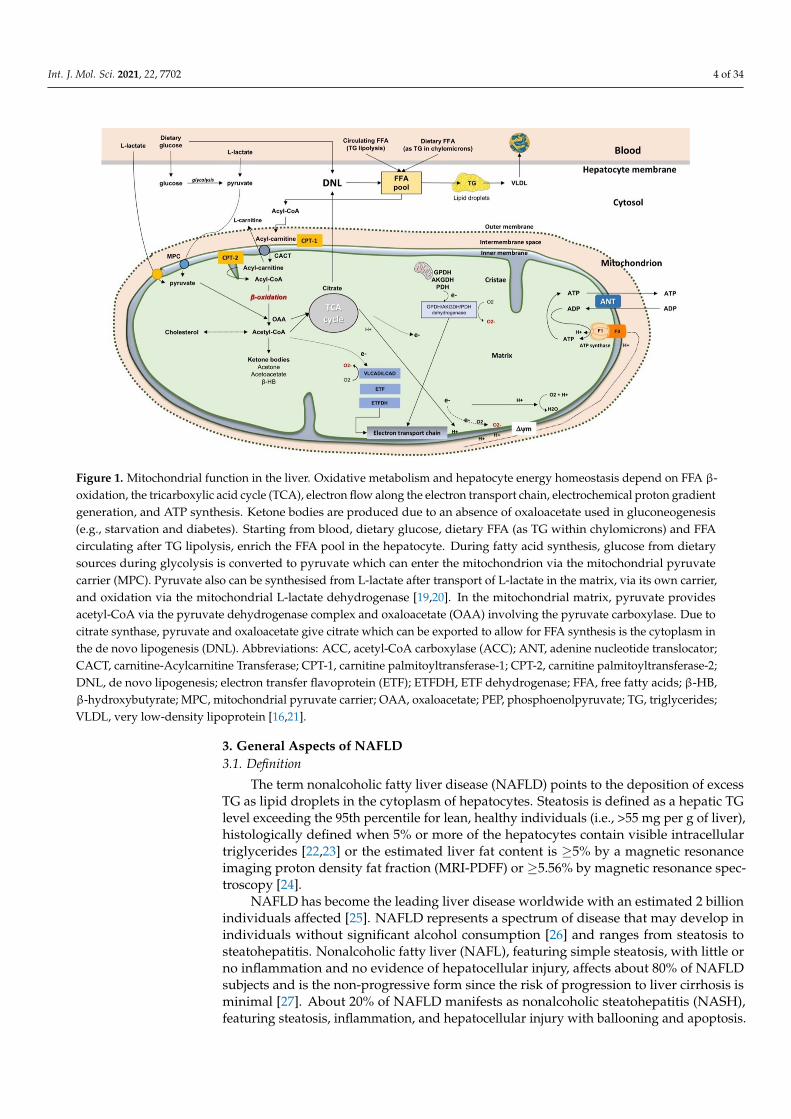
This important mitochondrial pathway includes FFA β-oxidation, the tricarboxylicacid cycle (TCA), electron flow along the electron transport chain, electrochemical protongradient generation, and ATP synthesis. In starvation, ketone bodies are produced due toabsence of oxaloacetate used in gluconeogenesis. Pyruvate can enter the mitochondrionvia the mitochondrial pyruvate carrier (MPC) as well as be synthesised from L-lactate aftertransport of L-Lactate in the matrix, via its own carrier, and oxidation via the mitochondrialL-lactate dehydrogenase [19,20]. In the matrix, pyruvate can provide Acetyl-CoA via thepyruvate dehydrogenase complex and oxaloacetate (OAA) via the pyruvate carboxylase.Due to citrate synthase, pyruvate and oxaloacetate give citrate which is exported for FFAsynthesis is the cytoplasm during DNL [21]. For fatty acid catabolism, the acyl-CoAsynthase transforms the cytosolic FFA into fatty acyl-CoA (Figure 1). Acyl-CoA+ carnitineare catalysed to CoA and acylcarnitine by the carnitine palmitoyl-transferase 1 (CPT-1)which is in the outer side of the inner mitochondrial membrane. Acylcarnitine can enter themitochondria across the inner membrane in exchange with L-carnitine. This step requiresthe acylcarnitine/L-carnitine antiporter. The carnitine palmitoyl-transferase 2 (CPT-2),localized at the matrix side of the inner membrane, will process the acyl-carnitine to Acyl-CoA+ L-carnitine (the latter ready to be exchanged with new incoming Acyl-carnitine). Theresulting acyl-CoA in the mitochondrial matrix is ultimately oxidised via the β-oxidation toacetyl-CoA which then enters the tricarboxylic acid (TCA) cycle with production of carbondioxide and water. A further step implies the activation of the electron transport chain andATP production.
Int. J. Mol. Sci. 2021, 22, 7702 4 of 34
Figure 1. Mitochondrial function in the liver. Oxidative metabolism and hepatocyte energy homeostasis depend on FFA β-oxidation, the tricarboxylic acid cycle (TCA), electron flow along the electron transport chain, electrochemical proton gradientgeneration, and ATP synthesis. Ketone bodies are produced due to an absence of oxaloacetate used in gluconeogenesis(e.g., starvation and diabetes). Starting from blood, dietary glucose, dietary FFA (as TG within chylomicrons) and FFAcirculating after TG lipolysis, enrich the FFA pool in the hepatocyte. During fatty acid synthesis, glucose from dietarysources during glycolysis is converted to pyruvate which can enter the mitochondrion via the mitochondrial pyruvatecarrier (MPC). Pyruvate also can be synthesised from L-lactate after transport of L-lactate in the matrix, via its own carrier,and oxidation via the mitochondrial L-lactate dehydrogenase [19,20]. In the mitochondrial matrix, pyruvate providesacetyl-CoA via the pyruvate dehydrogenase complex and oxaloacetate (OAA) involving the pyruvate carboxylase. Due tocitrate synthase, pyruvate and oxaloacetate give citrate which can be exported to allow for FFA synthesis is the cytoplasm inthe de novo lipogenesis (DNL). Abbreviations: ACC, acetyl-CoA carboxylase (ACC); ANT, adenine nucleotide translocator;CACT, carnitine-Acylcarnitine Transferase; CPT-1, carnitine palmitoyltransferase-1; CPT-2, carnitine palmitoyltransferase-2;DNL, de novo lipogenesis; electron transfer flavoprotein (ETF); ETFDH, ETF dehydrogenase; FFA, free fatty acids; β-HB,β-hydroxybutyrate; MPC, mitochondrial pyruvate carrier; OAA, oxaloacetate; PEP, phosphoenolpyruvate; TG, triglycerides;VLDL, very low-density lipoprotein [16,21].
3. General Aspects of NAFLD3.1. Definition
The term nonalcoholic fatty liver disease (NAFLD) points to the deposition of excessTG as lipid droplets in the cytoplasm of hepatocytes. Steatosis is defined as a hepatic TGlevel exceeding the 95th percentile for lean, healthy individuals (i.e., >55 mg per g of liver),histologically defined when 5% or more of the hepatocytes contain visible intracellulartriglycerides [22,23] or the estimated liver fat content is ≥5% by a magnetic resonanceimaging proton density fat fraction (MRI-PDFF) or ≥5.56% by magnetic resonance spec-troscopy [24].
NAFLD has become the leading liver disease worldwide with an estimated 2 billionindividuals affected [25]. NAFLD represents a spectrum of disease that may develop inindividuals without significant alcohol consumption [26] and ranges from steatosis tosteatohepatitis. Nonalcoholic fatty liver (NAFL), featuring simple steatosis, with little orno inflammation and no evidence of hepatocellular injury, affects about 80% of NAFLDsubjects and is the non-progressive form since the risk of progression to liver cirrhosis isminimal [27]. About 20% of NAFLD manifests as nonalcoholic steatohepatitis (NASH),featuring steatosis, inflammation, and hepatocellular injury with ballooning and apoptosis.
Int. J. Mol. Sci. 2021, 22, 7702 5 of 34
Histological findings may be indistinguishable from those of alcoholic steatohepatitis [28].Individuals afflicted by NASH are at high risk of developing fibrosis [29–32] and NASHhas an increased potential of progressing to (cryptogenic) cirrhosis and hepatocellularcarcinoma (HCC) [33,34].
Although NAFLD is by far the most prevalent cause of liver steatosis, ectopic fataccumulation may occur in the liver for a variety of reasons in viral hepatitis B and C (inparticular genotype 3), lipodystrophy, Wilson’s disease, starvation, parenteral nutrition,abetalipoproteinemia, hepatotoxic drugs (e.g., methotrexate, tamoxifen, glucocorticoids,amiodarone, valproate, and anti-retroviral agents for HIV), pregnancy, HELLP (hemolyticanemia, elevated liver enzymes, low platelet count) syndrome, Reye syndrome, and inbornerrors of metabolism (i.e., lecithin-cholesterol acyltransferase deficiency, cholesterol esterstorage disease, and Wolman disease). However, alcohol-associated liver injury remainsthe second most frequent aetiology of steatosis. The similarity and overlap between alcohol-associated liver disease and NAFLD has been the source of confusion and the subject ofacademic debate. The term “non-alcoholic” indeed overemphasizes “alcohol” and un-deremphasizes the role of metabolic risk factors, since NAFLD is commonly associatedwith obesity, hypertension, dyslipidaemia, and diabetes [17,26]. To acknowledge thatNAFLD is no longer a diagnosis of exclusion, and it represents a continuum of liver diseasecaused by metabolic derangements, a change in terminology from NAFLD to metabolicdysfunction-associated fatty liver disease (MAFLD) has been recently proposed. Accord-ingly, hepatic steatosis is associated with at least one of the following three comorbidities:overweight/obesity (especially expansion of visceral fat), presence of type 2 diabetes melli-tus, or evidence of metabolic dysregulation [35]. Nevertheless, some authors warned thatunderstanding of the molecular basis of the disease entity, new insights in risk stratification,and other important aspects of NAFLD may be more urgent than nosology itself [36].Indeed, there remains much to learn about the contribution of environment, comorbiditiesand the gut microbiome to the pathogenesis and natural history of NAFLD [16,37–39].
3.2. Prevalence and Natural History
NAFLD has become the most frequent liver disorder of our times [22,40–42]. Themedian prevalence of NAFLD is about 25% worldwide and trends are increasing [36,43,44].This is likely due to the increasing prevalence of obesity, type 2 diabetes mellitus, seden-tary lifestyles, dyslipidemia, and metabolic syndrome, mainly in North America andEurope [43,45–47]. However, the burden of NAFLD has also become evident in non-obeseindividuals (‘lean NAFLD’), with a prevalence of about 10%–30% in both Western andEastern countries [48], typically associated with metabolic dysfunction and a comparativelyincreased cardiovascular risk [46,49]. NAFLD puts the population at increased risk forliver-related mortality as well as all-cause-mortality due to increased risk of cardiovasculardisease and extrahepatic malignancies [50–52]. Liver fibrosis is currently the strongestknown predictor of poor clinical outcomes in NAFLD. The time sequence of fibrosis pro-gression in NAFL is significantly slower (average 14 years) than in NASH (about 7 years)and even less in a subgroup of ‘rapid progressors encompassing 10% to 20% of patientswith NAFLD [27]. Thus, much attention has been devoted to the identification of predictorsof rapid progression (i.e., higher serum ALT, morbid obesity, diabetes, and possibly geneticsusceptibility with family history of cirrhosis in first-degree relatives) [53–55]. Once cirrho-sis has developed in NAFLD, the incident risk of developing HCC is about 1.5%–2% peryear. Therefore, HCC screening in NASH-related cirrhosis is recommended [56]. NAFLD isnow the second leading indication for liver transplantation in the US, including a growingnumber of cases with NASH-related HCC [44].
3.3. Diagnosis
Liver biopsy followed by liver histology is the gold standard for diagnosing NAFLD.The procedure is usually echo-assisted and performed by transcutaneous puncture of theliver after local anaesthesia. A cylindric liver fragment is promptly placed in a solution
Int. J. Mol. Sci. 2021, 22, 7702 6 of 34
containing formalin. The procedure, however, is invasive, and exposes patients to the risk ofpotential complications. The compliance of the patients is therefore very low. Liver biopsyshould be reserved to subgroup of patients with suggestive signs/symptoms/evidence ofsteatohepatitis or early cirrhosis and when careful histological assessment is required toquantify the degree and stage of liver damage as fibrosis, inflammation, and necrosis, orduring research protocols looking at the progression of liver fibrosis and efficacy of specifictherapies.
Therefore, in clinical practice, the diagnosis of liver diseases relies on a history, physicalexam and tests that investigate morphological and functional aspects. The liver is essentialfor many metabolic and energetic processes in the body and there is no single test thatcould assess liver function in a comprehensive way. Each test provides a specific set ofinformation focusing on various mechanisms involved in liver function. A major challengein clinical hepatology is therefore to appropriately combine the results of diagnostic testsin an accurate and complementary way to achieve the final diagnosis.
By history, NAFLD patients often carry one or more components of metabolic syn-drome or “fellow travellers”, such as cholesterol cholelithiasis [57,58]. Other causes ofliver steatosis and chronic liver diseases must be therefore excluded [59]. In NAFLD,alcohol consumption should be absent or very limited. This includes not more than threestandard drinks/day (i.e., 21 drinks/week) in men or not more than 2 drinks/day inwomen (i.e., 14 drinks/week, equal to 14 g of pure alcohol/standard drink = 98 kcal),as indicated by the American Association for the Study of Liver Diseases [26]. In fact,alcohol consumption greater than the threshold puts individuals at risk of alcoholic liverdisease eventually associated with coexisting NAFLD. This situation makes the diagnosisof NAFLD and risk assessment even more difficult.
Laboratory tests include serum aminotransferase levels as markers of hepatocytecytolysis, but they are not sufficient for making the diagnosis, as laboratory tests maybe normal in patients with NAFLD and may be abnormal in patients with many otherconditions. Serum alanine aminotransferase (ALT) in NAFLD is typically higher thanserum aspartate aminotransferase (AST) unless the disease has already progressed [60].Serum tests to assess for other disorders include viral hepatitis serology, iron studies,and autoimmune antibody assays. Some of these ‘static’ tests measure serum parametersof synthesis (prothrombin, cholesterol, albumin), hepatocellular injury (transaminases),detoxification (ammonium), excretion and cholestasis (bilirubin, alkaline phosphatase,GGT) [61].
Imaging techniques in the evaluation of NAFLD include abdominal ultrasonography,computerized tomography (CT), and magnetic resonance.
Abdominal ultrasonography can easily detect a hyperechoic texture in the liver(“bright liver”) due to diffuse fatty infiltration. The main advantages of ultrasound includewide availability, safety, and low-cost. This non-invasive technique can easily allow ascreening of patients at risk and is a useful tool for monitoring treated patients. How-ever, liver ultrasound is not able to distinguish the necro-inflammatory changes typicalof steatohepatitis, and has a poor accuracy in diagnosing the presence of a mild steatosis(i.e., <30%) [62]. Therefore, the ultimate diagnosis of both NASH and NAFLD can beunderestimated.
Computed tomography can assess the liver brightness, measuring pixel values inHounsfield Unit with quantitative determination of attenuation in comparison with thefat-free spleen [63,64]. The possibility of quantitative results is the main advantage ofthis imaging technique. However, as for ultrasound, the diagnostic accuracy of liver CTdecreases with lesser severity of steatosis, with a sensitivity of 52–62% in case of mildsteatosis (i.e., fat fraction of 10–20%) [65].
MR-based methods including proton spectroscopy and calculation of the proton-density fat fraction (PDFF) are far superior to ultrasound or CT in measuring intrahepaticfat content but, as for ultrasound and computed tomography, cannot distinguish betweensimple steatosis and steatohepatitis. PDFF measure, however, represents an advantage, as
Int. J. Mol. Sci. 2021, 22, 7702 7 of 34
compared with CT, since it requires no internal calibration or reference standard. AdvancedMR techniques can also consider confounders as iron overload, and can easily and rapidlyallow a volumetric assessment of NAFLD [66].
By contrast, there has been significant progress in the non-invasive assessment offibrosis in NAFLD. Vibration-controlled transient elastography is increasingly used as apoint-of-care method to assess and regularly monitor fibrosis based on the liver stiffnessand can also be utilized to grade hepatic steatosis. While there are additional ultrasound-based liver stiffness measurement techniques, MR elastography has proven more accuratealthough this method currently remains primarily in the realm of research and clinicaltrials due to its significant cost.
Of note, none of the imaging techniques employed to diagnose NAFLD will explorethe true “dynamic” liver function and need to be integrated with further “functional”examination techniques, such as breath test.
The management of NAFLD is still a matter of debate. According to AASLD guide-lines, systematic screening for NAFLD is not advisable at this time, since there is noconsensus about the true cost-effectiveness of the screening [16,26]. In addition, there isno licensed or registered pharmacotherapy for NAFLD and management remains focusedon healthy lifestyles as previously discussed by our group [45,46,67]. Early identifica-tion of risk factors associated with NAFLD progression is therefore paramount to delayor prevent the consequences related to advanced liver disease. However, reliable, andsensitive non-invasive diagnostic tests are still lacking in NAFLD and are actively beinginvestigated. In this respect, diagnostic tests focusing on mitochondrial function mayprovide novel diagnostic and prognostic possibilities both during the evolution of diseaseand in therapeutical trials. These aspects are discussed in the following sections.
4. Mitochondrial Dysfunction in the Liver
Mitochondrial dysfunction is one of the most distinctive characteristics of NAFLD [68].In NAFLD patients, increased plasma levels of FFA are firstly associated with increased in-trahepatic inflow [24] and early mitochondrial biogenesis through peroxisome proliferator-activated receptor-α (PGC1-α) activation. This step, in turn, leads to increased FFA oxi-dation rates and increased or unchanged mitochondrial function [69]. Coupling of FFAoxidation to ATP generation might be dysfunctional already, because of emerging ultra-structural changes and increased expression of uncoupling proteins. With progressionof NAFLD, however, mitochondrial ATP generation is further impaired resulting in de-fective cellular energy charge [70–73]. The precise pathways governing such changes ofmitochondrial performance are still unknown.
The increased accumulation of FA in the hepatocytes (neutral lipid droplets) duringinsulin-resistance-associated NAFLD, which is pathologically defined as hepatic steatosis,lead to a series of mitochondrial alterations ranging between mitochondrial DNA (mtDNA)damage to sirtuin alteration. The mtDNA, a circular double-stranded molecule locatedin the mitochondrial matrix, encodes about the 10% of mitochondrial proteins, the othersbeing encoded by the nuclear DNA. mtDNA encodes proteins necessary for the assemblyand activity of mitochondrial respiratory complexes [74]. Ongoing oxidative stress duringsteatosis can severely impair mtDNA function [5] with further amplification of oxidativestress, mitochondrial biogenesis, and ultimately NAFLD severity and inflammation [75–78].
Alteration of the mitochondrial function compromises also the prooxidant/antioxidantbalance, with an increase in non-metabolized fatty acids (FA) in the cytosol as a consequenceof the blockade of FFA β-oxidation and the resulting stimulation of ROS production [79,80].Mitochondrial dysfunctions are often accompanied by considerable ultrastructural changessuch as megamitochondria, loss of cristae, and formation of paracrystalline inclusion bodiesin the organelle matrix [81].
In addition, in NAFLD, the excessive accumulation of lipotoxic lipids in the hepatocytegenerates a dysfunctional electron transfer chain with generation of abnormal levels ofROS via involvement of glycerol 3-phosphate dehydrogenase (GPDH), α-ketoglutarate
Int. J. Mol. Sci. 2021, 22, 7702 8 of 34
dehydrogenase (AKGDH), and pyruvate dehydrogenase (PDH). Besides, the excessiveaccumulation of FFA into mitochondria, subsequent to an increased uptake or an insulin-resistance situation, may elicit an increase of the inner mitochondrial membrane permeabil-ity. Mitochondrial cytochrome P450 2E1 (CYP2E1), a potential direct source of ROS, hasbeen shown to have an increased activity in a rodent model of NASH as well as in NASHpatients [82,83]. CYP2E1, a cytochrome responsible for long-chain fatty acid metabolism,produces oxidative radicals and could also act as a part of the “second hit” of the pathophys-iological mechanism of NAFLD [84]. In addition to the pro-oxidant mechanism, a decreasedactivity of several detoxifying enzymes was seen using an experimental model of NASH.Glutathione peroxidase (GPx) activity is reduced likely due to GSH depletion and impairedtransport of cytosolic GSH into the mitochondrial matrix [85]. The initial mitochondrialdysfunction can be further exacerbated by the production of mtDNA mutation by ROSand highly reactive aldehydes, such as malondialdehyde (MDA) and 4-hydroxy-2-nonenal(4-HNE), through lipid peroxidation following the interaction between ROS and PUFA.Cytochrome C oxidase may be directly blocked by MDA while 4-HNE may contribute to“electron leakage” uncoupling complex 2 of the ECT whose oxidative capacity may be alsodiminished by derivative damage by interaction between mitochondrial membranes andboth MDA and 4-HNE [86].
Aquaporin-8 (AQP8), a pleiotropic aquaporin channel [87–89] allowing movement ofhydrogen peroxide in addition to water and ammonia, localized at multiple subcellularlevels in hepatocytes [90], is also present in mitochondria, where it has been suggestedto facilitate the release of hydrogen peroxide across the inner mitochondrial membranefollowing ROS production [91].
Mitochondrial redox imbalance and high Ca2+ uptake have been shown to induce theopening of the permeability transition pore (PTP) with consequent disruption of energy-linked mitochondrial functions and triggering of cell death in many disease states includingnon-alcoholic fatty liver disorders [92].
In previous studies, we used the rat model of a choline-deprived diet for 30 daysinducing simple liver steatosis. In particular, peroxidation of the membrane lipid com-ponents participates in mechanisms of oxygen-free radical toxicity [93]. Cardiolipin is aphospholipid localized almost exclusively within the inner mitochondrial membrane closecomplexes I and III of the mitochondrial respiratory chain. Notably, cardiolipin becomesan early target of oxygen-free radical attack, a step leading to deranged mitochondrialbioenergetics. In a first study, we assessed various parameters related to mitochondrialfunction such as complex I activity, oxygen consumption, reactive oxygen species (ROS)generation and cardiolipin content and oxidation. Complex I decreased by 35% in mi-tochondria isolated from steatotic livers, compared with the controls, and changes wereassociated with parallel changes in state 3 respiration. At the same time, hydrogen peroxide(H2O2) generation increased significantly in mitochondria. The mitochondrial content ofcardiolipin, a phospholipid required for optimal activity of complex I, decreased by 38% inparallel with an increase in the level of peroxidised cardiolipin. Data confirm that dietarysteatosis induces mitochondrial dysfunction revealed by deranged complex I functionattributed to ROS-induced cardiolipin oxidation and function [94]. A putative scenario ofdamage is depicted in Figure 2.
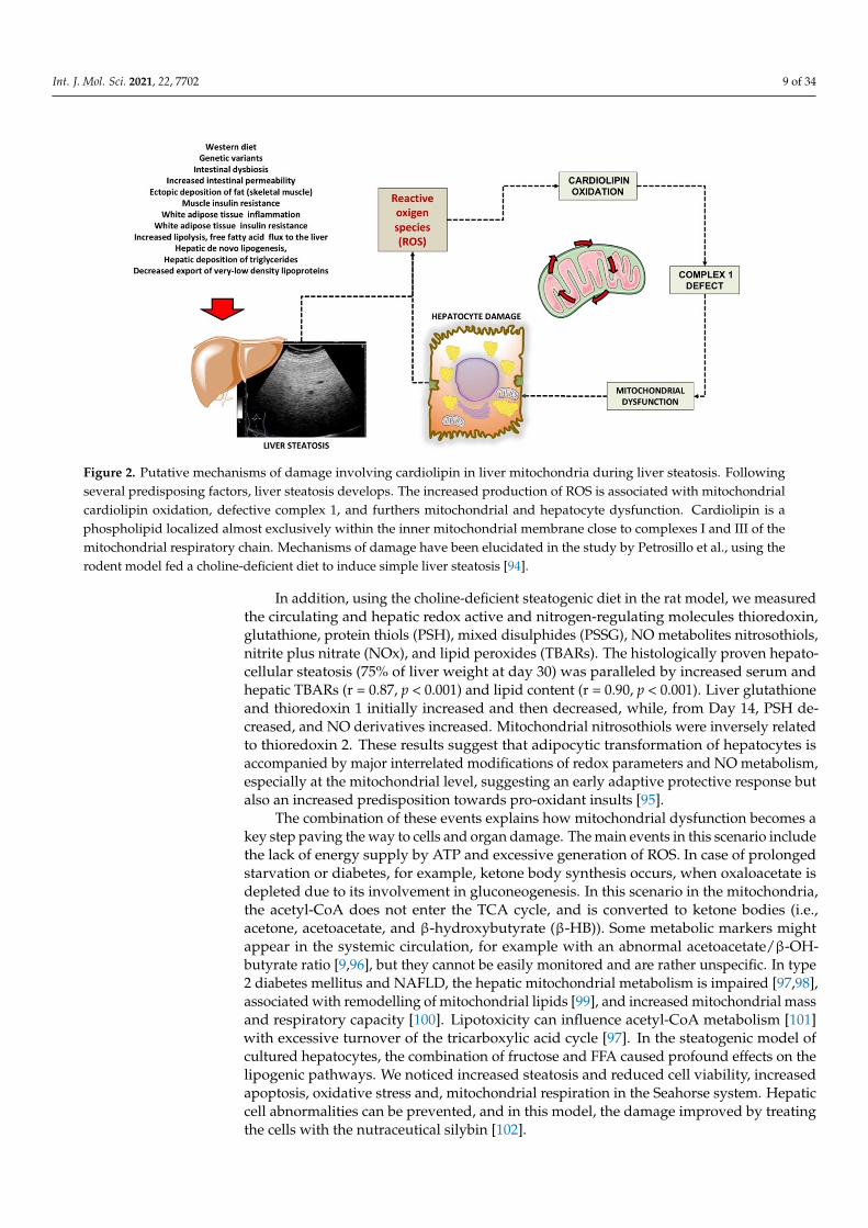
Int. J. Mol. Sci. 2021, 22, 7702 9 of 34
Figure 2. Putative mechanisms of damage involving cardiolipin in liver mitochondria during liver steatosis. Followingseveral predisposing factors, liver steatosis develops. The increased production of ROS is associated with mitochondrialcardiolipin oxidation, defective complex 1, and furthers mitochondrial and hepatocyte dysfunction. Cardiolipin is aphospholipid localized almost exclusively within the inner mitochondrial membrane close to complexes I and III of themitochondrial respiratory chain. Mechanisms of damage have been elucidated in the study by Petrosillo et al., using therodent model fed a choline-deficient diet to induce simple liver steatosis [94].
In addition, using the choline-deficient steatogenic diet in the rat model, we measuredthe circulating and hepatic redox active and nitrogen-regulating molecules thioredoxin,glutathione, protein thiols (PSH), mixed disulphides (PSSG), NO metabolites nitrosothiols,nitrite plus nitrate (NOx), and lipid peroxides (TBARs). The histologically proven hepato-cellular steatosis (75% of liver weight at day 30) was paralleled by increased serum andhepatic TBARs (r = 0.87, p < 0.001) and lipid content (r = 0.90, p < 0.001). Liver glutathioneand thioredoxin 1 initially increased and then decreased, while, from Day 14, PSH de-creased, and NO derivatives increased. Mitochondrial nitrosothiols were inversely relatedto thioredoxin 2. These results suggest that adipocytic transformation of hepatocytes isaccompanied by major interrelated modifications of redox parameters and NO metabolism,especially at the mitochondrial level, suggesting an early adaptive protective response butalso an increased predisposition towards pro-oxidant insults [95].
The combination of these events explains how mitochondrial dysfunction becomes akey step paving the way to cells and organ damage. The main events in this scenario includethe lack of energy supply by ATP and excessive generation of ROS. In case of prolongedstarvation or diabetes, for example, ketone body synthesis occurs, when oxaloacetate isdepleted due to its involvement in gluconeogenesis. In this scenario in the mitochondria,the acetyl-CoA does not enter the TCA cycle, and is converted to ketone bodies (i.e.,acetone, acetoacetate, and β-hydroxybutyrate (β-HB)). Some metabolic markers mightappear in the systemic circulation, for example with an abnormal acetoacetate/β-OH-butyrate ratio [9,96], but they cannot be easily monitored and are rather unspecific. In type2 diabetes mellitus and NAFLD, the hepatic mitochondrial metabolism is impaired [97,98],associated with remodelling of mitochondrial lipids [99], and increased mitochondrial massand respiratory capacity [100]. Lipotoxicity can influence acetyl-CoA metabolism [101]with excessive turnover of the tricarboxylic acid cycle [97]. In the steatogenic model ofcultured hepatocytes, the combination of fructose and FFA caused profound effects on thelipogenic pathways. We noticed increased steatosis and reduced cell viability, increasedapoptosis, oxidative stress and, mitochondrial respiration in the Seahorse system. Hepaticcell abnormalities can be prevented, and in this model, the damage improved by treatingthe cells with the nutraceutical silybin [102].
Int. J. Mol. Sci. 2021, 22, 7702 10 of 34
Mitochondrial dysfunction is associated, in NASH, with the ongoing oxidative state ofhepatocytes, and is able to affect intracellular signalling pathways by generation of DAMPsand to activate stellate cell [103].
A recent in vitro study demonstrated that circulating factors contained in plasmasamples from NAFLD patients were able to generate a NAFLD-like phenotype in iso-lated hepatocytes, with effects mediated by NLRP3-inflammasome pathways and by theactivation of intracellular signalling related to SREBP-1c, PPAR-γ, NF-kB and NOX2 [104].
Besides external conditions affecting the metabolic homeostasis and mitochondrialfunction, genetically driven conditions can also lead to altered hepatic mitochondrialactivity and peroxisomal β-oxidation [105–107]. In particular, the membrane bound O-acyltransferase domain containing 7-trans-membrane channel-like 4 (MBOAT7-TMC4) islocalized to the intracellular membranes of mitochondria, endoplasmic reticulum, andlipid droplets. MBOAT7-TMC4 acts as lysophospholipid acyltransferase, and regulatesthe incorporation of arachidonic acid into phosphatidylinositol [108]. This pathway, dueto its key role, might be considered a promising therapeutic target. The expression ofMBOAT7-TMC4 is decreased in the rs641738 polymorphism, and this leads to the onsetof liver steatosis and to an altered liver histology [109,110], to fibrosis in alcoholic liverdisease [111] and in chronic hepatitis C [112]. In a mice model of NASH, the deletionof hepatocyte Mboat7 is linked to with increased fibrosis, with no effects on inflamma-tion [113]. Mboat7 also promotes the degradation of lysophosphatidylinositol, and theaccumulation of this molecule in Mboat7 KO mice generates NASH [114], also through theactivation of the G-protein coupled receptor GPR55 [115]. Aging processes is associatedwith altered subcutaneous adipose tissue function, with mechanisms that involve a reducedmitochondrial activity [116,117], the accumulation of senescent adipocytes, and impaireddevelopment of pre-adipocytes [118]. Liver mitochondria also play a relevant role in lipid-induced hepatic insulin resistance, through mechanisms linking specific lipid metabolitesand cellular compartments and leading to subcellular dysfunctions [119]. In particular, thequantitative assessment of DAG stereoisomers (sn-1,2-DAGs, sn-2,3- DAGs, and sn-1,3-DAGs) and ceramides in the endoplasmic reticulum, mitochondria, plasma membrane,lipid droplets, and cytosol showed, using an antisense oligonucleotide, the onset of hepaticinsulin resistance in rats, which was associated with the acute liver-specific knockdown ofdiacylglycerol acyltransferase-2. The dysregulation of peroxisome proliferator-activatedreceptor-gamma co-activator-1α (PGC-1α) contributes to the pathogenesis and to the se-quence of NASH-HCC, with metabolic pathways involving gluconeogenesis, fatty acidoxidation, antioxidant response, DNL, and mitochondrial biogenesis [120].
Experimental data indicate that mitochondrial dysfunction is also a specific targetfor toxic chemicals of environmental origin mainly introduced bycontaminated food andwater and leading to NAFLD.
A recent study in 2446 young adults showed that toenail cadmium concentration,a marker of long-term exposure, was associated with higher odds of prevalent NAFLDindependently from race, sex, BMI or smoking status [121]. In a mouse model of chroniccadmium exposure, hepatic Cd concentrations ranging from 0.95 to 6.04 µg/g wet weightwere able to induce, following a 20-week exposure, NAFLD and NASH like phenotypeslinked with mitochondrial dysfunction, fatty acid oxidation deficiency and a significantsuppression of sirtuin 1 signalling pathway [122]. Epidemiologic studies point to a positiveassociation between arsenic exposure (i.e., urinary arsenic concentrations) and risk ofNAFLD [123]. This evidence is paralleled by experimental findings showing, in isolatedrat liver mitochondria exposed to arsenic, a marked decrease in total mitochondrial dehy-drogenase activity with increased ROS generation, MMP, and MDA levels, and decreasedactivity of mitochondrial catalase and GSH [124].
In a cohort of 6389 adolescents from the NHANES survey, blood mercury levelswere linked with the risk of NAFLD, with the most evident association in underweight ornormal weight subjects [125]. In a recent animal model, exposure to methylmercury during
Int. J. Mol. Sci. 2021, 22, 7702 11 of 34
12 weeks induced mitochondrial swelling, ROS overproduction, increased gluthationeoxidation, and reduced protein thiol content [126].
Similar pathways linking environmental pollution with NAFLD in terms of both epi-demiologic findings of increased NAFLD risk and animal/in vitro evidence of mitochon-drial dysfunction also have been shown in the case of air pollution [127–129], endocrinedisrupting chemicals [127–131], and pesticides [132–135].
There are few ways to investigate mitochondrial metabolic processes, i.e., usingisolated organelles, mitochondrial fractions, and cell culture [136]. Few studies exploredthe impaired mitochondrial function in NAFLD. Protocols investigating the effects ofxenobiotics and drugs on mitochondrial function can provide some information [137,138].Metabolomics can also explore specific mitochondrial functions [139] by studying geneticperturbations [140]. The measure of circulating mitochondrial DNA (mtDNA) is anotherbiomarker of mitochondrial dysfunction. Changes to liver mitochondrial DNA (mtDNA)can precede mitochondrial dysfunction and irreversible liver damage. Malik et al. [141] byusing a rodent dietary approach, demonstrated that a high-fat or a high-fat/high-sugardiet for 16 weeks was associated with fast alterations in mtDNA. Thus, dietary changes inliver mtDNA can occur in a relatively short time. Mouse liver contained a high mtDNAcontent (3617 +/− 233 copies per cell), which significantly increased when the mice werefed an HFD diet. This increase, however, was not functional; i.e., it was not translatedinto an increased expression of mitochondrial proteins. Furthermore, liver dysfunctionwas accelerated alongside the downregulation of mitochondrial oxidative phosphorylation(OXPHOS) and mtDNA replication machinery as well as upregulation of the mtDNA-induced inflammatory pathways.
5. Studying Liver Mitochondrial Function at a Translational Level
Strategies to diagnose mitochondrial damage and afterwards to prevent progressionby therapy in NAFLD are actively being investigated. All the above-mentioned proceduresfocusing on mitochondrial function, however, lack true translational value, can be complex,expensive, and make the comparison between different models and in vivo studies some-what difficult. A major problem is that mitochondria function at the crossroads of severalcomplex metabolic processes, which can be influenced by unknown precursors affectingmetabolic pathways.
Thus, no specific, easily available test provides information on liver mitochondrialstatus in clinical medicine, and we miss reliable biomarkers that inform on hepatocytemitochondrial function and fitness in the NAFLD. Further studies and novel diagnostictools are required in this field. The following paragraphs will focus on the use of breathtests by stable isotopes to explore liver mitochondrial function.
6. General Features of BT
BT represent an expanding field in diagnosis of liver function and are “dynamic” toolsdealing with distinct functional aspects of liver metabolism. BTs can be employed for thefollow-up of liver disease, including mitochondrial function in NAFLD [96]. Liver BTcan provide information about the complex metabolic function of the liver by marking agiven substrate with the stable isotope 13C, measured in breath as 13CO2. Our group hasprovided studies dealing with evaluation of both mitochondrial and microsomal activityin the liver [36,96,142–149]. The rationale of BT depends on a given substrate that ismetabolized at different levels in the body. The metabolized substrate produces gases (e.g.,CO2, H2) transferred to blood, excreted, and quantified in expired air. Various sensors candetect the end-product in breath. The measured metabolite becomes the biomarker of aspecific metabolic process [150]. Examples of BTs include the urea BT for the diagnosis ofinfection by H. pylori in the stomach, the hydrogen breath test for the diagnosis of lactoseintolerance, or the study of small intestinal bacterial overgrowth [151–154]. A few BTs arerelatively simple to perform, safe, and non-invasive, with potential applications in severalconditions. Liver BTs are used to assess the hepatocyte capacity to metabolize a substrate
Int. J. Mol. Sci. 2021, 22, 7702 12 of 34
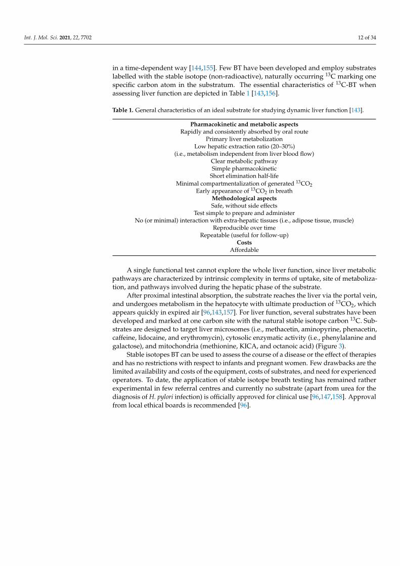
in a time-dependent way [144,155]. Few BT have been developed and employ substrateslabelled with the stable isotope (non-radioactive), naturally occurring 13C marking onespecific carbon atom in the substratum. The essential characteristics of 13C-BT whenassessing liver function are depicted in Table 1 [143,156].
Table 1. General characteristics of an ideal substrate for studying dynamic liver function [143].
Pharmacokinetic and metabolic aspectsRapidly and consistently absorbed by oral route
Primary liver metabolizationLow hepatic extraction ratio (20–30%)
(i.e., metabolism independent from liver blood flow)Clear metabolic pathwaySimple pharmacokinetic
Short elimination half-lifeMinimal compartmentalization of generated 13CO2
Early appearance of 13CO2 in breathMethodological aspectsSafe, without side effects
Test simple to prepare and administerNo (or minimal) interaction with extra-hepatic tissues (i.e., adipose tissue, muscle)
Reproducible over timeRepeatable (useful for follow-up)
CostsAffordable
A single functional test cannot explore the whole liver function, since liver metabolicpathways are characterized by intrinsic complexity in terms of uptake, site of metaboliza-tion, and pathways involved during the hepatic phase of the substrate.
After proximal intestinal absorption, the substrate reaches the liver via the portal vein,and undergoes metabolism in the hepatocyte with ultimate production of 13CO2, whichappears quickly in expired air [96,143,157]. For liver function, several substrates have beendeveloped and marked at one carbon site with the natural stable isotope carbon 13C. Sub-strates are designed to target liver microsomes (i.e., methacetin, aminopyrine, phenacetin,caffeine, lidocaine, and erythromycin), cytosolic enzymatic activity (i.e., phenylalanine andgalactose), and mitochondria (methionine, KICA, and octanoic acid) (Figure 3).
Stable isotopes BT can be used to assess the course of a disease or the effect of therapiesand has no restrictions with respect to infants and pregnant women. Few drawbacks are thelimited availability and costs of the equipment, costs of substrates, and need for experiencedoperators. To date, the application of stable isotope breath testing has remained ratherexperimental in few referral centres and currently no substrate (apart from urea for thediagnosis of H. pylori infection) is officially approved for clinical use [96,147,158]. Approvalfrom local ethical boards is recommended [96].
Int. J. Mol. Sci. 2021, 22, 7702 13 of 34
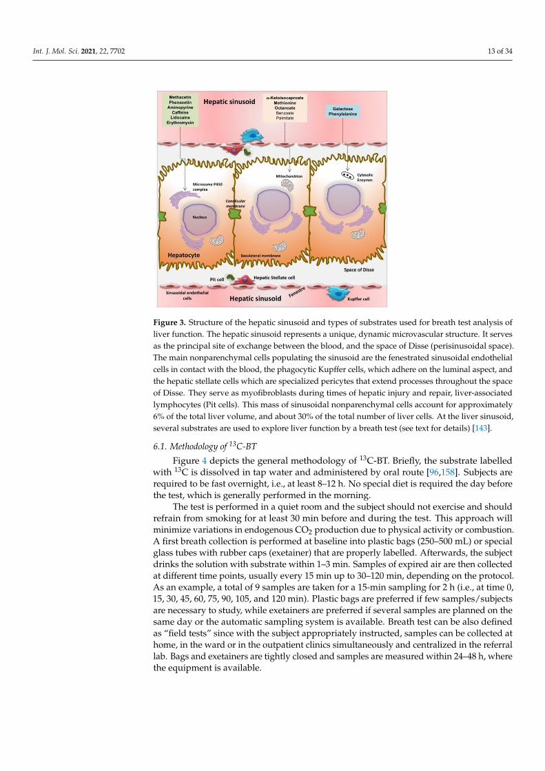
Figure 3. Structure of the hepatic sinusoid and types of substrates used for breath test analysis ofliver function. The hepatic sinusoid represents a unique, dynamic microvascular structure. It servesas the principal site of exchange between the blood, and the space of Disse (perisinusoidal space).The main nonparenchymal cells populating the sinusoid are the fenestrated sinusoidal endothelialcells in contact with the blood, the phagocytic Kupffer cells, which adhere on the luminal aspect, andthe hepatic stellate cells which are specialized pericytes that extend processes throughout the spaceof Disse. They serve as myofibroblasts during times of hepatic injury and repair, liver-associatedlymphocytes (Pit cells). This mass of sinusoidal nonparenchymal cells account for approximately6% of the total liver volume, and about 30% of the total number of liver cells. At the liver sinusoid,several substrates are used to explore liver function by a breath test (see text for details) [143].
6.1. Methodology of 13C-BT
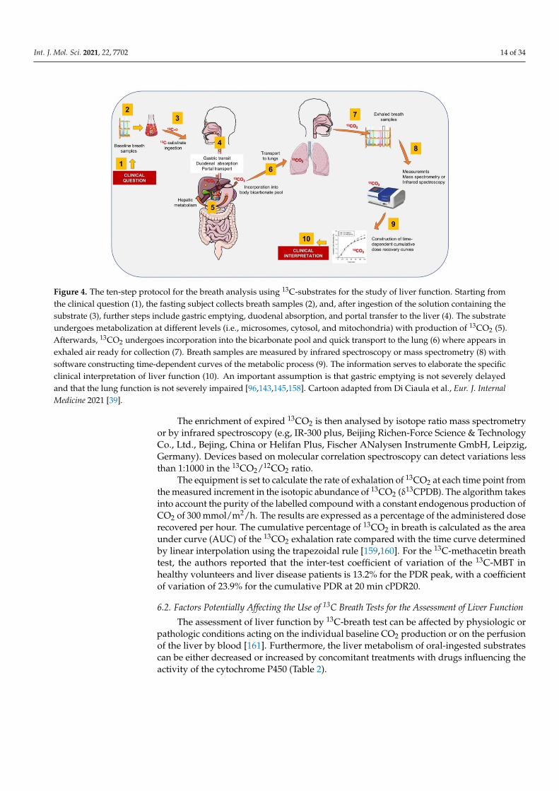
Figure 4 depicts the general methodology of 13C-BT. Briefly, the substrate labelledwith 13C is dissolved in tap water and administered by oral route [96,158]. Subjects arerequired to be fast overnight, i.e., at least 8–12 h. No special diet is required the day beforethe test, which is generally performed in the morning.
The test is performed in a quiet room and the subject should not exercise and shouldrefrain from smoking for at least 30 min before and during the test. This approach willminimize variations in endogenous CO2 production due to physical activity or combustion.A first breath collection is performed at baseline into plastic bags (250–500 mL) or specialglass tubes with rubber caps (exetainer) that are properly labelled. Afterwards, the subjectdrinks the solution with substrate within 1–3 min. Samples of expired air are then collectedat different time points, usually every 15 min up to 30–120 min, depending on the protocol.As an example, a total of 9 samples are taken for a 15-min sampling for 2 h (i.e., at time 0,15, 30, 45, 60, 75, 90, 105, and 120 min). Plastic bags are preferred if few samples/subjectsare necessary to study, while exetainers are preferred if several samples are planned on thesame day or the automatic sampling system is available. Breath test can be also definedas “field tests” since with the subject appropriately instructed, samples can be collected athome, in the ward or in the outpatient clinics simultaneously and centralized in the referrallab. Bags and exetainers are tightly closed and samples are measured within 24–48 h, wherethe equipment is available.
Int. J. Mol. Sci. 2021, 22, 7702 14 of 34
Figure 4. The ten-step protocol for the breath analysis using 13C-substrates for the study of liver function. Starting fromthe clinical question (1), the fasting subject collects breath samples (2), and, after ingestion of the solution containing thesubstrate (3), further steps include gastric emptying, duodenal absorption, and portal transfer to the liver (4). The substrateundergoes metabolization at different levels (i.e., microsomes, cytosol, and mitochondria) with production of 13CO2 (5).Afterwards, 13CO2 undergoes incorporation into the bicarbonate pool and quick transport to the lung (6) where appears inexhaled air ready for collection (7). Breath samples are measured by infrared spectroscopy or mass spectrometry (8) withsoftware constructing time-dependent curves of the metabolic process (9). The information serves to elaborate the specificclinical interpretation of liver function (10). An important assumption is that gastric emptying is not severely delayedand that the lung function is not severely impaired [96,143,145,158]. Cartoon adapted from Di Ciaula et al., Eur. J. InternalMedicine 2021 [39].
The enrichment of expired 13CO2 is then analysed by isotope ratio mass spectrometryor by infrared spectroscopy (e.g, IR-300 plus, Beijing Richen-Force Science & TechnologyCo., Ltd., Bejing, China or Helifan Plus, Fischer ANalysen Instrumente GmbH, Leipzig,Germany). Devices based on molecular correlation spectroscopy can detect variations lessthan 1:1000 in the 13CO2/12CO2 ratio.
The equipment is set to calculate the rate of exhalation of 13CO2 at each time point fromthe measured increment in the isotopic abundance of 13CO2 (δ13CPDB). The algorithm takesinto account the purity of the labelled compound with a constant endogenous production ofCO2 of 300 mmol/m2/h. The results are expressed as a percentage of the administered doserecovered per hour. The cumulative percentage of 13CO2 in breath is calculated as the areaunder curve (AUC) of the 13CO2 exhalation rate compared with the time curve determinedby linear interpolation using the trapezoidal rule [159,160]. For the 13C-methacetin breathtest, the authors reported that the inter-test coefficient of variation of the 13C-MBT inhealthy volunteers and liver disease patients is 13.2% for the PDR peak, with a coefficientof variation of 23.9% for the cumulative PDR at 20 min cPDR20.
6.2. Factors Potentially Affecting the Use of 13C Breath Tests for the Assessment of Liver Function
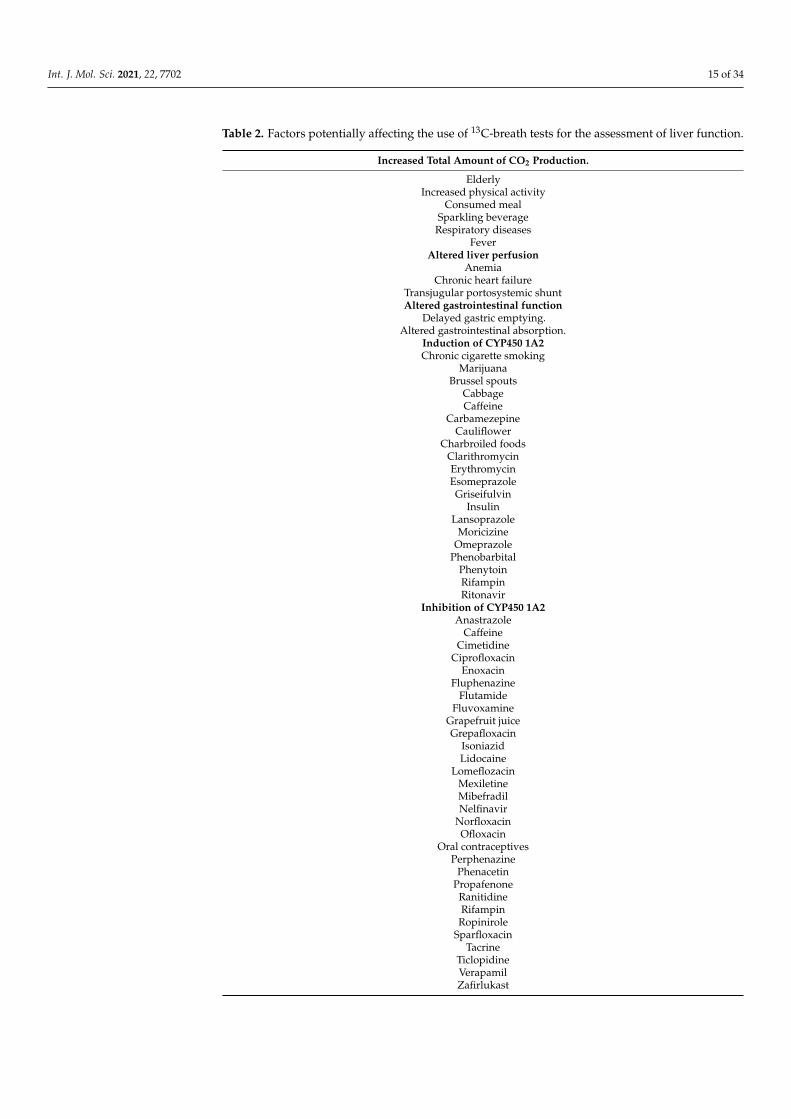
The assessment of liver function by 13C-breath test can be affected by physiologic orpathologic conditions acting on the individual baseline CO2 production or on the perfusionof the liver by blood [161]. Furthermore, the liver metabolism of oral-ingested substratescan be either decreased or increased by concomitant treatments with drugs influencing theactivity of the cytochrome P450 (Table 2).
Int. J. Mol. Sci. 2021, 22, 7702 15 of 34
Table 2. Factors potentially affecting the use of 13C-breath tests for the assessment of liver function.
Increased Total Amount of CO2 Production.
ElderlyIncreased physical activity
Consumed mealSparkling beverage
Respiratory diseasesFever
Altered liver perfusionAnemia
Chronic heart failureTransjugular portosystemic shuntAltered gastrointestinal function
Delayed gastric emptying.Altered gastrointestinal absorption.
Induction of CYP450 1A2Chronic cigarette smoking
MarijuanaBrussel spouts
CabbageCaffeine
CarbamezepineCauliflower
Charbroiled foodsClarithromycinErythromycinEsomeprazoleGriseifulvin
InsulinLansoprazole
MoricizineOmeprazole
PhenobarbitalPhenytoinRifampinRitonavir
Inhibition of CYP450 1A2Anastrazole
CaffeineCimetidine
CiprofloxacinEnoxacin
FluphenazineFlutamide
FluvoxamineGrapefruit juiceGrepafloxacin
IsoniazidLidocaine
LomeflozacinMexiletineMibefradilNelfinavir
NorfloxacinOfloxacin
Oral contraceptivesPerphenazine
PhenacetinPropafenone
RanitidineRifampin
RopiniroleSparfloxacin
TacrineTiclopidineVerapamilZafirlukast
Int. J. Mol. Sci. 2021, 22, 7702 16 of 34
A severely delayed gastric emptying (e.g., diabetic, or idiopathic gastroparesis, severemotility defects, inflammation, and malignancies) might interfere with the delivery of thesubstrate to the duodenum. Thus, before the examination, the operator should investigatethe clinical history to detect symptoms potentially related with altered gastric emptying orconditions affecting the absorption of the substrate from the gastrointestinal tract [161,162].In this case, the intravenous administration of the substrate (as the LiMAx® test) canbe useful to obtain accurate results, also limiting inter-individual variations in substrateabsorption after oral administration [163–166].
6.3. Assessing Liver Mitochondrial Function by BT
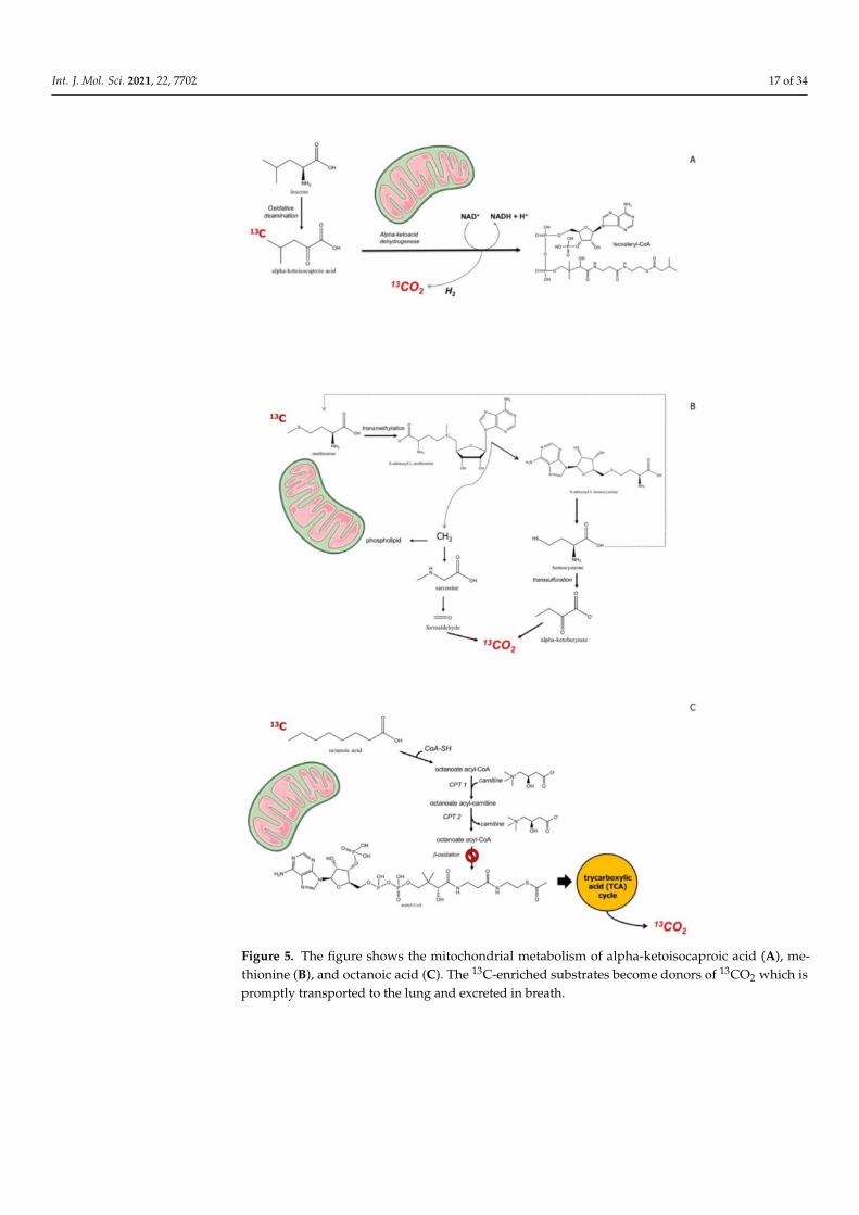
The assessment of liver function and responses to therapy is often required at anearly stage in clinical medicine. The assessment of early involvement of mitochondrialfunction could be part of this procedure adding prognostic information [167]. There aresubstrates that target mitochondria and can be marked with 13C, which becomes detectablein expired air. Substrates are alpha-ketoisocaproic acid (KICA), methionine, and octanoicacid [147]. Benzoic acid undergoes glycine conjugation, but was used only in the animalmodel of liver cirrhosis [168]. Following the mitochondrial catabolism of a certain substrate,CO2 is produced. The labelled carbon (*C) will appear in breath as *CO2, as a marker ofmitochondrial clearance of the substrate [96]. The radioactive isotope 14C had limitationsbecause of radiation exposure, and thus could not be used in pregnant women and children.13C, by contrast, is a stable, nonradioactive, naturally occurring isotope. It accounts forabout 1.1% of all the natural carbon on Earth and in plants and the food chain. Thefollowing assumption define this type of breath test, or a given exogenous substrate, i.e.,Hepatic clearance = Hepatic Perfusion x Hepatic Extraction (where Hepatic Extraction is theratio of the difference between inflow and outflow concentration ÷ by inflow concentrationof the probe) [169]. The hepatic clearance is either flow-limited (range 0.7–1.0) or enzyme-limited (<0.3) [170]. Few 13C-labeled substrates are suitable to explore mitochondrial liverfunction, and include alpha-ketoisocaproic acid, methionine, and octanoic acid (Figure 3).The mitochondrial metabolism of these three substrates is depicted in Figure 5.
KICA is a substrate for branched chain alpha ketoacid dehydrogenase, located inthe hepatic mitochondrial matrix [171]. Methionine is a substrate for protein synthe-sis or alternatively enters the methionine cycle, with subsequent transformation in S-adenosylmethionine, a methyl donor. S-adenosylhomocysteine, resulting from donation ofthe methyl group, is hydrolyzed to homocysteine, which, in turn, is destinated to trans-sulfuration or to remethylation to methionine. The trans-sulfuration step, in particular, gen-erates α-ketobutyrate, which enters mitochondria with subsequent decarboxylatation [172].Octanoic acid, on the other hand, is β-oxidized with generation of acetyl coenzyme A,which enters the Krebs cycle and undergoes oxidation to CO2 [172].
All these three substrates have been used to accurately assess hepatic mitochondrialfunction in NAFLD patients. By different substrates, BT can also allow to explore the hepa-tocyte at a cytosolic (13C-phenylalanine and galactose) or microsomal level (aminopyrine,phenacetin, caffeine, lidocaine, methacetin, and erythromycin) [172]. From a clinical pointof view, these substrates provide information in different clinical settings (Table 3).
Int. J. Mol. Sci. 2021, 22, 7702 17 of 34
Figure 5. The figure shows the mitochondrial metabolism of alpha-ketoisocaproic acid (A), me-thionine (B), and octanoic acid (C). The 13C-enriched substrates become donors of 13CO2 which ispromptly transported to the lung and excreted in breath.
Int. J. Mol. Sci. 2021, 22, 7702 18 of 34
Table 3. Liver mitochondrial breath tests: substrates and evidence for potential clinical applications.
Substrate/Clinical Applications Information
KICA
ALD Effect of acute alcohol consumption (even low-moderate doses)[171]
ALD Discrimination between chronic alcohol consumption andnonalcoholic chronic liver disease [173]
ALD Monitoring and ascertaining of alcohol withdrawal [173]
NAFLDDiscrimination between simple steatosis and steatohepatitis(NASH) and between low-grade and high-grade fibrosis [147](Figure 6)
HCCEffect of treatment (thermoablation, chemoembolization) andprediction of tumour recurrence after local treatment.Comparison with methacetin [158]
Drugs Evaluation of acute drug toxicity [171]Methionine
Healthy subjectsValidation studies with 2 mg/kg body weight(methyl-13C)-methionine. Breath 13CO2 enrichment measuredat base line and every 15 min thereafter for 180 min. [174]
Healthy subjectsEffect of alcohol consumption (30 min after the ingestion ofethanol 0.3 g/kg body weight). Decreased excretion withethanol, due to impaired mitochondrial oxidation [175].
Liver cirrhosis Discrimination between different degree of chronic liverdamage [175]
ALD Diagnosis of acute alcohol ingestion [174]NAFLD Discrimination between simple steatosis and NASH [176]
HCVDiscrimination between HCV infected patients and healthysubjects and toxicity of pegylated interferon plus ribavirintreatment [177]
Drugs Evaluation of chronic drug toxicity [178]Friedriech’s ataxia Diagnosis of neurological disorders [179]
OctanoateNAFLD Evaluation of altered lipid metabolism [180]
NASHTotal β-oxidation of octanoic acid remained normal betweencontrols and NASH patients, although cumulative 13CO2recovery was higher in women than men [181]
Abbreviations: ALD = alcoholic liver disease; HCC = hepatocellular carcinoma; HCV = hepatitics C virus; KICA = α-ketoisocaproic acid;NAFLD = nonalcoholic fatty liver disease; NASH = nonalcoholic steatohepatitis.
6.4. Potential Clinical Application
Each substrate has possible clinical applications (Table 3), as shown in patients withalcohol-related liver disease, NAFLD, viral hepatitis, liver cirrhosis, hepatocellular carci-noma, and evaluation of drug or alcohol toxicity.
6.4.1. 13C-KICA BT
KICA has a molecular weight of 130.141800 g/mol (MF: C6H10O3, IUPAC name: 4-methyl-2-oxopentanoic acid), and is part of the metabolic pathway of the amino acid leucine(Figure 5A). The biotransformation of KICA to isovaleryl-CoA depends on the alpha-ketoacid dehydrogenase in the liver. The decarboxylation of KICA is a specific function ofmitochondria, as confirmed in isolated mitochondria [182], in experimental models [157],and in humans. No gender difference exists when interpreting the results accordingto body composition [183]. The major competing pathway for KICA elimination is thetransamination of KICA to leucine. The 13C-BT requires the concomitant administration ofunlabelled leucine which suppresses this pathway, and this step makes the KICA BT highlysensitive for mitochondrial function. In addition, KICA decarboxylation is depends on theavailability of NADH. Thus, ethanol at 0.5 g/kg body weight increases the availability ofNADH (leading to decreased KICA decarboxylation), while 1 g of aspirin decreases liverNADH (leading to a higher KICA decarboxylation rate) [171]. These aspects need to be
Int. J. Mol. Sci. 2021, 22, 7702 19 of 34
considered during the test performance and the evaluation of results. 13C-KICA is given ata dose of 1 mg/kg body weight plus 1 g unlabeled L-leucine while 13C-methacetin is givenat a dose of 1.5 mg/kg body weight (generally 75 mg). The substrate is generally flavorlessand dissolved in 100 mL of tap water. Such a small volume shortens the drinking time andwill allow the prompt initiation of the gastric emptying process.
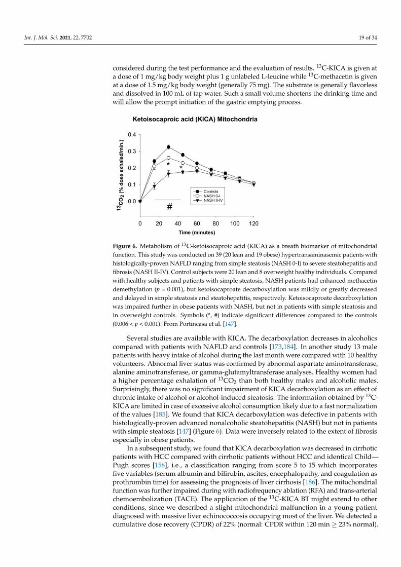
Figure 6. Metabolism of 13C-ketoisocaproic acid (KICA) as a breath biomarker of mitochondrialfunction. This study was conducted on 39 (20 lean and 19 obese) hypertransaminasemic patients withhistologically-proven NAFLD ranging from simple steatosis (NASH 0-I) to severe steatohepatitis andfibrosis (NASH II-IV). Control subjects were 20 lean and 8 overweight healthy individuals. Comparedwith healthy subjects and patients with simple steatosis, NASH patients had enhanced methacetindemethylation (p = 0.001), but ketoisocaproate decarboxylation was mildly or greatly decreasedand delayed in simple steatosis and steatohepatitis, respectively. Ketoisocaproate decarboxylationwas impaired further in obese patients with NASH, but not in patients with simple steatosis andin overweight controls. Symbols (*, #) indicate significant differences compared to the controls(0.006 < p < 0.001). From Portincasa et al. [147].
Several studies are available with KICA. The decarboxylation decreases in alcoholicscompared with patients with NAFLD and controls [173,184]. In another study 13 malepatients with heavy intake of alcohol during the last month were compared with 10 healthyvolunteers. Abnormal liver status was confirmed by abnormal aspartate aminotransferase,alanine aminotransferase, or gamma-glutamyltransferase analyses. Healthy women hada higher percentage exhalation of 13CO2 than both healthy males and alcoholic males.Surprisingly, there was no significant impairment of KICA decarboxylation as an effect ofchronic intake of alcohol or alcohol-induced steatosis. The information obtained by 13C-KICA are limited in case of excessive alcohol consumption likely due to a fast normalizationof the values [185]. We found that KICA decarboxylation was defective in patients withhistologically-proven advanced nonalcoholic steatohepatitis (NASH) but not in patientswith simple steatosis [147] (Figure 6). Data were inversely related to the extent of fibrosisespecially in obese patients.
In a subsequent study, we found that KICA decarboxylation was decreased in cirrhoticpatients with HCC compared with cirrhotic patients without HCC and identical Child—Pugh scores [158], i.e., a classification ranging from score 5 to 15 which incorporatesfive variables (serum albumin and bilirubin, ascites, encephalopathy, and coagulation asprothrombin time) for assessing the prognosis of liver cirrhosis [186]. The mitochondrialfunction was further impaired during with radiofrequency ablation (RFA) and trans-arterialchemoembolization (TACE). The application of the 13C-KICA BT might extend to otherconditions, since we described a slight mitochondrial malfunction in a young patientdiagnosed with massive liver echinococcosis occupying most of the liver. We detected acumulative dose recovery (CPDR) of 22% (normal: CPDR within 120 min ≥ 23% normal).
Int. J. Mol. Sci. 2021, 22, 7702 20 of 34
The 13C-methacetin breath test investigating the liver microsomal function was normal.Notably, mitochondrial liver function improved the following pericystectomy and limitedhepatectomy. Other applications of KICA BT are possible. Several drugs can enter themitochondria and accumulate, a step often interfering with respiratory complexes orelectron transfer [187]. For example, aspirin, ibuprofen (nonsteroidal anti-inflammatorydrugs), amiodarone (antiarrhythmic agent), and valproate (an anticonvulsant, histonedeacetylase inhibitor) inhibit mitochondrial fatty acid β-oxidation [187,188]. The nucleosideanalogues are widely used in transplanted patients or in HIV- and HBV-infected subjects.Drugs can incorporate into mitochondrial DNA, inhibit c-DNA polymerase, and hinder thereplication process [189]. Nevertheless, viral infection itself (HIV and HCV) may impairmitochondrial function, as confirmed in HCV-infected cells [190] and in patients [177]. Inaddition, xenobiotics can cause excessive activation of the mitochondrial permeability ofthe transition pores and alter mitochondrial function. Drugs include acetaminophen [191],N-nitrosofenfluramine [192], salicylate [193], and nimesulide (in vitro) [194]. The KICABT might provide helpful results to study the integrity of liver organelles before theadministration of potentially toxic drugs and to detect drug-induced mitochondrial damagebefore the appearance of symptoms to timely manage patients and prevent adverse effects.Examples are tacrolimus, aspirin [171], and ergot alkaloids [195].
6.4.2. 13C-Methionine BT
Methionine has a molecular weight of 149.21134 g/mol, MF: C5H11NO2S, IUPACname: (2S)-2-amino-4-methylsulfanylbutanoic acid. It is an essential amino acid that is in-volved in metabolic processes. Exogenous methionine contributes to protein synthesis [196].Methionine can be transformed into S-adenosylmethionine, the main biological methyldonor, which is hydrolysed to homocysteine, either undergoing trans-sulfuration to α-Ketobutyrate or remethylation to methionine. α-Ketobutyrate enters the mitochondria andundergoes decarboxylation. The administration of oral L-(1-13C)-methionine will then beassociated with production of labelled 13CO2. Transmethylation to S-adenosylmethioninealso provides the substrate for the synthesis of sarcosine, which is oxidized to formaldehydeand production of CO2 in mitochondria. Methionine differentially labelled in the methylgroup and in position 1 can be used to study the complex metabolism of methionine [175].For mitochondrial function studies, suitable substrates are either L-(1-13C) methionine or(methyl-13C)-methionine. There are some limitations, e.g., comparisons of different studiescan be quite difficult since more than one 13CO2 is formed according to the L-(1-13C) or(methyl-13C)-labelled methionine [197]. Some studies used intravenous rather than oralmethionine and in this case the comparisons become even more difficult [198]. MethionineBT can provide information about mitochondrial function during acute intoxication andin chronic liver diseases. However, acute ethanol consumption impairs 13C-methioninedecarboxylation in normal liver [174]. The metabolism of methionine decreases in patientswith liver cirrhosis and especially in those with an aethanol etiology [175], in patients withbiopsy-proven severe NAFLD in relation to the extent of steatosis [178], and in patientstaking high-dose valproic acid [178] or nucleoside analogues for the treatment of HIV [189].Methionine BT is reported in hepatitis C-infected cells [190], and in patients with Friedre-ich ataxia [179] an autosomal recessive degenerative disorder caused by loss of functionmutations in the frataxin gene (FXN gene), located on chromosome 9q13 [199].
6.4.3. 13C-octanoate BT
Octanoic acid (OA) has a molecular weight of 144.21144 g/mol, MF: C8H16O2, IUPACname: octanoic acid, caprylic acid. OA is a straight medium chain saturated fatty acidwith an 8-carbon backbone, it is found in the milk of various mammals and is a minorcomponent of coconut oil and palm kernel oil. OA enters mitochondria independentlyof the carnitine transport system and is β-oxidized to acetyl coenzyme A (AcCoA) [200].AcCoA enters the Krebs cycle and is oxidized to CO2 unless utilized for the synthesis ofother energy-rich compounds (Figure 5C). The 13C-octanoate should also reflect hepatic
Int. J. Mol. Sci. 2021, 22, 7702 21 of 34
mitochondrial function (β-oxidation capacity). For use in humans the test requires informedconsent. In the animal models 13-octanoate BT was informative about liver function in ratmodels of acute hepatitis and thioacetamide-induced liver cirrhosis, but not in cholestaticliver injury [201]. In NASH patients, the oxidation of octanoate was either unchanged,although greater in women than men [181] or increased [180], and unchanged in thosewith early stage and advanced cirrhosis with and without a porto-systemic shunt [202].Such apparently discrepant results with octanoate might be due to subtle differences in themetabolic pathways, the substrates employed, or by extra-hepatic mitochondrial oxidationof octanoate. Gender differences should be also taken into account, when consideringthe study of Schneider et al. [181], for example. Unfortunately, a comparison of differentsubstrates and BT in the same group of subjects/patients has not been performed, so far. Ifliver damage is absent, the 13C-octanoate BT is a useful diagnostic test to measure the rateof gastric emptying to solids, i.e., a muffin enriched with the labelled substrate [146,203].
7. Why Studying Liver Mitochondrial Function in NAFLD
There is no established therapy not as a monotherapy nor in combination with NAFLD.The complexity and the number of pathogenic mechanisms involved in the full spectrum ofNAFLD, makes this goal difficult to achieve and experiment with, so far [16]. Nevertheless,there might be some arguments for studying mitochondrial function in NAFLD patients(Table 4).
Table 4. Arguments for assessing liver mitochondrial function in NAFLD patients.
Increasing Scientific Interest About the Role of Mitochondria in NAFLD
Impaired liver mitochondrial function may occur early during the onset and progression ofNAFLD.
General measures for NAFLD can be beneficial to liver mitochondria as well.Few medications show some beneficial effects on liver mitochondria.
Improved mitochondrial function can contribute to ameliorate other liver dysfunctions in NAFLDpatients.
In general, a modification in lifestyle (i.e., diet and regular physical exercise [45,46])and other general measures serve to maintain body weight or reduce body weight inoverweigh/obese subjects. Ideally, weight loss should be in the range of 5–7% and 7–10%in NAFLD and NASH, respectively, in both overweight and obese patients [45]. Thisapproach can improve liver biochemical tests, liver histology, serum insulin levels, andquality of life [204–209]. We have learnt that in NASH, liver fibrosis can improve afterat least 10% weight reduction, although this goal is difficult to achieve in the majorityof patients and to maintain for a long time [142,207]. To improve insulin sensitivity andreduce body weight, the diet must be based on long-term caloric restriction rather thanintermittent fasting [210]. This approach will prevent oxidative damage [211,212]. Bariatricsurgery is indicated in the subgroup of morbid obese patients or obese patients withincreased cardiovascular risk. This choice can reduce the prevalence of NASH [213,214].Specific risk factors for cardiovascular disease, diabetes mellitus must be screened andappropriately treated in NAFLD patients (e.g., antidiabetic agents, lipid-lowering therapy).Alcohol consumption, even in small amounts, is not recommended, since it is associatedwith progression of liver fibrosis [215].
When considering the aspects related to liver mitochondria in NAFLD, potentialtargets include nuclear receptors and compounds involved in different signaling pathways,mitochondrial transporters, enzymes playing a major role in mitochondrial metabolism,biomolecules involved in pathways controlling reactive oxygen species (ROS) and oxidativestress. Therapeutic strategies, however, are highly experimental and in several cases tried inanimal or in vitro models. Although potentially able to ameliorate mitochondrial functionin NAFLD, these agents require further evidence about use in humans, safety, efficacy,duration of treatment, type of steatosis, etc.
Int. J. Mol. Sci. 2021, 22, 7702 22 of 34
A moderately hypocaloric diet plus physical exercise might improve mitochondrialstructure and function and alleviate inflammation [216–219]. Mitochondrial permeabil-ity transition [220,221] and mitochondrial integrity and function can improve and be-come more resistant to stress [221]. The exercise will decrease the insulin resistancestatus while increasing the hepatic mitochondrial oxidative capacity associated with in-creased FFA oxidation and decreased FA-derived ceramide and diacylglycerol synthe-sis [23,222]. With all limitations previously discussed, other options include as following:Bile acids, such as obeticholic acid [223–225] and ursodeoxycholic acid [226]. Agents actingas antioxidants, on nuclear receptors or mitochondrial metabolism, such as Vitamin E(α-Tocopherol) [26], Tempol [227], Resveratrol [228–231], Mitoquinone (Mito-Q) and Mi-tovitamin E (MitoVit-E) [232–234], Silymarin (major component is Silybin) [136,235,236],Corilagin [237], Anthocyanins (i.e., Cyanidin) [238,239], Dihydromyricetin [240], Berber-ine [241], Hydroxytyrosol [242], Cysteamine [243,244], Pentoxifilline [245–247], Avocadooil [248–250], and Pegbelfermin (via FGF21R beta) [251]. Antidiabetic drugs including Elafi-branor [252,253], Liraglutide [254], Metformin [255], Thiazolidinediones (pioglitazone) [256],and MSDC-0602K [257]. Various agents such as Aramchol [258,259], Baicalin [260], Nitro-oleicacid [261], Carboxyatractyloside [262], Genistein [263], and Firsocostat (acetyl-CoA car-boxylase (ACC) inhibitor) [264]. Mitotherapy implies exogenous mitochondria taggedwith green-fluorescence protein (GFP), retrieved in mouse liver, lungs, brain, muscle, andkidneys [265,266]. In this case, the improved energy production may restore hepatocytefunction [267]
In conclusion, there is no standard therapy for NAFLD, apart from lifestyle changeshelping weight maintenance or weight loss to achieve an ideal body weight. The role ofcombination therapies to act on different targets simultaneously is being actively investi-gated.
8. Future Perspectives and Conclusions
The interpretation of the results investigating the mitochondrial function by 13C- BTin vivo requires some considerations. Studies show that both marked steatosis and NASH,and ethanol consumption, cause mitochondrial dysfunction, which becomes detectableby KICA and methionine BT. This aspect should be particularly useful in the case of earlytreatments possibly limiting the progression of the disease, and in terms of secondaryprevention measures. On the other hand, experimental studies indicate that mitochondrialdysfunction can precede the onset and progression of NAFLD, and these findings also pavethe way to possible primary prevention measures. Several factors can influence resultsfrom BT, affecting liver perfusion and/or mitochondrial performances. Thus, particularattention is required in the selection of subjects undergoing 13C- BT and in confoundingfactors, also considering possible inter-individual differences. ‘Competing’ mitochondriamay be active in extra-hepatic tissues (e.g., muscle). The production of CO2 can varysubstantially among subjects [268] and therefore the labelled CO2 in breath might becomeindependent of circulating or renally excreted bicarbonate and the endogenous productionof unlabeled CO2. Thus, adequately validated BT will have clinical utility for diagnosis,prognosis, or efficacy of treatments. Remaining limitations of BT applications in clinicalpractice should be overcome by further translational studies and clinical trials, in parallelwith complementary diagnostic techniques. The implementation of novel substrates toinvestigate additional mitochondrial pathways is greatly warranted.
Author Contributions: Conceptualization, P.P.; literature review, H.S., M.K., G.C., L.B., draft prepara-tion, A.D.C., G.C., and P.P.; writing—review and editing, A.D.C., D.Q.-H.W., G.B. and P.P. All authorshave read and agreed to the published version of the manuscript.
Funding: This paper was been partly supported by funding from the European Union’s Horizon2020 Research and Innovation program under the Marie Skłodowska-Curie Grant Agreement No.722619 (FOIE GRAS) and Grant Agreement No. 734719 (mtFOIE GRAS).
Int. J. Mol. Sci. 2021, 22, 7702 23 of 34
Acknowledgments: The authors are grateful to Diana Schaubhut for kindly providing languagerevision of the manuscript.
Conflicts of Interest: The authors declare no conflict of interest.
References1. Lee, J.; Park, J.S.; Roh, Y.S. Molecular insights into the role of mitochondria in non-alcoholic fatty liver disease. Arch. Pharm. Res.
2019, 42, 935–946. [CrossRef]2. Xiang, L.; Shao, Y.; Chen, Y. Mitochondrial dysfunction and mitochondrion-targeted therapeutics in liver diseases. J. Drug Target.
2021. [CrossRef]3. Luangmonkong, T.; Suriguga, S.; Mutsaers, H.A.M.; Groothuis, G.M.M.; Olinga, P.; Boersema, M. Targeting Oxidative Stress for
the Treatment of Liver Fibrosis. Rev. Physiol. Biochem. Pharmacol. 2018, 175, 71–102. [CrossRef]4. Degli Esposti, D.; Hamelin, J.; Bosselut, N.; Saffroy, R.; Sebagh, M.; Pommier, A.; Martel, C.; Lemoine, A. Mitochondrial roles and
cytoprotection in chronic liver injury. Biochem. Res. Int. 2012, 2012, 387626. [CrossRef]5. Wei, Y.; Rector, R.S.; Thyfault, J.P.; Ibdah, J.A. Nonalcoholic fatty liver disease and mitochondrial dysfunction. World J. Gastroenterol.
WJG 2008, 14, 193–199. [CrossRef]6. Guerrieri, F.; Nicoletti, C.; Adorisio, E.; Caraccio, G.; Leonetti, P.; Zanotti, F.; Cantatore, P. Correlation between decreased
expression of mitochondrial F0F1-ATP synthase and low regenerating capability of the liver after partial hepatectomy inhypothyroid rats. J. Bioenerg. Biomembr. 2000, 32, 183–191. [CrossRef] [PubMed]
7. Diogo, C.V.; Grattagliano, I.; Oliveira, P.J.; Bonfrate, L.; Portincasa, P. Re-wiring the circuit: Mitochondria as a pharmacologicaltarget in liver disease. Curr. Med. Chem. 2011, 18, 5448–5465. [CrossRef] [PubMed]
8. Grattagliano, I.; Russmann, S.; Diogo, C.; Bonfrate, L.; Oliveira, P.J.; Wang, D.Q.; Portincasa, P. Mitochondria in chronic liverdisease. Curr. Drug. Targets 2011, 12, 879–893. [CrossRef] [PubMed]
9. Grattagliano, I.; de Bari, O.; Bernardo, T.C.; Oliveira, P.J.; Wang, D.Q.; Portincasa, P. Role of mitochondria in nonalcoholic fattyliver disease–from origin to propagation. Clin. Biochem. 2012, 45, 610–618. [CrossRef] [PubMed]
10. Grattagliano, I.; De Bari, O.; Di Palo, D.; Montecucco, F.; Carbone, F.; Oliveira, P.; Wang, D.Q.H.; Portincasa, P. Mitochondria inliver diseases. In Mitochondrial Biology and Experimental Therapeutics; Oliveira, P., Ed.; Springer Nature: Cham, Switzerland, 2018;pp. 91–126. [CrossRef]
11. Donnelly, K.L.; Smith, C.I.; Schwarzenberg, S.J.; Jessurun, J.; Boldt, M.D.; Parks, E.J. Sources of fatty acids stored in liver andsecreted via lipoproteins in patients with nonalcoholic fatty liver disease. J. Clin. Investig. 2005, 115, 1343–1351. [CrossRef][PubMed]
12. De Angelis, M.; Garruti, G.; Minervini, F.; Bonfrate, L.; Portincasa, P.; Gobbetti, M. The Food-gut Human Axis: The Effects of Dieton Gut Microbiota and Metabolome. Curr. Med. Chem. 2019, 26, 3567–3583. [CrossRef] [PubMed]
13. Di Ciaula, A.; Garruti, G.; Lunardi Baccetto, R.; Molina-Molina, E.; Bonfrate, L.; Wang, D.Q.; Portincasa, P. Bile Acid Physiology.Ann. Hepatol. 2017, 16, s4–s14. [CrossRef] [PubMed]
14. Arab, J.P.; Arrese, M.; Trauner, M. Recent Insights into the Pathogenesis of Nonalcoholic Fatty Liver Disease. Annu. Rev. Pathol.2018, 13, 321–350. [CrossRef]
15. Bai, L.; Li, H. Innate immune regulatory networks in hepatic lipid metabolism. J. Mol. Med. 2019, 97, 593–604. [CrossRef][PubMed]
16. Di Ciaula, A.; Passarella, S.; Shanmugam, H.; Noviello, M.; Bonfrate, L.; Wang, D.Q.-H.; Portincasa, P. Nonalcoholic Fatty LiverDisease (NAFLD). Mitochondria as Players and Targets of Therapies? Int. J. Mol. Sci. 2021, 22, 5375. [CrossRef] [PubMed]
17. Samuel, V.T.; Shulman, G.I. Nonalcoholic fatty liver disease as a nexus of metabolic and hepatic diseases. Cell metab. 2018, 27,22–41. [CrossRef]
18. Ono, M.; Okamoto, N.; Saibara, T. The latest idea in NAFLD/NASH pathogenesis. Clin. J. Gastroenterol. 2010, 3, 263–270.[CrossRef]
19. Paventi, G.; Pizzuto, R.; Passarella, S. The occurrence of l-lactate dehydrogenase in the inner mitochondrial compartment of pigliver. Biochem. Biophys. Res. Commun. 2017, 489, 255–261. [CrossRef]
20. Passarella, S.; de Bari, L.; Valenti, D.; Pizzuto, R.; Paventi, G.; Atlante, A. Mitochondria and L-lactate metabolism. FEBS Lett. 2008,582, 3569–3576. [CrossRef] [PubMed]
21. Chen, Z.; Tian, R.; She, Z.; Cai, J.; Li, H. Role of oxidative stress in the pathogenesis of nonalcoholic fatty liver disease. Free Radic.Biol. Med. 2020, 152, 116–141. [CrossRef]
22. European Association for the Study of the Liver (EASL); European Association for the Study of Diabetes (EASD); EuropeanAssociation for the Study of Obesity (EASO). EASL-EASD-EASO Clinical Practice Guidelines for the management of non-alcoholicfatty liver disease. J. Hepatol. 2016, 64, 1388–1402. [CrossRef] [PubMed]
23. Cohen, J.C.; Horton, J.D.; Hobbs, H.H. Human fatty liver disease: Old questions and new insights. Science 2011, 332, 1519–1523.[CrossRef] [PubMed]
24. Szczepaniak, L.S.; Nurenberg, P.; Leonard, D.; Browning, J.D.; Reingold, J.S.; Grundy, S.; Hobbs, H.H.; Dobbins, R.L. Magneticresonance spectroscopy to measure hepatic triglyceride content: Prevalence of hepatic steatosis in the general population. Am. J.Physiol. Endocrinol. Metab. 2005, 288, E462–E468. [CrossRef] [PubMed]
Int. J. Mol. Sci. 2021, 22, 7702 24 of 34
25. Loomba, R.; Friedman, S.L.; Shulman, G.I. Mechanisms and disease consequences of nonalcoholic fatty liver disease. Cell 2021,184, 2537–2564. [CrossRef]
26. Chalasani, N.; Younossi, Z.; Lavine, J.E.; Charlton, M.; Cusi, K.; Rinella, M.; Harrison, S.A.; Brunt, E.M.; Sanyal, A.J. The diagnosisand management of nonalcoholic fatty liver disease: Practice guidance from the American Association for the Study of LiverDiseases. Hepatology 2018, 67, 328–357. [CrossRef]
27. Singh, S.; Allen, A.M.; Wang, Z.; Prokop, L.J.; Murad, M.H.; Loomba, R. Fibrosis progression in nonalcoholic fatty liver vsnonalcoholic steatohepatitis: A systematic review and meta-analysis of paired-biopsy studies. Clin. Gastroenterol. Hepatol. 2015,13, 643–654.E9. [CrossRef]
28. Ludwig, J.; Viggiano, T.R.; McGill, D.B.; Oh, B.J. Nonalcoholic steatohepatitis: Mayo Clinic experiences with a hitherto unnameddisease. Mayo Clin. Proc. Mayo Clin. 1980, 55, 434–438.
29. Caldwell, S.H.; Oelsner, D.H.; Iezzoni, J.C.; Hespenheide, E.E.; Battle, E.H.; Driscoll, C.J. Cryptogenic cirrhosis: Clinicalcharacterization and risk factors for underlying disease. Hepatology 1999, 29, 664–669. [CrossRef]
30. Browning, J.D.; Kumar, K.S.; Saboorian, M.H.; Thiele, D.L. Ethnic differences in the prevalence of cryptogenic cirrhosis. Am. J.Gastroenterol. 2004, 99, 292–298. [CrossRef]
31. Nasr, P.; Ignatova, S.; Kechagias, S.; Ekstedt, M. Natural history of nonalcoholic fatty liver disease: A prospective follow-up studywith serial biopsies. Hepatol. Commun. 2018, 2, 199–210. [CrossRef]
32. Younossi, Z.; Anstee, Q.M.; Marietti, M.; Hardy, T.; Henry, L.; Eslam, M.; George, J.; Bugianesi, E. Global burden of NAFLD andNASH: Trends, predictions, risk factors and prevention. Nat. Rev. Gastroenterol. Hepatol. 2018, 15, 11–20. [CrossRef] [PubMed]
33. Mittal, S.; El-Serag, H.B.; Sada, Y.H.; Kanwal, F.; Duan, Z.; Temple, S.; May, S.B.; Kramer, J.R.; Richardson, P.A.; Davila, J.A.Hepatocellular Carcinoma in the Absence of Cirrhosis in United States Veterans is Associated With Nonalcoholic Fatty LiverDisease. Clin. Gastroenterol. Hepatol. 2016, 14, 124–131. [CrossRef] [PubMed]
34. Dulai, P.S.; Singh, S.; Patel, J.; Soni, M.; Prokop, L.J.; Younossi, Z.; Sebastiani, G.; Ekstedt, M.; Hagstrom, H.; Nasr, P.; et al.Increased risk of mortality by fibrosis stage in nonalcoholic fatty liver disease: Systematic review and meta-analysis. Hepatology2017, 65, 1557–1565. [CrossRef] [PubMed]
35. Eslam, M.; Newsome, P.N.; Sarin, S.K.; Anstee, Q.M.; Targher, G.; Romero-Gomez, M.; Zelber-Sagi, S.; Wai-Sun Wong, V.; Dufour,J.F.; Schattenberg, J.M.; et al. A new definition for metabolic dysfunction-associated fatty liver disease: An international expertconsensus statement. J. Hepatol. 2020, 73, 202–209. [CrossRef] [PubMed]
36. Younossi, Z.M.; Rinella, M.E.; Sanyal, A.J.; Harrison, S.A.; Brunt, E.M.; Goodman, Z.; Cohen, D.E.; Loomba, R. From NAFLD toMAFLD: Implications of a Premature Change in Terminology. Hepatology 2021, 73, 1194–1198. [CrossRef]
37. Di Ciaula, A.; Baj, J.; Garruti, G.; Celano, G.; De Angelis, M.; Wang, H.H.; Di Palo, D.M.; Bonfrate, L.; Wang, D.Q.-H.; Portincasa, P.Liver Steatosis, Gut-Liver Axis, Microbiome and Environmental Factors. A Never-Ending Bidirectional Cross-Talk. J. Clin. Med.2020, 9, 2648. [CrossRef]
38. Di Palo, D.M.; Garruti, G.; Di Ciaula, A.; Molina-Molina, E.; Shanmugam, H.; De Angelis, M.; Portincasa, P. Increased ColonicPermeability and Lifestyles as Contributing Factors to Obesity and Liver Steatosis. Nutrients 2020, 12, 564. [CrossRef] [PubMed]
39. Di Ciaula, A.; Carbone, F.; Shanmugham, H.; Molina-Molina, E.; Bonfrate, L.; Ministrini, S.; Montecucco, F.; Portincasa, P.Adiponectin involved in portal flow hepatic extraction of 13C-metacethin in obesity and non-alcoholic fatty liver. Eur. J. Intern.Med. 2021. [CrossRef]
40. Williams, C.D.; Stengel, J.; Asike, M.I.; Torres, D.M.; Shaw, J.; Contreras, M.; Landt, C.L.; Harrison, S.A. Prevalence of nonalcoholicfatty liver disease and nonalcoholic steatohepatitis among a largely middle-aged population utilizing ultrasound and liver biopsy:A prospective study. Gastroenterology 2011, 140, 124–131. [CrossRef]
41. Vernon, G.; Baranova, A.; Younossi, Z.M. Systematic review: The epidemiology and natural history of non-alcoholic fatty liverdisease and non-alcoholic steatohepatitis in adults. Aliment. Pharmacol. Ther. 2011, 34, 274–285. [CrossRef]
42. Lazo, M.; Hernaez, R.; Eberhardt, M.S.; Bonekamp, S.; Kamel, I.; Guallar, E.; Koteish, A.; Brancati, F.L.; Clark, J.M. Prevalence ofnonalcoholic fatty liver disease in the United States: The Third National Health and Nutrition Examination Survey, 1988–1994.Am. J. Epidemiol. 2013, 178, 38–45. [CrossRef]
43. Younossi, Z.M.; Stepanova, M.; Afendy, M.; Fang, Y.; Younossi, Y.; Mir, H.; Srishord, M. Changes in the prevalence of the mostcommon causes of chronic liver diseases in the United States from 1988 to 2008. Clin. Gastroenterol. Hepatol. 2011, 9, 524–530.e1,quiz e560. [CrossRef]
44. Younossi, Z.; Tacke, F.; Arrese, M.; Chander Sharma, B.; Mostafa, I.; Bugianesi, E.; Wai-Sun Wong, V.; Yilmaz, Y.; George, J.; Fan, J.;et al. Global Perspectives on Nonalcoholic Fatty Liver Disease and Nonalcoholic Steatohepatitis. Hepatology 2019, 69, 2672–2682.[CrossRef] [PubMed]
45. Molina-Molina, E.; Lunardi Baccetto, R.; Wang, D.Q.; de Bari, O.; Krawczyk, M.; Portincasa, P. Exercising the hepatobiliary-gutaxis. The impact of physical activity performance. Eur. J. Clin. Investig. 2018, 48, e12958. [CrossRef] [PubMed]
46. Molina-Molina, E.; Krawczyk, M.; Stachowska, E.; Lammert, F.; Portincasa, P. Non-Alcoholic Fatty Liver Disease in Non-ObeseIndividuals: Prevalence, Pathogenesis and Treatment. Clin. Res. Hepatol. Gastroenterol. 2019, 43, 638–645. [CrossRef]
47. Zhou, J.; Zhou, F.; Wang, W.; Zhang, X.J.; Ji, Y.X.; Zhang, P.; She, Z.G.; Zhu, L.; Cai, J.; Li, H. Epidemiological Features of NAFLDFrom 1999 to 2018 in China. Hepatology 2020, 71, 1851–1864. [CrossRef] [PubMed]
48. Kim, D.; Kim, W.R. Nonobese Fatty Liver Disease. Clin. Gastroenterol. Hepatol. 2017, 15, 474–485. [CrossRef]
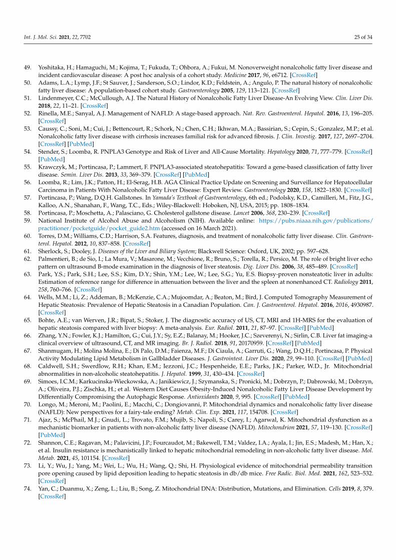
Int. J. Mol. Sci. 2021, 22, 7702 25 of 34
49. Yoshitaka, H.; Hamaguchi, M.; Kojima, T.; Fukuda, T.; Ohbora, A.; Fukui, M. Nonoverweight nonalcoholic fatty liver disease andincident cardiovascular disease: A post hoc analysis of a cohort study. Medicine 2017, 96, e6712. [CrossRef]
50. Adams, L.A.; Lymp, J.F.; St Sauver, J.; Sanderson, S.O.; Lindor, K.D.; Feldstein, A.; Angulo, P. The natural history of nonalcoholicfatty liver disease: A population-based cohort study. Gastroenterology 2005, 129, 113–121. [CrossRef]
51. Lindenmeyer, C.C.; McCullough, A.J. The Natural History of Nonalcoholic Fatty Liver Disease-An Evolving View. Clin. Liver Dis.2018, 22, 11–21. [CrossRef]
52. Rinella, M.E.; Sanyal, A.J. Management of NAFLD: A stage-based approach. Nat. Rev. Gastroenterol. Hepatol. 2016, 13, 196–205.[CrossRef]
53. Caussy, C.; Soni, M.; Cui, J.; Bettencourt, R.; Schork, N.; Chen, C.H.; Ikhwan, M.A.; Bassirian, S.; Cepin, S.; Gonzalez, M.P.; et al.Nonalcoholic fatty liver disease with cirrhosis increases familial risk for advanced fibrosis. J. Clin. Investig. 2017, 127, 2697–2704.[CrossRef] [PubMed]
54. Stender, S.; Loomba, R. PNPLA3 Genotype and Risk of Liver and All-Cause Mortality. Hepatology 2020, 71, 777–779. [CrossRef][PubMed]
55. Krawczyk, M.; Portincasa, P.; Lammert, F. PNPLA3-associated steatohepatitis: Toward a gene-based classification of fatty liverdisease. Semin. Liver Dis. 2013, 33, 369–379. [CrossRef] [PubMed]
56. Loomba, R.; Lim, J.K.; Patton, H.; El-Serag, H.B. AGA Clinical Practice Update on Screening and Surveillance for HepatocellularCarcinoma in Patients With Nonalcoholic Fatty Liver Disease: Expert Review. Gastroenterology 2020, 158, 1822–1830. [CrossRef]
57. Portincasa, P.; Wang, D.Q.H. Gallstones. In Yamada’s Textbook of Gastroenterology, 6th ed.; Podolsky, K.D., Camilleri, M., Fitz, J.G.,Kalloo, A.N., Shanahan, F., Wang, T.C., Eds.; Wiley-Blackwell: Hoboken, NJ, USA, 2015; pp. 1808–1834.
58. Portincasa, P.; Moschetta, A.; Palasciano, G. Cholesterol gallstone disease. Lancet 2006, 368, 230–239. [CrossRef]59. National Institute of Alcohol Abuse and Alcoholism (NIH). Available online: https://pubs.niaaa.nih.gov/publications/
practitioner/pocketguide/pocket_guide2.htm (accessed on 16 March 2021).60. Torres, D.M.; Williams, C.D.; Harrison, S.A. Features, diagnosis, and treatment of nonalcoholic fatty liver disease. Clin. Gastroen-
terol. Hepatol. 2012, 10, 837–858. [CrossRef]61. Sherlock, S.; Dooley, J. Diseases of the Liver and Biliary System; Blackwell Science: Oxford, UK, 2002; pp. 597–628.62. Palmentieri, B.; de Sio, I.; La Mura, V.; Masarone, M.; Vecchione, R.; Bruno, S.; Torella, R.; Persico, M. The role of bright liver echo
pattern on ultrasound B-mode examination in the diagnosis of liver steatosis. Dig. Liver Dis. 2006, 38, 485–489. [CrossRef]63. Park, Y.S.; Park, S.H.; Lee, S.S.; Kim, D.Y.; Shin, Y.M.; Lee, W.; Lee, S.G.; Yu, E.S. Biopsy-proven nonsteatotic liver in adults:
Estimation of reference range for difference in attenuation between the liver and the spleen at nonenhanced CT. Radiology 2011,258, 760–766. [CrossRef]
64. Wells, M.M.; Li, Z.; Addeman, B.; McKenzie, C.A.; Mujoomdar, A.; Beaton, M.; Bird, J. Computed Tomography Measurement ofHepatic Steatosis: Prevalence of Hepatic Steatosis in a Canadian Population. Can. J. Gastroenterol. Hepatol. 2016, 2016, 4930987.[CrossRef]
65. Bohte, A.E.; van Werven, J.R.; Bipat, S.; Stoker, J. The diagnostic accuracy of US, CT, MRI and 1H-MRS for the evaluation ofhepatic steatosis compared with liver biopsy: A meta-analysis. Eur. Radiol. 2011, 21, 87–97. [CrossRef] [PubMed]
66. Zhang, Y.N.; Fowler, K.J.; Hamilton, G.; Cui, J.Y.; Sy, E.Z.; Balanay, M.; Hooker, J.C.; Szeverenyi, N.; Sirlin, C.B. Liver fat imaging-aclinical overview of ultrasound, CT, and MR imaging. Br. J. Radiol. 2018, 91, 20170959. [CrossRef] [PubMed]
67. Shanmugam, H.; Molina Molina, E.; Di Palo, D.M.; Faienza, M.F.; Di Ciaula, A.; Garruti, G.; Wang, D.Q.H.; Portincasa, P. PhysicalActivity Modulating Lipid Metabolism in Gallbladder Diseases. J. Gastrointest. Liver Dis. 2020, 29, 99–110. [CrossRef] [PubMed]
68. Caldwell, S.H.; Swerdlow, R.H.; Khan, E.M.; Iezzoni, J.C.; Hespenheide, E.E.; Parks, J.K.; Parker, W.D., Jr. Mitochondrialabnormalities in non-alcoholic steatohepatitis. J. Hepatol. 1999, 31, 430–434. [CrossRef]
69. Simoes, I.C.M.; Karkucinska-Wieckowska, A.; Janikiewicz, J.; Szymanska, S.; Pronicki, M.; Dobrzyn, P.; Dabrowski, M.; Dobrzyn,A.; Oliveira, P.J.; Zischka, H.; et al. Western Diet Causes Obesity-Induced Nonalcoholic Fatty Liver Disease Development byDifferentially Compromising the Autophagic Response. Antioxidants 2020, 9, 995. [CrossRef] [PubMed]
70. Longo, M.; Meroni, M.; Paolini, E.; Macchi, C.; Dongiovanni, P. Mitochondrial dynamics and nonalcoholic fatty liver disease(NAFLD): New perspectives for a fairy-tale ending? Metab. Clin. Exp. 2021, 117, 154708. [CrossRef]
71. Ajaz, S.; McPhail, M.J.; Gnudi, L.; Trovato, F.M.; Mujib, S.; Napoli, S.; Carey, I.; Agarwal, K. Mitochondrial dysfunction as amechanistic biomarker in patients with non-alcoholic fatty liver disease (NAFLD). Mitochondrion 2021, 57, 119–130. [CrossRef][PubMed]
72. Shannon, C.E.; Ragavan, M.; Palavicini, J.P.; Fourcaudot, M.; Bakewell, T.M.; Valdez, I.A.; Ayala, I.; Jin, E.S.; Madesh, M.; Han, X.;et al. Insulin resistance is mechanistically linked to hepatic mitochondrial remodeling in non-alcoholic fatty liver disease. Mol.Metab. 2021, 45, 101154. [CrossRef]
73. Li, Y.; Wu, J.; Yang, M.; Wei, L.; Wu, H.; Wang, Q.; Shi, H. Physiological evidence of mitochondrial permeability transitionpore opening caused by lipid deposition leading to hepatic steatosis in db/db mice. Free Radic. Biol. Med. 2021, 162, 523–532.[CrossRef]
74. Yan, C.; Duanmu, X.; Zeng, L.; Liu, B.; Song, Z. Mitochondrial DNA: Distribution, Mutations, and Elimination. Cells 2019, 8, 379.[CrossRef]
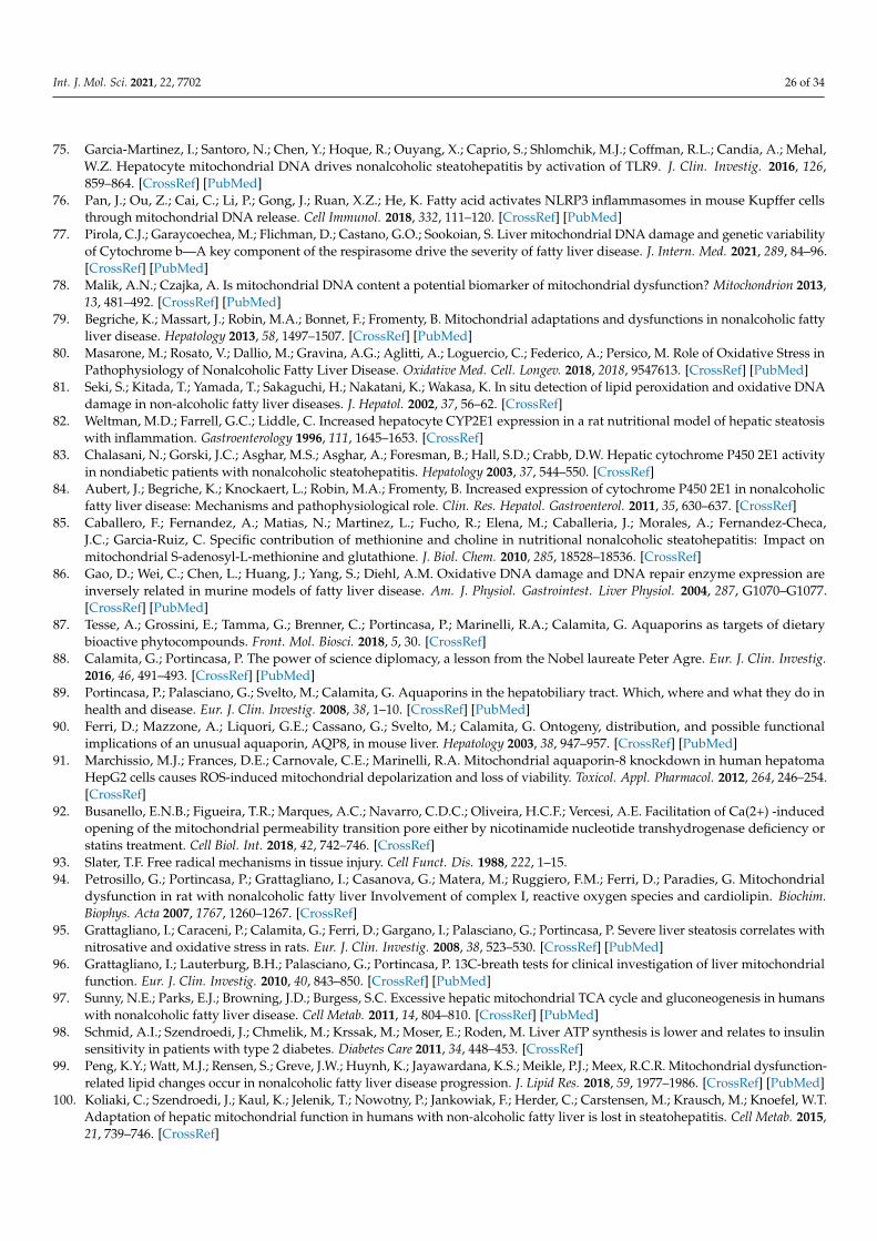
Int. J. Mol. Sci. 2021, 22, 7702 26 of 34
75. Garcia-Martinez, I.; Santoro, N.; Chen, Y.; Hoque, R.; Ouyang, X.; Caprio, S.; Shlomchik, M.J.; Coffman, R.L.; Candia, A.; Mehal,W.Z. Hepatocyte mitochondrial DNA drives nonalcoholic steatohepatitis by activation of TLR9. J. Clin. Investig. 2016, 126,859–864. [CrossRef] [PubMed]
76. Pan, J.; Ou, Z.; Cai, C.; Li, P.; Gong, J.; Ruan, X.Z.; He, K. Fatty acid activates NLRP3 inflammasomes in mouse Kupffer cellsthrough mitochondrial DNA release. Cell Immunol. 2018, 332, 111–120. [CrossRef] [PubMed]
77. Pirola, C.J.; Garaycoechea, M.; Flichman, D.; Castano, G.O.; Sookoian, S. Liver mitochondrial DNA damage and genetic variabilityof Cytochrome b—A key component of the respirasome drive the severity of fatty liver disease. J. Intern. Med. 2021, 289, 84–96.[CrossRef] [PubMed]
78. Malik, A.N.; Czajka, A. Is mitochondrial DNA content a potential biomarker of mitochondrial dysfunction? Mitochondrion 2013,13, 481–492. [CrossRef] [PubMed]
79. Begriche, K.; Massart, J.; Robin, M.A.; Bonnet, F.; Fromenty, B. Mitochondrial adaptations and dysfunctions in nonalcoholic fattyliver disease. Hepatology 2013, 58, 1497–1507. [CrossRef] [PubMed]
80. Masarone, M.; Rosato, V.; Dallio, M.; Gravina, A.G.; Aglitti, A.; Loguercio, C.; Federico, A.; Persico, M. Role of Oxidative Stress inPathophysiology of Nonalcoholic Fatty Liver Disease. Oxidative Med. Cell. Longev. 2018, 2018, 9547613. [CrossRef] [PubMed]
81. Seki, S.; Kitada, T.; Yamada, T.; Sakaguchi, H.; Nakatani, K.; Wakasa, K. In situ detection of lipid peroxidation and oxidative DNAdamage in non-alcoholic fatty liver diseases. J. Hepatol. 2002, 37, 56–62. [CrossRef]
82. Weltman, M.D.; Farrell, G.C.; Liddle, C. Increased hepatocyte CYP2E1 expression in a rat nutritional model of hepatic steatosiswith inflammation. Gastroenterology 1996, 111, 1645–1653. [CrossRef]
83. Chalasani, N.; Gorski, J.C.; Asghar, M.S.; Asghar, A.; Foresman, B.; Hall, S.D.; Crabb, D.W. Hepatic cytochrome P450 2E1 activityin nondiabetic patients with nonalcoholic steatohepatitis. Hepatology 2003, 37, 544–550. [CrossRef]
84. Aubert, J.; Begriche, K.; Knockaert, L.; Robin, M.A.; Fromenty, B. Increased expression of cytochrome P450 2E1 in nonalcoholicfatty liver disease: Mechanisms and pathophysiological role. Clin. Res. Hepatol. Gastroenterol. 2011, 35, 630–637. [CrossRef]
85. Caballero, F.; Fernandez, A.; Matias, N.; Martinez, L.; Fucho, R.; Elena, M.; Caballeria, J.; Morales, A.; Fernandez-Checa,J.C.; Garcia-Ruiz, C. Specific contribution of methionine and choline in nutritional nonalcoholic steatohepatitis: Impact onmitochondrial S-adenosyl-L-methionine and glutathione. J. Biol. Chem. 2010, 285, 18528–18536. [CrossRef]
86. Gao, D.; Wei, C.; Chen, L.; Huang, J.; Yang, S.; Diehl, A.M. Oxidative DNA damage and DNA repair enzyme expression areinversely related in murine models of fatty liver disease. Am. J. Physiol. Gastrointest. Liver Physiol. 2004, 287, G1070–G1077.[CrossRef] [PubMed]
87. Tesse, A.; Grossini, E.; Tamma, G.; Brenner, C.; Portincasa, P.; Marinelli, R.A.; Calamita, G. Aquaporins as targets of dietarybioactive phytocompounds. Front. Mol. Biosci. 2018, 5, 30. [CrossRef]
88. Calamita, G.; Portincasa, P. The power of science diplomacy, a lesson from the Nobel laureate Peter Agre. Eur. J. Clin. Investig.2016, 46, 491–493. [CrossRef] [PubMed]
89. Portincasa, P.; Palasciano, G.; Svelto, M.; Calamita, G. Aquaporins in the hepatobiliary tract. Which, where and what they do inhealth and disease. Eur. J. Clin. Investig. 2008, 38, 1–10. [CrossRef] [PubMed]
90. Ferri, D.; Mazzone, A.; Liquori, G.E.; Cassano, G.; Svelto, M.; Calamita, G. Ontogeny, distribution, and possible functionalimplications of an unusual aquaporin, AQP8, in mouse liver. Hepatology 2003, 38, 947–957. [CrossRef] [PubMed]
91. Marchissio, M.J.; Frances, D.E.; Carnovale, C.E.; Marinelli, R.A. Mitochondrial aquaporin-8 knockdown in human hepatomaHepG2 cells causes ROS-induced mitochondrial depolarization and loss of viability. Toxicol. Appl. Pharmacol. 2012, 264, 246–254.[CrossRef]
92. Busanello, E.N.B.; Figueira, T.R.; Marques, A.C.; Navarro, C.D.C.; Oliveira, H.C.F.; Vercesi, A.E. Facilitation of Ca(2+) -inducedopening of the mitochondrial permeability transition pore either by nicotinamide nucleotide transhydrogenase deficiency orstatins treatment. Cell Biol. Int. 2018, 42, 742–746. [CrossRef]
93. Slater, T.F. Free radical mechanisms in tissue injury. Cell Funct. Dis. 1988, 222, 1–15.94. Petrosillo, G.; Portincasa, P.; Grattagliano, I.; Casanova, G.; Matera, M.; Ruggiero, F.M.; Ferri, D.; Paradies, G. Mitochondrial
dysfunction in rat with nonalcoholic fatty liver Involvement of complex I, reactive oxygen species and cardiolipin. Biochim.Biophys. Acta 2007, 1767, 1260–1267. [CrossRef]
95. Grattagliano, I.; Caraceni, P.; Calamita, G.; Ferri, D.; Gargano, I.; Palasciano, G.; Portincasa, P. Severe liver steatosis correlates withnitrosative and oxidative stress in rats. Eur. J. Clin. Investig. 2008, 38, 523–530. [CrossRef] [PubMed]
96. Grattagliano, I.; Lauterburg, B.H.; Palasciano, G.; Portincasa, P. 13C-breath tests for clinical investigation of liver mitochondrialfunction. Eur. J. Clin. Investig. 2010, 40, 843–850. [CrossRef] [PubMed]
97. Sunny, N.E.; Parks, E.J.; Browning, J.D.; Burgess, S.C. Excessive hepatic mitochondrial TCA cycle and gluconeogenesis in humanswith nonalcoholic fatty liver disease. Cell Metab. 2011, 14, 804–810. [CrossRef] [PubMed]
98. Schmid, A.I.; Szendroedi, J.; Chmelik, M.; Krssak, M.; Moser, E.; Roden, M. Liver ATP synthesis is lower and relates to insulinsensitivity in patients with type 2 diabetes. Diabetes Care 2011, 34, 448–453. [CrossRef]
99. Peng, K.Y.; Watt, M.J.; Rensen, S.; Greve, J.W.; Huynh, K.; Jayawardana, K.S.; Meikle, P.J.; Meex, R.C.R. Mitochondrial dysfunction-related lipid changes occur in nonalcoholic fatty liver disease progression. J. Lipid Res. 2018, 59, 1977–1986. [CrossRef] [PubMed]
100. Koliaki, C.; Szendroedi, J.; Kaul, K.; Jelenik, T.; Nowotny, P.; Jankowiak, F.; Herder, C.; Carstensen, M.; Krausch, M.; Knoefel, W.T.Adaptation of hepatic mitochondrial function in humans with non-alcoholic fatty liver is lost in steatohepatitis. Cell Metab. 2015,21, 739–746. [CrossRef]
Int. J. Mol. Sci. 2021, 22, 7702 27 of 34
101. Fletcher, J.A.; Deja, S.; Satapati, S.; Fu, X.; Burgess, S.C.; Browning, J.D. Impaired ketogenesis and increased acetyl-CoA oxidationpromote hyperglycemia in human fatty liver. JCI Insight 2019, 5. [CrossRef]
102. Grasselli, E.; Baldini, F.; Vecchione, G.; Oliveira, P.J.; Sardao, V.A.; Voci, A.; Portincasa, P.; Vergani, L. Excess fructose and fattyacids trigger a model of nonalcoholic fatty liver disease progression in vitro: Protective effect of the flavonoid silybin. Int. J. Mol.Med. 2019, 44, 705–712. [CrossRef]
103. An, P.; Wei, L.L.; Zhao, S.; Sverdlov, D.Y.; Vaid, K.A.; Miyamoto, M.; Kuramitsu, K.; Lai, M.; Popov, Y.V. Hepatocyte mitochondria-derived danger signals directly activate hepatic stellate cells and drive progression of liver fibrosis. Nat. Commun. 2020, 11.[CrossRef]
104. Grossini, E.; Garhwal, D.P.; Calamita, G.; Romito, R.; Rigamonti, C.; Minisini, R.; Smirne, C.; Surico, D.; Bellan, M.; Pirisi, M.Exposure to Plasma From Non-alcoholic Fatty Liver Disease Patients Affects Hepatocyte Viability, Generates MitochondrialDysfunction, and Modulates Pathways Involved in Fat Accumulation and Inflammation. Front. Med. 2021, 8. [CrossRef]
105. Reddy, J.K. Nonalcoholic steatosis and steatohepatitis. III. Peroxisomal beta-oxidation, PPAR alpha, and steatohepatitis. Am. J.Physiol. Gastrointest. Liver Physiol. 2001, 281, G1333–G1339. [CrossRef]
106. Camporez, J.P.; Wang, Y.; Faarkrog, K.; Chukijrungroat, N.; Petersen, K.F.; Shulman, G.I. Mechanism by which arylamineN-acetyltransferase 1 ablation causes insulin resistance in mice. Proc. Natl. Acad. Sci. USA 2017, 114, E11285–E11292. [CrossRef][PubMed]
107. Chennamsetty, I.; Coronado, M.; Contrepois, K.; Keller, M.P.; Carcamo-Orive, I.; Sandin, J.; Fajardo, G.; Whittle, A.J.; Fathzadeh,M.; Snyder, M.; et al. Nat1 Deficiency Is Associated with Mitochondrial Dysfunction and Exercise Intolerance in Mice. Cell Rep.2016, 17, 527–540. [CrossRef] [PubMed]
108. Gijón, M.A.; Riekhof, W.R.; Zarini, S.; Murphy, R.C.; Voelker, D.R. Lysophospholipid Acyltransferases and Arachidonate Recyclingin Human Neutrophils*. J. Biol. Chem. 2008, 283, 30235–30245. [CrossRef] [PubMed]
109. Mancina, R.M.; Dongiovanni, P.; Petta, S.; Pingitore, P.; Meroni, M.; Rametta, R.; Boren, J.; Montalcini, T.; Pujia, A.; Wiklund,O.; et al. The MBOAT7-TMC4 Variant rs641738 Increases Risk of Nonalcoholic Fatty Liver Disease in Individuals of EuropeanDescent. Gastroenterology 2016, 150, 1219–1230. [CrossRef]
110. Luukkonen, P.K.; Zhou, Y.; Hyötyläinen, T.; Leivonen, M.; Arola, J.; Orho-Melander, M.; Orešic, M.; Yki-Järvinen, H. The MBOAT7variant rs641738 alters hepatic phosphatidylinositols and increases severity of non-alcoholic fatty liver disease in humans. J.Hepatol. 2016, 65, 1263–1265. [CrossRef]
111. Buch, S.; Stickel, F.; Trépo, E.; Way, M.; Herrmann, A.; Nischalke, H.D.; Brosch, M.; Rosendahl, J.; Berg, T.; Ridinger, M.; et al. Agenome-wide association study confirms PNPLA3 and identifies TM6SF2 and MBOAT7 as risk loci for alcohol-related cirrhosis.Nat. Genet. 2015, 47, 1443–1448. [CrossRef] [PubMed]
112. Thabet, K.; Asimakopoulos, A.; Shojaei, M.; Romero-Gomez, M.; Mangia, A.; Irving, W.L.; Berg, T.; Dore, G.J.; Grønbæk, H.;Sheridan, D.; et al. MBOAT7 rs641738 increases risk of liver inflammation and transition to fibrosis in chronic hepatitis C. Nat.Commun. 2016, 7. [CrossRef]
113. Thangapandi, V.R.; Knittelfelder, O.; Brosch, M.; Patsenker, E.; Vvedenskaya, O.; Buch, S.; Hinz, S.; Hendricks, A.; Nati, M.;Herrmann, A.; et al. Loss of hepatic Mboat7 leads to liver fibrosis. Gut 2021, 70, 940–950. [CrossRef]
114. Helsley, R.N.; Venkateshwari, V.; Brown, A.L.; Gromovsky, A.D.; Schugar, R.C.; Ramachandiran, I.; Fung, K.; Kabbany, M.N.;Banerjee, R.; Neumann, C.K. Obesity-linked suppression of membrane-bound Oacyltransferase 7 (MBOAT7) drives non-alcoholicfatty liver disease. Elife 2019, 8, e49882. [CrossRef]
115. Fondevila, M.F.; Fernandez, U.; Gonzalez-Rellan, M.J.; Da Silva Lima, N.; Buque, X.; Gonzalez-Rodriguez, A.; Alonso, C.;Iruarrizaga-Lejarreta, M.; Delgado, T.C.; Varela-Rey, M.; et al. The L-α-Lysophosphatidylinositol/G Protein–Coupled Receptor 55System Induces the Development of Nonalcoholic Steatosis and Steatohepatitis. Hepatology 2021, 73, 606–624. [CrossRef]
116. Petersen, K.F.; Befroy, D.; Dufour, S.; Dziura, J.; Ariyan, C.; Rothman, D.L.; DiPietro, L.; Cline, G.W.; Shulman, G.I. Mitochondrialdysfunction in the elderly: Possible role in insulin resistance. Science 2003, 300, 1140–1142. [CrossRef]
117. Lee, H.-Y.; Choi, C.S.; Birkenfeld, A.L.; Alves, T.C.; Jornayvaz, F.R.; Jurczak, M.J.; Zhang, D.; Woo, D.K.; Shadel, G.S.; Ladiges,W.; et al. Targeted Expression of Catalase to Mitochondria Prevents Age-Associated Reductions in Mitochondrial Function andInsulin Resistance. Cell Metab. 2010, 12, 668–674. [CrossRef]
118. Palmer, A.K.; Kirkland, J.L. Aging and adipose tissue: Potential interventions for diabetes and regenerative medicine. Exp.Gerontol. 2016, 86, 97–105. [CrossRef]
119. Lyu, K.; Zhang, Y.; Zhang, D.; Kahn, M.; ter Horst, K.W.; Rodrigues, M.R.S.; Gaspar, R.C.; Hirabara, S.M.; Luukkonen, P.K.; Lee,S.; et al. A Membrane-Bound Diacylglycerol Species Induces PKCε-Mediated Hepatic Insulin Resistance. Cell Metab. 2020, 32,654–664.e655. [CrossRef]
120. Piccinin, E.; Villani, G.; Moschetta, A. Metabolic aspects in NAFLD, NASH and hepatocellular carcinoma: The role of PGC1coactivators. Nat. Rev. Gastroenterol. Hepatol. 2019, 16, 160–174. [CrossRef] [PubMed]
121. Li, Y.; Chen, C.; Lu, L.; Guo, W.; VanWagner, L.B.; Shikany, J.M.; Zhang, S.; Kahe, K. Cadmium Exposure in Young Adulthood IsAssociated with Risk of Nonalcoholic Fatty Liver Disease in Midlife. Dig. Dis. Sci. 2021. [CrossRef]
122. He, X.; Gao, J.; Hou, H.; Qi, Z.; Chen, H.; Zhang, X.X. Inhibition of Mitochondrial Fatty Acid Oxidation Contributes toDevelopment of Nonalcoholic Fatty Liver Disease Induced by Environmental Cadmium Exposure. Environ. Sci. Technol. 2019, 53,13992–14000. [CrossRef] [PubMed]
Int. J. Mol. Sci. 2021, 22, 7702 28 of 34
123. Frediani, J.K.; Naioti, E.A.; Vos, M.B.; Figueroa, J.; Marsit, C.J.; Welsh, J.A. Arsenic exposure and risk of nonalcoholic fatty liverdisease (NAFLD) among U.S. adolescents and adults: An association modified by race/ethnicity, NHANES 2005–2014. Environ.Health Glob. Access Sci. Source 2018, 17, 6. [CrossRef] [PubMed]
124. Mozaffarian, F.; Dehghani, M.A.; Vanani, A.R.; Mahdavinia, M. Protective Effects of Alpha Lipoic Acid Against Arsenic InducedOxidative Stress in Isolated Rat Liver Mitochondria. Biol. Trace Elem. Res. 2021. [CrossRef]
125. Chen, R.; Xu, Y.; Xu, C.; Shu, Y.; Ma, S.; Lu, C.; Mo, X. Associations between mercury exposure and the risk of nonalcoholic fattyliver disease (NAFLD) in US adolescents. Environ. Sci. Pollut. Res. Int. 2019, 26, 31384–31391. [CrossRef] [PubMed]
126. Pereira, L.C.; de Paula, E.S.; Pazin, M.; Carneiro, M.F.H.; Grotto, D.; Barbosa, F., Jr.; Dorta, D.J. Niacin prevents mitochondrialoxidative stress caused by sub-chronic exposure to methylmercury. Drug Chem. Toxicol. 2020, 43, 64–70. [CrossRef]
127. Qiu, Y.N.; Wang, G.H.; Zhou, F.; Hao, J.J.; Tian, L.; Guan, L.F.; Geng, X.K.; Ding, Y.C.; Wu, H.W.; Zhang, K.Z. PM2.5 induces liverfibrosis via triggering ROS-mediated mitophagy. Ecotoxicol. Environ. Saf. 2019, 167, 178–187. [CrossRef]
128. Breton, C.V.; Song, A.Y.; Xiao, J.; Kim, S.J.; Mehta, H.H.; Wan, J.; Yen, K.; Sioutas, C.; Lurmann, F.; Xue, S.; et al. Effects of airpollution on mitochondrial function, mitochondrial DNA methylation, and mitochondrial peptide expression. Mitochondrion2019, 46, 22–29. [CrossRef] [PubMed]
129. Chen, J.; Wu, L.; Yang, G.; Zhang, C.; Liu, X.; Sun, X.; Chen, X.; Wang, N. The influence of PM2.5 exposure on non-alcoholic fattyliver disease. Life Sci. 2021, 270, 119135. [CrossRef] [PubMed]
130. Khan, S.; Beigh, S.; Chaudhari, B.P.; Sharma, S.; Aliul Hasan Abdi, S.; Ahmad, S.; Ahmad, F.; Parvez, S.; Raisuddin, S.Mitochondrial dysfunction induced by Bisphenol A is a factor of its hepatotoxicity in rats. Environ. Toxicol. 2016, 31, 1922–1934.[CrossRef]
131. Huc, L.; Lemarie, A.; Gueraud, F.; Helies-Toussaint, C. Low concentrations of bisphenol A induce lipid accumulation mediatedby the production of reactive oxygen species in the mitochondria of HepG2 cells. Toxicol. Vitr. Int. J. Publ. Assoc. BIBRA 2012, 26,709–717. [CrossRef]
132. Liu, Q.; Wang, Q.; Xu, C.; Shao, W.; Zhang, C.; Liu, H.; Jiang, Z.; Gu, A. Organochloride pesticides impaired mitochondrialfunction in hepatocytes and aggravated disorders of fatty acid metabolism. Sci. Rep. 2017, 7, 46339. [CrossRef]
133. Wahlang, B.; Appana, S.; Falkner, K.C.; McClain, C.J.; Brock, G.; Cave, M.C. Insecticide and metal exposures are associated with asurrogate biomarker for non-alcoholic fatty liver disease in the National Health and Nutrition Examination Survey 2003-2004.Environ. Sci. Pollut. Res. Int. 2020, 27, 6476–6487. [CrossRef]
134. Miranda, C.A.; Guimaraes, A.; Bizerra, P.F.V.; Mingatto, F.E. Diazinon impairs bioenergetics and induces membrane permeabilitytransition on mitochondria isolated from rat liver. J. Toxicol. Environ. Health A 2020, 83, 616–629. [CrossRef] [PubMed]
135. Rives, C.; Fougerat, A.; Ellero-Simatos, S.; Loiseau, N.; Guillou, H.; Gamet-Payrastre, L.; Wahli, W. Oxidative Stress in NAFLD:Role of Nutrients and Food Contaminants. Biomolecules 2020, 10, 1702. [CrossRef]
136. Vecchione, G.; Grasselli, E.; Voci, A.; Baldini, F.; Grattagliano, I.; Wang, D.Q.; Portincasa, P.; Vergani, L. Silybin counteracts lipidexcess and oxidative stress in cultured steatotic hepatic cells. World J. Gastroenterol. WJG 2016, 22, 6016–6026. [CrossRef] [PubMed]
137. Nadanaciva, S.; Will, Y. New insights in drug-induced mitochondrial toxicity. Curr. Pharm. Des. 2011, 17, 2100–2112. [CrossRef]138. Pereira, C.V.; Nadanaciva, S.; Oliveira, P.J.; Will, Y. The contribution of oxidative stress to drug-induced organ toxicity and its
detection in vitro and in vivo. Expert Opin. Drug Metab. Toxicol. 2012, 8, 219–237. [CrossRef]139. Masuo, Y.; Imai, T.; Shibato, J.; Hirano, M.; Jones, O.A.; Maguire, M.L.; Satoh, K.; Kikuchi, S.; Rakwal, R. Omic analyses unravels
global molecular changes in the brain and liver of a rat model for chronic Sake (Japanese alcoholic beverage) intake. Electrophoresis2009, 30, 1259–1275. [CrossRef] [PubMed]
140. Griffin, J.L.; Nicholls, A.W. Metabolomics as a functional genomic tool for understanding lipid dysfunction in diabetes, obesityand related disorders. Pharmacogenomics 2006, 7, 1095–1107. [CrossRef] [PubMed]
141. Malik, A.N.; Simoes, I.C.M.; Rosa, H.S.; Khan, S.; Karkucinska-Wieckowska, A.; Wieckowski, M.R. A Diet Induced MaladaptiveIncrease in Hepatic Mitochondrial DNA Precedes OXPHOS Defects and May Contribute to Non-Alcoholic Fatty Liver Disease.Cells 2019, 8, 1222. [CrossRef]
142. Molina-Molina, E.; Shanmugam, H.; Di Ciaula, A.; Grattagliano, I.; Di Palo, D.M.; Palmieri, V.O.; Portincasa, P. ((13)C)-Methacetinbreath test provides evidence of subclinical liver dysfunction linked to fat storage but not lifestyle. JHEP Rep. 2021, 3, 100203.[CrossRef]
143. Bonfrate, L.; Grattagliano, I.; Palasciano, G.; Portincasa, P. Dynamic carbon 13 breath tests for the study of liver function andgastric emptying. Gastroenterol. Rep. 2015, 3, 12–21. [CrossRef]
144. Grattagliano, I.; Bonfrate, L.; Lorusso, M.; Castorani, L.; de Bari, O.; Portincasa, P. Exploring liver mitochondrial function by(1)(3)C-stable isotope breath tests: Implications in clinical biochemistry. Methods Mol. Biol. 2015, 1241, 137–152. [CrossRef]
145. Grattagliano, I.; Bonfrate, L.; Oliveira, P.J.; Castorani, L.; Ruggiero, V.; Valenzano, A.T.; Ascensao, A.; Buzoianu, A.; Portincasa, P.Breath tests with novel 13C-substrates for clinical studies of liver mitochondrial function in health and disease. Eur. Rev. MedPharmacol. Sci. 2013, 17 (Suppl. 2), 72–81. [PubMed]
146. Perri, F.; Bellini, M.; Portincasa, P.; Parodi, A.; Bonazzi, P.; Marzio, L.; Galeazzi, F.; Usai, P.; Citrino, A.; Usai-Satta, P. (13)C-octanoicacid breath test (OBT) with a new test meal (EXPIROGer): Toward standardization for testing gastric emptying of solids. Dig.Liver Dis. 2010, 42, 549–553. [CrossRef] [PubMed]
147. Portincasa, P.; Grattagliano, I.; Lauterburg, B.H.; Palmieri, V.O.; Palasciano, G.; Stellaard, F. Liver breath tests non-invasivelypredict higher stages of non-alcoholic steatohepatitis. Clin. Sci. 2006, 111, 135–143. [CrossRef] [PubMed]
Int. J. Mol. Sci. 2021, 22, 7702 29 of 34
148. Festi, D.; Capodicasa, S.; Sandri, L.; Colaiocco-Ferrante, L.; Staniscia, T.; Vitacolonna, E.; Vestito, A.; Simoni, P.; Mazzella, G.;Portincasa, P.; et al. Measurement of hepatic functional mass by means of 13C-methacetin and 13C-phenylalanine breath tests inchronic liver disease: Comparison with Child-Pugh score and serum bile acid levels. World J. Gastroenterol. WJG 2005, 11, 142–148.[CrossRef] [PubMed]
149. Portincasa, P.; Moschetta, A.; Palasciano, G. Nuovi breath test per lo studio dello svuotamento gastrico e del transito intestinale.In Rilevanza in Pazienti Con Stipsi Funzionale; UCP News: Johar Town, Pakistan, 2001; in press.
150. Gasbarrini, A.; Corazza, G.R.; Gasbarrini, G.; Montalto, M.; Di Stefano, M.; Basilisco, G.; Parodi, A.; Usai-Satta, P.; Vernia, P.;Anania, C.; et al. Methodology and indications of H2-breath testing in gastrointestinal diseases: The Rome Consensus Conference.Aliment. Pharmacol. Ther. 2009, 29 (Suppl. 1), 1–49. [CrossRef]
151. Vitellio, P.; Celano, G.; Bonfrate, L.; Gobbetti, M.; Portincasa, P.; De Angelis, M. Effects of Bifidobacterium longum andLactobacillus rhamnosus on Gut Microbiota in Patients with Lactose Intolerance and Persisting Functional GastrointestinalSymptoms: A Randomised, Double-Blind, Cross-Over Study. Nutrients 2019, 11, 886. [CrossRef] [PubMed]
152. Portincasa, P.; Di Ciaula, A.; Vacca, M.; Montelli, R.; Wang, D.Q.; Palasciano, G. Beneficial effects of oral tilactase on patients withhypolactasia. Eur. J. Clin. Investig. 2008, 38, 835–844. [CrossRef] [PubMed]
153. Krawczyk, M.; Wolska, M.; Schwartz, S.; Gruenhage, F.; Terjung, B.; Portincasa, P.; Sauerbruch, T.; Lammert, F. Concordance ofgenetic and breath tests for lactose intolerance in a tertiary referral centre. J. Gastrointest. Liver Dis. JGLD 2008, 17, 135–139.
154. Bonfrate, L.; Krawczyk, M.; Lembo, A.; Grattagliano, I.; Lammert, F.; Portincasa, P. Effects of dietary education, followed by atailored fructose-restricted diet in adults with fructose malabsorption. Eur. J. Gastroenterol. Hepatol. 2015, 27, 785–796. [CrossRef]
155. Merkel, C.; Bolognesi, M.; Bellon, S.; Bianco, S.; Honisch, B.; Lampe, H.; Angeli, P.; Gatta, A. Aminopyrine breath test in theprognostic evaluation of patients with cirrhosis. Gut 1992, 33, 836–842. [CrossRef]
156. Armuzzi, A.; Candelli, M.; Zocco, M.A.; Andreoli, A.; De Lorenzo, A.; Nista, E.C.; Miele, L.; Cremonini, F.; Cazzato, I.A.; Grieco,A.; et al. Review article: Breath testing for human liver function assessment. Aliment. Pharmacol. Ther. 2002, 16, 1977–1996.[CrossRef]
157. Michaletz, P.A.; Cap, L.; Alpert, E.; Lauterburg, B.H. Assessment of mitochondrial function in vivo with a breath test utilizingalpha-ketoisocaproic acid. Hepatology 1989, 10, 829–832. [CrossRef]
158. Palmieri, V.O.; Grattagliano, I.; Minerva, F.; Pollice, S.; Palasciano, G.; Portincasa, P. Liver function as assessed by breath tests inpatients with hepatocellular carcinoma. J. Surg. Res. 2009, 157, 199–207. [CrossRef] [PubMed]
159. Dawson, B.; Trapp, R.G. Basic & Clinical Biostatistics; McGraw-Hill: New York, NY, USA, 2001; Volume 3.160. Hintze, J. NCSS 2020 Statistical Software; Number Cruncher Statistical System (NCSS): Kaysville, UT, USA, 2020.161. Gorowska-Kowolik, K.; Chobot, A.; Kwiecien, J. (13)C Methacetin Breath Test for Assessment of Microsomal Liver Function:
Methodology and Clinical Application. Gastroenterol. Res. Pract. 2017, 2017, 7397840. [CrossRef] [PubMed]162. Afolabi, P.; Wright, M.; Wootton, S.A.; Jackson, A.A. Clinical utility of 13C-liver-function breath tests for assessment of hepatic
function. Dig. Dis. Sci. 2013, 58, 33–41. [CrossRef] [PubMed]163. Stockmann, M.; Lock, J.F.; Riecke, B.; Heyne, K.; Martus, P.; Fricke, M.; Lehmann, S.; Niehues, S.M.; Schwabe, M.; Lemke, A.J.;
et al. Prediction of postoperative outcome after hepatectomy with a new bedside test for maximal liver function capacity. Ann.Surg. 2009, 250, 119–125. [CrossRef] [PubMed]
164. Buechter, M.; Kersting, S.; Gerken, G.; Kahraman, A. Enzymatic liver function measured by LiMAx—A reliable diagnostic andprognostic tool in chronic liver disease. Sci. Rep. 2019, 9, 13577. [CrossRef] [PubMed]
165. Holzhutter, H.G.; Wuensch, T.; Gajowski, R.; Berndt, N.; Bulik, S.; Meierhofer, D.; Stockmann, M. A novel variant of the (13)C-methacetin liver function breath test that eliminates the confounding effect of individual differences in systemic CO2 kinetics.Arch. Toxicol. 2020, 94, 401–415. [CrossRef] [PubMed]
166. Schmitz, S.M.; Kroh, A.; Ulmer, T.F.; Andruszkow, J.; Luedde, T.; Brozat, J.F.; Neumann, U.P.; Alizai, P.H. Evaluation of NAFLDand fibrosis in obese patients—A comparison of histological and clinical scoring systems. BMC Gastroenterol. 2020, 20, 254.[CrossRef]
167. Afolabi, P.R.; Scorletti, E.; Smith, D.E.; Almehmadi, A.A.; Calder, P.C.; Byrne, C.D. The characterisation of hepatic mitochondrialfunction in patients with non-alcoholic fatty liver disease (NAFLD) using the 13C-ketoisocaproate breath test. J. Breath Res. 2018,12, 046002. [CrossRef] [PubMed]
168. Krahenbuhl, L.; Ledermann, M.; Lang, C.; Krahenbuhl, S. Relationship between hepatic mitochondrial functions in vivo andin vitro in rats with carbon tetrachloride-induced liver cirrhosis. J. Hepatol. 2000, 33, 216–223. [CrossRef]
169. Miele, L.; Marrone, G.; Cefalo, C.; D’Achille, S.; Rapaccini, G.L.; Gasbarrini, A.; Grieco, A. Potential use of liver function breathtests in the clinical practice. Eur. Rev. Med Pharmacol. Sci. 2013, 17 (Suppl. 2), 82–89.
170. Giannini, E.; Fasoli, A.; Chiarbonello, B.; Malfatti, F.; Romagnoli, P.; Botta, F.; Testa, E.; Polegato, S.; Fumagalli, A.; Testa, R.13C-aminopyrine breath test to evaluate severity of disease in patients with chronic hepatitis C virus infection. Aliment. Pharmacol.Ther. 2002, 16, 717–725. [CrossRef]
171. Lauterburg, B.H.; Grattagliano, I.; Gmur, R.; Stalder, M.; Hildebrand, P. Noninvasive assessment of the effect of xenobiotics onmitochondrial function in human beings: Studies with acetylsalicylic acid and ethanol with the use of the carbon 13-labeledketoisocaproate breath test. J. Lab. Clin. Med. 1995, 125, 378–383.
172. Molina-Molina, E.; Shanmugam, H.; Di Palo, D.; Grattagliano, I.; Portincasa, P. Exploring Liver Mitochondrial Function by(13)C-Stable Isotope Breath Tests: Implications in Clinical Biochemistry. Methods Mol. Biol. 2021, 2310, 179–199. [CrossRef]
Int. J. Mol. Sci. 2021, 22, 7702 30 of 34
173. Lauterburg, B.H.; Liang, D.; Schwarzenbach, F.A.; Breen, K.J. Mitochondrial dysfunction in alcoholic patients as assessed bybreath analysis. Hepatology 1993, 17, 418–422. [CrossRef] [PubMed]
174. Armuzzi, A.; Marcoccia, S.; Zocco, M.A.; De Lorenzo, A.; Grieco, A.; Tondi, P.; Pola, P.; Gasbarrini, G.; Gasbarrini, A. Non-Invasiveassessment of human hepatic mitochondrial function through the 13C-methionine breath test. Scand. J. Gastroenterol. 2000, 35,650–653. [CrossRef] [PubMed]
175. Russmann, S.; Junker, E.; Lauterburg, B.H. Remethylation and transsulfuration of methionine in cirrhosis: Studies with L-[H3-methyl-1-C]methionine. Hepatology 2002, 36, 1190–1196. [CrossRef] [PubMed]
176. Banasch, M.; Ellrichmann, M.; Tannapfel, A.; Schmidt, W.E.; Goetze, O. The non-invasive (13)C-methionine breath test detectshepatic mitochondrial dysfunction as a marker of disease activity in non-alcoholic steatohepatitis. Eur. J. Med. Res. 2011, 16,258–264. [CrossRef] [PubMed]
177. Banasch, M.; Emminghaus, R.; Ellrichmann, M.; Schmidt, W.E.; Goetze, O. Longitudinal effects of hepatitis C virus treatment onhepatic mitochondrial dysfunction assessed by C-methionine breath test. Aliment. Pharmacol. Ther. 2008, 28, 443–449. [CrossRef][PubMed]
178. Spahr, L.; Negro, F.; Leandro, G.; Marinescu, O.; Goodman, K.J.; Rubbia-Brandt, L.; Jordan, M.; Hadengue, A. Impaired hepaticmitochondrial oxidation using the 13C-methionine breath test in patients with macrovesicular steatosis and patients with cirrhosis.Med. Sci. Monit. Int. Med. J. Exp. Clin. Res. 2003, 9, CR6–CR11.
179. Stuwe, S.H.; Goetze, O.; Arning, L.; Banasch, M.; Schmidt, W.E.; Schols, L.; Saft, C. Hepatic mitochondrial dysfunction inFriedreich ataxia. BMC Neurol. 2011, 11, 145. [CrossRef] [PubMed]
180. Miele, L.; Grieco, A.; Armuzzi, A.; Candelli, M.; Forgione, A.; Gasbarrini, A.; Gasbarrini, G. Hepatic mitochondrial beta-oxidationin patients with nonalcoholic steatohepatitis assessed by 13 C-octanoate breath test. Am. J. Gastroenterol. 2003, 98, 2335. [CrossRef][PubMed]
181. Schneider, A.R.J.; Kraut, C.; Lindenthal, B.; Braden, B.; Caspary, W.F.; Stein, J. Total body metabolism of 13C-octanoic acid ispreserved in patients with non-alcoholic steatohepatitis, but differs between women and men. Eur. J. Gastroenterol. Hepatol. 2005,17, 1181–1184. [CrossRef]
182. Grattagliano, I.; Vendemiale, G.; Lauterburg, B.H. Reperfusion injury of the liver: Role of mitochondria and protection byglutathione ester. J. Surg. Res. 1999, 86, 2–8. [CrossRef]
183. Berthold, H.K.; Giesen, T.A.; Gouni-Berthold, I. The stable isotope ketoisocaproic acid breath test as a measure of hepaticdecarboxylation capacity: A quantitative analysis in normal subjects after oral and intravenous administration. Liver Int. 2009, 29,1356–1364. [CrossRef] [PubMed]
184. Witschi, A.; Mossi, S.; Meyer, B.; Junker, E.; Lauterburg, B.H. Mitochondrial function reflected by the decarboxylation of[13C]ketoisocaproate is impaired in alcoholics. Alcohol. Clin. Exp. Res. 1994, 18, 951–955. [CrossRef]
185. Bendtsen, P.; Hannestad, U.; Pahlsson, P. Evaluation of the carbon 13-labeled Ketoisocaproate breath test to assess mitochondrialdysfunction in patients with high alcohol consumption. Alcohol. Clin. Exp. Res. 1998, 22, 1792–1795. [CrossRef]
186. Pugh, R.N.; Murray-Lyon, I.M.; Dawson, J.L.; Pietroni, M.C.; Williams, R. Transection of the oesophagus for bleeding oesophagealvarices. Br. J. Surg. 1973, 60, 646–649. [CrossRef]
187. Pessayre, D.; Mansouri, A.; Haouzi, D.; Fromenty, B. Hepatotoxicity due to mitochondrial dysfunction. Cell Biol. Toxicol. 1999, 15,367–373. [CrossRef]
188. Kass, G.E.; Price, S.C. Role of mitochondria in drug-induced cholestatic injury. Clin. Liver Dis. 2008, 12, 27–51, vii. [CrossRef]189. Milazzo, L.; Piazza, M.; Sangaletti, O.; Gatti, N.; Cappelletti, A.; Adorni, F.; Antinori, S.; Galli, M.; Moroni, M.; Riva, A.
[13C]Methionine breath test: A novel method to detect antiretroviral drug-related mitochondrial toxicity. J. Antimicrob. Chemother.2005, 55, 84–89. [CrossRef]
190. Li, Y.; Boehning, D.F.; Qian, T.; Popov, V.L.; Weinman, S.A. Hepatitis C virus core protein increases mitochondrial ROS productionby stimulation of Ca2+ uniporter activity. FASEB J. 2007, 21, 2474–2485. [CrossRef]
191. Jaeschke, H.; McGill, M.R.; Ramachandran, A. Oxidant stress, mitochondria, and cell death mechanisms in drug-induced liverinjury: Lessons learned from acetaminophen hepatotoxicity. Drug Metab. Rev. 2012, 44, 88–106. [CrossRef]
192. Nakagawa, Y.; Suzuki, T.; Kamimura, H.; Nagai, F. Role of mitochondrial membrane permeability transition in N-nitrosofenfluramine-induced cell injury in rat hepatocytes. Eur. J. Pharmacol. 2006, 529, 33–39. [CrossRef]
193. Trost, L.C.; Lemasters, J.J. Role of the mitochondrial permeability transition in salicylate toxicity to cultured rat hepatocytes:Implications for the pathogenesis of Reye’s syndrome. Toxicol. Appl. Pharmacol. 1997, 147, 431–441. [CrossRef]
194. Mingatto, F.E.; dos Santos, A.C.; Rodrigues, T.; Pigoso, A.A.; Uyemura, S.A.; Curti, C. Effects of nimesulide and its reducedmetabolite on mitochondria. Br. J. Pharmacol. 2000, 131, 1154–1160. [CrossRef] [PubMed]
195. Danicke, S.; Diers, S. Effects of ergot alkaloids on liver function of piglets as evaluated by the (13)C-methacetin and (13)C-alpha-ketoisocaproic acid breath test. Toxins 2013, 5, 139–161. [CrossRef]
196. Storch, K.J.; Wagner, D.A.; Burke, J.F.; Young, V.R. Quantitative study in vivo of methionine cycle in humans using [methyl-2H3]-and [1-13C]methionine. Am. J. Physiol. 1988, 255, E322–E331. [CrossRef] [PubMed]
197. Candelli, M.; Miele, L.; Armuzzi, A.; Nista, E.C.; Pignataro, G.; Fini, L.; Cazzato, I.A.; Zocco, M.A.; Bartolozzi, F.; Gasbarrini, G.;et al. 13C-methionine breath tests for mitochondrial liver function assessment. Eur. Rev. Med Pharmacol. Sci. 2008, 12, 245–249.[PubMed]
Int. J. Mol. Sci. 2021, 22, 7702 31 of 34
198. Duro, D.; Duggan, C.; Valim, C.; Bechard, L.; Fitzgibbons, S.; Jaksic, T.; Yu, Y.M. Novel intravenous (13)C-methionine breathtest as a measure of liver function in children with short bowel syndrome. J. Pediatric Surg. 2009, 44, 236–240, Discussion 240.[CrossRef]
199. Durr, A.; Cossee, M.; Agid, Y.; Campuzano, V.; Mignard, C.; Penet, C.; Mandel, J.L.; Brice, A.; Koenig, M. Clinical and geneticabnormalities in patients with Friedreich’s ataxia. N. Engl. J. Med. 1996, 335, 1169–1175. [CrossRef]
200. Walton, M.E.; Ebert, D.; Haller, R.G. Octanoate oxidation measured by 13C-NMR spectroscopy in rat skeletal muscle, heart, andliver. J. Appl. Physiol. 2003, 95, 1908–1916. [CrossRef]
201. Shalev, T.; Aeed, H.; Sorin, V.; Shahmurov, M.; Didkovsky, E.; Ilan, Y.; Avni, Y.; Shirin, H. Evaluation of the 13C-octanoate breathtest as a surrogate marker of liver damage in animal models. Dig. Dis. Sci. 2010, 55, 1589–1598. [CrossRef]
202. Van de Casteele, M.; Luypaerts, A.; Geypens, B.; Fevery, J.; Ghoos, Y.; Nevens, F. Oxidative breakdown of octanoic acid ismaintained in patients with cirrhosis despite advanced disease. Neurogastroenterol. Motil. 2003, 15, 113–120. [CrossRef]
203. Ghoos, Y.F.; Maes, B.D.; Geypens, B.J.; Mys, G.; Hiele, M.I.; Rutgeerts, P.J.; Vantrappen, G. Measurement of gastric emptying rateof solids by means of a carbon-labeled octanoic acid breath test. Gastroenterology 1993, 104, 1640–1647. [CrossRef]
204. Promrat, K.; Kleiner, D.E.; Niemeier, H.M.; Jackvony, E.; Kearns, M.; Wands, J.R.; Fava, J.L.; Wing, R.R. Randomized controlledtrial testing the effects of weight loss on nonalcoholic steatohepatitis. Hepatology 2010, 51, 121–129. [CrossRef] [PubMed]
205. Keating, S.E.; Hackett, D.A.; George, J.; Johnson, N.A. Exercise and non-alcoholic fatty liver disease: A systematic review andmeta-analysis. J. Hepatol. 2012, 57, 157–166. [CrossRef]
206. Keating, S.E.; Hackett, D.A.; Parker, H.M.; O’Connor, H.T.; Gerofi, J.A.; Sainsbury, A.; Baker, M.K.; Chuter, V.H.; Caterson, I.D.;George, J.; et al. Effect of aerobic exercise training dose on liver fat and visceral adiposity. J. Hepatol. 2015, 63, 174–182. [CrossRef][PubMed]
207. Vilar-Gomez, E.; Martinez-Perez, Y.; Calzadilla-Bertot, L.; Torres-Gonzalez, A.; Gra-Oramas, B.; Gonzalez-Fabian, L.; Friedman,S.L.; Diago, M.; Romero-Gomez, M. Weight Loss Through Lifestyle Modification Significantly Reduces Features of NonalcoholicSteatohepatitis. Gastroenterology 2015, 149, 367–378.e365, quiz e314–e365. [CrossRef] [PubMed]
208. Petersen, K.F.; Dufour, S.; Befroy, D.; Lehrke, M.; Hendler, R.E.; Shulman, G.I. Reversal of nonalcoholic hepatic steatosis, hepaticinsulin resistance, and hyperglycemia by moderate weight reduction in patients with type 2 diabetes. Diabetes 2005, 54, 603–608.[CrossRef] [PubMed]
209. Musso, G.; Cassader, M.; Rosina, F.; Gambino, R. Impact of current treatments on liver disease, glucose metabolism andcardiovascular risk in non-alcoholic fatty liver disease (NAFLD): A systematic review and meta-analysis of randomised trials.Diabetologia 2012, 55, 885–904. [CrossRef] [PubMed]
210. Cerqueira, F.M.; Cunha, F.M.d.; Silva, C.C.; Chausse, B.; Romano, R.L.; Garcia, C.; Colepicolo, P.; Medeiros, M.H.G.d.; Kowal-towski, A.J. Redox state, insulin sensitivity and aging. Resumos 2011, 37, 1333–1345.
211. Kowaltowski, A.J. Caloric restriction and redox state: Does this diet increase or decrease oxidant production? Redox Rep. 2011, 16,237–241. [CrossRef] [PubMed]
212. Walsh, M.E.; Shi, Y.; Van Remmen, H. The effects of dietary restriction on oxidative stress in rodents. Free Radic. Biol. Med. 2014,66, 88–99. [CrossRef]
213. Bower, G.; Toma, T.; Harling, L.; Jiao, L.R.; Efthimiou, E.; Darzi, A.; Athanasiou, T.; Ashrafian, H. Bariatric Surgery and Non-Alcoholic Fatty Liver Disease: A Systematic Review of Liver Biochemistry and Histology. Obes. Surg. 2015, 25, 2280–2289.[CrossRef]
214. Mathurin, P.; Hollebecque, A.; Arnalsteen, L.; Buob, D.; Leteurtre, E.; Caiazzo, R.; Pigeyre, M.; Verkindt, H.; Dharancy, S.;Louvet, A. Prospective study of the long-term effects of bariatric surgery on liver injury in patients without advanced disease.Gastroenterology 2009, 137, 532–540. [CrossRef]
215. Ekstedt, M.; Franzen, L.E.; Holmqvist, M.; Bendtsen, P.; Mathiesen, U.L.; Bodemar, G.; Kechagias, S. Alcohol consumption isassociated with progression of hepatic fibrosis in non-alcoholic fatty liver disease. Scand. J. Gastroenterol. 2009, 44, 366–374.[CrossRef] [PubMed]
216. Pessayre, D.; Berson, A.; Fromenty, B.; Mansouri, A. Mitochondria in steatohepatitis. Semin. Liver Dis. 2001, 21, 57–69. [CrossRef]217. Thoma, C.; Day, C.P.; Trenell, M.I. Lifestyle interventions for the treatment of non-alcoholic fatty liver disease in adults: A
systematic review. J. Hepatol. 2012, 56, 255–266. [CrossRef]218. Li, Z.; Li, Y.; Zhang, H.X.; Guo, J.R.; Lam, C.W.K.; Wang, C.Y.; Zhang, W. Mitochondria-Mediated Pathogenesis and Therapeutics
for Non-Alcoholic Fatty Liver Disease. Mol. Nutr. Food Res. 2019, 63, e1900043. [CrossRef] [PubMed]219. Stevanovic, J.; Beleza, J.; Coxito, P.; Ascensao, A.; Magalhaes, J. Physical exercise and liver “fitness”: Role of mitochondrial
function and epigenetics-related mechanisms in non-alcoholic fatty liver disease. Mol. Metab. 2020, 32, 1–14. [CrossRef] [PubMed]220. Venditti, P.; Di Meo, S. Antioxidants, tissue damage, and endurance in trained and untrained young male rats. Arch. Biochem.
Biophys. 1996, 331, 63–68. [CrossRef] [PubMed]221. Ascensao, A.; Martins, M.J.; Santos-Alves, E.; Goncalves, I.O.; Portincasa, P.; Oliveira, P.J.; Magalhaes, J. Modulation of hepatic
redox status and mitochondrial metabolism by exercise: Therapeutic strategy for liver diseases. Mitochondrion 2013, 13, 862–870.[CrossRef] [PubMed]
222. Samuel, V.T.; Shulman, G.I. Mechanisms for insulin resistance: Common threads and missing links. Cell 2012, 148, 852–871.[CrossRef] [PubMed]
Int. J. Mol. Sci. 2021, 22, 7702 32 of 34
223. Neuschwander-Tetri, B.A.; Loomba, R.; Sanyal, A.J.; Lavine, J.E.; Van Natta, M.L.; Abdelmalek, M.F.; Chalasani, N.; Dasarathy, S.;Diehl, A.M.; Hameed, B.; et al. Farnesoid X nuclear receptor ligand obeticholic acid for non-cirrhotic, non-alcoholic steatohepatitis(FLINT): A multicentre, randomised, placebo-controlled trial. Lancet 2015, 385, 956–965. [CrossRef]
224. Mudaliar, S.; Henry, R.R.; Sanyal, A.J.; Morrow, L.; Marschall, H.U.; Kipnes, M.; Adorini, L.; Sciacca, C.I.; Clopton, P.; Castelloe, E.;et al. Efficacy and safety of the farnesoid X receptor agonist obeticholic acid in patients with type 2 diabetes and nonalcoholicfatty liver disease. Gastroenterology 2013, 145, 574–582.e1. [CrossRef] [PubMed]
225. Ratziu, V.; Sanyal, A.J.; Loomba, R.; Rinella, M.; Harrison, S.; Anstee, Q.M.; Goodman, Z.; Bedossa, P.; MacConell, L.; Shringarpure,R.; et al. REGENERATE: Design of a pivotal, randomised, phase 3 study evaluating the safety and efficacy of obeticholic acid inpatients with fibrosis due to nonalcoholic steatohepatitis. Contemp. Clin. Trials 2019, 84, 105803. [CrossRef]
226. Chen, Y.S.; Liu, H.M.; Lee, T.Y. Ursodeoxycholic Acid Regulates Hepatic Energy Homeostasis and White Adipose TissueMacrophages Polarization in Leptin-Deficiency Obese Mice. Cells 2019, 8, 253. [CrossRef]
227. Xie, C.; Jiang, C.; Shi, J.; Gao, X.; Sun, D.; Sun, L.; Wang, T.; Takahashi, S.; Anitha, M.; Krausz, K.W.; et al. An Intestinal FarnesoidX Receptor-Ceramide Signaling Axis Modulates Hepatic Gluconeogenesis in Mice. Diabetes 2017, 66, 613–626. [CrossRef]
228. Ferramosca, A.; Di Giacomo, M.; Zara, V. Antioxidant dietary approach in treatment of fatty liver: New insights and updates.World J. Gastroenterol. WJG 2017, 23, 4146–4157. [CrossRef] [PubMed]
229. Ding, S.; Jiang, J.; Zhang, G.; Bu, Y.; Zhang, G.; Zhao, X. Resveratrol and caloric restriction prevent hepatic steatosis by regulatingSIRT1-autophagy pathway and alleviating endoplasmic reticulum stress in high-fat diet-fed rats. PLoS ONE 2017, 12, e0183541.[CrossRef] [PubMed]
230. Tian, Y.; Ma, J.; Wang, W.; Zhang, L.; Xu, J.; Wang, K.; Li, D. Resveratrol supplement inhibited the NF-κB inflammation pathwaythrough activating AMPKα-SIRT1 pathway in mice with fatty liver. Mol. Cell. Biochem. 2016, 422, 75–84. [CrossRef]
231. Shang, J.; Chen, L.L.; Xiao, F.X.; Sun, H.; Ding, H.C.; Xiao, H. Resveratrol improves non-alcoholic fatty liver disease by activatingAMP-activated protein kinase. Acta Pharmacol. Sin. 2008, 29, 698–706. [CrossRef] [PubMed]
232. Asin-Cayuela, J.; Manas, A.R.; James, A.M.; Smith, R.A.; Murphy, M.P. Fine-tuning the hydrophobicity of a mitochondria-targetedantioxidant. FEBS Lett. 2004, 571, 9–16. [CrossRef]
233. Rokitskaya, T.I.; Klishin, S.S.; Severina, I.I.; Skulachev, V.P.; Antonenko, Y.N. Kinetic analysis of permeation of mitochondria-targeted antioxidants across bilayer lipid membranes. J. Membr. Biol. 2008, 224, 9–19. [CrossRef]
234. Smith, R.A.; Porteous, C.M.; Gane, A.M.; Murphy, M.P. Delivery of bioactive molecules to mitochondria in vivo. Proc. Natl. Acad.Sci. USA 2003, 100, 5407–5412. [CrossRef]
235. Grattagliano, I.; Diogo, C.V.; Mastrodonato, M.; de Bari, O.; Persichella, M.; Wang, D.Q.; Liquori, A.; Ferri, D.; Carratu, M.R.;Oliveira, P.J.; et al. A silybin-phospholipids complex counteracts rat fatty liver degeneration and mitochondrial oxidative changes.World J. Gastroenterol. WJG 2013, 19, 3007–3017. [CrossRef]
236. Vecchione, G.; Grasselli, E.; Cioffi, F.; Baldini, F.; Oliveira, P.J.; Sardao, V.A.; Cortese, K.; Lanni, A.; Voci, A.; Portincasa, P.; et al. TheNutraceutic Silybin Counteracts Excess Lipid Accumulation and Ongoing Oxidative Stress in an In Vitro Model of Non-AlcoholicFatty Liver Disease Progression. Front. Nutr. 2017, 4, 42. [CrossRef]
237. Wu, N.; Zu, Y.; Fu, Y.; Kong, Y.; Zhao, J.; Li, X.; Li, J.; Wink, M.; Efferth, T. Antioxidant activities and xanthine oxidase inhibitoryeffects of extracts and main polyphenolic compounds obtained from Geranium sibiricum L. J. Agric. Food Chem. 2010, 58, 4737–4743.[CrossRef]
238. Ling, W.H.; Shen, T.R.; Tang, X.L.; Jiang, X.W. Anthocyanins Improved Mitochondrial Dysfunction in Mice of Non-alcoholic FattyLiver Disease Induced by High Fat Diet. Faseb J. 2016, 30, 915.
239. Tang, X.; Shen, T.; Jiang, X.; Xia, M.; Sun, X.; Guo, H.; Ling, W. Purified anthocyanins from bilberry and black currant attenuatehepatic mitochondrial dysfunction and steatohepatitis in mice with methionine and choline deficiency. J. Agric. Food Chem. 2015,63, 552–561. [CrossRef]
240. Zeng, X.; Yang, J.; Hu, O.; Huang, J.; Ran, L.; Chen, M.; Zhang, Y.; Zhou, X.; Zhu, J.; Zhang, Q. Dihydromyricetin amelioratesnonalcoholic fatty liver disease by improving mitochondrial respiratory capacity and redox homeostasis through modulation ofSIRT3 signaling. Antioxid. Redox Signal. 2019, 30, 163–183. [CrossRef]
241. Teodoro, J.S.; Duarte, F.V.; Gomes, A.P.; Varela, A.T.; Peixoto, F.M.; Rolo, A.P.; Palmeira, C.M. Berberine reverts hepaticmitochondrial dysfunction in high-fat fed rats: A possible role for SirT3 activation. Mitochondrion 2013, 13, 637–646. [CrossRef]
242. Echeverria, F.; Valenzuela, R.; Bustamante, A.; Alvarez, D.; Ortiz, M.; Espinosa, A.; Illesca, P.; Gonzalez-Manan, D.; Videla, L.A.High-fat diet induces mouse liver steatosis with a concomitant decline in energy metabolism: Attenuation by eicosapentaenoicacid (EPA) or hydroxytyrosol (HT) supplementation and the additive effects upon EPA and HT co-administration. Food Funct.2019, 10, 6170–6183. [CrossRef] [PubMed]
243. Schwimmer, J.B.; Lavine, J.E.; Wilson, L.A.; Neuschwander-Tetri, B.A.; Xanthakos, S.A.; Kohli, R.; Barlow, S.E.; Vos, M.B.; Karpen,S.J.; Molleston, J.P. In children with nonalcoholic fatty liver disease, cysteamine bitartrate delayed release improves liver enzymesbut does not reduce disease activity scores. Gastroenterology 2016, 151, 1141–1154.e1149. [CrossRef] [PubMed]
244. Dohil, R.; Schmeltzer, S.; Cabrera, B.L.; Wang, T.; Durelle, J.; Duke, K.B.; Schwimmer, J.B.; Lavine, J.E. Enteric-coated cysteaminefor the treatment of paediatric non-alcoholic fatty liver disease. Aliment. Pharmacol. Ther. 2011, 33, 1036–1044. [CrossRef][PubMed]
245. Ye, J.H.; Chao, J.; Chang, M.L.; Peng, W.H.; Cheng, H.Y.; Liao, J.W.; Pao, L.H. Pentoxifylline ameliorates non-alcoholic fatty liverdisease in hyperglycaemic and dyslipidaemic mice by upregulating fatty acid beta-oxidation. Sci. Rep. 2016, 6, 33102. [CrossRef]
Int. J. Mol. Sci. 2021, 22, 7702 33 of 34
246. Zein, C.O.; Lopez, R.; Yerian, L.; Anderson, K.A.; McCullough, A.J.; Rinella, M.E. 932 Pentoxifylline Improves Non-InvasiveSerum Markers of Fibrosis: Combined Results From 2 Randomized, Placebo-Controlled Trials. Gastroenterology 2012, 142, S936.[CrossRef]
247. Zein, C.O.; Yerian, L.M.; Gogate, P.; Lopez, R.; Kirwan, J.P.; Feldstein, A.E.; McCullough, A.J. Pentoxifylline improves nonalcoholicsteatohepatitis: A randomized placebo-controlled trial. Hepatology 2011, 54, 1610–1619. [CrossRef]
248. Rendon, D.A. Letter to the Editor: The bioenergetics of hepatic mitochondria isolated from avocado oil-treated rats: Typicalexperimental errors in the study of the bioenergetics of isolated mitochondria. J. Bioenerg. Biomembr. 2015, 47, 451–453. [CrossRef]
249. Ortiz-Avila, O.; Gallegos-Corona, M.A.; Sanchez-Briones, L.A.; Calderon-Cortes, E.; Montoya-Perez, R.; Rodriguez-Orozco, A.R.;Campos-Garcia, J.; Saavedra-Molina, A.; Mejia-Zepeda, R.; Cortes-Rojo, C. Protective effects of dietary avocado oil on impairedelectron transport chain function and exacerbated oxidative stress in liver mitochondria from diabetic rats. J. Bioenerg. Biomembr.2015, 47, 337–353. [CrossRef] [PubMed]
250. Garcia-Berumen, C.I.; Olmos-Orizaba, B.E.; Marquez-Ramirez, C.A.; Orozco, A.R.R.; Gonzalez-Cortez, A.; Saavedra-Molina,A.; Montoya-Perez, R.; Cortes-Rojo, C. Avocado Oil Ameliorates Non-Alcoholic Fatty Liver Disease by Down-RegulatingInflammatory Cytokines and Improving Mitochondrial Dynamics. FASEB J. 2019, 33, 660–666. [CrossRef]
251. Sanyal, A.; Charles, E.D.; Neuschwander-Tetri, B.A.; Loomba, R.; Harrison, S.A.; Abdelmalek, M.F.; Lawitz, E.J.; Halegoua-DeMarzio, D.; Kundu, S.; Noviello, S.; et al. Pegbelfermin (BMS-986036), a PEGylated fibroblast growth factor 21 analogue, inpatients with non-alcoholic steatohepatitis: A randomised, double-blind, placebo-controlled, phase 2a trial. Lancet 2019, 392,2705–2717. [CrossRef]
252. Staels, B.; Rubenstrunk, A.; Noel, B.; Rigou, G.; Delataille, P.; Millatt, L.J.; Baron, M.; Lucas, A.; Tailleux, A.; Hum, D.W.; et al.Hepatoprotective effects of the dual peroxisome proliferator-activated receptor alpha/delta agonist, GFT505, in rodent models ofnonalcoholic fatty liver disease/nonalcoholic steatohepatitis. Hepatology 2013, 58, 1941–1952. [CrossRef]
253. Ratziu, V.; Harrison, S.A.; Francque, S.; Bedossa, P.; Lehert, P.; Serfaty, L.; Romero-Gomez, M.; Boursier, J.; Abdelmalek, M.;Caldwell, S.; et al. Elafibranor, an Agonist of the Peroxisome Proliferator-Activated Receptor-alpha and -delta, Induces Resolutionof Nonalcoholic Steatohepatitis Without Fibrosis Worsening. Gastroenterology 2016, 150, 1147–1159. [CrossRef]
254. Tong, W.; Ju, L.; Qiu, M.; Xie, Q.; Chen, Y.; Shen, W.; Sun, W.; Wang, W.; Tian, J. Liraglutide ameliorates non-alcoholic fatty liverdisease by enhancing mitochondrial architecture and promoting autophagy through the SIRT1/SIRT3-FOXO3a pathway. Hepatol.Res. Off. J. Jpn. Soc. Hepatol. 2016, 46, 933–943. [CrossRef] [PubMed]
255. He, L.; Sabet, A.; Djedjos, S.; Miller, R.; Sun, X.; Hussain, M.A.; Radovick, S.; Wondisford, F.E. Metformin and insulin suppresshepatic gluconeogenesis through phosphorylation of CREB binding protein. Cell 2009, 137, 635–646. [CrossRef]
256. Sun, L.; Yuan, Q.; Xu, T.; Yao, L.; Feng, J.; Ma, J.; Wang, L.; Lu, C.; Wang, D. Pioglitazone Improves Mitochondrial Function in theRemnant Kidney and Protects against Renal Fibrosis in 5/6 Nephrectomized Rats. Front. Pharmacol. 2017, 8, 545. [CrossRef]
257. Harrison, S.A.; Alkhouri, N.; Davison, B.A.; Sanyal, A.; Edwards, C.; Colca, J.R.; Lee, B.H.; Loomba, R.; Cusi, K.; Kolterman, O.;et al. Insulin sensitizer MSDC-0602K in non-alcoholic steatohepatitis: A randomized, double-blind, placebo-controlled phase IIbstudy. J. Hepatol. 2020, 72, 613–626. [CrossRef]
258. Iruarrizaga-Lejarreta, M.; Varela-Rey, M.; Fernandez-Ramos, D.; Martinez-Arranz, I.; Delgado, T.C.; Simon, J.; Juan, V.G.;delaCruz-Villar, L.; Azkargorta, M.; Lavin, J.L.; et al. Role of Aramchol in steatohepatitis and fibrosis in mice. Hepatol. Commun.2017, 1, 911–927. [CrossRef]
259. Safadi, R.; Konikoff, F.M.; Mahamid, M.; Zelber-Sagi, S.; Halpern, M.; Gilat, T.; Oren, R.; Group, F. The fatty acid-bile acidconjugate Aramchol reduces liver fat content in patients with nonalcoholic fatty liver disease. Clin. Gastroenterol. Hepatol. 2014,12, 2085–2091.e1. [CrossRef]
260. Dai, J.; Liang, K.; Zhao, S.; Jia, W.; Liu, Y.; Wu, H.; Lv, J.; Cao, C.; Chen, T.; Zhuang, S.; et al. Chemoproteomics reveals baicalinactivates hepatic CPT1 to ameliorate diet-induced obesity and hepatic steatosis. Proc. Natl. Acad. Sci. USA 2018, 115, E5896–E5905.[CrossRef]
261. Fazzari, M.; Chartoumpekis, D.; Li, L.; Guimaraes, D.A.; Shiva, S.; Freeman, B.A.; Khoo, N. Nitro-oleic Acid Protects Mice fromDiet-Induced Hepatic Steatosis and Insulin Resistance without the Adverse Side Effects of Thiazolidinediones. Free Radic. Biol.Med. 2017, 112, 152. [CrossRef]
262. Cho, J.; Zhang, Y.; Park, S.-Y.; Joseph, A.-M.; Han, C.; Park, H.-J.; Kalavalapalli, S.; Chun, S.-K.; Morgan, D.; Kim, J.-S.Mitochondrial ATP transporter depletion protects mice against liver steatosis and insulin resistance. Nat. Commun. 2017, 8, 1–12.[CrossRef]
263. Amanat, S.; Eftekhari, M.H.; Fararouei, M.; Bagheri Lankarani, K.; Massoumi, S.J. Genistein supplementation improves insulinresistance and inflammatory state in non-alcoholic fatty liver patients: A randomized, controlled trial. Clin. Nutr. 2018, 37,1210–1215. [CrossRef] [PubMed]
264. Lawitz, E.J.; Neff, G.; Ruane, P.J.; Younes, Z.; Zhang, J.; Jia, C.; Chuang, J.; Huss, R.; Chung, C.; Subramanian, M. Fenofibratemitigates increases in serum triglycerides due to the ACC inhibitor firsocostat in patients with advanced fibrosis due to NASH: Aphase 2 randomized trial. Hepatology 2019, 70, 1489–1490.
265. Fu, A.; Shi, X.; Zhang, H.; Fu, B. Mitotherapy for Fatty Liver by Intravenous Administration of Exogenous Mitochondria in MaleMice. Front. Pharmacol. 2017, 8, 241. [CrossRef] [PubMed]
266. Ajith, T.A. Role of mitochondria and mitochondria-targeted agents in non-alcoholic fatty liver disease. Clin. Exp. Pharmacol.Physiol. 2018, 45, 413–421. [CrossRef] [PubMed]

Int. J. Mol. Sci. 2021, 22, 7702 34 of 34
267. Pessayre, D.; Fromenty, B. NASH: A mitochondrial disease. J. Hepatol. 2005, 42, 928–940. [CrossRef]268. Winchell, H.S.; Wiley, K. Considerations in analysis of breath 14CO2 data. J. Nucl. Med. Off. Publ. Soc. Nucl. Med. 1970, 11,
708–710.
Related Documents


