Systematic Approach to Reading a Non-Contrast Head CT Scan

Systematic Approach to Reading a Non-Contrast Head CT Scan.
Jan 04, 2016
Welcome message from author
This document is posted to help you gain knowledge. Please leave a comment to let me know what you think about it! Share it to your friends and learn new things together.
Transcript
Systematic Approach to Reading a Non-Contrast Head CT Scan
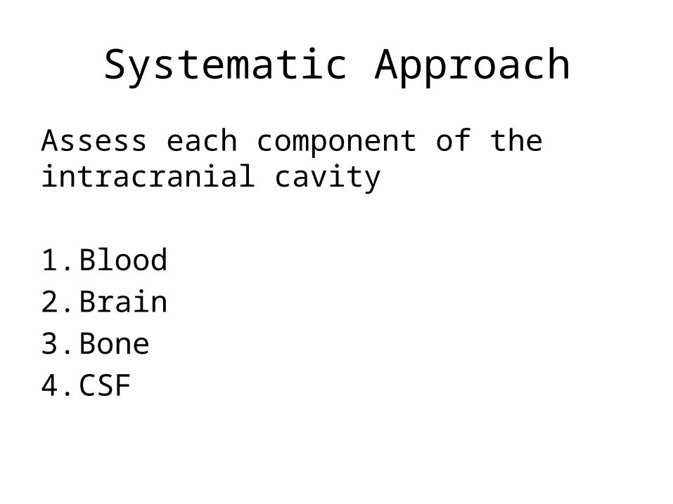
Systematic Approach
Assess each component of the intracranial cavity
1. Blood2. Brain3. Bone4. CSF

Blood
1. Epidural Hematoma (EDH)Arterial Bleed: High-Pressure “Lens” ShapeMiddle Meningeal Laceration (Skull Fracture)
2. Subdural Hematoma (SDH)Venous Bleed: Low-Pressure “Crescent” ShapeAcute / Chronic (Is Patient Anticoagulated? Alcoholic?)
3. Subarachnoid Hemorrhage (SAH)Traumatic / Aneurysmal
4. Intraventricular Hemorrhage (IVH)5. Intraparenchymal Hemorrhage (IPH)
Hypertensive Basal Ganglia or Lobar Cerebral Contusion “Coup-Contrecoup”
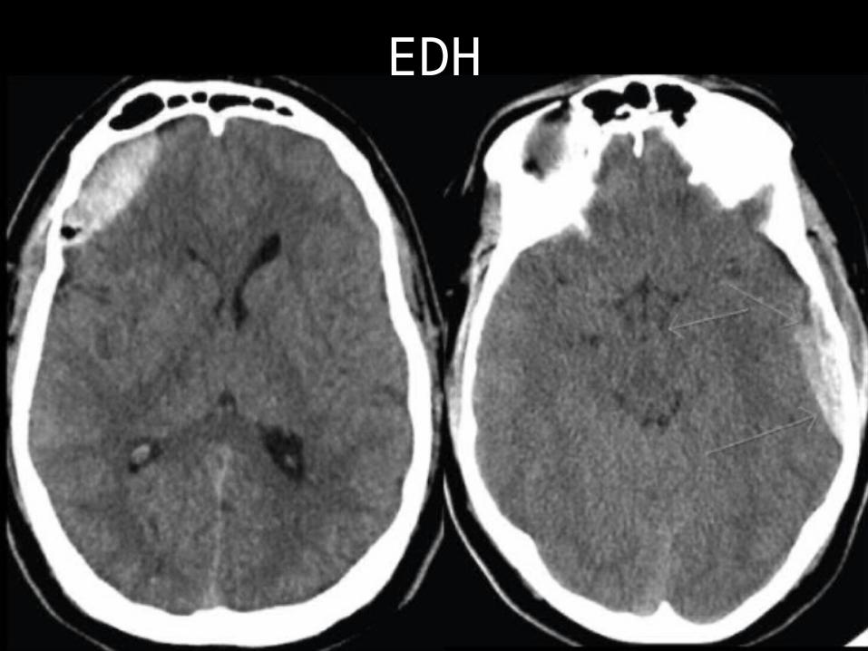
EDH
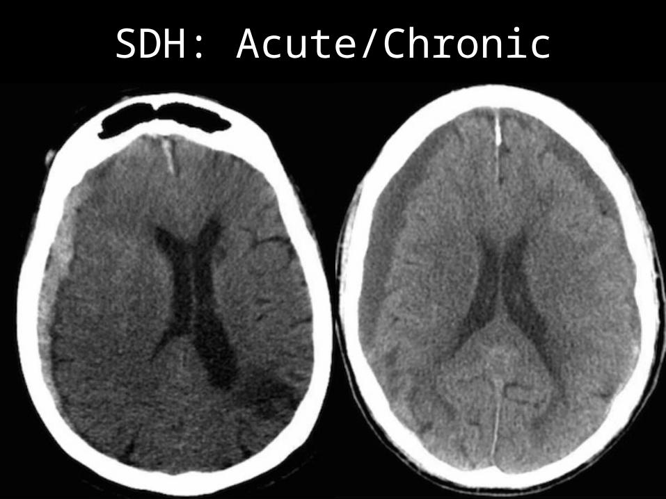
SDH: Acute/Chronic
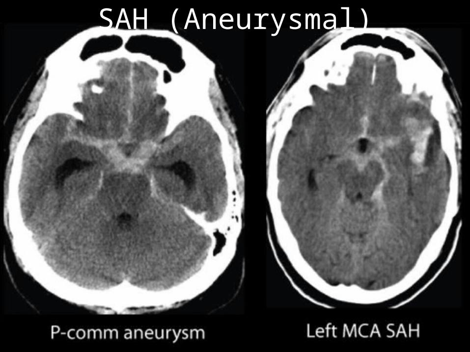
SAH (Aneurysmal)

IVH

IPH

IPH
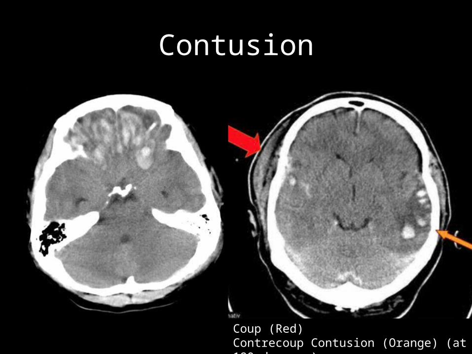
Contusion
Coup (Red) Contrecoup Contusion (Orange) (at 180 degrees)
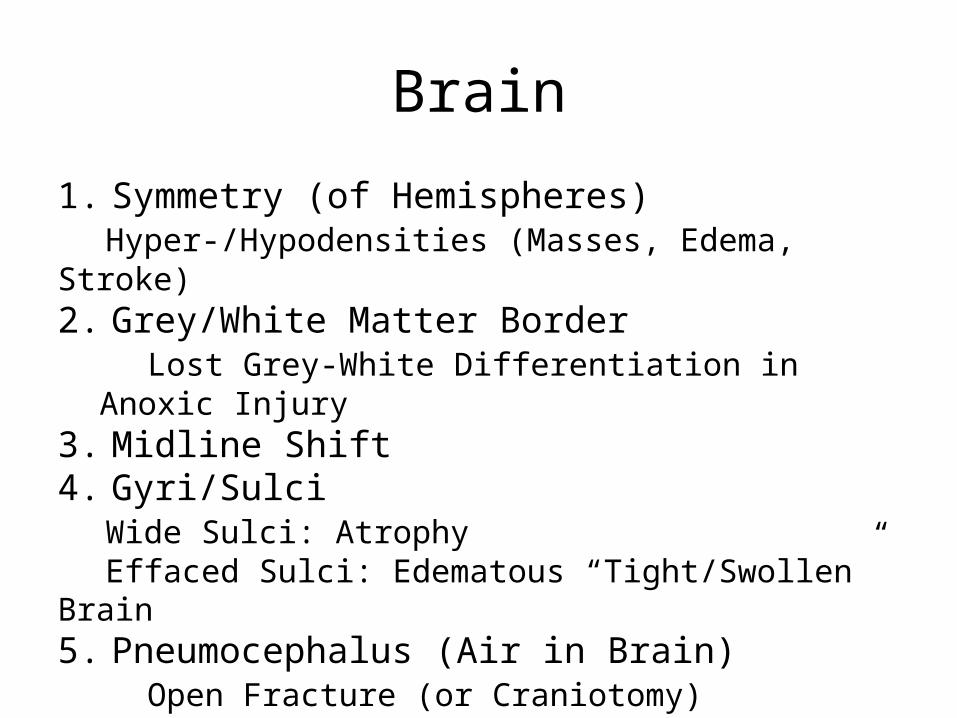
Brain
1. Symmetry (of Hemispheres)Hyper-/Hypodensities (Masses, Edema, Stroke)
2. Grey/White Matter BorderLost Grey-White Differentiation in Anoxic Injury
3. Midline Shift4. Gyri/Sulci
Wide Sulci: Atrophy Effaced Sulci: Edematous “Tight/Swollen” Brain
5. Pneumocephalus (Air in Brain)Open Fracture (or Craniotomy)Fracture through Sinus

Mass
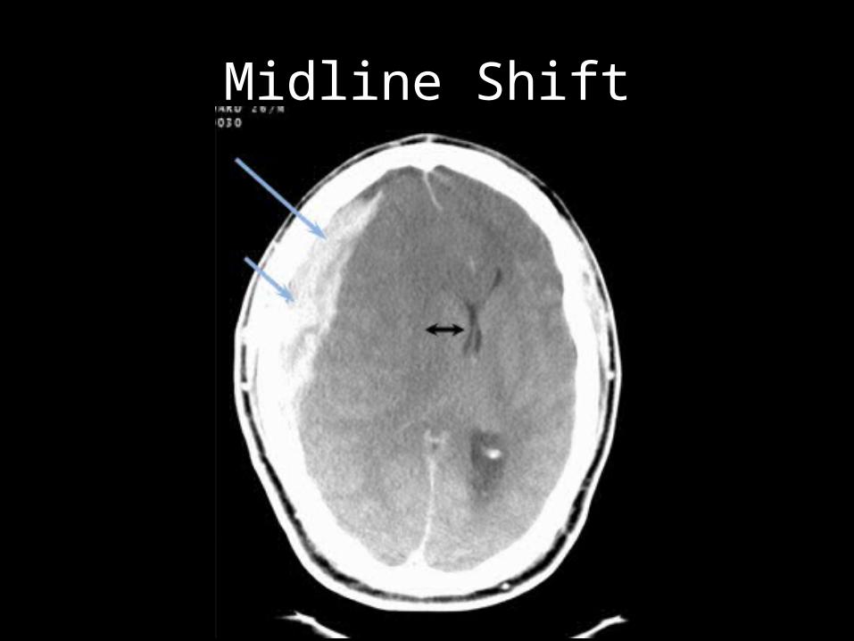
Midline Shift

Sulci/Gyri: Atrophy/Edema

Bone
1. FracturesEspecially Temporal Bones
2. Sinuses & Air CellsLook for Air-Fluid Levels
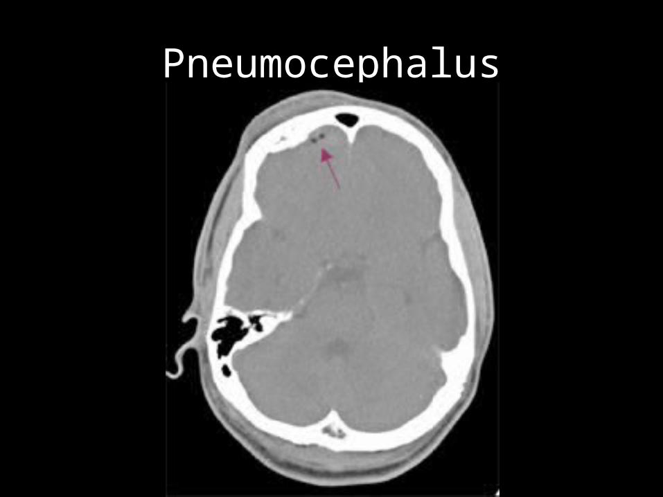
Pneumocephalus
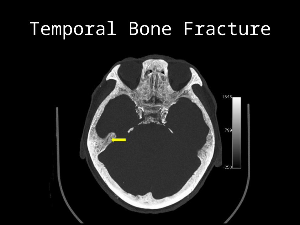
Temporal Bone Fracture

CSF: Ventricles & Cisterns
1. VentriclesBlood in Ventricles?Effacement/Asymmetry: Compression from Mass/HematomaHydrocephalus
Atrophy (“Ex Vacuo”) Communicating / Obstructive HydrocephalusIf Obstructive, Where?
Look at Lateral, 3rd, and 4th Ventricles
2. CisternsLook for Effacement (Edema/Early Herniation)Look for Blood

Cisterns/Effacement
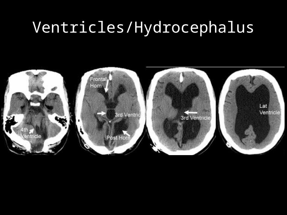
Ventricles/Hydrocephalus

Related Documents