Review Synthetic peptide arrays for investigating protein interaction domains Rudolf Volkmer ⇑ , Victor Tapia, Christiane Landgraf Institut für Medizinische Immunologie Berlin, Molecular Libraries and Recognition Group, Charité-Universitätsmedizin Berlin, Hessische Str. 3-4, 10115 Berlin, Germany article info Article history: Received 22 March 2012 Revised 16 April 2012 Accepted 17 April 2012 Available online 25 April 2012 Edited by Marius Sudol, Gianni Cesareni, Giulio Superti-Furga and Wilhelm Just Dedicated to the memory of Barbara Ann Townsend Winsor. She passed away on September 29, 2011 after returning to her beloved family from a conference. Keywords: SPOT synthesis Protein interaction domains SH3 domains WW domains PDZ domains Peptide arrays abstract Synthetic peptide array technology was first developed in the early 1990s by Ronald Frank. Since then the technique has become a powerful tool for high throughput approaches in biology and bio- chemistry. Here, we focus on peptide arrays applied to investigate the binding specificity of protein interaction domains such as WW, SH3, and PDZ domains. We describe array-based methods used to reveal domain networks in yeast, and briefly review rules as well as ideas about the synthesis and application of peptide arrays. We also provide initial results of a study designed to investigate the nature and evolution of SH3 domain interaction networks in eukaryotes. Ó 2012 Federation of European Biochemical Societies. Published by Elsevier B.V. All rights reserved. 1. Protein interaction networks Living systems are organized by complex, dynamic networks of molecular interactions where proteins are the central network components. Since they can bind not only to other proteins, but also to phospholipids, nucleic acids and small molecules, they link diverse physiological functions of the cell. It is tempting to suggest that a molecular recognition code rules such dynamic networks [1]. In analogy to human language, one could formulate a hierar- chical organization, starting from linear sequences of polypeptide chains, then simple three dimensional fold elements and complex structural motifs, up to protein complexes [2–4]. In fact, structural modules and motifs may have isolated functional ‘‘meaning’’ like words in human language [5,6]. To pursue this analogy, we can think of cellular wiring as a mas- terpiece of evolutionary tinkering, with structural elements used many times in different protein contexts, and trial and error creat- ing some rules of interconnectivity to achieve a favorable feature or message [7]. Therefore, it is not surprising that the idea of independent protein ‘‘linguistics’’ arose in the protein–protein interaction community [8]. Many believe that a complete under- standing of the protein–protein interaction network will enable researchers to break the protein recognition code or predict cellu- lar responses, or even positively interfere with the molecular basis of diseases. The yeast two-hybrid technique [9] was the first method ap- plied in a high-throughput manner to reveal the protein interac- tion network of a model organism [10,11]. Shortly afterwards, the classical pull-down in combination with mass spectrometry was the next high-throughput strategy to reveal the interactome in the same species [12,13]. The low intra- and inter-technique overlaps between the resulting networks (10–20%) indicate that these experimental approaches suffer from false positives and false negatives. In addition, graphical representation of such interaction networks is hard to understand. Recently Gianni Cesareni’s inter- esting analogy described such a network as a complete road map of a large city, but without any information on traffic flow or which routes represent large traffic arteries and which represent narrow one-way alleys [14]. A further aspect limits interpretation of experimentally obtained networks. The real networks have a so-called scale-free topology, meaning more proteins than expected interact with many partner proteins at a time [15]. It is hard to say how many partners these ‘‘hub’’ proteins can contact at the same time; 0014-5793/$36.00 Ó 2012 Federation of European Biochemical Societies. Published by Elsevier B.V. All rights reserved. http://dx.doi.org/10.1016/j.febslet.2012.04.028 ⇑ Corresponding author. Fax: +49 (0)30 450 524942. E-mail address: [email protected] (R. Volkmer). URL: http://immunologie.charite.de (R. Volkmer). FEBS Letters 586 (2012) 2780–2786 journal homepage: www.FEBSLetters.org

Welcome message from author
This document is posted to help you gain knowledge. Please leave a comment to let me know what you think about it! Share it to your friends and learn new things together.
Transcript
FEBS Letters 586 (2012) 2780–2786
journal homepage: www.FEBSLetters .org
Review
Synthetic peptide arrays for investigating protein interaction domains
Rudolf Volkmer ⇑, Victor Tapia, Christiane LandgrafInstitut für Medizinische Immunologie Berlin, Molecular Libraries and Recognition Group, Charité-Universitätsmedizin Berlin, Hessische Str. 3-4, 10115 Berlin, Germany
a r t i c l e i n f o a b s t r a c t
Article history:Received 22 March 2012Revised 16 April 2012Accepted 17 April 2012Available online 25 April 2012
Edited by Marius Sudol, Gianni Cesareni,Giulio Superti-Furga and Wilhelm Just
Dedicated to the memory of Barbara AnnTownsend Winsor. She passed away onSeptember 29, 2011 after returning to herbeloved family from a conference.
Keywords:SPOT synthesisProtein interaction domainsSH3 domainsWW domainsPDZ domainsPeptide arrays
0014-5793/$36.00 � 2012 Federation of European Biohttp://dx.doi.org/10.1016/j.febslet.2012.04.028
⇑ Corresponding author. Fax: +49 (0)30 450 524942E-mail address: [email protected] (R. Volkmer).URL: http://immunologie.charite.de (R. Volkmer).
Synthetic peptide array technology was first developed in the early 1990s by Ronald Frank. Sincethen the technique has become a powerful tool for high throughput approaches in biology and bio-chemistry. Here, we focus on peptide arrays applied to investigate the binding specificity of proteininteraction domains such as WW, SH3, and PDZ domains. We describe array-based methods used toreveal domain networks in yeast, and briefly review rules as well as ideas about the synthesis andapplication of peptide arrays. We also provide initial results of a study designed to investigate thenature and evolution of SH3 domain interaction networks in eukaryotes.� 2012 Federation of European Biochemical Societies. Published by Elsevier B.V. All rights reserved.
1. Protein interaction networks
Living systems are organized by complex, dynamic networks ofmolecular interactions where proteins are the central networkcomponents. Since they can bind not only to other proteins, butalso to phospholipids, nucleic acids and small molecules, they linkdiverse physiological functions of the cell. It is tempting to suggestthat a molecular recognition code rules such dynamic networks[1]. In analogy to human language, one could formulate a hierar-chical organization, starting from linear sequences of polypeptidechains, then simple three dimensional fold elements and complexstructural motifs, up to protein complexes [2–4]. In fact, structuralmodules and motifs may have isolated functional ‘‘meaning’’ likewords in human language [5,6].
To pursue this analogy, we can think of cellular wiring as a mas-terpiece of evolutionary tinkering, with structural elements usedmany times in different protein contexts, and trial and error creat-ing some rules of interconnectivity to achieve a favorable featureor message [7]. Therefore, it is not surprising that the idea ofindependent protein ‘‘linguistics’’ arose in the protein–protein
chemical Societies. Published by E
.
interaction community [8]. Many believe that a complete under-standing of the protein–protein interaction network will enableresearchers to break the protein recognition code or predict cellu-lar responses, or even positively interfere with the molecular basisof diseases.
The yeast two-hybrid technique [9] was the first method ap-plied in a high-throughput manner to reveal the protein interac-tion network of a model organism [10,11]. Shortly afterwards,the classical pull-down in combination with mass spectrometrywas the next high-throughput strategy to reveal the interactomein the same species [12,13]. The low intra- and inter-techniqueoverlaps between the resulting networks (10–20%) indicate thatthese experimental approaches suffer from false positives and falsenegatives. In addition, graphical representation of such interactionnetworks is hard to understand. Recently Gianni Cesareni’s inter-esting analogy described such a network as a complete road mapof a large city, but without any information on traffic flow or whichroutes represent large traffic arteries and which represent narrowone-way alleys [14].
A further aspect limits interpretation of experimentallyobtained networks. The real networks have a so-called scale-freetopology, meaning more proteins than expected interact withmany partner proteins at a time [15]. It is hard to say how manypartners these ‘‘hub’’ proteins can contact at the same time;
lsevier B.V. All rights reserved.
R. Volkmer et al. / FEBS Letters 586 (2012) 2780–2786 2781
consequently it is nearly impossible to predict the composition ofmultiprotein complexes. Fortunately, the modular architecture ofproteins can help solve the problem of assignment. A protein iscomposed of single domains (modules) separated on discrete se-quence patterns, which in turn comprise folding motifs. In general,isolated protein modules have the same globular folding as in thewhole protein, and therefore a reductionist approach can be ap-plied in practice [16,17].
Protein alignments indicate that even unrelated proteins fre-quently share sections of sequence similarity [18–20] and these re-gions often function as independently folded modules or domainsof autonomous functionality. It would be more informative if theprotein interactome could be split at each protein (node) into cova-lently linked domains, whereby each domain can interact with sev-eral proteins, but only one at a time [14]. In other words, if anexisting protein interaction network is supported by a domain net-work, this might provide enough information about the topology ofprotein–protein interactions.
2. Protein interaction domains
Structural analysis of functional protein complexes suggests atleast two classes of protein–protein interactions [21–23]. In thefirst class, which reflects the majority of protein–protein interac-tions, the complementary surfaces of both interacting partnersare extensive. This means that the residues involved in each inter-acting surface only come together upon protein folding (discontin-uous binding sites). The second class comprises asymmetricinteractions, where a modular protein domain may dock with ashort linear sequence motive on the partner protein. Such a mod-ular protein domain is called a protein interaction domain (PID).
Mapping discontinuous binding sites is a challenge; however,the concept of hot spots [24,25] shows the feasibility of interferingwith interactions mediated by extensive surfaces. In contrast, PIDbinding determinants may be mapped to short linear motifsmatching the sequence of the ligand peptide. The importance ofsmall protein recognition domains in forming protein complexesthat involve binding to short linear peptides was demonstratedin the late 1980s and early 1990s (reviewed in the excellent book[20]).
Such domains preferentially bind to peptides possessing spe-cific sequence or structure characteristics. For example, Sudoland Bork demonstrated that WW domains bind proline-rich pep-tides sharing a PPxY motif, and folded into a proline type II helix[26]. However, two factors should be noted: firstly, PID interac-tions are dynamic with affinities mostly in the middle to highmicromolar range, and secondly, one has to carefully define the do-main borderlines. The first factor often hampers complete charac-terization of a domain-interacting network when using pull-downbased experiments. The second factor is important since domainborderlines are generally defined by bioinformatic methods suchas multiple protein alignments without any information aboutstructural data. Therefore, preparing a domain according to the gi-ven borderlines might yield an unstructured shape with no func-tion. This is known for the TCERG1 WW-3 domain whereextending the bioinformatically defined borderline from a 43-mer up to a 53-mer sequence proved essential for generating aproperly folded domain (PDB ID code 2dk7) [27].
3. Phage display
Over the past decade two predominant experimental ap-proaches have been used to investigate PID recognition specificity,namely phage display and SPOT synthesis; both have highthroughput potential and can reveal domain interaction networks
with information on the stoichiometry of domain-peptide interac-tions. Phage display is a powerful biological library comprising109–1010 peptides of random sequence displayed on bacteriophagecapsids. After the pioneering work of Sparks et al. [28] and Rickleset al. [29] many groups have applied this approach to determinethe recognition specificity of several domains such as SH3, WW,PDZ, and GYF domains [20,30,31]. Due to the impressive recordof successful studies mapping PID recognition specificities, phagedisplay has become the first choice for determining the recognitionspecificity of new domains in the absence of any a prioriinformation.
Among the first to expand the study of peptide recognitionmodules to a genome-wide scale were Tong et al. [32]. They com-bined phage display and yeast two-hybrid to elucidate the peptideligand consensus of 20 SH3 domains of Saccharomyces cerevisiaeand draw up an SH3 domain network. Besides the impressiveexperimental results, some general conclusions could be drawnfrom this study. An overlap of about 25% interactions in commonsuggests that both methods lead to over-prediction, whereby asignificant part of the interactome remains undiscovered. Interest-ingly, we notice that exploring the same protein–protein interac-tion space with orthogonal approaches removes false positives. Incontrast to Tong et al., the interactome scanning study of Landgrafet al. [33] combined phage display with a semi-quantitative anal-ysis achieved using SPOT synthesis technology. This study allowedthe first comparative interpretation of the yeast SH3 domain inter-actome: in the devised diagram lines of different thickness corre-late with binding strength.
4. SPOT synthesis and synthetic peptide arrays
Array technologies, especially protein arrays, arrived late in thefield of protein–protein interactions due to critical factors such asnative folding stability or functionality [34,35]. Peptides, in con-trast, are easier to handle and retain partial features of proteinfunction. The fact that PIDs recognize short linear peptides per-fectly corresponds to the scope of synthetic peptide arrays. Thus,peptide arrays are predestined to support PID recognition studiessuch as revealing binding specificity, screening for cellular interac-tion partners, or developing selective PID inhibitors.
Two techniques for chemically synthesizing peptide arrayswere published almost simultaneously: Frank presented the SPOTsynthesis technique [36], while Fodor and co-workers [37] de-scribed the concept of light-directed, spatially addressable chemi-cal synthesis. The latter was only used by the group that originallydeveloped the method. In contrast, the majority of peptide arraysreported to date have been produced using the SPOT synthesis con-cept. This is due to the fact that SPOT synthesis is a very simple butextremely robust method for highly parallel synthesis of peptideson planar surfaces. The method itself has been reviewed severaltimes, e.g. [38,39] and over a period of 20 years the method has be-come a widespread and essential tool in biology and biochemistry,with a literature base of more than 400 original, peer-reviewedpapers.
Nowadays, we are in the comfortable situation that a diversecollection of SPOT technology methods are available, permittingthe application of peptide arrays to a broad spectrum of targets[39]. SPOT technology simplified the chemical synthesis of pep-tide arrays to the addressable deposition of reagents on a planarcellulose membrane (filter paper). Moreover, chemical synthesisallows one to incorporate phosphorylated [40], methylated oracetylated amino acids [41,42], use non-natural building blocks[43,44], prepare branched and cyclic structures [45] andlabel with chromophores or short biological tags such as biotin[46,47].
2782 R. Volkmer et al. / FEBS Letters 586 (2012) 2780–2786
SPOT synthesis can be performed fully automatically with aMultiPep synthesizer (Intavis AG, Köln, Germany) in an analyticalor preparative mode. The latter enables parallel synthesis of upto 1000 peptides at amounts of approximately 50–100 nmol ofcleaved material, achieved by producing spots with a diameter of8 mm dispensed in droplets of up to 1 ll. The preparative SPOTsynthesis mode has been used mostly for cell-based screening as-says [48,49] but also for quality control of spot synthesized pep-tides. The analytical mode enables the synthesis of about 6000cellulose membrane-bound peptides by producing spots with adiameter of �1 mm. Down-sizing the spot diameter below 1 mmfailed due to the capillary effect of cellulose membranes. In bothcases we use membranes with a dimension of 18.3 � 28.5 cm fora special platform with the fully automated MultiPep synthesizerthat allows four coupling steps per day. Fig. 1 summarizes the prin-
Fig. 1. The principals of SPOT synthesis. The analytical SPOT synthesis mode enablesespecially for on-support binding studies (left). On the other hand, high numbers of solubsynthesis mode suitable for several kinds of solution- and cell-based assays as well ascarbodiimide, NMI = N-methylimidazole, Ac2O = acetic anhydride, DIEA = N,N-diisopropy
cipal steps of both the analytical and preparative SPOT synthesismode.
5. Qualitative explorative investigations and quantitativeanalytical studies
In general, synthetic peptide arrays (prepared by the analyticalsynthesis mode) can be used for either qualitative exploratoryinvestigations or quantitative analytical studies; the initial choicesof array content and assay design often depend on which kind ofanalysis is required. Qualitative exploratory investigations may in-volve either simple or comparative functional assays.
In a simple functional assay the non-covalent binding interac-tions between a single sample and a set of immobilized peptides(probes) are analyzed in parallel to obtain relative peptide-binding
the synthesis of thousands of immobilized cellulose membrane-bound peptidesle peptides can be generated in sufficient quality and yield by the preparative SPOTfor quality control (right). Fmoc = fluorenylmethyloxycarbonyl, DIC = diisopropyl-lethylamine, Opfp = pentafluorphenyl ester.
Fig. 2. Evolutionary SH3 domain fingerprints. Four uniform peptide arrays aresynthesized on one cellulose membrane. The membrane is cut to yield individualarrays which were then individually probed with Abp1-1 SH3 domains of four yeastspecies: Saccharomyces cerevisiae (Sc, upper left), Ashbya gossypii (Ag, upper right),Candida albicans (Ca, lower left) and Schizosaccharomyces pombe (Sp, lower left).Domain concentrations are adjusted and equal assay conditions are applied. In afirst glance an individual peptide-binding interaction finger print of each SH3domain is yielded reflected by the differences in reactive spots. At the upper left,upper right and lower right corner five control spots are placed for betterorientation. These spots could not be used for array quality control.
R. Volkmer et al. / FEBS Letters 586 (2012) 2780–2786 2783
preferences for the sample. For instance, the sample could be apurified PID, which is challenged with relevant peptide probes col-lected in a peptide array. These peptides are defined by someshared characteristics, e.g. the targets of a consensus in a sequencedata bank such as an array of 12-mer human PPxY sequences[50,51] or an array of human C-terminal protein sequences (‘‘Hum-lib’’) [52]. Further systematic sets of peptides for simple functionalassays applied for mapping protein binding are scans of overlap-ping peptides [53,54], amino acid substitution scans such as thealanine scanning approach [55,56], or (complete) substitutionanalyses. Here, each amino acid of the original sequence is replacedby all other 19 genetically encoded amino acids. This approach hasbeen used in peptide array technology since the beginning (formore references see [39,57]).
For a simple functional assay, previous knowledge about abso-lute binding affinity is not required. After performing protein bind-ing experiments by challenging a peptide array with a singleprotein domain the spot intensities can be measured to yieldnon-dimensional numerical magnitudes: for instance, in ‘‘Boehrin-ger light units’’ using chemiluminescence as a readout [33] or asnon-dimensional values if using densitometry [58]. Comparing in-tra-array spot signal intensities results in ranking peptide-bindingpreferences (good binders, medium binders, non-binders) towardsa single sample. Unfortunately, no quantitative affinity informationis available from a simple functional assay; peptide binding couldbe in the range of nanomolar, micromolar or even millimolar affin-ity. However, an impression of affinity can be gained if the bindingaffinities of selected peptides are quantitatively determined in fol-low-up studies such as surface plasmon resonance or fluorescencepolarization studies. Spot signal intensities can then be correlatedto the measured binding affinities, yielding a semi-quantitativebinding assay. This approach has been demonstrated with theyeast SH3 interactome [33] and for the CAL PDZ domain interaction[59]. Such follow-up studies are important if peptide arrays areused to develop effective inhibitors or assess the biological rele-vance of the observed binding events (specific versus unspecific).However, these methods are time consuming and costly since se-lected peptides must be synthesized by standard solid-phase pep-tide synthesis before performing exact affinity measurements.
Comparative functional assays are based on inter-array compar-ison. For example, two peptide arrays with identical content arechallenged with two related proteins, e.g. a wild type and relatedmutant version. Spot signal intensities can then be compared whenproteins are applied at identical concentrations and arrays are ofequal synthetic quality. The quality of SPOT-synthesized peptideshas been investigated by several groups. Takahashi and co-workers[60] reported peptide purity higher than 92%, while Kramer andco-workers [61] reported lower purities. An extended HPLC analy-sis showed that purities of SPOT-synthesized short peptides of upto 15 amino acids are similar to those synthesized by solid-phasemethods in reactors [62]. Ay and co-workers analyzed a huge num-ber of SPOT-synthesized cytomegalovirus deduced non-americpeptides by HPLC/MS and found peptide purity in the range of50–85% [48]. This is in good agreement with Molina and co-work-ers reporting peptide purity in the range of 74.4–91.3% [63]. Evenlonger peptides such as the 34-meric FBP28 WW domain couldbe SPOT-synthesized with a high quality of 65% purity [64]. Besidesthe high synthetic peptide quality, equivalent peptide array qualityis achieved by applying identical chemical conditions during arraysynthesis. For this reason we use a special platform for the spotsynthesizer, and we strongly recommend taking peptide arraysfrom the same cellulose membrane (intra-membrane arrays). Asfar as possible, this ensures generating spots with similar peptidedensity (peptide concentration in a spot).
Recently, we used such a comparative functional assay to chal-lenge the PQBP WW domain and its Y65C missense mutant with
peptide arrays of potential human WW domain binding sequencesin order to understand the molecular basis of the Goalbi–Ito–Hallsyndrome (GIH) [50]. The two identical peptide arrays were syn-thesized in parallel on one cellulose membrane. A standard b-ala-nine membrane [36,62] was used and residues were coupled asamino acid OPfp ester derivatives (triple coupling) since this sim-plifies the synthesis process, particularly when using SPOT robots.After cleaving the side chain protection groups the peptide arrayswere separated by cutting the cellulose membrane. One arraywas probed with the wild type PQBP1 WW domain, and the otherwith the Y65C missense mutant (both at identical concentrations).
The inter-array comparison revealed that both PQBP1 WW do-mains recognize the same peptides, but with different bindingstrengths: peptide binding of the Y65C mutant was lower than thatof wild type. As a conclusion, the GIH syndrome is apparently notruled by a loss or gain of function but by lower binding affinity ofthe mutant.
Another comparative functional assay approach is being used toinvestigate the nature and evolution of SH3 domain interactionnetworks in the eukaryotes Schizosaccharomyces pombe, Candidaalbicans, Ashbya gossypii and S. cerevisiae. A total of 109 SH3 do-mains have been investigated so far. Uniform arrays from the samecellulose membrane were used to probe a family of SH3 domainsacross the four yeast species, and the concentrations of the do-mains were adjusted accordingly. However, some questions stillremain: the low member peptide array used in this study was opti-mized while investigating the S. cerevisiae SH3 domain interactionnetwork [33,65] and it is not clear whether it will also work withthe other yeast species; the displayed peptides (proteins) were de-duced from the S. cerevisiae proteome without knowing whetherorthologs, or paralogs exist in the three other yeast species; and fi-nally little is known about the evolution of the deduced peptide(proteins). A preliminary result of this study provided evolutionarydomain fingerprints of each SH3 domain, for example as shown forthe Abp1-1 SH3 domains of the four yeast species (Fig. 2). Finally, it
2784 R. Volkmer et al. / FEBS Letters 586 (2012) 2780–2786
should be mentioned that while intra-membrane arrays are a goodchoice for repeating simple functional assays, such comparative as-says are inadequate for directly calculating quantitative affinityvalues.
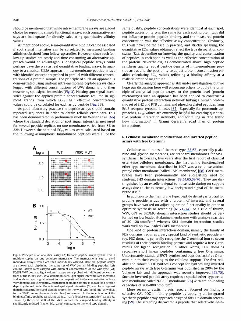
As mentioned above, semi-quantitative binding can be assessedif spot signal intensities can be correlated to measured bindingaffinities obtained from follow-up studies. However, since such fol-low-up studies are costly and time consuming an alternative ap-proach would be advantageous. Analytical peptide arrays couldperhaps pave the way as real quantitative binding assays. In anal-ogy to a classical ELISA approach, intra-membrane peptide arrayswith identical content are probed in parallel with different concen-trations of a protein sample. The principle of such an approach isdemonstrated using uniform intra-membrane peptide arrays chal-lenged with different concentrations of WW domains and thenmeasuring spot signal intensities (Fig. 3). Plotting spot signal inten-sities against the applied protein concentrations resulted in sig-moid graphs from which EC50 (half effective concentration)values could be calculated for each array peptide (Fig. 3B).
As good laboratory practice the peptide arrays should containrepeated sequences in order to obtain reliable error bars. Thishas been demonstrated in preliminary work by Weiser et al. [66]where the standard deviation of spot signal intensities measuredfor several peptide replicas on one membrane varied from 8% to22%. However, the obtained EC50 values were calculated based onthe following assumptions: Immobilized peptides were all of the
Fig. 3. Principle of an analytical assay. (A) Uniform peptide arrays synthesized inmultiple copies on one cellulose membrane. The membrane is cut to yieldindividual arrays, which are then individually assayed. Here six peptide arraysare shown each displaying the same set of WW domain binding peptides. Leftcolumn: arrays were assayed with different concentrations of the wild type (wt)PQBP1 WW domain. Right column: arrays were probed with different concentra-tions of the PQBP1 Y65C WW domain mutant. Spot signal intensities are measuredand as shown spot signal intensities are proportional to the concentration of bothWW domains. (B) Exemplarily, calculation of binding affinity is shown for a peptidedepict by the red circle. The obtained spot signal intensities (SI) are plotted againstdomain concentrations and sigmoid graphs for the wild type (solid line) as well asfor the Y65C mutant domain (dashed line) are resulted. At the inflection point thebinding affinity could be calculated as EC50 (half effective concentration) values. Asshown by the curve shift of the Y65C mutant the assigned binding affinity issignificantly reduced (higher EC50 value) compared to the wild type domain.
same quality, peptide concentrations were identical at each spot,peptide accessibility was the same for each spot, protein tags didnot influence protein-peptide binding, and the measured proteinconcentration was the effective assay concentration. Obviously,this will never be the case in practice, and strictly speaking, thequantitative EC50 values obtained reflect the true dissociation con-stants (KD), depending on knowing the quality and concentrationof peptides in each spot, as well as the effective concentration ofthe protein. Nevertheless, as demonstrated above, high peptidesynthesis quality, equal peptide density of intra-membrane pep-tide arrays and the possibility to adjust protein concentration en-ables calculating EC50 values reflecting a binding affinity at arealistic order of magnitude.
Clearly the analytic approach is still under investigation, but wehope our discussion here will encourage others to apply the prin-ciple of analytical peptide arrays. At the protein level (proteinmicroarrays) such an approach has been applied to construct aquantitative protein interaction network linking a human proteo-mic set of SH2 and PTB domains and phosphorylated peptides fromErbB receptor tyrosine kinases [67]. Especially for proteomic ap-proaches, EC50 values are extremely helpful for creating quantita-tive protein interaction networks, and for filling in ‘‘the trafficflow information’’ in Gianni Cesareni’s road map of proteininteractions.
6. Cellulose membrane modifications and inverted peptidearrays with free C-termini
Cellulose membranes of the ester type [36,62], especially b-ala-nine and glycine membranes, are standard membranes for SPOTsynthesis. Historically, five years after the first report of classicalester-type cellulose membranes, the first amino functionalizedether-type membrane described in 1997 was a cellulose-amino-propyl ether membrane (called CAPE membrane) [68]. CAPE mem-branes have been predominantly and successfully used forstudying SH3 domain interactions [33,54,65,69,70]. They are dis-tinguished by an excellent signal-to-noise ratio during on-supportassays due to the extremely low background signal of the mem-brane itself.
In addition to the membrane type, peptide density is crucial forprobing peptide arrays with a protein of interest, and severalgroups have worked on adjusting amino functionality in order tooptimize synthesis or screening [61,71–74]. As a rule of thumb,WW, GYF or BROMO domain interaction studies should be per-formed on low loaded b-alanine membranes with amino-capacitiesof 30–120 nmol/cm2 whereas SH3 domain interaction studieswork well on low loaded CAPE membranes.
One kind of protein interaction domain, namely the family ofPDZ domains, requires a very special kind of synthetic peptide ar-ray. PDZ domains generally recognize the C-terminal four to sevenresidues of their protein binding partner and require a free C-ter-minus for ligand recognition. In other words, PDZ domainsrecognize short linear peptides containing a free C-terminus.Unfortunately, standard SPOT-synthesized peptides lack free C-ter-mini due to their coupling to the cellulose support. The first reli-able and robust SPOT synthesis concept for synthesizing invertedpeptide arrays with free C-termini was published in 2004 by theVolkmer lab, and the approach was recently improved [52,75].Such an inverted peptide array requires a special, ether-type cellu-lose membrane called N-CAPE membrane [76] with amino-loadingcapacities of 200–800 nmol/cm2.
More recently, cystic fibrosis research focused on finding aselective CAL PDZ inhibitory peptide by applying an integratedsynthetic peptide array approach designed for PDZ domain screen-ing [59]. The screening discovered a peptide that selectively inhib-
R. Volkmer et al. / FEBS Letters 586 (2012) 2780–2786 2785
its the CAL PDZ domain, which in turn extends the half-life of theDF508-CFTR protein responsible for cystic fibrosis. Such an inhib-iting peptide might act as a drug or lead structure for drug devel-opment, and hence therapy to reduce the adverse effects of cysticfibrosis [77].
7. Combining phage display and SPOT synthesis
A new approach combines the relative strength of selectivephage display with the quantitative analysis achieved by SPOT syn-thesis. Initially, Landgraf et al. [33] applied a strategy to reveal allthe peptides in the yeast proteome that have the potential to bindto any domain of interest. Based on the strict consensus sequencesidentified by phage display, they designed a relaxed consensus; forexample the strict consensus sequence of the yeast SH3 domainRvs167 defined as RxFPRxP was relaxed to R/KxxPxxP. Subse-quently, all sequences within the yeast proteome matching a re-laxed consensus of a given SH3 domain were identified bycomputational methods, synthesized on a cellulose membrane,and probed with the SH3 domain of interest. This approach was re-peated for eight yeast SH3 domains, readily identifying peptidebinding to each domain, and leading to predicting protein partners.
More recently, this approach was extended to the complete SH3domain interactome of yeast [65]. A consortium comprising theCesareni, Volkmer, Drubin, Kim, Sidhu, and Boone labs applied acombined approach of orthogonal experimental proteomic tools,such as phage display, yeast two-hybrid and SPOT technology,linked to sophisticated computational and mathematical tools.The results from the three complementary experimental tech-niques were integrated using a Bayesian algorithm to generate ahigh confidence yeast SH3 domain interaction map.
8. Concluding remarks
Why have synthetic peptide arrays prepared by SPOT synthesisbecome so attractive for biologists and the protein domain com-munity? We believe it is due to the robustness, flexibility and sim-plicity of the economical synthesis method, along with a broadspectrum of well-established and highly sensitive assays (bindingaffinities detected down to the millimolar range) combined witha semi-quantitative readout of binding affinities. Further goodarguments are the opportunities to develop a novel analytical pep-tide array approach, the high quality of SPOT-synthesized peptides(50–91% purity), a variety of cellulose membranes with diversephysical properties suitable for nearly any kind of binding assay,as well as commercially available membranes (AIMS ScientificProducts, Braunschweig, Germany) and equipment (Intavis, Köln,Germany).
However, SPOT technology is limited by the number of peptidesthat can be synthesized on a membrane of reasonable size, and bythe fact that regenerating peptide arrays generally fails. The latteris a severe limitation of SPOT technology, especially for experimen-tal proteomics. Ideally, one would wish to screen a given peptidearray several times without any loss of quality, e.g. with differentrepresentatives from a protein domain family. One solution couldbe to use peptide microarrays that could be prepared for a multi-tude of replicas. Pre-synthesized soluble peptides have beenimmobilized on glass slides using several methods [78–81]. How-ever, the peptide microarray technique requires expensive equip-ment and special laboratory conditions. Producing peptidemicroarrays involves highly parallel and high throughput peptidesynthesis, as well as robotic-supported immobilization of pre-syn-thesized peptide derivatives on glass slides. Hence, SPOT synthesis(preparative mode) is essential for preparing peptide microarrays.Recently peptide microarrays were used for the first time to inves-
tigate the protein interaction network mediated by human SH3 do-mains [82].
The in situ synthesis of high-density peptide microarrays is stilla great challenge, and interestingly, the concept of light-directed,spatially addressable synthesis of peptides [37] is once againattractive. The group of Klaus-Peter Stengele at Roche NimbleGenhas developed a novel strategy for photolithographic in situ syn-thesis of thousands of peptides per cm2 on a glass surface. Finally,to complete the picture, Stadler and co-workers have used a mod-ified color laser printer to ‘‘print’’ the 20 amino acids in the form ofsolid amino acid toner particles at defined positions on a glass sup-port [83].
Acknowledgements
R.V. thank the Deutsche Forschungsgemeinschaft for theirfinancial support. V.T. appreciates financial support from the FEBSfederation. All master and diploma students, PhD students, post-docs and technical assistants of the molecular library and recogni-tion group who cannot be named here personally are thanked fortheir contribution.
References
[1] Sudol, M. (1998) From Src Homology domains to other signaling modules:proposal of the protein recognition code. Oncogene 17, 1469–1474.
[2] Searls, D.B. (1997) Linguistic approaches to biological sequences. Comput.Appl. Biosci. 13, 333–344.
[3] Searls, D.B. (2002) The language of genes. Nature 420, 211–217.[4] Phizicky, E., Bastiaens, P.I.H., Zhu, H., Snyder, M. and Fields, S. (2003) Protein
analysis on a proteomic scale. Nature 422, 208–215.[5] Smith, J.M. (1970) Natural selection and the concept of a protein space. Nature
225, 563–564.[6] Jacob, F. (1994) The Possible and the Actual (Jessie and John Danz Lectures),
University of Washington Press, Washington.[7] Lichtarge, O., Bourne, H.R. and Cohen, F.E. (1996) An evolutionary trace
method defines binding surfaces common to protein families. J. Mol. Biol. 257,342–358.
[8] Gimona, M. (2006) Protein linguistics – a grammar for modular proteinassembly? Nat. Rev. Mol. Cell Biol. 7, 68–73.
[9] Fields, S. and Song, O. (1989) A novel genetic system to detect protein–proteininteractions. Nature 340, 245–246.
[10] Uetz, P. et al. (2000) A comprehensive analysis of protein–protein interactionsin Saccharomyces cerevisiae. Nature 403, 623–627.
[11] Ito, T. et al. (2001) A comprehensive two-hybrid analysis to explore the yeastprotein interactome. Proc. Natl. Acad. Sci. USA 98, 4569–4574.
[12] Gavin, A.C. et al. (2002) Functional organization of the yeast proteome bysystematic analysis of protein complexes. Nature 415, 141–147.
[13] Ho, Y. et al. (2002) Systematic identification of protein complexes inSaccharomyces cerevisiae by mass spectrometry. Nature 415, 180–183.
[14] Santonico, E., Castagnoli, L. and Cesareni, G. (2005) Methods to reveal domainnetworks. Drug Discov. Today 10, 1111–1117.
[15] Yook, S.H. et al. (2004) Functional and topological characterization of proteininteraction networks. Proteomics 4, 928–942.
[16] Das, S. and Smith, T.F. (2000) Identifying nature’s protein Lego set. Adv.Protein Chem. 54, 159–183.
[17] Holm, L. and Sander, C. (1998) Dictionary of recurrent domains in proteinstructures. Proteins 33, 88–96.
[18] Pawson, T. and Nash, P. (2003) Assembly of cell regulatory systems throughprotein interaction domains. Science 300, 445–452.
[19] Bork, P., Schultz, J. and Ponting, C.P. (1997) Cytoplasmic signalling domains:the next generation. Trends Biochem. Sci. 22, 296–298.
[20] Cesareni, G. et al., Eds., (2004). Modular Protein Domains, Wiley-VCH VerlagsGmbH & KGaA, Weinheim.
[21] Jones, S. and Thorton, J.M. (1996) Principles of protein–protein interactions.Proc. Natl. Acad. Sci. USA 93, 13–20.
[22] Ma, B., Elkayam, T., Wolfson, H. and Nussinov, R. (2003) Protein–proteininteractions: structurally conserved residues distinguish between bindingsites and exposed protein surfaces. Proc. Natl. Acad. Sci. USA 100, 5772–5777.
[23] Reineke, U., Kramer, A. and Schneider-Mergener, J. (1999) Antigen sequence-and library-based mapping of linear and discontinuous protein–protein-interaction sites by spot synthesis. Curr. Top. Microbiol. Immunol. 243, 23–36.
[24] Bogan, A.A. and Thorn, K.S. (1998) Anatomy of hot spots in protein interfaces. J.Mol. Biol. 280, 1–9.
[25] Clackson, T. and Wells, J.A. (1995) A hot spot of binding energy in a hormone–receptor interface. Science 267, 383–386.
[26] Sudol, M., Chen, H.I., Bougeret, C., Einbond, A. and Bork, P. (1995)Characterization of a novel protein-binding module – the WW domain. FEBSLett. 369, 67–71.
2786 R. Volkmer et al. / FEBS Letters 586 (2012) 2780–2786
[27] Fidan, Z., Younis, A., Schmieder, P. and Volkmer, R. (2011) Chemical synthesisof the third WW domain of TCERG 1 by native chemical ligation. J. Pept. Sci.17, 644–649.
[28] Sparks, A.B., Quilliam, L.A., Thorn, J.M., Der, C.J. and Kay, B.K. (1994)Identification and characterization of Src SH3 ligands from phage-displayedrandom peptide libraries. J. Biol. Chem. 269, 23853–23856.
[29] Rickles, R.J. et al. (1994) Identification of Src, Fyn, Lyn, PI3K and Abl SH3domain ligands using phage display libraries. EMBO J. 13, 5598–5604.
[30] Huang, H. and Sidhu, S.S. (2011) Studying binding specificities of peptiderecognition modules by high-throughput phage display selections. MethodsMol. Biol. 781, 87–97.
[31] Tonikian, R. et al. (2008) A specificity map for the PDZ domain family. PLoSBiol. 6, E239.
[32] Tong, A.H. et al. (2002) A combined experimental and computational strategyto define protein interaction networks for peptide recognition modules.Science 295, 321–324.
[33] Landgraf, C. et al. (2004) Protein interaction networks by proteome peptidescanning. PLoS Biol. 2, E14.
[34] MacBeath, G. and Schreiber, S.L. (2000) Printing proteins as microarrays forhigh-throughput function determination. Science 289, 1760–1763.
[35] Zhu, H. et al. (2001) Global analysis of protein activities using proteome chips.Science 293, 2101–2105.
[36] Frank, R. (1992) SPOT-synthesis: an easy technique for the positionallyaddressable, parallel chemical synthesis on a membrane support. Tetrahedron48, 9217–9232.
[37] Fodor, S.P., Read, J.L., Pirrung, M.C., Stryer, L., Lu, A.T. and Solas, D. (1991) Light-directed, spatially addressable parallel chemical synthesis. Science 251, 767–773.
[38] Frank, R. and Schneider-Mergener, J. (2002) in: Peptide arrays on membranesupports – synthesis and applications. (Koch, J. and Mahler, M., Eds.), pp. 1–22.Springer Verlag, Heidelberg.
[39] Volkmer, R. (2009) Synthesis and application of peptide arrays: Quo vadisSPOT technology. ChemBioChem 10, 1431–1442.
[40] Tapia, V., Ay, B., Triebus, J., Wolter, E., Boisguerin, P. and Volkmer, R. (2008)Evaluating the coupling efficiency of phosphorylated amino acids for SPOTsynthesis. J. Pept. Sci. 14, 1309–1314.
[41] Rathert, P. et al. (2008) Protein lysine methyltransferase G9a acts on non-histone targets. Nat. Chem. Biol. 6, 344–346.
[42] Filippakopoulos, P. et al. (2012) Histone recognition and large-scale structuralanalysis of the human bromodomain family. Cell 149, 214–231.
[43] Hoffmann, B., Ast, T., Polakowski, T., Reineke, U. and Volkmer, R. (2006)Transformation of a biologically active peptide into peptoid analogs whileretaining biological activity. Protein Pept. Lett. 13, 829–833.
[44] Heine, N. et al. (2003) Synthesis and screening of peptoid arrays on cellulosemembranes. Tetrahedron 59, 9919–9930.
[45] Scharn, D., Wenschuh, H., Reineke, U., Schneider-Mergener, J. and Germeroth,L. (2000) Spatially addressed synthesis of amino- and amino-oxy-substituted1,3,5-triazine arrays on polymeric membranes. J. Comb. Chem. 2, 361–369.
[46] Mahrenholz, C.C., Tapia, V., Stigler, R.D. and Volkmer, R. (2010) A study to assessthe cross-reactivity of cellulose membrane-bound peptides with detectionsystems: an analysis at the amino acid level. J. Pept. Sci. 16, 297–302.
[47] Winkler, D.F. and McGeer, P.L. (2008) Protein labeling and biotinylation ofpeptides during spot synthesis using biotin p-nitrophenyl ester (biotin-ONp).Proteomics 8, 961–967.
[48] Ay, B. et al. (2007) Sorting and pooling strategy: a novel tool to map a virusproteome for CD8 T-cell epitopes. Biopolymers 88, 64–75.
[49] Kamradt, T. and Volkmer-Engert, R. (2004) Cross-reactivity of T lymphocytesin infection and autoimmunity. Mol. Divers. 8, 271–280.
[50] Tapia, V.E. et al. (2010) Y65C missense mutation in the WW domain of theGolabi–Ito–Hall syndrome protein PQBP1 affects its binding activity andderegulates pre-mRNA splicing. J. Biol. Chem. 285, 19391–19401.
[51] Hu, H. et al. (2004) A map of WW domain family interactions. Proteomics 4,643–655.
[52] Boisguerin, P. et al. (2004) An improved method for the synthesis of cellulosemembrane-bound peptides with free C termini is useful for PDZ domainbinding studies. Chem. Biol. 11, 449–459.
[53] Geysen, H.M., Meloen, R.H. and Barteling, S.J. (1984) Use of peptide synthesisto probe viral antigens for epitopes to a resolution of a single amino acid. Proc.Natl. Acad. Sci. USA 81, 3998–4002.
[54] Rottensteiner, H. et al. (2004) Peroxisomal membrane proteins containcommon Pex19p-binding sites that are an integral part of their targetingsignals. Mol. Biol. Cell 15, 3406–3417.
[55] Cunningham, B.C. and Wells, J.A. (1989) High-resolution epitope mapping ofhGH-receptor interactions by alanine-scanning mutagenesis. Science 244,1081–1085.
[56] Bolger, G.B. et al. (2006) Scanning peptide array analyses identify overlappingbinding sites for the signalling scaffold proteins, beta-arrestin and RACK1, incAMP-specific phosphodiesterase PDE4D5. Biochemical J. 398, 23–36.
[57] Reineke, U., Schneider-Mergener, J. and Schutkowski, M. (2005) Peptide arraysin proteomics and drug discovery in: BioMEMS and BiomedicalNanotechnology, Micro and Nano-technologies for Genomics andProteomics, vol. II (Ozkan, M. and Heller, M.J., Eds.), pp. 161–282, Springer,New York.
[58] Portwich, M. et al. (2007) A network of coiled-coil associations derived fromsynthetic GCN4 leucine-zipper arrays. Angew. Chem. Int. Ed. 46, 1654–1657.
[59] Vouilleme, L., Cushing, P.R., Volkmer, R., Madden, D.R. and Boisguerin, P.(2010) Engineering peptide inhibitors to overcome PDZ binding promiscuity.Angew. Chem. Int. Ed. 49, 9912–9916.
[60] Takahashi, M., Ueno, A. and Mihara, H. (2000) Peptide design based on anantibody complementarity determining region (CDR): construction ofporphyrin-binding peptides and their affinity maturation by a combinatorialmethod. Chem. Eur. J. 6, 3196–3203.
[61] Kramer, A. et al. (1999) Spot synthesis: observations and optimizations. J. Pept.Res. 54, 319–327.
[62] Wenschuh, H. et al. (2000) Coherent membrane supports for parallelmicrosynthesis and screening of bioactive peptides. Biopolymers 55, 188–206.
[63] Molina, F., Laune, D., Gougat, C., Pau, B. and Granier, C. (1996) Improvedperformances of spot multiple peptide synthesis. Peptide Res. 9, 151–155.
[64] Przezdziak, J. et al. (2006) Probing the ligand binding specificity and analyzingthe folding state of SPOT synthesized FBP28 WW domain variants.ChemBioChem 7, 780–788.
[65] Tonikian, R. et al. (2009) Bayesian modeling of the yeast SH3 domaininteractome predicts spatiotemporal dynamics of endocytosis proteins. PLoSBiol. 7, E1000218.
[66] Weiser, A.A. et al. (2005) SPOT synthesis: reliability of array-basedmeasurement of peptide binding affinity. Anal. Biochem. 342, 300–311.
[67] Jones, R.B., Gordus, A., Krall, J.A. and MacBeath, G. (2006) A quantitativeprotein interaction network for the ErbB receptors using protein microarrays.Nature 439, 168–174.
[68] Volkmer-Engert, R., Hoffman, B. and Schneider-Mergener, J. (1997) Stableattachment of the HMB-linker to continuous cellulose membranes for parallelsolid phase spot synthesis. Tetrahedron Lett. 38, 1029–1032.
[69] Schell-Steven, A. et al. (2005) Identification of a novel, intraperoxisomalPex14-binding bite in Pex13: Association of Pex13 with the docking complexis essential for peroxisomal matrix protein import. Mol. Cell. Biol. 25, 3007–3018.
[70] Pires, J.R. et al. (2003) The ScPex13p SH3 domain exposes two distinct bindingsites for Pex5p and Pex14p. J. Mol. Biol. 326, 1427–1435.
[71] Dürauer, A. et al. (2006) Evaluation of a sensitive detection method for peptidearrays prepared by SPOT synthesis. J. Biochem. Biophys. Methods 66, 45–57.
[72] Gail, R., Frank, R. and Wittinghofer, A. (2005) Systematic peptide array-baseddelineation of the differential beta-catenin interaction with Tcf4, E-cadherin,and adenomatous polyposis coli. J. Biol. Chem. 280, 7107–7117.
[73] Otvos Jr., L. et al. (2000) In situ stimulation of a T helper cell hybridoma with acellulose-bound peptide antigen. J. Immunol. Methods 233, 95–105.
[74] Lin, Q., O’Neill, J.C. and Blackwell, H.E. (2005) Small molecule macroarrayconstruction via Ugi four-component reactions. Org. Lett. 7, 4455–4458.
[75] Boisguerin, P. et al. (2007) Characterization of a putative phosphorylationswitch: adaptation of SPOT synthesis to analyze PDZ domain regulationmechanisms. ChemBioChem 8, 2302–2307.
[76] Licha, K. et al. (2000) Highly parallel nano-synthesis of cleavable peptide–dyeconjugates on cellulose membranes. Tetrahedon Lett. 41, 1711–1715.
[77] Cushing, P.R., Vouilleme, L., Pellegrini, M., Boisguerin, P. and Madden, D.R.(2010) A stabilizing influence: CAL PDZ inhibition extends the half-life ofDF508-CFTR. Angew. Chem. Int. Ed. 49, 9907–9911.
[78] Beutling, U., Städing, K., Stradal, T. and Frank, R. (2008) Large-scale analysis ofprotein–protein interactions using cellulose-bound peptide arrays. Adv.Biochem. Eng./Biotechnol. 110, 115–152.
[79] Henderson, G. and Bradley, M. (2007) Functional peptide arrays for high-throughput chemical biology based applications. Curr. Opin. Biotechnol. 18,326–330.
[80] Tapia, V.E., Ay, B. and Volkmer, R. (2009) Exploring and profiling proteinfunction with peptide arrays. Methods Mol. Biol. 570, 3–17.
[81] Reimer, U., Reineke, U. and Schneider-Mergener, J. (2002) Peptide arrays: frommacro to micro. Curr. Opin. Biotechnol. 13, 315–320.
[82] Carducci, M. et al. (2012) The protein interaction network mediated by humanSH3 domains. Biotechnol. Adv. 30, 4–15.
[83] Stadler, V. et al. (2008) Combinatorial synthesis of peptide arrays with a laserprinter. Angew. Chem. Int. Ed. 47, 7132–7135.
Related Documents


![RESEARCH Open Access Designed hybrid TPR peptide targeting ... · of the anaphase-promoting complex [4,5], TPR domains are now known to mediate specific protein interactions in numerous](https://static.cupdf.com/doc/110x72/5fb3fde4c1a5fe0c982b300b/research-open-access-designed-hybrid-tpr-peptide-targeting-of-the-anaphase-promoting.jpg)


