Synthetic partial agonists reveal key steps in IP 3 receptor activation Ana M. Rossi 1,4 , Andrew M. Riley 2,4 , Stephen C. Tovey 1 , Taufiq-Ur-Rahman 1 , Olivier Dellis 1 , Emily J. A. Taylor 1 , Valery G. Veresov 3 , Barry V. L. Potter 2,* , and Colin W. Taylor 1,* 1 Department of Pharmacology, University of Cambridge, Tennis Court Road, Cambridge, CB2 1PD, UK 2 Wolfson Laboratory of Medicinal Chemistry, Department of Pharmacy and Pharmacology, University of Bath, Claverton Down, Bath, BA2 7AY, UK 3 Department of Cell Biophysics, Institute of Biophysics and Cell Engineering, Minsk 220072, Academicheskaya St. 27, Belarus. Abstract Inositol 1,4,5-trisphosphate receptors (IP 3 R) are ubiquitous intracellular Ca 2+ channels. IP 3 binding to the IP 3 -binding core (IBC) near the N-terminal initiates conformational changes that lead to opening of a pore. The mechanisms are unresolved. We synthesized 2-O-modified IP 3 analogues that are partial agonists of IP 3 R. These are like IP 3 in their interactions with the IBC, but they are less effective than IP 3 in rearranging the relationship between the IBC and N-terminal suppressor domain (SD), and they open the channel at slower rates. IP 3 R with a mutation in the SD occupying a position similar to the 2-O-substituent of the partial agonists has a reduced open probability that is similar for full and partial agonists. Bulky or charged substituents from either the ligand or SD therefore block obligatory coupling of the IBC and SD. Analysis of ΔG for ligand binding shows that IP 3 is recognised by the IBC and conformational changes then propagate entirely via the SD to the pore. Inositol 1,4,5-trisphosphate receptors (IP 3 R) are ligand-gated channels. They are expressed in most animal cells and mediate release of Ca 2+ from the endoplasmic reticulum in response to the many stimuli that evoke IP 3 formation. IP 3 R are tetrameric, and each subunit of about 2700 residues has an IP 3 -binding site near the N-terminus and six transmembrane domains (TMD) towards the C-terminus (Fig. 1a)1. The pore is formed by the last pair of TMD and the intervening loop, the pore-loop (“P-loop”), from all four subunits1. The structure of the pore is predicted to be broadly similar to the pores of other tetrameric P-loop channels, like bacterial K + channels, for which high-resolution structures are available2. IP 3 binds to a discrete part of the IP 3 R, the IP 3 -binding core (IBC, residues 224-604, Fig. 1a)3. Although the extreme N-terminus (residues 1-223) is not required for IP 3 binding, it decreases the affinity for IP 3 and has therefore been called the suppressor domain (SD)4. The SD is thought to be required for channel gating because IP 3 binds to IP 3 R without an * Correspondence should be addressed to C.W.T. ([email protected]) or B.V.L.P ([email protected]) . 4 These authors contributed equally to this work. AUTHOR CONTRIBUTIONS A.M.R. (Cambridge), S.C.T., T-U-R, O.D. and E.J.A.T. completed the biology experiments. V.G.V. performed molecular modelling. A.M.R. (Bath) designed and synthesized the ligands and contributed to molecular modelling. B.V.L.P. (chemistry) and C.W.T. (biology) designed and coordinated the project. C.W.T. and A.M.R. (Cambridge) wrote the manuscript with input from the other authors. All authors discussed the results and commented on the manuscript. COMPETING INTERESTS STATEMENT The authors declare that they have no competing financial interests. Note: Supplementary information and chemical compound information is available on the Nature Chemical Biology website. Europe PMC Funders Group Author Manuscript Nat Chem Biol. Author manuscript; available in PMC 2010 May 13. Published in final edited form as: Nat Chem Biol. 2009 September ; 5(9): 631–639. doi:10.1038/nchembio.195. Europe PMC Funders Author Manuscripts Europe PMC Funders Author Manuscripts

Welcome message from author
This document is posted to help you gain knowledge. Please leave a comment to let me know what you think about it! Share it to your friends and learn new things together.
Transcript
Synthetic partial agonists reveal key steps in IP3 receptoractivation
Ana M. Rossi1,4, Andrew M. Riley2,4, Stephen C. Tovey1, Taufiq-Ur-Rahman1, OlivierDellis1, Emily J. A. Taylor1, Valery G. Veresov3, Barry V. L. Potter2,*, and Colin W. Taylor1,*
1Department of Pharmacology, University of Cambridge, Tennis Court Road, Cambridge, CB21PD, UK2Wolfson Laboratory of Medicinal Chemistry, Department of Pharmacy and Pharmacology,University of Bath, Claverton Down, Bath, BA2 7AY, UK3Department of Cell Biophysics, Institute of Biophysics and Cell Engineering, Minsk 220072,Academicheskaya St. 27, Belarus.
AbstractInositol 1,4,5-trisphosphate receptors (IP3R) are ubiquitous intracellular Ca2+ channels. IP3bindingto the IP3-binding core (IBC) near the N-terminal initiates conformational changes that lead toopening of a pore. The mechanisms are unresolved. We synthesized 2-O-modified IP3 analoguesthat are partial agonists of IP3R. These are like IP3 in their interactions with the IBC, but they areless effective than IP3 in rearranging the relationship between the IBC and N-terminal suppressordomain (SD), and they open the channel at slower rates. IP3R with a mutation in the SD occupyinga position similar to the 2-O-substituent of the partial agonists has a reduced open probability thatis similar for full and partial agonists. Bulky or charged substituents from either the ligand or SDtherefore block obligatory coupling of the IBC and SD. Analysis of ΔG for ligand binding showsthat IP3 is recognised by the IBC and conformational changes then propagate entirely via the SDto the pore.
Inositol 1,4,5-trisphosphate receptors (IP3R) are ligand-gated channels. They are expressedin most animal cells and mediate release of Ca2+ from the endoplasmic reticulum inresponse to the many stimuli that evoke IP3 formation. IP3R are tetrameric, and each subunitof about 2700 residues has an IP3-binding site near the N-terminus and six transmembranedomains (TMD) towards the C-terminus (Fig. 1a)1. The pore is formed by the last pair ofTMD and the intervening loop, the pore-loop (“P-loop”), from all four subunits1. Thestructure of the pore is predicted to be broadly similar to the pores of other tetrameric P-loopchannels, like bacterial K+ channels, for which high-resolution structures are available2. IP3binds to a discrete part of the IP3R, the IP3-binding core (IBC, residues 224-604, Fig. 1a)3.Although the extreme N-terminus (residues 1-223) is not required for IP3 binding, itdecreases the affinity for IP3 and has therefore been called the suppressor domain (SD)4.The SD is thought to be required for channel gating because IP3 binds to IP3R without an
*Correspondence should be addressed to C.W.T. ([email protected]) or B.V.L.P ([email protected]) .4These authors contributed equally to this work.AUTHOR CONTRIBUTIONS A.M.R. (Cambridge), S.C.T., T-U-R, O.D. and E.J.A.T. completed the biology experiments. V.G.V.performed molecular modelling. A.M.R. (Bath) designed and synthesized the ligands and contributed to molecular modelling.B.V.L.P. (chemistry) and C.W.T. (biology) designed and coordinated the project. C.W.T. and A.M.R. (Cambridge) wrote themanuscript with input from the other authors. All authors discussed the results and commented on the manuscript.
COMPETING INTERESTS STATEMENT The authors declare that they have no competing financial interests.
Note: Supplementary information and chemical compound information is available on the Nature Chemical Biology website.
Europe PMC Funders GroupAuthor ManuscriptNat Chem Biol. Author manuscript; available in PMC 2010 May 13.
Published in final edited form as:Nat Chem Biol. 2009 September ; 5(9): 631–639. doi:10.1038/nchembio.195.
Europe PM
C Funders A
uthor Manuscripts
Europe PM
C Funders A
uthor Manuscripts
SD, but it no longer opens the pore4,5. However, the links between IP3 binding and gatingare not understood, and nor do we have a structure of the entire IP3R at sufficient resolutionto provide insight into these gating mechanisms1,6.
Activation of ligand-gated ion channels begins with agonist binding to a stable closed stateand proceeds via many short-lived intermediates to a state in which the pore is open7. Thisactivation may proceed entirely through a sequence of incremental changes in the receptor8or be dominated by a single concerted transition between two stable conformations9.Agonists differ in both the strength of their binding (affinity) and in their ability to drive thereceptor to its open state (efficacy)10. A ligand with reduced efficacy must occupy morereceptors than a full agonist to evoke the same cellular response. Such partial agonists, byoccupying receptors, diminish the response to a full agonist11. Partial agonists areparticularly useful for exploring the mechanisms of receptor activation because they liebetween full agonists and antagonists in their ability to activate receptors7,10. This is truefor all receptors, but ligand-gated ion channels are uniquely amenable to such analysesbecause single-channel recording allows key conformational changes of single receptors tobe determined with outstanding temporal resolution7.
For these ligand-gated ion channels, full and partial agonists may differ in either thefrequency with which they cause the receptor to visit the fully active open state or they maystabilize different open states that mediate lesser ion fluxes. In both cases, a partial agonistevokes lesser activation. Two subtypes of ionotropic glutamate receptors (iGluR) illustratethe distinction. These receptors, which mediate most excitatory neurotransmission in thebrain, are those for which the structural basis of efficacy has been most thoroughlyexplored12,13. For the AMPA subtype of iGluR, a series of partial agonists, which differfrom each other by only a single atom, close the clam-like binding site to varying degrees,but less completely than do full agonists. Partial agonists thereby preferentially open thepore to states with lesser conductance12. For full and partial agonists of the NMDA subtypeof iGluR, the conformational changes in the binding site are more subtly different. Bothcause similar closure of the clam, and they fully open the pore, but the conformationalchanges proceed more slowly for partial agonists14,15.
Affinity and efficacy are distinguishable, but the two properties are not independent becauseenergy provided by agonist binding drives the conformational changes that cause channelopening10,16,17. This “binding-gating problem” is a fundamental issue in pharmacology10,but it can also be turned to advantage because the interplay depends upon both the efficacyof the ligand and the presence of the parts of the receptor through which conformationalchanges must pass. The former because partial agonists divert less binding energy than fullagonists into effective conformational changes; and the latter because receptors lackingessential domains are expected to be less able to divert binding energy into conformationalchanges (Fig. 1b). Analyses of the free energy changes for ligand binding (ΔG = -RTlnKd,where Kd is the equilibrium dissociation constant) can thus provide insight into theconformational changes evoked by agonist binding. Comparisons of ΔG for full and partialagonists, and for agonist binding to normal and truncated IP3R, can therefore contribute todefining the links between IP3 binding and opening of the pore.
Here we synthesize a new series of partial agonists of the IP3R, and, in defining theirproperties, we identify a novel form of partial agonism that allows us to define key steps inIP3R activation.
Rossi et al. Page 2
Nat Chem Biol. Author manuscript; available in PMC 2010 May 13.
Europe PM
C Funders A
uthor Manuscripts
Europe PM
C Funders A
uthor Manuscripts
RESULTSSynthesis of 2-O-modified analogues of IP3
All high-affinity agonists of all IP3R have structures equivalent to the vicinal 4,5-bisphosphate and 6-hydroxyl of IP3 (Fig. 1c), but the axial 2-hydroxyl is not required18. Theessential phosphate moieties interact predominantly with opposite sides of the clam-like IBC(P-4 with the β2-domain, and P-5 with the ARM domain)3 (Fig. 1a), suggesting thatagonists might close the clam in a manner reminiscent of glutamate binding to iGluR6,13.
In seeking to develop novel high-affinity ligands of IP3R that might differ in efficacy, wefocused on the 2-OH group of IP3 because earlier structure-activity analyses had suggestedthat analogues modified at this position retain activity18. The X-ray structure of the IBCwith IP3 bound subsequently confirmed that the 2-OH group of IP3 makes no significantcontacts with the IBC3.
We began by preparing homo-dimers of IP3 with linkers of various lengths (2, 3 and 4 inFig. 1c), aiming initially to define the separation of IP3-binding sites within a tetramericIP3R. However, informed by our initial results19,20 and cognizant that dimeric cGMP is apartial agonist of a cGMP-gated cation channel21, we extended our work to includesyntheses of additional 2-O-modified analogues (Fig. 1c) and an assessment of theirefficacy.
The shortest IP3 dimer (2) is a symmetrically substituted N,N’-diethyl urea, synthesized bycross-linking of a protected D-2-O-(2-aminoethyl)-IP3 building block using bis(4-nitrophenyl) carbonate19. A modification of this synthetic method was used to synthesizehetero-dimers such as 5, 6 and 7, in which the second IP3 moiety is replaced by a differentinositol phosphate or by inositol (Scheme 1). We also synthesised an L-IP3 homo-dimer (8,the enantiomer of 2) (Scheme 1) and an IP3-adamantane conjugate (9). The syntheses of 2-deoxy-IP3 (10)22 and 2-O-(2-aminoethyl)-IP3 (11)23 were reported previously. Details ofthe synthetic procedures and compound characterizations are provided in SupplementaryMethods online.
2-O-modified IP3 analogues are high-affinity agonists of IP3RWe used a cell line that expresses only recombinant rat IP3R1 (DT40-IP3R1 cells)24 tomeasure Ca2+ release from intracellular stores, and IP3R1 purified from rat cerebellum tomeasure IP3 binding. These analyses show that homo-dimers of IP3 linked through the 2-O-positions of the inositol rings are high-affinity agonists of IP3R. The shortest dimer (2) (Fig.1c) binds to IP3R1 with greater affinity than IP3 (Table 1 and Supplementary Fig. 1a online)and stimulates Ca2+ release from intracellular stores at lower concentrations than does IP3(Table 1 and Supplementary Fig. 1b online). 2 is the most potent inositol phosphate-basedagonist so far identified.
A homo-dimer of L-IP3 (8) is, as expected, inactive because L-IP3 does not bind to theIBC18. However, homo-dimers of IP3 with longer linkers (3, 4) also bind to IP3R withgreater affinity than IP3, as do hetero-dimers in which IP3 is linked to inositol (inositol-IP3,7), an unrelated bulky hydrophobic group (adamantane-IP3, 9), or to an inositol phosphatethat does not itself bind to the IBC (IP3-IP5, 6; or IP3-L-IP3, 5) (Table 1). The latter (5-7 and9) demonstrate that high-affinity binding of IP3 dimers does not result from an interactionwith a second specific IP3-binding site, nor does it result from alternating association of thetwo IP3 moieties with the IBC19. Furthermore, the two components of the dimer must belinked, because a high concentration of IP5 (12, 10μM) had no effect on the Ca2+ releaseevoked by IP3 or 2-deoxy-IP3 (10) (Supplementary Fig. 1c online). We conclude that
Rossi et al. Page 3
Nat Chem Biol. Author manuscript; available in PMC 2010 May 13.
Europe PM
C Funders A
uthor Manuscripts
Europe PM
C Funders A
uthor Manuscripts
addition of bulky or charged groups to the 2-O-position of IP3 produces high-affinityagonists of the IP3R.
A new family of partial agonists of IP3RBecause a partial agonist less effectively activates its receptor than a full agonist, it mustoccupy more receptors to evoke the same cellular response11. In our Ca2+ release assays,where each ligand caused the same maximal Ca2+ release as IP3 (Table 1), we can thereforegain some insight into the efficacy of a ligand by comparing the concentration that causes50% of the maximal response (EC50) with that which occupies 50% of the binding sites(Kd).
For each ligand, we compared the EC50/Kd ratio using DT40-IP3R1 cells for the functionalassays24 (EC50) and purified IP3R1 to measure IP3 binding (Kd). Our results suggest that 2occupies more IP3R than IP3 to evoke the same Ca2+ release (2 has a higher EC50/Kd ratio,Fig. 2a and Table 1). This indicates that 2 may be a partial agonist. These characteristics, anincrease in both affinity and EC50/Kd ratio, are shared by very different 2-O-modified IP3analogues (Fig. 2a and Table 1). They do not, therefore, depend upon precise structuralfeatures: an IP3 moiety that binds to the IBC and a 2-O-substitutent larger than adamantane(9) are sufficient to increase both the affinity and EC50/Kd ratio.
The EC50/Kd ratio for adenophostin A (AdA, 13, Fig. 1c)25, another high-affinity agonist ofIP3R, is similar to that for IP3 (Fig. 2a and Table 1). This is consistent with single channelanalyses, where IP3 and AdA cause the IP3R to open to the same maximal single channelopen probability (Po, the fraction of time that each channel spends in its open state) (Fig. 2band Supplementary Table 1 online)26. We conclude that IP3 and AdA are full agonists ofthe IP3R, whereas 2-7 and 9 appear to be partial agonists.
The results shown in Fig. 2c confirm that 2 must occupy more IP3R than the full agonist,AdA, to evoke the same Ca2+ release. DT40-IP3R1 cells were first pre-treated withconcentrations of AdA or 2 that caused the same Ca2+ release (5.7 ± 1.0% and 5.4 ± 1.3% ofthe intracellular stores, respectively) and then stimulated with IP3. More IP3 is required toevoke further Ca2+ release after treatment with 2 than after AdA (EC50 = 44.4 ± 3.19 and4.37 ± 0.18nM, respectively). This confirms that 2 occupies more IP3R than AdA to evokethe same Ca2+ release.
Because the nuclear envelope is continuous with the endoplasmic reticulum26, we can usepatch-clamp recording from the outer nuclear envelope of DT40-IP3R1 cells to resolve thebehaviour of single IP3R. To both maximize the amplitude of the currents recorded and toavoid the complexity of feedback regulation of IP3R by Ca2+ passing through them1, weused K+ as the charge-carrier in these experiments26,27. Both the single channel K+
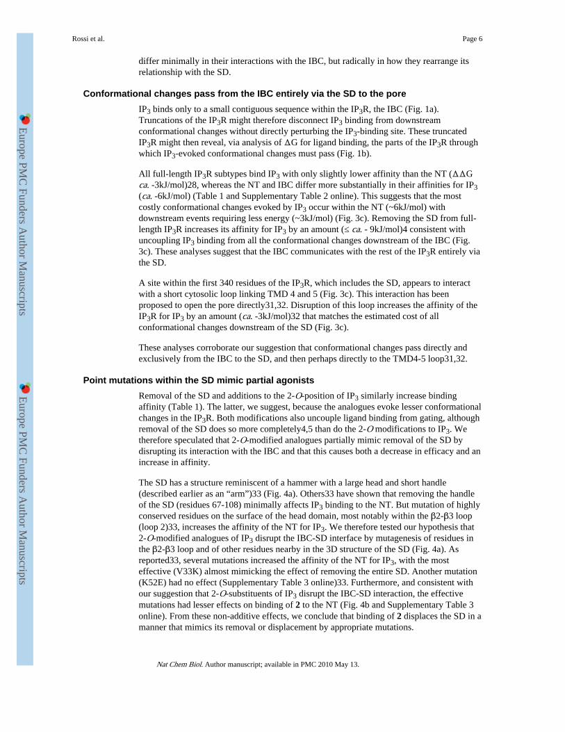
conductance (γK) and the mean channel open time (τo) were the same for all agonistsexamined (Fig. 2b,d and Supplementary Table 1 online). The open state of the IP3R thusappears to be similar whether it is evoked by binding of a full (AdA, IP3 and 10) or partialagonist (2, 6 and 9).
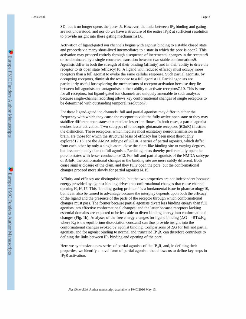
However, for IP3R activated by maximal concentrations of AdA, IP3 or 10, Po was higherthan with 2, 6 or 9 (Fig. 2b,e and Supplementary Table 1 online). Increasing theconcentration of 2 (from 0.5 to 10μM, Fig. 2b,e) did not further increase Po, and afterstimulation with a mixture of IP3 and 2 (10μM of each) Po was significantly less than withIP3 alone (Fig. 2b and Supplementary Table 1 online). These analyses of single IP3Rconfirm and extend the results obtained with Ca2+ release and binding assays (Table 1 andFig. 2a). Analogues of IP3 with bulky additions to the 2-O-position (2-7 and 9) are high-
Rossi et al. Page 4
Nat Chem Biol. Author manuscript; available in PMC 2010 May 13.
Europe PM
C Funders A
uthor Manuscripts
Europe PM
C Funders A
uthor Manuscripts
affinity partial agonists of IP3R1. Subsequent analyses of the mechanisms underlying theseproperties of the 2-O-substituted analogues focus on 2 (Fig. 1c).
Our single channel analysis shows that whereas τo is similar for all agonists (SupplementaryTable 1 online), mean channel closed times (τc) were longer for the partial agonists (2, 6 and9) than for full agonists (IP3, AdA and 10) (Fig. 2e and Supplementary Table 1 online). Thelatter, assuming a simplified activation scheme (Fig. 2e, Supplementary Methods online),reveals that the rate constant for channel opening (β = 1/τc) with partial agonists is less thanwith full agonists. The 2-O-modified analogues are the first partial agonists of IP3R forwhich the basis of their reduced efficacy has been established. They open the channel fully(Fig. 2b,d), the channel closes at the same rate whether it has a partial agonist or IP3 bound(α in Fig. 2e), but the rate constant for channel opening (β) is lower for partial agonists (Fig.2e and Supplementary Table 1 online). We conclude that these full and partial agonists drivethe IP3R into a similar open state, but the partial agonists do so less effectively.
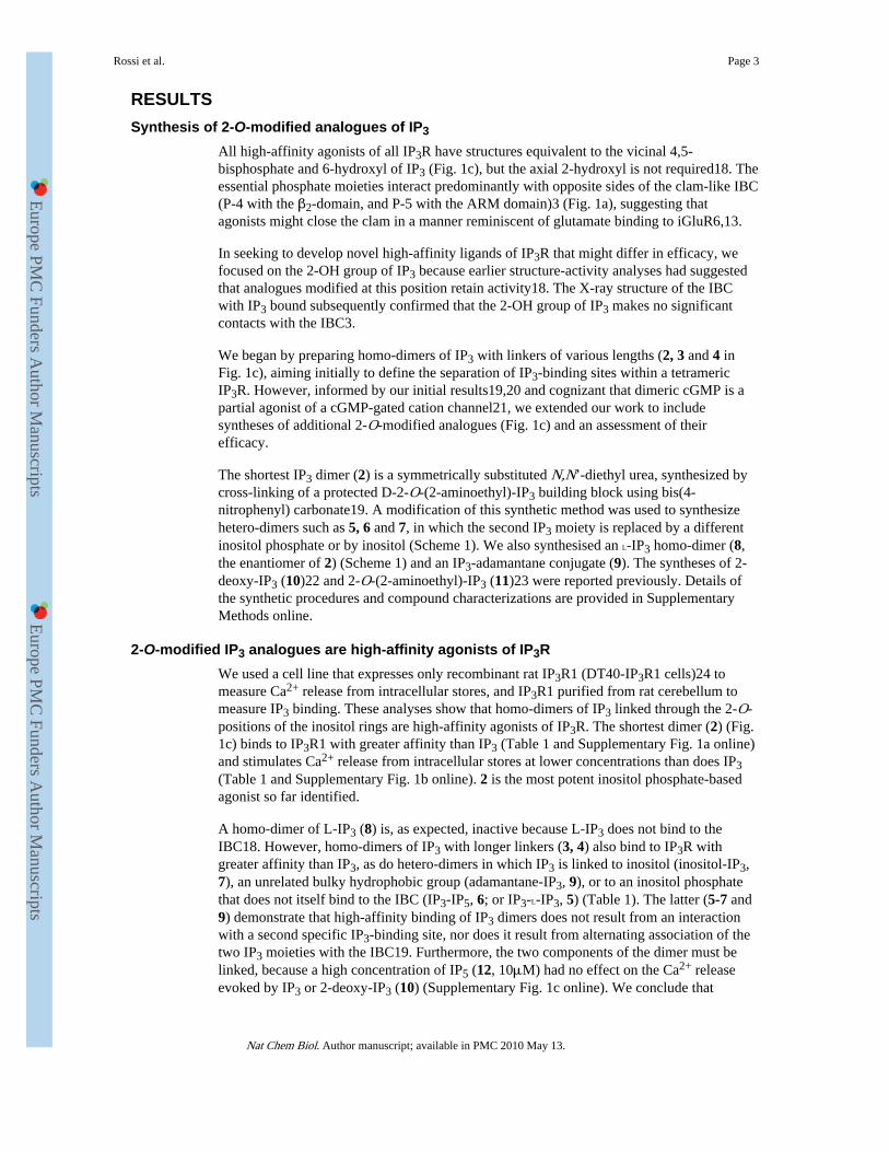
Full and partial agonists differ in how they rearrange the IBC-SDFor IP3R, conformational changes evoked by IP3 binding to the IBC near the N-terminalmust be transmitted to the pore formed by residues close to the C-terminal (Fig. 1a). Someof the energy provided by IP3 binding is used to drive the opening of the pore. The Kd (ΔG= -RTlnKd) measured in a binding assay is therefore determined by both the strength of thecontacts between IP3 and the IBC (“intrinsic binding affinity”)16 and the ensuingconformational changes10.
The IBC includes all the amino acid residues that contact IP3 (Fig. 1a)3,28 and each 2-O-modified agonist (2-7 and 9-11) (Fig. 1c) retains the groups within IP3 that interact with theIBC. Furthermore, each of these ligands binds with similar affinity to the IBC alone (Fig. 3aand Table 1). Because the full (IP3) and partial agonists (2-7, 9) are both expected to makethe same contacts with the IBC and are also observed to bind to it with similar affinity, wesuggest that they do not differ in the binding energy they divert into changing theconformation of the IBC. This contrasts with AMPA receptors, where the clam-like bindingsite closes more fully with more efficacious agonists12,13. The distinction highlights twofundamentally different ways of reducing efficacy, a defining feature of all ligand-receptorinteractions10. A partial agonist may fail to make optimal contacts with the binding site andso less effectively activate the receptor (e.g., AMPA receptors12), or it may impair onwardtransmission of conformational changes. Subsequent experiments demonstrate that ourpartial agonists (2-7 and 9) belong to the second category. They are thereby useful indefining the steps that follow IP3 binding.
For all three IP3R subtypes, IP3 binds to the IBC with greater affinity than to either full-length IP3R or the NT (Fig. 3a, Table 1 and Supplementary Table 2 online)28,29. The SDreduces the IP3 binding affinity through its intramolecular interaction with the IBC28 andappears also to mediate communication between the IBC and pore4,5. We thereforeexamined the contribution of the SD to the conformational changes initiated by IP3 viaanalysis of ΔG for ligand binding.
Removal of the SD increases the affinity of the NT for IP3, but it has lesser effects onbinding of the partial agonists (Table 1). Efficacy (reported by the EC50/Kd ratio) and thedifference in ΔG (ΔG = -RTlnKd) for binding to the IBC and NT (ΔΔG) are inverselycorrelated (Fig. 3b). Because we suggest that each agonist contributes similar “intrinsicbinding energy”16,17 through the similar interactions that each makes with the IBC (Table 1and Supplementary Table 2 online), the different ΔΔG for binding of full and partialagonists to the NT must reflect the extent to which each uses binding energy to rearrange therelationship between the IBC and SD16,17,30. We conclude that full and partial agonists
Rossi et al. Page 5
Nat Chem Biol. Author manuscript; available in PMC 2010 May 13.
Europe PM
C Funders A
uthor Manuscripts
Europe PM
C Funders A
uthor Manuscripts
differ minimally in their interactions with the IBC, but radically in how they rearrange itsrelationship with the SD.
Conformational changes pass from the IBC entirely via the SD to the poreIP3 binds only to a small contiguous sequence within the IP3R, the IBC (Fig. 1a).Truncations of the IP3R might therefore disconnect IP3 binding from downstreamconformational changes without directly perturbing the IP3-binding site. These truncatedIP3R might then reveal, via analysis of ΔG for ligand binding, the parts of the IP3R throughwhich IP3-evoked conformational changes must pass (Fig. 1b).
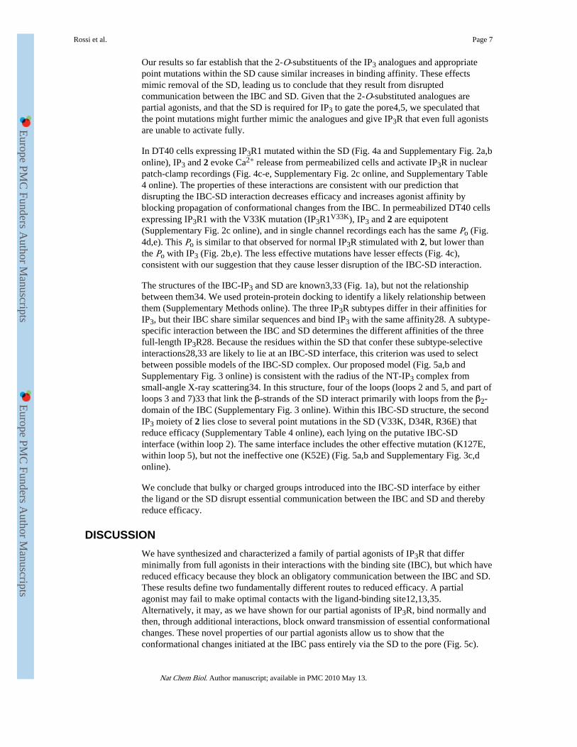
All full-length IP3R subtypes bind IP3 with only slightly lower affinity than the NT (ΔΔGca. -3kJ/mol)28, whereas the NT and IBC differ more substantially in their affinities for IP3(ca. -6kJ/mol) (Table 1 and Supplementary Table 2 online). This suggests that the mostcostly conformational changes evoked by IP3 occur within the NT (~6kJ/mol) withdownstream events requiring less energy (~3kJ/mol) (Fig. 3c). Removing the SD from full-length IP3R increases its affinity for IP3 by an amount (≤ ca. - 9kJ/mol)4 consistent withuncoupling IP3 binding from all the conformational changes downstream of the IBC (Fig.3c). These analyses suggest that the IBC communicates with the rest of the IP3R entirely viathe SD.
A site within the first 340 residues of the IP3R, which includes the SD, appears to interactwith a short cytosolic loop linking TMD 4 and 5 (Fig. 3c). This interaction has beenproposed to open the pore directly31,32. Disruption of this loop increases the affinity of theIP3R for IP3 by an amount (ca. -3kJ/mol)32 that matches the estimated cost of allconformational changes downstream of the SD (Fig. 3c).
These analyses corroborate our suggestion that conformational changes pass directly andexclusively from the IBC to the SD, and then perhaps directly to the TMD4-5 loop31,32.
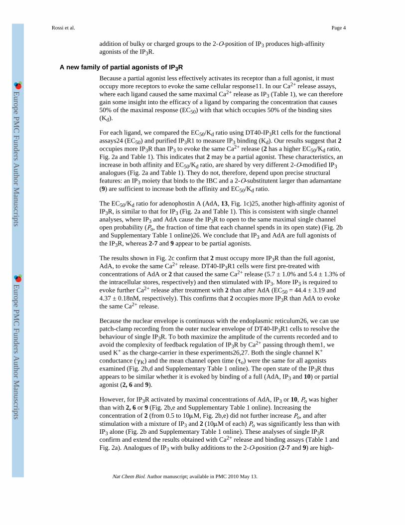
Point mutations within the SD mimic partial agonistsRemoval of the SD and additions to the 2-O-position of IP3 similarly increase bindingaffinity (Table 1). The latter, we suggest, because the analogues evoke lesser conformationalchanges in the IP3R. Both modifications also uncouple ligand binding from gating, althoughremoval of the SD does so more completely4,5 than do the 2-O modifications to IP3. Wetherefore speculated that 2-O-modified analogues partially mimic removal of the SD bydisrupting its interaction with the IBC and that this causes both a decrease in efficacy and anincrease in affinity.
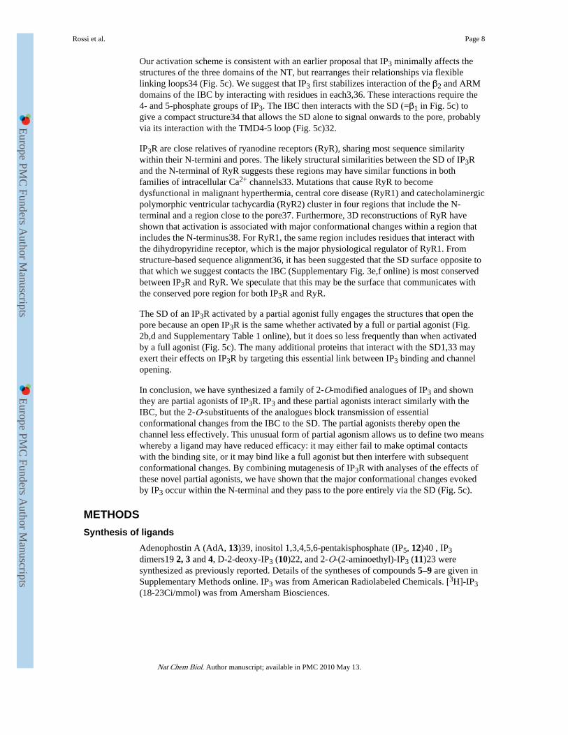
The SD has a structure reminiscent of a hammer with a large head and short handle(described earlier as an “arm”)33 (Fig. 4a). Others33 have shown that removing the handleof the SD (residues 67-108) minimally affects IP3 binding to the NT. But mutation of highlyconserved residues on the surface of the head domain, most notably within the β2-β3 loop(loop 2)33, increases the affinity of the NT for IP3. We therefore tested our hypothesis that2-O-modified analogues of IP3 disrupt the IBC-SD interface by mutagenesis of residues inthe β2-β3 loop and of other residues nearby in the 3D structure of the SD (Fig. 4a). Asreported33, several mutations increased the affinity of the NT for IP3, with the mosteffective (V33K) almost mimicking the effect of removing the entire SD. Another mutation(K52E) had no effect (Supplementary Table 3 online)33. Furthermore, and consistent withour suggestion that 2-O-substituents of IP3 disrupt the IBC-SD interaction, the effectivemutations had lesser effects on binding of 2 to the NT (Fig. 4b and Supplementary Table 3online). From these non-additive effects, we conclude that binding of 2 displaces the SD in amanner that mimics its removal or displacement by appropriate mutations.
Rossi et al. Page 6
Nat Chem Biol. Author manuscript; available in PMC 2010 May 13.
Europe PM
C Funders A
uthor Manuscripts
Europe PM
C Funders A
uthor Manuscripts
Our results so far establish that the 2-O-substituents of the IP3 analogues and appropriatepoint mutations within the SD cause similar increases in binding affinity. These effectsmimic removal of the SD, leading us to conclude that they result from disruptedcommunication between the IBC and SD. Given that the 2-O-substituted analogues arepartial agonists, and that the SD is required for IP3 to gate the pore4,5, we speculated thatthe point mutations might further mimic the analogues and give IP3R that even full agonistsare unable to activate fully.
In DT40 cells expressing IP3R1 mutated within the SD (Fig. 4a and Supplementary Fig. 2a,bonline), IP3 and 2 evoke Ca2+ release from permeabilized cells and activate IP3R in nuclearpatch-clamp recordings (Fig. 4c-e, Supplementary Fig. 2c online, and Supplementary Table4 online). The properties of these interactions are consistent with our prediction thatdisrupting the IBC-SD interaction decreases efficacy and increases agonist affinity byblocking propagation of conformational changes from the IBC. In permeabilized DT40 cellsexpressing IP3R1 with the V33K mutation (IP3R1V33K), IP3 and 2 are equipotent(Supplementary Fig. 2c online), and in single channel recordings each has the same Po (Fig.4d,e). This Po is similar to that observed for normal IP3R stimulated with 2, but lower thanthe Po with IP3 (Fig. 2b,e). The less effective mutations have lesser effects (Fig. 4c),consistent with our suggestion that they cause lesser disruption of the IBC-SD interaction.
The structures of the IBC-IP3 and SD are known3,33 (Fig. 1a), but not the relationshipbetween them34. We used protein-protein docking to identify a likely relationship betweenthem (Supplementary Methods online). The three IP3R subtypes differ in their affinities forIP3, but their IBC share similar sequences and bind IP3 with the same affinity28. A subtype-specific interaction between the IBC and SD determines the different affinities of the threefull-length IP3R28. Because the residues within the SD that confer these subtype-selectiveinteractions28,33 are likely to lie at an IBC-SD interface, this criterion was used to selectbetween possible models of the IBC-SD complex. Our proposed model (Fig. 5a,b andSupplementary Fig. 3 online) is consistent with the radius of the NT-IP3 complex fromsmall-angle X-ray scattering34. In this structure, four of the loops (loops 2 and 5, and part ofloops 3 and 7)33 that link the β-strands of the SD interact primarily with loops from the β2-domain of the IBC (Supplementary Fig. 3 online). Within this IBC-SD structure, the secondIP3 moiety of 2 lies close to several point mutations in the SD (V33K, D34R, R36E) thatreduce efficacy (Supplementary Table 4 online), each lying on the putative IBC-SDinterface (within loop 2). The same interface includes the other effective mutation (K127E,within loop 5), but not the ineffective one (K52E) (Fig. 5a,b and Supplementary Fig. 3c,donline).
We conclude that bulky or charged groups introduced into the IBC-SD interface by eitherthe ligand or the SD disrupt essential communication between the IBC and SD and therebyreduce efficacy.
DISCUSSIONWe have synthesized and characterized a family of partial agonists of IP3R that differminimally from full agonists in their interactions with the binding site (IBC), but which havereduced efficacy because they block an obligatory communication between the IBC and SD.These results define two fundamentally different routes to reduced efficacy. A partialagonist may fail to make optimal contacts with the ligand-binding site12,13,35.Alternatively, it may, as we have shown for our partial agonists of IP3R, bind normally andthen, through additional interactions, block onward transmission of essential conformationalchanges. These novel properties of our partial agonists allow us to show that theconformational changes initiated at the IBC pass entirely via the SD to the pore (Fig. 5c).
Rossi et al. Page 7
Nat Chem Biol. Author manuscript; available in PMC 2010 May 13.
Europe PM
C Funders A
uthor Manuscripts
Europe PM
C Funders A
uthor Manuscripts
Our activation scheme is consistent with an earlier proposal that IP3 minimally affects thestructures of the three domains of the NT, but rearranges their relationships via flexiblelinking loops34 (Fig. 5c). We suggest that IP3 first stabilizes interaction of the β2 and ARMdomains of the IBC by interacting with residues in each3,36. These interactions require the4- and 5-phosphate groups of IP3. The IBC then interacts with the SD (=β1 in Fig. 5c) togive a compact structure34 that allows the SD alone to signal onwards to the pore, probablyvia its interaction with the TMD4-5 loop (Fig. 5c)32.
IP3R are close relatives of ryanodine receptors (RyR), sharing most sequence similaritywithin their N-termini and pores. The likely structural similarities between the SD of IP3Rand the N-terminal of RyR suggests these regions may have similar functions in bothfamilies of intracellular Ca2+ channels33. Mutations that cause RyR to becomedysfunctional in malignant hyperthermia, central core disease (RyR1) and catecholaminergicpolymorphic ventricular tachycardia (RyR2) cluster in four regions that include the N-terminal and a region close to the pore37. Furthermore, 3D reconstructions of RyR haveshown that activation is associated with major conformational changes within a region thatincludes the N-terminus38. For RyR1, the same region includes residues that interact withthe dihydropyridine receptor, which is the major physiological regulator of RyR1. Fromstructure-based sequence alignment36, it has been suggested that the SD surface opposite tothat which we suggest contacts the IBC (Supplementary Fig. 3e,f online) is most conservedbetween IP3R and RyR. We speculate that this may be the surface that communicates withthe conserved pore region for both IP3R and RyR.
The SD of an IP3R activated by a partial agonist fully engages the structures that open thepore because an open IP3R is the same whether activated by a full or partial agonist (Fig.2b,d and Supplementary Table 1 online), but it does so less frequently than when activatedby a full agonist (Fig. 5c). The many additional proteins that interact with the SD1,33 mayexert their effects on IP3R by targeting this essential link between IP3 binding and channelopening.
In conclusion, we have synthesized a family of 2-O-modified analogues of IP3 and shownthey are partial agonists of IP3R. IP3 and these partial agonists interact similarly with theIBC, but the 2-O-substituents of the analogues block transmission of essentialconformational changes from the IBC to the SD. The partial agonists thereby open thechannel less effectively. This unusual form of partial agonism allows us to define two meanswhereby a ligand may have reduced efficacy: it may either fail to make optimal contactswith the binding site, or it may bind like a full agonist but then interfere with subsequentconformational changes. By combining mutagenesis of IP3R with analyses of the effects ofthese novel partial agonists, we have shown that the major conformational changes evokedby IP3 occur within the N-terminal and they pass to the pore entirely via the SD (Fig. 5c).
METHODSSynthesis of ligands
Adenophostin A (AdA, 13)39, inositol 1,3,4,5,6-pentakisphosphate (IP5, 12)40 , IP3dimers19 2, 3 and 4, D-2-deoxy-IP3 (10)22, and 2-O-(2-aminoethyl)-IP3 (11)23 weresynthesized as previously reported. Details of the syntheses of compounds 5–9 are given inSupplementary Methods online. IP3 was from American Radiolabeled Chemicals. [3H]-IP3(18-23Ci/mmol) was from Amersham Biosciences.
Rossi et al. Page 8
Nat Chem Biol. Author manuscript; available in PMC 2010 May 13.
Europe PM
C Funders A
uthor Manuscripts
Europe PM
C Funders A
uthor Manuscripts
Stable expression of IP3R1 in DT40 cellsRat IP3R1 were stably expressed in DT40 cells in which the genes for all endogenous IP3Rhad been disrupted41. The open reading frame42 of rat IP3R1 was amplified by PCR usingprimers P6 and P7 and cloned as an EcoRI fragment into pcDNA3. The CMV promoter wasreplaced by the chicken β-actin hybrid promoter, excised from the vector pAneo41, toproduce the construct pcDNA3-IP3R1. QuikChange II XL site-directed mutagenesis kit(Stratagene) was used to introduce point mutations in rat IP3R1, which had been previouslycloned into the pENTR1A vector. The primers are listed in Supplementary Table 5 online.Mutated IP3R1 was subcloned into pcDNA3.2 by recombination (Gateway, Invitrogen). Thesequences of all full-length IP3R constructs were confirmed. DT40 cells stably expressingIP3R1 and its mutants were generated and cultured as described24. Expression of mutantIP3R in DT40 cell lines was quantified by immunoblotting (Supplementary Fig. 2a,bonline).
Functional assay of IP3R1 in DT40 cellsA low-affinity Ca2+-indicator (Magfluo-4) trapped within the intracellular Ca2+ stores wasused to measure IP3-evoked Ca2+ release24.
Cloning and mutagenesis of N-terminal fragments of IP3R1Appropriate regions of rat IP3R1 were amplified by PCR from the full-length receptor clonelacking the S1 splice region (S1-). Fragments are numbered by reference to the full-length(S1+) rat IP3R1 (Accession number NM_001007235). PCR used P1 and P2 primers for thefragment including residues 1-604 (NT), and P3 and P2 for residues 224–604 (IBC). BothP1 and P3 insert a thrombin-cleavage site. Fragments were ligated into the pTrcHisA vectorat the XhoI/EcoRI sites (Invitrogen) to allow expression of N-terminally tagged His6proteins. Insertion of the S1 splice region into the IBC fragment used QuikChangemutagenesis kit with P4 and P5 primers. Mutagenesis of residues within the SD used thesame kit. The primers are listed in Supplementary Tables 5 and 6 online. The sequences ofall constructs were confirmed by DNA sequencing.
Expression of IP3R1 fragments in bacteriaConstructs were transformed into E. coli BL21(DE3)43 and 1ml of the culture was grownovernight at 37°C in Luria-Bertani medium (LBM) with 50μg/ml ampicillin. The inoculumwas cultured at 22°C in 100ml of LBM until the OD600 reached 1.0–1.5, isopropyl β-D-thiogalactoside (0.5mM) was added, and after 20h at 15°C, cells were harvested (5000xg,5min). The pellet was resuspended in Tris/EDTA medium (TEM: 50mM Tris, 1mM EDTA,pH 8.3) supplemented with 10% PopCulture (Novagen), 1mM 2-mercaptoethanol andprotease inhibitor cocktail (Sigma). The suspension was incubated with lysozyme (100μg/ml) and RNAase (10μg/ml) for 30min on ice, and the lysate was sonicated for 20s. Aftercentrifugation (30,000xg, 60min), aliquots of supernatant were frozen in liquid nitrogen andstored at -80°C.
For immunoblotting, samples were loaded onto SDS-PAGE gels, transferred to Immobilonmembranes (Millipore) and His6-tagged proteins were identified using an anti-His6antibody. Proteins were cleaved from their His6 tags by incubating bacterial lysates withbiotinylated thrombin (Novagen), and thrombin was removed with streptavidin-agarose(Novagen). Cleavage was monitored by immunoblotting using anti-His6, and Ab142 orAb1.1 antisera for the NT and IBC fragments, respectively (Supplementary Fig. 4 online andSupplementary Methods online).
Rossi et al. Page 9
Nat Chem Biol. Author manuscript; available in PMC 2010 May 13.
Europe PM
C Funders A
uthor Manuscripts
Europe PM
C Funders A
uthor Manuscripts
Purification of IP3R1 from rat cerebellumIP3R1 was purified at 4°C from cerebella of adult rats using heparin-affinitychromatography44. Frozen cerebella were homogenized in homogenization medium (HM:1M NaCl, 1mM EDTA, 50mM Tris, 1mM benzamidine, protease inhibitor cocktail tablet(Roche), pH 8.3) and centrifuged (100,000xg, 30min). The pellet was solubilized in HMwithout NaCl and supplemented with 1.2% CHAPS. After centrifugation (100,000xg, 1h),the NaCl concentration of the supernatant was increased to 250mM before loading ontoheparin-agarose beads (Sigma). After 30min, the beads were washed twice in glycerol-containing medium (250mM NaCl, 50mM Tris, 10% glycerol, 1mM 2-mercaptoethanol,1mM benzamidine, 1mM EGTA, 1% CHAPS, protease inhibitor cocktail, pH 8.0). IP3Rwere then eluted with elution medium (500mM NaCl, 50mM Tris, 10% glycerol, 1mM 2-mercaptoethanol, 1mM benzamidine, 1mM EGTA, 50mM Tris, 1% CHAPS, pH 8.0), andaliquots frozen in liquid nitrogen before storage at -80°C.
3H-IP3 bindingEquilibrium-competition binding assays were performed at 4°C for 5min in TEMcontaining 3H-IP3 (18-23Ci/mmol, 0.2-1.5nM), bacterial lysate (5-10μg) or purified IP3R(2.5μg), and competing ligands. Results were analysed by fitting to a Hill equation(GraphPad Prism) from which the IC50, and thereby the Kd, were calculated. The varianceof the ratios of mean values (a and b) were calculated from the variances (var) of each45:var(a/b) = (a/b)2[(var(a)/a2)+(var(b)/b2)].
Single channel recordingPatch-clamp recording from excised nuclear patches of DT40 cells used the methodsreported previously26,27. IP3R are relatively non-selective cation channels (PBa/PK ~6)1.K+ Ba was therefore used as charge-carrier to increase single channel current amplitudes26and avoid feedback regulation of IP3R by permeating Ca2+. QuB (http://www.qub.buffalo.edu) was used for analysis of all channel records (Supplementary Methodsonline).
Molecular modellingWe developed a model of the IBC-SD relationship from the coordinate files for the IBC(1N4K) and SD (1XZZ) using protein-protein docking. Coarse-grained models of thecomplex were first produced using the program Hex5.1 (http://www.csd.abdn.ac.uk/hex/)46.From these models we selected those in which the linked termini of the SD and IBC wereappropriately separated, and then considered only those models in which residues from theSD known to affect binding of IP3 to the IBC28,33 were located at an IBC-SD interface. Arepresentative structure was further refined using a local docking search withRosettaDock47. Detailed methods are given in Supplementary Methods online. Ourpredicted structure of the IBC-SD complex (Fig. 5a,b and Supplementary Fig. 3 online) hasan inertial radius of gyration (26.1Å), which is compatible with the Guinier radius ofgyration (30.7Å) obtained by small angle X-ray scattering34.
Supplementary MaterialRefer to Web version on PubMed Central for supplementary material.
AcknowledgmentsWe thank S. Dedos, P. da Fonseca, A. Burgen, S. Otto and M. Garcia Alai for helpful comments, and T. Woodmanfor advice on NMR spectroscopy. Supported by grants from the Wellcome Trust (to CWT, AMR (Bath) and BVLP)
Rossi et al. Page 10
Nat Chem Biol. Author manuscript; available in PMC 2010 May 13.
Europe PM
C Funders A
uthor Manuscripts
Europe PM
C Funders A
uthor Manuscripts
and the Biotechnology and Biological Sciences Research Council (to CWT). AMR (Cambridge) holds a JuniorResearch Fellowship at Queens’ College, Cambridge.
References1. Foskett JK, White C, Cheung KH, Mak DO. Inositol trisphosphate receptor Ca2+ release channels.
Physiol. Rev. 2007; 87:593–658. [PubMed: 17429043]
2. MacKinnon R. Potassium channels and the atomic basis of selective ion conduction (NobelLecture). Angew. Chem. Int. Edn. Engl. 2004; 43:4265–4277.
3. Bosanac I, et al. Structure of the inositol 1,4,5-trisphosphate receptor binding core in complex withits ligand. Nature. 2002; 420:696–701. [PubMed: 12442173]
4. Uchida K, Miyauchi H, Furuichi T, Michikawa T, Mikoshiba K. Critical regions for activationgating of the inositol 1,4,5-trisphosphate receptor. J. Biol. Chem. 2003; 278:16551–16560.[PubMed: 12621039]
5. Szlufcik K, et al. The suppressor domain of inositol 1,4,5-trisphosphate receptor plays an essentialrole in the protection against apoptosis. Cell Calcium. 2006; 39:325–336. [PubMed: 16458354]
6. Taylor CW, da Fonseca PCA, Morris EP. IP3 receptors: the search for structure. Trends Biochem.Sci. 2004; 29:210–219. [PubMed: 15082315]
7. Lape R, Colquhoun D, Sivilotti LG. On the nature of partial agonism in the nicotinic receptorsuperfamily. Nature. 2008; 454:722–727. [PubMed: 18633353]
8. Auerbach A. Gating of acetylcholine receptor channels: Brownian motion across a broad transitionstate. Proc. Natl. Acad. Sci. USA. 2005; 102:1408–1412. [PubMed: 15665102]
9. Monod J, Wyman J, Changeux JP. On the nature of allosteric transitions: a plausible model. J. Mol.Biol. 1965; 12:88–118. [PubMed: 14343300]
10. Colquhoun D. Binding, gating, affinity and efficacy. Br. J. Pharmacol. 1998; 125:923–947.
11. Stephenson RP. A modification of receptor theory. Br. J. Pharmacol. 1956; 11:379–393.
12. Jin R, Banke TG, Mayer ML, Traynelis SF, Gouax E. Structural basis for partial agonist action ationotropic glutamate receptors. Nature Neurosci. 2003; 6:803–810. [PubMed: 12872125]
13. Mayer ML. Glutamate receptors at atomic resolution. Nature. 2006; 440:456–462. [PubMed:16554805]
14. Banke TG, Traynelis SF. Activation of NR1/NR2B NMDA receptors. Nature Neurosci. 2003;6:144–152. [PubMed: 12524545]
15. Popescu G, Auerbach A. Modal gating of NMDA receptors and the shape of their synapticresponse. Nature Neurosci. 2003; 6:476–483. [PubMed: 12679783]
16. Jencks WP. Binding energy, specificity, and enzymic catalysis: the circe effect. Adv. Enzymol.1975; 43:219–410. [PubMed: 892]
17. Burgen ASV. Conformational changes and drug action. Fed. Proc. 1981; 40:2723–2728. [PubMed:7297703]
18. Potter BVL, Lampe D. Chemistry of inositol lipid mediated cellular signaling. Angew. Chem. Int.Edn. Engl. 1995; 34:1933–1972.
19. Riley AM, Laude AJ, Taylor CW, Potter BVL. Dimers of D-myo-inositol 1,4,5-trisphosphate:design, synthesis, and interaction with Ins(1,4,5)P3 receptors. Bioconj. Chem. 2004; 15:278–289.
20. Riley AM, et al. Interactions of inositol 1,4,5-trisphosphate (IP3) receptors with syntheticpoly(ethylene glycol)-linked dimers of IP3 suggest close spacing of IP3-binding sites. J. Biol.Chem. 2002; 277:40290–40295. [PubMed: 12183463]
21. Kramer RH, Karpen JW. Spanning binding sites on allosteric proteins with polymer-linked liganddimers. Nature. 1998; 395:710–713. [PubMed: 9790193]
22. Poinas A, et al. Study of the interaction of the catalytic domain of Ins(1,4,5)P3 3-kinase A withinositol phosphate analogues. ChemBioChem. 2005; 6:1449–1457. [PubMed: 15997461]
23. Riley AM, Dozol H, Spiess B, Potter BVL. 2-O-(2-aminoethyl)-myo-inositol 1,4,5-trisphosphateas a novel ligand for conjugation: physicochemical properties and synthesis of a new Ins(1,4,5)P3affinity matrix. Biochem. Biophys. Res. Commun. 2004; 318:444–452. [PubMed: 15120621]
Rossi et al. Page 11
Nat Chem Biol. Author manuscript; available in PMC 2010 May 13.
Europe PM
C Funders A
uthor Manuscripts
Europe PM
C Funders A
uthor Manuscripts
24. Tovey SC, Sun Y, Taylor CW. Rapid functional assays of intracellular Ca2+ channels. NatureProtocols. 2006; 1:258–262.
25. Takahashi M, Tanzawa K, Takahashi S. Adenophostins, newly discovered metabolites ofPenicillium brevicompactum, act as potent agonists of the inositol 1,4,5-trisphosphate receptor. J.Biol. Chem. 1994; 269:369–372. [PubMed: 8276820]
26. Dellis O, et al. Ca2+ entry through plasma membrane IP3 receptors. Science. 2006; 313:229–233.[PubMed: 16840702]
27. Rahman T-U, Skupin A, Falcke M, Taylor CW. Clustering of IP3 receptors by IP3 retunes theirregulation by IP3 and Ca2+ Nature. 2009; 458:655–659. [PubMed: 19348050]
28. Iwai M, Michikawa T, Bosanac I, Ikura M, Mikoshiba K. Molecular basis of the isoform-specificligand-binding affinity of inositol 1,4,5-trisphosphate receptors. J. Biol. Chem. 2007; 282:12755–12764. [PubMed: 17327232]
29. Yoshikawa F, et al. Mutational analysis of the ligand binding site of the inositol 1,4,5-trisphosphatereceptor. J. Biol. Chem. 1996; 271:18277–18284. [PubMed: 8663526]
30. Williams DH, Zhou M, Stephens E. Ligand binding energy and enzyme efficiency from reductionsin protein dynamics. J. Mol. Biol. 2006; 355:760–767. [PubMed: 16325850]
31. Boehning D, Joseph SK. Direct association of ligand-binding and pore domains in homo- andheterotetrameric inositol 1,4,5-trisphosphate receptors. EMBO J. 2000; 19:5450–5459. [PubMed:11032812]
32. Schug ZT, Joseph SK. The role of the S4-S5 linker and C-terminal tail in inositol 1,4,5-trisphosphate receptor function. J. Biol. Chem. 2006; 281:24431–24440. [PubMed: 16815846]
33. Bosanac I, et al. Crystal structure of the ligand binding suppressor domain of type 1 inositol 1,4,5-trisphosphate receptor. Mol. Cell. 2005; 17:193–203. [PubMed: 15664189]
34. Chan J, et al. Ligand-induced conformational changes via flexible linkers in the amino-terminalregion of the inositol 1,4,5-trisphosphate receptor. J. Mol. Biol. 2007; 373:1269–1280. [PubMed:17915250]
35. Kobilka BK, Deupi X. Conformational complexity of G-protein-coupled receptors. TrendsPharmacol. Sci. 2007; 28:397–406. [PubMed: 17629961]
36. Bosanac I, Michikawa T, Mikoshiba K, Ikura M. Structural insights into the regulatory mechanismof IP3 receptor. Biochim. Biophys. Acta. 2004; 1742:89–102. [PubMed: 15590059]
37. George CH, Jundi H, Thomas NL, Fry DL, Lai FA. Ryanodine receptors and ventriculararrhythmias: emerging trends in mutations, mechanisms and therapies. J. Mol. Cell. Cardiol. 2007;42:34–50. [PubMed: 17081562]
38. Wagenknecht T, Samsó M. Three-dimensional reconstruction of ryanodine receptors. Front.Biosci. 2002; 7:1464–1474.
39. Marwood RD, Correa V, Taylor CW, Potter BVL. Synthesis of adenophostin A. Tetrahedron:Asymmetry. 2000; 11:397–403.
40. Riley AM, et al. Scyllo-inositol pentakisphosphate as an analogue of myo-inositol 1,3,4,5,6-pentakisphosphate: chemical synthesis, physicochemistry and biological applications.ChemBioChem. 2006; 7:1114–1122. [PubMed: 16755629]
41. Sugawara H, Kurosaki M, Takata M, Kurosaki T. Genetic evidence for involvement of type 1, type2 and type 3 inositol 1,4,5-trisphosphate receptors in signal transduction through the B-cell antigenreceptor. EMBO J. 1997; 16:3078–3088. [PubMed: 9214625]
42. Cardy TJA, Traynor D, Taylor CW. Differential regulation of types 1 and 3 inositol trisphosphatereceptors by cytosolic Ca2+ Biochem. J. 1997; 328:785–793. [PubMed: 9396721]
43. Yoshikawa F, et al. High efficient expression of the functional ligand binding site of the inositol1,4,5-trisphosphate receptor in Escherichia coli. Biochem. Biophys. Res. Commun. 1999;257:792–797. [PubMed: 10208862]
44. Jiang Q-X, Thrower EC, Chester DW, Ehrlich BE, Sigworth FJ. Three-dimensional structure of thetype 1 inositol 1,4,5-trisphosphate receptor at 24 Å resolution. EMBO J. 2002; 21:3575–3581.[PubMed: 12110570]
45. Colquhoun, D. Lectures in biostatistics. Clarendon Press; Oxford: 1971.
Rossi et al. Page 12
Nat Chem Biol. Author manuscript; available in PMC 2010 May 13.
Europe PM
C Funders A
uthor Manuscripts
Europe PM
C Funders A
uthor Manuscripts
46. Ritchie DW, Kozakov D, Vajda S. Accelerating and focusing protein-protein docking correlationsusing multi-dimensional rotational FFT generating functions. Bioinform. 2008; 24:1865–1873.
47. Gray JJ, et al. Protein-protein docking with simultaneous optimization of rigid-body displacementand side-chain conformations. J. Mol. Biol. 2003; 331:281–299. [PubMed: 12875852]
48. Riley AM, Correa V, Mahon MF, Taylor CW, Potter BVL. Bicyclic analogues of D-myo-inositol1,4,5-trisphosphate related to adenophostin A: synthesis and biological activity. J. Med. Chem.2001; 44:2108–2117. [PubMed: 11405648]
49. Riley AM, Guédat P, Schlewer G, Spiess B, Potter BVL. A conformationally restricted cyclicphosphate analogue of inositol trisphosphate: synthesis and physicochemical properties. J. Org.Chem. 1998; 63:295–305.
50. Riley AM, Potter BVL. Poly(ethylene glycol)-linked dimers of D-myo-inositol 1,4,5-trisphosphate.Chem. Commun. 2000:983–984.
Rossi et al. Page 13
Nat Chem Biol. Author manuscript; available in PMC 2010 May 13.
Europe PM
C Funders A
uthor Manuscripts
Europe PM
C Funders A
uthor Manuscripts
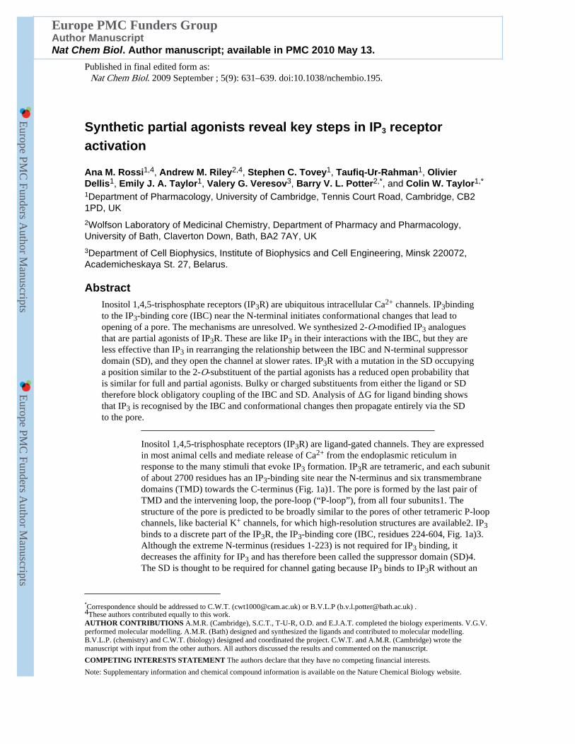
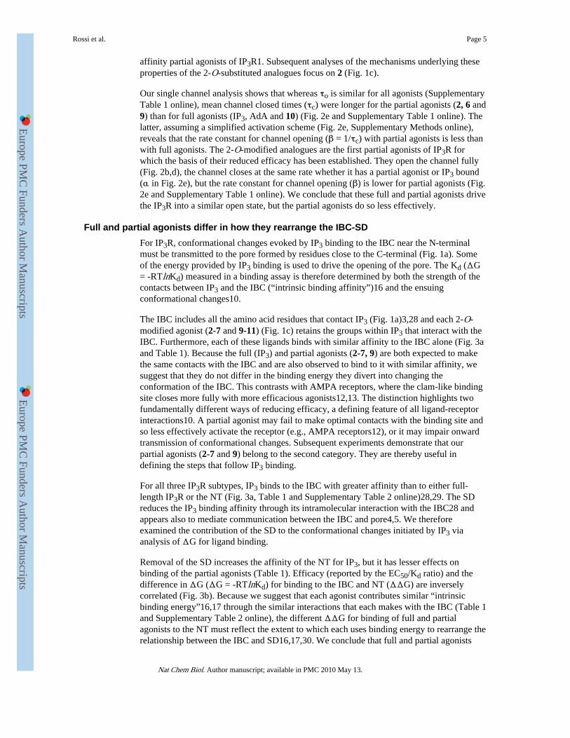
Figure 1.Structure of the IP3R and its ligands. (a) Key domains of IP3R (numbering from rat IP3R1,accession code NM_001007235). Pink denotes the SD (residues 1-223), red the IBC(224-604), and black vertical lines represent TMD. The SD (β1) and IBC (β2 and armadillo-like repeat, ARM) comprise 3 stably folded domains connected by flexible linkers (L1 andL2)34. Crystal structures are shown below3,33. (b) Agonist binding (yellow) to a discretesite on the receptor (IBC for IP3R, red) evokes conformational changes that propagatethrough the receptor and which then affect (arrows) the binding site. Removal (boxeddiagrams) of a domain through which conformational changes must pass prevents this
Rossi et al. Page 14
Nat Chem Biol. Author manuscript; available in PMC 2010 May 13.
Europe PM
C Funders A
uthor Manuscripts
Europe PM
C Funders A
uthor Manuscripts

energetic interplay between conformational changes and binding. (c) Structures of theligands used.
Rossi et al. Page 15
Nat Chem Biol. Author manuscript; available in PMC 2010 May 13.
Europe PM
C Funders A
uthor Manuscripts
Europe PM
C Funders A
uthor Manuscripts
Figure 2.2-O-modified IP3 analogues are partial agonists of IP3R. (a) From experiments similar tothose shown in Supplementary Fig. 1a,b online, EC50/Kd ratios (n ≥ 4) were calculated foreach ligand. (b) Traces (each typical of at least 3 similar records) show excised nuclearpatch-clamp recordings from DT40-IP3R1 cells with the pipette solution containing ATP(0.5mM), a free [Ca2+] of 200nM and the indicated ligands (10μM, except where shownotherwise). The holding potential was +40mV. C denotes the closed state. (c) Cells weretreated with IP3 alone, or for 30s with either 0.1nM AdA or 2 and then with the indicatedconcentrations of IP3. Results (n = 3) show the concentration-dependent release of Ca2+ by
Rossi et al. Page 16
Nat Chem Biol. Author manuscript; available in PMC 2010 May 13.
Europe PM
C Funders A
uthor Manuscripts
Europe PM
C Funders A
uthor Manuscripts
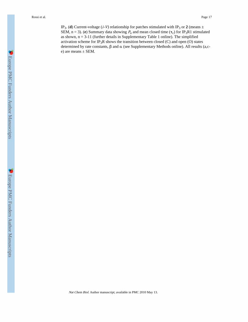
IP3. (d) Current-voltage (i-V) relationship for patches stimulated with IP3 or 2 (means ±SEM, n = 3). (e) Summary data showing Po and mean closed time (τc) for IP3R1 stimulatedas shown, n = 3-11 (further details in Supplementary Table 1 online). The simplifiedactivation scheme for IP3R shows the transition between closed (C) and open (O) statesdetermined by rate constants, β and α (see Supplementary Methods online). All results (a,c-e) are means ± SEM.
Rossi et al. Page 17
Nat Chem Biol. Author manuscript; available in PMC 2010 May 13.
Europe PM
C Funders A
uthor Manuscripts
Europe PM
C Funders A
uthor Manuscripts
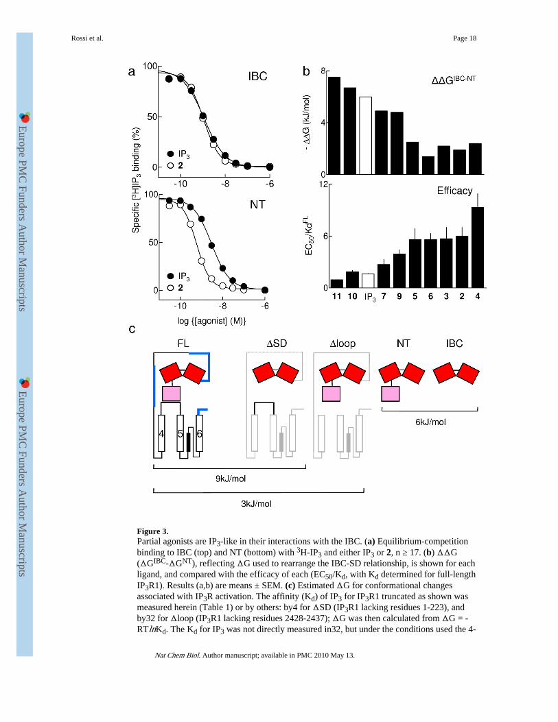
Figure 3.Partial agonists are IP3-like in their interactions with the IBC. (a) Equilibrium-competitionbinding to IBC (top) and NT (bottom) with 3H-IP3 and either IP3 or 2, n ≥ 17. (b) ΔΔG(ΔGIBC-ΔGNT), reflecting ΔG used to rearrange the IBC-SD relationship, is shown for eachligand, and compared with the efficacy of each (EC50/Kd, with Kd determined for full-lengthIP3R1). Results (a,b) are means ± SEM. (c) Estimated ΔG for conformational changesassociated with IP3R activation. The affinity (Kd) of IP3 for IP3R1 truncated as shown wasmeasured herein (Table 1) or by others: by4 for ΔSD (IP3R1 lacking residues 1-223), andby32 for Δloop (IP3R1 lacking residues 2428-2437); ΔG was then calculated from ΔG = -RTlnKd. The Kd for IP3 was not directly measured in32, but under the conditions used the 4-
Rossi et al. Page 18
Nat Chem Biol. Author manuscript; available in PMC 2010 May 13.
Europe PM
C Funders A
uthor Manuscripts
Europe PM
C Funders A
uthor Manuscripts
fold increase in IP3 binding after deletion of residues 2428-2437 (ie Δloop) is likely toreflect a 4-fold decrease in Kd. We assume that deletion of IP3R fragments through whichconformational changes must pass increases IP3 affinity because less binding energy isdiverted into re-arranging the protein (Fig. 1b). Deletions of many other regions (shown inblue) do not increase IP3 affinity4, suggesting that the IP3-evoked conformational changesdo not pass through them. This analysis is consistent with each IP3 binding event diverting~9kJ/mol into conformational changes of the IP3R, of which ~6kJ/mol rearranges the SD-IBC relationship, and ~3kJ/mol is used by the SD to gate the pore.
Rossi et al. Page 19
Nat Chem Biol. Author manuscript; available in PMC 2010 May 13.
Europe PM
C Funders A
uthor Manuscripts
Europe PM
C Funders A
uthor Manuscripts
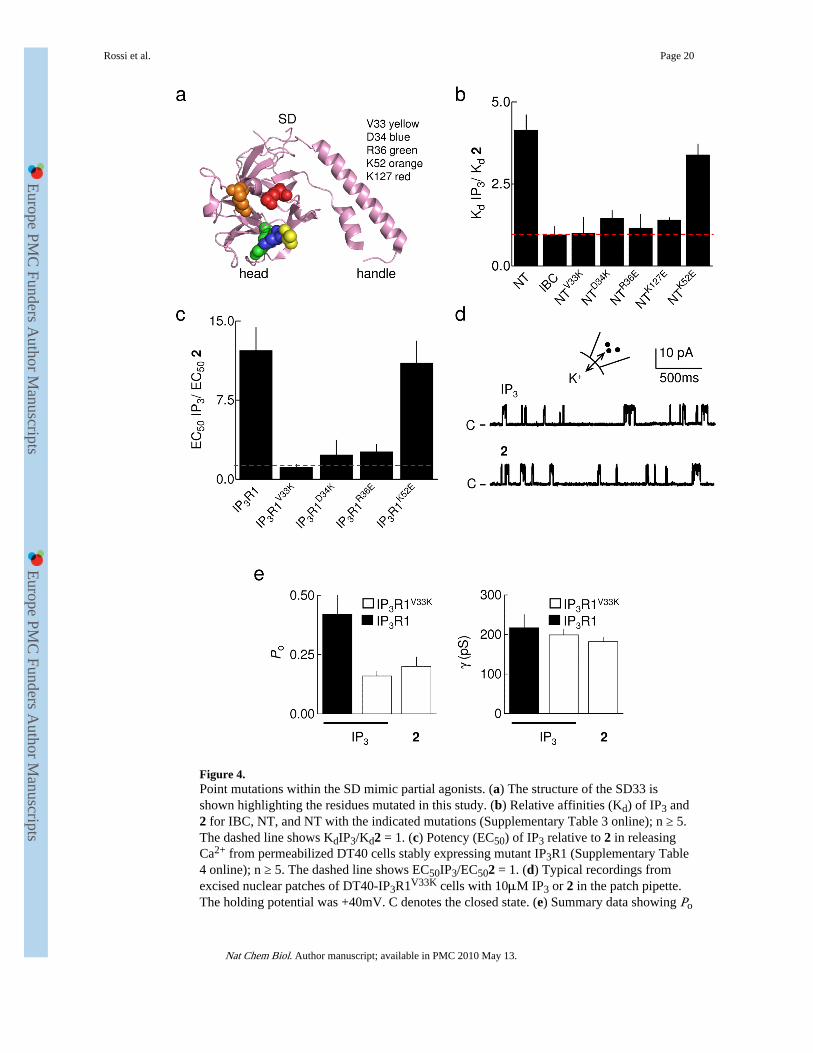
Figure 4.Point mutations within the SD mimic partial agonists. (a) The structure of the SD33 isshown highlighting the residues mutated in this study. (b) Relative affinities (Kd) of IP3 and2 for IBC, NT, and NT with the indicated mutations (Supplementary Table 3 online); n ≥ 5.The dashed line shows KdIP3/Kd2 = 1. (c) Potency (EC50) of IP3 relative to 2 in releasingCa2+ from permeabilized DT40 cells stably expressing mutant IP3R1 (Supplementary Table4 online); n ≥ 5. The dashed line shows EC50IP3/EC502 = 1. (d) Typical recordings fromexcised nuclear patches of DT40-IP3R1V33K cells with 10μM IP3 or 2 in the patch pipette.The holding potential was +40mV. C denotes the closed state. (e) Summary data showing Po
Rossi et al. Page 20
Nat Chem Biol. Author manuscript; available in PMC 2010 May 13.
Europe PM
C Funders A
uthor Manuscripts
Europe PM
C Funders A
uthor Manuscripts
and γK for IP3R1 and IP3R1V33K stimulated with 10μM IP3 or 2; n ≥ 3. Results (b,c,e) aremeans ± SEM.
Rossi et al. Page 21
Nat Chem Biol. Author manuscript; available in PMC 2010 May 13.
Europe PM
C Funders A
uthor Manuscripts
Europe PM
C Funders A
uthor Manuscripts
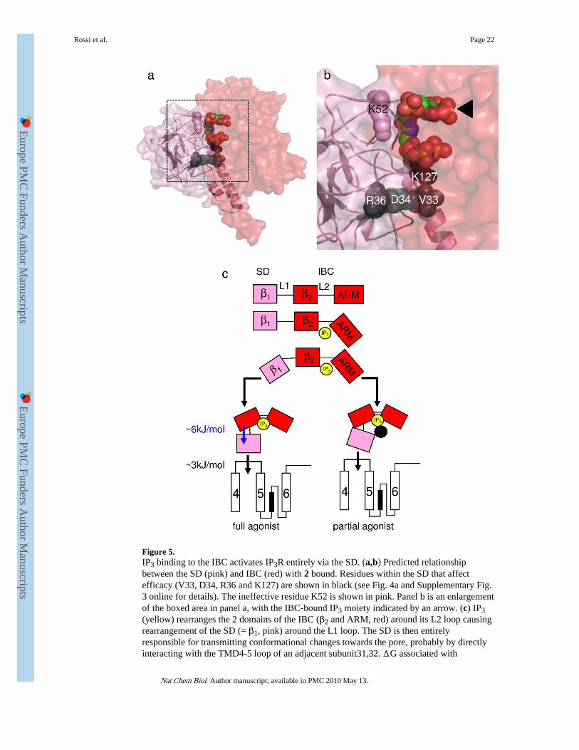
Figure 5.IP3 binding to the IBC activates IP3R entirely via the SD. (a,b) Predicted relationshipbetween the SD (pink) and IBC (red) with 2 bound. Residues within the SD that affectefficacy (V33, D34, R36 and K127) are shown in black (see Fig. 4a and Supplementary Fig.3 online for details). The ineffective residue K52 is shown in pink. Panel b is an enlargementof the boxed area in panel a, with the IBC-bound IP3 moiety indicated by an arrow. (c) IP3(yellow) rearranges the 2 domains of the IBC (β2 and ARM, red) around its L2 loop causingrearrangement of the SD (= β1, pink) around the L1 loop. The SD is then entirelyresponsible for transmitting conformational changes towards the pore, probably by directlyinteracting with the TMD4-5 loop of an adjacent subunit31,32. ΔG associated with
Rossi et al. Page 22
Nat Chem Biol. Author manuscript; available in PMC 2010 May 13.
Europe PM
C Funders A
uthor Manuscripts
Europe PM
C Funders A
uthor Manuscripts
rearranging the SD and its subsequent communication with the pore region is shown. Partialagonists effectively rearrange the IBC, but the inositol 2-O-substituent (or point mutations inthe SD; black circle) disrupt the IBC-SD interface and so block communication with the SD.The latter is now less likely to contact the TMD4-5 loop, but once it makes contact thechannel gates normally.
Rossi et al. Page 23
Nat Chem Biol. Author manuscript; available in PMC 2010 May 13.
Europe PM
C Funders A
uthor Manuscripts
Europe PM
C Funders A
uthor Manuscripts
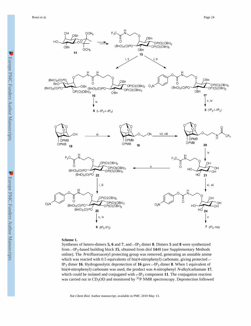
Scheme 1.Syntheses of hetero-dimers 5, 6 and 7, and L-IP3 dimer 8. Dimers 5 and 8 were synthesizedfrom L-IP3-based building block 15, obtained from diol 1448 (see Supplementary Methodsonline). The N-trifluoroacetyl protecting group was removed, generating an unstable aminewhich was reacted with 0.5 equivalents of bis(4-nitrophenyl) carbonate, giving protected L-IP3 dimer 16. Hydrogenolytic deprotection of 16 gave L-IP3 dimer 8. When 1 equivalent ofbis(4-nitrophenyl) carbonate was used, the product was 4-nitrophenyl N-alkylcarbamate 17,which could be isolated and conjugated with D-IP3 component 11. The conjugation reactionwas carried out in CD3OD and monitored by 31P NMR spectroscopy. Deprotection followed
Rossi et al. Page 24
Nat Chem Biol. Author manuscript; available in PMC 2010 May 13.
Europe PM
C Funders A
uthor Manuscripts
Europe PM
C Funders A
uthor Manuscripts
by anion-exchange chromatography then gave IP3–L-IP3 hetero-dimer 5. Dimers 6 and 7were synthesized from alcohol 1849. Nitrile 19 was reduced, and the amine product wastemporarily protected as the N-trifluoroacetamide (20). Acid-labile protecting groups werethen removed, giving pentaol 21, which was converted, via 22, into carbamate 23.Carbamate 23 was then conjugated with 11, and deprotection followed by anion-exchangechromatography gave IP3–IP5 hetero-dimer 6. Alternatively, conjugation of carbamate 24with 11 gave IP3–Ins dimer 7. Reagents and conditions: (i) LiOH, THF, MeOH, H2O; (ii)bis(4-nitrophenyl) carbonate (0.5 equiv), THF; (iii) bis(4-nitrophenyl) carbonate (1 equiv),THF; (iv) H2, Pd(OH)2/C, MeOH, H2O; (v) 11, CD3OD, Et3N; (vi) NaH, BrCH2CN,CH3CN; (vii) LiAlH4, THF; (viii) EtOC(O)CF3, THF; (ix) TFA, H2O; (x) (BnO)2PNiPr2,1H-tetrazole, CH2Cl2 then 3-chloroperoxybenzoic acid; (xi) Et3N, H2O, reflux; (xii) bis(4-nitrophenyl) carbonate (1 equiv), DMF, Et3N. Bn, benzyl; PMB, 4-methoxybenzyl. Allexperimental procedures are described in detail in Supplementary Methods online.
Rossi et al. Page 25
Nat Chem Biol. Author manuscript; available in PMC 2010 May 13.
Europe PM
C Funders A
uthor Manuscripts
Europe PM
C Funders A
uthor Manuscripts
Europe PM
C Funders A
uthor Manuscripts
Europe PM
C Funders A
uthor Manuscripts
Rossi et al. Page 26
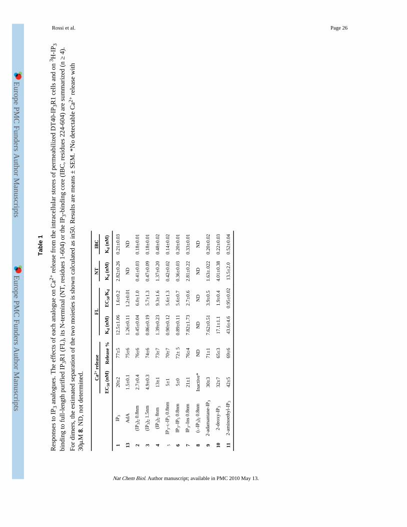
Tabl
e 1
Res
pons
es to
IP 3
ana
logu
es. T
he e
ffec
ts o
f ea
ch a
nalo
gue
on C
a2+ r
elea
se f
rom
the
intr
acel
lula
r st
ores
of
perm
eabi
lized
DT
40-I
P 3R
1 ce
lls a
nd o
n 3 H
-IP 3
bind
ing
to f
ull-
leng
th p
urif
ied
IP3R
1 (F
L),
its
N-t
erm
inal
(N
T, r
esid
ues
1-60
4) o
r th
e IP
3-bi
ndin
g co
re (
IBC
, res
idue
s 22
4-60
4) a
re s
umm
ariz
ed (
n ≥
4).
For
dim
ers,
the
estim
ated
sep
arat
ion
of th
e tw
o m
oiet
ies
is s
how
n ca
lcul
ated
as
in50
. Res
ults
are
mea
ns ±
SE
M. *
No
dete
ctab
le C
a2+ r
elea
se w
ith30μ
M 8
. ND
, not
det
erm
ined
.
Ca2+
rel
ease
FL
NT
IBC
EC
50 (
nM)
Rel
ease
%K
d (n
M)
EC
50/K
dK
d (n
M)
Kd
(nM
)
1IP
320
±2
77±
512
.5±
1.06
1.6±
0.2
2.82
±0.
260.
21±
0.03
13A
dA1.
5±0.
175
±6
1.26
±0.
111.
2±0.
01N
DN
D
2(I
P 3) 2
0.8
nm2.
7±0.
476
±6
0.45
±0.
046.
0±1.
00.
41±
0.03
0.18
±0.
01
3(I
P 3) 2
1.5
nm4.
9±0.
374
±6
0.86
±0.
195.
7±1.
30.
47±
0.09
0.18
±0.
01
4(I
P 3) 2
8nm
13±
173
±7
1.39
±0.
239.
3±1.
61.
37±
0.20
0.48
±0.
02
5IP
3–L-I
P 3 0
.8nm
5±1
70±
70.
90±
0.12
5.6±
1.3
0.42
±0.
020.
14±
0.02
6IP
3–IP
5 0.
8nm
5±0
72±
50.
89±
0.11
5.6±
0.7
0.36
±0.
030.
20±
0.01
7IP
3–In
s 0.
8nm
21±
176
±4
7.82
±1.
732.
7±0.
62.
81±
0.22
0.33
±0.
01
8(L
-IP 3
) 2 0
.8nm
Inac
tive*
ND
ND
ND
ND
ND
92-
adam
anta
ne-I
P 330
±3
71±
17.
62±
0.51
3.9±
0.5
1.63
±.0
220.
20±
0.02
102-
deox
y-IP
332
±7
65±
317
.1±
1.1
1.9±
0.4
4.01
±0.
380.
22±
0.03
112-
amin
oeth
yl-I
P 342
±5
69±
643
.6±
4.6
0.95
±0.
0213
.5±
2.0
0.52
±0.
04
Nat Chem Biol. Author manuscript; available in PMC 2010 May 13.
Related Documents