Synthesis, structure–activity relationship, and mechanistic investigation of lithocholic acid amphiphiles for colon cancer therapy† Manish Singh,‡ a Sandhya Bansal,‡ a Somanath Kundu,‡ ac Priyanshu Bhargava, a Ashima Singh, a Rajender K. Motiani,§ a Radhey Shyam, b Vedagopuram Sreekanth, ac Sagar Sengupta b and Avinash Bajaj * a We report a structure–activity relationship of lithocholic acid amphiphiles for their anticancer activities against colon cancer. We synthesized ten cationic amphiphiles, differing in nature of their cationic charged head groups, using lithocholic acid. We observed that anticancer activities of these amphiphiles against colon cancer cell lines are contingent on nature of the charged head group. The lithocholic acid-based amphiphile possessing a piperidine head group (LCA-PIP 1 ) is 10 times more cytotoxic than its precursor. Biochemical studies revealed that enhanced activity of LCA-PIP 1 compared to lithocholic acid is due to a greater activation of apoptosis. LCA-PIP 1 induces sub G 0 arrest and causes cleavage of caspases. A single dose of lithocholic acid–piperidine (LCA-PIP 1 ) derivative is enough to reduce the tumor burden by 75% in a tumor xenograft model. Introduction Colon cancer is the third most malignant tumor type, involving uncontrolled growth of cells in the colon. 1 Many small molecule and antibody-based therapeutics have been engineered and developed for cancer therapy to target this disease effectively. 2 Apart from genetic reasons, prolonged high consumption of a western-type diet augments the risk of colon cancer in the majority of patients. An increased uptake of fatty diets requires extensive recirculation of bile acids for its digestion. Therefore, colon epithelial cells experience enhanced exposure to this high concentration of bile acids. Clinical studies have revealed elevated levels of fecal secondary bile acids in patients diag- nosed with colon cancer. 3 Therefore, bile acids are critical for pathogenesis of colon cancer progression and renewal of colon epithelium. Bile acids are cholesterol-derived amphiphile molecules that help in the absorption of fats and fat-soluble vitamins. 4 Primary bile acids, like chenodeoxycholic acid and cholic acid, are synthesized and secreted from the liver and get re-circulated via a portal system. 5 Gut microora convert these primary bile acids to secondary bile acids, lithocholic acid and deoxycholic acid, by de-hydroxylation. 6 A high concentration of bile acids and their accumulation in the gut can induce apoptosis in the colon epithelium. 7 Bile acid-induced cytotoxicity causes colon cancer progression by disrupting the balance between cell growth and apoptosis, and helps in the selection of bile acid-resistant colon cancer cells. 8 Bile acids exercise their cytotoxic effect via different cellular mechanisms that are contingent on the structure, hydropho- bicity, and stereochemistry of the bile acids. 9 Glycine and taurine conjugated primary bile acids are the least toxic due to their low hydrophobicity, whereas unconjugated hydrophobic secondary bile acids are more cytotoxic. 10 Diverse cellular mechanisms, including membrane disruptions, the role of PKC, MAPK pathways, and nuclear factor-kappa b activation, have been proposed for the toxicity of bile acids. 11 Stenson and coworkers suggested an enantiospecic bile acid-mediated apoptosis in colon cancer cells. 12 Natural bile acids induce apoptosis, escalated cellular detachment and enhanced cas- pases-3 and -9 cleavage as compared to synthetic enantiomers of bile acids in HT-29 and HCT-116 cells. 13 Cationic lipid amphiphiles and their drug conjugates have been explored for potential biological activities as anticancer and antibacterial candidates as they have the ability to interact with cellular membranes effectively due to their charged hydrophobic character. 14 Chhikara et al. synthesized lipophilic derivatives of the anticancer drug doxorubicin for anticancer a The Laboratory of Nanotechnology and Chemical Biology, Regional Centre for Biotechnology, 180 Udyog Vihar, Phase 1, Gurgaon-122016, Haryana, India. E-mail: [email protected]; Fax: +91-124-4038117; Tel: +91-124-2848831 b National Institute of Immunology, Aruna Asif Ali Marg, New Delhi 110067, India c Manipal University, Manipal, Karnatka, India † Electronic supplementary information (ESI) available: Comparison of cytotoxicity of ester and amide derivatives of LCA-PIP 1 . Characterization of nal compounds LCA-PPZ 1 , LCA-MOR 1 , and LCA-TMOR 1 . HPLC proles of all the amphiphiles. See DOI: 10.1039/c4md00223g ‡ These authors contributed equally. § Current address: Institute of Genomics and Integrative Biology, New Delhi-110025. Cite this: Med. Chem. Commun., 2015, 6, 192 Received 28th May 2014 Accepted 14th October 2014 DOI: 10.1039/c4md00223g www.rsc.org/medchemcomm 192 | Med. Chem. Commun. , 2015, 6, 192–201 This journal is © The Royal Society of Chemistry 2015 MedChemComm CONCISE ARTICLE Published on 15 October 2014. Downloaded on 19/01/2015 16:54:35. View Article Online View Journal | View Issue

Welcome message from author
This document is posted to help you gain knowledge. Please leave a comment to let me know what you think about it! Share it to your friends and learn new things together.
Transcript
MedChemComm
CONCISE ARTICLE
Publ
ishe
d on
15
Oct
ober
201
4. D
ownl
oade
d on
19/
01/2
015
16:5
4:35
.
View Article OnlineView Journal | View Issue
Synthesis, structu
aThe Laboratory of Nanotechnology and
Biotechnology, 180 Udyog Vihar, Phase 1, G
[email protected]; Fax: +91-124-4038117; TebNational Institute of Immunology, Aruna AcManipal University, Manipal, Karnatka, In
† Electronic supplementary informatiocytotoxicity of ester and amide derivativecompounds LCA-PPZ1, LCA-MOR1, andamphiphiles. See DOI: 10.1039/c4md0022
‡ These authors contributed equally.
§ Current address: Institute of GenoDelhi-110025.
Cite this:Med. Chem. Commun., 2015,6, 192
Received 28th May 2014Accepted 14th October 2014
DOI: 10.1039/c4md00223g
www.rsc.org/medchemcomm
192 | Med. Chem. Commun., 2015, 6,
re–activity relationship, andmechanistic investigation of lithocholic acidamphiphiles for colon cancer therapy†
Manish Singh,‡a Sandhya Bansal,‡a Somanath Kundu,‡ac Priyanshu Bhargava,a
Ashima Singh,a Rajender K. Motiani,§a Radhey Shyam,b Vedagopuram Sreekanth,ac
Sagar Senguptab and Avinash Bajaj*a
We report a structure–activity relationship of lithocholic acid amphiphiles for their anticancer activities
against colon cancer. We synthesized ten cationic amphiphiles, differing in nature of their cationic
charged head groups, using lithocholic acid. We observed that anticancer activities of these amphiphiles
against colon cancer cell lines are contingent on nature of the charged head group. The lithocholic
acid-based amphiphile possessing a piperidine head group (LCA-PIP1) is �10 times more cytotoxic than
its precursor. Biochemical studies revealed that enhanced activity of LCA-PIP1 compared to lithocholic
acid is due to a greater activation of apoptosis. LCA-PIP1 induces sub G0 arrest and causes cleavage of
caspases. A single dose of lithocholic acid–piperidine (LCA-PIP1) derivative is enough to reduce the
tumor burden by 75% in a tumor xenograft model.
Introduction
Colon cancer is the third most malignant tumor type, involvinguncontrolled growth of cells in the colon.1 Many small moleculeand antibody-based therapeutics have been engineered anddeveloped for cancer therapy to target this disease effectively.2
Apart from genetic reasons, prolonged high consumption of awestern-type diet augments the risk of colon cancer in themajority of patients. An increased uptake of fatty diets requiresextensive recirculation of bile acids for its digestion. Therefore,colon epithelial cells experience enhanced exposure to this highconcentration of bile acids. Clinical studies have revealedelevated levels of fecal secondary bile acids in patients diag-nosed with colon cancer.3 Therefore, bile acids are critical forpathogenesis of colon cancer progression and renewal of colonepithelium.
Bile acids are cholesterol-derived amphiphile molecules thathelp in the absorption of fats and fat-soluble vitamins.4 Primary
Chemical Biology, Regional Centre for
urgaon-122016, Haryana, India. E-mail:
l: +91-124-2848831
sif Ali Marg, New Delhi 110067, India
dia
n (ESI) available: Comparison ofs of LCA-PIP1. Characterization of nalLCA-TMOR1. HPLC proles of all the3g
mics and Integrative Biology, New
192–201
bile acids, like chenodeoxycholic acid and cholic acid, aresynthesized and secreted from the liver and get re-circulated viaa portal system.5 Gut microora convert these primary bile acidsto secondary bile acids, lithocholic acid and deoxycholic acid,by de-hydroxylation.6 A high concentration of bile acids andtheir accumulation in the gut can induce apoptosis in the colonepithelium.7 Bile acid-induced cytotoxicity causes colon cancerprogression by disrupting the balance between cell growth andapoptosis, and helps in the selection of bile acid-resistant coloncancer cells.8
Bile acids exercise their cytotoxic effect via different cellularmechanisms that are contingent on the structure, hydropho-bicity, and stereochemistry of the bile acids.9 Glycine andtaurine conjugated primary bile acids are the least toxic due totheir low hydrophobicity, whereas unconjugated hydrophobicsecondary bile acids are more cytotoxic.10 Diverse cellularmechanisms, including membrane disruptions, the role ofPKC, MAPK pathways, and nuclear factor-kappa b activation,have been proposed for the toxicity of bile acids.11 Stenson andcoworkers suggested an enantiospecic bile acid-mediatedapoptosis in colon cancer cells.12 Natural bile acids induceapoptosis, escalated cellular detachment and enhanced cas-pases-3 and -9 cleavage as compared to synthetic enantiomersof bile acids in HT-29 and HCT-116 cells.13
Cationic lipid amphiphiles and their drug conjugates havebeen explored for potential biological activities as anticancerand antibacterial candidates as they have the ability to interactwith cellular membranes effectively due to their chargedhydrophobic character.14 Chhikara et al. synthesized lipophilicderivatives of the anticancer drug doxorubicin for anticancer
This journal is © The Royal Society of Chemistry 2015
Concise Article MedChemComm
Publ
ishe
d on
15
Oct
ober
201
4. D
ownl
oade
d on
19/
01/2
015
16:5
4:35
. View Article Online
therapy15 and emtricitabine-based prodrugs for enhanced anti-HIV activity.16 Banerjee and co-workers synthesized haloperidol-conjugated cationic lipids17 and lipids targeting estrogenreceptors for targeted delivery to cancer cells.18 Sreekanth et al.synthesized charged bile acid tamoxifen conjugates for breastcancer therapy and revealed that the conjugation of multipletamoxifen molecules to cationic cholic acid induces membraneperturbations and enhanced activity.19 We have recently shownthat the conjugation of single charged head groups on bile acidsinduces favorable interactions with model membranes andcancer cells for their cytotoxicity, whereas multi-headedamphiphiles do not interact with mammalian cells due to anenhanced hydration barrier between the amphiphiles andcellular membranes.20
In this paper, we hypothesize that anticancer activity ofamphiphiles is contingent on nature of single charged headgroup on a lithocholic acid amphiphile. Therefore, we synthe-sized ten lithocholic acid-based amphiphiles differing in natureof the charged head group attached to hydroxyl group of lith-ocholic acid. Anticancer activities of these amphiphiles wereinvestigated against three human colon cancer cell lines.Annexin-FITC and cell cycle analysis were performed to studythe mechanism of cellular death by these amphiphiles. We thenevaluated in vivo anticancer potential of the amphiphiles intumor xenogra models, and analyzed the mechanism ofactivity in tumor regression.
Results and discussionSynthesis of amphiphiles
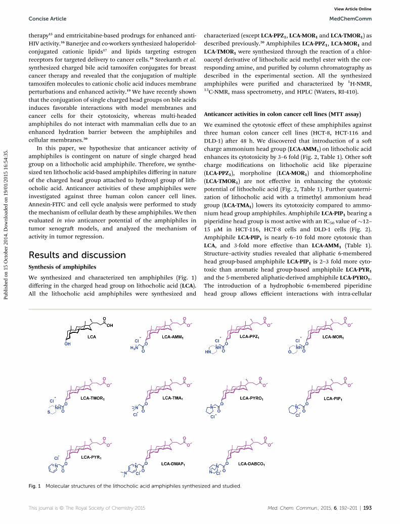
We synthesized and characterized ten amphiphiles (Fig. 1)differing in the charged head group on lithocholic acid (LCA).All the lithocholic acid amphiphiles were synthesized and
Fig. 1 Molecular structures of the lithocholic acid amphiphiles synthesiz
This journal is © The Royal Society of Chemistry 2015
characterized (except LCA-PPZ1, LCA-MOR1 and LCA-TMOR1) asdescribed previously.20 Amphiphiles LCA-PPZ1, LCA-MOR1 andLCA-TMOR1 were synthesized through the reaction of a chlor-oacetyl derivative of lithocholic acid methyl ester with the cor-responding amine, and puried by column chromatography asdescribed in the experimental section. All the synthesizedamphiphiles were puried and characterized by 1H-NMR,13C-NMR, mass spectrometry, and HPLC (Waters, RI-410).
Anticancer activities in colon cancer cell lines (MTT assay)
We examined the cytotoxic effect of these amphiphiles againstthree human colon cancer cell lines (HCT-8, HCT-116 andDLD-1) aer 48 h. We discovered that introduction of a socharge ammonium head group (LCA-AMM1) on lithocholic acidenhances its cytotoxicity by 3–6 fold (Fig. 2, Table 1). Other socharge modications on lithocholic acid like piperazine(LCA-PPZ1), morpholine (LCA-MOR1) and thiomorpholine(LCA-TMOR1) are not effective in enhancing the cytotoxicpotential of lithocholic acid (Fig. 2, Table 1). Further quaterni-zation of lithocholic acid with a trimethyl ammonium headgroup (LCA-TMA1) lowers its cytotoxicity compared to ammo-nium head group amphiphiles. Amphiphile LCA-PIP1 bearing apiperidine head group is most active with an IC50 value of �12–15 mM in HCT-116, HCT-8 cells and DLD-1 cells (Fig. 2).Amphiphile LCA-PIP1 is nearly 6–10 fold more cytotoxic thanLCA, and 3-fold more effective than LCA-AMM1 (Table 1).Structure–activity studies revealed that aliphatic 6-memberedhead group-based amphiphile LCA-PIP1 is 2–3 fold more cyto-toxic than aromatic head group-based amphiphile LCA-PYR1
and the 5-membered aliphatic-derived amphiphile LCA-PYRO1.The introduction of a hydrophobic 6-membered piperidinehead group allows efficient interactions with intra-cellular
ed and studied.
Med. Chem. Commun., 2015, 6, 192–201 | 193
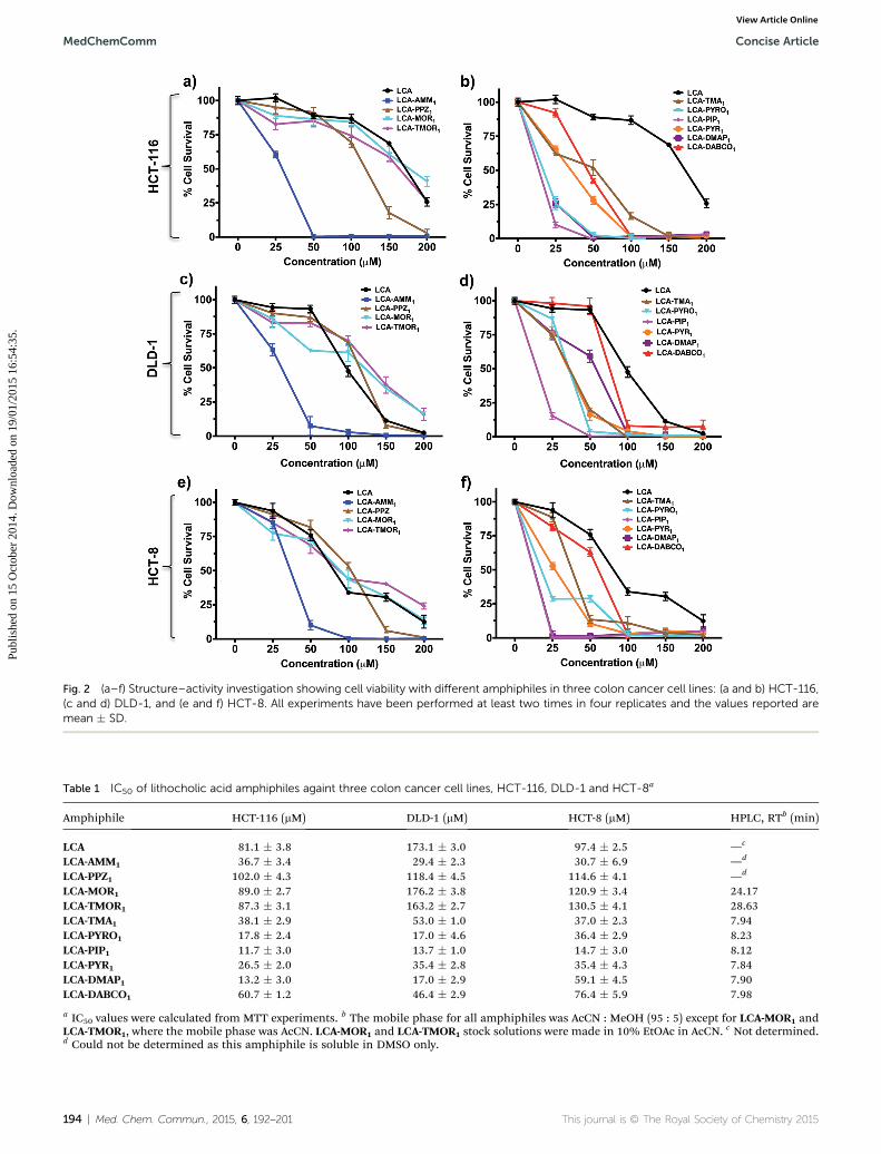
Fig. 2 (a–f) Structure–activity investigation showing cell viability with different amphiphiles in three colon cancer cell lines: (a and b) HCT-116,(c and d) DLD-1, and (e and f) HCT-8. All experiments have been performed at least two times in four replicates and the values reported aremean � SD.
Table 1 IC50 of lithocholic acid amphiphiles againt three colon cancer cell lines, HCT-116, DLD-1 and HCT-8a
Amphiphile HCT-116 (mM) DLD-1 (mM) HCT-8 (mM) HPLC, RTb (min)
LCA 81.1 � 3.8 173.1 � 3.0 97.4 � 2.5 —c
LCA-AMM1 36.7 � 3.4 29.4 � 2.3 30.7 � 6.9 —d
LCA-PPZ1 102.0 � 4.3 118.4 � 4.5 114.6 � 4.1 —d
LCA-MOR1 89.0 � 2.7 176.2 � 3.8 120.9 � 3.4 24.17LCA-TMOR1 87.3 � 3.1 163.2 � 2.7 130.5 � 4.1 28.63LCA-TMA1 38.1 � 2.9 53.0 � 1.0 37.0 � 2.3 7.94LCA-PYRO1 17.8 � 2.4 17.0 � 4.6 36.4 � 2.9 8.23LCA-PIP1 11.7 � 3.0 13.7 � 1.0 14.7 � 3.0 8.12LCA-PYR1 26.5 � 2.0 35.4 � 2.8 35.4 � 4.3 7.84LCA-DMAP1 13.2 � 3.0 17.0 � 2.9 59.1 � 4.5 7.90LCA-DABCO1 60.7 � 1.2 46.4 � 2.9 76.4 � 5.9 7.98
a IC50 values were calculated from MTT experiments. b The mobile phase for all amphiphiles was AcCN : MeOH (95 : 5) except for LCA-MOR1 andLCA-TMOR1, where the mobile phase was AcCN. LCA-MOR1 and LCA-TMOR1 stock solutions were made in 10% EtOAc in AcCN. c Not determined.d Could not be determined as this amphiphile is soluble in DMSO only.
194 | Med. Chem. Commun., 2015, 6, 192–201 This journal is © The Royal Society of Chemistry 2015
MedChemComm Concise Article
Publ
ishe
d on
15
Oct
ober
201
4. D
ownl
oade
d on
19/
01/2
015
16:5
4:35
. View Article Online
Concise Article MedChemComm
Publ
ishe
d on
15
Oct
ober
201
4. D
ownl
oade
d on
19/
01/2
015
16:5
4:35
. View Article Online
targets, making it more effective. Therefore, the structure–activity studies conclude that anticancer activities of amphi-philes are strongly contingent on nature of head group. As bileacids are known to bind with cell membrane TGR5 receptors ornuclear FXR receptors, nature of charged head group may helpin preferential interactions of these amphiphiles with cell
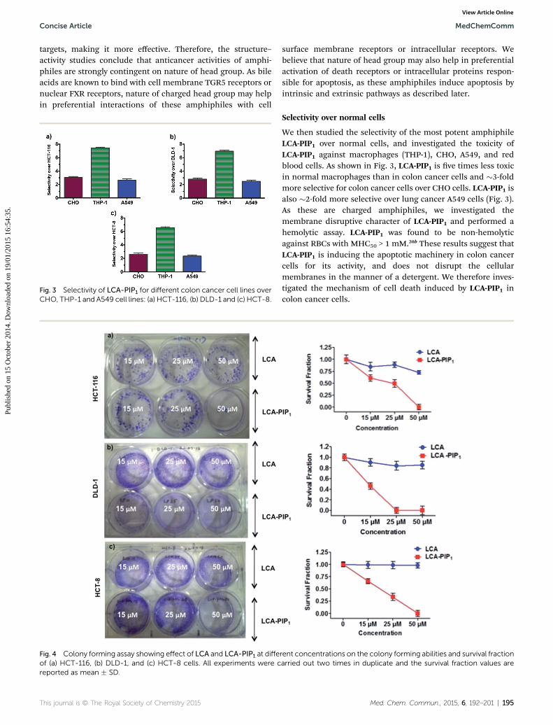
Fig. 3 Selectivity of LCA-PIP1 for different colon cancer cell lines overCHO, THP-1 and A549 cell lines: (a) HCT-116, (b) DLD-1 and (c) HCT-8.
Fig. 4 Colony forming assay showing effect of LCA and LCA-PIP1 at diffeof (a) HCT-116, (b) DLD-1, and (c) HCT-8 cells. All experiments were creported as mean � SD.
This journal is © The Royal Society of Chemistry 2015
surface membrane receptors or intracellular receptors. Webelieve that nature of head group may also help in preferentialactivation of death receptors or intracellular proteins respon-sible for apoptosis, as these amphiphiles induce apoptosis byintrinsic and extrinsic pathways as described later.
Selectivity over normal cells
We then studied the selectivity of the most potent amphiphileLCA-PIP1 over normal cells, and investigated the toxicity ofLCA-PIP1 against macrophages (THP-1), CHO, A549, and redblood cells. As shown in Fig. 3, LCA-PIP1 is ve times less toxicin normal macrophages than in colon cancer cells and �3-foldmore selective for colon cancer cells over CHO cells. LCA-PIP1 isalso �2-fold more selective over lung cancer A549 cells (Fig. 3).As these are charged amphiphiles, we investigated themembrane disruptive character of LCA-PIP1 and performed ahemolytic assay. LCA-PIP1 was found to be non-hemolyticagainst RBCs with MHC50 > 1 mM.20b These results suggest thatLCA-PIP1 is inducing the apoptotic machinery in colon cancercells for its activity, and does not disrupt the cellularmembranes in the manner of a detergent. We therefore inves-tigated the mechanism of cell death induced by LCA-PIP1 incolon cancer cells.
rent concentrations on the colony forming abilities and survival fractionarried out two times in duplicate and the survival fraction values are
Med. Chem. Commun., 2015, 6, 192–201 | 195
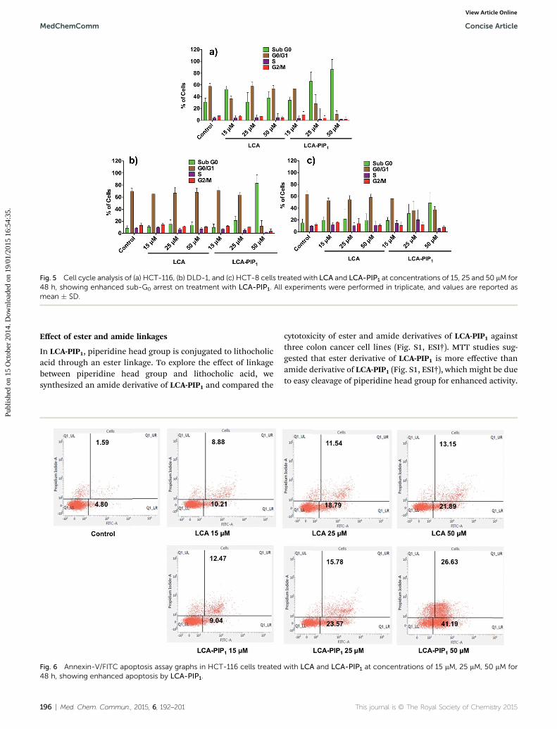
Fig. 5 Cell cycle analysis of (a) HCT-116, (b) DLD-1, and (c) HCT-8 cells treated with LCA and LCA-PIP1 at concentrations of 15, 25 and 50 mM for48 h, showing enhanced sub-G0 arrest on treatment with LCA-PIP1. All experiments were performed in triplicate, and values are reported asmean � SD.
MedChemComm Concise Article
Publ
ishe
d on
15
Oct
ober
201
4. D
ownl
oade
d on
19/
01/2
015
16:5
4:35
. View Article Online
Effect of ester and amide linkages
In LCA-PIP1, piperidine head group is conjugated to lithocholicacid through an ester linkage. To explore the effect of linkagebetween piperidine head group and lithocholic acid, wesynthesized an amide derivative of LCA-PIP1 and compared the
Fig. 6 Annexin-V/FITC apoptosis assay graphs in HCT-116 cells treated48 h, showing enhanced apoptosis by LCA-PIP1.
196 | Med. Chem. Commun., 2015, 6, 192–201
cytotoxicity of ester and amide derivatives of LCA-PIP1 againstthree colon cancer cell lines (Fig. S1, ESI†). MTT studies sug-gested that ester derivative of LCA-PIP1 is more effective thanamide derivative of LCA-PIP1 (Fig. S1, ESI†), which might be dueto easy cleavage of piperidine head group for enhanced activity.
with LCA and LCA-PIP1 at concentrations of 15 mM, 25 mM, 50 mM for
This journal is © The Royal Society of Chemistry 2015
Concise Article MedChemComm
Publ
ishe
d on
15
Oct
ober
201
4. D
ownl
oade
d on
19/
01/2
015
16:5
4:35
. View Article Online
Colony forming assay
We then studied the colony-forming abilities of colon cancercells on treatment with LCA and LCA-PIP1 at different concen-trations. The treatment of these cells with LCA does not inhibitcolony formation at 15, 25 and 50 mM concentrations, whereasLCA-PIP1 on treatment at 15 mM induces �50% inhibition inHCT-116 cells (Fig. 4a). We did not observe any colonies ontreatment with 50 mM LCA-PIP1. Similarly, treatment withLCA-PIP1 inhibits colony formation in DLD-1 and HCT-8 coloncancer cells (Fig. 4b and c). These studies show that introduc-tion of a single piperidine head group on LCA makes it highlyactive and able to inhibit the colony-forming properties ofcancer cells.
Cell cycle and Annexin-FITC analysis
We then performed cell cycle analysis to know the fate of cells indifferent phases of cell cycle on treatment with LCA andLCA-PIP1 at different concentrations for 48 h. Cell cycle analysisrevealed that LCA alone does not induce any concentration
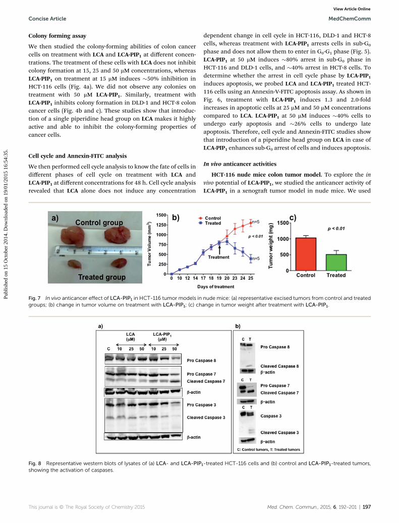
Fig. 7 In vivo anticancer effect of LCA-PIP1 in HCT-116 tumor models ingroups; (b) change in tumor volume on treatment with LCA-PIP1; (c) ch
Fig. 8 Representative western blots of lysates of (a) LCA- and LCA-PIPshowing the activation of caspases.
This journal is © The Royal Society of Chemistry 2015
dependent change in cell cycle in HCT-116, DLD-1 and HCT-8cells, whereas treatment with LCA-PIP1 arrests cells in sub-G0
phase and does not allow them to enter in G0-G1 phase (Fig. 5).LCA-PIP1 at 50 mM induces �80% arrest in sub-G0 phase inHCT-116 and DLD-1 cells, and �40% arrest in HCT-8 cells. Todetermine whether the arrest in cell cycle phase by LCA-PIP1induces apoptosis, we probed LCA and LCA-PIP1 treated HCT-116 cells using an Annexin-V-FITC apoptosis assay. As shown inFig. 6, treatment with LCA-PIP1 induces 1.3 and 2.0-foldincreases in apoptotic cells at 25 mM and 50 mM concentrationscompared to LCA. LCA-PIP1 at 50 mM induces �40% cells toundergo early apoptosis and �26% cells to undergo lateapoptosis. Therefore, cell cycle and Annexin-FITC studies showthat introduction of a piperidine head group on LCA in case ofLCA-PIP1 enhances sub-G0 arrest of cells and induces apoptosis.
In vivo anticancer activities
HCT-116 nude mice colon tumor model. To explore the invivo potential of LCA-PIP1, we studied the anticancer activity ofLCA-PIP1 in a xenogra tumor model in nude mice. We used
nude mice: (a) representative excised tumors from control and treatedange in tumor weight after treatment with LCA-PIP1.
1-treated HCT-116 cells and (b) control and LCA-PIP1-treated tumors,
Med. Chem. Commun., 2015, 6, 192–201 | 197
MedChemComm Concise Article
Publ
ishe
d on
15
Oct
ober
201
4. D
ownl
oade
d on
19/
01/2
015
16:5
4:35
. View Article Online
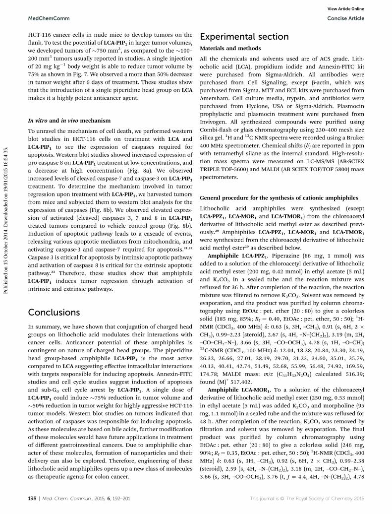
HCT-116 cancer cells in nude mice to develop tumors on theank. To test the potential of LCA-PIP1 in larger tumor volumes,we developed tumors of �750 mm3, as compared to the �100–200 mm3 tumors usually reported in studies. A single injectionof 20 mg kg�1 body weight is able to reduce tumor volume by75% as shown in Fig. 7. We observed a more than 50% decreasein tumor weight aer 6 days of treatment. These studies showthat the introduction of a single piperidine head group on LCAmakes it a highly potent anticancer agent.
In vitro and in vivo mechanism
To unravel the mechanism of cell death, we performed westernblot studies in HCT-116 cells on treatment with LCA andLCA-PIP1 to see the expression of caspases required forapoptosis. Western blot studies showed increased expression ofpro-caspase 8 on LCA-PIP1 treatment at low concentrations, anda decrease at high concentration (Fig. 8a). We observedincreased levels of cleaved caspase-7 and caspase-3 on LCA-PIP1treatment. To determine the mechanism involved in tumorregression upon treatment with LCA-PIP1, we harvested tumorsfrom mice and subjected them to western blot analysis for theexpression of caspases (Fig. 8b). We observed elevated expres-sion of activated (cleaved) caspases 3, 7 and 8 in LCA-PIP1treated tumors compared to vehicle control group (Fig. 8b).Induction of apoptotic pathway leads to a cascade of events,releasing various apoptotic mediators from mitochondria, andactivating caspase-3 and caspase-7 required for apoptosis.21,22
Caspase 3 is critical for apoptosis by intrinsic apoptotic pathwayand activation of caspase 8 is critical for the extrinsic apoptoticpathway.23 Therefore, these studies show that amphiphileLCA-PIP1 induces tumor regression through activation ofintrinsic and extrinsic pathways.
Conclusions
In summary, we have shown that conjugation of charged headgroups on lithocholic acid modulates their interactions withcancer cells. Anticancer potential of these amphiphiles iscontingent on nature of charged head groups. The piperidinehead group-based amphiphile LCA-PIP1 is the most activecompared to LCA suggesting effective intracellular interactionswith targets responsible for inducing apoptosis. Annexin-FITCstudies and cell cycle studies suggest induction of apoptosisand sub-G0 cell cycle arrest by LCA-PIP1. A single dose ofLCA-PIP1 could induce �75% reduction in tumor volume and�50% reduction in tumor weight for highly aggressive HCT-116tumor models. Western blot studies on tumors indicated thatactivation of caspases was responsible for inducing apoptosis.As these molecules are based on bile acids, further modicationof these molecules would have future applications in treatmentof different gastrointestinal cancers. Due to amphiphilic char-acter of these molecules, formation of nanoparticles and theirdelivery can also be explored. Therefore, engineering of theselithocholic acid amphiphiles opens up a new class of moleculesas therapeutic agents for colon cancer.
198 | Med. Chem. Commun., 2015, 6, 192–201
Experimental sectionMaterials and methods
All the chemicals and solvents used are of ACS grade. Lith-ocholic acid (LCA), propidium iodide and Annexin-FITC kitwere purchased from Sigma-Aldrich. All antibodies werepurchased from Cell Signaling, except b-actin, which waspurchased from Sigma. MTT and ECL kits were purchased fromAmersham. Cell culture media, trypsin, and antibiotics werepurchased from Hyclone, USA or Sigma-Aldrich. Plasmocinprophylactic and plasmocin treatment were purchased fromInvivogen. All synthesized compounds were puried usingCombi-ash or glass chromatography using 230–400 mesh sizesilica gel. 1H and 13C NMR spectra were recorded using a Bruker400 MHz spectrometer. Chemical shis (d) are reported in ppmwith tetramethyl silane as the internal standard. High-resolu-tion mass spectra were measured on LC-MS/MS (AB-SCIEXTRIPLE TOF-5600) and MALDI (AB SCIEX TOF/TOF 5800) massspectrometers.
General procedure for the synthesis of cationic amphiphiles
Lithocholic acid amphiphiles were synthesized (exceptLCA-PPZ1, LCA-MOR1 and LCA-TMOR1) from the chloroacetylderivative of lithocholic acid methyl ester as described previ-ously.20 Amphiphiles LCA-PPZ1, LCA-MOR1 and LCA-TMOR1
were synthesized from the chloroacetyl derivative of lithocholicacid methyl ester20 as described below.
Amphiphile LCA-PPZ1. Piperazine (86 mg, 1 mmol) wasadded to a solution of the chloroacetyl derivative of lithocholicacid methyl ester (200 mg, 0.42 mmol) in ethyl acetate (5 mL)and K2CO3 in a sealed tube and the reaction mixture wasreuxed for 36 h. Aer completion of the reaction, the reactionmixture was ltered to remove K2CO3. Solvent was removed byevaporation, and the product was puried by column chroma-tography using EtOAc : pet. ether (20 : 80) to give a colorlesssolid (185 mg, 85%; Rf ¼ 0.40, EtOAc : pet. ether, 50 : 50); 1H-NMR (CDCl3, 400 MHz) d: 0.63 (s, 3H, –CH3), 0.91 (s, 6H, 2 �CH3), 0.99–2.23 (steroid), 2.67 (s, 4H, –N–(CH2)2), 3.19 (m, 2H,–CO–CH2–N–), 3.66 (s, 3H, –CO–OCH3), 4.78 (s, 1H, –O–CH);13C-NMR (CDCl3, 100 MHz) d: 12.04, 18.28, 20.84, 23.30, 24.19,26.32, 26.66, 27.01, 28.19, 29.70, 31.23, 34.60, 35.01, 35.79,40.13, 40.41, 42.74, 51.49, 52.68, 55.99, 56.48, 74.92, 169.59,174.78; MALDI mass: m/z (C31H52N2O4) calculated 516.39;found (M)+ 517.402.
Amphiphile LCA-MOR1. To a solution of the chloroacetylderivative of lithocholic acid methyl ester (250 mg, 0.53 mmol)in ethyl acetate (5 mL) was added K2CO3 and morpholine (95mg, 1.1 mmol) in a sealed tube and the mixture was reuxed for48 h. Aer completion of the reaction, K2CO3 was removed byltration and solvent was removed by evaporation. The nalproduct was puried by column chromatography usingEtOAc : pet. ether (20 : 80) to give a colorless solid (246 mg,90%; Rf ¼ 0.35, EtOAc : pet. ether, 50 : 50); 1H-NMR (CDCl3, 400MHz) d: 0.63 (s, 3H, –CH3), 0.92 (s, 6H, 2 � CH3), 0.99–2.38(steroid), 2.59 (s, 4H, –N–(CH2)2), 3.18 (m, 2H, –CO–CH2–N–),3.66 (s, 3H, –CO–OCH3), 3.76 (t, J ¼ 4.4, 4H, –N–(CH2)2), 4.78
This journal is © The Royal Society of Chemistry 2015
Concise Article MedChemComm
Publ
ishe
d on
15
Oct
ober
201
4. D
ownl
oade
d on
19/
01/2
015
16:5
4:35
. View Article Online
(s, 1H, –O–CH); 13C-NMR (CDCl3, 100 MHz) d: 12.04, 18.28,20.84, 23.31, 24.19, 26.32, 26.67, 27.01, 28.19, 29.70, 31.01,31.07, 32.25, 34.60, 35.00, 35.38, 35.79, 40.14, 40.43, 41.91,42.74, 51.50, 53.25, 56.00, 56.50, 59.97, 66.75, 74.97, 74.92,169.50, 174.78; MALDI mass: m/z (C31H51NO5) calculated517.38; found (M)+ 518.377.
Amphiphile LCA-TMOR1. A reaction mixture of chloroacetylderivative of lithocholic acid methyl ester (300 mg, 0.64 mmol)in ethyl acetate (6 mL), K2CO3 (300 mg) and thiomorpholine(124 mg, 1.2 mmol) was reuxed in a sealed tube for 40 h. SolidK2CO3 was removed from reaction mixture and solvent wasremoved by evaporation. The product was puried by columnchromatography using EtOAc : pet. ether (20 : 80) to give acolorless solid (310 mg, 91%; Rf ¼ 0.62, EtOAc : pet. ether,50 : 50); 1H-NMR (CDCl3, 400 MHz) d: 0.63 (s, 3H, –CH3), 0.91(s, 6H, 2 � CH3), 0.99–2.33 (steroid), 2.72 (s, 4H, –S–(CH2)2),2.84 (d, J ¼ 4.4, 4H, –N–(CH2)2) 3.20 (s, 2H, –CO–CH2–N–), 3.66(s, 3H, –CO–OCH3), 4.77 (s, 1H, –O–CH); 13C-NMR (CDCl3, 100MHz) d: 12.04, 18.27, 20.84, 23.31, 24.18, 26.32, 26.71, 27.00,27.81, 28.18, 29.69, 31.01, 31.07, 3 32.28, 34.59, 35.00, 35.37,35.79, 40.13, 40.44, 41.92, 42.74, 51.48, 54.53, 56.00, 56.49,60.51, 74.94, 169.75, 174.74; MALDI Mass: m/z (C31H51NO4S)calculated 533.35; found (M)+ 534.284.
Amide derivative of LCA-PIP1
The amino derivative of lithocholic acid was synthesized asdescribed previously.24 The amide derivative of LCA-PIP1 wassynthesized using the amino derivative of lithocholic acid(LCA-NH2) using a similar procedure to that published.20 Yield80% (solid). 1H-NMR (CDCl3, 400 MHz) d: 0.62 (d, J ¼ 5.6, 3H,–CH3), 0.90 (s, 6H, 2 � CH3), 0.99–2.37 (steroid), 3.39 (s, 3H,–N–CH3), 3.42 (m, 2H, –N–CH2), 3.65 (s, 3H, –CO–OCH3), 3.91(m, 2H, –N–CH2), 4.69 (m, 2H, –CO–CH2–N–);
13C-NMR (CDCl3,100 MHz) d: 12.03, 18.27, 20.16, 20.87 23.38, 23.64, 24.18, 26.27,27.07, 28.16, 30.56, 32.75, 34.58, 35.37, 35.64, 40.00, 40.17,42.73, 50.92, 51.46, 55.97, 56.62, 62.69, 71.84, 162.86, 174.86;MALDI Mass: m/z (C33H57N2O3)
+ calculated 529.44; found (M)+
529.431.
Cell cultures
HCT-116, DLD-1, HCT-8, A549 and CHO cell lines were main-tained as monolayers. HCT-116 cells in McCoy's medium,DLD-1 and HCT-8 cells in RPMI-1640 medium, and A549 andCHO cells in DMEMmedium with 10% (v/v) fetal bovine serum,with antibiotics, were maintained at 37 �C in a humidiedatmosphere with 5% CO2. THP-1 cells were grown in DMEMmedium and differentiated to adherent macrophages by addingphorbol myristic acid (PMA).
Cytotoxicity assay25
MTT studies were performed for anticancer activities of allamphiphiles in three different colon cancer cell lines. The cellswere plated in 96 well plates at a density of 3–4 � 103 cells perwell for 24 h to allow adherence. The cells were treated atconcentrations of 25, 50, 100, 150 and 200 mM of syntheticlithocholic acid amphiphiles for 48 h. MTT solution (25 mL of
This journal is © The Royal Society of Chemistry 2015
5 mg mL�1) was added to the cells for incubation to produceformazan crystals. Aer 3 h of incubation, medium wasreplaced with 150 mL of DMSO to lyse the cells. The absorbanceof formazan crystals was recorded at 555 nm using a SpectramaxM5 (Molecular Devices). Cell viability was then calculatedusing the equation [{A555 (treated cells) � background]/[A555(untreated cells) � background}] � 100.
Colony formation assay26
Colony formation studies were performed with colon cancercells on treatment with LCA and LCA-PIP1 at different concen-trations. 200 cancer cells per well were plated in a 6-well plate.Aer adherence (24 h), cells were treated with differentconcentrations of LCA and LCA-PIP1. Aer 2 weeks, the cellswere stained with crystal violet and colonies were counted.From the number of colonies, we rst calculated the platingefficacy (PE) from untreated samples as:
PE ¼ number of colonies formed
number of cells seeded� 100
The number of colonies aer treatment of cells, called thesurvival fraction, is calculated using following equation:
SE ¼ number of colonies formed after treatment
number of cells seeded � PE
Cell cycle and Annexin-FITC analysis
In a 6-well plate, colon cancer cells at a density of �2 � 105 cellsper well were seeded. Aer 24 h, the cells were treated withamphiphiles for 48 h. Cells were trypsinized and collected bycentrifugation aer treatment. For cell cycle analysis, the cellswere washed twice with cold PBS and xed in 70% ice-coldethanol. Ethanol was removed by washing the cells again withPBS and cells were treated with RNase (10 mL of 20 mg mL�1) at37 �C for 1 h. Cells were stained with propidium iodide (50 mgmL�1) at room temperature for 20 min and counted by FACS(Becton Dickinson, Mountain View, CA). Annexin-FITC studieswere performed using a kit from Sigma-Aldrich according tomanufacturer's instructions. Cells were stained simultaneouslywith FITC-labeled Annexin V (50 mg mL�1) and propidiumiodide (100 mg mL�1) aer their re-suspension in binding bufferand analyzed using a ow cytometer (Becton Dickinson).
In vivo experiments
6 week old female nude mice were received from the Institu-tional Animal Facility and were allowed to acclimatize to facilityconditions before any experimental procedure. All the protocolsfor animal experiments were approved by the InstitutionalAnimal Ethical Committee of the National Institute of Immu-nology. Mycoplasma testing was performed in the cells to makesure that the cells were free of any sort of contamination. Weinjected 1.5 � 106 HCT-116 cells per mouse in the right ank ofeach mouse to generate tumor models. Tumors becamepalpable aer 7 days and we startedmeasuring tumor sizes aer
Med. Chem. Commun., 2015, 6, 192–201 | 199
MedChemComm Concise Article
Publ
ishe
d on
15
Oct
ober
201
4. D
ownl
oade
d on
19/
01/2
015
16:5
4:35
. View Article Online
the 10th day. Digital calipers were used for measuring tumorsizes throughout the study. Tumor volume was calculated usingthe formula: volume ¼ 0.5 L �W2, whereW ¼ width of tumor, L¼ length of tumor. Once the tumors attained an average volumeof �750 mm3, we divided the mice into two groups of 5 miceeach. On the 19th day, we started treatment of the mice witheither the vehicle control or amphiphile LCA-PIP1 at a dose of20 mg kg�1 near the tumor sites. Tumor measurements wereperformed regularly aer giving the treatment and eventuallythe mice were sacriced on the 25th day.
Western blot studies in cell lysates and tumor samples
Tumors were harvested from control and treated nude mice.These tumors were then subjected to homogenization usingRIPA lysis buffer. The tumor lysates were loaded in equalconcentration and then electro-transferred onto polyvinylideneuoride membranes. Blots were then blocked with 5% non-fatdry milk (NFDM) dissolved in Tris-buffered saline containing0.1% Tween 20 (TBST) for 2 h at room temperature. The blotswere washed three times with TBST and incubated overnight at4 �C, with specic primary antibodies in TBST containing 2%NFDM. The next day, the membranes were washed three timeswith TBST and incubated for 2 h at room temperature withsecondary horseradish peroxidase conjugated anti-mouse oranti-rabbit IgG in TBST containing 2% NFDM. Detection wasperformed using enhanced chemiluminescence (ECL) reagentand a bioluminescent image analyzer LAS-4000. Western blotswere performed from lysates originating from at least twodifferent tumors. Similarly, western blot studies were per-formed using cell lysates.
Acknowledgements
We thank RCB for intramural funding, and the Department ofBiotechnology for funding. AB thanks the Department ofScience and Technology for Ramanujan Fellowship. SK and VSthank RCB; AS and SB thank DBT for research fellowships. SSwould like to acknowledge the National Institute of Immu-nology core funds, Department of Biotechnology (DBT), India(BT/PR3148/AGR/36/706/2011); the Department of Science andTechnology (DST), India (SR/SO/BB-08/2010); the Indo-FrenchCentre for the Promotion of Advanced Research (IFCPAR) (IFC/4603 A/2011/1250); and the Council of Scientic and IndustrialResearch (CSIR), India [37(1541)/12/EMR-II], for nancialassistance. We thank Dr Nirpender Singh for helping inrecording mass spectra, and AIRF, JNU for recording NMRspectra.
References
1 (a) J. Schneikert and J. Behrens, Gut, 2007, 56, 417; (b)S. D. Shnyder, Y. Fu, A. Habtemariam, S. H. van Rijt,P. A. Cooper, P. M. Loadman and P. J. Sadler, Med. Chem.Commun., 2011, 2, 666.
2 (a) Q. Wu, Z. Bai, Q. Ma, W. Fan, L. Guo, G. Zhang, L. Qiu,H. Yu, G. Shao and R. Cao, Med. Chem. Commun., 2014, 5,
200 | Med. Chem. Commun., 2015, 6, 192–201
953; (b) D. Kumar, K. K. Raj, S. V. Malhotra andD. S. Rawat, Med. Chem. Commun., 2014, 5, 528; (c)C. Hess, D. Ventez and D. Neri, Med. Chem. Commun.,2014, 5, 408; (d) N. R. Penthala, V. N. Sonar, J. Horn,M. Laggas, J. S. K. B. Yadlapalli and P. A. Crooks, Med.Chem. Commun., 2013, 4, 1073.
3 (a) M. Baptissart, A. Vega, S. Maqdasy, F. Caira, S. Baron andJ. A. Lobaccaro, Biochimie, 2013, 95, 504; (b) J. R. Pearson,C. I. Gill and I. R. Rowland, Nutr. Rev., 2009, 67, 509.
4 (a) S. Mukhopadhyay and U. Maitra, Curr. Sci., 2004, 87,1666; (b) A. Gioiello, F. Venturoni, S. Ramimi, C. Custodi,R. Pellicciari and A. Macciarulo, Med. Chem. Commun.,2014, 5, 750.
5 J. Y. Chiang, J. Lipid Res., 2009, 1955.6 A. M. Gallimore and A. Godkin, N. Engl. J. Med., 2013, 368,282.
7 K. Schlottmann, F. Wachs, R. C. Krieg, F. Kullmann,J. Scholmerich and G. Rogler, Cancer Res., 2000, 60, 4270.
8 H. Bernstein, C. Bernstein, C. M. Payne and K. Dvorak,WorldJ. Gastroenterol., 2009, 15, 3329.
9 S. Akare and J. D. Martinez, Biochim. Biophys. Acta, 2005,1735, 59.
10 A. A. Powell, J. M. Larue, A. K. Batta and J. D. Martinez,Biochem. J., 2001, 356, 481.
11 C. Degirolamo, S. Modica, G. Palasciano and A. Moschetta,Trends Mol. Med., 2011, 17, 564.
12 B. W. Katona, S. Anant, D. F. Covey andW. F. Stenson, J. Biol.Chem., 2009, 284, 3354.
13 D. Qiao, E. D. Stratagouleas and J. D. Martinez,Carcinogenesis, 2001, 22, 35.
14 (a) S. O. Bajaj, P. Shu, P. J. Beuning and G. A. O'Doherty,Med.Chem. Commun., 2014, 5, 1138; (b) A. W. Young, Z. Liu,C. Zhou, F. Totsingan, N. Jiwraka, Z. Shi andN. R. Kallenbach, Med. Chem. Commun., 2011, 2, 308; (c)I. M. Herzog and M. Fridman, Med. Chem. Commun., 2014,5, 1014; (d) I. M. Herzog, M. Feldman, A. Eldar-Boock,R. Satchi-Fianaro and M. Fridman, Med. Chem. Commun.,2013, 4, 120.
15 B. S. Chhikara, D. Mandal and K. Parang, J. Med. Chem.,2012, 55, 1500.
16 H. K. Agarwal, B. S. Chhikara, S. Bhavaraju, D. Mandal,G. F. Doncel and K. Parang, Mol. Pharmaceutics, 2013, 10,467.
17 K. Pal, S. K. Pore, S. Sinha, R. Janardhanan,D. Mukhopadhyay and R. Banerjee, J. Med. Chem., 2011,54, 2378.
18 S. Sinha, S. Roy, B. S. Reddy, K. Pal, G. Sudhakar and S. Iyer,Mol. Cancer Res., 2011, 9, 364.
19 V. Sreekanth, S. Bansal, R. K. Motiani, S. Kundu,S. K. Muppu, T. D. Majumdar, S. Sengupta and A. Bajaj,Bioconjugate Chem., 2013, 18, 1468.
20 (a) M. Singh, A. Singh, S. Kundu, S. Bansal and A. Bajaj,Biochim. Biophys. Acta, 2013, 1828, 1926; (b) S. Bansal,M. Singh, S. Kidwai, P. Bhargava, A. Singh, V. Sreekanth,R. Singh and A. Bajaj, Med. Chem. Commun., 2014, 5, 1761.
This journal is © The Royal Society of Chemistry 2015
Concise Article MedChemComm
Publ
ishe
d on
15
Oct
ober
201
4. D
ownl
oade
d on
19/
01/2
015
16:5
4:35
. View Article Online
21 A. Kamal, S. Ponnampalli, M. V. P. S. Vishnuvardhan,M. P. N. Rao, K. Mullagiri, V. L. Nayak andB. Chandrakant, Med. Chem. Commun., 2014, 5, 1644.
22 S. Fulda and K. M. Debatin, Oncogene, 2006, 25, 4798.23 C. Liu, L. Zhou, J. Liu, J. Xiao, H. Gao and K. Yang, New J.
Chem., 2013, 37, 575.
This journal is © The Royal Society of Chemistry 2015
24 C. Li, A. Rehman, K. Dalley and P. B. Savage, TetrahedronLett., 1999, 40, 1861.
25 A. Sharma, S. Kundu, A. M. Reddy, A. Bajaj and A. Srivastava,Macromol. Biosci., 2013, 13, 927.
26 N. A. P. Franke, H. P. Rodermond, J. Stap, J. Haveman andC. Van Bree, Nat. Protoc., 2006, 1, 2315.
Med. Chem. Commun., 2015, 6, 192–201 | 201
Related Documents