FULL PAPER DOI: 10.1002/ejoc.200600729 Synthesis of Alaninyl and N-(2-Aminoethyl)glycinyl Amino Acid Derivatives Containing the Green Fluorescent Protein Chromophore in Their Side Chains for Incorporation into Peptides and Peptide Nucleic Acids Thorsten Stafforst [a] and Ulf Diederichsen* [a] Keywords: Fluorescence / Chromophores / Peptide nucleic acid / Green Fluorescent Protein (GFP) / Solid-phase synthesis Artificial amino acids carrying either the chromophore of the Green Fluorescent Protein (GFP) or a modification as their side chains have been synthesized: Boc-protected alaninyl derivatives and Fmoc-protected N-(2-aminoethyl)glycine- functionalized amino acids were obtained and could be ap- plied in solid-phase peptide synthesis. The incorporation of Introduction Since 1992, when the Green Fluorescent Protein (GFP) was cloned for the first time, it has become widely used as a highly efficient tool in cell biology, [1] thanks to its interest- ing photophysics, which allows monitoring of protein move- ments and interactions inside the living cell by various tech- niques such as Fluorescence Recovery After Photobleach- ing (FRAP), Fluorescence Loss In Photobleaching (FLIP), Fluorescence Localization After Photobleaching (FLAP), and Fluorescence Resonance Energy Transfer (FRET). [2] The photophysical properties of GFP and its homologues are the products of interactions between the remarkable small chromophore 1 and its immediate protein environ- ment. Fluorescence of the isolated chromophore in solution is quenched by radiationless internal conversion, but ap- pears upon freezing. [3] Fast internal conversion is also re- sponsible for photobleaching of the GFP fluorescence. A rotation mechanism has been suggested (Figure 1): either cis-trans isomerization, a rotation of the phenyl group, or a combination of both phenomena results in a decay of the chromophore’s excited state, so fluorescence can only be ob- served if the fast internal conversion is suppressed by re- striction of the chromophore flexibility, as is provided by the rigid protein environment. [4] The role of the protonation state of the chromophore 1 in internal conversion is still a matter of debate, as is the role of stacking interactions vs. the role of the hydrogen bond network for the restriction of the chromophore flexibility. [5] [a] Institut für Organische und Biomolekulare Chemie, Georg-Au- gust-Universität Göttingen, Tammannstrasse 2, 37077 Göttingen, Germany Fax: +49-551-392944 E-mail: [email protected] Eur. J. Org. Chem. 2007, 899–911 © 2007 Wiley-VCH Verlag GmbH & Co. KGaA, Weinheim 899 the GFP chromophore into N-(2-aminoethyl)glycine-PNA was achieved and fluorescence was studied as a function of hybridization with complementary DNA. (© Wiley-VCH Verlag GmbH & Co. KGaA, 69451 Weinheim, Germany, 2007) Figure 1. The proposed fluorescence quenching mechanism of the GFP chromophore 1 and comparison with thiazole orange. Thiazole orange is a chromophore with a similar mecha- nism for fluorescence quenching through cis-trans isomer- ization; it shows a strong increase in fluorescence emission upon intercalation in DNA, a property that has been used to test for single-point mutations in DNA. [6] This raises the question of the minimal environment required for the small GFP chromophore (GFPC) to show fluorescence and to display the interesting photophysical properties we know

Welcome message from author
This document is posted to help you gain knowledge. Please leave a comment to let me know what you think about it! Share it to your friends and learn new things together.
Transcript

FULL PAPER
DOI: 10.1002/ejoc.200600729
Synthesis of Alaninyl and N-(2-Aminoethyl)glycinyl Amino Acid DerivativesContaining the Green Fluorescent Protein Chromophore in Their Side Chains
for Incorporation into Peptides and Peptide Nucleic Acids
Thorsten Stafforst[a] and Ulf Diederichsen*[a]
Keywords: Fluorescence / Chromophores / Peptide nucleic acid / Green Fluorescent Protein (GFP) / Solid-phase synthesis
Artificial amino acids carrying either the chromophore of theGreen Fluorescent Protein (GFP) or a modification as theirside chains have been synthesized: Boc-protected alaninylderivatives and Fmoc-protected N-(2-aminoethyl)glycine-functionalized amino acids were obtained and could be ap-plied in solid-phase peptide synthesis. The incorporation of
Introduction
Since 1992, when the Green Fluorescent Protein (GFP)was cloned for the first time, it has become widely used asa highly efficient tool in cell biology,[1] thanks to its interest-ing photophysics, which allows monitoring of protein move-ments and interactions inside the living cell by various tech-niques such as Fluorescence Recovery After Photobleach-ing (FRAP), Fluorescence Loss In Photobleaching (FLIP),Fluorescence Localization After Photobleaching (FLAP),and Fluorescence Resonance Energy Transfer (FRET).[2]
The photophysical properties of GFP and its homologuesare the products of interactions between the remarkablesmall chromophore 1 and its immediate protein environ-ment. Fluorescence of the isolated chromophore in solutionis quenched by radiationless internal conversion, but ap-pears upon freezing.[3] Fast internal conversion is also re-sponsible for photobleaching of the GFP fluorescence. Arotation mechanism has been suggested (Figure 1): eithercis-trans isomerization, a rotation of the phenyl group, or acombination of both phenomena results in a decay of thechromophore’s excited state, so fluorescence can only be ob-served if the fast internal conversion is suppressed by re-striction of the chromophore flexibility, as is provided bythe rigid protein environment.[4] The role of the protonationstate of the chromophore 1 in internal conversion is still amatter of debate, as is the role of stacking interactions vs.the role of the hydrogen bond network for the restriction ofthe chromophore flexibility.[5]
[a] Institut für Organische und Biomolekulare Chemie, Georg-Au-gust-Universität Göttingen,Tammannstrasse 2, 37077 Göttingen, GermanyFax: +49-551-392944E-mail: [email protected]
Eur. J. Org. Chem. 2007, 899–911 © 2007 Wiley-VCH Verlag GmbH & Co. KGaA, Weinheim 899
the GFP chromophore into N-(2-aminoethyl)glycine-PNAwas achieved and fluorescence was studied as a function ofhybridization with complementary DNA.
(© Wiley-VCH Verlag GmbH & Co. KGaA, 69451 Weinheim,Germany, 2007)
Figure 1. The proposed fluorescence quenching mechanism of theGFP chromophore 1 and comparison with thiazole orange.
Thiazole orange is a chromophore with a similar mecha-nism for fluorescence quenching through cis-trans isomer-ization; it shows a strong increase in fluorescence emissionupon intercalation in DNA, a property that has been usedto test for single-point mutations in DNA.[6] This raises thequestion of the minimal environment required for the smallGFP chromophore (GFPC) to show fluorescence and todisplay the interesting photophysical properties we know
T. Stafforst, U. DiederichsenFULL PAPERfrom the GFP. To answer this question, here we present thesynthesis of two GFP chromophores and their functionali-zation as alaninyl and N-(2-aminoethyl)glycinyl amino ac-ids for incorporation into regular peptides and into N-(2-aminoethyl)glycine-PNA[7] by solid-phase peptide synthesis(Figure 2). From the incorporation of the GFP chromo-phore into peptides and nucleic acid analogues we mightexpect to learn about the interactions between the GFPchromophore and the protein environment, and one futureprospect might be the application of the fluorescence prop-erties of the GFP chromophore 1 to test for DNA binding.
Figure 2. GFP chromophores 2 and 3 and their incorporation intoalanyl derivatives 4–6 and N-(2-aminoethyl)glycinyl amino acids 7and 8.
Results and Discussion
Synthesis
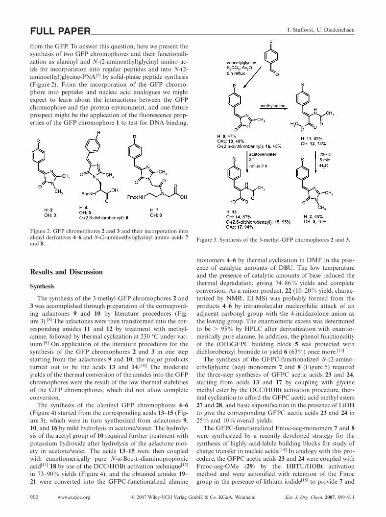
The synthesis of the 3-methyl-GFP chromophores 2 and3 was accomplished through preparation of the correspond-ing azlactones 9 and 10 by literature procedures (Fig-ure 3).[8] The azlactones were then transformed into the cor-responding amides 11 and 12 by treatment with methyl-amine, followed by thermal cyclization at 230 °C under vac-uum.[9] On application of the literature procedures for thesynthesis of the GFP chromophores 2 and 3 in one stepstarting from the azlactones 9 and 10, the major productsturned out to be the acids 13 and 14.[10] The moderateyields of the thermal conversion of the amides into the GFPchromophores were the result of the low thermal stabilitiesof the GFP chromophores, which did not allow completeconversion.
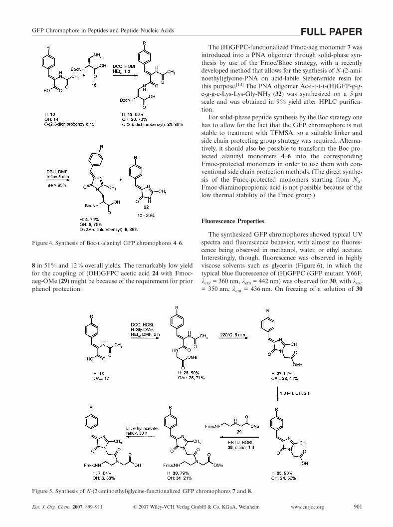
The synthesis of the alaninyl GFP chromophores 4–6(Figure 4) started from the corresponding acids 13–15 (Fig-ure 3), which were in turn synthesized from azlactones 9,10, and 16 by mild hydrolysis in acetone/water. The hydroly-sis of the acetyl group of 10 required further treatment withpotassium hydroxide after hydrolysis of the azlactone moi-ety in acetone/water. The acids 13–15 were then coupledwith enantiomerically pure N-α-Boc--diaminopropionicacid[11] 18 by use of the DCC/HOBt activation technique[12]
in 73–90% yields (Figure 4), and the obtained amides 19–21 were converted into the GFPC-functionalized alanine
www.eurjoc.org © 2007 Wiley-VCH Verlag GmbH & Co. KGaA, Weinheim Eur. J. Org. Chem. 2007, 899–911900
Figure 3. Synthesis of the 3-methyl-GFP chromophores 2 and 3.
monomers 4–6 by thermal cyclization in DMF in the pres-ence of catalytic amounts of DBU. The low temperatureand the presence of catalytic amounts of base reduced thethermal degradation, giving 74–86% yields and completeconversion. As a minor product, 22 (10–20% yield, charac-terized by NMR, EI-MS) was probably formed from theproducts 4–6 by intramolecular nucleophilic attack of anadjacent carbonyl group with the 4-imidazolone anion asthe leaving group. The enantiomeric excess was determinedto be 95% by HPLC after derivatization with enantio-merically pure alanine. In addition, the phenol functionalityof the (OH)GFPC building block 5 was protected withdichlorobenzyl bromide to yield 6 (63%) once more.[13]
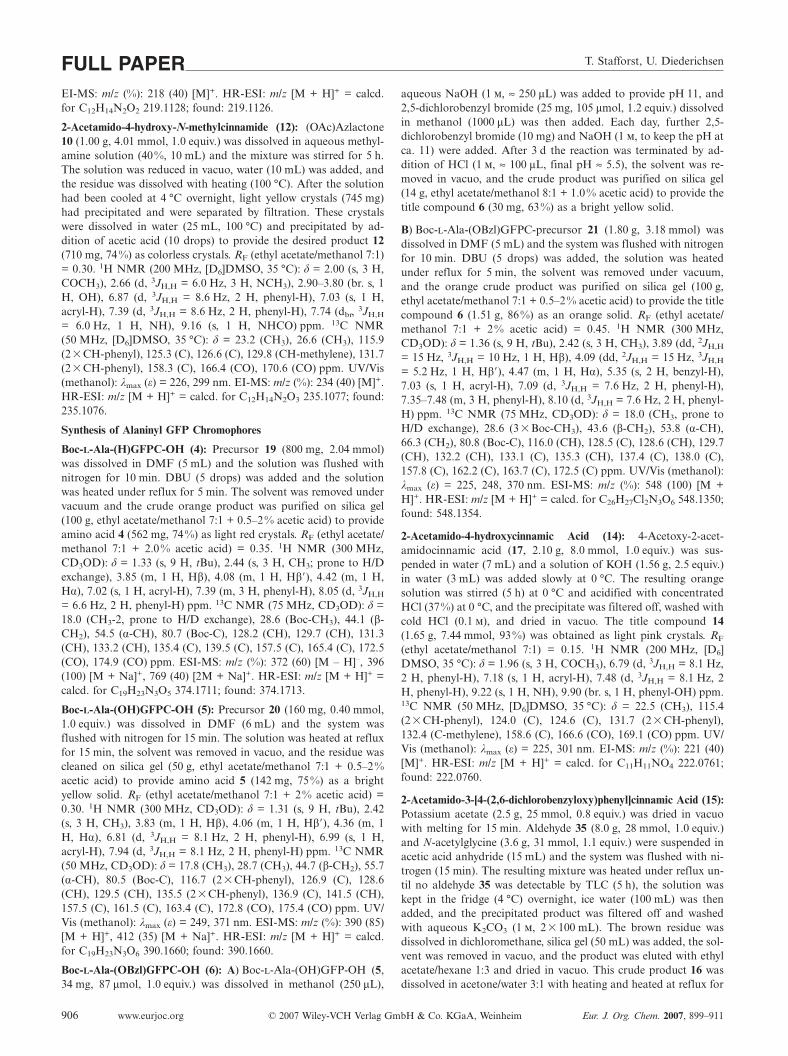
The synthesis of the GFPC-functionalized N-(2-amino-ethyl)glycine (aeg) monomers 7 and 8 (Figure 5) requiredthe three-step syntheses of GFPC acetic acids 23 and 24,starting from acids 13 and 17 by coupling with glycinemethyl ester by the DCC/HOBt activation procedure, ther-mal cyclization to afford the GFPC acetic acid methyl esters27 and 28, and basic saponification in the presence of LiOHto give the corresponding GFPC acetic acids 23 and 24 in25% and 10% overall yields.
The GFPC-functionalized Fmoc-aeg-monomers 7 and 8were synthesized by a recently developed strategy for thesynthesis of highly acid-labile building blocks for study ofcharge transfer in nucleic acids.[14] In analogy with this pro-cedure, the GFPC acetic acids 23 and 24 were coupled withFmoc-aeg-OMe (29) by the HBTU/HOBt activationmethod and were saponified with retention of the Fmocgroup in the presence of lithium iodide[15] to provide 7 and
GFP Chromophore in Peptides and Peptide Nucleic Acids FULL PAPER
Figure 4. Synthesis of Boc--alaninyl GFP chromophores 4–6.
8 in 51% and 12% overall yields. The remarkably low yieldfor the coupling of (OH)GFPC acetic acid 24 with Fmoc-aeg-OMe (29) might be because of the requirement for priorphenol protection.
Figure 5. Synthesis of N-(2-aminoethyl)glycine-functionalized GFP chromophores 7 and 8.
Eur. J. Org. Chem. 2007, 899–911 © 2007 Wiley-VCH Verlag GmbH & Co. KGaA, Weinheim www.eurjoc.org 901
The (H)GFPC-functionalized Fmoc-aeg monomer 7 wasintroduced into a PNA oligomer through solid-phase syn-thesis by use of the Fmoc/Bhoc strategy, with a recentlydeveloped method that allows for the synthesis of N-(2-ami-noethyl)glycine-PNA on acid-labile Sieberamide resin forthis purpose.[14] The PNA oligomer Ac-t-t-t-t-(H)GFP-g-g-c-g-g-c-Lys-Lys-Gly-NH2 (32) was synthesized on a 5 µ
scale and was obtained in 9% yield after HPLC purifica-tion.
For solid-phase peptide synthesis by the Boc strategy onehas to allow for the fact that the GFP chromophore is notstable to treatment with TFMSA, so a suitable linker andside chain protecting group strategy was required. Alterna-tively, it should also be possible to transform the Boc-pro-tected alaninyl monomers 4–6 into the correspondingFmoc-protected monomers in order to use them with con-ventional side chain protection methods. (The direct synthe-sis of the Fmoc-protected monomers starting from Nα-Fmoc-diaminopropionic acid is not possible because of thelow thermal stability of the Fmoc group.)
Fluorescence Properties
The synthesized GFP chromophores showed typical UVspectra and fluorescence behavior, with almost no fluores-cence being observed in methanol, water, or ethyl acetate.Interestingly, though, fluorescence was observed in highlyviscose solvents such as glycerin (Figure 6), in which thetypical blue fluorescence of (H)GFPC (GFP mutant Y66F,λexc = 360 nm, λem = 442 nm) was observed for 30, with λexc
= 350 nm, λem = 436 nm. On freezing of a solution of 30

T. Stafforst, U. DiederichsenFULL PAPER
Figure 6. Temperature dependences of the fluorescence emissions of Fmoc-aeg-H(GFPC)-OMe (30) [λexc = 350 nm, A350nm = 0.9, λem =436 nm, Φrel = 1 (80 °C) and λem = 413 nm, Φrel = 5 (–10 °C)] and of Fmoc-aeg-(OH)GFPC-OMe (31) [λexc = 430 nm, A430nm = 1.6, λem
= 499 nm, Φrel = 1 (80 °C) and λem = 492 nm, Φrel = 11 (–10 °C)] in glycerin (+ 0.1% NEt3 for 31) at 80 °C and in glycerin glass at–10 °C. A = absorption; Φ = fluorescence quantum yield.
to –10 °C the fluorescence intensity increased by a factor offive with a strong blue shift of the fluorescence maximumof 23 nm (Figure 6).
Analogous behavior was found for the (OH)GFP chro-mophore 31. In glycerin with 0.1% NEt3, typical greenfluorescence with λexc = 430 nm, λem = 499 nm was ob-served (wild-type GFP, λexc = 395/475 nm, λem = 509 nm),while on freezing to –10 °C the fluorescence intensity
Figure 7. UV melting curve of oligomer 32 after hybridization with complementary DNA 33 (5-G-C2-G-C2-A5-3), each 3 µm in 10 mmNaH2PO4 buffer, 0.1 NaCl, pH 6.85. The binding of Uronium-t-t-t-t-t-g-g-c-g-g-c-Lys-Lys-Gly-NH2 (34) with DNA 33 is also shown,as a reference.[17]
www.eurjoc.org © 2007 Wiley-VCH Verlag GmbH & Co. KGaA, Weinheim Eur. J. Org. Chem. 2007, 899–911902
strongly increased by a factor of 11, with a slight blue shiftof the fluorescence maximum of 7 nm.
The PNA oligomer 32, containing the (H)GFP chromo-phore, formed an antiparallel double strand with the com-plementary DNA 33 (5-G-C2-G-C2-A5-3). Adenine waschosen as the counterbase for the (H)GFP chromophore.Temperature-dependent UV spectroscopy yielded a meltingtemperature of 71 °C for the duplex 32+33, with a small

GFP Chromophore in Peptides and Peptide Nucleic Acids FULL PAPERheating–cooling hysteresis of 3 °C (Figure 7). In compari-son, the UV experiment with the reference oligomer 34+33,with replacement of the (H)GFP chromophore by thy-mine – therefore giving the canonical A-T base pair – re-sulted in a melting temperature of 70 °C. Overall, this indi-cates a well stacked (H)GFP chromophore.[16]
CD spectroscopy with the DNA/PNA pairing complex32+33 showed typical temperature-dependent bands at theoptical transitions of the nucleobases (Figure 8, top) indi-cating the formation of a B-DNA-like, right-handed pairingcomplex. At the optical transitions of the (H)GFP chromo-phore (Figure 8, bottom), centered around 293 nm(+3 mdeg) and 347 nm (–3 mdeg), significant CD bandswere found, their temperature dependency consistent withthe UV melting behavior (Figure 9). The appearance of CDbands at the optical transitions of the (H)GFP chromo-phore once more supported the assumption of a wellstacked chromophore buried inside the DNA/PNA doublestrand.
Figure 8. Top: CD spectra of oligomer 32 after hybridization with complementary DNA 33, each 3 µm in 10 mm NaH2PO4 buffer, 0.1 NaCl, pH 6.85. Bottom: Same pairing complex, but oligomer and DNA each 30 µm in the same buffer. The concentration needed to beincreased for monitoring of the CD effect at the optical transitions of the (H)GFP chromophore since its absorption coefficient (ε350nm
≈ 9·103 cm2·mol–1) is much smaller than that of the double strand (ε260nm ≈ 206·103 cm2·mol–1).
Eur. J. Org. Chem. 2007, 899–911 © 2007 Wiley-VCH Verlag GmbH & Co. KGaA, Weinheim www.eurjoc.org 903
The fluorescence spectrum of oligomer 32 upon exci-tation at 350 nm showed broad emission maxima at413 nm, 440 nm, and 510 nm (Figure 10). This fluorescencewas sensitive to the addition of complementary DNA 33,which produced an increase in fluorescence intensity by afactor of two and the upraising of a sharp emission bandat 417 nm with shoulders at 440 and 506 nm. In compari-son, the fluorescence of the free (H)GFP chromophore (27as reference) showed a weak emission at 398 nm with 1/12of the intensity of the double strand.
The emission bands at 413 nm and 440 nm appearedclose to the emission of the Y66F mutant of the GFP con-taining the (H)GFP chromophore (442 nm) and are consis-tent with the fluorescence behavior of the chromophoremonomer 30 observed in glycerin at 80 °C (440 nm) and–10 °C (413 nm). In comparison to the free chromophore27 in methanol, with an emission maximum at 398 nm, thetypical blue fluorescence requires the incorporation of thechromophore into the base stack. Interestingly, a more red-

T. Stafforst, U. DiederichsenFULL PAPER
Figure 9. Temperature dependency of the CD effect at the optical transitions of the (H)GFP chromophore at 293 nm and 347 nm.Oligomer 32 + DNA 33 each 30 µm in 10 mm NaH2PO4 buffer, 0.1 NaCl, pH 6.85.
Figure 10. Fluorescence and UV spectra of oligomer 32 depending on hybridization; concentrations of oligomer 32 and DNA 33 each30 µm in 10 mm NaH2PO4 buffer, 0.1 NaCl, pH 6.85. UV absorption: λmax = 356 nm; fluorescence emission: λexc = 350 nm, A350nm =0.27; single strand λem = 413, 440, 510 nm, Φrel = 6; double strand Φrel = 12; reference (H)GFPC-methyl acetate 27, λexc = 350 nm, A350nm
= 0.27 in methanol, λem = 398 nm, Φrel = 1; fluorescence excitation: 32+33, λem = 500 nm (fix), λmax = 356 nm (scanned). A = absorption,Φ = fluorescence quantum yield.
shifted band, at 506–510 nm, was found both for duplex32+33 and for single-strand 32, and might result from π-stacking inside the base stack as indicated by UV and CDspectroscopy. On switching from single-strand 32 to duplex32+33 the fluorescence emission spectra changed not onlyin fluorescence intensity but also in the emission bandshape, which might be the result of the transition from aless ordered single strand with several conformations to thewell defined double helix structure. The (H)GFP chromo-
www.eurjoc.org © 2007 Wiley-VCH Verlag GmbH & Co. KGaA, Weinheim Eur. J. Org. Chem. 2007, 899–911904
phore was clearly identified as the source of the red-shiftedfluorescence by collection of a fluorescence excitation spec-trum at 500 nm, revealing a total overlap with the UV ab-sorption spectra with λmax = 356 nm. Like the fluorescenceemission, the UV ground state absorption was slightly red-shifted (8 nm) in relation to that of the free chromophore27 with λexc = 348 nm. It is known from mutagenesis experi-ments that stacking of the chromophore with an aromaticamino acid as in T203F/H/Y mutations yields GFP variants

GFP Chromophore in Peptides and Peptide Nucleic Acids FULL PAPERwith red-shifted (15–20 nm) fluorescence emission maxima.X-ray structure analysis revealed coplanar stacking of thearomatic amino acid in position 203 with the GFP chromo-phore with a distance of 3.3–3.8 Å, comparable with theraise of 3.4 Å in the DNA base stack.[18] Nevertheless, theoverall low quantum yield (Φ 0.5%)[19] indicates thatblocking of the radiationless deactivation channels ad-ditionally requires the incorporation of the chromophoreinto the hydrogen bond network of the protein. It remainsan open question whether the hydrogen bond network in-side the GFP protein barrel blocks the radiationless deacti-vation through a protonation mechanism or simply by moreefficient restriction of the chromophore flexibility.[5,20]
Conclusions
The synthesis and characterization of two GFP chromo-phores has been performed. In order to incorporate theminto alaninyl or N-(2-aminoethyl)glycinyl peptide nucleicacids the chromophores were prepared as the side chains ofthe corresponding amino acids. Incorporation into an N-(2-aminoethyl)glycine-PNA oligomer was achieved by care-fully optimized solid-phase peptide synthesis. The doublestrand stability of an N-(2-aminoethyl)glycine-PNA func-tionalized with the GFP chromophore with a complemen-tary DNA was investigated by UV and CD spectroscopy,while – since fluorescence of the GFP strongly depends onnearest-neighbor interactions and shielding from solvent –fluorescence within this model system was also investigated,with the appearance of blue fluorescence with sensitivity tothe presence of the DNA counter-strand being observed.Interestingly, an additional green fluorescence was foundand assumed to be caused by π-stacking interactions. Un-fortunately, the overall quantum yield was very low, indicat-ing insufficient blocking of the radiationless internal con-version by simple π-stacking, which might limit utility inmolecular biology diagnostics.
Experimental SectionGeneral Methods: Infrared spectra were recorded on a Perkin–El-mer FT-IR 1600 instrument. NMR spectra were recorded on a Var-ian Unity 300 or a Varian Inova 600 spectrometer. UV absorptionspectra were recorded with a Jasco V-550 spectrometer; all extinc-tion coefficients are given in cm2·mol–1. Fluorescence emission andexcitation spectra were collected with a Jasco FP 6200 instrument.Fluorescence emission spectra were not corrected for the wave-length dependency of the detector sensitivity, and fluorescence exci-tation spectra were not corrected for the wavelength dependency ofthe lamp emission intensity. ESI mass spectra were recorded witha Finnigan LCQ iontrap spectrometer and high-resolution massspectra (HR-ESI) were recorded on a Bruker FTMS-7APEX® IV 70e FT-ICR spectrometer. Reversed-phase HPLC wasperformed with a Pharmacia Biotech Äkta Basic 900 system [el-uents: A: deionized water (18 MΩ·cm–1) + 0.1% TFA, B: acetoni-trile/water 8:2 + 0.1% TFA], analytical HPLC was carried out witha Grom-Sil 80 ODS, 250 4.6 mm, 4 µm, 80 Å, C-18 column(0.5 mLmin–1), and preparative HPLC was done on a J’sphere
Eur. J. Org. Chem. 2007, 899–911 © 2007 Wiley-VCH Verlag GmbH & Co. KGaA, Weinheim www.eurjoc.org 905
ODS-H80, 25020 mm ID, 8–4 µm, 8 nm, C18 (YMC) column(10 mLmin–1). DNA was purchased from Carl Roth GmbH.
Analytical Data: The NMR analysis of compounds 7, 8, 30, and31 suffered from high rotation barriers caused by the tertiary amidebond of the N-acyl-(2-aminoethyl)glycine, which produced broad-ening and partly doubling of 1H and 13C NMR signals.[21] Mostly,a complete set of NMR assignments was obtained separately foreach rotamer with the aid of H,H-COSY, HSQC, and HMBC ex-periments. The 2-methyl group in the 4-imidazolone moiety wasfound to be quite acidic, as is known from the literature.[22] H/Dexchange takes place within some hours and needs to be taken intoaccount for 2D NMR spectroscopy. The enantiomeric excesseswere determined by deprotection of monomer 4–6 with TFA fol-lowed by coupling with enantiomerically pure - and -Ala-OSuand analytical HPLC. In all cases the enantiomeric excess washigher than 95%.
Synthesis of 3-Methyl-GFP Chromophores
3-Methyl-(H)GFPC (2): The GFPC-precursor 11 (218 mg,1.00 mmol, 1.0 equiv.) was heated quickly to 230 °C under vacuumin an oil bath and stirred for 5 min at 230 °C. After cooling, theblack residue was extracted with hexane (105 mL) to yield anorange solution, which was purified on silica gel (70 g, ethyl ace-tate/hexane 3:1) to provide the chromophore 2 (89 mg, 45%) aslight red crystals. RF (ethyl acetate/hexane 3:1) = 0.49. 1H NMR(200 MHz, CDCl3): δ = 2.34 (s, 3 H, CH3), 3.11 (s, 3 H, NCH3),7.04 (s, 1 H, acryl-H), 7.34 (m, 3 H, phenyl-H), 8.06 (d, J = 8.1 Hz,2 H, phenyl-H) ppm. 13C NMR (50 MHz, CDCl3): δ = 15.7 (CH3-2), 26.6 (CH3-3), 127.2 (CH), 128.7 (CH), 130.0 (CH), 132.1 (CH),134.1 (C-phenyl), 138.7 (C-5), 162.6 (C-2), 170.7 (CO-4) ppm. UV/Vis (methanol): λmax (ε) = 236, 293, 350 nm. EI-MS: m/z (%): 200(100) [M]+. HR-ESI: m/z [2M + H]+ = calcd. for C12H12N2O401.1972; found: 401.1971.
3-Methyl-(OH)GFPC (3): Acrylamide 12 (298 mg, 1.27 mmol,1.0 equiv.) was heated quickly to 230 °C under vacuum in an oilbath and stirred at 230 °C for 5 min. After cooling, the residue wasextracted with hot (70 °C) ethyl acetate (85 mL) to provide anorange raw product (238 mg) that was purified on silica gel (80 gethyl acetate/hexane 3:1) to give the chromophore 3 (124 mg, 45%)as bright yellow crystals. RF (ethyl acetate/hexane 3:1) = 0.36. 1HNMR (200 MHz, [D6]DMSO, 35 °C): δ = 2.32 (s, 3 H, CH3), 3.09(s, 3 H, NCH3), 6.84 [d, 3JH,H = 8.4 Hz, 2 H, phenyl-H], 6.88 (s, 1H, acryl-H), 8.07 (d, 3JH,H = 8.4 Hz, 2 H, phenyl-H), 9.84 (br. s, 1H, phenyl-OH) ppm. 13C NMR (50 MHz, [D6]DMSO, 35 °C): δ =15.6 (CH3), 26.9 (CH3), 116.4 (2CH-phenyl), 125.9 (C), 126.8(CH-methylene), 134.7 (2CH-phenyl), 136.6 (C), 159.9 (C), 163.3(CO), 170.9 (CO) ppm. UV/Vis (methanol): λmax (ε) = 248, 371 nm.EI-MS: m/z (%): 216 (100) [M]+. HR-ESI: m/z [M + H]+ = calcd.for C12H12N2O2 217.0972; found: 217.0971.
2-Acetylamino-N-methylcinnamide (11): (H)Azlactone 9 (1.64 g,8.76 mmol, 1.0 equiv.) was dissolved in ethanolic methylamine (8 ,15 mL) and the mixture was stirred for 1 d. The product precipi-tated in small crystals and was separated by filtration, and the titlecompound 11 (1.02 g, 53%) was obtained as a colorless solid afterwashing of the crystals with hexane and drying in vacuo. RF (ethylacetate/methanol 7:1) = 0.38. 1H NMR (200 MHz, [D6]DMSO,35 °C): δ = 2.00 (s, 3 H, COCH3), 2.67 (d, 3JH,H = 5.2 Hz, 3 H,CH3), 7.05 (s, 1 H, acryl-H), 7.28–7.45 (m, 3 H, phenyl-H), 7.52(d, 3JH,H = 8.0 Hz, 2 H, phenyl-H), 7.87 (br. s, 1 H, NHMe), 9.30(s, 1 H, NHCO) ppm. 13C NMR (50 MHz, CDCl3): δ = 22.8 (Ac-CH3), 26.1 (CH3), 127.4 (CH), 128.4 (2CH-phenyl), 129.2(2CH-phenyl), 130.1 (CH-methylene), 134.2 (C-phenyl), 165.2(CO), 169.2 (CO) ppm. UV/Vis (methanol): λmax (ε) = 216, 275 nm.
T. Stafforst, U. DiederichsenFULL PAPEREI-MS: m/z (%): 218 (40) [M]+. HR-ESI: m/z [M + H]+ = calcd.for C12H14N2O2 219.1128; found: 219.1126.
2-Acetamido-4-hydroxy-N-methylcinnamide (12): (OAc)Azlactone10 (1.00 g, 4.01 mmol, 1.0 equiv.) was dissolved in aqueous methyl-amine solution (40%, 10 mL) and the mixture was stirred for 5 h.The solution was reduced in vacuo, water (10 mL) was added, andthe residue was dissolved with heating (100 °C). After the solutionhad been cooled at 4 °C overnight, light yellow crystals (745 mg)had precipitated and were separated by filtration. These crystalswere dissolved in water (25 mL, 100 °C) and precipitated by ad-dition of acetic acid (10 drops) to provide the desired product 12(710 mg, 74%) as colorless crystals. RF (ethyl acetate/methanol 7:1)= 0.30. 1H NMR (200 MHz, [D6]DMSO, 35 °C): δ = 2.00 (s, 3 H,COCH3), 2.66 (d, 3JH,H = 6.0 Hz, 3 H, NCH3), 2.90–3.80 (br. s, 1H, OH), 6.87 (d, 3JH,H = 8.6 Hz, 2 H, phenyl-H), 7.03 (s, 1 H,acryl-H), 7.39 (d, 3JH,H = 8.6 Hz, 2 H, phenyl-H), 7.74 (dbr, 3JH,H
= 6.0 Hz, 1 H, NH), 9.16 (s, 1 H, NHCO) ppm. 13C NMR(50 MHz, [D6]DMSO, 35 °C): δ = 23.2 (CH3), 26.6 (CH3), 115.9(2CH-phenyl), 125.3 (C), 126.6 (C), 129.8 (CH-methylene), 131.7(2CH-phenyl), 158.3 (C), 166.4 (CO), 170.6 (CO) ppm. UV/Vis(methanol): λmax (ε) = 226, 299 nm. EI-MS: m/z (%): 234 (40) [M]+.HR-ESI: m/z [M + H]+ = calcd. for C12H14N2O3 235.1077; found:235.1076.
Synthesis of Alaninyl GFP Chromophores
Boc-L-Ala-(H)GFPC-OH (4): Precursor 19 (800 mg, 2.04 mmol)was dissolved in DMF (5 mL) and the solution was flushed withnitrogen for 10 min. DBU (5 drops) was added and the solutionwas heated under reflux for 5 min. The solvent was removed undervacuum and the crude orange product was purified on silica gel(100 g, ethyl acetate/methanol 7:1 + 0.5–2% acetic acid) to provideamino acid 4 (562 mg, 74%) as light red crystals. RF (ethyl acetate/methanol 7:1 + 2.0% acetic acid) = 0.35. 1H NMR (300 MHz,CD3OD): δ = 1.33 (s, 9 H, tBu), 2.44 (s, 3 H, CH3; prone to H/Dexchange), 3.85 (m, 1 H, Hβ), 4.08 (m, 1 H, Hβ), 4.42 (m, 1 H,Hα), 7.02 (s, 1 H, acryl-H), 7.39 (m, 3 H, phenyl-H), 8.05 (d, 3JH,H
= 6.6 Hz, 2 H, phenyl-H) ppm. 13C NMR (75 MHz, CD3OD): δ =18.0 (CH3-2, prone to H/D exchange), 28.6 (Boc-CH3), 44.1 (β-CH2), 54.5 (α-CH), 80.7 (Boc-C), 128.2 (CH), 129.7 (CH), 131.3(CH), 133.2 (CH), 135.4 (C), 139.5 (C), 157.5 (C), 165.4 (C), 172.5(CO), 174.9 (CO) ppm. ESI-MS: m/z (%): 372 (60) [M – H]–, 396(100) [M + Na]+, 769 (40) [2M + Na]+. HR-ESI: m/z [M + H]+ =calcd. for C19H23N3O5 374.1711; found: 374.1713.
Boc-L-Ala-(OH)GFPC-OH (5): Precursor 20 (160 mg, 0.40 mmol,1.0 equiv.) was dissolved in DMF (6 mL) and the system wasflushed with nitrogen for 15 min. The solution was heated at refluxfor 15 min, the solvent was removed in vacuo, and the residue wascleaned on silica gel (50 g, ethyl acetate/methanol 7:1 + 0.5–2%acetic acid) to provide amino acid 5 (142 mg, 75%) as a brightyellow solid. RF (ethyl acetate/methanol 7:1 + 2% acetic acid) =0.30. 1H NMR (300 MHz, CD3OD): δ = 1.31 (s, 9 H, tBu), 2.42(s, 3 H, CH3), 3.83 (m, 1 H, Hβ), 4.06 (m, 1 H, Hβ), 4.36 (m, 1H, Hα), 6.81 (d, 3JH,H = 8.1 Hz, 2 H, phenyl-H), 6.99 (s, 1 H,acryl-H), 7.94 (d, 3JH,H = 8.1 Hz, 2 H, phenyl-H) ppm. 13C NMR(50 MHz, CD3OD): δ = 17.8 (CH3), 28.7 (CH3), 44.7 (β-CH2), 55.7(α-CH), 80.5 (Boc-C), 116.7 (2CH-phenyl), 126.9 (C), 128.6(CH), 129.5 (CH), 135.5 (2CH-phenyl), 136.9 (C), 141.5 (CH),157.5 (C), 161.5 (C), 163.4 (C), 172.8 (CO), 175.4 (CO) ppm. UV/Vis (methanol): λmax (ε) = 249, 371 nm. ESI-MS: m/z (%): 390 (85)[M + H]+, 412 (35) [M + Na]+. HR-ESI: m/z [M + H]+ = calcd.for C19H23N3O6 390.1660; found: 390.1660.
Boc-L-Ala-(OBzl)GFPC-OH (6): A) Boc--Ala-(OH)GFP-OH (5,34 mg, 87 µmol, 1.0 equiv.) was dissolved in methanol (250 µL),
www.eurjoc.org © 2007 Wiley-VCH Verlag GmbH & Co. KGaA, Weinheim Eur. J. Org. Chem. 2007, 899–911906
aqueous NaOH (1 , ≈ 250 µL) was added to provide pH 11, and2,5-dichlorobenzyl bromide (25 mg, 105 µmol, 1.2 equiv.) dissolvedin methanol (1000 µL) was then added. Each day, further 2,5-dichlorobenzyl bromide (10 mg) and NaOH (1 , to keep the pH atca. 11) were added. After 3 d the reaction was terminated by ad-dition of HCl (1 , ≈ 100 µL, final pH ≈ 5.5), the solvent was re-moved in vacuo, and the crude product was purified on silica gel(14 g, ethyl acetate/methanol 8:1 + 1.0% acetic acid) to provide thetitle compound 6 (30 mg, 63%) as a bright yellow solid.
B) Boc--Ala-(OBzl)GFPC-precursor 21 (1.80 g, 3.18 mmol) wasdissolved in DMF (5 mL) and the system was flushed with nitrogenfor 10 min. DBU (5 drops) was added, the solution was heatedunder reflux for 5 min, the solvent was removed under vacuum,and the orange crude product was purified on silica gel (100 g,ethyl acetate/methanol 7:1 + 0.5–2% acetic acid) to provide the titlecompound 6 (1.51 g, 86%) as an orange solid. RF (ethyl acetate/methanol 7:1 + 2% acetic acid) = 0.45. 1H NMR (300 MHz,CD3OD): δ = 1.36 (s, 9 H, tBu), 2.42 (s, 3 H, CH3), 3.89 (dd, 2JH,H
= 15 Hz, 3JH,H = 10 Hz, 1 H, Hβ), 4.09 (dd, 2JH,H = 15 Hz, 3JH,H
= 5.2 Hz, 1 H, Hβ), 4.47 (m, 1 H, Hα), 5.35 (s, 2 H, benzyl-H),7.03 (s, 1 H, acryl-H), 7.09 (d, 3JH,H = 7.6 Hz, 2 H, phenyl-H),7.35–7.48 (m, 3 H, phenyl-H), 8.10 (d, 3JH,H = 7.6 Hz, 2 H, phenyl-H) ppm. 13C NMR (75 MHz, CD3OD): δ = 18.0 (CH3, prone toH/D exchange), 28.6 (3Boc-CH3), 43.6 (β-CH2), 53.8 (α-CH),66.3 (CH2), 80.8 (Boc-C), 116.0 (CH), 128.5 (C), 128.6 (CH), 129.7(CH), 132.2 (CH), 133.1 (C), 135.3 (CH), 137.4 (C), 138.0 (C),157.8 (C), 162.2 (C), 163.7 (C), 172.5 (C) ppm. UV/Vis (methanol):λmax (ε) = 225, 248, 370 nm. ESI-MS: m/z (%): 548 (100) [M +H]+. HR-ESI: m/z [M + H]+ = calcd. for C26H27Cl2N3O6 548.1350;found: 548.1354.
2-Acetamido-4-hydroxycinnamic Acid (14): 4-Acetoxy-2-acet-amidocinnamic acid (17, 2.10 g, 8.0 mmol, 1.0 equiv.) was sus-pended in water (7 mL) and a solution of KOH (1.56 g, 2.5 equiv.)in water (3 mL) was added slowly at 0 °C. The resulting orangesolution was stirred (5 h) at 0 °C and acidified with concentratedHCl (37%) at 0 °C, and the precipitate was filtered off, washed withcold HCl (0.1 ), and dried in vacuo. The title compound 14(1.65 g, 7.44 mmol, 93%) was obtained as light pink crystals. RF
(ethyl acetate/methanol 7:1) = 0.15. 1H NMR (200 MHz, [D6]DMSO, 35 °C): δ = 1.96 (s, 3 H, COCH3), 6.79 (d, 3JH,H = 8.1 Hz,2 H, phenyl-H), 7.18 (s, 1 H, acryl-H), 7.48 (d, 3JH,H = 8.1 Hz, 2H, phenyl-H), 9.22 (s, 1 H, NH), 9.90 (br. s, 1 H, phenyl-OH) ppm.13C NMR (50 MHz, [D6]DMSO, 35 °C): δ = 22.5 (CH3), 115.4(2CH-phenyl), 124.0 (C), 124.6 (C), 131.7 (2CH-phenyl),132.4 (C-methylene), 158.6 (C), 166.6 (CO), 169.1 (CO) ppm. UV/Vis (methanol): λmax (ε) = 225, 301 nm. EI-MS: m/z (%): 221 (40)[M]+. HR-ESI: m/z [M + H]+ = calcd. for C11H11NO4 222.0761;found: 222.0760.
2-Acetamido-3-[4-(2,6-dichlorobenzyloxy)phenyl]cinnamic Acid (15):Potassium acetate (2.5 g, 25 mmol, 0.8 equiv.) was dried in vacuowith melting for 15 min. Aldehyde 35 (8.0 g, 28 mmol, 1.0 equiv.)and N-acetylglycine (3.6 g, 31 mmol, 1.1 equiv.) were suspended inacetic acid anhydride (15 mL) and the system was flushed with ni-trogen (15 min). The resulting mixture was heated under reflux un-til no aldehyde 35 was detectable by TLC (5 h), the solution waskept in the fridge (4 °C) overnight, ice water (100 mL) was thenadded, and the precipitated product was filtered off and washedwith aqueous K2CO3 (1 , 2100 mL). The brown residue wasdissolved in dichloromethane, silica gel (50 mL) was added, the sol-vent was removed in vacuo, and the product was eluted with ethylacetate/hexane 1:3 and dried in vacuo. This crude product 16 wasdissolved in acetone/water 3:1 with heating and heated at reflux for

GFP Chromophore in Peptides and Peptide Nucleic Acids FULL PAPER2 h. After the system had been stirred overnight at room tempera-ture, the product precipitated, and was filtered off, washed withethyl acetate/hexane, and dried in vacuo to provide the title com-pound 15 (4.6 g, 43%) as a light yellow solid. 1H NMR (300 MHz,[D6]DMSO, 35 °C): δ = 1.99 (s, 3 H, CH3), 5.28 (s, 2 H, CH2), 7.10(d, 3JH,H = 8.0 Hz, 2 H, phenyl-H), 7.22 (s, 1 H, acryl-H), 7.40–7.62 (m, 5 H, phenyl-H), 9.31 (s, 1 H, NH), 12.50 (br. s, 1 H,OH) ppm. 13C NMR (75 MHz, [D6]DMSO, 35 °C): δ = 22.5 (CH3),62.8 (CH), 64.9 (CH2), 102.1 (CH), 114.6 (CH), 125.4 (C), 126.8(C), 128.7 (CH), 131.3 (C), 131.4 (CH), 136.0 (C), 159.0 (C), 166.5(C), 169.0 (C) ppm. EI-MS: m/z (%): 379.2 (50) [M]+. HR-ESI: m/z[M + Na]+ = calcd. for C14H10Cl2O2 402.0270; found: 402.0269.
2-Acetamido-4-acetoxycinnamic Acid (17): (OAc)Azlactone 10(11.4 g, 46.5 mmol, 1.0 equiv.) was heated under reflux in acetone/water 3:1 (120 mL) for 4 h, the solution was kept at 4 °C overnight,and the precipitate was filtered off with a Buchner funnel, washedwith cold water (30 mL), cold acetone (10 mL), and cold ethyl ace-tate (20 mL), and dried in vacuo to provide the title compound 17(11.5 g, 43.7 mmol, 94%) as light yellow crystals. 1H NMR(300 MHz, [D6]DMSO, 35 °C): δ = 1.99 (s, 3 H, CH3), 2.27 (s, 3H, CH3), 7.17 (d, 3JH,H = 8.1 Hz, 2 H, phenyl-H), 7.23 (s, 1 H,acryl-H), 7.65 (d, 3JH,H = 8.1 Hz, 2 H, phenyl-H), 9.42 (s, 1 H,NH), 12.5 (br. s, 1 H, COOH) ppm. 13C NMR (125.7 MHz, [D6]DMSO, 35 °C): δ = 20.8 (CH3), 22.5 (CH3), 121.9 (2CH-phenyl),127.3 (C), 124.6 (C), 130.2 (C-methylene), 130.9 (2CH-phenyl),132.4 (C), 150.8 (C), 166.3 (CO), 169.0 (CO), 169.2 (CO) ppm. UV/Vis (methanol): λmax (ε) = 225, 301 nm. ESI-MS: m/z (%): 264.1(20) [M + H]+, 286.1 (100) [M + Na]+, 549.2 (35) [2M + Na]+.HR-ESI: m/z [M + H]+ = calcd. for C13H13NO5 264.0867; found:264.0866.
Boc-L-Ala-GFPC-Precursor 19: 2-Acetamidocinnamic acid (13,900 mg, 4.40 mmol, 1.05 equiv.) and HOBt (770 mg, 5.7 mmol,1.3 equiv.) were dissolved in DMF (3 mL). DCC (850 mg,4.11 mmol, 1.0 equiv.) dissolved in DMF (2 mL) was added at 0 °Cand the solution was stirred for 60 min at 0 °C and for 120 min atroom temperature. The precipitated urea was filtered off andwashed with DMF (18 mL) until the urea was colorless, (S)-3-amino-2-(tert-butoxycarbonylamino)propionic acid (18, 1.00 g,4.90 mmol, 1.1 equiv.), and triethylamine (1.2 mL, 890 mg,8.8 mmol, 2.0 equiv.) were added to the obtained yellow solution,and the resulted suspension was stirred until a clear solution wasobtained (1 d). The solvent was removed in vacuo and the crudeproduct was purified on silica gel (170 g, ethyl acetate/methanol7:1, + 0.5–1.5% acetic acid) to provide the title compound 19(1.42 g, 88%) as a colorless solid. RF (ethyl acetate/methanol 7:1 +1% acetic acid) = 0.18. 1H NMR (200 MHz, [D6]DMSO, 35 °C):δ = 1.39 (s, 9 H, tBu), 2.00 (s, 3 H, COCH3), 3.30–3.48 (m, 2 H,Hβ, Hβ), 3.75 (m, 1 H, Hα), 6.44 (d, 3JH,H = 5.6 Hz, 1 H, NHBoc),7.08 (s, 1 H, acryl-H), 7.28–7.44 (m, 3 H, phenyl-H), 7.54 (d, 3JH,H
= 8.3 Hz, 2 H, phenyl-H), 8.30 (br. s, 1 H, NHCH2), 9.56 (s, 1 H,NHCO) ppm. 13C NMR (50 MHz, [D6]DMSO, 35 °C): δ = 22.6(CH3), 28.1 (Boc-CH3), 42.8 (β-CH2), 54.2 (α-CH), 77.8 (Boc-C),128.0 (CH), 128.4 (CH), 129.2 (CH), 129.8 (CH), 134.0 (CH), 155.2(C), 164.4 (C), 169.5 (CO), 173.3 (CO) ppm. UV/Vis (methanol):λmax (ε) = 217, 277 nm. ESI-MS: m/z (%): 391 (100) [M – H]–, 414(100) [M + Na]+. HR-ESI: m/z [M + Na]+ = calcd. for C19H25N3O6
414.1636; found: 414.1638.
Boc-L-Ala-(OH)GFPC-Precursor 20: 2-Acetamido-4-hydroxycin-namic acid (14, 370 mg, 1.67 mmol, 1.05 equiv.) and HOBt(300 mg, 2.22 mmol, 1.3 equiv.) were dissolved in DMF (1 mL),DCC (310 mg, 1.50 mmol, 1.0 equiv.) dissolved in DMF (1 mL)was added at 0 °C, the resulting solution was stirred for 1 h at 0 °C
Eur. J. Org. Chem. 2007, 899–911 © 2007 Wiley-VCH Verlag GmbH & Co. KGaA, Weinheim www.eurjoc.org 907
and for 45 min at room temperature, and the precipitated urea wasfiltered off and washed with DMF (6 mL). (S)-3-Amino-2-(tert-bu-toxycarbonylamino)propionic acid (18, 332 mg, 1.62 mmol,1.05 equiv.) and triethylamine (500 µL, 360 mg, 3.60 mmol,2.5 equiv.) were added to the DMF solution, the resulting suspen-sion was stirred until a clear solution was obtained (1 d), the sol-vent was removed in vacuo, and the crude product was purified onsilica gel (50 g, ethyl acetate/methanol 7:1, + 0.5–2% acetic acid)to provide the title compound 20 (446 mg, 73%) as a colorless so-lid. RF (ethyl acetate/methanol 7:1 + 2% acetic acid) = 0.15. 1HNMR (200 MHz, CD3OD): δ = 1.42 (s, 9 H, tBu), 2.15 (s, 3 H,COCH3), 3.64 (m, 2 H, Hβ,β), 4.20 (m, 1 H, Hα), 6.79 (d, 3JH,H
= 8.0 Hz, 2 H, phenyl-H), 7.19 (s, 1 H, acryl-H), 7.40 (d, 3JH,H =8.0 Hz, 2 H, phenyl-H) ppm. 13C NMR (50 MHz, CD3OD): δ =21.8 (CH3), 27.3 (CH3), 43.0 (β-CH2), 53.3 (α-CH), 77.2 (Boc-C),114.5 (2CH-phenyl), 123.9 (C), 125.8 (C), 128.1 (CH-methylene),130.3 (2CH-phenyl), 154.4 (C), 157.3 (C), 164.3 (C), 168.5 (C),177.0 (CO) ppm. UV/Vis (methanol): λmax (ε) = 229, 300 nm. ESI-MS: m/z (%): 406 (95) [M – H]–, 430 (95) [M + Na]+. HR-ESI: m/z[M + H]+ = calcd. for C19H25N3O7 408.1765; found: 408.1766.
Boc-L-Ala-(OBzl)GFPC-Precursor 21: Acrylic acid 15 (2.50 g,6.6 mmol, 1.05 equiv.) and HOBt (1.10 g, 8.1 mmol, 1.3 equiv.)were dissolved in DMF (8 mL), DCC (1.29 g, 6.3 mmol, 1.0 equiv.)dissolved in DMF (2 mL) was added at 0 °C, and the solution wasstirred for 30 min at 0 °C and for 60 min at room temperature. Theprecipitated urea was filtered off and washed with DMF (25 mL)until the urea was colorless, (S)-3-amino-2-(tert-butoxycarbonyla-mino)propionic acid 18 (1.41 g, 6.9 mmol, 1.1 equiv.) and triethyl-amine (1.5 mL, 1.33 mg, 13 mmol, 2.0 equiv.) were added to theobtained yellow solution, and the resulting suspension was stirreduntil a clear solution was obtained (1 d). The solvent was removedin vacuo and the crude product was purified on silica gel (200 g,ethyl acetate/methanol 7:1, + 0.5–1.5% acetic acid) to provide thetitle compound 21 (3.21 g, 90%) as a colorless solid. 1H NMR(300 MHz, [D6]DMSO, 35 °C): δ = 1.38 (s, 9 H, Boc), 2.05 (s, 3 H,CH3), 3.31–3.42 (m, 2 H, β-H), 3.90 (m, 1 H, α-H), 5.21 (s, 2 H,CH2), 6.63 (br. s, 1 H, NH), 7.05 (s, 1 H, acryl-H), 7.06 (d, 3JH,H
= 8.0 Hz, 2 H, phenyl-H), 7.40–7.60 (m, 5 H, phenyl-H), 8.12 (br.s, 1 H, NH), 9.40 (s, 1 H, NH), 12.00 (br. s, 1 H, OH) ppm. 13CNMR (50 MHz, [D6]DMSO, 35 °C): δ = 22.7 (CH3), 28.1 (3Boc-CH3), 41.8 (β-CH2), 54.1 (α-CH), 64.9 (CH2), 78.0 (Boc-C), 114.6(CH), 127.2 (C), 128.0 (C), 128.3 (CH), 128.7 (CH), 131.0 (CH),131.4 (C), 131.6 (CH), 136.0 (C), 155.3 (C), 158.6 (C), 165.0 (C),169.3 (C), 172.4 (C) ppm. ESI-MS: m/z (%): 588.5 (100) [M +Na]+. HR-ESI: m/z [M + Na]+ = calcd. for C26H29Cl2N3O7
588.1275; found: 588.1277.
4-(2,6-Dichlorobenzyloxy)benzaldehyde (35): 4-Hydroxybenzalde-hyde (10 g, 90 mmol, 1.1 equiv.) and 2,6-dichlorobenzyl bromide(19.5 g, 80 mmol, 1.0 equiv.) were dissolved in dichloromethane(60 mL), DBU (21 mL, 1.5 equiv.) was added, and the resultingmixture was stirred for 17 h at room temperature. The reaction wasstopped by addition of acetic acid (3 mL, 0.6 equiv.), the solventwas removed in vacuo, ethyl acetate (100 mL) was added, and themixture was stirred for 10 min. The resulting suspension was fil-tered off and the organic layer was extracted with aqueous K2CO3
solution (0.1 , 2100 mL), washed with brine (100 mL), anddried with Na2SO4 to provide the title compound 35 (14.4 g, 64%)as a light brown solid. This product could be used without furtherpurification for the Erlenmeyer azlactone synthesis, but for NMRanalysis a small sample was recrystallized from ethyl acetate/hexane1:6, 5 mL·g–1 to provide the title compound as long, colorless need-les. RF (ethyl acetate/hexane 1:1) = 0.80. 1H NMR (200 MHz,CDCl3): δ = 5.39 (s, 2 H, CH2), 7.12 (d, 3JH,H = 8.0 Hz, 2 H,
T. Stafforst, U. DiederichsenFULL PAPERphenyl-H), 7.20–7.42 (m, 3 H, phenyl-H), 7.88 (d, 3JH,H = 8.0 Hz,2 H, phenyl-H), 9.92 (s, 1 H, HCO) ppm. 13C NMR (50 MHz,CDCl3): δ = 65.3 (CH2), 115.0 (CH), 128.5 (CH), 130.3 (C), 130.8(CH), 131.2 (C), 132.0 (CH), 137.0 (C), 163.7 (C), 190.8(CHO) ppm. EI-MS: m/z (%): 280.2 (10) [M]+, 159.1 (100) [benzylcation]. HR-ESI: m/z [M + Na]+ = calcd. for C14H10Cl2O2
302.9950; found: 302.9948.
Synthesis of (2-Aminoethyl)glycinyl GFPC Chromophores
Fmoc-aeg-(H)GFPC-OH (7): Fmoc-aeg-(H)GFPC-OMe (30,463 mg, 0.83 mmol) was dissolved in ethyl acetate (20 mL), LiI(480 mg, 3.6 mmol) was added, and the obtained solution washeated under reflux for 25 h. After the mixture had cooled to roomtemperature, ethyl acetate (200 mL) was added and the organiclayer was washed with NaHSO3/HCl (0.5 , pH 2, 3100 mL) andsaturated NaCl/HCl (pH 2, 2100 mL) and dried with Na2SO4.After purification on RP-18 silica gel (methanol/water 7:3) titlecompound 7 (0.30 g, 0.53 mmol, 64%) was obtained as a red solid.M.p. 118 °C; NMR: two rotamers 1:1. 1H NMR (300 MHz, [D6]DMSO, 35 °C): δ = 8.20 (d, 3JH,H = 8.1 Hz, 2 H, CH-phenyl), 7.88(d, 3JH,H = 7.2 Hz, 2 H, CH-Fmoc), 7.68 (t, 3JH,H = 6.6 Hz, 2 H,CH-Fmoc), 7.48–7.30 (m, 7 H, CH-Fmoc, CH-Fmoc, CH-phenyl,CH-phenyl), 6.99 (s, 1 H, CH-methylene), 4.62 (s, 1 H, CH2-GFP),4.46 (s, 1 H, CH2-GFP), 4.38–4.28 (m, 2 H, CH2-Fmoc), 4.28–4.21(m, 1 H, CH-Fmoc), 3.97 (s, 2 H, CH2COOH), 3.49 (t, 3JH,H =6.9 Hz, 2 H, CH2–N), 3.16–3.12 (m, 2 H, CH2–NH), 2.20 (s, 1.5H, CH3), 2.18 (s, 1.5 H, CH3) ppm. 13C NMR (150 MHz, [D6]-DMSO, 35 °C): δ = 170.4 (COOH), 169.6 (CO-GFP), 167.0(CH2CON), 164.3 (N=C–N), 156.4 (OCON), 143.9 (2C-Fmoc),140.7 (2C-Fmoc), 138.8 (CH=C–N), 134.0 (C-phenyl), 131.9(2CH-phenyl), 130.0 (CH-phenyl), 128.7 (2CH-phenyl), 127.6(2CH-Fmoc), 127.1 (2CH-Fmoc), 125.1 (CH-methylene),125.0 (2CH-Fmoc), 120.1 (2CH-Fmoc), 65.5 (CH2-Fmoc),47.0 (CH-Fmoc), 46.7 (CH2–N, CH2COOH), 40.3 (CH2-GFP),39.4 (CH2–NH), 15.2 (CH3) ppm. IR (KBr): ν = 3441, 1718, 1653,1458, 1253, 742 cm–1. UV/Vis (methanol): λmax = 347, 299, 289,264, 210 nm. ESI-MS: m/z (%) = 565.2 (100) [M – H]–, 679.1 (90)[M + TFA]–, 1131.2 (25) [2M – H]–, 1697.3 (10) [3M – H]–. HR-ESI-MS: m/z [M + H]+ = calcd. for C32H30N4O6 567.2238; found:567.2239.
Fmoc-aeg-(OH)GFPC-OH (8): Fmoc-aeg-(OH)GFPC-OMe (31,394 mg, 0.66 mmol) was dissolved in ethyl acetate (17 mL), LiI(408 mg, 3.1 mmol) was added, and the resulting solution washeated under reflux for 25 h. After removal of the solvent underreduced pressure, NaHSO3 (106 mg, 1.5 equiv.) in HCl (1.0 ,4 equiv.) was added. Methanol (≈ 2 mL) was then added to thesuspension until a clear solution had been obtained, and this waspurified on RP-18 silica gel (methanol/water 7:3). The title com-pound 8 (223 mg, 0.38 mmol, 58%) was obtained as a yellow solid.M.p. 196 °C; NMR: two rotamers 1.4:1. First rotamer (major com-ponent): 1H NMR (300 MHz, [D6]DMSO, 35 °C): δ = 8.08 (d,3JH,H = 9.0 Hz, 2 H, phenyl-CH), 7.88 (d, 3JH,H = 7 Hz, 2 H,Fmoc-CH), 7.69 (d, 3JH,H = 7 Hz, 2 H, Fmoc-CH), 7.41 (t, 3JH,H
= 7 Hz, 3 H, Fmoc-CH, NH), 7.33 (t, 3JH,H = 7 Hz, 2 H, Fmoc-CH), 6.91 (s, 1 H, CH=C), 6.85 (d, 3JH,H = 9.0 Hz, 2 H, phenyl-CH), 4.44 (s, 2 H, GFP-CH2), 4.36 (d, 3JH,H = 6 Hz, 2 H, Fmoc-CH2), 4.24 (t, 3JH,H = 6 Hz, 1 H, Fmoc-CH), 3.97 (s, 2 H,CH2COOH), 3.49 (m, 2 H, CH2–N), 3.26 (m, 2 H, CH2–NH), 2.15(s, 3 H, CCH3) ppm. 13C NMR (150 MHz, [D6]DMSO, 35 °C): δ= 170.3 (COOH), 169.5 (GFP-CO), 167.4 (GFP-CH2–CO), 162.0(C=N), 159.6 (phenyl-C–OH), 156.3 (Fmoc-CO), 143.8 (2Fmoc-C), 140.6 (2Fmoc-C), 135.9 (C=CH), 134.0 (2phenyl-CH),127.5 (2Fmoc-CH), 127.0 (2Fmoc-CH), 125.8 (CH=C), 125.2
www.eurjoc.org © 2007 Wiley-VCH Verlag GmbH & Co. KGaA, Weinheim Eur. J. Org. Chem. 2007, 899–911908
(phenyl-C–CH), 125.9 (2Fmoc-CH), 120.0 (2Fmoc-CH),115.7 (2phenyl-CH), 65.4 (Fmoc-CH2), 47.8 (CH2COOH), 46.7(Fmoc-CH), 46.6 (CH2–N), 41.1 (GFP-CH2), 37.9 (CH2–NH), 14.9(CH3C) ppm. Second rotamer: 1H NMR (300 MHz, [D6]DMSO,35 °C): δ = 8.08 (d, 3JH,H = 9.0 Hz, 2 H, phenyl-CH), 7.88 (d, 3JH,H
= 7 Hz, 2 H, Fmoc-CH), 7.67 (d, 3JH,H = 7 Hz, 2 H, Fmoc-CH),7.41 (t, 3JH,H = 7 Hz, 2 H, Fmoc-CH), 7.33 (t, 3JH,H = 7 Hz, 3 H,Fmoc-CH, NH), 6.91 (s, 1 H, CH=C), 6.85 (d, 3JH,H = 9.0 Hz, 2H, phenyl-CH), 4.60 (s, 2 H, GFP-CH2), 4.28 (d, 3JH,H = 6 Hz, 2H, Fmoc-CH2), 4.24 (t, 3JH,H = 6 Hz, 1 H, Fmoc-CH), 4.17 (s, 2H, CH2COOH), 3.36 (m, 2 H, CH2–N), 3.13 (m, 2 H, CH2–NH),2.18 (s, 3 H, CCH3) ppm. 13C NMR (150 MHz, [D6]DMSO,35 °C): δ = 170.9 (COOH), 169.5 (GFP-CO), 166.9 (GFP-CH2–CO), 161.8 (C=N), 159.6 (phenyl-C–OH), 156.1 (Fmoc-CO), 143.8(2Fmoc-C), 140.7 (2Fmoc-C), 136.0 (C=CH), 134.0(2phenyl-CH), 127.5 (2Fmoc-CH), 127.0 (2Fmoc-CH),125.7 (CH=C), 125.2 (phenyl-C–CH), 125.9 (2Fmoc-CH), 120.0(2Fmoc-CH), 115.7 (2phenyl-CH), 65.4 (Fmoc-CH2), 47.1(CH2COOH), 46.7 (Fmoc-CH), 46.6 (CH2–N), 40.6 (GFP-CH2),37.9 (CH2–NH), 15.0 (CH3C) ppm. IR (KBr): ν = 3412, 1707,1646, 1599, 1516, 1447, 1252, 850, 746 cm–1. UV/Vis (methanol):λmax (OD) = 370 (0.3), 300 (0.14), 223 (0.1) same sample: UV/Vis (+ 0.01% NEt3): λmax (OD) = 431 (0.22), 386 (0.22), 300 (0.15),228 (3.0) nm. ESI-MS: m/z (%) = 583.2 (100) [M + H]+, 581.1 (90)[M – H]–, 695.0 (100) [M + CF3COO–]–. HR-ESI-MS: m/z [M +H]+ = calcd. for C21H30N4O7 583.2187; found: 583.2187.
(H)GFPC-Acetic Acid (23): Aqueous LiOH (1.0 , 12 mL, 2 equiv.)was slowly added (15 min) to a stirred solution of (H)GFPC-methylacetate (27, 1.50 g, 5.8 mmol, 1 equiv.) in methanol (60 mL). Afterconversion was complete (TLC, 2 h), aqueous HCl (1.0 , 6 mL,1 equiv.) was added and methanol was removed under reducedpressure. The obtained aqueous suspension was acidified with sev-eral drops HCl (12 ) and the precipitated product was filtered offand washed with a small amount of cold aqueous HCl solution(0.1 ) and dried in vacuo to provide product 27 (1.27 g, 5.2 mmol,90%) as a yellow solid. RF (ethyl acetate/methanol 7:1) = 0.2 (prod-uct), 0.86 (starting material); m.p. 79 °C (combustion). 1H NMR(300 MHz, CD3OD): δ = 8.11 (dd, 3JH,H = 7.8 Hz, 2 H, CH-phenyl), 7.46–7.39 (m, 3 H, CH-phenyl), 7.08 (s, 1 H, CH-methyl-ene), 4.45 (s, 2 H, CH2), 2.35 (s, 3 H, CH3; prone to H/D ex-change) ppm. 13C NMR (75.5 MHz, CD3OD): δ = 171.7 (CO),171.0 (COOH), 164.8 (CCH3), 139.3 (CCH), 135.3 (C-phenyl),133.3 (2CH-phenyl), 131.5 (CH-phenyl), 129.7 (2CH-phenyl),128.6 (methylene-CH), 42.2 (CH2), 15.5 (CH3 prone to H/D ex-change) ppm. IR (KBr): ν = 3500, 1715, 1645, 1413, 1260, 770,688, 608 cm–1. UV/Vis (methanol): λmax = 346, 291, 233, 205 nm.ESI-MS: m/z (%) = 243.0 (100) [M – H]–, 487.1 (50) [2M – H]–,489.2 (100) [2M + H]+. HR-ESI-MS: m/z [M + H]+ = calcd. forC13H12N2O3 245.0921; found: 245.00920.
(OH)GFPC-Acetic Acid (24): (OAc)GFPC-Methyl acetate (28,2.00 g, 6.33 mmol) was dissolved in methanol (100 mL) and aque-ous LiOH (1.0 , 22.2 mL, 3.5 equiv.) was added slowly (1 h). Afterthe system had been stirred for 2 h at room temperature, aqueousHCl (1.0 , 19 mL, 3.0 equiv.) was added and methanol was re-moved under reduced pressure. To complete precipitation, furtheraqueous HCl (1.0 , 9.5 mL, 1.5 equiv.) was added. The obtainedsuspension was purified on RP-18 silica gel (methanol/water 2:3)and the title compound 24 (860 mg, 3.3 mmol, 52%) was obtainedas an orange solid. M.p. 160 °C. 1H NMR (300 MHz, [D6]DMSO,35 °C): δ = 8.07 (d, 3JH,H = 8.7 Hz, 2 H, CH-phenyl), 7.91(s, 1 H,CH), 6.82 (d, 3JH,H = 8.7 Hz, 2 H, CH-phenyl), 4.35 (s, 2 H, CH2),2.26 (s, 3 H, CH3) ppm. 13C NMR (75.5 MHz, [D6]DMSO, 35 °C):δ = 169.5 (CO), 169.4 (CO), 161.2 (NC=N), 159.7 (phenyl-C–OH),
GFP Chromophore in Peptides and Peptide Nucleic Acids FULL PAPER135.6 (CH=C), 134.1 (2phenyl-CH), 126.2 (CH=C), 125.1(phenyl-C), 115.7 (2phenyl-CH), 41.2 (CH2), 15.0 (CH3) ppm.IR (KBr): ν = 3426, 1595, 1383, 1251, 1165, 837 cm–1. UV/Vis(methanol): λmax = 370, 246 nm. ESI-MS: m/z (%) = 259.1 (100)[M – H]–, 518.8 (12) [2M – H]–. HR-ESI-MS: m/z [M + H]+ =calcd. for C13H12N2O4 261.0870; found: 261.0870.
(H)GFPC-Methyl Acetate Precursor 25: α-Acetamidocinnamic acid(13, 10 g, 48.7 mmol) was dissolved in DMF (100 mL), DCC(10.6 g, 1.05 equiv.) was added, and the system was stirred for30 min at 0 °C and for 1 h at room temperature. Glycine methylester hydrochloride (7.96 g, 63.4 mmol 1.3 equiv.) and DIPEA(33.4 mL, 4 equiv.) were added and the system was again stirredovernight. The resulting suspension was filtered, the solid residuewas washed with DMF (50 mL), the combined organic layers werereduced to dryness and taken up with ethyl acetate (300 mL), andthe organic layer was extracted with saturated KHCO3 solution(3100 mL) and HCl solution (0.1 , 3100 mL). The title com-pound 25 was obtained from the acidic water layer as a yellow solid(2.80 g, 10.1 mmol, 21%) without further purification. The crudeproduct from the basic layer was cleaned by filtration through silicagel (30 g silica gel, ethyl acetate/methanol 9:1 + 0.5% triethylamine,1 L) to provide further title compound 25 as a yellow solid (3.92 g,14.2 mmol, 29%). RF (ethyl acetate/methanol 9:1 + 0.5 triethyl-amine) = 0.53; m.p. 117 °C. 1H NMR (300 MHz, CDCl3): δ = 7.91(s, 1 H, NH), 7.58–7.48 (m, 1 H, NHCH2), 7.37–7.29 (m, 5 H, CH-phenyl), 6.96 (s, 1 H, CH), 4.01 (d, 3JH,H = 5.7 Hz, 2 H, CH2),3.68 (s, 3 H, OCH3), 2.01 (s, 3 H, CH3CONH) ppm. 13C NMR(75.5 MHz, CDCl3): δ = 170.7 (CO), 170.5 (CO), 166.2 (CO), 133.4(phenyl-C), 130.1–128.5 (CH=C–N, 5phenyl-CH), 52.3 (OCH3),41.5 (CH2), 22.9 (NHCOCH3) ppm. IR (KBr): ν = 3445, 1635,1376, 1203, 573 cm–1. UV/Vis (methanol): λmax = 208, 281, 327 nm.ESI-MS: m/z (%) = 299.1 (20) [M + Na]+, 574.8 (100) [2M +Na]+, 275.1 (100) [M – H]–, 550.8 (30) [2M – H]–. HR-ESI-MS:m/z [M + Na]+ = calcd. for C14H16N2O4 299.1002; found: 299.1003.
(OAc)GFPC-Methyl Acetate Precursor 26: 2-Acetamido-4-acetoxy-cinnamic acid (17, 10 g, 38 mmol) was dissolved in DMF (100 mL),and DCC (7.84 g, 38 mmol) dissolved in DMF (10 mL) was addedslowly (5 min) at 0 °C with stirring in an ice bath. The resultingsolution was stirred at 0 °C for 30 min and for 1 h at room tempera-ture, and the formed urea was filtered off and washed with DMF(40 mL). Triethylamine (21.2 mL, 152 mmol, 4.0 equiv.) and glycinemethyl ester hydrochloride (6.18 g, 49 mmol, 1.3 equiv.) wereadded, the DMF solution was stirred overnight, the solvent wasremoved in vacuo, and the obtained solid was dissolved in HCl(0.1 , 60 mL) and extracted several times with chloroform(150 mL overall). Silica gel (30 g) was added to the organic layer,which was reduced to dryness, and the product was eluted from thesilica gel (ethyl acetate/methanol 95:5 + 2% triethylamine), concen-trated, and dried in vacuo. The title compound 26 (8.92 g,27 mmol, 71%) was obtained as a yellow solid. RF (ethyl acetate/methanol 95:5 + 2% triethylamine) = 0.29 (product); m.p. 94 °C.1H NMR (300 MHz, CD3OD): δ = 7.55 (d, 3JH,H = 8.7 Hz, 2 H,CH-phenyl), 7.22 (s, 1 H, CH), 7.14 (d, 3JH,H = 8.7 Hz, 2 H, CH-phenyl), 4.02 (s, 2 H, CH2), 3.73 (s, 3 H, OCH3), 2.27 (s, 3 H,CH3CO), 2.11 (s, 3 H, CH3CONH) ppm. 13C NMR (75.5 MHz,CD3OD): δ = 173.3 (CO), 171.8 (CO), 170.9 (CO), 168.3 (CO),152.7 (phenyl-C-OAc), 132.8 (CH=C), 131.7 (2phenyl-CH),130.6 (CH), 129.9 (phenyl-C), 123.1 (2phenyl-CH), 52.6 (OCH3),42.3 (CH2), 22.7 (NHCOCH3), 20.9 (OCOCH3) ppm. IR (KBr): ν= 3272, 1756, 1658, 1534, 1371, 1205, 1014, 910, 655 cm–1. UV/Vis(methanol): λmax = 358, 281, 216 nm. ESI-MS: m/z (%) = 357.1(15) [M + Na]+, 590.8 (100) [2M + Na]+, 1024.3 (20) [3M + Na]+,333.1 (100) [M – H]–, 378.8 (70) [M + HCOO]–, 666.6 (30) [2M –
Eur. J. Org. Chem. 2007, 899–911 © 2007 Wiley-VCH Verlag GmbH & Co. KGaA, Weinheim www.eurjoc.org 909
H]–. HR-ESI-MS: m/z [M + H]+ = calcd. for C16H18N2O6
335.1238; found: 335.1238.
(H)GFPC-Methyl Acetate (27): Under vacuum, (H)GFPC-methylacetate precursor 25 (2.80 g, 10.1 mmol) was quickly heated up to220 °C in an oil bath and stirred at 220 °C for 10 min. The crudeproduct was dissolved in DCM and cleaned on silica gel (110 g,ethyl acetate/pentane 1:2), and the title compound 27 was obtainedas a yellow solid (1.62 g, 6.3 mmol, 62%). RF (ethyl acetate/hexane1:2) = 0.2 (product), 0.0 (starting material); m.p. 112 °C. 1H NMR(300 MHz, CDCl3): δ = 8.11 (dd, 3JH,H = 8.1 Hz, 2 H, CH-phenyl),7.43–7.32 (m, 3 H, CH-phenyl), 7.11 (s, 1 H, CH-methylene), 4.37(s, 2 H, CH2), 2.93 (s, 3 H, OCH3), 2.31 (s, 3 H, CH3) ppm. 13CNMR (75.5 MHz, CDCl3): δ = 170.0 (CO), 168.0 (COOMe), 161.3(CCH3), 137.9 (CCH), 133.9 (C-phenyl), 132.2 (2CH-phenyl),130.3 (CH-phenyl), 128.7 (2CH-phenyl), 128.2 (methylene-CH),52.8 (OCH3), 41.2 (CH2); 15.5 (CH3) ppm. IR (KBr): ν = 3500,1743, 1645, 1561, 1406, 1363, 1232, 1144, 981, 904, 775, 700,610 cm–1. UV/Vis (methanol): λmax = 348, 290, 234, 209 nm. ESI-MS: m/z (%) = 257.1 (40) [M – H]–, 259.1 (60) [M + H]+, 281.0(100) [M + Na]+, 538.8 (95) [2M + Na]+. HR-ESI-MS: m/z [M +H]+ = calcd. for C14H14N2O3 259.1077; found: 259.1077.
(OAc)GFPC-Methyl Acetate (28): Under vacuum, (OAc)GFPC-methyl acetate precursor 26 (5.3 g, 15.9 mmol) was quickly heatedup to 200 °C in an oil bath and stirred at 200 °C for 12 min. Thecrude product was dissolved in DCM (5 mL) and cleaned on silicagel (ethyl acetate/pentane 1:1), and the title compound 28 (2.2 g,7.0 mmol, 44%) was obtained as a yellow solid. M.p. 75 °C. 1HNMR (300 MHz, CDCl3): δ = 8.14 (d, 3JH,H = 8.7 Hz, 2 H, CH-phenyl), 7.12 (d, 3JH,H = 8.7 Hz, 2 H, CH-phenyl), 7.07 (s, 1 H,CH), 4.36 (s, 2 H, CH2), 3.75 (s, 3 H, OCH3), 2.30 (s, 3 H, CH3CO),2.28 (s, 3 H, CH3) ppm. 13C NMR (75.5 MHz, CDCl3): δ = 169.9(CO), 169.0 (CO), 167.9 (CO), 161.5 (NC=N), 151.9 (phenyl-C-OAc), 137.9 (CH=C), 133.4 (2phenyl-CH), 131.7 (phenyl-C),126.9 (CH=C), 121.9 (2phenyl-CH), 52.8 (OCH3), 41.2 (CH2),21.1 (OCOCH3), 15.5 (CH3) ppm. IR (KBr): ν = 1747, 1646, 1367,1216, 980, 908, 609 cm–1. UV/Vis (methanol): λmax = 351, 292, 236,203 nm. ESI-MS: m/z (%) = 339.1 (90) [M + Na]+, 654.9 (100)[2M + Na]+. HR-ESI-MS: m/z [M + H]+ = calcd. for C16H16N2O5
317.1132; found: 317.1132.
Fmoc-aeg-(H)GFPC-OMe (30): (H)GFPC-Acetic acid (23, 0.61 g,2.5 mmol, 1.3 equiv.), Fmoc-aeg-OMe·HCl[14] (29, 0.75 g,1.9 mmol, 1.0 equiv.), HBTU (0.95 g, 2.5 mmol, 1.3 equiv.), andHOBt (0.52 g, 3.8 mmol, 2.0 equiv.) were dissolved in DMF(25 mL). DIPEA (1.65 mL, 5 equiv.) was added and the resultingsolution was stirred for 14 h at room temperature. The solvent wasremoved in vacuo, and the obtained solid was dissolved in ethylacetate (100 mL), washed with NaHCO3 (0.5 , 100 mL), brine(100 mL), saturated NH4Cl solution (100 mL), and brine (100 mL),and dried with Na2SO4. After purification on silica gel (ethyl ace-tate, 600 mL) title compound 30 was obtained as a yellow solid(0.87 g, 1.5 mmol, 79%). M.p. 165 °C; NMR: two rotamers 2:1. 1HNMR (300 MHz, CDCl3): δ = 8.10 (d, 3JH,H = 10.8 Hz, 2 H, CH-phenyl), 7.72 (d, 3JH,H = 9.6 Hz, 2 H, CH-Fmoc), 7.57 (t, 3JH,H =6.9 Hz, 2 H, CH-Fmoc), 7.43–7.24 (m, 7 H, CH-Fmoc, CH-Fmoc,CH-phenyl, CH-phenyl), 7.09 (s, 0.66 H, CH-methylene), 7.07 (s,0.33 H, CH-methylene), 5.85 (t, 3JH,H = 6.0 Hz, 0.66 H, NH), 5.30(t, 3JH,H = 5.7 Hz, 0.33 H, NH), 4.50 (d, 3JH,H = 6.3 Hz, 1.3 H,CH2-Fmoc), 4.36 (d, 3JH,H = 8.7 Hz, 2.7 H, CH2-Fmoc, CH2-GFP), 4.19 (t, 3JH,H = 5.4 Hz, 1 H, CH-Fmoc), 3.99 (s, 2 H,CH2COOMe), 3.75 (s, 2.0 H, OCH3), 3.72 (s, 1.0 H, OCH3), 3.53(m, 2 H, CH2–N), 3.40–3.32 (m, 2 H, CH2–NH), 2.32 (s, 2.0 H,CH3), 2.15 (s, 1.0 H, CH3) ppm. 13C NMR (150 MHz, CD3OD):

T. Stafforst, U. DiederichsenFULL PAPERfirst rotamer (major component): δ = 170.4 (COOMe), 169.8 (CO-GFP), 167.1 (CH2CON), 162.4 (N=C–N), 156.6 (OCON), 143.6(2C-Fmoc), 141.3 (2C-Fmoc), 138.2 (CH=C–N), 134.1 (C-phenyl), 132.2 (2CH-phenyl), 130.1 (1 CH-phenyl), 128.6(2CH-phenyl), 127.7 (2CH-Fmoc), 127.6 (CH-methylene),127.0 (2CH-Fmoc), 124.9 (2CH-Fmoc), 120.0 (2CH-Fmoc), 66.6 (CH2-Fmoc), 52.6 (OCH3), 48.9 (CH2–N,CH2COOMe), 47.3 (CH-Fmoc), 40.9 (CH2-GFP), 39.2 (CH2–NH),15.4 (CH3) ppm; second rotamer: δ = 170.3 (COOMe), 169.8 (CO-GFP), 167.8 (NCO–CH2), 162.4 (N=C–N), 156.61 (OCON), 143.8(2C-Fmoc), 141.3 (2C-Fmoc), 138.2 (CH=C–N), 134.0 (C-phenyl), 132.2 (2CH-phenyl), 130.2 (1CH-phenyl), 128.6(2CH-phenyl), 127.7 (2CH-Fmoc), 127.6 (CH-methylene),127.0 (2CH-Fmoc), 125.1 (2CH-Fmoc), 119.9 (2CH-Fmoc), 66.8 (CH2-Fmoc), 53.0 (OCH3), 48.9 (CH2–N,CH2COOMe), 47.1 (CH-Fmoc), 41.5 (CH2-GFP), 39.2 (CH2–NH),15.6 (CH3) ppm. IR (KBr): ν = 3412, 2928, 1750, 1722, 1663, 1470,1210, 1045, 745 cm–1. UV/Vis (methanol): λmax = 347, 299, 289,264, 210 nm. Fluorescence (glycerin): λexc = 350 nm, λmax, em = 413,436, 477 nm. ESI-MS: m/z (%) = 581.2 (25) [M + H]+, 1182.8 (100)[2M + Na]+, 624.9 (100) [M + TFA]–, 1204.7 (70) [2M + TFA]–.HR-ESI-MS: m/z [M + H]+ = calcd. for C33H32N4O6 581.2395;found: 581.2396.
Fmoc-aeg-(OH)GFPC-OMe (31): (OH)GFPC-Acetic acid (24,508 mg, 1.95 mmol, 1.1 equiv.), Fmoc-aeg-OMe·HCl[14] (29,694 mg, 1.77 mmol, 1.0 equiv.), and HBTU (740 mg, 1.95 mmol,1.1 equiv.) were dissolved in DMF (15 mL). DIPEA (1.22 mL,920 mg, 7.1 mmol, 4 equiv.) was added, the resulting solution wasstirred for 2 d, the solvent was removed under reduced pressure,and the residue was dissolved in ethyl acetate (100 mL), washed(3100 mL 0.5 NH4Cl, 3100 mL 0.5 NaHCO3, 1100 mLbrine), and dried with Na2SO4. The crude product was purified onsilica gel (200 mL, ethyl acetate) and the title compound 31(0.223 g, 0.37 mmol, 21%) was obtained as a yellow solid. M.p.174 °C; NMR: two rotamers 2:1. First rotamer (major component):1H NMR (300 MHz, CDCl3): δ = 7.89 (d, 3JH,H = 8.7 Hz, 2 H,phenyl-CH), 7.70 (d, 3JH,H = 6.9 Hz, 2 H, Fmoc-CH), 7.57 (d,3JH,H = 6.6 Hz, 2 H, Fmoc-CH), 7.35 (t, 3JH,H = 6.9 Hz, 2 H,Fmoc-CH), 7.27 (t, 3JH,H = 6.6 Hz, 2 H, Fmoc-CH), 6.95 (s, 1 H,CH=C), 6.76 (d, 3JH,H = 8.7 Hz, 2 H, phenyl-CH), 5.91 (t, 3JH,H
= 6.0 Hz, 1 H, NH), 4.48 (d, 3JH,H = 6.3 Hz, 2 H, Fmoc-CH2),4.35 (s, 2 H, GFP-CH2), 4.19 (t, 3JH,H = 6.3 Hz, 1 H, Fmoc-CH),3.98 (s, 2 H, CH2COOMe), 3.71 (s, 3 H, OCH3), 3.52–3.49 (m, 2H, CH2–N), 3.41–3.39 (m, 2 H, CH2–NH), 2.11 (s, 3 H,CCH3) ppm. 13C NMR (150 MHz, CD3OD): δ = 170.4 (GFP-CO),169.7 (COOMe), 167.7 (GFP-CH2–CO), 160.4 (C=N), 158.7(phenyl-C–OH), 156.8 (Fmoc-CO), 143.6 (2Fmoc-C), 141.3(2Fmoc-C), 135.7 (C=CH), 134.5 (2phenyl-CH), 128.7(CHC), 127.8 (2Fmoc-CH), 127.1 (2Fmoc-CH), 126.4(phenyl-C–CH), 124.9 (2Fmoc-CH), 120.0 (2Fmoc-CH),115.9 (2phenyl-CH), 66.7 (Fmoc-CH2), 52.6 (OCH3), 50.0(CH2COOMe), 49.0 (CH2–N), 47.2 (Fmoc-CH), 41.0 (GFP-CH2),39.2 (CH2–NH), 15.4 (CH3C) ppm. Second rotamer: 1H NMR(300 MHz, CDCl3): δ = 7.89 (d, 3JH,H = 8.7 Hz, 2 H, phenyl-CH),7.72 (d, 3JH,H = 6.9 Hz, 2 H, Fmoc-CH), 7.55 (d, 3JH,H = 6.6 Hz,2 H, Fmoc-CH), 7.35 (t, 3JH,H = 6.9 Hz, 2 H, Fmoc-CH), 7.27 (t,3JH,H = 6.6 Hz, 2 H, Fmoc-CH), 6.95 (s, 1 H, CH=C), 6.76 (d,3JH,H = 8.7 Hz, 2 H, phenyl-CH), 5.39 (t, 3JH,H = 5.7 Hz, 1 H,NH), 4.35 (s, 2 H, GFP-CH2), 4.33 (d, 3JH,H = 6.3 Hz, 2 H, Fmoc-CH2), 4.19 (t, 3JH,H = 6.3 Hz, 1 H, Fmoc-CH), 4.16 (s, 2 H,CH2COOMe), 3.71 (s, 3 H, OCH3), 3.52–3.49 (m, 2 H, CH2–N),3.37–3.31 (m, 2 H, CH2–NH), 2.27 (s, 3 H, CCH3) ppm. 13C NMR(150.8 MHz, CD3OD): δ = 170.3 (GFP-CO), 169.7 (COOMe),
www.eurjoc.org © 2007 Wiley-VCH Verlag GmbH & Co. KGaA, Weinheim Eur. J. Org. Chem. 2007, 899–911910
167.7 (GFP-CH2–CO), 160.4 (C=N), 158.7 (phenyl-C–OH), 156.8(Fmoc-CO), 143.8 (2Fmoc-C), 141.3 (2Fmoc-C), 135.5(C=CH), 134.5 (2phenyl-CH), 129.0 (CHC), 127.7 (2Fmoc-CH), 127.1 (2Fmoc-CH), 126.4 (phenyl-C–CH), 125.0(2Fmoc-CH), 120.0 (2Fmoc-CH), 115.9 (2phenyl-CH), 66.9(Fmoc-CH2), 53.0 (OCH3), 50.0 (CH2COOMe), 49.0 (CH2–N),47.2 (Fmoc-CH), 41.5 (GFP-CH2), 38.9 (CH2–NH), 15.2(CH3C) ppm. IR (KBr): ν = 3405, 1703, 1601, 1513, 1448, 1252,750 cm–1. UV/Vis (methanol): λmax (OD) = 370 (0.83), 300 (0.46),265 (0.8), 221 (1.0) same sample: UV/Vis (+ 0.01% NEt3): λmax
(OD) = 431 (0.86), 300 (0.43), 264 (0.88), 227 (3.3). Fluorescence(glycerin + 0.01% NEt3): λexc. = 430 nm, λem. = 499 nm. ESI-MS:m/z (%) = 597.3 (40) [M + H]+, 1193.4 (100) [2M + H]+. HR-ESI-MS: m/z [M + H]+ = calcd. for C33H32N4O7 597.2344; found:597.2346.
PNA Oligomer Ac-t-t-t-t-(H)GFPC-g-g-c-g-g-c-Lys-Lys-Gly-NH2
(32): The solid-phase peptide synthesis was performed on a 5 µ
scale in a plastic syringe by the Fmoc/Bhoc strategy on Sieberamideresin as described elsewhere.[14] Commercially available amino acidsFmoc-aeg-thymine-OH, Fmoc-aeg-guanine(Bhoc)-OH, Fmoc-aeg-cytosine(Bhoc)-OH, Fmoc-Lys(Mtt)-OH, and Fmoc-Gly-OH onSieberamide resin (0.12 mmol·g–1) were used for the oligomer syn-thesis with use of the HBTU/HATU activation technique. Doublecoupling was performed in cases involving the coupling of guaninylamino acids following a thyminyl or guaninyl amino acid. Doublecoupling was not required for introduction of Fmoc-aeg-(H)GFPC-OH (7). Oligomer 32 was cleaved from the resin by con-tinuous flow with 2% trifluoroacetic acid, 4% triethylsilane in1,1,1,3,3,3-hexafluoropropan-2-ol/dichloromethane 1:2 and wasobtained (1.5 mg, 9% yield) after preparative HPLC purification.HR-ESI-MS: [M]+: calcd. for C141H184N64O41 3429.4280; found:3429.4306, [M + H]+: calcd. 3430.4329; found: 3430.4329, [M +3H]3+: calcd. 1144.1500; found: 1144.1510, [M + 4H]4+: calcd.858.3643; found: 858.3647; HPLC: analytical: 10% 40% in30 min (Grom25), tR = 26.00 min. UV: ε260nm = 96.9·103 [approxi-mated by nearest neighbor approach with ε260nm = 9.0·103 for the(H)GFP chromophore]. UV/Vis (pH 6.85, phosphate buffer): λmax
(ε/103) = 350 (9.0), 260 (97.0), 204 (204) nm. UV-melting: pairingwith complementary DNA 33 (5-G-C-C-G-C-C-A-A-A-A-A-3,ε260nm = 110.5·103) each 3 µ in 10 mm phosphate buffer pH =6.85, NaCl (0.10 ): H260nm = 10 % at Tm = 71.0 °C, heating–cool-ing hysteresis 3 °C; CD: pairing with complementary DNA 33 each3 µm in phosphate buffer (10 mm) pH = 6.85, NaCl (0.10 ): tem-perature-dependent band at 265 nm (10 mdeg at 20 °C), meltingtemperature ca. 75 °C; pairing with complementary DNA 33 each30 µm in 10 mm phosphate buffer pH = 6.85, NaCl (0.10 ): tem-perature-dependent band at 293 nm (3 mdeg at 20 °C) and 347 nm(–3 mdeg, 20 °C); fluorescence: oligomer 32 with and without com-plementary DNA 33 (5-G-C2–G–C2–A5-3) each 30 µm in phos-phate buffer (10 mm) pH 6.85, NaCl (0.10 ); excitation: 350 nm(OD = 0.27), 20 nm bandwidth, emission: single strand 413, 441,510 nm. pairing complex: 416, 440, 505 nm, quantum yield 0.5%.
Abbreviations: aeg = N-(2-aminoethyl)glycine, Bhoc = benzhy-dryloxycarbonyl, Boc = tert-butyloxycarbonyl, DAP = 2-amino-propionic acid, DBU = 1,3-diazabicyclo[5.4.0]undecane, Fmoc =9-fluorenylmethoxycarbonyl, GFPC = Green Fluorescent Proteinchromophore, HFIP = 1,1,1,3,3,3-hexafluoropropan-2-ol, HATU= O-(1H-7-azabenzotriazol-1-yl)-1,1,3,3-tetramethyluronium hexa-fluorophosphate, HOAt = 1-hydroxy-7-azabenzotriazole, Mtt =monomethyltrityl, PNA = peptide nucleic acid, OD = optical den-sity, TES = triethylsilane, TFA = trifluoroacetic acid, TFMSA =trifluoromethanesulfonic acid, Z = benzyloxycarbonyl.
GFP Chromophore in Peptides and Peptide Nucleic Acids FULL PAPER
Acknowledgments
This work was supported by the Volkswagen Stiftung and the Deut-sche Forschungsgemeinschaft (GRK 782). We are grateful for thefellowship from the Fonds der Chemischen Industrie for T. S.
[1] a) D. C. Prasher, V. K. Eckenrode, W. W. Ward, F. G. Prender-gast, M. J. Cormier, Gene 1992, 111, 229–233; b) R. Y. Tsien,Annu. Rev. Biochem. 1998, 67, 509–544; c) V. Tozzini, V. Pelleg-rini, F. Beltram, CRC Handbook of Organic Photochemistry andPhotobiology (Ed.: W. M. Horspool), Vol. 139, 1–17, 2nd edi-tion, CRC Press LLC, 2004.
[2] a) M. Chattoraj, B. A. King, G. U. Bublitz, S. G. Boxer, Proc.Natl. Acad. Sci. USA 1996, 93, 8362–8367; b) J. A. Schmid, H.Neumeier, ChemBioChem 2005, 6, 1149–1156; c) J. Lippincott-Schwartz, G. H. Patterson, Science 2003, 300, 87–90; d) D. M.Chudakov, K. A. Lukyanow, Biochem. (Moscow) 2003, 68,1166–1172.
[3] a) H. Niwa, S. Inouye, T. Hirano, T. Matsuno, S. Kojima, M.Kubota, M. Ohashi, F. I. Tsuji, Proc. Natl. Acad. Sci. USA1996, 93, 13617–13622; b) N. M. Webber, K. L. Litvinenko,S. R. Meech, J. Phys. Chem. B 2001, 105, 8036–8039; A. D.Kummer, C. Kompa, H. Niwa, T. Hirano, S. Kojima, M.-E.Michel-Beyerle, J. Phys. Chem. B 2002, 106, 7554–7559.
[4] a) W. Weber, V. Helms, J. A. McCammon, P. W. Langhoff,Proc. Natl. Acad. Sci. USA 1999, 96, 6177–6182; b) A. D.Kummer, C. Kompa, H. Lossau, F. Pöllinger-Dammer, M.-E.Michel-Beyerle, C. M. Silva, E. J. Bylina, W. J. Coleman,M. M. Yang, D. C. Youvan, Chem. Phys. 1998, 237, 183–193;c) S. S. Patnaik, S. Trohalaki, R. Pachter, Biopolymers 2004, 75,441–452.
[5] a) A. D. Kummer, J. Wiehler, T. A. Schüttrigkeit, B. W. Berger,B. Steipe, M.-E. Michel-Beyerle, ChemBioChem 2002, 3, 659–663; b) A. Voityuk, M.-E. Michel-Beyerle, N. Rösch, Chem.Phys. Lett. 1998, 296, 269–276.
[6] a) O. Köhler, D. V. Jarikote, O. Seitz, ChemBioChem 2005, 6,69–77; b) O. Köhler, O. Seitz, Chem. Commun. 2003, 2938–2939.
[7] P. E. Nielsen, M. Egholm, R. H. Berg, O. Buchardt, Science1991, 254, 1497–1500.
[8] a) R. M. Herbst, D. Shemin, Org. Synth., Coll. Vol. 1943, II,1–11; b) Y. S. Rao, R. Filler, Chem. Heterocycl. Compd. 1986,
Eur. J. Org. Chem. 2007, 899–911 © 2007 Wiley-VCH Verlag GmbH & Co. KGaA, Weinheim www.eurjoc.org 911
45, 361–730; c) H. E. Carter, Org. React. 1946, 3, 198–239; d)J. R. Johnson, Org. React. 1942, 1, 210–265.
[9] C. Gränacher, M. Mahler, Helv. Chim. Acta 1927, 10, 246–262.[10] a) S. Kojima, T. Hirano, M. Ohashi, S. Inouye, F. Tsuji, Tetra-
hedron Lett. 1997, 38, 2875–2878; b) S. Kojima, H. Ohkawa, S.Maki, H. Niwa, T. Hirano, M. Ohashi, S. Inouye, F. Tsuji,Tetrahedron Lett. 1998, 39, 5239–5242; c) A. Harhash, N. Kas-sab, A. Elbanani, Ind. J. Chem. 1971, 9, 789; d) A. Kjaer, ActaChem. Scand. 1953, 7, 889–899; e) A. Kjaer, Acta Chem. Scand.1953, 7, 900–905; f) P. K. Tripathy, A. K. Mukerjee, Synthesis1985, 285–288.
[11] An easy and cheap synthesis of Boc- and Fmoc-protected DAPis given in L. Zhang, G. S. Kauffman, J. A. Pesti, J. Yin, J. Org.Chem. 1997, 62, 6918–6920.
[12] W. König, R. Geiger, Chem. Ber. 1970, 103, 788–798.[13] a) Y. Kiso, M. Satomi, K. Ukawa, T. Akita, J. Chem. Soc.,
Chem. Commun. 1980, 1063–1064; b) T. Greene, ProtectiveGroups in Organic Synthesis, Wiley, New York 1981, 270–271.
[14] T. Stafforst, U. Diederichsen, Eur. J. Org. Chem. 2007, 681–688. The synthesis of Fmoc-aeg-OMe 29 is given.
[15] J. W. Fisher, K. L. Trinkle, Tetrahedron Lett. 1994, 35, 2505–2508.
[16] a) U. Diederichsen, Bioorg. Med. Chem. Lett. 1997, 7, 1743–1746; b) U. Diederichsen, D. P. Weicherding, Synlett 1999, S1,917–920.
[17] T. Stafforst, U. Diederichsen, Angew. Chem. 2006, 118, 5502–5506; Angew. Chem. Int. Ed. 2006, 45, 5376–5380.
[18] R. M. Wachter, M.-A. Elsliger, K. Kallio, G. T. Hanson, S. J.Remington, Structure 1998, 6, 1267–1277.
[19] The quantum yield was determined with quinine/sulfuric acid(0.1 ), λexc = 350 nm, λem = 450 nm, Φ = 0.546; D. F. Eaton,Pure Appl. Chem. 1988, 60, 1107–1114.
[20] K. Mauring, J. Deich, F. I. Rosell, T. B. McAnaney, W. E. Mo-erner, S. G. Boxer, J. Phys. Chem. B 2005, 109, 12976–12981.
[21] S. A. Thomson, J. A. Josey, R. Cadilla, M. D. Gaul, C. F. Hass-man, M. J. Luzzio, A. J. Pipe, K. L. Reed, D. J. Ricca, R. W.Wiethe, S. A. Noble, Tetrahedron 1995, 51, 6179–6194.
[22] a) R. Pfleger, G. Markert, Chem. Ber. 1957, 90, 1494–1499; b)D. L. Williams, A. R. Ronzio, J. Am. Chem. Soc. 1946, 68, 647–649; c) P. Kumar, H. D. Mishra, A. K. Mukerjee, Synthesis1980, 836–839.
Received: August 17, 2006Published Online: November 21, 2006
Related Documents