research communications Acta Cryst. (2020). E76, 239–244 https://doi.org/10.1107/S2056989020000225 239 Received 17 December 2019 Accepted 9 January 2020 Edited by H. Stoeckli-Evans, University of Neucha ˆtel, Switzerland ‡ Current address: Xellia Ltd, Slavonska avenija 24/6, HR-10000 Zagreb, Croatia. Keywords: crystal structure; entacapone; hydrogen bonding; Hirshfeld surface analysis. CCDC reference: 1957893 Supporting information: this article has supporting information at journals.iucr.org/e Synthesis, crystal structure and spectroscopic and Hirshfeld surface analysis of 4-hydroxy-3-methoxy- 5-nitrobenzaldehyde Vitomir Vusak, a * Darko Vusak, b Kresimir Molcanov c and Mestrovic Ernest a ‡ a PLIVA Croatia Ltd., Prilaz baruna Filipovic ´a 29, HR-10000 Zagreb, Croatia, b Department of Chemistry, Faculty of Science, University of Zagreb, Horvatovac, 102a, HR-10000 Zagreb, Croatia, and c Department of Physical Chemistry, Ru¤er Bos ˇkovic ´ Institute, Bijenic ˇka cesta 54, HR-10000 Zagreb, Croatia. *Correspondence e-mail: [email protected] The title compound, C 8 H 7 NO 5 , is planar with an r.m.s. deviation for all non- hydrogen atoms of 0.018 A ˚ . An intramolecular O—HO hydrogen bond involving the adjacent hydroxy and nitro groups forms an S(6) ring motif. In the crystal, molecules are linked by O—HO hydrogen bonds, forming chains propagating along the b-axis direction. The chains are linked by C—HO hydrogen bonds, forming layers parallel to the bc plane. The layers are linked by a further C—HO hydrogen bond, forming slabs, which are linked by C O interactions, forming a three-dimensional supramolecular structure. Hirshfeld surface analysis was used to investigate intermolecular interactions in the solid state. The molecule was also characterized spectroscopically and its thermal stability investigated by differential scanning calorimetry and by thermogravimetric analysis. 1. Chemical context The title compound is a key starting material in the prepara- tion of entacapone (Srikanth et al., 2012; Mantegazza et al., 2008; Chinnapillai Rajendiran et al., 2007; Deshpande et al. , 2010). Entacapone, (E)-2-cyano-N,N-diethyl-3-(3,4-dihy- droxy-5-nitrophenyl)propenamide (II), whose crystal struc- ture has been reported by Leppa ¨nen et al. (2001), is a selective and reversible catechol-O-methyltransferase inhibitor used in the treatment of Parkinson’s disease in combination with levodopa and carbidopa (Najib, 2001; Pahwa & Lyons, 2009). Entacapone (II), prevents metabolism and inactivation of levodopa and carbidopa, which allows better bio-availability of these compounds. Several synthetic routes for the synthesis of entacapone have been reported (Bartra Sanmarti et al., 2008; Harisha et al., 2015; Jasti et al., 2005; Czia ´ky, 2006); however, only a few intermediates/starting materials have been characterized crystallographically (Keng et al., 2011; Babu et al., 2009; Vladimirova et al., 2016). Knowledge of the crystal structure is beneficial for understanding the properties of the starting materials as well as being the gold standard for the identification of starting materials. Recently, we have synthesized and studied the influence of different entacapone- related compounds on the crystallization of the final forms of entacapone. As part of this work, the title compound, 4-hy- droxy-3-methoxy-5-nitrobenzaldehyde (I), was synthesized and its spectroscopic and structural features were studied. There are two reasons for this study, one is connected with the utilization of crystal structures in the identification of mate- ISSN 2056-9890

Welcome message from author
This document is posted to help you gain knowledge. Please leave a comment to let me know what you think about it! Share it to your friends and learn new things together.
Transcript

research communications
Acta Cryst. (2020). E76, 239–244 https://doi.org/10.1107/S2056989020000225 239
Received 17 December 2019
Accepted 9 January 2020
Edited by H. Stoeckli-Evans, University of
Neuchatel, Switzerland
‡ Current address: Xellia Ltd, Slavonska avenija
24/6, HR-10000 Zagreb, Croatia.
Keywords: crystal structure; entacapone;
hydrogen bonding; Hirshfeld surface analysis.
CCDC reference: 1957893
Supporting information: this article has
supporting information at journals.iucr.org/e
Synthesis, crystal structure and spectroscopic andHirshfeld surface analysis of 4-hydroxy-3-methoxy-5-nitrobenzaldehyde
Vitomir Vusak,a* Darko Vusak,b Kresimir Molcanovc and Mestrovic Ernesta‡
aPLIVA Croatia Ltd., Prilaz baruna Filipovica 29, HR-10000 Zagreb, Croatia, bDepartment of Chemistry, Faculty of
Science, University of Zagreb, Horvatovac, 102a, HR-10000 Zagreb, Croatia, and cDepartment of Physical Chemistry,
Ru¤er Boskovic Institute, Bijenicka cesta 54, HR-10000 Zagreb, Croatia. *Correspondence e-mail:
The title compound, C8H7NO5, is planar with an r.m.s. deviation for all non-
hydrogen atoms of 0.018 A. An intramolecular O—H� � �O hydrogen bond
involving the adjacent hydroxy and nitro groups forms an S(6) ring motif. In the
crystal, molecules are linked by O—H� � �O hydrogen bonds, forming chains
propagating along the b-axis direction. The chains are linked by C—H� � �O
hydrogen bonds, forming layers parallel to the bc plane. The layers are linked by
a further C—H� � �O hydrogen bond, forming slabs, which are linked by
C O� � �� interactions, forming a three-dimensional supramolecular structure.
Hirshfeld surface analysis was used to investigate intermolecular interactions in
the solid state. The molecule was also characterized spectroscopically and its
thermal stability investigated by differential scanning calorimetry and by
thermogravimetric analysis.
1. Chemical context
The title compound is a key starting material in the prepara-
tion of entacapone (Srikanth et al., 2012; Mantegazza et al.,
2008; Chinnapillai Rajendiran et al., 2007; Deshpande et al.,
2010). Entacapone, (E)-2-cyano-N,N-diethyl-3-(3,4-dihy-
droxy-5-nitrophenyl)propenamide (II), whose crystal struc-
ture has been reported by Leppanen et al. (2001), is a selective
and reversible catechol-O-methyltransferase inhibitor used in
the treatment of Parkinson’s disease in combination with
levodopa and carbidopa (Najib, 2001; Pahwa & Lyons, 2009).
Entacapone (II), prevents metabolism and inactivation of
levodopa and carbidopa, which allows better bio-availability
of these compounds. Several synthetic routes for the synthesis
of entacapone have been reported (Bartra Sanmarti et al.,
2008; Harisha et al., 2015; Jasti et al., 2005; Cziaky, 2006);
however, only a few intermediates/starting materials have
been characterized crystallographically (Keng et al., 2011;
Babu et al., 2009; Vladimirova et al., 2016). Knowledge of the
crystal structure is beneficial for understanding the properties
of the starting materials as well as being the gold standard for
the identification of starting materials. Recently, we have
synthesized and studied the influence of different entacapone-
related compounds on the crystallization of the final forms of
entacapone. As part of this work, the title compound, 4-hy-
droxy-3-methoxy-5-nitrobenzaldehyde (I), was synthesized
and its spectroscopic and structural features were studied.
There are two reasons for this study, one is connected with the
utilization of crystal structures in the identification of mate-
ISSN 2056-9890
rials in the solid state, and the other is to build a library of
structurally related compounds of entacapone that will be
utilized in a future crystallization study.
2. Structural commentary
The molecular structure of the title compound I is illustrated
in Fig. 1. The intramolecular O1—H1� � �O5 hydrogen bond
(Table 1), involving the adjacent hydroxyl and nitro groups,
forms an S(6) ring motif. The molecule is planar (r.m.s.
deviation for all non-hydrogen atoms is 0.018 A) with the
maximum deviation from the mean plane being 0.038 (1) A
for atom O5. The bonds lengths and bond angles are close to
those found for similar structures (see x4. Database survey).
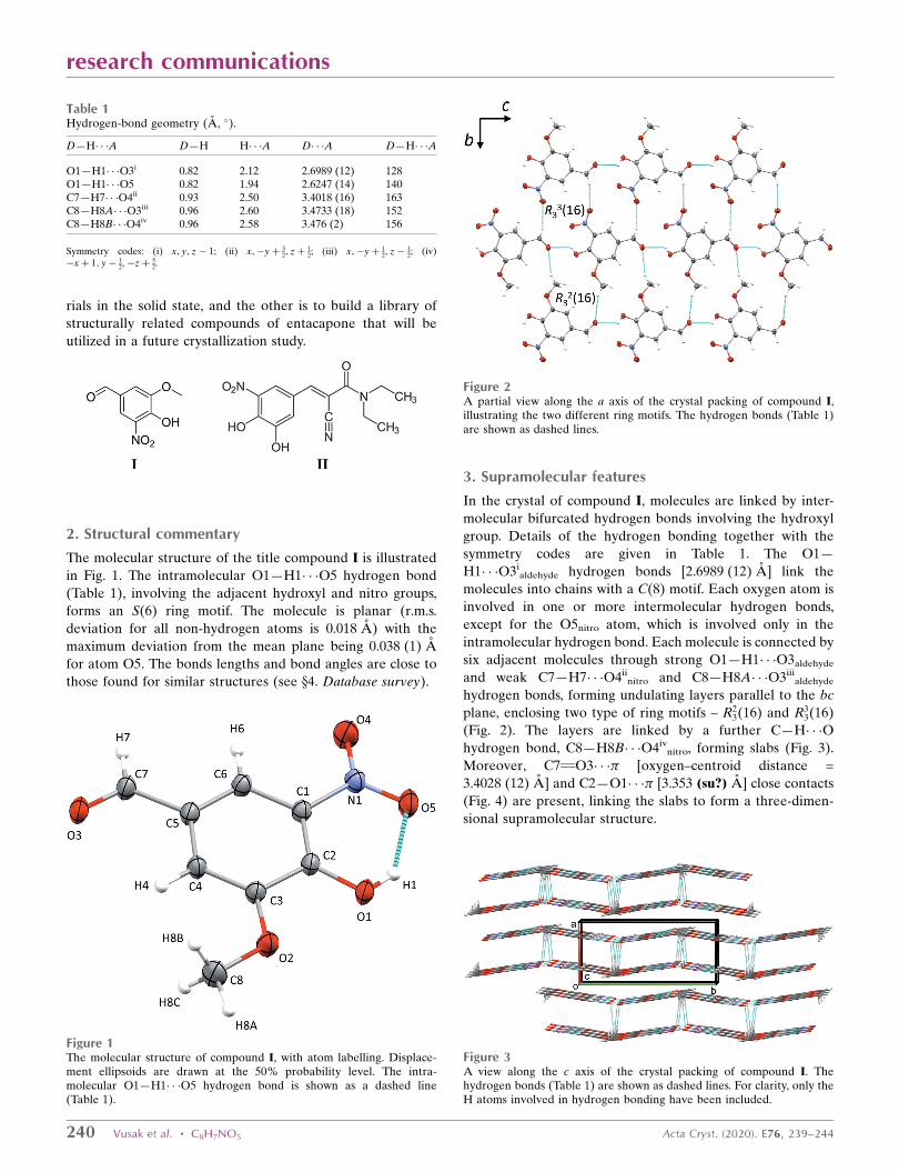
3. Supramolecular features
In the crystal of compound I, molecules are linked by inter-
molecular bifurcated hydrogen bonds involving the hydroxyl
group. Details of the hydrogen bonding together with the
symmetry codes are given in Table 1. The O1—
H1� � �O3ialdehyde hydrogen bonds [2.6989 (12) A] link the
molecules into chains with a C(8) motif. Each oxygen atom is
involved in one or more intermolecular hydrogen bonds,
except for the O5nitro atom, which is involved only in the
intramolecular hydrogen bond. Each molecule is connected by
six adjacent molecules through strong O1—H1� � �O3aldehyde
and weak C7—H7� � �O4iinitro and C8—H8A� � �O3iii
aldehyde
hydrogen bonds, forming undulating layers parallel to the bc
plane, enclosing two type of ring motifs – R23(16) and R3
3(16)
(Fig. 2). The layers are linked by a further C—H� � �O
hydrogen bond, C8—H8B� � �O4ivnitro, forming slabs (Fig. 3).
Moreover, C7 O3� � �� [oxygen–centroid distance =
3.4028 (12) A] and C2—O1� � �� [3.353 (su?) A] close contacts
(Fig. 4) are present, linking the slabs to form a three-dimen-
sional supramolecular structure.
240 Vusak et al. � C8H7NO5 Acta Cryst. (2020). E76, 239–244
research communications
Figure 1The molecular structure of compound I, with atom labelling. Displace-ment ellipsoids are drawn at the 50% probability level. The intra-molecular O1—H1� � �O5 hydrogen bond is shown as a dashed line(Table 1).
Table 1Hydrogen-bond geometry (A, �).
D—H� � �A D—H H� � �A D� � �A D—H� � �A
O1—H1� � �O3i 0.82 2.12 2.6989 (12) 128O1—H1� � �O5 0.82 1.94 2.6247 (14) 140C7—H7� � �O4ii 0.93 2.50 3.4018 (16) 163C8—H8A� � �O3iii 0.96 2.60 3.4733 (18) 152C8—H8B� � �O4iv 0.96 2.58 3.476 (2) 156
Symmetry codes: (i) x; y; z� 1; (ii) x;�yþ 32; zþ 1
2; (iii) x;�y þ 12; z� 1
2; (iv)�xþ 1; y� 1
2;�zþ 52.
Figure 2A partial view along the a axis of the crystal packing of compound I,illustrating the two different ring motifs. The hydrogen bonds (Table 1)are shown as dashed lines.
Figure 3A view along the c axis of the crystal packing of compound I. Thehydrogen bonds (Table 1) are shown as dashed lines. For clarity, only theH atoms involved in hydrogen bonding have been included.

4. Database survey
A search of the Cambridge Structural Database (CSD,
Version V5.41, last update November 2019; Groom et al.,
2016), for crystal structures containing a nitro group on the
benzene ring, oxygen atoms bonded on carbon position 2 and
3, and the –C O group located on position 5, gave three hits,
out of which only one entry contained the title molecule, viz. a
tin complex of the 4-hydroxy-3-methoxy-5-nitrobenzaldehyde
with a deprotonated hydroxyl group and benzyl anions (CSD
refcode EREWII; Keng et al., 2011). The other two entries do
not contain an aldehyde group, but a methylketo (MUCDOE;
Babu et al., 2009) and carboxylic group (TAFSAX; Vladi-
mirova et al., 2016) instead.
A second search of the CSD for a nitro group on a benzene
ring, OH groups on carbon atoms 2 and 3, and a carbon atom
on position 5 gave eight hits for seven structures. These
include the structure of entacapone II (OFAZUQ; Leppanen
et al., 2001), and four of its acyl esters, viz. (E)-2-cyano-3-(3,4-
dihydroxy-5-nitrophenyl)-N,N-diethylprop-2-enamide 1,3-di-
methyl-3,7-dihydro-1H-purine-2,6-dione monohydrate
(XIPNOC; Bommaka et al., 2018), (E)-2-cyano-3-(3,4-dihy-
droxy-5-nitrophenyl)-N,N-diethylprop-2-enamide pyridine-4-
carboxamide (XIPNUI; Bommaka et al., 2018), (E)-2-cyano-
3-(3,4-dihydroxy-5-nitrophenyl)-N,N-diethylprop-2-enamide
pyrazine-2-carboxamide (XIPPAQ; Bommaka et al., 2018),
and (E)-2-cyano-3-(3,4-dihydroxy-5-nitrophenyl)-N,N-di-
ethylprop-2-enamide acetamide (XIPPEU; Bommaka et al.,
2018). These four compounds were prepared by solvent-
assisted grinding, and the study was aimed at improving the
aqueous solubility, diffusion permeability, and co-crystal
stability of entacapone.
5. Hirshfeld surface analysis
Intermolecular interactions in the crystal of compound I were
further investigated by the Hirshfeld surfaces. Calculations
were performed using CrystalExplorer17 (Turner et al., 2017).
The dnorm values were mapped onto the Hirshfeld surface over
the whole molecule (Fig. 5). Red areas represent contacts of
the atoms shorter then the sum of the van der Waals radii, such
as hydrogen bonds, or C O� � �� contacts, whereas blue areas
represent contacts between atoms longer then the sum of the
van der Waals radii. White areas represent contacts equal to
the sum of the van der Waals radii.
The two-dimensional fingerprint plots show intermolecular
contacts and distances between atoms (Fig. 6). The most
abundant contacts are between oxygen and hydrogen atoms,
research communications
Acta Cryst. (2020). E76, 239–244 Vusak et al. � C8H7NO5 241
Figure 5A view of the Hirshfeld surface of compound I, mapped over dnorm in thecolour range�0.448 to 1.186 a.u.. Red areas show intermolecular contactsshorter than the sum of the van der Waals radii of the atoms. The shortestintermolecular O—H� � �O hydrogen bond is also shown.
Figure 6(a) Two-dimensional fingerprint plot for compound I, and the fingerprintplots delineated into (b) H� � �O/O� � �H (47.3%), (c) H� � �H (19.8%), (d)C� � �O/O� � �C (12.0%), (e) C� � �H/H� � �C (8.5%), (f) O� � �O (4.6%), (g)C� � �C (3.9%), (h) N� � �O/O� � �N (3.1%), (i) H� � �N/N� � �H (0.6%).
Figure 4Short C—Ohydroxy� � �� and C Oaldehyde� � �� contacts.

comprising almost half of the Hirshfeld surface area (47.3%).
This finding is not surprising having in mind the number of
oxygen atoms located on the edges of the molecule with
respect its size, each of them is involved in one or more
hydrogen bonds. Also, because of the large number of oxygen
atoms and C O� � �� contacts, there is a high proportion of
C� � �O/O� � �C contacts, which comprise 12.0% of the surface
area. Since there is no stacking of the aromatic rings, only
3.9% of the surface derives from C� � �C contacts.
Electrostatic potentials were calculated using TONTO with
a 3-21G basis set at the Hartree–Fock level of theory and were
mapped on the Hirshfeld surface (Fig. 7) in the energy range
between �0.0923 and 0.1232 a.u.. The most positive region is
around the hydroxyl hydrogen atom (Fig. 7a), while the most
negative region is around the carbonyl oxygen atom (Fig. 7b).
Those two atoms are involved in the shortest intermolecular
hydrogen bond in the crystal structure (O1—H1� � �O3i), where
O1� � �O3i = 2.6989 (12) A; see Table 1.
6. Synthesis and crystallization
4-Hydroxy-3-methoxybenzaldehyde (20 g; 131.4 mmol) was
dissolved in acetic acid (200 ml) and the solution was cooled to
283–288 K and 65% HNO3 (10.5 ml) was added dropwise over
30 min. The reaction mixture was stirred for 30 min at 283–
288 K and 30 min at 293–298 K. The suspension was then
filtered and the crystals obtained were washed with water (3�
20 ml). The crystals were dried in a vacuum dryer (10 mbar,
313 K, 16 h) to obtain pure yellow compound I (yield 20.28 g;
78.3%). Yellow block-like crystals, suitable for X-ray diffrac-
tion analysis, were obtained by slow evaporation of a solution
in acetone after 10 d at room temperature.
Spectroscopic analysis:
The structure of compound I (Fig. 8) was confirmed by 1H
and 13C NMR, recorded on a Bruker Avance DRX 500 at
500.1 MHz (1H) and 125.8 MHz (13C) in CD3OD (Fig. 9a and
9b, respectively); see Tables 2 and 3 for further details.
7. Thermal analysis
The thermal stability of compound I was investigated in the
solid state by differential scanning calorimetry (DSC) and by
thermogravimetric analysis (TGA). DSC analysis was
242 Vusak et al. � C8H7NO5 Acta Cryst. (2020). E76, 239–244
research communications
Figure 7Calculated electrostatic potentials over the Hirshfeld surface ofcompound I. Electrostatic potential was mapped in the energy range�0.0923 to 0.1232 a.u.. The blue area around the hydroxyl oxygen atom in(a) represents the most positive part, while the red area around thecarbonyl oxygen atom in (b) represents the most negative part of themolecule.
Figure 8Structure of compound I in relation to the NMR data in Tables 2 and 3.
Table 2Chemical shifts of protons (DMSO-d6) of 4-hydroxy-3-methoxy-5-nitro-benzaldehyde (I).
Chemical shift (�, p.p.m.) Multiplicity Number of protons Assignment
3.962 s 3 H97.622–7.626 d 1 H38.095–8.098 d 1 H59.867 s 1 H10
Table 3Chemical shifts of carbons (DMSO-d6) of 4-hydroxy-3-methoxy-5-nitrobenzaldehyde (I).
Chemical shift (�, p.p.m.) Number of carbons Assignment
56.78 1 C9112.52 1 C3120.87 1 C5126.81 1 C4137.04 1 C6147.73 1 C1150.03 1 C2190.42 1 C10

performed on a TA Instruments Discovery DSC in a closed
aluminium pan (40 mL) under nitrogen flow (50 ml min�1) and
a heating rate of 10�C min�1 in the temperature range 25–300�C (Fig. 10). Thermogravimetric analysis was performed on a
TA Instruments Discovery TG in a closed aluminium pan
(40 mL) under nitrogen flow (50 ml min�1) and a heating rate
of 10�C min�1 in the temperature range 25–300�C (Fig. 11).
DSC analysis shows one endotherm at about 176�C that
corresponds to the melting point of the title compound.
Thermogravimetric analysis does not show any weight loss
during heating up to 140�C where a change in mass can be
observed that can be attributed to the thermal decomposition
of the sample.
8. IR spectroscopy
The IR spectrum (Fig. 12) of compound I was recorded on a
Thermo Scientific Nicolet instrument by ATR (attenuated
total reflectance) technique. It shows a broad band at about
3200 cm�1, which corresponds to the O—H stretching vibra-
tions. Strong stretching vibrations of C O (aldehyde) and
C—O (aromatic ether) appear at 1683 and 1266 cm�1,
respectively. Bands corresponding to N—O asymmetric and
symmetric stretching modes can be found at 1547 and
1366 cm�1, respectively. Characteristic weak overtones of the
aromatic ring can be seen at 1800–1700 cm�1.
research communications
Acta Cryst. (2020). E76, 239–244 Vusak et al. � C8H7NO5 243
Figure 91H NMR spectra of compound I (CD3OD); (b) 13C NMR spectra ofcompound I (CD3OD)
Figure 10DSC curve of compound I.
Figure 11TG curve of compound I.
Figure 12IR spectra (ATR) of compound I.

244 Vusak et al. � C8H7NO5 Acta Cryst. (2020). E76, 239–244
research communications
Table 4Experimental details.
Crystal dataChemical formula C8H7NO5
Mr 197.15Crystal system, space group Monoclinic, P21/cTemperature (K) 293a, b, c (A) 6.8249 (2), 14.3395 (5), 8.9089 (3)� (�) 106.678 (4)V (A3) 835.21 (5)Z 4Radiation type Cu K�� (mm�1) 1.16Crystal size (mm) 0.30 � 0.15 � 0.13
Data collectionDiffractometer Rigaku Xcalibur Ruby NovaAbsorption correction Multi-scan (CrysAlis PRO; Rigaku
OD, 2018)Tmin, Tmax 0.647, 1No. of measured, independent and
observed [I > 2�(I)] reflections3633, 1697, 1574
Rint 0.016(sin �/)max (A�1) 0.629
RefinementR[F 2 > 2�(F 2)], wR(F 2), S 0.040, 0.174, 0.81No. of reflections 1697No. of parameters 127H-atom treatment H-atom parameters constrained�max, �min (e A�3) 0.27, �0.17
Computer programs: CrysAlis PRO (Rigaku OD, 2018), SHELXS97 (Sheldrick, 2008),Mercury (Macrae et al., 2008), SHELXL2017 (Sheldrick, 2015), PLATON (Spek, 2020)and publCIF (Westrip, 2010).
9. Refinement details
Crystal data, data collection and structure refinement details
are summarized in Table 4. Hydrogen atoms were located in a
difference-Fourier map and refined as riding on their parent
atom: O—H = 0.82 A, C—H = 0.93-0.96 A, with Uiso(H) =
1.5Ueq(O) and 1.2Ueq(C) for other H atoms.
Funding information
VV and EM acknowledge PLIVA for financial support.
References
Babu, S., Raghavamenon, A. C., Fronczek, F. R. & Uppu, R. M.(2009). Acta Cryst. E65, o2292–o2293.
Bartra Sanmarti, M., Solsona Rocabert, J. G., Palomo Nicolau, F. &Molina Ponce, A. (2008). WO Patent 2008/119793.
Bommaka, M. K., Mannava, M. K. C., Suresh, K., Gunnam, A. &Nangia, A. (2018). Cryst. Growth Des. 18, 6061–6069.
Chinnapillai Rajendiran, C., Kondakindi Indrasena Reddy, K., AravaVerra Reddy, A. & Jasti Venkateswaralu, J. (2007). WO Patent2007/020654.
Cziaky, Z. (2006). HU Patent 0402573A2.Deshpande, P. B., Randey, K. A., Dhameliya, D. R., Dayawant, R. B.
& Luthra, K. P. (2010). WO Patent 2010/0234632.Groom, C. R., Bruno, I. J., Lightfoot, M. P. & Ward, S. C. (2016). Acta
Cryst. B72, 171–179.Harisha, A. S., Nayak, S. P., Pavan, M. S., Shridhara, K., Sundarraya,
R. K., Rayendra, K., Pari, K., Sivaramkrishnan, H., Guru Row, T. N.& Nagarajan, K. (2015). J. Chem. Sci. 127, 1977–1991.
Jasti, V., Veera Reddy, A., Rajendiran, C. & Qadeeruddin, S. (2005).WO Patent 2018/167776.
Keng, T. C., Lo, K. M. & Ng, S. W. (2011). Acta Cryst. E67, m710.Leppanen, J., Wegelius, E., Nevalainen, T., Jarvinen, T., Gynther, J. &
Huuskonen, J. (2001). J. Mol. Struct. 562, 129–135.Macrae, C. F., Bruno, I. J., Chisholm, J. A., Edgington, P. R., McCabe,
P., Pidcock, E., Rodriguez-Monge, L., Taylor, R., van de Streek, J. &Wood, P. A. (2008). J. Appl. Cryst. 41, 466–470.
Mantegazza, S., Allegrini, P. & Razzetti, G. (2008). US Patent 2008/0097127A1.
Najib, J. (2001). Clin. Ther. 23, 802–832.Pahwa, R. & Lyons, K. E. (2009). Curr. Med. Res. Opin. 25, 841–849.Rigaku OD, (2018). CrysAlis PRO. Rigaku Oxford Diffraction,
Rigaku Corporation, Oxford, England.Sheldrick, G. M. (2008). Acta Cryst. A64, 112–122.Sheldrick, G. M. (2015). Acta Cryst. C71, 3–8.Spek, A. L. (2020). Acta Cryst. E76, 1–11.Srikanth, G., Ray, U. K., Srinivas Rao, D. V. N., Gupta, P. B., Lavanya,
P. & Islam, A. (2012). Synth. Commun. 42, 1359–1366.Turner, M. J., McKinnon, J. J., Wolff, S. K., Grimwood, D. J.,
Spackman, P. R., Jayatilaka, D. & Spackman, M. A. (2017).CrystalExplorer17. University of Western Australia. http://hirsh-feldsurface.net
Vladimirova, A., Patskovsky, Y., Fedorov, A. A., Bonanno, J. B.,Fedorov, E. V., Toro, R., Hillerich, B., Seidel, R. D., Richards, N. G.J., Almo, S. C. & Raushel, F. M. (2016). J. Am. Chem. Soc. 138, 826–836.
Westrip, S. P. (2010). J. Appl. Cryst. 43, 920–925.
supporting information
sup-1Acta Cryst. (2020). E76, 239-244
supporting information
Acta Cryst. (2020). E76, 239-244 [https://doi.org/10.1107/S2056989020000225]
Synthesis, crystal structure and spectroscopic and Hirshfeld surface analysis of
4-hydroxy-3-methoxy-5-nitrobenzaldehyde
Vitomir Vusak, Darko Vusak, Kresimir Molcanov and Mestrovic Ernest
Computing details
Data collection: CrysAlis PRO (Rigaku OD, 2018); cell refinement: CrysAlis PRO (Rigaku OD, 2018); data reduction:
CrysAlis PRO (Rigaku OD, 2018); program(s) used to solve structure: SHELXS97 (Sheldrick, 2008); program(s) used to
refine structure: SHELXL2017 (Sheldrick, 2015); molecular graphics: Mercury (Macrae et al., 2008); software used to
prepare material for publication: SHELXL2017 (Sheldrick, 2015), PLATON (Spek, 2020) and publCIF (Westrip, 2010).
4-Hydroxy-3-methoxy-5-nitrobenzaldehyde
Crystal data
C8H7NO5
Mr = 197.15Monoclinic, P21/cHall symbol: -P 2ybca = 6.8249 (2) Åb = 14.3395 (5) Åc = 8.9089 (3) Åβ = 106.678 (4)°V = 835.21 (5) Å3
Z = 4
F(000) = 408Dx = 1.568 Mg m−3
Cu Kα radiation, λ = 1.54184 ÅCell parameters from 2167 reflectionsθ = 3.2–75.6°µ = 1.16 mm−1
T = 293 KBlock, yellow0.30 × 0.15 × 0.13 mm
Data collection
Rigaku Xcalibur Ruby Nova diffractometer
Radiation source: micro-focus sealed X-ray tubeMirror monochromatorDetector resolution: 10.4323 pixels mm-1
ω scansAbsorption correction: multi-scan
(CrysAlis PRO; Rigaku OD, 2018)Tmin = 0.647, Tmax = 1
3633 measured reflections1697 independent reflections1574 reflections with I > 2σ(I)Rint = 0.016θmax = 76.0°, θmin = 6.8°h = −8→8k = −16→17l = −7→11
Refinement
Refinement on F2
Least-squares matrix: fullR[F2 > 2σ(F2)] = 0.040wR(F2) = 0.174S = 0.811697 reflections127 parameters0 restraints
Primary atom site location: structure-invariant direct methods
Secondary atom site location: difference Fourier map
Hydrogen site location: inferred from neighbouring sites
H-atom parameters constrained

supporting information
sup-2Acta Cryst. (2020). E76, 239-244
w = 1/[σ2(Fo2) + (0.2P)2]
where P = (Fo2 + 2Fc
2)/3(Δ/σ)max = 0.001
Δρmax = 0.27 e Å−3
Δρmin = −0.16 e Å−3
Special details
Geometry. All esds (except the esd in the dihedral angle between two l.s. planes) are estimated using the full covariance matrix. The cell esds are taken into account individually in the estimation of esds in distances, angles and torsion angles; correlations between esds in cell parameters are only used when they are defined by crystal symmetry. An approximate (isotropic) treatment of cell esds is used for estimating esds involving l.s. planes.
Fractional atomic coordinates and isotropic or equivalent isotropic displacement parameters (Å2)
x y z Uiso*/Ueq
O1 0.24420 (14) 0.44411 (6) 0.92251 (10) 0.0442 (3)H1 0.258885 0.488242 0.868197 0.066*O2 0.19059 (16) 0.31818 (6) 1.10856 (11) 0.0506 (3)O3 0.23301 (16) 0.48763 (7) 1.62543 (10) 0.0508 (3)O4 0.3265 (2) 0.72037 (7) 1.06656 (14) 0.0655 (4)O5 0.29317 (17) 0.62262 (8) 0.87895 (11) 0.0547 (3)N1 0.30056 (15) 0.64166 (7) 1.01585 (12) 0.0422 (3)C1 0.27609 (15) 0.56669 (8) 1.11855 (13) 0.0353 (3)C2 0.24838 (14) 0.47461 (7) 1.06403 (13) 0.0347 (3)C3 0.22165 (16) 0.40479 (8) 1.17059 (14) 0.0375 (3)C4 0.22427 (16) 0.42865 (8) 1.32044 (13) 0.0386 (3)H4 0.206582 0.382706 1.389078 0.046*C5 0.25347 (16) 0.52194 (8) 1.37123 (13) 0.0377 (3)C6 0.27967 (15) 0.59084 (8) 1.27155 (13) 0.0376 (3)H6 0.299441 0.652439 1.305155 0.045*C7 0.25478 (19) 0.54494 (9) 1.53231 (14) 0.0424 (3)H7 0.273358 0.60689 1.564115 0.051*C8 0.1668 (2) 0.24460 (10) 1.21046 (17) 0.0577 (4)H8A 0.145807 0.186668 1.154094 0.086*H8B 0.287788 0.240337 1.297763 0.086*H8C 0.05098 0.257453 1.247879 0.086*
Atomic displacement parameters (Å2)
U11 U22 U33 U12 U13 U23
O1 0.0671 (5) 0.0406 (5) 0.0279 (5) −0.0002 (3) 0.0184 (4) −0.0015 (3)O2 0.0823 (6) 0.0336 (5) 0.0392 (6) −0.0058 (4) 0.0229 (5) −0.0032 (3)O3 0.0749 (6) 0.0513 (6) 0.0302 (5) −0.0034 (4) 0.0214 (4) −0.0005 (3)O4 0.1106 (9) 0.0351 (6) 0.0529 (7) −0.0071 (5) 0.0268 (6) 0.0028 (4)O5 0.0838 (7) 0.0512 (6) 0.0326 (6) −0.0033 (4) 0.0223 (5) 0.0063 (4)N1 0.0540 (5) 0.0378 (6) 0.0353 (6) −0.0005 (4) 0.0135 (4) 0.0052 (4)C1 0.0423 (5) 0.0336 (6) 0.0304 (6) 0.0017 (4) 0.0112 (4) 0.0029 (4)C2 0.0415 (5) 0.0366 (6) 0.0274 (6) 0.0017 (4) 0.0120 (4) 0.0014 (4)C3 0.0481 (5) 0.0335 (7) 0.0318 (6) −0.0002 (4) 0.0126 (4) 0.0002 (4)C4 0.0517 (6) 0.0353 (6) 0.0316 (6) −0.0014 (4) 0.0163 (5) 0.0022 (4)C5 0.0453 (6) 0.0396 (7) 0.0296 (6) 0.0012 (4) 0.0131 (4) −0.0015 (4)

supporting information
sup-3Acta Cryst. (2020). E76, 239-244
C6 0.0469 (6) 0.0340 (6) 0.0325 (7) 0.0010 (4) 0.0124 (5) −0.0017 (4)C7 0.0575 (6) 0.0407 (7) 0.0312 (6) 0.0001 (4) 0.0162 (5) −0.0024 (4)C8 0.0881 (9) 0.0352 (7) 0.0513 (8) −0.0040 (6) 0.0225 (7) 0.0040 (5)
Geometric parameters (Å, º)
O1—C2 1.3271 (14) C3—C4 1.3733 (16)O1—H1 0.82 C4—C5 1.4080 (17)O2—C3 1.3510 (15) C4—H4 0.93O2—C8 1.4310 (15) C5—C6 1.3740 (15)O3—C7 1.2068 (16) C5—C7 1.4700 (16)O4—N1 1.2097 (15) C6—H6 0.93O5—N1 1.2367 (15) C7—H7 0.93N1—C1 1.4520 (15) C8—H8A 0.96C1—C6 1.3998 (15) C8—H8B 0.96C1—C2 1.4008 (17) C8—H8C 0.96C2—C3 1.4270 (15)
C2—O1—H1 109.5 C5—C4—H4 119.7C3—O2—C8 116.83 (9) C6—C5—C4 120.54 (10)O4—N1—O5 122.31 (11) C6—C5—C7 120.27 (11)O4—N1—C1 119.11 (11) C4—C5—C7 119.18 (11)O5—N1—C1 118.58 (10) C5—C6—C1 118.84 (11)C6—C1—C2 122.25 (11) C5—C6—H6 120.6C6—C1—N1 117.22 (11) C1—C6—H6 120.6C2—C1—N1 120.52 (10) O3—C7—C5 123.43 (12)O1—C2—C1 127.16 (10) O3—C7—H7 118.3O1—C2—C3 115.39 (10) C5—C7—H7 118.3C1—C2—C3 117.46 (10) O2—C8—H8A 109.5O2—C3—C4 125.69 (11) O2—C8—H8B 109.5O2—C3—C2 114.08 (10) H8A—C8—H8B 109.5C4—C3—C2 120.22 (11) O2—C8—H8C 109.5C3—C4—C5 120.69 (11) H8A—C8—H8C 109.5C3—C4—H4 119.7 H8B—C8—H8C 109.5
O4—N1—C1—C6 1.36 (17) O1—C2—C3—C4 179.72 (9)O5—N1—C1—C6 −178.32 (10) C1—C2—C3—C4 −0.22 (16)O4—N1—C1—C2 −179.20 (11) O2—C3—C4—C5 −178.50 (10)O5—N1—C1—C2 1.12 (16) C2—C3—C4—C5 −0.07 (17)C6—C1—C2—O1 −179.40 (10) C3—C4—C5—C6 0.07 (17)N1—C1—C2—O1 1.19 (17) C3—C4—C5—C7 179.82 (10)C6—C1—C2—C3 0.52 (16) C4—C5—C6—C1 0.22 (16)N1—C1—C2—C3 −178.89 (8) C7—C5—C6—C1 −179.52 (10)C8—O2—C3—C4 −2.73 (18) C2—C1—C6—C5 −0.53 (16)C8—O2—C3—C2 178.76 (10) N1—C1—C6—C5 178.90 (8)O1—C2—C3—O2 −1.68 (14) C6—C5—C7—O3 −179.85 (11)C1—C2—C3—O2 178.39 (9) C4—C5—C7—O3 0.40 (19)

supporting information
sup-4Acta Cryst. (2020). E76, 239-244
Hydrogen-bond geometry (Å, º)
D—H···A D—H H···A D···A D—H···A
O1—H1···O3i 0.82 2.12 2.6989 (12) 128O1—H1···O5 0.82 1.94 2.6247 (14) 140C7—H7···O4ii 0.93 2.50 3.4018 (16) 163C8—H8A···O3iii 0.96 2.60 3.4733 (18) 152C8—H8B···O4iv 0.96 2.58 3.476 (2) 156
Symmetry codes: (i) x, y, z−1; (ii) x, −y+3/2, z+1/2; (iii) x, −y+1/2, z−1/2; (iv) −x+1, y−1/2, −z+5/2.
Related Documents