Journal of the Korean Chemical Society 2021, Vol. 65, No. 3 Printed in the Republic of Korea https://doi.org/10.5012/jkcs.2021.65.3.203 -203- Synthesis, Characterization and in vitro Antibacterial Studies on Mixed Ligand Complexes of Iron(III) Based on 1,10-phenanthroline Getinet Tamiru Tigineh*, Getu Sitotaw, Amogne Workie † , and Atakilt Abebe Bahir Dar University, Science College, Chemistry Department, P.O. Box; 79, Bahir Dar, Ethiopia. *E-mail: [email protected]; [email protected] † Institute of Chemistry, Academia Sinica, 128, Sec. Academia Rd., Nangang, Taipei 115, Taiwan. (Received February 7, 2021; Accepted March 3, 2021) ABSTRACT. As part of our attempt to discover novel active compounds against multi-drug resistant pathogens, we hereby report two new complexes of iron(III) with formulae: [Fe(L 1 ) 2 (H 2 O) 2 ]Cl 3 and [Fe(L 1 ) 2 (L 2 )(H 2 O)]Cl 2 where L 1 = 1,10-phenan- throline (C 12 H 8 N 2 ) and L 2 = guanide (C 5 H 4 N 5 O - ). The synthesized complexes were characterized using spectroscopic analysis (ESI-MS, ICP-OES, FT-IR, and UV-Vis), cyclic voltammetry, CHN analysis, gravimetric chloride determination, melting point deter- mination, and conductance measurement. Octahedral geometries are assigned to both complexes. In vitro antibacterial activity was tested on two Gram-positive (Staphylococcus aureus, Streptococcus epidermidis) and two Gram-negative (Escherichia coli and Klebsi- ella pneumoniae) bacteria using the disc diffusion method. The complexes demonstrated appreciable activity against these pathogens. Interestingly, the [Fe(L 1 ) 2 (L 2 )(H 2 O)]Cl 2 complex manifested a higher degree of inhibition against the drug-resistant Gram-negative bac- teria than the commercially available drug, namely erythromycin. Key words: Iron(III), Mixed ligand complexes, 1,10-Phenanthroline, Guanide, Antibacterial activity INTRODUCTION Drugs derived from transition metals have been used for treating many infectious diseases 1 including cancer, 2 infections, 3 and inflammation. 4 The dynamic properties of transition metal compounds such as stability, redox, or coordination have been of interest, especially for multi- drug-resistant bacteria, 5–8 where natural product-based drugs become ineffective. 9–10 Quick penetrating in the cell membrane 11–12 or strong coordination to the DNA strand enhances the efficiency of metal-based drugs. 13–15 The use of different ligands having different structures and properties is the primary strategy in tuning the prop- erties of transition metal ions to obtain the desired appli- cations. 16–18 Among various transition metals, Fe(III) is stable under physiological conditions and results in a ther- modynamically stable complex. In particular, its mixed-ligand complexes have attracted growing attention for their bio- logical activities. Especially, its complexes with aromatic heterocyclic ligands showed promising antibacterial activ- ity. 12,19 Nevertheless, any report on the synthesis and biological application in the form of mixed-ligand complexes con- taining flat heterocyclic ligands with nucleobases could not be found. A large number of biologically active pharmaceutical ingredients containing flat heterocyclic ligands are com- monly used in a wide variety of therapeutic areas. 1,10- Phenanthroline, among flat heterocyclic ligands, is a superb chelating bidentate ligand due to its ideally placed nitrogen atoms in the π-acidic rigid electron-poor heteroaromatic planar structure (Fig. 1). These properties endowed it with stacking interaction ability with DNA base pairs. Further- more, when coordinated to metal ions, its complexes experi- ence ionic or covalent or both interactions through the metal center with the organic base residues of the genetic mate- rials. 20–22 Guanine is an oxypurine heteroaromatic organic base. It is a component of DNA and capable of interacting with cytosine residue of the genetic material of the pathogen, which interrupts the normal growth of bacteria (Fig. 1). 23 Previously, our group reported ruthenium complexes with similar ligands for biological applications. 24 However, ruthenium is rare and expensive, whereas iron is compara- Figure 1. Ligands used in this work.

Welcome message from author
This document is posted to help you gain knowledge. Please leave a comment to let me know what you think about it! Share it to your friends and learn new things together.
Transcript
Journal of the Korean Chemical Society2021, Vol. 65, No. 3Printed in the Republic of Koreahttps://doi.org/10.5012/jkcs.2021.65.3.203
-203-
Synthesis, Characterization and in vitro Antibacterial Studies on Mixed Ligand Complexes of Iron(III) Based on 1,10-phenanthroline
Getinet Tamiru Tigineh*, Getu Sitotaw, Amogne Workie†, and Atakilt Abebe
Bahir Dar University, Science College, Chemistry Department, P.O. Box; 79, Bahir Dar, Ethiopia.
*E-mail: [email protected]; [email protected]†Institute of Chemistry, Academia Sinica, 128, Sec. Academia Rd., Nangang, Taipei 115, Taiwan.
(Received February 7, 2021; Accepted March 3, 2021)
ABSTRACT. As part of our attempt to discover novel active compounds against multi-drug resistant pathogens, we hereby
report two new complexes of iron(III) with formulae: [Fe(L1)2(H2O)2]Cl3 and [Fe(L1)2(L2)(H2O)]Cl2 where L1 = 1,10-phenan-
throline (C12H8N2) and L2 = guanide (C5H4N5O-). The synthesized complexes were characterized using spectroscopic analysis
(ESI-MS, ICP-OES, FT-IR, and UV-Vis), cyclic voltammetry, CHN analysis, gravimetric chloride determination, melting point deter-
mination, and conductance measurement. Octahedral geometries are assigned to both complexes. In vitro antibacterial activity was
tested on two Gram-positive (Staphylococcus aureus, Streptococcus epidermidis) and two Gram-negative (Escherichia coli and Klebsi-
ella pneumoniae) bacteria using the disc diffusion method. The complexes demonstrated appreciable activity against these pathogens.
Interestingly, the [Fe(L1)2(L2)(H2O)]Cl2 complex manifested a higher degree of inhibition against the drug-resistant Gram-negative bac-
teria than the commercially available drug, namely erythromycin.
Key words: Iron(III), Mixed ligand complexes, 1,10-Phenanthroline, Guanide, Antibacterial activity
INTRODUCTION
Drugs derived from transition metals have been used for
treating many infectious diseases1 including cancer,2
infections,3 and inflammation.4 The dynamic properties of
transition metal compounds such as stability, redox, or
coordination have been of interest, especially for multi-
drug-resistant bacteria,5–8 where natural product-based
drugs become ineffective.9–10 Quick penetrating in the cell
membrane11–12 or strong coordination to the DNA strand
enhances the efficiency of metal-based drugs.13–15
The use of different ligands having different structures
and properties is the primary strategy in tuning the prop-
erties of transition metal ions to obtain the desired appli-
cations.16–18 Among various transition metals, Fe(III) is
stable under physiological conditions and results in a ther-
modynamically stable complex. In particular, its mixed-ligand
complexes have attracted growing attention for their bio-
logical activities. Especially, its complexes with aromatic
heterocyclic ligands showed promising antibacterial activ-
ity.12,19
Nevertheless, any report on the synthesis and biological
application in the form of mixed-ligand complexes con-
taining flat heterocyclic ligands with nucleobases could
not be found.
A large number of biologically active pharmaceutical
ingredients containing flat heterocyclic ligands are com-
monly used in a wide variety of therapeutic areas. 1,10-
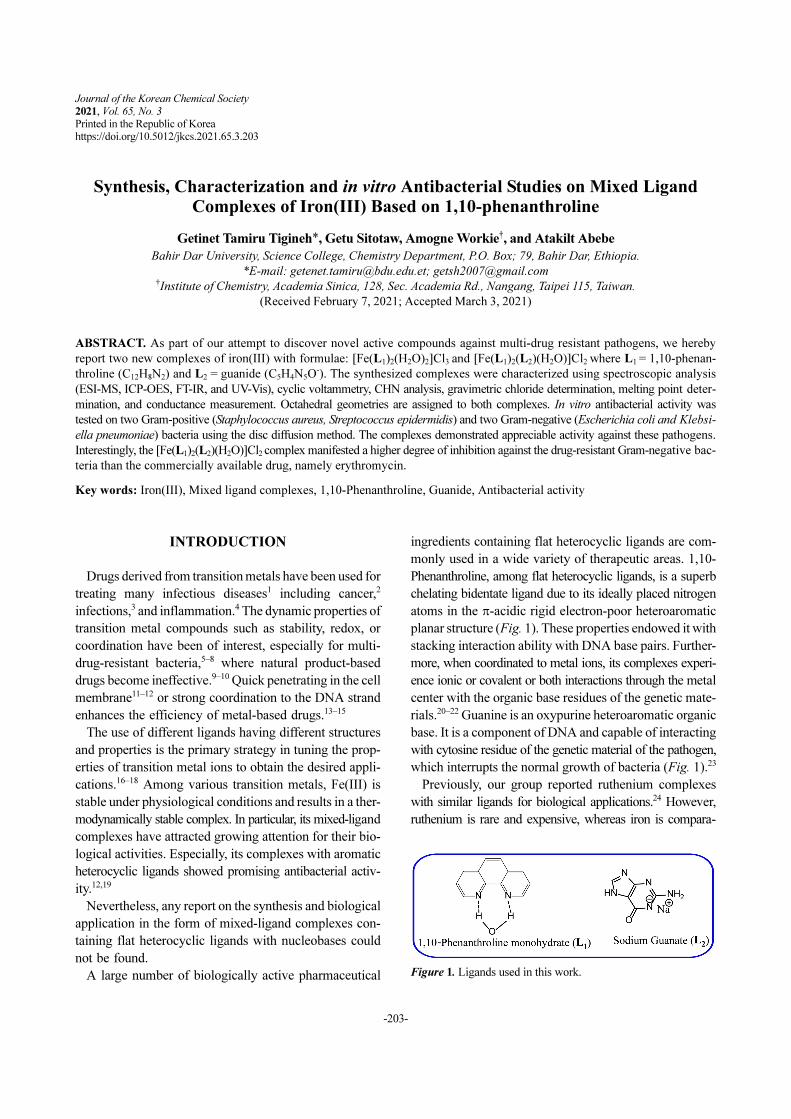
Phenanthroline, among flat heterocyclic ligands, is a superb
chelating bidentate ligand due to its ideally placed nitrogen
atoms in the π-acidic rigid electron-poor heteroaromatic
planar structure (Fig. 1). These properties endowed it with
stacking interaction ability with DNA base pairs. Further-
more, when coordinated to metal ions, its complexes experi-
ence ionic or covalent or both interactions through the metal
center with the organic base residues of the genetic mate-
rials.20–22 Guanine is an oxypurine heteroaromatic organic
base. It is a component of DNA and capable of interacting
with cytosine residue of the genetic material of the pathogen,
which interrupts the normal growth of bacteria (Fig. 1).23
Previously, our group reported ruthenium complexes
with similar ligands for biological applications.24 However,
ruthenium is rare and expensive, whereas iron is compara-
Figure 1. Ligands used in this work.
Journal of the Korean Chemical Society
204 Getinet Tamiru Tigineh, Getu Sitotaw, Amogne Workie, and Atakilt Abebe
tively abundant and cheap.25 In terms of health benefits
also iron is physiologically more relevant than ruthenium.
These properties make us interested in the development of
iron-based drugs along with ruthenium for multi-drug resis-
tant pathogens.
Therefore, in this study, the structural investigation and
biological activities of new iron complexes in a 1:2 Fe(III)
to phen (L1) ratio alone as well as mixed with guanide (L2)
in a 1:2:1 Fe(III) to phen (L1) to guanide (L2) are exam-
ined. The composition would result in a rigid three-dimen-
sional structure and orchestrate the binding ability of iron(III)
with a range of pathogen molecules and hinders their pro-
liferation in the body of the host organism. As a result, the
complexes would be a potential alternative drug for drug-
resistant pathogens.
EXPERIMENTAL
Synthesis
New mononuclear Iron(III) complexes containing 1,10-
phenanthroline alone and mixed with guanide were syn-
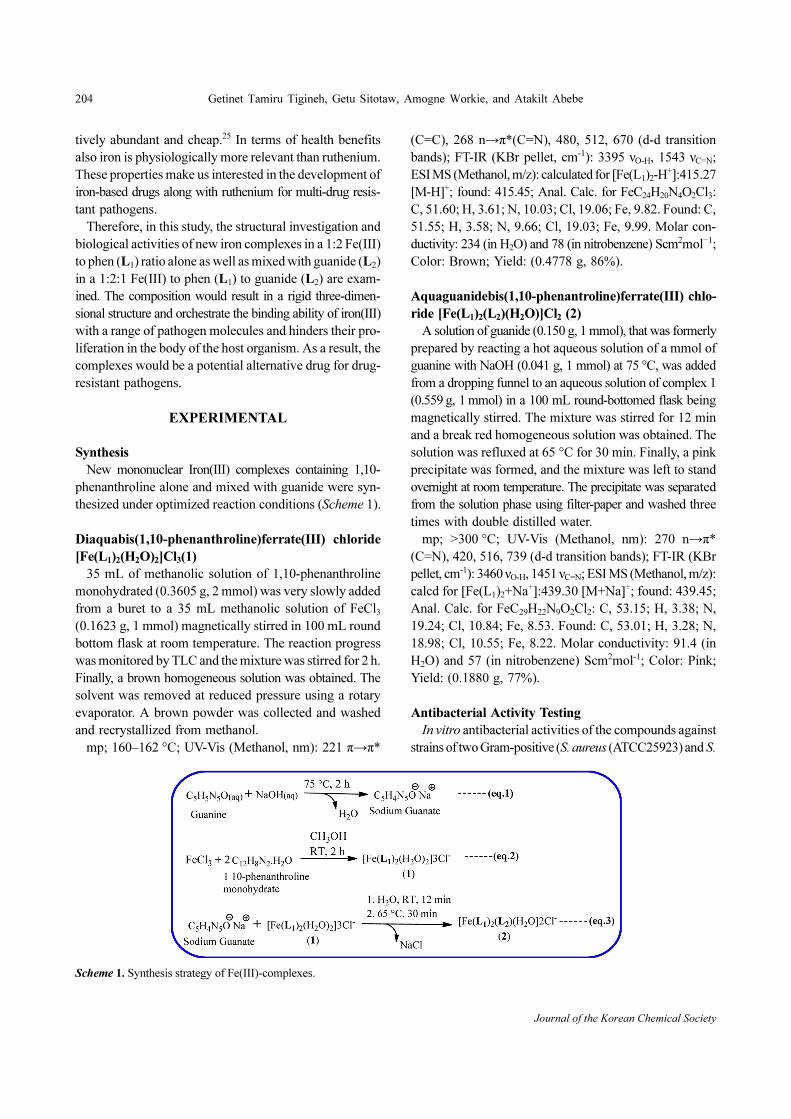
thesized under optimized reaction conditions (Scheme 1).
Diaquabis(1,10-phenanthroline)ferrate(III) chloride
[Fe(L1)2(H2O)2]Cl3(1)
35 mL of methanolic solution of 1,10-phenanthroline
monohydrated (0.3605 g, 2 mmol) was very slowly added
from a buret to a 35 mL methanolic solution of FeCl3
(0.1623 g, 1 mmol) magnetically stirred in 100 mL round
bottom flask at room temperature. The reaction progress
was monitored by TLC and the mixture was stirred for 2 h.
Finally, a brown homogeneous solution was obtained. The
solvent was removed at reduced pressure using a rotary
evaporator. A brown powder was collected and washed
and recrystallized from methanol.
mp; 160–162 °C; UV-Vis (Methanol, nm): 221 π→π*
(C=C), 268 n→π*(C=N), 480, 512, 670 (d-d transition
bands); FT-IR (KBr pellet, cm-1): 3395 νO-H, 1543 νC=N;
ESI MS (Methanol, m/z): calculated for [Fe(L1)2-H+]:415.27
[M-H]+; found: 415.45; Anal. Calc. for FeC24H20N4O2Cl3:
C, 51.60; H, 3.61; N, 10.03; Cl, 19.06; Fe, 9.82. Found: C,
51.55; H, 3.58; N, 9.66; Cl, 19.03; Fe, 9.99. Molar con-
ductivity: 234 (in H2O) and 78 (in nitrobenzene) Scm2mol−1;
Color: Brown; Yield: (0.4778 g, 86%).
Aquaguanidebis(1,10-phenantroline)ferrate(III) chlo-
ride [Fe(L1)2(L2)(H2O)]Cl2 (2)
A solution of guanide (0.150 g, 1 mmol), that was formerly
prepared by reacting a hot aqueous solution of a mmol of
guanine with NaOH (0.041 g, 1 mmol) at 75 °C, was added
from a dropping funnel to an aqueous solution of complex 1
(0.559 g, 1 mmol) in a 100 mL round-bottomed flask being
magnetically stirred. The mixture was stirred for 12 min
and a break red homogeneous solution was obtained. The
solution was refluxed at 65 °C for 30 min. Finally, a pink
precipitate was formed, and the mixture was left to stand
overnight at room temperature. The precipitate was separated
from the solution phase using filter-paper and washed three
times with double distilled water.
mp; >300 °C; UV-Vis (Methanol, nm): 270 n→π*
(C=N), 420, 516, 739 (d-d transition bands); FT-IR (KBr
pellet, cm-1): 3460 νO-H, 1451 νC=N; ESI MS (Methanol, m/z):
calcd for [Fe(L1)2+Na+]:439.30 [M+Na]+; found: 439.45;
Anal. Calc. for FeC29H22N9O2Cl2: C, 53.15; H, 3.38; N,
19.24; Cl, 10.84; Fe, 8.53. Found: C, 53.01; H, 3.28; N,
18.98; Cl, 10.55; Fe, 8.22. Molar conductivity: 91.4 (in
H2O) and 57 (in nitrobenzene) Scm2mol-1; Color: Pink;
Yield: (0.1880 g, 77%).
Antibacterial Activity Testing
In vitro antibacterial activities of the compounds against
strains of two Gram-positive (S. aureus (ATCC25923) and S.
Scheme 1. Synthesis strategy of Fe(III)-complexes.
Synthesis, Characterization and in vitro Antibacterial Studies on Mixed Ligand Complexes of Iron(III) Based on 1,10-phenanthroline 205
2021, Vol. 65, No. 3
epidermidis (ATCC12228) and two Gram-negative E. coli
(ATCC25922) and K. pneumoniae (ATCC986605) bac-
teria were investigated by disc diffusion methods. Muller
Hinton agar (MHA) and nutrient blood agar (BA) were
used for culturing the bacterial isolates while diagnostic
sensitivity test agar (oxoid Ltd BASINGSTOKE England)
was used for sensitivity. All plates and Nutrient Broth
(Difco) were autoclaved for 30 min at 121 °C in a steam
sterilizer. The compounds dissolved in double-distilled
water were applied at a fixed concentration of 500 μg/mL.
Zones of inhibition were measured after 24 h incubation at
37 °C. Antibiotic discs Erythromycin was used as a ref-
erence. The minimum inhibitory concentration (MIC) of
the two complexes against each bacterium was determined
by preparing different concentrations of the complexes by
serial dilution (75 μg/mL, 150 μg/mL, 300 μg/mL, 500 μg/
mL, and 600 μg/mL). The antibacterial tests were performed
at the Amhara Regional Health Research Microbiology
Laboratory Center, Bahir Dar, Ethiopia.
RESULTS AND DISCUSSION
The analytical data of the complexes agree well with the
assigned molecular formulae of complexes 1 and 2. The
gravimetric chloride determination confirmed the pres-
ence of three and two chloride ions in the outer sphere of
complexes 1 and 2, respectively. The molar conductivity
values of complexes 1 and 2 revealed their electrolytic nature.
Their conductivities were found higher in an aqueous
solution than in nitrobenzene. The result is in close agree-
ment with the literature.26 ICP-OES metal determination
showed the anticipated amount of iron in complexes. Fur-
thermore, the experimental and theoretical data of C, H,
and N elemental analyses for complexes are in good agree-
ment, which confirms the achievement of the intended
complexes (see experimental section).
FT-IR Spectroscopy
The infrared spectra of the ligands and the complex are
indicated in Fig. S1 and selected characteristic frequen-
cies are listed in Table S1. The sharp band at 3430 cm-1 (s),
characteristic for ν(OH) stretching in the free 1,10-phenanth-
roline monohydrate appeared broad at 3395 cm-1 (m) in
[Fe(L1)2(H2O)2]Cl3. This change in the absorption frequency
of water explains the change like its interaction, consequently
confirmed the transformation of crystalline H2O to coor-
dinated H2O. The band arising due to vibrational ν(C=N)
mode at 1588 cm-1(m) in the free 1,10-phenanthroline was
shifted to lower frequencies, at 1543 cm-1(m) in [Fe(L1)2
(H2O)2]Cl3. This shift suggested that 1,10-phenanthroline
binds with the metal center via ring nitrogen.
The presence of the characteristic peaks, in the region of
1490–1475 cm-1, of guanine in [Fe(L1)2(L2)(H2O)]Cl2 attributed
to the successful coordination of guanide to the metal cen-
ter. Besides, the coordination of guanide with metal via the
imidazole ring nitrogen was confirmed by the shift in ν(C=N)
vibration.27−29 Furthermore, the similarity in the stretching fre-
quency of ν(C=O) and scissoring of δ(NH2) in free gua-
nine with that of the stretching frequency of ν(C=O) and
scissoring of δ(NH2) in [Fe(L1)2(L2)(H2O)]Cl2 respectively
confirmed oxygen of the carbonyl group (C=O) and nitrogen
of amine (NH2) group of guanide are not used for coor-
dination.27
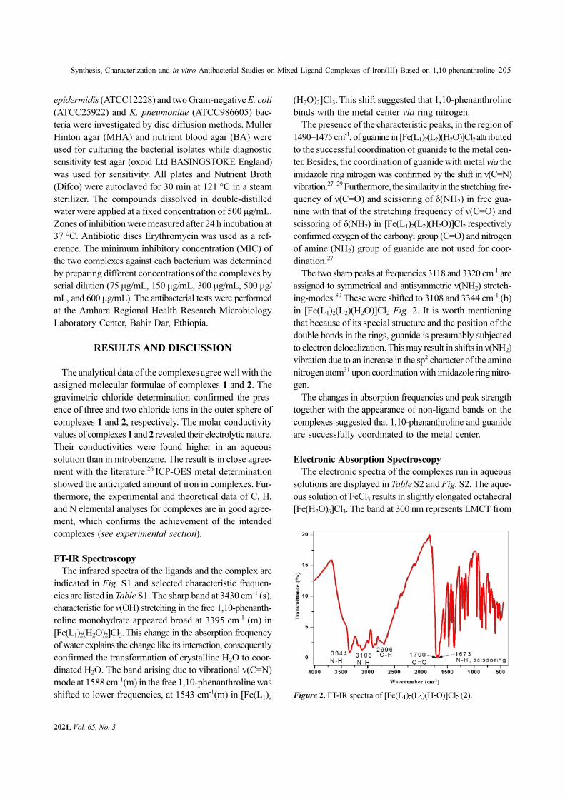
The two sharp peaks at frequencies 3118 and 3320 cm-1 are
assigned to symmetrical and antisymmetric ν(NH2) stretch-
ing-modes.30 These were shifted to 3108 and 3344 cm-1 (b)
in [Fe(L1)2(L2)(H2O)]Cl2 Fig. 2. It is worth mentioning
that because of its special structure and the position of the
double bonds in the rings, guanide is presumably subjected
to electron delocalization. This may result in shifts in ν(NH2)
vibration due to an increase in the sp2 character of the amino
nitrogen atom31 upon coordination with imidazole ring nitro-
gen.
The changes in absorption frequencies and peak strength
together with the appearance of non-ligand bands on the
complexes suggested that 1,10-phenanthroline and guanide
are successfully coordinated to the metal center.
Electronic Absorption Spectroscopy
The electronic spectra of the complexes run in aqueous
solutions are displayed in Table S2 and Fig. S2. The aque-
ous solution of FeCl3 results in slightly elongated octahedral
[Fe(H2O)6]Cl3. The band at 300 nm represents LMCT from
Figure 2. FT-IR spectra of [Fe(L1)2(L2)(H2O)]Cl2 (2).
Journal of the Korean Chemical Society
206 Getinet Tamiru Tigineh, Getu Sitotaw, Amogne Workie, and Atakilt Abebe
the electron-rich H2O orbitals to the low-lying all singly
occupied Fe(III) t2g orbitals. In complex 1, the coordina-
tion of the electron-deficient, bidentate strong field 1,10-
phenanthroline in the equatorial position results in a short
but strong bond. This results in the lowering of the dz2 as
well as the population of all the d electrons in the t2g orbit-
als of Fe(III). Consequently, the LMCT takes place in the
low-lying dz2 orbitals. This fact is signaled by the band at
353 nm. The increase in the LMCT resonance wavelength
indicates the lower energy gap between the donor ligand
and recipient metal orbitals.32 The non-ligand bands newly
observed in complexes 1 and 2 in the range of 13333–
23809 cm-1 corresponding to the transitions 2T2g→2A2g,
2T2g→2T1g,
2T2g→2Eg confirm the possible coordination
of metal and ligands.33
MS Spectroscopy
The ESI MS spectra of the complexes recorded dis-
solving in methanol showed a characteristic molecular ion
peak of [Fe(L1)2(H2O)2]Cl3 with the best accuracy at m/z
calculated for [Fe(L1)2-H+]:415.27 [M-H]+; found: 415.45.
This is presumably due to the electron-deficient [Fe(L1)2]3+
complex captured low-energy electron generated in the
ionization chamber and undergone reduction to [Fe(L1)2]2+
before deprotonation.34 Alternatively, the observed unique
fragmentation patterns of [Fe(L1)2(L2)(H2O)]Cl2 at m/z =
439.3 and 465.3, which represent the molecular ion peaks
of [Fe(L1)2+Na+]35 and [Fe(L1)2(H2O)(CH3OH)-H+],36
respectively, and the multiple-deprotonated species at
563.2, and 383.1 corresponds to [Fe(L1)2(L2)-3H+] and
[Fe(L1)(L2)-3H+] respectively37 Fig. S3, windup the shreds
of evidence found in the former techniques in confirming
the achievement of the intended complexes.
Cyclic Voltammetry
Fig. S4 shows the CVs of [Fe(L1)2(H2O)2]Cl3 and
[Fe(L1)2(L2)(H2O)]Cl2 for successive cycles at a scan rate
of 100 mVs-1. The voltammogram displays a cathodic peak
at the potentials 0.84 and 0.84 V and an anodic peak at 0.87
and 0.86 V of [Fe(L1)2(H2O)2]Cl3 and [Fe(L1)2(L2)(H2O)]Cl2,
respectively. The pair of peaks in both complexes corre-
sponds to the reversible redox process of the species as
Fe(II) and Fe(III) system.38 The absence of peaks other
than expected cathodic and anodic peaks for the Fe(II) and
Fe(III) system signifies that the complexes are stable in
the voltage range scanned here. These results conform to
the literature values.39
Antimicrobial Activity
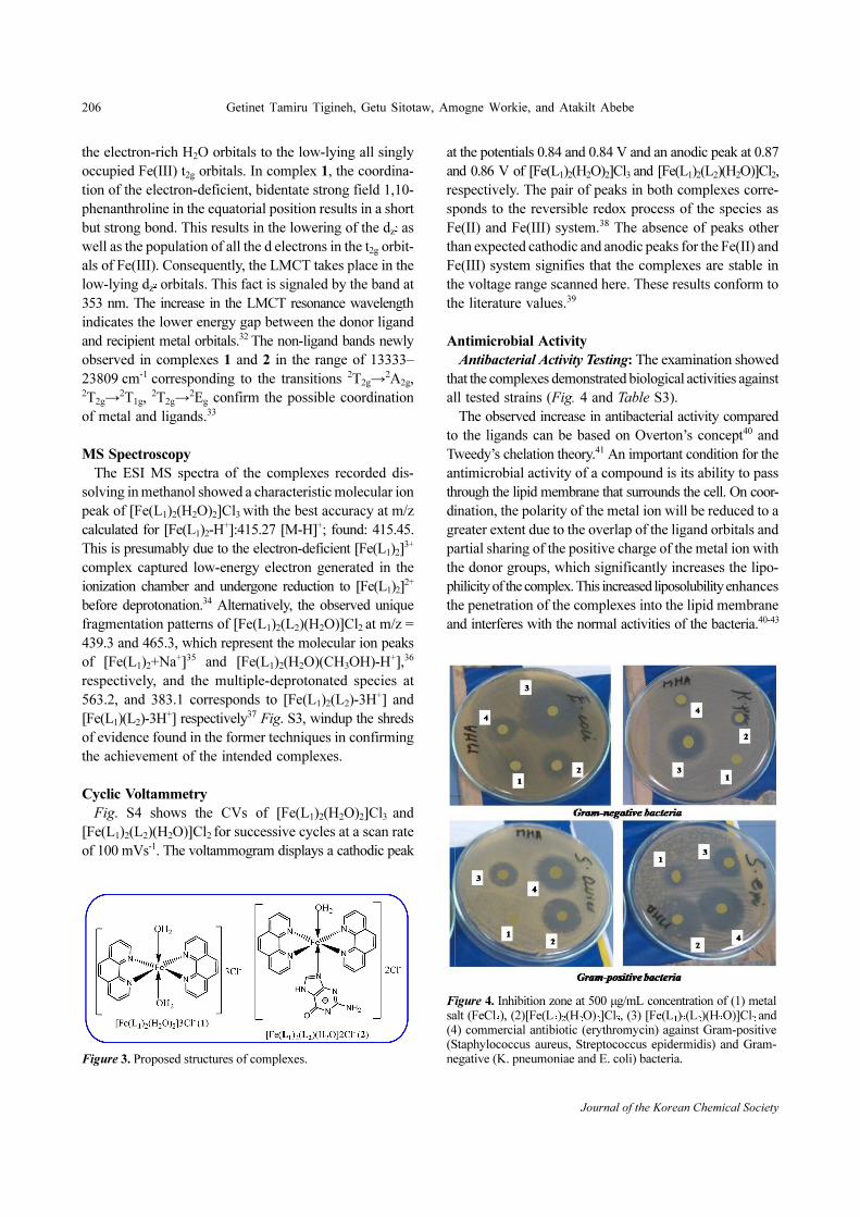
Antibacterial Activity Testing: The examination showed
that the complexes demonstrated biological activities against
all tested strains (Fig. 4 and Table S3).
The observed increase in antibacterial activity compared
to the ligands can be based on Overton’s concept40 and
Tweedy’s chelation theory.41 An important condition for the
antimicrobial activity of a compound is its ability to pass
through the lipid membrane that surrounds the cell. On coor-
dination, the polarity of the metal ion will be reduced to a
greater extent due to the overlap of the ligand orbitals and
partial sharing of the positive charge of the metal ion with
the donor groups, which significantly increases the lipo-
philicity of the complex. This increased liposolubility enhances
the penetration of the complexes into the lipid membrane
and interferes with the normal activities of the bacteria.40-43
Figure 3. Proposed structures of complexes.
Figure 4. Inhibition zone at 500 µg/mL concentration of (1) metalsalt (FeCl3), (2)[Fe(L2)2(H2O)2]Cl3, (3) [Fe(L1)2(L2)(H2O)]Cl2 and(4) commercial antibiotic (erythromycin) against Gram-positive(Staphylococcus aureus, Streptococcus epidermidis) and Gram-negative (K. pneumoniae and E. coli) bacteria.
Synthesis, Characterization and in vitro Antibacterial Studies on Mixed Ligand Complexes of Iron(III) Based on 1,10-phenanthroline 207
2021, Vol. 65, No. 3
The two newly synthesized Fe(III)-complexes manifested
two interesting phenomena compared with commercially
available drug erythromycin. A comparative study verified
that [Fe(L1)(H2O)2]Cl3 showed virtually equal activity to
erythromycin against Gram-positive bacteria, whereas
[Fe(L1)2(L2)(H2O)]Cl2 demonstrated a notably high anti-
bacterial activity than erythromycin even against Gram-
negative bacteria. The latter observation is crucial as Gram-
negative bacteria are highly drug-resistant due to their thick
impenetrable cell wall and deadliest pathogens.44 The better
activities demonstrated by [Fe(L1)2(L2)(H2O)]Cl2 against
Gram-negative bacteria compared to [Fe(L1)2(H2O)2]Cl3
are presumably due to its additional interaction with the
cytosine residue of the genetic material of the cell by guanide.23
The observed biological activities of the complexes are
different from those of the starting materials and the pos-
sible fragments, which suggested a unique characteristic
of complexes and their inertness in the media. This argument
is based on the strong field nature of the ligands coordi-
nated to the metal.
Literature data revealed that Fe(II) complexes with acyclic
chelating ligands demonstrated low or virtually no anti-
bacterial activity against the tested Gram-negative and Gram-
positive bacterial.45-46 This shows a mixed ligand Fe(III)
complexes of aromatic heterocyclic chelating ligands and
nucleobase are promising combinations to develop new
metal-based drugs with better antibacterial activity. The small
ionic radii of the Fe(III) ion comparing to Fe(II) together
with the aforementioned mixed ligands effect explains the
exceptional antibacterial activity due to the ease of pen-
etration of Fe(III) complexes of the cell membrane.12 A
comparative study of the latter complexes with clinical drugs
such as Gentamycin17 and Ciprofloxacin18 also showed an
analogous or even higher antibacterial activity against the
tested Gram-negative and Gram-positive bacterial depend-
ing on the ligand and metal. In our current investigation,
erythromycin, which was used as a clinical drug for com-
parison, is an antibiotic obtained from the bacterial Strep-
tomyces erythreus and is effective against many Gram-
positive and some Gram-negative bacteria.47
The high antibacterial activity of [Fe(L1)2(L2)(H2O)]Cl2
against Gram-negative bacteria compared to erythromycin,
makes the complex potential alternative drug for treating
diseases caused by Gram-negative bacteria after passing
cytotoxicity testing.
Minimum Inhibitory Concentration (MIC) Determination:
The minimum inhibitory concentrations of [Fe(L1)2
(L2)(H2O)]Cl2 complex are listed in Table S4. Both com-
plexes at 75 μg/mL were inactive against the whole bac-
terial species. As far as this specific condition is concerned, the
lowest concentration of [Fe(L1)2(L2)(H2O)]Cl2 complex that
completely inhibited the growth of microorganisms after
24 h of exposure is 150 μg/mL for Gram-negative bacteria
and 500μg/mL for Gram-positive bacteria. The aforemen-
tioned complex has higher antibacterial activity for Gram-
negative bacteria at lower concentrations than for Gram-
positive bacteria.
CONCLUSION
In this work, we successfully synthesized new iron(III)-
1,10-phenanthroline complex in 1:2 metal to ligand ratio
and its mixed ligand complex with guanide. The physi-
cochemical and spectroscopic data suggest that Fe(III) and
the ligands are brought together with rigid configuration and
hence both the complexes proposed having octahedral
geometry (Fig. 3). The cyclic voltammetry study confirmed
the redox stability of the synthesized complexes. The anti-
bacterial test showed a high degree of inhibition by the
complex with guanide against drug resisting Gram-negative
bacteria. The results provide new insights on metal-based
drugs, particularly coordinated with nucleobases, thus the
overall biological findings of this work would be useful in
improving the effectiveness of therapeutic drugs. Based
on the observations, the in vivo shall be investigated, as a
continuation of this study.
Competing Interests. There is no conflict of interest
between the authors and the funding institution.
Acknowledgments. The authors express sincere grat-
itude to Bahir Dar University for providing the necessary
facilities. We thank Dr.Yonas Beyene for running CV at the
electroanalytical laboratory, Bahir Dar University, Ethiopia.
We also thank Academia Sinica, Institute of Chemistry,
for allowing us to run ESI-MS and EA in Taiwan. And the
Publication cost of this paper was supported by the Korean
Chemical Society.
Supporting Information. Additional supporting infor-
mation may be found online in the Supporting Information
section at the end of the article.
REFERENCES
1. Rafique, S.; Idrees, M.; Nasim, A.; Akbar, H.; Athar, A.
Biotechnol. Mol. Biol. Rev. 2010, 5, 38.
2. Abu-Dief, A. M.; Abdel-Rahman, L. H.; Shehata, M. R.;
Journal of the Korean Chemical Society
208 Getinet Tamiru Tigineh, Getu Sitotaw, Amogne Workie, and Atakilt Abebe
Abdel-Mawgoud, A. A. H. J. Phys. Org. Chem. 2019, 32,
e4009.
3. Mukherjee, T.; Pessoa, J. C.; Kumar, A.; Sarkar, A. R.;
Dalton Trans. 2013, 42, 2594.
4. Weder, J. E.; Dillon, C. T.; Hambley, T. W.; Kennedy, B.
J.; Lay, P. A.; Biffin, J. R.; Davies, N. M. Coord. Chem.
Rev. 2002, 232, 95.
5. Hambley, T. W. Dalton Trans. 2007, 43, 4929.
6. Ronconi, L.; Sadler, P. J. Coord. Chem. Rev. 2007, 251,
1633.
7. Graf, N.; Lippard, S. J. Adv. Drug Deliv. Rev. 2012, 64,
993.
8. Ming, L. J. Med. Res. Rev. 2003, 23, 697.
9. Al Qaraghuli, M. M.; Alzahrani, A. R.; Niwasabutra, K.;
Obeid, M. A.; Ferro, V. A. Ann. Pharm. & Pharm. Sci.
2017, 2, 1.
10. David, B.; Wolfender, J. L.; Dias, D. A. Phytochem Rev.
2015, 14, 299.
11. Turel, I. Coord. Chem. Rev. 2002, 232, 27.
12. Hassoon, A. A.; Harrison, R. G.; Nawar, N.; Smith, S. J.;
Mostafa, M. M. J. Mol. Struct. 2020, 1203, 127240.
13. Fleisher, M. B.; Waterman, K. C.; Turro, N. J.; Barton, J.
K. Inorg. Chem. 1986, 25, 3549.
14. Long, E. C.; Barton, J. K. Acc. Chem. Res. 1990, 23, 271.
15. Prabhakara, M. C.; Basavaraju, B.; Naik, H. S. Bioinorg
Chem. Appl. 2007, 2007.
16. Bryan Sears, R.; Joyce, L. E.; Turro, C. Photochem. Pho-
tobiol. 2010, 86, 1230.
17. Tamiru, G.; Abebe, A.; Abebe, M.; Liyew, M. Ethiop. J.
Sci. & Technol. 2019, 12, 69.
18. Abebe, A.; Bayeh, Y.; Belay, M.; Gebretsadik, T.; Thomas,
M.; Linert, W. Future J. Pharm. Sci. 2020, 6, 1.
19. Soliman, S. M.; Al-Rasheed, H. H.; Albert, J. H.; El-Faham,
A. Molecules 2020, 25, 5750.
20. Reed, J. E.; White, A. J.; Neidle, S.; Vilar, R. Dalton Trans.
2009, 14, 2558.
21. Melnic, E.; Coropceanu, E. B.; Kulikova, O. V.; Siminel,
A. V.; Anderson, D.; Rivera-Jacquez, H. J.; Masunov, A.
E.; Fonari, M. S.; Kravtsov, V. C. J. Phys. Chem. C. 2014,
118, 30087.
22. Abebe, A.; Atlabachew, M.; Liyew, M.; Ferede, E. Cogent
Chem. 2018, 4, 1476077.
23. Shapiro, R. In Progress in Nucleic Acid Research and
Molecular Biology; 1968; Academic Press: Vol. 8, pp 73-
112.
24. Abebe, A.; Hailemariam, T. Bioinorg. Chem. Appl. 2016,
3607924.
25. Wenger, O. S. Chem. Eur. J. 2019, 25, 6043.
26. Ali, I.; Wani, W. A.; Saleem, K. Synthesis and Reactivity
in Inorganic, Metal-Organic, and Nano-Metal Chemistry
2013, 43, 1162.
27. Mikulski, C. M.; Mattucci, L.; Smith, Y.; Tran, T. B.;
Karayannis, N. M. Inorg. Chim. Acta 1983, 80, 127.
28. Mastropietro, T. F.; Armentano, D.; Grisolia, E.; Zanchini,
C.; Lloret, F.; Julve, M.; De Munno, G. Dalton Trans.
2008, 4, 514.
29. Sheina, G. G.; Stepanian, S. G.; Radchenko, E. D.; Blagoi,
Y. P. J. Mol. Struct. 1987, 158, 275.
30. Mathlouthi, M.; Seuvre, A. M.; Koenig, J. L. Carbohydr.
Res. 1986, 146, 15.
31. Chandra, A. K.; Nguyen, M. T.; Uchimaru, T.; Zeegers-
Huyskens, T. J. Phys. Chem. A 1999, 103, 8853.
32. Lever, A. P. Inorganic Electronic Spectroscopy 2nd ed.,
Elsevier: Amsterdam, 1984.
33. Lobanov, S. S.; Hsu, H.; Lin, J. F.; Yoshino, T.; Gon-
charov, A. F. J. Geophys. Res., Solid Earth 2017, 122,
3565.
34. Nicolescu, T. O. Mass Spectrometry 2017, 24.
35. Looi, D. W.; Eyler, J. R.; Brajter-Toth, A. Electrochim.
Acta 2011, 56, 2633.
36. Cheng, P.; Li, Y.; Li, S.; Zhang, M.; Zhou, Z. Phys. Chem.
Chem. Phys. 2010, 12, 4667.
37. Banerjee, S.; Mazumdar, S. Int. J. Anal. Chem. 2012,
2012.
38. Zhang, Q. L.; Liu, J. G.; Chao, H.; Xue, G. Q.; Ji, L. N.
J. Inorg. Biochem. 2001, 83, 49.
39. de Castro, M. L.; Valcarcel, M.; Albahadily, F. N.; Mot-
tola, H. A. J. Electroanal. Chem. 1987, 219, 139.
40. Anjaneyulu, Y.; Rao, R. P. Synth. React. Inorg. Met. Org.
Chem. 1986, 16, 257.
41. Tweedy, B. G. Phytopathology 1964, 55, 910.
42. Warra, A. A. J. Chem. Pharm. Res. 2011, 3, 951.
43. Mohamad, A. D. M.; Abualreish, M. J. A.; Abu-Dief, A.
M. J. Mol. Liq. 2019, 290, 111162.
44. Moon, M. S. Asian Spine J. 2019, 13, 343.
45. Mahmud, T.; Rehman, R.; Gulzar, A.; Khalid, A.; Anwar,
J.; Shafique, U.; Salman, M. Arab. J. Chem. 2010, 3, 219.
46. Lawal, A.; Shodeinde, A. S.; Amolegbe, S. A.; Elaigwu,
S. E.; Yunus-Issa, M. T. J. Appl. Sci. Environ. Manage.
2017, 21, 568.
47. World Health Organization. WHO Model Prescribing
Information: Drugs Used in Skin Diseases; World Health
Organization; Geneva, 1997; 1-126.
Related Documents


