Synthesis, characterization and functionalization of nearly mono-disperse copper ferrite Cu x Fe 3x O 4 nanoparticles† Bahar Nakhjavan, a Muhammad Nawaz Tahir, a M. Panth€ ofer, a Haitao Gao, a Thomas D. Schladt, a Teuta Gasi, a Vadim Ksenofontov, a Robert Branscheid, b Stefan Weber, c Ute Kolb, b Laura Maria Schreiber c and Wolfgang Tremel * a Received 30th December 2010, Accepted 22nd February 2011 DOI: 10.1039/c0jm04577b Magnetic nanocrystals are of great interest for a fundamental understanding of nanomagnetism and for their technological applications. Cu x Fe 3x O 4 nanocrystals (x z 0.32) with sizes ranging between 5 and 7 nm were synthesized starting from Cu(HCOO) 2 and Fe(CO) 5 using oleic acid and oleylamine as surfactants. The nanocrystals were characterized by high-resolution transmission electron microscopy (HRTEM), electron diffraction (ED), magnetization studies and M€ ossbauer spectroscopy. The Cu x Fe 3x O 4 particles are superparamagnetic at room temperature 300 K with a saturation magnetization of 30.5 emu g 1 . Below their blocking temperature of 60 K, they become ferrimagnetic, and at 5 K they show a coercive field of 122 Oe and a saturation magnetization of 36.1 emu g 1 . The Cu x Fe 3x O 4 nanoparticles were functionalized using a hydrophilic multifunctional polymeric ligand containing PEG(800) groups and a fluorophore. By virtue of their magnetic properties these nanoparticles may serve as contrast enhancing agents for magnetic resonance imaging (MRI). Introduction Magnetic nanoparticles (MNPs) are playing increasingly important roles in biotechnology and biomedicine. 1 MNPs have been used as carriers for magnetic drug targeting, 2 as tags for biomolecular sensors, 3,4 in biomolecule separation and purifica- tion, 5–7 as well as for in vivo imaging, 8–10 and hyper-thermia treatment. 11,12 As these and other applications become more advanced, precise control over particle composition, stability and surface functionality is crucial. Among the magnetic materials, the ferrites with general formula MFe 2 O 4 have been used in many applications. By adjusting the M 2+ cation, the magnetic configurations of the spinel-type MFe 2 O 4 can be engineered to provide a wide range of magnetic properties. 13 Several studies on pure nanoferrites such as Fe 3 O 4 , 14 NiFe 2 O 4 , 15 CoFe 2 O 4 , 16 ZnFe 2 O 4 , 17 and MnFe 2 O 4 18,19 have demonstrated the interplay of composition, 20 cation distribution 21,22 and size 23 in view of their properties and applications. Among the ferrites, CuFe 2 O 4 has received significant attention in recent years. 24,25 CuFe 2 O 4 coatings based on highly aggregated nanoparticles were prepared using electrochemical methods. 26 Plate-like CuFe 2 O 4 particles were obtained using reverse micelle and hydrothermal methods. 27 Nanocrystalline CuFe 2 O 4 was prepared by co-precipitation, 28 mechanical milling, 29 sol–gel methods, 30 or precipitation in a polymer matrix. 31 Goya et al. 32 who synthesized CuFe 2 O 4 by high-energy ball milling showed that the milling process reduces the average grain size of CuFe 2 O 4 but induces severe cation redistribution between tetrahedral and octahedral sites. Ferrites are among the most important and interesting oxides owing to their wide variety of applications in sensors, electronics, and catalysts. 33,34 e.g. as abatement of gaseous pollutants 35 and the water gas shift reaction. 36 Recently, copper ferrites have been proposed as a reforming catalyst for hydrogen production from oxygenated hydrocarbons. 37–40 In spite of the availability of different synthetic methods and promising potential applications, the synthesis of highly monodisperse and non-aggregated CuFe 2 O 4 nanoparticles has not been mastered so far. Previous reports based on high temperature or hydrothermal procedures described the synthesis of agglomerated and mostly polydispersed material, where the question of site preference could only partially be addressed and resolved. 24–32 Here we demonstrate a facile and simple method for the synthesis of very uniform and non- aggregated Cu x Fe 3x O 4 (x z 0.32) nanoparticles by using two suitable precursors in a hot organic solvent. The Cu x Fe 3x O 4 a Institut f € ur Anorganische Chemie und Analytische Chemie, Johannes Gutenberg-Universit € at, Duesbergweg 10-14, D-55099 Mainz, Germany. E-mail: [email protected]; Fax: +49 6131 39-25605; Tel: +49 6131 39-25135 b Institut f € ur Physikalische Chemie, Johannes Gutenberg-Universit € at, Welderweg 11, D-55099 Mainz, Germany c Bereich Medizinische Physik, Klinik und Poliklinik f € ur diagnostische und interventionelle Radiologie, Klinikum der Johannes Gutenberg-Universi- t € at Mainz, Langenbeckstraße, 1, 55131 Mainz, Germany † Electronic supplementary information (ESI) available. See DOI: 10.1039/c0jm04577b This journal is ª The Royal Society of Chemistry 2011 J. Mater. Chem., 2011, 21, 6909–6915 | 6909 Dynamic Article Links C < Journal of Materials Chemistry Cite this: J. Mater. Chem., 2011, 21, 6909 www.rsc.org/materials PAPER Downloaded by Johannes Gutenberg Universitaet Mainz on 02 August 2011 Published on 31 March 2011 on http://pubs.rsc.org | doi:10.1039/C0JM04577B View Online

Welcome message from author
This document is posted to help you gain knowledge. Please leave a comment to let me know what you think about it! Share it to your friends and learn new things together.
Transcript
Dynamic Article LinksC<Journal ofMaterials Chemistry
Cite this: J. Mater. Chem., 2011, 21, 6909
www.rsc.org/materials PAPER
Dow
nloa
ded
by J
ohan
nes
Gut
enbe
rg U
nive
rsita
et M
ainz
on
02 A
ugus
t 201
1Pu
blis
hed
on 3
1 M
arch
201
1 on
http
://pu
bs.r
sc.o
rg |
doi:1
0.10
39/C
0JM
0457
7BView Online
Synthesis, characterization and functionalization of nearly mono-dispersecopper ferrite CuxFe3�xO4 nanoparticles†
Bahar Nakhjavan,a Muhammad Nawaz Tahir,a M. Panth€ofer,a Haitao Gao,a Thomas D. Schladt,a Teuta Gasi,a
Vadim Ksenofontov,a Robert Branscheid,b Stefan Weber,c Ute Kolb,b Laura Maria Schreiberc
and Wolfgang Tremel*a
Received 30th December 2010, Accepted 22nd February 2011
DOI: 10.1039/c0jm04577b
Magnetic nanocrystals are of great interest for a fundamental understanding of nanomagnetism and for
their technological applications. CuxFe3�xO4 nanocrystals (x z 0.32) with sizes ranging between 5 and
7 nm were synthesized starting from Cu(HCOO)2 and Fe(CO)5 using oleic acid and oleylamine as
surfactants. The nanocrystals were characterized by high-resolution transmission electron microscopy
(HRTEM), electron diffraction (ED), magnetization studies and M€ossbauer spectroscopy. The
CuxFe3�xO4 particles are superparamagnetic at room temperature 300 K with a saturation
magnetization of 30.5 emu g�1. Below their blocking temperature of 60 K, they become ferrimagnetic,
and at 5 K they show a coercive field of 122 Oe and a saturation magnetization of 36.1 emu g�1. The
CuxFe3�xO4 nanoparticles were functionalized using a hydrophilic multifunctional polymeric ligand
containing PEG(800) groups and a fluorophore. By virtue of their magnetic properties these
nanoparticles may serve as contrast enhancing agents for magnetic resonance imaging (MRI).
Introduction
Magnetic nanoparticles (MNPs) are playing increasingly
important roles in biotechnology and biomedicine.1 MNPs have
been used as carriers for magnetic drug targeting,2 as tags for
biomolecular sensors,3,4 in biomolecule separation and purifica-
tion,5–7 as well as for in vivo imaging,8–10 and hyper-thermia
treatment.11,12 As these and other applications become more
advanced, precise control over particle composition, stability and
surface functionality is crucial. Among the magnetic materials,
the ferrites with general formula MFe2O4 have been used in
many applications. By adjusting the M2+ cation, the magnetic
configurations of the spinel-type MFe2O4 can be engineered to
provide a wide range of magnetic properties.13 Several studies on
pure nanoferrites such as Fe3O4,14 NiFe2O4,15 CoFe2O4,16
ZnFe2O4,17 and MnFe2O418,19 have demonstrated the interplay of
composition,20 cation distribution21,22 and size23 in view of their
properties and applications.
aInstitut f€ur Anorganische Chemie und Analytische Chemie, JohannesGutenberg-Universit€at, Duesbergweg 10-14, D-55099 Mainz, Germany.E-mail: [email protected]; Fax: +49 6131 39-25605; Tel: +49 613139-25135bInstitut f€ur Physikalische Chemie, Johannes Gutenberg-Universit€at,Welderweg 11, D-55099 Mainz, GermanycBereich Medizinische Physik, Klinik und Poliklinik f€ur diagnostische undinterventionelle Radiologie, Klinikum der Johannes Gutenberg-Universi-t€at Mainz, Langenbeckstraße, 1, 55131 Mainz, Germany
† Electronic supplementary information (ESI) available. See DOI:10.1039/c0jm04577b
This journal is ª The Royal Society of Chemistry 2011
Among the ferrites, CuFe2O4 has received significant attention
in recent years.24,25 CuFe2O4 coatings based on highly aggregated
nanoparticles were prepared using electrochemical methods.26
Plate-like CuFe2O4 particles were obtained using reverse micelle
and hydrothermal methods.27 Nanocrystalline CuFe2O4 was
prepared by co-precipitation,28 mechanical milling,29 sol–gel
methods,30 or precipitation in a polymer matrix.31 Goya et al.32
who synthesized CuFe2O4 by high-energy ball milling showed
that the milling process reduces the average grain size of
CuFe2O4 but induces severe cation redistribution between
tetrahedral and octahedral sites.
Ferrites are among the most important and interesting oxides
owing to their wide variety of applications in sensors, electronics,
and catalysts.33,34 e.g. as abatement of gaseous pollutants35 and
the water gas shift reaction.36 Recently, copper ferrites have been
proposed as a reforming catalyst for hydrogen production from
oxygenated hydrocarbons.37–40
In spite of the availability of different synthetic methods and
promising potential applications, the synthesis of highly
monodisperse and non-aggregated CuFe2O4 nanoparticles has
not been mastered so far. Previous reports based on high
temperature or hydrothermal procedures described the
synthesis of agglomerated and mostly polydispersed material,
where the question of site preference could only partially be
addressed and resolved.24–32 Here we demonstrate a facile and
simple method for the synthesis of very uniform and non-
aggregated CuxFe3�xO4 (x z 0.32) nanoparticles by using two
suitable precursors in a hot organic solvent. The CuxFe3�xO4
J. Mater. Chem., 2011, 21, 6909–6915 | 6909
Dow
nloa
ded
by J
ohan
nes
Gut
enbe
rg U
nive
rsita
et M
ainz
on
02 A
ugus
t 201
1Pu
blis
hed
on 3
1 M
arch
201
1 on
http
://pu
bs.r
sc.o
rg |
doi:1
0.10
39/C
0JM
0457
7BView Online
copper ferrite particles can be functionalized with a multifunc-
tional polymeric ligand to yield highly water soluble and fluo-
rescent magnetic nanoparticles that may be used for
bioimaging.
Experimental section
Materials
Iron(0) pentacarbonyl (Fe(CO)5, 99.5%, Acros), 1-octadecene
(ODE, 90%, Acros), copper(II) formate (Cu(HCOO)2, 99%,
Fluka), oleic acid (90%, Aldrich), oleylamine (90%, Acros),
di-tert-butyl dicarbonate ((Boc)2O, >99%, Aldrich), dioxane
(P.A., Fisher), H2N–PEG(800)–NH2 (Aldrich), triethylamine
(>99%, Aldrich), 3-hydroxytyramine hydrochloride (dop-
amine$HCl, 98%, Aldrich), NBD chloride (98%, Fluka),
trifluoroacetic acid (TFA, 99%, Aldrich), ethanol (99.8%, Roth),
toluene (>99%, Aldrich), hexane (P.A., Fisher), dichloromethane
(DCM, P.A., Fisher), N,N-dimethylformamide (DMF, extra dry,
>99.8%, Acros), diethyl ether (P.A., Fisher) were used as received
without further purification.
Synthesis of CuxFe3�xO4 nanoparticles
In a typical synthesis, under highly inert conditions 5 mL of
octadecane, 0.195 mL of oleylamine and 0.5415 mL of oleic
acid were mixed and heated to 100 �C for 45 min. In a sepa-
rate flask were added 2 mL of oleylamine and 550 mg of
Cu(HCOO)2 and the flask was placed in a liquid metal bath at
room temperature and programmed to reach 75 �C at a heat-
ing rate 3 �C min�1. At this temperature, Cu(HCOO)2 solution
from this flask was injected into the other flask containing
mixed surfactant solution, and the temperature was increased
with the same rate. After reaching to 120 �C, 0.975 mL of
Fe(CO)5 were injected into the mixture. Subsequently, the
solution was heated to 200 �C and kept at this temperature for
1 h. Throughout this period Ar was kept flowing through the
flask and the mixture was stirred mechanically. Finally, the
mixture was cooled to room temperature, the product was
separated by precipitation with ethanol and centrifugation
using 9000 rpm for 10 min and re-dispersed in hexane several
times.
Synthesis of the dopa–PEG–polymer (DA–PEG–PP)
N-Boc–NH2–PEG(800)–NH2. N-Boc–NH2–PEG(800)–NH2
was synthesized according to the procedure as described.41 The
poly (active ester) poly(pentafluorophenylacrylate) (PFA) was
prepared as reported earlier.42–45 GPC analysis of the obtained
polymer (THF, light scattering detection) gave the following
values: Mn ¼ 16 390 g mol�1, PDI ¼ 1.39, with an average of
70 repeat units. For the synthesis of the multifunctional
poly(acrylamides), poly(active ester) poly(pentafluoro-
phenylacrylate) (700 mg, 2.94 mmol repeating units) was
dissolved in a mixture of 9 mL of dry DMF and 0.7 mL of
triethylamine. 12 mg of pip-NBD was added to the solution
and stirred for 2 h. Subsequently, 3-hydroxytyramine hydro-
chloride (24 mg) dissolved in 3 mL of DMF and 0.4 mL of
triethylamine was added, and the reaction mixture was stirred
for 3 h at 50 �C. In the final step the remaining active ester
6910 | J. Mater. Chem., 2011, 21, 6909–6915
groups were substituted using an excess of N-Boc–PEG(800)–
NH2 (dissolved in 3 mL of dry DMF) and stirring for 5 h at
50 �C. The solution was concentrated to about 2 mL, and the
polymeric ligand was precipitated by addition of cold ethyl
ether. The precipitated polymer was centrifuged and the solvent
was decanted. Upon drying, 486 mg of colorless oil was
obtained.
The polymer obtained above was dissolved in CH2Cl2 (30 mL).
Subsequently, trifluoroacetic acid (2.0 mL) was added and the
mixture was stirred at room temperature for 2 h. Afterwards the
reaction solution was treated with a mixture of water and hexane
(30 mL/50 mL) and stirred vigorously for 30 min. The aqueous
phase containing the polymer was separated and concentrated to
2 mL and dialyzed against deionized water for 2 days (cellulose
bag, MWCO ¼ 3 500). Finally, the water was evaporated, and
the product was re-dissolved in chloroform to make a stock
solution which was kept in the refrigerator.
DA–PEG–NH2. Conjugation of N-Boc–PEG–NH2 to
3,4-dihydroxyhydrocinnamic acid (DA) was performed by
a common DCC coupling reaction under inert conditions. First,
3,4-dihydroxyhydrocinnamic acid (5 mmol) and HOBt
(5.1 mmol) were dissolved in 10 mL of dry DMF and stirred at
room temperature. After 10 minutes DCC (5.1 mmol in 10 mL of
dry DMF) was added, and the solution was stirred for another
10 minutes before NHS (5.1 mmol in 10 mL of dry DMF) was
added dropwise over a period of 30 minutes. The reaction was
continued for 2 hours. The resulting DA–NHS ester was subse-
quently added to a stirred solution of N-Boc–PEG–NH2
(5 mmol) in 15 mL of dry DMF over a period of 45 minutes. The
solution was stirred overnight at room temperature. After
removal of the urea side product by filtration the crude product
was transferred to chloroform. The organic solution was
extracted several times with a saturated NaCl solution and
washed with deionized water. The solvent was evaporated and
the oily residue redissolved in dichloromethane. Cleavage of the
BOC protection group was accomplished by addition of
trifluoroacetic acid and stirring at room temperature for two
hours. After removal of DCM the product was dissolved in
40 mL of chloroform and washed with a saturated aqueous
NaHCO3 solution and deionized water. The organic phase was
dried over MgSO4 and the solvent removed in vacuo to produce
a light brown oil.
Functionalization of the CuxFe3�xO4 nanoparticles. 10 mg of
CuxFe3�xO4 nanoparticles dispersed in 15 mL of chloroform
were dropped slowly over 1 h into the above synthesized poly-
meric ligand solution (20 mg in 10 mL of chloroform). The
reaction was stirred continuously at room temperature, over-
night under inert conditions. The functionalized nanoparticles
were precipitated by addition of hexane and separated from the
unbound polymer and surfactants by centrifugation. The nano-
particles were washed twice by dissolving them in chloroform
and precipitation with hexane. Finally, the particles were stored
in DMF in a refrigerator.
The functionalized CuxFe3�xO4 nanoparticles were charac-
terized by TEM and Fourier Transform Infrared (FT-IR) spec-
troscopy (Mattson Instruments 2030 Galaxy-FT-IR
spectrometer). Unless mentioned differently, all nanoparticle
This journal is ª The Royal Society of Chemistry 2011
Dow
nloa
ded
by J
ohan
nes
Gut
enbe
rg U
nive
rsita
et M
ainz
on
02 A
ugus
t 201
1Pu
blis
hed
on 3
1 M
arch
201
1 on
http
://pu
bs.r
sc.o
rg |
doi:1
0.10
39/C
0JM
0457
7BView Online
concentrations are referred to the Fe concentration measured
with AAS.
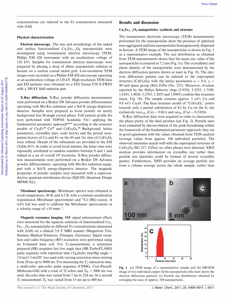
Fig. 1 (a) TEM image of a representative sample and (b) HRTEM
image of two individual copper ferrite nanoparticles (the inset shows the
electron diffraction pattern). (c) Particle size distribution obtained by
averaging the sizes of approx. 100 nanoparticles.
Physical characterization
Electron microscopy. The size and morphology of the naked
and surface functionalized CuxFe3�xO4 nanoparticles were
investigated using transmission electron microscopy (TEM,
Philips EM 420 instrument with an acceleration voltage of
120 kV). Samples for transmission electron microscopy were
prepared by placing a drop of dilute nanoparticle solution in
hexane on a carbon coated nickel grid. Low-resolution TEM
images were recorded on a Philips EM 420 microscope operating
at an acceleration voltage of 120 kV. High-resolution TEM data
and ED patterns were obtained on a FEI Tecnai F30 S-TWIN
with a 300 kV field emission gun.
X-Ray diffraction. X-Ray powder diffraction measurements
were performed on a Bruker D8 Advance powder diffractometer
operating with Mo-Ka radiation and a Sol-X energy-dispersive
detector. Samples were prepared as loose powder on nearly
background free Si-single crystal plates. Full pattern profile fits
were performed with TOPAS Academic V4.1 applying the
fundamental parameter approach46,47 according to the structure
models of Cu2O,48 Cu49 and CuFe2O4.50 Background, lattice
parameters, crystallite sizes, scale factors and the partial occu-
pation factors of Cu and Fe on the 8b and 16c sites of CuFe2O4
were refined. Details of the refinement are provided in the ESI
(Table S1†). In order to avoid local minima, the latter ones were
repeatedly attributed to random numbers between 0 and 1 and
refined in a set of overall 106 iterations. X-Ray powder diffrac-
tion measurements were performed on a Bruker D8 Advance
powder diffractometer, operating with Mo-Ka radiation equip-
ped with a Sol-X energy-dispersive detector. The magnetic
properties of powder samples were measured with a supercon-
ductive quantum interference device (SQUID, Quantum Design
MPMS XL).
M€ossbauer spectroscopy. M€ossbauer spectra were obtained at
room temperature, 80 K and 4.2 K with a constant acceleration
transmission M€ossbauer spectrometer and 57Co (Rh) source. A
a-Fe foil was used to calibrate the M€ossbauer spectrometer in
a velocity range of �10 mms�1.
Magnetic resonance imaging. MR signal enhancement effects
were measured for the aqueous solutions of functionalized Cux-
Fe3�xO4 nanoparticles at different Fe concentrations (measured
with AAS) on a clinical 3.0 T MRI scanner (Magnetom Trio,
Siemens Medical Solutions, Erlangen, Germany). Signal recep-
tion and radio frequency (RF) excitation were performed using
an 8-channel knee coil. For T1-measurement, a saturation
prepared (SR) snapshot fast low angle shot (SR-TurboFLASH)
pulse sequence with repetition time (TR)/echo time/flip angle ¼3.0 ms/1.5 ms/20� was used with varying saturation times starting
from 20 ms up to 8000 ms. For measuring the T2 relaxation time,
a multi-echo spin-echo pulse sequence (CPMG, Carr-Purcell-
Meiboom-Gill) with a total of 32 echos and TR ¼ 5000 ms was
used, the echo time was varied from 7 ms to 224 ms. In a second
T2 measurement TE was varied from 15 ms up to 480 ms.
This journal is ª The Royal Society of Chemistry 2011
Results and discussion
CuxFe3�xO4 nanoparticles: synthesis and structure
The transmission electronic microscopy (TEM) measurements
performed for the nanoparticles show the presence of spherical
non-aggregated uniform nanoparticles homogeneously dispersed
in hexane. A TEM image of the nanoparticles is shown in Fig. 1
as a representative example. The size distribution as obtained
from TEM measurements shows that the mean size value of the
nanoparticles is centered at 7.2 nm (Fig. 1c). The crystallinity and
phase identity of the nanoparticles were demonstrated by the
electron diffraction pattern shown as inset in Fig. 1b. The elec-
tron diffraction pattern can be indexed to the cuprospinel
structure (CuFe2O4) with the lattice parameters a ¼ 8.4, a ¼90�and space group (SG) Fd�3m (No. 227). Moreover, d-values
reported by the Debye–Scherrer rings (2.9326, 2.5253, 1.7109,
1.6103, 1.4826, 1.2763, 1.2055 and 1.0905) confirm this structure
(inset, Fig. 1b). The sample contains approx. 1 wt% Cu and
9.8 wt% Cu2O. The final structure model of ‘‘CuFe2O4’’ points
towards only a partial substitution of Fe by Cu on the 8c site
exclusively (occ16c (Cu) ¼ 0.0(1) and occ8c (Cu) ¼ 0.32(9)).
X-Ray diffraction data were acquired in order to characterize
the phase purity of the final product (see Fig. 2). Particle sizes
were estimated by deconvolution of the peak-broadening within
the framework of the fundamental parameter approach; they are
in good agreement with the values obtained from TEM analysis
(average values from approx. 100 individual particles). The
observed intensities match well with the cuprospinel structure of
CuFe2O4 (SG 227, Fd�3m); no other phases were detected. XRD
analysis provides information on crystallite size rather than
particle size (particles could be formed of several crystallite
grains). Furthermore, XRD provides an average particle size
from a volume average across the whole sample, rather than
J. Mater. Chem., 2011, 21, 6909–6915 | 6911
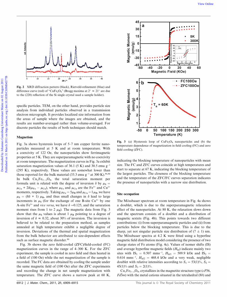
Fig. 2 XRD diffraction pattern (black), Rietveld-refinement (blue) and
difference curve (red) of ‘‘CuFe2O4’’ (Bragg maxima at 2� z 21� are due
to the (220) reflection of the Si single crystal used a sample holder).
Fig. 3 (a) Hysteresis loop of CuFe2O4 nanoparticles and (b) the
temperature dependence of magnetization in field cooling (FC) and zero
field cooling (ZFC).
Dow
nloa
ded
by J
ohan
nes
Gut
enbe
rg U
nive
rsita
et M
ainz
on
02 A
ugus
t 201
1Pu
blis
hed
on 3
1 M
arch
201
1 on
http
://pu
bs.r
sc.o
rg |
doi:1
0.10
39/C
0JM
0457
7BView Online
specific particles. TEM, on the other hand, provides particle size
analysis from individual particles observed in a transmission
electron micrograph. It provides localized size information from
the areas of sample where the images are obtained, and the
results are number-averaged rather than volume-averaged. For
discrete particles the results of both techniques should match.
Magnetism
Fig. 3a shows hysteresis loops of 5–7 nm copper ferrite nano-
particles measured at 5 K and at room temperature. With
a coercivity of 122 Oe, the nanoparticles show ferrimagnetic
properties at 5 K. They are superparamagnetic with no coercivity
at room temperature. The magnetization curves in Fig. 3a exhibit
saturation magnetization values of 36.1 (5 K) and 30.5 emu g�1
(295 K), respectively. These values are somewhat lower than
those reported for the bulk material (33.3 emu g�1 at 300 K).51,52
In bulk CuxFe3�xO4, the total saturation moment per
formula unit is related with the degree of inversion d by mS ¼mCu + 2d(mFe � mCu), where mFe and mCu are the Fe3+ and Cu2+
moments, respectively. Taking mFe¼ 5mB and mCu¼ l mB, we have
mS ¼ (8d + 1) mB, and thus small changes in d lead to large
increments in mS (for the exchange of one B-site Cu2+ by one
A-site Fe3+ and vice versa, we have d ¼0.125, and the saturation
moment rises from 1 to 2 mB). The magnetic data from Fig. 3
show that the mS values is about 1 mB pointing to a degree of
inversion of d z 0.32, about 30% of inversion. The inversion is
believed to be related to the preparation method, as samples
annealed at high temperature exhibit a negligible degree of
inversion. Deviations of the thermal and spatial magnetization
from the bulk behavior are attributed to size-dependent effects
such as surface magnetic disorder.53
Fig. 3b shows the zero field-cooled (ZFC)/field-cooled (FC)
magnetization curves in the range of 4–300 K. For the ZFC
experiment, the sample is cooled in zero field and then heated in
a field of (100 Oe) while the net magnetization of the sample is
recorded. The FC data are obtained by cooling the sample under
the same magnetic field of (100 Oe) after the ZFC experiments
and recording the change in net sample magnetization with
temperature. The ZFC curve shows a narrow peak at 60 K,
6912 | J. Mater. Chem., 2011, 21, 6909–6915
indicating the blocking temperature of nanoparticles with mean
size. The FC and ZFC curves coincide at high temperatures and
start to separate at 67 K, indicating the blocking temperature of
the largest particles. The closeness of the blocking temperature
and the temperature of the ZFC/FC curves separation indicates
the presence of nanoparticles with a narrow size distribution.
Site occupation
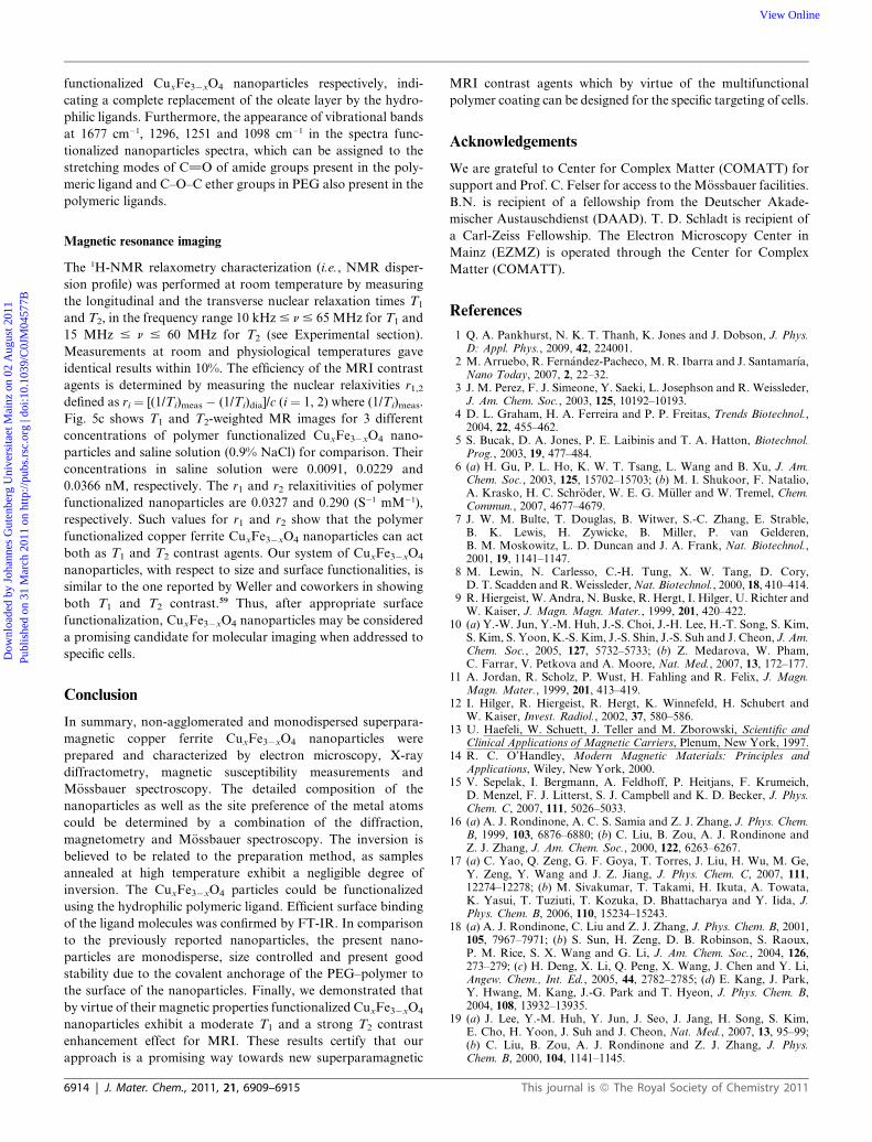
The M€ossbauer spectrum at room temperature in Fig. 4a shows
a doublet, which is due to the superparamagnetic relaxation
effect of the nanoparticles. At 80 K, the relaxation slows down
and the spectrum consists of a doublet and a distribution of
magnetic sextets (Fig. 4b). This points towards two different
contributions: (i) from superparamagnetic particles and (ii) from
particles below the blocking temperature. This is due to the
sharp, yet not singular particle size distribution of (7 � 1) nm.
The M€ossbauer spectra at 4.2 K were fitted using a hyperfine
magnetic field distribution model considering the presence of two
charge states of Fe atoms (Fig. 4c). Values of isomer shifts (IS)
and average hyperfine magnetic fields (Hhf) indicate mainly two
sites with IS1 ¼ 0.507 mms�1, Hhf1 ¼ 509.9 kOe and IS2 ¼0.614 mms�1, Hhf2 ¼ 468.4 kOe and a very weak, negligible
doublet with relative intensities according to S1 ¼ 53(1)%, S2 ¼45(1)% and S3 ¼ 2(1)%.
CuxFe3�xO4, crystallizes in the magnetite structure type (cF56,
Fd�3m) with the metal cations situated in the tetrahedral (8b) and
This journal is ª The Royal Society of Chemistry 2011
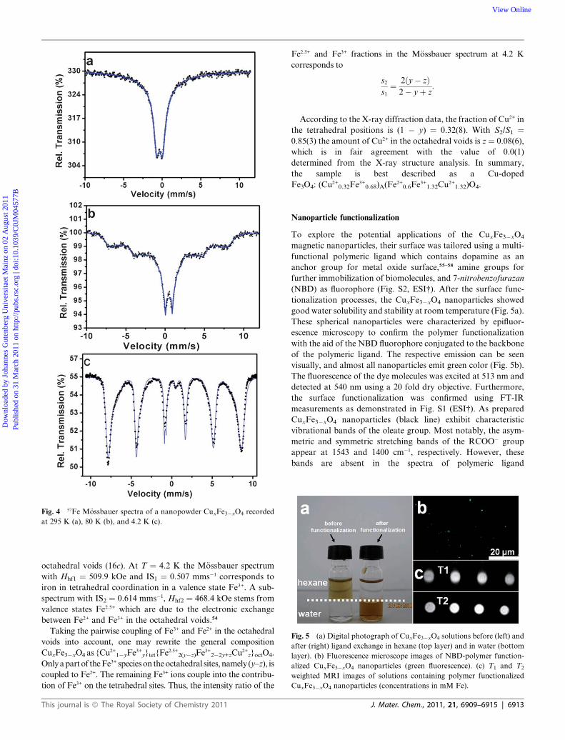
Fig. 4 57Fe M€ossbauer spectra of a nanopowder CuxFe3�xO4 recorded
at 295 K (a), 80 K (b), and 4.2 K (c).
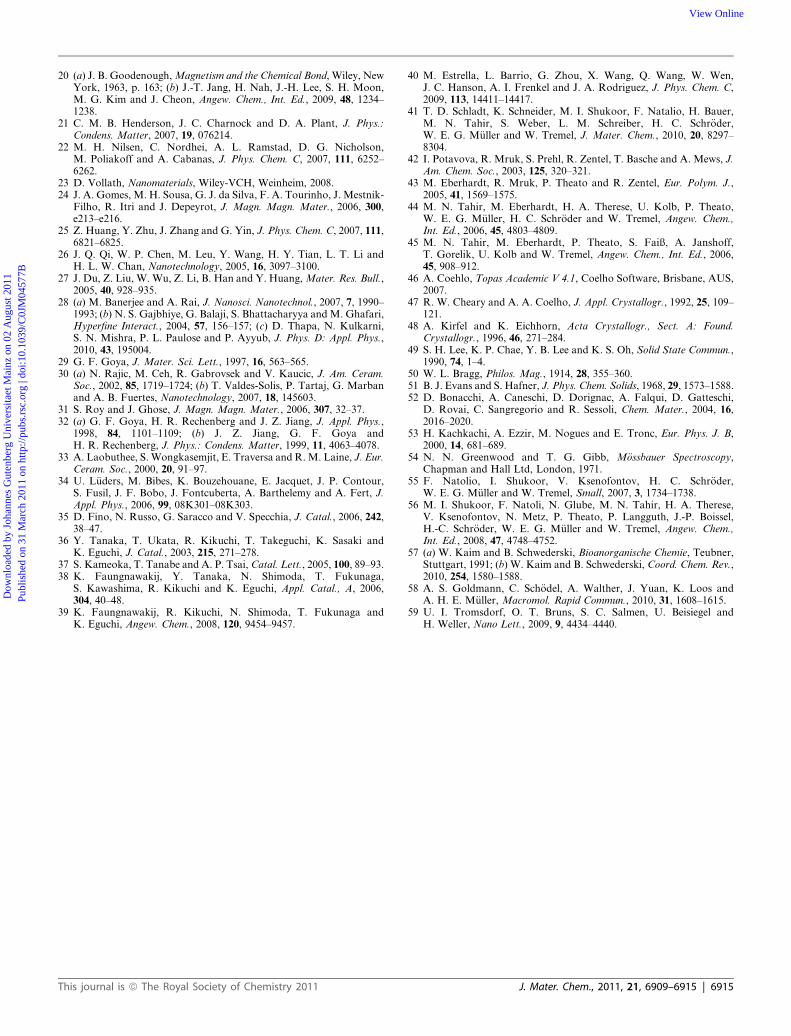
Fig. 5 (a) Digital photograph of CuxFe3�xO4 solutions before (left) and
after (right) ligand exchange in hexane (top layer) and in water (bottom
layer). (b) Fluorescence microscope images of NBD-polymer function-
alized CuxFe3�xO4 nanoparticles (green fluorescence). (c) T1 and T2
weighted MRI images of solutions containing polymer functionalized
CuxFe3�xO4 nanoparticles (concentrations in mM Fe).
Dow
nloa
ded
by J
ohan
nes
Gut
enbe
rg U
nive
rsita
et M
ainz
on
02 A
ugus
t 201
1Pu
blis
hed
on 3
1 M
arch
201
1 on
http
://pu
bs.r
sc.o
rg |
doi:1
0.10
39/C
0JM
0457
7BView Online
octahedral voids (16c). At T ¼ 4.2 K the M€ossbauer spectrum
with Hhf1 ¼ 509.9 kOe and IS1 ¼ 0.507 mms�1 corresponds to
iron in tetrahedral coordination in a valence state Fe3+. A sub-
spectrum with IS2 ¼ 0.614 mms�1, Hhf2 ¼ 468.4 kOe stems from
valence states Fe2.5+ which are due to the electronic exchange
between Fe2+ and Fe3+ in the octahedral voids.54
Taking the pairwise coupling of Fe3+ and Fe2+ in the octahedral
voids into account, one may rewrite the general composition
CuxFe3�xO4 as {Cu2+1�yFe3+
y}tet{Fe2.5+2(y�z)Fe3+
2�2y+zCu2+z}octO4.
Only a part of the Fe3+ species on the octahedral sites, namely (y–z), is
coupled to Fe2+. The remaining Fe3+ ions couple into the contribu-
tion of Fe3+ on the tetrahedral sites. Thus, the intensity ratio of the
This journal is ª The Royal Society of Chemistry 2011
Fe2.5+ and Fe3+ fractions in the M€ossbauer spectrum at 4.2 K
corresponds to
s2
s1
¼ 2ðy� zÞ2� yþ z
:
According to the X-ray diffraction data, the fraction of Cu2+ in
the tetrahedral positions is (1 � y) ¼ 0.32(8). With S2/S1 ¼0.85(3) the amount of Cu2+ in the octahedral voids is z ¼ 0.08(6),
which is in fair agreement with the value of 0.0(1)
determined from the X-ray structure analysis. In summary,
the sample is best described as a Cu-doped
Fe3O4: (Cu2+0.32Fe3+
0.68)A(Fe2+0.6Fe3+
1.32Cu2+1.32)O4.
Nanoparticle functionalization
To explore the potential applications of the CuxFe3�xO4
magnetic nanoparticles, their surface was tailored using a multi-
functional polymeric ligand which contains dopamine as an
anchor group for metal oxide surface,55–58 amine groups for
further immobilization of biomolecules, and 7-nitrobenzofurazan
(NBD) as fluorophore (Fig. S2, ESI†). After the surface func-
tionalization processes, the CuxFe3�xO4 nanoparticles showed
good water solubility and stability at room temperature (Fig. 5a).
These spherical nanoparticles were characterized by epifluor-
escence microscopy to confirm the polymer functionalization
with the aid of the NBD fluorophore conjugated to the backbone
of the polymeric ligand. The respective emission can be seen
visually, and almost all nanoparticles emit green color (Fig. 5b).
The fluorescence of the dye molecules was excited at 513 nm and
detected at 540 nm using a 20 fold dry objective. Furthermore,
the surface functionalization was confirmed using FT-IR
measurements as demonstrated in Fig. S1 (ESI†). As prepared
CuxFe3�xO4 nanoparticles (black line) exhibit characteristic
vibrational bands of the oleate group. Most notably, the asym-
metric and symmetric stretching bands of the RCOO� group
appear at 1543 and 1400 cm�1, respectively. However, these
bands are absent in the spectra of polymeric ligand
J. Mater. Chem., 2011, 21, 6909–6915 | 6913
Dow
nloa
ded
by J
ohan
nes
Gut
enbe
rg U
nive
rsita
et M
ainz
on
02 A
ugus
t 201
1Pu
blis
hed
on 3
1 M
arch
201
1 on
http
://pu
bs.r
sc.o
rg |
doi:1
0.10
39/C
0JM
0457
7BView Online
functionalized CuxFe3�xO4 nanoparticles respectively, indi-
cating a complete replacement of the oleate layer by the hydro-
philic ligands. Furthermore, the appearance of vibrational bands
at 1677 cm�1, 1296, 1251 and 1098 cm�1 in the spectra func-
tionalized nanoparticles spectra, which can be assigned to the
stretching modes of C]O of amide groups present in the poly-
meric ligand and C–O–C ether groups in PEG also present in the
polymeric ligands.
Magnetic resonance imaging
The 1H-NMR relaxometry characterization (i.e., NMR disper-
sion profile) was performed at room temperature by measuring
the longitudinal and the transverse nuclear relaxation times T1
and T2, in the frequency range 10 kHz # n # 65 MHz for T1 and
15 MHz # n # 60 MHz for T2 (see Experimental section).
Measurements at room and physiological temperatures gave
identical results within 10%. The efficiency of the MRI contrast
agents is determined by measuring the nuclear relaxivities r1,2
defined as ri ¼ [(1/Ti)meas � (1/Ti)dia]/c (i ¼ 1, 2) where (1/Ti)meas.
Fig. 5c shows T1 and T2-weighted MR images for 3 different
concentrations of polymer functionalized CuxFe3�xO4 nano-
particles and saline solution (0.9% NaCl) for comparison. Their
concentrations in saline solution were 0.0091, 0.0229 and
0.0366 nM, respectively. The r1 and r2 relaxitivities of polymer
functionalized nanoparticles are 0.0327 and 0.290 (S�1 mM�1),
respectively. Such values for r1 and r2 show that the polymer
functionalized copper ferrite CuxFe3�xO4 nanoparticles can act
both as T1 and T2 contrast agents. Our system of CuxFe3�xO4
nanoparticles, with respect to size and surface functionalities, is
similar to the one reported by Weller and coworkers in showing
both T1 and T2 contrast.59 Thus, after appropriate surface
functionalization, CuxFe3�xO4 nanoparticles may be considered
a promising candidate for molecular imaging when addressed to
specific cells.
Conclusion
In summary, non-agglomerated and monodispersed superpara-
magnetic copper ferrite CuxFe3�xO4 nanoparticles were
prepared and characterized by electron microscopy, X-ray
diffractometry, magnetic susceptibility measurements and
M€ossbauer spectroscopy. The detailed composition of the
nanoparticles as well as the site preference of the metal atoms
could be determined by a combination of the diffraction,
magnetometry and M€ossbauer spectroscopy. The inversion is
believed to be related to the preparation method, as samples
annealed at high temperature exhibit a negligible degree of
inversion. The CuxFe3�xO4 particles could be functionalized
using the hydrophilic polymeric ligand. Efficient surface binding
of the ligand molecules was confirmed by FT-IR. In comparison
to the previously reported nanoparticles, the present nano-
particles are monodisperse, size controlled and present good
stability due to the covalent anchorage of the PEG–polymer to
the surface of the nanoparticles. Finally, we demonstrated that
by virtue of their magnetic properties functionalized CuxFe3�xO4
nanoparticles exhibit a moderate T1 and a strong T2 contrast
enhancement effect for MRI. These results certify that our
approach is a promising way towards new superparamagnetic
6914 | J. Mater. Chem., 2011, 21, 6909–6915
MRI contrast agents which by virtue of the multifunctional
polymer coating can be designed for the specific targeting of cells.
Acknowledgements
We are grateful to Center for Complex Matter (COMATT) for
support and Prof. C. Felser for access to the M€ossbauer facilities.
B.N. is recipient of a fellowship from the Deutscher Akade-
mischer Austauschdienst (DAAD). T. D. Schladt is recipient of
a Carl-Zeiss Fellowship. The Electron Microscopy Center in
Mainz (EZMZ) is operated through the Center for Complex
Matter (COMATT).
References
1 Q. A. Pankhurst, N. K. T. Thanh, K. Jones and J. Dobson, J. Phys.D: Appl. Phys., 2009, 42, 224001.
2 M. Arruebo, R. Fern�andez-Pacheco, M. R. Ibarra and J. Santamar�ıa,Nano Today, 2007, 2, 22–32.
3 J. M. Perez, F. J. Simeone, Y. Saeki, L. Josephson and R. Weissleder,J. Am. Chem. Soc., 2003, 125, 10192–10193.
4 D. L. Graham, H. A. Ferreira and P. P. Freitas, Trends Biotechnol.,2004, 22, 455–462.
5 S. Bucak, D. A. Jones, P. E. Laibinis and T. A. Hatton, Biotechnol.Prog., 2003, 19, 477–484.
6 (a) H. Gu, P. L. Ho, K. W. T. Tsang, L. Wang and B. Xu, J. Am.Chem. Soc., 2003, 125, 15702–15703; (b) M. I. Shukoor, F. Natalio,A. Krasko, H. C. Schr€oder, W. E. G. M€uller and W. Tremel, Chem.Commun., 2007, 4677–4679.
7 J. W. M. Bulte, T. Douglas, B. Witwer, S.-C. Zhang, E. Strable,B. K. Lewis, H. Zywicke, B. Miller, P. van Gelderen,B. M. Moskowitz, L. D. Duncan and J. A. Frank, Nat. Biotechnol.,2001, 19, 1141–1147.
8 M. Lewin, N. Carlesso, C.-H. Tung, X. W. Tang, D. Cory,D. T. Scadden and R. Weissleder, Nat. Biotechnol., 2000, 18, 410–414.
9 R. Hiergeist, W. Andra, N. Buske, R. Hergt, I. Hilger, U. Richter andW. Kaiser, J. Magn. Magn. Mater., 1999, 201, 420–422.
10 (a) Y.-W. Jun, Y.-M. Huh, J.-S. Choi, J.-H. Lee, H.-T. Song, S. Kim,S. Kim, S. Yoon, K.-S. Kim, J.-S. Shin, J.-S. Suh and J. Cheon, J. Am.Chem. Soc., 2005, 127, 5732–5733; (b) Z. Medarova, W. Pham,C. Farrar, V. Petkova and A. Moore, Nat. Med., 2007, 13, 172–177.
11 A. Jordan, R. Scholz, P. Wust, H. Fahling and R. Felix, J. Magn.Magn. Mater., 1999, 201, 413–419.
12 I. Hilger, R. Hiergeist, R. Hergt, K. Winnefeld, H. Schubert andW. Kaiser, Invest. Radiol., 2002, 37, 580–586.
13 U. Haefeli, W. Schuett, J. Teller and M. Zborowski, Scientific andClinical Applications of Magnetic Carriers, Plenum, New York, 1997.
14 R. C. O’Handley, Modern Magnetic Materials: Principles andApplications, Wiley, New York, 2000.
15 V. Sepelak, I. Bergmann, A. Feldhoff, P. Heitjans, F. Krumeich,D. Menzel, F. J. Litterst, S. J. Campbell and K. D. Becker, J. Phys.Chem. C, 2007, 111, 5026–5033.
16 (a) A. J. Rondinone, A. C. S. Samia and Z. J. Zhang, J. Phys. Chem.B, 1999, 103, 6876–6880; (b) C. Liu, B. Zou, A. J. Rondinone andZ. J. Zhang, J. Am. Chem. Soc., 2000, 122, 6263–6267.
17 (a) C. Yao, Q. Zeng, G. F. Goya, T. Torres, J. Liu, H. Wu, M. Ge,Y. Zeng, Y. Wang and J. Z. Jiang, J. Phys. Chem. C, 2007, 111,12274–12278; (b) M. Sivakumar, T. Takami, H. Ikuta, A. Towata,K. Yasui, T. Tuziuti, T. Kozuka, D. Bhattacharya and Y. Iida, J.Phys. Chem. B, 2006, 110, 15234–15243.
18 (a) A. J. Rondinone, C. Liu and Z. J. Zhang, J. Phys. Chem. B, 2001,105, 7967–7971; (b) S. Sun, H. Zeng, D. B. Robinson, S. Raoux,P. M. Rice, S. X. Wang and G. Li, J. Am. Chem. Soc., 2004, 126,273–279; (c) H. Deng, X. Li, Q. Peng, X. Wang, J. Chen and Y. Li,Angew. Chem., Int. Ed., 2005, 44, 2782–2785; (d) E. Kang, J. Park,Y. Hwang, M. Kang, J.-G. Park and T. Hyeon, J. Phys. Chem. B,2004, 108, 13932–13935.
19 (a) J. Lee, Y.-M. Huh, Y. Jun, J. Seo, J. Jang, H. Song, S. Kim,E. Cho, H. Yoon, J. Suh and J. Cheon, Nat. Med., 2007, 13, 95–99;(b) C. Liu, B. Zou, A. J. Rondinone and Z. J. Zhang, J. Phys.Chem. B, 2000, 104, 1141–1145.
This journal is ª The Royal Society of Chemistry 2011
Dow
nloa
ded
by J
ohan
nes
Gut
enbe
rg U
nive
rsita
et M
ainz
on
02 A
ugus
t 201
1Pu
blis
hed
on 3
1 M
arch
201
1 on
http
://pu
bs.r
sc.o
rg |
doi:1
0.10
39/C
0JM
0457
7BView Online
20 (a) J. B. Goodenough, Magnetism and the Chemical Bond, Wiley, NewYork, 1963, p. 163; (b) J.-T. Jang, H. Nah, J.-H. Lee, S. H. Moon,M. G. Kim and J. Cheon, Angew. Chem., Int. Ed., 2009, 48, 1234–1238.
21 C. M. B. Henderson, J. C. Charnock and D. A. Plant, J. Phys.:Condens. Matter, 2007, 19, 076214.
22 M. H. Nilsen, C. Nordhei, A. L. Ramstad, D. G. Nicholson,M. Poliakoff and A. Cabanas, J. Phys. Chem. C, 2007, 111, 6252–6262.
23 D. Vollath, Nanomaterials, Wiley-VCH, Weinheim, 2008.24 J. A. Gomes, M. H. Sousa, G. J. da Silva, F. A. Tourinho, J. Mestnik-
Filho, R. Itri and J. Depeyrot, J. Magn. Magn. Mater., 2006, 300,e213–e216.
25 Z. Huang, Y. Zhu, J. Zhang and G. Yin, J. Phys. Chem. C, 2007, 111,6821–6825.
26 J. Q. Qi, W. P. Chen, M. Leu, Y. Wang, H. Y. Tian, L. T. Li andH. L. W. Chan, Nanotechnology, 2005, 16, 3097–3100.
27 J. Du, Z. Liu, W. Wu, Z. Li, B. Han and Y. Huang, Mater. Res. Bull.,2005, 40, 928–935.
28 (a) M. Banerjee and A. Rai, J. Nanosci. Nanotechnol., 2007, 7, 1990–1993; (b) N. S. Gajbhiye, G. Balaji, S. Bhattacharyya and M. Ghafari,Hyperfine Interact., 2004, 57, 156–157; (c) D. Thapa, N. Kulkarni,S. N. Mishra, P. L. Paulose and P. Ayyub, J. Phys. D: Appl. Phys.,2010, 43, 195004.
29 G. F. Goya, J. Mater. Sci. Lett., 1997, 16, 563–565.30 (a) N. Rajic, M. Ceh, R. Gabrovsek and V. Kaucic, J. Am. Ceram.
Soc., 2002, 85, 1719–1724; (b) T. Valdes-Solis, P. Tartaj, G. Marbanand A. B. Fuertes, Nanotechnology, 2007, 18, 145603.
31 S. Roy and J. Ghose, J. Magn. Magn. Mater., 2006, 307, 32–37.32 (a) G. F. Goya, H. R. Rechenberg and J. Z. Jiang, J. Appl. Phys.,
1998, 84, 1101–1109; (b) J. Z. Jiang, G. F. Goya andH. R. Rechenberg, J. Phys.: Condens. Matter, 1999, 11, 4063–4078.
33 A. Laobuthee, S. Wongkasemjit, E. Traversa and R. M. Laine, J. Eur.Ceram. Soc., 2000, 20, 91–97.
34 U. L€uders, M. Bibes, K. Bouzehouane, E. Jacquet, J. P. Contour,S. Fusil, J. F. Bobo, J. Fontcuberta, A. Barthelemy and A. Fert, J.Appl. Phys., 2006, 99, 08K301–08K303.
35 D. Fino, N. Russo, G. Saracco and V. Specchia, J. Catal., 2006, 242,38–47.
36 Y. Tanaka, T. Ukata, R. Kikuchi, T. Takeguchi, K. Sasaki andK. Eguchi, J. Catal., 2003, 215, 271–278.
37 S. Kameoka, T. Tanabe and A. P. Tsai, Catal. Lett., 2005, 100, 89–93.38 K. Faungnawakij, Y. Tanaka, N. Shimoda, T. Fukunaga,
S. Kawashima, R. Kikuchi and K. Eguchi, Appl. Catal., A, 2006,304, 40–48.
39 K. Faungnawakij, R. Kikuchi, N. Shimoda, T. Fukunaga andK. Eguchi, Angew. Chem., 2008, 120, 9454–9457.
This journal is ª The Royal Society of Chemistry 2011
40 M. Estrella, L. Barrio, G. Zhou, X. Wang, Q. Wang, W. Wen,J. C. Hanson, A. I. Frenkel and J. A. Rodriguez, J. Phys. Chem. C,2009, 113, 14411–14417.
41 T. D. Schladt, K. Schneider, M. I. Shukoor, F. Natalio, H. Bauer,M. N. Tahir, S. Weber, L. M. Schreiber, H. C. Schr€oder,W. E. G. M€uller and W. Tremel, J. Mater. Chem., 2010, 20, 8297–8304.
42 I. Potavova, R. Mruk, S. Prehl, R. Zentel, T. Basche and A. Mews, J.Am. Chem. Soc., 2003, 125, 320–321.
43 M. Eberhardt, R. Mruk, P. Theato and R. Zentel, Eur. Polym. J.,2005, 41, 1569–1575.
44 M. N. Tahir, M. Eberhardt, H. A. Therese, U. Kolb, P. Theato,W. E. G. M€uller, H. C. Schr€oder and W. Tremel, Angew. Chem.,Int. Ed., 2006, 45, 4803–4809.
45 M. N. Tahir, M. Eberhardt, P. Theato, S. Faiß, A. Janshoff,T. Gorelik, U. Kolb and W. Tremel, Angew. Chem., Int. Ed., 2006,45, 908–912.
46 A. Coehlo, Topas Academic V 4.1, Coelho Software, Brisbane, AUS,2007.
47 R. W. Cheary and A. A. Coelho, J. Appl. Crystallogr., 1992, 25, 109–121.
48 A. Kirfel and K. Eichhorn, Acta Crystallogr., Sect. A: Found.Crystallogr., 1996, 46, 271–284.
49 S. H. Lee, K. P. Chae, Y. B. Lee and K. S. Oh, Solid State Commun.,1990, 74, 1–4.
50 W. L. Bragg, Philos. Mag., 1914, 28, 355–360.51 B. J. Evans and S. Hafner, J. Phys. Chem. Solids, 1968, 29, 1573–1588.52 D. Bonacchi, A. Caneschi, D. Dorignac, A. Falqui, D. Gatteschi,
D. Rovai, C. Sangregorio and R. Sessoli, Chem. Mater., 2004, 16,2016–2020.
53 H. Kachkachi, A. Ezzir, M. Nogues and E. Tronc, Eur. Phys. J. B,2000, 14, 681–689.
54 N. N. Greenwood and T. G. Gibb, M€ossbauer Spectroscopy,Chapman and Hall Ltd, London, 1971.
55 F. Natolio, I. Shukoor, V. Ksenofontov, H. C. Schr€oder,W. E. G. M€uller and W. Tremel, Small, 2007, 3, 1734–1738.
56 M. I. Shukoor, F. Natoli, N. Glube, M. N. Tahir, H. A. Therese,V. Ksenofontov, N. Metz, P. Theato, P. Langguth, J.-P. Boissel,H.-C. Schr€oder, W. E. G. M€uller and W. Tremel, Angew. Chem.,Int. Ed., 2008, 47, 4748–4752.
57 (a) W. Kaim and B. Schwederski, Bioanorganische Chemie, Teubner,Stuttgart, 1991; (b) W. Kaim and B. Schwederski, Coord. Chem. Rev.,2010, 254, 1580–1588.
58 A. S. Goldmann, C. Sch€odel, A. Walther, J. Yuan, K. Loos andA. H. E. M€uller, Macromol. Rapid Commun., 2010, 31, 1608–1615.
59 U. I. Tromsdorf, O. T. Bruns, S. C. Salmen, U. Beisiegel andH. Weller, Nano Lett., 2009, 9, 4434–4440.
J. Mater. Chem., 2011, 21, 6909–6915 | 6915
Related Documents