HAL Id: hal-03432195 https://hal.archives-ouvertes.fr/hal-03432195 Submitted on 17 Nov 2021 HAL is a multi-disciplinary open access archive for the deposit and dissemination of sci- entific research documents, whether they are pub- lished or not. The documents may come from teaching and research institutions in France or abroad, or from public or private research centers. L’archive ouverte pluridisciplinaire HAL, est destinée au dépôt et à la diffusion de documents scientifiques de niveau recherche, publiés ou non, émanant des établissements d’enseignement et de recherche français ou étrangers, des laboratoires publics ou privés. Synthesis, Characterization and Evaluation of Peptide Nanostructures for Biomedical Applications Fanny D’orlyé, Laura Trapiella-Alfonso, Camille Lescot, Marie Pinvidic, Bich-Thuy Doan, Anne Varenne To cite this version: Fanny D’orlyé, Laura Trapiella-Alfonso, Camille Lescot, Marie Pinvidic, Bich-Thuy Doan, et al.. Synthesis, Characterization and Evaluation of Peptide Nanostructures for Biomedical Applications. Molecules, MDPI, 2021, 26 (15), pp.4587. 10.3390/molecules26154587. hal-03432195

Welcome message from author
This document is posted to help you gain knowledge. Please leave a comment to let me know what you think about it! Share it to your friends and learn new things together.
Transcript
HAL Id: hal-03432195https://hal.archives-ouvertes.fr/hal-03432195
Submitted on 17 Nov 2021
HAL is a multi-disciplinary open accessarchive for the deposit and dissemination of sci-entific research documents, whether they are pub-lished or not. The documents may come fromteaching and research institutions in France orabroad, or from public or private research centers.
L’archive ouverte pluridisciplinaire HAL, estdestinée au dépôt et à la diffusion de documentsscientifiques de niveau recherche, publiés ou non,émanant des établissements d’enseignement et derecherche français ou étrangers, des laboratoirespublics ou privés.
Synthesis, Characterization and Evaluation of PeptideNanostructures for Biomedical Applications
Fanny D’orlyé, Laura Trapiella-Alfonso, Camille Lescot, Marie Pinvidic,Bich-Thuy Doan, Anne Varenne
To cite this version:Fanny D’orlyé, Laura Trapiella-Alfonso, Camille Lescot, Marie Pinvidic, Bich-Thuy Doan, et al..Synthesis, Characterization and Evaluation of Peptide Nanostructures for Biomedical Applications.Molecules, MDPI, 2021, 26 (15), pp.4587. �10.3390/molecules26154587�. �hal-03432195�
molecules
Review
Synthesis, Characterization and Evaluation of PeptideNanostructures for Biomedical Applications
Fanny d’Orlyé, Laura Trapiella-Alfonso , Camille Lescot, Marie Pinvidic, Bich-Thuy Doan and Anne Varenne *
�����������������
Citation: d’Orlyé, F.;
Trapiella-Alfonso, L.; Lescot, C.;
Pinvidic, M.; Doan, B.-T.; Varenne, A.
Synthesis, Characterization and
Evaluation of Peptide Nanostructures
for Biomedical Applications.
Molecules 2021, 26, 4587. https://doi.
org/10.3390/molecules26154587
Academic Editors: Luca D. D’Andrea
and Lucia De Rosa
Received: 2 June 2021
Accepted: 17 July 2021
Published: 29 July 2021
Publisher’s Note: MDPI stays neutral
with regard to jurisdictional claims in
published maps and institutional affil-
iations.
Copyright: © 2021 by the authors.
Licensee MDPI, Basel, Switzerland.
This article is an open access article
distributed under the terms and
conditions of the Creative Commons
Attribution (CC BY) license (https://
creativecommons.org/licenses/by/
4.0/).
Chimie ParisTech PSL, CNRS 8060, Institute of Chemistry for Life and Health (i-CLeHS), 75005 Paris, France;[email protected] (F.d.); [email protected] (L.T.-A.);[email protected] (C.L.); [email protected] (M.P.);[email protected] (B.-T.D.)* Correspondence: [email protected]; Tel.: +33-1-8578-4252
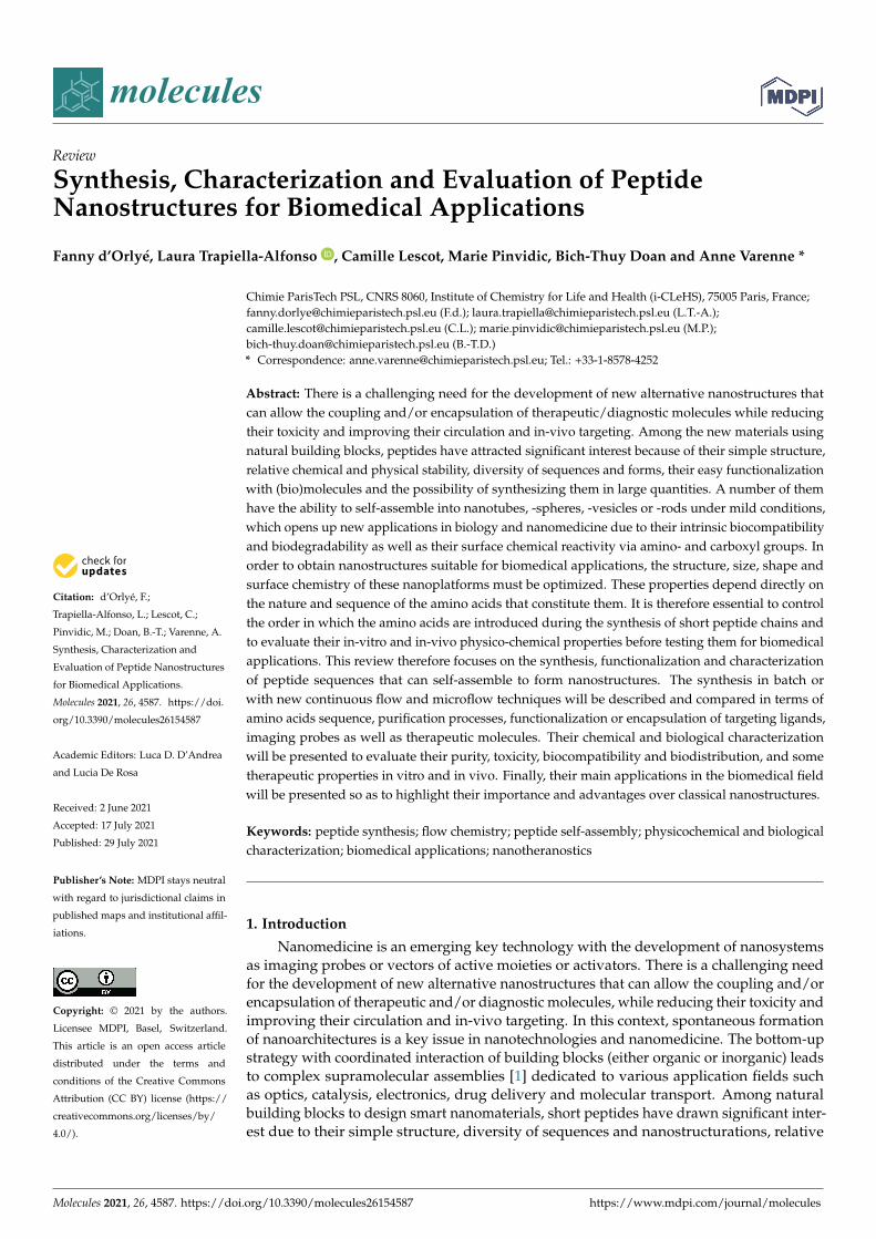
Abstract: There is a challenging need for the development of new alternative nanostructures thatcan allow the coupling and/or encapsulation of therapeutic/diagnostic molecules while reducingtheir toxicity and improving their circulation and in-vivo targeting. Among the new materials usingnatural building blocks, peptides have attracted significant interest because of their simple structure,relative chemical and physical stability, diversity of sequences and forms, their easy functionalizationwith (bio)molecules and the possibility of synthesizing them in large quantities. A number of themhave the ability to self-assemble into nanotubes, -spheres, -vesicles or -rods under mild conditions,which opens up new applications in biology and nanomedicine due to their intrinsic biocompatibilityand biodegradability as well as their surface chemical reactivity via amino- and carboxyl groups. Inorder to obtain nanostructures suitable for biomedical applications, the structure, size, shape andsurface chemistry of these nanoplatforms must be optimized. These properties depend directly onthe nature and sequence of the amino acids that constitute them. It is therefore essential to controlthe order in which the amino acids are introduced during the synthesis of short peptide chains andto evaluate their in-vitro and in-vivo physico-chemical properties before testing them for biomedicalapplications. This review therefore focuses on the synthesis, functionalization and characterizationof peptide sequences that can self-assemble to form nanostructures. The synthesis in batch orwith new continuous flow and microflow techniques will be described and compared in terms ofamino acids sequence, purification processes, functionalization or encapsulation of targeting ligands,imaging probes as well as therapeutic molecules. Their chemical and biological characterizationwill be presented to evaluate their purity, toxicity, biocompatibility and biodistribution, and sometherapeutic properties in vitro and in vivo. Finally, their main applications in the biomedical fieldwill be presented so as to highlight their importance and advantages over classical nanostructures.
Keywords: peptide synthesis; flow chemistry; peptide self-assembly; physicochemical and biologicalcharacterization; biomedical applications; nanotheranostics
1. Introduction
Nanomedicine is an emerging key technology with the development of nanosystemsas imaging probes or vectors of active moieties or activators. There is a challenging needfor the development of new alternative nanostructures that can allow the coupling and/orencapsulation of therapeutic and/or diagnostic molecules, while reducing their toxicity andimproving their circulation and in-vivo targeting. In this context, spontaneous formationof nanoarchitectures is a key issue in nanotechnologies and nanomedicine. The bottom-upstrategy with coordinated interaction of building blocks (either organic or inorganic) leadsto complex supramolecular assemblies [1] dedicated to various application fields suchas optics, catalysis, electronics, drug delivery and molecular transport. Among naturalbuilding blocks to design smart nanomaterials, short peptides have drawn significant inter-est due to their simple structure, diversity of sequences and nanostructurations, relative
Molecules 2021, 26, 4587. https://doi.org/10.3390/molecules26154587 https://www.mdpi.com/journal/molecules
Molecules 2021, 26, 4587 2 of 45
chemical and physical stability, simplicity to be modified or decorated with biological andchemical entities, and their ability to be synthesized on a large scale [2]. Although somenaturally occurring peptide self-assemblies can lead to medical disorder (amyloid fibrils),peptidic nanostructures have become an important strategy for nanomedicine due to theirbiocompatibility, biodegradability, robustness and their surface chemical reactivity viaamino and carboxylic groups. Peptides, as short as dipeptides, contain all the molecu-lar information needed to form well-ordered structures at the nanoscale. Peptide-basedbuilding blocks allow to control the structure and properties of well-structured nanoscalearchitectures, such as nanotubes, -spheres, -vesicles, -rods, -fibrils and even hydrogels,under mild conditions.
In order to obtain nanostructures suitable for biomedical applications, the structure,size, shape and surface chemistry of these nanoplatforms must be optimized and controlled.These properties depend directly on the nature and sequence of the amino acids thatconstitute them. Therefore, the first step for efficiently developing such nanoarchitecturesis to design adequate short peptide chains and control their synthesis, i.e., the efficientamide bond formation and the order in which the amino acids are introduced during thesynthesis. From the very first peptide synthesis by Theodor Curtius in 1882 [3], differentmethodological improvements were performed, with the solid-phase peptide synthesis,the reduction of the number of synthesis steps and process dimensions. Whatever thepeptide sequences and their auto-assembly into nanoarchitectures, both entities have thento be extensively physico-chemically characterized. It is indeed crucial to control the purityand sequence of the peptides, as well as to understand the driving forces that control thepeptide self-assembly, so as to design the most robust, biocompatible and biodistributablenanostructures in biological conditions. In addition to the peptide sequence, peptideauto-assembly can also be modulated or modified by external environmental factors andcan lead to stimuli-responsive nanomaterials. Classical methods are employed so far(spectroscopic, microscopic and scattering methods) to determine the global self-assembledpeptide nanostructures (sequence, size, diameter, charge density and shape), as well astheir secondary, tertiary and quaternary structure. Due to their dynamic self-assemblyprocesses, multiple characterization methods have to be implemented jointly, allowing forlarge time and length scales.
We will first present in this review a brief summary of classical synthesis methodolo-gies and will concentrate on recent techniques such as continuous flow chemistry to gainin speed, purity and ease of production, and to promote specific self-assembly by deeplycontrolling the experimental conditions and coupling with adequate methods within thesynthesis process. We will then present the main driving forces for self-assembly anddescribe the most synthesized short peptides and their identified self-assembly, i.e., peptideamphiphiles, aromatic peptides and the particular case of the diphenylalanine peptide(FF) and cyclic peptides. The interest of combining classical characterization methods forthese short peptides of diverse composition will be described, so as to elucidate the interac-tions governing the self-assembly and the final nanostructure. The interest of analyticalmethods based on separation will be highlighted, as a prospective field. In the last part,the interest of these peptidic nanostructures for current or future biomedical applicationswill be described, going from pharmaceutical purposes, medical diagnosis and imaging,drug targeting and delivery, therapy against cancer, microbes, photo- and gene-therapy,regenerative medicine to tissue engineering.
2. Combination of Classical Peptide Synthesis and Assembly to Form Nanoobjects
Synthesis of peptide nanoobjects relies first on the preparation of the linear peptideby coupling amino acids, using homogenous coupling conditions, solid-phase peptidesynthesis, in batch or using flow chemistry. Secondly, the peptide will auto-assemble toform the nanoobject. The auto-assembling properties of the peptide will depend on itssequence, and could be controlled by external factors like pH, temperature, organic orphoto stimulation, and also by microfluidics. We will describe here some of the most recent
Molecules 2021, 26, 4587 3 of 45
methodologies and techniques for peptide synthesis with an emphasis on the use of flowchemistry, as well as for controlling peptide auto-assembly in a second part of this review.
2.1. Peptide Synthesis with Focus on Flow Chemistry
If we go back to the beginning, the first peptide synthesis was performed in 1882 byTheodor Curtius by coupling silver salt of glycine and benzoyl chloride, leading to the firstN-protected dipeptide Benzoylglycylglycine (GG) [3].
Since this first milestone based on homogeneous liquid organic chemistry, lot ofwork has been performed, with the development of solid-phase peptide synthesis (SPPS)notably [4] to optimize and facilitate the synthesis of peptides. We will describe hereinsome of the recent examples.
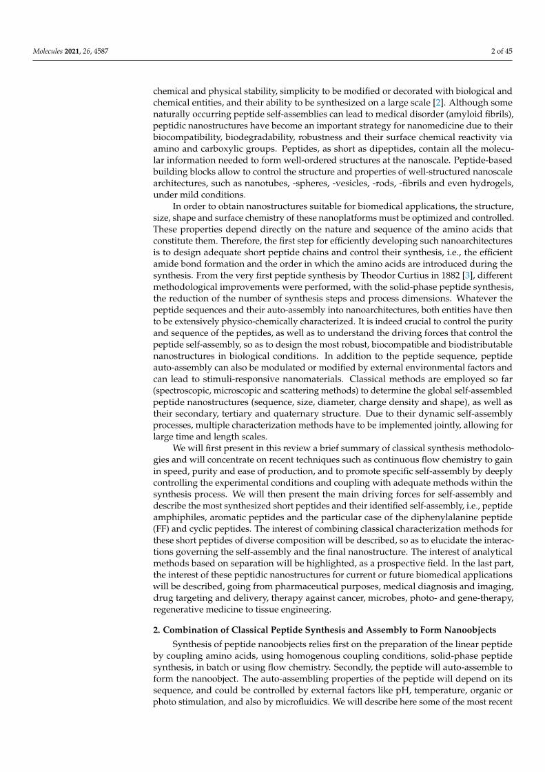
Merrifield [5] proposed for the first time this new approach for the synthesis ofpeptides relying on the stepwise addition of protected amino acids to a growing peptidechain attached to a solid resin particle through covalent bond. This strategy allows thereagents and by-products to be filtered off the resin, for a great gain of time and simplicity.In this article, the proof of concept was done with the synthesis of the model tetrapeptideL-leucyl-L-alanylglycyl-L-valine (LAGV) Scheme 1a.
Beyond linear peptides, cyclic and heterodimeric peptides are very attractive to syn-thesize because of their very interesting biological properties for various applications [6]. Itis known that Cyclic Peptides (CP) can auto-assemble to form peptide nanotubes, thanksto the backbone-backbone stacking due to hydrogen bonding between antiparallel β-sheetoriented from the N-H bond of one CP to a C=O of another CP. In 2016 our team per-formed the design, synthesis and characterization of new cyclic D, L-α-alternate aminoacid peptides [7]. As the formation of CP nanotubes depends on their sequence, threeseries of novel cyclic peptides were synthesized: each series was varied in chain length andamino acid nature, leading to 8 CPs of different van der Waals inner diameter and differentproperties for future applications. The synthesis of the linear peptide sequence (first step)was undergone by classical SPPS and orthogonal protection methods, followed by thecyclization step using propane phosphonic acid. While the linear peptide was obtained ina relatively good purity, the cyclization was performed in 40–90% yield, higher than thepreviously reported methodologies [8].
Although their synthesis is described as complicated and low yielding, heterodimericpeptides which are cystine-rich represent also highly interesting drug targets. Hossainand his group worked on an improved synthetic route to heterodimeric peptides, whichreduces the number of synthesis steps compared to classical methodologies [9]. Usually,the two chains are synthesized separately, released from the solid support and submittedto multistep solution-phase reactions to control the disulfide pairing. Hossein’s protocolconsists in the sequential synthesis of both chains on the same solid support separatedby a chemically cleavable bis-linker (Scheme 1b). The linear dipeptide linked by thetether is then released and used for the formation of the intra peptide-peptide compoundby disulfide or thioether bond. The tether is finally cleaved by a 5% hydrazine buffer,leading to the desired conjugate with an overall yield of 27%. In a simpler way, Hossainand colleagues also developed an improved SPPS synthesis strategy using orthogonallyprotected monomeric building blocks, which they successfully applied to the synthesis ofinsulin by incorporating the thioether moiety in place of the A6–A11 cystine bridge [10].
Molecules 2021, 26, 4587 4 of 45Molecules 2021, 26, x FOR PEER REVIEW 4 of 48
Scheme 1. (a) Merrifield first solid supported peptide synthesis strategy (b) Hossein’s strategy for the synthesis of heterodimeric peptides. Adapte with permission from reference [9].
Although their synthesis is described as complicated and low yielding, heterodimeric peptides which are cystine-rich represent also highly interesting drug targets. Hossain and his group worked on an improved synthetic route to heterodimeric peptides, which reduces the number of synthesis steps compared to classical methodologies [9]. Usually, the two chains are synthesized separately, released from the solid support and submitted to multistep solution-phase reactions to control the disulfide pairing. Hossein’s protocol consists in the sequential synthesis of both chains on the same solid support separated by a chemically cleavable bis-linker Scheme 1b). The linear dipeptide linked by the tether is then released and used for the formation of the intra peptide-peptide compound by disul-fide or thioether bond. The tether is finally cleaved by a 5% hydrazine buffer, leading to the desired conjugate with an overall yield of 27%. In a simpler way, Hossain and col-leagues also developed an improved SPPS synthesis strategy using orthogonally pro-tected monomeric building blocks, which they successfully applied to the synthesis of in-sulin by incorporating the thioether moiety in place of the A6–A11 cystine bridge [10].
Although the SPPS has revolutionized peptides synthesis, a review underlined in 2009 the need for more powerful methods for the synthesis of peptides with longer chains [11]. The SPPS is and will be anyway part of the current developments of new methodol-ogies for peptide synthesis. Among those, we have chosen to concentrate our attention on methodologies relying on continuous flow chemistry. Before addressing the question of solid or liquid phase process, we might look back at the center of peptide synthesis: the amide bond formation. The efficiency of this chemical reaction is determinant for the whole process. Amide bond formation relies on the activation of the carboxylic acid part, followed by the coupling with the amine part of another amino acid. A large panel of coupling reagents is available for the chemists, and the question has been well docu-mented [12]. Takahashi and co-workers developed in 2014 a highly efficient amide bond formation methodology, relying on the very rapid activation of carboxylic acids before their also rapid conversion into amides [13]. They achieved in a microflow synthesis reac-tor an activation time of 0.5 s, with a reaction time of 4.3 s, allowing the synthesis of pep-tides with excellent yields, the lowest being 74%.
Scheme 1. (a) Merrifield first solid supported peptide synthesis strategy (b) Hossein’s strategy forthe synthesis of heterodimeric peptides. Adapte with permission from reference [9].
Although the SPPS has revolutionized peptides synthesis, a review underlined in 2009the need for more powerful methods for the synthesis of peptides with longer chains [11].The SPPS is and will be anyway part of the current developments of new methodologiesfor peptide synthesis. Among those, we have chosen to concentrate our attention onmethodologies relying on continuous flow chemistry. Before addressing the question ofsolid or liquid phase process, we might look back at the center of peptide synthesis: theamide bond formation. The efficiency of this chemical reaction is determinant for the wholeprocess. Amide bond formation relies on the activation of the carboxylic acid part, followedby the coupling with the amine part of another amino acid. A large panel of couplingreagents is available for the chemists, and the question has been well documented [12].Takahashi and co-workers developed in 2014 a highly efficient amide bond formationmethodology, relying on the very rapid activation of carboxylic acids before their also rapidconversion into amides [13]. They achieved in a microflow synthesis reactor an activationtime of 0.5 s, with a reaction time of 4.3 s, allowing the synthesis of peptides with excellentyields, the lowest being 74%.
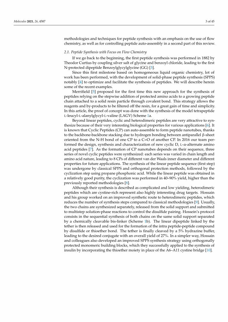
This methodology is based on the concept of “flash chemistry” i.e., the quasi in-stantaneous activation and reaction of chemical compounds, reducing the formation ofby-products, and, in the case of peptides, epimerization. This is one of the first examplesof the use of flash chemistry for amide bond formation. The microreactor consisted in thecombination of 2 T-shape mixers (Figure 1), the first one dedicated to the activation of thecarboxylic acid part of the first amino acid (flow rate A on Figure 1), with triphosgene (flowrate B on Figure 1), and the second to the coupling with the protected amino acid (flowrate C on Figure 1). The whole microsystem was connected by Teflon tubing and flowed bysyringe pumps. This process afforded the coupling of 2 amino acids in a very short time(less than 5 s residence time) in very good yield and purity. The authors compared theirflow system with the batch equivalent and showed highly superior results in flow in everycase (97% yield in flow vs. 74% in batch for the best results). This study illustrates the highpotential of flow chemistry for peptide synthesis.
Molecules 2021, 26, 4587 5 of 45
Molecules 2021, 26, x FOR PEER REVIEW 5 of 48
This methodology is based on the concept of “flash chemistry” i.e., the quasi instan-taneous activation and reaction of chemical compounds, reducing the formation of by-products, and, in the case of peptides, epimerization. This is one of the first examples of the use of flash chemistry for amide bond formation. The microreactor consisted in the combination of 2 T-shape mixers (Figure 1), the first one dedicated to the activation of the carboxylic acid part of the first amino acid (flow rate A on Figure 1), with triphosgene (flow rate B on Figure 1), and the second to the coupling with the protected amino acid (flow rate C on Figure 1). The whole microsystem was connected by Teflon tubing and flowed by syringe pumps. This process afforded the coupling of 2 amino acids in a very short time (less than 5 s residence time) in very good yield and purity. The authors com-pared their flow system with the batch equivalent and showed highly superior results in flow in every case (97% yield in flow vs. 74% in batch for the best results). This study illustrates the high potential of flow chemistry for peptide synthesis.
Figure 1. Microflow synthesis of peptides based on rapid and strong activation of carboxylic acids. Adapted with permission from Reference [13].
In parallel, we can underline the work of Pentelute and his team on the development of a rapid flow-based peptide synthesis methodology [14] and its successful concomitant application to the total synthesis of DARPin pE59 (Designed Ankyrin Repeat Protein, 130 amino acids residues) and Barnase (RNase B. a., 113 amino acids) [15]. Their methodology consisted in a fully automated solid-phase peptide synthesis in continuous flow, allowing the incorporation of one amino acid every 1.8 min. The main advantage of this work com-pared to the previous literature is the design of a low-volume and low-pressure reaction vessel. This could overcome the problems of high volume of wash solvent, and of low flow rates required due to high pressures of the systems. This vessel allowed the authors to impulse high flow rates, so that the reaction time is shorter, without raising the pressure too high. This methodology allowed the authors to synthesize native DARP in pE59 and Barnase proteins through the rapid and efficient production of high-quality peptide frag-ments which were assembled via convergent N, C Ligation. The final full lengths proteins (130 and 113 residues) were found to be biologically active. Three years later the same authors improved their protocol, providing a SPPS approach where the amide bond for-mation takes only 7 s and the total synthesis time is 40 s per amino acids [16].
Among the recent developments presented in recent reviews [17,18], the group of Seeberger in 2019 improved the methodology to overcome some limitations of solid sup-port reactors [19]. Indeed, as main continuous flow protocols are developed with fixed bed reactors, the two major drawbacks are the reagent channeling and the high back pres-sure. Therefore, Seeberger and his team envisaged the development of a variable bed re-actor. A differencial pressure sensing was applied to monitor pressure changes across the reaction bed caused by resin swelling and shrinking, leading to autonomous adjustments
Figure 1. Microflow synthesis of peptides based on rapid and strong activation of carboxylic acids.Adapted with permission from Reference [13].
In parallel, we can underline the work of Pentelute and his team on the developmentof a rapid flow-based peptide synthesis methodology [14] and its successful concomitantapplication to the total synthesis of DARPin pE59 (Designed Ankyrin Repeat Protein,130 amino acids residues) and Barnase (RNase B. a., 113 amino acids) [15]. Their method-ology consisted in a fully automated solid-phase peptide synthesis in continuous flow,allowing the incorporation of one amino acid every 1.8 min. The main advantage of thiswork compared to the previous literature is the design of a low-volume and low-pressurereaction vessel. This could overcome the problems of high volume of wash solvent, andof low flow rates required due to high pressures of the systems. This vessel allowed theauthors to impulse high flow rates, so that the reaction time is shorter, without raising thepressure too high. This methodology allowed the authors to synthesize native DARP inpE59 and Barnase proteins through the rapid and efficient production of high-quality pep-tide fragments which were assembled via convergent N, C Ligation. The final full lengthsproteins (130 and 113 residues) were found to be biologically active. Three years later thesame authors improved their protocol, providing a SPPS approach where the amide bondformation takes only 7 s and the total synthesis time is 40 s per amino acids [16].
Among the recent developments presented in recent reviews [17,18], the group ofSeeberger in 2019 improved the methodology to overcome some limitations of solid supportreactors [19]. Indeed, as main continuous flow protocols are developed with fixed bedreactors, the two major drawbacks are the reagent channeling and the high back pressure.Therefore, Seeberger and his team envisaged the development of a variable bed reactor. Adifferencial pressure sensing was applied to monitor pressure changes across the reactionbed caused by resin swelling and shrinking, leading to autonomous adjustments made bya piston to the resin bed size for maintaining the pressure while the resin swells freely.
Compared to a fixed bed reactor where the reactions are monitored in line, a variablebed allows a real time monitoring of the elongation efficiency and peptide tertiary structure,while maintaining a low overall system pressure throughout peptide syntheses. This newreactor could also enable tracking of problematic couplings, on-resin aggregation, andfurther understanding the effects of some synthetic conditions on peptide sequences.
Generally, peptide synthesis gains speed and synthesis control via continuous flowmethodologies. Gain in the reaction time is very important in the field of peptide synthesisfor nanostructures, since the assembly of the peptide relies mainly on the amino acidsequence. The use of continuous flow would thus allow several fast assays and changes inthe amino acids sequence to have in hands a panel of peptidic structures for studying theirassembly properties.
Molecules 2021, 26, 4587 6 of 45
2.2. Self-Assembly to Form Peptide Nanoobjects
The assembly of nanopeptides is influenced by the conditions of the solution, suchas pH, temperature, ionic strength, salt or solvent nature. We will present here somearticles highlighting the influence of these three parameters of the environment on the self-assembly of peptides to form nanostructures, as well as some of the techniques developedto promote this assembly; the intrinsic properties related to the sequence itself will befurther explored in the next session of this review.
Some literature highlights the influence of pH on nanostructuration. For example,the KLVFFAE peptide sequence of Alzheimer’s disease (AD) was demonstrated to be verysensitive to environmental pH as it was shown to auto-assemble into fibers at neutralpH and into tubes at acidic pH [20]. Ghosh et al. also developed a strategy for preciselycontrolling the self-assembly of the Peptide Amphiphiles (PAs) by adjusting the pH ofthe solution [21]. They found that PAs could self-assemble into nanofibers at pH 4 andspherical nanomicelles at pH 10.
Another illustrative example has been published by the group of Fojan [22] in 2010.The authors present the synthesis and characterization of a novel amphiphilic peptideKA6 which exhibits a clear charge separation controllable by the pH of the environment.As the self-assembly of this system is largely governed by electrostatic interactions, amodification of the pH causes a modification of the micellar structure, revealed by atomicforce microscopy (AFM) and circular dichroism (CD) characterizations (see part III). Atbasic pH, the micellar structure is inverted, exposing the opposite end of the peptide chainto the solution, going from pH 2 to pH 11.
L-Carnosine (β-alanine-histidine, βAH), is a peptide providing a large range of bio-logical activities. Peptide βAH is highly water-soluble, but it does not self-assemble inwater. Castelletto and Hamley explored the construction of novel βAH supramolecularself-assemblies [23]. Their strategy to drive βAH self-assembly involves turning the dipep-tide into a PA through the lipidation of βAH by adding a C16 palmitoyl lipid chain to thepeptide by classical homogeneous synthesis techniques. They further demonstrated that apeptide amphiphile undergoes reversible thermal transition between nanotubes and helicalribbons and twisted bands at higher temperature [24]. The nature of this transition waselucidated using a combination of microscopy, x-ray scattering and spectroscopic methods(see part III). This transition implies a change of curvature of the PA bilayer, which can bedue to changes in the solubility of the peptide caused by temperature changes in hydrogenbonding, both in the β-peptide sheets and with the water solvent molecules. In the contextof their study of the amyloid-like nanosheet peptide (KLVFFAK) as a retrovirus carrier,Liu and coworkers found that the size and yield of amyloid type nanoscale foil can befine-tuned by changing the ionic strength in aqueous solution [25]. While increasing theconcentration of NaCl (from 0 to 1 M), the width of the nanoparticle keeps increasing from0.2–0.4 µm to 0.6–1.0 µm with a plateau at 0.5 M, and the yield (% of peptide in solution)increased also with the NaCl concentration. A similar trend was observed in the presenceof MgCl2, but the plateau appeared at a lower concentration due to the strongest ioniccontribution of the divalent magnesium cation. The authors suggest that the salt additionmay improve the aggregation capacity by eliminating repulsive interactions between posi-tively charged KK contacts, which are concentrated on the surfaces of the nanoparticle. Inaddition, it has been observed that the morphology of the nanoparticle is stable for morethan 20 days at 37 ◦C under stirring, indicating that it is thermodynamically favorable.
Acuna and Toledo studied both the self-assembly of the diphenylalanine peptide(L-Phe-L-Phe, FF) dissolved in water and the effect of electrolyte type, concentration andpH on the formed nanostructures. [26] SEM and TEM were used for characterization of thedifferent structures obtained (see part III). Results show that FF nanotube formation throughself-assembly is a fine balance between electrostatic, hydrogen bonding, and hydrophobicinteractions; any perturbation in these equilibria can prevent nanotube formation. Salts,such as NaCl and CaCl2 (at 50, to 200 mM concentration), have been found to promote theformation of very long nanotube structures. This would be due to a screening effect and
Molecules 2021, 26, 4587 7 of 45
the fact that cations are layout-forming and stimulate hydrophobic interactions; therefore,nanotube assembly occurs and also benefits electrostatic interactions, hydrogen bonds,and longer nanotubes. The presence of AlCl3 produces an imbalance in the electrostaticinteractions and hydrogen bonding because of excess Cl−, a structure-breaking anion thatimpedes the nanostructure formation.
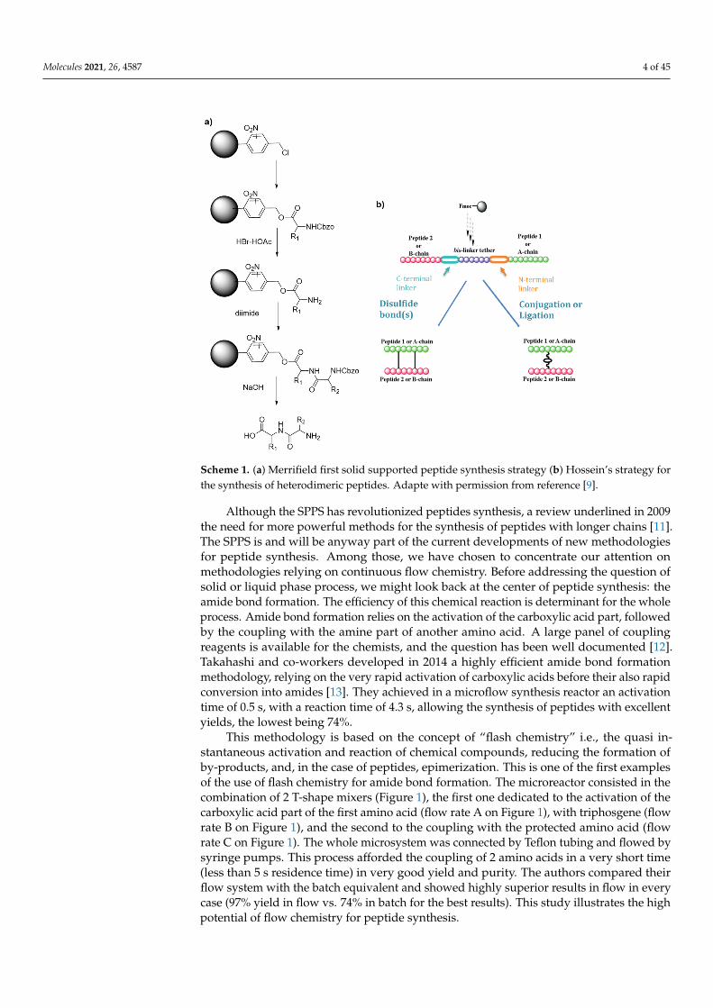
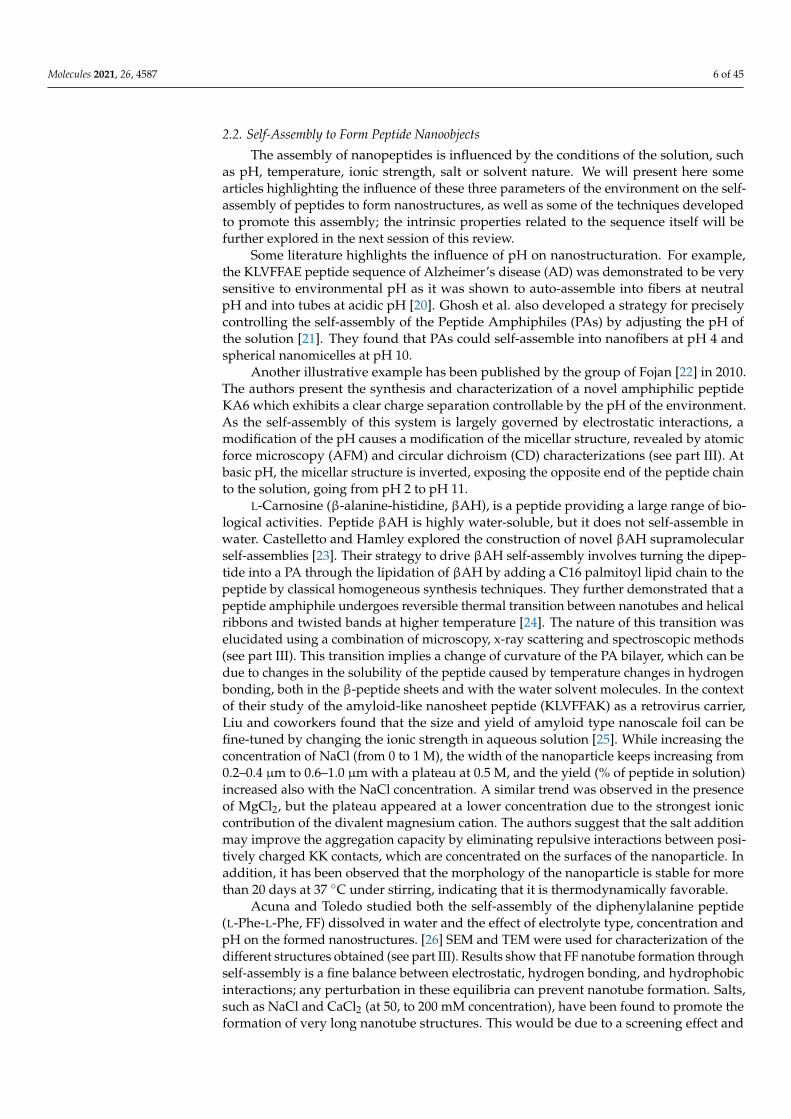
Besides the control of the external environmental parameters influencing peptideauto-assembly, we can underline some synthesis protocols and methodological tools whichcan be used to drive on-line self-assembly of peptides. The group of Park in 2008 reported anovel solid-phase growth of crystalline peptide nanowires at high temperatures driven byaniline vapor under anhydrous conditions [27]. For this study, an amorphous peptide thinfilm was prepared by drying a drop of 1,1,1,3,3,3-hexafluoro-2-propanol (HFIP) solutioncontaining diphenylalanine on a Silica substrate. Since water vapor could modify thesurface structure of the peptidic thin film the experiments were conducted under anhydrousconditions (vacuum dessicator). From the amorphous peptide film, the authors were ableto grow vertically well-aligned peptide nanowires by aging the film at temperatures above100 ◦C with aniline vapor (Figure 2). The specific influence of the aniline vapor was studiedby aging the film at different temperatures, with or without aniline vapor.
Molecules 2021, 26, x FOR PEER REVIEW 7 of 48
observed in the presence of MgCl2, but the plateau appeared at a lower concentration due to the strongest ionic contribution of the divalent magnesium cation. The authors suggest that the salt addition may improve the aggregation capacity by eliminating repulsive in-teractions between positively charged KK contacts, which are concentrated on the surfaces of the nanoparticle. In addition, it has been observed that the morphology of the nanopar-ticle is stable for more than 20 days at 37 °C under stirring, indicating that it is thermody-namically favorable.
Acuna and Toledo studied both the self-assembly of the diphenylalanine peptide (L-Phe-L-Phe, FF) dissolved in water and the effect of electrolyte type, concentration and pH on the formed nanostructures. [26] SEM and TEM were used for characterization of the different structures obtained (see part III). Results show that FF nanotube formation through self-assembly is a fine balance between electrostatic, hydrogen bonding, and hy-drophobic interactions; any perturbation in these equilibria can prevent nanotube for-mation. Salts, such as NaCl and CaCl2 (at 50, to 200 mM concentration), have been found to promote the formation of very long nanotube structures. This would be due to a screen-ing effect and the fact that cations are layout-forming and stimulate hydrophobic interac-tions; therefore, nanotube assembly occurs and also benefits electrostatic interactions, hy-drogen bonds, and longer nanotubes. The presence of AlCl3 produces an imbalance in the electrostatic interactions and hydrogen bonding because of excess Cl−, a structure-break-ing anion that impedes the nanostructure formation.
Besides the control of the external environmental parameters influencing peptide auto-assembly, we can underline some synthesis protocols and methodological tools which can be used to drive on-line self-assembly of peptides. The group of Park in 2008 reported a novel solid-phase growth of crystalline peptide nanowires at high tempera-tures driven by aniline vapor under anhydrous conditions [27]. For this study, an amor-phous peptide thin film was prepared by drying a drop of 1,1,1,3,3,3-hexafluoro-2-propa-nol (HFIP) solution containing diphenylalanine on a Silica substrate. Since water vapor could modify the surface structure of the peptidic thin film the experiments were con-ducted under anhydrous conditions (vacuum dessicator). From the amorphous peptide film, the authors were able to grow vertically well-aligned peptide nanowires by aging the film at temperatures above 100 °C with aniline vapor (Figure 2). The specific influence of the aniline vapor was studied by aging the film at different temperatures, with or with-out aniline vapor.
Figure 2. Peptidic nanowires vertically grown under aniline vapors. With permission from Reference [27].
At 50 °C, no change in the film was observed in the absence of aniline, whereas thick nanorods were formed in the presence of aniline. At temperatures of 100 and 150 °C, and with or without aniline vapour, one-dimensional nanostructures were formed, but with different shapes: while the high-temperature aniline vapor aging resulted in the formation of uniform and well-aligned peptide nanowires, dry air aging without aniline at the high temperatures promoted the growth of highly flexible nanofibrils with an irregular shape,
Figure 2. Peptidic nanowires vertically grown under aniline vapors. With permission fromReference [27].
At 50 ◦C, no change in the film was observed in the absence of aniline, whereas thicknanorods were formed in the presence of aniline. At temperatures of 100 and 150 ◦C, andwith or without aniline vapour, one-dimensional nanostructures were formed, but withdifferent shapes: while the high-temperature aniline vapor aging resulted in the formationof uniform and well-aligned peptide nanowires, dry air aging without aniline at the hightemperatures promoted the growth of highly flexible nanofibrils with an irregular shape,illustrating the crucial role of aniline vapors in this study. According to the authors, thisrole relies on the presence of the amine part of aniline, which can be a hydrogen-bonddonor. This hypothesis might require further experimentation to be validated, but it seemsplausible since toluene and benzene vapors did not show any change due to the amorphousFF film.
More recently, Yan and co-workers explored the role of trace solvent in the dipeptideself-assembly [28]. In this work, they discovered that a trace amount of solvent may be adominant factor for directing and mediating self-assembly of FF. The FF/dichloromethane(CH2Cl2) solution (from Commercial FF) was selected as a model, and compared to threeother types of solvents. Type I solvents (such as ethanol, DMF and acetone) have hydrogen-bonding interactions with FF. Type II solvents (toluene) can lead to possible π-π interactionswith FF. Type III solvents (n-hexane) can generate van der Waals interactions with FF. Theoptical microscopy images of samples in solution showed that FF underwent crystallizationin pure CH2Cl2, whereas gelling occurred when a trace amount of hydrogen-bond-formingsolvent (type I) was added in CH2Cl2. Therefore, in pure CH2Cl2, crystallization wasfavored with the growth of FF into each dimension at a comparable rate. When hydrogen-
Molecules 2021, 26, 4587 8 of 45
bond-forming solvents were added to CH2Cl2, directional hydrogen bonding would drivethe assembly of FF molecules in one dimension and resulted in the formation of fibers (inethanol) or even ribbon structures (DMF or acetone). On the contrary, the addition of atrace amount of toluene and n-hexane did not promote the formation of fiber structures.These results highlight the key role of hydrogen bonding in the formation of fibers, whichcan be tuned by controlling solvent composition.
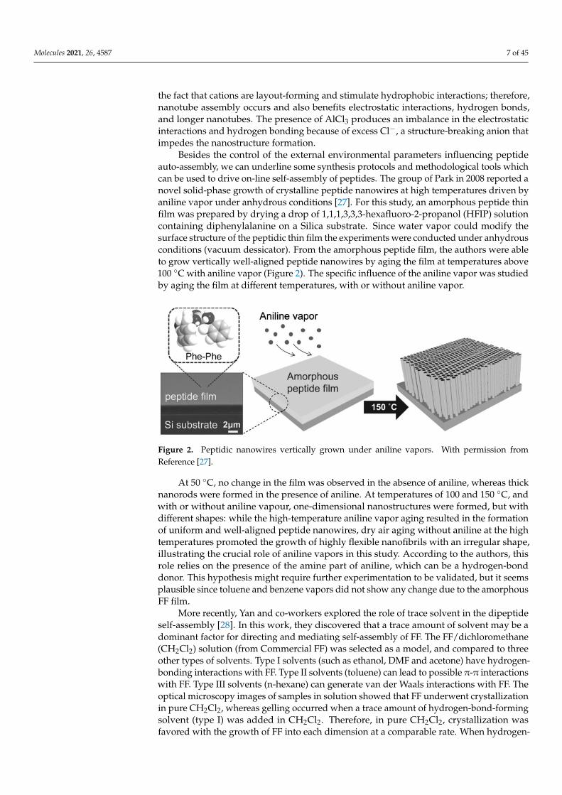
Some articles present synthetic processes to promote specific self-assembly, withthe help of photochemistry light-impulsion or enzymatic stimulators. Inspired by theswitchable structures in biomolecules, Stupp and his group have investigated the synthesisof photoresponsive PAs, well-known to self-assemble into supramolecular nanofiber [29].They reported the discovery of a quadruple helical fiber formed by photoresponsive PA 1(Palmitoyl tail-nitrobenzoyl group-GV3A3E3.) and its conversion into single fibers uponphotochemical cleavage of the 2-nitrobenzyl group in 1 (Scheme 2).
Molecules 2021, 26, x FOR PEER REVIEW 8 of 48
illustrating the crucial role of aniline vapors in this study. According to the authors, this role relies on the presence of the amine part of aniline, which can be a hydrogen-bond donor. This hypothesis might require further experimentation to be validated, but it seems plausible since toluene and benzene vapors did not show any change due to the amor-phous FF film.
More recently, Yan and co-workers explored the role of trace solvent in the dipeptide self-assembly [28]. In this work, they discovered that a trace amount of solvent may be a dominant factor for directing and mediating self-assembly of FF. The FF/dichloromethane (CH2Cl2) solution (from Commercial FF) was selected as a model, and compared to three other types of solvents. Type I solvents (such as ethanol, DMF and acetone) have hydro-gen-bonding interactions with FF. Type II solvents (toluene) can lead to possible π-π in-teractions with FF. Type III solvents (n-hexane) can generate van der Waals interactions with FF. The optical microscopy images of samples in solution showed that FF underwent crystallization in pure CH2Cl2, whereas gelling occurred when a trace amount of hydro-gen-bond-forming solvent (type I) was added in CH2Cl2. Therefore, in pure CH2Cl2, crys-tallization was favored with the growth of FF into each dimension at a comparable rate. When hydrogen-bond-forming solvents were added to CH2Cl2, directional hydrogen bonding would drive the assembly of FF molecules in one dimension and resulted in the formation of fibers (in ethanol) or even ribbon structures (DMF or acetone). On the con-trary, the addition of a trace amount of toluene and n-hexane did not promote the for-mation of fiber structures. These results highlight the key role of hydrogen bonding in the formation of fibers, which can be tuned by controlling solvent composition.
Some articles present synthetic processes to promote specific self-assembly, with the help of photochemistry light-impulsion or enzymatic stimulators. Inspired by the switch-able structures in biomolecules, Stupp and his group have investigated the synthesis of photoresponsive PAs, well-known to self-assemble into supramolecular nanofiber [29]. They reported the discovery of a quadruple helical fiber formed by photoresponsive PA 1 (Palmitoyl tail-nitrobenzoyl group-GV3A3E3.) and its conversion into single fibers upon photochemical cleavage of the 2-nitrobenzyl group in 1 (Scheme 2).
Scheme 2. Photochemical cleavage of 2-nitrobenzoyl group in PA 1. Reprinted with permission from Reference [29].
The amphiphilic structure of PA1 is expected to promote self-assembly into cylindri-cal nanofibers. The nitrogen of the N-terminal amide of PA1 has a 2-nitrobenzyl group that can be cleaved by irradiation at 350 nm to afford PA2. The lack of hydrogen bonding on the amide closest to the alkyl chain and the bulkiness of the 2-nitrobenzyl group made the authors expecting that PA1 and PA2 would differ in their supramolecular architecture after self-assembly. A transmission electron microscopy (TEM) image of one of the supra-
Scheme 2. Photochemical cleavage of 2-nitrobenzoyl group in PA 1. Reprinted with permission fromReference [29].
The amphiphilic structure of PA1 is expected to promote self-assembly into cylindricalnanofibers. The nitrogen of the N-terminal amide of PA1 has a 2-nitrobenzyl group thatcan be cleaved by irradiation at 350 nm to afford PA2. The lack of hydrogen bonding onthe amide closest to the alkyl chain and the bulkiness of the 2-nitrobenzyl group madethe authors expecting that PA1 and PA2 would differ in their supramolecular architec-ture after self-assembly. A transmission electron microscopy (TEM) image of one of thesupramolecular structures revealed a quadruple helix, which was previously very scarcelydescribed. Interestingly, after a 5-min irradiation of PA1, the helical structures disappearedcompletely in the TEM images, and only cylindrical fibrils with a diameter of 11 nm wereobserved. In matrix-assisted laser desorption ionization-time of flight-mass spectrometry(MALDI-TOFMS) spectrometry, the signals corresponding to PA2 were clearly observed af-ter photoirradiation and high performance liquid chromatography (HPLC) showed nearlycomplete conversion from PA1 to PA2.
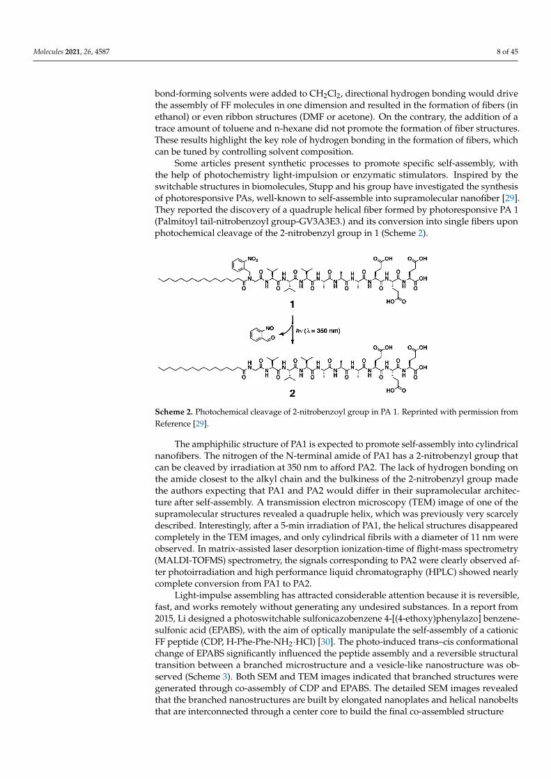
Light-impulse assembling has attracted considerable attention because it is reversible,fast, and works remotely without generating any undesired substances. In a report from2015, Li designed a photoswitchable sulfonicazobenzene 4-[(4-ethoxy)phenylazo] benzene-sulfonic acid (EPABS), with the aim of optically manipulate the self-assembly of a cationicFF peptide (CDP, H-Phe-Phe-NH2·HCl) [30]. The photo-induced trans–cis conformationalchange of EPABS significantly influenced the peptide assembly and a reversible structuraltransition between a branched microstructure and a vesicle-like nanostructure was ob-served (Scheme 3). Both SEM and TEM images indicated that branched structures weregenerated through co-assembly of CDP and EPABS. The detailed SEM images revealedthat the branched nanostructures are built by elongated nanoplates and helical nanobeltsthat are interconnected through a center core to build the final co-assembled structure
Molecules 2021, 26, 4587 9 of 45
Molecules 2021, 26, x FOR PEER REVIEW 9 of 48
molecular structures revealed a quadruple helix, which was previously very scarcely de-scribed. Interestingly, after a 5-min irradiation of PA1, the helical structures disappeared completely in the TEM images, and only cylindrical fibrils with a diameter of 11 nm were observed. In matrix-assisted laser desorption ionization-time of flight-mass spectrometry (MALDI-TOFMS) spectrometry, the signals corresponding to PA2 were clearly observed after photoirradiation and high performance liquid chromatography (HPLC) showed nearly complete conversion from PA1 to PA2.
Light-impulse assembling has attracted considerable attention because it is reversi-ble, fast, and works remotely without generating any undesired substances. In a report from 2015, Li designed a photoswitchable sulfonicazobenzene 4-[(4-ethoxy)phenylazo] benzenesulfonic acid (EPABS), with the aim of optically manipulate the self-assembly of a cationic FF peptide (CDP, H-Phe-Phe-NH2 ·HCl) [30]. The photo-induced trans–cis con-formational change of EPABS significantly influenced the peptide assembly and a reversi-ble structural transition between a branched microstructure and a vesicle-like nanostruc-ture was observed (Scheme 3). Both SEM and TEM images indicated that branched struc-tures were generated through co-assembly of CDP and EPABS. The detailed SEM images revealed that the branched nanostructures are built by elongated nanoplates and helical nanobelts that are interconnected through a center core to build the final co-assembled structure
Scheme 3. Photoswitchable system for auto-assembly of CDP. Reprinted with permission from Ref-erence [30].
The authors proposed a possible mechanism of the assembly and transformation pro-cess. Before ultra-violet (UV) illumination, trans-EPABS is shown to be inserted into the CDP molecular arrangement thanks to electrostatic and π-π interactions. According to Fourier-Transform Infra Red (FTIR) measurements, the aromatic rings of EPABS overlap with the CDP aromatic rings, leading to the formation of branched structures (on the left on Scheme 3). After being irradiated by UV light, trans-EPABS in the branched structure is gradually transformed into cis-EPABS. The higher hydrophilic property and steric hin-drance of cis-EPABS may conduct its leaving from the branched structures, signaling a disassembly of the structure. Free CDP molecules undertake a self-assembly procedure to form vesicle-like edifices. When this system is then exposed to visible light, EPABS would return to its trans-form, leading to the previous co-assembled branched structures. Other external factors have been demonstrated to promote peptide self-assemblies, like enzy-matic stimulators [31] but we have chosen not to detail those in this review.
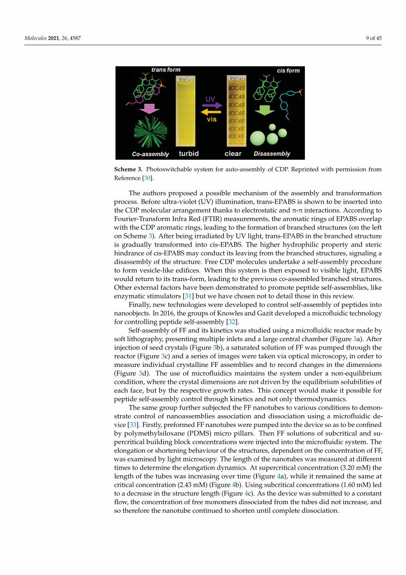
Finally, new technologies were developed to control self-assembly of peptides into nanoobjects. In 2016, the groups of Knowles and Gazit developed a microfluidic technol-ogy for controlling peptide self-assembly [32].
Self-assembly of FF and its kinetics was studied using a microfluidic reactor made by soft lithography, presenting multiple inlets and a large central chamber (Figure 3a). After injection of seed crystals (Figure 3b), a saturated solution of FF was pumped through the reactor (Figure 3c) and a series of images were taken via optical microscopy, in order to
Scheme 3. Photoswitchable system for auto-assembly of CDP. Reprinted with permission fromReference [30].
The authors proposed a possible mechanism of the assembly and transformationprocess. Before ultra-violet (UV) illumination, trans-EPABS is shown to be inserted intothe CDP molecular arrangement thanks to electrostatic and π-π interactions. According toFourier-Transform Infra Red (FTIR) measurements, the aromatic rings of EPABS overlapwith the CDP aromatic rings, leading to the formation of branched structures (on the lefton Scheme 3). After being irradiated by UV light, trans-EPABS in the branched structureis gradually transformed into cis-EPABS. The higher hydrophilic property and sterichindrance of cis-EPABS may conduct its leaving from the branched structures, signaling adisassembly of the structure. Free CDP molecules undertake a self-assembly procedureto form vesicle-like edifices. When this system is then exposed to visible light, EPABSwould return to its trans-form, leading to the previous co-assembled branched structures.Other external factors have been demonstrated to promote peptide self-assemblies, likeenzymatic stimulators [31] but we have chosen not to detail those in this review.
Finally, new technologies were developed to control self-assembly of peptides intonanoobjects. In 2016, the groups of Knowles and Gazit developed a microfluidic technologyfor controlling peptide self-assembly [32].
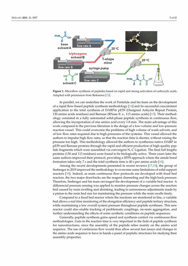
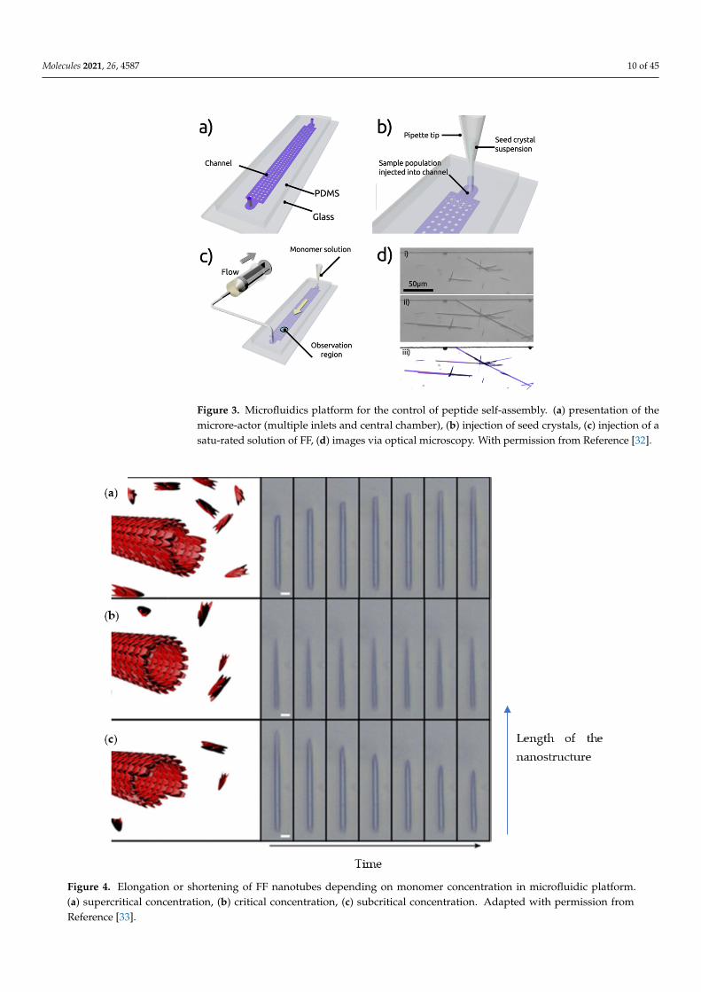
Self-assembly of FF and its kinetics was studied using a microfluidic reactor made bysoft lithography, presenting multiple inlets and a large central chamber (Figure 3a). Afterinjection of seed crystals (Figure 3b), a saturated solution of FF was pumped through thereactor (Figure 3c) and a series of images were taken via optical microscopy, in order tomeasure individual crystalline FF assemblies and to record changes in the dimensions(Figure 3d). The use of microfluidics maintains the system under a non-equilibriumcondition, where the crystal dimensions are not driven by the equilibrium solubilities ofeach face, but by the respective growth rates. This concept would make it possible forpeptide self-assembly control through kinetics and not only thermodynamics.
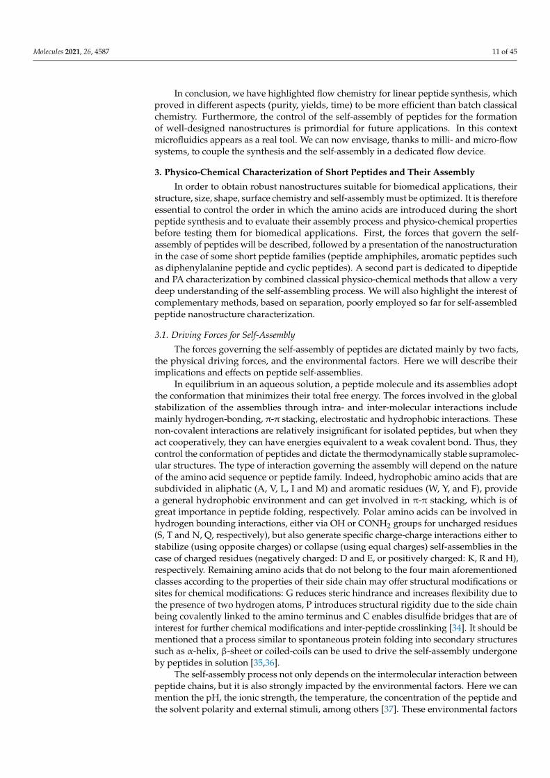
The same group further subjected the FF nanotubes to various conditions to demon-strate control of nanoassemblies association and dissociation using a microfluidic de-vice [33]. Firstly, preformed FF nanotubes were pumped into the device so as to be confinedby polymethylsiloxane (PDMS) micro pillars. Then FF solutions of subcritical and su-percritical building block concentrations were injected into the microfluidic system. Theelongation or shortening behaviour of the structures, dependent on the concentration of FF,was examined by light microscopy. The length of the nanotubes was measured at differenttimes to determine the elongation dynamics. At supercritical concentration (3.20 mM) thelength of the tubes was increasing over time (Figure 4a), while it remained the same atcritical concentration (2.43 mM) (Figure 4b). Using subcritical concentrations (1.60 mM) ledto a decrease in the structure length (Figure 4c). As the device was submitted to a constantflow, the concentration of free monomers dissociated from the tubes did not increase, andso therefore the nanotube continued to shorten until complete dissociation.
Molecules 2021, 26, 4587 10 of 45
Molecules 2021, 26, x FOR PEER REVIEW 10 of 48
measure individual crystalline FF assemblies and to record changes in the dimensions (Figure 3d). The use of microfluidics maintains the system under a non-equilibrium con-dition, where the crystal dimensions are not driven by the equilibrium solubilities of each face, but by the respective growth rates. This concept would make it possible for peptide self-assembly control through kinetics and not only thermodynamics.
Figure 3. Microfluidics platform for the control of peptide self-assembly. (a) presentation of the mi-crore-actor (multiple inlets and central chamber), (b) injection of seed crystals, (c) injection of a satu-rated solution of FF, (d) images via optical microscopy. With permission from Reference [32].
The same group further subjected the FF nanotubes to various conditions to demon-strate control of nanoassemblies association and dissociation using a microfluidic device [33]. Firstly, preformed FF nanotubes were pumped into the device so as to be confined by polymethylsiloxane (PDMS) micro pillars. Then FF solutions of subcritical and super-critical building block concentrations were injected into the microfluidic system. The elon-gation or shortening behaviour of the structures, dependent on the concentration of FF, was examined by light microscopy. The length of the nanotubes was measured at different times to determine the elongation dynamics. At supercritical concentration (3.20 mM) the length of the tubes was increasing over time (Figure 4a), while it remained the same at critical concentration (2.43 mM) (Figure 4b). Using subcritical concentrations (1.60 mM) led to a decrease in the structure length (Figure 4c). As the device was submitted to a constant flow, the concentration of free monomers dissociated from the tubes did not in-crease, and so therefore the nanotube continued to shorten until complete dissociation.
Figure 3. Microfluidics platform for the control of peptide self-assembly. (a) presentation of themicrore-actor (multiple inlets and central chamber), (b) injection of seed crystals, (c) injection of asatu-rated solution of FF, (d) images via optical microscopy. With permission from Reference [32].
Molecules 2021, 26, x FOR PEER REVIEW 11 of 48
Figure 4. Elongation or shortening of FF nanotubes depending on monomer concentration in microfluidic platform. (a) supercritical concentration, (b) critical concentration, (c) subcritical concentration. Adapted with permission from Refer-ence [33].
In conclusion, we have highlighted flow chemistry for linear peptide synthesis, which proved in different aspects (purity, yields, time) to be more efficient than batch classical chemistry. Furthermore, the control of the self-assembly of peptides for the for-mation of well-designed nanostructures is primordial for future applications. In this con-text microfluidics appears as a real tool. We can now envisage, thanks to milli- and micro-flow systems, to couple the synthesis and the self-assembly in a dedicated flow device.
3. Physico-Chemical Characterization of Short Peptides and Their Assembly In order to obtain robust nanostructures suitable for biomedical applications, their
structure, size, shape, surface chemistry and self-assembly must be optimized. It is there-fore essential to control the order in which the amino acids are introduced during the short peptide synthesis and to evaluate their assembly process and physico-chemical properties before testing them for biomedical applications. First, the forces that govern the self-as-sembly of peptides will be described, followed by a presentation of the nanostructuration in the case of some short peptide families (peptide amphiphiles, aromatic peptides such as diphenylalanine peptide and cyclic peptides). A second part is dedicated to dipeptide and PA characterization by combined classical physico-chemical methods that allow a very deep understanding of the self-assembling process. We will also highlight the inter-est of complementary methods, based on separation, poorly employed so far for self-as-sembled peptide nanostructure characterization.
Figure 4. Elongation or shortening of FF nanotubes depending on monomer concentration in microfluidic platform.(a) supercritical concentration, (b) critical concentration, (c) subcritical concentration. Adapted with permission fromReference [33].
Molecules 2021, 26, 4587 11 of 45
In conclusion, we have highlighted flow chemistry for linear peptide synthesis, whichproved in different aspects (purity, yields, time) to be more efficient than batch classicalchemistry. Furthermore, the control of the self-assembly of peptides for the formationof well-designed nanostructures is primordial for future applications. In this contextmicrofluidics appears as a real tool. We can now envisage, thanks to milli- and micro-flowsystems, to couple the synthesis and the self-assembly in a dedicated flow device.
3. Physico-Chemical Characterization of Short Peptides and Their Assembly
In order to obtain robust nanostructures suitable for biomedical applications, theirstructure, size, shape, surface chemistry and self-assembly must be optimized. It is thereforeessential to control the order in which the amino acids are introduced during the shortpeptide synthesis and to evaluate their assembly process and physico-chemical propertiesbefore testing them for biomedical applications. First, the forces that govern the self-assembly of peptides will be described, followed by a presentation of the nanostructurationin the case of some short peptide families (peptide amphiphiles, aromatic peptides suchas diphenylalanine peptide and cyclic peptides). A second part is dedicated to dipeptideand PA characterization by combined classical physico-chemical methods that allow a verydeep understanding of the self-assembling process. We will also highlight the interest ofcomplementary methods, based on separation, poorly employed so far for self-assembledpeptide nanostructure characterization.
3.1. Driving Forces for Self-Assembly
The forces governing the self-assembly of peptides are dictated mainly by two facts,the physical driving forces, and the environmental factors. Here we will describe theirimplications and effects on peptide self-assemblies.
In equilibrium in an aqueous solution, a peptide molecule and its assemblies adoptthe conformation that minimizes their total free energy. The forces involved in the globalstabilization of the assemblies through intra- and inter-molecular interactions includemainly hydrogen-bonding, π-π stacking, electrostatic and hydrophobic interactions. Thesenon-covalent interactions are relatively insignificant for isolated peptides, but when theyact cooperatively, they can have energies equivalent to a weak covalent bond. Thus, theycontrol the conformation of peptides and dictate the thermodynamically stable supramolec-ular structures. The type of interaction governing the assembly will depend on the natureof the amino acid sequence or peptide family. Indeed, hydrophobic amino acids that aresubdivided in aliphatic (A, V, L, I and M) and aromatic residues (W, Y, and F), providea general hydrophobic environment and can get involved in π-π stacking, which is ofgreat importance in peptide folding, respectively. Polar amino acids can be involved inhydrogen bounding interactions, either via OH or CONH2 groups for uncharged residues(S, T and N, Q, respectively), but also generate specific charge-charge interactions either tostabilize (using opposite charges) or collapse (using equal charges) self-assemblies in thecase of charged residues (negatively charged: D and E, or positively charged: K, R and H),respectively. Remaining amino acids that do not belong to the four main aforementionedclasses according to the properties of their side chain may offer structural modifications orsites for chemical modifications: G reduces steric hindrance and increases flexibility due tothe presence of two hydrogen atoms, P introduces structural rigidity due to the side chainbeing covalently linked to the amino terminus and C enables disulfide bridges that are ofinterest for further chemical modifications and inter-peptide crosslinking [34]. It should bementioned that a process similar to spontaneous protein folding into secondary structuressuch as α-helix, β-sheet or coiled-coils can be used to drive the self-assembly undergoneby peptides in solution [35,36].
The self-assembly process not only depends on the intermolecular interaction betweenpeptide chains, but it is also strongly impacted by the environmental factors. Here we canmention the pH, the ionic strength, the temperature, the concentration of the peptide andthe solvent polarity and external stimuli, among others [37]. These environmental factors
Molecules 2021, 26, 4587 12 of 45
can affect the equilibria between different structures, or even inside the same structure, thefinal morphology (e.g., compactness) by altering the interaction forces between peptidesand thus triggering a change in the structural conformation of those peptides. In general,changes in the pH and ionic strength of the solubilization medium will alter the electro-static interactions between side chains and/or between peptides and solvent, whereasmodifications in the peptide concentration or solvent nature will predominantly affect Vander Waals and hydrophobic interactions established between self-assembled structures.Several authors have observed the structural changes induced by the pH and played with itto favor the desired structure or to render certain properties to the supramolecular structurewith the aim to use it as drug delivery systems. For example, Versluis and coworkers [38]reported the reversible sphere-to-fiber transition of vesicles composed of amphiphilicβ-cyclodextrin (β-CD) decorated with adamantane modified octapeptides (LELELELE). Inthis two-component system three orthogonal interactions are combined: (i) hydrophobicinteractions in the cyclodextrin vesicle bilayers; (ii) inclusion complex formation of β-CDand adamantane; (iii) hydrogen bonding in β-sheet domains composed of octapeptide.Using the reversible secondary structure transitions of the octapeptide from random coil toβ-sheet domains as a function of pH, a reversible morphological change of the supramolec-ular complex from vesicles to fibers occurred, thus allowing for a pH-triggered release ofthe encapsulated contents.
Another important parameter that governs the assembly process is the amino acid se-quence and the peptide concentration [39]. Thus, Bowerman [40] and coworkers attemptedto elucidate the influence of aromatic amino acids on peptide self-assembly processes. TheAc-(XKXK)2-NH2 peptide was used to elucidate the relative contributions of π-π versusgeneral hydrophobic interactions to peptide self-assembly in aqueous solutions. Position Xwas standing for either V, I, F, pentafluoro-F or cyclohexyl-A. At low pH, these peptidesremained monomeric because of repulsive forces between protonated K residues. Increas-ing the solution ionic strength to shield repulsive charge-charge interactions facilitatedcross-β fibril formation. As peptide hydrophobicity increased, the required ionic strengthto induce self-assembly decreased. Thus, the V sequence failed to assemble in NaCl, what-ever the ionic strength was in the range 0 to 1000 mM, whereas β-sheet formation forpentafluoro-F and cyclohexyl-A sequences was observed at only 20 and 60 mM NaCl,respectively. While self-assembly propensity was correlated to peptide hydrophobicity, thepresence of aromatic amino acids impacted on the properties of self-assembled structures.Nonaromatic peptides formed fibrils of 3–15 nm in diameter, whereas aromatic peptidesformed nanotape or nanoribbon architectures of 3–7 nm widths. In addition, all peptidesformed fibrillar hydrogels at sufficient peptide concentrations (8 mM in NaCl solutions ofadequate ionic strength to promote prior self-assembly), but nonaromatic peptides formedweak gels, whereas aromatic peptides formed rigid gels.
Eventually, the time left for the peptide to self-assemble is crucial in view of theresulting nanostructure. In this context, Bourbo et al. [41] evidenced that PAs presentinga β-sheet secondary structure self-assembled as spherical micelle-like aggregates whilesonicated for 30 min, whereas they rearranged into fibril helical structures when thissonication time increased up to 2 h.
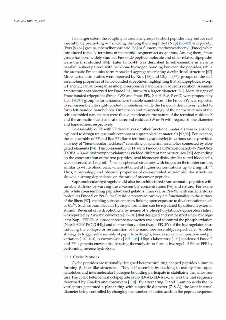
As mentioned above, peptides self-assemble under mild conditions, into a diverserange of nanostructures that are the result of an intricate and cooperative balance betweendifferent intermolecular interactions and environmental factors. Thus, linear peptidesand their derivatives often self-assemble into nanostructures, such as nanofibers, nanorib-bons, nanotubes, or vesicles; cyclic peptides normally stack into nanotubes and branchedpeptides often form micelles or vesicle [42,43]. The final sizes of these self-assembliesvary greatly, from a few nanometers to hundreds of microns and the latter nanostructuresoften template a hierarchy at larger length scales. Among self-assembling peptide systems(Figure 5), we will focus in the following section on short synthetic peptide precursors togive a deeper insight into their engineering strategy, as well as derived nanostructures tillthe hydrogel state for their interest in nanomedicine.
Molecules 2021, 26, 4587 13 of 45
3.2. Short Peptide Families (Self-Assembling Peptides)—Self Assembling Structures3.2.1. Peptide Amphiphiles (PAs)
PAs are among the most studied self-assembling entities of peptidic nature. These self-assembling peptides have lipid- or surfactant-like characteristics. Their design (Figure 5A)consists in (i) a hydrophobic tail that is composed of nonpolar amino acid residues [44]or aliphatic alkyl C12-C16 chains [45] or a combination of both, (ii) a linking region thatis a short peptide sequence capable of forming intermolecular hydrogen bonding, (iii) ahydrophilic head that contains charged amino acids [44]. for enhanced solubility in waterbut also for the design of pH and salt-responsive nanostructures and networks, and even-tually (iv) a bioactive peptide epitope that can interact with cells [46]. In aqueous medium,hydrophobic interaction is the main driving force governing the self-assembly of nanoar-chitectures thus resulting in the sequestration of non-polar groups to form hydrophobiccavities/cores. Hydrophilic end groups are assembled through hydrogen bonding. Thebalance between hydrophobic interaction and hydrogen bonding drives the self-assemblyof peptide amphiphiles. Eventually, electrostatic interactions can enhance the stability ofthe overall structure when combining PAs of opposite charges [47]. Thus, by rationallycontrolling the amino acid sequence length and composition, specific nanostructures canbe obtained on going from nanofibers and nanoribbons to micelles and vesicles [48,49].
Considering PAs with only amino acids, the tails are normally composed of non-polar amino acid residues. Peptide containing A and V tails were reported to form morehomogeneous and stable structures than those made of G, L and I. Such lipid-like peptidescan readily self-assemble into dynamic tubular and vesicle structures [50]. The effectof molecular structures was evidenced more deeply on the AmK (m = 3, 6 and 9) serie,reporting [51] a decreasing critical aggregation concentration (CAC, down to 15 µM forA9K) when increasing the length of the hydrophobic region. The size and shape of theself-assembled nanostructures also changed from unstable plate-like structures (A3K) tolong nanofibers with uniform diameter of 8 nm (A6K) and short nanorods with diameterof 3 nm and length of 100 nm (A9K). Increased nanostructure polydispersity was alsoreported when increasing the tail length of the Glycine rich amphiphilic peptide GnD2(n = 4 to 10) [52].
Considering PAs engineered with alkyl tail, a minimum of a ten-carbon long aliphaticresidue is necessary to induce the hydrophobic effect to cause self-assembly [53]. A dialkyltail was also reported with two palmitic (C16) chains conjugated to a peptide promotingthe self-assembly into cylindrical micelles 8.0 ± 2.3 nm in diameter at 2.2 µM CAC [54].
The composition of the peptide segment directly following the hydrophobic region ofPA molecules can control the propensity to intermolecular β-sheet hydrogen bonding andthus impact peptide self-assembly and final nanostructuration. Paramonov et al. foundthat disruption of these hydrogen bonds eliminated the ability of a PA to form elongatedcylindrical nanostructures and resulted in spherical micelles [55]. When distressing thehydrogen bonding capacity by alternating hydrophobic and hydrophilic amino acids, anoriginal nanobelt flat architecture was also evidenced [56]. Moreover, the propensity toform β-sheet secondary structures influence the stiffness of nanofiber networks. Stuppand coworkers showed that (i) both the number and fraction of valine residues are es-pecially effective at raising mechanical stiffness whereas alanine residues tend to reduceit [57] and that (ii) the extent of internal order depends on the molecular architecture andpeptide sequence of PAs, with branched PAs yielding nanofibers with the lowest degreeof internal order [58]. Thus, they further designed stiff nanofibers (Figure 5A) resultingfrom the ordered arrangement of PA molecules incorporating V and A residues, whilethe combination of A and G residues resulted in “weaker β-sheet region” promoting softnanofibers in which PA molecules were more disordered [59].
Amino acids further away from the hydrophobic region play a less important role instabilizing the supramolecular nanostructure. Thus, functional groups or peptide sequenceswith specific biological activity in view of specific applications may be introduced withminimal risk of modifying the self-assembly properties of PAs. An extensive variety of
Molecules 2021, 26, 4587 14 of 45
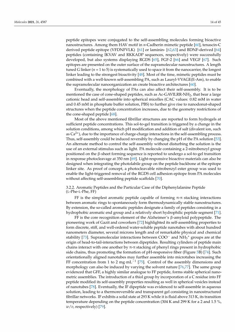
peptide epitopes were conjugated to the self-assembling molecules forming bioactivenanostructures. Among them HAV motif in n-Cadherin mimetic peptide [60], tenascin-Cderived peptide epitope (VFDNFVLK) [61] or laminin- [62,63] and BDNF-derived [64]peptides (containing IKVAV and RKKADP sequences, respectively) were successfullydeveloped, but also systems displaying RGDS [65], FGF-2 [66] and VEGF [67]. Suchepitopes are presented on the outer surface of the supramolecular nanostructures. A lengthtuned G linker (n = 1 to 5) is systematically used to space it from the nanocarrier, the longestlinker leading to the strongest bioactivity [48]. Most of the time, mimetic peptides must becombined with a well-known self-assembling PA, such as Lauryl-VVAGE(E-Am), to enablethe supramolecular nanoorganization an create bioactive architectures [60].
Eventually, the morphology of PAs can also affect their self-assembly. It is to bementioned the case of cone-shaped peptides, such as Ac-GAVILRR-NH2, that bear a largecationic head and self-assemble into spherical micelles (CAC values: 0.82 mM in waterand 0.45 mM in phosphate buffer solution, PBS) to further give rise to nanodonut-shapedstructures when the peptide concentration increases, due to the geometry restrictions ofthe cone-shaped peptide [68].
Most of the above mentioned fibrillar structures are reported to form hydrogels atsufficient peptide concentrations. This sol-to-gel transition is triggered by a change in thesolution conditions, among which pH modification and addition of salt (divalent ion, suchas Ca2+), due to the importance of charge-charge interactions in the self-assembling process.Thus, self-assembly could be induced reversibly by changing the pH of the PA solution [53].An alternate method to control the self-assembly without disturbing the solution is theuse of an external stimulus such as light. PA molecule containing a 2-nitrobenzyl grouppositioned on the β-sheet forming sequence is reported to undergo a sol-to-gel transitionin response photocleavage at 350 nm [69]. Light-responsive bioactive materials can also bedesigned when integrating the photolabile group on the peptide backbone at the epitopelinker site. As proof of concept, a photocleavable nitrobenzyl ester group was used toenable the light-triggered removal of the RGDS cell adhesion epitope from PA moleculeswithout affecting self-assembling peptide scaffolds [70].
3.2.2. Aromatic Peptides and the Particular Case of the Diphenylalanine Peptide(L-Phe-L-Phe, FF)
FF is the simplest aromatic peptide capable of forming π-π stacking interactionsbetween aromatic rings to spontaneously form thermodynamically stable nanostructures.By extension, the so-called aromatic peptides designate a family of peptides consisting in ahydrophobic aromatic end group and a relatively short hydrophilic peptide segment [71].
FF is the core recognition element of the Alzheimer’s β-amyloid polypeptide. Thepioneering work of Gazit and coworkers [72] highlighted its self-assembling properties toform discrete, stiff, and well-ordered water-soluble peptide nanotubes with about hundrednanometers diameter, several microns length and of remarkable physical and chemicalstability [73]. Supramolecular interactions between COO− and NH3
+ groups are at theorigin of head-to-tail-interactions between dipeptides. Resulting cylinders of peptide mainchains interact with one another by π-π stacking of phenyl rings present in hydrophobicside chains, thus promoting the formation of pH-responsive fiber (Figure 5B) [74]. Suchorientationally aligned nanotubes may further assemble into microtubes increasing theFF concentration from 1 to 2 mg·mL−1 [75]. Control of the assembly dimensions andmorphology can also be induced by varying the solvent nature [76,77]. The same groupevidenced that GFF, a highly similar analogue to FF peptide, forms stable spherical nano-metric assemblies. The introduction of a thiol group by incorporation of a C residue into FFpeptide modified its self-assembly properties resulting as well in spherical vesicles insteadof nanotubes [78]. Eventually, the IF dipeptide was evidenced to self-assemble in aqueoussolution, leading to a thermoreversible and transparent gel consisting in nanostructuredfibrillar networks. IF exhibits a solid state at 293 K while it is fluid above 313 K, its transitiontemperature depending on the peptide concentration (304 K and 299 K for a 2 and 1.5 %,w/v, respectively) [79].
Molecules 2021, 26, 4587 15 of 45
To a larger extent the coupling of aromatic groups to short peptides may induce self-assembly by promoting π-π stacking. Among them naphthyl (Nap) [80–82] and pyridyl(Pyr) [83,84] groups, phenylboronic acid [85] or fluorenylmethoxycarboenyl (Fmoc) whenintroduced to the N-terminus of the peptide segment act as gelators. Among them, Fmocgroup has been widely studied. Fmoc-LD peptide molecule and other related dipeptideswere the first studied [86]. Later Fmoc-FF was described to self-assemble in an anti-parallel β-sheet pattern with backbone hydrogen bonding between the peptides, whilethe aromatic Fmoc units form π-stacked aggregates creating a cylindrical structure [87].More systematic studies were reported by Xu’s [88] and Ulijn’s [87]. groups on the self-assembling properties of Fmoc-bonded dipeptides, highlighting that all dipeptides, exceptGT and GF, can auto-organize into pH-responsive nanofibers in aqueous solution. A similararchitecture was observed for Fmoc-LLL, but with a larger diameter [89]. More designs ofFmoc-bonded tripeptides (Fmoc-FWX and Fmoc-FFX, X = H, R, S, E or D) were proposed byHe’s [90,91] group to form handedness-tunable nanohelices. The Fmoc-FW was reportedto self-assemble into right-handed nanohelices, while the Fmoc-FF derivatives tended toform left-handed nanohelices. Dimension and morphology of the nanostructures of theself-assembled nanohelices were thus dependent on the nature of the terminal residues Xand the aromatic side chains at the second residues (W or F) with regards to the diameterand handedness, respectively.
Co-assembly of FF with FF derivatives or other functional materials was extensivelyexplored to design unique multicomponent supramolecular materials [92,93]. For instance,the co-assembly of FF and Boc-FF (Boc = tert-butoxycarbonyle) in various ratios provideda variety of “biomolecular necklaces” consisting of spherical assemblies connected by elon-gated elements [94]. The co-assembly of FF with Fmoc-L-DOPA(acetonated)-D-Phe-OMe(DOPA = 3,4-dihydroxyphenylalanine) yielded different nanostructures [95] dependingon the concentration of the two peptides: oval biconcave disks, similar to red blood cells,were observed at 1 mg·mL−1, while spherical structures with bulges on their outer surface,similar to white blood cells, where obtained at higher concentrations up to 2 mg·mL−1.Thus, morphology and physical properties of co-assembled supramolecular structuresshowed a strong dependence on the ratio of precursor peptides.
Supramolecular hydrogels could also be architectured from aromatic peptides withtunable stiffness by varying the co-assembly concentrations [96] and nature. For exam-ple, while co-assembling peptide-based gelators Fmoc-YL or Pyr-YL with surfactant-likemolecules Fmoc-S or Pyr-S, the S residue presented carboxylate functionality to the surfaceof the fibers [97], enabling subsequent cross-linking upon exposure to divalent cations suchas Ca2+. Such supramolecular hydrogel formation can be regulated by different externalstimuli. Reversal of hydrophobicity by means of Y phosphorylation/dephosphorylationwas reported by Xu’s and coworkers [98–101] that designed and synthesized a new hydroge-lator Nap−FFGEY. A kinase/phosphatase switch was used to control the phosphorylation(Nap-FFGEY-P(O)(OH)2) and dephosphorylation (Nap−FFGEY) of the hydrogelator, thusinducing the collapse or restauration of the nanofiber assembly, respectively. Anotherstrategy to trigger self-assembly of peptide hydrogels, besides solvent composition and pHvariation [102–104], is enzymolysis [105–108]. Ulijn’s laboratory [109] condensed Fmoc-Fand FF segments enzymatically using thermolysin to form a hydrogel of Fmoc-FFF byperforming reverse hydrolysis.
3.2.3. Cyclic Peptides
Cyclic peptides are rationally designed heterochiral ring-shaped peptides subunitsforming β-sheet-like structures. They self-assemble by stacking to mainly form opennanotubes and intermolecular hydrogen bounding participate in stabilizing the nanostruc-ture The cyclic heterochiral octapeptide cyclo [D-AL-ED-AL-Q)2] was the first sequencedescribed by Ghadiri and coworkers [110]. By alternating D and L-amino acids the in-vestigators generated a planar ring with a specific diameter (7–8 Å), the later internaldiameter being controlled by changing the number of amino acids in the peptide sequence
Molecules 2021, 26, 4587 16 of 45
(Figure 5C) [111]. The authors hypothesized that cyclic peptide having an even numberof alternating D-and L-amino acids could adopt a low-energy ring shaped flat conforma-tion, all backbone peptide amide groups being orientated perpendicular to the plane ofthe structure, and participate in backbone-backbone intermolecular hydrogen bondingto generate an adjacent antiparallel β-sheet structure [110]. From molecular modelingand experimental results cyclic octapeptides were found to present the optimum balancebetween a low ring strain structure and a flat ring-shaped conformational state. Fournew peptide nanotube solid-state ensembles where further described and characterized:cyclo [(L-QD-A)4-], cyclo [(L-QD-L)4-], and cyclo [(L-QD-F)4-] subunits giving rise tohighly ordered nanotube crystals, while cyclo [(L-QD-V)4-] only formed semicrystallineneedles [111].
Molecules 2021, 26, x FOR PEER REVIEW 17 of 48
Figure 5. (A) Molecular structure of PAs engineered with alkyl tail and scheme of its-self-assem-bling into stiff cylindrical nanofibers. Adapted with permission from reference [59] (B) Molecular structure of FF and model for the construction of hollow FF fibers. Adapted with permission from reference [74]. (C) Molecular structure of a cyclic octapeptide with alternat-ing D- and L-amino acids sequence and scheme of its-self-assembling into ordered parallel ar-rays of solid-state nano-tubes figuring antiparallel ring stacking and the presence of intermo-lecular hydrogen-bonding interactions. Reproduced with permission from Reference [111].
Ring stacking is favored at acidic pH, due to weakened intermolecular repulsive elec-trostatic interactions and strengthened attractive side chain/side chain hydrogen bonding. Ghadiri and coworkers exploited the ionization state of the carboxylic acid moiety in E residue side chain to trigger the phase transition toward self-assembled nanotube parti-
C
A
B
Figure 5. (A) Molecular structure of PAs engineered with alkyl tail and scheme of its-self-assemblinginto stiff cylindrical nanofibers. Adapted with permission from reference [59] (B) Molecular structureof FF and model for the construction of hollow FF fibers. Adapted with permission from refer-ence [74]. (C) Molecular structure of a cyclic octapeptide with alternat-ing D- and L-amino acidssequence and scheme of its-self-assembling into ordered parallel ar-rays of solid-state nanotubesfiguring antiparallel ring stacking and the presence of intermo-lecular hydrogen-bonding interactions.Reproduced with permission from Reference [111].
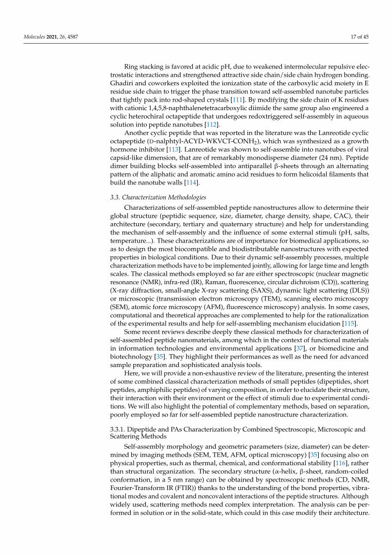
Molecules 2021, 26, 4587 17 of 45
Ring stacking is favored at acidic pH, due to weakened intermolecular repulsive elec-trostatic interactions and strengthened attractive side chain/side chain hydrogen bonding.Ghadiri and coworkers exploited the ionization state of the carboxylic acid moiety in Eresidue side chain to trigger the phase transition toward self-assembled nanotube particlesthat tightly pack into rod-shaped crystals [111]. By modifying the side chain of K residueswith cationic 1,4,5,8-naphthalenetetracarboxylic diimide the same group also engineered acyclic heterochiral octapeptide that undergoes redoxtriggered self-assembly in aqueoussolution into peptide nanotubes [112].
Another cyclic peptide that was reported in the literature was the Lanreotide cyclicoctapeptide (D-nalphtyl-ACYD-WKVCT-CONH2), which was synthesized as a growthhormone inhibitor [113]. Lanreotide was shown to self-assemble into nanotubes of viralcapsid-like dimension, that are of remarkably monodisperse diameter (24 nm). Peptidedimer building blocks self-assembled into antiparallel β-sheets through an alternatingpattern of the aliphatic and aromatic amino acid residues to form helicoidal filaments thatbuild the nanotube walls [114].
3.3. Characterization Methodologies
Characterizations of self-assembled peptide nanostructures allow to determine theirglobal structure (peptidic sequence, size, diameter, charge density, shape, CAC), theirarchitecture (secondary, tertiary and quaternary structure) and help for understandingthe mechanism of self-assembly and the influence of some external stimuli (pH, salts,temperature...). These characterizations are of importance for biomedical applications, soas to design the most biocompatible and biodistributable nanostructures with expectedproperties in biological conditions. Due to their dynamic self-assembly processes, multiplecharacterization methods have to be implemented jointly, allowing for large time and lengthscales. The classical methods employed so far are either spectroscopic (nuclear magneticresonance (NMR), infra-red (IR), Raman, fluorescence, circular dichroism (CD)), scattering(X-ray diffraction, small-angle X-ray scattering (SAXS), dynamic light scattering (DLS))or microscopic (transmission electron microscopy (TEM), scanning electro microscopy(SEM), atomic force microscopy (AFM), fluorescence microscopy) analysis. In some cases,computational and theoretical approaches are complemented to help for the rationalizationof the experimental results and help for self-assembling mechanism elucidation [115].
Some recent reviews describe deeply these classical methods for characterization ofself-assembled peptide nanomaterials, among which in the context of functional materialsin information technologies and environmental applications [37], or biomedicine andbiotechnology [35]. They highlight their performances as well as the need for advancedsample preparation and sophisticated analysis tools.
Here, we will provide a non-exhaustive review of the literature, presenting the interestof some combined classical characterization methods of small peptides (dipeptides, shortpeptides, amphiphilic peptides) of varying composition, in order to elucidate their structure,their interaction with their environment or the effect of stimuli due to experimental condi-tions. We will also highlight the potential of complementary methods, based on separation,poorly employed so far for self-assembled peptide nanostructure characterization.
3.3.1. Dipeptide and PAs Characterization by Combined Spectroscopic, Microscopic andScattering Methods
Self-assembly morphology and geometric parameters (size, diameter) can be deter-mined by imaging methods (SEM, TEM, AFM, optical microscopy) [35] focusing also onphysical properties, such as thermal, chemical, and conformational stability [116], ratherthan structural organization. The secondary structure (α-helix, β-sheet, random-coiledconformation, in a 5 nm range) can be obtained by spectroscopic methods (CD, NMR,Fourier-Transform IR (FTIR)) thanks to the understanding of the bond properties, vibra-tional modes and covalent and noncovalent interactions of the peptide structures. Althoughwidely used, scattering methods need complex interpretation. The analysis can be per-formed in solution or in the solid-state, which could in this case modify their architecture.
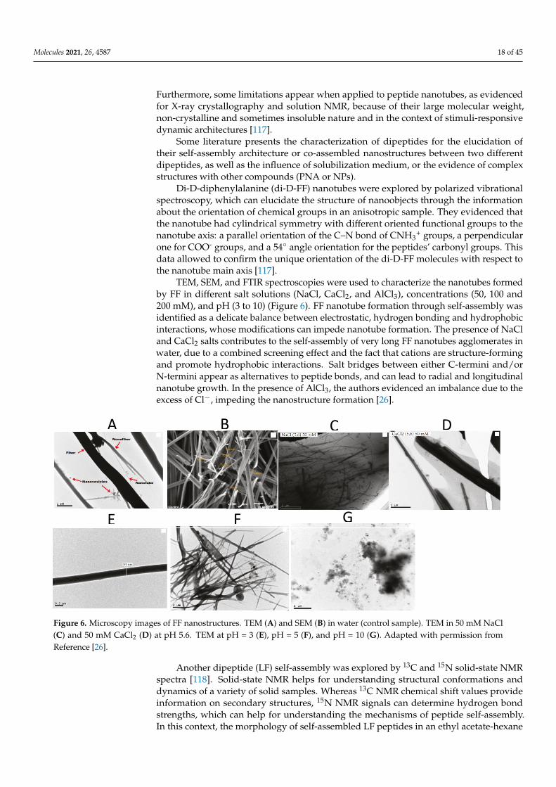
Molecules 2021, 26, 4587 18 of 45
Furthermore, some limitations appear when applied to peptide nanotubes, as evidencedfor X-ray crystallography and solution NMR, because of their large molecular weight,non-crystalline and sometimes insoluble nature and in the context of stimuli-responsivedynamic architectures [117].
Some literature presents the characterization of dipeptides for the elucidation oftheir self-assembly architecture or co-assembled nanostructures between two differentdipeptides, as well as the influence of solubilization medium, or the evidence of complexstructures with other compounds (PNA or NPs).
Di-D-diphenylalanine (di-D-FF) nanotubes were explored by polarized vibrationalspectroscopy, which can elucidate the structure of nanoobjects through the informationabout the orientation of chemical groups in an anisotropic sample. They evidenced thatthe nanotube had cylindrical symmetry with different oriented functional groups to thenanotube axis: a parallel orientation of the C–N bond of CNH3
+ groups, a perpendicularone for COO- groups, and a 54◦ angle orientation for the peptides’ carbonyl groups. Thisdata allowed to confirm the unique orientation of the di-D-FF molecules with respect tothe nanotube main axis [117].
TEM, SEM, and FTIR spectroscopies were used to characterize the nanotubes formedby FF in different salt solutions (NaCl, CaCl2, and AlCl3), concentrations (50, 100 and200 mM), and pH (3 to 10) (Figure 6). FF nanotube formation through self-assembly wasidentified as a delicate balance between electrostatic, hydrogen bonding and hydrophobicinteractions, whose modifications can impede nanotube formation. The presence of NaCland CaCl2 salts contributes to the self-assembly of very long FF nanotubes agglomerates inwater, due to a combined screening effect and the fact that cations are structure-formingand promote hydrophobic interactions. Salt bridges between either C-termini and/orN-termini appear as alternatives to peptide bonds, and can lead to radial and longitudinalnanotube growth. In the presence of AlCl3, the authors evidenced an imbalance due to theexcess of Cl−, impeding the nanostructure formation [26].
Molecules 2021, 26, x FOR PEER REVIEW 19 of 48
performed in solution or in the solid-state, which could in this case modify their architec-ture. Furthermore, some limitations appear when applied to peptide nanotubes, as evi-denced for X-ray crystallography and solution NMR, because of their large molecular weight, non-crystalline and sometimes insoluble nature and in the context of stimuli-re-sponsive dynamic architectures [117].
Some literature presents the characterization of dipeptides for the elucidation of their self-assembly architecture or co-assembled nanostructures between two different dipep-tides, as well as the influence of solubilization medium, or the evidence of complex struc-tures with other compounds (PNA or NPs).
Di-D-diphenylalanine (di-D-FF) nanotubes were explored by polarized vibrational spectroscopy, which can elucidate the structure of nanoobjects through the information about the orientation of chemical groups in an anisotropic sample. They evidenced that the nanotube had cylindrical symmetry with different oriented functional groups to the nanotube axis: a parallel orientation of the C–N bond of CNH3+ groups, a perpendicular one for COO- groups, and a 54° angle orientation for the peptides’ carbonyl groups. This data allowed to confirm the unique orientation of the di-D-FF molecules with respect to the nanotube main axis [117].
TEM, SEM, and FTIR spectroscopies were used to characterize the nanotubes formed by FF in different salt solutions (NaCl, CaCl2, and AlCl3), concentrations (50, 100 and 200 mM), and pH (3 to 10) (Figure 6). FF nanotube formation through self-assembly was iden-tified as a delicate balance between electrostatic, hydrogen bonding and hydrophobic in-teractions, whose modifications can impede nanotube formation. The presence of NaCl and CaCl2 salts contributes to the self-assembly of very long FF nanotubes agglomerates in water, due to a combined screening effect and the fact that cations are structure-forming and promote hydrophobic interactions. Salt bridges between either C-termini and/or N-termini appear as alternatives to peptide bonds, and can lead to radial and longitudinal nanotube growth. In the presence of AlCl3, the authors evidenced an imbalance due to the excess of Cl−, impeding the nanostructure formation [26].
Figure 6. Microscopy images of FF nanostructures. TEM (A) and SEM (B) in water (control sample). TEM in 50 mM NaCl (C) and 50 mM CaCl2 (D) at pH 5.6. TEM at pH = 3 (E), pH = 5 (F), and pH = 10 (G). Adapted with permission from Reference [26].
A B C D
E F G
Figure 6. Microscopy images of FF nanostructures. TEM (A) and SEM (B) in water (control sample). TEM in 50 mM NaCl(C) and 50 mM CaCl2 (D) at pH 5.6. TEM at pH = 3 (E), pH = 5 (F), and pH = 10 (G). Adapted with permission fromReference [26].
Another dipeptide (LF) self-assembly was explored by 13C and 15N solid-state NMRspectra [118]. Solid-state NMR helps for understanding structural conformations anddynamics of a variety of solid samples. Whereas 13C NMR chemical shift values provideinformation on secondary structures, 15N NMR signals can determine hydrogen bondstrengths, which can help for understanding the mechanisms of peptide self-assembly.In this context, the morphology of self-assembled LF peptides in an ethyl acetate-hexane
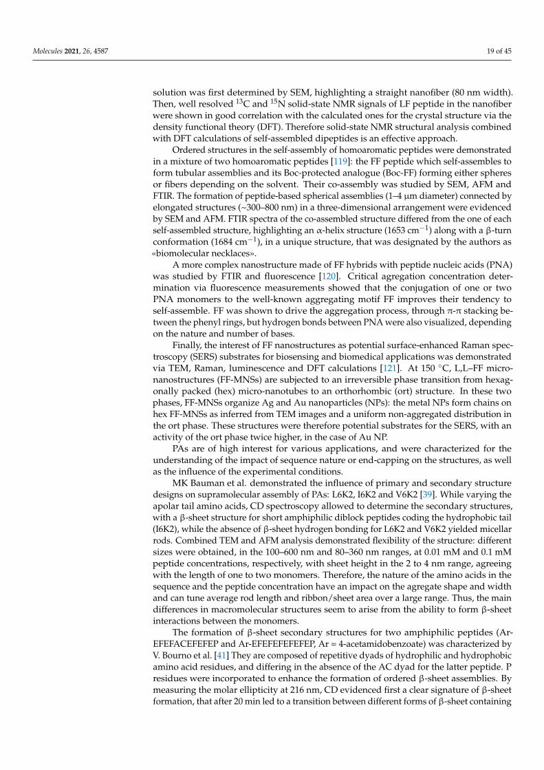
Molecules 2021, 26, 4587 19 of 45
solution was first determined by SEM, highlighting a straight nanofiber (80 nm width).Then, well resolved 13C and 15N solid-state NMR signals of LF peptide in the nanofiberwere shown in good correlation with the calculated ones for the crystal structure via thedensity functional theory (DFT). Therefore solid-state NMR structural analysis combinedwith DFT calculations of self-assembled dipeptides is an effective approach.
Ordered structures in the self-assembly of homoaromatic peptides were demonstratedin a mixture of two homoaromatic peptides [119]: the FF peptide which self-assembles toform tubular assemblies and its Boc-protected analogue (Boc-FF) forming either spheresor fibers depending on the solvent. Their co-assembly was studied by SEM, AFM andFTIR. The formation of peptide-based spherical assemblies (1–4 µm diameter) connected byelongated structures (~300–800 nm) in a three-dimensional arrangement were evidencedby SEM and AFM. FTIR spectra of the co-assembled structure differed from the one of eachself-assembled structure, highlighting an α-helix structure (1653 cm−1) along with a β-turnconformation (1684 cm−1), in a unique structure, that was designated by the authors as«biomolecular necklaces».
A more complex nanostructure made of FF hybrids with peptide nucleic acids (PNA)was studied by FTIR and fluorescence [120]. Critical agregation concentration deter-mination via fluorescence measurements showed that the conjugation of one or twoPNA monomers to the well-known aggregating motif FF improves their tendency toself-assemble. FF was shown to drive the aggregation process, through π-π stacking be-tween the phenyl rings, but hydrogen bonds between PNA were also visualized, dependingon the nature and number of bases.
Finally, the interest of FF nanostructures as potential surface-enhanced Raman spec-troscopy (SERS) substrates for biosensing and biomedical applications was demonstratedvia TEM, Raman, luminescence and DFT calculations [121]. At 150 ◦C, L,L–FF micro-nanostructures (FF-MNSs) are subjected to an irreversible phase transition from hexag-onally packed (hex) micro-nanotubes to an orthorhombic (ort) structure. In these twophases, FF-MNSs organize Ag and Au nanoparticles (NPs): the metal NPs form chains onhex FF-MNSs as inferred from TEM images and a uniform non-aggregated distribution inthe ort phase. These structures were therefore potential substrates for the SERS, with anactivity of the ort phase twice higher, in the case of Au NP.
PAs are of high interest for various applications, and were characterized for theunderstanding of the impact of sequence nature or end-capping on the structures, as wellas the influence of the experimental conditions.
MK Bauman et al. demonstrated the influence of primary and secondary structuredesigns on supramolecular assembly of PAs: L6K2, I6K2 and V6K2 [39]. While varying theapolar tail amino acids, CD spectroscopy allowed to determine the secondary structures,with a β-sheet structure for short amphiphilic diblock peptides coding the hydrophobic tail(I6K2), while the absence of β-sheet hydrogen bonding for L6K2 and V6K2 yielded micellarrods. Combined TEM and AFM analysis demonstrated flexibility of the structure: differentsizes were obtained, in the 100–600 nm and 80–360 nm ranges, at 0.01 mM and 0.1 mMpeptide concentrations, respectively, with sheet height in the 2 to 4 nm range, agreeingwith the length of one to two monomers. Therefore, the nature of the amino acids in thesequence and the peptide concentration have an impact on the agregate shape and widthand can tune average rod length and ribbon/sheet area over a large range. Thus, the maindifferences in macromolecular structures seem to arise from the ability to form β-sheetinteractions between the monomers.
The formation of β-sheet secondary structures for two amphiphilic peptides (Ar-EFEFACEFEFEP and Ar-EFEFEFEFEFEP, Ar = 4-acetamidobenzoate) was characterized byV. Bourno et al. [41] They are composed of repetitive dyads of hydrophilic and hydrophobicamino acid residues, and differing in the absence of the AC dyad for the latter peptide. Presidues were incorporated to enhance the formation of ordered β-sheet assemblies. Bymeasuring the molar ellipticity at 216 nm, CD evidenced first a clear signature of β-sheetformation, that after 20 min led to a transition between different forms of β-sheet containing
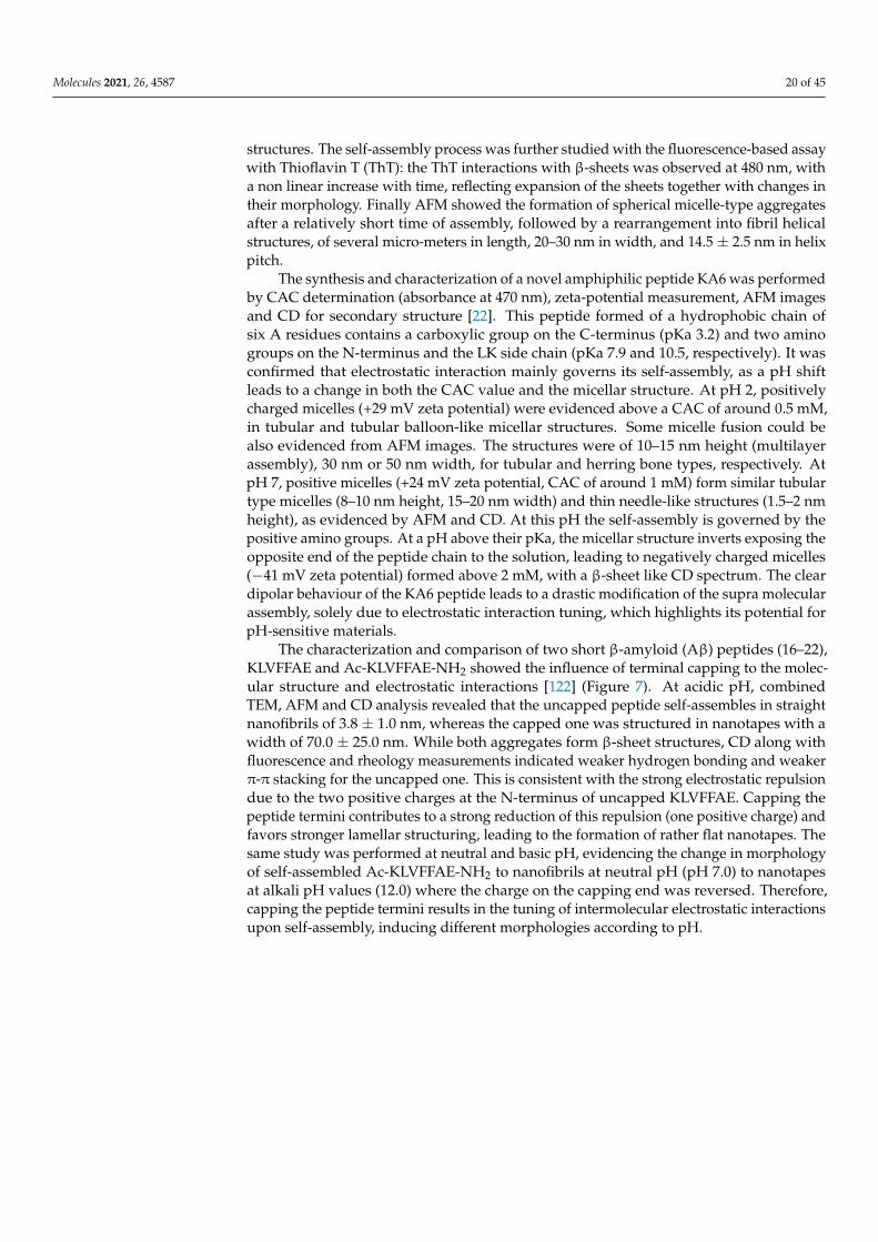
Molecules 2021, 26, 4587 20 of 45
structures. The self-assembly process was further studied with the fluorescence-based assaywith Thioflavin T (ThT): the ThT interactions with β-sheets was observed at 480 nm, witha non linear increase with time, reflecting expansion of the sheets together with changes intheir morphology. Finally AFM showed the formation of spherical micelle-type aggregatesafter a relatively short time of assembly, followed by a rearrangement into fibril helicalstructures, of several micro-meters in length, 20–30 nm in width, and 14.5 ± 2.5 nm in helixpitch.
The synthesis and characterization of a novel amphiphilic peptide KA6 was performedby CAC determination (absorbance at 470 nm), zeta-potential measurement, AFM imagesand CD for secondary structure [22]. This peptide formed of a hydrophobic chain ofsix A residues contains a carboxylic group on the C-terminus (pKa 3.2) and two aminogroups on the N-terminus and the LK side chain (pKa 7.9 and 10.5, respectively). It wasconfirmed that electrostatic interaction mainly governs its self-assembly, as a pH shiftleads to a change in both the CAC value and the micellar structure. At pH 2, positivelycharged micelles (+29 mV zeta potential) were evidenced above a CAC of around 0.5 mM,in tubular and tubular balloon-like micellar structures. Some micelle fusion could bealso evidenced from AFM images. The structures were of 10–15 nm height (multilayerassembly), 30 nm or 50 nm width, for tubular and herring bone types, respectively. AtpH 7, positive micelles (+24 mV zeta potential, CAC of around 1 mM) form similar tubulartype micelles (8–10 nm height, 15–20 nm width) and thin needle-like structures (1.5–2 nmheight), as evidenced by AFM and CD. At this pH the self-assembly is governed by thepositive amino groups. At a pH above their pKa, the micellar structure inverts exposing theopposite end of the peptide chain to the solution, leading to negatively charged micelles(−41 mV zeta potential) formed above 2 mM, with a β-sheet like CD spectrum. The cleardipolar behaviour of the KA6 peptide leads to a drastic modification of the supra molecularassembly, solely due to electrostatic interaction tuning, which highlights its potential forpH-sensitive materials.
The characterization and comparison of two short β-amyloid (Aβ) peptides (16–22),KLVFFAE and Ac-KLVFFAE-NH2 showed the influence of terminal capping to the molec-ular structure and electrostatic interactions [122] (Figure 7). At acidic pH, combinedTEM, AFM and CD analysis revealed that the uncapped peptide self-assembles in straightnanofibrils of 3.8 ± 1.0 nm, whereas the capped one was structured in nanotapes with awidth of 70.0 ± 25.0 nm. While both aggregates form β-sheet structures, CD along withfluorescence and rheology measurements indicated weaker hydrogen bonding and weakerπ-π stacking for the uncapped one. This is consistent with the strong electrostatic repulsiondue to the two positive charges at the N-terminus of uncapped KLVFFAE. Capping thepeptide termini contributes to a strong reduction of this repulsion (one positive charge) andfavors stronger lamellar structuring, leading to the formation of rather flat nanotapes. Thesame study was performed at neutral and basic pH, evidencing the change in morphologyof self-assembled Ac-KLVFFAE-NH2 to nanofibrils at neutral pH (pH 7.0) to nanotapesat alkali pH values (12.0) where the charge on the capping end was reversed. Therefore,capping the peptide termini results in the tuning of intermolecular electrostatic interactionsupon self-assembly, inducing different morphologies according to pH.
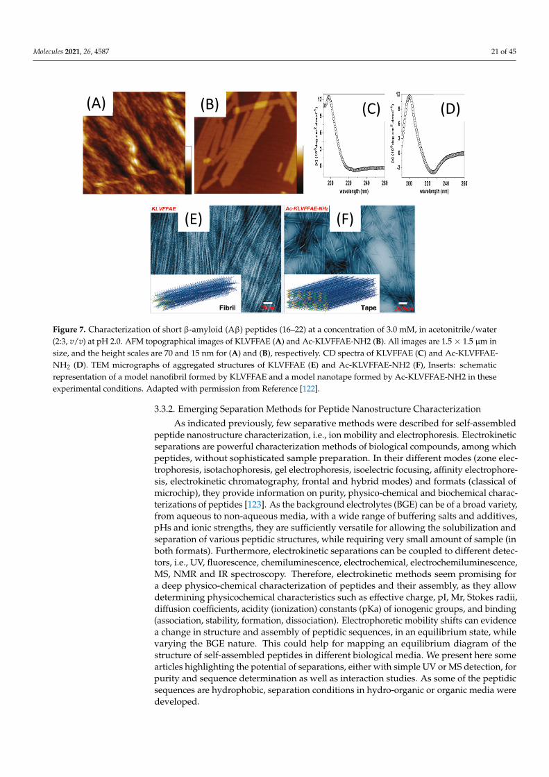
Molecules 2021, 26, 4587 21 of 45Molecules 2021, 26, x FOR PEER REVIEW 22 of 48
Figure 7. Characterization of short β-amyloid (Aβ) peptides (16–22) at a concentration of 3.0 mM, in acetonitrile/water (2:3, v/v) at pH 2.0. AFM topographical images of KLVFFAE (A) and Ac-KLVFFAE-NH2 (B). All images are 1.5 × 1.5 μm in size, and the height scales are 70 and 15 nm for (A) and (B), respectively. CD spectra of KLVFFAE (C) and Ac-KLVFFAE-NH2 (D). TEM micrographs of aggregated structures of KLVFFAE (E) and Ac-KLVFFAE-NH2 (F), Inserts: schematic rep-resentation of a model nanofibril formed by KLVFFAE and a model nanotape formed by Ac-KLVFFAE-NH2 in these experimental conditions. Adapted with permission from Reference [122].
3.3.2. Emerging Separation Methods for Peptide Nanostructure Characterization As indicated previously, few separative methods were described for self-assembled
peptide nanostructure characterization, i.e., ion mobility and electrophoresis. Electroki-netic separations are powerful characterization methods of biological compounds, among which peptides, without sophisticated sample preparation. In their different modes (zone electrophoresis, isotachophoresis, gel electrophoresis, isoelectric focusing, affinity electro-phoresis, electrokinetic chromatography, frontal and hybrid modes) and formats (classical of microchip), they provide information on purity, physico-chemical and biochemical characterizations of peptides [123]. As the background electrolytes (BGE) can be of a broad variety, from aqueous to non-aqueous media, with a wide range of buffering salts and additives, pHs and ionic strengths, they are sufficiently versatile for allowing the solubil-ization and separation of various peptidic structures, while requiring very small amount of sample (in both formats). Furthermore, electrokinetic separations can be coupled to dif-ferent detectors, i.e., UV, fluorescence, chemiluminescence, electrochemical, electrochem-iluminescence, MS, NMR and IR spectroscopy. Therefore, electrokinetic methods seem promising for a deep physico-chemical characterization of peptides and their assembly, as they allow determining physicochemical characteristics such as effective charge, pI, Mr, Stokes radii, diffusion coefficients, acidity (ionization) constants (pKa) of ionogenic groups, and binding (association, stability, formation, dissociation). Electrophoretic mo-bility shifts can evidence a change in structure and assembly of peptidic sequences, in an equilibrium state, while varying the BGE nature. This could help for mapping an equilib-rium diagram of the structure of self-assembled peptides in different biological media. We present here some articles highlighting the potential of separations, either with simple UV or MS detection, for purity and sequence determination as well as interaction studies. As some of the peptidic sequences are hydrophobic, separation conditions in hydro-organic or organic media were developed.
Water insoluble cyclic peptide [Gly6]-antamanide and its complexes with sodium and potassium ions were successfully separated in non-aqueous media (methanolic BGE)
(C) (D)
D E
(A) (B)
(E) (F)
Figure 7. Characterization of short β-amyloid (Aβ) peptides (16–22) at a concentration of 3.0 mM, in acetonitrile/water(2:3, v/v) at pH 2.0. AFM topographical images of KLVFFAE (A) and Ac-KLVFFAE-NH2 (B). All images are 1.5 × 1.5 µm insize, and the height scales are 70 and 15 nm for (A) and (B), respectively. CD spectra of KLVFFAE (C) and Ac-KLVFFAE-NH2 (D). TEM micrographs of aggregated structures of KLVFFAE (E) and Ac-KLVFFAE-NH2 (F), Inserts: schematicrepresentation of a model nanofibril formed by KLVFFAE and a model nanotape formed by Ac-KLVFFAE-NH2 in theseexperimental conditions. Adapted with permission from Reference [122].
3.3.2. Emerging Separation Methods for Peptide Nanostructure Characterization
As indicated previously, few separative methods were described for self-assembledpeptide nanostructure characterization, i.e., ion mobility and electrophoresis. Electrokineticseparations are powerful characterization methods of biological compounds, among whichpeptides, without sophisticated sample preparation. In their different modes (zone elec-trophoresis, isotachophoresis, gel electrophoresis, isoelectric focusing, affinity electrophore-sis, electrokinetic chromatography, frontal and hybrid modes) and formats (classical ofmicrochip), they provide information on purity, physico-chemical and biochemical charac-terizations of peptides [123]. As the background electrolytes (BGE) can be of a broad variety,from aqueous to non-aqueous media, with a wide range of buffering salts and additives,pHs and ionic strengths, they are sufficiently versatile for allowing the solubilization andseparation of various peptidic structures, while requiring very small amount of sample (inboth formats). Furthermore, electrokinetic separations can be coupled to different detec-tors, i.e., UV, fluorescence, chemiluminescence, electrochemical, electrochemiluminescence,MS, NMR and IR spectroscopy. Therefore, electrokinetic methods seem promising fora deep physico-chemical characterization of peptides and their assembly, as they allowdetermining physicochemical characteristics such as effective charge, pI, Mr, Stokes radii,diffusion coefficients, acidity (ionization) constants (pKa) of ionogenic groups, and binding(association, stability, formation, dissociation). Electrophoretic mobility shifts can evidencea change in structure and assembly of peptidic sequences, in an equilibrium state, whilevarying the BGE nature. This could help for mapping an equilibrium diagram of thestructure of self-assembled peptides in different biological media. We present here somearticles highlighting the potential of separations, either with simple UV or MS detection, forpurity and sequence determination as well as interaction studies. As some of the peptidicsequences are hydrophobic, separation conditions in hydro-organic or organic media weredeveloped.
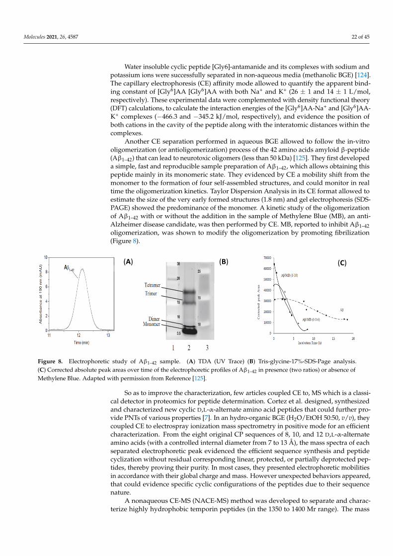
Molecules 2021, 26, 4587 22 of 45
Water insoluble cyclic peptide [Gly6]-antamanide and its complexes with sodium andpotassium ions were successfully separated in non-aqueous media (methanolic BGE) [124].The capillary electrophoresis (CE) affinity mode allowed to quantify the apparent bind-ing constant of [Gly6]AA [Gly6]AA with both Na+ and K+ (26 ± 1 and 14 ± 1 L/mol,respectively). These experimental data were complemented with density functional theory(DFT) calculations, to calculate the interaction energies of the [Gly6]AA-Na+ and [Gly6]AA-K+ complexes (−466.3 and −345.2 kJ/mol, respectively), and evidence the position ofboth cations in the cavity of the peptide along with the interatomic distances within thecomplexes.
Another CE separation performed in aqueous BGE allowed to follow the in-vitrooligomerization (or antioligomerization) process of the 42 amino acids amyloid β-peptide(Aβ1–42) that can lead to neurotoxic oligomers (less than 50 kDa) [125]. They first developeda simple, fast and reproducible sample preparation of Aβ1–42, which allows obtaining thispeptide mainly in its monomeric state. They evidenced by CE a mobility shift from themonomer to the formation of four self-assembled structures, and could monitor in realtime the oligomerization kinetics. Taylor Dispersion Analysis in its CE format allowed toestimate the size of the very early formed structures (1.8 nm) and gel electrophoresis (SDS-PAGE) showed the predominance of the monomer. A kinetic study of the oligomerizationof Aβ1–42 with or without the addition in the sample of Methylene Blue (MB), an anti-Alzheimer disease candidate, was then performed by CE. MB, reported to inhibit Aβ1–42oligomerization, was shown to modify the oligomerization by promoting fibrilization(Figure 8).
Molecules 2021, 26, x FOR PEER REVIEW 23 of 48
[124]. The capillary electrophoresis (CE) affinity mode allowed to quantify the apparent binding constant of [Gly6]AA [Gly6]AA with both Na+ and K+ (26 ± 1 and 14 ± 1 L/mol, respectively). These experimental data were complemented with density functional the-ory (DFT) calculations, to calculate the interaction energies of the [Gly6]AA-Na+ and [Gly6]AA-K+ complexes (−466.3 and −345.2 kJ/mol, respectively), and evidence the position of both cations in the cavity of the peptide along with the interatomic distances within the complexes.
Another CE separation performed in aqueous BGE allowed to follow the in-vitro ol-igomerization (or antioligomerization) process of the 42 amino acids amyloid β-peptide (Aβ1–42) that can lead to neurotoxic oligomers (less than 50 kDa) [125]. They first developed a simple, fast and reproducible sample preparation of Aβ1–42, which allows obtaining this peptide mainly in its monomeric state. They evidenced by CE a mobility shift from the monomer to the formation of four self-assembled structures, and could monitor in real time the oligomerization kinetics. Taylor Dispersion Analysis in its CE format allowed to estimate the size of the very early formed structures (1.8 nm) and gel electrophoresis (SDS-PAGE) showed the predominance of the monomer. A kinetic study of the oligomerization of Aβ1–42 with or without the addition in the sample of Methylene Blue (MB), an anti-Alz-heimer disease candidate, was then performed by CE. MB, reported to inhibit Aβ1–42 oli-gomerization, was shown to modify the oligomerization by promoting fibrilization (Fig-ure 8).
Figure 8. Electrophoretic study of Aβ1–42 sample. (A) TDA (UV Trace) (B) Tris-glycine-17%-SDS-Page analysis. (C) Cor-rected absolute peak areas over time of the electrophoretic profiles of Aβ1–42 in presence (two ratios) or absence of Meth-ylene Blue. Adapted with permission from Reference [125].
So as to improve the characterization, few articles coupled CE to, MS which is a clas-sical detector in proteomics for peptide determination. Cortez et al. designed, synthesized and characterized new cyclic D,L-α-alternate amino acid peptides that could further pro-vide PNTs of various properties [7]. In an hydro-organic BGE (H2O/EtOH 50:50, v/v), they coupled CE to electrospray ionization mass spectrometry in positive mode for an efficient characterization. From the eight original CP sequences of 8, 10, and 12 D,L-α-alternate amino acids (with a controlled internal diameter from 7 to 13 Å), the mass spectra of each separated electrophoretic peak evidenced the efficient sequence synthesis and peptide cy-clization without residual corresponding linear, protected, or partially deprotected pep-tides, thereby proving their purity. In most cases, they presented electrophoretic mobili-ties in accordance with their global charge and mass. However unexpected behaviors ap-peared, that could evidence specific cyclic configurations of the peptides due to their se-quence nature.
A nonaqueous CE-MS (NACE-MS) method was developed to separate and charac-terize highly hydrophobic temporin peptides (in the 1350 to 1400 Mr range). The mass
(A) (B) (C)
Figure 8. Electrophoretic study of Aβ1–42 sample. (A) TDA (UV Trace) (B) Tris-glycine-17%-SDS-Page analysis.(C) Corrected absolute peak areas over time of the electrophoretic profiles of Aβ1–42 in presence (two ratios) or absence ofMethylene Blue. Adapted with permission from Reference [125].
So as to improve the characterization, few articles coupled CE to, MS which is a classi-cal detector in proteomics for peptide determination. Cortez et al. designed, synthesizedand characterized new cyclic D,L-α-alternate amino acid peptides that could further pro-vide PNTs of various properties [7]. In an hydro-organic BGE (H2O/EtOH 50:50, v/v), theycoupled CE to electrospray ionization mass spectrometry in positive mode for an efficientcharacterization. From the eight original CP sequences of 8, 10, and 12 D,L-α-alternateamino acids (with a controlled internal diameter from 7 to 13 Å), the mass spectra of eachseparated electrophoretic peak evidenced the efficient sequence synthesis and peptidecyclization without residual corresponding linear, protected, or partially deprotected pep-tides, thereby proving their purity. In most cases, they presented electrophoretic mobilitiesin accordance with their global charge and mass. However unexpected behaviors appeared,that could evidence specific cyclic configurations of the peptides due to their sequencenature.
A nonaqueous CE-MS (NACE-MS) method was developed to separate and charac-terize highly hydrophobic temporin peptides (in the 1350 to 1400 Mr range). The mass
Molecules 2021, 26, 4587 23 of 45
spectrometer offered a second dimension of separation for peptides having identical mi-gration times but different structures [126].
A comparative study by CE and ion mobility spectroscopy (IMS) was performed forfollowing folding transition of a 13-mer polyproline peptide (from the all-cis polyproline Ito the all-trans polyproline II) conformation upon immersion in an aqueous solvent [127].Synchroneous folding processes were evidenced by both methods. From the eight con-formers observed using ion mobility, only five peaks were observed in CE, which can bemodeled as sums of the observed IMS conformers, proving the first direct evidence thatmultiple folding intermediates are present in solution.
IMS coupled to MS was used to reveal the peptide self-assembly mechanism duringamyloid fibril formation. The authors studied a series of amyloid-forming peptides clippedfrom larger peptides or proteins associated with disease. IMS-MS allowed to study thestructural evolution of soluble peptide oligomers one monomer at a time [128]. IMS-MSanalysis of peptide self-assembly thus opens new avenues for the investigation, detectionand eventual treatment of pathogenic processes in amyloid diseases implicated by solublepeptide or protein oligomers.
These few works combining separation methods with UV or MS detection highlightthe interest for such methodological developments, that could also be further coupledto NMR. They would provide in one single experiment various information, going frompurity, size, charge, sequence determination, interactions and the identification and controlof self-assembled nanostructures, in any type of medium mimicking biological conditions,so as to employ them for biomedical applications.
4. Biological Evaluation and Applications in the Biomedical Field
The use of materials (biological, inorganic, organic or hybrid) at the nanolength scalehas revolutionized the biomedical field by means of the so-called nanomedicine spanningmany areas such as drug delivery, diagnosis and imaging tools, therapeutics, regenerativemedicine, and tissue engineering, among others [129]. In this scenario peptides with theability to self-assemble are gaining much attention thanks to already mentioned features ofbiocompatibility, easy way of design and synthesis, versatility of structures and functions,stability, and capability of being stimuli responsive. In this section recent advances in theapplication of peptide-based nanostructures for biomedical purposes will be discussed.Two main issues will be afforded: (i) biological tests commonly used for the validation ofpeptide nanostructures for biomedical applications and (ii) biomedical applications of suchstructures going from the development of diagnostic tools to in vivo applications for themonitoring, follow-up, and treatment of diseases (e.g., cancer).
4.1. Biological Tests for Validation of Self-Assembled Peptide Nanostructures
Before the development of any in vivo application and clinical translation of anynew product a series of biological tests should be done to ensure the safety, figure outthe mechanism of interaction and uptake by cells, tissues and organs, evaluation of thefate, clearance, or bioaccumulation pathways, etc. [130]. A large panel of in vitro andin vivo evaluations is available, their selection is not trivial and could vary as functionof the envisioned application. In this section we will report the main in vitro and in vivopreclinical tests allowing the full characterization of self-assembled peptide nanostructuresfor their further application in the biomedical field.
4.1.1. In-Vitro Tests
The assessment of hazards and risks of new products by the in-vitro tests is a crucialstep towards the in-vivo application and clinical translation. These tests not only ensure thesafety of the proposed product but also are the first proof of concept on the feasibility of theenvisioned product application. Thus, a wide panel of assays is available, and the selectionof appropriate evaluations is not trivial. After a pertinent physicochemical characterizationby different analytical methods (microscopic, spectroscopic, scattering, and hyphenated
Molecules 2021, 26, 4587 24 of 45
ones, as well as in silico studies) biological tests are mandatory. In contact with a physi-ological environment, their “synthetic identity” and properties could indeed be affectedand modified, leading to a “biological identity” with specific physiological responses. Asthese innovative nanostructures for nanomedicines or nanotherapies are quite recent, theirnanosafety has remained poorly evaluated mainly because the conventional approachesare not well-adapted to the nanosize characterization and the lack of specific standardsand protocols [131]. In this view a recent review focuses on proposed guidelines for theevaluation of their biological activity, toxicological effects, nanomaterial-cell interaction anduptake mechanisms, and test endpoints based on the analysis of an extensive literature inthe field (more than 200 works) [132]. They have identified that the main processes for nano-materials to cause cellular damages are via oxidative stress leading to pro-inflammatoryeffects, fiber paradigm for those nanomaterials presenting nanofiber structures causinggranulomas in the peritoneal cavity and lung, genotoxicity and/or release of toxic ions ormolecules from their structure. They therefore propose different cellular evaluations: (i)cytotoxicity evaluation via (a) the visualization of cellular morphological alteration, theassessment of plasma membrane integrity, and the cellular metabolism (mitochondrialactivity, the well-known MTT test), (b) cell death evaluation (apoptosis or necrosis), (c) cellproliferation, and (d) epithelial cell barrier damage (cell adhesion); (ii) Oxidative stressevaluation (detection of reactive oxygen species, ROS); (iii) Pro-inflammatory reactionsevaluation via the analysis of related proteins (cytokines, chemokines, and growth fac-tors) by enzyme-linked immunosorbent assay (ELISA) tests; (iv) genotoxicity evaluation,including DNA lesions, chromosome aberrations and mutations determined via Cometand micronucleus assays in mammalian cells. Finally, in the report of Fabbrizi et al. [133]the importance of the cell line (primary vs. immortalized cells), type of system (2D vs.3D), model of culture (co-culture, perfusion, single cell) selection is discussed. Anotherimportant aspect is the tendency to go towards the development of methodologies for theassessment of risks and hazards based in the 3Rs (replacement, reduction, and refinement)limiting the in vivo tests and animal experimentations and searching for alternatives suchas 3D cultures, human primary cells and organoids approaches [131–133].
FF, one of the most studied dipeptides forming nanostructures for a wide range ofbiomedical applications, has been tested with several in vitro tests. In the work of Silvaet al. [134] different in vitro tests were performed for the evaluation of the microtubesformed by the self-assembly of the FF in view of their use as nanocarriers for drug delivery.The cytotoxicity test showed a good cell viability (>60%) up to 5 mg·mL−1 peptide con-centration. More interestingly, the authors performed a hemolytic assay to figure out thetoxicity of FF microtubes, via membrane interactions studies. The results showed that thehemolytic behavior of FF microtubes were similar to that of well-described and low-toxicnanocarriers, such as cyclodextrins and polymeric nanocapsules, inducing the hemolysisat concentrations higher than 2.8 mg·mL−1. They further developed different formulationsof FF with phthalocyanines as photosensitizers in the treatment of cancer by photother-apy [135]. The in vitro tests showed that the presence of the FF nanotubes enhances theantitumor activity of the phthalocyanines photosensitizer because it facilitates and im-proves around three times the uptake of the phthalocyanine by cells. They also evidencedthat the FF nanostructure alone does not induce any apoptosis (<15%) having a good cellviability (by the neutral red uptake assay and an annexin V-fluorescein isothiocyanate(FITC)/iodide propidium double staining flow cytometric analysis) at 0.2 mg·mL−1. Onthe contrary when they were associated to the phthalocyanine more than 80% of cell deathwas observed.
The hydrogel state of peptide self-assemblies presents a high biocompatibility assessedby seeding and viability cellular tests. The toxicity of the peptide’s gelators may becorrelated to their tendency to self-assemble, but this toxicity disappears once the hydrogelis formed [136]. Actually, peptide-based hydrogels have already shown their potential asa matrix for cell culture experiments creating adequate 3D microenvironments [137–140].It has been demonstrated that they promote cell differentiation, stem cell proliferation,
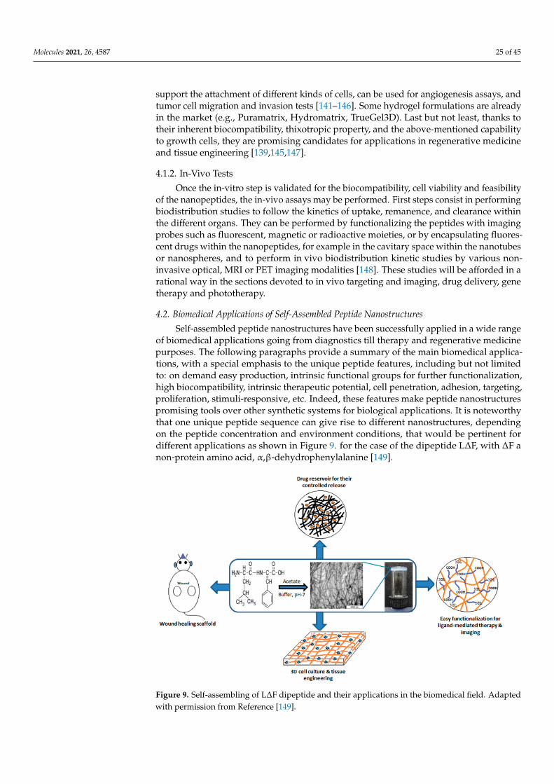
Molecules 2021, 26, 4587 25 of 45
support the attachment of different kinds of cells, can be used for angiogenesis assays, andtumor cell migration and invasion tests [141–146]. Some hydrogel formulations are alreadyin the market (e.g., Puramatrix, Hydromatrix, TrueGel3D). Last but not least, thanks totheir inherent biocompatibility, thixotropic property, and the above-mentioned capabilityto growth cells, they are promising candidates for applications in regenerative medicineand tissue engineering [139,145,147].
4.1.2. In-Vivo Tests
Once the in-vitro step is validated for the biocompatibility, cell viability and feasibilityof the nanopeptides, the in-vivo assays may be performed. First steps consist in performingbiodistribution studies to follow the kinetics of uptake, remanence, and clearance withinthe different organs. They can be performed by functionalizing the peptides with imagingprobes such as fluorescent, magnetic or radioactive moieties, or by encapsulating fluores-cent drugs within the nanopeptides, for example in the cavitary space within the nanotubesor nanospheres, and to perform in vivo biodistribution kinetic studies by various non-invasive optical, MRI or PET imaging modalities [148]. These studies will be afforded in arational way in the sections devoted to in vivo targeting and imaging, drug delivery, genetherapy and phototherapy.
4.2. Biomedical Applications of Self-Assembled Peptide Nanostructures
Self-assembled peptide nanostructures have been successfully applied in a wide rangeof biomedical applications going from diagnostics till therapy and regenerative medicinepurposes. The following paragraphs provide a summary of the main biomedical applica-tions, with a special emphasis to the unique peptide features, including but not limitedto: on demand easy production, intrinsic functional groups for further functionalization,high biocompatibility, intrinsic therapeutic potential, cell penetration, adhesion, targeting,proliferation, stimuli-responsive, etc. Indeed, these features make peptide nanostructurespromising tools over other synthetic systems for biological applications. It is noteworthythat one unique peptide sequence can give rise to different nanostructures, dependingon the peptide concentration and environment conditions, that would be pertinent fordifferent applications as shown in Figure 9. for the case of the dipeptide L∆F, with ∆F anon-protein amino acid, α,β-dehydrophenylalanine [149].
Molecules 2021, 26, x FOR PEER REVIEW 27 of 48
Figure 9. Self-assembling of LΔF dipeptide and their applications in the biomedical field. Adapted with permission from Reference [149].
4.2.1. Biosensors Peptide self-assembled nanostructures are mainly used in the development of elec-
trochemical biosensors aiming at improving the analytical performances (sensibility, se-lectivity, and limit of detection) by playing two roles: (i) increasing the active surface area of the electrode so as to generate signal amplification or increase the electron transfer (electron mediators); (ii) being functionalized by or encapsulating either the biorecogni-tion element or the signal probes/reporters or both. Thus, they have been employed for direct observation of molecular interactions in a wide range of affinity and enzymatic as-says [150]. Castillo-Leon et al. [151] provide an overview of the fabrication and deposition processes of self-assembled peptides onto transducer surfaces, highlighting different functionalization strategies with biorecognition elements (antibodies, enzymes), nano-materials (gold NP, carbon nanostructures), polymers (poly(3,4-ethylenedioxythiophene) PEDOT, polyaniline PANI) and their application in the biomedical and environmental fields. Some interesting derivatized amino acid and peptide sequences for the construc-tion of electrochemical devices are presented such as Boc-F-OH, H-F-OMe, FF, Cyclo [(Q-L)4], NSGAITIG, and EAK16-II for their ability to self-assemble in controlled nanotubes or nanofibers structures.
Peptide hydrogels have also been successfully employed for the fabrication of bio-sensors for the encapsulation of bioreceptors (enzymes, aptamers), antigens, cells, and signal reporters (QDs). They create smart bio-interfaces that improve the performances of the diagnostic tool for the determination of a wide variety of compounds going from small molecules of clinical/biological interest (H2O2, glucose, phenols) till the detection of path-ogens, cancer biomarkers, and nucleotide polymorphisms [152–155]. Both electrochemical and optical biosensors were developed with peptide hydrogels, showing practical ad-vantages such as simple fabrication, efficient diffusion of target molecules, and high en-capsulation capacity.
Another interesting advantage in constructing biosensors with self-assembled pep-tides is their ability to prevent the electrode surface from non-specific adsorption of bio-molecules thanks to their inherent antifouling properties [155,156]. As an example, the presence of a self-assembled zwitterionic peptides and IgE-aptamer was demonstrated to provide an ultrasensitive device (LOD 42 fg·mL−1) for the IgE determination in biological complex matrix [157].
Figure 9. Self-assembling of L∆F dipeptide and their applications in the biomedical field. Adaptedwith permission from Reference [149].
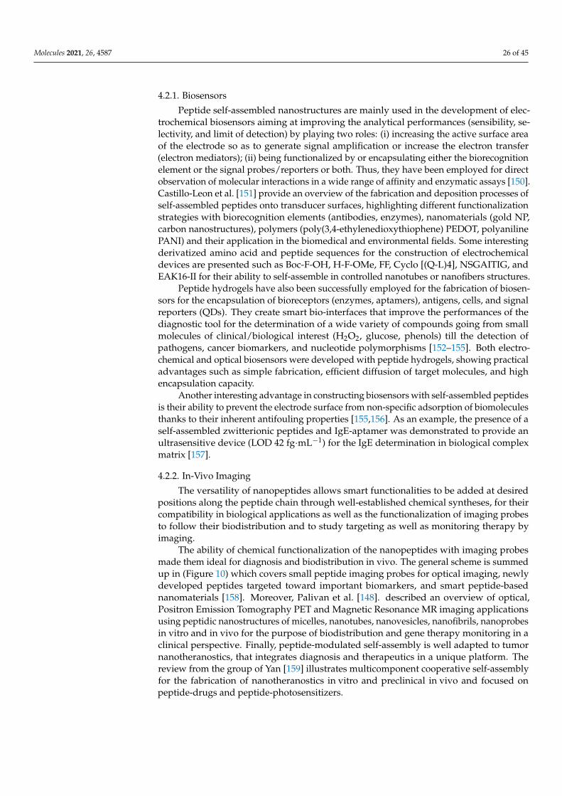
Molecules 2021, 26, 4587 26 of 45
4.2.1. Biosensors
Peptide self-assembled nanostructures are mainly used in the development of elec-trochemical biosensors aiming at improving the analytical performances (sensibility, se-lectivity, and limit of detection) by playing two roles: (i) increasing the active surface areaof the electrode so as to generate signal amplification or increase the electron transfer(electron mediators); (ii) being functionalized by or encapsulating either the biorecognitionelement or the signal probes/reporters or both. Thus, they have been employed for directobservation of molecular interactions in a wide range of affinity and enzymatic assays [150].Castillo-Leon et al. [151] provide an overview of the fabrication and deposition processes ofself-assembled peptides onto transducer surfaces, highlighting different functionalizationstrategies with biorecognition elements (antibodies, enzymes), nanomaterials (gold NP,carbon nanostructures), polymers (poly(3,4-ethylenedioxythiophene) PEDOT, polyanilinePANI) and their application in the biomedical and environmental fields. Some interestingderivatized amino acid and peptide sequences for the construction of electrochemicaldevices are presented such as Boc-F-OH, H-F-OMe, FF, Cyclo [(Q-L)4], NSGAITIG, andEAK16-II for their ability to self-assemble in controlled nanotubes or nanofibers structures.
Peptide hydrogels have also been successfully employed for the fabrication of biosen-sors for the encapsulation of bioreceptors (enzymes, aptamers), antigens, cells, and signalreporters (QDs). They create smart bio-interfaces that improve the performances of thediagnostic tool for the determination of a wide variety of compounds going from smallmolecules of clinical/biological interest (H2O2, glucose, phenols) till the detection ofpathogens, cancer biomarkers, and nucleotide polymorphisms [152–155]. Both electro-chemical and optical biosensors were developed with peptide hydrogels, showing practicaladvantages such as simple fabrication, efficient diffusion of target molecules, and highencapsulation capacity.
Another interesting advantage in constructing biosensors with self-assembled peptidesis their ability to prevent the electrode surface from non-specific adsorption of biomoleculesthanks to their inherent antifouling properties [155,156]. As an example, the presence of aself-assembled zwitterionic peptides and IgE-aptamer was demonstrated to provide anultrasensitive device (LOD 42 fg·mL−1) for the IgE determination in biological complexmatrix [157].
4.2.2. In-Vivo Imaging
The versatility of nanopeptides allows smart functionalities to be added at desiredpositions along the peptide chain through well-established chemical syntheses, for theircompatibility in biological applications as well as the functionalization of imaging probesto follow their biodistribution and to study targeting as well as monitoring therapy byimaging.
The ability of chemical functionalization of the nanopeptides with imaging probesmade them ideal for diagnosis and biodistribution in vivo. The general scheme is summedup in (Figure 10) which covers small peptide imaging probes for optical imaging, newlydeveloped peptides targeted toward important biomarkers, and smart peptide-basednanomaterials [158]. Moreover, Palivan et al. [148]. described an overview of optical,Positron Emission Tomography PET and Magnetic Resonance MR imaging applicationsusing peptidic nanostructures of micelles, nanotubes, nanovesicles, nanofibrils, nanoprobesin vitro and in vivo for the purpose of biodistribution and gene therapy monitoring in aclinical perspective. Finally, peptide-modulated self-assembly is well adapted to tumornanotheranostics, that integrates diagnosis and therapeutics in a unique platform. Thereview from the group of Yan [159] illustrates multicomponent cooperative self-assemblyfor the fabrication of nanotheranostics in vitro and preclinical in vivo and focused onpeptide-drugs and peptide-photosensitizers.
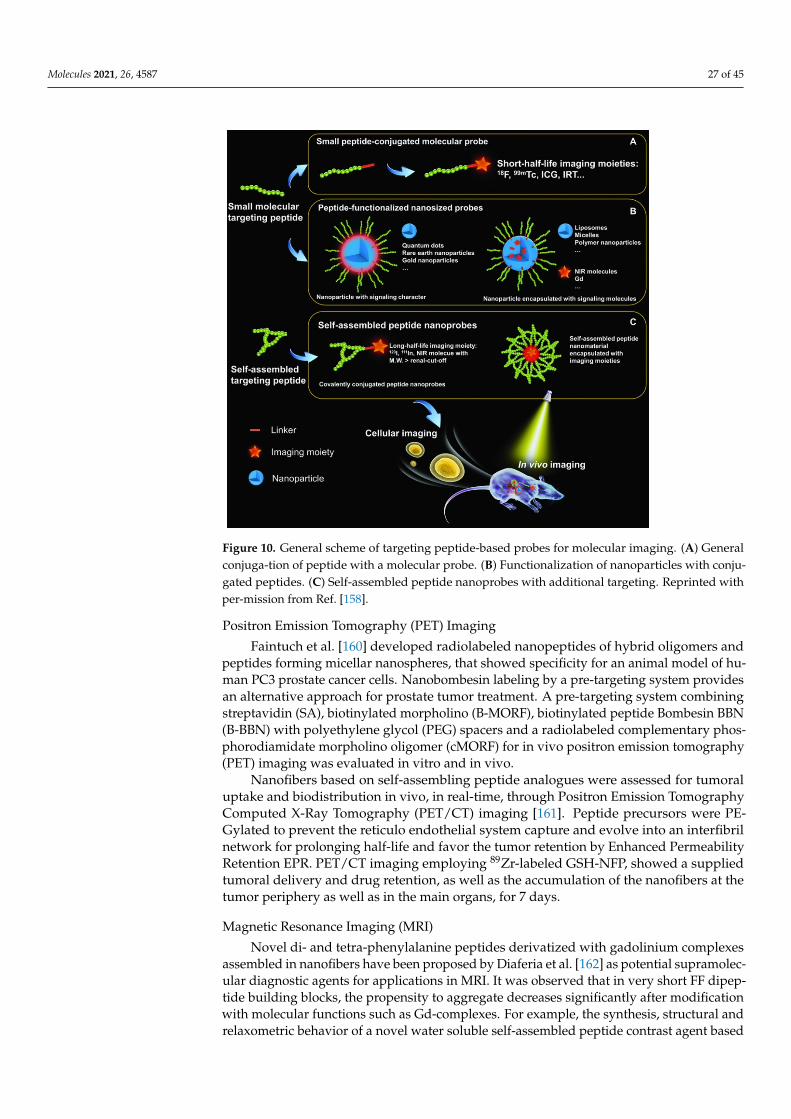
Molecules 2021, 26, 4587 27 of 45
Molecules 2021, 26, x FOR PEER REVIEW 28 of 48
4.2.2. In-Vivo Imaging The versatility of nanopeptides allows smart functionalities to be added at desired
positions along the peptide chain through well-established chemical syntheses, for their compatibility in biological applications as well as the functionalization of imaging probes to follow their biodistribution and to study targeting as well as monitoring therapy by imaging.
The ability of chemical functionalization of the nanopeptides with imaging probes made them ideal for diagnosis and biodistribution in vivo. The general scheme is summed up in (Figure 10) which covers small peptide imaging probes for optical imaging, newly developed peptides targeted toward important biomarkers, and smart peptide-based na-nomaterials [158]. Moreover, Palivan et al. [148]. described an overview of optical, Posi-tron Emission Tomography PET and Magnetic Resonance MR imaging applications using peptidic nanostructures of micelles, nanotubes, nanovesicles, nanofibrils, nanoprobes in vitro and in vivo for the purpose of biodistribution and gene therapy monitoring in a clinical perspective. Finally, peptide-modulated self-assembly is well adapted to tumor nanotheranostics, that integrates diagnosis and therapeutics in a unique platform. The re-view from the group of Yan [159] illustrates multicomponent cooperative self-assembly for the fabrication of nanotheranostics in vitro and preclinical in vivo and focused on pep-tide-drugs and peptide-photosensitizers.
Figure 10. General scheme of targeting peptide-based probes for molecular imaging. (A) General conjuga-tion of peptide with a molecular probe. (B) Functionalization of nanoparticles with conju-gated peptides. (C) Self-assembled peptide nanoprobes with additional targeting. Reprinted with per-mission from Ref. [158].
Positron Emission Tomography (PET) Imaging Faintuch et al. [160] developed radiolabeled nanopeptides of hybrid oligomers and
peptides forming micellar nanospheres, that showed specificity for an animal model of human PC3 prostate cancer cells. Nanobombesin labeling by a pre-targeting system pro-
Figure 10. General scheme of targeting peptide-based probes for molecular imaging. (A) Generalconjuga-tion of peptide with a molecular probe. (B) Functionalization of nanoparticles with conju-gated peptides. (C) Self-assembled peptide nanoprobes with additional targeting. Reprinted withper-mission from Ref. [158].
Positron Emission Tomography (PET) Imaging
Faintuch et al. [160] developed radiolabeled nanopeptides of hybrid oligomers andpeptides forming micellar nanospheres, that showed specificity for an animal model of hu-man PC3 prostate cancer cells. Nanobombesin labeling by a pre-targeting system providesan alternative approach for prostate tumor treatment. A pre-targeting system combiningstreptavidin (SA), biotinylated morpholino (B-MORF), biotinylated peptide Bombesin BBN(B-BBN) with polyethylene glycol (PEG) spacers and a radiolabeled complementary phos-phorodiamidate morpholino oligomer (cMORF) for in vivo positron emission tomography(PET) imaging was evaluated in vitro and in vivo.
Nanofibers based on self-assembling peptide analogues were assessed for tumoraluptake and biodistribution in vivo, in real-time, through Positron Emission TomographyComputed X-Ray Tomography (PET/CT) imaging [161]. Peptide precursors were PE-Gylated to prevent the reticulo endothelial system capture and evolve into an interfibrilnetwork for prolonging half-life and favor the tumor retention by Enhanced PermeabilityRetention EPR. PET/CT imaging employing 89Zr-labeled GSH-NFP, showed a suppliedtumoral delivery and drug retention, as well as the accumulation of the nanofibers at thetumor periphery as well as in the main organs, for 7 days.
Magnetic Resonance Imaging (MRI)
Novel di- and tetra-phenylalanine peptides derivatized with gadolinium complexesassembled in nanofibers have been proposed by Diaferia et al. [162] as potential supramolec-ular diagnostic agents for applications in MRI. It was observed that in very short FF dipep-tide building blocks, the propensity to aggregate decreases significantly after modificationwith molecular functions such as Gd-complexes. For example, the synthesis, structural andrelaxometric behavior of a novel water soluble self-assembled peptide contrast agent based
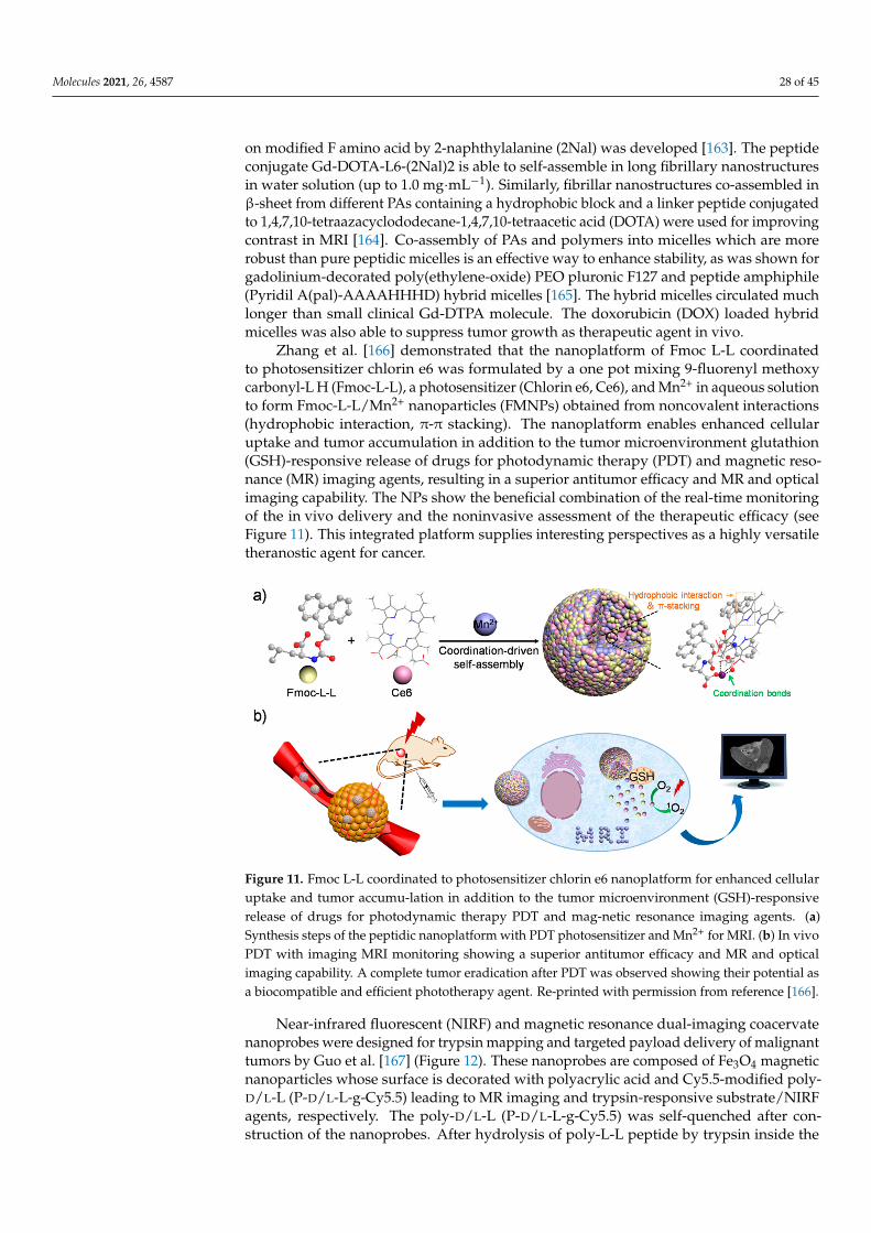
Molecules 2021, 26, 4587 28 of 45
on modified F amino acid by 2-naphthylalanine (2Nal) was developed [163]. The peptideconjugate Gd-DOTA-L6-(2Nal)2 is able to self-assemble in long fibrillary nanostructuresin water solution (up to 1.0 mg·mL−1). Similarly, fibrillar nanostructures co-assembled inβ-sheet from different PAs containing a hydrophobic block and a linker peptide conjugatedto 1,4,7,10-tetraazacyclododecane-1,4,7,10-tetraacetic acid (DOTA) were used for improvingcontrast in MRI [164]. Co-assembly of PAs and polymers into micelles which are morerobust than pure peptidic micelles is an effective way to enhance stability, as was shown forgadolinium-decorated poly(ethylene-oxide) PEO pluronic F127 and peptide amphiphile(Pyridil A(pal)-AAAAHHHD) hybrid micelles [165]. The hybrid micelles circulated muchlonger than small clinical Gd-DTPA molecule. The doxorubicin (DOX) loaded hybridmicelles was also able to suppress tumor growth as therapeutic agent in vivo.
Zhang et al. [166] demonstrated that the nanoplatform of Fmoc L-L coordinatedto photosensitizer chlorin e6 was formulated by a one pot mixing 9-fluorenyl methoxycarbonyl-L H (Fmoc-L-L), a photosensitizer (Chlorin e6, Ce6), and Mn2+ in aqueous solutionto form Fmoc-L-L/Mn2+ nanoparticles (FMNPs) obtained from noncovalent interactions(hydrophobic interaction, π-π stacking). The nanoplatform enables enhanced cellularuptake and tumor accumulation in addition to the tumor microenvironment glutathion(GSH)-responsive release of drugs for photodynamic therapy (PDT) and magnetic reso-nance (MR) imaging agents, resulting in a superior antitumor efficacy and MR and opticalimaging capability. The NPs show the beneficial combination of the real-time monitoringof the in vivo delivery and the noninvasive assessment of the therapeutic efficacy (seeFigure 11). This integrated platform supplies interesting perspectives as a highly versatiletheranostic agent for cancer.
Molecules 2021, 26, x FOR PEER REVIEW 30 of 48
Figure 11. Fmoc L-L coordinated to photosensitizer chlorin e6 nanoplatform for enhanced cellular uptake and tumor accumu-lation in addition to the tumor microenvironment (GSH)-responsive re-lease of drugs for photodynamic therapy PDT and mag-netic resonance imaging agents. (a) Synthe-sis steps of the peptidic nanoplatform with PDT photosensitizer and Mn2+ for MRI. (b) In vivo PDT with imaging MRI monitoring showing a superior antitumor efficacy and MR and optical imaging capability. A complete tumor eradication after PDT was observed showing their potential as a bio-compatible and efficient phototherapy agent. Re-printed with permission from reference [166].
Near-infrared fluorescent (NIRF) and magnetic resonance dual-imaging coacervate nanoprobes were designed for trypsin mapping and targeted payload delivery of malig-nant tumors by Guo et al. [167] (Figure 12). These nanoprobes are composed of Fe3O4 mag-netic nanoparticles whose surface is decorated with polyacrylic acid and Cy5.5-modified poly-D/L-L (P-D/L-L-g-Cy5.5) leading to MR imaging and trypsin-responsive sub-strate/NIRF agents, respectively. The poly-D/L-L (P-D/L-L-g-Cy5.5) was self-quenched af-ter construction of the nanoprobes. After hydrolysis of poly-L-L peptide by trypsin inside the tumor cells, the 100 nm nanoprobes were selectively disintegrated into fragmented segments, resulting in a 18-fold amplification of the NIRF intensity compared with the initial nanopeptide as well as strong enhancement of the MR imaging. In vitro and in vivo studies demonstrate that the coacervate nanoprobes display remarkable trypsin-sensitive NIRF and MR dual-imaging capabilities to efficiently map malignant tumors in which trypsin is overexpressed.
Figure 11. Fmoc L-L coordinated to photosensitizer chlorin e6 nanoplatform for enhanced cellularuptake and tumor accumu-lation in addition to the tumor microenvironment (GSH)-responsiverelease of drugs for photodynamic therapy PDT and mag-netic resonance imaging agents. (a)Synthesis steps of the peptidic nanoplatform with PDT photosensitizer and Mn2+ for MRI. (b) In vivoPDT with imaging MRI monitoring showing a superior antitumor efficacy and MR and opticalimaging capability. A complete tumor eradication after PDT was observed showing their potential asa biocompatible and efficient phototherapy agent. Re-printed with permission from reference [166].
Near-infrared fluorescent (NIRF) and magnetic resonance dual-imaging coacervatenanoprobes were designed for trypsin mapping and targeted payload delivery of malignanttumors by Guo et al. [167] (Figure 12). These nanoprobes are composed of Fe3O4 magneticnanoparticles whose surface is decorated with polyacrylic acid and Cy5.5-modified poly-D/L-L (P-D/L-L-g-Cy5.5) leading to MR imaging and trypsin-responsive substrate/NIRFagents, respectively. The poly-D/L-L (P-D/L-L-g-Cy5.5) was self-quenched after con-struction of the nanoprobes. After hydrolysis of poly-L-L peptide by trypsin inside the
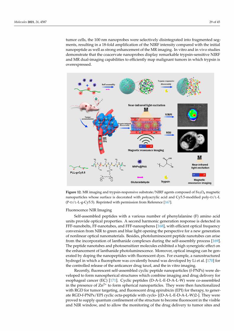
Molecules 2021, 26, 4587 29 of 45
tumor cells, the 100 nm nanoprobes were selectively disintegrated into fragmented seg-ments, resulting in a 18-fold amplification of the NIRF intensity compared with the initialnanopeptide as well as strong enhancement of the MR imaging. In vitro and in vivo studiesdemonstrate that the coacervate nanoprobes display remarkable trypsin-sensitive NIRFand MR dual-imaging capabilities to efficiently map malignant tumors in which trypsin isoverexpressed.
Molecules 2021, 26, x FOR PEER REVIEW 31 of 48
Figure 12. MR imaging and trypsin-responsive substrate/NIRF agents composed of Fe3O4 magnetic nanoparticles whose surface is decorated with polyacrylic acid and Cy5.5-modified poly-D/L-L (P-D/L-L-g-Cy5.5). Reprinted with permission from Reference [167].
Fluorescence NIR Imaging Self-assembled peptides with a various number of phenylalanine (F) amino acid units
provide optical properties. A second harmonic generation response is detected in FFF-nanobelts, FF-nanotubes, and FFF-nanospheres [168], with efficient optical frequency con-version from NIR to green and blue light opening the perspective for a new generation of nonlinear optical nanomaterials. Besides, photoluminescent peptide nanotubes can arise from the incorporation of lanthanide complexes during the self-assembly process [169]. The peptide nanotubes and photosensitizer molecules exhibited a high synergistic effect on the enhancement of lanthanide photoluminescence. Moreover, optical imaging can be generated by doping the nanopeptides with fluorescent dyes. For example, a nanostruc-tured hydrogel in which a fluorophore was covalently bound was developed by Li et al.
[170] for the controlled release of the anticancer drug taxol, and the in vitro imaging. Recently, fluorescent self-assembled cyclic peptide nanoparticles (f-PNPs) were de-
veloped to form nanospherical structures which combine imaging and drug delivery for esophageal cancer (EC) [171]. Cyclic peptides (D-A-L-E-D-A-L-W) were co-assembled in the presence of Zn2+ to form spherical nanoparticles. They were then functionalized with RGD for tumor targeting, and fluorescent drug epirubicin (EPI) for therapy, to generate RGD-f-PNPs/EPI cyclic octa-peptide with cyclo- [(D-A-L-E-D-A-L-W)2-]. They were proved to supply quantum confinement of the structure to become fluorescent in the vis-ible and NIR window, and to allow the monitoring of the drug delivery to tumor sites and of the therapeutic responses. In vitro and histology tests proved significantly reduced car-diotoxicity and improved anti-tumor activity compared to the drug alone. This unique nanoparticle system may lead to potential approaches for bioorganic fluorescence-based delivering, imaging, and drug release tracking (Figure 13).
Figure 12. MR imaging and trypsin-responsive substrate/NIRF agents composed of Fe3O4 magneticnanoparticles whose surface is decorated with polyacrylic acid and Cy5.5-modified poly-D/L-L(P-D/L-L-g-Cy5.5). Reprinted with permission from Reference [167].
Fluorescence NIR Imaging
Self-assembled peptides with a various number of phenylalanine (F) amino acidunits provide optical properties. A second harmonic generation response is detected inFFF-nanobelts, FF-nanotubes, and FFF-nanospheres [168], with efficient optical frequencyconversion from NIR to green and blue light opening the perspective for a new generationof nonlinear optical nanomaterials. Besides, photoluminescent peptide nanotubes can arisefrom the incorporation of lanthanide complexes during the self-assembly process [169].The peptide nanotubes and photosensitizer molecules exhibited a high synergistic effect onthe enhancement of lanthanide photoluminescence. Moreover, optical imaging can be gen-erated by doping the nanopeptides with fluorescent dyes. For example, a nanostructuredhydrogel in which a fluorophore was covalently bound was developed by Li et al. [170] forthe controlled release of the anticancer drug taxol, and the in vitro imaging.
Recently, fluorescent self-assembled cyclic peptide nanoparticles (f-PNPs) were de-veloped to form nanospherical structures which combine imaging and drug delivery foresophageal cancer (EC) [171]. Cyclic peptides (D-A-L-E-D-A-L-W) were co-assembledin the presence of Zn2+ to form spherical nanoparticles. They were then functionalizedwith RGD for tumor targeting, and fluorescent drug epirubicin (EPI) for therapy, to gener-ate RGD-f-PNPs/EPI cyclic octa-peptide with cyclo- [(D-A-L-E-D-A-L-W)2-]. They wereproved to supply quantum confinement of the structure to become fluorescent in the visibleand NIR window, and to allow the monitoring of the drug delivery to tumor sites and
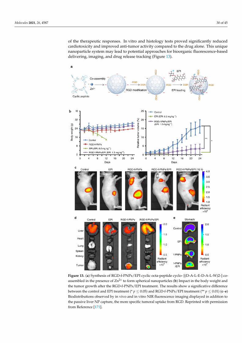
Molecules 2021, 26, 4587 30 of 45
of the therapeutic responses. In vitro and histology tests proved significantly reducedcardiotoxicity and improved anti-tumor activity compared to the drug alone. This uniquenanoparticle system may lead to potential approaches for bioorganic fluorescence-baseddelivering, imaging, and drug release tracking (Figure 13).
Molecules 2021, 26, x FOR PEER REVIEW 32 of 48
Figure 13. (a) Synthesis of RGD-f-PNPs/EPI cyclic octa-peptide cyclo- [(D-A-L-E-D-A-L-W)2-] co-assembled in the presence of Zn2+ to form spherical nanoparticles (b) Impact in the body weight and the tumor growth after the RGD-f-PNPs/EPI treatment. The results show a significative difference between the control and EPI treatment (* p ≤ 0.05) and RGD-f-PNPs/EPI treatment (** p ≤ 0.01) (c–e) Biodistributions observed by in vivo and in vitro NIR fluorescence imaging displayed in addition to the passive liver NP capture, the more specific tumoral uptake from RGD. Reprinted with per-mission from Reference [171].
As demonstrated with previous multimodal theranostic peptide based nanoplat-forms presented in this review, multimodal association of multiple imaging probes, tar-geting moieties and therapeutic substances should provide efficient therapeutical strategy
b
Figure 13. (a) Synthesis of RGD-f-PNPs/EPI cyclic octa-peptide cyclo- [(D-A-L-E-D-A-L-W)2-] co-assembled in the presence of Zn2+ to form spherical nanoparticles (b) Impact in the body weight andthe tumor growth after the RGD-f-PNPs/EPI treatment. The results show a significative differencebetween the control and EPI treatment (* p ≤ 0.05) and RGD-f-PNPs/EPI treatment (** p ≤ 0.01) (c–e)Biodistributions observed by in vivo and in vitro NIR fluorescence imaging displayed in addition tothe passive liver NP capture, the more specific tumoral uptake from RGD. Reprinted with permissionfrom Reference [171].
Molecules 2021, 26, 4587 31 of 45
As demonstrated with previous multimodal theranostic peptide based nanoplatformspresented in this review, multimodal association of multiple imaging probes, targetingmoieties and therapeutic substances should provide efficient therapeutical strategy withclinical envision. These peptidic multicomponent synergetic self-assembly is a promisingapproach for nanotheranostic strategy for innovative therapy.
4.2.3. Drug Delivery
To overcome the side effects induced by chemotherapies, to improve the solubility,bioavailability and uptake of drugs, to facilitate the barrier transfer, among others, greatprogress is being made in the research of drug carriers. In this context nanocarriers havegained much attention in the last decades since they can circumvent some problems encoun-tered by conventional drug delivery systems associated to non-specificity, burst release ordamage of normal cells [172]. They should improve the pharmacokinetics, bioavailabilityand therapeutic efficacy of the drug while providing preferential accumulation in thetargeted tissue so as to decrease the side effects. Since one can modulate their physico-chemical properties and thus their behavior, the main parameters to consider in designingnanocarriers are their composition (either organic, inorganic, or hybrid), their size andsurface charge density, their geometric shape and curvature, and their ability to be stimuliresponsive (pH, temperature, enzyme, light, etc.) so as to release the drug in a preciseenvironment or conditions [172,173].
Peptides self-assembly into nanocarriers offers many advantages for drug delivery,such as the ability to carry both hydrophilic and hydrophobic drugs, with a high efficiencyof drug loading and a low ratio of drug loss, while having the capacity for sustaineddrug delivery at the targeted site, and high biocompatibility and stability. In addition,nanopeptides can be customized to incorporate peptide sequences or targeting moietiesfor specific cell targeting and responses making them attractive for drug-delivery applica-tions. Thus, several structures of peptide assemblies have been recently described as drugdelivery systems in different reviews [174–176]. The work of Pentlavalli et al. [174] wasdevoted to the study of peptide self-assembly mechanisms and the influence of externalstimuli (pH, ionic strength, temperature, enzymes) inducing the delivery of the drugs.Tesauro et al. [175] focused on amphiphilic peptides design, emphasizing the wide rangeof sequence selection to provide the peptides with features such as specific targeting, cleav-age, or stimuli responsivity enhancing the uptake, specific release of the drug and thustherapeutic efficiency. Gupta et al. [176] highlighted the potential of single amino acid andultrashort peptides (di- and tri- peptides) and their derivates as drug/gene carriers.
Currently the most common nanostructural peptide arrangements used as nanocar-riers for drug delivery are nanoparticles, nanoformulations [177], micelles [178], vesi-cles [179,180], nanotubes [134,181,182] and hydrogels [152,183,184]. They have been provedto successfully perform the carry and controlled release of both hydrophobic and hy-drophilic drugs including anti-cancer, anti-fungal, anti-infectious and antibiotics as forexample doxorubicin, curcumin, paclitaxel, cisplatin, mitoxantrone, streptomycin. InTable 1 are collected specific sequences that introduce special features to the peptides fordrug delivery and biological applications. Note that these sequences can form part ofa larger peptide sequence and be as well further derivatized with other molecules likepolymers (PEG), phospholipids, cholesterol, contrast imaging agents (DOTA).
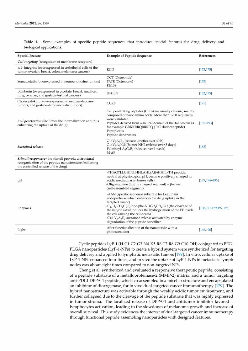
Molecules 2021, 26, 4587 32 of 45
Table 1. Some examples of specific peptide sequences that introduce special features for drug delivery andbiological applications.
Special Feature Example of Peptide Sequence References
Cell targeting (recognition of membrane receptors)
α,β-Integrins (overexpressed in endothelial cells of thetumor; ovarian, breast, colon, melanoma cancers) RGD [173,175]
Somatostatin (overexpressed in neuroendocrine tumors)OCT (Octreotide)TATE (Octreotate)KE108
[175]
Bombesin (overexpressed in prostate, breast, small celllung, ovarian, and gastrointestinal cancers) [7-4]BN [162,175]
Cholecystokinin (overexpressed in neuroendrocrinetumors, and gastroenteropancreatic tumors) CCK8 [175]
Cell penetration (facilitates the internalization and thusenhancing the uptake of the drug)
Cell penetrating peptides (CPPs) are usually cationic, mainlycomposed of basic amino acids. More than 1700 sequenceswere validated.Peptides derived from α-helical domain of the Tat protein asfor example GRKKRRQRRRPQ (TAT dodecapeptide)PeptiplexesPeptide dendrimers
[185–193]
Sustained release
C16V2A2E2 (release kinetics over 40 h)C16V3A3K3K(folate)-NH2 (release over 5 days)Palmitoyl-A4G3E3 (release over 1 week)M-∆F
[183]
Stimuli-responsive (the stimuli provoke a structuralreorganization of the peptide nanostructure facilitatingthe controlled release of the drug)
pH
-THAGYLLGHINLHHLAHL(Aib)HHIL (TH peptide;neutral at physiological pH, become positively charged inacidic medium as in tumor cells)-Oligoarginine (highly charged segment) + β-sheet(self-assembled segment)
[179,194–196]
Enzymes
-AAN (specific sequence substrate for Legumainendoprotease which enhances the drug uptake in thetargeted tumor)-C10H7CH2C(O)-phe-phe-NHCH2CH2OH (the cleavage ofthe butyric diacid induces the hydrogelation of the FF insidethe cell causing the cell death)-C16-V3A3E3 sustained release activated by enzymedegradation of the peptide nanofiber
[108,171,179,197,198]
Light After functionalization of the nanopetide with aphotosensitizer [166,196]
Cyclic peptides LyP-1 (H-C1-C2-G3-N4-K5-R6-T7-R8-G9-C10-OH) conjugated to PEG–PLGA nanoparticles (LyP-1-NPs) to create a hybrid system were synthetized for targetingdrug delivery and applied to lymphatic metastatic tumors [199]. In vitro, cellular uptake ofLyP-1-NPs enhanced four times, and in vivo the uptake of LyP-1-NPs in metastasis lymphnodes was about eight times compared to non-targeted NPs.
Cheng et al. synthetized and evaluated a responsive therapeutic peptide, consistingof a peptide substrate of a metalloproteinase-2 (MMP-2) matrix, and a tumor targetinganti-PDL1 DPPA-1 peptide, which co-assembled in a micellar structure and encapsulatedan inhibitor of dioxygenase, for in vivo dual-targeted cancer immunotherapy [179]. Thehybrid nanostructure was activable through the weakly acidic tumor environment, andfurther collapsed due to the cleavage of the peptide substrate that was highly expressedin tumor stroma. The localized release of DPPA-1 and antitumor inhibitor favored Tlymphocytes activation, leading to the slowdown of melanoma growth and increase ofoverall survival. This study evidences the interest of dual-targeted cancer immunotherapythrough functional peptide assembling nanoparticles with designed features.
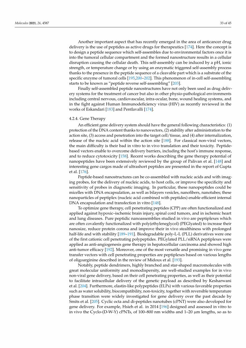
Molecules 2021, 26, 4587 33 of 45
Another important aspect that has recently emerged in the area of anticancer drugdelivery is the use of peptides as active drugs for therapeutics [174]. Here the concept isto design a peptide sequence which self-assembles due to environmental factors once it isinto the tumoral cellular compartment and the formed nanostructure results in a cellulardisruption causing the cellular death. This self-assembly can be induced by a pH, ionicstrength, or temperature change or by using an enzymatic triggered self-assembly processthanks to the presence in the peptide sequence of a cleavable part which is a substrate of thespecific enzyme of tumoral cells [195,200–202]. This phenomenon of in-cell self-assemblingstarts to be known as “peptide reverse self-assembling” [203].
Finally self-assembled peptide nanostructures have not only been used as drug deliv-ery systems for the treatment of cancer but also in other physio-pathological environmentsincluding central nervous, cardiovascular, intra-ocular, bone, wound healing systems, andin the fight against Human Immunodeficiency virus (HIV) as recently reviewed in theworks of Eskandari [183] and Pentlavalli [174].
4.2.4. Gene Therapy
An efficient gene delivery system should have the general following characteristics: (1)protection of the DNA content thanks to nanovectors, (2) stability after administration to theaction site, (3) access and penetration into the target cell/tissue, and (4) after internalization,release of the nucleic acid within the action site [188]. For classical non-viral vectors,the main difficulty is their bad in vitro to in vivo translation and their toxicity. Peptide-based vectors enable to overcome delivery barriers, including the host’s immune response,and to reduce cytotoxicity [158]. Recent works describing the gene therapy potential ofnanopeptides have been extensively reviewed by the group of Palivan et al. [148] andinteresting gene cargos made of ultrashort peptides are presented in the report of Guptaet al. [176].
Peptide-based nanostructures can be co-assembled with nucleic acids and with imag-ing probes, for the delivery of nucleic acids, to host cells, or improve the specificity andsensitivity of probes in diagnostic imaging. In particular, these nanopeptides could bemicelles with DNA encapsulation, as well as bilayers vesicles, nanofibers, nanotubes; thesenanoparticles of peptiplex (nucleic acid combined with peptides) enable efficient internalDNA encapsulation and transfection in vitro [148].
To optimize gene therapy, cell penetrating peptides (CPP) are often functionalized andapplied against hypoxic–ischemic brain injury, spinal cord tumors, and in ischemic heartand lung diseases. Pure peptidic nanoassemblies studied in vivo are peptiplexes whichare often covalently functionalized with poly(ethyleneglycol) (PEGlyated) to increase theirnanosize, reduce protein corona and improve their in vivo stealthiness with prolongedhalf-life and with stability [189–191]. Biodegradable poly-L-L (PLL) derivatives were oneof the first cationic cell penetrating polypeptides. PEGylated PLL/siRNA peptiplexes wereapplied as anti-angiogenesis gene therapy in hepatocellular carcinoma and showed highanti-tumor efficacy [192]. Moreover, one of the most versatile and promising in vivo genetransfer vectors with cell penetrating properties are peptiplexes based on various lengthsof oligoarginine described in the review of Midoux et al. [193].
Notably, peptide dendrimers, highly branched and star-shaped macromolecules withgreat molecular uniformity and monodispersity, are well-studied examples for in vivonon-viral gene delivery, based on their cell penetrating properties, as well as their potentialto facilitate intracellular delivery of the genetic payload as described by Kesharwaniet al. [204]. Furthermore, elastin-like polypeptides (ELPs) with various favorable propertiessuch as water solubility, biocompatibility, non-toxicity, together with reversible temperaturephase transition were widely investigated for gene delivery over the past decade bySmits et al. [205]. Cyclic octa and di-peptides nanotubes (cPNT) were also developed forgene delivery. For example, Hsieh et al. in 2014 [196] designed and assessed in vitro toin vivo the Cyclo-(D-W-Y) cPNTs, of 100–800 nm widths and 1–20 µm lengths, so as to
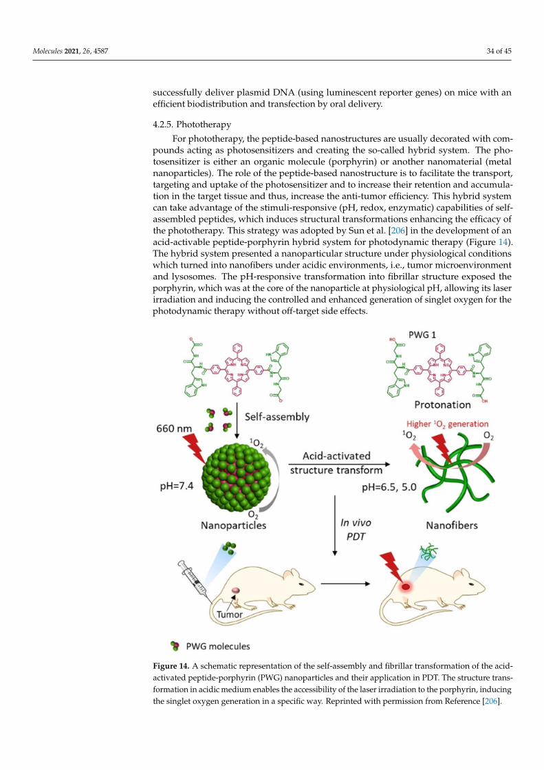
Molecules 2021, 26, 4587 34 of 45
successfully deliver plasmid DNA (using luminescent reporter genes) on mice with anefficient biodistribution and transfection by oral delivery.
4.2.5. Phototherapy
For phototherapy, the peptide-based nanostructures are usually decorated with com-pounds acting as photosensitizers and creating the so-called hybrid system. The pho-tosensitizer is either an organic molecule (porphyrin) or another nanomaterial (metalnanoparticles). The role of the peptide-based nanostructure is to facilitate the transport,targeting and uptake of the photosensitizer and to increase their retention and accumula-tion in the target tissue and thus, increase the anti-tumor efficiency. This hybrid systemcan take advantage of the stimuli-responsive (pH, redox, enzymatic) capabilities of self-assembled peptides, which induces structural transformations enhancing the efficacy ofthe phototherapy. This strategy was adopted by Sun et al. [206] in the development of anacid-activable peptide-porphyrin hybrid system for photodynamic therapy (Figure 14).The hybrid system presented a nanoparticular structure under physiological conditionswhich turned into nanofibers under acidic environments, i.e., tumor microenvironmentand lysosomes. The pH-responsive transformation into fibrillar structure exposed theporphyrin, which was at the core of the nanoparticle at physiological pH, allowing its laserirradiation and inducing the controlled and enhanced generation of singlet oxygen for thephotodynamic therapy without off-target side effects.
Molecules 2021, 26, x FOR PEER REVIEW 37 of 48
Figure 14. A schematic representation of the self-assembly and fibrillar transformation of the acid-activated peptide-porphyrin (PWG) nanoparticles and their application in PDT. The structure trans-formation in acidic medium enables the accessibility of the laser irradiation to the porphyrin, induc-ing the singlet oxygen generation in a specific way. Reprinted with permission from Reference [206].
Zou et al. [207] has also formulated an interesting self-assembled peptide-porphyrin (TPP-G-FF) to form nanodots with a porphyrin photosensitizer delivery for photothermal therapy (PTT). In vivo biodistribution studies and thermal and photoacoustic in vivo im-aging were studied showing a major expected liver as well as 10% tumoral uptake. These nanodots are particularly soluble in aqueous media, with a stable scaffold against dilution and irradiation, and high light-to-heat conversions (54.2%) leading to photoacoustic and thermal imaging features. They have been proved to prevent the growth of tumor in a preclinical mice model.
4.2.6. Nanopeptides as Drugs: Antiviral and Antibacterial Effects, and Vaccine Engineering
Self-assembled peptide nanostructures (β-sheets, α-helices, peptide amphiphiles, amino acid pairing, elastin like polypeptides, cyclic peptides, short peptides, Fmoc-pro-tected peptides, and peptide hydrogels) are well adapted for further application in vaccine engineering as developed in the review of the group of Eskandari [183]. Cyclic di-, tri-, tetra-, hexa-, octa-, and decapeptides with various amino acid sequences, enantiomers, and functionalized side chains can be also applied for antiviral and antibacterial drugs as detailed in the work of Hsieh et al. [182].
Some works evidenced peptide nanostructure’s antibacterial activity. Gao et al. [208] developed amphiphiles self-assembled nanorods of peptides against bacterial activity for efficiently treating antibiotic-resistant bacteria in vitro. A soluble antibiotic (ciprofloxacin)
Figure 14. A schematic representation of the self-assembly and fibrillar transformation of the acid-activated peptide-porphyrin (PWG) nanoparticles and their application in PDT. The structure trans-formation in acidic medium enables the accessibility of the laser irradiation to the porphyrin, inducingthe singlet oxygen generation in a specific way. Reprinted with permission from Reference [206].
Molecules 2021, 26, 4587 35 of 45
Zou et al. [207] has also formulated an interesting self-assembled peptide-porphyrin(TPP-G-FF) to form nanodots with a porphyrin photosensitizer delivery for photothermaltherapy (PTT). In vivo biodistribution studies and thermal and photoacoustic in vivoimaging were studied showing a major expected liver as well as 10% tumoral uptake. Thesenanodots are particularly soluble in aqueous media, with a stable scaffold against dilutionand irradiation, and high light-to-heat conversions (54.2%) leading to photoacoustic andthermal imaging features. They have been proved to prevent the growth of tumor in apreclinical mice model.
4.2.6. Nanopeptides as Drugs: Antiviral and Antibacterial Effects, and Vaccine Engineering
Self-assembled peptide nanostructures (β-sheets, α-helices, peptide amphiphiles,amino acid pairing, elastin like polypeptides, cyclic peptides, short peptides, Fmoc-protected peptides, and peptide hydrogels) are well adapted for further application invaccine engineering as developed in the review of the group of Eskandari [183]. Cyclic di-,tri-, tetra-, hexa-, octa-, and decapeptides with various amino acid sequences, enantiomers,and functionalized side chains can be also applied for antiviral and antibacterial drugs asdetailed in the work of Hsieh et al. [182].
Some works evidenced peptide nanostructure’s antibacterial activity. Gao et al. [208]developed amphiphiles self-assembled nanorods of peptides against bacterial activity forefficiently treating antibiotic-resistant bacteria in vitro. A soluble antibiotic (ciprofloxacin)and a hydrophobic tripeptide ((D)L-F-F) self-assembled into supramolecular nanostructuresto form a macroscopic hydrogel [209] exhibiting a mild anti-bacterial activity against Gram-negative bacteria and importantly no major haemolytic toxicity on human red blood cellsor in mouse fibroblast cell cultures. Antibiotic effect of cyclic PNT was demonstrated byGhadiri group in 2001 [210]. The PNTs was demonstrated efficient for surface coating toprevent microbial colonization inn health care.
Antibacterial activity of self-assembled diphenylalanine emerges as the minimalsupramolecular assembly [211]. The diphenylalanine nanoassemblies completely inhibitbacterial growth, induce disruption of bacterial morphology, and cause membrane per-meation and depolarization. Indeed, Schnaider et al. [212] evidenced the specificity ofnanopeptides to interact with microbial membranes and the development of antibacterialmaterials (e.g., ultrashort cationic hybrid naphthalene derived peptides) and these studylead to the development of antimicrobial agents and materials. For example, ultrashortnon-steroidal inflammatory drug peptides attached to diphenyl lysine (FFKK-OH) peptideself-assembled in hydrogel nanosponges [213] or self-assembly of cationic multidomainpeptide hydrogels [214] have been able to destroy in vitro bacterial cultures.
Concerning vaccine strategy, introduction of autophagy inducing transactivator oftranscription TAT autophagy inducing peptides eliminated latent HIV infection in vitroin selective latently HIV-infected CD4+ T cells (major reservoir of HIV latent infection)via lipid-coated hybrid polylactic co-glycolic acid (PLGA) nanoparticles for a strategy toprevent the reactivation of the virus and new infection in bystander cells in a vaccine typemanner [215].
4.2.7. Tissue Engineering and Regenerative Medicine
Tissue engineering is based on the growth of cells into a scaffold that has the me-chanical and biological features well-adapted to this end, close to those of extracellularmatrix (ECM). Thus, growing within the scaffold matrix, tissue cells are regenerated orprepared to replace skin, bone, cartilage, or part of an organ. Tissue engineering aims atdeveloping materials and methods to promote cell differentiation and proliferation towardthe formation of a new tissue [181]. Among the different materials proposed to be scaffolds,peptides are one of the most promising biomaterials for tissue engineering and regener-ative medicine since the main signaling language in the ECM is mediated via peptideepitopes and, as mentioned previously, they possess the thixotropic property enabling aneasy way of administration directly into the injured location [147,174]. Indeed, thanks to
Molecules 2021, 26, 4587 36 of 45
these features, along with its similarity to ECM that limits chronic inflammation responsesor immunological reactions and toxicity, peptides are postulated as improved scaffoldscompared to polymers and biopolymers that have been used as artificial scaffolds in thisdomain [216]. In addition, self-assembled nanostructures are well suited for regenerativeissues thanks to their structures and mechanical properties which enable them to fulfillthe main challenges of tissue engineering: (1) adaptable mechanical strength suitable fordifferent parts of the body, (2) porosity for lighter weight and ensuring sufficient blood andoxygen supplies and waste transport, and (3) biocompatibility to prevent immune rejectionand inflammation [176,181]. Thus, for example nanotube and nanofiber structures frompeptide sequences such as C16A4G3LRKKLGKA; KLD12; and V3A3E3 serve as guidance forthe growth of vessels, cartilages, or bones [217–219]. The hydrogel state of self-assembledpeptides is also performant in tissue modeling in diverse medicine areas such as oncol-ogy [145] or cardiovascular [143]; as well as tissue engineering and regeneration [147].For example, the 3D nanofiber peptide hydrogel formed by RADA16-I peptide is similarto the structure naturally present in the ECM, and possesses features of cell attachment,proliferation, and differentiation, proved to be efficient for bone regeneration showing animmediate hemostasis and accelerative osteosis [220]. This peptide RADA16 thereforeseems to play a pivotal role in regenerative medicine since it has also proved its value forliver, cardiovascular and neuronal tissue regeneration being alone or in combination withother peptide sequences and molecules [202,220,221].
5. Conclusions
In this review, we have highlighted from the very rich literature the growing im-portance of peptide nanostructures for biomedical applications, as well as the need forpowerful synthesis processes and in-vitro and in-vivo physico-chemical and biologicalcharacterization methods.
We described first the impact of developing new synthesis methodologies, such asflow chemistry, for the efficient production of peptide sequences in high purity, goodyields and short time, leading to their convenient functionalization with (bio)molecules.Furthermore microfluidic appears as a real tool for monitoring the self-assembly of peptidesinto well-designed nanostructures, through kinetics and thermodynamics control. Thanksto milli- and micro-flow systems, the literature allows to envisage in a near future to couplesynthesis, functionalization and self-assembly in a unique flow device, allowing for a totalcontrol of the nanoarchitecture.
As the peptide self–assembly is dictated by physical driving forces and environmentalfactors, the design of the peptide sequences is a main parameter to generate the expectednanostructure. As evidenced with short peptide families, a deep characterization in terms ofglobal peptide structure and their self-assembly into nanoarchitectures is therefore crucial.The combination of various classical microscopic, spectroscopic and scattering methods,along with computational and theoretical approaches, provides a precise physico-chemicalcharacterization as well as self-assembling mechanism elucidation. The emergence ofseparation methods coupled to efficient detection modes highlights their interest to pro-pose in one single experiment various information, going from deep characterization ofthe peptide sequence, identification and control of the self-assembly process as well aspossible interactions in a medium mimicking biological conditions (such as protein corona).Then the in-vitro and in-vivo biological characterizations of the peptide nanostructurescomplement the overall information to help for the determination of their biocompatibility,toxicological effects and main biological activity and, consequently, for the selection ofefficient nanostructures for biomedical applications.
This review finally presents the principal biomedical applications of self-assembledpeptide nanostructures, which evidences the high potential of such architectures in thisdomain, going from biosensors, in-vivo imaging, drug delivery, gene- or photo-therapy,antiviral and antibacterial therapies, to vaccine and tissue engineering and regenerativemedicine.
Molecules 2021, 26, 4587 37 of 45
Therefore, this review demonstrates the need for interdisciplinarity and developmentof emerging methodologies in terms of synthesis and characterization so as to help for therapid and efficient design of peptide nanostructures, which will open the way for a verybroad field of applications.
Author Contributions: Writing—original draft preparation: C.L., L.T.-A., M.P., F.d., B.-T.D., A.V.;writing—review and editing: A.V.; visualization, A.V., C.L.; funding acquisition, F.d., A.V. All authorshave read and agreed to the published version of the manuscript.
Funding: This research received no external funding.
Institutional Review Board Statement: Not applicable.
Informed Consent Statement: Not applicable.
Acknowledgments: The authors would like to thank the MITI “auto-organization” CNRS pro-gramme, and the Institut Carnot Pierre Gilles de Gennes, for their fundings.
Conflicts of Interest: The authors declare no conflict of interest.
References1. Berl, V.; Huc, I.; Khoury, R.G.; Krische, M.J.; Lehn, J.M. Interconversion of single and double helices formed from synthetic
molecular strands. Nature 2000, 407, 720–723. [CrossRef]2. Gazit, E. Self-assembled peptide nanostructures: The design of molecular buildng blocks and their technological utilization.
Chem. Soc. Rev. 2007, 36, 1263–1269. [CrossRef]3. Curtius, T. Ueber einige neue der Hippursäure analog constituirte, synthetisch dargestellte Amidosäuren. J. Prakt. Chem. 1882, 26,
145–208. [CrossRef]4. Behrendt, R.; White, P.; Offer, J. Advances in Fmoc solid-phase peptide synthesis. J. Pept. Sci. 2016, 22, 4–27. [CrossRef]5. Merrifield, R.B. Solid Phase Peptide Synthesis. I. The Synthesis of a Tetrapeptide. J. Am. Chem. Soc. 1963, 14, 2149–2154. [CrossRef]6. Brea, R.J.; Reiriza, C.; Granja, J.R. Towards functional bionanomaterials based on self-assembling cyclic peptide nanotubes. Chem.
Soc. Rev. 2010, 39, 1448–1456. [CrossRef] [PubMed]7. Cortez-Diaz, M.D.; d’Orlyé, F.; Gutierrez-Granados, S.; de Leon-Rodriguez, L.M.; Varenne, A. Design, synthesis, and characteri-
zation of new cyclic D,L-α-alternate amino acid peptides by capillary electrophoresis coupled to electrospray ionization massspectrometry. Anal. Biochem. 2016, 502, 8–15. [CrossRef]
8. Brady, S.F.; Varga, S.L.; Freidinger, R.M.; Schwenk, D.A.; Mendlowski, M.; Holly, F.W.; Veber, D.F. Practical synthesis of cyclicpeptides, with an example of dependence of cyclization yield upon linear sequence. J. Org. Chem. 1979, 44, 3101–3105. [CrossRef]
9. Patil, N.A.; Tailhades, J.; Karas, J.A.; Separovic, F.; Wade, J.D.; Hossain, M.A. A One-Pot Chemically Cleavable Bis-Linker TetherStrategy for the Synthesis of Heterodimeric Peptides. Angew. Chem. Int. Ed. 2016, 55, 14552–14556. [CrossRef]
10. Karas, J.A.; Patil, N.A.; Tailhades, J.; Sani, M.A.; Scanlon, D.B.; Forbes, B.E.; Gardiner, J.; Separovic, F.; Wade, J.D.; Hossain, M.A.Total Chemical Synthesis of an Intra-A-Chain Cystathionine Human Insulin Analogue with Enhanced Thermal Stability. Angew.Chem. Int. Ed. 2016, 55, 14743–14747. [CrossRef] [PubMed]
11. Kent, S.B.H. Total chemical synthesis of proteins. Chem. Soc. Rev. 2009, 38, 338–351. [CrossRef]12. El-Faham, A.; Albericio, F. Peptide Coupling Reagents, More than a Letter Soup. Chem. Rev. 2011, 111, 6557–6602. [CrossRef]13. Fuse, S.; Mifune, Y.; Takahashi, T. Efficient Amide Bond Formation through a Rapid and Strong Activation of Carboxylic Acids in
a Microflow Reactor, Angew. Chem. Int. Ed. 2014, 53, 851–855. [CrossRef]14. Simon, M.D.; Heider, P.L.; Adamo, A.; Vinogradov, A.A.; Mong, S.K.; Li, X.; Berger, T.; Policarpo, R.L.; Zhang, C.; Zou, Y.; et al.
Rapid Flow-Based Peptide Synthesis. ChembioChem 2014, 15, 712–720. [CrossRef]15. Mong, S.K.; Vinogradov, A.A.; Simon, M.D.; Pentelute, B.L. Rapid Total Synthesis of DARPin pE59 and Barnase. ChemBioChem
2014, 15, 721–733. [CrossRef]16. Mijalis, A.J.; Thomas, D.A.; Simon, M.D.; Adamo, A.; Beaumont, R.; Jensen, K.F.; Pentelute, B.L. A fully automated flow-based
approach for accelerated peptide synthesis. Nat. Chem. Biol. 2017, 13, 464–466. [CrossRef]17. Hartrampf, N.; Saebi, A.; Poskus, M.; Gates, Z.P.; Callahan, A.J.; Cowfer, A.E.; Hanna, S.; Antilla, S.; Schissel, C.K.; Quartararo,
A.J.; et al. Synthesis of proteins by automated flow chemistry. Science 2020, 368, 980–987. [CrossRef] [PubMed]18. Spare, L.K.; Laude, V.; Harman, D.G.; Aldrich-wright, J.R.; Gordon, C.P. An optimized approach for continuous-flow solid-phase
peptide synthesis utilising a rudimentary flow reactor. React. Chem. Eng. 2018, 3, 875–882. [CrossRef]19. Sletten, E.T.; Nuno, M.; Guthrieb, D.; Seeberger, P.H. Real-time monitoring of solid-phase peptide synthesis using a variable bed
flow reactor. Chem. Commun. 2019, 55, 14598–14601. [CrossRef] [PubMed]20. Hsieh, M.C.; Liang, C.; Mehta, A.K.; Lynn, D.G.; Grover, M.A. Multistep Conformation Selection in Amyloid Assembly. J. Am.
Chem. Soc. 2017, 139, 17007–17010. [CrossRef] [PubMed]21. Ghosh, A.; Haverick, M.; Stump, K.; Yang, X.; Tweedle, M.F.; Goldberger, J.E. Fine-funing the pH trigger of self-assembly. J. Am.
Chem. Soc. 2012, 134, 3647–3650. [CrossRef]
Molecules 2021, 26, 4587 38 of 45
22. Gurevich, L.; Poulsen, T.W.; Andersen, O.Z.; Kildeby, N.L.; Fojan, P. pH-Dependent self-assembly of the short surfactant-likepeptide KA6. J. Nanosci. Nanotechnol. 2010, 10, 7946–7950. [CrossRef] [PubMed]
23. Castelletto, V.; Cheng, G.; Stain, C.; Connon, C.J.; Hamley, I.W. Self-assembly of a peptide amphiphile containing l-carnosine andits mixtures with a multilamellar vesicle forming lipid. Langmuir 2012, 28, 11599–11608. [CrossRef] [PubMed]
24. Hamley, I.W.; Dehsorkhi, A.; Castelletto, V.; Furzeland, S.; Atkins, D.; Seitsonenc, J.; Ruokolainenc, J. Reversible helical unwindingtransition of a self-assembling peptide amphiphile. Soft Matter 2013, 9, 9290–9293. [CrossRef]
25. Daia, B.; Lia, D.; Xib, W.; Luoa, F.; Zhanga, X.; Zoua, M.; Caoc, M.; Hud, J.; Wanga, W.; Weib, G.; et al. Tunable assembly ofamyloid-forming peptides into nanosheets as a retrovirus carrier. Proc. Natl. Acad. Sci. USA 2015, 112, 2996–3001. [CrossRef][PubMed]
26. Acuña, S.M.; Veloso, M.C.; Toledo, P.G. Self-assembly of diphenylalanine-based nanostructures in water and electrolyte solutions.J. Nanomater. 2018, 8140954. [CrossRef]
27. Ryu, J.; Park, C.B. High-temperature self-assembly of peptides into vertically well-aligned nanowires by aniline vapor. Adv. Mater.2008, 20, 3754–3758. [CrossRef]
28. Wang, J.; Liu, K.; Yan, L.; Wang, A.; Bai, S.; Yan, X. Trace solvent as a predominant factor to tune dipeptide self-assembly. ACSNano 2016, 10, 2138–2143. [CrossRef] [PubMed]
29. Muraoka, T.; Cui, H.; Stupp, S.I. Quadruple helix formation of a photoresponsive peptide amphiphile and its light-triggereddissociation into single fibers. J. Am. Chem. Soc. 2008, 130, 2946–2947. [CrossRef] [PubMed]
30. Ma, H.; Fei, J.; Li, Q.; Li, J. Photo-induced reversible structural transition of cationic diphenylalanine peptide self-assembly. Small2015, 11, 1787–1791. [CrossRef]
31. Yuan, D.; Shi, J.; Du, X.; Huang, Y.; Gao, Y.; Xu, A.A.B.B. The enzyme-instructed assembly of the core of yeast prion sup35 to formsupramolecular hydrogels. J. Mater. Chem. B 2016, 4, 1318–1323. [CrossRef] [PubMed]
32. Mason, T.O.; Michaels, T.C.T.; Levin, A.; Gazit, E.; Dobson, C.M.; Buell, A.K.; Knowles, T.P.J. Synthesis of NonequilibriumSupramolecular peptide polymers on a microfluidic platform. J. Am. Chem. Soc. 2016, 138, 9589–9596. [CrossRef]
33. Arnon, Z.A.; Vitalis, A.; Levin, A.; Michaels, T.C.T.; Caflisch, A.; Knowles, T.P.J.; Adler-Abramovitch, L.; Gazit, E. Dynamicmicrofluidic control of supramolecular peptide self-assembly. Nat. Commun. 2016, 7, 13190. [CrossRef]
34. Ulijn, R.V.; Smith, A. Designing peptide-based nanomaterials. Chem. Soc. Rev. 2008, 37, 664–675. [CrossRef] [PubMed]35. Hu, X.; Liao, M.; Gong, H.; Zhang, L.; Cox, H.; Waigh, T.A.; Lu, J.R. Recent advances in short peptide self-assembly: From rational
design to novel applications. Curr. Opin. Colloid Interface Sci. 2020, 45, 1–13. [CrossRef]36. Gerbelli, B.B.; Vassiliades, S.V.; Rojas, J.E.U.; Pelin, J.N.B.D.; Mancini, R.S.N.; Pereira, W.S.G.; Aguilar, A.M.; Venanzi, M.; Cavalieri,
F.; Giuntini, F.; et al. Hierarchical self-assembly of peptides and its applicatons in bionanotechnology. Macromol. Chem. Phys. 2019,220, 1900085. [CrossRef]
37. Ekiz, M.S.; Cinar, G.; Khalily, M.A.; Guler, M.O. Self-assembled peptide nanostructures for functional materials. Nanotechnology2016, 27, 402002. [CrossRef] [PubMed]
38. Versluis, F.; Tomatsu, I.; Kehr, S.; Fregonese, C.; Tepper, A.W.J.W.; Stuart, M.C.A.; Ravoo, B.J.; Koning, R.I.; Kros, A. Shape andrelease control of a peptide decorated vesicle through pH sensitive orthogonal supramolecular interactions. J. Am. Chem. Soc.2009, 131, 13186–13187. [CrossRef] [PubMed]
39. Baumann, M.K.; Textor, M.; Reimhult, E. Understanding Self-assembled amphiphilic peptide supramolecular structures fromprimary structure helix propensity. Langmuir 2008, 24, 7645–7647. [CrossRef] [PubMed]
40. Bowerman, C.J.; Liyanage, W.; Federation, A.J.; Nilsson, B.L. Tuning β-sheet peptide self-assembly and hydrogelation behaviorby modification of sequence hydrophobicity and aromaticity. Biomacromolecules 2011, 12, 2735–2745. [CrossRef]
41. Bourbo, V.; Matmor, M.; Shtelman, E.; Rubinov, B.; Ashkenasy, N.; Ashkenasy, G. Self-assembly and self-replication of shortamphiphilic β-sheet peptides. Orig. Life Evol. Biosph. 2011, 41, 563–567. [CrossRef] [PubMed]
42. Tao, K.; Levin, A.; Adler-Abramovich, L.; Gazit, E. Fmoc-modified amino acids and short peptides: Simple bio-inspired buildingblocks for the fabrication of functional materials. Chem. Soc. Rev. 2016, 45, 3935–3953. [CrossRef]
43. Chapman, R.; Joliffe, K.A.; Perrier, S. Multi-shell soft nanotubes from cyclic peptide templates. Adv. Mater. 2013, 25, 1170–1172.[CrossRef]
44. Mandal, D.; Shirazi, A.N.; Parang, K. Self-assembly of peptides to nanostructures. Org. Biomol. Chem. 2014, 12, 3544–3561.[CrossRef]
45. Wan, C.; Zhou, Z.; Li, Z. Peptide Engineering strategies. In Peptide-Based Biomaterials; Guler, M.O., Ed.; Soft Matter Series; RSC:London, UK, 2021; pp. 47–75.
46. Cui, H.; Webber, M.J.; Stupp, S.I. Self-assembly of peptide amphiphiles: From molecules to nanostructures to biomaterials.Biopolymers 2010, 94, 1–18. [CrossRef] [PubMed]
47. Niece, K.L.; Hartgerink, J.D.; Donners, J.J.; Stupp, S.I. Self-assembly combining two bioactive peptide-amphiphile molecules intonanofibers by electrostatic attraction. J. Am. Chem. Soc. 2003, 125, 7146–7147. [CrossRef] [PubMed]
48. Guler, M.O.; Garifullin, R. Design and synthesis of peptides for developing biomaterials. In Peptide-Based Biomaterials; Guler,M.O., Ed.; Soft Matter Series; RSC: London, UK, 2021; pp. 1–18.
49. Hendricks, M.P.; Sato, K.; Palmer, L.C.; Stupp, S.I. Supramolecular assembly of peptide amphiphiles. Acc. Chem. Res. 2017, 50,2440–2448. [CrossRef] [PubMed]
50. Zhao, X.; Zhang, S. Molecular designer self-assembling peptides, Chem. Soc. Rev. 2006, 35, 1105–1110. [CrossRef] [PubMed]
Molecules 2021, 26, 4587 39 of 45
51. Xu, H.; Wang, J.; Han, S.; Wang, J.; Yu, D.; Zhang, H.; Xia, D.; Zhao, X.; Waigh, T.A.; Lu, J.R. Hydrophobic-region-inducedtransitions in self-assembled peptide nanostructures. Langmuir 2009, 25, 4114–4123. [CrossRef]
52. Santoso, S.; Hwang, W.; Hartman, H.; Zhang, S. Self-assembly of surfactant-like peptides with variable glycine tails to formnanotubes and nanovesicles. Nano Lett. 2002, 2, 687–691. [CrossRef]
53. Hartgerink, J.D.; Beniash, E.; Stupp, S.I. Peptide-amphiphile nanofibers: A versatile scaffold for the preparation of self-assemblingmaterials. Proc. Natl. Acad. Sci. USA 2002, 99, 5133. [CrossRef] [PubMed]
54. Black, M.; Trent, A.; Kostenko, Y.; Lee, J.S.; Olive, C.; Tirrell, M. Self-assembled peptide amphiphile micelles containing a cytotoxicT-cell epitope promote a protective immune response in vivo. Adv. Mat. 2012, 24, 3845–3849. [CrossRef] [PubMed]
55. Paramonov, S.E.; Jun, H.W.; Artgerink, J.D. Self-assembly of peptide-amphiphile nanofibers: The roles of hydrogen bonding andamphiphilic packing. J. Am. Chem. Soc. 2006, 128, 7291–7298. [CrossRef]
56. Cui, H.; Muraoka, T.; Cheetham, A.G.; Stupp, S.I. Self-assembly of giant peptide nanobelts. Nano Lett. 2009, 9, 945–951. [CrossRef]57. Pashuck, E.T.; Cui, H.; Stupp, S.I. Tuning supramolecular rigidity of peptide fibers through molecular structure. J. Am. Chem. Soc.
2010, 132, 6041–6046. [CrossRef]58. Jiang, H.; Guler, M.O.; Stupp, S.I. The internal structure of self-assembled peptide amphiphiles nanofibers. Soft Matter 2007, 3,
454–462. [CrossRef] [PubMed]59. Sur, S.; Newcomb, C.J.; Webber, M.J.; Stupp, S.I. Tuning supramolecular mechanics to guide neuron development. Biomaterials
2013, 34, 4749–4757. [CrossRef]60. Cimenci, C.E.; Kurtulus, G.U.; Caliskan, O.S.; Guler, M.O.; Tekinay, A.B. N-Cadherin mimetic peptide nanofiber system induces
chondrogenic differentiation of mesenchymal stem cells. Bioconjug. Chem. 2019, 30, 2417–2426. [CrossRef]61. Seve, M.; Gunay, G.; Guler, M.O.; Tekinay, A.B. Tenascin-C derived signaling induces neuronal differentiation in a three-
dimensional peptide nanofiber gel. Biomater. Sci. 2018, 6, 1859–1868. [CrossRef] [PubMed]62. Gunay, G.; Sever, M.; Tekinay, A.B.; Guler, M.O. Three-dimensional laminin mimetic peptide nanofiber gels for in vitro neural
differentiation. Biotechnol. J. 2017, 12, 1700080. [CrossRef]63. Berns, E.J.; Alvarez, Z.; Goldberger, J.E.; Boekhoven, J.; Kessler, J.A.; Kuhn, H.G.; Stupp, S.I. A tenascin-C mimetic peptide
amphiphile nanofiber gel promotes neurite outgrowth and cell migration of neurosphere-derived cells. Acta Biomater. 2016, 37,50–58. [CrossRef] [PubMed]
64. Edelbrock, A.N.; Alvarez, Z.; Simkin, D.; Fyrner, T.; Chi, S.M.; Sato, K.; Kiskinis, E.; Stupp, S.I. Supramolecular nanostructureactivates TrkB receptor signaling of neuronal cells by mimicking brain-derived neurotrophic factor. Nano Lett. 2018, 12,e2123–e2133. [CrossRef]
65. Webber, M.J.; Tongers, J.; Renault, M.-A.; Roncalli, J.G.; Lorsodo, D.W.; Stupp, S.I. Development of bioactive peptide amphiphilesfor therapeutic cell delivery. Acta Biomater. 2010, 6, 3–11. [CrossRef]
66. Perez, C.M.R.; Alvarez, Z.; Chen, F.; Aytun, T.; Stupp, S.I. Mimicking the bioactivity of fibroblast growth factor-2 usingsupramolecular n anoribbons. ACS Biomater. Sci. Eng. 2017, 3, 2166–2175. [CrossRef]
67. Webber, M.J.; Tongers, J.; Newcom, C.J.; Marquardt, K.-T.; Bauersachs, J.; Losordo, D.W.; Stupp, S.I. Supramolecular nanostructuresthat mimic VEGF as a strategy for ischemic tissue repair. Proc. Natl. Acad. Sci. USA 2011, 108, 13438. [CrossRef]
68. Khoe, U.; Yang, Y.; Zhang, S. Self-assembly of nanodonut structure from a cone-shaped designer lipid-like peptide surfactant.Langmuir 2009, 25, 4111–4114. [CrossRef] [PubMed]
69. Muraoka, T.; Koh, C.-Y.; Cui, H.; Stupp, S.I. Light-triggered bioactivity in three dimensions. Angew. Chem. 2009, 48, 5946–5949.[CrossRef] [PubMed]
70. Sur, S.; Matson, J.B.; Webber, M.J.; Newcomb, C.J.; Stupp, S.I. Photodynamic control of bioactivity in a nanofiber matrix. ACSNano 2012, 6, 10776–10785. [CrossRef]
71. Guo, C.; Luo, Y.; Zhou, R.; Wei, G. Probing the self-assembly mechanism of diphenylalanine-based peptide nanovesicles andnanotubes. ACS Nano 2012, 6, 3907–3918. [CrossRef] [PubMed]
72. Reches, M.; Gazit, E. Casting metal nanowires within discrete self-assembled peptide nanotubes. Science 2003, 300, 625–627.[CrossRef]
73. Kol, N.; Adler-Abramovich, L.; Barlam, D.; Shneck, R.Z.; Gazit, E.; Rousso, I. Self-assembled peptide nanotubes are uniquelyrigid bioinspired supramolecular structures. Nano Lett. 2005, 5, 1343–1346. [CrossRef]
74. Gorbitz, C.H. The structure of nanotubes formed by diphenylalanine, the core recognition motif of Alzheimer’s β-amyloidpolypeptide. Chem. Commun. 2006, 2332–2334. [CrossRef]
75. Yan, X.; Li, J.; Mohwald, H. Self-assembly of hexagonal peptide microtubes and their optical waveguiding. Adv. Mater. 2011, 23,2796–2801. [CrossRef] [PubMed]
76. Rissanou, A.N.; Georgilis, E.; Kasotakis, E.; Mitraki, A.; Harmandaris, V. Effect of solvent on the self-assembly of dialanine anddiphenylalanine peptides. J. Phys. Chem. B 2013, 117, 3962–3975. [CrossRef]
77. Arnon, Z.; Adler-Abramovich, L.; Levin, A.; Gazit, E. Solvent-induced self-assembly of highly hydrophobic tetra- and pen-taphenylalanine peptides. Isr. J. Chem. 2015, 55, 756–762. [CrossRef]
78. Reches, M.; Gazit, E. Formation of closed-cage nanostructures by self-assembly of aromatic dipeptides. Nano Lett. 2004, 4, 581–585.[CrossRef]
79. De Groot, N.S.; Parella, T.; Aviles, F.X.; Vendrell, J.; Ventura, S. Ile-Phe dipeptide self-assembly: Clues to amyloid formation.Biophys. J. 2007, 92, 1732–1741. [CrossRef]
Molecules 2021, 26, 4587 40 of 45
80. Qin, S.-Y.; Pei, Y.; Liu, X.-J.; Zhuo, R.-X.; Zhang, X.-Z. Hierarchical self-assembly of a β-amyloid peptide derivative. J. Mater. Chem.B 2013, 1, 668–675. [CrossRef] [PubMed]
81. Yang, Z.; Liang, G.; Ma, M.; Abbah, A.S.; Lu, W.W.; Xu, B. D-Glucosamine-based supramolecular hydrogels to improve woundhealing. Chem. Commun. 2007, 843–845. [CrossRef]
82. Yang, Z.; Liang, G.; Ma, M.; Gao, Y.; Xu, B. Conjugates of naphthalene and dipeptides produce molecular hydrogelators with highefficiency of hydrogelation and superhelical nanofibers. J. Mater. Chem. 2007, 17, 850–854. [CrossRef]
83. Bartocci, S.; Morbioli, I.; Maggini, M.; Mba, M. Solvent-tunable morphology and emission of pyrene-dipeptide organogels. J. Pept.Sci. 2015, 21, 871–878. [CrossRef] [PubMed]
84. Zhang, Y.; Yang, Z.; Yuan, F.; Gu, H.; Gao, P.; Xu, B. Molecular recognition remolds the self-assembly of hydrogelators andincreases the elasticity of the hydrogel by 10(6)-fold. J. Am. Chem. Soc. 2004, 126, 15028–15029. [CrossRef]
85. Chen, C.S.; Ji, T.J.; Xu, X.D.; Zhang, X.Z.; Zhuo, R.X. Nanofibers self-assembled from structural complementary borono-decapeptides. Macromol. Rapid Commun. 2010, 31, 1903–1908. [CrossRef] [PubMed]
86. Vegners, R.; Shestakova, I.; Kalvinsh, I.; Ezzell, R.M.; Janmey, P.A. Use of a gel-forming dipeptide derivative as a carrier forantigen presentation. J. Pept. Sci. 1995, 1, 371–378. [CrossRef]
87. Smith, A.M.; Williams, R.J.; Tang, C.; Coppo, P.; Collins, R.F.; Turner, M.L.; Saiani, A.; Ulijn, R.V. Fmoc-Diphenylalanine selfassembles to a hydrogel via a novel architecture based on π–π interlocked β-Sheets. Adv. Mater. 2008, 20, 37–41. [CrossRef]
88. Zhang, Y.; Gu, H.; Yang, Z.; Xu, B. Supramolecular hydrogels respond to ligand−receptor interaction. J. Am. Chem. Soc. 2003, 125,13680–13681. [CrossRef]
89. Xu, H.; Das, A.K.; Horie, M.; Shaik, M.S.; Smith, A.M.; Luo, Y.; Lu, X.; Collins, R.; Liem, S.Y.; Song, A.; et al. An investigation of theconductivity of peptidenanotube networks prepared by enzyme-triggered self-assembly. Nanoscale 2010, 2, 960–966. [CrossRef]
90. Xing, Q.; Zhang, J.; Xie, Y.; Wang, Y.; Qi, W.; Rao, H.; Su, R.; He, Z. Aromatic motifs dictate nanohelix handedness of tripeptides.ACS Nano 2018, 12, 12305–12314. [CrossRef]
91. Wang, Y.; Qi, W.; Wang, J.; Li, Q.; Zhang, X.; Liu, X.; Huang, R.; Wang, M.; Su, R.; He, Z. Columnar liquid crystals self-assembledby minimalistic peptides for chiral sensing and synthesis of ordered mesoporous silica. Chem. Mater. 2018, 30, 7902–7911.[CrossRef]
92. Adler-Abramovich, L.; Marco, P.; Arnon, Z.A.; Creasy, R.C.; Michaels, T.C.; Levin, A.; Scurr, D.J.; Roberts, C.J.; Knowles, T.P.;Tendler, S.J.; et al. Controlling the physical dimensions of peptide nanotubes by supramolecular polymer coassembly. ACS Nano2016, 10, 7436–7442. [CrossRef]
93. Creasy, R.C.; Louzao, I.; Arnon, Z.A.; Marco, P.; Adler-Abramovich, L.; Roberts, C.J.; Gazit, E.; Tendler, S.J. Disruption ofdiphenylalanine assembly by a Boc-modified variant. Soft Matter 2016, 12, 9451–9457. [CrossRef]
94. Yuran, S.; Razvag, Y.; Reches, M. Coassembly of aromatic dipeptides into biomolecular necklaces. ACS Nano 2012, 6, 9559–9566.[CrossRef]
95. Maity, S.; Nir, S.; Reches, M. Co-assembly of aromatic dipeptides into spherical structures that are similar in morphology to redand white blood cells. J. Mater. Chem. B 2014, 2, 2583–2591. [CrossRef]
96. Alakpa, E.V.; Jayawarna, V.; Lampel, A.; Burgess, K.V.; West, C.C.; Bakker, S.C.J.; Roy, S.; Javid, N.; Fleming, S.; Lamprou, D.A.;et al. Tunable supramolecular hydrogels for selection of lineage-guiding metabolites in stem cell cultures. Chem 2016, 1, 298–319.[CrossRef]
97. Fleming, S.; Debnath, S.; Frederix, P.W.J.M.; Hunt, N.T.; Ulijn, R.V. Insights into the coassembly of hydrogelators and surfactantsbased on aromatic peptide amphiphiles. Biomacromolecules 2014, 15, 1171–1184. [CrossRef]
98. Yang, Z.; Liang, G.; Wang, L.; Xu, B. Using a kinase/phosphatase switch to regulate a supramolecular hydrogel and forming thesupramolecular hydrogel in vivo. J. Am. Chem. Soc. 2006, 128, 3038–3043. [CrossRef]
99. Yang, Z.; Xu, B. Using enzymes to control molecular hydrogelation. Adv. Mater. 2006, 18, 3043–3046. [CrossRef]100. Yang, Z.; Liang, G.; Guo, Z.; Guo, Z.; Xu, B. Intracellular hydrogelation of small molecules inhibits bacterial growth. Angew. Chem.
Int. Ed. Engl. 2007, 46, 8216–8219. [CrossRef] [PubMed]101. Yang, Z.; Xu, B. A simple visual assay based on small molecule hydrogels for detecting inhibitors of enzymes. Chem. Commun.
2004, 2424–2425. [CrossRef]102. Yang, Z.; Gu, H.; Zhang, Y.; Wang, L.; Xu, B. Small molecule hydrogels based on a class of antiinflammatory agents. Chem.
Commun. 2004, 208–209. [CrossRef] [PubMed]103. Jayawarna, V.; Ali, M.; Jowitt, T.A.; Miller, A.F.; Saiani, A.; Gough, J.E.; Ulijn, R.V. Nanostructured hHydrogels for three-
dimensional cell culture through self-assembly of fluorenylmethoxycarbonyl-dipeptides. Adv. Mater. 2006, 18, 611–614. [CrossRef]104. Tang, J.D.; Mura, C.; Lampe, K.J. Stimuli-responsive, pentapeptide, nanofiber hydrogel for tissue engineering. J. Am. Chem. Soc.
2019, 141, 4886–4899. [CrossRef]105. Yang, Z.; Liang, G.; Xu, B. Enzymatic hydrogelation of small molecules. Acc. Chem. Res. 2008, 41, 315–326. [CrossRef] [PubMed]106. Yang, Z.; Liang, G.; Xu, B. Enzymatic control of the self-assembly of small molecules: A new way to generate supramolecular
hydrogels. Soft Matter 2007, 3, 515–520. [CrossRef]107. Yang, Z.; Ho, P.L.; Liang, G.; Chow, K.H.; Wang, Q.; Cao, Y.; Guo, Z.; Xu, B. Using β-lactamase to trigger supramolecular
hydrogelation. J. Am. Chem. Soc. 2007, 129, 266–267. [CrossRef] [PubMed]108. Yang, Z.M.; Xu, K.M.; Guo, Z.F.; Guo, Z.H.; Xu, B. Intracellular enzymatic formation of nanofibers results in hydrogelation and
regulated cell death. Adv. Mater. 2007, 19, 3152–3156. [CrossRef]
Molecules 2021, 26, 4587 41 of 45
109. Toledano, S.; Williams, R.J.; Jayawarna, V.; Ulijn, R.V. Enzyme-triggered self-assembly of peptide hydrogels via reversedhydrolysis. J. Am. Chem. Soc. 2006, 128, 1070–1071. [CrossRef]
110. Ghadiri, M.R.; Granja, J.R.; Milligan, R.A.; McRee, D.E.; Khazanovich, N. Self-assembling organic nanotubes based on a cyclicpeptide architecture. Nature 1993, 366, 324–327. [CrossRef] [PubMed]
111. Granja, J.D.H.J.R.; Milligan, R.A.; Ghadiri, M.R. Self-assembling peptide nanotubes. J. Am. Chem. Soc. 1996, 118, 43–50. [CrossRef]112. Ashkenasy, N.; Horne, W.S.; Ghadiri, M.R. Design of self-assembling peptide nanotubes with delocalized electronic states. Small
2006, 2, 99–102. [CrossRef]113. Valery, C.; Paternostre, M.; Robert, B.; Gulik-Krzywicki, T.; Narayanan, T.; Dedieu, J.; Keller, G.; Torres, M.; Cherif-Cheikh, R.;
Calvo, P.; et al. Biomimetic organization: Octapeptide self-assembly into nanotubes of viral capsid-like dimension. Proc. Natl.Acad. Sci. USA 2003, 100, 10258–10262. [CrossRef] [PubMed]
114. Pandit, A.; Fay, N.; Bordes, L.; Valery, C.; Cherif-Cheikh, R.; Robert, B.; Artzner, F.; Paternostre, M. Self-assembly of the octapeptidelanreotide and lanreotide-based derivatives: The role of the aromatic residues. J. Pept. Sci. 2008, 14, 66–75. [CrossRef]
115. Colombo, G.; Soto, P.; Gazit, E. Peptide self-assembly at the nanoscale: A challenging target for computational and experimentalbiotechnology. Trends Biotechnol. 2007, 25, 211–218. [CrossRef]
116. Wang, Y.; Qi, W.; Huang, R.; Yang, X.; Wang, M.; Su, R.; He, Z. Rational Design of Chiral Nanostructures from Self-Assembly of aFerrocene-Modified Dipeptide. J. Am. Chem. Soc. 2015, 137, 7869–7880. [CrossRef]
117. Sereda, V.; Ralbovsky, N.M.; Vasudev, M.C.; Naik, R.R.; Lednev, I.K. Polarized raman spectroscopy for determining the orientationof di-D-phenylalanine molecules in a nanotube. J. Raman Spectrosc. 2016, 47, 1056–1062. [CrossRef] [PubMed]
118. Kawamura, I.; Shirakata, H.; Ozawa, Y.; Mijiddorj, B.; Ueda, K. Solid-State NMR Characterization of the Structure of Self-Assembled Ile–Phe–OH. Magnetochemistry 2018, 4, 30. [CrossRef]
119. Yuran, S.; Reches, M. Formation of Ordered Biomolecular Structures by the Self-assembly of Short Peptides. J. Vis. Exp. 2013, 81,e50946. [CrossRef]
120. Avitabile, C.; Diaferia, C.; Roviello, V.; Altamura, D.; Giannini, C.; Vitagliano, L.; Accardo, A.; Romanelli, A. Fluorescence andMorphology of Self-Assembled Nucleobases and Their Diphenylalanine Hybrid Aggregates. Chem. Eur. J. 2019, 25, 14850–14857.[CrossRef]
121. Kogikoski, S.; Khanra, S.; Alves, W.A.; Guha, S. SERS active self-assembled diphenylalanine micro/nanostructures: A combinedexperimental and theoretical investigation. J. Chem. Phys. 2017, 147, 084703. [CrossRef]
122. Tao, K.; Wang, J.; Zhou, P.; Wang, C.; Xu, H.; Zhao, X.; Lu, J.R. Self-Assembly of Short Aβ(16-22) Peptides: Effect of TerminalCapping and the Role of Electrostatic Interaction. Langmuir 2011, 27, 2723–2730. [CrossRef] [PubMed]
123. Kasicka, V. Recent developments in capillary and microchip electroseparations of peptides (2015–mid 2017). Electrophoresis 2018,39, 209–234. [CrossRef]
124. Pangavhane, S.; Bohm, S.; Makrlik, E.; Ruzza, P.; Kasicka, V. Affinity capillary electrophoresis and quantum mechanicalcalculations applied to investigation of [Gly6]-antamanide binding with sodium and potassium ions. Electrophoresis 2017, 38,1551–1559. [CrossRef]
125. Brinet, D.; Kaffy, J.; Oukacine, F.; Glumm, S.; Ongeri, S.; Taverna, M. An improved capillary electrophoresis method for in vitromonitoring of the challenging early steps of A_1–42 peptide oligomerization: Application to anti-Alzheimer’s drug discovery.Electrophoresis 2014, 35, 3302–3309. [CrossRef] [PubMed]
126. Cheng, J.; Chen, D.D.Y. Nonaqueous capillary electrophoresis mass spectrometry method for determining highly hydrophobicpeptides. Electrophoresis 2018, 39, 1216–1221. [CrossRef] [PubMed]
127. Barr, J.D.; Shi, L.Q.; Russell, D.H.; Clemmer, D.E.; Holliday, A.E. Following a folding transision with capillary electrophoresis andion mobility spectrometry. Anal. Chem. 2016, 88, 10933–10939. [CrossRef]
128. Bleiholder, C.; Dupuis, N.F.; Wyttenbach, T.; Bowers, M.T. Ion mobility-mass spectrometry reveals a conformational conversionfrom random assembly to β-sheet in amyloid fibril formation. Nat. Chem. 2011, 3, 172–177. [CrossRef] [PubMed]
129. Pelaz, B.; Alexiou, C.; Alvarez-Puebla, R.A.; Alves, F.; Andrews, A.M.; Ashraf, S.; Balogh, L.P.; Ballerini, L.; Bestetti, A.; Brendel,C.; et al. Diverse applications of nanomedicine. ACS Nano 2017, 11, 2313–2381. [CrossRef]
130. Hua, S.; de Matos, M.B.C.; Metselaar, J.M.; Storm, G. Current Trends and Challenges in the Clinical Translation of NanoparticulateNanomedicines: Pathways for Translational Development and Commercialization. Front. Pharmacol. 2018, 9, 790. [CrossRef]
131. Accomasso, L.; Cristallini, C.; Giachino, C. Risk Assessment and Risk Minimization in Nanomedicine: A Need for Predictive,Alternative, and 3Rs Strategies. Front. Pharmacol. 2018, 9, 228. [CrossRef]
132. Drasler, B.; Sayre, P.; Steinhäuser, K.G.; Petri-Fink, A.; Rothen-Rutishauser, B. In vitro approaches to assess the hazard ofnanomaterials. NanoImpact 2017, 8, 99–116. [CrossRef]
133. Fabbrizi, M.R.; Duff, T.; Oliver, J.; Wilde, C. Advanced in vitro systems for efficacy and toxicity testing in nanomedicine. Eur. J.Nanomed. 2014, 6, 171–183. [CrossRef]
134. Silva, R.F.; Araújo, D.R.; Silva, E.R.; Ando, R.A.; Alves, W.A. L-Diphenylalanine Microtubes as a Potential Drug-Delivery System:Characterization, Release Kinetics, and Cytotoxicity. Langmuir 2013, 29, 10205–10212. [CrossRef]
135. Souza, M.I.; Prieto, T.; Rodrigues, T.; Ferreira, F.F.; Nascimento, F.B.; Ribeiro, A.O.; Silva, E.R.; Giuntini, F.; Alves, W.A. Conjugationwith L,L-diphenylalanine Self-Assemblies Enhances In Vitro Antitumor Activity of Phthalocyanine Photosensitizer. Sci. Rep.2017, 7, 13166. [CrossRef]
Molecules 2021, 26, 4587 42 of 45
136. Zanna, N.; Focaroli, S.; Merlettini, A.; Gentilucci, L.; Teti, G.; Falconi, M.; Tomasini, C. Thixotropic Peptide-Based PhysicalHydrogels Applied to Three- Dimensional Cell Culture. ACS Omega 2017, 2, 2374–2381. [CrossRef]
137. Asthana, A.; Kisaalita, W.S. Biophysical microenvironment and 3D culture physiological relevance. Drug Discov. Today 2013, 18,533–540. [CrossRef]
138. Asthana, A.; Kisaalita, W.S. Microtissue size and hypoxia in HTS with 3D cultures. Drug Discov. Today 2012, 17, 810–817.[CrossRef]
139. Dou, X.-Q.; Feng, C.-L. Amino Acids and Peptide-Based Supramolecular Hydrogels for Three-Dimensional Cell Culture. Adv.Mater. 2017, 29, 1604062. [CrossRef]
140. Worthington, P.; Pochan, D.J.; Langhans, S.A. Peptide Hydrogels—Versatile Matrices for 3D Cell Culture in Cancer Medicine.Front Oncol. 2015, 5, 92. [CrossRef]
141. Semino, C.E.; Merok, J.R.; Crane, G.G.; Panagiotakos, G.; Zhang, S. Functional differentiation of hepatocyte-like spheroidstructures from putative liver progenitor cells in three-dimensional peptide scaffolds. Differentiation 2003, 71, 262–270. [CrossRef][PubMed]
142. Semino, C.E.; Kasahara, J.; Hayashi, Y.; Zhang, S. Entrapment of migrating hippocampal neural cells in 3D peptide nanofiberscaffold. Tissue Eng. 2004, 10, 643–655. [CrossRef] [PubMed]
143. Chiu, Y.-W.; Chen, W.-P.; Su, C.-Y.; Lee, Y.-C.; Hsieh, P.-H.; Ho, Y.-L. The arrhythmogenic effect of self-assembling nanopeptidehydrogel scaffolds on neonatal mouse cardiomyocytes. Nanomed. NBM 2014, 10, 1065–1073. [CrossRef]
144. Narmoneva, D.A.; Oni, O.; Sieminski, A.L.; Zhang, S.; Gertler, J.P.; Kamm, R.D.; Lee, R.T. Self-assembling short oligopeptides andthe promotion of angiogenesis. Biomaterials 2005, 26, 4837–4846. [CrossRef]
145. Yang, Z.; Xu, H.; Zhao, X. Designer Self-Assembling Peptide Hydrogels to Engineer 3D Cell Microenvironments for CellConstructs Formation and Precise Oncology Remodeling in Ovarian Cancer. Adv. Sci. 2020, 7, 1903718. [CrossRef]
146. Balion, Z.; Sipailaite, E.; Stasyte, G.; Vailionyte, A.; Mazetyte-Godiene, A.; Seskeviciute, I.; Bernotiene, R.; Phopase, J.; Jekabsone,A. Investigation of Cancer Cell Migration and Proliferation on Synthetic Extracellular Matrix Peptide Hydrogels. Front. Bioeng.Biotechnol. 2020, 8, 773. [CrossRef] [PubMed]
147. Ahn, W.; Lee, J.-H.; Kim, S.R.; Lee, J.; Lee, E.J. Designed protein- and peptide-based hydrogels for biomedical sciences. J. Mater.Chem. B 2021, 9, 1919–1940. [CrossRef]
148. Tarvirdipour, S.; Huang, X.; Mihali, V.; Schoenenberger, C.-G.; Palivan, C.G. Peptide-Based Nanoassemblies in Gene Therapy andDiagnosis: Paving the Way for Clinical Application. Molecules 2020, 25, 3482. [CrossRef]
149. Thota, C.K.; Yadav, N.; Chauhan, V.S. A novel highly stable and injectable hydrogel based on a conformationally restrictedultrashort peptide. Sci. Rep. 2016, 10, 31167. [CrossRef]
150. Puiu, M.; Bala, C. Peptide-based biosensors: From self-assembled interfaces to molecular probes in electrochemical assays.Bioelectrochem 2018, 120, 66–75. [CrossRef]
151. Castillo-León, J.; Zór, K.; Svendsen, W.E. Self-Assembled Peptide Nanostructures for the Development of ElectrochemicalBiosensors. In Handbook of Nanoelectrochemistry; Aliofkhazraei, M., Makhlouf, A., Eds.; Springer: Cham, Switzerland, 2015.[CrossRef]
152. Yadav, N.; Chauhan, M.K.; Chauhan, V.S. Short to ultrashort peptide-based hydrogels as a platform for biomedical applications.Biomater. Sci. 2020, 8, 84–100. [CrossRef]
153. Kim, J.H.; Lim, S.Y.; Nam, D.H.; Ryu, J.; Ku, S.H.; Park, C.B. Self-assembled, photoluminescent peptide hydrogel as a versatileplatform for enzyme-based optical biosensors. Biosens. Bioelectron. 2011, 26, 1860–1865. [CrossRef] [PubMed]
154. King, P.J.S.; Saiani, A.; Bichenkova, E.V.; Miller, A.F. A de novo self-assembling peptide hydrogel biosensor with covalentlyimmobilised DNA-recognising motifs. Chem. Commun. 2016, 52, 6697–6700. [CrossRef]
155. Wang, W.; Han, R.; Chen, M.; Luo, X. Antifouling Peptide Hydrogel Based Electrochemical Biosensors for Highly SensitiveDetection of Cancer Biomarker HER2 in Human Serum. Anal. Chem. 2021, 93, 7355–7361. [CrossRef]
156. Tan, D.; Li, F.; Zhou, B. Antifouling Self-Assembled Monolayers for Designing of Electrochemical Biosensors. Int. J. Electrochem.Sci. 2020, 15, 9446–9458. [CrossRef]
157. Wang, Y.; Cui, M.; Jiao, M.; Luo, X. Antifouling and ultrasensitive biosensing interface based on self-assembled peptide andaptamer on macroporous gold for electrochemical detection of immunoglobulin E in serum. Anal. Bioanal. Chem. 2018, 410,5871–5878. [CrossRef]
158. Wang, W.; Hu, Z. Targeting Peptide-Based Probes for Molecular Imaging and Diagnosis. Adv. Mater. (Weinh.) 2019, 31, e1804827.[CrossRef] [PubMed]
159. Li, S.; Zou, Q.; Xing, R.; Govindaraju, T.; Fakhrullin, R.; Yan, X. Peptide-modulated self-assembly as a versatile strategy for tumorsupramolecular nanotheranostics. Theranostics 2019, 9, 3249–3261. [CrossRef] [PubMed]
160. Faintuch, B.L.; Núñez, G.E.; Teodoro, R.; Moro, A.M.; Mengatti, J. Radiolabeled nano-peptides show specificity for an animalmodel of human PC3 prostate cancer cells. Clinics (Sao Paulo) 2011, 66, 327–336. [CrossRef] [PubMed]
161. Bellat, V.; Ting, R.; Southard, T.L.; Vahdat, L.; Molina, H.; Fernandez, J.; Aras, O.; Stokol, T.; Law, B. Functional Peptide Nanofiberswith Unique Tumor Targeting and Enzyme-Induced Local Retention Properties. Adv. Funct. Mater. 2018, 28, 1803969. [CrossRef]
162. Diaferia, C.; Gianolio, E.; Accardo, A. Peptide-based building blocks as structural elements for supramolecular Gd-containingMRI contrast agents. J. Pept. Sci. 2019, 25, e3157. [CrossRef]
Molecules 2021, 26, 4587 43 of 45
163. Diaferia, C.; Gianolio, E.; Sibillano, T.; Mercurio, F.A.; Leone, M.; Giannini, C.; Balasco, N.; Vitagliano, L.; Morelli, G.; Accardo, A.Cross-β nanostructures based on dinaphthylalanine Gd-conjugates loaded with doxorubicin. Sci. Rep. 2017, 7, 41327. [CrossRef][PubMed]
164. Kim, I.; Han, E.H.; Ryu, J.; Min, J.-Y.; Ahn, H.; Chung, Y.-H.; Lee, E. One-Dimensional Supramolecular Nanoplatforms forTheranostics Based on Co-Assembly of Peptide Amphiphiles. Biomacromolecules 2016, 17, 3234–3243. [CrossRef] [PubMed]
165. Ma, J.; Dong, H.; Zhu, H.; Li, C.; Li, Y.; Shi, D. Deposition of gadolinium onto the shell structure of micelles for integratedmagnetic resonance imaging and robust drug delivery systems. J. Mater. Chem. B 2016, 4, 6094–6102. [CrossRef] [PubMed]
166. Zhang, H.; Zhang, H.; Liu, K.; Li, S.; Xin, X.; Yuan, S.; Ma, G.; Yan, X. Assembled Minimalist Multifunctional TheranosticNanoplatform for Magnetic Resonance Imaging-Guided Tumor Photodynamic Therapy. ACS Nano 2018, 12, 8266–8276. [CrossRef][PubMed]
167. Guo, H.; Song, S.; Dai, T.; Sun, K.; Zhou, G.; Li, M.; Mann, S.; Dou, H. Near-Infrared Fluorescent and Magnetic ResonanceDual-Imaging Coacervate Nanoprobes for Trypsin Mapping and Targeted Payload Delivery of Malignant Tumors. ACS Appl.Mater. Interfaces 2020, 12, 17302–17313. [CrossRef]
168. Handelman, A.; Lavrov, S.; Kudryavtsev, A.; Khatchatouriants, A.; Rosenberg, Y.; Mishina, E.; Rosenman, G. Nonlinear OpticalBioinspired Peptide Nanostructures. Adv. Opt. Mater. 2013, 1, 875–884. [CrossRef]
169. Ryu, J.; Lim, S.Y.; Park, C.B. Photoluminescent Peptide Nanotubes. Adv. Mater. 2009, 21, 1577–1581. [CrossRef]170. Li, J.; Gao, Y.; Kuang, Y.; Shi, J.; Du, X.; Zhou, J.; Wang, H.; Yang, Z.; Xu, B. Dephosphorylation of D-Peptide Derivatives to
Form Biofunctional, Supramolecular Nanofibers/Hydrogels and Their Potential, Applications for Intracellular Imaging andIntratumoral Chemotherapy. J. Am. Chem. Soc. 2013, 135, 9907–9914. [CrossRef]
171. Fan, Z.; Chang, Y.; Cui, C.; Sun, L.; Wang, D.H.; Pan, Z.; Zhang, M. Near infrared fluorescent peptide nanoparticles for enhancingesophageal cancer therapeutic efficacy. Nat. Commun. 2018, 9, 2605. [CrossRef]
172. Edis, Z.; Wang, J.; Waqas, M.K.; Ijaz, M.; Ijaz, M. Nanocarriers-Mediated Drug Delivery Systems for Anticancer Agents: AnOverview and Perspectives. Int. J. Nanomed. 2021, 16, 1313–1330. [CrossRef]
173. Din, F.; Aman, W.; Ullah, I.; Qureshi, O.S.; Mustapha, O.; Shafique, S.; Zeb, A. Effective use of nanocarriers as drug deliverysystems for the treatment of selected tumors. Int. J. Nanomed. 2017, 12, 7291–7309. [CrossRef]
174. Pentlavalli, S.; Coulter, S.; Laverty, G. Peptide Nanomaterials for Drug Delivery Applications. Curr. Protein Pept. Sci. 2020, 21,401–412. [CrossRef]
175. Tesauro, D.; Accardo, A.; Diaferia, C.; Milano, V.; Guillon, J.; Ronga, L.; Rossi, F. Peptide-Based Drug-Delivery Systems inBiotechnological Applications: Recent Advances and Perspectives. Molecules 2019, 24, 351. [CrossRef]
176. Gupta, S.; Singh, I.; Sharma, A.K.; Kumar, P. Ultrashort Peptide Self-Assembly: Front-Runners to Transport Drug and GeneCargos. Front. Bioeng. Biotechnol. 2020, 8, 504. [CrossRef] [PubMed]
177. Lin, W.; Ma, G.; Yuan, Z.; Qian, H.; Xu, L.; Sidransky, E.; Chen, S. Development of Zwitterionic Polypeptide Nanoformulationwith High Doxorubicin Loading Content for Targeted Drug Delivery. Langmuir 2019, 35, 1273–1283. [CrossRef]
178. Ma, G.; Lin, W.; Wang, Z.; Zhang, J.; Qian, H.; Yuan, L.Z.; Chen, S. Development of polypeptide-based zwitterionic amphiphilicmicelles for nanodrug delivery. J. Mater. Chem. B 2016, 4, 5256–5264. [CrossRef]
179. Cheng, K.; Ding, Y.; Zhao, Y.; Ye, S.; Zhao, X.; Zhang, Y.; Ji, T.; Wu, H.; Wang, B.; Anderson, G.J.; et al. Sequentially ResponsiveTherapeutic Peptide Assembling Nanoparticles for Dual-Targeted Cancer Immunotherapy. Nano Lett. 2018, 18, 3250–3258.[CrossRef] [PubMed]
180. Alam, S.; Panda, J.J.; Chauhan, V.S. Novel dipeptide nanoparticles for effective curcumin delivery. Int. J. Nanomed. 2012, 7,4207–4222. [CrossRef]
181. Park, B.-W.; Kim, D.-S. Chapter 12: Peptide nanotubes in biomedical and environmental applications. In Nanoscale MultifunctionalMaterials: Science and Applications; Mukhopadhyay, S.M., Ed.; John Wiley & Sons: Hoboken, NJ, USA, 2012; ISBN 978-0-470-50891-6.
182. Hsieh, W.-H.; Liaw, J. Applications of cyclic peptide nanotubes (cPNTs). J. Food Drug Anal. 2019, 27, 32–47. [CrossRef] [PubMed]183. Eskandari, S.; Guerin, T.; Toth, I.; Stephenson, R.J. Recent advances in self-assembled peptides: Implications for targeted drug
delivery and vaccine engineering. Adv. Drug Deliv. Rev. 2017, 132, 169–187. [CrossRef] [PubMed]184. Lee, A.-J.; Lee, Y.-J.; Kim, H.-Y.J.M.; Han, E.-T.; Park, W.S.; Kim, S.H.H.Y.-M.; Ha, K.-S. Application of elastin-like biopolymer-
conjugated C-peptide hydrogel for systemic long-term delivery against diabetic aortic dysfunction. Acta Biomater. 2020, 118,32–43. [CrossRef]
185. Pujals, S.; Fernandez-Carneado, J.; Lopez-Iglesias, C.; Kogan, M.J.; Giralt, E. Mechanistic aspects of cell-penetrating peptide-mediated intracellular drug delivery: Relevance of CPP self-assembly. Biochim. Biophys. Acta 2006, 1758, 264–279. [CrossRef][PubMed]
186. Borrelli, A.; Tornesello, A.L.; Tornesello, M.L.; Buonaguro, F.M. Cell Penetrating Peptides as Molecular Carriers for Anti-CancerAgents. Molecules 2018, 23, 295. [CrossRef]
187. Gallo, M.; Defaus, S.; Andreu, D. 1988–2018: Thirty years of drug smuggling at the nano scale. Challenges and opportunities ofcell-penetrating peptides in biomedical research. Arch. Biochem. Biophys. 2019, 661, 74–86. [CrossRef]
188. Tomich, J.M.; Wessel, E.; Choi, J.; Avila, L.A. Chapter 8-Nonviral Gene Therapy: Peptiplexes. In Nucleic Acid Nanotheranostics;Filice, M., Ruiz-Cabello, J., Eds.; Micro and Nano Technologies; Elsevier: Amsterdam, The Netherlands, 2019; pp. 247–276.ISBN 978-0-12-814470-1.
Molecules 2021, 26, 4587 44 of 45
189. Khalil, I.A.; Harashima, H. An efficient PEGylated gene delivery system with improved targeting: Synergism between octaarginineand a fusogenic peptide. Int. J. Pharm. 2018, 538, 179–187. [CrossRef] [PubMed]
190. Mishra, S.; Webster, P.; Davis, M.E. PEGylation significantly affects cellular uptake and intracellular trafficking of non-viral genedelivery particles. Eur. J. Cell Biol. 2004, 83, 97–111. [CrossRef] [PubMed]
191. Wang, G.; Gao, X.; Gu, G.; Shao, Z.; Li, M.; Wang, P.; Yang, J.; Cai, X.; Li, Y. Polyethylene glycol–poly(ε-benzyloxycarbonyl-l-lysine)-conjugated VEGF siRNA for antiangiogenic gene therapy in hepatocellular carcinoma. Int. J. Nanomed. 2017, 12, 3591–3603.[CrossRef]
192. Midoux, P.; Breuzard, G.; Gomez, J.; Pichon, C. Polymer-Based Gene Delivery: A Current Review on the Uptake and IntracellularTrafficking of Polyplexes. Curr. Gene Ther. 2008, 8, 335–352. [CrossRef]
193. Kesharwani, P.; Iyer, A.K. Recent advances in dendrimer-based nanovectors for tumor-targeted drug and gene delivery. DrugDiscov. Today 2015, 20, 536–547. [CrossRef]
194. Zhang, Q.; Tang, J.; Fu, L.; Ran, R.; Liu, Y.; Yuan, M.; He, Q. A pH-responsive α-helical cell penetrating peptide-mediatedliposomal delivery system. Biomaterials 2013, 34, 7980–7993. [CrossRef]
195. Waqas, M.; Jeong, W.J.; Lee, Y.J.; Kim, D.H.; Ryou, C.; Lim, Y.B. pH-dependent in-cell self-assembly of peptide inhibitors increasesthe anti-prion activity while decreasing the cytotoxicity. Biomacromolecules 2017, 18, 943–950. [CrossRef]
196. Sun, B.; Chang, R.; Cao, S.; Yuan, C.; Zhao, L.; Yang, H.; Li, J.; Yan, X.; van Hest, J.C.M. Acid-Activatable TransmorphicPeptide-Based Nanomaterials for Photodynamic Therapy. Angew. Chem. Int. Ed. 2020, 59, 20582–20588. [CrossRef]
197. Liu, Z.; Xiong, M.; Gong, J.; Zhang, Y.; Bai, N.; Luo, Y.; Li, L.; Wei, Y.; Liu, Y.; Tan, X. Legumain protease-activated TAT-liposomecargo for targeting tumours and their microenvironment. Nat. Commun. 2014, 5, 4280. [CrossRef]
198. Conda-Sheridan, M.; Lee, S.S.; Preslar, A.T.; Stupp, S.I. Esterase-activated release of naproxen from supramolecular nanofibers.Chem. Commun. 2014, 50, 13757–13760. [CrossRef]
199. Luo, G.; Yu, X.; Jin, C.; Yang, F.; Fu, D.; Long, J.; Xu, J.; Zhan, C.; Lu, W. LyP-1-conjugated nanoparticles for targeting drug deliveryto lymphatic metastatic tumors. Int. J. Pharm. 2010, 385, 150–156. [CrossRef] [PubMed]
200. Jeena, M.T.; Palanikumar, L.; Go, E.M.; Kim, I.; Kang, M.G.; Lee, S.; Park, S.; Choi, H.; Kim, C.; Jin, S.M.; et al. Mitochondrialocalization induced self-assembly of PAs for cellular dysfunction. Nat. Commun. 2017, 8, 26. [CrossRef] [PubMed]
201. Kuang, Y.; Xu, B. Disruption of the dynamics of microtubules and selective inhibition of glioblastoma cells by nanofibers of smallhydrophobic molecules. Angew. Chem. Int. Ed. 2013, 52, 6944–6948. [CrossRef]
202. Lee, S.; Trinh, T.H.T.; Yoo, M.; Shin, J.; Lee, H.; Kim, J.; Hwang, E.; Lim, Y.-B.; Ryou, C. Self-Assembling Peptides and TheirApplication in the Treatment of Diseases. Int. J. Mol. Sci. 2019, 20, 5850. [CrossRef] [PubMed]
203. Whitehead, K.A.; Langer, R.; Anderson, D.G. Knocking down barriers: Advances in siRNA delivery. Nat. Rev. Drug Discov. 2009,8, 129–138. [CrossRef] [PubMed]
204. Smits, F.C.M.; Buddingh, B.C.; van Eldijk, M.B.; van Hest, J.C.M. Elastin-Like Polypeptide Based Nanoparticles: Design RationaleToward Nanomedicine. Macromol. Biosci. 2015, 15, 36–51. [CrossRef]
205. Hsieh, W.H.; Chang, S.F.; Chen, H.M.; Chen, J.H.; Liaw, J. Oral gene delivery with cyclo-(D-Trp-Tyr) peptide nanotubes. Mol.Pharm. 2012, 9, 1231–1249. [CrossRef] [PubMed]
206. Zou, Q.; Abbas, M.; Zhao, L.; Li, S.; Shen, G.; Yan, X. Biological Photothermal Nanodots Based on Self-Assembly of Peptide-Porphyrin Conjugates for Antitumor Therapy. J. Am. Chem. Soc. 2017, 5, 1921–1927. [CrossRef]
207. Gao, M.; Chang, R.; Wang, D.; Li, Y.; Sun, L.; Lustig, S.R.; Webster, T.J. Fructose-Enhanced Antibacterial Activity of Self-AssembledNano-PAs for Treating Antibiotic-Resistant Bacteria. Int. J. Nanomed. 2020, 15, 513–519. [CrossRef]
208. Marchesan, S.; Qu, Y.; Waddington, L.J.; Easton, C.D.; Glattauer, V.; Lithgow, T.J.; McLean, K.M.; Forsythe, J.S.; Hartley, P.G.Self-assembly of ciprofloxacin and a tripeptide into an antimicrobial nanostructured hydrogel. Biomaterials 2013, 34, 3678–3687.[CrossRef] [PubMed]
209. Fernandez-Lopez, S.; Kim, H.S.; Choi, E.C.; Delgado, M.; Granja, J.R.; Khasanov, A.; Kraehenbuehl, K.; Long, G.; Weinberger, D.A.;Wilcoxen, K.M.; et al. Antibacterial agents based on the cyclic d,l—Peptide architecture. Nature 2001, 412, 452–455. [CrossRef][PubMed]
210. Schnaider, L.; Brahmachari, S.; Schmidt, N.W.; Mensa, B.; Shaham-Niv, S.; Bychenko, D.; Adler-Abramovich, L.; Shimon, L.J.W.;Kolusheva, S.; DeGrado, W.F.; et al. Self-assembling dipeptide antibacterial nanostructures with membrane disrupting activity.Nat. Commun. 2017, 8, 1365. [CrossRef]
211. Laverty, G.; McCloskey, A.P.; Gilmore, B.F.; Jones, D.S.; Zhou, J.; Xu, B. Ultrashort cationic naphthalene-derived self-assembledpeptides as antimicrobial nanomaterials. Biomacromolecules 2014, 15, 3429–3439. [CrossRef] [PubMed]
212. McCloskey, A.P.; Gilmore, S.M.; Zhou, J.; Draper, E.R.; Porter, S.; Gilmore, B.F.; Xu, B.; Laverty, G. Self-assembling ultrashortNSAID-peptide nanosponges: Multifunctional antimicrobial and anti-inflammatory materials. RSC Adv. 2016, 6, 114738–114749.[CrossRef]
213. Jiang, L.; Xu, D.; Sellati, T.J.; Dong, H. Self-assembly of cationic multidomain peptide hydrogels: Supramolecular nanostructureand rheological properties dictate antimicrobial activity. Nanoscale 2015, 7, 19160–19169. [CrossRef]
214. Zhang, G.; Luk, B.T.; Wei, X.; Campbell, G.R.; Fang, R.H.; Zhang, L.; Spector, S.A. Selective cell death of latently HIV-infectedCD4 + T cells mediated by autosis inducing nanopeptides. Cell Death Dis. 2019, 10, 419. [CrossRef] [PubMed]
215. Sun, L.; Zheng, C.; Webster, T.J. Self-assembled peptide nanomaterials for biomedical applications: Promises and pitfalls. Int. J.Nanomed. 2017, 12, 73–86. [CrossRef] [PubMed]
Molecules 2021, 26, 4587 45 of 45
216. Kisiday, J.; Jin, M.; Kurz, B.; Hung, H.; Semino, C.; Zhang, S.; Grodzinsky, A.J. Self-assembling peptide hydrogel fosterschondrocyte extracellular matrix production and cell division: Implications for cartilage tissue repair. Proc. Natl. Acad. Sci. USA2002, 99, 9996–10001. [CrossRef]
217. Lee, S.S.; Huang, B.J.; Kaltz, S.R.; Sur, S.; Newcomb, C.J.; Stock, S.R.; Shah, R.N.; Stupp, S.I. Bone regeneration with low doseBMP-2 amplified by biomimetic supramolecular nanofibers within collagen scaffolds. Biomaterials 2013, 34, 452–459. [CrossRef][PubMed]
218. Tripathi, J.K.; Pal, S.; Awasthi, B.; Kumar, A.; Tandon, A.; Mitra, K.; Chattopadhyay, N.; Ghosh, J.K. Variants of self-assemblingpeptide, KLD-12 that show both rapid fracture healing and antimicrobial properties. Biomaterials 2015, 56, 92–103. [CrossRef][PubMed]
219. Wu, M.; Ye, Z.; Zhu, H.; Zhao, X. Self-Assembling Peptide Nanofibrous Hydrogel on Immediate Hemostasis and AccelerativeOsteosis. Biomacromolecules 2015, 16, 3112–3118. [CrossRef] [PubMed]
220. Guo, H.D.; Cui, G.H.; Yang, J.J.; Wang, C.; Zhu, J.; Zhang, L.S.; Jiang, J.; Shao, S.J. Sustained delivery of VEGF from designerself-assembling peptides improves cardiac function after myocardial infarction. Biochem. Biophys. Res. Commun. 2012, 424,105–111. [CrossRef] [PubMed]
221. Holmes, T.C.; de Lacalle, S.; Su, X.; Liu, G.; Rich, A.; Zhang, S. Extensive neurite outgrowth and active synapse formation onself-assembling peptide scaffolds. Proc. Natl. Acad. Sci. USA 2000, 97, 6728–6733. [CrossRef]
Related Documents