SYNTHESIS, CHARACTERISATION, THERMOACOUSTICAL, ANTIMICROBIAL AND MOLECULAR DOCKING STUDIES OF METAL COMPLEXES OF MANNICH BASES A Thesis submitted to the Bharathidasan University, Tiruchirappalli – 620 024 for the award of the Degree of DOCTOR OF PHILOSOPHY in CHEMISTRY by Mr. S. FAROOK BASHA Under the guidance of Dr. M. SYED ALI PADUSHA M.Sc., Ph.D., Since 1951 PG AND RESEARCH DEPARTMENT OF CHEMISTRY JAMAL MOHAMED COLLEGE (Autonomous) College with Potential for Excellence Accredited at „A‟ Grade by NAAC – CGPA 3.6 out of 4.0 (Affiliated to Bharathidasan University) TIRUCHIRAPPALLI – 620 020 DECEMBER 2015

Welcome message from author
This document is posted to help you gain knowledge. Please leave a comment to let me know what you think about it! Share it to your friends and learn new things together.
Transcript
SYNTHESIS, CHARACTERISATION, THERMOACOUSTICAL, ANTIMICROBIAL AND MOLECULAR DOCKING STUDIES OF
METAL COMPLEXES OF MANNICH BASES
A Thesis submitted to the Bharathidasan University, Tiruchirappalli – 620 024 for the award of the Degree of
DOCTOR OF PHILOSOPHY
in
CHEMISTRY
by
Mr. S. FAROOK BASHA
Under the guidance of Dr. M. SYED ALI PADUSHA M.Sc., Ph.D.,
Since 1951
PG AND RESEARCH DEPARTMENT OF CHEMISTRY JAMAL MOHAMED COLLEGE (Autonomous)
College with Potential for Excellence Accredited at „A‟ Grade by NAAC – CGPA 3.6 out of 4.0
(Affiliated to Bharathidasan University) TIRUCHIRAPPALLI – 620 020
DECEMBER 2015
PG & RESEARCH DEPARTMENT OF CHEMISTRY JAMAL MOHAMED COLLEGE (Autonomous)
College with Potential for Excellence Accredited with “A” Grade by NAAC - CGPA 3.6 out of 4.0
(Affiliated Bharathidasan University) Tiruchirappalli-620 020, Tamil Nadu, India
Since 1951
Dr. M. Syed Ali Padusha, M.Sc., Ph.D., Email: [email protected] Associate Professor Mobile: +91 98654 47289
Date:
CERTIFICATE
This is to certify that the thesis entitled “Synthesis, Characterisation,
Thermoacoustical, Antimicrobial and Molecular Docking Studies of Metal
Complexes of Mannich Bases” submitted by S. FAROOK BASHA (Ref. No:
27785/Ph.D.1/Chemistry/Part-time/October 2011) is a bonafide record of
research work done by him under my guidance in the PG & Research
Department of Chemistry, Jamal Mohamed College, Tiruchirappalli and that
thesis has not previously formed the basis for the award of any degree or any
other similar title. The thesis is the outcome of original research work done by
the candidate under my overall supervision.
Date: (M. SYED ALI PADUSHA)
Station: Tiruchirappalli
S. FAROOK BASHA Research Scholar, PG and Research Department of Chemistry, Jamal Mohamed College (Autonomous), Tiruchirappalli – 620 020.
DECLARATION
I hereby declare that the thesis entitled “Synthesis, Characterisation,
Thermoacoustical, Antimicrobial and Molecular Docking Studies of Metal
Complexes of Mannich Bases” which submit for the award of the degree of
Doctor of Philosophy in the Bharathidasan University is the original work
carried out by me under the guidance and supervision of Dr. M. Syed Ali
Padusha, Associate Professor, Department of Chemistry, Jamal Mohamed
College (Autonomous), Tiruchirappalli – 620 020.
I further declare that this work has not been submitted earlier in full or in
parts to any other university for the award of any other degree or diploma.
Place: Tiruchirappalli-20 (S. FAROOK BASHA)
Date: 2015

ACKNOWLEDGEMENT
First and foremost my head bows with rapturous dedication
within my heart to the Almighty God. I wish to make devote
supplication to the Almighty God, “the great scientist of this lovely
world” without whose blessings and benevolence my endeavours would
not have reached to the zenith of success.
I deem it to be my proud privilege to express my whole heartedly
sense of gratitude and thanks to the President, Secretary and
Correspondent, Treasurer, Assistant Secretary and Members of the
noble management committee of this great institution, Jamal Mohamed
College (Autonomous), Tiruchirappalli, for giving me an opportunity to
work and to do the research work in part-time.
I express my sincere thanks to Dr. M. Mohamed Salique,
Principal, Jamal Mohamed College (Autonomous), Tiruchirappalli, for
his encouragement and support in the execution of the research work.
I am bound to extend my special thanks to Dr. M. Mohamed
Sihabudeen, Associate Professor and Head, Post Graduate and
Research Department of Chemistry, Jamal Mohamed College
(Autonomous), Tiruchirappalli, for his moral care, motivation and
evergreen support in the Department to do the research work
successfully.
I would like to express my deep sense of gratitude and
acknowledge my sincere indebtedness to my research advisor,
Dr. M. Syed Ali Padusha, Associate Professor, Post Graduate and
Research Department of Chemistry, Jamal Mohamed College
(Autonomous), Tiruchirappalli, for his unceasing interest, incessant
encouragement, constructive suggestions and gifted guidance
throughout the progress of this research work. I consider myself
fortunate in having a guide like him and my gratefulness to him cannot

be expressed in words. I pray to the Almighty, that I may come to his
expectations in present as well as in future.
I wish to record my thanks and gratitude to the Doctoral
Committee Members Dr. A. Jafar Ahamed, Associate Professor, Post
Graduate and Research Department of Chemistry, Jamal Mohamed
College (Autonomous), Tiruchirappalli and Dr. Shameela Rajam,
Associate Professor, Post Graduate and Research Department of
Chemistry, Bishop Heber College (Autonomous), Tiruchirappalli, for
their valuable suggestions and support throughout the research
programme.
I owe my respectful thanks to Dr. M. Sheik Mohamed, Dr. R.
Khader Mohideen and Dr. A.M. Mohamed Sindhasha, Former
Principals, Jamal Mohamed College (Autonomous), Tiruchirappalli, for
their continuous support to me in this great institution, Jamal
Mohamed College (Autonomous), Tiruchirappalli.
I express my sincere thanks whole heartedly to Dr. T. Janakiram,
Dr. K. Sithick Ali, Dr. A. Abdul Jameel, and Dr. M.I. Fazal Mohamed,
Former Heads of the Department of Chemistry, Jamal Mohamed
College (Autonomous), Tiruchirappalli, and to Dr. S.M. Mazhar Nazeeb
Khan, Controller of Examinations, Jamal Mohamed College
(Autonomous), Tiruchirappalli, for their support and constant
encouragement.
I express my sincere thanks to Dr. M. Seeni Mubarak, Associate
Professor, Post Graduate and Research Department of Chemistry,
Jamal Mohamed College (Autonomous), Tiruchirappalli, for his valuable
suggestions during the course of the research work.
I express my thanks to all of the faculty members of the Post
Graduate and Research Department of Chemistry, Jamal Mohamed

College (Autonomous), Tiruchirappalli, for their parental support and
co-operation in the Department.
I wish to record a sincere thanks to the non-teaching staff
members of the College, for their support and encouragement to me for
completing the research work successfully.
I convey my sincere thanks whole heartedly to
Prof. A. Balasundaram and Prof. V. Jeevanandham, Assistant
Professors, Jamal Mohamed College of Teacher Education,
Tiruchirappalli, for their suggestions, co-operation and their great
efforts for doing the research work successfully.
I express my wholeheartedly thanks to Prof. Y. Moydheen Sha,
Assistant Professor of Commerce, Dr. A. Raja, Assistant Professor of
Microbiology, Prof. M. Mohamed Rafi, Assistant Professor of Chemistry
and to my research mates Prof. Mashood Ahamed and
Mr. T. Chandrasekaran and to the College Administrative Staff
members Mr. Kajamideen, Mr. M. Mohamed Ali,
Mr. M. Mohamed Rafi, Mr. M. Mohamed Azarudeen and to all of the
research scholars, for their kind help and support to carry out my
research work successfully.
I owe my sincere thanks whole heartedly to my brotherhood
friends Mr. S. Alaudeen and Mr. Y. Mohamed Zameer, by whom I get
this achievement with tremendous support and marvellous
cooperation. They stood beside me with their helping hands and moral
support at every stage of my life. I gratefully thank them by the grace of
the Almighty.
I feel great pleasure to acknowledge my deepest sense of
indebtedness to my family. My words fail to express my feeling and
acknowledging the tremendous debt to my father Mr. B. Shebbeer
Basha and to my mother Mrs. S. Aziza Bi, because they are the basic
stone of my life. Nobody is able to give justice in giving entirely and
adequately thanks to parents for their keen care.
I owe my special thanks to my brother-in-law, Mr. S. Hakeem Ali,
my sister Mrs. S. Peerani, Assistant Professor of English and to my
brother Mr. S. PeerBasha, Assistant Professor of Information
Technology and Mrs. Dilrash Roshini, for their endless and continuous
support in my life. They helped me a lot with continuous source of
inspiration, motivation and devotion to reach the goal successfully.
I cherish my thanks to my better half Mrs. F. Thabassum
Thahira and my kids F. Munawwara Siddiqua, F. Mohamed Abubacker
Siddiq, H. Shifana and H. Jamal for their patience, encouragement and
tremendous help in my life and also to complete the research
programme successfully.
Once again, I thank the Almighty, for giving me enough strength,
good health and knowledge for completing the task with utmost
satisfaction.
S. Farook Basha
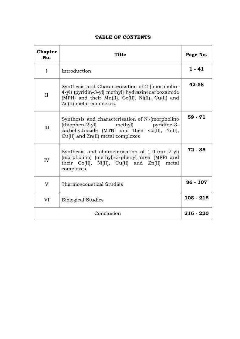
TABLE OF CONTENTS
Chapter No. Title Page No.
I Introduction 1 - 41
II
Synthesis and Characterisation of 2-[(morpholin-4-yl) (pyridin-3-yl) methyl] hydrazinecarboxamide (MPH) and their Mn(II), Co(II), Ni(II), Cu(II) and Zn(II) metal complexes.
42-58
III
Synthesis and characterisation of Nꞌ-(morpholino (thiophen-2-yl) methyl) pyridine-3-carbohydrazide (MTN) and their Co(II), Ni(II), Cu(II) and Zn(II) metal complexes
59 - 71
IV
Synthesis and characterisation of 1-(furan-2-yl) (morpholino) (methyl)-3-phenyl urea (MFP) and their Co(II), Ni(II), Cu(II) and Zn(II) metal complexes
72 - 85
V Thermoacoustical Studies 86 - 107
VI Biological Studies 108 - 215
Conclusion 216 - 220
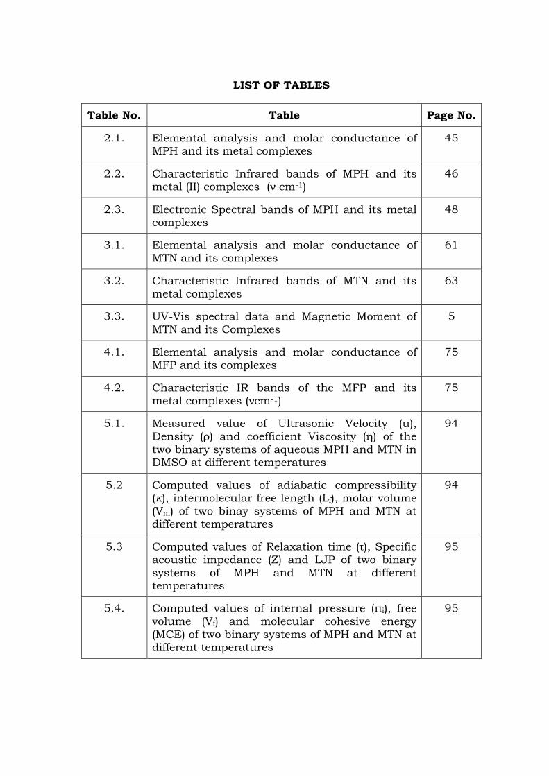
LIST OF TABLES
Table No. Table Page No.
2.1. Elemental analysis and molar conductance of MPH and its metal complexes
45
2.2. Characteristic Infrared bands of MPH and its metal (II) complexes (ν cm-1)
46
2.3. Electronic Spectral bands of MPH and its metal complexes
48
3.1. Elemental analysis and molar conductance of MTN and its complexes
61
3.2. Characteristic Infrared bands of MTN and its metal complexes
63
3.3. UV-Vis spectral data and Magnetic Moment of MTN and its Complexes
5
4.1. Elemental analysis and molar conductance of MFP and its complexes
75
4.2. Characteristic IR bands of the MFP and its metal complexes (νcm-1)
75
5.1. Measured value of Ultrasonic Velocity (u), Density (ρ) and coefficient Viscosity (η) of the two binary systems of aqueous MPH and MTN in DMSO at different temperatures
94
5.2 Computed values of adiabatic compressibility (κ), intermolecular free length (Lf), molar volume (Vm) of two binay systems of MPH and MTN at different temperatures
94
5.3 Computed values of Relaxation time (τ), Specific acoustic impedance (Z) and LJP of two binary systems of MPH and MTN at different temperatures
95
5.4. Computed values of internal pressure (πi), free volume (Vf) and molecular cohesive energy (MCE) of two binary systems of MPH and MTN at different temperatures
95
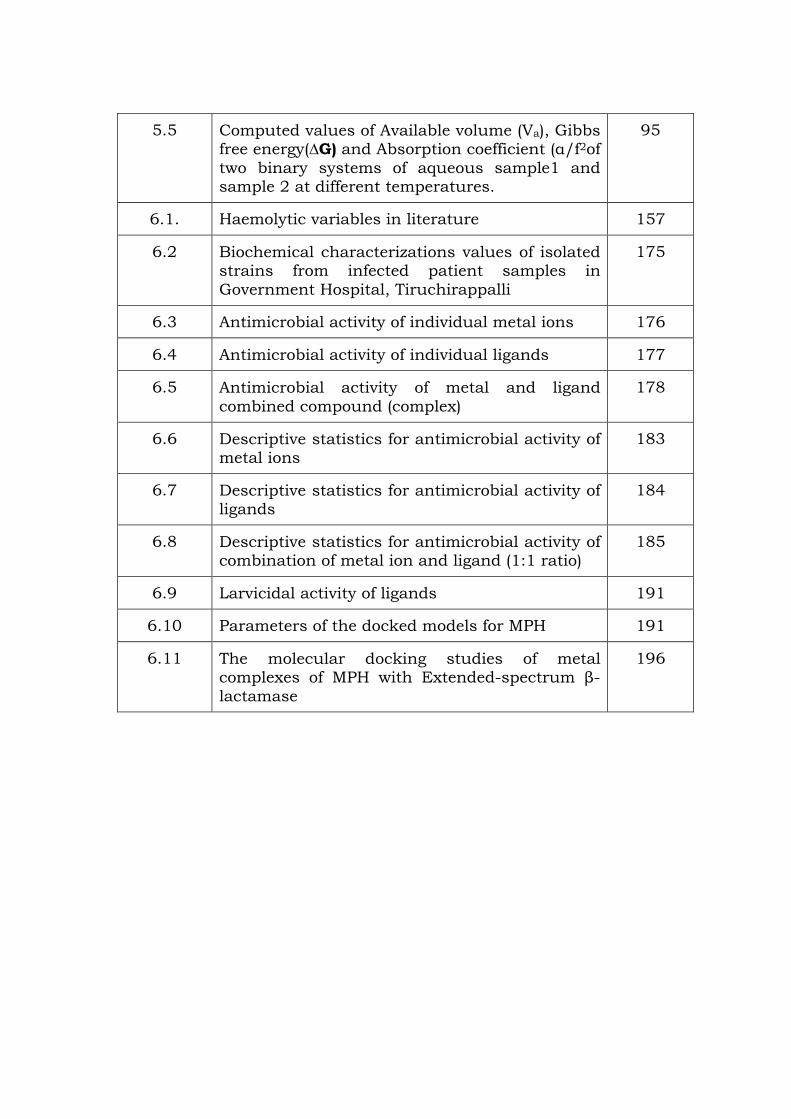
5.5 Computed values of Available volume (Va), Gibbs free energy(∆G) and Absorption coefficient (α/f2of two binary systems of aqueous sample1 and sample 2 at different temperatures.
95
6.1. Haemolytic variables in literature 157
6.2 Biochemical characterizations values of isolated strains from infected patient samples in Government Hospital, Tiruchirappalli
175
6.3 Antimicrobial activity of individual metal ions 176
6.4 Antimicrobial activity of individual ligands 177
6.5 Antimicrobial activity of metal and ligand combined compound (complex)
178
6.6 Descriptive statistics for antimicrobial activity of metal ions
183
6.7 Descriptive statistics for antimicrobial activity of ligands
184
6.8 Descriptive statistics for antimicrobial activity of combination of metal ion and ligand (1:1 ratio)
185
6.9 Larvicidal activity of ligands 191
6.10 Parameters of the docked models for MPH 191
6.11 The molecular docking studies of metal complexes of MPH with Extended-spectrum β-lactamase
196
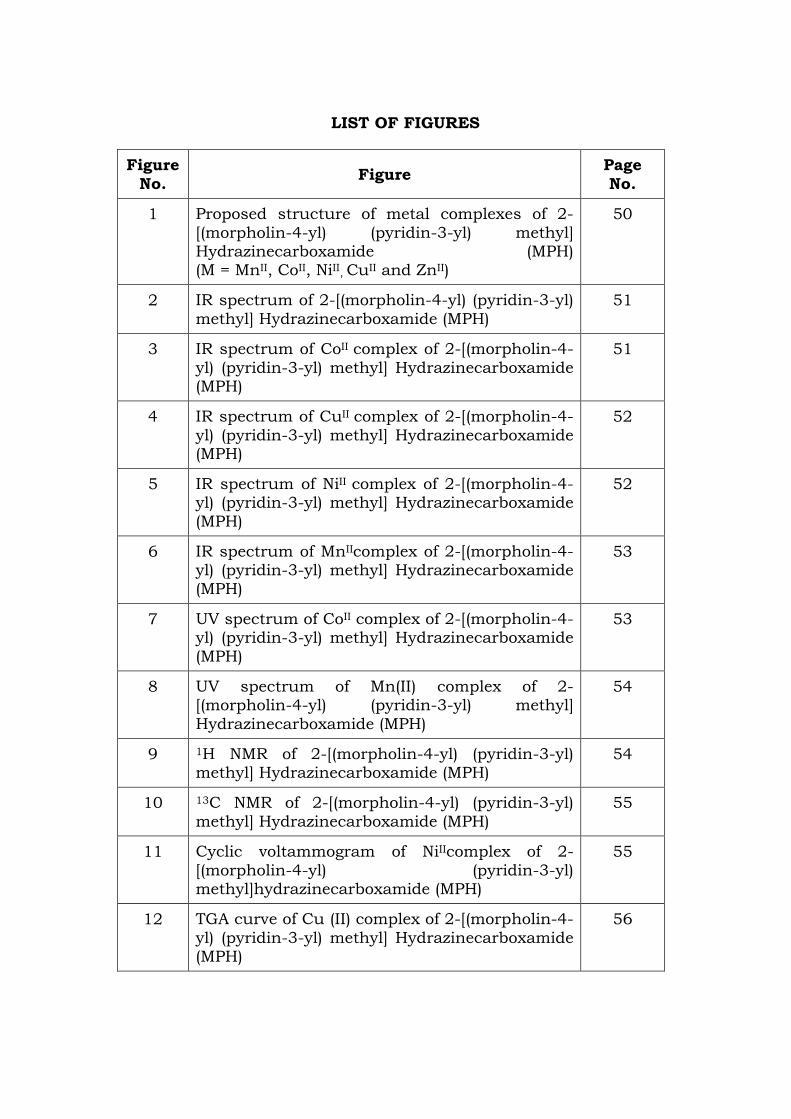
LIST OF FIGURES
Figure No. Figure Page
No.
1 Proposed structure of metal complexes of 2-[(morpholin-4-yl) (pyridin-3-yl) methyl] Hydrazinecarboxamide (MPH) (M = MnII, CoII, NiII, CuII and ZnII)
50
2 IR spectrum of 2-[(morpholin-4-yl) (pyridin-3-yl) methyl] Hydrazinecarboxamide (MPH)
51
3 IR spectrum of CoII complex of 2-[(morpholin-4-yl) (pyridin-3-yl) methyl] Hydrazinecarboxamide (MPH)
51
4 IR spectrum of CuII complex of 2-[(morpholin-4-yl) (pyridin-3-yl) methyl] Hydrazinecarboxamide (MPH)
52
5 IR spectrum of NiII complex of 2-[(morpholin-4-yl) (pyridin-3-yl) methyl] Hydrazinecarboxamide (MPH)
52
6 IR spectrum of MnIIcomplex of 2-[(morpholin-4-yl) (pyridin-3-yl) methyl] Hydrazinecarboxamide (MPH)
53
7 UV spectrum of CoII complex of 2-[(morpholin-4-yl) (pyridin-3-yl) methyl] Hydrazinecarboxamide (MPH)
53
8 UV spectrum of Mn(II) complex of 2-[(morpholin-4-yl) (pyridin-3-yl) methyl] Hydrazinecarboxamide (MPH)
54
9 1H NMR of 2-[(morpholin-4-yl) (pyridin-3-yl) methyl] Hydrazinecarboxamide (MPH)
54
10 13C NMR of 2-[(morpholin-4-yl) (pyridin-3-yl) methyl] Hydrazinecarboxamide (MPH)
55
11 Cyclic voltammogram of NiIIcomplex of 2-[(morpholin-4-yl) (pyridin-3-yl) methyl]hydrazinecarboxamide (MPH)
55
12 TGA curve of Cu (II) complex of 2-[(morpholin-4-yl) (pyridin-3-yl) methyl] Hydrazinecarboxamide (MPH)
56
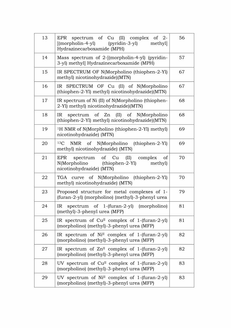
13 EPR spectrum of Cu (II) complex of 2- [(morpholin-4-yl) (pyridin-3-yl) methyl] Hydrazinecarboxamide (MPH)
56
14 Mass spectrum of 2-[(morpholin-4-yl) (pyridin-3-yl) methyl] Hydrazinecarboxamide (MPH)
57
15 IR SPECTRUM OF N(Morpholino (thiophen-2-Yl) methyl) nicotinohydrazide)(MTN)
67
16 IR SPECTRUM OF Cu (II) of N(Morpholino (thiophen-2-Yl) methyl) nicotinohydrazide)(MTN)
67
17 IR spectrum of Ni (II) of N(Morpholino (thiophen-2-Yl) methyl) nicotinohydrazide)(MTN)
68
18 IR spectrum of Zn (II) of N(Morpholino (thiophen-2-Yl) methyl) nicotinohydrazide)(MTN)
68
19 1H NMR of N(Morpholino (thiophen-2-Yl) methyl) nicotinohydrazide) (MTN)
69
20 13C NMR of N(Morpholino (thiophen-2-Yl) methyl) nicotinohydrazide) (MTN)
69
21 EPR spectrum of Cu (II) complex of N(Morpholino (thiophen-2-Yl) methyl) nicotinohydrazide) (MTN)
70
22 TGA curve of N(Morpholino (thiophen-2-Yl) methyl) nicotinohydrazide) (MTN)
70
23 Proposed structure for metal complexes of 1-(furan-2-yl) (morpholino) (methyl)-3-phenyl urea
79
24 IR spectrum of 1-(furan-2-yl) (morpholino) (methyl)-3-phenyl urea (MFP)
81
25 IR spectrum of CuII complex of 1-(furan-2-yl) (morpholino) (methyl)-3-phenyl urea (MFP)
81
26 IR spectrum of NiII complex of 1-(furan-2-yl) (morpholino) (methyl)-3-phenyl urea (MFP)
82
27 IR spectrum of ZnII complex of 1-(furan-2-yl) (morpholino) (methyl)-3-phenyl urea (MFP)
82
28 UV spectrum of CuII complex of 1-(furan-2-yl) (morpholino) (methyl)-3-phenyl urea (MFP)
83
29 UV spectrum of NiII complex of 1-(furan-2-yl) (morpholino) (methyl)-3-phenyl urea (MFP)
83

30 1H NMR of 1-(furan-2-yl) (morpholino) (methyl)-3-phenyl urea (MFP)
84
31 13C NMR of 1-(furan-2-yl) (morpholino) (methyl)-3-phenyl urea (MFP)
84
32 TGA curve of Cu(II) Chloro complex of 1-(furan-2-yl) (morpholino) (methyl)-3- phenyl urea (MFP)
85
33 Mass spectrum of 1-(furan-2-yl) (morpholino) (methyl)-3-phenyl urea (MFP)
85
34 Plots for ultrasonic velocity versus concentration for aqueous a) MPH and b) MTN at different temperatures () T = 303 K, (•) 308 K and () 313 K
97
35 Plots for adiabatic compressibility versus concentration for aqueous a) MPH and b) MTN at different temperatures () T = 303 K, (•) 308 K and () 313 K
98
36 Plots for internal pressure versus concentration for aqueous a) MPH and b) MTN at different temperatures () T = 303 K, (•) 308 K and () 313 K
99
37 Plots for Enthalpy versus concentration for aqueous a) MPH and b) MTN at different temperatures () T = 303 K, (•) 308 K and () 313 K
100
38 Plots for Gibbs free energy versus concentration for aqueous a) MPH and b) MTN at different temperatures () T = 303 K, (•) 308 K and () 313 K
101
39 Antibacterial activity of metal ions of MPH by agar Disc diffusion method
179
40 Antibacterial activity of metal ions and ligand MPH alone by agar Disc diffusion method
179
41 Antibacterial activity of metal ions and ligand MTN alone by agar Disc diffusion method
180
42 Antibacterial activity of metal ions and ligand MFP alone by agar Disc diffusion method
181
43 Combined Antibacterial activity of ligand MPH and metal ions (1:1) by agar well diffusion method
181
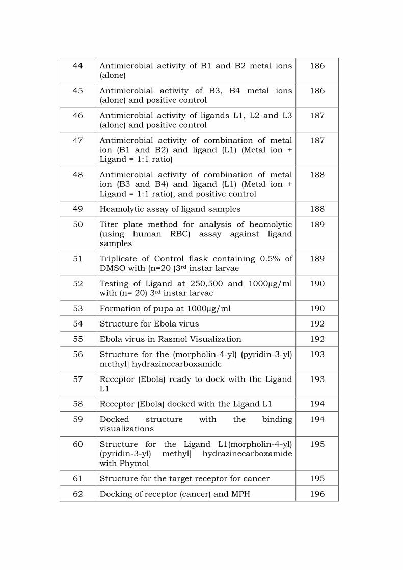
44 Antimicrobial activity of B1 and B2 metal ions (alone)
186
45 Antimicrobial activity of B3, B4 metal ions (alone) and positive control
186
46 Antimicrobial activity of ligands L1, L2 and L3 (alone) and positive control
187
47 Antimicrobial activity of combination of metal ion (B1 and B2) and ligand (L1) (Metal ion + Ligand = 1:1 ratio)
187
48 Antimicrobial activity of combination of metal ion (B3 and B4) and ligand (L1) (Metal ion + Ligand = 1:1 ratio), and positive control
188
49 Heamolytic assay of ligand samples 188
50 Titer plate method for analysis of heamolytic (using human RBC) assay against ligand samples
189
51 Triplicate of Control flask containing 0.5% of DMSO with (n=20 )3rd instar larvae
189
52 Testing of Ligand at 250,500 and 1000µg/ml with (n= 20) 3rd instar larvae
190
53 Formation of pupa at 1000µg/ml 190
54 Structure for Ebola virus 192
55 Ebola virus in Rasmol Visualization 192
56 Structure for the (morpholin-4-yl) (pyridin-3-yl) methyl] hydrazinecarboxamide
193
57 Receptor (Ebola) ready to dock with the Ligand L1
193
58 Receptor (Ebola) docked with the Ligand L1 194
59 Docked structure with the binding visualizations
194
60 Structure for the Ligand L1(morpholin-4-yl) (pyridin-3-yl) methyl] hydrazinecarboxamide with Phymol
195
61 Structure for the target receptor for cancer 195
62 Docking of receptor (cancer) and MPH 196
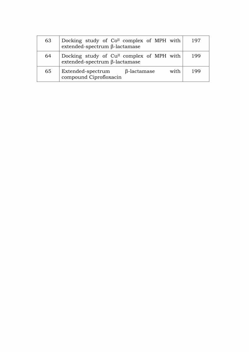
63 Docking study of CoII complex of MPH with extended-spectrum β-lactamase
197
64 Docking study of CuII complex of MPH with extended-spectrum β-lactamase
199
65 Extended-spectrum β-lactamase with compound Ciprofloxacin
199
Chapter I
INTRODUCTION
Chemistry has accomplished rapid progress in understanding the
properties of all of the elements. Among the various branches of
chemistry, inorganic chemistry is of fundamental importance not only
as a basic science but also as one of the most useful sources for
modern technologies. The main purpose of inorganic chemistry in near
future will be the synthesis of the compounds with unexpected bonding
modes and structures and with the discoveries of novel reactions and
physical properties of new compounds. Inorganic compounds are also
indispensable in the frontier chemistry of organic synthesis using metal
complexes, homogeneous catalysis, bioinorganic functions etc.
Coordination chemistry plays a vital role and it is one of the most
active research fields in inorganic chemistry. Coordination chemistry
assumed a vital significance with the development of bioinorganic
chemistry, which is mainly the chemistry of coordination compounds
[1]. Coordination chemistry, emerged from the work of Alfred Werner, a
Swiss chemist who examined different compounds composed of cobalt
(III) chloride and ammonia. The coordination chemists have showed
their major interest in the stereochemistry of the coordination
compounds. Nowadays, the research on coordination complexes has
been increased due to their magnetic, optical, electronic properties and
also due to their complex structures.
Due to the catalytic and bioinorganic relevance, the chemistry of
transition metal complexes has received a considerable interest in the
research field [2-7]. The literature survey clearly reveals that transition
1
metal ions have been subjected to detailed investigations. Nowadays,
very large number of ligands are widely used with many number of
transition metal ions. Research on higher dimensional compounds
such as multinuclear complexes, cluster compounds and solid-state
inorganic compounds in which metal atoms and ligands are bonded in
a complex manner is becoming much easier. Most of the molecular
compounds of transition metals are metal complexes and
organometallic compounds in which ligands are coordinated to metals.
These molecular compounds include not only mononuclear complexes
with a metal centre but also multinuclear complexes containing several
metals, or cluster complexes having metal-metal bonds. Among the
various types of ligands, the heterocyclic bases containing oxygen and
nitrogen donors are considered to be the potential ligand centres for the
coordination of the metal atom. In the recent years, a variety of ligands
have been studied are the ligands containing oxygen and nitrogen as
donor [8-14]. Due to this, the coordination chemistry of nitrogen-
oxygen donor ligands has made a considerable interest in the field of
research. The Mannich and Schiff base compounds generally contain
the nitrogen, oxygen and sulphur donor atoms as donor and these
types of compounds exhibits a wide range of biological properties such
as antibacterial, antifungal [15-28], anti HIV [19-23 & 29-30], antiviral
[31-32], anticonvulsant [33-36], antitubercular [37-39] and anticancer
[40-42].
Mannich Reaction
The Mannich reaction is a classical method for the preparation of
β-amino ketones and aldehydes. Mannich reaction is one of the most
2
important basic reaction types in organic chemistry. It is the key step
in the synthesis of numerous pharmaceuticals and natural products.
The amino alkylation of CH-acidic compounds was described by
several authors as early as the 19th century. However, it was Carl
Mannich, who was the first to recognize the enormous significance of
this reaction type, and it was he who extended the chemistry into a
broad based synthetic methodology through systematic research. Since
then this reaction that now carries his name has developed into one of
the most important C C bond forming reactions in organic chemistry
[43-44].
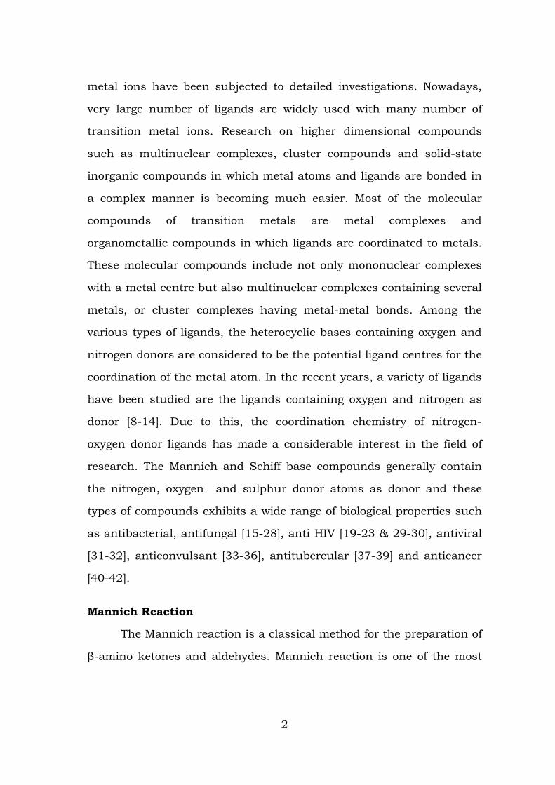
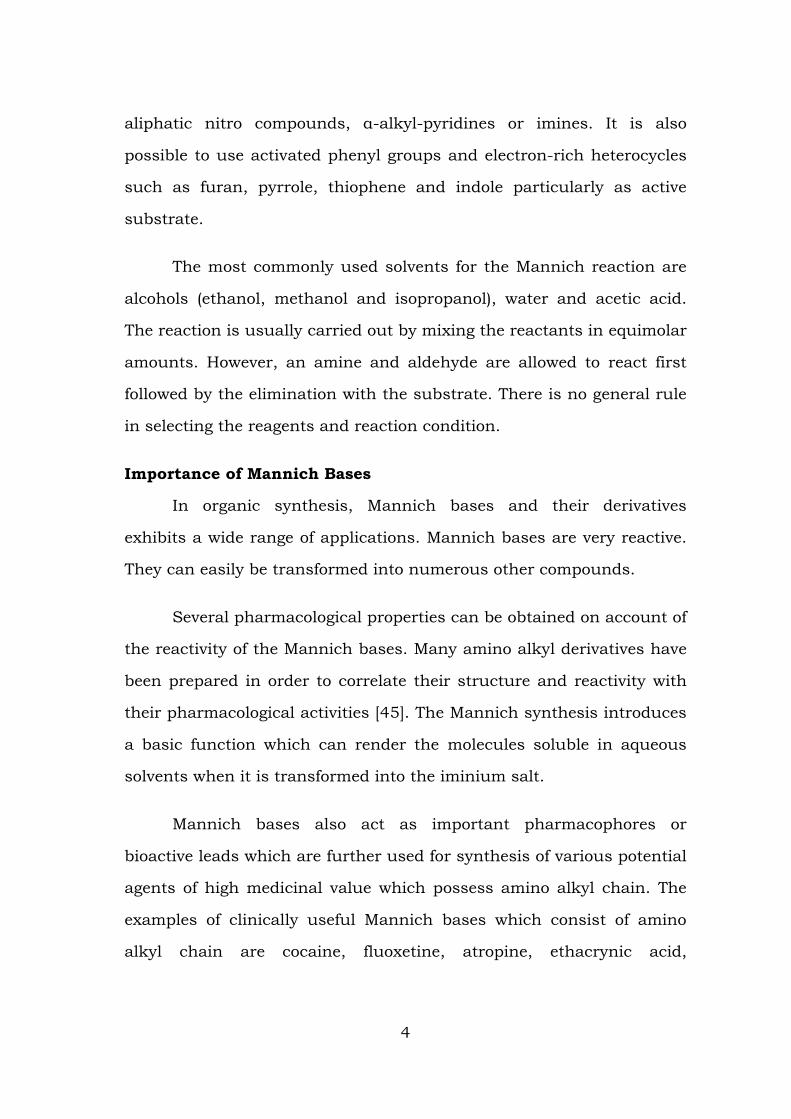
Thus, the Mannich reaction is a three-component reaction, which
involves the condensation of a compound capable of supplying one or
more active hydrogen atoms with an aldehyde (usually formaldehyde)
and an N-H derivative (ammonia, any primary or secondary amine or
amide) in the presence of an acid to give β-amino carbonyl compounds.
In the Mannich reaction, the Mannich base is the end product, which is
a nucleophilic addition of an amine to a carbonyl group followed by
dehydration to the Schiff base.
The activation of aldehyde, primary or secondary amines or
ammonia are employed in the above reaction. For forming the
intermediate enamine, the tertiary amines are not used due to the lack
of an N–H proton. The nucleophiles (α-CH-acidic compounds) employed
for this reaction include carbonyl compounds, nitriles, acetylenes,
3
aliphatic nitro compounds, α-alkyl-pyridines or imines. It is also
possible to use activated phenyl groups and electron-rich heterocycles
such as furan, pyrrole, thiophene and indole particularly as active
substrate.
The most commonly used solvents for the Mannich reaction are
alcohols (ethanol, methanol and isopropanol), water and acetic acid.
The reaction is usually carried out by mixing the reactants in equimolar
amounts. However, an amine and aldehyde are allowed to react first
followed by the elimination with the substrate. There is no general rule
in selecting the reagents and reaction condition.
Importance of Mannich Bases
In organic synthesis, Mannich bases and their derivatives
exhibits a wide range of applications. Mannich bases are very reactive.
They can easily be transformed into numerous other compounds.
Several pharmacological properties can be obtained on account of
the reactivity of the Mannich bases. Many amino alkyl derivatives have
been prepared in order to correlate their structure and reactivity with
their pharmacological activities [45]. The Mannich synthesis introduces
a basic function which can render the molecules soluble in aqueous
solvents when it is transformed into the iminium salt.
Mannich bases also act as important pharmacophores or
bioactive leads which are further used for synthesis of various potential
agents of high medicinal value which possess amino alkyl chain. The
examples of clinically useful Mannich bases which consist of amino
alkyl chain are cocaine, fluoxetine, atropine, ethacrynic acid,
4
trihexyphenidyl, procyclidine, ranitidine, biperiden [46-48], and so
forth.
Mannich bases are known to play a vital role in the development
of synthetic pharmaceutical chemistry.
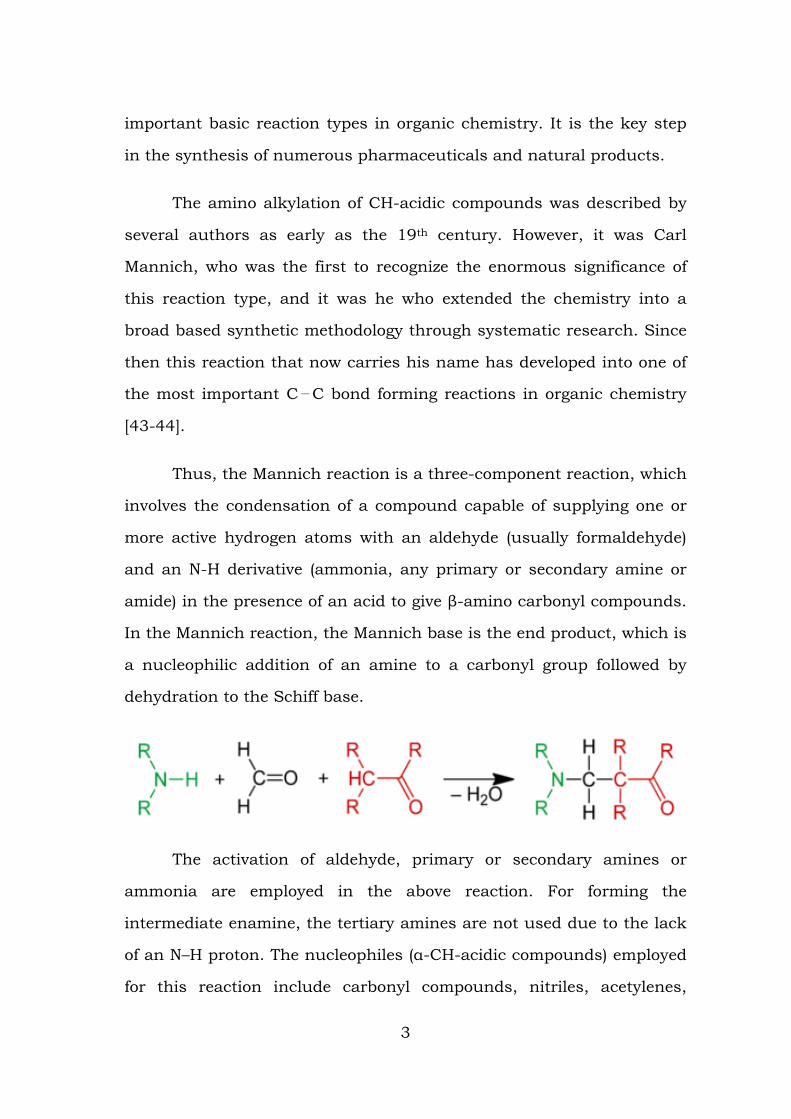
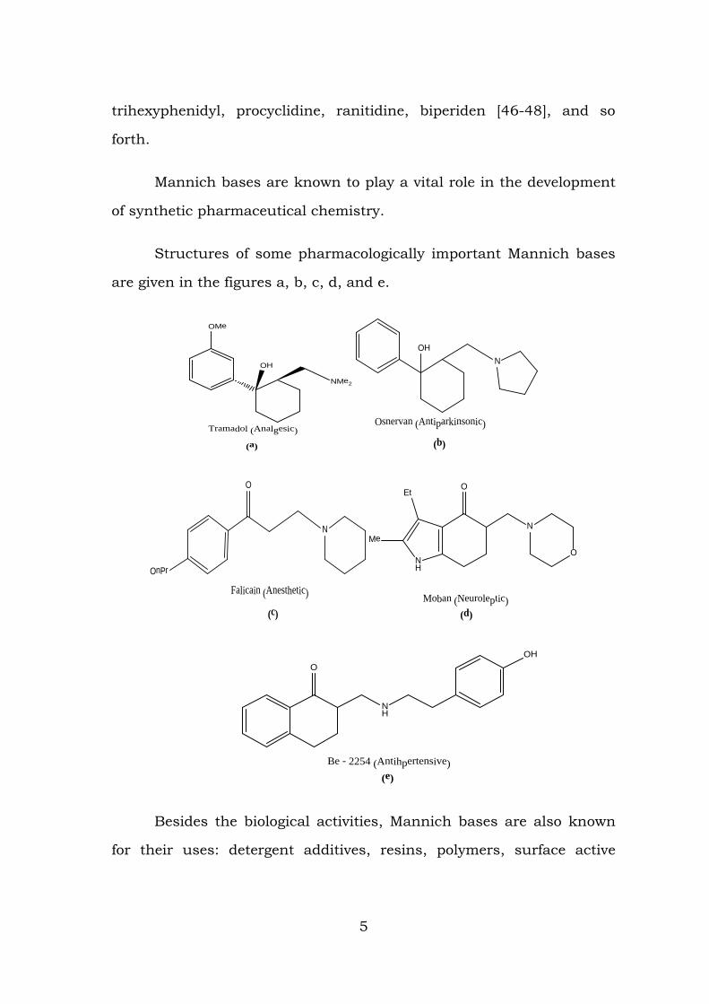
Structures of some pharmacologically important Mannich bases
are given in the figures a, b, c, d, and e.
OMe
OH
NMe2
Tramadol (Analgesic)
(a)
N
Osnervan (Antiparkinsonic)
OH
(b)
OnPr
O
N
Falicain (Anesthetic)
(c)
NH
Et
MeN
O
O
Moban (Neuroleptic)(d)
NH
OHO
Be - 2254 (Antihpertensive)(e)
Besides the biological activities, Mannich bases are also known
for their uses: detergent additives, resins, polymers, surface active
5
agents, and so forth. Pro drugs of Mannich bases of various active
compounds have been prepared to overcome the limitations [49].
Mannich bases (optically pure chiral) of 2-naphthol are employed
for catalysis (ligand accelerated and metal mediated) of the enantio
selective carbon-carbon bond formation. Mannich bases and their
derivatives are intermediates for the synthesis of bioactive molecules
[50, 51].
Mannich reaction is widely used for the construction of nitrogen
containing compounds [52]. Mannich bases are used in agrochemicals
as plant growth regulators.
Denton and his co-workers [53] indicated that the reduction of
the piperidino Mannich bases to the corresponding amino alcohols led
to the increase in antispasmodic activity.
Mannich bases derived from bis-[2-chloroethyl]-amine, melamine
or ethyldiamines are found to possess antimicrobial and cardio tonic
activities [54-56].
It has been reported that the aminobenzylated Mannich bases of
3-bromobenzaldehyde with suitable secondary amines and the
compounds containing amide moiety were found to possess many
interesting biological properties [57].
GENERAL SURVEY OF THE LIGAND CONSTITUENTS
Aldehydes
Survey of literature reveals that a large number of reports are
available for the synthesis of Mannich reaction by using aldehydes
other than Formaldehyde. Aromatic aldehydes, aliphatic aldehydes and
6
heteroaldehydes are some of the types of aldehydes which are used in
the Mannich reaction. A large number of reports are available in the
literature, benzaldehyde as a reactant in the synthesis of Mannich
Base. Much work has been carried out by using chloro, bromo, nitro
and amino substituted benzaldehydes.
A survey on the literature [58] shows that in the orientation of
the Mannich base reaction, the unsymmetrical dialkylketone,
secondary aliphatic amine and formaldehyde condensation takes place
mainly on the C – atom having less number of carbon unless this C is
sterically hindered. But in the case of aromatic aldehydes, due to steric
hindrance, the reaction seems to take place on the C - atom which are
having more number of hydrogen atoms. The second bulk aldehydic
group cannot remove other hydrogen atoms to incorporate itself and
hence the reaction does not proceed further.
Amines
Amines and their derivatives are important functional groups in a
variety of natural products. Drugs based on amine moieties have been
extensively used in the pharmaceutical industries [59]. Amines and
their derivatives act as precursors to a variety of biologically active
compounds like pharmaceuticals and agrochemicals [60]. The direct
reductive amination of aldehydes and ketones is one of the most useful
methods for the synthesis of secondary and tertiary amines [61].
Among the various types of organic compounds, amines constitute an
important type by replacing one or more hydrogen atoms of ammonia
molecule by alkyl or aryl group. The Nitrogen atom of amines carries an
unshared pair of electrons and it is trivalent. The lower aliphatic
amines can form hydrogen bonds with water molecules and hence they
7
are soluble in water. However, solubility decreases with the increase in
molar mass of amines due to the increase in size of the hydrophobic
alkyl part and will cause the intermolecular association in the primary
and secondary amines. This is due to the hydrogen bonding between
nitrogen of the one and nitrogen of the other molecule.
This intermolecular association is more in primary amines than in
secondary amines as there are two hydrogen atoms available for
hydrogen bond formation in it. But in the case of tertiary amines, the
intermolecular association does not happens, due to the absence of
hydrogen atom available for hydrogen bond formation. The reaction of
the amines can be decided by the number of hydrogen atoms attached
to the nitrogen atoms and in this way only, the primary, secondary and
tertiary amines differ with each other in the reactions. Due to the
presence of unshared electron pair in the amines, they act as
nucleophiles. In the case of aniline or other aryl amines, the –NH2
group is attached directly to the benzene ring and it makes less for
protonation. In most of the Mannich reactions, the amines used are
either primary or secondary amine. But secondary amines are preferred
in Mannich base synthesis since it possess only one replaceable
hydrogen atom (R2NH). The product with primary amine is secondary
amine, which further reacts to give secondary amine [62].
The Mannich donors like ketones or aldehydes, will have the
chiral amines. The resulting enamine can attack a Mannich acceptor,
usually a pro chiral aldimine, by introducing one or two chiral centres
in the Mannich base. Several acyclic chiral amines, amino acids and
amides are served as amines in the synthesis of Mannich bases. In the
8
present study Morpholine has been employed as an amine for the
synthesis of Mannich Bases.
Active Hydrogen atom
A large number of reports are available by employing active
hydrogen atom as a substrate in the synthesis of Mannich base. The
amide moieties of the organic compounds has placed a significant role
in biology, because they are the repeating unit of polypeptide
macromolecules. The coordination chemists has made a considerable
interest on these polypeptide macromolecules, because they serve as
models for peptide interaction and to metalloenzymes [63, 64]. The
N – amino methylation of acyclic carboxamides has been carried out
with primary and secondary amines [65, 66].
A number of reports available in the literature using amide
moieties as substrates for the synthesis of Mannich base. A study
concerning amino methylation reaction in several benzamides and
chloroacetamides established that the reaction only takes place with
sufficiently basic amines or with amides derived from sufficiently strong
carboxylic acids [67] have synthesized Mannich bases employing
thiourea and thiocarboxamides as substrates.
Many researchers have employed acetamide, urea, thiourea and
their derivatives as a compound containing active hydrogen for the
synthesis of Mannich bases [68-78]. Besides, semicarbazide, nicotinic
acid hydrazide, benzohydrazide and piperazine carboxamide are also
employed as substrate in the Mannich base synthesis in recent years.
Considerable interest has been paid on the synthesis of Mannich
bases using heterocyclic derivatives, many new drugs has been
9
developed. A large number of pharmacological activities are present in
the heterocyclic compounds such as Pyrrole, furan and thiophene and
their derivatives. The potential entity of the heterocyclic compounds
which possess the pharmacological characteristics has been
established by the thiophene nucleus. The medicinal chemists has paid
a considerable interest on the physicochemical parameters of the
heterocyclic compounds in order to produce combinatorial library and
carry out exhaustive efforts in the search of lead molecules.
A probe into the literature clearly reveals that aldehydes like
formaldehyde, benzaldehyde and substituted benzaldehyde are used
along with secondary amines such as piperidine and piperazine etc.,
and a compound containing an active hydrogen atom like alkyl ketones,
phenols, carboxylic acid derivatives, heterocyclic compounds, alkynes
and amides are used for the synthesis of the Mannich bases. It has
been found that, a few reports are available for the synthesis of
Mannich bases using thiophene-2-carboxaldehyde and pyridine-3-
carboxaldehyde as the component.
Based on the above considerable facts, the present study is
aimed at the synthesis of Mannich bases by reacting Furfuraldehyde,
Thiophene-2-Carboxaldehyde and Pyridine-3-Carboxaldehyde,
Morpholine as an amine and the compounds such as semicarbazide,
nicotinic acid hydrazide and phenyl urea as the compound containing
active hydrogen atom. Employing the synthesized product of the
reactants as ligand, metal complexes of Mn (II), Co (II), Ni (II), Cu (II),
and Zn (II) have been prepared.
10
Major applications of the ligand constituents
Semicarbazide
Semicarbazide is prepared by treating urea with hydrazine [79].
OC (NH2)2 + N2H4→ OC (NH2) (N2H3) + NH3
Many derivatives of Semicarbazide and their metal complexes are
found as efficient drugs against influenza, tuberculosis and some kinds
of tumours. Semicarbazide is used in preparing pharmaceuticals
including nitro furan anti bacterials (furazolidone, nitrofurazone,
nitrofurantoin) and related compounds. Semicarbazide is used as a
detection reagent in thin layer chromatography (TLC). Semicarbazide
stains α-keto acids on the TLC plate, which must then be viewed under
ultraviolet light to see the results.
Clinical use of Semicarbazide (a SSAO-inhibitor) gives the
evidence that an inflammatory reaction can be reduced by blocking the
enzymatic activity of the enzyme Semicarbazide-sensitive amine oxidase
(SSAO). SSAO activity was found significantly increased in blood and
tissues in some pathological conditions. The enzyme activity has been
reported to be elevated in diabetes and cancer. The mean specific
activity of SSAO was significantly elevated in the group of patients
having prostate cancer with skeletal metastases. Semicarbazide (and
SSAO blockers) can reduce inflammatory response and can protect
against the progressive vascular complications caused by oxidative
stress. It also can reduce pain. The SSAO inhibitors also appears able
to protect endothelial cells against toxic effects of free radicals [80].
Semicarbazide itself is a standard enzyme inhibitor and new SSAO
inhibitors are in development. SSAO inhibitors significantly blocked the
11
catalytic activity of VAP-1 in tumour, attenuated tumour progression,
and reduced neo-angiogenesis [81]. Semicarbazide is a known inhibitor
of glutamic acid decarboxylase (GAD), the enzyme responsible for GABA
synthesis. GABA has emerged as a tumour signalling molecule in the
periphery that controls the proliferation of tumour cells and perhaps
tumour stem cells [82].
Morpholine
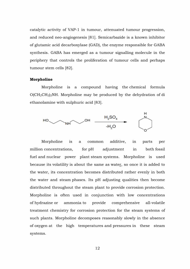
Morpholine is a compound having the chemical formula
O(CH2CH2)2NH. Morpholine may be produced by the dehydration of di
ethanolamine with sulphuric acid [83].
Morpholine is a common additive, in parts per
million concentrations, for pH adjustment in both fossil
fuel and nuclear power plant steam systems. Morpholine is used
because its volatility is about the same as water, so once it is added to
the water, its concentration becomes distributed rather evenly in both
the water and steam phases. Its pH adjusting qualities then become
distributed throughout the steam plant to provide corrosion protection.
Morpholine is often used in conjunction with low concentrations
of hydrazine or ammonia to provide comprehensive all-volatile
treatment chemistry for corrosion protection for the steam systems of
such plants. Morpholine decomposes reasonably slowly in the absence
of oxygen at the high temperatures and pressures in these steam
systems.
12
Morpholine undergoes most chemical reactions typical for other
secondary amines, though the presence of the ether oxygen withdraws
electron density from the nitrogen, rendering it less nucleophilic (and
less basic) than structurally similar secondary amines such
as piperidine. For this reason, it forms a stable chloramine [84].
It is commonly used to generate enamine [85]. Morpholine is
widely used in organic synthesis. For example, it is a building block in
the preparation of the antibiotic linezolid, the anticancer agent gefitinib
(Iressa) and the analgesic dextromoramide. In research and in industry,
the low cost and polarity of Morpholine lead to its common use as a
solvent for chemical reactions.
Morpholine is used as a chemical emulsifier in the process of
waxing fruit. Naturally, fruits make waxes to protect against insects
and fungal contamination [86].
Furfuraldehyde
Furfural is an organic compound derived from a variety of
agricultural by products, including corn, cobs, oat, wheat bran,
and sawdust. Furfural is a heterocyclic aldehyde, with the ring
structure. Its chemical formula is OC4H3CHO.
Furfural is an important renewable, non-petroleum based,
chemical feedstock. Hydrogenation of furfural provides furfuryl
alcohol (FA), which is a useful chemical intermediate and which may be
further hydrogenated to tetra hydro furfuryl alcohol (THFA). THFA is
used as a non-hazardous solvent in agricultural formulations and as an
adjuvant to help herbicides penetrate the leaf structure. It is used to
13
make other furan chemicals, such as furoic acid, via oxidation [87] and
furan itself via palladium catalyzed vapour phase decarbonylation [88].
Biological importance and Coordination Chemistry of Mn (II),
Co (II), Ni (II), Cu (II) and Zn (II) Metal Ions
Transition metal ions play an important role in the living systems
as metal binding systems. The active research of metal complexes has
made a considerable interest in the field of biochemical reactions. Many
researchers has made an interest on the role of metal ions in drug
metabolism, biology and cross linking of biomolecules from the various
disciplines such as chemistry, biology, medicine and agriculture.
Copper
Copper proteins have diverse roles in biological electron transport
and oxygen transportation, processes that exploit the easy inter
conversion of Cu (II) and Co (II) [89-92] the biological role for copper
commenced with the appearance of oxygen in earth's atmosphere [93].
The protein hemocyanin is the oxygen carrier in most mollusks and
some arthropods such as the horseshoe crab (Limulus polyphemus)
[94], because, hemocyanin is blue, these organisms have blue blood,
not the red blood found in organisms that rely on hemoglobin for this
purpose. Structurally related to hemocyanin are the laccases and
tyrosinases. Instead of reversibly binding oxygen, these proteins
hydroxylate substrates, illustrated by their role in the formation of
lacquers [95].
Copper is also a component of other proteins associated with the
processing of oxygen. In cytochrome c oxidase, which is required for
aerobic respiration, copper and iron cooperate in the reduction of
14

oxygen. Copper is also found in many superoxide dismutases, proteins
that catalyze the decomposition of superoxides, by converting it (by
disproportionation) to oxygen and hydrogen peroxide.
2HO2→ H2O2 + O2
Several copper proteins, such as the "blue copper proteins", do
not interact directly with substrates, hence they are not enzymes.
These proteins relay electrons by the process called electron transfer.
Photosynthesis functions by an elaborate electron transport
chain within the thylakoid membrane. A central "link" in this chain is
plastocyanin, a blue copper protein.
Copper, like all metals, forms coordination complexes with
ligands. In aqueous solution, copper (II) exists as [Cu (H2O) 6]2+. This
complex exhibits the fastest water exchange rate (speed of water
ligands attaching and detaching) for any transition metal aquo
complex. Adding aqueous sodium hydroxide causes the precipitation of
light blue solid copper (II) hydroxide. A simplified equation is:
Cu2+ + 2 OH−→Cu (OH) 2
Aqueous ammonia results in the same precipitate. Upon
adding excess ammonia, the precipitate dissolves, forming
tetraamminecopper (II):
Cu (H2O) 4(OH) 2 + 4 NH3→ [Cu (H2O) 2(NH3)4]2+ + 2 H2O + 2 OH−
Many other oxyanions form complexes; these include copper (II)
acetate, copper (II) nitrate, and copper (II) carbonate.Copper (II) sulfate
15
forms a blue crystalline pentahydrate, which is the most familiar
copper compound in the laboratory.
Cobalt
Cobalt is essential to all animals. It is a key constituent of
cobalamin, also known as vitamin B12, which is the primary biological
reservoir of cobalt as an "ultra trace" element [96, 97]. Bacteria in the
guts of ruminant animals convert cobalt salts into vitamin B12, a
compound which can only be produced by bacteria or archaea. The
minimum presence of cobalt in soils therefore markedly improves the
health of grazing animals, and an uptake of 0.20 mg/kg a day is
recommended for them, as they can obtain vitamin B12 in no other way
[98]. The cobalt deficiency was overcome by the development of "cobalt
bullets", dense pellets of cobalt oxide mixed with clay, which are orally
inserted to lodge in the animal's rumen.
Manganese
Manganese is a metal with important industrial metal alloy uses,
particularly in stainless steels.
In biology, manganese (II) ions function as cofactors for a large
variety of enzymes with many functions [99]. Manganese enzymes are
particularly essential in detoxification of superoxide free radicals in
organisms that must deal with elemental oxygen. Manganese also
functions in the oxygen-evolving complex of photosynthetic plants. The
element is a required trace mineral for all known living organisms but
is a neurotoxin. In larger amounts, and apparently with far greater
effectiveness through inhalation, it can cause a poisoning syndrome in
mammals, with neurological damage which is sometimes irreversible.
16
Nickel
Nickel (II) forms a very large number of complexes with a variety
of structures. Six-coordinated octahedral, five coordinated trigonal,
bipyramid and square pyramid and four coordinated tetrahedral and
square planar structures are some of them.
In aqueous solution, Ni2+ is present as [Ni (H2O)] 2+. The ligands
will easily replace the water molecules. In biological chemistry, Nickel is
a versatile metal and it is necessary of certain metallo-proteins. Nickel
acts as a tool for the study of the nucleic acid structure which is in the
form of tetraza macrocyclic complexes. Nickel plays a significant role in
pigmentation of metals which are found in ribo-nucleic acids.
Zinc
Zinc is the only transition metal, which requires by at least one
enzyme in each of the major classes of enzymatic activities. Zinc is a
versatile metal. It predominately forms four-coordinated tetrahedral
complexes. Complexes like [Zn (NH3)4]2+, [Zn (NH3)2Cl2], [Zn (Py)2 Cl2],
[Zn (CN) 4]2- are all tetrahedral. Zinc is a co-factor in a number of
enzyme systems. Zinc acts predominately as a Lewis acid and is found
in many metalloenzymes such as carboxypeptidase and carbonic
anhydrase for the following reasons:
1. Zinc (II) is active in many model systems.
2. Zinc (II) has no redox behaviour associated with it under
biological conditions.
3. The ligand exchange processes on Zinc (II) are rapid so that
substrates produced can be easily introduced and can be
removed.
17
Carbonic anhydrase, a Zinc metalloenzyme present in the
animals and plants, catalyses the interconversion of carbondioxide and
carbonates. Large number of diseases are associated with lowered zinc
level in blood. Various neurological diseases such as Alzheimer’s
disease, Parkinson’s disease, hypoxia-ischemia and epilepsy are closely
related to a disorder of zinc metabolism.
LITERATURE SURVEY OF MANNICH BASE AND ITS METAL
COMPLEXES
Metal complexes of Mannich base are formed by the strong ability
of the organic chelating ligands. These ligands contains amide moiety
which acts as a functional group. And also they exhibit a wide range of
biological properties such as bacteriocidal, fungicidal, herbicidal and
insecticidal activities [100-101].The amide group has two potential
binding sites viz., oxygen and nitrogen for complexation with metal ion.
Generally amide acts as a unidentate neutral ligand with oxygen
coordination to Lewis acid; however in the form of amidate anion
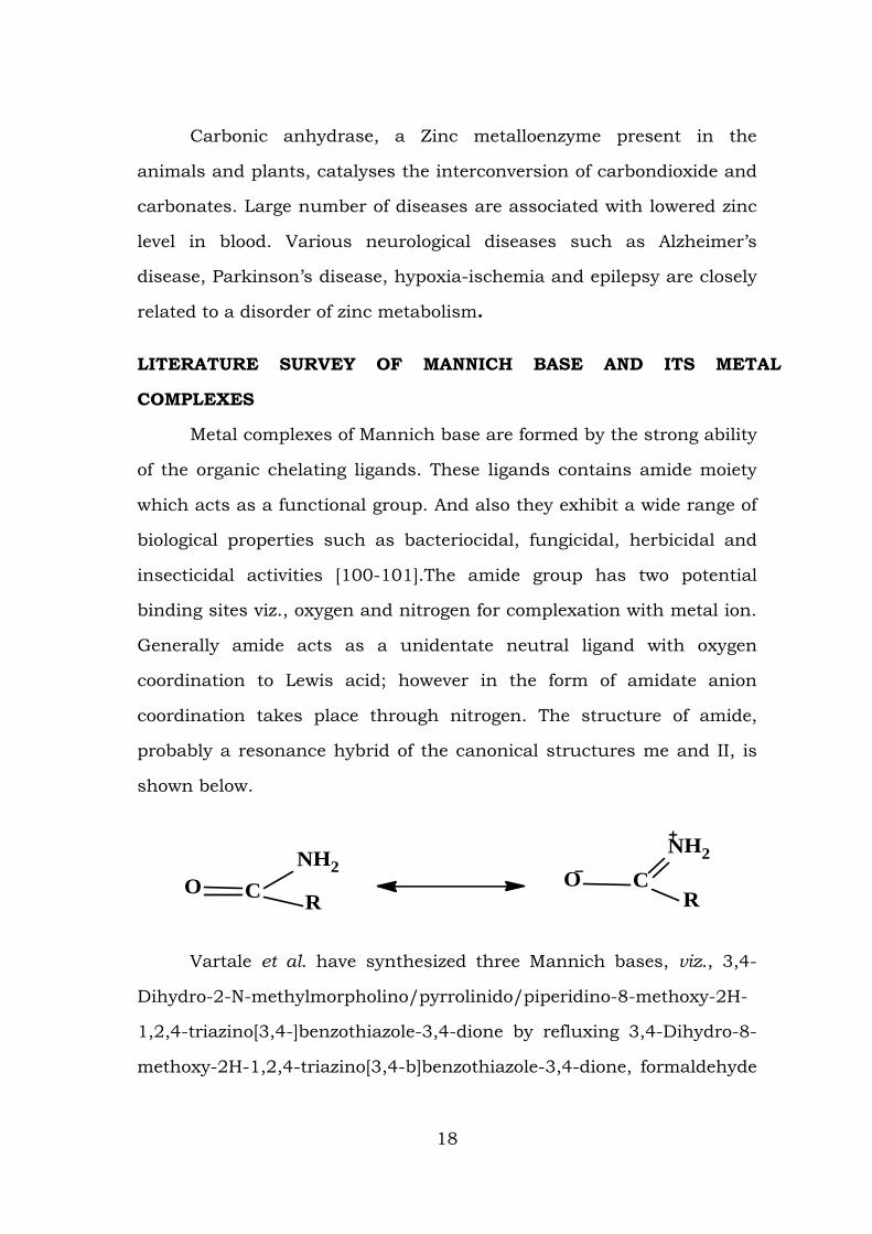
coordination takes place through nitrogen. The structure of amide,
probably a resonance hybrid of the canonical structures me and II, is
shown below.
CONH2
RCO
NH2
R
Vartale et al. have synthesized three Mannich bases, viz., 3,4-
Dihydro-2-N-methylmorpholino/pyrrolinido/piperidino-8-methoxy-2H-
1,2,4-triazino[3,4-]benzothiazole-3,4-dione by refluxing 3,4-Dihydro-8-
methoxy-2H-1,2,4-triazino[3,4-b]benzothiazole-3,4-dione, formaldehyde
18
and morpholine/ pyrolidene / piperidine with catalytic amount of
dioxane the compounds have been characterized by spectral studies.
Further, the compounds were screened for their antibacterial activities
against gram-positive and gram-negative bacteria [102].
Transition Metal Complexes of N-(1-Piperidinosalicylyl) acetamide
(PSA) and their Biological activities were studied by D. Sathya et al. IR
study of the PSA and its complexes show involvement of the nitrogen
atom from the piperidine ring and oxygen atom of amide are in
coordination with the metal ion. The electronic spectra of the metal
complexes suggest distorted octahedral structure for Cu (II) complex,
tetrahedral geometry for Co (II) complex, square planar geometry for Ni
(II) and Zn (II) complexes.
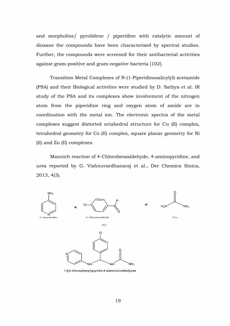
Mannich reaction of 4-Chlorobenzaldehyde, 4-aminopyridine, and
urea reported by G. Vishnuvardhanaraj et al., Der Chemica Sinica,
2013, 4(3).
19
Idhayadhulla et al. have synthesized six Mannich bases by fixing
Morpholine as an amine and changing the aldehyde and active
hydrogen compound. Elemental analysis and spectral studies have
been carried out to establish the structures of the compounds. The
synthesized compounds were screened for antimicrobial studies.
Structure activity relationship (SAR) has also been studied.
Bui Trung Hieu et al. prepared and characterized a series of
novel Mannich bases including (E)-1-(4-Chlorophenyl)-3-(4-
hydroxy-3-morpholine-4-yl-methyl-phenyl)-propenone. The synthesized
compounds were screened for their In vitro cytotoxic activity against the
human hepta cellular carcinoma, human lung carcinoma and human
breast cancer.
N-(morpholinobenzyl)benzamide, a Mannich base has been
prepared by the condensation of morpholine, benzamide and
benzaldehyde, and its Cu(II), Co(II), Ni(II) and Zn(II) complexes have
been synthesized by Raman et al. All the products have characterized
by spectral studies, magnetic moment, cyclic voltammetry and
conductance methods. All the complexes exhibit square planar
geometry. The synthesized Mannich base and its complexes have been
screened for their antimicrobial activities. The complexes have higher
antimicrobial activities than that of the free Mannich base and the
control.
Kadry et al. have synthesized two series of novel Mannich bases
such as1-(4-hydroxy-3-(morpholinomethyl) phenyl)-3-(1H-indole-3-yl)
prop-2-en-1- one and 1-(4-bromophenyl)-3-(4-hydroxy-3-methoxy-
5(morpholinomethyl) phenyl) prop-2-en-1-one. These compounds have
20
been characterized by elemental analysis, spectroscopic method and
chromatographic techniques and evaluated for their cytotoxic activity.
All the tested compounds exhibited a broad spectrum of antitumor
activity against renal cancer UO-31.
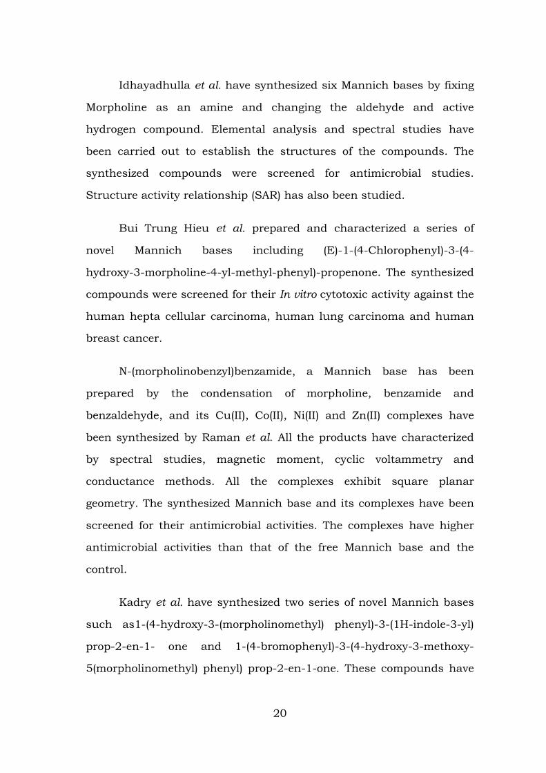
Mannich reaction of benzaldehyde, piperidine and semicarbazide
reported by * Dr. S. Ravichandran*, et al., Asian Journal of Biochemical
and Pharmaceutical Research Issue 3 (Vol. 1) 2011.
A Mannich base, N-[1-morpholino( 4-nitrobenzyl)] acetamide,
derived by the condensation of morpholine, 4-nitrobenzaldehyde and
acetamide, and its Cu(II), Co(II), Ni(II) and Zn(II) complexes have been
synthesized by Ravichandranet al. Both the ligand and the complexes
were characterized by molar conductance, magnetic susceptibility,
cyclic voltammetry and spectral studies. The antimicrobial activities of
the ligand and the complexes have also been studied and found that all
the complexes had higher activity than the free ligand and their
standard.
Kasim et al. have synthesized two Mannich bases viz. N, Nʹ-bis
(1- piperidino-4-methylbenzyl) urea by the condensation of urea,
piperidine and 4- methylbenzaldehyde and N, Nʹ-bis (1-piperidino-4-
chlorobenzyl) urea by the condensation of urea, piperidine and p-
chlorobenzaldehyde. These synthesized compounds have been
21
characterized on the basis of micro analytical and spectral studies.
Further these compounds were screened for their antimicrobial
activities against gram-positive and gram-negative bacteria.
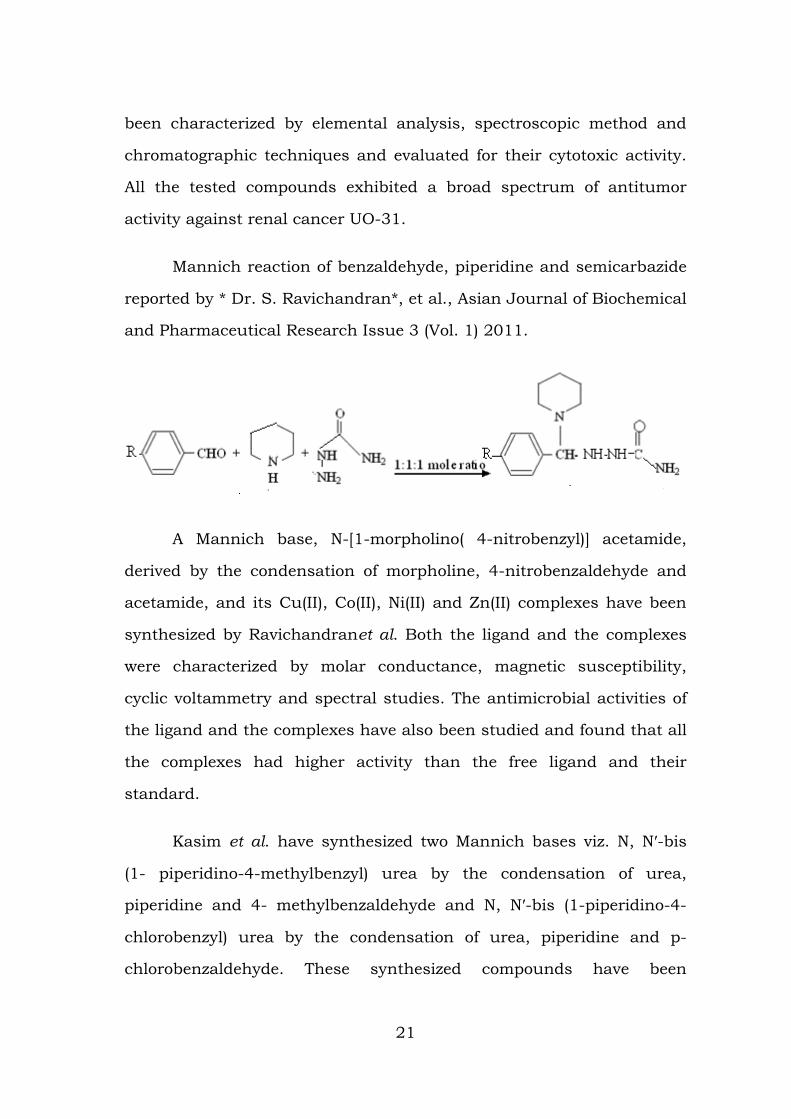
Chakkaravarthy et al. have synthesized Mannich bases, such as
1- ((1H-benzo[d]imidazole-1-yl) methyl) urea (4) by the condensation
urea, benzimidazole and formaldehyde and 1-((3-hydroxynaphthalne-2-
yl) methyl) thiourea (5) by the condensation of thiourea, β-naphthol and
formaldehyde and characterized them by elemental analysis and
spectral studies.
4 5
The synthesized Mannich bases were screened for their
antimicrobial and anti-oxidant activity. The structure and biological
activity relationship denotes the higher activity in compound 4 may be
due to the presence of two N atoms in the benzimidazole adjoin with
amide group. The antioxidant activities of the synthesized Mannich
bases are due to the presence of electron releasing amide group in it. In
the above studies compound 4 shows better activities compared to the
compound 5.
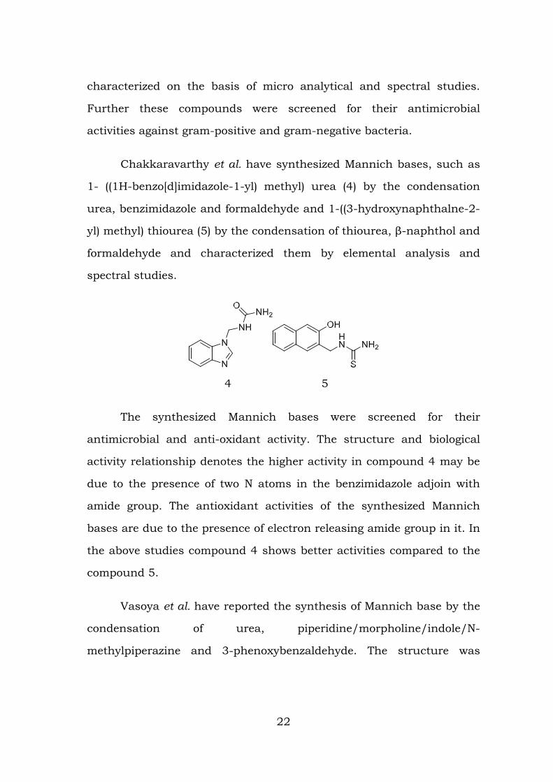
Vasoya et al. have reported the synthesis of Mannich base by the
condensation of urea, piperidine/morpholine/indole/N-
methylpiperazine and 3-phenoxybenzaldehyde. The structure was
22
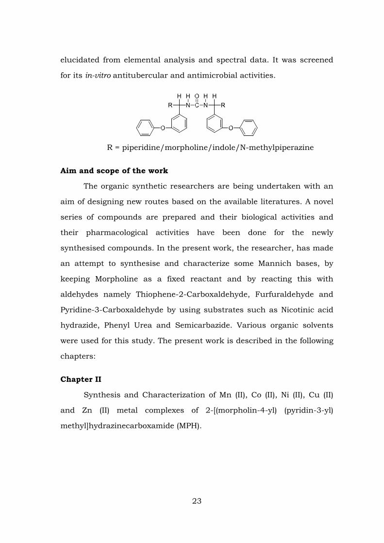
elucidated from elemental analysis and spectral data. It was screened
for its in-vitro antitubercular and antimicrobial activities.
R = piperidine/morpholine/indole/N-methylpiperazine
Aim and scope of the work
The organic synthetic researchers are being undertaken with an
aim of designing new routes based on the available literatures. A novel
series of compounds are prepared and their biological activities and
their pharmacological activities have been done for the newly
synthesised compounds. In the present work, the researcher, has made
an attempt to synthesise and characterize some Mannich bases, by
keeping Morpholine as a fixed reactant and by reacting this with
aldehydes namely Thiophene-2-Carboxaldehyde, Furfuraldehyde and
Pyridine-3-Carboxaldehyde by using substrates such as Nicotinic acid
hydrazide, Phenyl Urea and Semicarbazide. Various organic solvents
were used for this study. The present work is described in the following
chapters:
Chapter II
Synthesis and Characterization of Mn (II), Co (II), Ni (II), Cu (II)
and Zn (II) metal complexes of 2-[(morpholin-4-yl) (pyridin-3-yl)
methyl]hydrazinecarboxamide (MPH).
23
Chapter – III
Synthesis and Characterization of Co (II), Ni (II), Cu (II) and Zn (II)
metal complexes of (Morpholino (thiophen-2-Yl) methyl) nicotino
hydrazide (MTN).
Chapter - IV
Synthesis and Characterization of Co (II), Ni (II), Cu (II) and Zn (II)
metal complexes of 1-(furan-2-yl) (morpholino) (methyl)-3-phenyl urea
(MFP).
Chemicals used
All the chemicals used were of Merck and Sigma Aldrich
products, which are available commercially. The purchased chemicals
were used without any further purification.
Characterization techniques used
The synthesised ligands and their metal complexes have been
characterised by the following techniques.
1. Elemental Analysis.
2. Thin Layer Chromatography.
3. Infrared Spectroscopy.
4. Ultraviolet-visible Spectroscopy.
5. Nuclear Magnetic Resonance Spectroscopy.
6. Electron Paramagnetic Resonance Spectroscopy.
7. Mass spectroscopy.
8. Cyclic Voltammetry.
9. Magnetic Susceptibility Measurements.
10. Molar Conductance Measurements.
11. Thermal Analysis (TGA-DTA).
24
12. Thermo acoustical studies.
13. Antimicrobial and Molecular Docking Studies.
Elemental Analysis
The chemical analysis is quite helpful in fixing the stoichiometric
composition of the ligand as well as its metal complexes. The elemental
analysis is carried out to find out the molecular formula. The sample
was encapsulated in a thin tin foil and it is dropped into the furnace at
a temperature about 900˚ C. On ignition, the C, H, N and S present in
the sample converts to their respective combustion products. The
percentage of elements C, H, N and S is worked out by using Vario EL
III analyser available at the Sophisticated Test and Instrumentation
Centre, Cochin University, Kerala. By using the ACD/ Chemsketch /
Chemdraw / Freeware software, the elemental structures were drawn
based upon the elemental composition. By using the volumetric,
gravimetric and spectrophotometric methods, the metal content of the
complex were estimated.
Thin Layer Chromatography
Due to the extent separation and application of the compounds
in organic chemistry, the Thin Layer Chromatography is used as an
analytical tool to check the purity of the synthesised compounds by
using appropriate solvent as eluent.
Infrared Spectroscopy
In general, the spectra give sufficient information about the
structure of the compound. When Infrared light is passed through a
sample, some of the frequencies of the sample are absorbed while other
frequencies of the sample are transmitted through the sample without
25
being absorbed. The resulting plot of percent transmittance or percent
absorbance against frequency is an infrared spectrum. The IR
spectroscopy is widely used as a characterization technique for metal
complexes. The basic theory involved is that the stretching modes of
the ligands changes upon complexation due to weakening or
strengthening of the bonds involved in the bond formation resulting in
subsequent change in the position of the bands appearing in the IR
Spectrum. The changes in the structural features of the ligands are
observed as changes in bands observed, mainly in the fingerprint
region (4000-400 cm-1). The bands due to the metal ligand bonds are
mainly observed in the far IR region (600-100 cm-1).
For amines, the N-H stretching vibrations occur in the region
3300-3500 cm-1 in the dilute solution. Many workers have reported
medium intensity bands for (C-O-C) of furan ring vibrations in the
region 1020-1250 cm-1.The M-N stretching frequency is of particular
interest since it provides direct information regarding metal-nitrogen
coordinate bond. Different amines complexes exhibited the metal-
nitrogen frequencies in the 428-530 cm-1 region. Ketones, aldehydes,
carboxylic acids, carboxylic esters, lactones, acid halides, anhydrides,
amides and lactams show a strong stretching absorption band in the
region of 1870-1540 cm-1. Its relatively constant position and high
intensity and relative freedom from interfering bands make it as one of
the easiest band to recognize in infrared spectra.
Infrared spectra of the complexes were recorded on a Thermo
Nicolet Avatar 370 DTGS model FT-IR Spectrophotometer with KBR
pellets at St. Joseph’s College (Autonomous), Tiruchirappalli,
Tamil Nadu, India. For some of the complexes, the IR spectra were
26
recorded on a FT-IR Shimadzu IR Affinity-1 Spectrophotometer in the
range of 4000-400 cm-1 by using KBR pellets at Jamal Mohamed
College (Autonomous), Tiruchirappalli, Tamil Nadu, India.
Ultraviolet Spectroscopy
Most of the chemists use Ultraviolet spectroscopy as a valuable
tool to know about the important structural aspects of the complex.
The ligands which are considered as the organic compounds, shows
their absorption bands in the ultraviolet region of 200 – 300 nm of the
electromagnetic spectrum. Due to conjugation in some cases, these
bands extend over to higher wavelength region. In the transition metal
complex ions, there will be an interesting change in the electronic
properties of the system, due to the interaction with the metal ion. Due
to d-d absorption and charge transfer spectra from metal to ligand or
ligand to metal, new features or bands in the visible region can be
observed and this data can be processed to obtain information
regarding the structure and geometry of the compounds (38/EXPTL.
TECH).We can get the information about the possible distortions of
symmetry environment, magnitude of the ligand-field splitting (10 Dq),
certain bonding characteristics (charge-transfer bands) etc., from the
analysis of the spectrum of the complex.
The UV Spectrum of the compounds and complexes of the study
were recorded on Perkin Elmer, Lambda 35 model in the range
190-1100 nm at ACIC, St. Joseph’s College (Autonomous),
Tiruchirappalli.
27
Nuclear Magnetic Resonance Spectroscopy
1H NMR
The transitions between the magnetically inducted spin states are
known by the use of the NMR study. This study is concerned with the
magnetic properties of atomic nuclei with an integral value I. The
protons in an organic compound exposed to a powerful field, by the use
of this technique. The protons will process at different frequencies.
These processing protons are irradiating with steady changing
frequencies and observe the frequencies at which absorptions occur
and the signals obtained corresponding to the absorption is known as
NMR Spectrum. NMR spectroscopy is widely used to study the molecule
and it enables us to record the differences in the magnetic properties of
various magnetic nuclei present and to deduce the positions of this
nucleus within the molecule. By the use of the NMR spectroscopy, we
can deduce how many different kinds of environments there are in the
molecule and also which atoms are present in neighbouring groups. In
general, the number of atoms present in each of these environments is
measured. Therefore, the diagnostic features of the NMR Spectra are
the number of signals, position of signals, splitting pattern of signals
and area of signals. 1H NMR of the ligands were recorded using Bruker
300 MHz Avance–II FT-NMR Spectrometer with DMSO-d6 as the solvent
and TMS as internal standard at CARISM, SASTRA University,
Thanjavur, Tamil Nadu.
13C NMR
1H NMR and 13C NMR spectra differs both in the mode of
recording as well as in the appearance. The spin quantum number,
I for 12C is equal to zero since 12C isotope has an even number of
28
protons and even number of neutrons and hence no magnetic spin. It
is, therefore, non-magnetic and does not give any NMR signal. The
natural abundance of 13C is only about 1.1% and has an odd number
of neutrons. So, 13C has a spin quantum number equal to ½ and its
nuclear magnetic resonance can be observed in a magnetic field of
23,500 gauss at 25.2 mega cycles per second. 1H spectrum is normally
obtained by sweeping either the excitation frequency or the through the
region of precession frequencies. The inefficiency of this method is clear
from the fact that only one line can be observed at a given point in
time. The problem arises when 13C with intrinsically narrow lines
covering a wide absorption range is studied. It is, therefore,
advantageous to excite the whole band of frequencies simultaneously. It
is done by strong pulse of radio-frequency covering a large band of
frequencies which is capable of exciting all resonance of interest at
once. At the end of the pulse period, the nuclei will process freely with
their characteristic frequencies reflecting with the chemical
environment and exhibit chemical shifts. 13C NMR of the synthesized
compounds were recorded on 400 MHz Bruker Spectrometer at 298.6 K
using DMSO as solvent at CARISM, SASTRA University, Thanjavur, and
Tamil Nadu.
Electron Paramagnetic Resonance Spectroscopy
Electron Paramagnetic Resonance Spectroscopy is otherwise
known as Electron Spin Resonance (ESR) and Electron Magnetic
Resonance (EMR) spectroscopy. This technique is used for the study of
systems having uncompensated electron spins and deals with the
transition between Zeeman levels. An unpaired electron that is not
subject to interactions with other unpaired electrons or with magnetic
29
nuclei will show a single sharp absorption for its transition which in
turn corresponds to the position of magnetic field at which it comes
into resonance . Electron Paramagnetic Resonance Spectroscopy is a
powerful technique and it can be used to locate the distribution of
unpaired electron in a molecule and to some extent, decide the extent
to which electrons are delocalized over the ligands. The study gives the
numerical values for the parameters in the spin Hamiltonian
components of ʽgʼ tensor, detectable hyperfine splitting and principle
components of the hyperfine tensor.
The resonant position of EPR is referred to as the ʽgʼ value and it
is directly determined by the separation of the energy levels of the
system under investigation. The variation of the ʽgʼ value is interpreted
in terms of the first and the second order perturbation by the spin orbit
interactions. Investigation of the correlation of the line width with
chemical nature of bonding is of special importance.
The EPR spectrum of Cu (II) complexes of the synthesized ligands
were recorded on Jeol Model JES FA 200 spectroscope in X- band
frequencies at Room Temperature at SAIF, IIT, Madras.
Mass spectroscopy
For determining the molecular mass of the compound, the Mass
spectrometry is used and it is considered as one of the most accurate
method. In this technique, molecules are bombarded with a beam of
energetic electrons.
The molecules are ionized and broken up into many fragments,
some of which are positive ions. Each kind of ion has a particular ratio
30
of mass to charge, i.e. m/z ratio. For most ions, the charge is one and
thus, m/z ratio is simply the molecular mass of the ion.
The set of ions are analyzed in such a way that a signal is
obtained for each value of m/z that is represented. The relative
abundance of the ion is represented by the intensity of each signal. The
largest peak in the structure is called the base peak and its intensity is
taken as 100. Mass spectrum of a compound is a plot which represents
the intensities of the signals at various m/e values.
The mass spectrums of the synthesized compounds were
recorded on Jeol GC mate II Mass spectrometer at SAIF, IIT, Chennai,
and Tamil Nadu.
Cyclic voltammetry
The redox behaviour of the coordinated complexes can be studied
by the use of the Cyclic Voltammetry. The Cyclic Voltammetry studies
gives an insight into the stability of the compound under investigation
against electrolytic oxidation and reduction in the solution. In this
technique, the potential of a small stationary working electrode is
changed linearly with the time, starting from a potential where no
electrode reaction occurs and moving to potentials where reduction or
oxidation of a sample occurs. After traversing the potential region in
which one or more electrode reaction takes place, the direction of the
linear sweep is reversed and the electrode reactions of the
intermediates and products formed during the forward scan often can
be detected. The CV technique can be carried out using a suitable
reference, working and counter electrode, the selection of which can be
made depending on the nature of the compound and solvent used in
31
the presence of a supporting electrolyte. By selecting a range of voltages
and the variation involtammogram can be recorded at different sweep
rates. The peaks in the forward and the reverse sweeps can be
interpreted to assess the stability of the species. Depending upon the
nature of the voltammogram obtained, they may be termed as
reversible (IPA=Ipc), quasi - reversible (IPA>Ipc) and irreversible
process.
Cyclic Volta metric of all of the synthesized complexes were
carried out on Princeton voltammetry Applied instrumentation in the
frequency range -1 Hz to +1 MHz at ACIC, St. Joseph's College
(Autonomous), Tiruchirappalli, Tamil Nadu.
Magnetic susceptibility
Magnetic susceptibility studies can be used in conjunction with
electronic spectra to establish the geometry of the complexes.
Substances which contain one or more unpaired electrons have a
permanent magnetic moment which exists in the absence of a magnetic
field and also arises from the net spin and orbital momentum of the
electrons. Two properties of an unpaired electron, the spin and orbital
moments contribute to the magnitude of the paramagnetic moment.
The magnetic susceptibility measurements of the complexes are
done in order to find out the effective magnetic moment per each metal
atom in the complexes. The number of unpaired electrons possessed by
the metal ion can be determined from the effective magnetic moment of
the metal ion. From the knowledge of unpaired electrons it is possible
to infer the valence state of the metal ion.
32
The magnetic susceptibility measurements of the powdered
sample were done at room temperature using a Sherwood Scientific
Magnetic Susceptibility Balance at
Conductivity Measurements
The molar conductance value of an electrolyte in a particular
solvent depends upon the various properties of the solvent such as
dielectric constant, viscosity, specific conductivity, purity, ligating
tendency towards metal ions and the temperature of measurement. The
electrical conductance measurements were done to see whether the
anions of the metal salts remain inside or outside the coordination
sphere of the central metal ion.
Molar conductivities of the complexes in Dimethyl formamide
solution (10-3μ) were measured at room temperature using a systronic
model 303 direct reading conductivity bridge at the PG and Research
Dept. of Chemistry, Jamal Mohamed College, Tiruchirappalli.
Thermo Gravimetric Analysis
Thermal methods of analysis are techniques in which changes in
physical and or chemical properties of a substance are measured as a
function of temperature. From the simultaneous recordings of
temperature, weight and heat capacities, the TG and DTA curves are
drawn. From the analysis of the complexes one can understand the
modes of decomposition of the complexes and the nature of the phase
changes taking place during the programmed heating. The thermal
stabilities and the decomposition temperatures can also be ascertained.
In addition, the purity of the complexes can also be checked. This
technique examines the various changes like thermal decomposition,
33
oxidation, solvent and water desorption, evaporation, sublimation etc.
which may take place with a consequent change in weight of the
sample when heated at a desired heating rate with proper furnace
atmosphere.
TG-DTG analysis of the complexes were carried out in a Perkin
Elmer Pyris 6 TG/DTA analyser at the temperature rate of 10.0 °C/min
in an atmosphere of nitrogen at the Department of Chemistry, SAIF,
Chennai, Tamil Nadu.
Thermo acoustical studies
Ultrasonic studies finds application in several industrial and
technological processes. The Ultrasonic investigations of liquid
mixtures consisting of polar and non-polar components are of highly
importance in understanding the physical nature and strength of
molecular interaction in the liquid mixtures.
In the present study, the Ultrasonic velocity, density, adiabatic
compressibility and viscosity measurements have been carried out for
tertiary mixture for various concentrations at different temperatures in
the Department of Physics, Jamal Mohamed College (Autonomous),
Tiruchirappalli, Tamil Nadu.
Antimicrobial and Molecular Docking studies
A preliminary biological study has been made on the synthesized
compounds and their metal complexes. The biological study includes
antibacterial and molecular docking studies, toxicity assay, larvicidal
studies and docking studies.
34
References
1. Gopalan R and V Ramalingam, Concise Coordination Chemistry., VIKAS Publishing House (P) Ltd, Second Reprint 2008.
2. Haidue L, Coord. Chem. Rev., 1990, 99, 253.
3. Cleare M J, Coord. Chem. Rev., 1974, 12, 349.
4. Singh B, Singh R N and Aggarwal R C, Polyhedron., 1985, 4, 401.
5. Mishra A P and Srivastavan S K, J. Indian Council Chem., 1994, 10, 2.
6. Deshmuck M D, Orient J. Chem., 1995, 11, 185.
7. Varma R S, Rastogi N and Singh A P, Ind. J.Chem., 2002, 4, 231.
8. R.V. Sing, Rakhi dwived and Sanjay Sharma, J. Indian chem. Soc., 81 (2004), 454-456.
9. L.S. Sharma, V. Muresan, S. Sbirna, N. Muresan and C.I. Lepadatu. J.Indian chem. Soc., 81 (2004), 150-152.
10. E.I. Mostapha Jouad, Amedee Riou, Magali Allian, M.A.Khan, Polyhedron., 20 (2001), 67-74.
11. A.P. Mishra and A.K. Gowtham, J. Indian chem. Soc., 81 (2004), 324-326.
12. L.S. Sbirna, V. Muresan, S. Sbirna and V. Muresan, J. Indian chem Soc., 82 (2005), 389-392.
13. H.G. Petering, H.H. Buskirk, J.A. Crim and G.J. Van Geisen, Pharmacol., 5 (1965), 271.
14. K. Naresh Kumar and R. Ramesh, Spectrochimica Acta Part-A., 60 (2004), 2913-2918.
15. Pandeya S N, Sriram D, Acta Pharm. Turc., 1998, 40, 33-38.
16. Sarangapani M, Reddy V M. Synthesis, Indian J. Heterocycl.Chem., 1994, 3, 257-260.
17. Varma R S, Nobels W L, J. Pharm. Sci., 1975, 64, 881-882.
18. Pandeya S N, Sriram D, Nath G, DeClercq E, Farmaco., 1999, 54, 624-628.
35
19. Pandeya S N, Sriram D, Nath G and De Clercq E, Indian J. Pharm.Sci., 1999, 61, 358-361.
20. Pandeya S N, Sriram D, Nath G and De Clercq E, Pharm. Acta Helv., 1999, 74, 11-17.
21. Pandeya S N, Sriram D, Nath G and De Clercq E, Eur. J. Pharm.Sci., 1999, 9, 25-31.
22. Pandeya S N, Yogeeswari P, Sriram D, De Clercq E and Pannecouque C, Chemotherapy., 1999, 45, 192-96.
23. Pandeya S N, Sriram D, Nath G and De Clercq E, Eur. J. Med. Chem., 2000, 35, 249-255.
24. Pandeya S N, Sriram D, Nath G and De Clercq E, Drug. Res., 2000, 50, 55-59.
25. Pandeya S N, Yogeeswari P, Sriram D and Nath G, Chim. Farm., 1998, 137, 321-324.
26. Ravichandran V, Mohan S and Kumar S K, Arkivoc., 2007, (xiv), 51-57.
27. Patel A, Bari S, Telele G, Patel J and Sarangapani M, Iran. J.Pharm.Sci., 2006, 4, 249-254.
28. Jarrahpour A, Khalili D, Clercq De E, Salmi C and Brunel M J, Molecules., 2007, 12, 1720-1730.
29. BAL T R, Anand B, Yogeswari P and Sriram D, Bioorg. Med. Chem.Let., 2005, 15, 4451-4455.
30. Sriram D, BAL T R and Yogeswari P, J.Pharm. Pharmaceut. Sci., 2005, 8(3), 565-577.
31. Singh S P, Shukla S K and Awasthi L P, Curr. Sci., 1983, 52, 766-769.
32. Selvam P, Murgesh N, Chandramohan M, Clercq De E, Keyaerts E,Vijgen L, Maes P, Neyts J and Ranst M V, Ind. J. Pharm. Sci., 2008,70(1), 91-94.
33. Pandeya S N, Smith S and Stable J P, Arch. Pharm., 2002, 4, 129-134.
36
34. Verma M, Pandeya S N, Singh K N and Stables J P, Acta Pharm., 2004, 54, 49-56.
35. Smitha S, Pandeya S N, Stables J P and Ganpathy S, Sci. Pharm., 2008, 76, 621-636.
36. Sridhar S K, Pandeya S N, Stables J P and Ramesh A, Euro. J. Pharm.Sci., 2002, 16, 129-132.
37. Fadl AT and Bin-Jubair FA S, Int. J. Res. Pharm. Sci., 2010, 1(2), 113-126.
38. Hussein M A, Fadl T A and Hussein A, Pharm Sci; Assiut University., 2005, 28, 131-136.
39. Karali N, Gursoy A, Kandemirli F, Shvets N, Kaynak F B, Ozbey Suhelyla,Koalishyn V and Dimoglo A, Bioorg. Med. Chem., 2007, 15, 5888-5904.
40. Vine K L, Locke J M, Ranson M, Pyne S G and Bremner J B, Bioorg.Med. Chem., 2007, 15, 931-938.
41. Yogeeswari P, Sriram D, Kavya R and Tiwari S, Biomed and Pharmacother., 2005, 59, 501-510.
42. Hussain MM, Islam N Khar R and Islam Md R, Bangladesh J.Pharmacol., 2007, 2, 66-70.
43. Intramolecular Mannich reactions: L. E. Overmann, D. J. Ricca in Comprehensive Organic Synthesis, Vol. 2 (Eds.: B. M. Trost, I.Fleming, C. H. Heathcock), Pergamon, Oxford, 1991, p. 1007.
44. Bondendorff and Koralewski, Arch.Pharma., 101 (1933) 271.
45. L. Racane, V. T. Kulenovic, L. F. Jakic, D. W. Boykin, and G. K. Zamola, “Synthesis of bis-substituted amidino-benzothiazoles as potential anti-HIV agents,” Heterocycles, 2001, Vol. 55, pp. 2085–2098.
46. E. Kashiyama, I.Hutchinson, M.-S. Chua et al., “Antitumor benzothiazoles.8.1 Synthesis, metabolic formation, and biological properties of the C- and N-oxidation products of antitumor 2- (4-aminophenyl)-benzothiazoles,” Journal of Medicinal Chemistry, 1999, vol. 42, no. 20, pp. 4172–4184.
37
47. S. R. Bhusare, R. P. Pawar, and Y. B. Vibhute, “Synthesis and antibacterial activity of some new 2-substituted phenyl sulfonamido)-6-substituted benzothiazoles,” Indian Journal of Heterocyclic Chemistry, 2001, vol. 11, no. 1, pp. 79–80.
48. N. Raman, S. Esthar, and C.Thangaraja, “A new Mannich base and its transition metal (II) complexes—synthesis, structural, characterization and electrochemical study,” Journal of Chemical Sciences, 2004, vol. 116, no. 4, pp. 209–213.
49. P.J. J. Huang, D. Youssef, T. S. Cameron, and A. Jha, “Microwave-assisted synthesis of novel 2-naphthol bis-Mannich Bases,” Arkivoc, 2008, vol. 2008, no. 16, pp. 165–177.
50. M. Arend, B. Westermann, and N. Risch, “Modern variants of the Mannich reaction,” Angewandte Chemie—International Edition, 1998, vol. 37, no. 8, pp. 1045–1070.
51. B. S. Holla, M. K. Shivananda, M. S. Shenoy, and G. Antony, “Studies on aryl furan derivatives. Part VII. Synthesis and characterization of some Mannich bases carrying halophenyl furyl moieties as promising antibacterial agents,” Farmaco, 1998, vol. 53, no.8-9, pp. 531–535.
52. J.J. Denton, R.J. Turner, W.B. Neier, V.A. Lawson and H.P. Schedl, J.Am.Chem.Soc., 71 (1949) 2048, 2050, 2053, 2054.
53. H. Schonenberger et al., Pharma. Acta. Hel., 44 (1969) 961.
54. Y.K. Agarwal and J.N. Tayal Indian J. pharmacy., 29 (1967) 46.
55. K.Von Thiele, U.Schimassek, A.Von Schilichtegroll, Arzneimittel forschung., 16 (1967) 81894.
56. K.S. Nimavat. K.H. Popat, S.L. Vasoya and H.S. Joshi J. Indian chem, Soc., 80 (2003) 711-713.
57. Meyer B N, Ferrigini N R, Putnam J F, Jacobson L B, Nichols D E and Mc Lahghlin J L, Planta Medica., 1982, 45, 31.
58. Mannich and Ball, Arch Pharm., 1926, 264, 65.
59. Henkel T. Brunne R M, Mueller H & Reichel F, Angew Chem, 38, 1999, 643
38
60. Landquist J K, in Comprehensive Heterocyclic Chemistry, Vol. 1, edited by Katrizky A R & Rees C W (Pergamon, New York), 1984, pp 166-170.
61. Hutchins R O & Hutchins M K, in Comprehensive Organic Synthesis, Vol. 8, edited by Trost B M & Fleming I (Pergamon, Oxford), 1991, pp. 25.
62. Cooperman B S, Met. Ions Biol. Systems., 1976, 5, 79.
63. Swamy S J and Dharma Reddy A, Indian J.Chem., 2000, 77, 336-338.
64. Bohme H and Fuchs G, Chem. Ber., 1970, 103, 2775.
65. Drusvyatskaya S K, Kolosova M O and Stavrovskaya V I, Zh. Org.Khim., 1970, 6, 2532.
66. Mozolis V, Jokubaityte S, Liet. Mokslu Akad T S R, Darb. Ser., 1970, 73, 77152.
67. Mozolis V, Jokubaityte S, Liet. Mokslu Akad T S R, Darb. Ser., 1970, 73, 77152.
68. Muruganandam L, Rajeswari S, Tamil Vendan D, RamkumarV, Venkatesa Prabhu G, Acta CrystallographicaSection., 2009, 65, 578.
69. Rajeswari, Venkatesa Prabhu G, Krishnakumar K, Ramkumar V andBabu Varghese Acta Crystallographica Section E., 2007, 63, 2929.
70. Venkatesa Prabhu, Nagarajan Vembu, Loganathan MuruganandamandAnthony Linden Acta Crystallographica Section E., 2007, 63, 2841–2842.
71. Venkatesa Prabhu G and Mohamed Kasim AN N, Asian Journal of Chemistry., 2000, 12 (2), 379-384.
72. Venkatesa Prabhu G and Mohamed Kasim AN N, Asian Journal of Chemistry., 2000, 12 (2), 385-388.
73. Venkatesa Prabhu G, Mohamed Kasim AN N and VenkappayaD, Journal of Indian Chemical Society., 1999, 76, 67-69.
74. Venkatesa Prabhu G, Mohamed Kasim AN N and VenkappayyaD, Samyak Journal of Chemistry., 1998, 2(1), 26.
39
75. Venkatesa Prabhu G and Venkappayya D Samyak Journal of Chemistry., 1998, 2, 1, 15-18.
76. Venkatesa Prabhu G and Venkappayya D, Journal of Indian ChemicalSociety., 1995, 72, 681-684.
77. Abdul Jameel A and Syed Ali Padusha M, Indian J.Heterocyclic Chem., 2006, 16, 197 -198.
78. Jean-Pierre Schirmann, Paul Bourdauducq "Hydrazine" in Ullmann's Encyclopedia of Industrial Chemistry, Wiley-VCH, Weinheim, 2002.
79. Euromedica, Internationaler Medizinischer Kongress Hannover., 2007, Germany.
80. Cellular Immunology 2013, 283 (1–2): 61–69.
81. Young, SZ; Bordey, (2009). "GABA's control of stem and cancer cell proliferation in adult neural and peripheral niches". Physiology (Bethesda) 24: 171–85.
82. Klaus Weissermel, Hans-Jürgen Arpe, Charlet R. Lindley, Stephen Hawkins (2003), "Chap. 7. Oxidation Products of Ethylene". Industrial Organic Chemistry. Wiley-VCH. pp. 159–161.
83. Lindsay Smith, J. R.; McKeer, L. C.; Taylor, J. M. (1993), "4-Chlorination of Electron-Rich Benzenoid Compounds: 2, 4-Dichloromethoxybenzene". Org. Synth.; Coll. Vol. 8, p. 167.
84. Noyori, R.; Yokoyama, K.; Hayakawa, Y. (1988). "Cyclopentenones from α, α’-Dibromoketones and Enamines: 2, 5-Dimethyl-3-Phenyl-2-Cyclopenten-1-one". Org. Synth.; Coll. Vol. 6, p. 520
85. Raymond G. McGuire; Dimitrios A. Dimitroglou (1999). "Evaluation of Shellac and Sucrose Ester Fruit Coating Formulations that Support Biological Control of Post-harvest Grapefruit Decay". Bio-control Science and Technology 9 (1): 53–65.
86. R. J. Harrison and M. Moyle, Organic Syntheses, Coll. Vol. 4, p.493 (1963), Vol. 36, p.36 (1956).
87. Ronnie Ozer, "Vapor phase decarbonylation process" WIPO Patent Application WO/2011/026059 (2011)
40
88. Vest, Katherine E.; Hashemi, Hayaa F.; Cobine, Paul A. (2013). "Chapter 13 the Copper Metallome in Eukaryotic Cells". In Banci, Lucia. Metallomics and the Cell. Metal Ions in Life Sciences 12. Springer.
89. Vest, Katherine E.; Hashemi, Hayaa F.; Cobine, Paul A. (2013). "Chapter 12 the Copper Metallome in Prokaryotic Cells". In Banci, Lucia. Metallomics and the Cell. Metal Ions in Life Sciences 12. Springer.
90. Yee, Gereon M.; Tolman, William B. (2015), Metal Ions in Life Sciences 15, Springer. pp. 175–192.
91. S. J. Lippard, J. M. Berg, 1994, "Principles of bioinorganic chemistry" University Science Books: Mill Valley, CA.
92. Decker, H. & Terwilliger, N. (2000), Journal of Experimental Biology 203 (Pt 12): 1777–1782.
93. Horseshoe crab. University of Delaware. 13 July 2008.
94. Schneider, Lisa K, Wüst, Anja, Pomowski, Anja; Zhang, Lin, Einsle, Oliver (2014), Metal Ions in Life Sciences 14. Springer. pp. 177–210.
95. Copper Touch Surfaces. 8 November 2011.
96. Yamada, Kazuhiro (2013), Metal Ions in Life Sciences 13. Springer. pp. 295–320.
97. Cracan, Valentin; Banerjee, Ruma (2013), Metal Ions in Life Sciences 12. Springer.
98. Schwarz, F. J.; Kirchgessner, M.; Stangl, G. I. (2000). Journal of Animal Physiology and Animal Nutrition 83 (3): 121.
99. Roth, Jerome; Ponzoni, Silvia; Aschner, Michael (2013), Metal Ions in Life Sciences 12. Springer.
100. Chandra S, Jain D, Sharma A K, Sharma P, Molecules, 2009, 14, 174.
101. Prasad K S, Kumar L S, Prasad R, Revanasiddappa H D, Bioinorg. Chem. App, 2010, 9.
102. Gupta M K, Har Lal S, Varshney S, Varshney A K, Bioinorg Chem. 2003, 1, 309.
41
CHAPTER II
Synthesis and Characterization of Mn (II), Co (II), Ni (II), Cu (II) and Zn (II) metal complexes of 2-[(morpholin-4-yl)
(pyridin-3-yl) methyl] Hydrazinecarboxamide (MPH)
This chapter deals with the Synthesis and Characterization of
Mn (II), Co (II), Ni (II), Cu (II) and Zn (II) metal complexes of
2-[(morpholin-4-yl) (pyridin-3-yl) methyl] Hydrazinecarboxamide (MPH).
Materials and Methods
All the chemicals and solvents used, were of Merck and Sigma
Alrich products (Analytical Reagent grade) and were used without
further purification. The synthesized ligand and their metal complexes
were characterized by the respective techniques, as discussed in
Chapter I.
Synthesis and Characterisation of 2-[(morpholin-4-yl) (pyridin-3-yl)
methyl] Hydrazinecarboxamide (MPH)
Pyridine-3-carboxaldehyde, Morpholine and Semicarbazide were
taken in the molar ratio of 1:1:1 and were reacted as shown in the
Scheme 1. To the ethanolic solution of semicarbazide (1.5g, 25 mmol)
taken in a round bottom flask, morpholine (2.1 mL, 25 mmol) and
pyridine-3-carboxaldehyde (2.6 mL, 25 mmol) were added. The reaction
mixture was kept on a magnetic stirrer and stirred under ice cold
condition for 3 h, the yellow coloured solid formed was filtered and
washed several times with petroleum ether (40-60%) and then dried.
The crude solid thus obtained was dried and recrystallized from
ethanol. The recrystallized product was dried over vacuum.
42
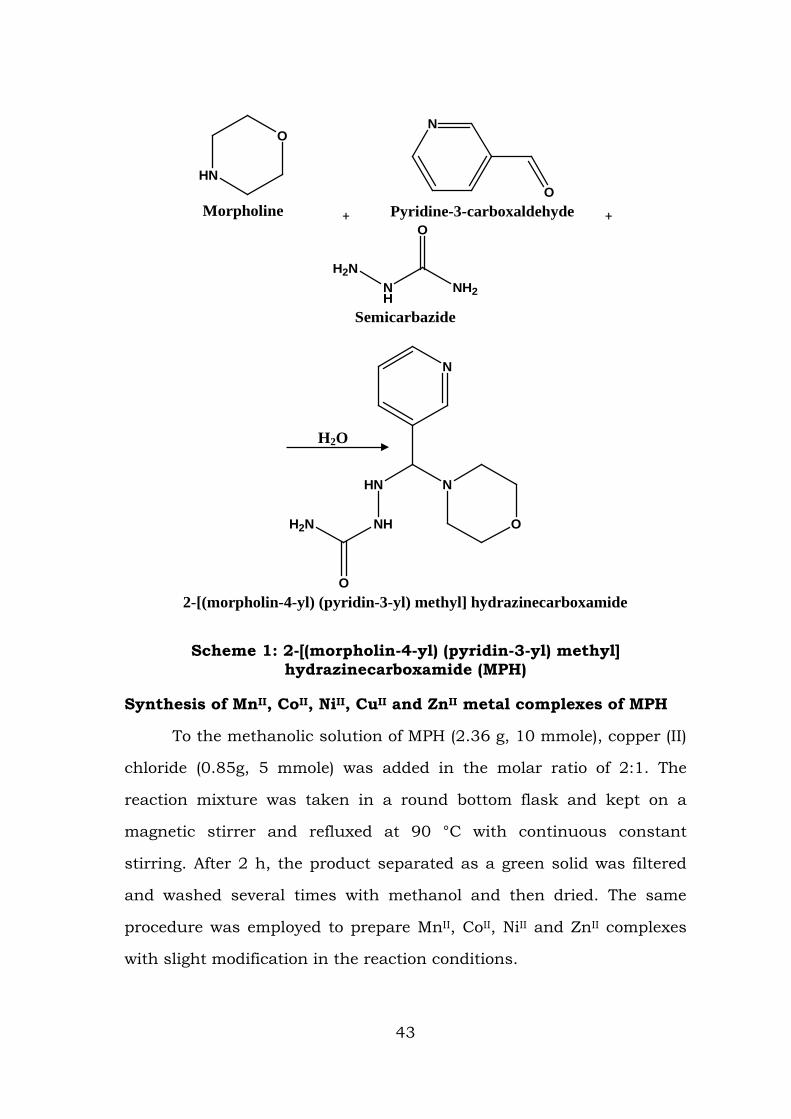
HN
O
Morpholine +
N
OPyridine-3-carboxaldehyde +
H2NNH
O
NH2
Semicarbazide
NH
HN
O
H2N
N
N
O
2-[(morpholin-4-yl) (pyridin-3-yl) methyl] hydrazinecarboxamide
Scheme 1: 2-[(morpholin-4-yl) (pyridin-3-yl) methyl] hydrazinecarboxamide (MPH)
Synthesis of MnII, CoII, NiII, CuII and ZnII metal complexes of MPH
To the methanolic solution of MPH (2.36 g, 10 mmole), copper (II)
chloride (0.85g, 5 mmole) was added in the molar ratio of 2:1. The
reaction mixture was taken in a round bottom flask and kept on a
magnetic stirrer and refluxed at 90 °C with continuous constant
stirring. After 2 h, the product separated as a green solid was filtered
and washed several times with methanol and then dried. The same
procedure was employed to prepare MnII, CoII, NiII and ZnII complexes
with slight modification in the reaction conditions.
H2O
43
Characterization of MPH and its MnII, CoII, NiII, CuII and ZnII
complexes
Elemental analysis
After ascertaining the elements that are present in the
synthesised compound, the proportionate percentage of various
elements was found out. The observed and calculated percentage
composition of C, H, N and O elements of the ligand MPH and its MnII,
CoII, NiII, CuII and ZnII complexes and the values of the molar
conductance of the complexes are presented in Table 2.1.
From the observed C, H, N and O values, the molecular formula
of the compound 2-[(morpholin-4-yl) (pyridin-3-yl) methyl]
Hydrazinecarboxamide (MPH)was assigned as C11H17N5O2 which was in
agreement with the calculated value. The molar conductance of the
complexes are in the range of μc is 23-31 Ω-1 mol-1cm2, in 10-3 M
DMSO. From the molar conductance values observed, it has been
revealed that, all the chloro complexes are non-electrolyte and the
anion was coordinated to the metal(II) ion [1, 2].
44
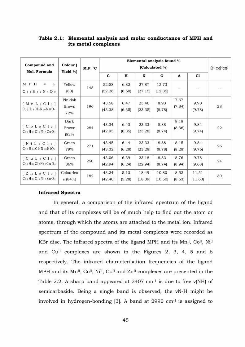
Table 2.1: Elemental analysis and molar conductance of MPH and its metal complexes
Compound and
Mol. Formula
Colour (
Yield %) M.P. ˚C
Elemental analysis found %
(Calculated %) Ω-1 mol-1cm2
C H N O A Cl
M P H = L
C 1 1 H 1 7 N 5 O 2
Yellow
(80) 145
52.58
(52.26)
6.82
(6.50)
27.87
(27.15)
12.73
(12.35) -- -- --
[ M n L 2 C l 2 ] C 2 2 H 3 4 C l 2 N 1 0 MnO 4
Pinkish
Brown
(72%)
196 43.58
(43.38)
6.47
(6.35)
23.46
(23.35)
8.93
(8.78)
7.67
(7.84) 9.90
(9.78) 28
[ C o L 2 C l 2 ] C 2 2 H 3 4 C l 2 N 1 0 C oO 4
Dark
Brown
(82%
284 43.34
(42.95)
6.43
(6.35)
23.33
(23.28)
8.88
(8.74)
8.18
(8.36) 9.84
(9.74) 22
[ N i L 2 C l 2 ] C 2 2 H 3 4 C l 2 N 1 0 N iO 4
Green
(79%) 271
43.45
(43.32)
6.44
(6.28)
23.33
(23.28)
8.88
(8.78)
8.15
(8.28)
9.84
(9.76) 26
[ C u L 2 C l 2 ] C 2 2 H 3 4 C l 2 N 1 0 C uO 4
Green
(86%) 250
43.06
(42.94)
6.39
(6.24)
23.18
(22.94)
8.83
(8.74)
8.76
(8.94)
9.78
(9.63) 24
[ Z n L 2 C l 2 ] C 2 2 H 3 4 Cl 2 N 1 0 ZnO 4
Colourles
s (84%) 182
43.24
(42.40)
5.13
(5.28)
18.49
(18.39)
10.80
(10.50)
8.52
(8.63)
11.51
(11.63) 30
Infrared Spectra
In general, a comparison of the infrared spectrum of the ligand
and that of its complexes will be of much help to find out the atom or
atoms, through which the atoms are attached to the metal ion. Infrared
spectrum of the compound and its metal complexes were recorded as
KBr disc. The infrared spectra of the ligand MPH and its MnII, CoII, NiII
and CuII complexes are shown in the Figures 2, 3, 4, 5 and 6
respectively. The infrared characterisation frequencies of the ligand
MPH and its MnII, CoII, NiII, CuII and ZnII complexes are presented in the
Table 2.2. A sharp band appeared at 3407 cm-1 is due to free ν(NH) of
semicarbazide. Being a single band is observed, the νN-H might be
involved in hydrogen-bonding [3]. A band at 2990 cm-1 is assigned to
45
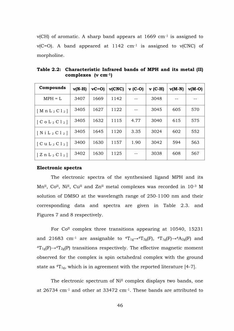
ν(CH) of aromatic. A sharp band appears at 1669 cm-1 is assigned to
ν(C=O). A band appeared at 1142 cm-1 is assigned to ν(CNC) of
morpholine.
Table 2.2: Characteristic Infrared bands of MPH and its metal (II) complexes (ν cm-1)
Compounds ν(N-H) νC=O) ν(CNC) ν (C-O) ν (C-H) ν(M-N) ν(M-O)
MPH = L 3407 1669 1142 -- 3048 -- --
[ M n L 2 C l 2 ] 3405 1627 1122 -- 3045 605 570
[ C o L 2 C l 2 ] 3405 1632 1115 4.77 3040 615 575
[ N i L 2 C l 2 ] 3405 1645 1120 3.35 3024 602 552
[ C u L 2 C l 2 ] 3400 1630 1157 1.90 3042 594 563
[ Z n L 2 C l 2 ] 3402 1630 1125 -- 3038 608 567
Electronic spectra
The electronic spectra of the synthesised ligand MPH and its
MnII, CoII, NiII, CuII and ZnII metal complexes was recorded in 10-3 M
solution of DMSO at the wavelength range of 250-1100 nm and their
corresponding data and spectra are given in Table 2.3. and
Figures 7 and 8 respectively.
For CoII complex three transitions appearing at 10540, 15231
and 21683 cm-1 are assignable to 4T1g→4T2g(F), 4T1g(F)→4A2g(F) and
4T1g(F)→4T2g(P) transitions respectively. The effective magnetic moment
observed for the complex is spin octahedral complex with the ground
state as 4T1g, which is in agreement with the reported literature [4-7].
The electronic spectrum of NiII complex displays two bands, one
at 26734 cm-1 and other at 33472 cm-1. These bands are attributed to
46
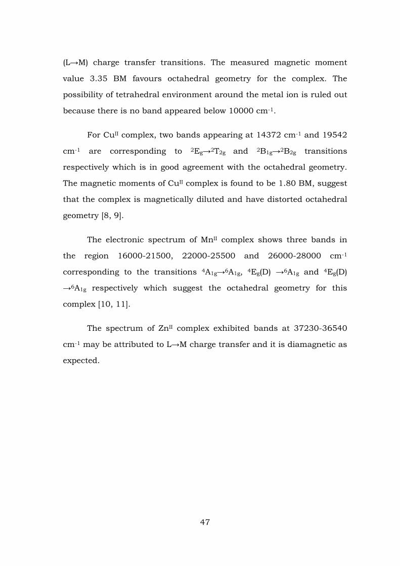
(L→M) charge transfer transitions. The measured magnetic moment
value 3.35 BM favours octahedral geometry for the complex. The
possibility of tetrahedral environment around the metal ion is ruled out
because there is no band appeared below 10000 cm-1.
For CuII complex, two bands appearing at 14372 cm-1 and 19542
cm-1 are corresponding to 2Eg→2T2g and 2B1g→2B2g transitions
respectively which is in good agreement with the octahedral geometry.
The magnetic moments of CuII complex is found to be 1.80 BM, suggest
that the complex is magnetically diluted and have distorted octahedral
geometry [8, 9].
The electronic spectrum of MnII complex shows three bands in
the region 16000-21500, 22000-25500 and 26000-28000 cm-1
corresponding to the transitions 4A1g→6A1g, 4Eg(D) →6A1g and 4Eg(D)
→6A1g respectively which suggest the octahedral geometry for this
complex [10, 11].
The spectrum of ZnII complex exhibited bands at 37230-36540
cm-1 may be attributed to L→M charge transfer and it is diamagnetic as
expected.
47
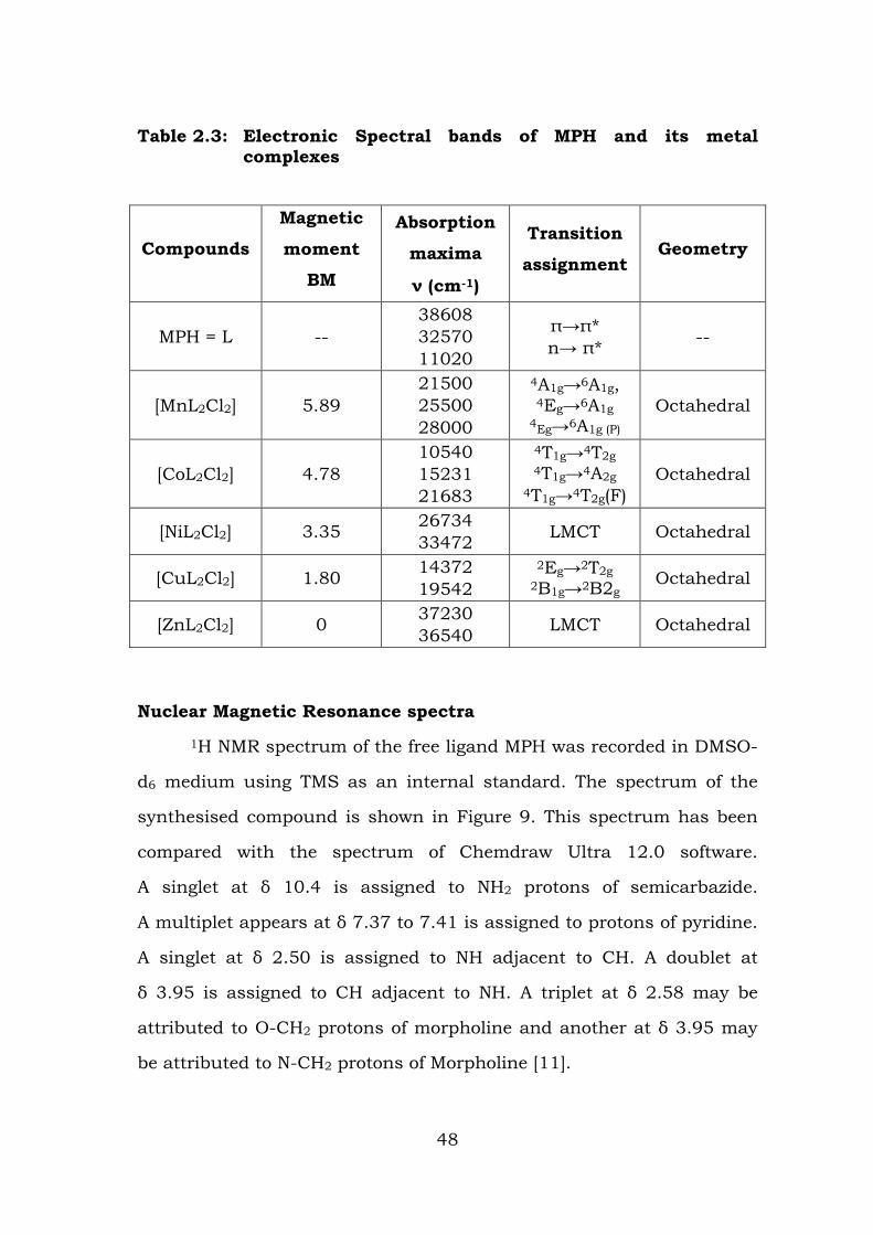
Table 2.3: Electronic Spectral bands of MPH and its metal complexes
Compounds
Magnetic
moment
BM
Absorption
maxima
ν (cm-1)
Transition
assignment Geometry
MPH = L -- 38608 32570 11020
π→π* n→ π* --
[MnL2Cl2] 5.89 21500 25500 28000
4A1g→6A1g, 4Eg→6A1g
4Eg→6A1g (P)
Octahedral
[CoL2Cl2] 4.78 10540 15231 21683
4T1g→4T2g 4T1g→4A2g
4T1g→4T2g(F) Octahedral
[NiL2Cl2] 3.35 26734 33472 LMCT Octahedral
[CuL2Cl2] 1.80 14372 19542
2Eg→2T2g 2B1g→2B2g
Octahedral
[ZnL2Cl2] 0 37230 36540 LMCT Octahedral
Nuclear Magnetic Resonance spectra
1H NMR spectrum of the free ligand MPH was recorded in DMSO-
d6 medium using TMS as an internal standard. The spectrum of the
synthesised compound is shown in Figure 9. This spectrum has been
compared with the spectrum of Chemdraw Ultra 12.0 software.
A singlet at δ 10.4 is assigned to NH2 protons of semicarbazide.
A multiplet appears at δ 7.37 to 7.41 is assigned to protons of pyridine.
A singlet at δ 2.50 is assigned to NH adjacent to CH. A doublet at
δ 3.95 is assigned to CH adjacent to NH. A triplet at δ 2.58 may be
attributed to O-CH2 protons of morpholine and another at δ 3.95 may
be attributed to N-CH2 protons of Morpholine [11].
48
1H NMR spectrum of MPH was compared with 1H NMR spectrum
of Zn(II) complex. It has been observed that the signal due to NH-
proton adjacent to CH appeared at δ 2.50 in the spectrum of the ligand
has been found shifted slightly down field to δ 2.90 in the spectrum of
the complex. A signal due to N-CH2 protons of Morpholine is also
shifted to downfield and appeared at δ 2.8 in the complex. These
changes indicate the coordination of oxygen atom carbonyl adjacent to
NH and nitrogen atom of Morpholine.
13C NMR Spectrum of the ligand MPH is shown in Figure 10 and
was recorded in DMSO-d6 medium using TMS as an internal solvent. A
peak at δ 163.4 is assigned to carbonyl carbon. The peaks at δ 145,
142, 138, 131 and 129 are attributed to Pyridine ring. The peaks at δ
45.4 and 52 ppm are due to C-N-C and C-O-C of Morpholine. It has
been compared with the 13C NMR of spectrum of ZnII complex of MPH.
The peak due to carbonyl carbon appeared at δ 163 in the spectrum of
the ligand has been shifted to δ 168 in the spectrum of the complex.
Further, a peak δ 52 has been shifted to downfield in the spectrum of
the complex. These two changes are evidence for the involvement of
oxygen atom of carbonyl and nitrogen atom of Morpholine. The residue
of metal atom remains behind because under nitrogen atmosphere, no
metal oxide will be formed.
Mass Spectrum
Mass Spectrum of the ligand MPH was recorded by Electron
Ionization method and is shown in Figure 14. It shows a peak at m/z =
255.64 and it corresponds to molecular ion peak and a peak at m/z =
67.59 is due to the fragment CONH2. A peak at m/z 96.43 corresponds
to Morpholine group. A peak at m/z 222.59 corresponds to
49
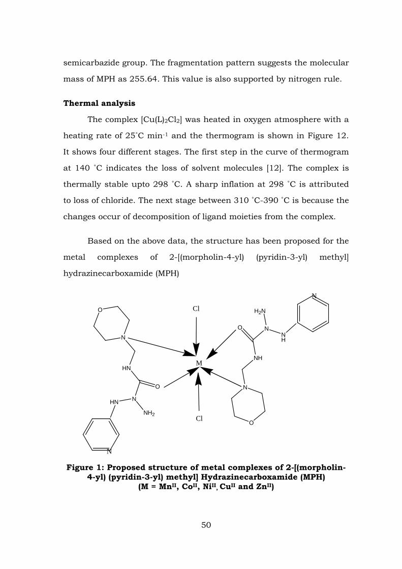
semicarbazide group. The fragmentation pattern suggests the molecular
mass of MPH as 255.64. This value is also supported by nitrogen rule.
Thermal analysis
The complex [Cu(L)2Cl2] was heated in oxygen atmosphere with a
heating rate of 25˚C min-1 and the thermogram is shown in Figure 12.
It shows four different stages. The first step in the curve of thermogram
at 140 ˚C indicates the loss of solvent molecules [12]. The complex is
thermally stable upto 298 ˚C. A sharp inflation at 298 ˚C is attributed
to loss of chloride. The next stage between 310 ˚C-390 ˚C is because the
changes occur of decomposition of ligand moieties from the complex.
Based on the above data, the structure has been proposed for the
metal complexes of 2-[(morpholin-4-yl) (pyridin-3-yl) methyl]
hydrazinecarboxamide (MPH)
N
H2N
O
NH
N
O
NH
N
M
Cl
Cl
N
NH2
O
HN
N
O
HN
N
Figure 1: Proposed structure of metal complexes of 2-[(morpholin-4-yl) (pyridin-3-yl) methyl] Hydrazinecarboxamide (MPH)
(M = MnII, CoII, NiII, CuII and ZnII)
50
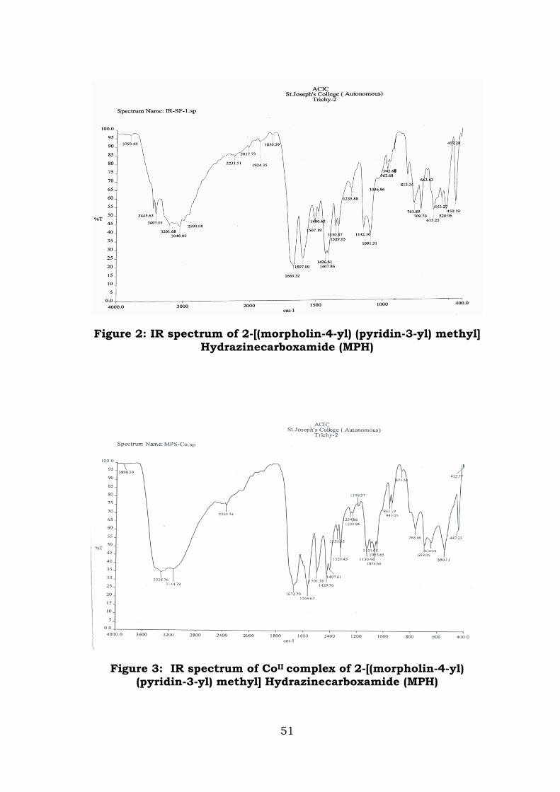
Figure 2: IR spectrum of 2-[(morpholin-4-yl) (pyridin-3-yl) methyl] Hydrazinecarboxamide (MPH)
Figure 3: IR spectrum of CoII complex of 2-[(morpholin-4-yl) (pyridin-3-yl) methyl] Hydrazinecarboxamide (MPH)
51
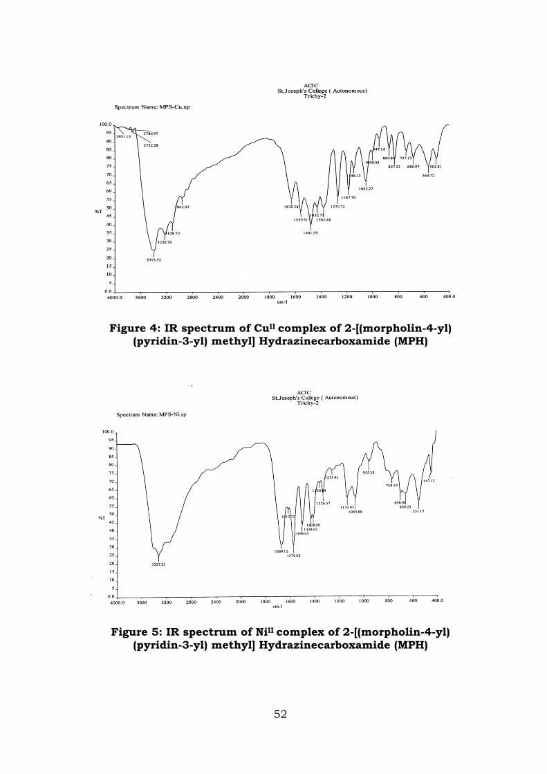
Figure 4: IR spectrum of CuII complex of 2-[(morpholin-4-yl) (pyridin-3-yl) methyl] Hydrazinecarboxamide (MPH)
Figure 5: IR spectrum of NiII complex of 2-[(morpholin-4-yl) (pyridin-3-yl) methyl] Hydrazinecarboxamide (MPH)
52
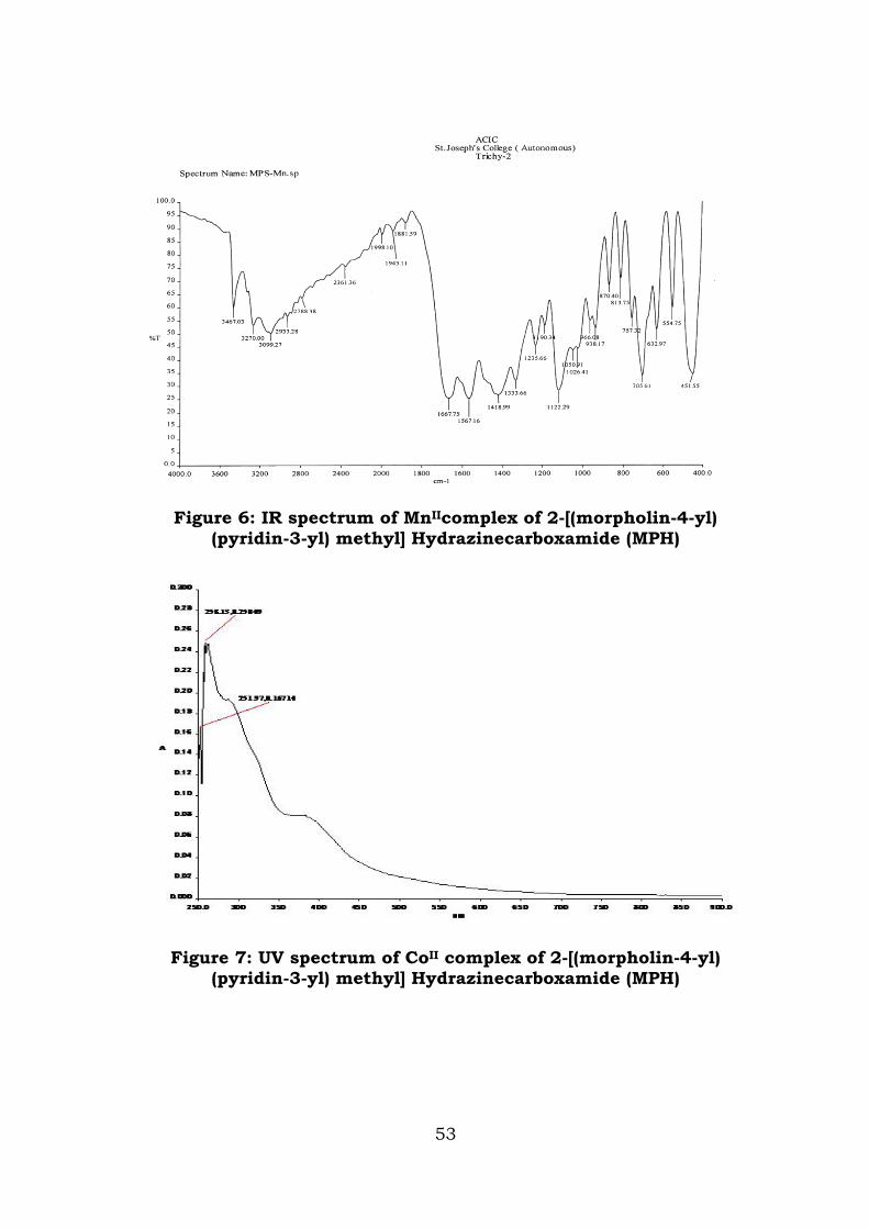
Figure 6: IR spectrum of MnIIcomplex of 2-[(morpholin-4-yl) (pyridin-3-yl) methyl] Hydrazinecarboxamide (MPH)
Figure 7: UV spectrum of CoII complex of 2-[(morpholin-4-yl) (pyridin-3-yl) methyl] Hydrazinecarboxamide (MPH)
53
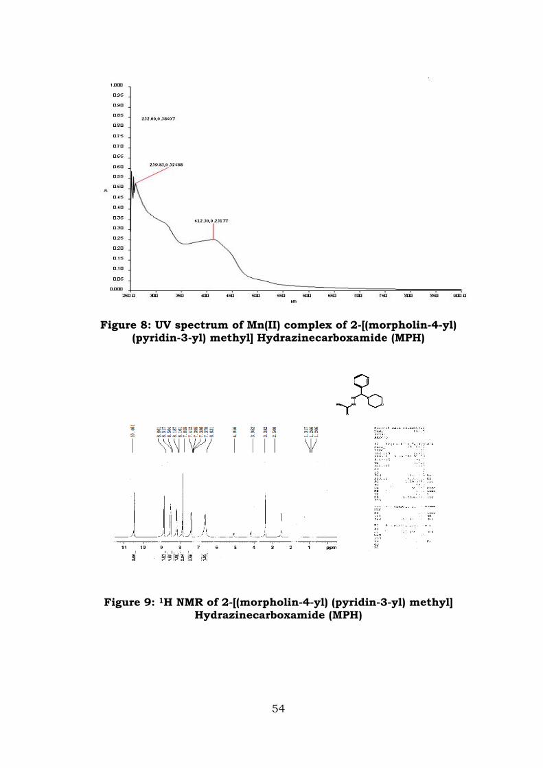
Figure 8: UV spectrum of Mn(II) complex of 2-[(morpholin-4-yl) (pyridin-3-yl) methyl] Hydrazinecarboxamide (MPH)
Figure 9: 1H NMR of 2-[(morpholin-4-yl) (pyridin-3-yl) methyl] Hydrazinecarboxamide (MPH)
54
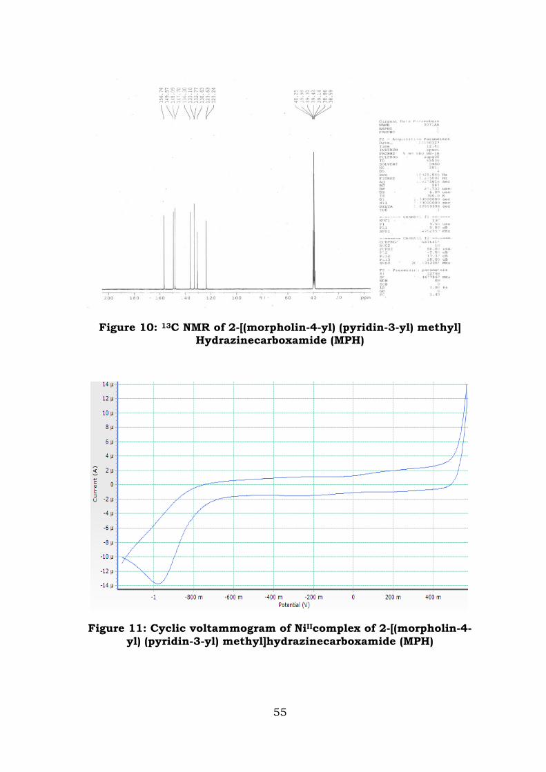
Figure 10: 13C NMR of 2-[(morpholin-4-yl) (pyridin-3-yl) methyl] Hydrazinecarboxamide (MPH)
Figure 11: Cyclic voltammogram of NiIIcomplex of 2-[(morpholin-4-yl) (pyridin-3-yl) methyl]hydrazinecarboxamide (MPH)
55
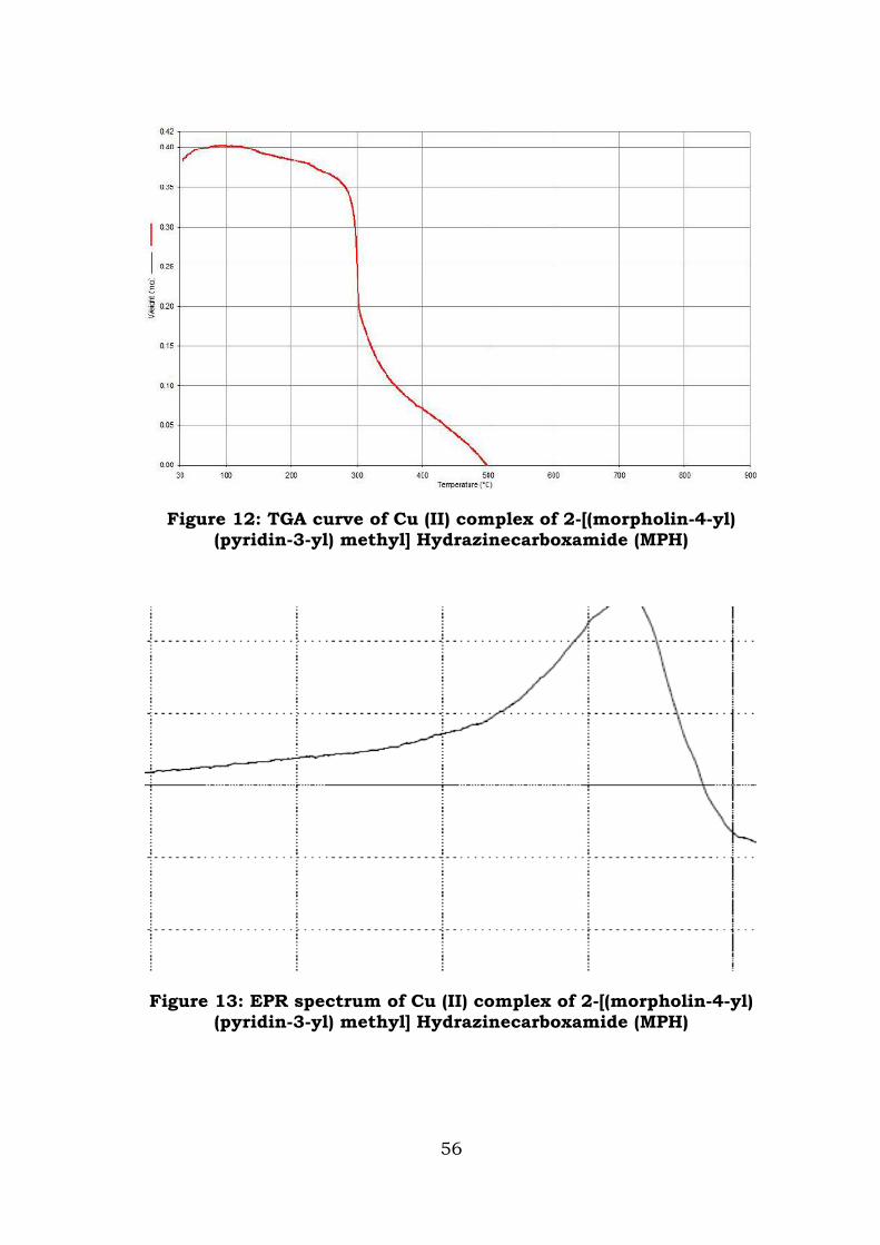
Figure 12: TGA curve of Cu (II) complex of 2-[(morpholin-4-yl) (pyridin-3-yl) methyl] Hydrazinecarboxamide (MPH)
Figure 13: EPR spectrum of Cu (II) complex of 2-[(morpholin-4-yl) (pyridin-3-yl) methyl] Hydrazinecarboxamide (MPH)
56
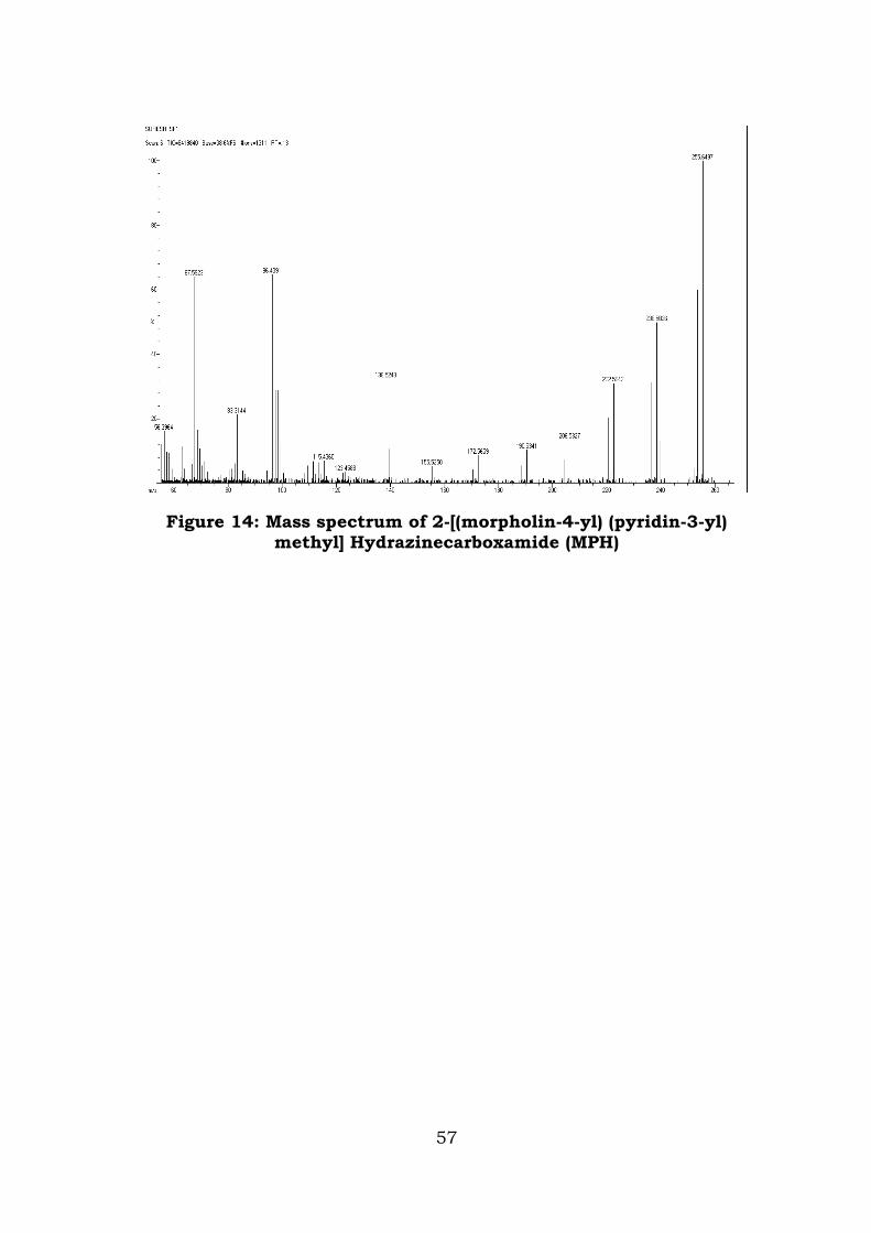
Figure 14: Mass spectrum of 2-[(morpholin-4-yl) (pyridin-3-yl) methyl] Hydrazinecarboxamide (MPH)
57
REFERENCES
1. Syed Ali Padusha M, Abdul Jameel A, Ind. J. Het. Cyclic. Chem, 2006, 16, 197.
2. Viswanathan M, Krishnan. G, Asian J. Chem, 2008, 16, 156.
3. Sharma Y R, Elementary Organic Spectroscopy, 2008, 4, 149.
4. Carlin R L, Stereochemistry of cobalt(II) complexes in Transition metal chemistry, Marcel Deckker, New York, 1972 88.
5. Cotton F A, Wilkinson, Advanced Inorganic Chemistry, vol. 1, 3rd edi., Wiley interscience, New York, 1972.
6. Chiswell B, Livingstone S E, J.Chem. Soc, 1960, 97.
7. Patel K S, Open Journal of Metal, 2012, 2, 49.
8. Alhadi A A, Bull.Chem.Soc.Ethiop, 2012, 26, 95.
9. Raman N, Esther S, Thangaraja C, J. Chem. Sci, 2004,116, 209.
10. Shelke V A, Jadhav S M, Shankarwar S G, Munde A S, Chondhekar T K, Soc. Ethiop Bull. Chem, 2011, 25, 381.
11. Vural Ufuk S, Hasan Mart H, Okkes D, Ozlem S, Vefa M, Koca M C, Bull. Chem. Soc. Ethiop, 2006, 20, 219.
12. Patel K S, Patel J C, Dholariya H R, Patel V K, Patel K D, Open Journal of Metal, 2012, 2, 49.
58
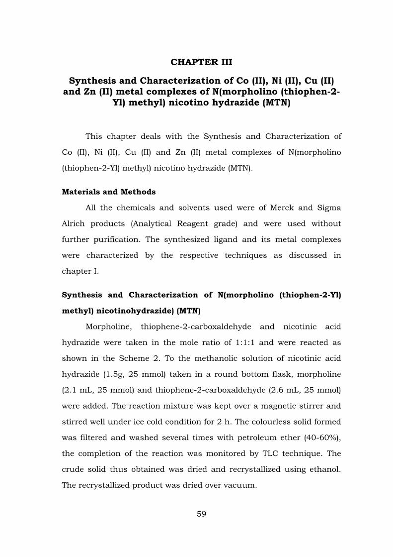
CHAPTER III
Synthesis and Characterization of Co (II), Ni (II), Cu (II) and Zn (II) metal complexes of N(morpholino (thiophen-2-
Yl) methyl) nicotino hydrazide (MTN)
This chapter deals with the Synthesis and Characterization of
Co (II), Ni (II), Cu (II) and Zn (II) metal complexes of N(morpholino
(thiophen-2-Yl) methyl) nicotino hydrazide (MTN).
Materials and Methods
All the chemicals and solvents used were of Merck and Sigma
Alrich products (Analytical Reagent grade) and were used without
further purification. The synthesized ligand and its metal complexes
were characterized by the respective techniques as discussed in
chapter I.
Synthesis and Characterization of N(morpholino (thiophen-2-Yl)
methyl) nicotinohydrazide) (MTN)
Morpholine, thiophene-2-carboxaldehyde and nicotinic acid
hydrazide were taken in the mole ratio of 1:1:1 and were reacted as
shown in the Scheme 2. To the methanolic solution of nicotinic acid
hydrazide (1.5g, 25 mmol) taken in a round bottom flask, morpholine
(2.1 mL, 25 mmol) and thiophene-2-carboxaldehyde (2.6 mL, 25 mmol)
were added. The reaction mixture was kept over a magnetic stirrer and
stirred well under ice cold condition for 2 h. The colourless solid formed
was filtered and washed several times with petroleum ether (40-60%),
the completion of the reaction was monitored by TLC technique. The
crude solid thus obtained was dried and recrystallized using ethanol.
The recrystallized product was dried over vacuum.
59
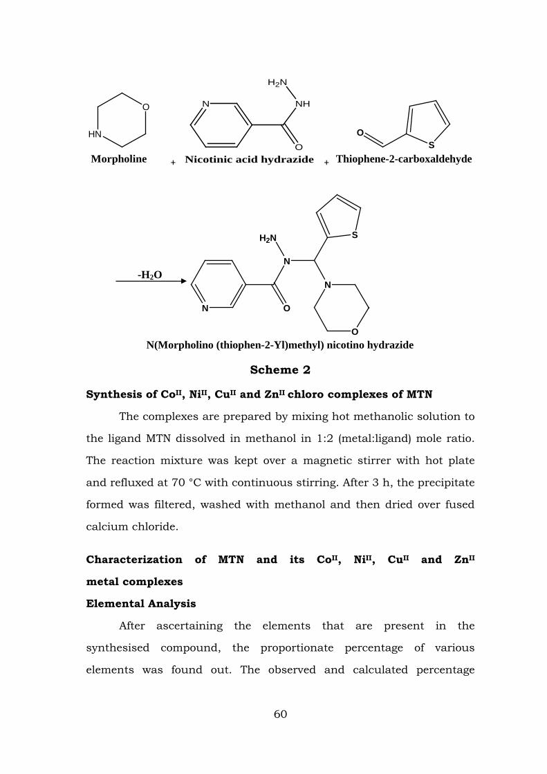
HN
O
Morpholine +
N
O
NH
H2N
Nicotinic acid hydrazide +
SO
Thiophene-2-carboxaldehyde
N O
N
H2N S
O
N
N(Morpholino (thiophen-2-Yl)methyl) nicotino hydrazide
Scheme 2
Synthesis of CoII, NiII, CuII and ZnII chloro complexes of MTN
The complexes are prepared by mixing hot methanolic solution to
the ligand MTN dissolved in methanol in 1:2 (metal:ligand) mole ratio.
The reaction mixture was kept over a magnetic stirrer with hot plate
and refluxed at 70 °C with continuous stirring. After 3 h, the precipitate
formed was filtered, washed with methanol and then dried over fused
calcium chloride.
Characterization of MTN and its CoII, NiII, CuII and ZnII
metal complexes
Elemental Analysis
After ascertaining the elements that are present in the
synthesised compound, the proportionate percentage of various
elements was found out. The observed and calculated percentage
-H2O
60
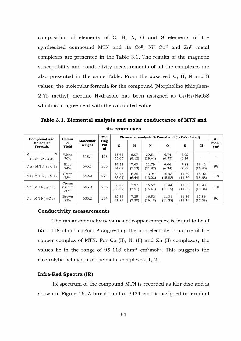
composition of elements of C, H, N, O and S elements of the
synthesized compound MTN and its CoII, NiII CuII and ZnII metal
complexes are presented in the Table 3.1. The results of the magnetic
susceptibility and conductivity measurements of all the complexes are
also presented in the same Table. From the observed C, H, N and S
values, the molecular formula for the compound (Morpholino (thiophen-
2-Yl) methyl) nicotino Hydrazide has been assigned as C15H18N4O2S
which is in agreement with the calculated value.
Table 3.1. Elemental analysis and molar conductance of MTN and
its complexes
Compound and Molecular Formula
Colour &
Yield
Molecular Weight
Melting Point
Elemental analysis % Found and (% Calculated) Ω-1 mol-1 cm2 C H N O S Cl
M T N C1 5H1 8N4O2S
White 70% 318.4 198 55.68
(55.05) 8.07 (8.12)
29.51 (29.41)
6.74 (6.53)
8.02 (8.14) -- --
C u ( M T N ) 2 C l 2 Blue 74% 645.1 226 54.53
(54.02) 7.63 (7.53)
31.79 (31.87)
6.06 (6.34)
7.88 (7.92)
16.42 (16.85) 98
N i ( M T N ) 2 C l 2 Green 78% 640.2 274 63.77
(63.04) 6.36 (6.44)
13.94 (13.23)
15.93 (15.88)
11.52 (11.50)
18.02 (18.68) 110
Z n ( M T N ) 2 C l 2 Creamy white
80% 646.9 256 66.88
(66.32) 7.37 (7.21)
16.62 (16.41)
11.44 (11.12)
11.53 (11.55)
17.98 (18.34) 110
C o ( M T N ) 2 C l 2 Brown 83% 635.2 234 62.86
(61.89) 7.25 (7.20)
16.52 (16.48)
11.31 (11.28)
11.56 (11.49)
17.86 (17.58) 96
Conductivity measurements
The molar conductivity values of copper complex is found to be of
65 – 118 ohm-1 cm2mol-2 suggesting the non-electrolytic nature of the
copper complex of MTN. For Co (II), Ni (II) and Zn (II) complexes, the
values lie in the range of 95-118 ohm-1 cm2mol-2. This suggests the
electrolytic behaviour of the metal complexes [1, 2].
Infra-Red Spectra (IR)
IR spectrum of the compound MTN is recorded as KBr disc and is
shown in Figure 16. A broad band at 3421 cm-1 is assigned to terminal
61
ν(NH2) and a sharp band at 3298 cm-1 is assigned to ν(N2H). A broad
band appearing at 1647 cm-1 is assigned to ν(C=O). The bands at 1396
cm-1 and 1244 cm-1 are assigned to ν(CNC) and ν(COC) of Morpholine.
The spectrum of MTN has been compared with the spectrum of
its complexes. By the comparison, it has been observed that, ν(C=O)
appeared at 1647 cm-1 in the spectrum of the ligand has been shifted to
downfield in the spectrum of the complexes. Ν(CNC) of the ligand
appeared at 1396 cm-1 in the spectrum of the ligand has also been
shifted to lower frequency by 15-25 cm-1 in the spectrum of the
complexes. No change has been observed in the vibrational frequencies
of other groups. Hence, it is concluded that the ligand acts as a neutral
bidentate ligand. The IR spectrum of both the ligand and the complexes
show intense bands at 3421 cm-1 which is the characteristic feature of
ν(N-H), indicating the existence of an –NH group [3-5]. This value is in
agreement with the literature values and are presented in Table 3.2.
A sharp band appeared at 1647 cm-1 can be attributed to the
ν(C=O) of the ligand, which in agreement with the literature value(1680
cm-1 to 1630 cm-1) of ν(C=O) of amide [6]. In the far IR region, all the
complexes exhibit bands around 572-505 cm-1 and 440-418 cm-1 which
are assignable to ν(M-O) [7] and ν(M-N) [8-10] respectively. Due to the
larger dipole moment change in the vibration of (M-O) band in
comparison to that of νM-N band, the band due to M-O usually appear
at higher frequency region which are sharp and stronger than (M-N)
band. These two ranges of bands collaborate the coordination of the
ligand with the metal ion.
62
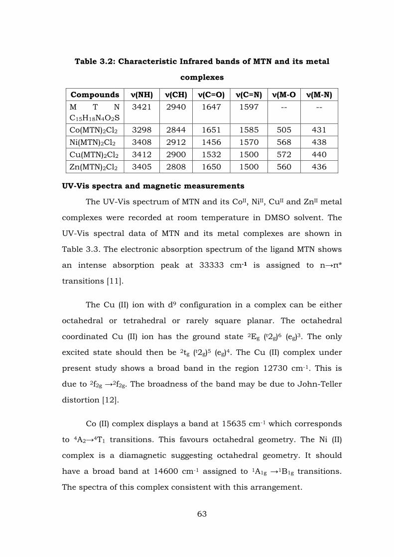
Table 3.2: Characteristic Infrared bands of MTN and its metal
complexes
Compounds ν(NH) ν(CH) ν(C=O) ν(C=N) ν(M-O ν(M-N) M T N C15H18N4O2S
3421 2940 1647 1597 -- --
Co(MTN)2Cl2 3298 2844 1651 1585 505 431 Ni(MTN)2Cl2 3408 2912 1456 1570 568 438 Cu(MTN)2Cl2 3412 2900 1532 1500 572 440 Zn(MTN)2Cl2 3405 2808 1650 1500 560 436
UV-Vis spectra and magnetic measurements
The UV-Vis spectrum of MTN and its CoII, NiII, CuII and ZnII metal
complexes were recorded at room temperature in DMSO solvent. The
UV-Vis spectral data of MTN and its metal complexes are shown in
Table 3.3. The electronic absorption spectrum of the ligand MTN shows
an intense absorption peak at 33333 cm-1 is assigned to n→π*
transitions [11].
The Cu (II) ion with d9 configuration in a complex can be either
octahedral or tetrahedral or rarely square planar. The octahedral
coordinated Cu (II) ion has the ground state 2Eg (t2g)6 (eg)3. The only
excited state should then be 2tg (t2g)5 (eg)4. The Cu (II) complex under
present study shows a broad band in the region 12730 cm-1. This is
due to 2f2g →2f2g. The broadness of the band may be due to John-Teller
distortion [12].
Co (II) complex displays a band at 15635 cm-1 which corresponds
to 4A2→4T1 transitions. This favours octahedral geometry. The Ni (II)
complex is a diamagnetic suggesting octahedral geometry. It should
have a broad band at 14600 cm-1 assigned to 1A1g →1B1g transitions.
The spectra of this complex consistent with this arrangement.
63
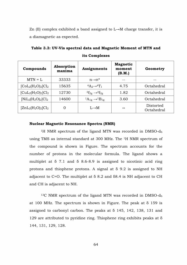
Zn (II) complex exhibited a band assigned to L→M charge transfer, it is
a diamagnetic as expected.
Table 3.3: UV-Vis spectral data and Magnetic Moment of MTN and
its Complexes
Compounds Absorption maxima Assignments
Magnetic moment
(B.M.) Geometry
MTN = L 33333 n→π* -- --
[CoL2(H2O)2]Cl2 15635 4A2→4T1 4.75 Octahedral
[CuL2(H2O)2]Cl2 12730 2f2g →2f2g 1.82 Octahedral
[NiL2(H2O)2]Cl2 14600 1A1g →1B1g 3.60 Octahedral
[ZnL2(H2O)2]Cl2 0 L→M -- Distorted Octahedral
Nuclear Magnetic Resonance Spectra (NMR)
1H NMR spectrum of the ligand MTN was recorded in DMSO-d6
using TMS as internal standard at 300 MHz. The 1H NMR spectrum of
the compound is shown in Figure. The spectrum accounts for the
number of protons in the molecular formula. The ligand shows a
multiplet at δ 7.1 and δ 8.6-8.9 is assigned to nicotinic acid ring
protons and thiophene protons. A signal at δ 9.2 is assigned to NH
adjacent to C=O. The multiplet at δ 8.2 and δ8.4 is NH adjacent to CH
and CH is adjacent to NH.
13C NMR spectrum of the ligand MTN was recorded in DMSO-d6
at 100 MHz. The spectrum is shown in Figure. The peak at δ 159 is
assigned to carbonyl carbon. The peaks at δ 145, 142, 138, 131 and
129 are attributed to pyridine ring. Thiophene ring exhibits peaks at δ
144, 131, 129, 128.
64
Mass spectrum
Mass spectrum of MTN was recorded by electron ionization mode.
The spectrum shows a molecular ion peak at m/z 323 which confirms
the assigned molecular mass to the Mannich base MTN and also obeys
Nitrogen rule. There upon on fragmentation, it records intense signals
at m/z 148, 121 and 106 are due to the removal of thiophene ring,
NCH and NH groups respectively. Finally the signal at m/z 78 is due to
the presence of C5H4N+ cation after the removal of CO.
Electron Spin Paramagnetic Resonance Spectrum (EPR)
The EPR spectrum of CuII complex was recorded at room
temperature in DMF solution on the X-band at 9.1 GHz under the
magnetic field strength 3400 G and is shown in Figure 22. The analysis
of spectrum gives g = 2.21 and g = 2.02. The trend g>2.0023,
observed for the complex, under study, indicates that the unpaired
electron is localized in dx2-y2 orbital of the CuII ion and the spectral
features are characteristic for axial symmetry. Thus tetrahedral
elongated octahedral geometry is confirmed for the above complex
[13-15].
Cyclic Voltammetry
The electrochemical properties of Cu(II) complex of MTN were
investigated in DMSO solution containing 0.1 M ammonium chloride as
a supporting electrolyte by cyclic voltammetry. All the measurements
were carried out in 1 MM DMSO solution of the complex at room
temperature, in the potential range from +1.0 to -1, at the scan rate of
100 mVs-1. The Voltammogram shows a well-defined redox process
corresponding to the formation of MnII/Mn(0) couple at Epa = -0.985V
and the cathodic peak Epc = +0.506V. This couple is found to be
reversible with Ep = -1.49V and the ratio of cathodic to anodic currents
65
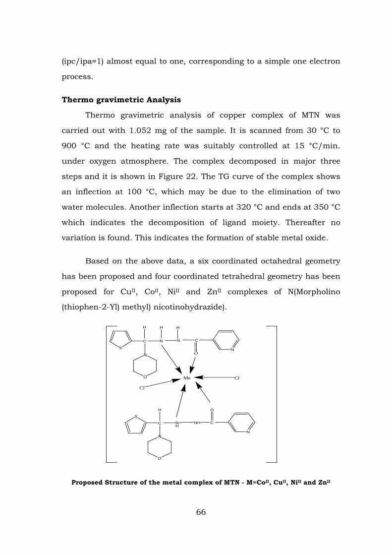
(ipc/ipa≈1) almost equal to one, corresponding to a simple one electron
process.
Thermo gravimetric Analysis
Thermo gravimetric analysis of copper complex of MTN was
carried out with 1.052 mg of the sample. It is scanned from 30 °C to
900 °C and the heating rate was suitably controlled at 15 °C/min.
under oxygen atmosphere. The complex decomposed in major three
steps and it is shown in Figure 22. The TG curve of the complex shows
an inflection at 100 °C, which may be due to the elimination of two
water molecules. Another inflection starts at 320 °C and ends at 350 °C
which indicates the decomposition of ligand moiety. Thereafter no
variation is found. This indicates the formation of stable metal oxide.
Based on the above data, a six coordinated octahedral geometry
has been proposed and four coordinated tetrahedral geometry has been
proposed for CuII, CoII, NiII and ZnII complexes of N(Morpholino
(thiophen-2-Yl) methyl) nicotinohydrazide).
Proposed Structure of the metal complex of MTN - M=CoII, CuII, NiII and ZnII
S
S
C
O
N
H
NH
NH C
N
O
Cl
Cl
C
O
N
H
N N
H H
C
ON
Me.H2O
66

Figure 16: IR SPECTRUM OF N(Morpholino (thiophen-2-Yl) methyl) nicotinohydrazide)(MTN)
Figure 17: IR SPECTRUM OF Cu (II) of N(Morpholino (thiophen-2-Yl) methyl) nicotinohydrazide)(MTN)
67
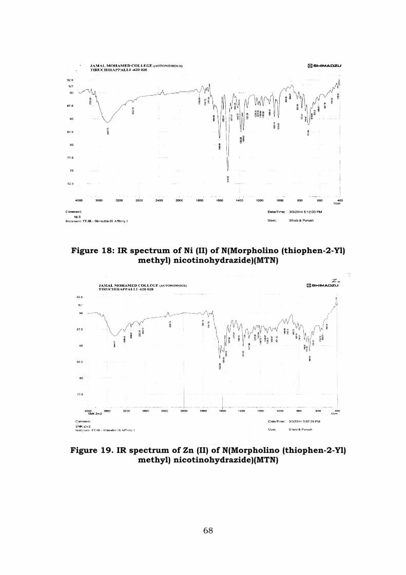
Figure 18: IR spectrum of Ni (II) of N(Morpholino (thiophen-2-Yl) methyl) nicotinohydrazide)(MTN)
Figure 19. IR spectrum of Zn (II) of N(Morpholino (thiophen-2-Yl) methyl) nicotinohydrazide)(MTN)
68
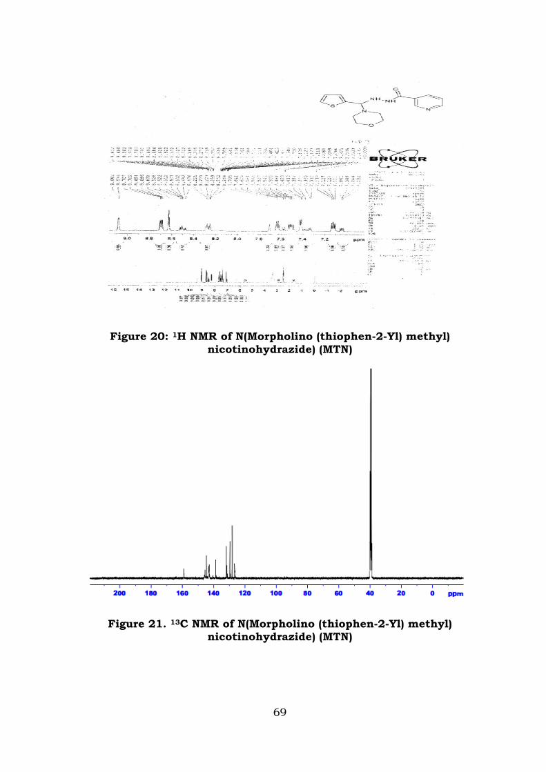
Figure 20: 1H NMR of N(Morpholino (thiophen-2-Yl) methyl) nicotinohydrazide) (MTN)
Figure 21. 13C NMR of N(Morpholino (thiophen-2-Yl) methyl) nicotinohydrazide) (MTN)
69
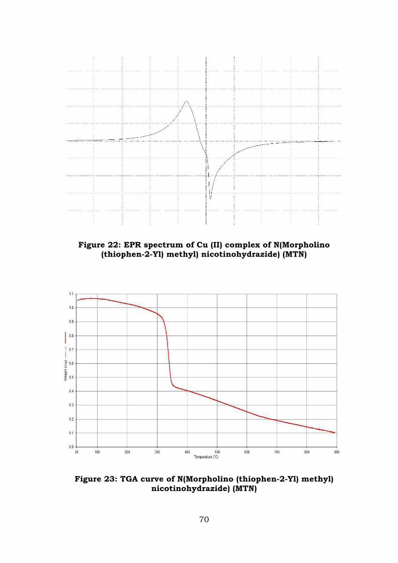
Figure 22: EPR spectrum of Cu (II) complex of N(Morpholino (thiophen-2-Yl) methyl) nicotinohydrazide) (MTN)
Figure 23: TGA curve of N(Morpholino (thiophen-2-Yl) methyl) nicotinohydrazide) (MTN)
70
REFERENCES
1. Rekha S, Nagasundara K R, Ind. J. Chem, 2006, 45, 2421.
2. Douglas X.W, Heloisa B, Amal A N, Fathy A E, Mohammed I A, Trans Met. Chem, 1999, 24421.
3. Raman N, Pitchikani Raja Y, Kulandaisamy, Indian Acad. Sci, 2001,113, 183.
4. Chandra S, Sharma A K, J. Indian Chem. Soc, 2009, 86, 690.
5. Yaul S R, Yaul A R, Pathe G B, Anand S, Amer-Eura. J. Sci. Res, 2009, 4, 229.
6. SharmaY R, Elementary Organic Chemistry, 4th edition, 2009, 148.
6. Sivakami M et al., Int. J. Pharm. Sci. Rev. Res., 2014, 27(1), 336-342.
7. Singh U K, Pandeya S N, Sethia S K, Pandey M, Singh A, Garg A, Kumar P, Int. J. Pharm. Soc. Drug. Res, 2010, 2 151.
8. Abdul Jameel A, Syed Ali Padusha M, Der Chemica Sinica, 2012, 3, 1098.
9. Shah T B, Gupta A, Indan J. Chem, 2010, 49, 578.
10. Ramesh M, Sabastiyan A, Der Chemica Sinica, 2012, 3, 534.
11. Kriza A, J. Serb.Chem. Soc, 2010, 75, 229.
12. N. Raman, C. Thangaraja and S.J. Raja, Ind. J. Chem., 44A 2005 693; K.R. Reddy, K.M. Reddy and K.N. Mahendra, Ind. J. Chem., 45A 2006 378
13. Sathya D ,Senthil Kumaran J, Priya S, Jayachandramani N, Mahlakshmi S Emelda A R, Int. J. Chem. Tech. Res, 2011, 3(1), 248.
14. Veeraraj A, Sami P and Raman N, Proc. Ind. Acad. Sci, 2000, 112, 515.
15. Raman N, Esther S, Thangarja C, J. Chem. Sci, 2004, 116, 209.
71
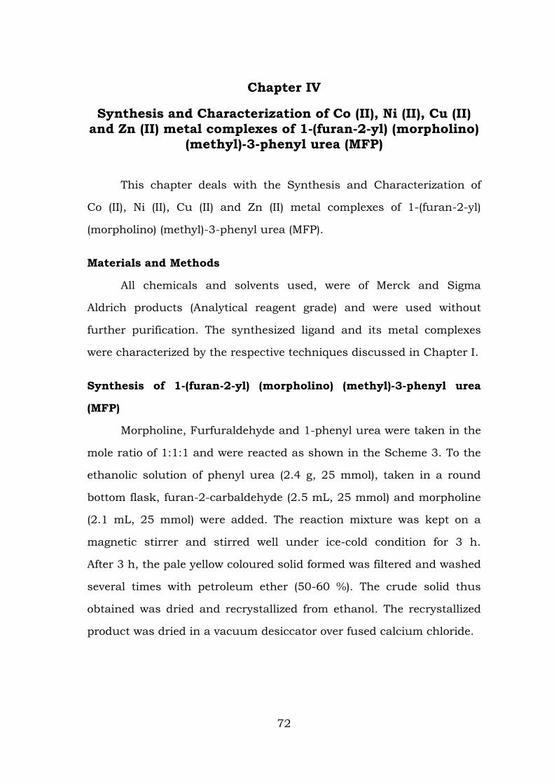
Chapter IV
Synthesis and Characterization of Co (II), Ni (II), Cu (II) and Zn (II) metal complexes of 1-(furan-2-yl) (morpholino)
(methyl)-3-phenyl urea (MFP)
This chapter deals with the Synthesis and Characterization of
Co (II), Ni (II), Cu (II) and Zn (II) metal complexes of 1-(furan-2-yl)
(morpholino) (methyl)-3-phenyl urea (MFP).
Materials and Methods
All chemicals and solvents used, were of Merck and Sigma
Aldrich products (Analytical reagent grade) and were used without
further purification. The synthesized ligand and its metal complexes
were characterized by the respective techniques discussed in Chapter I.
Synthesis of 1-(furan-2-yl) (morpholino) (methyl)-3-phenyl urea
(MFP)
Morpholine, Furfuraldehyde and 1-phenyl urea were taken in the
mole ratio of 1:1:1 and were reacted as shown in the Scheme 3. To the
ethanolic solution of phenyl urea (2.4 g, 25 mmol), taken in a round
bottom flask, furan-2-carbaldehyde (2.5 mL, 25 mmol) and morpholine
(2.1 mL, 25 mmol) were added. The reaction mixture was kept on a
magnetic stirrer and stirred well under ice-cold condition for 3 h.
After 3 h, the pale yellow coloured solid formed was filtered and washed
several times with petroleum ether (50-60 %). The crude solid thus
obtained was dried and recrystallized from ethanol. The recrystallized
product was dried in a vacuum desiccator over fused calcium chloride.
72
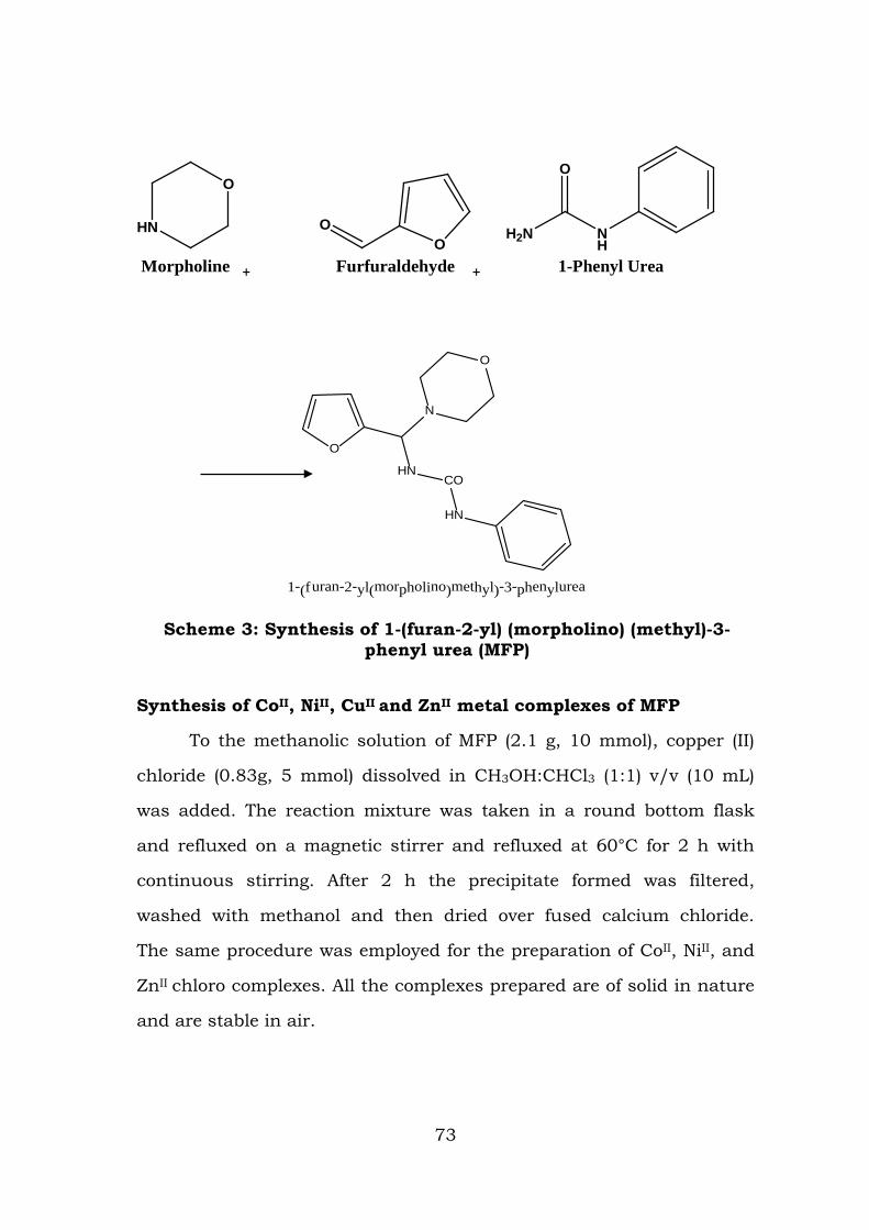
HN
O
Morpholine +
OO
Furfuraldehyde +
NH
O
H2N
1-Phenyl Urea
Scheme 3: Synthesis of 1-(furan-2-yl) (morpholino) (methyl)-3-phenyl urea (MFP)
Synthesis of CoII, NiII, CuII and ZnII metal complexes of MFP
To the methanolic solution of MFP (2.1 g, 10 mmol), copper (II)
chloride (0.83g, 5 mmol) dissolved in CH3OH:CHCl3 (1:1) v/v (10 mL)
was added. The reaction mixture was taken in a round bottom flask
and refluxed on a magnetic stirrer and refluxed at 60°C for 2 h with
continuous stirring. After 2 h the precipitate formed was filtered,
washed with methanol and then dried over fused calcium chloride.
The same procedure was employed for the preparation of CoII, NiII, and
ZnII chloro complexes. All the complexes prepared are of solid in nature
and are stable in air.
O
N
HNCO
HN
O
1-(furan-2-yl(morpholino)methyl)-3-phenylurea
73
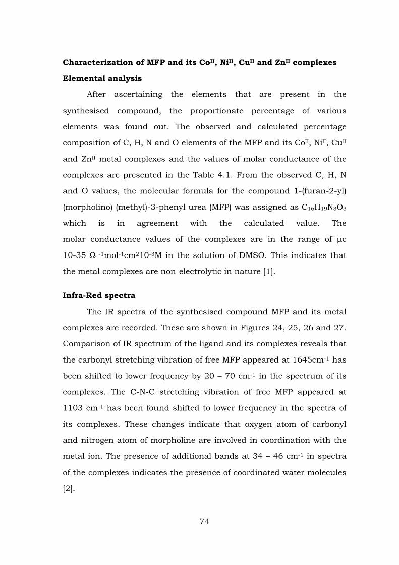
Characterization of MFP and its CoII, NiII, CuII and ZnII complexes
Elemental analysis
After ascertaining the elements that are present in the
synthesised compound, the proportionate percentage of various
elements was found out. The observed and calculated percentage
composition of C, H, N and O elements of the MFP and its CoII, NiII, CuII
and ZnII metal complexes and the values of molar conductance of the
complexes are presented in the Table 4.1. From the observed C, H, N
and O values, the molecular formula for the compound 1-(furan-2-yl)
(morpholino) (methyl)-3-phenyl urea (MFP) was assigned as C16H19N3O3
which is in agreement with the calculated value. The
molar conductance values of the complexes are in the range of μc
10-35 Ω -1mol-1cm210-3M in the solution of DMSO. This indicates that
the metal complexes are non-electrolytic in nature [1].
Infra-Red spectra
The IR spectra of the synthesised compound MFP and its metal
complexes are recorded. These are shown in Figures 24, 25, 26 and 27.
Comparison of IR spectrum of the ligand and its complexes reveals that
the carbonyl stretching vibration of free MFP appeared at 1645cm-1 has
been shifted to lower frequency by 20 – 70 cm-1 in the spectrum of its
complexes. The C-N-C stretching vibration of free MFP appeared at
1103 cm-1 has been found shifted to lower frequency in the spectra of
its complexes. These changes indicate that oxygen atom of carbonyl
and nitrogen atom of morpholine are involved in coordination with the
metal ion. The presence of additional bands at 34 – 46 cm-1 in spectra
of the complexes indicates the presence of coordinated water molecules
[2].
74
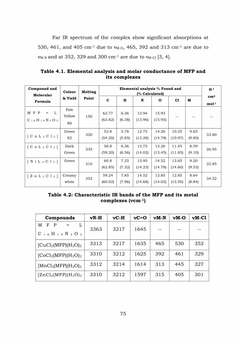
Far IR spectrum of the complex show significant absorptions at
530, 461, and 405 cm-1 due to νM-O, 465, 392 and 313 cm-1 are due to
νM-N and at 352, 329 and 300 cm-1 are due to νM-Cl [3, 4].
Table 4.1. Elemental analysis and molar conductance of MFP and its complexes
Compound and
Molecular
Formula
Colour
& Yield
Melting
Point
Elemental analysis % Found and (% Calculated)
Ω-1
cm2
mol-1 C H N O Cl M
M F P = L
C 1 6 H 1 9 N 3 O 3
Pale
Yellow
80
150 63.77
(63.82)
6.36
(6.38)
13.94
(13.96)
15.93
(15.95) -- -- --
[ C u L 2 C l 2 ] Green
82 320
53.8
(54.20)
5.78
(5.85)
12.75
(13.28)
14.26
(14.78)
10.35
(10.97)
9.65
(9.80) 33.80
[ C o L 2 C l 2 ]
Dark
Green 335
58.8
(59.20)
6.36
(6.56)
13.75
(14.02)
13.26
(13.45)
11.45
(11.85)
8.59
(9.10) 26.95
[ N i L 2 C l 2 ]
Green 310
60.8
(62.85)
7.25
(7.32)
13.95
(14.23)
14.52
(14.78)
13.65
(14.60)
9.20
(9.53) 32.85
[ Z n L 2 C l 2 ]
Creamy
white 352
59.24
(60.02)
7.85
(7.96)
14.52
(14.68)
13.85
(14.02)
12.85
(13.50)
8.64
(8.84) 34.52
Table 4.2: Characteristic IR bands of the MFP and its metal complexes (νcm-1)
Compounds νN-H νC-H νC=O νM-N νM-O νM-Cl
M F P = L
C 1 6 H 1 9 N 3 O 3 3363 3217 1645 -- -- --
[CuCl2(MFP)(H2O)2 3313 3217 1635 465 530 352
[CoCl2(MFP)(H2O)2 3310 3212 1625 392 461 329
[MnCl2(MFP)(H2O)2 3312 3214 1614 313 445 327
[ZnCl2(MFP)(H2O)2 3310 3212 1597 315 405 301
75
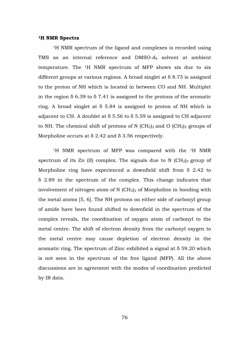
1H NMR Spectra
1H NMR spectrum of the ligand and complexes is recorded using
TMS as an internal reference and DMSO-d6 solvent at ambient
temperature. The 1H NMR spectrum of MFP shows six due to six
different groups at various regions. A broad singlet at δ 8.73 is assigned
to the proton of NH which is located in between CO and NH. Multiplet
in the region δ 6.39 to δ 7.41 is assigned to the protons of the aromatic
ring. A broad singlet at δ 5.84 is assigned to proton of NH which is
adjacent to CH. A doublet at δ 5.56 to δ 5.59 is assigned to CH adjacent
to NH. The chemical shift of protons of N (CH2)2 and O (CH2)2 groups of
Morpholine occurs at δ 2.42 and δ 3.56 respectively.
1H NMR spectrum of MFP was compared with the 1H NMR
spectrum of its Zn (II) complex. The signals due to N (CH2)2 group of
Morpholine ring have experienced a downfield shift from δ 2.42 to
δ 2.89 in the spectrum of the complex. This change indicates that
involvement of nitrogen atom of N (CH2)2 of Morpholine in bonding with
the metal atoms [5, 6]. The NH protons on either side of carbonyl group
of amide have been found shifted to downfield in the spectrum of the
complex reveals, the coordination of oxygen atom of carbonyl to the
metal centre. The shift of electron density from the carbonyl oxygen to
the metal centre may cause depletion of electron density in the
aromatic ring. The spectrum of Zinc exhibited a signal at δ 59.20 which
is not seen in the spectrum of the free ligand (MFP). All the above
discussions are in agreement with the modes of coordination predicted
by IR data.
76
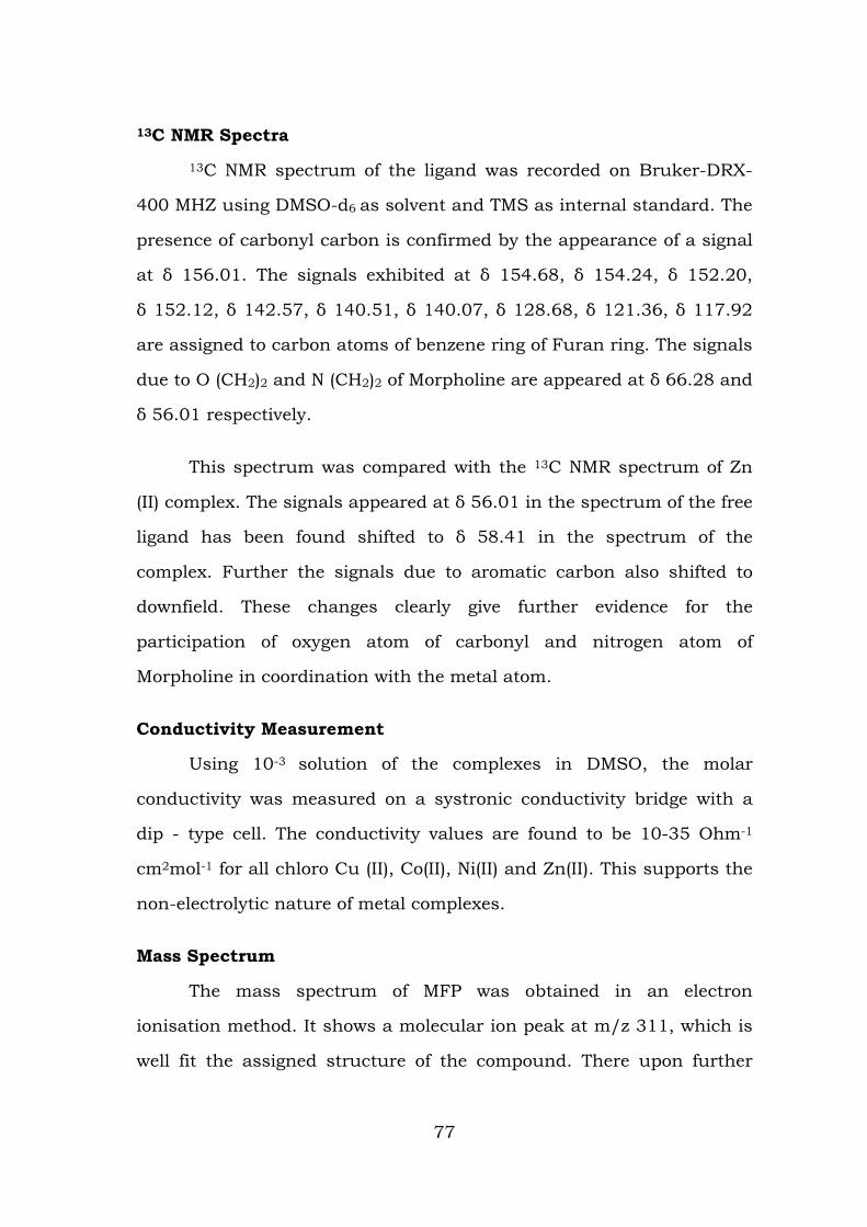
13C NMR Spectra
13C NMR spectrum of the ligand was recorded on Bruker-DRX-
400 MHZ using DMSO-d6 as solvent and TMS as internal standard. The
presence of carbonyl carbon is confirmed by the appearance of a signal
at δ 156.01. The signals exhibited at δ 154.68, δ 154.24, δ 152.20,
δ 152.12, δ 142.57, δ 140.51, δ 140.07, δ 128.68, δ 121.36, δ 117.92
are assigned to carbon atoms of benzene ring of Furan ring. The signals
due to O (CH2)2 and N (CH2)2 of Morpholine are appeared at δ 66.28 and
δ 56.01 respectively.
This spectrum was compared with the 13C NMR spectrum of Zn
(II) complex. The signals appeared at δ 56.01 in the spectrum of the free
ligand has been found shifted to δ 58.41 in the spectrum of the
complex. Further the signals due to aromatic carbon also shifted to
downfield. These changes clearly give further evidence for the
participation of oxygen atom of carbonyl and nitrogen atom of
Morpholine in coordination with the metal atom.
Conductivity Measurement
Using 10-3 solution of the complexes in DMSO, the molar
conductivity was measured on a systronic conductivity bridge with a
dip - type cell. The conductivity values are found to be 10-35 Ohm-1
cm2mol-1 for all chloro Cu (II), Co(II), Ni(II) and Zn(II). This supports the
non-electrolytic nature of metal complexes.
Mass Spectrum
The mass spectrum of MFP was obtained in an electron
ionisation method. It shows a molecular ion peak at m/z 311, which is
well fit the assigned structure of the compound. There upon further
77
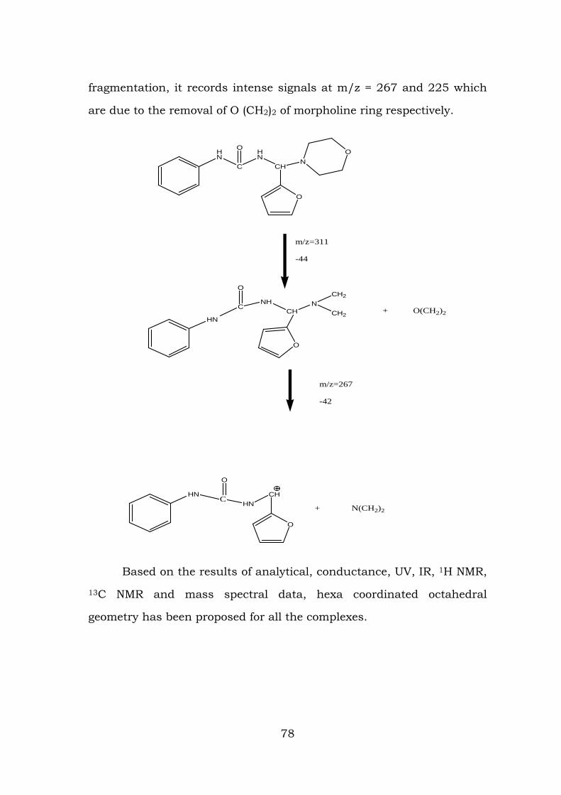
fragmentation, it records intense signals at m/z = 267 and 225 which
are due to the removal of O (CH2)2 of morpholine ring respectively.
HN
C
HN
CH
O
NO
O
m/z=311
-44
HN
C
O
NHCH
N
O
CH2
CH2+ O(CH2)2
m/z=267
-42
HNC
O
HNCH
O
+ N(CH2)2
Based on the results of analytical, conductance, UV, IR, 1H NMR,
13C NMR and mass spectral data, hexa coordinated octahedral
geometry has been proposed for all the complexes.
78
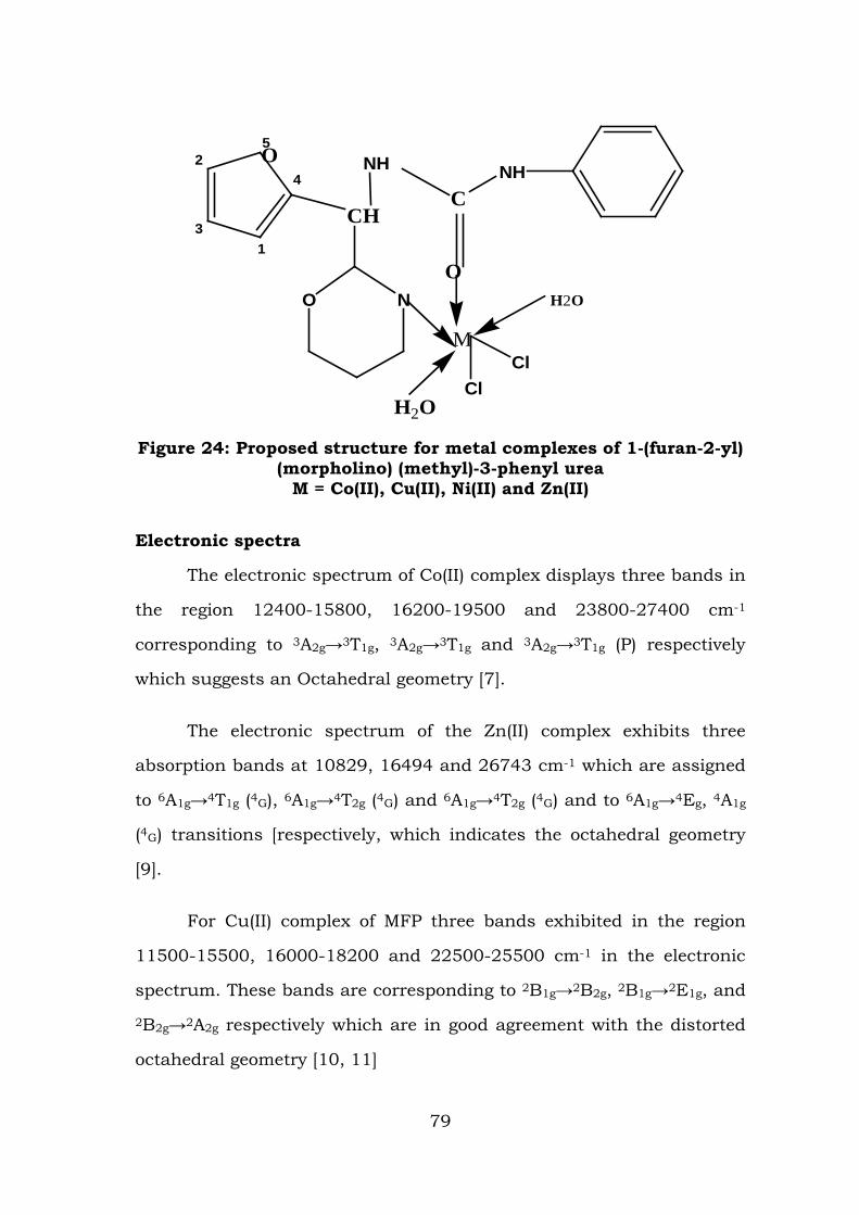
2
31
4
5O
CH
O N
NH
C
O
NH
ClH2O
Cl
H2O
M
Figure 24: Proposed structure for metal complexes of 1-(furan-2-yl) (morpholino) (methyl)-3-phenyl urea
M = Co(II), Cu(II), Ni(II) and Zn(II)
Electronic spectra
The electronic spectrum of Co(II) complex displays three bands in
the region 12400-15800, 16200-19500 and 23800-27400 cm-1
corresponding to 3A2g→3T1g, 3A2g→3T1g and 3A2g→3T1g (P) respectively
which suggests an Octahedral geometry [7].
The electronic spectrum of the Zn(II) complex exhibits three
absorption bands at 10829, 16494 and 26743 cm-1 which are assigned
to 6A1g→4T1g (4G), 6A1g→4T2g (4G) and 6A1g→4T2g (4G) and to 6A1g→4Eg, 4A1g
(4G) transitions [respectively, which indicates the octahedral geometry
[9].
For Cu(II) complex of MFP three bands exhibited in the region
11500-15500, 16000-18200 and 22500-25500 cm-1 in the electronic
spectrum. These bands are corresponding to 2B1g→2B2g, 2B1g→2E1g, and
2B2g→2A2g respectively which are in good agreement with the distorted
octahedral geometry [10, 11]
79
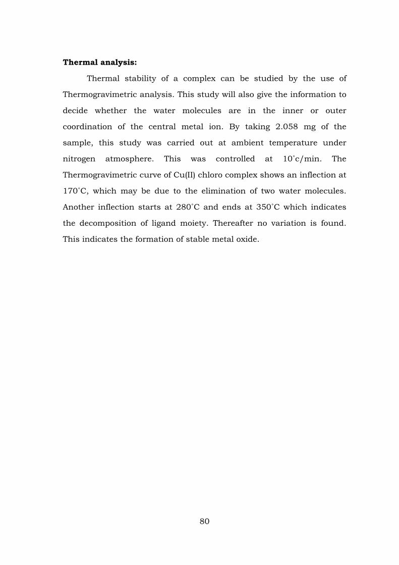
Thermal analysis:
Thermal stability of a complex can be studied by the use of
Thermogravimetric analysis. This study will also give the information to
decide whether the water molecules are in the inner or outer
coordination of the central metal ion. By taking 2.058 mg of the
sample, this study was carried out at ambient temperature under
nitrogen atmosphere. This was controlled at 10˚c/min. The
Thermogravimetric curve of Cu(II) chloro complex shows an inflection at
170˚C, which may be due to the elimination of two water molecules.
Another inflection starts at 280˚C and ends at 350˚C which indicates
the decomposition of ligand moiety. Thereafter no variation is found.
This indicates the formation of stable metal oxide.
80
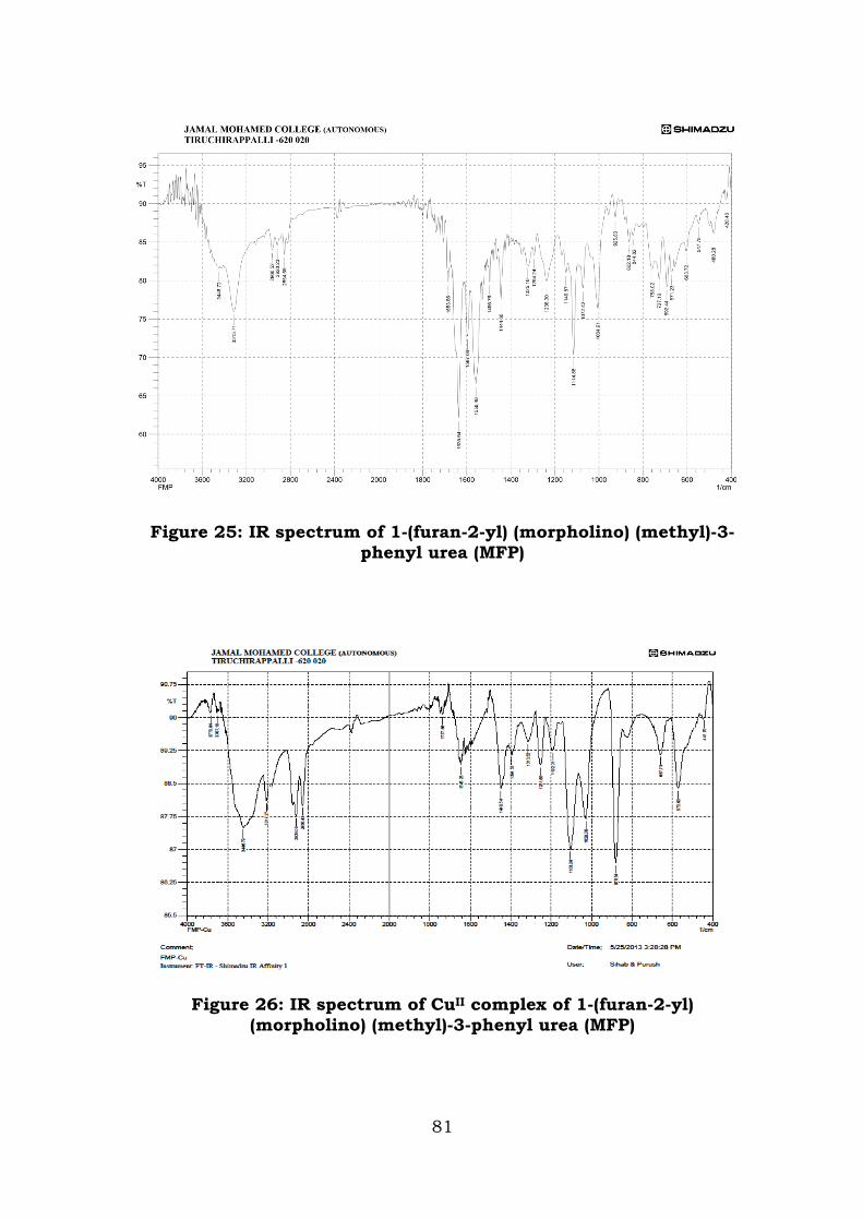
Figure 25: IR spectrum of 1-(furan-2-yl) (morpholino) (methyl)-3-phenyl urea (MFP)
Figure 26: IR spectrum of CuII complex of 1-(furan-2-yl)
(morpholino) (methyl)-3-phenyl urea (MFP)
81
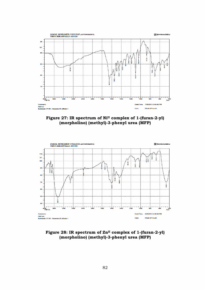
Figure 27: IR spectrum of NiII complex of 1-(furan-2-yl)
(morpholino) (methyl)-3-phenyl urea (MFP)
Figure 28: IR spectrum of ZnII complex of 1-(furan-2-yl) (morpholino) (methyl)-3-phenyl urea (MFP)
82
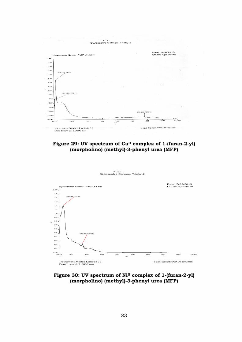
Figure 29: UV spectrum of CuII complex of 1-(furan-2-yl) (morpholino) (methyl)-3-phenyl urea (MFP)
Figure 30: UV spectrum of NiII complex of 1-(furan-2-yl) (morpholino) (methyl)-3-phenyl urea (MFP)
UV-Vis Spectrum
ACICSt.Joseph's College, Trichy-2
Data Interval: 1.0000 nm
Spectrum Name: FMP-NI.SP
Instrument Model: Lambda 35 Scan Speed: 960.00 nm/min
Date: 5/29/2013
220.0 300 400 500 600 700 800 900 1000 1100.0
0.00
0.1
0.2
0.3
0.4
0.5
0.6
0.7
0.8
0.9
1.0
1.1
1.2
1.3
1.4
1.5
1.60
nm
A
373.94,0.25612
246.44,1.2152
83
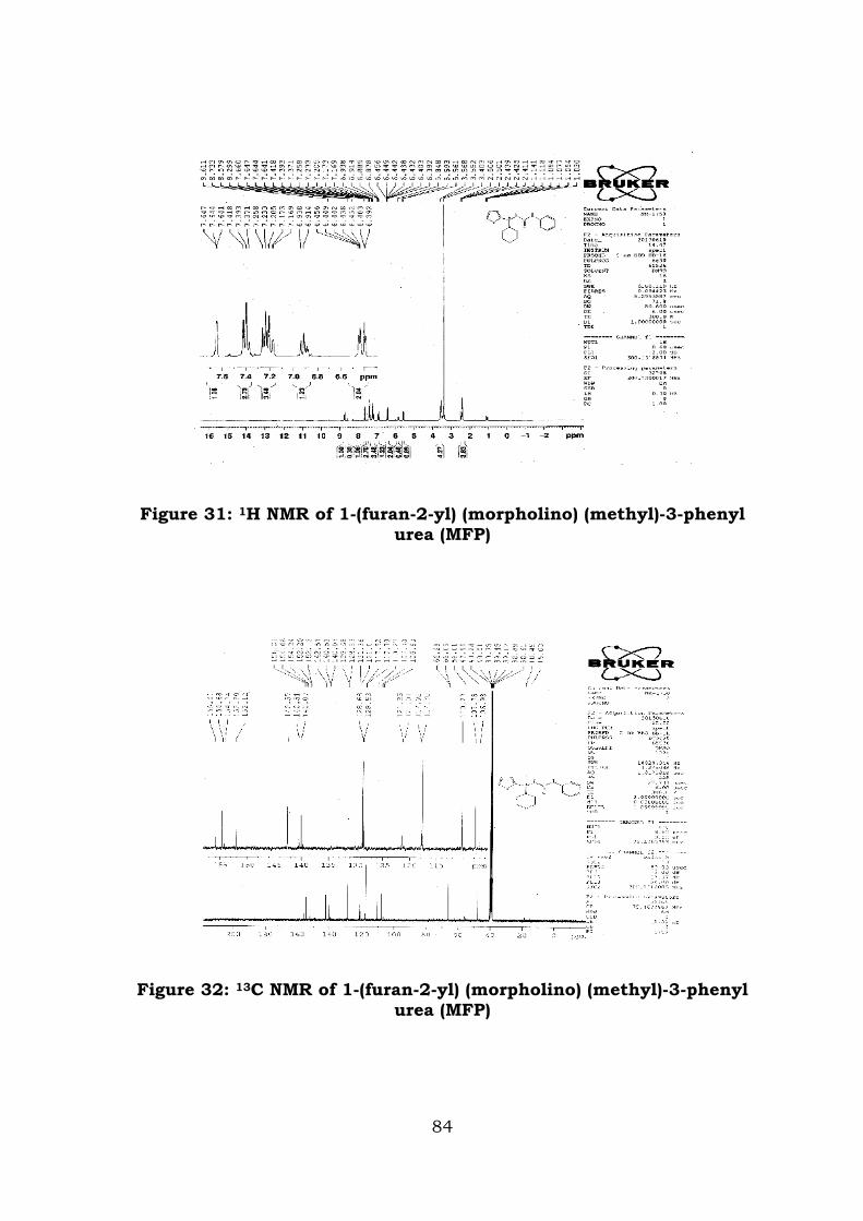
Figure 31: 1H NMR of 1-(furan-2-yl) (morpholino) (methyl)-3-phenyl urea (MFP)
Figure 32: 13C NMR of 1-(furan-2-yl) (morpholino) (methyl)-3-phenyl urea (MFP)
84
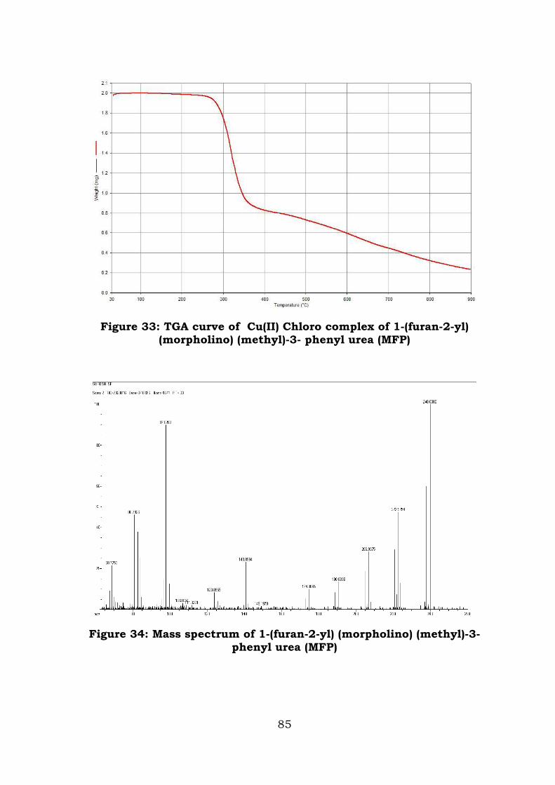
Figure 33: TGA curve of Cu(II) Chloro complex of 1-(furan-2-yl) (morpholino) (methyl)-3- phenyl urea (MFP)
Figure 34: Mass spectrum of 1-(furan-2-yl) (morpholino) (methyl)-3-phenyl urea (MFP)
85
References
1. Paul R C, Kapila P A, Bedi S and Vasisht K K, J. Indian Chem. Soc., 1976, 53, 768-773.
2. K Hussain Reddy, M Suredra Babu, P Suresh Babu and S Dhayanantha, Ind. J. Chem., 43A 2004 1233.
3. B M Gate House, S E Livingstone and R S Nyholm, J. Inorg Nucl. Chem., 8 1958 75
4. Mohan et al., J. Ind. Chem. Soc., 83 2006 331.
5. Maurya R C, Patel P and Rajput S, Synth. React. Inorg. Met-org. Chem.2003, 23, 817-827.
6. Nakamoto K, Infrared and Raman spectra of Inorganic and Coordination compounds, Wiley Interscience, New York, 1971.
7. Ravichandran S, Raman N and Thangaraja C, Journal of Chem. Science., 2004, (116(4), 215-219.
8. Sabastiyan A and Yosuva Suvaikin, Advances in Applied Science Research., 2012, 3(1), 45-50.
9. Murali Krishna P, Shankara B S, Shashidhar Reddy N, Mahesh B and Basavaraj C, International Journal of Inorganic Chemistry., 2013, 10, 1.
10. Muhannd A Mahmoud, Abbas A Salih Al-Hamdani and Shaimaa R Bakir, Baghdad Science Journal, 2013, 9(2), 82-95.
11. Lever A B P, Inorganic Electronic spectroscopy, Elsevier Amstardam,1968, 32,420.
86
CHAPTER V
THERMOACOUSTICAL STUDIES
Introduction
This chapter deals with the thermo acoustical studies of the
Mannich bases 2-[(morpholin-4-yl) (pyridin-3-yl) methyl]
Hydrazinecarboxamide (MPH) and (Morpholino (Thiophen-2-Yl) Methyl)
Nicotino Hydrazide (MTN) by the measurements of parameters such as
ultrasonic velocity(U), density(ρ), viscosity(η), adiabatic compressibility
(κ), intermolecular free length (Lf), molar volume (Vm), Relaxation time
(τ), Specific acoustic impedance (Z), Lenard Jones Potential (LJP),
internal pressure (πi), free volume (Vf) and molecular cohesive energy
(MCE), Available volume (Va), Gibbs free energy(∆G) and Absorption
coefficient (α/f2).
Ultrasonic velocity measurements have been successfully
employed to detect and assess weak and strong molecular interactions,
present in binary [1, 2] and ternary liquid mixtures [3, 4]. The
ultrasonic studies find extensive applications in characterizing aspects
of physico-chemical behaviour of several liquid mixtures [5, 6]. A large
number of studies have been made on the molecular interaction in
liquid systems by various physical methods like Infrared [7, 8], Raman
Effect [9, 10], Nuclear magnetic resonance, Dielectric constant [11],
ultra violet [12] and ultrasonic method [13, 14]. In the recent years,
ultrasonic technique has become a powerful tool in providing
information regarding the molecular behaviour of liquids and solids
owing to its ability of characterising physiochemical behaviour of the
medium. The thermodynamic, acoustic and transport properties of
87
non-electrolyte liquid-liquid mixtures provide information about type
and extent of molecular interactions, and can be used for the
development of molecular models for describing the behaviour of
solutions [15-19].
Besides the solvent-solvent interactions, this study is helpful to
determine ion-solvent interactions ion solid interaction in the case of
solution containing electrolyte and molecular interactions between the
molecules of non-electrolyte and also interaction between non-
electrolytes and solvent. Many research work have been carried out to
study the molecular interaction between the liquid mixtures and the
solution of electrolyte.
The compounds 2-[(morpholin-4-yl) (pyridin-3-yl) methyl]
hydrazinecarboxamide (MPH) and (Morpholino (Thiophen-2-Yl) Methyl)
Nicotinohydrazide (MTN) are found to possess N-H-CO groups in its
structure. This has been characterised by spectral studies. The
compounds containing amide groups may involve inter and intra
molecular hydrogen bonding, because of the presence of CO and NH
groups. This present study is aimed at to validate the existence of
clusture of amide moieties in the synthesised compounds.
Experimental Procedure:
Accurately weighed amount of the synthesised compounds were
dissolved in the solvent DMSO to obtain solution in the concentration
range 1x10⁻³- 1x10⁻² M. The ultrasonic velocities (U) have been
measured in ultrasonic interferometer operating at a frequency of
2 MHz with an accuracy of ±0.1%. The viscosities of the pure
compounds and their mixtures were determined using Ostwald’s
88
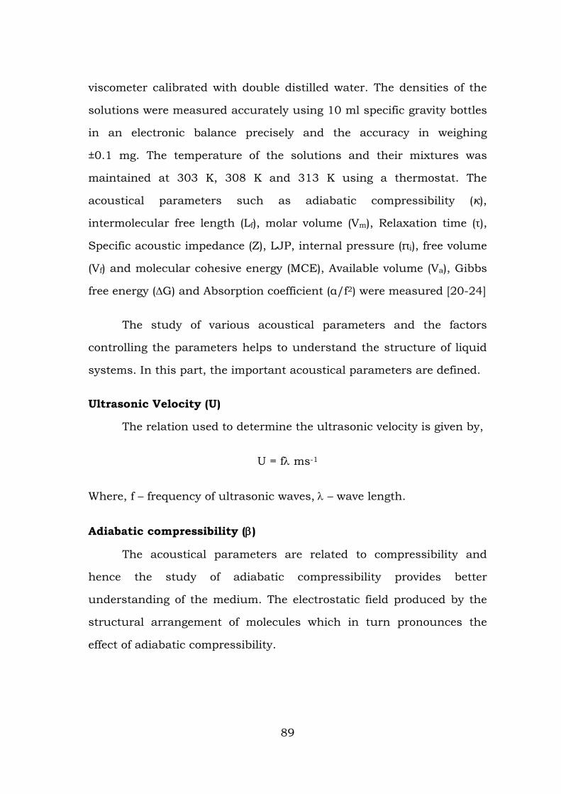
viscometer calibrated with double distilled water. The densities of the
solutions were measured accurately using 10 ml specific gravity bottles
in an electronic balance precisely and the accuracy in weighing
±0.1 mg. The temperature of the solutions and their mixtures was
maintained at 303 K, 308 K and 313 K using a thermostat. The
acoustical parameters such as adiabatic compressibility (κ),
intermolecular free length (Lf), molar volume (Vm), Relaxation time (τ),
Specific acoustic impedance (Z), LJP, internal pressure (πi), free volume
(Vf) and molecular cohesive energy (MCE), Available volume (Va), Gibbs
free energy (∆G) and Absorption coefficient (α/f2) were measured [20-24]
The study of various acoustical parameters and the factors
controlling the parameters helps to understand the structure of liquid
systems. In this part, the important acoustical parameters are defined.
Ultrasonic Velocity (U)
The relation used to determine the ultrasonic velocity is given by,
U = fλ ms-1
Where, f – frequency of ultrasonic waves, λ – wave length.
Adiabatic compressibility (β)
The acoustical parameters are related to compressibility and
hence the study of adiabatic compressibility provides better
understanding of the medium. The electrostatic field produced by the
structural arrangement of molecules which in turn pronounces the
effect of adiabatic compressibility.
89
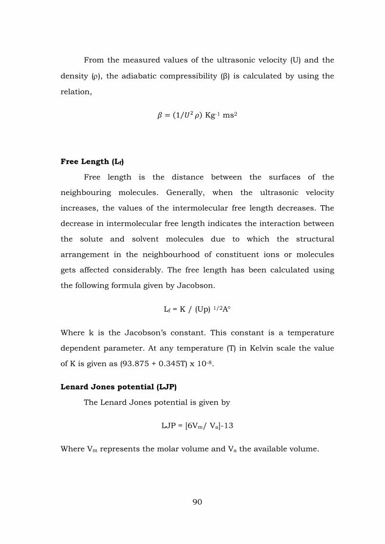
From the measured values of the ultrasonic velocity (U) and the
density (ρ), the adiabatic compressibility (β) is calculated by using the
relation,
𝛽 = (1 𝑈2⁄ 𝜌) Kg-1 ms2
Free Length (Lf)
Free length is the distance between the surfaces of the
neighbouring molecules. Generally, when the ultrasonic velocity
increases, the values of the intermolecular free length decreases. The
decrease in intermolecular free length indicates the interaction between
the solute and solvent molecules due to which the structural
arrangement in the neighbourhood of constituent ions or molecules
gets affected considerably. The free length has been calculated using
the following formula given by Jacobson.
Lf = K / (Up) 1/2A°
Where k is the Jacobson’s constant. This constant is a temperature
dependent parameter. At any temperature (T) in Kelvin scale the value
of K is given as (93.875 + 0.345T) x 10-8.
Lenard Jones potential (LJP)
The Lenard Jones potential is given by
LJP = [6Vm/ Va]-13
Where Vm represents the molar volume and Va the available volume.
90
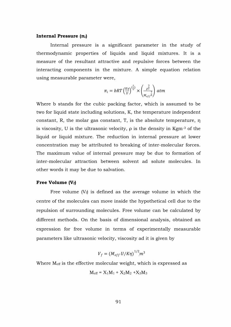
Internal Pressure (πi)
Internal pressure is a significant parameter in the study of
thermodynamic properties of liquids and liquid mixtures. It is a
measure of the resultant attractive and repulsive forces between the
interacting components in the mixture. A simple equation relation
using measurable parameter were,
𝜋𝑖 = 𝑏𝑅𝑇𝐾𝜂𝑈 (1
2)× 𝜌
32
𝑀𝑒𝑓𝑓76 𝑎𝑡𝑚
Where b stands for the cubic packing factor, which is assumed to be
two for liquid state including solutions, K, the temperature independent
constant, R, the molar gas constant, T, is the absolute temperature, η
is viscosity, U is the ultrasonic velocity, ρ is the density in Kgm-3 of the
liquid or liquid mixture. The reduction in internal pressure at lower
concentration may be attributed to breaking of inter-molecular forces.
The maximum value of internal pressure may be due to formation of
inter-molecular attraction between solvent ad solute molecules. In
other words it may be due to salvation.
Free Volume (Vf)
Free volume (Vf) is defined as the average volume in which the
centre of the molecules can move inside the hypothetical cell due to the
repulsion of surrounding molecules. Free volume can be calculated by
different methods. On the basis of dimensional analysis, obtained an
expression for free volume in terms of experimentally measurable
parameters like ultrasonic velocity, viscosity ad it is given by
𝑉𝑓 = (𝑀𝑒𝑓𝑓 𝑈 𝐾𝜂⁄ ) 𝑚3.
1/2
Where Meff is the effective molecular weight, which is expressed as
Meff = X1M1 + X2M2 +X3M3
91
Where X1 and M1 are the mole – fraction and molecular weight of the
component in the mixture respectively. K is the temperature
independent constant and the value of K is 4.28 x 10⁹ and η is the
viscosity coefficient of the solution.
Cohesive energy (CE)
Cohesive energy is usually given as a product of internal pressure
and molar volume (Vm).
Available Volume (Va)
Another parameter, which can be calculated from the
experimental ultrasonic velocity do several investigators for study the
available volume pure as well as for ternary liquid systems. The
following relation is used for available volume.
Va = Vm 31 U mU∞
−
Where ρ the density of the liquid, Vm represents the molar volume, U
the ultrasonic velocity and U∞ is limiting value taken as 1600 ms-1 for
liquids.
Results and Discussion
Dimethyl Sulphoxide (DMSO) is a very good solvent and it is
widely used for the synthesis of organic compounds. Compounds
contain amide moieties (Urea, acetamide, semi and thio-semicarbazide,
phenyl urea) are easily soluble in water. But, in some synthesis water
may not be used as a solvent because of several reasons. In such cases,
DMSO is used instead of water, it may not cause the difficulty in
getting pure product.
92
DMSO is an organo sulphur compound with the formula
(CH3)2SO. This colourless liquid is an important polar aprotic solvent
that dissolves both polar and non-polar compounds and is miscible in a
wide range of organic solvents as well as water. Through oxygen it may
have the chances to form hydrogen bonding with other molecules.
Keeping in this view, binary liquid mixture containing DMSO and
MPH/MTN at five different concentrations were prepared and the
parameters studied at three different temperatures.
The experimental results of ultrasonic velocity(U), density(ρ),
viscosity(η), adiabatic compressibility (κ), intermolecular free length (Lf),
molar volume (Vm), Relaxation time (τ), Specific acoustic impedance (Z),
Lenard Jones Potential (LJP), internal pressure (πi), free volume (Vf) and
molecular cohesive energy (MCE), Available volume (Va), Gibbs free
energy(∆G) and Absorption coefficient (α/f2) are presented in Tables
5.1 – 5.5. The effect of temperature on these parameters are
represented graphically in Figures 35 - 39.
93
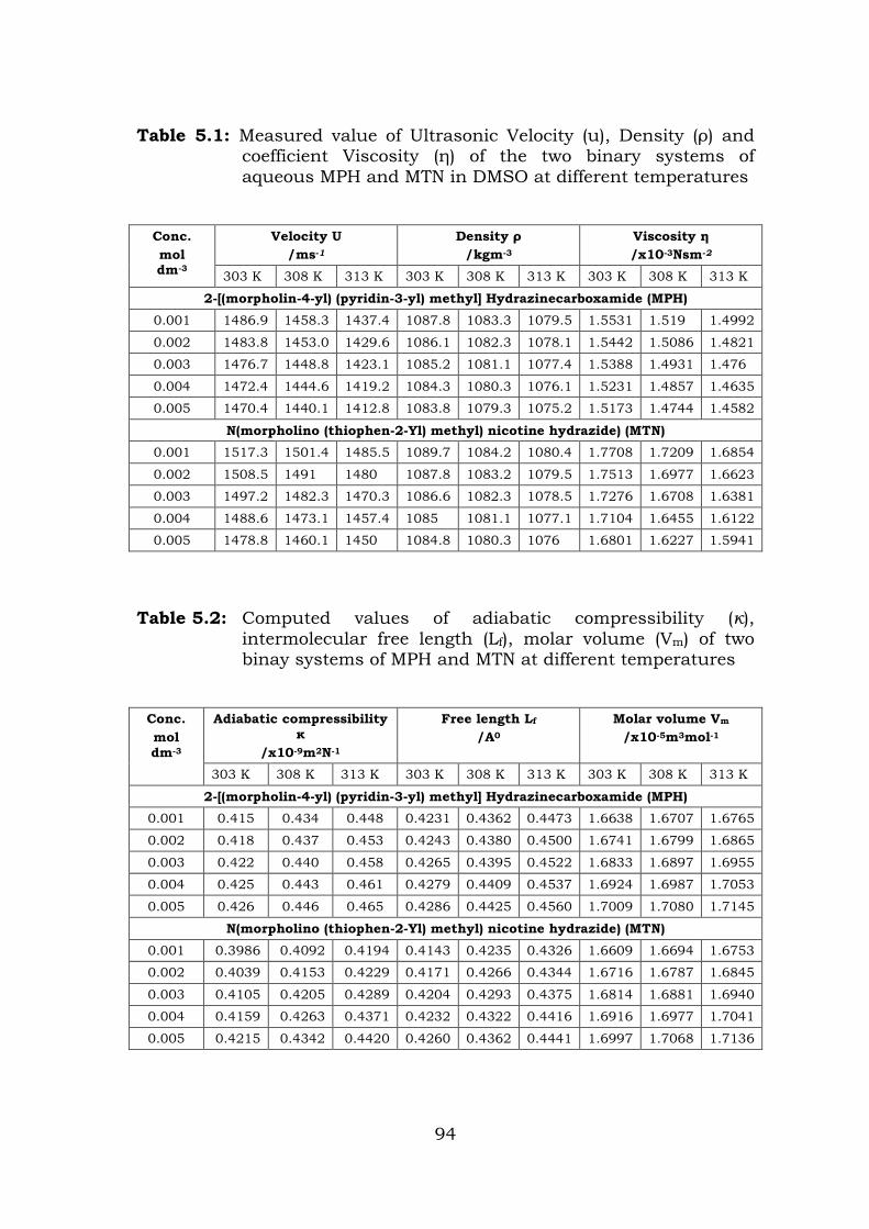
Table 5.1: Measured value of Ultrasonic Velocity (u), Density (ρ) and coefficient Viscosity (η) of the two binary systems of aqueous MPH and MTN in DMSO at different temperatures
Conc. mol dm-3
Velocity U /ms-1
Density ρ /kgm-3
Viscosity η /x10-3Nsm-2
303 K 308 K 313 K 303 K 308 K 313 K 303 K 308 K 313 K
2-[(morpholin-4-yl) (pyridin-3-yl) methyl] Hydrazinecarboxamide (MPH) 0.001 1486.9 1458.3 1437.4 1087.8 1083.3 1079.5 1.5531 1.519 1.4992
0.002 1483.8 1453.0 1429.6 1086.1 1082.3 1078.1 1.5442 1.5086 1.4821
0.003 1476.7 1448.8 1423.1 1085.2 1081.1 1077.4 1.5388 1.4931 1.476
0.004 1472.4 1444.6 1419.2 1084.3 1080.3 1076.1 1.5231 1.4857 1.4635
0.005 1470.4 1440.1 1412.8 1083.8 1079.3 1075.2 1.5173 1.4744 1.4582
N(morpholino (thiophen-2-Yl) methyl) nicotine hydrazide) (MTN) 0.001 1517.3 1501.4 1485.5 1089.7 1084.2 1080.4 1.7708 1.7209 1.6854
0.002 1508.5 1491 1480 1087.8 1083.2 1079.5 1.7513 1.6977 1.6623
0.003 1497.2 1482.3 1470.3 1086.6 1082.3 1078.5 1.7276 1.6708 1.6381
0.004 1488.6 1473.1 1457.4 1085 1081.1 1077.1 1.7104 1.6455 1.6122
0.005 1478.8 1460.1 1450 1084.8 1080.3 1076 1.6801 1.6227 1.5941
Table 5.2: Computed values of adiabatic compressibility (κ), intermolecular free length (Lf), molar volume (Vm) of two binay systems of MPH and MTN at different temperatures
Conc. mol dm-3
Adiabatic compressibility κ
/x10-9m2N-1
Free length Lf /A0
Molar volume Vm /x10-5m3mol-1
303 K 308 K 313 K 303 K 308 K 313 K 303 K 308 K 313 K
2-[(morpholin-4-yl) (pyridin-3-yl) methyl] Hydrazinecarboxamide (MPH) 0.001 0.415 0.434 0.448 0.4231 0.4362 0.4473 1.6638 1.6707 1.6765
0.002 0.418 0.437 0.453 0.4243 0.4380 0.4500 1.6741 1.6799 1.6865
0.003 0.422 0.440 0.458 0.4265 0.4395 0.4522 1.6833 1.6897 1.6955
0.004 0.425 0.443 0.461 0.4279 0.4409 0.4537 1.6924 1.6987 1.7053
0.005 0.426 0.446 0.465 0.4286 0.4425 0.4560 1.7009 1.7080 1.7145
N(morpholino (thiophen-2-Yl) methyl) nicotine hydrazide) (MTN) 0.001 0.3986 0.4092 0.4194 0.4143 0.4235 0.4326 1.6609 1.6694 1.6753
0.002 0.4039 0.4153 0.4229 0.4171 0.4266 0.4344 1.6716 1.6787 1.6845
0.003 0.4105 0.4205 0.4289 0.4204 0.4293 0.4375 1.6814 1.6881 1.6940
0.004 0.4159 0.4263 0.4371 0.4232 0.4322 0.4416 1.6916 1.6977 1.7041
0.005 0.4215 0.4342 0.4420 0.4260 0.4362 0.4441 1.6997 1.7068 1.7136
94
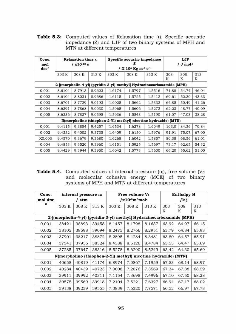
Table 5.3: Computed values of Relaxation time (τ), Specific acoustic impedance (Z) and LJP of two binary systems of MPH and MTN at different temperatures
Conc. mol dm-3
Relaxation time τ / x10-13 s
Specific acoustic impedance Z
/ X 106 Kg m-2 s-1
LJP / J mol-1
303 K 308 K 313 K 303 K 308 K 313 K 303 K
308 K
313 K
2-[(morpholin-4-yl) (pyridin-3-yl) methyl] Hydrazinecarboxamide (MPH) 0.001 8.6104 8.7913 8.9623 1.6174 1.5797 1.5516 71.88 54.74 46.04
0.002 8.6104 8.8031 8.9686 1.6115 1.5725 1.5412 69.61 52.30 43.33
0.003 8.6701 8.7729 9.0193 1.6025 1.5662 1.5332 64.85 50.49 41.26
0.004 8.6391 8.7868 9.0030 1.5965 1.5606 1.5272 62.23 48.77 40.09
0.005 8.6336 8.7827 9.0595 1.5936 1.5543 1.5190 61.07 47.03 38.28
N(morpholino (thiophen-2-Yl) methyl) nicotine hydrazide) (MTN) 0.001 9.4115 9.3884 9.4257 1.6534 1.6278 1.6049 103.0 84.36 70.84
0.002 9.4332 9.4002 9.3735 1.6409 1.6150 1.5976 91.91 75.07 67.00
X0.003 9.4570 9.3679 9.3680 1.6268 1.6042 1.5857 80.38 68.56 61.01
0.004 9.4853 9.3520 9.3960 1.6151 1.5925 1.5697 73.17 62.65 54.32
0.005 9.4429 9.3944 9.3950 1.6042 1.5773 1.5600 66.20 55.62 51.00
Table 5.4. Computed values of internal pressure (πi), free volume (Vf) and molecular cohesive energy (MCE) of two binary systems of MPH and MTN at different temperatures
Conc.
mol dm-
3
internal pressure πi / atm
Free volume Vf /x10-9m3mol-
Enthalpy H /k j
303 K 308 K 313 K 303 K 308 K 313 K 303 K
308 K
313 K
2-[(morpholin-4-yl) (pyridin-3-yl) methyl] Hydrazinecarboxamide (MPH) 0.001 38421 38893 39458 8.1457 8.1798 8.1637 63.92 64.97 66.15
0.002 38105 38598 39094 8.2475 8.2766 8.2951 63.79 64.84 65.93 0.003 37901 38217 38872 8.2895 8.4284 8.3481 63.80 64.57 65.91 0.004 37541 37956 38524 8.4388 8.5126 8.4784 63.53 64.47 65.69
0.005 37285 37647 38316 8.5278 8.6290 8.5249 63.42 64.30 65.69 N(morpholino (thiophen-2-Yl) methyl) nicotine hydrazide) (MTN)
0.001 40658 40819 41174 6.8974 7.0867 7.1959 67.53 68.14 68.97
0.002 40284 40439 40723 7.0008 7.2076 7.3569 67.34 67.88 68.59 0.003 39911 39992 40311 7.1154 7.3698 7.4996 67.10 67.50 68.28 0.004 39575 39569 39918 7.2104 7.5221 7.6327 66.94 67.17 68.02
0.005 39138 39239 39555 7.3839 7.6320 7.7571 66.52 66.97 67.78
95
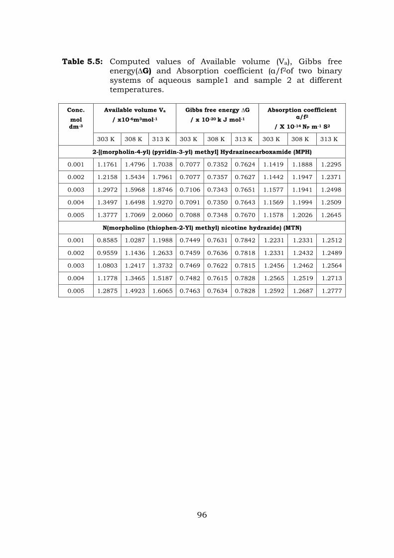
Table 5.5: Computed values of Available volume (Va), Gibbs free energy(∆G) and Absorption coefficient (α/f2of two binary systems of aqueous sample1 and sample 2 at different temperatures.
Conc. mol dm-3
Available volume Va / x10-6m3mol-1
Gibbs free energy ∆G / x 10-20 k J mol-1
Absorption coefficient α/f2
/ X 10-14 NP m-1 S2
303 K 308 K 313 K 303 K 308 K 313 K 303 K 308 K 313 K
2-[(morpholin-4-yl) (pyridin-3-yl) methyl] Hydrazinecarboxamide (MPH)
0.001 1.1761 1.4796 1.7038 0.7077 0.7352 0.7624 1.1419 1.1888 1.2295
0.002 1.2158 1.5434 1.7961 0.7077 0.7357 0.7627 1.1442 1.1947 1.2371
0.003 1.2972 1.5968 1.8746 0.7106 0.7343 0.7651 1.1577 1.1941 1.2498
0.004 1.3497 1.6498 1.9270 0.7091 0.7350 0.7643 1.1569 1.1994 1.2509
0.005 1.3777 1.7069 2.0060 0.7088 0.7348 0.7670 1.1578 1.2026 1.2645
N(morpholino (thiophen-2-Yl) methyl) nicotine hydrazide) (MTN)
0.001 0.8585 1.0287 1.1988 0.7449 0.7631 0.7842 1.2231 1.2331 1.2512
0.002 0.9559 1.1436 1.2633 0.7459 0.7636 0.7818 1.2331 1.2432 1.2489
0.003 1.0803 1.2417 1.3732 0.7469 0.7622 0.7815 1.2456 1.2462 1.2564
0.004 1.1778 1.3465 1.5187 0.7482 0.7615 0.7828 1.2565 1.2519 1.2713
0.005 1.2875 1.4923 1.6065 0.7463 0.7634 0.7828 1.2592 1.2687 1.2777
96
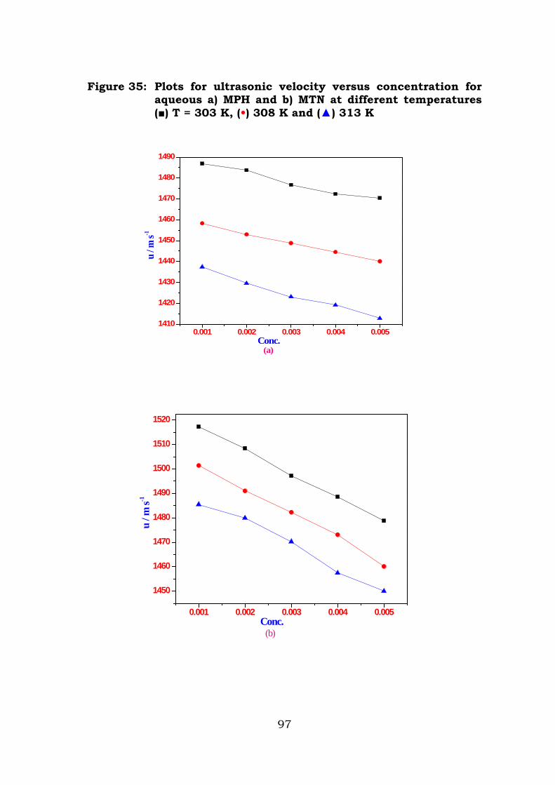
Figure 35: Plots for ultrasonic velocity versus concentration for aqueous a) MPH and b) MTN at different temperatures () T = 303 K, (•) 308 K and () 313 K
0.001 0.002 0.003 0.004 0.0051410
1420
1430
1440
1450
1460
1470
1480
1490
Conc.
u / m
s-1
(a)
0.001 0.002 0.003 0.004 0.005
1450
1460
1470
1480
1490
1500
1510
1520
Conc.
u / m
s-1
(b)
97
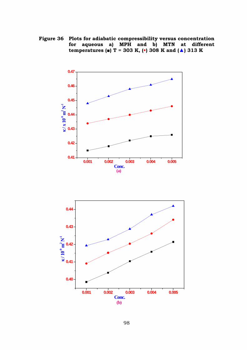
Figure 36 Plots for adiabatic compressibility versus concentration for aqueous a) MPH and b) MTN at different temperatures () T = 303 K, (•) 308 K and () 313 K
0.001 0.002 0.003 0.004 0.0050.41
0.42
0.43
0.44
0.45
0.46
0.47
Conc.(a)
κ / x
10-9
m2 N
-1
0.001 0.002 0.003 0.004 0.005
0.40
0.41
0.42
0.43
0.44
Conc.
κ / 1
0-9 m
2 N-1
(b)
98
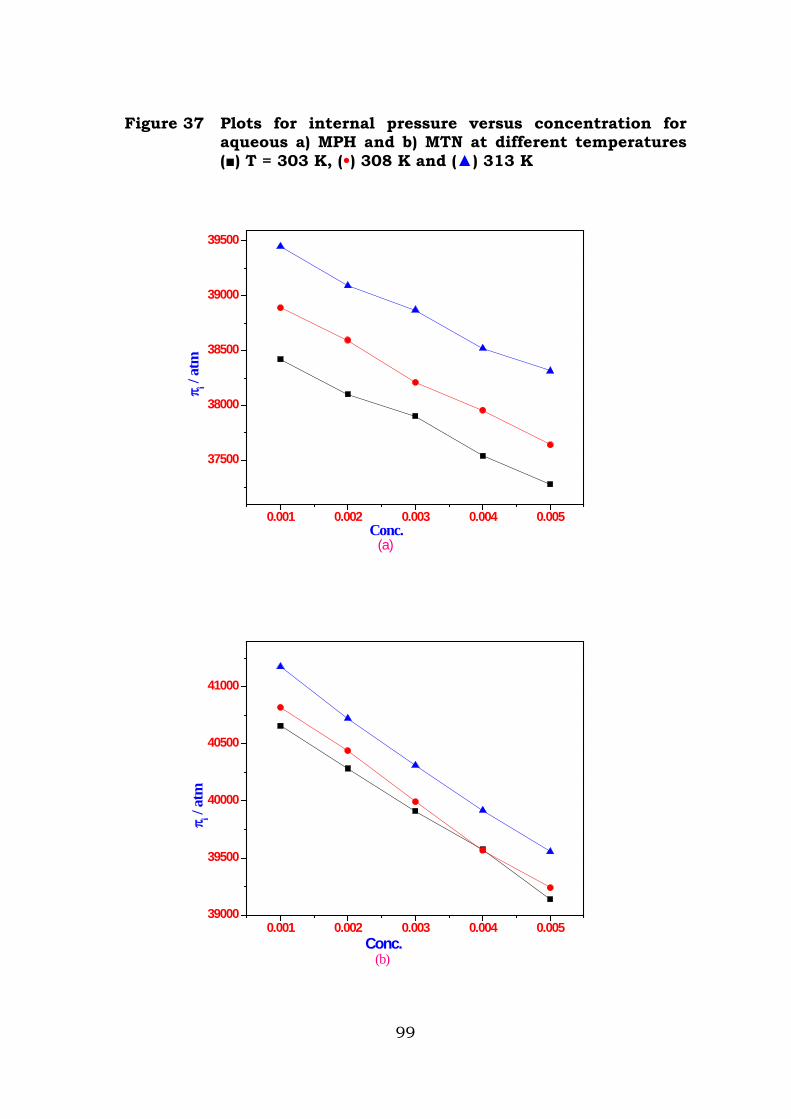
Figure 37 Plots for internal pressure versus concentration for aqueous a) MPH and b) MTN at different temperatures () T = 303 K, (•) 308 K and () 313 K
0.001 0.002 0.003 0.004 0.005
37500
38000
38500
39000
39500
Conc.(a)
π i / at
m
0.001 0.002 0.003 0.004 0.00539000
39500
40000
40500
41000
Conc.(b)
π i / at
m
99
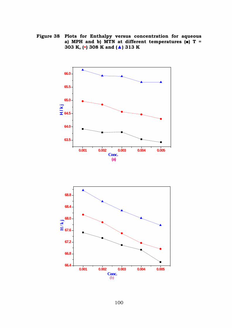
Figure 38 Plots for Enthalpy versus concentration for aqueous a) MPH and b) MTN at different temperatures () T = 303 K, (•) 308 K and () 313 K
0.001 0.002 0.003 0.004 0.005
63.5
64.0
64.5
65.0
65.5
66.0
Conc.(a)
H / k
j
0.001 0.002 0.003 0.004 0.00566.4
66.8
67.2
67.6
68.0
68.4
68.8
Conc.(b)
H /
k j
100
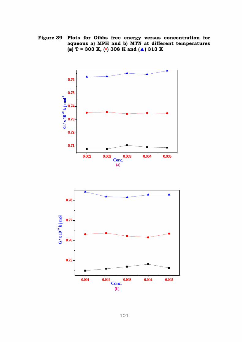
Figure 39 Plots for Gibbs free energy versus concentration for aqueous a) MPH and b) MTN at different temperatures () T = 303 K, (•) 308 K and () 313 K
0.001 0.002 0.003 0.004 0.005
0.71
0.72
0.73
0.74
0.75
0.76
Conc.(a)
G /
x 10
-20 k
j m
ol-1
0.001 0.002 0.003 0.004 0.005
0.75
0.76
0.77
0.78
Conc.(b)
G /
x 10
-20 k
j m
ol
101
The excess properties derived from these physical property data
reflect the physico-chemical behaviour of the liquid mixtures with
respect to the solution, structure and intermolecular interactions
between the component molecules of the mixture. The trends of
changes have been interpreted by the earlier works reported by various
researchers [15-17]. It has revealed from the literature, that the trends
of changes are mainly depends on the differences in the size of the
molecule and strength of interactions taking place between the
component of the mixtures. The velocity of sound through liquid
mixtures could be helpful in assessing the degree of association
between the molecules. The molar sound velocity on non-associated
liquids has been found to be independent, while that for associated
liquids is dependent on temperature.
Velocity
From the Table 1, for both the samples it has been observed that,
as the concentration increases, velocity decreases. This decrease in
velocity is due to increased association between the solute and the
solvent molecules. The compounds MPH and MTN has NH-CO-NH
arrangement, this may interacts with solvent molecules (DMSO). This
shows that, the existence of hydrogen bonding present in the liquid
mixture. Further, it has been observed that when temperature
increases velocity decreases, this may be attributed to the increased
vibration or collision between the solvent and solute molecules.
Adiabatic Compressibility
It has been observed from the Table 2, the adiabatic
compressibility values computed for the samples at different
concentrations, has been found to increase as the concentration
102
increases. This increase in concentration is as a result of molecular
aggregation. Molecular aggregation takes place through hydrogen
bonding between the solute and solvent. This result clearly reveals the
existence of hydrogen bonding in the liquid mixture.
Free Length (Lf)
The free length (Lf) values measured at different concentrations
are listed in Table 2. It has been found that, Lf values are found to
increase as the concentration increases. In general, dilute solutions the
distance between the molecules will be large and hence, the length of
the non-covalent interaction will be less. While in concentrated
solutions, due to hydrogen bonding the distance between the molecules
will be less. Therefore, the intermolecular free length (Lf) is found to
increase in both the samples.
Free Volume
Free volume is one of the significant factors in explaining the
variations in the physico-chemical properties of liquid mixture. The free
space and its dependent properties has a close connection with
molecular structure and it may show interesting features about
interactions. This molecular interactions between like and unlike
molecules are influenced by structural arrangements along with the
shape and size of the molecules.
From the results of computed values of free volume of the binary
liquid mixture at different concentrations, it has been observed that
there is a increase in free volume as concentration increases. This
increase in free volume is attributed to the strong interaction between
the molecules when the solute concentration increases.
103
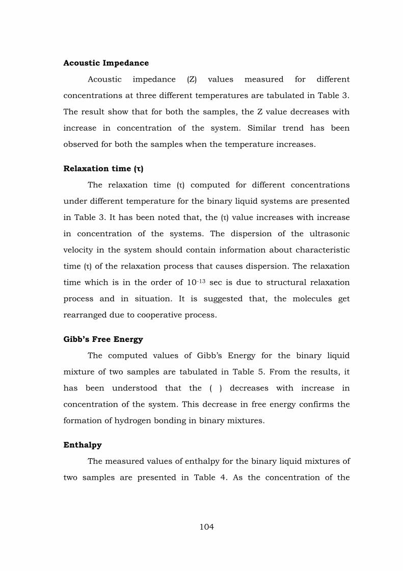
Acoustic Impedance
Acoustic impedance (Z) values measured for different
concentrations at three different temperatures are tabulated in Table 3.
The result show that for both the samples, the Z value decreases with
increase in concentration of the system. Similar trend has been
observed for both the samples when the temperature increases.
Relaxation time (τ)
The relaxation time (τ) computed for different concentrations
under different temperature for the binary liquid systems are presented
in Table 3. It has been noted that, the (τ) value increases with increase
in concentration of the systems. The dispersion of the ultrasonic
velocity in the system should contain information about characteristic
time (τ) of the relaxation process that causes dispersion. The relaxation
time which is in the order of 10-13 sec is due to structural relaxation
process and in situation. It is suggested that, the molecules get
rearranged due to cooperative process.
Gibb’s Free Energy
The computed values of Gibb’s Energy for the binary liquid
mixture of two samples are tabulated in Table 5. From the results, it
has been understood that the ( ) decreases with increase in
concentration of the system. This decrease in free energy confirms the
formation of hydrogen bonding in binary mixtures.
Enthalpy
The measured values of enthalpy for the binary liquid mixtures of
two samples are presented in Table 4. As the concentration of the
104
solute increases the enthalpy is found to increases. The same trend has
been observed when the temperature of the system increases.
Conclusion
The ultrasonic velocity, adiabatic compressibility, free length,
viscosity, free volume, free length, molar volume, density, internal
pressure, free energy and enthalpy have been measured. The measured
values of ultrasonic velocity, internal pressure, enthalpy and free
energy have been found to increase as the concentration increases. The
calculated values of adiabatic compressibility are found to decrease as
the concentration increases. These results are inferred that the strong
interaction is exist between the solvent (DMSO) and the solute (MPH
and MTN). Hence it is concluded that the association of solute
molecules occurs through hydrogen bonding.
105
References
1. Kannappan V & Jaya Shanthi R, Indian J Pure & Appl Phys, 43 (2005) 750.
2. Kannappan V, Xavier Jesu Raja S & Jaya Santhi R, Indian J Pure & Appl Phys, 41 (2003) 690.
3. Jayakumar S, Karunanithi N, Kannappan V & Gunasekaran S, Asian Chem Lett, 3 (1999), 224.
4. Neuman M S & Blum, J Am Soc, 86 (1964) 5600.
5. Dash S K & Swain B B, Chem. Papers, 48 (1994) 146.
6. Dash S K Chakravorthy V & Swain B B, Acoustic Lett, 19 (1996) 142.
7. Eyring H and Incaid K, Chem. Phys, 6.620 (1938)
8. Singh S, Singh R, Prasad N and Prakash S, Ind. J. Pure and Appl. Phy., 3, 156 (1977)
9. Ramamurthy M and Sastry O S, Indian J. Pure and Appl. Phys, 21, 579, (1983)
10. Hobbs M E and Bates W W, J. Am. Chem Soc., 74, 746, (1952)
11. Negakuva, J Am Chem Soc, 76, 3070, (1954).
12. Freedman E, J Chem Phys, 21, 1784, (1955).
13. Kannappan A N and Rajendran V, Indian J. Pure and Appl. Phys., 30(176, (1992)
14. Kinsler L E and Rray A R., Fundamentals of Acoustics, Wiley Edition, 1989.
15. J.M. Prausnitz, R.N. Lichtenthaler, E.G. Azevedo, Molecular Thermodynamic of Fluid-Phase Equilibria, 2nd Ed., Prentice Hall, Inc., 1986.
16. S.L. Oswal, N.B. Patel, J.Chem. Eng Data 40 (1995) 840-844.
17. S.L. Oswal, P. Oswal, J.P. Dave, J.Mol.Liq. 94 (2000) 203-219.
18. S.L. Oswal, K.D. Prajapati, N.Y. Ghael, S.P. Ijardar, Fluid Phase Equilib, 218 (2004) 131-140.
106
19. S.L. Oswal, R.L. Gardas, R.P. Phalak, J.Mol. Liq. 116 (2005) 109-118.
20. Kannappan, V, Jaya Santhi R & Malar E, J P, Phys Chem Liq, 40 (2002) 507.
21. Tabhane V A, Sangeeta Agarwal & Revetkar K G, J. Acous Soc Ind, 8 (2000) 369.
22. Anwar Ali & Anil Kumar Nain, Acoustics Lett, 19 (1996) 181.
23. Bhatt S C, Harikrishnan Semwal & Vijendra Lingwal, J Acous Soc Ind, 28 (2000) 275.
24. Nikam P S & Hiray, Indian J Pure & Appl. Phys, 29 (1991) 601.
25. Moelwyn Hughes E. A., Thorpe P.L., Proc. Roy. Soc. London 268 (1964) 574.
26. Prakash S., Srivastava S.B., Chem, Thermodynamics 7 (1975) 997.
27. Vileu R., Simion A., Rev. Roum, Chim 21 (1976) 117.
107
Chapter VI
Biological Studies
Introduction
Bacterial infection often produces pain and inflammation.
Inflammation remains a common with poorly controlled clinical
problem which can be life threatening in extreme form of allergy,
autoimmune diseases and rejection of transplanted organs. The
treatment options which can be used for inflammatory diseases are
unsatisfactory and complicated due to their lack of efficacy and adverse
effect profile. It seemed worthwhile to look for persons acting on more
than one pathway involved in inflammatory conditions [1].
Antimicrobial résistance (AMR) is a major public health threat. Despite
the need for new antimicrobials, very few effective molecules have been
brought to the market these last decades. The urgency for novel drug
candidates or for novel strategies to fight AMR is especially true when
considering the increasing resistance of Gram negative bacteria to all
known antibiotics. Our strategy for combatting this bacterial resistance
was first to focus on targets, not yet explored with antibiotics available
on the market.
Microorganisms Enterococcus faecalis Taxonomy
Domain : Bacteria Kingdom : Eubacteria Phylum : Firmicutes Class : Cocci Order : Lactobacillales Family : Enterococcaceae Genus : Enterococcus Species : faecalis
108
Enterococci are associated with both community and hospital
acquired infections. Enterococci can grow at a temperature range of 10˚
to 42°C and in environments with broad pH values. Some are known to
be motile. While there are over 15 species of the Enterococcus genus,
80-90% of clinical isolates are E. faecalis [2]. Enterococci are Gram-
positive cocci that typically form short chains or are arranged in pairs.
Under certain growth conditions they can elongate and appear
coccobacillary. 40% of the cell wall is made up of peptidoglycan, while
the rest of the cell wall is made up of a “rhamnose-containing
polysaccharide and a ribitol-containing teichoic acid” [3]. In general,
Enterococci are alpha-hemolytic. Some possess the group D Lancefield
antigen and can be detected using monoclonal antibody-based
agglutination tests. Enterococci are typically catalase negative, and are
anaerobic. They are able to grow in 6.5% NaCl, can hydrolyze esculin in
the presence of 40% bile salts and are pyrrolidonyl arylamidase and
leucine arylamidase positive.
Proteus mirabilis
Taxonomy
Kingdom : Bacteria
Phylum : Proteobacteria
Class : Gamma proteobacteria
Order : Enterobacteriales
Family : Enterobacteria
Genus : Proteus
Species : Proteus mirabilis
Proteus mirabilis was first discovered by a German pathologist
named Gustav Hauser. Hauser named this genus Proteus, after the
character in Homer’s The Odyssey that was good at changing shape
109
and evading being questioned [4], a name that seems apt given this
organism’s uncanny ability to avoid the host’s immune
system. P.mirabilis is a gram-negative, rod-shaped bacterium that can
be found as part of the micro flora in the human intestine. This
organism is not usually a pathogen, but does become a problem when
it comes into contact with urea in the urinary tract. From there,
infection can spread to other parts of the body. P.mirabilis accounts for
most of the urinary tract infections that occur in hospital settings and
for ninety percent of Proteus infections [5]. Proteus species are among
the commonly implicated pathogens in hospital as well as community
acquired infections [6]. This pathogen has a diverse mode of
transmission, and hence can cause infection in different anatomical
sites of the body. Some of the incriminating sources of transmission are
soil, contaminated water, food, equipments, intravenous solutions, the
hands of patients and healthcare personnel [7]. There are reports of 9.8
to 14.6% prevalence rates of Proteus infections in MGM [8].
Staphylococcus aureus
Taxonomy
Domain : Bacteria
Kingdom : Bacteria
Phylum : Firmicutes
Class : Cocci
Order : Bacillales
Family : Staphylococcaceae
Genus : Staphylococcus
Species : Staphylococcus aureus
Staphylococci sp. are spherical gram-positive bacteria, which are
immobile and form grape-like clusters. They form bunches because
110
they divide in two planes as opposed to their close relatives streptococci
which form chains because they divide only in one plane [9]. Colonies
formed by S.aureus are yellow (thus the name aureus, Latin for gold)
and grow large on a rich medium. Staphylococcus aureus and their
genus Staphylococci are facultative anaerobes which means they grow
by aerobic respiration or fermentation that produces lactic acid. As a
pathogen, it is important to understand the virulence mechanisms of
S. aureus especially the Methicillin-resistant Staphylococcus aureus
(MRSA) in order to successfully combat the pathogen
S. aureus is a versatile human pathogen with the ability to cause
a large spectrum of human diseases, ranging from skin lesions
(abscesses, impetigo) to invasive and more serious infections
(osteomyelitis, septic arthritis, pneumonia, endocarditis). The ability
of S. aureus to cause disease has been attributed to an impressive
spectrum of cell-wall-associated (protein A, clumping factors,
fibronectin binding proteins, and other adhesive matrix molecules)
factors, and extracellular toxins (coagulase, hemolysins, enterotoxins,
toxic-shock syndrome toxin 1, exfoliative toxins, and Panton-Valentine
leukocidin) as virulence determinants [10].
Pseudomonas aeruginosa Taxonomy
Domain : Bacteria
Kingdom : Eubacteria Phylum : Proteobacteria Class : Gamma Proteobacteria Order : Pseudomonadales Family : Pseudomonadaceae Genus : Pseudomonas
111

Species : aeruginosa
Pseudomonas aeruginosa is a gram-negative, rod-shaped,
asporogenous, and mono-flagellated bacterium that has an incredible
nutritional versatility. It is a rod about 1-5 µm long and 0.5-1.0 µm
wide. The opportunistic bacterial pathogen currently known as
P. aeruginosa has received several names throughout its history based
on the characteristic blue-green coloration produced during culture.
Lucke was the first to associate this pigment with rod-shaped
organisms. P. aeruginosa was not successfully isolated in pure culture
until 1882, when Carle Gessard reported in a publication entitled “On
the Blue and Green Coloration of Bandages” the growth of the organism
from cutaneous wounds of two patients with bluish-green pus [11].
The ability of P. aeruginosa to survive on minimal nutritional
requirements and to tolerate a variety of physical conditions has
allowed this organism to persist in both community and hospital
settings. In the hospital, P. aeruginosa can be isolated from a variety of
sources, including respiratory therapy equipment, antiseptics, soap,
sinks, mops, medicines, and physiotherapy and hydrotherapy pools
[12]. Community reservoirs of this organism include swimming pools,
whirlpools, hot tubs, contact lens solution, home humidifiers, soil and
rhizosphere, and vegetables.
Escherichia coli Taxonomy
Domain : Bacteria Kingdom : Eubacteria Phylum : Proteobacteria Class : Gamma Proteobacteria Order : Enterobacteriales
112
Family : Enterobacteriaceae Genus : Escherichia Species : coli
E.coli was first discovered in 1885 by Theodor Escherich, a
German bacteriologist. E.coli has since been commonly used for
biological lab experiment and research. E.coli is a facultative (aerobic
and anaerobic growth) gram-negative, rod shaped bacteria that can be
commonly found in animal feces, lower intestines of mammals, and
even on the edge of hot springs. They grow best at 37° C. E.coli is a
Gram-negative organism that cannot sporulate. E.coli can also be
classified into hundreds of strains on the basis of different
serotypes. E.coli O157:H7, for example, is a well-studied strain of the
bacterium E.coli, which produces Shiga-like toxins, causing severe
illness by eating cheese and contaminated meat [13]. Escherichia coli
represent a large array of genetic subtypes defined by the somatic (O)
and flagellar antigen (H). Most subtypes are harmless whilst some can
cause severe diarrhea. E. coli O157:H7 is an important subtype that
causes many food borne outbreaks worldwide in the past decades (2).
Other serotypes such as E. coli O26:H11, O111:H8, O103:H2,
O113:H21 and O104:H21 have also been implicated in causing
foodborne outbreaks [14].
Klebsiella pneumoniae Taxonomy
Domain : Bacteria Kingdom : Eubacteria Phylum : Proteobacteria Class : Gamma Proteobacteria Order : Enterobacteriales Family : Enterobacteriaceae
113
Genus : Klebsiella Species : pneumoniae
Bacteria belonging to the genus Klebsiella frequently cause
human nosocomial infections. Klebsiella is well known to most
clinicians as a cause of community-acquired bacterial pneumonia,
occurring particularly in chronic alcoholics and showing characteristic
radiographic abnormalities due to a severe pyogenic infection which
has a high fatality rate if untreated [15]. Klebsiella spp. are ubiquitous
in nature. Klebsiella probably have two common habitats, one being the
environment, where they are found in surface water, sewage, and soil
and on plants [16] and the other being the mucosal surfaces of
mammals such as humans, horses, or swine, which they colonize. In
this respect, the genus Klebsiella is like Enterobacter and Citrobacter
but unlike Shigella spp. or E. coli, which are common in humans but
not in the environment.
Antibiotic resistance
At present, serious infection caused by microorganism has
become difficult to treat because of their resistant to vast array of
antibiotics [17]. Antibiotic resistance is the reduction of effectiveness of
a drug when it is not intended to kill or inhibit a pathogen due to
altered enzyme targets [18]. Escherichia coli, Klebsiella pneumoniae,
Acinetobacter baumannii, Staphylococcus aureus, Streptococcus
pneumoniae, Enterococcus, and Mycobacterium tuberculosis are most
frequently reported as multi drug resistance producers among the
hospital acquired and community infection. Volume of antibiotic used
in hospitals and healthcare created a selective pressure on spread of
resistance, has become major threat to treat disease.
114
Spread of antibiotic resistance is contributed by indiscriminate
and widespread usage of antibiotics. Resistance is a pandemic and
complex have a wide range of physiological and biochemical mechanism
[19]. The molecular mechanism of drug resistance is a complex and
diverse among the microorganisms followed by nature and
anthropogenic activities. Furthermore, research on antibiotic resistance
reveals that the genetic mutation is a major global factor for the
evolution of antibiotic resistance by enzyme inactivation, efflux pump
inhibition, altering active site etc., [20]. Among the antibiotic
resistance, Beta-lactam antimicrobial agents exhibit the most common
bacterial infections and continue to be the prominent cause of
resistance to β-lactam antibiotics among Gram negative bacteria
worldwide. Mutation of β-lactamases results bacteria expanding their
activity even against the newly developed β-lactam antibiotics. These
enzymes are known as extended-spectrum β-lactamases (ESBLs) [21].
Molecular docking
Cancer can be defined as the uncontrolled growth of abnormal
cells. The epidermal growth factor receptor is responsible for the breast
cancer and the receptor – ligand interaction plays a significant role in
structural based drug designing. Similar to cancer study, the specific
receptors were found in the surface (antigen) of the Ebola virus. That
particular receptors were docked to the ligand morpholine and the
energy values are obtained separately. To detect the binding efficiency
and steric compatibility using docking studies. Docking is used to
predict the binding orientation of small molecule drug candidates to
their targets in order to predict the affinity and activity of the small
molecule. Hence docking plays an important role in the rational design
115
of drugs. The orientation of the ligand to the receptor as well as the
conformation of the ligand and receptor when bound to each other.
Docking is most recent technique which has been widely used for
virtual screening. Its default search function is based on Lamarckian
Genetic Algorithm (LGA), a hybrid genetic algorithm with local
optimization that uses a parameterized free-energy scoring function to
estimate the binding energy [22]. Each docking is comprised of multiple
independent executions of LGA and a potential way to increase its
performance is to parallelize the aspects for execution. Docking of small
molecules in the receptor binding site and estimation of binding affinity
of the complex is a vital part of structure based drug design.
Description and clinical significance of Enterococcus infection
Enterococci have proven to present a therapeutic challenge
because of their resistance to many antimicrobial drugs, “including
cell-wall active agents; aminoglycosides, penicillin and ampicillin, and
vancomycin” [23]. The Enterococci have the capacity to acquire a wide
variety of antimicrobial resistance factors, which present serious
problems in the management of patients with Enterococcal infections.
In general, Enterococcal isolates with lowered susceptibility to
vancomycin can be categorized as vanA, vanB, and vanC. vanA and
vanB pose the greatest threat because they are the most resistant and
the resistance genes are carried on a plasmid. Since the resistance
genes are carried on a plasmid they are readily transferable, E. faecalis
can transfer these plasmids by conjugation [24]. E. faecalis are also
resistant to teicoplanin. Enterococcal strains that are vancomycin-
dependent have been found, but are rare and less common than
116
vancomycin-resistant strains (referred to as “vancomycin-resistant
Enterococci” or “VRE”).
E. faecalis is generally considered a non-encapsulated organism,
shown by the “lack of a detectable mucoid phenotype”. However,
subsets of E. faecalis isolates possess a capsular polysaccharide. E.
faecalis can exchange genetic material (plasmids) by conjugation
processes induced by small peptide pheromones. Surface protein
“aggregation substances that recognize a specific ligand on recipient
cells” ensure successful connections for conjugation. E. faecalis also
have the capability to make surface pili which can lead to the formation
of a biofilm. The E. faecalis strains that cause endocarditis contain
large amounts of these pili. The pili allow for attachment to host
surfaces (e.g. the heart tissue). The strains of E. faecalis that cause
endocarditis produce the “biofilm significantly more often and also to a
greater degree than non-endocarditis isolates” [25].
Due to many public health dangers, the genome sequence data
from a strain of Enterococcus was necessary. The strain chosen for
genome DNA sequencing was E. faecalis V583, the first vancomycin-
resistant isolate in the United States. The genome of strain V583 was
sequenced by The Institute for Genome Research (TIGR). The
enterococcal genome shows E. faecalis is metabolically diverse and
contains a wide range of regulatory systems.. These differences
associated with antibiotic resistance or virulence suggested the
acquisition of genetic material from a foreign species through horizontal
transfer. It is still unknown whether the transfers are responsible for
the variations in DNA makeup.
117
The information contained in the genome of E. faecalis V583 will
greatly aid the understanding of how the organism has adapted to be a
versatile human pathogen. More studies like these will suggest new
medicines to bacterial infections caused by the Enterococci. The genome
also contains 3 Ebp (encoding for the endocarditis biofilm-associated
pili) operons which are important for biofilm production of E. faecalis
strain OG1RF. This strain uses these operons to produce surface pili. .
The surface pili are used for “attachment to the host surfaces and are
antigenic in humans during endocarditis”.
E. faecalis is a very diverse species of Enterococci. It interacts
with many other organisms and has effects on the environment. They
can be found in soil, water, and plants. Some strains are used in the
manufacture of foods whereas others are the cause of serious human
and animal infections (e.g. they are known to colonize the
gastrointestinal and genital tracts of humans). The Enterococci are
members of the bacterial community inhabiting the large bowel in
humans. They also are a natural part of the intestinal flora in most
other mammals and birds.
The Enterococci are also found in soil, plants, and water. When
they are found in water it is typically because the water had been
contaminated with fecal matter. The ecology of antibiotic resistance and
virulence gene transfer in the environment is still not well understood.
Insects, such as houseflies (HF), that develop in decaying organic
material can transmit antibiotic-resistant bacteria from the manure of
animals and other decaying organic substrates to residential settings
[26]. HF are perfect transmitters because of the live microbial
communities present in the habitats where they develop (e.g. feces).
118

Adding to the good transfer qualities are the way in which HF
feed their young (regurgitation) and their attraction to human food. A
recent study screened for antibiotic resistance and virulence genes
in Enterococci from HF in fast-food restaurants in Kanasas. This study
showed that “houseflies in food-handling and serving facilities can
carry antibiotic-resistant and potentially virulent Enterococci that have
the capacity for horizontal transfer of antibiotic resistance genes to
other bacteria”. The effects that E. faecalis has on the environment tend
to be more negative. They typically contaminate water supplies that can
lead to infected plants as well as infections in people. The antibiotic
factors can also be transported by various insects (e.g. house flies) and
animals, leading to increasing numbers of virulent E. faecalis.
Enterococci have emerged as a major cause of nosocomial
infections, and within this group Enterococcus faecalis causes the
majority of human enterococcus infections. Since E. faecalis are
capable of surviving numerous environmental challenges (such as
temperature extremes and the presence of bile salts) and because they
can acquire resistance to multiple antibiotics, these bacteria have
become a major health problem. The National Nosocomial Infection
Surveillance (NNIS) system has reported increases in the incidence of
infections due to vancomycin-resistant Enterococci (VRE) since 1989.
This can mean serious health problems, which include the lack
of available antibiotic therapy for VRE infections, because most VRE
strains harbor resistance to multiple antibiotics besides vancomycin
(e.g. aminoglycosides and ampicillin). The transfer of vancomycin
resistant genes from VRE to other Gram-positive pathogens is a serious
public health concern. Enterococci can be carried on the hands of
119
health care workers and be carried (transferred) from one patient to
another. It has been shown that VRE on the hands can persist for up to
60 minutes. The transmission from a health care worker’s hands to the
patient could take place upon contact with the patient’s intravenous or
urinary catheters. Rectal thermometers, not properly cleaned after use,
can transmit the VRE from patient to patient as well. Sometimes the
transmission can result in colonization of the patient’s GI tract with the
acquired strain. The new strain then becomes part of the patient’s
endogenous flora. The acquired strain, carrying antibiotic resistance
genes, is able to live in the GI tract. Infections then arise from these
newly acquired E. faecalis strains.
E. faecalis can cause many infections within the human body.
The most common infection caused by Enterococci is infection of the
urinary tract. E. faecalis can cause lower urinary tract infections (UTI),
such as cystitis, prostatitis, and epididymitis. E. faecalis are also found
in intra-abdominal, pelvic, and soft tissue infections. The E. faecalis
can cause nosocomial bacteremia. The source of bacteremia is most
often the urinary tract, occurring from an infected intravenous
catheter. Endocarditis is the most serious Enterococcai infection, as it
causes inflammation of the heart valves. In many cases of endocarditis,
antibiotic treatment fails and surgery to remove the infected valve is
necessary. Less common infections caused by E. faecalis include
meningitis, hematogenous, osteomyelitis, septic arthritis, and (very
rarely) pneumonia. Due to the resistance of Enterococci to many
antibiotics, treatment of these infections is difficult.
The Enterococci inhabit harsh environments, like the intestinal
tracts of humans and animals. Growth under these hostile conditions
120
requires that E. faecalis have a metabolism that is flexible. E. faecalis
are capable of not only fermentation to produce lactic acid but also can
“catabolize a spectrum of energy sources from carbohydrates, glycerol,
lactate, malate, citrate, diamino acids and many α-keto acids”. It has
been shown that under selected growth conditions E. faecaliscan
enhance growth through oxidative phosphorylation using a proton
motive force established by electron transport. The tolerance of this
stress, combined with other severe growth conditions, allows the E.
faecalis to grow at 10 to 45°C, in bile salts, and at extremely low and
high pHs. In addition, E. faecalis can resist azide, detergents, heavy
metals, and ethanol.
In the intestine, E. faecalis derive most of their energy from the
fermentation of non-absorbed sugars. E. faecalis can also get energy by
degrading mucins, a carbohydrate that is heavily glycosylated and
produced by intestinal goblet cells. The E. faecalis uses a
“phosphoenolpyruvate phosphotransferase system (PTS) to sense
sugars outside the cell and couples uptake of sugars with
phosphorylation”. In doing so, less energy (ATP) is wasted compared to
how sugar is accumulated by non-PTS systems. Sugars metabolized
by E. faecalis are include: D-glucose, D-fructose, lactose, maltose (all
PTS substrates). E. faecalis is one of a few low-G+C content Gram-
positive bacteria that expel sugar during growth on glucose, a
phenomenon known as inducer expulsion. E. faecalis can even ferment
glycerol under aerobic and microaerophilic conditions.
Glycerol can cross the cell membrane using a protein called the
glycerol diffusion facilitator (GlpF). The GlpF makes the concentration
of glycerol inside and outside the cell equal, the protein is inhibited by
121
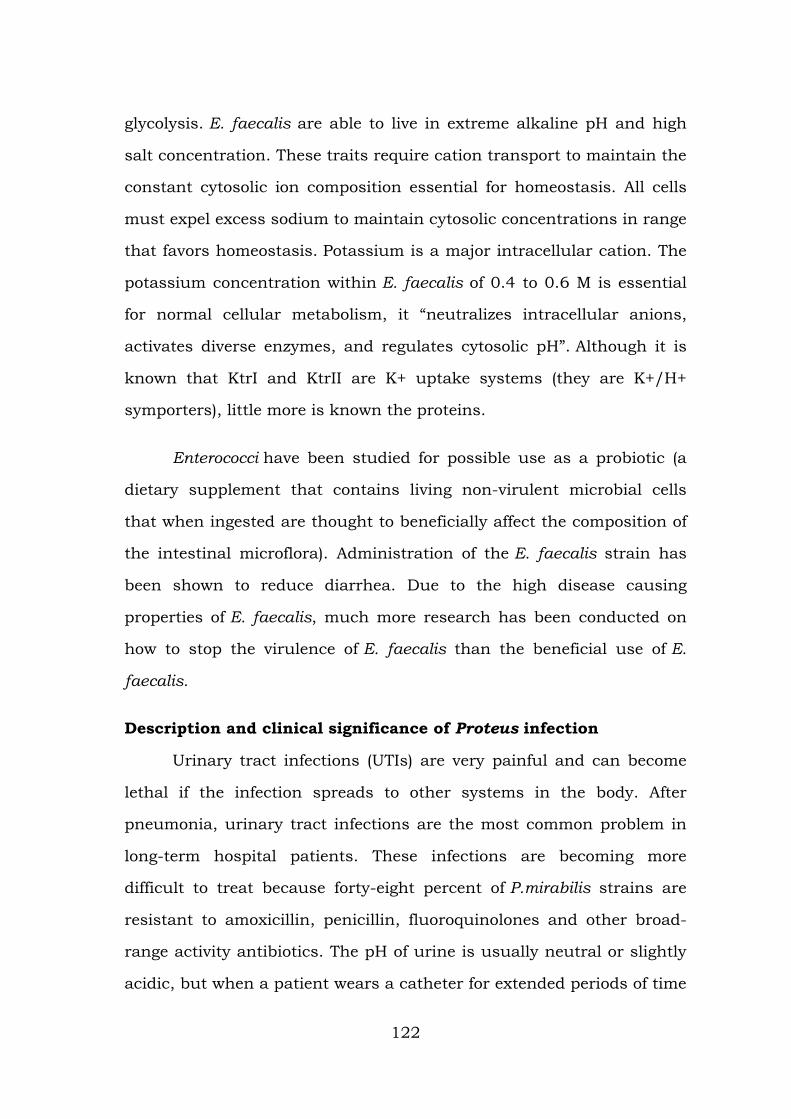
glycolysis. E. faecalis are able to live in extreme alkaline pH and high
salt concentration. These traits require cation transport to maintain the
constant cytosolic ion composition essential for homeostasis. All cells
must expel excess sodium to maintain cytosolic concentrations in range
that favors homeostasis. Potassium is a major intracellular cation. The
potassium concentration within E. faecalis of 0.4 to 0.6 M is essential
for normal cellular metabolism, it “neutralizes intracellular anions,
activates diverse enzymes, and regulates cytosolic pH”. Although it is
known that KtrI and KtrII are K+ uptake systems (they are K+/H+
symporters), little more is known the proteins.
Enterococci have been studied for possible use as a probiotic (a
dietary supplement that contains living non-virulent microbial cells
that when ingested are thought to beneficially affect the composition of
the intestinal microflora). Administration of the E. faecalis strain has
been shown to reduce diarrhea. Due to the high disease causing
properties of E. faecalis, much more research has been conducted on
how to stop the virulence of E. faecalis than the beneficial use of E.
faecalis.
Description and clinical significance of Proteus infection
Urinary tract infections (UTIs) are very painful and can become
lethal if the infection spreads to other systems in the body. After
pneumonia, urinary tract infections are the most common problem in
long-term hospital patients. These infections are becoming more
difficult to treat because forty-eight percent of P.mirabilis strains are
resistant to amoxicillin, penicillin, fluoroquinolones and other broad-
range activity antibiotics. The pH of urine is usually neutral or slightly
acidic, but when a patient wears a catheter for extended periods of time
122
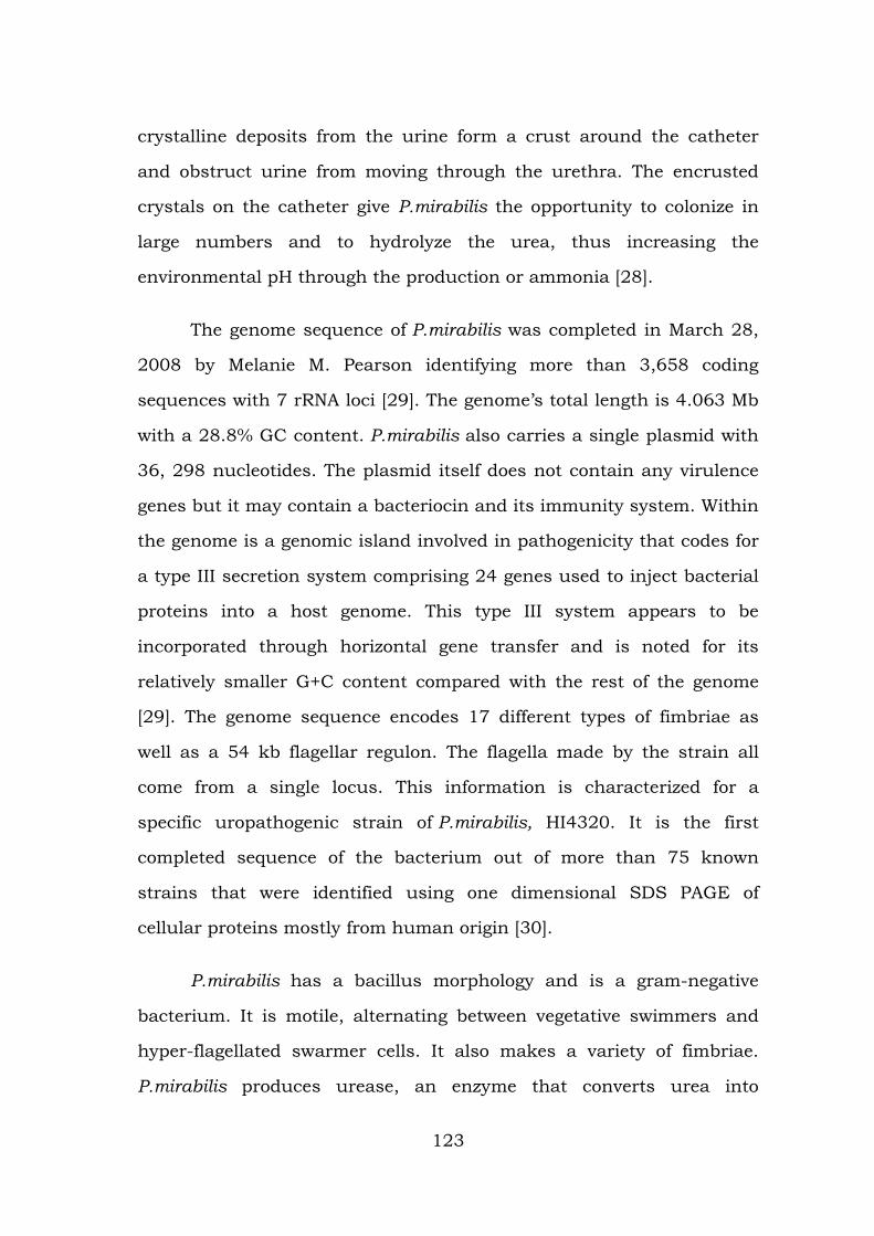
crystalline deposits from the urine form a crust around the catheter
and obstruct urine from moving through the urethra. The encrusted
crystals on the catheter give P.mirabilis the opportunity to colonize in
large numbers and to hydrolyze the urea, thus increasing the
environmental pH through the production or ammonia [28].
The genome sequence of P.mirabilis was completed in March 28,
2008 by Melanie M. Pearson identifying more than 3,658 coding
sequences with 7 rRNA loci [29]. The genome’s total length is 4.063 Mb
with a 28.8% GC content. P.mirabilis also carries a single plasmid with
36, 298 nucleotides. The plasmid itself does not contain any virulence
genes but it may contain a bacteriocin and its immunity system. Within
the genome is a genomic island involved in pathogenicity that codes for
a type III secretion system comprising 24 genes used to inject bacterial
proteins into a host genome. This type III system appears to be
incorporated through horizontal gene transfer and is noted for its
relatively smaller G+C content compared with the rest of the genome
[29]. The genome sequence encodes 17 different types of fimbriae as
well as a 54 kb flagellar regulon. The flagella made by the strain all
come from a single locus. This information is characterized for a
specific uropathogenic strain of P.mirabilis, HI4320. It is the first
completed sequence of the bacterium out of more than 75 known
strains that were identified using one dimensional SDS PAGE of
cellular proteins mostly from human origin [30].
P.mirabilis has a bacillus morphology and is a gram-negative
bacterium. It is motile, alternating between vegetative swimmers and
hyper-flagellated swarmer cells. It also makes a variety of fimbriae.
P.mirabilis produces urease, an enzyme that converts urea into
123
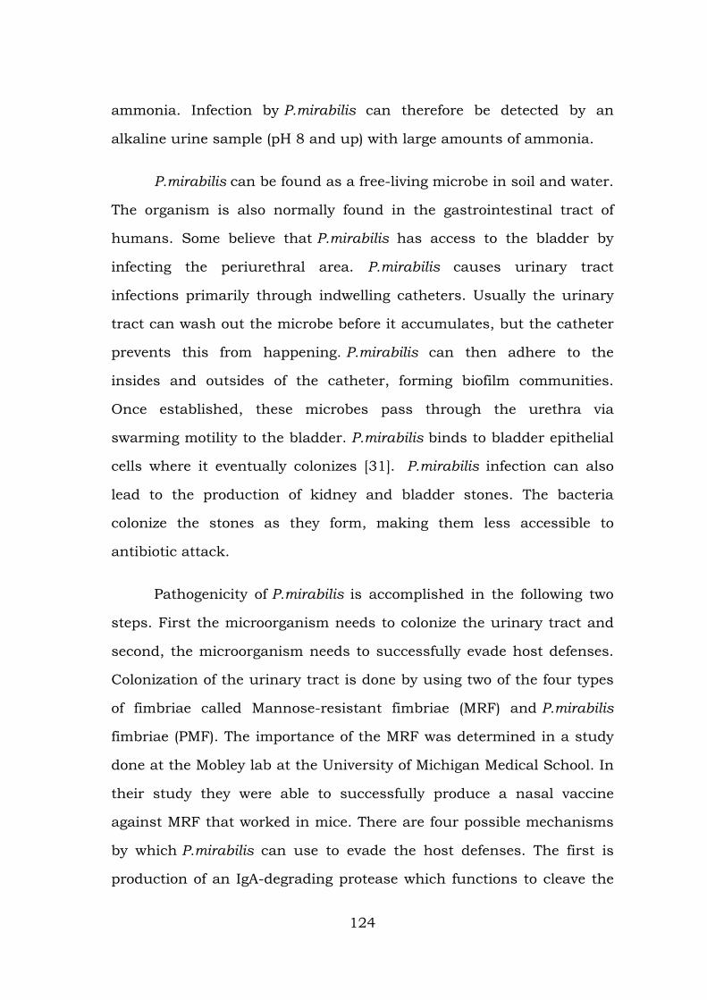
ammonia. Infection by P.mirabilis can therefore be detected by an
alkaline urine sample (pH 8 and up) with large amounts of ammonia.
P.mirabilis can be found as a free-living microbe in soil and water.
The organism is also normally found in the gastrointestinal tract of
humans. Some believe that P.mirabilis has access to the bladder by
infecting the periurethral area. P.mirabilis causes urinary tract
infections primarily through indwelling catheters. Usually the urinary
tract can wash out the microbe before it accumulates, but the catheter
prevents this from happening. P.mirabilis can then adhere to the
insides and outsides of the catheter, forming biofilm communities.
Once established, these microbes pass through the urethra via
swarming motility to the bladder. P.mirabilis binds to bladder epithelial
cells where it eventually colonizes [31]. P.mirabilis infection can also
lead to the production of kidney and bladder stones. The bacteria
colonize the stones as they form, making them less accessible to
antibiotic attack.
Pathogenicity of P.mirabilis is accomplished in the following two
steps. First the microorganism needs to colonize the urinary tract and
second, the microorganism needs to successfully evade host defenses.
Colonization of the urinary tract is done by using two of the four types
of fimbriae called Mannose-resistant fimbriae (MRF) and P.mirabilis
fimbriae (PMF). The importance of the MRF was determined in a study
done at the Mobley lab at the University of Michigan Medical School. In
their study they were able to successfully produce a nasal vaccine
against MRF that worked in mice. There are four possible mechanisms
by which P.mirabilis can use to evade the host defenses. The first is
production of an IgA-degrading protease which functions to cleave the
124
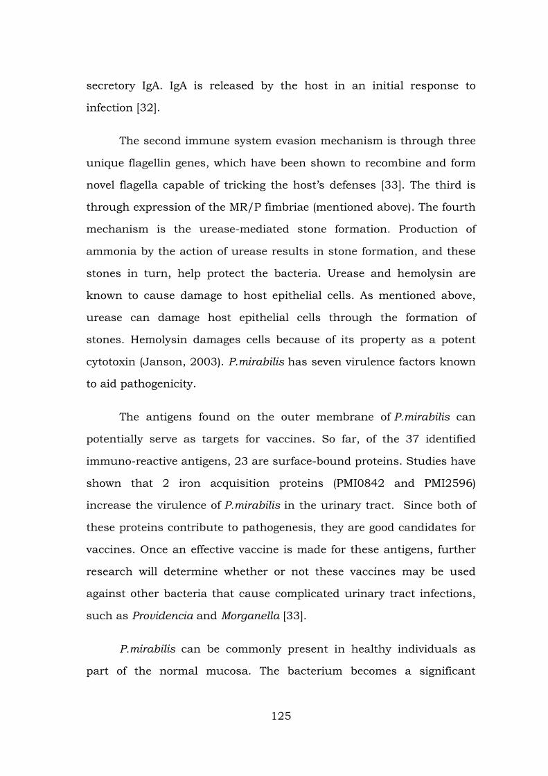
secretory IgA. IgA is released by the host in an initial response to
infection [32].
The second immune system evasion mechanism is through three
unique flagellin genes, which have been shown to recombine and form
novel flagella capable of tricking the host’s defenses [33]. The third is
through expression of the MR/P fimbriae (mentioned above). The fourth
mechanism is the urease-mediated stone formation. Production of
ammonia by the action of urease results in stone formation, and these
stones in turn, help protect the bacteria. Urease and hemolysin are
known to cause damage to host epithelial cells. As mentioned above,
urease can damage host epithelial cells through the formation of
stones. Hemolysin damages cells because of its property as a potent
cytotoxin (Janson, 2003). P.mirabilis has seven virulence factors known
to aid pathogenicity.
The antigens found on the outer membrane of P.mirabilis can
potentially serve as targets for vaccines. So far, of the 37 identified
immuno-reactive antigens, 23 are surface-bound proteins. Studies have
shown that 2 iron acquisition proteins (PMI0842 and PMI2596)
increase the virulence of P.mirabilis in the urinary tract. Since both of
these proteins contribute to pathogenesis, they are good candidates for
vaccines. Once an effective vaccine is made for these antigens, further
research will determine whether or not these vaccines may be used
against other bacteria that cause complicated urinary tract infections,
such as Providencia and Morganella [33].
P.mirabilis can be commonly present in healthy individuals as
part of the normal mucosa. The bacterium becomes a significant
125
problem mostly in individuals that have vulnerable immune systems
and are in danger of nosocomial transmission, such as hospital
patients [34]. Current studies show that there are a number of
antibiotics that were once effective against P.mirabilis that are now
useless due to extended spectrum beta lactamases (ESBLs).
Description and clinical significance of Staphylococcus infection
The Staphylococcus aureus genome, which is the most common
species among the Staphylococcus genome projects, is the most
completed genome sequence compared to any other microbial species.
The original genome map of Staphylococcus aureus was based on the
strain NCTC 8325, initiated by Peter A. Pattee and colleagues. By 2000,
the entire genome of strain 8325 had been sequenced and annotated.
Since then, at least six other ‘‘S.aureus’’ strains have been completed
(COL, N315, Mu50, MW2, MRSA252, MSSA476). The Staphylococcus
aureus strain NCTC 8325 complete circular genome map shows ~2,900
open reading frames, 61 tRNA genes, 3 structural RNAs, and 5
complete ribosomal RNA operons. This strain has about 33% G+C
content and an average gene length of 824 nucleotides with 85% coding
sequence, similar to other S.aureus strains. Half the coding sequence is
located predominantly on one replichore and the second half is located
predominantly on the other replichore.
Virulence factors are encoded by phages, plasmids, pathogenicity
islands and staphylococcus cassette chromosome [35]. Increased
resistance for antibiotics is encoded by a transposon (Tn 1546) that
was inserted into a conjugated plasmid that also encoded resistance to
other things including disinfectants. MRSA (Methicillin-
Resistant Staphylococcus aureus), which is resistant to the antibiotic
126
methicillin, expresses a modified penicillin-binding protein encoded by
mecA gene. This was brought about by many evolutions thought
horizontal gene transfer of mecA to a wide variety of methicillin
susceptible S.aureus strains. The genes for antibiotic resistance
in Staphylococcus aureus are located on plasmids or other similar
structures. Evolution of this bacterium can occur through
asymptomatic colonization and/or during the course of the caused
disease. The increasing population of "super germs" and antibiotic
resistant pathogens have increased pressure on researchers to find
alternative, more effective ways of fighting these "super germs." DNA
sequencing of this microbe has already isolated the source code of its'
resistance to antibiotics, and further research will more than likely lead
us to the path of our next artillery against this and many other
pathogens.
Staphylococcus aureus is a gram-positive bacteria, which means
that the cell wall of this bacteria consists of a very thick peptidoglycan
layer. They form spherical colonies in clusters in 2 planes and have no
flagella [36]. Secretions are numerous, but include surface associated
adhesins, endotoxins, exoenzymes, and capsular polysaccharides. The
capsule is responsible for enhanced virulence of a mucoid strain.
Lactate is the end product of anaerobic glucose metabolism and acetate
and CO2 are the products of aerobic growth conditions. S.aureus can
uptake a variety of nutrients including glucose, mannose, mannitol,
glucosamine, N-acetylglucosamine, sucrose, lactose, galactose and
beta-glucosides.
Staphylococcus aureus is among the most common hospital
acquired pathogens. It is a normal inhabitant of the skin and mucous
127
membranes in the nose of a healthy human. S.aureus is infectious to
both animals and humans and may only survive on dry skin. It can be
spread through contaminated surfaces, through the air and through
people. Though some host colonization can be benign, a puncture or
break in the skin can prompt this bacterium to enter a wound and
cause infections. The best preventive measure is simply regular hand
washing (preferably without antibacterial soaps or hand sanitizers, but
that’s another story) and daily bathing.
The MRSA, resistant to the antibiotic methicillin, was eventually
isolated. Consequently, vancomycin (the most powerful antibiotic in our
arsenal) became the primary antibiotic used to combat staphylococcus
infection. In 1997 a strain of S.aureus resistant to vancomycin was
isolated, and people are once again exposed to the threat of untreatable
staphylococcus infection. MRSA strains are currently a very significant
health care problem. The sequencing of the S.aureusgenome will
hopefully provide insight into how the organism generates such a
variety of toxins, and aid researchers in developing ways of combating
the versatile bacterium [37].
Description and clinical significance of Pseudomonas infections
A review of surveillance data collected by the CDC National
Nosocomial Infections Surveillance System from 1986 to 1998 shows
that P. aeruginosa was identified as the fifth most frequently isolated
nosocomial pathogen, accounting for 9% of all hospital-acquired
infections in the United States [38]. P. aeruginosa was also the second
leading cause of nosocomial pneumonia (14 to 16%), third most
common cause of urinary tract infections (7 to 11%), fourth most
frequently isolated pathogen in surgical site infections (8%), and
128
seventh leading contributor to bloodstream infections (2 to 6%). Data
from more recent studies continue to show P. aeruginosa as the second
most common cause of nosocomial pneumonia, health care-associated
pneumonia, and ventilator-associated pneumonia and the leading
cause of pneumonia among pediatric patients in the intensive care unit
(ICU).
P. aeruginosa presents a serious therapeutic challenge for
treatment of both community-acquired and nosocomial infections, and
selection of the appropriate antibiotic to initiate therapy is essential to
optimizing the clinical outcome. Unfortunately, selection of the most
appropriate antibiotic is complicated by the ability of P. aeruginosa to
develop resistance to multiple classes of antibacterial agents, even
during the course of treating an infection. Epidemiological outcome
studies have shown that infections caused by drug-resistant P.
aeruginosa are associated with significant increases in morbidity,
mortality, need for surgical intervention, length of hospital stay and
chronic care, and overall cost of treating the infection [39].
P.aeruginosa a very ubiquitous microorganism, for it has been
found in environments such as soil, water, humans, animals, plants,
sewage, and hospitals (Lederberg and Joshua, 2000). In all oligotropic
aquatic ecosystems, which contain high-dissolved oxygen content but
low plant nutrients throughout, P.aeruginosa is the predominant
inhabitant and this clearly makes it the most abundant organism on
earth [40].
P.aeruginosa is an opportunistic human pathogen. It is
“opportunistic” because it seldom infects healthy individuals. Instead, it
129
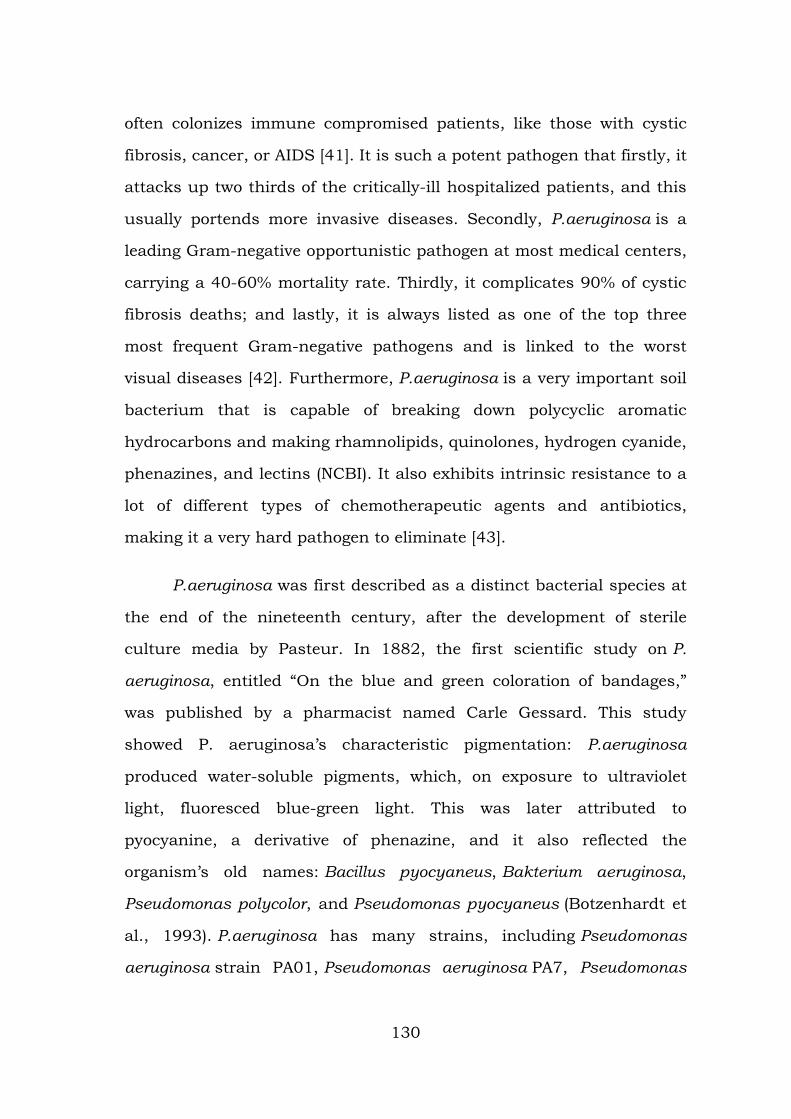
often colonizes immune compromised patients, like those with cystic
fibrosis, cancer, or AIDS [41]. It is such a potent pathogen that firstly, it
attacks up two thirds of the critically-ill hospitalized patients, and this
usually portends more invasive diseases. Secondly, P.aeruginosa is a
leading Gram-negative opportunistic pathogen at most medical centers,
carrying a 40-60% mortality rate. Thirdly, it complicates 90% of cystic
fibrosis deaths; and lastly, it is always listed as one of the top three
most frequent Gram-negative pathogens and is linked to the worst
visual diseases [42]. Furthermore, P.aeruginosa is a very important soil
bacterium that is capable of breaking down polycyclic aromatic
hydrocarbons and making rhamnolipids, quinolones, hydrogen cyanide,
phenazines, and lectins (NCBI). It also exhibits intrinsic resistance to a
lot of different types of chemotherapeutic agents and antibiotics,
making it a very hard pathogen to eliminate [43].
P.aeruginosa was first described as a distinct bacterial species at
the end of the nineteenth century, after the development of sterile
culture media by Pasteur. In 1882, the first scientific study on P.
aeruginosa, entitled “On the blue and green coloration of bandages,”
was published by a pharmacist named Carle Gessard. This study
showed P. aeruginosa’s characteristic pigmentation: P.aeruginosa
produced water-soluble pigments, which, on exposure to ultraviolet
light, fluoresced blue-green light. This was later attributed to
pyocyanine, a derivative of phenazine, and it also reflected the
organism’s old names: Bacillus pyocyaneus, Bakterium aeruginosa,
Pseudomonas polycolor, and Pseudomonas pyocyaneus (Botzenhardt et
al., 1993). P.aeruginosa has many strains, including Pseudomonas
aeruginosa strain PA01, Pseudomonas aeruginosa PA7, Pseudomonas
130
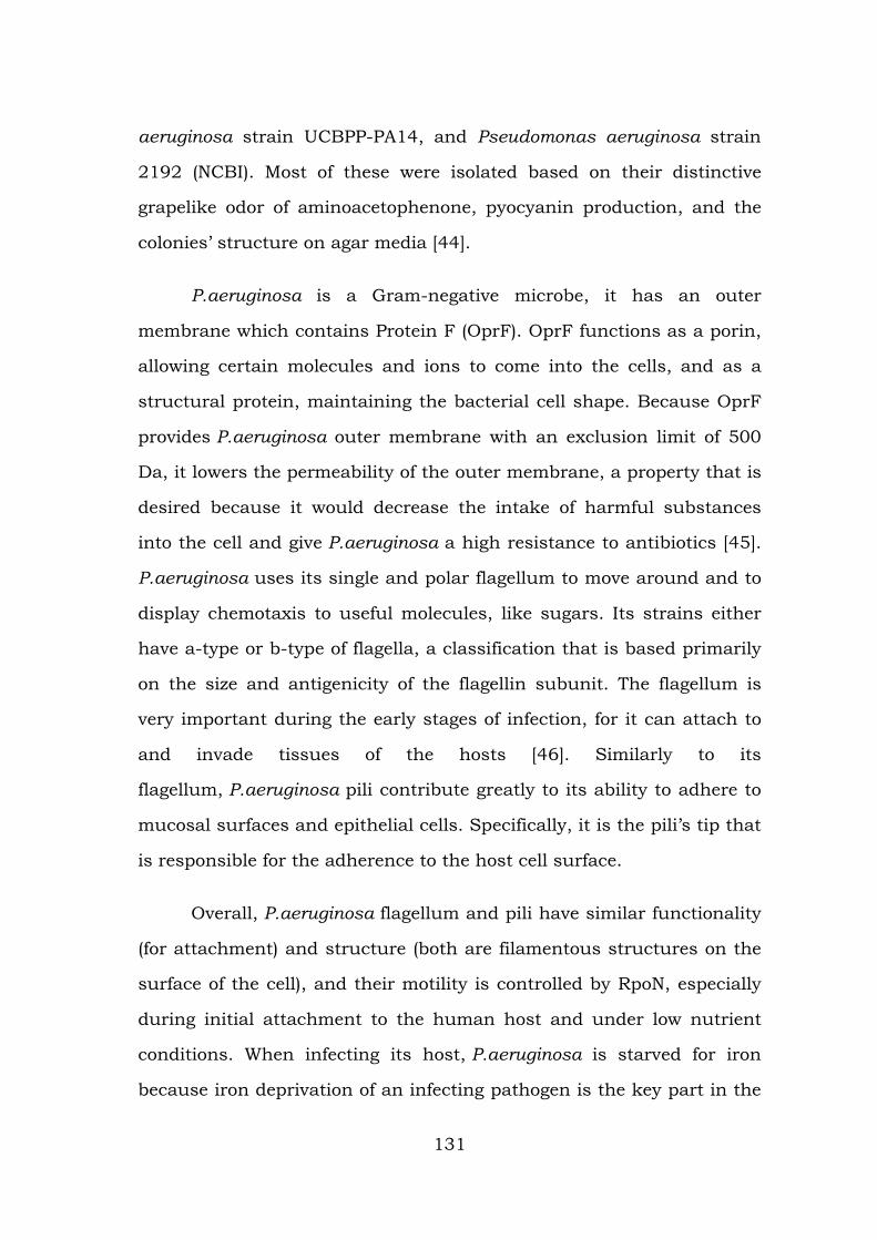
aeruginosa strain UCBPP-PA14, and Pseudomonas aeruginosa strain
2192 (NCBI). Most of these were isolated based on their distinctive
grapelike odor of aminoacetophenone, pyocyanin production, and the
colonies’ structure on agar media [44].
P.aeruginosa is a Gram-negative microbe, it has an outer
membrane which contains Protein F (OprF). OprF functions as a porin,
allowing certain molecules and ions to come into the cells, and as a
structural protein, maintaining the bacterial cell shape. Because OprF
provides P.aeruginosa outer membrane with an exclusion limit of 500
Da, it lowers the permeability of the outer membrane, a property that is
desired because it would decrease the intake of harmful substances
into the cell and give P.aeruginosa a high resistance to antibiotics [45].
P.aeruginosa uses its single and polar flagellum to move around and to
display chemotaxis to useful molecules, like sugars. Its strains either
have a-type or b-type of flagella, a classification that is based primarily
on the size and antigenicity of the flagellin subunit. The flagellum is
very important during the early stages of infection, for it can attach to
and invade tissues of the hosts [46]. Similarly to its
flagellum, P.aeruginosa pili contribute greatly to its ability to adhere to
mucosal surfaces and epithelial cells. Specifically, it is the pili’s tip that
is responsible for the adherence to the host cell surface.
Overall, P.aeruginosa flagellum and pili have similar functionality
(for attachment) and structure (both are filamentous structures on the
surface of the cell), and their motility is controlled by RpoN, especially
during initial attachment to the human host and under low nutrient
conditions. When infecting its host, P.aeruginosa is starved for iron
because iron deprivation of an infecting pathogen is the key part in the
131
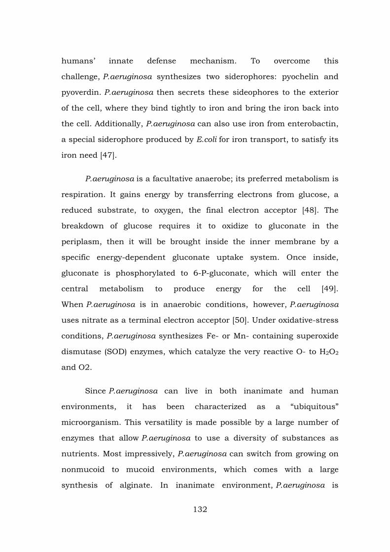
humans’ innate defense mechanism. To overcome this
challenge, P.aeruginosa synthesizes two siderophores: pyochelin and
pyoverdin. P.aeruginosa then secrets these sideophores to the exterior
of the cell, where they bind tightly to iron and bring the iron back into
the cell. Additionally, P.aeruginosa can also use iron from enterobactin,
a special siderophore produced by E.coli for iron transport, to satisfy its
iron need [47].
P.aeruginosa is a facultative anaerobe; its preferred metabolism is
respiration. It gains energy by transferring electrons from glucose, a
reduced substrate, to oxygen, the final electron acceptor [48]. The
breakdown of glucose requires it to oxidize to gluconate in the
periplasm, then it will be brought inside the inner membrane by a
specific energy-dependent gluconate uptake system. Once inside,
gluconate is phosphorylated to 6-P-gluconate, which will enter the
central metabolism to produce energy for the cell [49].
When P.aeruginosa is in anaerobic conditions, however, P.aeruginosa
uses nitrate as a terminal electron acceptor [50]. Under oxidative-stress
conditions, P.aeruginosa synthesizes Fe- or Mn- containing superoxide
dismutase (SOD) enzymes, which catalyze the very reactive O- to H2O2
and O2.
Since P.aeruginosa can live in both inanimate and human
environments, it has been characterized as a “ubiquitous”
microorganism. This versatility is made possible by a large number of
enzymes that allow P.aeruginosa to use a diversity of substances as
nutrients. Most impressively, P.aeruginosa can switch from growing on
nonmucoid to mucoid environments, which comes with a large
synthesis of alginate. In inanimate environment, P.aeruginosa is
132
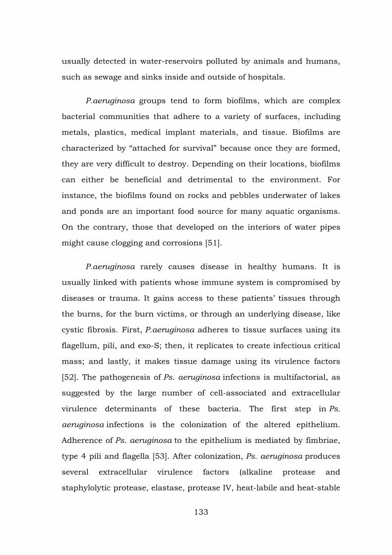
usually detected in water-reservoirs polluted by animals and humans,
such as sewage and sinks inside and outside of hospitals.
P.aeruginosa groups tend to form biofilms, which are complex
bacterial communities that adhere to a variety of surfaces, including
metals, plastics, medical implant materials, and tissue. Biofilms are
characterized by “attached for survival” because once they are formed,
they are very difficult to destroy. Depending on their locations, biofilms
can either be beneficial and detrimental to the environment. For
instance, the biofilms found on rocks and pebbles underwater of lakes
and ponds are an important food source for many aquatic organisms.
On the contrary, those that developed on the interiors of water pipes
might cause clogging and corrosions [51].
P.aeruginosa rarely causes disease in healthy humans. It is
usually linked with patients whose immune system is compromised by
diseases or trauma. It gains access to these patients’ tissues through
the burns, for the burn victims, or through an underlying disease, like
cystic fibrosis. First, P.aeruginosa adheres to tissue surfaces using its
flagellum, pili, and exo-S; then, it replicates to create infectious critical
mass; and lastly, it makes tissue damage using its virulence factors
[52]. The pathogenesis of Ps. aeruginosa infections is multifactorial, as
suggested by the large number of cell-associated and extracellular
virulence determinants of these bacteria. The first step in Ps.
aeruginosa infections is the colonization of the altered epithelium.
Adherence of Ps. aeruginosa to the epithelium is mediated by fimbriae,
type 4 pili and flagella [53]. After colonization, Ps. aeruginosa produces
several extracellular virulence factors (alkaline protease and
staphylolytic protease, elastase, protease IV, heat-labile and heat-stable
133
hemolysins, phospholipases C and exotoxins A, S, T, U, Y), responsible
for extensive tissue damage, bloodstream invasion, and dissemination
[54].
This disruption in the salt and water balance in the cell results in
the production of a thick mucus, which becomes the ideal home for
potential pathogens. P.aeruginosa attacks CF patients via airway and
once it is in, it uses its flagellum to go to the hypoxic zone, an oxygen-
depleted environment. At this location, P.aeruginosa undergoes a
transition from an aerobic to an anaerobic microbe and starts forming
biofilms anaerobically. Once this is formed, the P.aeruginosa in this
community can sense their population via quorum sensing, where they
secret low molecular weight pheromones that enable them to
communicate with each other [55]. This gives them the ability to resist
many defenses, including anti-Pseudomonas antibiotics such as
ticarcillin, ceftazidime, tobramycin, and ciprofloxacin, because once the
bacteria sense that their outer layer of biofilm is being destroyed, the
inner layers will grow stronger to reestablish the community [56].
P.aeruginosa is also resistant to many antibiotics and
chemotherapeutic agents due to their intrinsic resistance.
This is caused by the low permeability to antibiotics of the outer
membrane and by the production of β-lactamases against multidrug
efflux pumps and β-lactam antibiotics [57]. P.aeruginosa communicates
with other cells through quorum-sensing. This form of communication
allows the cells to regulate gene production which results in control of
certain cell functions. One of the enzymes responsible for quorum
sensing is tyrosine phosphatase (TpbA). This enzyme relays
extracellular quorum sensing signals to polysaccharide production and
134
biofilm formation outside the cells [58]. P.aeruginosa attaches to
surfaces by way of biofilm production. Quorum-sensing can be a drug
target to cure infections caused by P. aeruginosa. Quorum-quenching is
used to blocks the signaling mechanism of quorum-sensing and
prevents biofilm formation in P. aeruginosa. Yi-Hu Dong and his
colleagues were able to prevent biofilm formation in mice under
laboratory conditions [59].
P.aeruginosa secrets many virulent factors to colonize the cells of
its host. For example, exotoxin A, the most toxic protein produced by P.
aeruginosa, catalyzes the ADP-ribosylation to form ADP-ribosyl-EF-2,
which inhibits the protein synthesis of the host’s cells. Moreover,
elastase, an extracellular zinc protease, attacks eukaryotic proteins
such as collagen and elastin and destroys the structural proteins of the
cell.
Pseudomonas aeruginosa is an environmentally ubiquitous
opportunistic pathogen. Epidermal infections often result from
P.aeruginosa infiltrating through a human host’s first line of defenses,
entering the body through the skin at the site of an open wound.
P.aeruginosa is a common member of hospital bacterial communities
where it can infect immunocompromised individuals including burn
victims. P.aeruginosa is a source of bacteremia in burn victims.
Following severe skin damage, the prevalence of P.aeruginosa in the
environment increases the probability of the organism accessing the
bloodstream through the burn victim’s exposed deep epidermal tissue.
Previous research of antibody-mediated host defenses indicates that on
the fifth day after the initial burn, Fc receptor expression is reduced in
polymorphonuclear leukocytes (PMNs). Without the Fc receptor, PMN
135
chemotaxis is greatly reduced and the PMNs become less effective at
preventing infection.
P.aeruginosa can be transmitted to a host via fomites, vectors,
and hospital workers who are potential carriers for multiply-antibiotic-
resistant strains of the pathogen. Furthermore, any P.aeruginosa
already present on a burn victim’s skin before the injury can transform
from an innocuous organism on the surface of the skin to a source of
infection in the bloodstream and body tissues of the same individual.
The pili and flagella of P.aeruginosa play a vital role in the infection of
burns and wounds. Controlled infection of burn wounds on animal and
plant models with P.aeruginosa strains devoid of pili and flagella
demonstrate a trend of decreased virulence. Without these
morphological virulence factors, the bacteria exhibit a substantially
decreased survival rate at the wound site and a decreased ability to
disseminate within the host organism. The spread of P.aeruginosa
within host organisms is also dependent on the microorganism’s
elastase production and other protease mechanisms. Bacterial elastase
and other bacterial proteases degrade the host’s proteins, including the
structural proteins within membranes, disrupting the host’s physical
barriers against the spread of infection. Elastase also assists
P.aeruginosa in avoiding phagocytotic antibody-mediated cytotoxicity at
the site of the wound by inhibiting monocyte chemotaxis [60].
The two strains that have the complete genome sequence
are Pseudomonas aeruginosa PA01 and Pseudomonas aeruginosa PA14
(PGD). In 2000, a group of volunteer "Pseudomonas scientists",
including those from the Washington PathoGenesis Corportaion and
the Department of Biology of the University of California, San Diego,
136
worked under the Pseudomonas aeruginosa Community Annotation
Project (PseudoCAP) to publish the complete genome sequence of
Pseudomonas aeruginosa PA01. This was done because knowing the
genomic sequence would provide new information about this bacterium
as a pathogen and about its ecological versatility and genetic
complexity. At 6,264,403 base pairs, its bacterial genome is the largest
to ever be sequenced. It also contains 5,570 predicted open reading
frames (ORFs), and thus it almost has the genetic complexity of simple
eukaryotes, such as Saccharomyces cerevisiae. Using whole-genome-
shotgun sampling, the complete 6.3 Mbp genome of Pseudomonas
aeruginosa PA01 is very much similar to the P. aeruginosa’s physical
map, with only one major exception, which is the inversion of about a
quarter of the Pseudomonas aeruginosa PA01 genome.
This inversion comes from the homologous recombination of the
rrnA and rrnB loci, and earlier studies on genomic sequence inversions
of ribosomal DNA loci in S. typhimurium and E.coli suggest that this
inversion might have adaptive significance [61]. The complete genome
sequence of Pseudomonas aeruginosa PA14 is currently being done by
Harvard Medical School scientists. The goal of this study is achieve a
public data of Pseudomonas aeruginosa PA14 genome. The shotgun-
sequencing phase of the project was finished in 2005, yielding 6.54
Mbp of PA14 sequence. It is currently being compared to the genome
of Pseudomonas aeruginosa PA01 and preliminary results have shown
that they are very similar but have several regions of marked
differences, such as the insertion of the 107911bp in PA14, which is
absent in PA01. Approximately, there is 96.3% of the DNA sequence of
PAO1 is in PA14, and 92.4% of PA14 DNA sequence is in PA01 .
137
P. aeruginosa, as well as many other Pseudomonas, can degrade
aromatic hydrocarbons such as methylbenzenes, which are the by-
products of petroleum industries and are commonly used as solvents
for enamels and paints as well as in the production of drugs and
chemicals. Methylbenzenes are considered as environmental
contaminants that are present in the atmosphere, underground and
soils, and in surface water [62]. P.aeruginosa can break down toluene,
the simplest form of methylbenzene. P.aeruginosa degrades toluene
through the oxidation of the methyl group to aldehyde, alcohol, and an
acid, which is then converted to catechol. Hence, P.aeruginosa can be
used in pollution control [63].
Description and clinical significance of E. coli infections
E. coli comprises of non-pathogenic commensal isolates that
forms part of the normal flora of humans and various animals. In
humans, they are the major aerobic organism residing in the intestine,
typically with around 106 to 109 colony forming units per gram of
stool. The organism is also found in soil and water, usually as a result
of fecal contamination. Several variants or pathotypes of E. coli have
been described that cause infections of the gastrointestinal system (i.e.
intestinal pathogenic E. coli) while other pathotypes cause infections
outside the gastrointestinal system (i.e. extraintestinal pathogenic E.
coli) [64]. E. coli is the most common cause of urinary tract infections
(UTIs) in humans, and is a leading cause of enteric infections and
systemic infections [65]. The systemic infections include bacteremia,
nosocomial pneumonia, cholecystitis, cholangitis, peritonitis, cellulitis,
osteomyelitis, and infectious arthritis. E. coli is also leading cause of
neonatal meningitis
138
Furthermore, enteric E.coli can be classified into six categories
based on its virulence properties [66]. These enteric E.coli can cause
several intestinal and extra-intestinal infections such as urinary tract
infection and mastitis. However, E.coli are not always harmful to
human bodies or other animals. Most E.coli live in our intestines, where
they help our body breakdown the food we eat as well as assist with
waste processing, vitamin K production, and food absorption. Extra
intestinal pathogenic E. coli (ExPEC), especially the uro-pathogenic E.
coli (UPEC) pathotype, is most commonly associated with human
infections due to E. coli outside the intestinal tract. UPEC are important
causes of lower urinary tract infections (UTIs) and systemic infections
in humans. The systemic infections include upper UTIs, bacteremia,
nosocomial pneumonia, cholecystitis, cholangitis, peritonitis, cellulitis,
osteomyelitis, infectious arthritis. ExPEC is also leading cause of
neonatal meningitis (referred to as NMEC) [67].
E.coli has only one circular chromosome, some along with a
circular plasmid. Its chromosomal DNA has been completely sequenced
by lab researchers. E.coli has a single chromosome with about 4,600
kb, about 4,300 potential coding sequences, and only about 1,800
known E.coli proteins. 70% of the chromosome is composed of single
genes (monocistronic), and 6% is polycistronic. Moreover, there are
many different strains of E.coli; each of these strains differs in its
genotype from wild-type E.coli. The genotype will then affect the
phenotype that is expressed, and further influences the physiology and
life cycle of each strain. Therefore, different strains of E.coli can live in
different kinds of animals. The natural biological process of mutation in
genomes is the major cause to produce so many different strains
139
of E.coli. In addition, similar to most bacteria, E.coli can transfer its
DNA materials through bacterial conjugation with other related bacteria
to produce more mutation and add more strains into the existing
population.
Escherichia coli can be commonly found in lower intestines of
human and mammals. When E.coli locates in human large intestines, it
can help digestion processes, food breakdown and absorption, and
vitamin K production. Different strains of E.coli can be found in
different type of animals, so we can determine the source (from human
or from other animals) of the stools by examining which strain
of E.coli is present in the stools. E.coli can also be found in
environments at higher temperature, such as on the edge of hot
springs. E.coli is commonly used as an indicator in the field of water
purification. The E.coli-index can indicate how much human feces is in
the water. The reason why E.coli is used as an indicator is due to a
significant larger amount of E.coli in human feces than other bacterial
organisms. Most strains of "E.coli" are not harmful to their hosts;
however, more and more newly discovered strains are contributing into
existing population through mutation and evolution. Some can cause
severe disease, such as E.coli O157:H7.
Although E.coli in human large intestine can assist with waste
processing and food absorption, some strains of E.coli can cause severe
infections in many animals, such as humans, sheep, horses, dogs, etc.
The one that only found in humans is called enter aggregative E.coli.
Urinary tract infection, for example, can be caused by ascending
infections of urethra. Such infections can be found in both adult male
and female, and some infants can be infected as well. E.coli O157:H7 is
140
one of the most infective strains that can cause food poisoning. It
belongs to enter hemorrhagic strain of the E.coli and can lead to bloody
diarrhea and kidney failure when one gets infected by contaminated
ground beef, unpasteurized milk or contaminated water. The toxin
that E.coli O157:H7 produces is a Shiga-like toxin which is a regulated
toxin that catalytically inactivate 60S ribosomal subunits of most
eukaryotic cells, blocking mRNA translation and thus causing cell
death [68]. Some important symptoms are diarrhea that is acute and
severe, either bloody or not bloody, stomach cramping, vomiting, and
loss of appetite, abdominal pain, and fever. The causes can usually
clear up on their own in 1-3 days with no treatment required. However,
patients should avoid dairy products because those products may
induce temporary lactose intolerance, and therefore make the diarrhea
worse.
E.coli plays an important role in current biological engineering
because of its manipulation and long laboratory history. It has been
widely used to synthesize DNA and proteins. Most results from E.coli
research can be applied to animals and humans. The most useful
contribution of recombinant DNA from E.coli is to use the manipulation
of E.coli to produce human insulin for diabetes patients.
Since E.coli can produce human enzymes through recombinant DNA
techniques, it is widely used as a very good tool to produce useful
compounds or enzymes for medication. In a recent study (Apr. 2007),
scientists has explored a new method to cure Alzheimer's disease (AD),
which is a neurodegenerative disorder characterized by a progressive
loss of cognitive function due to extra deposition of the longer form of
the amyloid peptide Abeta. In this study, scientists use E.coli to enable
141
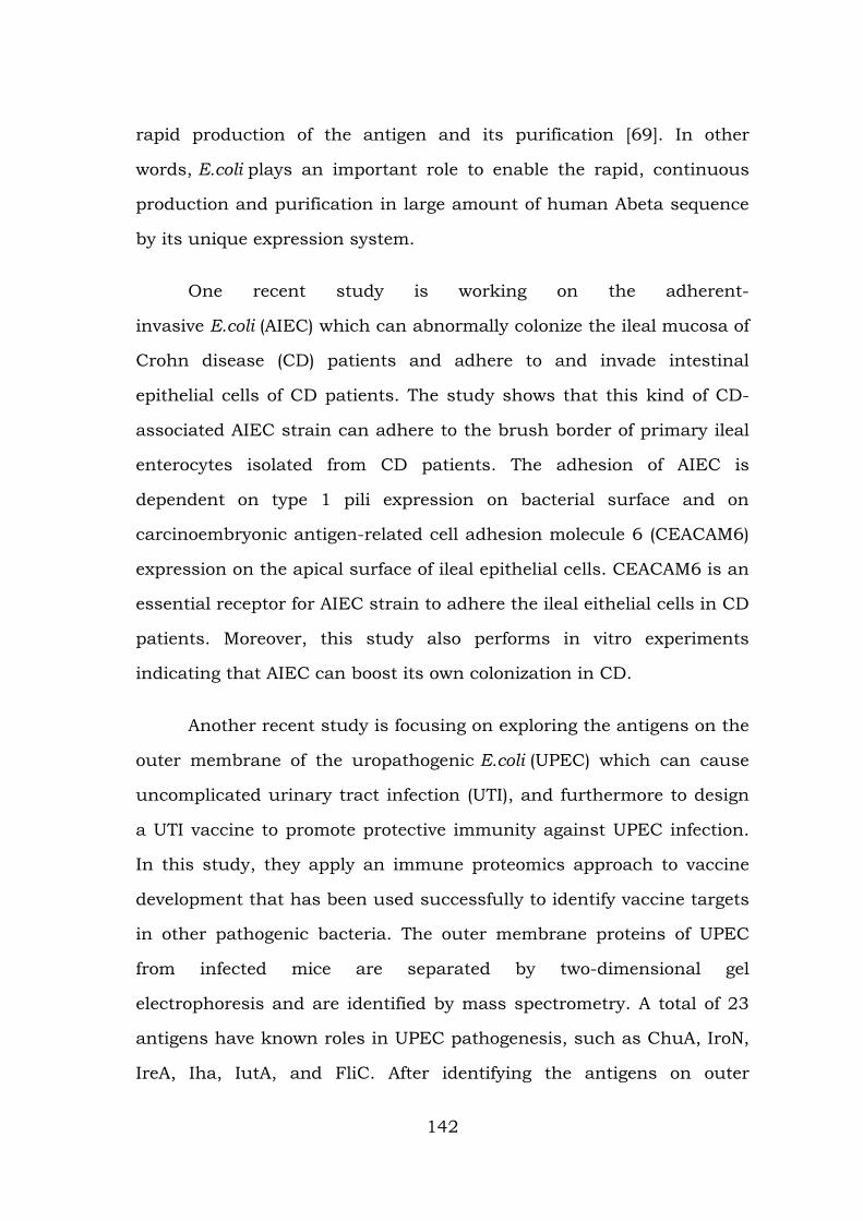
rapid production of the antigen and its purification [69]. In other
words, E.coli plays an important role to enable the rapid, continuous
production and purification in large amount of human Abeta sequence
by its unique expression system.
One recent study is working on the adherent-
invasive E.coli (AIEC) which can abnormally colonize the ileal mucosa of
Crohn disease (CD) patients and adhere to and invade intestinal
epithelial cells of CD patients. The study shows that this kind of CD-
associated AIEC strain can adhere to the brush border of primary ileal
enterocytes isolated from CD patients. The adhesion of AIEC is
dependent on type 1 pili expression on bacterial surface and on
carcinoembryonic antigen-related cell adhesion molecule 6 (CEACAM6)
expression on the apical surface of ileal epithelial cells. CEACAM6 is an
essential receptor for AIEC strain to adhere the ileal eithelial cells in CD
patients. Moreover, this study also performs in vitro experiments
indicating that AIEC can boost its own colonization in CD.
Another recent study is focusing on exploring the antigens on the
outer membrane of the uropathogenic E.coli (UPEC) which can cause
uncomplicated urinary tract infection (UTI), and furthermore to design
a UTI vaccine to promote protective immunity against UPEC infection.
In this study, they apply an immune proteomics approach to vaccine
development that has been used successfully to identify vaccine targets
in other pathogenic bacteria. The outer membrane proteins of UPEC
from infected mice are separated by two-dimensional gel
electrophoresis and are identified by mass spectrometry. A total of 23
antigens have known roles in UPEC pathogenesis, such as ChuA, IroN,
IreA, Iha, IutA, and FliC. After identifying the antigens on outer
142
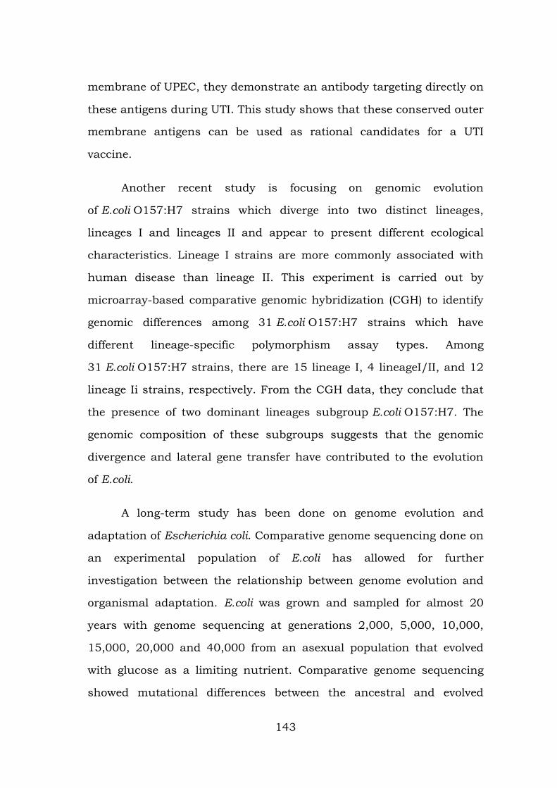
membrane of UPEC, they demonstrate an antibody targeting directly on
these antigens during UTI. This study shows that these conserved outer
membrane antigens can be used as rational candidates for a UTI
vaccine.
Another recent study is focusing on genomic evolution
of E.coli O157:H7 strains which diverge into two distinct lineages,
lineages I and lineages II and appear to present different ecological
characteristics. Lineage I strains are more commonly associated with
human disease than lineage II. This experiment is carried out by
microarray-based comparative genomic hybridization (CGH) to identify
genomic differences among 31 E.coli O157:H7 strains which have
different lineage-specific polymorphism assay types. Among
31 E.coli O157:H7 strains, there are 15 lineage I, 4 lineageI/II, and 12
lineage Ii strains, respectively. From the CGH data, they conclude that
the presence of two dominant lineages subgroup E.coli O157:H7. The
genomic composition of these subgroups suggests that the genomic
divergence and lateral gene transfer have contributed to the evolution
of E.coli.
A long-term study has been done on genome evolution and
adaptation of Escherichia coli. Comparative genome sequencing done on
an experimental population of E.coli has allowed for further
investigation between the relationship between genome evolution and
organismal adaptation. E.coli was grown and sampled for almost 20
years with genome sequencing at generations 2,000, 5,000, 10,000,
15,000, 20,000 and 40,000 from an asexual population that evolved
with glucose as a limiting nutrient. Comparative genome sequencing
showed mutational differences between the ancestral and evolved
143
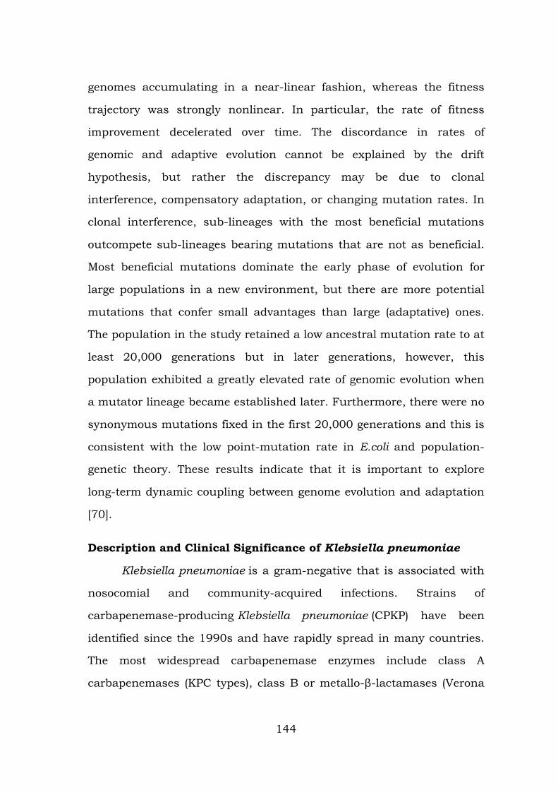
genomes accumulating in a near-linear fashion, whereas the fitness
trajectory was strongly nonlinear. In particular, the rate of fitness
improvement decelerated over time. The discordance in rates of
genomic and adaptive evolution cannot be explained by the drift
hypothesis, but rather the discrepancy may be due to clonal
interference, compensatory adaptation, or changing mutation rates. In
clonal interference, sub-lineages with the most beneficial mutations
outcompete sub-lineages bearing mutations that are not as beneficial.
Most beneficial mutations dominate the early phase of evolution for
large populations in a new environment, but there are more potential
mutations that confer small advantages than large (adaptative) ones.
The population in the study retained a low ancestral mutation rate to at
least 20,000 generations but in later generations, however, this
population exhibited a greatly elevated rate of genomic evolution when
a mutator lineage became established later. Furthermore, there were no
synonymous mutations fixed in the first 20,000 generations and this is
consistent with the low point-mutation rate in E.coli and population-
genetic theory. These results indicate that it is important to explore
long-term dynamic coupling between genome evolution and adaptation
[70].
Description and Clinical Significance of Klebsiella pneumoniae
Klebsiella pneumoniae is a gram-negative that is associated with
nosocomial and community-acquired infections. Strains of
carbapenemase-producing Klebsiella pneumoniae (CPKP) have been
identified since the 1990s and have rapidly spread in many countries.
The most widespread carbapenemase enzymes include class A
carbapenemases (KPC types), class B or metallo-β-lactamases (Verona
144
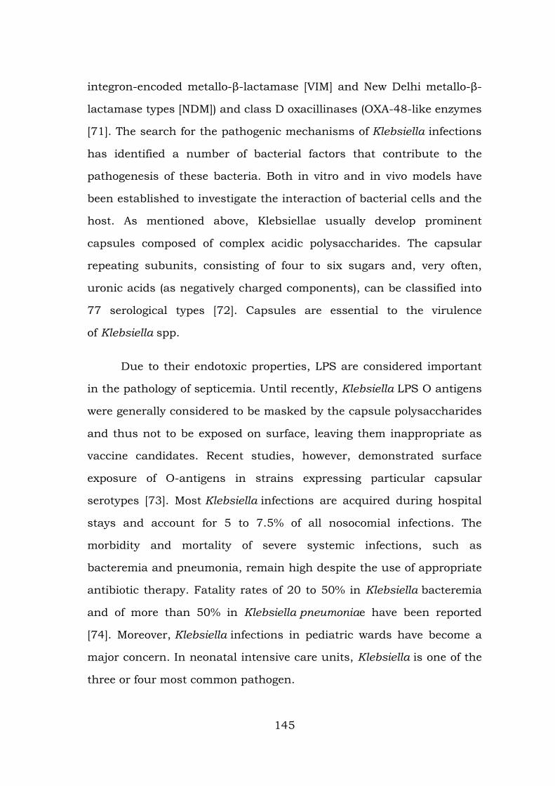
integron-encoded metallo-β-lactamase [VIM] and New Delhi metallo-β-
lactamase types [NDM]) and class D oxacillinases (OXA-48-like enzymes
[71]. The search for the pathogenic mechanisms of Klebsiella infections
has identified a number of bacterial factors that contribute to the
pathogenesis of these bacteria. Both in vitro and in vivo models have
been established to investigate the interaction of bacterial cells and the
host. As mentioned above, Klebsiellae usually develop prominent
capsules composed of complex acidic polysaccharides. The capsular
repeating subunits, consisting of four to six sugars and, very often,
uronic acids (as negatively charged components), can be classified into
77 serological types [72]. Capsules are essential to the virulence
of Klebsiella spp.
Due to their endotoxic properties, LPS are considered important
in the pathology of septicemia. Until recently, Klebsiella LPS O antigens
were generally considered to be masked by the capsule polysaccharides
and thus not to be exposed on surface, leaving them inappropriate as
vaccine candidates. Recent studies, however, demonstrated surface
exposure of O-antigens in strains expressing particular capsular
serotypes [73]. Most Klebsiella infections are acquired during hospital
stays and account for 5 to 7.5% of all nosocomial infections. The
morbidity and mortality of severe systemic infections, such as
bacteremia and pneumonia, remain high despite the use of appropriate
antibiotic therapy. Fatality rates of 20 to 50% in Klebsiella bacteremia
and of more than 50% in Klebsiella pneumoniae have been reported
[74]. Moreover, Klebsiella infections in pediatric wards have become a
major concern. In neonatal intensive care units, Klebsiella is one of the
three or four most common pathogen.
145
Prevalence of ESBL pathogens
The most well-known of the “newer” β-lactamases was first
described in 1983 and have been named the extended-spectrum β-
lactamases or ESBLs. These enzymes have the ability to hydrolyse the
penicillins, cephalosporins and monobactams, but not the cephamycins
and carbapenems. ESBLs are inhibited by “classical” b-lactamase
inhibitors such as clavulanic acid, sulbactam and
tazobactam. Although ESBLs have been identified in a range of
Enterobacteriaceae, they are most often present in E. coli and K.
pneumoniae. The decreased susceptibility of Enterobacteriaceae to
carbapenems may be caused by either extended-spectrum beta-
lactamases (ESBLs) or AmpC enzymes as well as decreased drug
permeability caused by porin loss. Imported resistance to the β-lactams
involves the production of inactivating β-lactamases, for which several
families have been identified among clinical isolates of P. aeruginosa.
The variety, prevalence, and clinical significance of imported β-
lactamases in P. aeruginosa have been addressed in several reviews
over the last decade [75]. The most common imported β-lactamases
found among P. aeruginosa isolates are penicillinases belonging to the
molecular class A serine β-lactamases (PSE, CARB, and TEM families).
Within this group, enzymes belonging to the PSE family appear to be
the most prevalent. β-Lactam antimicrobial agents represent the most
common treatment for bacterial infections and continue to be the
leading cause of resistance to β-lactam antibiotics among Gram-
negative bacteria worldwide. The persistent exposure of bacterial
strains to a multitude of β-lactams has induced dynamic and
continuous production and mutation of β-lactamases in these bacteria,
146
expanding their activity even against the newly developed β-lactam
antibiotics. These enzymes are known as extended-spectrum β-
lactamases (ESBLs) [76].
Treatment of these multiple drug-resistant organisms is a
therapeutic challenge. At the level of a wider geographic scale, the
incidence of ESBL-producing organisms is difficult to determine due to
various reasons, difficulty in detecting ESBL production and
inconsistencies in reporting. In recent surveys, a significant increase in
the ESBL rate was reported from all parts of the world [77]. Klebsiella
pneumoniae and Escherichia coli remain the major ESBL-producing
organisms isolated worldwide which are recommended to be routinely
tested for and reported by the Clinical and Laboratory Standards
Institute. Prevalence of ESBLs varies from an institute to another.
Previous studies from India and abroad have reported ESBL production
varying from 8 to 80%. However, there is paucity scientific information
available on antibiotic profile with rate of ESBL production in Klebsiella
pneumoniae isolates.
Keeping in view the above facts, the present study was
undertaken to find the prevalence of ESBL producers among Klebsiella
pneumoniae isolates at our institute [78]. Resistance to aminopenicillins
(e.g. ampicillin) and early-generation cephalosporins (e.g. cefazolin)
among E. coli is often mediated by the production of narrow-
spectrum b-lactamases such as TEM-1, TEM-2 and to a lesser extent
SHV-1 enzyme [79]. Most importantly among E. coli, is the increasing
recognition of isolates producing the so-called “newer b-lactamases”
that causes resistance to the expanded-spectrum cephalosporins
and/or the carbapenems. These enzymes consist of the plasmid-
147

mediated AmpC b-lactamases (e.g. CMY types), extended-spectrum b-
lactamases (e.g. TEM, SHV, CTX-M types), and carbapenemases (KPC
types, metallo-b-lactamases (MBLs) and OXA-types).
Surveys from several countries worldwide have illustrated an
alarming trend of associated resistance to other classes of antimicrobial
agents among CTX-M-producing E. coli that included trimethoprim-
sulfamethoxazole, tetracycline, gentamicin, tobramycin and
ciprofloxacin. Studies consistently show that infections due to ESBL-
producing Enterobacteriaceae are associated with a delay in initiation
of appropriate antibiotic therapy, which consequently prolongs hospital
stays and increases hospital costs [80]. More importantly, failure to
initiate appropriate antibiotic therapy from the start appears to be
responsible for higher patient mortality.
Toxicity assay
The antibiotic resistant pattern and resistant of mosquito larvae
have been changing frequently. There is a need for establishing a
particular type of drug which is harmful to larvae without affecting
human system. The medicinal value of some chemical substances lies
in definite physiological action on the human body. So, there is a need
for exploring the Hemolytic activity of Red Blood Cells (RBC) resembles
the activities of other cells, the study was designed to check the effect
of ligand for larvicidal and hemolysis. Though a drug is very potential
against some diseases, it is of no value if toxic metals are present in
higher concentration
148
Larvicidal assay
The mosquito is the principal vector of many of the vector-borne
diseases affecting human beings and other animals. Mosquitoes
constitute a major public health problem as vectors of serious human
diseases. In India, malaria is one of the most important causes of direct
or indirect infant, child, and adult mortality. About 2 million confirmed
malaria cases and 1,000 deaths are reported annually, although 15
million cases and 20,000 deaths are estimated by WHO South East
Asia Regional Office. India contributes 77% of the total malaria in
South East Asia. Mosquitoes are the primary source for the spread of
diseases among the arthropods. Organophosphates and insect growth
regulators are generally used for the control of mosquito larvae,
however, their continuous use has demonstrated undesirable effects on
non-target organisms, and their influence on the environment and
human health has been the main concern.
India is struggling to cope with outbreaks of mosquito borne
diseases. Overburden hospitals and clinics in several cities in the North
of India. Half of the entire human population, an estimated 3.3 billion
people, lives in malaria-risk areas around the world with about 250
million people infected annually. Malaria is believed to be responsible
for approximately one million deaths per year, particularly among
children under 5 year’s old and pregnant women. The other continents
contribute the remainder of the 35 million cases according to the report
of WHO in 2008. Anopheles subpictus is a complex isomorphic sibling
species and is recognised as a vector of malaria, a disease of great
socio-economic importance, and also a vector of some helminth and
arboviruses. Malaria and lymphatic filariasis (LF) are two of the most
149
common and identifiable mosquito-borne parasitic diseases worldwide.
The highest malaria burden is found in Africa with an estimation of 212
million cases (86% globally) distributed in 45 countries.
Repeated use of synthetic insecticides for mosquito control has
disrupted natural biological control systems and led to resurgences in
mosquito populations. It has also resulted in the development of
resistance, undesirable effects on non-target organisms and fostered
environmental and human health concern, which initiated a search for
alternative control measures. The control of mosquito larvae is
dependent on regular applications of organophosphates and different
insecticides. The major drawback with the use of chemical insecticides
is that they are non-selective and could be more harmful to other non-
target organisms. Moreover, after few years the mosquitoes develop
resistance against the insecticides due to frequent use of them.
Pesticide exposure among humans has been linked to immune
dysfunction, various forms of cancer and birth defects. It is, therefore,
necessary to identify a safe, eco-friendly alternate source of larvicide in
order to reduce mosquito menace. Studies have been concentrated in
Aedes aegypti species because of its role in the arboviruses
transmission, responsible for yellow fever and dengue fever, diseases
which are endemic in South and Central America, Asia and Africa.
Dengue is a mosquito-borne viral disease found in three clinical forms:
dengue fever (DF), dengue haemorrhagic fever (DHF) and dengue shock
syndrome. It is endemic in more than 100 countries in Africa, the
Americas, the West Pacific and Asia (including Sri Lanka), and has
become the most important and the fastest-growing mosquito-borne
disease in these countries. A total of 40% of the world’s population
150
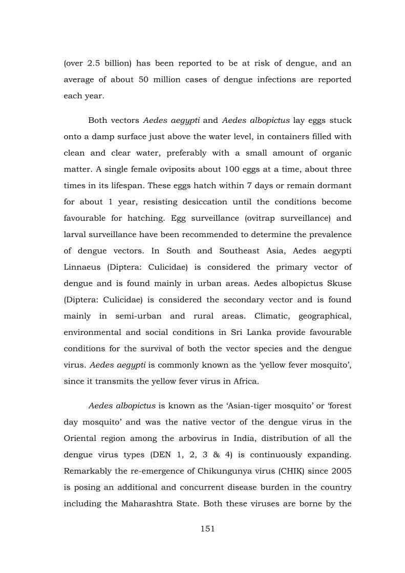
(over 2.5 billion) has been reported to be at risk of dengue, and an
average of about 50 million cases of dengue infections are reported
each year.
Both vectors Aedes aegypti and Aedes albopictus lay eggs stuck
onto a damp surface just above the water level, in containers filled with
clean and clear water, preferably with a small amount of organic
matter. A single female oviposits about 100 eggs at a time, about three
times in its lifespan. These eggs hatch within 7 days or remain dormant
for about 1 year, resisting desiccation until the conditions become
favourable for hatching. Egg surveillance (ovitrap surveillance) and
larval surveillance have been recommended to determine the prevalence
of dengue vectors. In South and Southeast Asia, Aedes aegypti
Linnaeus (Diptera: Culicidae) is considered the primary vector of
dengue and is found mainly in urban areas. Aedes albopictus Skuse
(Diptera: Culicidae) is considered the secondary vector and is found
mainly in semi-urban and rural areas. Climatic, geographical,
environmental and social conditions in Sri Lanka provide favourable
conditions for the survival of both the vector species and the dengue
virus. Aedes aegypti is commonly known as the ‘yellow fever mosquito’,
since it transmits the yellow fever virus in Africa.
Aedes albopictus is known as the ‘Asian-tiger mosquito’ or ‘forest
day mosquito’ and was the native vector of the dengue virus in the
Oriental region among the arbovirus in India, distribution of all the
dengue virus types (DEN 1, 2, 3 & 4) is continuously expanding.
Remarkably the re-emergence of Chikungunya virus (CHIK) since 2005
is posing an additional and concurrent disease burden in the country
including the Maharashtra State. Both these viruses are borne by the
151
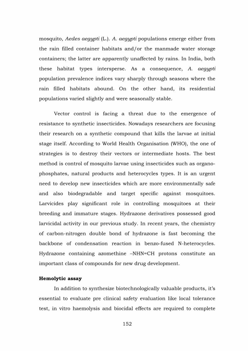
mosquito, Aedes aegypti (L.). A. aegypti populations emerge either from
the rain filled container habitats and/or the manmade water storage
containers; the latter are apparently unaffected by rains. In India, both
these habitat types intersperse. As a consequence, A. aegypti
population prevalence indices vary sharply through seasons where the
rain filled habitats abound. On the other hand, its residential
populations varied slightly and were seasonally stable.
Vector control is facing a threat due to the emergence of
resistance to synthetic insecticides. Nowadays researchers are focusing
their research on a synthetic compound that kills the larvae at initial
stage itself. According to World Health Organisation (WHO), the one of
strategies is to destroy their vectors or intermediate hosts. The best
method is control of mosquito larvae using insecticides such as organo-
phosphates, natural products and heterocycles types. It is an urgent
need to develop new insecticides which are more environmentally safe
and also biodegradable and target specific against mosquitoes.
Larvicides play significant role in controlling mosquitoes at their
breeding and immature stages. Hydrazone derivatives possessed good
larvicidal activity in our previous study. In recent years, the chemistry
of carbon-nitrogen double bond of hydrazone is fast becoming the
backbone of condensation reaction in benzo-fused N-heterocycles.
Hydrazone containing azomethine –NHN=CH protons constitute an
important class of compounds for new drug development.
Hemolytic assay
In addition to synthesize biotechnologically valuable products, it’s
essential to evaluate pre clinical safety evaluation like local tolerance
test, in vitro haemolysis and biocidal effects are required to complete
152
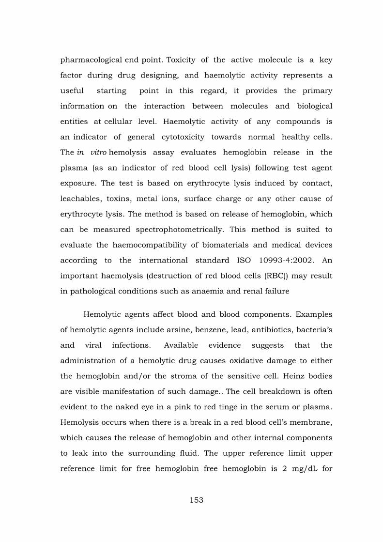
pharmacological end point. Toxicity of the active molecule is a key
factor during drug designing, and haemolytic activity represents a
useful starting point in this regard, it provides the primary
information on the interaction between molecules and biological
entities at cellular level. Haemolytic activity of any compounds is
an indicator of general cytotoxicity towards normal healthy cells.
The in vitro hemolysis assay evaluates hemoglobin release in the
plasma (as an indicator of red blood cell lysis) following test agent
exposure. The test is based on erythrocyte lysis induced by contact,
leachables, toxins, metal ions, surface charge or any other cause of
erythrocyte lysis. The method is based on release of hemoglobin, which
can be measured spectrophotometrically. This method is suited to
evaluate the haemocompatibility of biomaterials and medical devices
according to the international standard ISO 10993-4:2002. An
important haemolysis (destruction of red blood cells (RBC)) may result
in pathological conditions such as anaemia and renal failure
Hemolytic agents affect blood and blood components. Examples
of hemolytic agents include arsine, benzene, lead, antibiotics, bacteria’s
and viral infections. Available evidence suggests that the
administration of a hemolytic drug causes oxidative damage to either
the hemoglobin and/or the stroma of the sensitive cell. Heinz bodies
are visible manifestation of such damage.. The cell breakdown is often
evident to the naked eye in a pink to red tinge in the serum or plasma.
Hemolysis occurs when there is a break in a red blood cell’s membrane,
which causes the release of hemoglobin and other internal components
to leak into the surrounding fluid. The upper reference limit upper
reference limit for free hemoglobin free hemoglobin is 2 mg/dL for
153
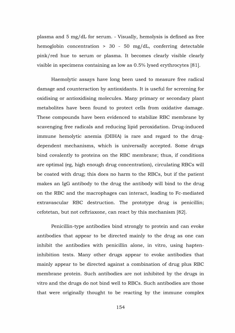
plasma and 5 mg/dL for serum. - Visually, hemolysis is defined as free
hemoglobin concentration > 30 - 50 mg/dL, conferring detectable
pink/red hue to serum or plasma. It becomes clearly visible clearly
visible in specimens containing as low as 0.5% lysed erythrocytes [81].
Haemolytic assays have long been used to measure free radical
damage and counteraction by antioxidants. It is useful for screening for
oxidising or antioxidising molecules. Many primary or secondary plant
metabolites have been found to protect cells from oxidative damage.
These compounds have been evidenced to stabilize RBC membrane by
scavenging free radicals and reducing lipid peroxidation. Drug-induced
immune hemolytic anemia (DIIHA) is rare and regard to the drug-
dependent mechanisms, which is universally accepted. Some drugs
bind covalently to proteins on the RBC membrane; thus, if conditions
are optimal (eg, high enough drug concentration), circulating RBCs will
be coated with drug; this does no harm to the RBCs, but if the patient
makes an IgG antibody to the drug the antibody will bind to the drug
on the RBC and the macrophages can interact, leading to Fc-mediated
extravascular RBC destruction. The prototype drug is penicillin;
cefotetan, but not ceftriaxone, can react by this mechanism [82].
Penicillin-type antibodies bind strongly to protein and can evoke
antibodies that appear to be directed mainly to the drug as one can
inhibit the antibodies with penicillin alone, in vitro, using hapten-
inhibition tests. Many other drugs appear to evoke antibodies that
mainly appear to be directed against a combination of drug plus RBC
membrane protein. Such antibodies are not inhibited by the drugs in
vitro and the drugs do not bind well to RBCs. Such antibodies are those
that were originally thought to be reacting by the immune complex
154
mechanism [83]. Piperacillin can cause DIIHA and/or positive DATs; it
is the third most common drug to cause DIIHA. Although a semi-
synthetic penicillin, unlike other semi-synthetic penicillins (eg,
ampicillin), it reacts differently than penicillin G. In contrast to
penicillin, the in vivo RBC destruction can be complement-mediated;
most of the DATs are positive due to RBC-bound complement.
The denaturation of cell membrane proteins by proton-donating
groups is the major underlying mechanism for membrane damage. The
haemolytic activity is highly specific and seems to depend only on the
concentration of negatively charged groups that are accessible by the
cell membranes of erythrocytes. For example strong distortion of the
membrane after interaction with silica particles can lead to loss of
membrane flexibility and resiliency as well as the release of
haemoglobin (haemolysis). The toxic potency of the majority of
proteinaceous toxins is based on membrane interaction, pore formation
and, finally lysis and damage of cells. Such cytolysins are produced by
a variety of living organisms, particularly bacteria, certain insects,
poisonous reptiles and stinging marine invertebrates. These toxins need
to be secreted as water-soluble proteins but have to be transformed
into membrane proteins before penetrating the membrane. It can be
assumed that the toxin is concentrated on the membrane by means of
special cell surface features such as a protein receptor molecules, lipid
clusters or carbohydrate side chains [84].
The toxic activity of cell lysis is often measured by the lysis of red
blood cells (erythrocytes) and the detection of released haemoglobin
after cell disruption. Erythrocytes from diverse species have been
applied for haemolytic assays, and they express different
155
susceptibilities for the same lysine. This phenomenon indicates that the
composition of erythrocyte cell membranes influences the haemolytic
effect of cytolytic toxins [85]. Cell lysis by surfactants is a process of
great fundamental and practical importance. Much research has been
done in order to understand the mechanism underlying this process,
mostly using erythrocytes as a convenient model system. Because the
human erythrocyte has no internal organelles and since it is the
simplest cellular model obtainable, it is the most popular cell
membrane system to study the surfactant membrane interaction [86].
As most known cyclotides are to some extent haemolytic, a
haemolytic activity assay was considered an appropriate method for an
initial biological activity screen and to assess the biological effects of
structural permutations in cyclotides. While a haemolytic activity assay
is a relatively simple procedure, it is a biological assay and therefore
subject to a range of variable factors. The previously reported HD50
values for kalata B1 are indeed widely divergent, with values of 50 µM
and 1510 µM [87]. Some of the haemolytic assays reported in the
literature used rat, sheep or bovine blood. Blood cells from different
mammalian species can have varying susceptibility to haemolysis [88].
Rat and sheep blood had drastically reduced susceptibility to
haemolysis by the peptides compared to human blood. In the context of
exploring drug for intended use in humans, assaying against blood
from other animals would therefore be misleading, as the peptides
would appear almost non-haemolytic in the case of rat and sheep
blood.
Some examples of variables in reported haemolytic assays of
amphipathic antimicrobial and haemolytic peptides are listed in Table
156
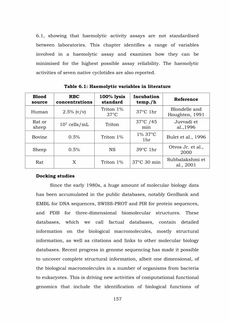
6.1, showing that haemolytic activity assays are not standardised
between laboratories. This chapter identifies a range of variables
involved in a haemolytic assay and examines how they can be
minimised for the highest possible assay reliability. The haemolytic
activities of seven native cyclotides are also reported.
Table 6.1: Haemolytic variables in literature
Blood source
RBC concentrations
100% lysis standard
Incubation temp./h Reference
Human 2.5% (v/v) Triton 1% 37°C 37°C 1hr Blondelle and
Houghten, 1991 Rat or sheep 107 cells/mL Triton 37°C /45
min Juvvadi et al.,1996
Bovine 0.5% Triton 1% 1% 37°C 1hr Bulet et al., 1996
Sheep 0.5% NS 39°C 1hr Otvos Jr. et al., 2000
Rat X Triton 1% 37°C 30 min Subbalakshmi et al., 2001
Docking studies
Since the early 1980s, a huge amount of molecular biology data
has been accumulated in the public databases, notably GenBank and
EMBL for DNA sequences, SWISS-PROT and PIR for protein sequences,
and PDB for three-dimensional biomolecular structures. These
databases, which we call factual databases, contain detailed
information on the biological macromolecules, mostly structural
information, as well as citations and links to other molecular biology
databases. Recent progress in genome sequencing has made it possible
to uncover complete structural information, albeit one dimensional, of
the biological macromolecules in a number of organisms from bacteria
to eukaryotes. This is driving new activities of computational functional
genomics that include the identification of biological functions of
157
unknown gene products, reconstruction of functional modules or
pathways from a set of genes in the genome, comparative analysis of
genes and genomes in different species, and analysis and simulation of
gene expressions in different cells or in different developmental stages
[89]. In order to facilitate such post-genome sequencing analyses, it has
become a high priority to construct a new breed of database that
defines functional aspects of genes, cells and organisms. But later the
bioinformatics analysis focused to the medical chemistry, in which the
ligands and metal complexes were graphically represented and find
their affinity/ binding level with the respective chemistry compounds
through different bioinformatics tools.
The ligand chemical database for enzyme reactions, which was
originally constructed by Nishioka and co-workers [90]. Ligand now
consists of two sections: the expanded ENZYME section and the new
compound section. The enzyme section is a collection of all known
enzymatic reactions classified according to the nomenclature of the
International Union of Biochemistry and Molecular Biology [91]. The
compound section is a collection of metabolic compounds, including
substrates, products, inhibitors, cofactors and effectors, and other
chemical compounds that play important functional roles in living cells.
Both sections are tightly coupled with the KEGG metabolic pathway
database: the enzyme section for the network of genes and the
compound section for the network of chemical compounds. The Ligand
database thus provides fundamental data on both biological and
chemical aspects of life.
The emergence of antibiotic-resistant strains of pathogenic
bacteria is an increasing threat to global health that underscores an
158
urgent need for an expanded antibacterial armamentarium. Gram-
negative bacteria, such as Escherichia coli, have become increasingly
important clinical pathogens with limited treatment. The increased
prevalence of pathogenic bacteria with resistance to clinically useful
antibiotics is a growing threat to public health. Despite this urgent,
unmet medical need, few novel classes of antibiotics and antibiotic
leads, e.g. linezolid, daptomycin, and platensimycin, have been
discovered in the last 30 years. Antibiotic-resistant Gram-negative
bacteria are of growing medical significance and consist of pathogens
such as Acinetobacter baumannii, Escherichia coli, Haemophilus
influenzae, Klebsiella pneumoniae, Neisseria meningitidis, and
Pseudomonas aeruginosa. E. coli and P. aeruginosa are commonly found
in hospital-acquired infections such as pneumonia and septicemia [92].
Therefore, the chemical compounds were analyzed whether the
compound is bind with respective cancer cell/ pathogens. It is less cost
and is not required for the wet chemistry analysis. So, now a day,
people are focusing the bioinformatics studies for predicting their
compounds may be act as an alternative drug or not.
MATERIALS AND METHODS
Sampling
The test cultures were isolated from the patient’s sample (Urine)
in Mahatma Gandhi Government Hospital at Tiruchirappalli, Tamil
Nadu, India, during May 2015. The samples were collected from the
patients with proper procedure during the morning time. All the
collected samples were separately stored in sterile containers and
stored in ice boxes at 4° C. The sterile storage containers contain the
transport medium for culture maintaining purposes. All the samples
159
were immediately transported in to the laboratory and were processed
within 6 h.
Isolation of the bacterial cultures
The patient samples were subjected to the pure culture
techniques for isolation of the individual bacteria among the bacterial
groups. The CLED and MacConkey agar medium was commonly used
for isolation of the bacterial culture and the spread/ streak plate
method was used for separation of pure (individual) culture.
Culture maintenance
All the test bacteria were grown in nutrient agar medium and
incubated at 37 ±1 °C for 24 - 48 h followed by frequent subculture to
fresh (Nutrient broth) medium and were used as test bacteria. The pure
cultures were separately maintained in the sterile test tubes and were
subjected for the identification.
Identification of the cultures
Gram Staining
The cultures from both medical samples were smeared on the
slide and heat fixed. The crystal violet dye was added and allowed to
cover the whole smear to act for 1 minute and rinsed with tap water.
Few drops of Grams-iodine was added and allowed to react for 30
seconds to 1 minute. It was decolorized with 95% ethanol and
immediately washed under running tap water. Then, safranine was
added and allowed to act for 1 minute, slide was rinsed with tap water
and blot dried and examined under oil immersion objective. Gram
positive cells were appeared in purple color and gram negative cells
were appeared in red color [1].
160
Motility determination
Hanging drop method
A small amount of Vaseline was placed at each corner of clean
cover glass. Two loopful of the 24 hour culture of the organism was
placed at the center of the cover glass. A depression slide was pressed
over the cover glass, such that the depressions cover the culture drop,
and quickly inverted. The completed preparation was observed
microscopically for motility [2].
Biochemical Characterization
Indole production test
Peptone broth was prepared and sterilized at 121ºC for 15
minutes at 15 1bs pressure. The culture was inoculated into the
peptone broth under aseptic condition and incubated at 37ºC for 24
hours. Few drops of Kovac’s reagent were added to the tube after
incubation. The formation of cherry red colored ring in the upper
surface of the medium indicates positive result for the identification of
bacterial isolates.
Methyl red test
MR – VP broth was prepared and sterilized at 121ºC for 15
minutes at 15 1bs pressure. The culture was inoculated into the broth
under aseptic condition and incubated at 37ºC for 24 hours. After
incubation, the indicator methyl red was added to the tubes. The
development of red colour indicates acid production and positive result.
Voges proskauer test
MR – VP broth was prepared and sterilized at 121ºC for 15
minutes at 15 1bs pressure. The culture was inoculated in to the broth
161
under aseptic condition and incubated at 37ºC for 24 hours. After
incubation the indicator Barritt’s reagent 1 and 2 were added. The
development of pink colour indicates the positive result.
Citrate utilization test
Simmon’s citrate agar was prepared and sterilized at 121ºC for
15 minutes at 15 1bs pressure. The medium was poured into the tube
and slant was prepared. The culture was streaked in the slant and
incubated at 37ºC for 24 hours. Development of blue colored slant after
incubation indicates the positive result.
Oxidase test
The deionized water wet disc was filled with few drops of 1%
tetramethyl-t-phenyline diamine dihydrochloride and inoculated a
loopful colony of pure culture of bacteria on the surface of the disc and
incubated for 1 min. If the area of inoculation turn dark blue to maroon
and finally to black colour indicates positive results.
Catalase test
Isolates were grown in Nutrient Agar Medium for 48-72 h at
50ºC. 3 % hydrogen peroxide was poured onto the colonies. Formation
of air bubbles indicates the presence of catalase enzyme.
Triple Sugar Iron test (TSI)
The growth of the organisms on the TSI slant indicates the type
of sugar fermented and in addition, identifying the hydrogen sulfide
products with an acid production and the color of the phenol red
indicator turns yellow. The alkaline reaction of the medium was
indicated by purple color production and hydrogen sulphide production
was indicated by formation of black color. The TSI agar medium was
162
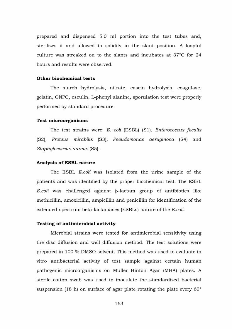
prepared and dispensed 5.0 ml portion into the test tubes and,
sterilizes it and allowed to solidify in the slant position. A loopful
culture was streaked on to the slants and incubates at 37°C for 24
hours and results were observed.
Other biochemical tests
The starch hydrolysis, nitrate, casein hydrolysis, coagulase,
gelatin, ONPG, esculin, L-phenyl alanine, sporulation test were properly
performed by standard procedure.
Test microorganisms
The test strains were: E. coli (ESBL) (S1), Enterococcus fecalis
(S2), Proteus mirabilis (S3), Pseudomonas aeruginosa (S4) and
Staphylococcus aureus (S5).
Analysis of ESBL nature
The ESBL E.coli was isolated from the urine sample of the
patients and was identified by the proper biochemical test. The ESBL
E.coli was challenged against β-lactam group of antibiotics like
methicillin, amoxicillin, ampicillin and penicillin for identification of the
extended-spectrum beta-lactamases (ESBLs) nature of the E.coli.
Testing of antimicrobial activity
Microbial strains were tested for antimicrobial sensitivity using
the disc diffusion and well diffusion method. The test solutions were
prepared in 100 % DMSO solvent. This method was used to evaluate in
vitro antibacterial activity of test sample against certain human
pathogenic microorganisms on Muller Hinton Agar (MHA) plates. A
sterile cotton swab was used to inoculate the standardized bacterial
suspension (18 h) on surface of agar plate rotating the plate every 60°
163
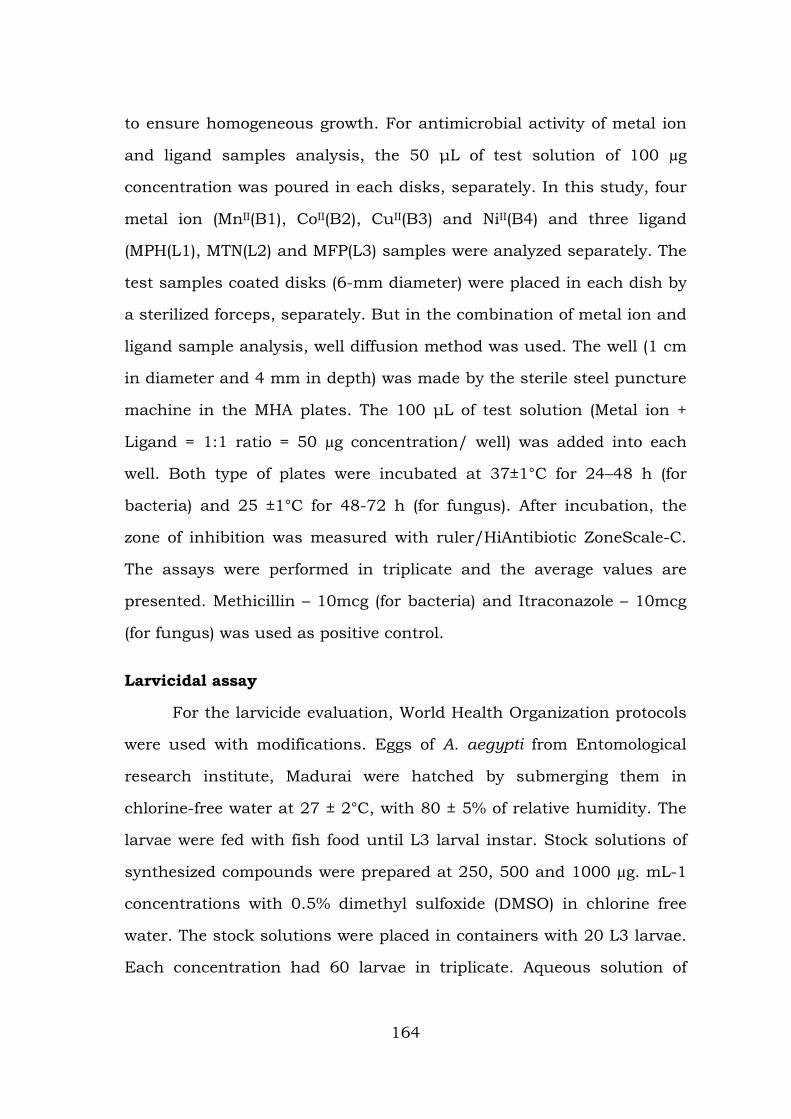
to ensure homogeneous growth. For antimicrobial activity of metal ion
and ligand samples analysis, the 50 μL of test solution of 100 µg
concentration was poured in each disks, separately. In this study, four
metal ion (MnII(B1), CoII(B2), CuII(B3) and NiII(B4) and three ligand
(MPH(L1), MTN(L2) and MFP(L3) samples were analyzed separately. The
test samples coated disks (6-mm diameter) were placed in each dish by
a sterilized forceps, separately. But in the combination of metal ion and
ligand sample analysis, well diffusion method was used. The well (1 cm
in diameter and 4 mm in depth) was made by the sterile steel puncture
machine in the MHA plates. The 100 μL of test solution (Metal ion +
Ligand = 1:1 ratio = 50 µg concentration/ well) was added into each
well. Both type of plates were incubated at 37±1°C for 24–48 h (for
bacteria) and 25 ±1°C for 48-72 h (for fungus). After incubation, the
zone of inhibition was measured with ruler/HiAntibiotic ZoneScale-C.
The assays were performed in triplicate and the average values are
presented. Methicillin – 10mcg (for bacteria) and Itraconazole – 10mcg
(for fungus) was used as positive control.
Larvicidal assay
For the larvicide evaluation, World Health Organization protocols
were used with modifications. Eggs of A. aegypti from Entomological
research institute, Madurai were hatched by submerging them in
chlorine-free water at 27 ± 2°C, with 80 ± 5% of relative humidity. The
larvae were fed with fish food until L3 larval instar. Stock solutions of
synthesized compounds were prepared at 250, 500 and 1000 µg. mL-1
concentrations with 0.5% dimethyl sulfoxide (DMSO) in chlorine free
water. The stock solutions were placed in containers with 20 L3 larvae.
Each concentration had 60 larvae in triplicate. Aqueous solution of
164
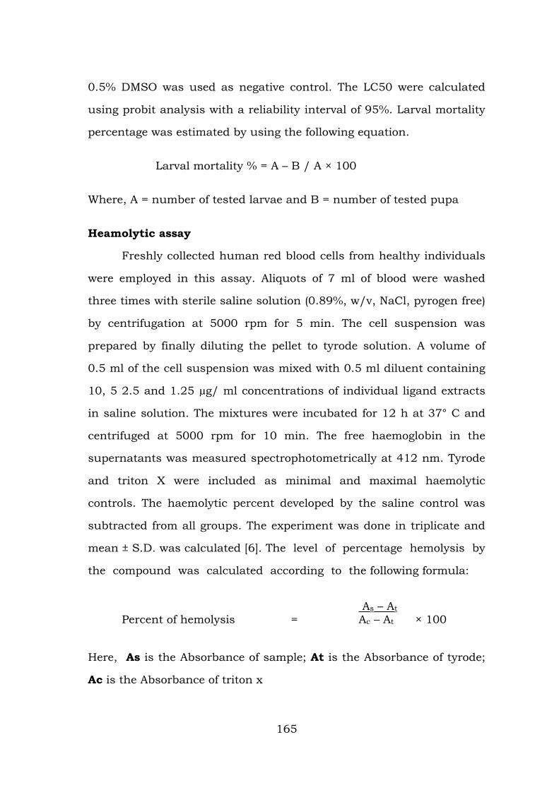
0.5% DMSO was used as negative control. The LC50 were calculated
using probit analysis with a reliability interval of 95%. Larval mortality
percentage was estimated by using the following equation.
Larval mortality % = A – B / A × 100
Where, A = number of tested larvae and B = number of tested pupa
Heamolytic assay
Freshly collected human red blood cells from healthy individuals
were employed in this assay. Aliquots of 7 ml of blood were washed
three times with sterile saline solution (0.89%, w/v, NaCl, pyrogen free)
by centrifugation at 5000 rpm for 5 min. The cell suspension was
prepared by finally diluting the pellet to tyrode solution. A volume of
0.5 ml of the cell suspension was mixed with 0.5 ml diluent containing
10, 5 2.5 and 1.25 µg/ ml concentrations of individual ligand extracts
in saline solution. The mixtures were incubated for 12 h at 37° C and
centrifuged at 5000 rpm for 10 min. The free haemoglobin in the
supernatants was measured spectrophotometrically at 412 nm. Tyrode
and triton X were included as minimal and maximal haemolytic
controls. The haemolytic percent developed by the saline control was
subtracted from all groups. The experiment was done in triplicate and
mean ± S.D. was calculated [6]. The level of percentage hemolysis by
the compound was calculated according to the following formula:
As – At
Percent of hemolysis = Ac – At × 100
Here, As is the Absorbance of sample; At is the Absorbance of tyrode;
Ac is the Absorbance of triton x
165
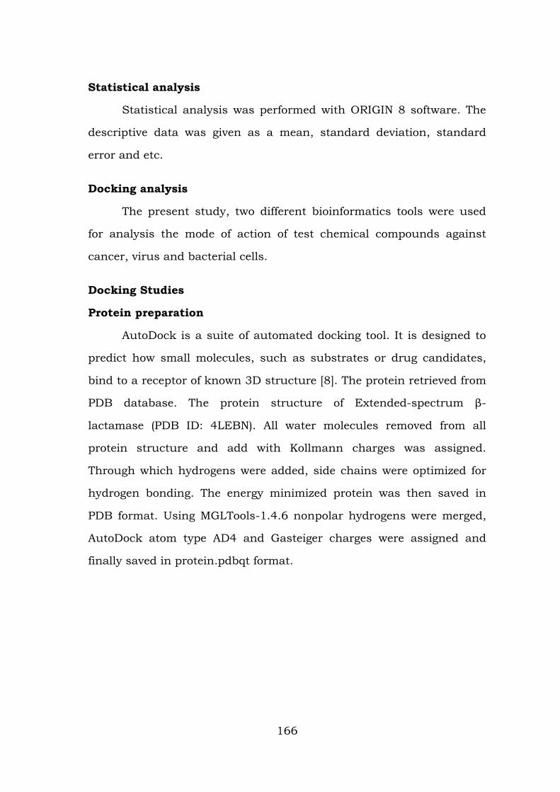
Statistical analysis
Statistical analysis was performed with ORIGIN 8 software. The
descriptive data was given as a mean, standard deviation, standard
error and etc.
Docking analysis
The present study, two different bioinformatics tools were used
for analysis the mode of action of test chemical compounds against
cancer, virus and bacterial cells.
Docking Studies
Protein preparation
AutoDock is a suite of automated docking tool. It is designed to
predict how small molecules, such as substrates or drug candidates,
bind to a receptor of known 3D structure [8]. The protein retrieved from
PDB database. The protein structure of Extended-spectrum β-
lactamase (PDB ID: 4LEBN). All water molecules removed from all
protein structure and add with Kollmann charges was assigned.
Through which hydrogens were added, side chains were optimized for
hydrogen bonding. The energy minimized protein was then saved in
PDB format. Using MGLTools-1.4.6 nonpolar hydrogens were merged,
AutoDock atom type AD4 and Gasteiger charges were assigned and
finally saved in protein.pdbqt format.
166
Ligand preparation
Structure of ligands were drawn using ChemSketch, optimized
with 3D-geometry and the two-dimensional structures of synthetic were
converted into 3-D structure using the open Babel format molecule
converter and saved in PDB format for AutoDock compatibility.
MGLTools-1.4.6 (The Sripps Research Institute) was used to convert
ligand.pdb files to ligand.pdbqt files.
Active site prediction
The active site of the protein is the binding site or usually a
pocket at the surface of the protein that contains residues responsible
for substrate specificity which often act as proton donors or acceptors.
Identification and characterization of binding site is the key step in
structure based drug design. The binding site has been identified by
computational and literature reports. The active site region of the
protein is identified by castP. These servers analytically furnish the
area and the volume at the probable active site of each pocket to
envisage the binding site.
Docking protocol-MGL tools
Grid parameter files (protein.gpf) and docking parameter files
(ligand.dpf) have written using MGLTools-1.4.6. Receptor grids were
generated using 80x80x60 grid points in xyz with grid spacing of 0.375
Å. Grid box was centered co crystallized ligand map types were
generated using autogrid4. Docking of macromolecule was performed
using an empirical free energy function and Lamarckian Genetic
Algorithm, with an initial population of 250 randomly placed
individuals, a maximum number of 106 energy evaluations, a mutation
rate of 0.02, and a crossover rate of 0.80. One hundred independent
167
docking runs were performed for each ligand. Results differing by 2.0 Å
in positional root-mean square deviation (RMSD) were clustered
together and represented by the result with the most favourable free
energy of binding.
Docking protocol-Hex tools
The molecular docking between the target receptor and ligand
was performed by Hex tool. This tool is for interactive docking and can
able to run in any operating system. There are advanced versions in
this tool and it provides the energy values for all the models. The model
can visualize in any forms with docked parameters. The structure for
the ligand MPH can be visualized in the phymol viewer and docked with
the structure of ebola virus and the target receptor for cancer. The
ligand structure was drawn by chemsketch and subjected to docking.
Both the structures were docked and show energy values such as E-
max, E-min, E-shape, and E-total were calculated. The Net charges
with number of orientations were also calculated. Finally, the results
were compared based on the docking parameters.
Test microorganisms
The test microorganisms were isolated from the patient’s medical
samples and were identified by different biochemical test analysis Table
6.2. The identified test bacterial cultures were E. coli, E. faecalis, P.
aeruginosa, P. mirabilis and S. aureus. The identification of test cultures
were confirmed by the Bergy’s manual of determinative bacteriology.
Antimicrobial activity
The antimicrobial activity of test sample was examined with
various pathogenic microorganisms using the (measure the inhibition
168
zone) disk / well diffusion methods. The descriptive statistic of the
antimicrobial activity for three different samples were presented in
Table 6.3. The tested concentration produce zone of inhibition on MHA
plates for bacteria. In this study, four different samples such as metal
ions (B1, B2, B3 and B4), ligand (L1, L2 and L3) and combined metal
ion + ligand samples (B1+L1, B2+L1, B3+L1 and B4+LA) were
challenged against certain pathogenic microorganisms. In metal ion
(alone) samples, nil effect was recorded in the B1 samples against all
the microorganisms, except E. coli (ESBL) (8 mm). The nil was also
observed in B3 (E. faecalis) and B4 (E. coli - ESBL). The test sample B2
was most effective against E. faecalis while less significant activity was
observed against E. coli (ESBL). But in B3 sample, it was found to be
more effective against S. aureus while smaller effect was noticed in
E. faecalis. The B4 sample was effective against E. faecalis whereas nil
effect was observed in E. coli – ESBL and smaller effect against other
strains. The decreasing antimicrobial activity trends in metal ion
complex were: B2 > B3 > B4 > B1 Table 6.3. and Figure 39.
In ligand (alone) samples, nil effect was recorded in the L3
samples against E. coli (ESBL) and P. aeruginosa. In test bacterial
strains, the test sample L1 was most effective against E. faecalis and
P. mirabilis while smaller effect was noticed from E. coli (ESBL). But in
L2 sample was most effective against P. mirabilis while smaller effect
was noticed from E. coli (ESBL). The L3 sample was effective against S.
aureus whereas smaller effect was observed from E. coli (ESBL) and P.
aeruginosa. The decreasing antimicrobial activity trends in ligand
complex were: L1 > L2 > L3 Table 6.4.
169
In combination (metal ion + ligand = 1:1 ratio) samples, nil effect
was not recorded in any samples against all the microorganisms. The
test sample B1+L1 was most effective against E. faecalis while smaller
effect was noticed from P. mirabilis. But in B2+L1 sample was most
effective against E. faecalis and S. aureus while smaller effect was
noticed from E. coli (ESBL) and P. aeruginosa. The B3+L1 sample was
effective against S. aureus whereas smaller effect was observed from
P. mirabilis. The B4+L1 sample was effective against P. aeruginosa and
S. aureus whereas smaller effect was observed from E. coli (ESBL). The
decreasing antimicrobial activity trends in combined samples were:
B2+L1 > B3+L1 > B1+L1 > B4+L1 Table 6.5 and Figure.
All the microbial strains depict higher sensitivity to the test
samples (except few) when compared to the positive control. There is no
antimicrobial activity in solution devoid of sample used as a vehicle
control (concentrated DMSO), reflecting that antimicrobial activity was
directly related to the sample. This study shows that the test samples
have great promise as antimicrobial agent against microorganisms. It
may be concluded that the test complexes inhibits growth of microbes
to a good extent.
Descriptive statistics
In B1 sample, the mean, standard deviation, standard error and
variance level of S1was 8.3, 0.57, 0.33 and 0.33, respectively. But in
S2, S3, S4 and S5, the mean, standard deviation, standard error and
variance level were nil all the studies. In B4 sample, the mean,
standard deviation, standard error and variance level of S1, S2, S3, S4
and S5 were 0, 0, 0 and 0; 17.33, 1.52, 0.88 and 2.33; 8.33, 0.57, 0.33
170
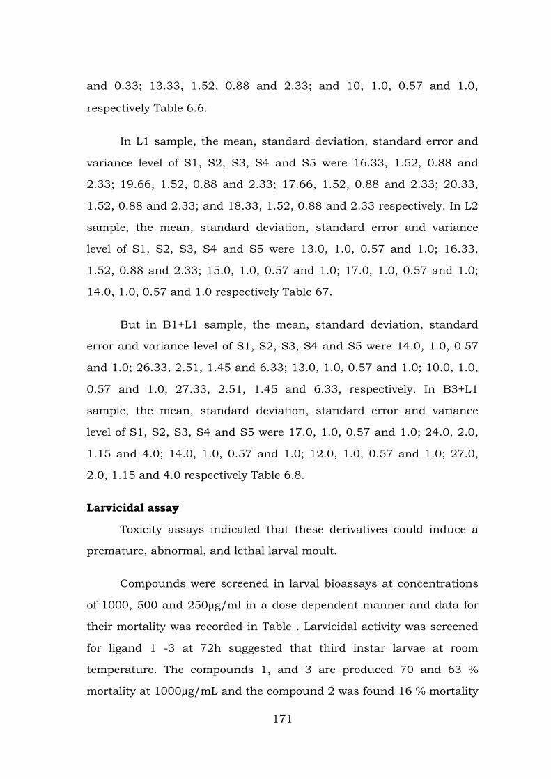
and 0.33; 13.33, 1.52, 0.88 and 2.33; and 10, 1.0, 0.57 and 1.0,
respectively Table 6.6.
In L1 sample, the mean, standard deviation, standard error and
variance level of S1, S2, S3, S4 and S5 were 16.33, 1.52, 0.88 and
2.33; 19.66, 1.52, 0.88 and 2.33; 17.66, 1.52, 0.88 and 2.33; 20.33,
1.52, 0.88 and 2.33; and 18.33, 1.52, 0.88 and 2.33 respectively. In L2
sample, the mean, standard deviation, standard error and variance
level of S1, S2, S3, S4 and S5 were 13.0, 1.0, 0.57 and 1.0; 16.33,
1.52, 0.88 and 2.33; 15.0, 1.0, 0.57 and 1.0; 17.0, 1.0, 0.57 and 1.0;
14.0, 1.0, 0.57 and 1.0 respectively Table 67.
But in B1+L1 sample, the mean, standard deviation, standard
error and variance level of S1, S2, S3, S4 and S5 were 14.0, 1.0, 0.57
and 1.0; 26.33, 2.51, 1.45 and 6.33; 13.0, 1.0, 0.57 and 1.0; 10.0, 1.0,
0.57 and 1.0; 27.33, 2.51, 1.45 and 6.33, respectively. In B3+L1
sample, the mean, standard deviation, standard error and variance
level of S1, S2, S3, S4 and S5 were 17.0, 1.0, 0.57 and 1.0; 24.0, 2.0,
1.15 and 4.0; 14.0, 1.0, 0.57 and 1.0; 12.0, 1.0, 0.57 and 1.0; 27.0,
2.0, 1.15 and 4.0 respectively Table 6.8.
Larvicidal assay
Toxicity assays indicated that these derivatives could induce a
premature, abnormal, and lethal larval moult.
Compounds were screened in larval bioassays at concentrations
of 1000, 500 and 250µg/ml in a dose dependent manner and data for
their mortality was recorded in Table . Larvicidal activity was screened
for ligand 1 -3 at 72h suggested that third instar larvae at room
temperature. The compounds 1, and 3 are produced 70 and 63 %
mortality at 1000µg/mL and the compound 2 was found 16 % mortality
171
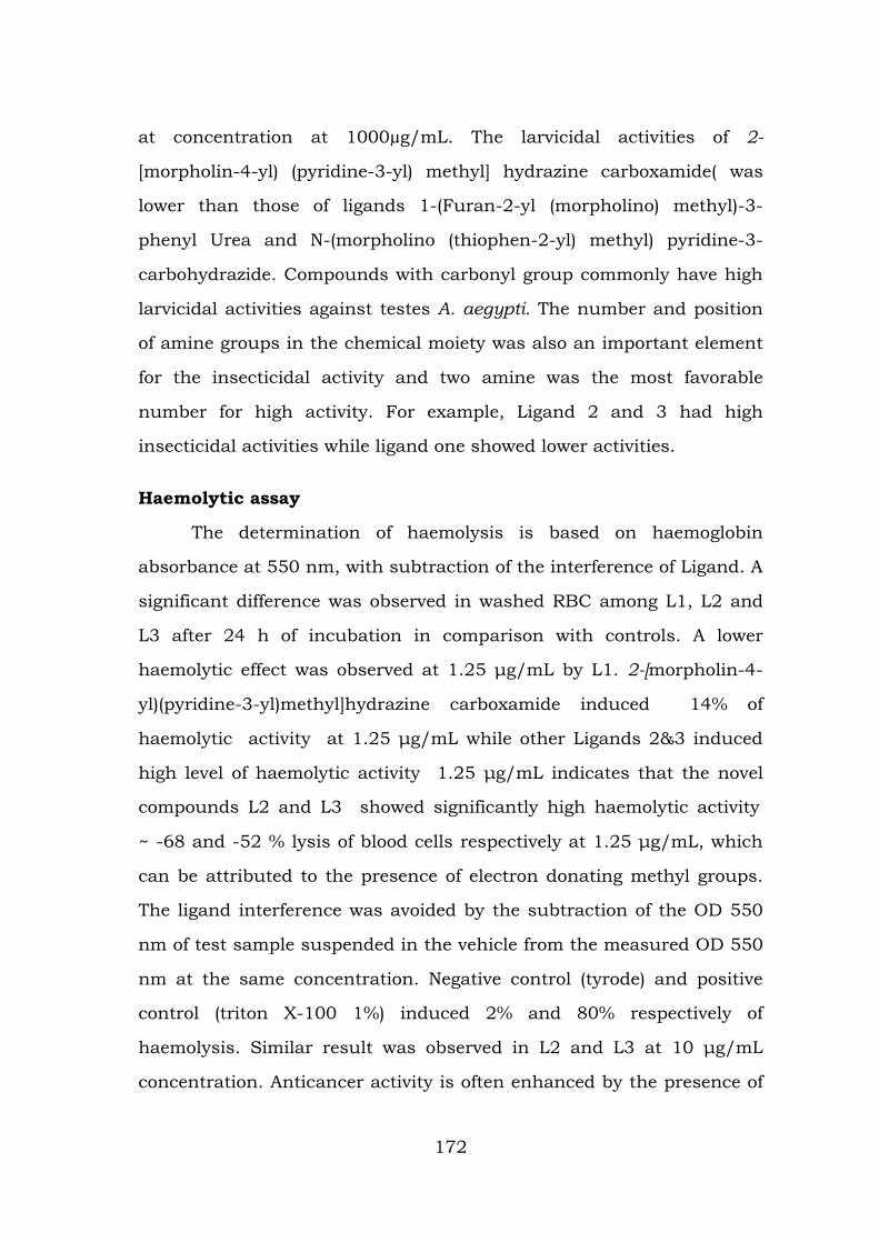
at concentration at 1000µg/mL. The larvicidal activities of 2-
[morpholin-4-yl) (pyridine-3-yl) methyl] hydrazine carboxamide( was
lower than those of ligands 1-(Furan-2-yl (morpholino) methyl)-3-
phenyl Urea and N-(morpholino (thiophen-2-yl) methyl) pyridine-3-
carbohydrazide. Compounds with carbonyl group commonly have high
larvicidal activities against testes A. aegypti. The number and position
of amine groups in the chemical moiety was also an important element
for the insecticidal activity and two amine was the most favorable
number for high activity. For example, Ligand 2 and 3 had high
insecticidal activities while ligand one showed lower activities.
Haemolytic assay
The determination of haemolysis is based on haemoglobin
absorbance at 550 nm, with subtraction of the interference of Ligand. A
significant difference was observed in washed RBC among L1, L2 and
L3 after 24 h of incubation in comparison with controls. A lower
haemolytic effect was observed at 1.25 μg/mL by L1. 2-[morpholin-4-
yl)(pyridine-3-yl)methyl]hydrazine carboxamide induced 14% of
haemolytic activity at 1.25 μg/mL while other Ligands 2&3 induced
high level of haemolytic activity 1.25 μg/mL indicates that the novel
compounds L2 and L3 showed significantly high haemolytic activity
~ -68 and -52 % lysis of blood cells respectively at 1.25 μg/mL, which
can be attributed to the presence of electron donating methyl groups.
The ligand interference was avoided by the subtraction of the OD 550
nm of test sample suspended in the vehicle from the measured OD 550
nm at the same concentration. Negative control (tyrode) and positive
control (triton X-100 1%) induced 2% and 80% respectively of
haemolysis. Similar result was observed in L2 and L3 at 10 μg/mL
concentration. Anticancer activity is often enhanced by the presence of
172
electron releasing groups (Mologni et al., 2010). In contrast, the
compound L1 exhibited haemolytic activity below 10%. In view of the
observed differences in the % lysis of RBC values, it is inferred that the
electron withdrawing and electron donating nature of the substituent
groups have an influence on the haemolytic activity of the compounds
(Ding et al., 2012). Moreover, the cytotoxicity of the compounds viz. L2
and L3 can be optimized by making appropriate changes in the
molecular structures for the purpose of their use as toxic compounds to
control the uncontrolled proliferation of cells.
Docking studies
ESBL enzyme studies
Extended-spectrum β-lactamases (ESBLs) are a rapidly evolving
group of β-lactamases which share the ability to hydrolyze third-
generation cephalosporins and aztreonam but are inhibited by
clavulanic acid. They represent the first example in which β-lactamase–
mediated resistance to β-lactam antibiotics resulted from fundamental
changes in the substrate spectra of the enzymes. The total number of
ESBLs now characterized exceeds 200. These are detailed on the
authoritative website on the nomenclature of ESBLs hosted by George
Jacoby and Karen Bush (http://www.lahey.org/studies/webt.htm).
Published research on ESBLs has now originated from more than 30
different countries, reflecting the truly worldwide distribution of ESBL-
producing organisms. So Extended-spectrum β-lactamase is attractive
drug target for antibiotic resistance. Binding energies of the protein-
ligand (drug) interactions are important to describe how fit the drug
binds to the target macromolecule. We applied the docking procedures,
to dock copper compounds to dock with ESBL into the binding pocket
of this enzyme. Our docking simulation resulted in a very close target
173
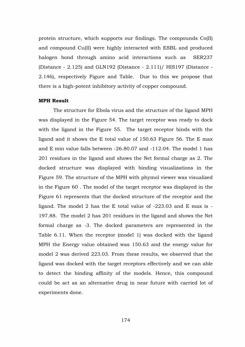
protein structure, which supports our findings. The compounds Co(II)
and compound Cu(II) were highly interacted with ESBL and produced
halogen bond through amino acid interactions such as SER237
(Distance - 2.125) and GLN192 (Distance - 2.111)/ HIS197 (Distance -
2.146), respectively Figure and Table. Due to this we propose that
there is a high-potent inhibitory activity of copper compound.
MPH Result
The structure for Ebola virus and the structure of the ligand MPH
was displayed in the Figure 54. The target receptor was ready to dock
with the ligand in the Figure 55. The target receptor binds with the
ligand and it shows the E total value of 150.63 Figure 56. The E max
and E min value falls between -26.80.07 and -112.04. The model 1 has
201 residues in the ligand and shows the Net formal charge as 2. The
docked structure was displayed with binding visualizations in the
Figure 59. The structure of the MPH with phymol viewer was visualized
in the Figure 60 . The model of the target receptor was displayed in the
Figure 61 represents that the docked structure of the receptor and the
ligand. The model 2 has the E total value of -223.03 and E max is -
197.88. The model 2 has 201 residues in the ligand and shows the Net
formal charge as -3. The docked parameters are represented in the
Table 6.11. When the receptor (model 1) was docked with the ligand
MPH the Energy value obtained was 150.63 and the energy value for
model 2 was derived 223.03. From these results, we observed that the
ligand was docked with the target receptors effectively and we can able
to detect the binding affinity of the models. Hence, this compound
could be act as an alternative drug in near future with carried lot of
experiments done.
174
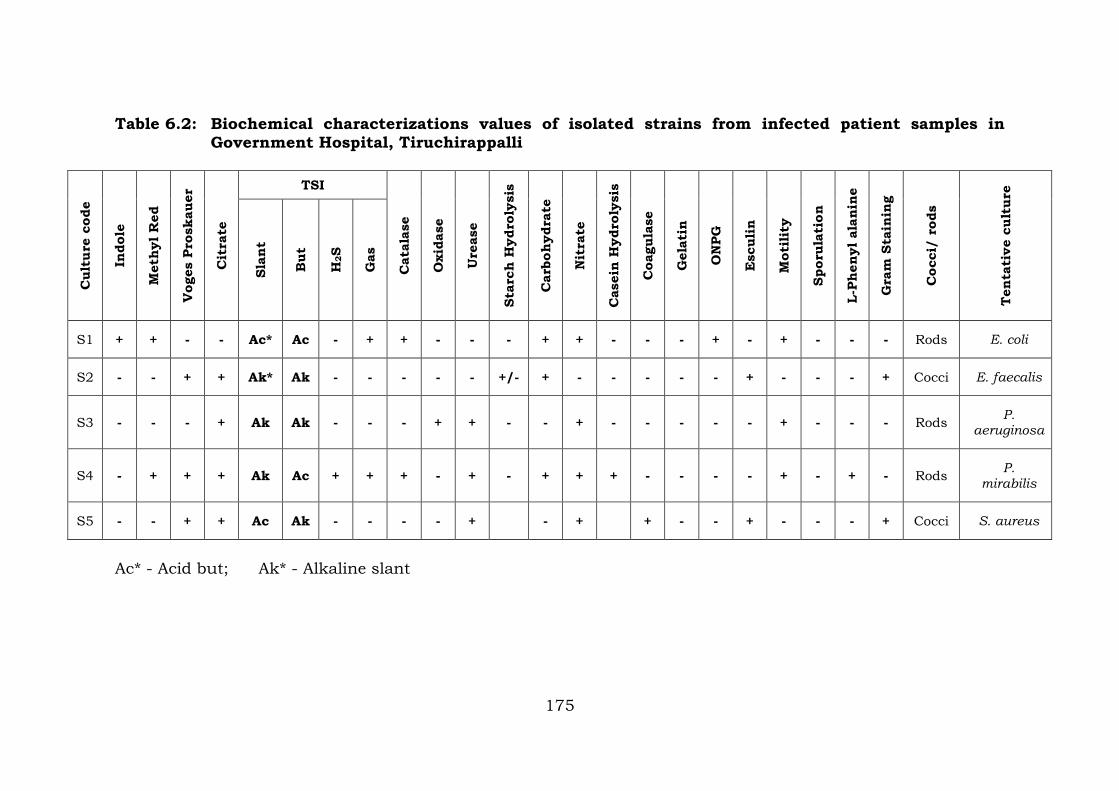
Table 6.2: Biochemical characterizations values of isolated strains from infected patient samples in Government Hospital, Tiruchirappalli
Cul
ture
cod
e
Indo
le
Met
hyl R
ed
Vog
es P
rosk
auer
Cit
rate
TSI
Cat
alas
e
Oxi
dase
Ure
ase
Star
ch H
ydro
lysi
s
Car
bohy
drat
e
Nit
rate
Cas
ein
Hyd
roly
sis
Coa
gula
se
Gel
atin
ON
PG
Esc
ulin
Mot
ilit
y
Spor
ulat
ion
L-Ph
enyl
ala
nine
Gra
m S
tain
ing
Coc
ci/
rods
Ten
tati
ve c
ultu
re
Slan
t
But
H2S
Gas
S1 + + - - Ac* Ac - + + - - - + + - - - + - + - - - Rods E. coli
S2 - - + + Ak* Ak - - - - - +/- + - - - - - + - - - + Cocci E. faecalis
S3 - - - + Ak Ak - - - + + - - + - - - - - + - - - Rods P. aeruginosa
S4 - + + + Ak Ac + + + - + - + + + - - - - + - + - Rods P. mirabilis
S5 - - + + Ac Ak - - - - + - + + - - + - - - + Cocci S. aureus
Ac* - Acid but; Ak* - Alkaline slant
175
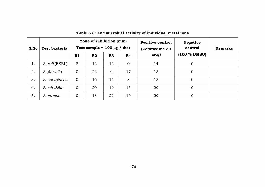
Table 6.3: Antimicrobial activity of individual metal ions
S.No Test bacteria
Zone of inhibition (mm)
Test sample = 100 µg / disc Positive control
(Cefotaxime 30 mcg)
Negative control
(100 % DMSO) Remarks
B1 B2 B3 B4
1. E. coli (ESBL) 8 12 12 0 14 0
2. E. faecalis 0 22 0 17 18 0
3. P. aeruginosa 0 16 15 8 18 0
4. P. mirabilis 0 20 19 13 20 0
5. S. aureus 0 18 22 10 20 0
176
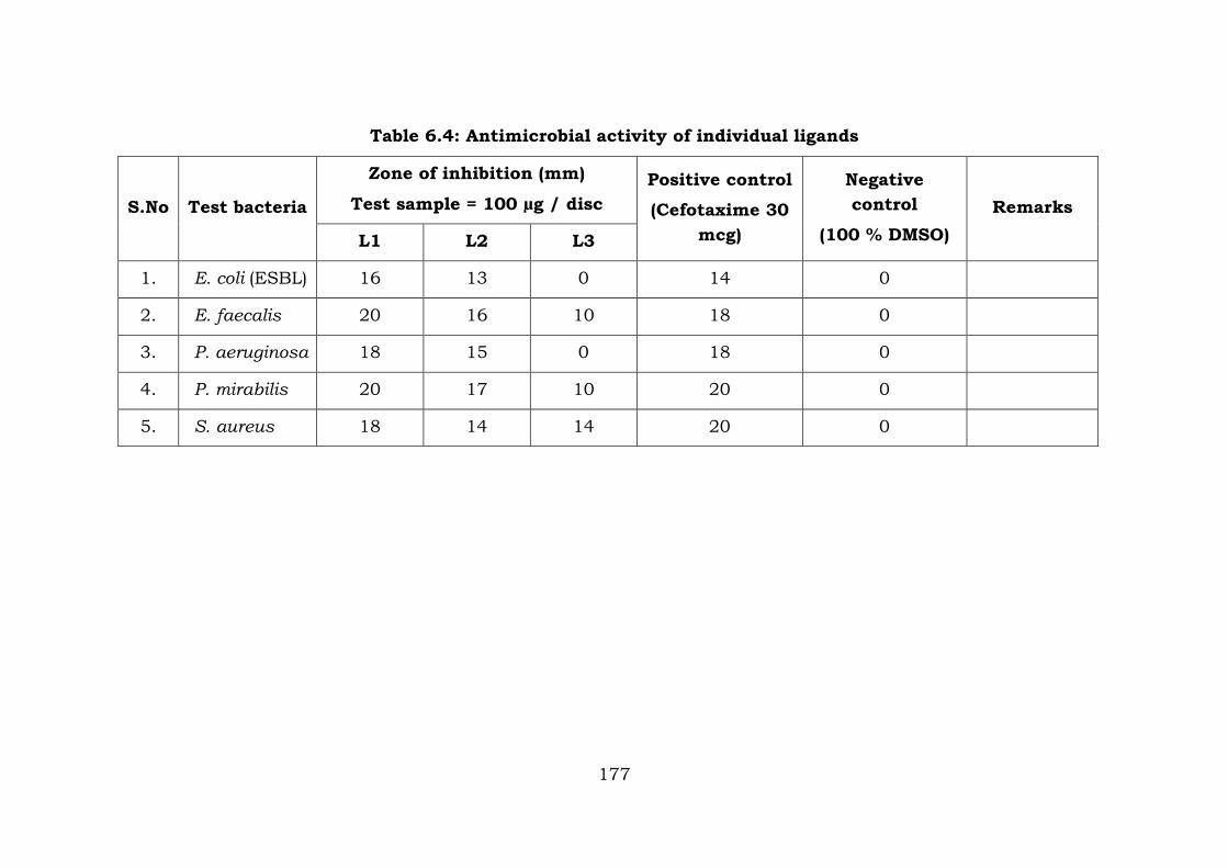
Table 6.4: Antimicrobial activity of individual ligands
S.No Test bacteria
Zone of inhibition (mm)
Test sample = 100 µg / disc Positive control
(Cefotaxime 30 mcg)
Negative control
(100 % DMSO) Remarks
L1 L2 L3
1. E. coli (ESBL) 16 13 0 14 0
2. E. faecalis 20 16 10 18 0
3. P. aeruginosa 18 15 0 18 0
4. P. mirabilis 20 17 10 20 0
5. S. aureus 18 14 14 20 0
177
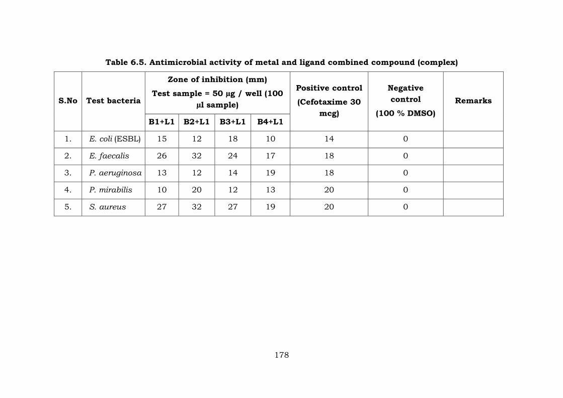
Table 6.5. Antimicrobial activity of metal and ligand combined compound (complex)
S.No Test bacteria
Zone of inhibition (mm)
Test sample = 50 µg / well (100 µl sample)
Positive control
(Cefotaxime 30 mcg)
Negative control
(100 % DMSO) Remarks
B1+L1 B2+L1 B3+L1 B4+L1
1. E. coli (ESBL) 15 12 18 10 14 0
2. E. faecalis 26 32 24 17 18 0
3. P. aeruginosa 13 12 14 19 18 0
4. P. mirabilis 10 20 12 13 20 0
5. S. aureus 27 32 27 19 20 0
178
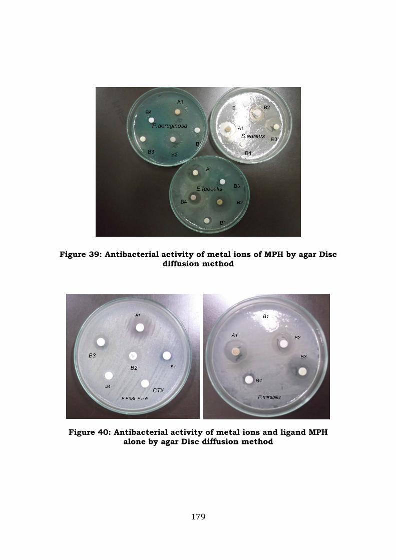
Figure 39: Antibacterial activity of metal ions of MPH by agar Disc diffusion method
Figure 40: Antibacterial activity of metal ions and ligand MPH alone by agar Disc diffusion method
179
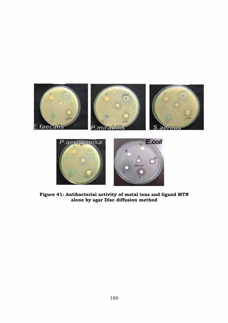
Figure 41: Antibacterial activity of metal ions and ligand MTN alone by agar Disc diffusion method
180
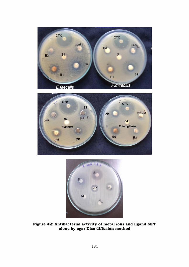
Figure 42: Antibacterial activity of metal ions and ligand MFP alone by agar Disc diffusion method
181
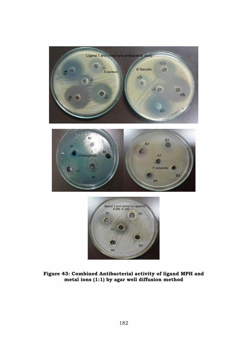
Figure 43: Combined Antibacterial activity of ligand MPH and
metal ions (1:1) by agar well diffusion method
182
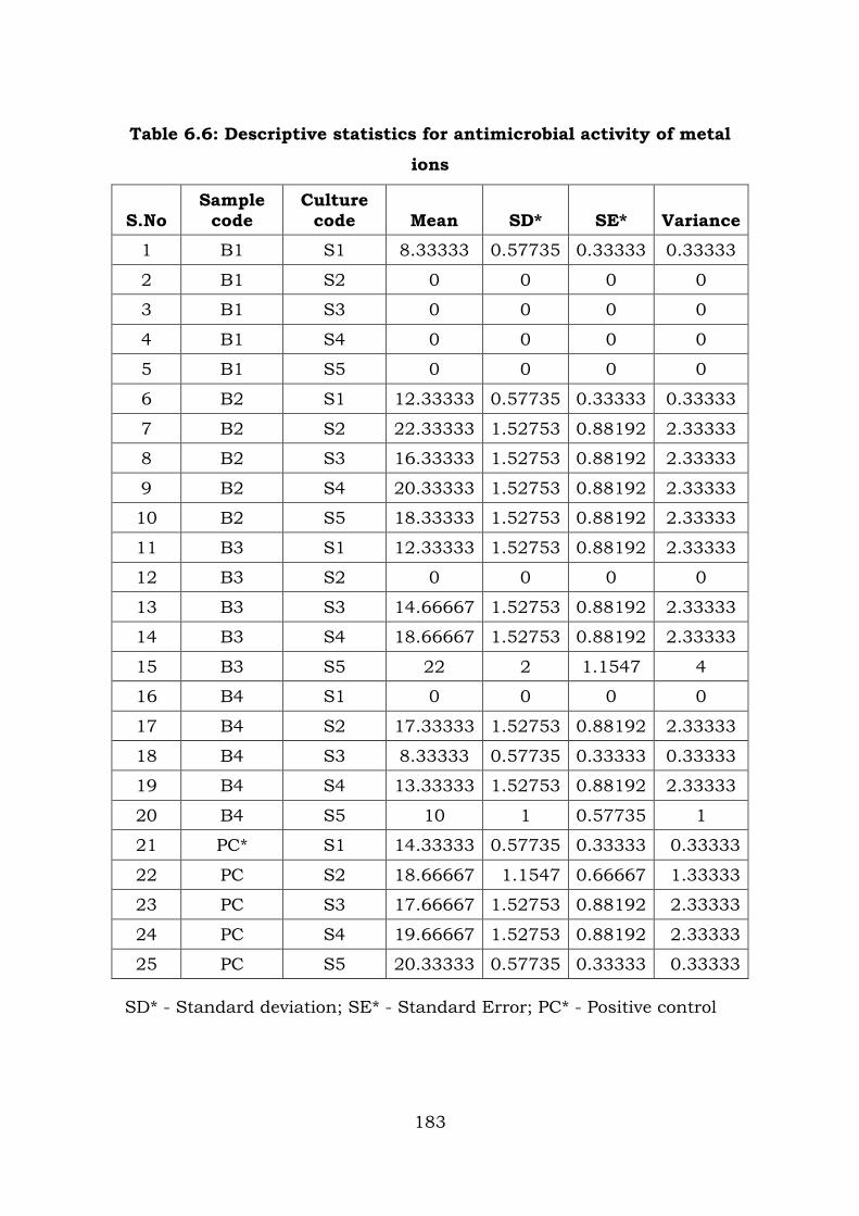
Table 6.6: Descriptive statistics for antimicrobial activity of metal
ions
S.No Sample
code Culture
code Mean SD* SE* Variance
1 B1 S1 8.33333 0.57735 0.33333 0.33333
2 B1 S2 0 0 0 0
3 B1 S3 0 0 0 0
4 B1 S4 0 0 0 0
5 B1 S5 0 0 0 0
6 B2 S1 12.33333 0.57735 0.33333 0.33333
7 B2 S2 22.33333 1.52753 0.88192 2.33333
8 B2 S3 16.33333 1.52753 0.88192 2.33333
9 B2 S4 20.33333 1.52753 0.88192 2.33333
10 B2 S5 18.33333 1.52753 0.88192 2.33333
11 B3 S1 12.33333 1.52753 0.88192 2.33333
12 B3 S2 0 0 0 0
13 B3 S3 14.66667 1.52753 0.88192 2.33333
14 B3 S4 18.66667 1.52753 0.88192 2.33333
15 B3 S5 22 2 1.1547 4
16 B4 S1 0 0 0 0
17 B4 S2 17.33333 1.52753 0.88192 2.33333
18 B4 S3 8.33333 0.57735 0.33333 0.33333
19 B4 S4 13.33333 1.52753 0.88192 2.33333
20 B4 S5 10 1 0.57735 1
21 PC* S1 14.33333 0.57735 0.33333 0.33333
22 PC S2 18.66667 1.1547 0.66667 1.33333
23 PC S3 17.66667 1.52753 0.88192 2.33333
24 PC S4 19.66667 1.52753 0.88192 2.33333
25 PC S5 20.33333 0.57735 0.33333 0.33333
SD* - Standard deviation; SE* - Standard Error; PC* - Positive control
183
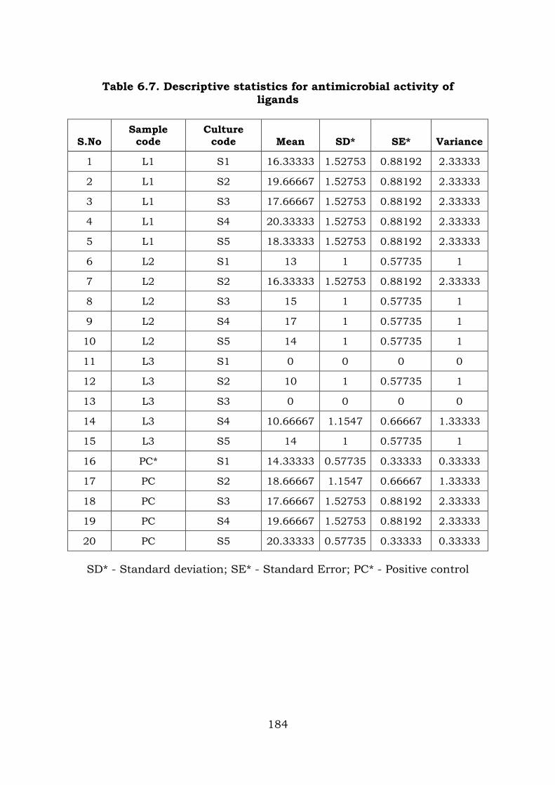
Table 6.7. Descriptive statistics for antimicrobial activity of ligands
S.No Sample
code Culture
code Mean SD* SE* Variance
1 L1 S1 16.33333 1.52753 0.88192 2.33333
2 L1 S2 19.66667 1.52753 0.88192 2.33333
3 L1 S3 17.66667 1.52753 0.88192 2.33333
4 L1 S4 20.33333 1.52753 0.88192 2.33333
5 L1 S5 18.33333 1.52753 0.88192 2.33333
6 L2 S1 13 1 0.57735 1
7 L2 S2 16.33333 1.52753 0.88192 2.33333
8 L2 S3 15 1 0.57735 1
9 L2 S4 17 1 0.57735 1
10 L2 S5 14 1 0.57735 1
11 L3 S1 0 0 0 0
12 L3 S2 10 1 0.57735 1
13 L3 S3 0 0 0 0
14 L3 S4 10.66667 1.1547 0.66667 1.33333
15 L3 S5 14 1 0.57735 1
16 PC* S1 14.33333 0.57735 0.33333 0.33333
17 PC S2 18.66667 1.1547 0.66667 1.33333
18 PC S3 17.66667 1.52753 0.88192 2.33333
19 PC S4 19.66667 1.52753 0.88192 2.33333
20 PC S5 20.33333 0.57735 0.33333 0.33333
SD* - Standard deviation; SE* - Standard Error; PC* - Positive control
184
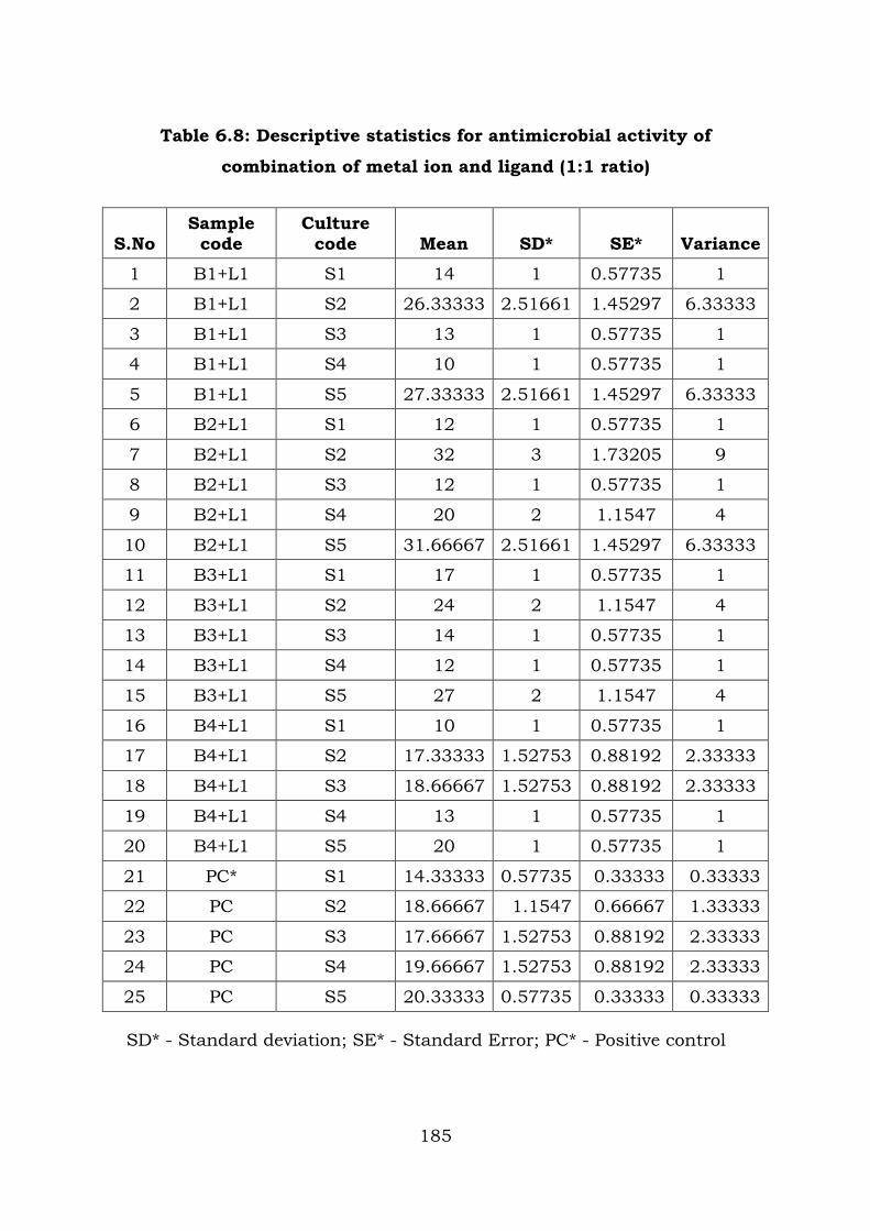
Table 6.8: Descriptive statistics for antimicrobial activity of
combination of metal ion and ligand (1:1 ratio)
S.No Sample
code Culture
code Mean SD* SE* Variance
1 B1+L1 S1 14 1 0.57735 1
2 B1+L1 S2 26.33333 2.51661 1.45297 6.33333
3 B1+L1 S3 13 1 0.57735 1
4 B1+L1 S4 10 1 0.57735 1
5 B1+L1 S5 27.33333 2.51661 1.45297 6.33333
6 B2+L1 S1 12 1 0.57735 1
7 B2+L1 S2 32 3 1.73205 9
8 B2+L1 S3 12 1 0.57735 1
9 B2+L1 S4 20 2 1.1547 4
10 B2+L1 S5 31.66667 2.51661 1.45297 6.33333
11 B3+L1 S1 17 1 0.57735 1
12 B3+L1 S2 24 2 1.1547 4
13 B3+L1 S3 14 1 0.57735 1
14 B3+L1 S4 12 1 0.57735 1
15 B3+L1 S5 27 2 1.1547 4
16 B4+L1 S1 10 1 0.57735 1
17 B4+L1 S2 17.33333 1.52753 0.88192 2.33333
18 B4+L1 S3 18.66667 1.52753 0.88192 2.33333
19 B4+L1 S4 13 1 0.57735 1
20 B4+L1 S5 20 1 0.57735 1
21 PC* S1 14.33333 0.57735 0.33333 0.33333
22 PC S2 18.66667 1.1547 0.66667 1.33333
23 PC S3 17.66667 1.52753 0.88192 2.33333
24 PC S4 19.66667 1.52753 0.88192 2.33333
25 PC S5 20.33333 0.57735 0.33333 0.33333
SD* - Standard deviation; SE* - Standard Error; PC* - Positive control
185
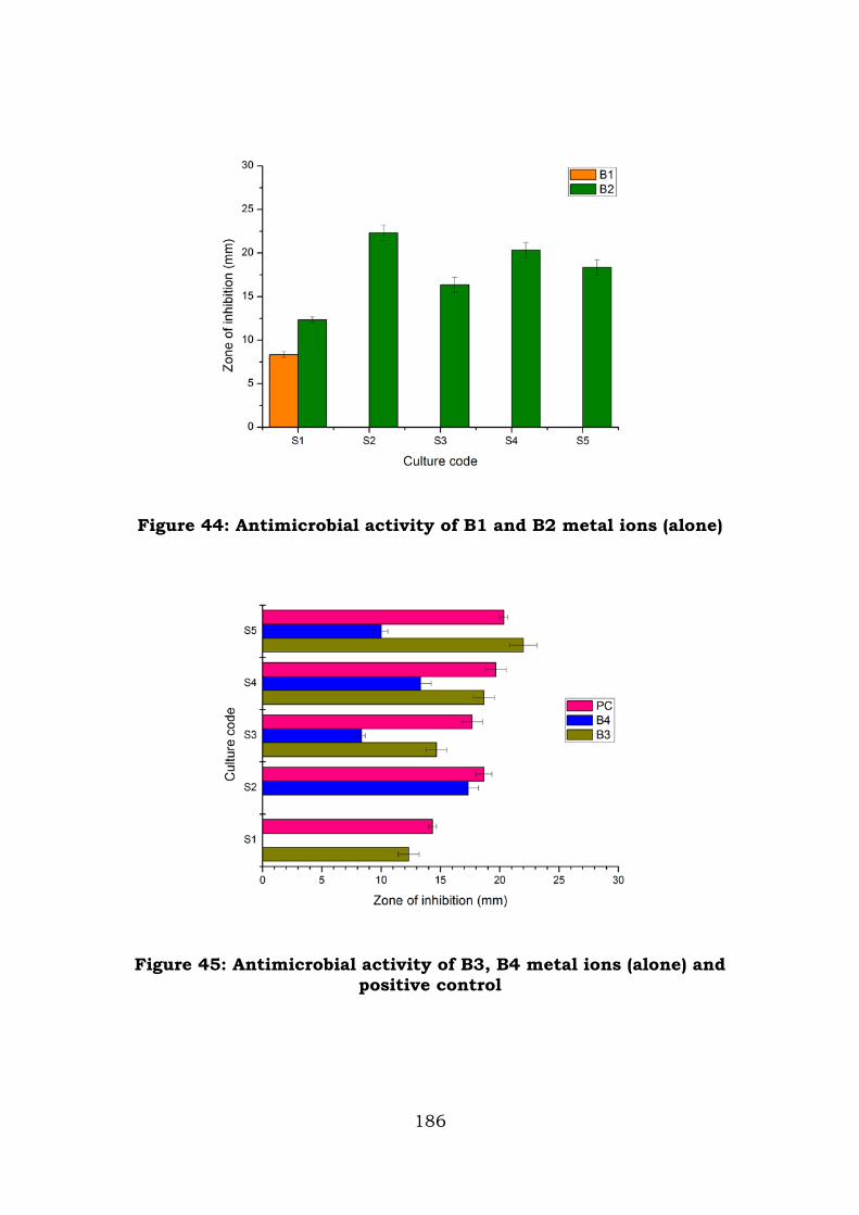
Figure 44: Antimicrobial activity of B1 and B2 metal ions (alone)
Figure 45: Antimicrobial activity of B3, B4 metal ions (alone) and positive control
186
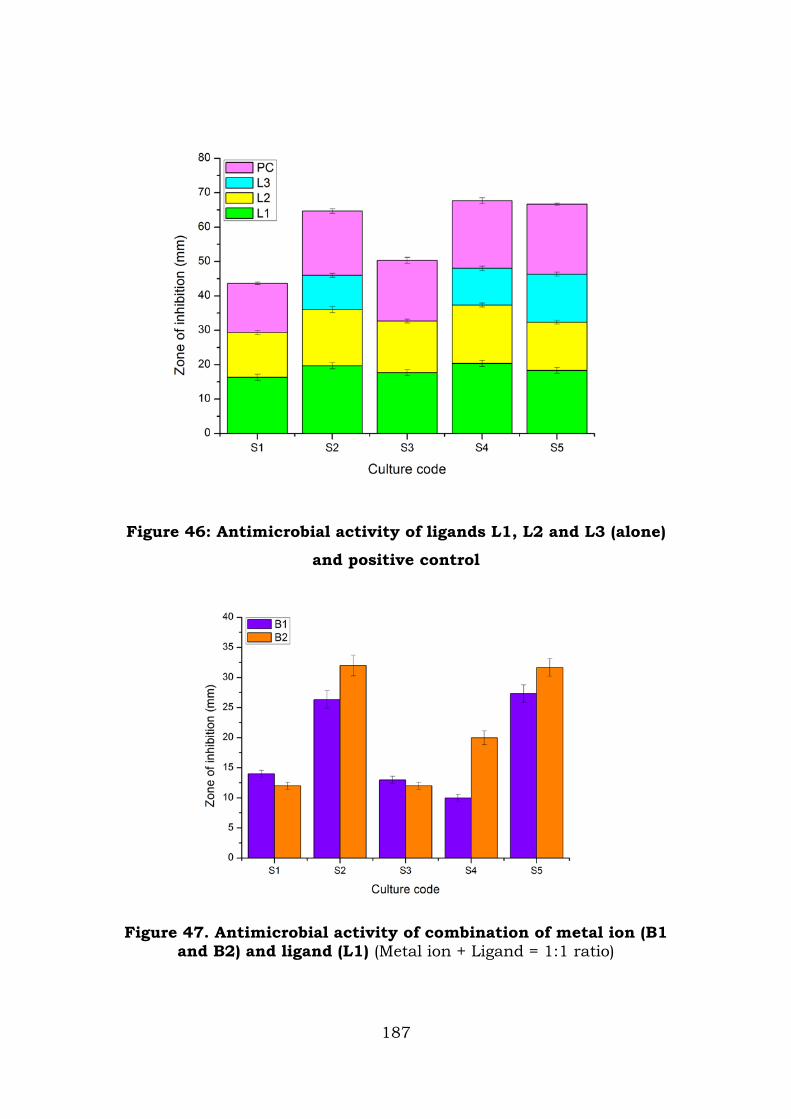
Figure 46: Antimicrobial activity of ligands L1, L2 and L3 (alone)
and positive control
Figure 47. Antimicrobial activity of combination of metal ion (B1
and B2) and ligand (L1) (Metal ion + Ligand = 1:1 ratio)
187
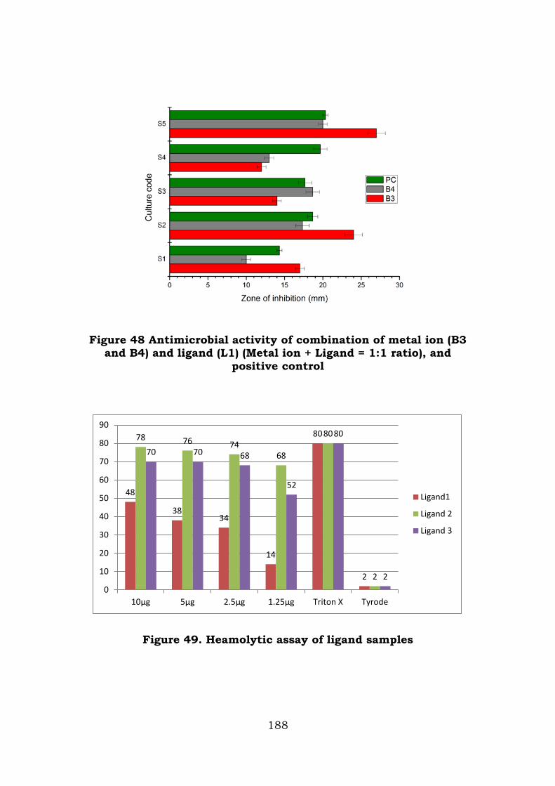
Figure 48 Antimicrobial activity of combination of metal ion (B3 and B4) and ligand (L1) (Metal ion + Ligand = 1:1 ratio), and
positive control
Figure 49. Heamolytic assay of ligand samples
48
38 34
14
80
2
78 76 74 68
80
2
70 70 68
52
80
2 0
10
20
30
40
50
60
70
80
90
10µg 5µg 2.5µg 1.25µg Triton X Tyrode
Ligand1
Ligand 2
Ligand 3
188
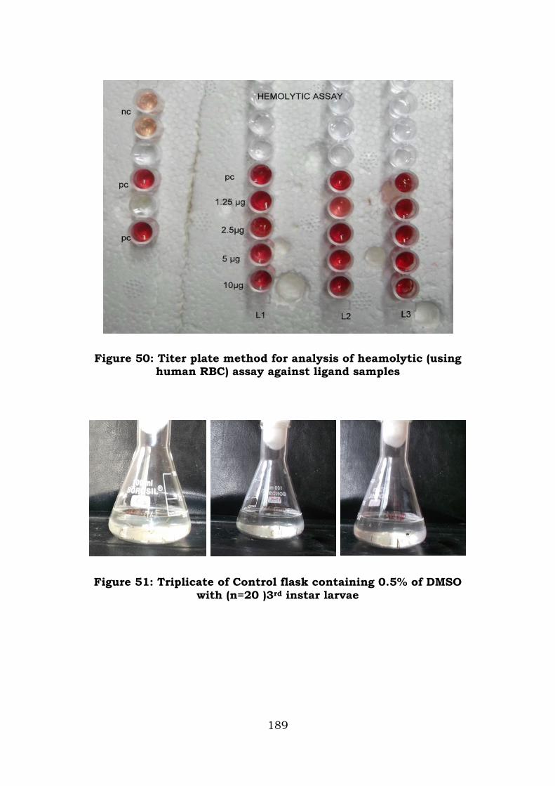
Figure 50: Titer plate method for analysis of heamolytic (using human RBC) assay against ligand samples
Figure 51: Triplicate of Control flask containing 0.5% of DMSO with (n=20 )3rd instar larvae
189
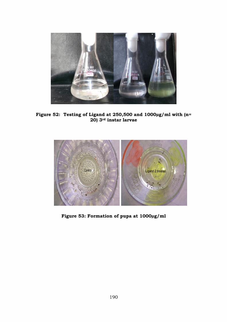
Figure 52: Testing of Ligand at 250,500 and 1000µg/ml with (n= 20) 3rd instar larvae
Figure 53: Formation of pupa at 1000µg/ml
190
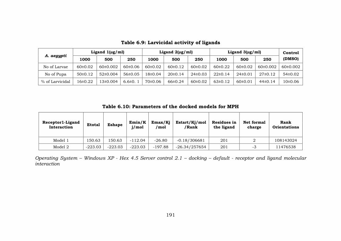
Table 6.9: Larvicidal activity of ligands
A. aegypti Ligand 1(µg/ml) Ligand 2(µg/ml) Ligand 3(µg/ml) Control
(DMSO) 1000 500 250 1000 500 250 1000 500 250
No of Larvae 60±0.02 60±0.002 60±0.06 60±0.02 60±0.12 60±0.02 60±0.22 60±0.02 60±0.002 60±0.002
No of Pupa 50±0.12 52±0.004 56±0.05 18±0.04 20±0.14 24±0.03 22±0.14 24±0.01 27±0.12 54±0.02
% of Larvicidal 16±0.22 13±0.004 6.6±0. 1 70±0.06 66±0.24 60±0.02 63±0.12 60±0.01 44±0.14 10±0.06
Table 6.10: Parameters of the docked models for MPH
Receptor1-Ligand Interaction Etotal Eshape Emin/K
j/mol Emax/Kj
/mol Estart/Kj/mol
/Rank Residues in the ligand
Net formal charge
Rank Orientations
Model 1 150.63 150.63 -112.04 -26.80 -0.18/306681 201 2 108143024 Model 2 -223.03 -223.03 -223.03 -197.88 -26.34/257654 201 -3 11476538
Operating System – Windows XP - Hex 4.5 Server control 2.1 – docking – default - receptor and ligand molecular interaction
191
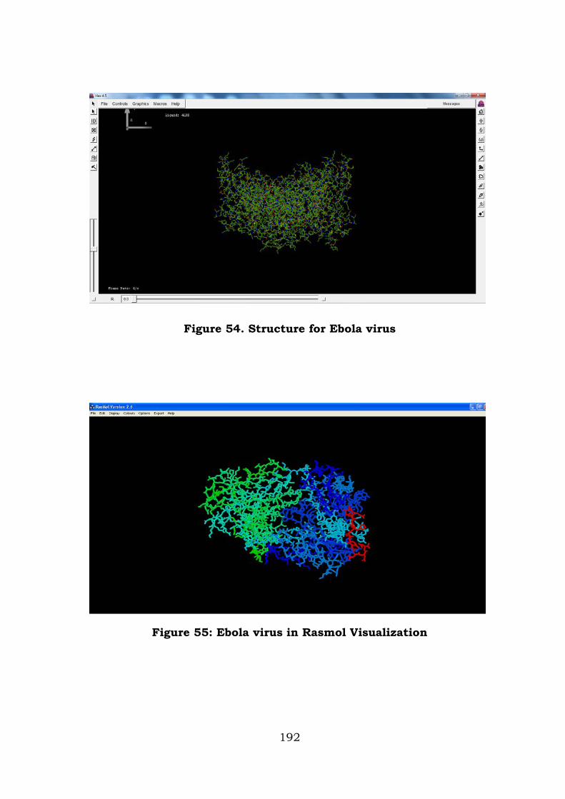
Figure 54. Structure for Ebola virus
Figure 55: Ebola virus in Rasmol Visualization
192
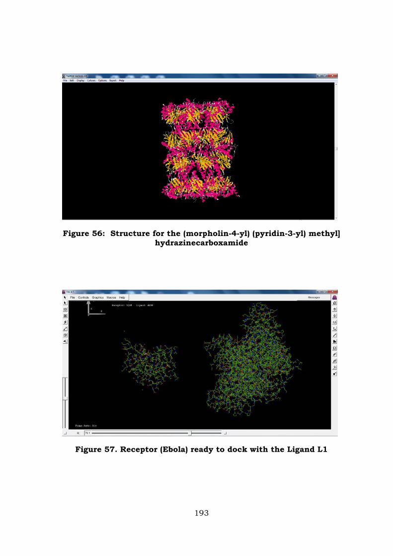
Figure 56: Structure for the (morpholin-4-yl) (pyridin-3-yl) methyl] hydrazinecarboxamide
Figure 57. Receptor (Ebola) ready to dock with the Ligand L1
193
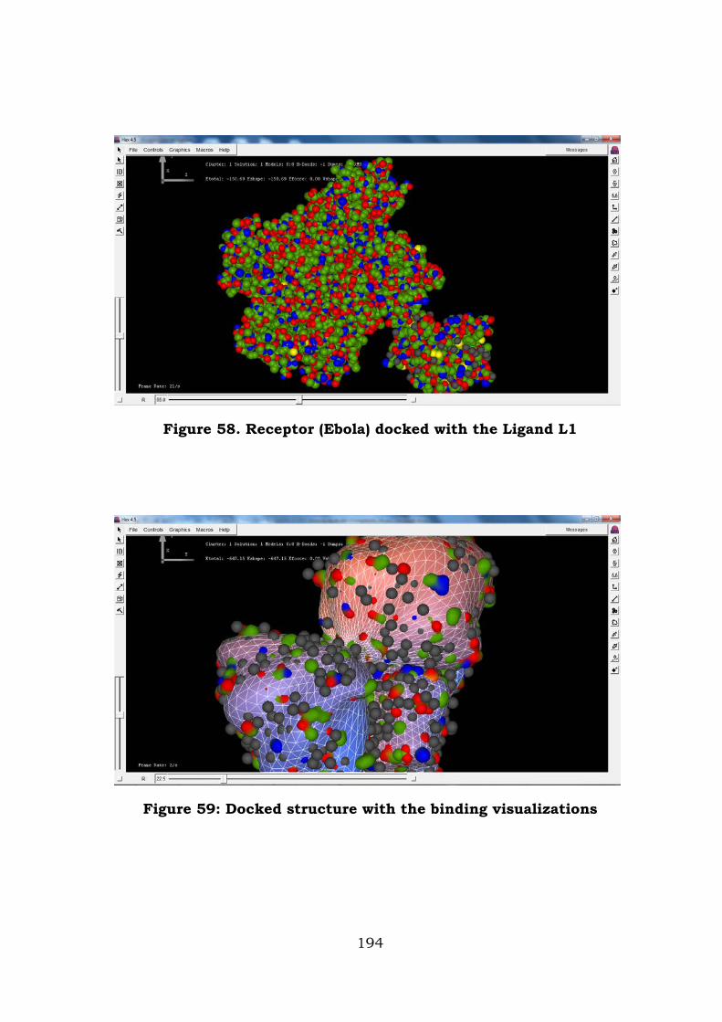
Figure 58. Receptor (Ebola) docked with the Ligand L1
Figure 59: Docked structure with the binding visualizations
194
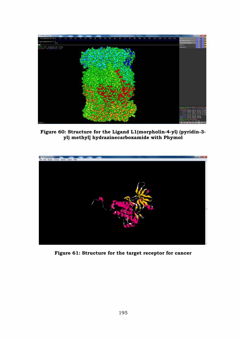
Figure 60: Structure for the Ligand L1(morpholin-4-yl) (pyridin-3-yl) methyl] hydrazinecarboxamide with Phymol
Figure 61: Structure for the target receptor for cancer
195
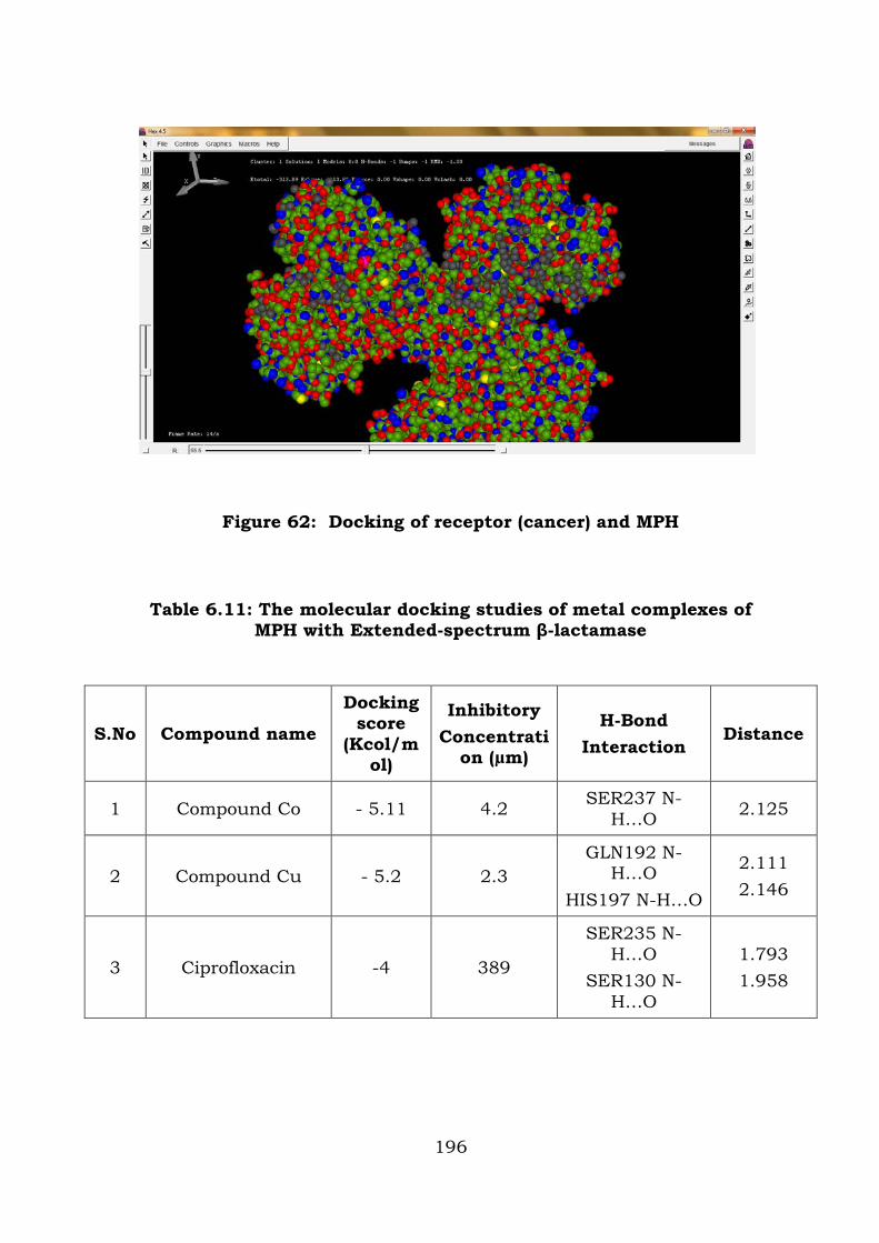
Figure 62: Docking of receptor (cancer) and MPH
Table 6.11: The molecular docking studies of metal complexes of MPH with Extended-spectrum β-lactamase
S.No Compound name
Docking score
(Kcol/mol)
Inhibitory Concentrati
on (µm)
H-Bond Interaction
Distance
1 Compound Co - 5.11 4.2 SER237 N-H…O 2.125
2 Compound Cu - 5.2 2.3 GLN192 N-
H…O HIS197 N-H…O
2.111 2.146
3 Ciprofloxacin -4 389
SER235 N-H…O
SER130 N-H…O
1.793 1.958
196
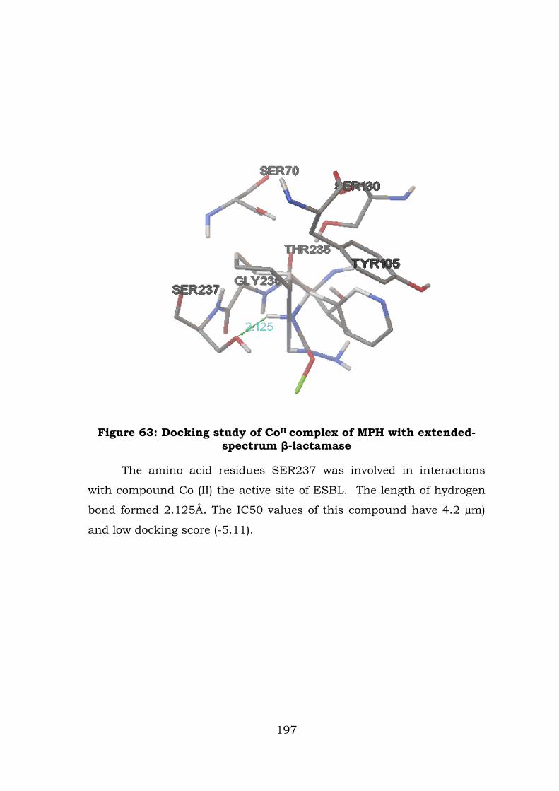
Figure 63: Docking study of CoII complex of MPH with extended-spectrum β-lactamase
The amino acid residues SER237 was involved in interactions
with compound Co (II) the active site of ESBL. The length of hydrogen
bond formed 2.125Å. The IC50 values of this compound have 4.2 µm)
and low docking score (-5.11).
197
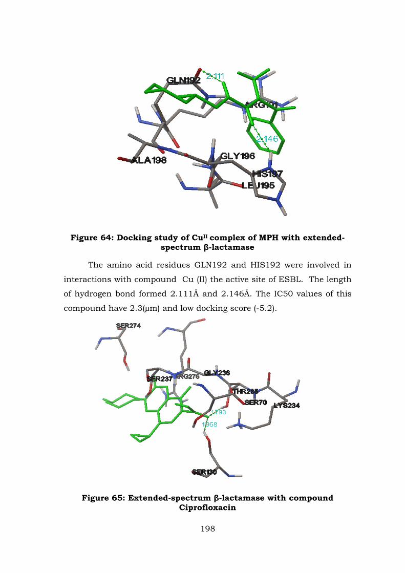
Figure 64: Docking study of CuII complex of MPH with extended-spectrum β-lactamase
The amino acid residues GLN192 and HIS192 were involved in
interactions with compound Cu (II) the active site of ESBL. The length
of hydrogen bond formed 2.111Å and 2.146Å. The IC50 values of this
compound have 2.3(µm) and low docking score (-5.2).
Figure 65: Extended-spectrum β-lactamase with compound
Ciprofloxacin
198
DISCUSSIONS
Antimicrobial activity
The emerging bacterial resistance causes a widespread problem
for the treatment of various infections. Therefore, the search for
antimicrobials is a never-ending task. Now-a-days a number of
hydrazone derivatives have been developed and evaluated for their
antibacterial activity. Aslan et al., (2012) investigated the antibacterial
activity of sulfonyl derivatives. Certain steroidal hydrazines have been
synthesized by Khan (2008) which possess in-vitro antibacterial activity.
Hydrazones bearing imidazoles have been synthesized and screened for
antibacterial activity against numerous bacterial strains by Abdel-
Wahab et al. (2011). Palekar et al., (2009) synthesized different
thiazolidinone derivatives using hydrazine hydrate and evaluated them
for their in-vitro antibacterial activity. In this study, three different
samples [metal ions (B1, B2, B3 and B4), ligand (L1, L2 and L3) and
combined metal ion + ligand samples (B1+L1, B2+L1, B3+L1 AND
B4+LA)] were challenged against certain pathogenic microorganisms for
antimicrobial studies. The nil effect was observed in metal ions and
ligand (alone) samples whereas no nil effect was noticed in the
combination samples. Wang et al., (2013) synthesized hydrazone
derivatives with significant antibacterial activity. Hydrazone derivatives
containing transition metal complex were synthesized and evaluated for
antimicrobial activity by Babahan et al. (2013) Ozkay et al., (2010)
synthesized novel benzimidazole derivatives bearing hydrazone moiety
with antibacterial activity against different bacterial strains.
Khalil et al., (2009) synthesized hydrazone derivatives and
reported them as potential antibacterial agent. Hydrazone derivatives
199
synthesized by Abdel-Aziz and Mekawey (2009) exhibited antibacterial
activity with minimum inhibitory concentration (MIC) of 75 μg/mL.
Good antibacterial activity of hydrazone derivatives was reported by
Bawa et al. (2009). In this present study, the decreasing antimicrobial
activity trends of metal ion complex were: B2 > B3 > B4 > B1. But in
ligand samples were: L1 > L2 > L3. The decreasing antimicrobial
activity trends in combined sample (metal ion + ligand) were: B2+L1 >
B3+L1 > B1+L1 > B4+L1. Among the ligand samples, ligand 1 gave a
better antimicrobial effect against most of the microorganisms.
Therefore, ligand 1 was combined with all the metal ions separately.
The antimicrobial activity of metal ion (alone) samples were compared
to the combination samples. Both the results were not varied, except
the position of B1 and B4 samples due to the effect of combination with
ligand. Most of the combination samples showed good antimicrobial
activity than the metal ion and ligand samples alone. It indicated that
the combination samples could be used for the alternative drug.
Hydrazone derivatives, synthesized by Sharma et al., (2011)
exhibited antibacterial activity against various bacterial strains.
Antibacterial activity of certain hydrazone derivatives was reported by
Kendall et al. (2007) Jubie et al. (2010) synthesized hydrazone
derivatives and reported them as promising antibacterial agents.
Govindasami et al. (2011) synthesized and evaluated vanillin related
hydrazone derivatives for their antibacterial activity. The activity of the
newer agents is mostly tested against virulent H37Rv strain. Kamal et
al., (2007) synthesized nitro heterocyclic based 1, 2, 4-
benzothiadizines, which exhibited MIC of 1 μg/mL. But in the present
study, the 100 µg concentration of both test samples (metal ion and
200
ligand) showed considerable activity against certain pathogenic
microorganism. But in the combination samples showed greater
antimicrobial effect against test microorganisms with smaller test
concentrations (50 µg). Raja et al., (2010) synthesized hydrazone
derivatives and reported to have MIC of 6.25 μg/mL. Telvekar et al.,
(2012) developed benzofuran-3-carbohydrazide derivatives with good
anti-tubercular activity. Hydrazone derivatives synthesized by Gemma
et al., (2009) exhibited MIC of 6.25 μg/mL.
Mahajan et al., (2011) synthesized ferrocene-based hydrazone
derivatives with significant antitubercular activity. 1H-indole-2,3-dione
based hydrazones, synthesized by Karali et al., (2007) exhibited half
maximal inhibitory concentration (IC50) of 7.6 μg/mL. Hydrazones,
synthesized by Eswaran et al., (2010) exhibited a MIC of 6.25 μg/mL.
Hydrazones synthesized by Imramovský et al., (2007) based on
isonicotinoylhydrazide, pyrazinamide, p-aminosalicylic acid,
ethambutol, and ciprofloxacin exhibited MIC of 0.78 μg/mL and 3.13
μg/mL respectively. Hearn et al., (2009) synthesized anti-tubercular
agents which showed MIC of 0.06 μg/mL and 0.20 μg/mL respectively.
But in the present study, common water and food borne pathogens
were used and the three different test samples were active against
certain pathogens. The results of antimicrobial activity of metal ions
(alone) showed considerable fluctuation between the four samples
depend on their nature. Similar pattern was also observed in ligand
(alone) samples. Nayyar and Jain (2008) synthesized disubstituted
quinolone based hydrazides with good activity profiles. Turan-Zitouni et
al., (2008) synthesized thiazolyl hydrazones having anti-tubercular
activity with MIC of 2.5 μg/mL. 4-(adamantan-1-yl)-2-substituted
201
quinoline based hydrazones synthesized by Nayyar et al., (2007)
showed MIC of 1.00 μg/mL.
Secci et al., (2012) developed novel Hydrazine derivative and
evaluated for in-vitro anti-Candidal activity which exhibited MIC of 0.25
μg/mL. Novel hydrazine thiazole derivatives have been synthesized by
Maillard et al., (2013) and reported to exhibit anti-Candidal activity
with MIC of 0.25 μg/mL. Altyntop et al., (2012) developed, evaluated
novel hydrazone derivatives for in-vitro anti-Candidal activity, and
reported to have MIC of 0.05 μg/mL. Hydrazide derivatives synthesized
and exhibited MIC of <15.62 μg/mL. Chimenti et al. (2007) synthesized
2-thiazolylhydrazones and reported to have potential activity against
various strains of Candida species. This present study, in metal ion
groups, the test samples B2 were most effective against E. faecalis and
S. aureus in B3 while smaller (nil) effect was noticed from all the metal
ion division, except B2.
In ligand samples, the test sample L1(MPH) was most effective
against E. faecalis whereas the test sample L3(MFP) showed smaller
effect was observed from E. coli (ESBL) and P. aeruginosa. But in
combination samples (metal ion + ligand), The test sample B1+L1 and
B3+L3 were most effective against E. faecalis while test samples B1+L1
and B4+L1 showed smaller effect against P. mirabilis and E. coli (ESBL),
respectively. Kocyigit-Kaymakcioglu et al. synthesized and evaluated
the antifungal activity of various 3-acetyl-2,5-disubstituted-2,3-
dihydro-1,3,4-oxadiazoles. Out of these, 4-Fluorobenzoic acid ([5-
bromothiophen-2-yl] methylene) hydrazide exhibited highest inhibitory
activity against Candida albicans, with MIC value of 125 μg/mL.
(2012). El-Sabbagh and Rady (2009) evaluated the antiviral activity of
202
hydrazone derivatives against hepatitis A virus. Tian et al., (2009)
synthesized hydrazone derivatives as potential targets of human
immunodeficiency virus-1 capsid protein. The half maximal effective
concentration (EC50) value of the agents was reported to be 0.21 and
0.17 μM respectively.
Heamolytic assay
Larvicidal activity
Larvicides play significant role in controlling mosquitoes at their
breeding and immature stages. Larvicidal activity of hydrazone
derivative also reported in previous studies (Tabanca et al., 2013;
Kocyigit-Kaymakcioglu et al., 2013; Narasimhan et al., 2010).
Hydrazones are present in many of the bioactive heterocyclic
compounds because of their various biological and clinical applications
such as antimicrobial, antiplatelet, anticancer, antifungal, antiviral,
antitumoral, antibacterial and antimalarial activities (Abdel-Aal et al.,
2010; Koçyiğit-Kaymakçıoğlu et al., 2009). Recently, our group has
been investigating the possible pharmacological potential of new
molecules that contain a amide-hydrazine scaffold. Amide Hydrazine
derivatives (L1-L3) synthesized via the nucleophilic addition–elimination
reaction of Morpholine and other compounds were tested for their
larvicidal activity. All synthetic amide hydrazine derivatives were also
screened against A. Aegypti for their larvicidal activity. The compounds
were first screened in larval bioassays at concentrations of 1000, 500,
and 250 ppm in a dose-dependent manner and percent mortality was
observed. Most of the researchers in previous work preserved the amide
moiety, which suggested that this structure was an important
pharmacophore in those compounds.
203
Ligand 2 carrying methyl substituent on phenyl ring showed the
highest deterrent effect on larvicidal activity. This result confirms that
phenyl and methyl substituted phenyl groups may have a role in this
larvicidal activity. Personal protection and reduction in mosquito
populations through chemical control are the effective ways to eliminate
mosquito borne diseases (Faradin and Day, 2002). The common
approach for the control of mosquito vectors and reducing arthropod
transmitted diseases is based on the use of chemical insecticides from
different chemical classes. However, frequent use of insecticides has
failed to achieve these objectives due to the development of insecticide
resistance among mosquito populations. Pesticides also pose concerns
on their toxic effects on human and animals and deterioration of
nontarget species in the ecosystem. In previous studies hydrazone
derivatives have been reported to exhibit a wide spectrum of biological
effects including antifungal and insecticidal activity in the literatures.
Our larvicidal results correlate with Legocki et al. (2003) reported that
2,4-dihydroxythiobenzoyl derivatives substituted with amide,
hydrazine, hydrazide, hydrazone, and semicarbazide groups showed
different levels of biological activity.
Docking studies
In the field of molecular modeling, docking is a method which
predicts the preferred orientation of one molecule to a second when
bound to each other to form a stable complex. Knowledge of the
preferred orientation in turn may be used to predict the strength of
association or binding affinity between two molecules using, for
example, scoring functions. The associations between biologically
relevant molecules such as proteins, nucleic acids, carbohydrates, and
204
lipids play a central role in signal transduction. Furthermore, the
relative orientation of the two interacting partners may affect the type of
signal produced (e.g., agonism vs antagonism) (Andrew Binkowski et
al., 2003). Therefore docking is useful for predicting both the strength
and type of signal produced.
Docking is frequently used to predict the binding orientation of
small molecule drug candidates to their protein targets in order to in
turn predict the affinity and activity of the small molecule. Hence
docking plays an important role in the rational design of drugs. We
wish to computerize all the molecular components and the network of
molecular interactions to describe, utilize and predict functional
aspects of living systems. The molecular components include not only
genes and gene products, namely DNAs, RNAs and proteins, but also
other chemical substances in living cells (Morris et al., 1998) [40].
Although the interests of most biologists are biased towards DNAs,
RNAs and proteins, we believe that small chemical substances and
metal ions must have played important roles, together with the
biological macromolecules, for small chemical substances and
biological macromolecules are nothing different when they interact with
each other to form a molecular network or assembly. Thus, the
complete catalogue of chemical substances, together with the complete
catalogues of all molecules.
In this study at MPH sample analysis, the E max and E min
value falls between -26.80.07 and -112.04. The model 1 has 201
residues in the ligand and shows the Net formal charge as 2. But, the
model 2 has the E total value of -223.03 and E max is -197.88. The
binding affinity and energy charge of this ligand against cancer cell and
205

Ebola virus were very high. This study was witnessed that this
compound may have good anticancer and antiviral activity. In ESBL
enzyme docking the compounds Co(II) and compound Cu(II) were highly
interacting with ESBL and it indicated that the high-potent inhibitory
activity of cobalt compound was observed from the affinity and energy
charge of the ESBL enzyme. This results showed that these compounds
may act as a good antimicrobial and anticancer agent to large extent in
near future.
206
References
1. Craig, W., and Ebert, S 1994. Antimirobial Therapy in Pseudomonas aeruginosa Infections. Pseudomonas aeruginosa Infections and Treatment. 470-491.
2. Williams FD and Schwarzhoff RH, 1978. Nature of the swarming phenomenon in Proteus. Annu Rev Microbiol, 32: 101-122.
3. Da Silva E, Shahgaldian P, Coleman AW, Haemolytic properties of some water-soluble para-sulphonato-calix-[n]-arenes. Int J Pharm. 2004;273(1-2):57-62.
4. Ding H, Chen Z, Zhang C, Xin T, Wang Y, Song H, Jiang Y, Chen Y, Xu Y, Tan C: Synthesis and Cytotoxic Activity of Some Novel N-Pyridinyl-2-(6-phenylimidazo[2,1-b]thiazol-3-yl)acetamide Derivatives.Molecules 2012, 17(4):4703-4716.
5. Gonzales, Gus, Last edited 2006, Accessed 2008. Proteus Infections. EMedicine from WebMD.<http://www.emedicine.com/med/TOPIC1929.HTM>
6. Douglas, G., et al. 2000. Bennett's Principles and Practice of Infectious Diseases. 5th ed. Philadelphia, Pa: Churchill Livingstone. 9:121-126.
7. Heinzelmann, M., et al. 2002. Factors predisposing to bacterial invasion and infection. Am J Surg; 183(2): 179-90.
8. Newman, M.J., et al. 2006. Resistance to Antimicrbial Drugs in Ghana. The Ghanaian Dutch collaboration for Health Research and Development, 1- 6.
9. Chan V, Sherman P, Bourke B, 2006. Bacterial genomes and infectious diseases. Totowa, N.J: Humana Press.
10. Jarraud S, Mougel C, Thioulouse J, Lina G, Meugnier H, Forey F, Nesme X, Etienne J, Vandenesch F. Relationships between Staphylococcus aureus genetic background, virulence factors, agr groups (alleles), and human disease. Infect. Immun. 2002;70:631–641.
11. Gessard, C. 1984. Classics in infectious diseases. On the blue and green coloration that appears on bandages. Rev. Infect. Dis. 6(Suppl. 3):S775-S776.
207
12. Pollack, M. 1995. Pseudomonas aeruginosa, p. 1820-2003. InG. L. Mandell, R. Dolan, and J. E. Bennett (ed.), Principles and practices of infectious diseases. Churchill Livingstone, New York, NY.
13. Brinkman Kerry K and Larsen RA, 2008. Interactions of the energy transducer TonB with noncognate energy-harvesting complexes. Journal of Bacteriology. 190 (1): 421-427.
14. WHO. Factsheet on Enterohaemorrhagic Escherichia coli (EHEC). Revised May 2005. Available at: http://www.who.int/mediacentre/factsheets/fs125/en/print.html accessed on 22 January 2008.
15. Felson B., Rosenberg L. S., Hamburger M. J. (1949) Roentgen findings in acute Friedländer’s pneumonia. Radiology 53:559–565.
16. Edberg S. C., Piscitelli V., Cartter M. (1986) Phenotypic characteristics of coliform and noncoliform bacteria from a public water supply compared with regional and national clinical species. Appl. Environ. Microbiol. 52:474–478.
17. Alanis AJ , Resistance to antibiotics: are we in the post-antibiotic era? Archive of Med. Res., 2005; 36:697.
18. Fisher, J.F., Mobashery, S., 2010. Enzymology of Bacterial Resistance. Comprehensive Natural Products II. In: Enzymes and Enzyme Mechanisms, vol. 8. Elsevier, pp. 443–487.
19. Alekshun, M.N., Levy, S.B., 2007. Molecular mechanisms of antibacterial multidrug resistance. Cell 128, 1037–1050.
20. Lieberman TD, Michel JB, Aingaran M, Potter-Bynoe G, Roux D, et al. Parallel bacterial evolution within multiple patients identifies candidate pathogenicity genes. Nat Genet 2011; 43: 1275–1280.
21. Paterson, D.L., Bonomo, R.A., 2005. Extended-spectrum betalactamases: a clinical update. Clin. Microbiol. Rev. 18, 657–686.
22. Madeswaran A, Umamaheswari M, Asokkumar K, Sivashanmugam T, Subhadradevi V, Jagannath P. 2012. In silico docking studies of cyclooxygenase inhibitory activity of
208
commercially available flavonoids, Asian Journal of Pharmacy and Life Science 2 (2): 174-181.
23. Paulsen, IT., L. Banerjei, G. S. A. Myers, K. E. Nelson, R. Seshadri, T. D. Read, D. E. Fouts, J. A. Eisen, S. R. Gill, J. F. Heidelberg, H. Tettelin, R. J. Dodson, L. Umayam, L. Brinkac, M. Beanan, S. Daugherty, R. T. DeBoy, S. Durkin, J. Kolonay, R. Madupu, W. Nelson, J. Vamathevan, B. Tran, J. Upton, T. Hansen, J. Shetty, H. Khouri, T. Utterback, D. Radune, K. A. Ketchum, B. A. Dougherty, C. M. Fraser, 2003. Role of Mobile DNA in the Evolution of Vancomycin-Resistant Enterococcus faecalis. Science. 299 (5615): 2071-2074.
24. Murrary BE, 1998. Diversity among the multidrug-resistant Enterococci. Emerging Infectious Diseases. 4(1): 46-65.
25. Nallapareddy SR, Singh KV, Sillanpää J, Garsin DA, Höök M, Erlandsen SL, Murray BE. 2006. Endocarditis and biofilm-associated pili of Enterococcus faecalis. The Journal of Clinical Investigation. 116: 2799-2807.
26. Macovei, L., Zurek, L. 2006. Ecology of Antibiotic Resistance Genes: Characterization of Enterococci from Houseflies Collected in Food Settings. Applied and Environmental Microbiology. 72(6): 4028-4035.
28. Stickler DJ, 2005. Observations on the adherence of Proteus mirabilis onto polymer surfaces. Journal of Applied Microbiology: 1028–1033.
29. Pearson MM, Sebaihia M, Churcher C, Quail MA, Seshasayee AS, Luscombe NM, Abdellah Z, Arrosmith C, Atkin B, Chillingworth T, Hauser H, Jagels K, Moule S, Mungall K, Norbertczak H, Rabbinowitsch E, Walker D, Whithead S, Thomson NR, Rather PN, Parkhill J, Mobley HL. 2008. Complete genome sequence of uropathogenic Proteus mirabilis, a master of both adherence and motility. J Bacteriol. 190.11: 4027-4037.
30. Holmes B, Costas M, and Wood AC, 1991. Typing of Proteus mirabilis from clinical sources by computerized analysis of electrophoretic protein patterns. Pub Med. NCBI. 21 Nov. 2008 <http://www.ncbi.nlm.nih.gov/pubmed/>.
209
31. Coker, Christopher, Carrie A. Poore, Xin Li, Harry L.T. Mobley, 2000. Pathogenesis of Proteus mirabilis urinary tract infection. Microbes and Infection. 2: 1497-1505.
32. Janson, Anglea M., Victoria Lockatell, David E. Johnson, and Harry L. Mobley 2003. Visualization of Proteus Mirabilis Morphotypes in the Urinary Tract: the Elongated Swarmer Cell is Rarely Observed in Ascending Urinary Tract Infection. Infection and Immunity: 3607-3613.
33. Belas R, 1996. Proteus mirabilis Swarmer Cell Differentiation and Urinary Tract Infection in Urinary Tract Infections: Molecular Pathogenesis and Clinical Management. J.W. Warren, Editor, ASM Press: Washington, D.C: 271-298.
33. Nielubowicz, Greta R., Sara N. Smith, Harry L.T. Mobley, 2008. Outer membrane antigens of the uropathogen Proteus mirabilis recognized by the humoral response during experimental murine urinary tract infection. Infection and Immunity.76.9: 4222-4231.
34. Farkosh, S. Mary, 2008. Extended-Spectrum beta-lactamase Producing Gram Negative Bacilli. JHH HEIC. www.nosoweb.org/infectious_diseases/esbl.htm.
35. Skarr EP, Humayun M, Bae T, 2004. Iron-source preference of Staphylococcus aureus infections. Science, 305(5690):1626-1628
36. Kuroda M, 2001. Whole genome sequencing of methicillin-resistant Staphylococcus aureus. The Lancet, 357: 1225-1240.
37. Takai K, Sugai A, Itoh T, and Horikoshi, K, 2000. Palaeococcus ferrophilus gen. nov., sp. nov., a barophilic, hyperthermophilic archaeon from a deep-sea hydrothermal vent chimney. International Journal of Systematic and Evolutionary Microbiology. 50: 489-500.
38. National Nosocomial Infections Surveillance System. 1998. National Nosocomial Infections Surveillance (NNIS) System report, data summary from October 1986 to April 1998, issued June 1998. Am. J. Infect. Control 26:522-533.
39. Aloush, V., S. Navon-Venezia, Y. Seigman-Igra, S. Cabili, and Y. Carmeli.2006. Multidrug-resistant Pseudomonas aeruginosa: risk
210
factors and clinical impact. Antimicrob. Agents Chemother. 50:43-48.
40. Costerton W and Anwar H, 1994. Pseudomonas aeruginosa: The Microbe and Pathogen. Pseudomonas aeruginosa Infections and Treatment. 1-17.
41. Botzenhardt K and Doring G, 1993. Ecology and epidemiology of Pseudomonas aeruginosa. Pseudomonas aeruginosa as an Opportunistic Pathogen. 1-7.
42. Fick R, 1993. Pseudomonas aeruginosa the Microbial Hyena and Its Role in Disease: An Introduciton. Pseudomonas aeruginosa: The Opportunist. 1-6.
43. Lederberg, Joshua, 2000. Pseudomonas. Encyclopedia of Microbiology. Second Edition. 3: 876-891.
44. Gilardi G, 1985. Cultural and Biochemical Aspects for Identification of Glucose-Nonfermenting Gram-Negative Rods. Nonfermenting Gram-Negative Rods.17-24.
45. Stanisich, V and Richmond M, 1975. Gene Transfer in the Genus Pseudomonas. Genetics and Biochemistry of Pseudomonas.170-175.
46. Delden, C, 2004. Virulence Fators in Pseudomonas aeruginosa. Pseudomonas. 1-7.
47. Cox C, 1993. Iron and the Virulence of Pseudomonas aeruginosa. Pseudomonas aeruginosa: The Opportunist. 41-45.
48. Rabaey K and Verstraete W, 2005. Microbial Fuel Cells: novel biotechnology for energy generation. Trends in Biotechnology. 23.
49. Rojo F and Dinamarca A 2004. Catabolite Repression and Physiological Control. Pseudomonas. 365-366.
50. Valls M, Cases I and Lorenzo V, 2004. Transcription Mediated by rpoN-Dependent Promoters. Pseudomonas. 398-302.
51. Brown M and Simith A, 2003. Antimicrobial Agents and Biofilms. Medical Implications of Biofilms. 36-38.
211
52. Irvin, 1993. Randall. Attachment and Colonization of Pseudomonas aerugionsa: Role of the Surface Structures. 'Pseudomonas aeruginosa as an Opportunistic Pathogen. 19-36.
53. Delden CV, Iglewski BH. Cell-to-cell signaling and Pseudomonas aeruginosainfections. Emerg. Infect. Dis. 1998;4:551–558.
54. Zhu H, Bandara R, Conibear TCR, Thuruthyil SJ, Rice SA, Kjelleberg S, Givskov M, Willcox MDP. Pseudomonas aeruginosa with lasI quorum-sensing deficiency during corneal infection. Invest. Ophthalmol. Vis. Sci. 2004;45:1897–1903.
55. Holden M and Williams P, 2001. Quorum Sensing. Encyclopedia of Life Sciences.
56. Smiley A and Hassett D, 2006. Pseudomonas aeruginosa Biofilm Infections in Cystic Fibrosis. Biofilms, Infection, and Antimicrobial Therapy. 155-158.
57. Lowbury, E, 1975. Biological Importance of Pseudomonas aeruginosa: Medical Aspects. Genetics and Biochemistry of Pseudomonas. 37-43.
58. Uedal A and Wood TK, 2009. Connecting quorum sensing, c-di-GMP, pel polysaccharide, and biofilm formation in Pseudomonas aeruginosa through tyrosine phosphatase TpbA (PA3885). Public Library of Science Pathogens. 5.
59. Dong YH, Wang LH and Zhang LH, 2007. Quorum-quenching microbial infections: mechanisms and implications. Philosophical Transactions of the Royal Society B, 362(1483): 1201-1211.
60. Katie Bates and Ashley Mores 2000. M Glogowski at Loyola University. Lyczak, JB, Cannon, CL, Pier, GB. Establishment of Pseudomonas aeruginosa infection: lessons from a versatile opportunist. Microbes and Infection. (2) 9: 1051-1060.
61. Stover, K., Pham, Q., Erwin, L., Mizoguchi, D., Warrener, P., Hickey, J., Brinkman, L., Hufnagle, O., Kowalid, D., M. Lagrou, R. L. Garber, L. Goltry, E. Tolentino, S. Westbrock-Wadman, Y. Yuan, L. L. Brody, S. N. Coulter, Folger, R., Kas, A., Larbig, K., Lim², R., Smith², K., Spencer², D., Wong², K.,Wu², Z., Paulsenk, J., Reizer, Z., Saier, H., Hancock³, R., Lory, S., and Olson, V
212
2000. Complete genome sequence of Pseudomonas aeruginosa PA01, an opportunistic pathogen”. Nature. (406):961-964.
62. Pieper D, Stadler-Fritzche K, Scholomann M and Knackmuss H, 1992. Metabolism of 2-Chloro-4-Methylphenoxyacetate by Alcaligenes eutrophus JMP 134: Implications for the Degradation of Chloro- and Methyl-Substituted Aromatics via ortho Cleavage. Pseudomonas Molecular Biology and Biotechnology. 268-272.
63. Johnson G and Olsen R. 1997. Multiple Pathways for Toluene Degradation in Burkholderia sp. Strain JS150.” Applied and Environmental Microbiology. 63: 4047-4052.
64. Croxen MA, Finlay BB. Molecular mechanisms of Escherichia coli pathogenicity. Nature Rev Microbiol. 2010;8:26-38.
65. Foxman B. The epidemiology of urinary tract infection. Nat Rev Urol. 2010;7:653-60.
66. Keller Kimberly L, Brinkman KK and Larsen RA, 2007. TonB/TolA amino-terminal domain modeling. Methods in Enzymology. (5): 134-148.
67. Kim KS. Current concepts on the pathogenesis of Escherichia coli meningitis: implications for therapy and prevention. Curr Opin Infect Dis. 2012;25:273-8.
68. Larsen Ray A. Deckert GE, Kastead KA, Devanathan S, Keller KL, and Postle K, 2007. His20 provides the sole functionally significant side chain in the essential TonB transmembrane domain. Journal of bacteriology. 189 (7): 2825-2833.
69. Postle Kathleen and Larsen RA, 2007. TonB-dependant energy transduction between outer and cytoplasmic membranes. Biometals. (20):453-465.
70. Barrick J, 2009. Genome evolution and adaptation in a long-term experiment with Escherichia coli. Nature, (461):1243-1247.
71. Doumith M, Ellington MJ, Livermore DM, Woodford N. Molecular mechanisms disrupting porin expression in ertapenem-resistant Klebsiella and Enterobacter spp. clinical isolates from the UK. J Antimicrob Chemother. 2009; 63:659-667.
213
72. Highsmith A. K., Jarvis W. R. (1985) Klebsiella pneumoniae: selected virulence factors that contribute to pathogenicity. Infect. Control 6:75–77.
73. Tomás J. M., Camprubi S., Williams P. (1988) Surface exposure of the O-antigen in Klebsiella pneumoniae O1:K1 serotype strains. Microb. Pathog. 5:141–147.
74. Hart C. A. (1993) Klebsiellae and neonates. J. Hosp. Infect. 23:83–86.
75. Bert, F., C. Branger, and N. Lambert-Zechovsky. 2002. Identification of PSE and OXA beta-lactamase genes in Pseudomonas aeruginosa using PCR-restriction fragment length polymorphism. J. Antimicrob. Chemother. 50:11-18.
76. Pitout J. D. and K. B. Laupland, “Extended-spectrum β-lactamase-producing Enterobacteriaceae: an emerging public-health concern,” The Lancet Infectious Diseases, vol. 8, no. 3, pp. 159–166, 2008.
77. Perez F., A. Endimiani, K. M. Hujer, and R. A. Bonomo, “The continuing challenge of ESBLs,” Current Opinion in Pharmacology, vol. 7, no. 5, pp. 459–469, 2007.
78. Jacoby G. A. and L. S. Munoz-Price, “The new β-lactamases,” The New England Journal of Medicine, vol. 352, no. 4, pp. 380–391, 2005.
79. Bush K, Jacoby GA. Updated functional classification of beta-lactamases. Antimicrob Agents and Chemother 2010;54:969-76.
80. Pitout JD, Gregson DB, Church DL, Laupland KB. Population-based laboratory surveillance for AmpC beta-lactamase-producing Escherichia coli, Calgary. Emerg Infect Dis 2007;13:443-8.
81. Yucel D, Dalva K. Effect of in vitro hemolysis on 25 common biochemical tests. Clin Chem 1992 Apr;38(4):575-7.
82. Andrés E, Maloisel F. Idiosyncratic drug-induced agranulocytosis or acute neutropenia. Curr Opin Hematol. 2008;15:15–2.
84. Parker M.W., S.C. Feil, Pore-forming protein toxins: from structure to function, Prog. Biophys. Mol. Biol. 88 (2005) 91–142.
214
85. Jackson K.E., T. Spielmann, E. Hanssen, A. Adisa, F. Separovic, M.W.A. Dixon, K.R. Trenholme, P.L. Hawthorne, D.L. Gardiner, T. Gilberger, L. Tilley, Selective permeabilization of the host cell membrane of Plasmodium falciparum-infected red blood cells with streptolysin O and equinatoxin II, Biochem. J. 403 (2007) 167–175.
86. Svetina S., D. Kuzman R.E., Waugh P., Ziberl and B. Zeks. 2004. Bioelectrochemistry, 62, 107-113.
87. Tam, J.P., Lu, Y.-A., Yang, J.-L., and Chiu, K.-W. (1999). An unusual structural motif of antimicrobial peptides containing and-to-end macrocycle and cystine-knot disulfides. Proc. Natl. Acad. Sci. U. S. A. 96, 8913-8918.
88. Zou, C.-G., Agar, N.S., and Jones, G.L. (2000). Haemolysis of human and sheep red blood cells in glycerol media: the effect of pH and the role of band 3. Comp. Biochem. Physiol., Part A 127, 347-353.
89. Benson,D.A., Boguski,M.S., Lipman,D. and Ostell,J. (1997) GenBank. Nucleic Acids Res., 25, 1–6.
90. Nishioka,T., Sumi,K. and Oda,J. (1989) Finding lead structures from amino acid sequence similarities of target proteins. In Magee,P.S., Henry,D.R. and Block,J.H. (eds), Probing Bioactive Mechanism. American Chemical Society, New York, pp. 105–122.
91. IUBMB (1992) Enzyme Nomenclature: Recommendations (1992) of the Nomenclature Committee of the International Union of Biochemistry and Molecular Biology. Academic Press, New York.
92. Wang, J., Soisson, S. M., Young, K., Shoop, W., Kodali, S., Galgoci, A.,Painter, R., Parthasarathy, G., Tang, Y. S., Cummings, R., Ha, S., Dorso, K.,Motyl, M., Jayasuriya, H., Ondeyka, J., Herath, K., Zhang, C., Hernandez,L., Allocco, J., Basilio, A., Tormo, J. R., Genilloud, O., Vicente, F., Pelaez, F.,Colwell, L., Lee, S. H., Michael, B., Felcetto, T., Gill, C., Silver, L. L.,Hermes, J. D., Bartizal, K., Barrett, J., Schmatz, D., Becker, J. W., Cully, D., and Singh, S. B. (2006) Platensimycin is a selective FabF inhibitor with potent antibiotic properties. Nature 441, 358–361.
215
CONCLUSION
The chapter I of the thesis describes the introduction about
Mannich reaction and its applications. The detailed literature
survey of the Mannich base has been discussed. The results of
the metal complexes of the Mannich bases reported in the
literature have also been discussed. The aim and objective of the
present work have been discussed in this chapter.
The second chapter of the thesis describes the synthesis of
MPH and its characterisation by analytical and spectral methods
have been discussed. From the results of the analytical and
spectral methods, the structure of the compound has been
established. Employing MPH as ligand, metal complexes have
been prepared and characterised through analytical and spectral
methods. By comparing the results of characteristic frequencies of
ligand and the metal complexes prepared from it, the ligating
atoms are identified. In MPH, the nitrogen atom of Morpholine
and oxygen atom of carbonyl are act as donors. Thus, the ligand
MPH acts as a neutral bidentate ligand. From the electronic
spectral data, the geometries of the complexes have been
established. The presence of other ligating groups are identified
through thermal studies.
Synthesis of MTN has been discussed and the compound
MTN has been characterised by analytical and spectral methods.
The results of analytical and spectral data helped us to establish
216
the structure of the molecule. MTN has been used as the ligand
and the metal complexes have been prepared. The prepared metal
complexes have been characterised through analytical and
spectral methods. The results of molar conductance values reveal
that the complexes behave as non-electrolyte. The IR spectrum of
the ligand has been compared with the IR spectra of metal
complexes that have been prepared from the ligand (MTN). This
result shows that the ligand coordinated to the metal through the
nitrogen atom of the Morpholine and oxygen atom of carbonyl.
From the electronic spectral studies, the structures of the
complexes have been established. Further thermal studies has
been carried out to confirm the coordinated moieties. These are
discussed in chapter III of the thesis.
Chapter IV of the thesis describes the synthesis of MFP and
its characterisation. The compound MFP served as a ligand for the
synthesis of metal complexes. The synthesised complexes have
been characterised by analytical and spectral methods. The
results of the analytical and spectral studies clearly indicates that
the MFP acts as a neutral bidentate ligand. The coordination to
the metal from the ligand is occurred through oxygen and
nitrogen atoms has been established by the IR spectral studies.
The geometry of the metal complexes has been established from
the electronic spectral data.
Chapter V describes the thermoacoustical studies of MPH
and MTN. The binary liquid mixtures have been prepared for MPH
217
and MTN using DMSO as a solvent. In fact, water is a best solvent
for the dissolution of amide moieties, DMSO is generally employed
as a solvent for the dissolution of compound containing amide
groups by synthetic chemists. This is because the removal of
water molecules from the synthesised products is very difficult.
Hence, in the present investigation, DMSO is used as a solvent to
prepare liquid mixtures. The measured values of ultrasonic
velocity, internal pressure, enthalpy and free energy have been
found to increase as the concentration increases. The calculated
values of adiabatic compressibility are found to decrease as the
concentration increases. These results are inferred that the strong
interaction is exist between the solvent (DMSO) and the solute
(MPH and MTN). Hence it is concluded that the association of
solute molecules occurs through hydrogen bonding.
Chapter VI describes the antimicrobial activity of the ligands
and the metal complexes. In this study, three different samples
[metal ions (B1, B2, B3 and B4), ligand (L1, L2 and L3) and
combined metal ion + ligand samples (B1+L1, B2+L1, B3+L1 AND
B4+LA)] were challenged against certain pathogenic
microorganisms for antimicrobial studies. The nil effect was
observed in metal ions and ligand (alone) samples whereas no nil
effect was noticed in the combination samples. In this present
study, the decreasing antimicrobial activity trends of metal ion
complex were: B2 > B3 > B4 > B1. But in ligand samples were: L1
> L2 > L3. The decreasing antimicrobial activity trends in
218
combined sample (metal ion + ligand) were: B2+L1 > B3+L1 >
B1+L1 > B4+L1. Among the ligand samples, ligand 1 gave a better
antimicrobial effect against most of the microorganisms.
Therefore, ligand 1 was combined with all the metal ions
separately. The antimicrobial activity of metal ion (alone) samples
were compared to the combination samples. Both the results were
not varied, except the position of B1 and B4 samples due to the
effect of combination with ligand. Most of the combination
samples showed good antimicrobial activity than the metal ion
and ligand samples alone. It indicated that the combination
samples could be used for the alternative drug.
All synthetic amide hydrazine derivatives were also screened
against A. Aegypti for their larvicidal activity. The compounds
were first screened in larval bioassays at concentrations of 1000,
500, and 250 ppm in a dose-dependent manner and percent
mortality was observed. Ligand 2 carrying methyl substituent on
phenyl ring showed the highest deterrent effect on larvicidal
activity
Molecular docking studies has been performed for the metal
complexes of Cu (II) and Co(II). The study reveals ligand 1 sample
analysis, the E max and E min value falls between -26.80.07 and
-112.04. The model 1 has 201 residues in the ligand and shows
the Net formal charge as 2. But, the model 2 has the E total value
of -223.03 and E max is -197.88. The binding affinity and energy
charge of this ligand against cancer cell and Ebola virus were very
219
high. This study was witnessed that this compound may have
good anticancer and antiviral activity. In ESBL enzyme docking
the compounds Co and compound CU (II) were highly interacting
with ESBL and it indicated that the high-potent inhibitory activity
of copper compound was observed from the affinity and energy
charge of the ESBL enzyme. This results showed that these
compounds may act as a good antimicrobial and anticancer agent
to large extent in near future.
220
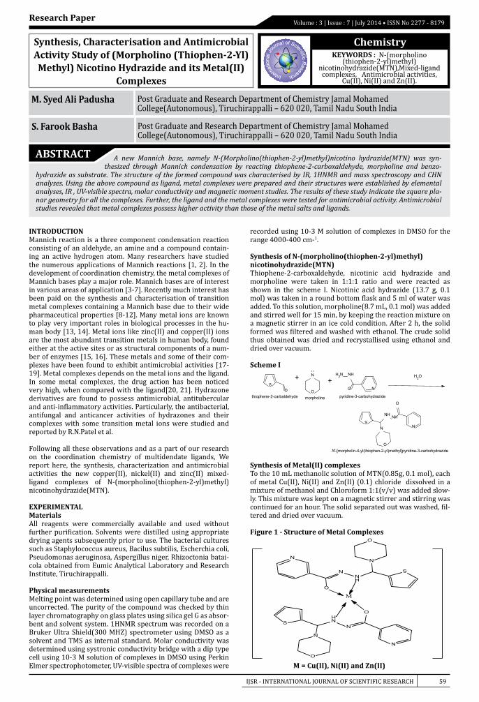
IJSR - INTERNATIONAL JOURNAL OF SCIENTIFIC RESEARCH 59
Volume : 3 | Issue : 7 | July 2014 • ISSN No 2277 - 8179Research Paper
Chemistry
M. Syed Ali Padusha Post Graduate and Research Department of Chemistry Jamal Mohamed College(Autonomous), Tiruchirappalli – 620 020, Tamil Nadu South India
S. Farook Basha Post Graduate and Research Department of Chemistry Jamal Mohamed College(Autonomous), Tiruchirappalli – 620 020, Tamil Nadu South India
ABSTRACT A new Mannich base, namely N-(Morpholino(thiophen-2-yl)methyl)nicotino hydrazide(MTN) was syn-thesized through Mannich condensation by reacting thiophene-2-carboxaldehyde, morpholine and benzo-
hydrazide as substrate. The structure of the formed compound was characterised by IR, 1HNMR and mass spectroscopy and CHN analyses. Using the above compound as ligand, metal complexes were prepared and their structures were established by elemental analyses, IR , UV-visible spectra, molar conductivity and magnetic moment studies. The results of these study indicate the square pla-nar geometry for all the complexes. Further, the ligand and the metal complexes were tested for antimicrobial activity. Antimicrobial studies revealed that metal complexes possess higher activity than those of the metal salts and ligands.
Synthesis, Characterisation and Antimicrobial Activity Study of (Morpholino (Thiophen-2-Yl)Methyl) Nicotino Hydrazide and its Metal(II)
Complexes
KEYWORDS : N-(morpholino (thiophen-2-yl)methyl)
nicotinohydrazide(MTN),Mixed-ligand complexes, Antimicrobial activities,
Cu(II), Ni(II) and Zn(II).
INTRODUCTIONMannich reaction is a three component condensation reaction consisting of an aldehyde, an amine and a compound contain-ing an active hydrogen atom. Many researchers have studied the numerous applications of Mannich reactions [1, 2]. In the development of coordination chemistry, the metal complexes of Mannich bases play a major role. Mannich bases are of interest in various areas of application [3-7]. Recently much interest has been paid on the synthesis and characterisation of transition metal complexes containing a Mannich base due to their wide pharmaceutical properties [8-12]. Many metal ions are known to play very important roles in biological processes in the hu-man body [13, 14]. Metal ions like zinc(II) and copper(II) ions are the most abundant transition metals in human body, found either at the active sites or as structural components of a num-ber of enzymes [15, 16]. These metals and some of their com-plexes have been found to exhibit antimicrobial activities [17-19]. Metal complexes depends on the metal ions and the ligand. In some metal complexes, the drug action has been noticed very high, when compared with the ligand[20, 21]. Hydrazone derivatives are found to possess antimicrobial, antitubercular and anti-inflammatory activities. Particularly, the antibacterial, antifungal and anticancer activities of hydrazones and their complexes with some transition metal ions were studied and reported by R.N.Patel et al.
Following all these observations and as a part of our research on the coordination chemistry of multidendate ligands, We report here, the synthesis, characterization and antimicrobial activities the new copper(II), nickel(II) and zinc(II) mixed-ligand complexes of N-(morpholino(thiophen-2-yl)methyl)nicotinohydrazide(MTN).
EXPERIMENTALMaterialsAll reagents were commercially available and used without further purification. Solvents were distilled using appropriate drying agents subsequently prior to use. The bacterial cultures such as Staphylococcus aureus, Bacilus subtilis, Escherchia coli, Pseudomonas aeruginosa, Aspergillus niger, Rhizoctonia batai-cola obtained from Eumic Analytical Laboratory and Research Institute, Tiruchirappalli.
Physical measurementsMelting point was determined using open capillary tube and are uncorrected. The purity of the compound was checked by thin layer chromatography on glass plates using silica gel G as absor-bent and solvent system. 1HNMR spectrum was recorded on a Bruker Ultra Shield(300 MHZ) spectrometer using DMSO as a solvent and TMS as internal standard. Molar conductivity was determined using systronic conductivity bridge with a dip type cell using 10-3 M solution of complexes in DMSO using Perkin Elmer spectrophotometer, UV-visible spectra of complexes were
recorded using 10-3 M solution of complexes in DMSO for the range 4000-400 cm-1.
Synthesis of N-(morpholino(thiophen-2-yl)methyl)nicotinohydrazide(MTN)Thiophene-2-carboxaldehyde, nicotinic acid hydrazide and morpholine were taken in 1:1:1 ratio and were reacted as shown in the scheme I. Nicotinic acid hydrazide (13.7 g, 0.1 mol) was taken in a round bottom flask and 5 ml of water was added. To this solution, morpholine(8.7 mL, 0.1 mol) was added and stirred well for 15 min, by keeping the reaction mixture on a magnetic stirrer in an ice cold condition. After 2 h, the solid formed was filtered and washed with ethanol. The crude solid thus obtained was dried and recrystallised using ethanol and dried over vacuum.
Scheme I
Synthesis of Metal(II) complexesTo the 10 mL methanolic solution of MTN(0.85g, 0.1 mol), each of metal Cu(II), Ni(II) and Zn(II) (0.1) chloride dissolved in a mixture of methanol and Chloroform 1:1(v/v) was added slow-ly. This mixture was kept on a magnetic stirrer and stirring was continued for an hour. The solid separated out was washed, fil-tered and dried over vacuum.
Figure 1 - Structure of Metal Complexes
M = Cu(II), Ni(II) and Zn(II)
NH2 NH
O N
pyridine-3-carbohydrazide
+NH
O
+S
Othiophene-2-carbaldehyde morpholine
OH2
SN
O
NH NH
O
N
N'-[morpholin-4-yl(thiophen-2-yl)methyl]pyridine-3-carbohydrazide
N
NH
N
N
O
S
O
N
HN
N
N
O
S
O
M
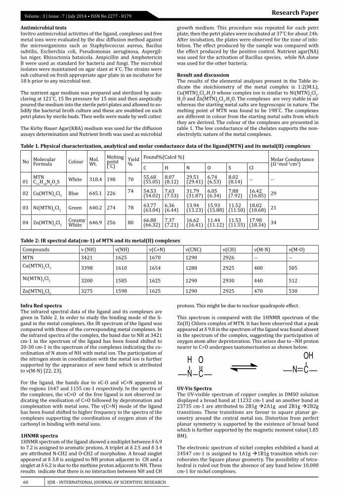
60 IJSR - INTERNATIONAL JOURNAL OF SCIENTIFIC RESEARCH
Volume : 3 | Issue : 7 | July 2014 • ISSN No 2277 - 8179 Research Paper
Antimicrobial testsInvitro antimicrobial activities of the ligand, complexes and free metal ions were evaluated by the disc diffusion method against the microorganisms such as Staphylococcus aureus, Bacilus subtilis, Escherchia coli, Pseudomonas aeruginosa, Aspergil-lus niger, Rhizoctonia bataicola. Ampicillin and Amphotericin B were used as standard for bacteria and fungi. The microbial isolates were maintained on agar slant at 4˚C. The strains were sub cultured on fresh appropriate agar plate in an incubator for 18 h prior to any microbial test.
The nutrient agar medium was prepared and sterilized by auto-claving at 121˚C, 15 lbs pressure for 15 min and then aseptically poured the medium into the sterile petri plates and allowed to so-lidify the bacterial broth culture and these are swabbed on each petri plates by sterile buds. Then wells were made by well cutter.
The Kirby Bauer Agar(KBA) medium was used for the diffusion assays determination and Nutrient broth was used as microbial
growth medium. This procedure was repeated for each petri plate, then the petri plates were incubated at 37˚C for about 24h. After incubation, the plates were observed for the zone of inhi-bition. The effect produced by the sample was compared with the effect produced by the positive control. Nutrient agar(NA) was used for the activation of Bacillus species, while NA alone was used for the other bacteria.
Result and discussionThe results of the elemental analyses present in the Table in-dicate the stoichiometry of the metal complex is 1:2(M:L), Cu(MTN)2Cl2.H2O whose complex ion is similar to Ni(MTN)2Cl2.H2O and Zn(MTN)2Cl2.H2O. The complexes are very stable in air whereas the starting metal salts are hygroscopic in nature. The melting point of MTN was found to be 198˚C. The complexes are different in colour from the starting metal salts from which they are derived. The colour of the complexes are presented in table 1. The low conductance of the chelates supports the non-electrolytic nature of the metal complexes.
Table 1. Physical characterization, analytical and molar conductance data of the ligand(MTN) and its metal(II) complexes
No Molecular Formula Colour Mol.
Wt.Melting point(˚C)
Yield%
Found%(Calcd %) Molar Conductance(Ω-1mol-1cm2)
C H N O S Cl
01MTNC15H18N4O2S White 318.4 198 70 55.68
(55.05)8.07 (8.12)
29.51 (29.41)
6.74 (6.53)
8.02 (8.14) -- --
02 Cu(MTN)2Cl2 Blue 645.1 226 74 54.53 (54.02)
7.63 (7.53)
31.79 (31.87)
6.05 (6.34)
7.88 (7.92)
16.42 (16.85) 29
03 Ni(MTN)2Cl2 Green 640.2 274 78 63.77 (63.04)
6.36 (6.44)
13.94 (13.23)
15.93 (15.88)
11.52 (11.50)
18.02 (18.68) 21
04 Zn(MTN)2Cl2Creamy White 646.9 256 80 66.88
(66.32)7.37 (7.21)
16.62 (16.41)
11.44 (11.12)
11.53 (11.55)
17.98 (18.34) 34
Table 2: IR spectral data(cm-1) of MTN and its metal(II) complexes
Compounds ν (NH) ν(NH) ν(C=N) ν(CNC) ν(CH) ν(M-N) ν(M-O)MTN 3421 1625 1670 1290 2926 -- --Cu(MTN)2Cl2 3398 1610 1654 1288 2925 480 505
Ni(MTN)2Cl2 3200 1585 1625 1290 2930 440 512
Zn(MTN)2Cl2 3275 1598 1625 1290 2925 470 530
Infra Red spectraThe infrared spectral data of the ligand and its complexes are given in Table 2. In order to study the binding mode of the li-gand in the metal complexes, the IR spectrum of the ligand was compared with those of the corresponding metal complexes. In the infrared spectra of the complex, the band due to NH at 3421 cm-1 in the spectrum of the ligand has been found shifted to 20-30 cm-1 in the spectrum of the complexes indicating the co-ordination of N atom of NH with metal ion. The participation of the nitrogen atom in coordination with the metal ion is further supported by the appearance of new band which is attributed to ν(M-N) [22, 23].
For the ligand, the bands due to νC-O and νC=N appeared in the regions 1647 and 1155 cm-1 respectively. In the spectra of the complexes, the νC=O of the free ligand is not observed in-dicating the enolisation of C=O followed by deprotonation and complexation with metal ions. The ν(C=N) mode of the ligand has been found shifted to higher frequency in the spectra of the complexes supporting the coordination of oxygen atom of the carbonyl in binding with metal ions.
1HNMR spectra1HNMR spectrum of the ligand showed a multiplet between δ 6.9 to 7.2 is assigned to aromatic protons. A triplet at δ 2.5 and δ 3.4 are attributed N-CH2 and O-CH2 of morpholine. A broad singlet appeared at δ 3.8 is assigned to NH proton adjacent to CH and a singlet at δ 6.2 is due to the methine proton adjacent to NH. These results indicate that there is no interaction between NH and CH
protons. This might be due to nuclear quadrapole effect.
This spectrum is compared with the 1HNMR spectrum of the Zn(II) Chloro complex of MTN. It has been observed that a peak appeared at δ 9.8 in the spectrum of the ligand was found absent in the spectrum of the complex, suggesting the participation of oxygen atom after deprotoration. This arises due to –NH proton nearer to C=O undergoes tautomerisation as shown below.
UV-Vis SpectraThe UV-visible spectrum of copper complex in DMSO solution displayed a broad band at 11232 cm-1 and an another band at 23735 cm-1 are attributed to 2B1g 2A1g and 2B1g 2B2g transitions. These transitions are favour to square planar ge-ometry around the central metal ion. Distortion from perfect planar symmetry is supported by the existence of broad band which is further supported by the magnetic moment value(1.85 BM).
The electronic spectrum of nickel complex exhibited a band at 24547 cm-1 is assigned to 1A1g 1B1g transition which cor-roborates the Square planar geometry. The possibility of tetra-hedral is ruled out from the absence of any band below 10,000 cm-1 for nickel complexes.
NH
CO
N COH
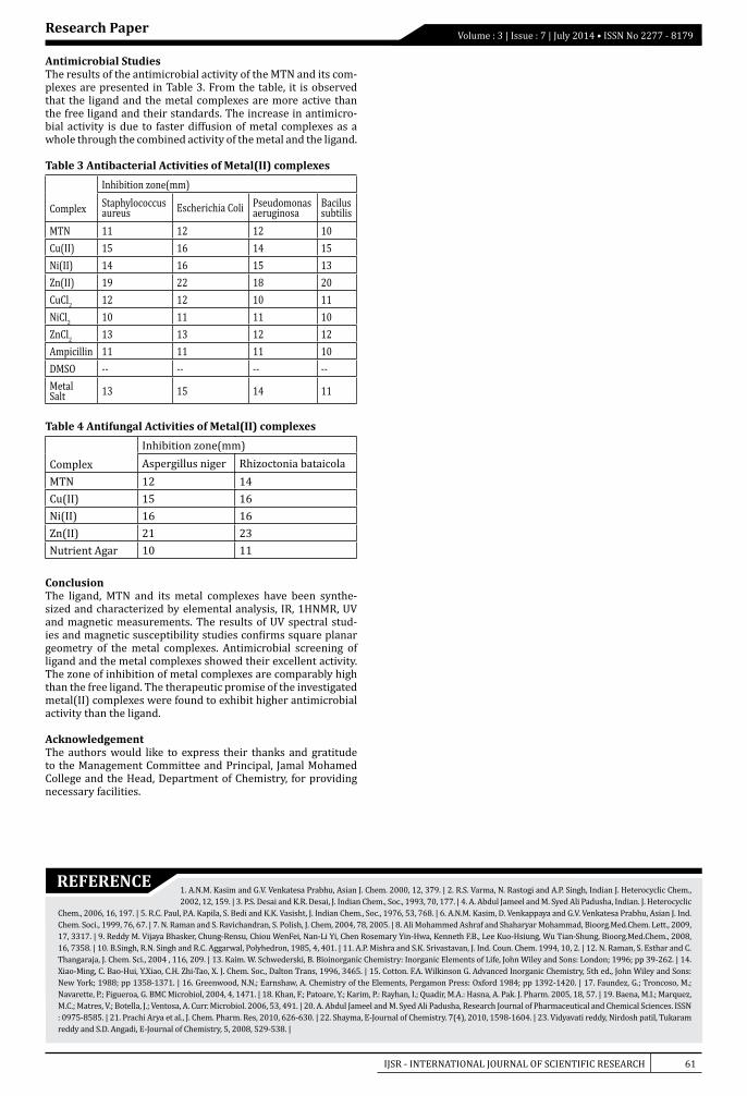
IJSR - INTERNATIONAL JOURNAL OF SCIENTIFIC RESEARCH 61
Volume : 3 | Issue : 7 | July 2014 • ISSN No 2277 - 8179Research Paper
REFERENCE1. A.N.M. Kasim and G.V. Venkatesa Prabhu, Asian J. Chem. 2000, 12, 379. | 2. R.S. Varma, N. Rastogi and A.P. Singh, Indian J. Heterocyclic Chem., 2002, 12, 159. | 3. P.S. Desai and K.R. Desai, J. Indian Chem., Soc., 1993, 70, 177. | 4. A. Abdul Jameel and M. Syed Ali Padusha, Indian. J. Heterocyclic
Chem., 2006, 16, 197. | 5. R.C. Paul, P.A. Kapila, S. Bedi and K.K. Vasisht, J. Indian Chem., Soc., 1976, 53, 768. | 6. A.N.M. Kasim, D. Venkappaya and G.V. Venkatesa Prabhu, Asian J. Ind. Chem. Soci., 1999, 76, 67. | 7. N. Raman and S. Ravichandran, S. Polish, J. Chem, 2004, 78, 2005. | 8. Ali Mohammed Ashraf and Shaharyar Mohammad, Bioorg.Med.Chem. Lett., 2009, 17, 3317. | 9. Reddy M. Vijaya Bhasker, Chung-Rensu, Chiou WenFei, Nan-Li Yi, Chen Rosemary Yin-Hwa, Kenneth F.B., Lee Kuo-Hsiung, Wu Tian-Shung, Bioorg.Med.Chem., 2008, 16, 7358. | 10. B.Singh, R.N. Singh and R.C. Aggarwal, Polyhedron, 1985, 4, 401. | 11. A.P. Mishra and S.K. Srivastavan, J. Ind. Coun. Chem. 1994, 10, 2. | 12. N. Raman, S. Esthar and C. Thangaraja, J. Chem. Sci., 2004 , 116, 209. | 13. Kaim. W. Schwederski, B. Bioinorganic Chemistry: Inorganic Elements of Life, John Wiley and Sons: London; 1996; pp 39-262. | 14. Xiao-Ming, C. Bao-Hui, Y.Xiao, C.H. Zhi-Tao, X. J. Chem. Soc., Dalton Trans, 1996, 3465. | 15. Cotton. F.A. Wilkinson G. Advanced Inorganic Chemistry, 5th ed., John Wiley and Sons: New York; 1988; pp 1358-1371. | 16. Greenwood, N.N.; Earnshaw, A. Chemistry of the Elements, Pergamon Press: Oxford 1984; pp 1392-1420. | 17. Faundez, G.; Troncoso, M.; Navarette, P.; Figueroa, G. BMC Microbiol, 2004, 4, 1471. | 18. Khan, F.; Patoare, Y.; Karim, P.: Rayhan, I.; Quadir, M.A.: Hasna, A. Pak. J. Pharm. 2005, 18, 57. | 19. Baena, M.I.; Marquez, M.C.; Matres, V.; Botella, J.; Ventosa, A. Curr. Microbiol. 2006, 53, 491. | 20. A. Abdul Jameel and M. Syed Ali Padusha, Research Journal of Pharmaceutical and Chemical Sciences. ISSN : 0975-8585. | 21. Prachi Arya et al., J. Chem. Pharm. Res, 2010, 626-630. | 22. Shayma, E-Journal of Chemistry. 7(4), 2010, 1598-1604. | 23. Vidyavati reddy, Nirdosh patil, Tukaram reddy and S.D. Angadi, E-Journal of Chemistry, 5, 2008, 529-538. |
Antimicrobial StudiesThe results of the antimicrobial activity of the MTN and its com-plexes are presented in Table 3. From the table, it is observed that the ligand and the metal complexes are more active than the free ligand and their standards. The increase in antimicro-bial activity is due to faster diffusion of metal complexes as a whole through the combined activity of the metal and the ligand.
Table 3 Antibacterial Activities of Metal(II) complexes
Complex
Inhibition zone(mm)Staphylococcus aureus Escherichia Coli Pseudomonas
aeruginosaBacilus subtilis
MTN 11 12 12 10Cu(II) 15 16 14 15Ni(II) 14 16 15 13Zn(II) 19 22 18 20CuCl2 12 12 10 11NiCl2 10 11 11 10ZnCl2 13 13 12 12Ampicillin 11 11 11 10DMSO -- -- -- --Metal Salt 13 15 14 11
Table 4 Antifungal Activities of Metal(II) complexes
ComplexInhibition zone(mm)Aspergillus niger Rhizoctonia bataicola
MTN 12 14Cu(II) 15 16Ni(II) 16 16Zn(II) 21 23Nutrient Agar 10 11
ConclusionThe ligand, MTN and its metal complexes have been synthe-sized and characterized by elemental analysis, IR, 1HNMR, UV and magnetic measurements. The results of UV spectral stud-ies and magnetic susceptibility studies confirms square planar geometry of the metal complexes. Antimicrobial screening of ligand and the metal complexes showed their excellent activity. The zone of inhibition of metal complexes are comparably high than the free ligand. The therapeutic promise of the investigated metal(II) complexes were found to exhibit higher antimicrobial activity than the ligand.
AcknowledgementThe authors would like to express their thanks and gratitude to the Management Committee and Principal, Jamal Mohamed College and the Head, Department of Chemistry, for providing necessary facilities.
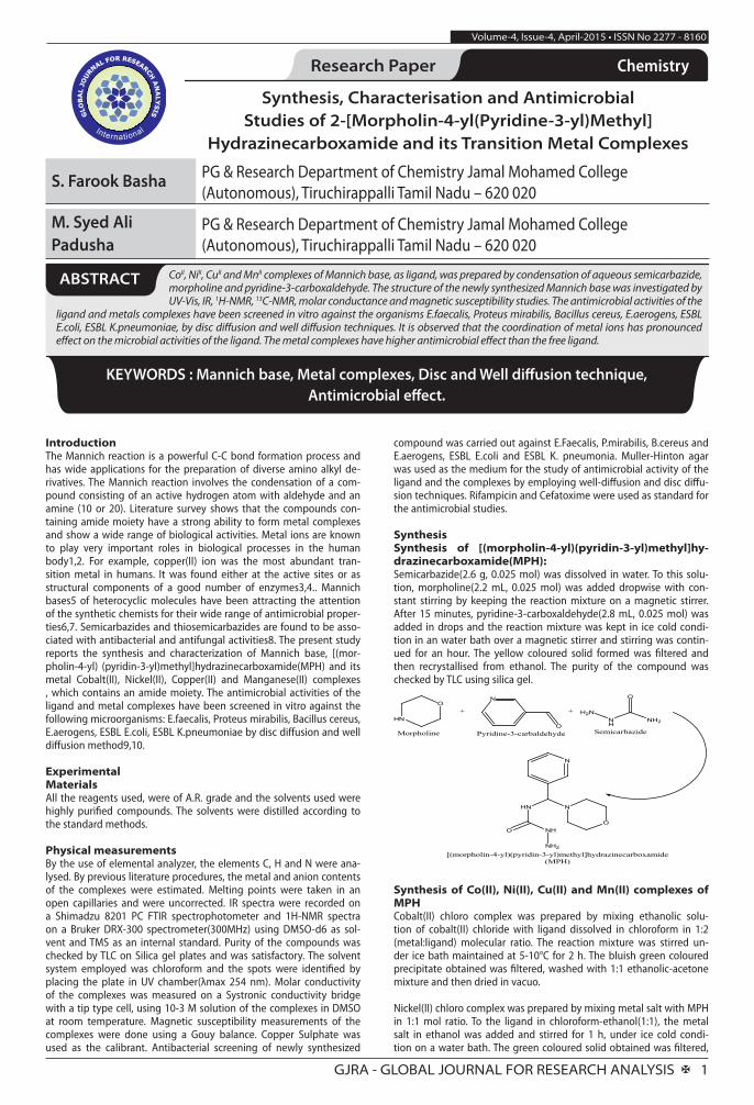
GJRA - GLOBAL JOURNAL FOR RESEARCH ANALYSIS X 1
Volume-4, Issue-4, April-2015 • ISSN No 2277 - 8160
Research Paper Commerce Chemistry
Synthesis, Characterisation and Antimicrobial Studies of 2-[Morpholin-4-yl(Pyridine-3-yl)Methyl]
Hydrazinecarboxamide and its Transition Metal Complexes
S. Farook Basha PG & Research Department of Chemistry Jamal Mohamed College (Autonomous), Tiruchirappalli Tamil Nadu – 620 020
M. Syed Ali Padusha
PG & Research Department of Chemistry Jamal Mohamed College (Autonomous), Tiruchirappalli Tamil Nadu – 620 020
CoII, NiII, CuII and MnII complexes of Mannich base, as ligand, was prepared by condensation of aqueous semicarbazide, morpholine and pyridine-3-carboxaldehyde. The structure of the newly synthesized Mannich base was investigated by UV-Vis, IR, 1H-NMR, 13C-NMR, molar conductance and magnetic susceptibility studies. The antimicrobial activities of the
ligand and metals complexes have been screened in vitro against the organisms E.faecalis, Proteus mirabilis, Bacillus cereus, E.aerogens, ESBL E.coli, ESBL K.pneumoniae, by disc diffusion and well diffusion techniques. It is observed that the coordination of metal ions has pronounced effect on the microbial activities of the ligand. The metal complexes have higher antimicrobial effect than the free ligand.
ABSTRACT
KEYWORDS : Mannich base, Metal complexes, Disc and Well diffusion technique, Antimicrobial effect.
IntroductionThe Mannich reaction is a powerful C-C bond formation process and has wide applications for the preparation of diverse amino alkyl de-rivatives. The Mannich reaction involves the condensation of a com-pound consisting of an active hydrogen atom with aldehyde and an amine (10 or 20). Literature survey shows that the compounds con-taining amide moiety have a strong ability to form metal complexes and show a wide range of biological activities. Metal ions are known to play very important roles in biological processes in the human body1,2. For example, copper(II) ion was the most abundant tran-sition metal in humans. It was found either at the active sites or as structural components of a good number of enzymes3,4.. Mannich bases5 of heterocyclic molecules have been attracting the attention of the synthetic chemists for their wide range of antimicrobial proper-ties6,7. Semicarbazides and thiosemicarbazides are found to be asso-ciated with antibacterial and antifungal activities8. The present study reports the synthesis and characterization of Mannich base, [(mor-pholin-4-yl) (pyridin-3-yl)methyl]hydrazinecarboxamide(MPH) and its metal Cobalt(II), Nickel(II), Copper(II) and Manganese(II) complexes , which contains an amide moiety. The antimicrobial activities of the ligand and metal complexes have been screened in vitro against the following microorganisms: E.faecalis, Proteus mirabilis, Bacillus cereus, E.aerogens, ESBL E.coli, ESBL K.pneumoniae by disc diffusion and well diffusion method9,10.
ExperimentalMaterialsAll the reagents used, were of A.R. grade and the solvents used were highly purified compounds. The solvents were distilled according to the standard methods.
Physical measurementsBy the use of elemental analyzer, the elements C, H and N were ana-lysed. By previous literature procedures, the metal and anion contents of the complexes were estimated. Melting points were taken in an open capillaries and were uncorrected. IR spectra were recorded on a Shimadzu 8201 PC FTIR spectrophotometer and 1H-NMR spectra on a Bruker DRX-300 spectrometer(300MHz) using DMSO-d6 as sol-vent and TMS as an internal standard. Purity of the compounds was checked by TLC on Silica gel plates and was satisfactory. The solvent system employed was chloroform and the spots were identified by placing the plate in UV chamber(λmax 254 nm). Molar conductivity of the complexes was measured on a Systronic conductivity bridge with a tip type cell, using 10-3 M solution of the complexes in DMSO at room temperature. Magnetic susceptibility measurements of the complexes were done using a Gouy balance. Copper Sulphate was used as the calibrant. Antibacterial screening of newly synthesized
compound was carried out against E.Faecalis, P.mirabilis, B.cereus and E.aerogens, ESBL E.coli and ESBL K. pneumonia. Muller-Hinton agar was used as the medium for the study of antimicrobial activity of the ligand and the complexes by employing well-diffusion and disc diffu-sion techniques. Rifampicin and Cefatoxime were used as standard for the antimicrobial studies.
SynthesisSynthesis of [(morpholin-4-yl)(pyridin-3-yl)methyl]hy-drazinecarboxamide(MPH):Semicarbazide(2.6 g, 0.025 mol) was dissolved in water. To this solu-tion, morpholine(2.2 mL, 0.025 mol) was added dropwise with con-stant stirring by keeping the reaction mixture on a magnetic stirrer. After 15 minutes, pyridine-3-carboxaldehyde(2.8 mL, 0.025 mol) was added in drops and the reaction mixture was kept in ice cold condi-tion in an water bath over a magnetic stirrer and stirring was contin-ued for an hour. The yellow coloured solid formed was filtered and then recrystallised from ethanol. The purity of the compound was checked by TLC using silica gel.
HN
O
Morpholine
N
OPyridine-3-carbaldehyde
H2NNH
O
NH2
Semicarbazide
+ +
NH
NH2
O
HN
N
N
O
[(morpholin-4-yl)(pyridin-3-yl)methyl]hydrazinecarboxamide (MPH)
Synthesis of Co(II), Ni(II), Cu(II) and Mn(II) complexes of MPHCobalt(II) chloro complex was prepared by mixing ethanolic solu-tion of cobalt(II) chloride with ligand dissolved in chloroform in 1:2 (metal:ligand) molecular ratio. The reaction mixture was stirred un-der ice bath maintained at 5-10°C for 2 h. The bluish green coloured precipitate obtained was filtered, washed with 1:1 ethanolic-acetone mixture and then dried in vacuo.
Nickel(II) chloro complex was prepared by mixing metal salt with MPH in 1:1 mol ratio. To the ligand in chloroform-ethanol(1:1), the metal salt in ethanol was added and stirred for 1 h, under ice cold condi-tion on a water bath. The green coloured solid obtained was filtered,
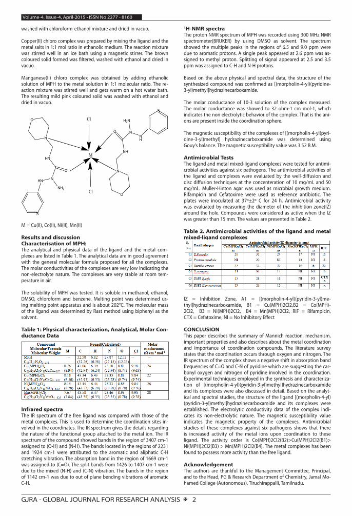
GJRA - GLOBAL JOURNAL FOR RESEARCH ANALYSIS X 2
Volume-4, Issue-4, April-2015 • ISSN No 2277 - 8160
washed with chloroform-ethanol mixture and dried in vacuo.
Copper(II) chloro complex was prepared by mixing the ligand and the metal salts in 1:1 mol ratio in ethanolic medium. The reaction mixture was stirred well in an ice bath using a magnetic stirrer. The brown coloured solid formed was filtered, washed with ethanol and dried in vacuo.
Manganese(II) chloro complex was obtained by adding ethanolic solution of MPH to the metal solution in 1:1 molecular ratio. The re-action mixture was stirred well and gets warm on a hot water bath. The resulting mild pink coloured solid was washed with ethanol and dried in vacuo.
N
H2N
O
NH
N
O
NH
N
M
Cl
Cl
N
NH2
O
HN
N
O
HN
N
M = Cu(II), Co(II), Ni(II), Mn(II)
Results and discussionCharacterisation of MPH:The analytical and physical data of the ligand and the metal com-plexes are listed in Table 1. The analytical data are in good agreement with the general molecular formula proposed for all the complexes. The molar conductivities of the complexes are very low indicating the non-electrolyte nature. The complexes are very stable at room tem-perature in air.
The solubility of MPH was tested. It is soluble in methanol, ethanol, DMSO, chloroform and benzene. Melting point was determined us-ing melting point apparatus and is about 202°C. The molecular mass of the ligand was determined by Rast method using biphenyl as the solvent.
Table 1: Physical characterization, Analytical, Molar Con-ductance Data
Infrared spectraThe IR spectrum of the free ligand was compared with those of the metal complexes. This is used to determine the coordination sites in-volved in the coordinates. The IR spectrum gives the details regarding the nature of the functional group attached to the metal ion. The IR spectrum of the compound showed bands in the region of 3407 cm-1 assigned to (O-H) and (N-H). The bands located in the regions of 2231 and 1924 cm-1 were attributed to the aromatic and aliphatic C-H stretching vibration. The absorption band in the region of 1669 cm-1 was assigned to (C=O). The split bands from 1426 to 1407 cm-1 were due to the mixed (N-H) and (C-N) vibration. The bands in the region of 1142 cm-1 was due to out of plane bending vibrations of aromatic C-H.
1H-NMR spectraThe proton NMR spectrum of MPH was recorded using 300 MHz NMR spectrometer(BRUKER) by using DMSO as solvent. The spectrum showed the multiple peaks in the regions of 6.5 and 9.0 ppm were due to aromatic protons. A single peak appeared at 2.6 ppm was as-signed to methyl proton. Splitting of signal appeared at 2.5 and 3.5 ppm was assigned to C-H and N-H protons.
Based on the above physical and spectral data, the structure of the synthesized compound was confirmed as [(morpholin-4-yl)(pyridine-3-yl)methyl]hydrazinecarboxamide.
The molar conductance of 10-3 solution of the complex measured. The molar conductance was showed to 32 ohm-1 cm mol-1, which indicates the non electrolytic behavior of the complex. That is the ani-ons are present inside the coordination sphere.
The magnetic susceptibility of the complexes of [(morpholin-4-yl(pyri-dine-3-yl)methyl] hydrazinecarboxamide was determined using Gouy’s balance. The magnetic susceptibility value was 3.52 B.M.
Antimicrobial TestsThe ligand and metal mixed-ligand complexes were tested for antimi-crobial activities against six pathogens. The antimicrobial activities of the ligand and complexes were evaluated by the well-diffusion and disc diffusion techniques at the concenteration of 10 mg/mL and 50 mg/mL. Muller-Hinton agar was used as microbial growth medium. Rifampicin and Cefatoxime were used as reference antibiotic. The plates were inoculated at 37º±2º C for 24 h. Antimicrobial activity was evaluated by measuring the diameter of the inhibition zone(IZ) around the hole. Compounds were considered as active when the IZ was greater than 15 mm. The values are presented in Table 2.
Table 2. Antimicrobial activities of the ligand and metal mixed-ligand complexes
IZ = Inhibition Zone, A1 = [(morpholin-4-yl)(pyridin-3-yl)me-thyl]hydrazinecarboxamide, B1 = Cu(MPH)2Cl2,B2 = Co(MPH)-2Cl2, B3 = Ni(MPH)2Cl2, B4 = Mn(MPH)2Cl2, RIF = Rifampicin, CTX = Cefatoxime, NI = No Inhibitory Effect
CONCLUSIONThis paper describes the summary of Mannich reaction, mechanism, important properties and also describes about the metal coordination and importance of coordination compounds. The literature survey states that the coordination occurs through oxygen and nitrogen. The IR spectrum of the complex shows a negative shift in absorption band frequencies of C=O and C-N of pyridine which are suggesting the car-bonyl oxygen and nitrogen of pyridine involved in the coordination. Experimental techniques employed in the synthesis and characteriza-tion of [(morpholin-4-yl)(pyridin-3-yl)methyl]hydrazinecarboxamide and its complexes were also discussed in detail. Based on the analyt-ical and spectral studies, the structure of the ligand [(morpholin-4-yl)(pyridin-3-yl)methyl]hydrazinecarboxamide and its complexes were established. The electrolytic conductivity data of the complex indi-cates its non-electrolytic nature. The magnetic susceptibility value indicates the magnetic property of the complexes. Antimicrobial studies of these complexes against six pathogens shows that there is increased activity of the metal ions upon coordination to these ligand. The activity order is Co(MPH)2Cl2(B2)>Cu(MPH)2Cl2(B1)> Ni(MPH)2Cl2(B3) > Mn(MPH)2Cl2(B4). The metal complexes has been found to possess more activity than the free ligand.
AcknowledgementThe authors are thankful to the Management Committee, Principal, and to the Head, PG & Research Department of Chemistry, Jamal Mo-hamed College (Autonomous), Tiruchirappalli, Tamilnadu.
GJRA - GLOBAL JOURNAL FOR RESEARCH ANALYSIS X 3
Volume-4, Issue-4, April-2015 • ISSN No 2277 - 8160
REFERENCES 1. Kaim. W. Schwederski, B. Bioinorganic Chemistry: Inorganic Elements of Life. John Wiley and Sons: London, 1996; pp 39-262. | 2. Xiao-Ming, C.: Bao-Hui, Y.: Xiao, C.H.: Zhi-Tao, X.J. Chem Soc, Dalton Trans 1996, 3465. | 3. Cotton. F.A.: Wilkinson G. Advanced Inorganic Chemistry, 5th ed. John Wiley and Sons, New York, 1988, pp 1358-1371. | 4. Greenword, N.N.: Earnshaw, A.: Chemistry of the Elements, Pergamon Press, Oxford. 1984, pp
1392-1420. | 5. F. Aydogan, Z. Turgut, N.Ocal, Turk. J. Chem, 2002, 26, 159. | 6. C.R. Katica, D. Vesna, K. Vlado, G.M. Dora, B. Aleksandra, Molecules, 2001,6, 815-824. | 7. R. Valarmathi, S. Akilandeswari, V.N.I. Latha, G. Umadevi, Der ChemicaSinica, 2011, 2(5), 64-68. | 8. R.M. Silverstein, G.C. Bassier, T.C. Morrill, Spectrometric Identification of organic compounds 4th Ed. John Wiley and Sons, 1981. | 9. NCCLS, Performance Standards for Antimicrobial Disk Suspectibility Tests. Approved Standard NCCLS Publication M2-A5, Villanova, PA, U.S.A., 1993. | 10. C. H. Collins, P.M. Lyre and J. M. Grange, Microbiological Methods. 6th Ed. Butterworths, London, 1989. |
Related Documents