Original article Synthesis, antimicrobial and cytotoxic activities, and structureeactivity relationships of gypsogenin derivatives against human cancer cells Safiye Emirda g- € Oztürk a, * , Tamer Karayıldırım a , Aysun Çapcı-Karag € oz b , € Ozgen Alankus ¸-Çalıs ¸ kan a , Ali € Ozmen c , Esin Poyrazo glu-Çoban c a Chemistry Department, Faculty of Science, Ege University, Bornova, Izmir 35100, Turkey b Institute of Organic Chemistry I, University of Erlangen-Nuremberg, Henkestrasse 42, 91054 Erlangen, Germany c Biology Department, Adnan Menderes University, Aydin 09010, Turkey article info Article history: Received 5 February 2014 Received in revised form 15 May 2014 Accepted 31 May 2014 Available online 7 June 2014 Keywords: Gypsogenin Saponin Gypsophila Cytotoxic Apoptosis abstract A series of gypsogenin (1) derivatives (1aei) was synthesized in good yields, and the derivatives’ structures were established using UV, IR, 1 H NMR, 13 C NMR, and LCMS spectroscopic data. Among the tested compounds, 1a, 1b, 1d, 1e, and gypsogenin (1) showed antimicrobial activities against Bacillus subtilis and Bacillus thrungiensis, with inhibition zones of 10e14 mm. In addition, com- pounds 1b, 1d, and 1e showed antimicrobial activities against Bacillus cereus, with inhibition zones of 9e14 mm. Using six human cancer cell lines in vitro, the cytotoxic activities of all tested compounds were determined by calculating the IC 50 values. Doxorubicin and paclitaxel were used as controls. Among the tested compounds, 1a, 1c, and 1d had inhibitory effects with IC 50 values of 3.9 mM (HL-60 cells), 5.15 mM (MCF-7 cells), and 5.978 mM (HL-60), respectively. To determine the type of cell death, Hoechst 33258 (HO) and propidium iodide (PI) double staining was used. Especially, gypsogenin (1) and compound 1a triggered the apoptotic mechanism at a concentration of 20 mM. Thus, gypsogenin (1) and compounds 1a, 1c, and 1d possess varying degrees of biological activities and can be considered as potential antitumor agents. © 2014 Elsevier Masson SAS. All rights reserved. 1. Introduction Saponins, which are detected in a number of plant families, are glycosides with a polycyclic aglycone and a sugar moiety. Sugars can attach to aglycone, which is also called sapogenin, at one or two different positions; thus, they are named monodesmosidic and bidesmosidic saponins, respectively [1e3]. These secondary me- tabolites have hydrophilic and hydrophobic sides; hence, they have surface activity, and saponin-containing plants are used as soap. Saponins have various structure-dependent biological activities such as glucosidase inhibiting [4], antiviral [5,6], anti-inflammatory [7], spermicidal [8], hypocholesterolemic [9], antitumor [10,11], anticarcinogenic [12], and antioxidant activities [13]. Moreover, saponins have been evaluated against cancer cells for anticancer activity [14,15]. Triterpene saponins can be found in many plant species [16e19], and several recent studies have reported on saponins produced from Gypsophila species [20e22]. Some saponins from Gypsophila have shown a variety of bio- logical activities including anticarcinogenic [23], immunostimula- tory [24], and cytotoxic activities [25]. Gypsophila accumulate gypsogenin aglycone with sugar chains, which has been attributed to various biological properties. For example, these compounds have exhibited inhibitory activity [26] and have shown significant growth inhibition in in vitro cultures [27]. In addition, some of them have shown very high activity against different human cancer cell lines [28,29]. Thus, there is strong evidence that gypsogenin has anticancer activity. Gypsogenin aglycone is found at high concentrations in Gyp- sophila [30]; therefore, it can be obtained with ease [31]. In this study, nine new gypsogenin derivatives (1aei) were synthesized from gypsogenin aglycone (1). In addition, they were evaluated for their antibacterial and antifungal activities as well as cytotoxic activities against six different human cancer cell cultures. * Corresponding author. E-mail addresses: safi[email protected], safi[email protected] (S. Emirda g- € Oztürk). Contents lists available at ScienceDirect European Journal of Medicinal Chemistry journal homepage: http://www.elsevier.com/locate/ejmech http://dx.doi.org/10.1016/j.ejmech.2014.05.084 0223-5234/© 2014 Elsevier Masson SAS. All rights reserved. European Journal of Medicinal Chemistry 82 (2014) 565e573

Welcome message from author
This document is posted to help you gain knowledge. Please leave a comment to let me know what you think about it! Share it to your friends and learn new things together.
Transcript
lable at ScienceDirect
European Journal of Medicinal Chemistry 82 (2014) 565e573
Contents lists avai
European Journal of Medicinal Chemistry
journal homepage: http: / /www.elsevier .com/locate/ejmech
Original article
Synthesis, antimicrobial and cytotoxic activities, andstructureeactivity relationships of gypsogenin derivatives againsthuman cancer cells
Safiye Emirda�g-€Oztürk a, *, Tamer Karayıldırım a, Aysun Çapcı-Karag€oz b,€Ozgen Alankus-Çalıskan a, Ali €Ozmen c, Esin Poyrazo�glu-Çoban c
a Chemistry Department, Faculty of Science, Ege University, Bornova, Izmir 35100, Turkeyb Institute of Organic Chemistry I, University of Erlangen-Nuremberg, Henkestrasse 42, 91054 Erlangen, Germanyc Biology Department, Adnan Menderes University, Aydin 09010, Turkey
a r t i c l e i n f o
Article history:Received 5 February 2014Received in revised form15 May 2014Accepted 31 May 2014Available online 7 June 2014
Keywords:GypsogeninSaponinGypsophilaCytotoxicApoptosis
* Corresponding author.E-mail addresses: [email protected]
(S. Emirda�g-€Oztürk).
http://dx.doi.org/10.1016/j.ejmech.2014.05.0840223-5234/© 2014 Elsevier Masson SAS. All rights re
a b s t r a c t
A series of gypsogenin (1) derivatives (1aei) was synthesized in good yields, and the derivatives’structures were established using UV, IR, 1H NMR, 13C NMR, and LCMS spectroscopic data.
Among the tested compounds, 1a, 1b, 1d, 1e, and gypsogenin (1) showed antimicrobial activitiesagainst Bacillus subtilis and Bacillus thrungiensis, with inhibition zones of 10e14 mm. In addition, com-pounds 1b, 1d, and 1e showed antimicrobial activities against Bacillus cereus, with inhibition zones of9e14 mm. Using six human cancer cell lines in vitro, the cytotoxic activities of all tested compounds weredetermined by calculating the IC50 values. Doxorubicin and paclitaxel were used as controls. Among thetested compounds, 1a, 1c, and 1d had inhibitory effects with IC50 values of 3.9 mM (HL-60 cells), 5.15 mM(MCF-7 cells), and 5.978 mM (HL-60), respectively. To determine the type of cell death, Hoechst 33258(HO) and propidium iodide (PI) double staining was used. Especially, gypsogenin (1) and compound 1atriggered the apoptotic mechanism at a concentration of 20 mM. Thus, gypsogenin (1) and compounds 1a,1c, and 1d possess varying degrees of biological activities and can be considered as potential antitumoragents.
© 2014 Elsevier Masson SAS. All rights reserved.
1. Introduction
Saponins, which are detected in a number of plant families, areglycosides with a polycyclic aglycone and a sugar moiety. Sugarscan attach to aglycone, which is also called sapogenin, at one or twodifferent positions; thus, they are named monodesmosidic andbidesmosidic saponins, respectively [1e3]. These secondary me-tabolites have hydrophilic and hydrophobic sides; hence, they havesurface activity, and saponin-containing plants are used as soap.
Saponins have various structure-dependent biological activitiessuch as glucosidase inhibiting [4], antiviral [5,6], anti-inflammatory[7], spermicidal [8], hypocholesterolemic [9], antitumor [10,11],anticarcinogenic [12], and antioxidant activities [13]. Moreover,saponins have been evaluated against cancer cells for anticanceractivity [14,15].
served.
Triterpene saponins can be found inmany plant species [16e19],and several recent studies have reported on saponins producedfrom Gypsophila species [20e22].
Some saponins from Gypsophila have shown a variety of bio-logical activities including anticarcinogenic [23], immunostimula-tory [24], and cytotoxic activities [25].
Gypsophila accumulate gypsogenin aglycone with sugar chains,which has been attributed to various biological properties. Forexample, these compounds have exhibited inhibitory activity [26]and have shown significant growth inhibition in in vitro cultures[27]. In addition, some of them have shown very high activityagainst different human cancer cell lines [28,29].
Thus, there is strong evidence that gypsogenin has anticanceractivity.
Gypsogenin aglycone is found at high concentrations in Gyp-sophila [30]; therefore, it can be obtained with ease [31]. In thisstudy, nine new gypsogenin derivatives (1aei) were synthesizedfrom gypsogenin aglycone (1). In addition, they were evaluated fortheir antibacterial and antifungal activities as well as cytotoxicactivities against six different human cancer cell cultures.
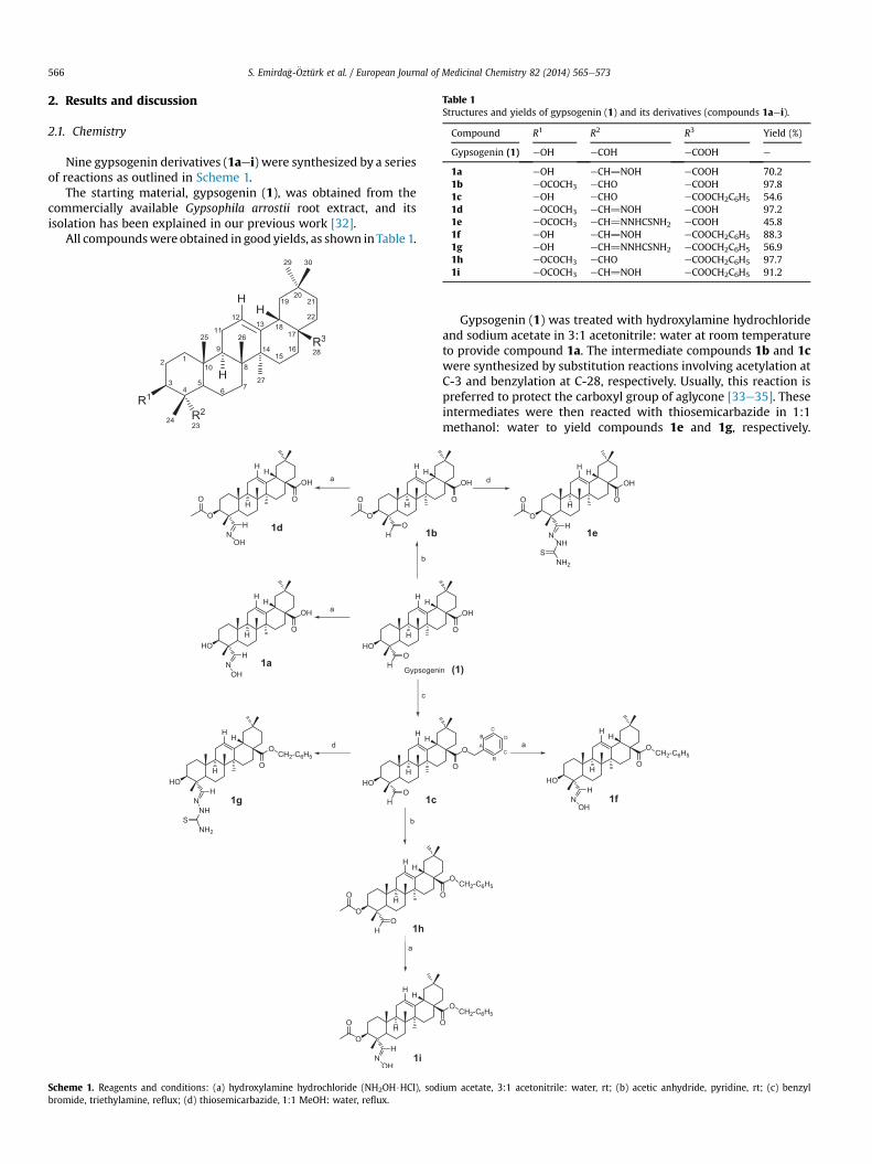
Table 1Structures and yields of gypsogenin (1) and its derivatives (compounds 1aei).
Compound R1 R2 R3 Yield (%)
Gypsogenin (1) eOH eCOH eCOOH e
1a eOH eCH]NOH eCOOH 70.21b eOCOCH3 eCHO eCOOH 97.81c eOH eCHO eCOOCH2C6H5 54.61d eOCOCH3 eCH]NOH eCOOH 97.21e eOCOCH3 eCH]NNHCSNH2 eCOOH 45.81f eOH eCH]NOH eCOOCH2C6H5 88.3
S. Emirda�g-€Oztürk et al. / European Journal of Medicinal Chemistry 82 (2014) 565e573566
2. Results and discussion
2.1. Chemistry
Nine gypsogenin derivatives (1aei) were synthesized by a seriesof reactions as outlined in Scheme 1.
The starting material, gypsogenin (1), was obtained from thecommercially available Gypsophila arrostii root extract, and itsisolation has been explained in our previous work [32].
All compoundswere obtained in good yields, as shown inTable 1.
Scheme 1. Reagents and conditions: (a) hydroxylamine hydrochloride (NH2OH$HCl), sodbromide, triethylamine, reflux; (d) thiosemicarbazide, 1:1 MeOH: water, reflux.
1g eOH eCH]NNHCSNH2 eCOOCH2C6H5 56.91h eOCOCH3 eCHO eCOOCH2C6H5 97.71i eOCOCH3 eCH]NOH eCOOCH2C6H5 91.2
Gypsogenin (1) was treated with hydroxylamine hydrochlorideand sodium acetate in 3:1 acetonitrile: water at room temperatureto provide compound 1a. The intermediate compounds 1b and 1cwere synthesized by substitution reactions involving acetylation atC-3 and benzylation at C-28, respectively. Usually, this reaction ispreferred to protect the carboxyl group of aglycone [33e35]. Theseintermediates were then reacted with thiosemicarbazide in 1:1methanol: water to yield compounds 1e and 1g, respectively.
ium acetate, 3:1 acetonitrile: water, rt; (b) acetic anhydride, pyridine, rt; (c) benzyl
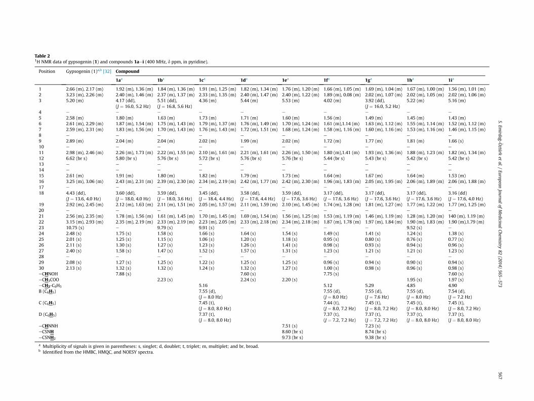
Table 21H NMR data of gypsogenin (1) and compounds 1aei (400 MHz, d ppm, in pyridine).
Position Gypsogenin (1)a,b [32] Compound
1aa 1ba 1ca 1da 1ea 1fa 1ga 1ha 1ia
1 2.66 (m), 2.17 (m) 1.92 (m), 1.36 (m) 1.84 (m), 1.36 (m) 1.91 (m), 1.25 (m) 1.82 (m), 1.34 (m) 1.76 (m), 1.20 (m) 1.66 (m), 1.05 (m) 1.69 (m), 1.04 (m) 1.67 (m), 1.00 (m) 1.56 (m), 1.01 (m)2 3.23 (m), 2.26 (m) 2.40 (m), 1.46 (m) 2.37 (m), 1.37 (m) 2.33 (m), 1.35 (m) 2.40 (m), 1.47 (m) 2.40 (m), 1.22 (m) 1.89 (m), 0.08 (m) 2.02 (m), 1.07 (m) 2.02 (m), 1.05 (m) 2.02 (m), 1.06 (m)3 5.20 (m) 4.17 (dd),
(J ¼ 16.0, 5.2 Hz)5.51 (dd),(J ¼ 16.8, 5.6 Hz)
4.36 (m) 5.44 (m) 5.53 (m) 4.02 (m) 3.92 (dd),(J ¼ 16.0, 5.2 Hz)
5.22 (m) 5.16 (m)
4 e e e e e e e e e e
5 2.58 (m) 1.80 (m) 1.63 (m) 1.73 (m) 1.71 (m) 1.60 (m) 1.56 (m) 1.49 (m) 1.45 (m) 1.43 (m)6 2.61 (m), 2.29 (m) 1.87 (m), 1.54 (m) 1.75 (m), 1.43 (m) 1.79 (m), 1.37 (m) 1.76 (m), 1.49 (m) 1.70 (m), 1.24 (m) 1.61 (m),1.14 (m) 1.63 (m), 1.12 (m) 1.55 (m), 1.14 (m) 1.52 (m), 1.12 (m)7 2.59 (m), 2.31 (m) 1.83 (m), 1.56 (m) 1.70 (m), 1.43 (m) 1.76 (m), 1.43 (m) 1.72 (m), 1.51 (m) 1.68 (m), 1.24 (m) 1.58 (m), 1.16 (m) 1.60 (m), 1.16 (m) 1.53 (m), 1.16 (m) 1.46 (m), 1.15 (m)8 e e e e e e e e e e
9 2.89 (m) 2.04 (m) 2.04 (m) 2.02 (m) 1.99 (m) 2.02 (m) 1.72 (m) 1.77 (m) 1.81 (m) 1.66 (s)10 e e e e e e e e e e
11 2.98 (m), 2.46 (m) 2.26 (m), 1.73 (m) 2.22 (m), 1.55 (m) 2.10 (m), 1.61 (m) 2.21 (m), 1.61 (m) 2.26 (m), 1.50 (m) 1.80 (m),1.41 (m) 1.93 (m), 1.36 (m) 1.88 (m), 1.23 (m) 1.82 (m), 1.34 (m)12 6.62 (br s) 5.80 (br s) 5.76 (br s) 5.72 (br s) 5.76 (br s) 5.76 (br s) 5.44 (br s) 5.43 (br s) 5.42 (br s) 5.42 (br s)13 e e e e e e e e e e
14 e e e e e e e e e e
15 2.61 (m) 1.91 (m) 1.80 (m) 1.82 (m) 1.79 (m) 1.73 (m) 1.64 (m) 1.67 (m) 1.64 (m) 1.53 (m)16 3.25 (m), 3.06 (m) 2.43 (m), 2.31 (m) 2.39 (m), 2.30 (m) 2.34 (m), 2.19 (m) 2.42 (m), 1.77 (m) 2.42 (m), 2.30 (m) 1.96 (m), 1.83 (m) 2.05 (m), 1.95 (m) 2.06 (m), 1.89 (m) 2.06 (m), 1.88 (m)17 e e e e e e e e e e
18 4.43 (dd),(J ¼ 13.6, 4.0 Hz)
3.60 (dd),(J ¼ 18.0, 4.0 Hz)
3.59 (dd),(J ¼ 18.0, 3.6 Hz)
3.45 (dd),(J ¼ 18.4, 4.4 Hz)
3.58 (dd),(J ¼ 17.6, 4.4 Hz)
3.59 (dd),(J ¼ 17.6, 3.6 Hz)
3.17 (dd),(J ¼ 17.6, 3.6 Hz)
3.17 (dd),(J ¼ 17.6, 3.6 Hz)
3.17 (dd),(J ¼ 17.6, 3.6 Hz)
3.16 (dd)(J ¼ 17.6, 4.0 Hz)
19 2.92 (m), 2.45 (m) 2.12 (m), 1.63 (m) 2.11 (m), 1.51 (m) 2.05 (m), 1.57 (m) 2.11 (m), 1.59 (m) 2.10 (m), 1.45 (m) 1.74 (m), 1.28 (m) 1.81 (m), 1.27 (m) 1.77 (m), 1.22 (m) 1.77 (m), 1.25 (m)20 e e e e e e e e e e
21 2.56 (m), 2.35 (m) 1.78 (m), 1.56 (m) 1.61 (m), 1.45 (m) 1.70 (m), 1.45 (m) 1.69 (m), 1.54 (m) 1.56 (m), 1.25 (m) 1.53 (m), 1.19 (m) 1.46 (m), 1.19 (m) 1.28 (m), 1.20 (m) 140 (m), 1.19 (m)22 3.15 (m), 2.93 (m) 2.35 (m), 2.19 (m) 2.33 (m), 2.19 (m) 2.23 (m), 2.05 (m) 2.33 (m), 2.18 (m) 2.34 (m), 2.18 (m) 1.87 (m), 1.78 (m) 1.97 (m), 1.84 (m) 1.90 (m), 1.83 (m) 1.90 (m),1.79 (m)23 10.75 (s) e 9.79 (s) 9.91 (s) e e e e 9.52 (s) e
24 2.48 (s) 1.75 (s) 1.58 (s) 1.66 (s) 1.64 (s) 1.54 (s) 1.49 (s) 1.41 (s) 1.24 (s) 1.38 (s)25 2.01 (s) 1.25 (s) 1.15 (s) 1.06 (s) 1.20 (s) 1.18 (s) 0.95 (s) 0.80 (s) 0.76 (s) 0.77 (s)26 2.11 (s) 1.30 (s) 1.27 (s) 1.23 (s) 1.26 (s) 1.41 (s) 0.98 (s) 0.93 (s) 0.94 (s) 0.96 (s)27 2.40 (s) 1.58 (s) 1.47 (s) 1.52 (s) 1.57 (s) 1.31 (s) 1.23 (s) 1.21 (s) 1.21 (s) 1.23 (s)28 e e e e e e e e e e
29 2.08 (s) 1.27 (s) 1.25 (s) 1.22 (s) 1.25 (s) 1.25 (s) 0.96 (s) 0.94 (s) 0.90 (s) 0.94 (s)30 2.13 (s) 1.32 (s) 1.32 (s) 1.24 (s) 1.32 (s) 1.27 (s) 1.00 (s) 0.98 (s) 0.96 (s) 0.98 (s)eCHNOH 7.88 (s) 7.60 (s) 7.75 (s) 7.60 (s)eCH3COO 2.23 (s) 2.24 (s) 2.20 (s) 1.95 (s) 1.97 (s)eCH2-C6H5 5.16 5.12 5.29 4.85 4.90B (C6H5) 7.55 (d),
(J ¼ 8.0 Hz)7.55 (d),(J ¼ 8.0 Hz)
7.55 (d),(J ¼ 7.6 Hz)
7.55 (d),(J ¼ 8.0 Hz)
7.54 (d),(J ¼ 7.2 Hz)
C (C6H5) 7.45 (t),(J ¼ 8.0, 8.0 Hz)
7.44 (t),(J ¼ 8.0, 7.2 Hz)
7.45 (t),(J ¼ 8.0, 7.2 Hz)
7.45 (t),(J ¼ 8.0, 8.0 Hz)
7.45 (t),(J ¼ 8.0, 7.2 Hz)
D (C6H5) 7.37 (t),(J ¼ 8.0, 8.0 Hz)
7.37 (t),(J ¼ 7.2, 7.2 Hz)
7.37 (t),(J ¼ 7.2, 7.2 Hz)
7.37 (t),(J ¼ 8.0, 8.0 Hz)
7.37 (t),(J ¼ 8.0, 8.0 Hz)
eCHNNH 7.51 (s) 7.23 (s)eCSNH 8.60 (br s) 8.74 (br s)eCSNH2 9.73 (br s) 9.38 (br s)
a Multiplicity of signals is given in parentheses: s, singlet; d, doublet; t, triplet; m, multiplet; and br, broad.b Identified from the HMBC, HMQC, and NOESY spectra.
S.Emirda
�g- €Oztürket
al./European
Journalof
Medicinal
Chemistry
82(2014)
565e573
567
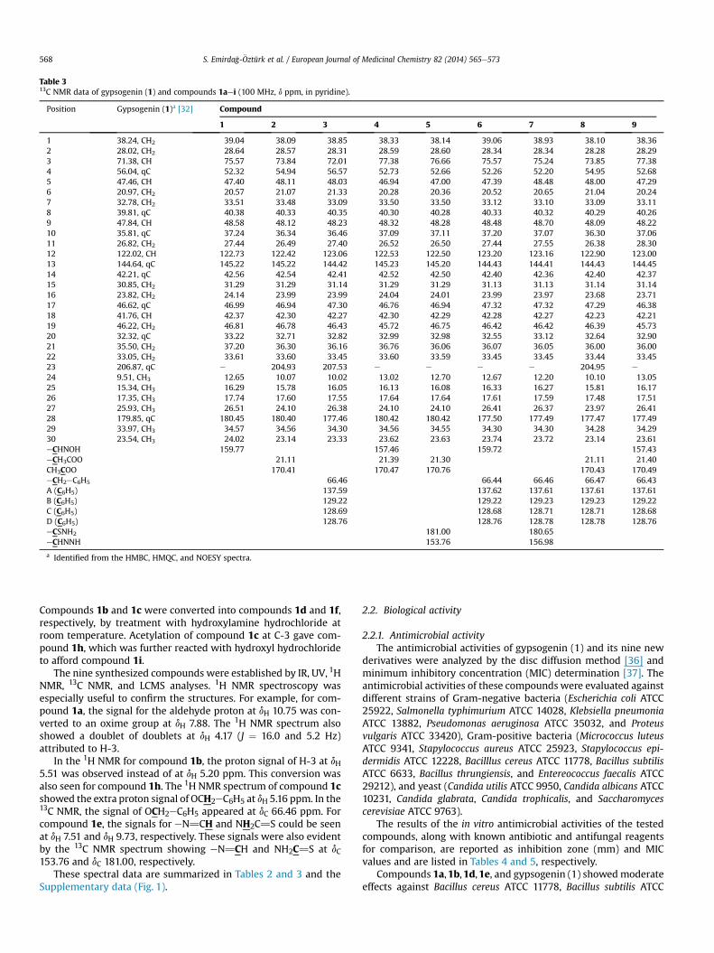
Table 313C NMR data of gypsogenin (1) and compounds 1aei (100 MHz, d ppm, in pyridine).
Position Gypsogenin (1)a [32] Compound
1 2 3 4 5 6 7 8 9
1 38.24, CH2 39.04 38.09 38.85 38.33 38.14 39.06 38.93 38.10 38.362 28.02, CH2 28.64 28.57 28.31 28.59 28.60 28.34 28.34 28.28 28.293 71.38, CH 75.57 73.84 72.01 77.38 76.66 75.57 75.24 73.85 77.384 56.04, qC 52.32 54.94 56.57 52.73 52.66 52.26 52.20 54.95 52.685 47.46, CH 47.40 48.11 48.03 46.94 47.00 47.39 48.48 48.00 47.296 20.97, CH2 20.57 21.07 21.33 20.28 20.36 20.52 20.65 21.04 20.247 32.78, CH2 33.51 33.48 33.09 33.50 33.50 33.12 33.10 33.09 33.118 39.81, qC 40.38 40.33 40.35 40.30 40.28 40.33 40.32 40.29 40.269 47.84, CH 48.58 48.12 48.23 48.32 48.28 48.48 48.70 48.09 48.2210 35.81, qC 37.24 36.34 36.46 37.09 37.11 37.20 37.07 36.30 37.0611 26.82, CH2 27.44 26.49 27.40 26.52 26.50 27.44 27.55 26.38 28.3012 122.02, CH 122.73 122.42 123.06 122.53 122.50 123.20 123.16 122.90 123.0013 144.64, qC 145.22 145.22 144.42 145.23 145.20 144.43 144.41 144.43 144.4514 42.21, qC 42.56 42.54 42.41 42.52 42.50 42.40 42.36 42.40 42.3715 30.85, CH2 31.29 31.29 31.14 31.29 31.29 31.13 31.13 31.14 31.1416 23.82, CH2 24.14 23.99 23.99 24.04 24.01 23.99 23.97 23.68 23.7117 46.62, qC 46.99 46.94 47.30 46.76 46.94 47.32 47.32 47.29 46.3818 41.76, CH 42.37 42.30 42.27 42.30 42.29 42.28 42.27 42.23 42.2119 46.22, CH2 46.81 46.78 46.43 45.72 46.75 46.42 46.42 46.39 45.7320 32.32, qC 33.22 32.71 32.82 32.99 32.98 32.55 33.12 32.64 32.9021 35.50, CH2 37.20 36.30 36.16 36.76 36.06 36.07 36.05 36.00 36.0022 33.05, CH2 33.61 33.60 33.45 33.60 33.59 33.45 33.45 33.44 33.4523 206.87, qC e 204.93 207.53 e e e e 204.95 e
24 9.51, CH3 12.65 10.07 10.02 13.02 12.70 12.67 12.20 10.10 13.0525 15.34, CH3 16.29 15.78 16.05 16.13 16.08 16.33 16.27 15.81 16.1726 17.35, CH3 17.74 17.60 17.55 17.64 17.64 17.61 17.59 17.48 17.5127 25.93, CH3 26.51 24.10 26.38 24.10 24.10 26.41 26.37 23.97 26.4128 179.85, qC 180.45 180.40 177.46 180.42 180.42 177.50 177.49 177.47 177.4929 33.97, CH3 34.57 34.56 34.30 34.56 34.55 34.30 34.30 34.28 34.2930 23.54, CH3 24.02 23.14 23.33 23.62 23.63 23.74 23.72 23.14 23.61eCHNOH 159.77 157.46 159.72 157.43eCH3COO 21.11 21.39 21.30 21.11 21.40CH3COO 170.41 170.47 170.76 170.43 170.49eCH2eC6H5 66.46 66.44 66.46 66.47 66.43A (C6H5) 137.59 137.62 137.61 137.61 137.61B (C6H5) 129.22 129.22 129.23 129.23 129.22C (C6H5) 128.69 128.68 128.71 128.71 128.68D (C6H5) 128.76 128.76 128.78 128.78 128.76eCSNH2 181.00 180.65eCHNNH 153.76 156.98
a Identified from the HMBC, HMQC, and NOESY spectra.
S. Emirda�g-€Oztürk et al. / European Journal of Medicinal Chemistry 82 (2014) 565e573568
Compounds 1b and 1c were converted into compounds 1d and 1f,respectively, by treatment with hydroxylamine hydrochloride atroom temperature. Acetylation of compound 1c at C-3 gave com-pound 1h, which was further reacted with hydroxyl hydrochlorideto afford compound 1i.
The nine synthesized compounds were established by IR, UV, 1HNMR, 13C NMR, and LCMS analyses. 1H NMR spectroscopy wasespecially useful to confirm the structures. For example, for com-pound 1a, the signal for the aldehyde proton at dH 10.75 was con-verted to an oxime group at dH 7.88. The 1H NMR spectrum alsoshowed a doublet of doublets at dH 4.17 (J ¼ 16.0 and 5.2 Hz)attributed to H-3.
In the 1H NMR for compound 1b, the proton signal of H-3 at dH5.51 was observed instead of at dH 5.20 ppm. This conversion wasalso seen for compound 1h. The 1H NMR spectrum of compound 1cshowed the extra proton signal of OCH2eC6H5 at dH 5.16 ppm. In the13C NMR, the signal of OCH2eC6H5 appeared at dC 66.46 ppm. Forcompound 1e, the signals for eN]CH and NH2C]S could be seenat dH 7.51 and dH 9.73, respectively. These signals were also evidentby the 13C NMR spectrum showing eN]CH and NH2C]S at dC153.76 and dC 181.00, respectively.
These spectral data are summarized in Tables 2 and 3 and theSupplementary data (Fig. 1).
2.2. Biological activity
2.2.1. Antimicrobial activityThe antimicrobial activities of gypsogenin (1) and its nine new
derivatives were analyzed by the disc diffusion method [36] andminimum inhibitory concentration (MIC) determination [37]. Theantimicrobial activities of these compounds were evaluated againstdifferent strains of Gram-negative bacteria (Escherichia coli ATCC25922, Salmonella typhimurium ATCC 14028, Klebsiella pneumoniaATCC 13882, Pseudomonas aeruginosa ATCC 35032, and Proteusvulgaris ATCC 33420), Gram-positive bacteria (Micrococcus luteusATCC 9341, Stapylococcus aureus ATCC 25923, Stapylococcus epi-dermidis ATCC 12228, Bacilllus cereus ATCC 11778, Bacillus subtilisATCC 6633, Bacillus thrungiensis, and Entereococcus faecalis ATCC29212), and yeast (Candida utilis ATCC 9950, Candida albicans ATCC10231, Candida glabrata, Candida trophicalis, and Saccharomycescerevisiae ATCC 9763).
The results of the in vitro antimicrobial activities of the testedcompounds, along with known antibiotic and antifungal reagentsfor comparison, are reported as inhibition zone (mm) and MICvalues and are listed in Tables 4 and 5, respectively.
Compounds 1a,1b,1d,1e, and gypsogenin (1) showedmoderateeffects against Bacillus cereus ATCC 11778, Bacillus subtilis ATCC
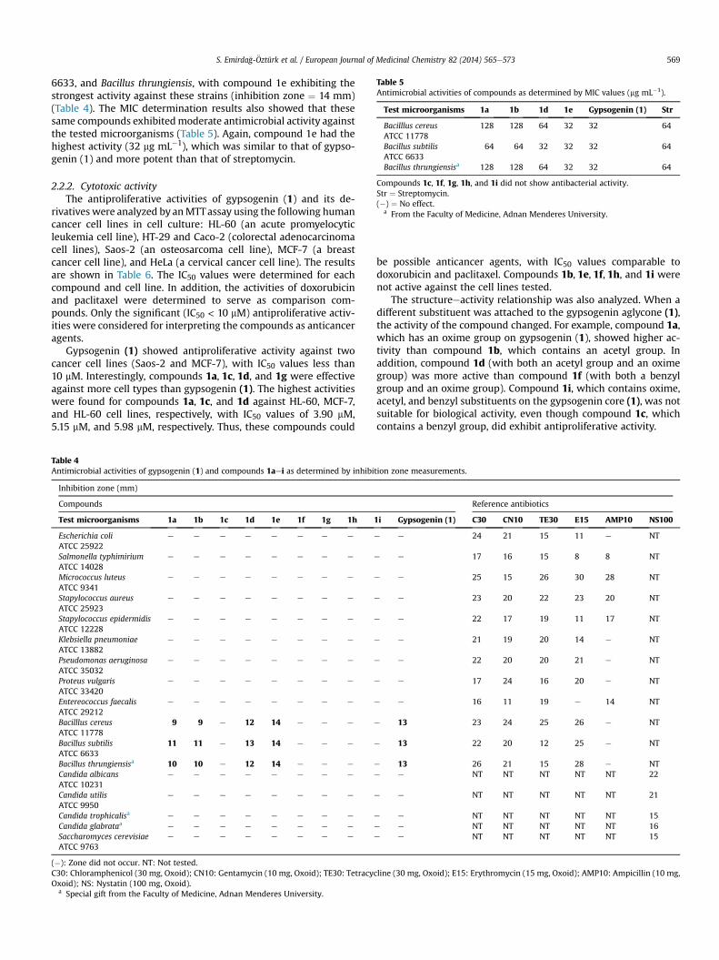
Table 5Antimicrobial activities of compounds as determined by MIC values (mg mL�1).
Test microorganisms 1a 1b 1d 1e Gypsogenin (1) Str
Bacilllus cereusATCC 11778
128 128 64 32 32 64
Bacillus subtilisATCC 6633
64 64 32 32 32 64
Bacillus thrungiensisa 128 128 64 32 32 64
Compounds 1c, 1f, 1g, 1h, and 1i did not show antibacterial activity.Str ¼ Streptomycin.(�) ¼ No effect.
a From the Faculty of Medicine, Adnan Menderes University.
S. Emirda�g-€Oztürk et al. / European Journal of Medicinal Chemistry 82 (2014) 565e573 569
6633, and Bacillus thrungiensis, with compound 1e exhibiting thestrongest activity against these strains (inhibition zone ¼ 14 mm)(Table 4). The MIC determination results also showed that thesesame compounds exhibitedmoderate antimicrobial activity againstthe tested microorganisms (Table 5). Again, compound 1e had thehighest activity (32 mg mL�1), which was similar to that of gypso-genin (1) and more potent than that of streptomycin.
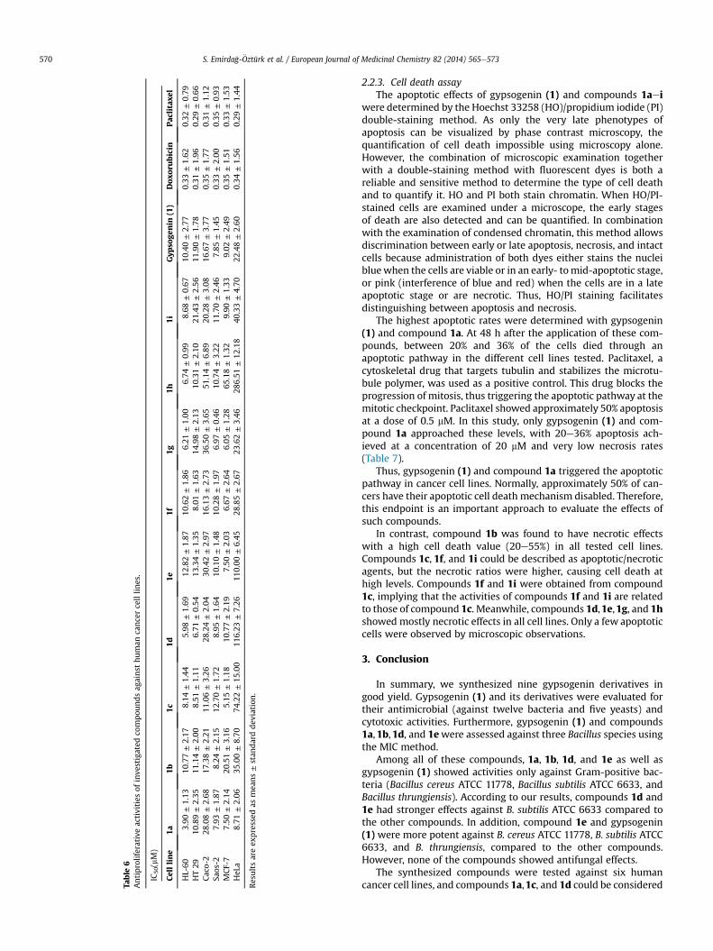
2.2.2. Cytotoxic activityThe antiproliferative activities of gypsogenin (1) and its de-
rivatives were analyzed by anMTTassay using the following humancancer cell lines in cell culture: HL-60 (an acute promyelocyticleukemia cell line), HT-29 and Caco-2 (colorectal adenocarcinomacell lines), Saos-2 (an osteosarcoma cell line), MCF-7 (a breastcancer cell line), and HeLa (a cervical cancer cell line). The resultsare shown in Table 6. The IC50 values were determined for eachcompound and cell line. In addition, the activities of doxorubicinand paclitaxel were determined to serve as comparison com-pounds. Only the significant (IC50 < 10 mM) antiproliferative activ-ities were considered for interpreting the compounds as anticanceragents.
Gypsogenin (1) showed antiproliferative activity against twocancer cell lines (Saos-2 and MCF-7), with IC50 values less than10 mM. Interestingly, compounds 1a, 1c, 1d, and 1g were effectiveagainst more cell types than gypsogenin (1). The highest activitieswere found for compounds 1a, 1c, and 1d against HL-60, MCF-7,and HL-60 cell lines, respectively, with IC50 values of 3.90 mM,5.15 mM, and 5.98 mM, respectively. Thus, these compounds could
Table 4Antimicrobial activities of gypsogenin (1) and compounds 1aei as determined by inhibi
Inhibition zone (mm)
Compounds
Test microorganisms 1a 1b 1c 1d 1e 1f 1g 1h
Escherichia coliATCC 25922
e e e e e e e e
Salmonella typhimiriumATCC 14028
e e e e e e e e
Micrococcus luteusATCC 9341
e e e e e e e e
Stapylococcus aureusATCC 25923
e e e e e e e e
Stapylococcus epidermidisATCC 12228
e e e e e e e e
Klebsiella pneumoniaeATCC 13882
e e e e e e e e
Pseudomonas aeruginosaATCC 35032
e e e e e e e e
Proteus vulgarisATCC 33420
e e e e e e e e
Entereococcus faecalisATCC 29212
e e e e e e e e
Bacilllus cereusATCC 11778
9 9 e 12 14 e e e
Bacillus subtilisATCC 6633
11 11 e 13 14 e e e
Bacillus thrungiensisa 10 10 e 12 14 e e e
Candida albicansATCC 10231
e e e e e e e e
Candida utilisATCC 9950
e e e e e e e e
Candida trophicalisa e e e e e e e e
Candida glabrataa e e e e e e e e
Saccharomyces cerevisiaeATCC 9763
e e e e e e e e
(�): Zone did not occur. NT: Not tested.C30: Chloramphenicol (30 mg, Oxoid); CN10: Gentamycin (10 mg, Oxoid); TE30: TetracycOxoid); NS: Nystatin (100 mg, Oxoid).
a Special gift from the Faculty of Medicine, Adnan Menderes University.
be possible anticancer agents, with IC50 values comparable todoxorubicin and paclitaxel. Compounds 1b, 1e, 1f, 1h, and 1i werenot active against the cell lines tested.
The structureeactivity relationship was also analyzed. When adifferent substituent was attached to the gypsogenin aglycone (1),the activity of the compound changed. For example, compound 1a,which has an oxime group on gypsogenin (1), showed higher ac-tivity than compound 1b, which contains an acetyl group. Inaddition, compound 1d (with both an acetyl group and an oximegroup) was more active than compound 1f (with both a benzylgroup and an oxime group). Compound 1i, which contains oxime,acetyl, and benzyl substituents on the gypsogenin core (1), was notsuitable for biological activity, even though compound 1c, whichcontains a benzyl group, did exhibit antiproliferative activity.
tion zone measurements.
Reference antibiotics
1i Gypsogenin (1) C30 CN10 TE30 E15 AMP10 NS100
e e 24 21 15 11 e NT
e e 17 16 15 8 8 NT
e e 25 15 26 30 28 NT
e e 23 20 22 23 20 NT
e e 22 17 19 11 17 NT
e e 21 19 20 14 e NT
e e 22 20 20 21 e NT
e e 17 24 16 20 e NT
e e 16 11 19 e 14 NT
e 13 23 24 25 26 e NT
e 13 22 20 12 25 e NT
e 13 26 21 15 28 e NTe e NT NT NT NT NT 22
e e NT NT NT NT NT 21
e e NT NT NT NT NT 15e e NT NT NT NT NT 16e e NT NT NT NT NT 15
line (30 mg, Oxoid); E15: Erythromycin (15 mg, Oxoid); AMP10: Ampicillin (10 mg,
Table
6Antiproliferativeactivities
ofinve
stigated
compou
ndsag
ainst
human
cancercelllin
es.
IC50(mM)
Cellline
1a1b
1c1d
1e1f
1g1h
1iGyp
soge
nin
(1)
Doxo
rubicin
Pac
litaxe
l
HL-60
3.90
±1.13
10.77±2.17
8.14
±1.44
5.98
±1.69
12.82±1.87
10.62±1.86
6.21
±1.00
6.74
±0.99
8.68
±0.67
10.40±2.77
0.33
±1.62
0.32
±0.79
HT29
10.89±2.35
11.14±2.00
8.51
±1.11
6.71
±0.54
13.34±1.35
8.01
±1.63
14.98±2.13
10.31±2.10
21.43±2.56
11.90±1.78
0.31
±1.96
0.29
±0.66
Caco-2
28.08±2.68
17.38±2.21
11.06±3.26
28.24±2.04
30.42±2.97
16.13±2.73
36.50±3.65
51.14±6.89
20.28±3.08
16.67±3.77
0.35
±1.77
0.31
±1.12
Saos-2
7.93
±1.87
8.24
±2.15
12.70±1.72
8.95
±1.64
10.10±1.48
10.28±1.97
6.97
±0.46
10.74±3.22
11.70±2.46
7.85
±1.45
0.33
±2.00
0.35
±0.93
MCF-7
7.50
±2.14
20.51±3.16
5.15
±1.18
10.77±2.19
7.50
±2.03
6.67
±2.64
6.05
±1.28
65.18±1.32
9.90
±1.33
9.02
±2.49
0.35
±1.51
0.33
±1.53
HeL
a8.71
±2.06
35.00±8.70
74.22±15
.00
116.23
±7.26
110.00
±6.45
28.85±2.67
23.62±3.46
286.51
±12
.18
40.33±4.70
22.48±2.60
0.34
±1.56
0.29
±1.44
Resultsareex
pressed
asmea
ns±stan
darddev
iation
.
S. Emirda�g-€Oztürk et al. / European Journal of Medicinal Chemistry 82 (2014) 565e573570
2.2.3. Cell death assayThe apoptotic effects of gypsogenin (1) and compounds 1aei
were determined by the Hoechst 33258 (HO)/propidium iodide (PI)double-staining method. As only the very late phenotypes ofapoptosis can be visualized by phase contrast microscopy, thequantification of cell death impossible using microscopy alone.However, the combination of microscopic examination togetherwith a double-staining method with fluorescent dyes is both areliable and sensitive method to determine the type of cell deathand to quantify it. HO and PI both stain chromatin. When HO/PI-stained cells are examined under a microscope, the early stagesof death are also detected and can be quantified. In combinationwith the examination of condensed chromatin, this method allowsdiscrimination between early or late apoptosis, necrosis, and intactcells because administration of both dyes either stains the nucleibluewhen the cells are viable or in an early- tomid-apoptotic stage,or pink (interference of blue and red) when the cells are in a lateapoptotic stage or are necrotic. Thus, HO/PI staining facilitatesdistinguishing between apoptosis and necrosis.
The highest apoptotic rates were determined with gypsogenin(1) and compound 1a. At 48 h after the application of these com-pounds, between 20% and 36% of the cells died through anapoptotic pathway in the different cell lines tested. Paclitaxel, acytoskeletal drug that targets tubulin and stabilizes the microtu-bule polymer, was used as a positive control. This drug blocks theprogression of mitosis, thus triggering the apoptotic pathway at themitotic checkpoint. Paclitaxel showed approximately 50% apoptosisat a dose of 0.5 mM. In this study, only gypsogenin (1) and com-pound 1a approached these levels, with 20e36% apoptosis ach-ieved at a concentration of 20 mM and very low necrosis rates(Table 7).
Thus, gypsogenin (1) and compound 1a triggered the apoptoticpathway in cancer cell lines. Normally, approximately 50% of can-cers have their apoptotic cell death mechanism disabled. Therefore,this endpoint is an important approach to evaluate the effects ofsuch compounds.
In contrast, compound 1b was found to have necrotic effectswith a high cell death value (20e55%) in all tested cell lines.Compounds 1c, 1f, and 1i could be described as apoptotic/necroticagents, but the necrotic ratios were higher, causing cell death athigh levels. Compounds 1f and 1i were obtained from compound1c, implying that the activities of compounds 1f and 1i are relatedto those of compound 1c. Meanwhile, compounds 1d,1e,1g, and 1hshowed mostly necrotic effects in all cell lines. Only a few apoptoticcells were observed by microscopic observations.
3. Conclusion
In summary, we synthesized nine gypsogenin derivatives ingood yield. Gypsogenin (1) and its derivatives were evaluated fortheir antimicrobial (against twelve bacteria and five yeasts) andcytotoxic activities. Furthermore, gypsogenin (1) and compounds1a,1b,1d, and 1ewere assessed against three Bacillus species usingthe MIC method.
Among all of these compounds, 1a, 1b, 1d, and 1e as well asgypsogenin (1) showed activities only against Gram-positive bac-teria (Bacillus cereus ATCC 11778, Bacillus subtilis ATCC 6633, andBacillus thrungiensis). According to our results, compounds 1d and1e had stronger effects against B. subtilis ATCC 6633 compared tothe other compounds. In addition, compound 1e and gypsogenin(1) were more potent against B. cereus ATCC 11778, B. subtilis ATCC6633, and B. thrungiensis, compared to the other compounds.However, none of the compounds showed antifungal effects.
The synthesized compounds were tested against six humancancer cell lines, and compounds 1a,1c, and 1d could be considered
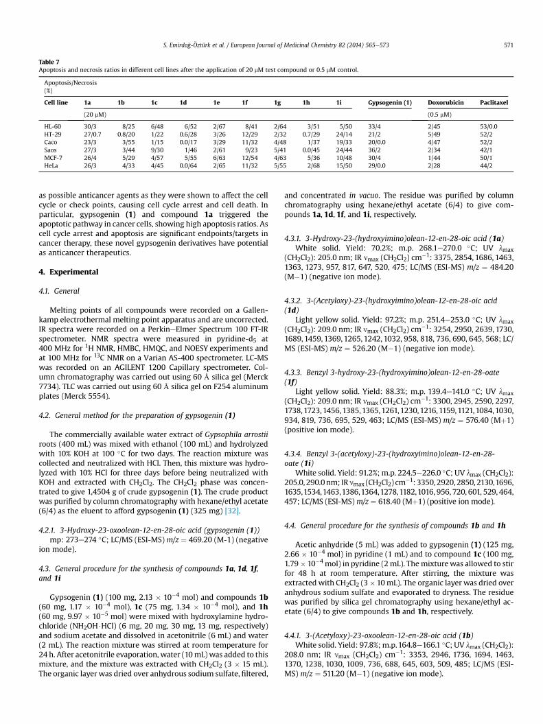
Table 7Apoptosis and necrosis ratios in different cell lines after the application of 20 mM test compound or 0.5 mM control.
Apoptosis/Necrosis(%)
Cell line 1a 1b 1c 1d 1e 1f 1g 1h 1i Gypsogenin (1) Doxorubicin Paclitaxel
(20 mM) (0.5 mM)
HL-60 30/3 8/25 6/48 6/52 2/67 8/41 2/64 3/51 5/50 33/4 2/45 53/0.0HT-29 27/0.7 0.8/20 1/22 0.6/28 3/26 12/29 2/32 0.7/29 24/14 21/2 5/49 52/2Caco 23/3 3/55 1/15 0.0/17 3/29 11/32 4/48 1/37 19/33 20/0.0 4/47 52/2Saos 27/3 3/44 9/30 1/46 2/61 9/23 5/41 0.0/45 24/44 36/2 2/34 42/1MCF-7 26/4 5/29 4/57 5/55 6/63 12/54 4/63 5/36 10/48 30/4 1/44 50/1HeLa 26/3 4/33 4/45 0.0/64 2/65 11/32 5/55 2/68 15/50 29/0.0 2/28 44/2
S. Emirda�g-€Oztürk et al. / European Journal of Medicinal Chemistry 82 (2014) 565e573 571
as possible anticancer agents as they were shown to affect the cellcycle or check points, causing cell cycle arrest and cell death. Inparticular, gypsogenin (1) and compound 1a triggered theapoptotic pathway in cancer cells, showing high apoptosis ratios. Ascell cycle arrest and apoptosis are significant endpoints/targets incancer therapy, these novel gypsogenin derivatives have potentialas anticancer therapeutics.
4. Experimental
4.1. General
Melting points of all compounds were recorded on a Gallen-kamp electrothermal melting point apparatus and are uncorrected.IR spectra were recorded on a PerkineElmer Spectrum 100 FT-IRspectrometer. NMR spectra were measured in pyridine-d5 at400 MHz for 1H NMR, HMBC, HMQC, and NOESY experiments andat 100 MHz for 13C NMR on a Varian AS-400 spectrometer. LC-MSwas recorded on an AGILENT 1200 Capillary spectrometer. Col-umn chromatography was carried out using 60 Å silica gel (Merck7734). TLC was carried out using 60 Å silica gel on F254 aluminumplates (Merck 5554).
4.2. General method for the preparation of gypsogenin (1)
The commercially available water extract of Gypsophila arrostiiroots (400 mL) was mixed with ethanol (100 mL) and hydrolyzedwith 10% KOH at 100 �C for two days. The reaction mixture wascollected and neutralized with HCl. Then, this mixture was hydro-lyzed with 10% HCl for three days before being neutralized withKOH and extracted with CH2Cl2. The CH2Cl2 phase was concen-trated to give 1,4504 g of crude gypsogenin (1). The crude productwas purified by column chromatography with hexane/ethyl acetate(6/4) as the eluent to afford gypsogenin (1) (325 mg) [32].
4.2.1. 3-Hydroxy-23-oxoolean-12-en-28-oic acid (gypsogenin (1))mp: 273e274 �C; LC/MS (ESI-MS) m/z ¼ 469.20 (M-1) (negative
ion mode).
4.3. General procedure for the synthesis of compounds 1a, 1d, 1f,and 1i
Gypsogenin (1) (100 mg, 2.13 � 10�4 mol) and compounds 1b(60 mg, 1.17 � 10�4 mol), 1c (75 mg, 1.34 � 10�4 mol), and 1h(60 mg, 9.97 � 10�5 mol) were mixed with hydroxylamine hydro-chloride (NH2OH$HCl) (6 mg, 20 mg, 30 mg, 13 mg, respectively)and sodium acetate and dissolved in acetonitrile (6 mL) and water(2 mL). The reaction mixture was stirred at room temperature for24 h. After acetonitrile evaporation, water (10mL)was added to thismixture, and the mixture was extracted with CH2Cl2 (3 � 15 mL).The organic layer was dried over anhydrous sodium sulfate, filtered,
and concentrated in vacuo. The residue was purified by columnchromatography using hexane/ethyl acetate (6/4) to give com-pounds 1a, 1d, 1f, and 1i, respectively.
4.3.1. 3-Hydroxy-23-(hydroxyimino)olean-12-en-28-oic acid (1a)White solid. Yield: 70.2%; m.p. 268.1e270.0 �C; UV lmax
(CH2Cl2): 205.0 nm; IR nmax (CH2Cl2) cm�1: 3375, 2854, 1686, 1463,1363, 1273, 957, 817, 647, 520, 475; LC/MS (ESI-MS) m/z ¼ 484.20(M�1) (negative ion mode).
4.3.2. 3-(Acetyloxy)-23-(hydroxyimino)olean-12-en-28-oic acid(1d)
Light yellow solid. Yield: 97.2%; m.p. 251.4e253.0 �C; UV lmax(CH2Cl2): 209.0 nm; IR nmax (CH2Cl2) cm�1: 3254, 2950, 2639, 1730,1689, 1459, 1369, 1265, 1242, 1032, 958, 818, 736, 690, 645, 568; LC/MS (ESI-MS) m/z ¼ 526.20 (M�1) (negative ion mode).
4.3.3. Benzyl 3-hydroxy-23-(hydroxyimino)olean-12-en-28-oate(1f)
Light yellow solid. Yield: 88.3%; m.p. 139.4e141.0 �C; UV lmax(CH2Cl2): 209.0 nm; IR nmax (CH2Cl2) cm�1: 3300, 2945, 2590, 2297,1738,1723,1456,1385,1365,1261,1230,1216,1159,1121,1084,1030,934, 819, 736, 695, 529, 463; LC/MS (ESI-MS) m/z ¼ 576.40 (Mþ1)(positive ion mode).
4.3.4. Benzyl 3-(acetyloxy)-23-(hydroxyimino)olean-12-en-28-oate (1i)
White solid. Yield: 91.2%; m.p. 224.5e226.0 �C; UV lmax (CH2Cl2):205.0, 290.0nm; IRnmax (CH2Cl2) cm�1: 3350, 2920, 2850, 2130,1696,1635,1534,1463,1386,1364,1278,1182,1016, 956, 720, 601, 529, 464,457; LC/MS (ESI-MS) m/z ¼ 618.40 (Mþ1) (positive ion mode).
4.4. General procedure for the synthesis of compounds 1b and 1h
Acetic anhydride (5 mL) was added to gypsogenin (1) (125 mg,2.66 � 10�4 mol) in pyridine (1 mL) and to compound 1c (100 mg,1.79� 10�4 mol) in pyridine (2mL). Themixturewas allowed to stirfor 48 h at room temperature. After stirring, the mixture wasextracted with CH2Cl2 (3� 10mL). The organic layer was dried overanhydrous sodium sulfate and evaporated to dryness. The residuewas purified by silica gel chromatography using hexane/ethyl ac-etate (6/4) to give compounds 1b and 1h, respectively.
4.4.1. 3-(Acetyloxy)-23-oxoolean-12-en-28-oic acid (1b)White solid. Yield: 97.8%; m.p.164.8e166.1 �C; UV lmax (CH2Cl2):
208.0 nm; IR nmax (CH2Cl2) cm�1: 3353, 2946, 1736, 1694, 1463,1370, 1238, 1030, 1009, 736, 688, 645, 603, 509, 485; LC/MS (ESI-MS) m/z ¼ 511.20 (M�1) (negative ion mode).
S. Emirda�g-€Oztürk et al. / European Journal of Medicinal Chemistry 82 (2014) 565e573572
4.4.2. Benzyl 3-(acetyloxy)-23-oxoolean-12-en-28-oate (1h)White solid. Yield: 97.7%; m.p. 159.6e161.1 �C; UV lmax (CH2Cl2):
208.0, 290.0 nm; IR nmax (CH2Cl2) cm�1: 3100, 2947, 2876, 1733,1686, 1458, 1369, 1238, 1159, 1121, 1030, 1010, 976, 897, 736, 647,604, 558; LC/MS (ESI-MS)m/z ¼ 603.40 (Mþ1) (positive ion mode).
4.5. General procedure for the synthesis of compound 1c
A mixture of gypsogenin (1) (400 mg, 8.51 � 10�4) and trie-thylamine (3 mL) was stirred at room temperature for 1 h beforebenzyl bromide (2mL) was added. The mixturewas refluxed for 3 hand then stirred at room temperature for 48 h. Water (10 mL) wasadded to the reaction mixture, and the mixture was extracted withCH2Cl2 (3 � 10 mL). The organic layer was dried over anhydroussodium sulfate and evaporated to dryness. The residue was purifiedby column chromatography on silica gel using hexane/ethyl acetate(4/6) to give compound 1c as an amorphous powder (260 mg).
4.5.1. Benzyl 3-hydroxy-23-oxoolean-12-en-28-oate (1c)Yield: 54.6%; UV lmax (CH2Cl2): 207.0 nm; IR nmax (CH2Cl2) cm�1:
3290, 2964, 2855, 2700, 1750, 1466, 1458, 1383, 1358, 1317, 1258,1217, 1158, 1117, 1075, 1042, 1025, 958, 833, 733, 683, 608, 542; LC/MS (ESI-MS) m/z ¼ 561.40 (Mþ1) (positive ion mode).
4.6. General procedure for the synthesis of compounds 1e and 1g
A solution of thiosemicarbazide in water (7 mL) was added to asolution of compounds 1b (125 mg, 2.44 � 10�4) and 1c (87 mg,1.55 � 10�4) in MeOH (7 mL). The mixture was refluxed for 13 h.After cooling, the mixture was extracted with CH2Cl2 (3 � 10 mL).The organic layer was dried over anhydrous sodium sulfate andevaporated to dryness. The residue was purified by column chro-matography on silica gel using hexane/ethyl acetate (6/4) to givecompounds 1e and 1g, respectively.
4.6.1. 3-(Acetyloxy)-23-[(aminocarbonothioyl) hydrazono]olean-12-en-28-oic acid (1e)
Light yellow solid. Yield: 45.8%; m.p. 205.3e207.4 �C; UV lmax(CH2Cl2): 211.0, 261.0 nm; IR nmax (CH2Cl2) cm�1: 3267, 3164, 2950,1734, 1691, 1597, 1534, 1459, 1366, 1274, 1260, 1245, 1088, 1028,1008, 1055, 839, 820, 764, 749, 642, 530, 462; LC/MS (ESI-MS) m/z ¼ 584.20 (M�1) (negative ion mode).
4.6.2. Benzyl 23-[(aminocarbonothioyl)hydrazono]-3-hydroxyolean-12-en-28-oate (1g)
White solid. Yield: 56.9%; m.p.138.1e139.0 �C; UV lmax (CH2Cl2):207.0; 270.0 nm; IR nmax (CH2Cl2) cm�1: 3244, 2970, 2300, 1743,1593, 1531, 1365, 1276, 1229, 1216, 1158, 1082, 958, 832, 749, 662,529, 474, 464, 457; LC/MS (ESI-MS) m/z ¼ 634.40 (Mþ1) (positiveion mode).
4.7. Pharmacology
4.7.1. Antibacterial and antifungal activityAll of the compounds used in this study were tested for their
in vitro antibacterial and antifungal effects (antimicrobial activity)following standard methods (Supplementary data, Fig. 2).
In this study, twelve bacterial cultures and five different yeastswere used. Eleven bacterial strains and three yeast strains wereobtained from the American Type Culture Collection (ATCC; Rock-ville, MD, USA). The other bacterial strain, B. thrungiensis, was iso-lated from human fecal samples obtained from Adnan MenderesUniversity (Clinical Microbiology Laboratory, Faculty of Medicine)and was cultured in Nutrient Broth (Merck) at 30 �C for 24 h. The
other two yeast strains, Candida glabrata and Candida trophicalis,were cultured in Malt Extract Broth (Merck, USA) at 30 �C for 24 h.
The antibacterial and antifungal activities of all nine synthesizedcompounds were analyzed by the disc diffusion method and thebroth dilution method to determine the MIC.
4.7.1.1. Disc diffusion method. The antimicrobial activities weredetermined according to the standard Antimicrobial Disc Suscep-tibility Tests summarized by the National Committee for ClinicalLaboratory Standards [37]. Fresh stock solutions (1000 mg mL�1) ofall the synthesized compounds were prepared in DMSO accordingto the desired concentrations for the experiments. The inoculumsuspensions of the tested bacteria and yeasts were prepared fromthe broth cultures (18e24 h), and the turbidity was adjusted to beequivalent to a 0.5 McFarland standard tube to give a concentrationof 1 � 108 bacterial cells and 1 � 106 yeast cells mL�1, respectively.To test the antimicrobial activity of all the synthesized compounds,a Mueller Hinton Agar (MHA) plate was inoculated with 0.1 mL ofbroth culture of bacteria or yeast. Then, a hole of 6 mm in diameterand depth was made on top of the agar with a sterile stick and filledwith 30 mL of each synthesized compound, respectively.
Plates inoculated with Escherichia coli, S. typhimurium, Stapylo-coccus aureus, Stapylococcus epidermidis, E. faecalis, K. pneumonia,Pseudomonas aeruginosa, and Proteus vulgaris were incubated at37 �C for 24 h, while those with Micrococcus luteus, B. subtilis,B. cereus, B. thrungiensis, Saccharomyces cerevisiae, Candida albicans,Candida utilis, C. glabrata, and C. trophicalis were incubated at 30 �Cfor 24 h. At the end of the incubation time, the diameters of theinhibition zones formed on theMHAwere evaluated inmillimeters.Discs of chloramphenicol (C30), gentamycine (CN10), tetracycline(TE30), erytromycine (E15), ampicillin (AMP10), and nystatine(NS100) were used as positive controls. The measured inhibitionzones of the study compounds were compared with those of thereference discs.
4.7.1.2. Dilution method. Screening for antibacterial activities wascarried out by preparing a microdilution broth, following the pro-cedure outlined in the Manual of Clinical Microbiology [38]. All thebacteria were inoculated in the nutrient broth and incubated at30 �C for 24 h. The compounds were dissolved in DMSO(2 mg mL�1) and then diluted in Mueller Hinton broth. Two-foldserial dilutions of the compounds were employed to determinetheMIC, ranging from 256 to 0.125 mgmL�1. Cultures were grown at30 �C for 18e20 h, and the final inoculum was approximately106 cfu mL�1. Test cultures were incubated at 37 �C for 24 h. Thelowest concentration of antimicrobial agent that resulted in com-plete inhibition of the microorganisms was represented as the MIC(mg mL�1). Streptomycin (I.E. Ulagay) was used as a positive controlin the dilution method.
4.7.2. Cell cultureHL-60, HT-29, Caco-2, Saos-2, MCF-7, and HeLa cell lines were
purchased from ATCC. Growth media and its supplements werepurchased from Life Science Tech. (Sacem, Turkey). Cells weregrown in RPMI-1540, Dulbecco's modified Eagle’s medium, orMcCoys 5A medium supplemented with 10e15% heat inactivatedfetal calf serum, 1% L-glutamine, 1% penicillin/streptomycin, andnonessential amino acids and cultivated at 37 �C in a humidifiedatmosphere containing 5% CO2. Control chemicals doxorubicin andpaclitaxel were purchased from Cell Signaling Tech., Inc. (Beverly,MA, USA).
4.7.3. Cytotoxicity testingCells were seeded in 48-well plates at a concentration of
1 � 104e1 � 105 cells mL�1 and incubated with increasing
S. Emirda�g-€Oztürk et al. / European Journal of Medicinal Chemistry 82 (2014) 565e573 573
concentrations of agents (corresponding to 0.5, 10, 20, and 40 mM ofthe compounds or controls). Cell viability was determined at 24 and72 h with the MTT assay carried out with a Thermo Scientific Mul-tiskan spectrometer. The absorbance at 590 nmwas recorded usingThermo Scientific SkanIt Software. The IC50 values were analyzedusing GraphPad Prism 5.0 software. Experiments were done intriplicate. The percentage of viable cells was compared to the un-treated control and calculated as follows:[(C72 h þ drug � C24 h þ drug)/(C72 h � drug �C24 h� drug)]� 100¼ % cell division, where C72 hþ drug is the cellnumberafterdrug treatment for 72h, C24hþdrug is the cell numberafter drug treatment for 24 h, C72 h drug is the cell number after 72 hwithout drug treatment, and C24 h e drug, is the cell number after24 h without drug treatment.
4.7.4. Hoechst 33258 (HO) and propidium iodide (PI) doublestaining
Cells were seeded in 48-well plates and incubated withincreasing concentrations of agents (corresponding to 0.5, 10, 20,and 40 mM of the compounds or controls) for 24 and 48 h(Supplementary data, Fig. 3). After microscopic examination of thecolored cells and considering the IC50 values, it was decided to usethe effect obtained with a compound concentration of 20 mM in thecell death assay. The effective time point was defined as 48 h. Celldeath quantification was performed according to a previouslydescribedmethod [39]. Formicroscopic analyses, an Olympus BX52fluorescence microscope was used, and the fluorescent dyes HOand PI were purchased from Sigma.
Acknowledgments
We are grateful to Professor Dr. Hüseyin Anıl for his valuablesuggestions for the synthetic routes.
Appendix A. Supplementary data
Supplementary data related to this article can be found at http://dx.doi.org/10.1016/j.ejmech.2014.05.084.
References
[1] D. Frechet, B. Christ, B.M. Sorbier, H. Fischer, M. Vuilhorgne, Four triterpenoidsaponins from dried roots of Gypsophila species, Phytochemistry 30 (1991)927e931.
[2] J.-G. Luo, L.-Y. Kong, Y. Takaya, M. Niwa, Two new monodesmosidic triterpenesaponins from Gypsophila oldhamiana, Chem. Pharm. Bull. 54 (2006)1200e1202.
[3] Q. Chen, J.-G. Luo, L.-Y. Kong, New triterpenoid saponins from the roots ofGypsophila perfoliata Linn, Carbohydr. Res. 346 (2011) 2206e2212.
[4] T. Narender, G. Madhur, N. Jaiswal, M. Agrawal, C.K. Maurva, N. Rahuia,A.K. Srivastava, A.K. Tamrakar, Synthesis of novel triterpene and N-allylated/N-alkylated niacin hybrids as a-glucosidase inhibitors, Eur. J. Med. Chem. 63(2013) 162e169.
[5] M. Amoros, B. Fauconnier, R.L. Girre, In vitro antiviral activity of a saponinfrom Anagallis arvensis, Primulaceae, against herpes simplex virus and polio-virus, Antivir. Res. 8 (1987) 13e25.
[6] N. Ding, Q. Chen, W. Zhang, S. Ren, Y. Guo, Y. Li, Structure-activity relation-ships of saponin derivatives: a series of entry inhibitors for highly pathogenicH5N1 influenza virus, Eur. J. Med. Chem. 53 (2012) 316e326.
[7] J.-R. Wang, H. Zhou, Z.-H. Jiang, L. Liu, Two new triterpene saponins from theanti-inflammatory saponin fraction of Ilex pubescens root, Chem. Biodivers. 5(2008) 1369e1376.
[8] M. Rajasekaran, A.G.R. Nair, W.J.G. Hellstrom, S.C. Sikka, Spermicidal activity ofan antifungal saponin obtained from the tropical herb Mollugo pentaphylla,Contraception 47 (1993) 401e412.
[9] G.S. Sidhu, D.G. Oakenfull, A mechanism for the hypocholesterolaemic activityof saponins, Br. J. Nutr. 55 (1986) 643e649.
[10] I.N. Karasteva, R.A. Toshkova, S.D. Nikolov, Protective effect of Astragaluscorniculatus saponins against myeloid graffi tumour in hamsters, Phytother-apy Res. 18 (2004) 255e257.
[11] S.-C. Bang, J.-H. Lee, G.-Y. Song, D.-H. Kim, M.-Y. Yoon, B.-Z. Ahn, Antitumoractivity of Pulsatilla koreana saponins and their structure-activity relationship,Chem. Pharm. Bull. 53 (2005) 1451e1454.
[12] A.V. Rao, M.K. Sung, Saponins as anticarcinogens, J. Nutrition 125 (1995)717Se724S.
[13] I. Gülçin, V. Mshvildadze, A. Gepdiremen, R. Elias, Antioxidant activity of sa-ponins isolated from ivy: a-Hederin, hederasaponin-C, hederacolchiside-Eand hederacolchiside-F, Planta Medica 70 (2004) 561e563.
[14] H.-Y. Kim, R. Yu, J.-S. Kim, Y.-K. Kim, M.-K. Sung, Antiproliferative crude soysaponin extract modulates the expression of IkBa, protein kinase C, andcyclooxygenase-2 in human colon cancer cells, Cancer Lett. 210 (2004) 1e6.
[15] Q. Liu, H. Liu, L. Zhang, T. Guo, P. Wang, M. Geng, Y. Li, Synthesis and anti-tumor activities of naturally occurring oleanolic acid triterpenoid saponinsand their derivatives, Eur. J. Med. Chem. 64 (2013) 1e15.
[16] B. Yeskaliyeva, M.A. Mesaik, A. Abbaskhan, A. Kulsoom, G.S.H. Burasheva,Z.A. Abilov, M.I. Choudhary, A. ur-Rahman, Bioactive flavonoids and saponinsfrom Climacoptera obtusifolia, Phytochemistry 67 (2006) 2392e2397.
[17] Z. Jia, K. Koike, M. Kudo, H. Li, T. Nikaido, Triterpenoid saponins and sapo-genins from Vaccaria segetalis, Phytochemistry 48 (1998) 529e536.
[18] M. Larhsini, A. Marston, K. Hostettmann, Triterpenoid saponins from the rootsof Silene cucubalus, Fitoterapia 74 (2003) 237e241.
[19] C. Lavaud, L. Voutquenne, G. Massiot, L.L. Men-Olivier, B.C. Das, O. Laprevote,L. Serani, C. Delaude, M. Becchi, Saponins from the stem bark of Filiciumdecipiens, Phytochemistry 47 (1998) 441e449.
[20] J.-G. Luo, W. Nie, L.-Y. Kong, Three new sulfated triterpenoids from the rootsof Gypsophila pacifica, J. Asian Nat. Prod. Res. 13 (2011) 529e533.
[21] I. Arslan, A. Celik, M.F. Melzig, Nebulosides A-B, novel triterpene saponinsfrom under-ground parts of Gypsophila arrostii Guss. var. nebulosa, BioorganicMed. Chem. 21 (2013) 1279e1283.
[22] W. Nie, J.G. Luo, L.Y. Kong, New triterpenoid saponins from the roots ofGypsophila pacifica Kom, Carbohydr. Res. 345 (2010) 68e73.
[23] M.-K. Sung, C.W.C. Kendall, A.V. Rao, Effect of soybean saponins and gyp-sophila saponin on morphology of colon carcinoma cells in culture, FoodChem. Toxicol. 33 (1995) 357e366.
[24] D.J. Marciani, Triterpene Saponin Analogs having Adjuvant and Immunosti-mulatory Activity, 1999. U.S. Patent 5977081.
[25] L. Voutquenne-Nazabadioko, R. Gevrenova, N. Borie, D. Harakat, C. Sayagh,A. Weng, M. Thakur, M. Zaharieva, M. Henry, Triterpenoid saponins fromthe roots of Gypsophila trichotoma Wender, Phytochemistry 90 (2013)114e127.
[26] J.-G. Luo, L. Ma, L.-Y. Kong, New triterpenoid saponins with strong a-gluco-sidase inhibitory activity from the roots of Gypsophila oldhamiana, BioorganicMed. Chem. 16 (2008) 2912e2920.
[27] R. Gevrenova, T. Stancheva, Y. Voynikov, D. Laurain-Mattar, M. Henry, Rootin vitro cultures of six Gypsophila species and their saponin contents, EnzymeMicrob. Technol. 47 (2010) 97e104.
[28] H. Bai, Y. Zhong, Y.-Y. Xie, Y.-S. Wang, L. Liu, L. Zhou, J. Wang, Y.-L. Mu, C.-X. Zuo, A major triterpenoid saponin from Gypsophila oldhamiana, Chem.Biodivers. 4 (2007) 955e960.
[29] M. Yotova, I. Krasteva, K. Jenett-Siems, P. Zdraveva, S. Nikolov, Triterpenoidsin Gypsophila trichotoma Wend, Phytochem. Lett. 5 (2012) 752e755.
[30] S. B€ottger, M.F. Melzig, Triterpenoid saponins of the Caryophyllaceae andIllecebraceae family, Phytochem. Lett. 4 (2011) 59e68.
[31] Y.C. Kim, R. Higuchi, T. Komori, Application of hydrothermolysis to the studieson constituents of the Merck saponin, Liebigs Ann. Chem. (1992) 941e946.
[32] S. Emirda�g-€Oztürk, _I. Babahan, A. €Ozmen, Synthesis, characterization andin vitro anti-neoplastic activity of gypsogenin derivatives, Bioorganic Chem.53 (2014) 15e23. http://dx.doi.org/10.1016/j.bioorg.2013.12.001.
[33] Y. Liu, W.-X. Lu, M.-C. Yan, Y. Yu, T. Ikejima, M.-S. Cheng, Synthesis and tumorcytotoxicity of novel Amide derivatives of b-hederin, Molecules 15 (2010)7871e7883.
[34] M.-C. Yan, Y. Liu, H. Chen, Y. Ke, Q.-C. Xu, M.-S. Cheng, Synthesis and anti-tumor activity of two natural N-acetylglucosamine-bearing triterpenoid sa-ponins: Lotoidoside D and E, Bioorg. Med. Chem. Lett. 16 (2006) 4200e4204.
[35] M.-S. Cheng, M.-C. Yan, Y. Liu, L.-G. Zheng, J. Liu, Synthesis of b-hederin andHederacolchiside A1: triterpenoid triterpenoid saponins bearing a uniquecytotoxicity-inducing disaccharide moiety, Carbohydr. Res. 341 (2006) 60e67.
[36] C.H. Collins, P.M. Lyne, J.M. Grange, Collins and Lyne's MicrobiologicalMethods, seventh ed., Butterworth Heinemann, London, UK, 1995, pp.121e136 (Chap. 8).
[37] National Committee for Clinical Laboratory Standards, Performance Standardsfor Antimicrobial Disk Susceptibility Tests, Approved Standard NCCLS Publi-cation, Villanova, PA, USA, M2-A51-32, 1993.
[38] P.R. Murray, E.J. Baron, M.A. Pfaller, F.C. Tenover, R.H. Yolke, Manual of ClinicMicrobiology, seventh ed., ASM Press, D.C., Washington, 1995, p. 1773.
[39] M. Grusch, D. Polgar, S. Gfatter, K. Leuhuber, S. Huettenbrenner, C. Leisser,G. Fuhrmann, F. Kassie, H. Steinkellner, K. Smid, G.J. Peters, H. Jayaram,W. Klepal, T. Szekeres, S. Knasmuller, G. Krupitza, Maintenance of ATP favoursapoptosis over necrosis triggered by benzamide riboside, Cell. Death Differ. 9(2) (2002) 169e178.
Related Documents


![Chemical Biology & Drug Design Volume 81 Issue 4 2013 [Doi 10.1111%2Fcbdd.12102] Hidalgo-Figueroa, Sergio; RamÃ-rez-Espinosa, Juan J.; Estrada- -- Discovery of Thiazolidine-2,4-DioneBiphenylcarbonitrile](https://static.cupdf.com/doc/110x72/577cca0d1a28aba711a53e06/chemical-biology-drug-design-volume-81-issue-4-2013-doi-1011112fcbdd12102.jpg)


![link.springer.com10.1007/s11164... · Web viewSYNTHESIS OF NOVEL 9-((ARYLIDENE) HYDRAZONO)-2,4,6,8-TETRAKIS (4-METHOXY PHENYL)-3,7-DIAZABICYCLO[3.3.1]NONANE AZINE DERIVATIVES AS POTIENTIAL](https://static.cupdf.com/doc/110x72/5abe15dc7f8b9a8e3f8c93dc/link-101007s11164web-viewsynthesis-of-novel-9-arylidene-hydrazono-2468-tetrakis.jpg)

