SYNTHESIS AND PRECLINICAL EVALUATION OF PEPTIDE RECEPTOR-TARGETED DIAGNOSTIC AND THERAPEUTIC RADIOPHARMACEUTICALS FOR PROSTATE CANCER _______________________________________ A Dissertation presented to the Faculty of the Graduate School at the University of Missouri-Columbia _______________________________________________________ In partial fulfillment of the requirements for the degree Doctor of Philosophy _____________________________________________________ Submitted by NKEMAKONAM CHUKWUEBUKA OKOYE Under the Supervision of Professor Silvia S. Jurisson and Professor Timothy J. Hoffman MAY 2019

Welcome message from author
This document is posted to help you gain knowledge. Please leave a comment to let me know what you think about it! Share it to your friends and learn new things together.
Transcript

SYNTHESIS AND PRECLINICAL EVALUATION OF
PEPTIDE RECEPTOR-TARGETED DIAGNOSTIC AND
THERAPEUTIC RADIOPHARMACEUTICALS FOR
PROSTATE CANCER
_______________________________________
A Dissertation
presented to
the Faculty of the Graduate School
at the University of Missouri-Columbia
_______________________________________________________
In partial fulfillment of the requirements for the degree
Doctor of Philosophy
_____________________________________________________
Submitted by
NKEMAKONAM CHUKWUEBUKA OKOYE
Under the Supervision of
Professor Silvia S. Jurisson and Professor Timothy J. Hoffman
MAY 2019

© Copyright by Nkemakonam C. Okoye 2019
All Rights Reserved

The undersigned, appointed by the dean of the Graduate School at the University of
Missouri-Columbia, have examined the dissertation entitled
Synthesis and Preclinical Evaluation of Peptide Receptor-targeted Diagnostic and
Therapeutic Radiopharmaceuticals for Prostate Cancer
presented by Nkemakonam C. Okoye, a candidate for the degree of Doctor of Philosophy
in Chemistry, and hereby certify that, in their opinion, it is worthy of acceptance.
_______________________________________________
Professor Silvia S. Jurisson
_______________________________________________
Professor Timothy J. Hoffman
_______________________________________________
Professor Heather M. Hennkens
_______________________________________________
Professor J. David Robertson
_______________________________________________
Professor Gary A. Baker

Dedication
This dissertation is dedicated to my lovely parents, Sir Josiah Okoye and Lady Charity
Okoye, for their selfless sacrifice in ensuring that I got a good education.

ii
Acknowledgments
I am very grateful to my advisors, Dr. Silvia Jurisson and Dr. Timothy Hoffman for
their incredible support, encouragement, mentorship and guidance throughout my graduate
studies. I owe the success of the research described in this dissertation to their dedication
in nurturing me into a competent scientist by patiently teaching me the skills and expertise
needed to be successful in my research. I will like to specially acknowledge Tammy Rold
for her contributions towards the in vitro cell studies and in vivo mice studies described in
this dissertation. Also, special thanks to Ashley Berendzen for her contributions to the mice
SPECT/CT imaging studies. Tammy and Ashley made the times I spent over in the
Hoffman research labs at the Harry S. Truman VA Hospital very memorable and I am truly
grateful. The members of my graduate committee, Dr. Heather Hennkens, Dr. J. David
Robertson and Dr. Gary Baker, provided very thoughtful insights that assisted me in
critically thinking about my research. I will also like to thank other individuals that
contributed to the success of my research including Dr. Fabio Gallazzi (LC-MS facility),
James Guthrie (ICP-MS facility at MURR), Dr. John Lydon (help with HPGe studies and
105Rh production), Dr. Wei Wycoff (NMR facility), and members of the
radiopharmaceutical group at MURR (Mary Embree, Marina Kuchuk and Daniel
Oconnor). Also, I will like to acknowledge present and past members of the Jurisson
research group for their support and for providing an intellectually stimulating environment
during my studies. Thank you to the University of Missouri-Columbia (MU), MU
Department of Chemistry, MU Research Reactor (MURR), and the Research Division at
the Harry S. Truman Memorial VA Hospital for providing the necessary facilities,
equipment and financial support for my research. The work described here was supported

iii
in part by grant funding from The United States Department of Veterans Affairs (VA Merit
Award # 1I01BX001699 and VA Research Career Scientist Award to Timothy Hoffman),
The National Cancer Institute (NCI-RO1 Award # CA222293) and The Society of Nuclear
Medicine and Molecular Imaging (2018 Bradley-Alavi Student Fellowship to
Nkemakonam Okoye).
To my parents, Sir Josiah Okoye and Lady Charity Okoye, thank you for your
constant love, support, and prayers. I am grateful to my siblings (Chisom, Jideofor, Olisa
and Ezinne) and my amazing friends for their encouragement and moral support. I also
acknowledge Dr. Kenneth Okafor and his family for their incredible support and for being
my family in the United States. Finally, I am grateful to God for His grace and for blessing
me with life, good health, and a sound mind, without which I would not have done any of
this work.

iv
Table of Contents
Acknowledgments ............................................................................................................. ii
List of Illustrations ........................................................................................................... viii
List of Figures ........................................................................................................... viii
List of Tables ............................................................................................................ xiii
List of Schemes .......................................................................................................... xv
List of Equations ........................................................................................................ xv
Academic Abstract ......................................................................................................... xvi
CHAPTER 1: General Introduction ............................................................................... 1
1.1. Radiopharmaceuticals as a Tool in Cancer Diagnosis and Therapy .................. 1
1.2. Radiopharmaceutical Design ............................................................................. 4
1.3. Targeted Alpha Therapy .................................................................................... 5
1.4. Alpha-emitting Radionuclides of Clinical Relevance ........................................ 7
1.4-1. Astatine-211........................................................................................... 7
1.4-2. Radium-223 ........................................................................................... 8
1.4-3. Actinum-225 / Bismuth-213 .................................................................. 9
1.4-4. Lead-212 / Bismuth-212 ...................................................................... 11
1.5. Bombesin Receptors as Molecular Targets in Prostate Cancer ....................... 13
1.6. Dissertation Outline ......................................................................................... 15
1.7. References ........................................................................................................ 16
CHAPTER 2: Diagnostic Imaging of Prostate Cancer Using a 203Pb-labeled BB2
Receptor Antagonist ....................................................................................................... 23
2.1. Introduction ...................................................................................................... 23
2.2. Experimental .................................................................................................... 25
2.2-1. Materials and Methods ........................................................................ 25

v
2.2-2. Synthesis of Non-radioactive Pb-RM2 ................................................ 26
2.2-3. Purification of [203Pb]PbCl2 ................................................................. 27
2.2-4. Efficiency of Fe Separation from Pb Using Pb-resin .......................... 28
2.2-5. 203Pb Labeling ...................................................................................... 28
2.2-6. Cell Culture.......................................................................................... 29
2.2-7. In Vitro Receptor Binding Affinity Studies ......................................... 29
2.2-8. Mice and Husbandry ............................................................................ 30
2.2-9. [203Pb]PbCl2 Biodistribution Studies ................................................... 31
2.2-10. [203Pb]Pb-RM2 Biodistribution Studies ............................................. 31
2.2-11. Micro SPECT/CT/MRI Imaging Studies ........................................... 32
2.3. Results and Discussion ..................................................................................... 33
2.3-1. Chemistry and Radiochemistry ........................................................... 33
2.3-2. In vitro Receptor Binding Affinity Studies ......................................... 37
2.3-3. [203Pb]PbCl2 Biodistribution Studies ................................................... 38
2.3-4. [203Pb]Pb-RM2 Biodistribution Studies............................................... 39
2.3-5. Micro SPECT/microCT Imaging Studies ............................................ 44
2.4. Conclusions ...................................................................................................... 46
2.5. Future Studies .................................................................................................. 47
2.6. References ........................................................................................................ 50
CHAPTER 3: 212Pb Targeted Alpha Therapy of BB2 Receptor Positive Prostate
Cancer .............................................................................................................................. 54
3.1. Introduction ...................................................................................................... 54
3.2. Experimental .................................................................................................... 58
3.2-1. Materials and Methods ........................................................................ 58
3.2-2. Elution and Purification of 212Pb From 224Ra/212Pb Generators .......... 59
3.2-3. Synthesis of [212Pb]Pb-RM2 ................................................................ 60

vi
3.2-4. Cell Culture.......................................................................................... 61
3.2-5. Mice and Husbandry ............................................................................ 61
3.2-6. [212Pb]Pb-RM2 Biodistribution Studies............................................... 62
3.3. Results and Discussion ..................................................................................... 63
3.3-1. [212Pb]PbCl2 Elution and Purification.................................................. 63
3.3-2. Synthesis and In Vitro Stability of [212Pb]Pb-RM2 ............................. 67
3.3-3. [212Pb]Pb-RM2 Biodistribution Studies............................................... 71
3.4. On-going Studies .............................................................................................. 81
3.4-1. In-vitro Evaluation of [212Pb]Pb-RM2 Therapeutic Efficacy .............. 81
3.4-2. Evaluation of [212Pb]Pb-RM2 Maximum Tolerated Dose and In Vivo
Toxicity ............................................................................................................ 81
3.5. Conclusions ...................................................................................................... 83
3.6. Future Studies .................................................................................................. 87
3.7. References ........................................................................................................ 89
CHAPTER 4: Microwave-assisted Synthesis of Rh(III) Complexes and
Radiochemical Evaluation of 105Rh Produced from Recycled 104Ru Metal Target .. 95
4.1. Introduction ...................................................................................................... 95
4.2. Experimental .................................................................................................. 100
4.2-1. Materials and Methods ...................................................................... 100
4.2-2. Synthesis of methyl 2-((2-chloroethyl)thio)acetate [C5H9ClO2S],
intermediate 1................................................................................................. 102
4.2-3. Synthesis of dimethyl 3,6,9,12-tetrathiatetradecanedioate [C12H22O4S4],
222S4diAcOMe ............................................................................................... 102
4.2-4. Synthesis of 3,6,9,12-tetrathiatetradecanedioic acid [C10H18O4S4],
222S4diAcOH ................................................................................................. 103
4.2-5. Synthesis of [Rh(III)Cl2-222S4diAcOMe]X (X = Cl- or PF6-) ........... 104
4.2-6. Synthesis of [Rh(III)Cl2-222S4diAcOH]Cl ........................................ 105

vii
4.2-7. Synthesis of [Rh(III)Cl2-16S4diol]Cl ................................................. 106
4.2-8. Production of 105Rh from Recycled 104Ru Metal Target .................... 107
4.2-9. 105Rh-labeling .................................................................................... 108
4.2-10. Radiochemical Analyses .................................................................. 109
4.3. Results and Discussion ................................................................................... 110
4.3-1. Rh(III) Complexes With 222S4diAcOMe Chelate System ............... 120
4.3-2. Rh(III) Complexes With 222S4diAcOH Chelate System .................. 129
4.3-3. Rh(III) Complexes With 16S4diol Chelate System ........................... 132
4.3-4. Production of 105Rh from Recycled 104Ru Metal Target .................... 135
4.3-5. 105Rh-labeling .................................................................................... 138
4.4. Conclusions .................................................................................................... 140
4.5. Future Studies ................................................................................................ 141
4.6. References ...................................................................................................... 143
Supplementary Information ........................................................................................ 145
Supplementary Data - Chapter 2 .............................................................................. 145
Supplementary Data – Chapter 3 ............................................................................. 148
Supplementary Data – Chapter 4 ............................................................................. 154
VITA............................................................................................................................... 165

viii
List of Illustrations
List of Figures
Page
Chapter 1
Figure 1-1. Schematic of the bifunctional chelate (BFC) method 5
Figure 1-2. Astatine-211 decay chain 8
Figure 1-3. Radium-223 decay chain 9
Figure 1-4. 225Ac / 213Bi decay chain 10
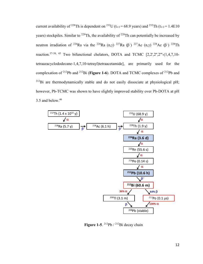
Figure 1-5. 212Pb / 212Bi decay chain 12
Figure 1-6. Chemical structures of DOTA (left) and TCMC (right) 13
Chapter 2
Figure 2-1. Chemical structure of RM2 24
Figure 2-2. ESI-MS of nonradioactive Pb-RM2 35
Figure 2-3. Efficiency of 59Fe stripping from Pb-resin 36
Figure 2-4. Radio-HPLC profiles of [203Pb]PbCl2 and [203Pb]Pb-RM2 36
Figure 2-5. IC50 curve of Pb-RM2 37
Figure 2-6. Biodistribution [203Pb]Pb-RM2 in PC3 tumor bearing mice 40
Figure 2-7. Comparison of [203Pb]Pb-RM2 uptake in tumor and pancreas 43
Figure 2-8. Comparison of [203Pb]Pb-RM2 and [203Pb]PbCl2 uptake in select
organs at 4 hours post injection 44
Figure 2-9A. MIP SPECT/CT/MRI image of [203Pb]Pb-RM2 in PC3 xenograft
SCID mouse 24 hours. 45
ix
Figure 2-9B. Comparison between [203Pb]Pb-RM2 (left panel) and [203Pb]PbCl2
(right panel) 45
Figure 2-10. Calculated excitation function for the 203Tl (p, n) 203Pb nuclear
reaction 49
Chapter 3
Figure 3-1. Lead-212 decay scheme 57
Figure 3-2. Picture of a 224Ra/212Pb generator housed in a lead pig (left) and
disassembled to show the cation exchange resin column (right) 64
Figure 3-3. Decay curve of 224Ra and 212Pb from a 10 mCi 224Ra/212Pb generator 65
Figure 3-4. Gamma spectrum of crude 212Pb generator eluent (A) and after
purification using Pb-resin (B) 66
Figure 3-5. A flow chart comparing the conventional method for the synthesis of
212Pb-labeled biomolecules with the new method developed in this study 68
Figure 3-6A. Radio-HPLC profiles of [212Pb]PbCl2 and [212Pb]Pb-RM2 using
gradient 1 (10% to 70% ACN:H2O over 10 minutes) 69
Figure 3-6B. HPLC profiles of [212Pb]Pb-RM2, Pb-RM2, and Bi-RM2 using
gradient 2 (22% to 25% ACN:H2O over 20 minutes) 69
Figure 3-7. Stability of [212Pb]Pb-RM2 in saline and 0.03 M gentisic acid
solution in saline 71
Figure 3-8. Biodistribution of [212Pb]Pb-RM2 in PC3 tumor bearing mice 75
Figure 3-9A. Comparison of [212Pb]Pb-RM2 and [203Pb]Pb-RM2 uptake in
PC3 tumor 75
Figure 3-9B. Comparison of [212Pb]Pb-RM2 and [203Pb]Pb-RM2 uptake in
pancreas 76
Figure 3-10A. Comparison of Pb contamination in [212Pb]PbCl2 generator
eluent before and after purification 77
Figure 3-10B. Comparison of Fe contamination in [212Pb]PbCl2 generator
eluent before and after purification 78
Figure 3-10C. Comparison of Zn contamination in [212Pb]PbCl2 generator
eluent before and after purification 78

x
Figure 3-11. Comparison of clonogenic response of PC3 and LNCAP cells
lines to [177Lu]Lu-RM2 treatment (left) and [212Pb]Pb-RM2 (right) 82
Figure 3-12. Effect of [212Pb]Pb-RM2 administration on body weight in
CF-1 mice 84
Figure 3-13. Effect of [212Pb]Pb-RM2 administration on white blood cells in
CF-1 mice 84
Figure 3-14. Effect of [212Pb]Pb-RM2 administration on platelets count in
CF-1 mice 85
Figure 3-15. Experimental and theoretical yields of 228Th, 227Ac, 228Ra and 229 Th from 226Ra irradiation at ORNL HFIR 88
Chapter 4
Figure 4-1. Schematic for 105Rh separation 97
Figure 4-2A. Crystal structure of cis-[RhCl2-222S4diAcOMe]PF6 as reported
by Dame 99
Figure 4-2B. Crystal structure of trans-[RhCl2-333S4diAcOMe]PF6 as reported
by Crenshaw 100
Figure 4-3. Synthesis of 222S4diAcOMe and 222S4diAcOH 112
Figure 4-4A. 1H NMR spectrum of Intermediate 1 113
Figure 4-4B. 13C NMR spectrum of Intermediate 1 114
Figure 4-5A. 1H NMR spectrum of 222S4diAcOMe 115
Figure 4-5B. 13C NMR spectrum of 222S4diAcOMe 116
Figure 4-5C. HMQC NMR spectrum of 222S4diAcOMe 117
Figure 4-6A. 1H NMR spectrum of 222S4diAcOH 118
Figure 4-6B. 13C NMR spectrum of 222S4diAcOH 119
Figure 4-6C. HMQC NMR spectrum of 222S4diAcOH 120
Figure 4-7. ESI-MS of 222S4diAcOH 121

xi
Figure 4-8. ESI-MS of cis-[Rh(III)Cl2-222S4diAcOMe]+ 123
Figure 4-9. ESI-MS of C11H20Cl2O4RhS4+ 124
Figure 4-10. ESI-MS of C11H19ClO4RhS4+ 125
Figure 4-11. ESI-MS of C10H16O4RhS4+ 126
Figure 4-12A. 13C NMR spectrum of [Rh(III)Cl2-222S4diAcOMe]Cl 128
Figure 4-12B. 1H NMR spectrum of [Rh(III)Cl2-222S4diAcOMe]Cl 129
Figure 4-12C. HMQC NMR spectrum of [Rh(III)Cl2-222S4diAcOMe]Cl 130
Figure 4-13. ESI-MS of C10H17ClO4RhS4+ 131
Figure 4-14. ESI-MS of C10H16O4RhS4+ 132
Figure 4-15. ESI-MS of for C10H18Cl2O4RhS4+ 133
Figure 4-16. ESI-MS of for [Rh(III)Cl2-16S4diol]+ 135
Figure 4-17. ESI-MS of for [Rh(III)OH2-16S4diol]+ 136
Supplementary Data – Chapter 2
Figure S2-1. HPLC profile of Pb-RM2 146
Supplementary Data – Chapter 3
Figure S3-1. Gamma spectrum peak analysis report for Pb-212 generator eluent 150
Figure S3-2. Gamma spectrum peak analysis report for purified Pb-212 152
Supplementary Data – Chapter 4
Figure S4-1A. HPLC spectrum for reaction 1 155
Figure S4-1B. HPLC spectrum for reaction 2 155

xii
Figure S4-1C. HPLC spectrum for reaction 3 155
Figure S4-1D. HPLC spectrum for reaction 4 156
Figure S4-1E. HPLC spectrum for reaction 5 156
Figure S4-1F. HPLC spectrum for reaction 6 156
Figure S4-1G. HPLC spectrum for reaction 7 157
Figure S4-1H. HPLC spectrum for reaction 8 157
Figure S4-1I. HPLC spectrum for reaction 9 157
Figure S4-2A. HPLC spectrum for reaction 10 158
Figure S4-2B. HPLC spectrum for reaction 11 158
Figure S4-2C. HPLC spectrum for reaction 12 158
Figure S4-2D. HPLC spectrum for reaction 13 158
Figure S4-2E. HPLC spectrum for reaction 14 158
Figure S4-2F. HPLC spectrum for reaction 15 159
Figure S4-3A. HPLC spectrum for reaction 16 159
Figure S4-3B. HPLC spectrum for reaction 17 159
Figure S4-3C. HPLC spectrum for reaction 18 159
Figure S4-3D. HPLC spectrum for reaction 19 160
Figure S4-3E. HPLC spectrum for reaction 20 160
Figure S4-3F. HPLC spectrum for reaction 21 160
Figure S4-4. TLC scan of [105Rh]RhCl3 solution (pH > 7) with 0.9% saline
as mobile phase 161
Figure S4-5. TLC scan of [105Rh]RhCl3 solution (pH ~ 4) using 0.9% saline
as mobile phase 161
Figure S4-6. TLC scan of [105Rh]RhCl2-222S4diAcOH using 0.9% saline
as mobile phase 162

xiii
Figure S4-7. TLC scan of [105Rh]RhCl2-222S4diAcOMe using 0.9% saline
as mobile phase 162
Figure S4-8. TLC scan of [105Rh]RhCl2-16S4diol using 0.9% saline as
mobile phase 163
Figure S4-9. TLC scan of [105Rh]RhCl3 solution using ACN as mobile phase 163
Figure S4-10. TLC scan of [105Rh]RhCl2-222S4diAcOH using ACN as
mobile phase 163
Figure S4-11. TLC scan of [105Rh]RhCl2-222S4diAcOMe using ACN as
mobile phase 164
Figure S4-12. TLC scan of [105Rh]RhCl2-16S4diol using ACN as mobile phase 164
Figure S4-13. TLC scan of [105Rh]RhCl3 solution using 0.4M NaBPh4 in
ACN as mobile phase 164
Figure S4-14. TLC scan of [105Rh]RhCl2-222S4diAcOH using 0.4M NaBPh4
in ACN as mobile phase 165
Figure S4-15. TLC scan of [105Rh]RhCl2-222S4diAcOMe using 0.4M NaBPh4
in ACN as mobile phase 165
Figure S4-16. TLC scan of [105Rh]RhCl2-16S4diol using 0.4M NaBPh4 in
ACN as mobile phase 165
List of Tables
Page
Chapter 2
Table 2-1. Technical data for [203Pb]PbCl2 purchased from Lantheus 28
Table 2-2. [203Pb]PbCl2 biodistribution in CF1 mice 38
Table 2-3. [203Pb]Pb-RM2 biodistribution in PC3 tumor bearing mice 41
Table 2-4. [203Pb]Pb-RM2 tumor uptake specificity 42
Chapter 3

xiv
Table 3-1. The significant X-rays and -rays associated with 212Pb and its
daughter radionuclides 67
Table 3-2A. [212Pb]Pb-RM2 in vitro stability in saline 70
Table 3-2B. [212Pb]Pb-RM2 in vitro stability in saline with gentisic acid 71
Table 3-3. [212Pb]Pb-RM2 biodistribution in PC3 tumor bearing mice 74
Table 3-4. Variations in [212Pb]Pb-RM2 pancreas and tumor uptake with the
date of synthesis 80
Table 3-5. Variations in [212Pb]Pb-RM2 specific activity and activity
concentration with the date of synthesis 80
Chapter 4
Table 4-1. 105Rh production from recycled 104Ru metal target at MURR 137
Table 4-2. ICP-MS analysis of metals in decayed 105Rh stock solution 139
Table 4-3. Silica gel TLC analysis of 105Rh complexes 140
Supplementary Data – Chapter 2
Table S2-1. Efficiency of 59Fe stripping from Pb-resin 146
Table S2-2. [203Pb]PbCl2 biodistribution in CF1 mice 147
Table S2-3. [203Pb]Pb-RM2 biodistribution in PC3 tumor-bearing mice 148
Supplementary Data – Chapter 3
Table S3-1. [212Pb]Pb-RM2 biodistribution in PC3 tumor bearing mice 153
Table S3-2. ICP-MS analysis of crude and purified 212Pb samples 154

xv
List of Schemes
Page
Chapter 4
Scheme 4-1. Reaction conditions for the synthesis of [RhCl2-222S4diAcOMe]Cl 106
Scheme 4-2. Reaction conditions investigated for the synthesis of
[RhCl2-222S4diAcOH]Cl 107
Scheme 4-3. Reaction conditions investigated for the synthesis of
[RhCl2-16S4diol]Cl 108
List of Equations
Page
Chapter 4
Equation 4-1. Production of 105Rh 97
Equation 4-2. Possible production route for 106Pd 139
Equation 4-3. Possible production route for 103Rh 139

xvi
Academic Abstract
The overexpression of certain peptide receptors on cancers cells can be exploited
for the development of radiopharmaceuticals that are selectively delivered to cancer cells
for diagnostic imaging or therapeutic purposes. Parts of this dissertation explore the
development and preclinical evaluation of a radiolabeled antagonist peptide conjugate
(RM2 = DOTA-4-amino-1-carboxymethyl-piperidine-D-Phe-Gln-Trp-Ala-Val-Gly-His-
Sta-Leu-NH2) that targets the bombesin receptor (BB2r) overexpressed in human prostate
cancer. Two radionuclides, 203Pb and 212Pb, were selected due to their uniqueness as a
chemically identical theranostic matched pair. Lead-203 (t1/2 = 51.9 hours) decays by
electron capture to stable 203Tl, with the emission of 278 keV gamma rays (81% intensity)
suitable for single-photon emission computed tomography (SPECT) imaging. On the other
hand, 212Pb (t1/2 = 10.6 hours) decays by beta emission into 212Bi (t1/2 = 60.6 minutes), which
subsequently decays into stable 208Pb through a branched decay chain consisting of one
alpha particle and one beta particle emission in each decay pathway. Hence, 212Pb is of
interest as an in vivo generator of 212Bi for targeted alpha therapy. The fundamental
chemistry and radiochemistry involved in the synthesis, purification and characterization
of both [203Pb]Pb-RM2 and [212Pb]Pb-RM2 is described. Additionally, in vivo preclinical
evaluation of the radiolabeled peptide conjugates was performed in male mouse models
inoculated with PC3 human prostate cancer cells.
The last portion of the work described in this dissertation focuses on 105Rh as a
potential therapeutic radionuclide. Rhodium-105 (t1/2 = 35.4 hours) is a moderate energy
beta-emitting radionuclide [
avg = 152 keV], with low energy gamma emissions [319 keV
(19%) and 306 keV (5%)]. The production of 105Rh from recycled 104Ru metal target via

xvii
the 104Ru (p, n) 105Ru 105Rh reaction was reported. In addition, a microwave-assisted
procedure for the synthesis of Rh(III) complexes without the addition of refluxing ethanol
or SnCl2 as reducing agents is described.

1
CHAPTER 1: General Introduction
1.1. Radiopharmaceuticals as a Tool in Cancer Diagnosis and
Therapy
Simply put, a radiopharmaceutical is a radioactive drug. Radiopharmaceuticals
have emerged as a very useful tool for physicians in the fight against cancer by
revolutionizing the ability to non-invasively detect, stage, treat, and monitor the
progression of cancer. Similar to other pharmaceutical drugs, the development of a new
radiopharmaceutical is a long and complex process that involves a great deal of
interdisciplinary research. This process typically includes understanding the disease at a
molecular level, identifying molecular targets specific to the disease, designing a lead
compound that has a desired influence on the identified molecular target, optimizing the
lead compound to meet minimal toxicity and favorable pharmacokinetics, and performing
numerous preclinical and clinical trials to ascertain the safety and efficacy of the drug in
humans.
Nevertheless, the radiopharmaceutical drug development process is uniquely
different from that of a conventional pharmaceutical drug. This difference stems mainly
from the fact that the key component in a radiopharmaceutical is the radionuclide, whose
nuclear properties (e.g., half-life, type of radioactive decay, etc.) determine the clinical
utility of the drug. Unfortunately, scientists have no control over the nuclear properties of
a radionuclide; hence the choices for clinically-relevant radionuclides are limited. In some
cases, the initial chemical form of the radionuclide may create additional challenges that
would not have been otherwise encountered for conventional pharmaceutical development.

2
Despite these challenges, several radiopharmaceuticals have been successfully developed
and are currently being used in patient care.
For a new radiopharmaceutical to be successful, it must fulfill one of two clinical
needs: (1) be used as diagnostic agent or (2) be used as a therapeutic agent. Diagnostic
radiopharmaceuticals require radionuclides that emit either gamma () rays or positrons
(+). Gamma rays are electromagnetic radiation, which minimally interact with body
tissues and can easily penetrate the body, enabling them to be externally detected by
radiation detectors. Gamma ray-emitting radionuclides (e.g., 99mTc, 111In, 201Tl, 203Pb, etc.)
are useful for single photon emission computed tomography (SPECT), where the gamma
rays are detected by a single or a pair of lead-collimated radiation detectors (typically
NaI(Tl) scintillation detectors).1 On the other hand, positrons are positively charged
electrons emitted from the nucleus of proton-rich nuclides. Positrons interact with
negatively charged electrons through a process called positron annihilation to produce two
coincident 511 keV photons approximately 180° apart. Positron-emitting radionuclides
(e.g., 18F, 89Zr, 68Ga, etc.) are useful for positron emission tomography (PET), where the
511 keV annihilation photons are detected in coincidence by a ring of scintillation radiation
detectors (e.g., lutetium oxyorthosilicate, LSO), which allows for the use of electronic
collimation to distinguish between true and interfering photons.1 In both SPECT and PET
imaging modalities, mathematical algorithms are utilized to reconstruct a 3D image
showing the distribution of the radionuclide in the body.1
In contrast, therapeutic radiopharmaceuticals require radionuclides that emit
ionizing particulate radiation, such as alpha () particles, beta (-) particles, or Auger
electrons. Alpha particles are the same as a helium nucleus consisting of two protons, two

3
neutrons and have a +2 charge. Alpha particles are predominantly emitted by high mass
radionuclides (A ≥ 210). Beta particles are negative electrons ejected from a neutron-rich
nucleus. Auger electrons are low energy (0.1 – 1 keV) orbital electrons that are emitted by
radionuclides that decay by either electron capture or internal conversion. Alpha particles,
beta particles and Auger electrons all strongly interact with target tissues (e.g., cancerous
tumor) and lead to extensive localized ionization, which can damage chemical bonds in
DNA molecules and potentially induce cytotoxicity.2
For most nuclear medicine applications, it is desired that a diagnostic
radiopharmaceutical be coupled with a therapeutic radiopharmaceutical. This concept is
commonly known as “theranostics”.3 It is important that the chemical and pharmacokinetic
behaviors of both the diagnostic and therapeutic radiopharmaceuticals match. Ideally, the
diagnostic and therapeutic radionuclides are a chemically identical radioisotope pair (also
known as “matched pair”). The most well-known matched pair for theranostic
radiopharmaceutical application is the 123I/131I pair, where 123I-labeled compounds (e.g.,
[123I]NaI capsules) are used for diagnosis, while 131I-labeled compounds (e.g., [131I]NaI
capsules) are used for therapy. Another promising theranostic matched pair is the
203Pb/212Pb pair, which is the focus of Chapters 2 and 3, respectively. Alternatively,
radionuclide pairs from different elements can be utilized for theranostic
radiopharmaceutical development provided that their chemistry is very similar (e.g.,
99mTc/186/188Re) and there is no significant difference in the pharmacokinetic behavior
between the diagnostic and therapeutic analogues. A good example is the 68Ga/177Lu pair,
where 68Ga is used for diagnosis and 177Lu is used for therapy. The 68Ga/177Lu pair has been
utilized in the development of two FDA-approved radiopharmaceuticals, [68Ga]Ga-

4
Dotatate (NETSPOT™)4 and [177Lu]Lu-Dotatate (LUTATHERA®)5, for the diagnosis and
treatment of gastroenteropancreatic neuroendocrine tumors, respectively.
1.2. Radiopharmaceutical Design
Some radionuclides possess an intrinsic affinity to certain organs/tissues due to
their physiological properties. These radionuclides can therefore be used as
radiopharmaceuticals by direct administration of the anionic or cationic salt. For example,
iodine is primarily absorbed in the thyroid and is essential for the formation thyroid
hormones. Therefore, radioactive iodine capsules ([131I]NaI) are administered for the
treatment of thyroid cancer. Similarly, radium acts as a calcium mimic and readily forms
complexes with the bone mineral, hydroxyapatite. Therefore, radioactive radium salt
([223Ra]RaCl2) is used for the treatment of bone metastases. Other radiopharmaceuticals
require that the radionuclide be incorporated into a biologically-active targeting molecule
for it to be delivered to a specific target organ/tissue. This can be accomplished by two
methods: the integrated method (also direct labeling method) and the bifunctional chelate
(BFC) method. In the integrated method, the radionuclide is an integral part of the
biologically-active molecule and is directly incorporated into it. This method is
predominantly utilized in radiopharmaceuticals containing non-metallic radionuclides
(e.g., [18F]fluorodeoxyglucose, [11C]choline, etc.). However, radiopharmaceuticals
containing metallic radionuclides have also been developed using the integrated method
(e.g., [99mTc]Tc-Sestamibi, [99mTc]Tc-methylene diphosphonate, etc.). The BFC method is
predominantly utilized in radiopharmaceuticals containing metallic radionuclides. The
radiometal is tightly bound to a suitable bifunctional chelator that has been covalently
conjugated to a targeting vector (e.g., peptides, proteins, antibodies, etc.) through a linker

5
or spacer, as shown in Figure 1-1. The targeting vector serves as the vehicle for delivery
of the radionuclide to the organ/tissue of interest. Targeting vectors localize at their
biological targets through various mechanisms including receptor binding,
antigen/antibody reactions, etc.
Figure 1-1. Schematic of the bifunctional chelate (BFC) method
1.3. Targeted Alpha Therapy
Out of all the therapeutic radiations (i.e., , -, and Auger electrons), alpha particles
have the greatest biological effectiveness because of their high linear energy transfer (LET)
properties (~100 keV/m), which is a result of their densely ionizing track and short path
length (50-100 m) in tissues.6-8 Due to their high LET, a few alpha particles (between 1
and 20) traversing through a cancer cell can induce irreparable double-strand DNA
breakage, subsequently resulting in cell death through a number of mechanisms including
apoptosis, autophagy, and necrosis.6-7 In contrast, low LET radiation like beta particles
(~0.2 keV/m) produce mainly single-strand DNA breakage, which has a lower cytotoxic
effect. The cytotoxic effect of alpha particles has been shown to be independent of dose
rate and tissue oxygen levels, which makes them effective against hypoxic tumors.6, 9
target cell
receptor
targeting vector bifunctional
chelator
radiometal linker

6
The cytotoxic superiority of alpha particles over beta particles has been
demonstrated in several preclinical studies. In vitro studies comparing 213Bi, an alpha-
emitting radionuclide, with 177Lu, a beta-emitting radionuclide, demonstrated that 213Bi has
significantly greater cytotoxic potency.10-11 Greater DNA double-strand breaks, as
quantified by immunofluorescence staining of H2AX-foci, was observed for [225Ac]Ac-
DOTATOC compared to [177Lu]Lu-DOTATOC.12 Wild et al.13 reported that the median
survival time of mouse models bearing human prostate carcinoma (PC3) xenografts was
increased by about 15 weeks when treated with [213Bi]Bi-DOTA-PESIN compared to
[177Lu]Lu-DOTA-PESIN.
The first reported clinical trial for alpha particle radioimmunotherapy was
performed using 213Bi-labeled HuM195 (a humanized anti-CD33 monoclonal antibody) for
the treatment of myeloid leukemia, where therapeutic response in 14 out of the 15 evaluated
patients was reported.14 In another study, long-lasting therapeutic response was reported
after administration of [213Bi]Bi-DOTATOC to patients who were refractory to prior
treatment with the beta-emitting compounds ([90Y]Y-DOTATOC and [177Lu]Lu-
DOTATOC).15 Studies using 211At-labeled antibodies have shown positive clinical
outcomes for the treatment of glioblastoma16 and ovarian cancer.17-19 The safety and
therapeutic efficacy of [212Pb]Pb-TCMC-trastuzumab in patients with HER2-positive
peritoneal malignancies has been recently demonstrated in clinical studies by Meredith et
al.20-22 Perhaps the alpha-emitting radionuclide that has gained the most attention in the
past five years is 225Ac. This increased attention is partly due to the remarkable therapeutic
outcomes observed after administration of [225Ac]Ac-PSMA-617 to patients with

7
metastatic castration-resistant prostate cancer who failed to respond to prior treatment with
[177Lu]Lu-PSMA-617.23-25
In a recent phase III clinical trial, treatment with [223Ra]RaCl2 resulted in significant
survival benefits in men with bone metastases resulting from castration-resistant prostate
cancer.26 This study subsequently led to the FDA approval of [223Ra]RaCl2 (Xofigo®),
which is currently the only FDA-approved alpha-emitting radiopharmaceutical.
1.4. Alpha-emitting Radionuclides of Clinical Relevance
Based on the promising preclinical and clinical studies supporting the cytotoxic
superiority of alpha particles over beta particles, there has been an increased interest for
alpha-emitting radionuclides that are useful for targeted alpha therapy.8, 27-28 The most
promising clinically-relevant alpha-emitting radionuclides are discussed below.
1.4-1. Astatine-211
Astatine-211 (t1/2 = 7.2 hours) decays through a branched pathway into stable 207Pb
with the net emission of one alpha particle through each decay pathway (Figure 1-2).
Astatine-211 is cyclotron produced by bombarding a bismuth target with alpha particles
(~28 MeV) via the 209Bi(, 2n)211At reaction.29-30 Unfortunately, very few cyclotron
facilities have the capability of generating > 25 MeV alpha particles, which has greatly
limited the availability of 211At. The lack of stable astatine isotopes has limited the
understanding of astatine chemistry. As a member of the halogen family, the chemical
properties of astatine are believed to be similar to iodine; however, astatine exhibits more
metallic characteristics.31 A major concern with 211At radiopharmaceuticals is in vivo
stability due to the relatively weak carbon-astatine bond strength, which can lead to

8
deastatination and subsequent physiological accumulation of free astatine in thyroid and
stomach.32 In addition, the presence of a relatively long-lived daughter radionuclide from
211At decay (207Bi, t1/2 = 31.5 years) is not ideal.
Figure 1-2. Astatine-211 decay chain
1.4-2. Radium-223
Radium-223 (t1/2 = 11.4 days) decays into stable 207Pb through a series of
intermediate daughter radionuclides, with the net emission of four alpha particles and two
beta particles in its decay pathway (Figure 1-3). Radium-223 is obtained as a product of
227Th decay, which is the daughter radionuclide from 227Ac decay. As an alkaline earth
metal, 223Ra has similar chemical properties to calcium and readily accumulates in the
inorganic bone matrix through complexation to the bone mineral, hydroxyapatite.26 This
property of 223Ra was exploited for the development of [223Ra]RaCl2 (Xofigo®) for the
treatment of bone metastases resulting from castration-resistant prostate cancer.26 The
application of 223Ra for the development of radiopharmaceuticals that target other diseases
has been challenging due to the lack of suitable bifunctional chelators for 223Ra
complexation.
58.2% EC
211At (7.2 h)
207Pb (stable)
211Po (0.5 s)
41.8% , 5.9 MeV
207Bi (31.5 y)
, 7.5 MeVEC

9
Figure 1-3. Radium-223 decay chain
1.4-3. Actinum-225 / Bismuth-213
Actinium-225 (t1/2 = 9.9 days) is a member of the 233U decay chain (Figure 1-4).
Actinium-225 decays into 213Bi (t1/2 = 45.6 minutes) through two intermediate alpha-
emitting daughter radionuclides (221Fr and 217At). Bismuth-213 subsequently undergoes a
branched decay into stable 209Bi, with the emission of one alpha particle and two beta
particles through each decay pathway. Therefore, the decay of 225Ac into stable 209Bi results
in the net emission of four alpha particles and two beta particles. Both 225Ac and 213Bi are
being investigated for the development of targeted alpha radiopharmaceuticals; however,
the short half-life of 213Bi poses a challenge during the radiopharmaceutical synthesis and
purification process. Using 225Ac as an in vivo generator of 213Bi overcomes the challenges
223Ra (11.4 d)
219Rn (3.96 s)
215Po (1.78 ms)
211Pb (36.1 m)
211Bi (2.13 m)
207Pb (stable)
211Po (0.52 s)
99.7%
207Tl (4.77 m)
0.3%
227Ac (21.8 y)
227Th (18.7 d)

10
associated with the short half-life of 213Bi. The current chelators of choice for the formation
of 225Ac/213Bi complexes are polyaminocarboxylic acid-based chelators, particularly
DOTA (1,4,7,10-tetraazacyclododecane-1,4,7,10-tetraacetic acid) and its derivatives
(Figure 1-6, left).
Figure 1-4. 225Ac / 213Bi decay chain
The primary source of 225Ac/213Bi is from 229Th build-up resulting from 233U decay.
The current estimated annual global supply of 225Ac/213Bi from this source is 1.7 Ci (63
GBq), which will not be sufficient to meet clinical and research demands.33 Another
potential production route for 225Ac that is currently being investigated is the proton
spallation of thorium targets via the 232Th(p,x)225Ac reaction.34-36 However, potential co-
production of 227Ac (t1/2 = 21.8 years) is of concern. Efforts have also been made to increase
225Ra (14.9 d)
225Ac (9.9 d)
221Fr (4.8 m)
217At (32 ms)
213Bi (45.6 m)
209Pb (3.3 h)
213Po (4.2 s)
2%
209Tl (2.2 m)
98%
229Th (7340 y)
209Bi (stable)
233U (1.6 x 10⁵ y)

11
the 229Th stockpile by neutron irradiation of 226Ra via the 226Ra (n,) 227Ra (-) 227Ac (n,)
228Ac (-) 228Th (n,) 229Th reaction.37-38 Apostolidis et al.39 reported the production of 225Ac
in a cyclotron via the 226Ra (p, 2n) 225Ac reaction with a relatively high cross-section of
710 mb at 16.8 MeV. It is estimated that the irradiation of an ~1 g of 226Ra target with a 20
MeV proton beam at 500 A beam current could produce a theoretical maximum of 108
Ci (4 TBq) of 225Ac per month.33 The indirect production of 225Ac from the photonuclear
transmutation of 226Ra via the 226Ra (, n) 225Ra (-) 225Ac reaction is also very promising.40-
43
1.4-4. Lead-212 / Bismuth-212
Lead-212 (t1/2 = 10.6 hours) and its daughter, 212Bi (t1/2 = 60.6 minutes), are both
members of the natural 232Th decay series (Figure 1-5). Lead-212 decays by beta emission
into 212Bi, which subsequently undergoes a branched decay chain into stable 208Pb, with
the emission of one alpha particle and one beta particle through each decay pathway.
Therefore, the decay of 212Pb results in the net emission of one alpha particle and two beta
particles. Similar to 213Bi, the relatively short half-life of 212Bi can be a challenging factor
during the radiopharmaceutical synthesis and purification process. However, using 212Pb
as an in vivo generator of 212Bi significantly compensates for the shorter half-life of 212Bi
and allows sufficient time for the preparation, administration, and localization of the
radiopharmaceutical at the target organ/tissue within the patient.44
The primary source for 212Pb/212Bi is from 228Th (t1/2 = 1.9 years), which decays into
224Ra that is subsequently used for producing 224Ra/212Pb generators. Thorium-228 is the
product of 232U decay and is also the second member of the 232Th decay series; hence, the
12
current availability of 228Th is dependent on 232U (t1/2 = 68.9 years) and 232Th (t1/2 = 1.4E10
years) stockpiles. Similar to 229Th, the availability of 228Th can potentially be increased by
neutron irradiation of 226Ra via the 226Ra (n,) 227Ra (-) 227Ac (n,) 228Ac (-) 228Th
reaction.37-38, 45 Two bifunctional chelators, DOTA and TCMC [2,2',2'',2'''-(1,4,7,10-
tetraazacyclododecane-1,4,7,10-tetrayl)tetraacetamide], are primarily used for the
complexation of 212Pb and 212Bi (Figure 1-6). DOTA and TCMC complexes of 212Pb and
212Bi are thermodynamically stable and do not easily dissociate at physiological pH;
however, Pb-TCMC was shown to have slightly improved stability over Pb-DOTA at pH
3.5 and below.46
Figure 1-5. 212Pb / 212Bi decay chain

13
Figure 1-6. Chemical structures of DOTA (left) and TCMC (right)
1.5. Bombesin Receptors as Molecular Targets in Prostate
Cancer
The gastrin-releasing peptide receptor (GRPR), also known as BB2 receptor is a
cell surface transmembrane protein belonging to the family of bombesin receptors, a type
of G-protein-coupled receptor possessing a seven transmembrane domain.47 Several
reports have demonstrated that the BB2 receptor is overexpressed in prostate cancer
tumors.48-52 Using in vitro receptor autoradiography, Körner et al. 51 observed high
incidence of BB2 receptor expression in prostate carcinoma (77%, n = 60) and high grade
prostatic intraepithelial neoplasia (73%, n = 55), whereas very weak expression was
observed in normal prostate glands (18%, n = 111). Similarly, high BB2 receptor
expression was observed in 100% (n = 30) of prostate carcinoma tissues studied by
Markwalder et al. 48, with very high receptor density found in 83% of the studied tissues.
The native ligand for the BB2 receptor has been identified as the gastrin-releasing peptide
and was shown to bind with high affinity. The biologically active portion of the gastrin-
releasing peptide is its carboxyl terminus consisting of a seven amino acid sequence (-Trp-
Ala-Val-Gly-His-Leu-Met-NH2). The selective overexpression of BB2 receptor in prostate
cancer tumors, along with the high binding affinity of gastrin-releasing peptide analogues
for the BB2 receptor, has inspired research into the development of targeted prostate cancer

14
theranostic radiopharmaceuticals using radiolabeled gastrin-releasing peptide analogues.53-
55
Initial studies focused on radiolabeled gastrin-releasing peptide agonists that are
internalized upon binding to BB2 receptors. Several compounds such as [99mTc]Tc-RP527
56-57, [177Lu]Lu-AMBA 58-59, [177Lu]Lu-DOTA-PESIN,13, 60 and [111In]In-DOTA-X-
BBN(7-14)NH2 61-62 were developed and investigated. Several clinical trials have
demonstrated the safety of radiolabeled BB2 receptors agonist peptide conjugates as
molecular imaging agents.57, 63 However, in one unpublished clinical trial with [177Lu]Lu-
AMBA, side effects such as nausea, diarrhea and abdominal cramps were reported.59 These
side effects could possibly be attributed to the fact that activation of the BB2 receptor by
gastrin-releasing peptide agonists can trigger a range of physiological responses like
release of gastrointestinal hormones, contraction of smooth muscles in the gastrointestinal
tract, and proliferation of cells.47, 64-65 As a result, there has been a shift towards the
investigation of gastrin-releasing peptide antagonists. Comparison between antagonist and
agonist gastrin-releasing peptides revealed improved pharmacokinetics and tumor
targeting properties with the antagonist peptides.66-67 Of particular interest is RM2 (DOTA-
4-amino-1-carboxymethyl-piperidine-D-Phe-Gln-Trp-Ala-Val-Gly-His-Sta-Leu-NH2), a
BB2 receptor antagonist that was shown to exhibit high and specific tumor accumulation
and retention in PC3 human prostate tumor-bearing mice.68 Clinical studies using
[68Ga]Ga-RM2 have demonstrated its high sensitivity and specificity for the detection of
metastatic prostate cancer lesions in patients with biochemically recurrent disease.69-72
Additionally, no drug related toxicity has been reported. Other BB2 receptor antagonist
peptide conjugates such as NeoBOMB1 73-74 and SB3 75-76 have also been reported.

15
1.6. Dissertation Outline
In this work, the fundamental chemistry and radiochemistry involved in the
synthesis of targeted radiopharmaceuticals are described.
Chapter 2 describes the synthesis and characterization of [203Pb]Pb-RM2 as a
potential agent for diagnostic imaging of prostate cancer. As previously mentioned, RM2
is a potent BB2 receptor antagonist peptide conjugate that binds with high affinity and
specificity to the BB2 receptors overexpressed on prostate cancer cells. A method for
purification of the commercially-purchased [203Pb]PbCl2 is described using a lead-selective
chromatographic resin (Pb-resin). Biodistribution and in vivo imaging studies of [203Pb]Pb-
RM2 in male mouse models inoculated with PC3 human prostate cancer cells are also
reported.
In Chapter 3, the automated synthesis of [212Pb]Pb-RM2, the therapeutic analogue
of [203Pb]Pb-RM2, is reported. An improved method for the elution and purification of the
224Ra/212Pb generator eluent is described. Additionally, preclinical evaluation of [212Pb]Pb-
RM2 in male mouse models inoculated with PC3 human prostate cancer cells is reported.
Finally, the fundamental chemistry, radiochemistry, and production of 105Rh from
recycled 104Ru metal target via the 104Ru (p, n) 105Ru 105Rh reaction is reported in
Chapter 4. A microwave-assisted method for the synthesis of Rh(III) complexes that does
not require the addition of refluxing ethanol or SnCl2 as reducing agents is also described.

16
1.7. References
1. Bailey, D. L.; Humm, J., Nuclear medicine physics: a handbook for teachers and students. IAEA: 2014.
2. Kassis, A. I.; Adelstein, S. J., Radiobiologic principles in radionuclide therapy. Journal of Nuclear Medicine 2005, 46 (1 suppl), 4S-12S.
3. Yordanova, A.; Eppard, E.; Kürpig, S.; Bundschuh, R. A.; Schoenberger, S.; Gonzalez-Carmona, M.; Feldmann, G.; Ahmadzadehfar, H.; Essler, M., Theranostics in nuclear medicine practice. OncoTargets and Therapy 2017, 10, 4821.
4. Deppen, S. A.; Liu, E.; Blume, J. D.; Clanton, J.; Shi, C.; Jones-Jackson, L. B.; Lakhani, V.; Baum, R. P.; Berlin, J.; Smith, G. T., Safety and efficacy of 68Ga-DOTATATE PET/CT for diagnosis, staging, and treatment management of neuroendocrine tumors. Journal of Nuclear Medicine 2016, 57 (5), 708-714.
5. Strosberg, J.; El-Haddad, G.; Wolin, E.; Hendifar, A.; Yao, J.; Chasen, B.; Mittra, E.; Kunz, P. L.; Kulke, M. H.; Jacene, H., Phase 3 trial of 177Lu-Dotatate for midgut neuroendocrine tumors. New England Journal of Medicine 2017, 376 (2), 125-135.
6. Sgouros, G.; Roeske, J. C.; McDevitt, M. R.; Palm, S.; Allen, B. J.; Fisher, D. R.; Brill, A. B.; Song, H.; Howell, R. W.; Akabani, G., MIRD Pamphlet No. 22 (abridged): radiobiology
and dosimetry of α-particle emitters for targeted radionuclide therapy. Journal of Nuclear Medicine 2010, 51 (2), 311-328.
7. Baidoo, K. E.; Yong, K.; Brechbiel, M. W., Molecular pathways: targeted α-particle radiation therapy. Clinical Cancer Research 2013, 19 (3), 530-537.
8. Parker, C.; Lewington, V.; Shore, N.; Kratochwil, C.; Levy, M.; Lindén, O.; Noordzij, W.; Park, J.; Saad, F., Targeted Alpha Therapy, an Emerging Class of Cancer Agents: A Review. JAMA Oncology 2018, 4 (12), 1765-1772.
9. Wulbrand, C.; Seidl, C.; Gaertner, F. C.; Bruchertseifer, F.; Morgenstern, A.; Essler, M.; Senekowitsch-Schmidtke, R., Alpha-particle emitting 213Bi-anti-EGFR immunoconjugates eradicate tumor cells independent of oxygenation. PloS One 2013, 8 (5), e64730.
10. Nayak, T.; Norenberg, J.; Anderson, T.; Atcher, R., A comparison of high-versus low-linear energy transfer somatostatin receptor targeted radionuclide therapy in vitro. Cancer Biotherapy & Radiopharmaceuticals 2005, 20 (1), 52-57.
11. Chan, H. S.; de Blois, E.; Morgenstern, A.; Bruchertseifer, F.; de Jong, M.; Breeman, W.; Konijnenberg, M., In Vitro comparison of 213Bi-and 177Lu-radiation for peptide receptor radionuclide therapy. PloS One 2017, 12 (7), e0181473.
12. Graf, F.; Fahrer, J.; Maus, S.; Morgenstern, A.; Bruchertseifer, F.; Venkatachalam, S.; Fottner, C.; Weber, M. M.; Huelsenbeck, J.; Schreckenberger, M.; Kaina, B.; Miederer, M., DNA Double Strand Breaks as Predictor of Efficacy of the Alpha-Particle Emitter Ac-

17
225 and the Electron Emitter Lu-177 for Somatostatin Receptor Targeted Radiotherapy. Plos One 2014, 9 (2), e88239.
13. Wild, D.; Frischknecht, M.; Zhang, H.; Morgenstern, A.; Bruchertseifer, F.; Boisclair, J.; Provencher-Bolliger, A.; Reubi, J.-C.; Maecke, H. R., Alpha-versus beta-particle radiopeptide therapy in a human prostate cancer model (213Bi-DOTA-PESIN and 213Bi-AMBA versus 177Lu-DOTA-PESIN). Cancer Research 2011, 71 (3), 1009-1018.
14. Jurcic, J. G.; Larson, S. M.; Sgouros, G.; McDevitt, M. R.; Finn, R. D.; Divgi, C. R.;
Ballangrud, Å. M.; Hamacher, K. A.; Ma, D.; Humm, J. L., Targeted α particle immunotherapy for myeloid leukemia. Blood 2002, 100 (4), 1233-1239.
15. Kratochwil, C.; Giesel, F.; Bruchertseifer, F.; Mier, W.; Apostolidis, C.; Boll, R.; Murphy, K.; Haberkorn, U.; Morgenstern, A., 213Bi-DOTATOC receptor-targeted alpha-radionuclide therapy induces remission in neuroendocrine tumours refractory to beta radiation: a first-in-human experience. European Journal of Nuclear Medicine and Molecular Imaging 2014, 41 (11), 2106-2119.
16. Zalutsky, M. R.; Reardon, D. A.; Akabani, G.; Coleman, R. E.; Friedman, A. H.;
Friedman, H. S.; McLendon, R. E.; Wong, T. Z.; Bigner, D. D., Clinical experience with α-particle–emitting 211At: treatment of recurrent brain tumor patients with 211At-labeled chimeric antitenascin monoclonal antibody 81C6. Journal of Nuclear Medicine 2008, 49 (1), 30-38.
17. Andersson, H.; Cederkrantz, E.; Bäck, T.; Divgi, C.; Elgqvist, J.; Himmelman, J.;
Horvath, G.; Jacobsson, L.; Jensen, H.; Lindegren, S., Intraperitoneal α-particle radioimmunotherapy of ovarian cancer patients: pharmacokinetics and dosimetry of 211At-Mx35 F(ab′)2 - A phase I study. Journal of Nuclear Medicine 2009, 50 (7), 1153-1160.
18. Cederkrantz, E.; Andersson, H.; Bernhardt, P.; Bäck, T.; Hultborn, R.; Jacobsson, L.; Jensen, H.; Lindegren, S.; Ljungberg, M.; Magnander, T., Absorbed doses and risk estimates of 211At-MX35 F (ab') 2 in intraperitoneal therapy of ovarian cancer patients. International Journal of Radiation Oncology* Biology* Physics 2015, 93 (3), 569-576.
19. Hallqvist, A.; Bergmark, K.; Bäck, T. A.; Andersson, H.; Dahm-Kähler, P.; Johansson, M.; Lindegren, S.; Jensen, H.; Jacobsson, L.; Hultborn, R., Intraperitoneal alpha-emitting radio immunotherapy with Astatine-211 in relapsed ovarian cancer; long-term follow-up with individual absorbed dose estimations. Journal of Nuclear Medicine 2019, jnumed. 118.220384.
20. Meredith, R.; Torgue, J.; Shen, S.; Fisher, D. R.; Banaga, E.; Bunch, P.; Morgan, D.; Fan, J.; Straughn, J. M., Jr., Dose escalation and dosimetry of first-in-human alpha radioimmunotherapy with 212Pb-TCMC-trastuzumab. Journal of Nuclear Medicine 2014, 55 (10), 1636-42.
21. Meredith, R. F.; Torgue, J.; Azure, M. T.; Shen, S.; Saddekni, S.; Banaga, E.; Carlise, R.; Bunch, P.; Yoder, D.; Alvarez, R., Pharmacokinetics and imaging of 212Pb-TCMC-

18
trastuzumab after intraperitoneal administration in ovarian cancer patients. Cancer Biotherapy & Radiopharmaceuticals 2014, 29 (1), 12-7.
22. Meredith, R. F.; Torgue, J. J.; Rozgaja, T. A.; Banaga, E. P.; Bunch, P. W.; Alvarez, R. D.; Straughn Jr, J. M.; Dobelbower, M. C.; Lowy, A. M., Safety and outcome measures of
first-in-human intraperitoneal α radioimmunotherapy with 212Pb-TCMC-trastuzumab. American Journal of Clinical Oncology 2018, 41 (7), 716.
23. Kratochwil, C.; Bruchertseifer, F.; Giesel, F. L.; Weis, M.; Verburg, F. A.; Mottaghy, F.; Kopka, K.; Apostolidis, C.; Haberkorn, U.; Morgenstern, A., 225Ac-PSMA-617 for PSMA-
targeted α-radiation therapy of metastatic castration-resistant prostate cancer. Journal of Nuclear Medicine 2016, 57 (12), 1941-1944.
24. Kratochwil, C.; Bruchertseifer, F.; Rathke, H.; Bronzel, M.; Apostolidis, C.;
Weichert, W.; Haberkorn, U.; Giesel, F. L.; Morgenstern, A., Targeted α-Therapy of Metastatic Castration-Resistant Prostate Cancer with 225Ac-PSMA-617: Dosimetry Estimate and Empiric Dose Finding. Journal of Nuclear Medicine 2017, 58 (10), 1624-1631.
25. Rahbar, K.; Ahmadzadehfar, H.; Kratochwil, C.; Haberkorn, U.; Schäfers, M.; Essler, M.; Baum, R. P.; Kulkarni, H. R.; Schmidt, M.; Drzezga, A., German multicenter study investigating 177Lu-PSMA-617 radioligand therapy in advanced prostate cancer patients. Journal of Nuclear Medicine 2017, 58 (1), 85-90.
26. Parker, C.; Nilsson, S.; Heinrich, D.; Helle, S. I.; O'Sullivan, J.; Fosså, S. D.; Chodacki, A.; Wiechno, P.; Logue, J.; Seke, M., Alpha emitter radium-223 and survival in metastatic prostate cancer. New England Journal of Medicine 2013, 369 (3), 213-223.
27. Kim, Y.-S.; Brechbiel, M. W., An overview of targeted alpha therapy. Tumor Biology 2012, 33 (3), 573-590.
28. Mulford, D. A.; Scheinberg, D. A.; Jurcic, J. G., The promise of targeted α-particle therapy. Journal of Nuclear Medicine 2005, 46 (1 suppl), 199S-204S.
29. R Zalutsky, M.; Pruszynski, M., Astatine-211: production and availability. Current Radiopharmaceuticals 2011, 4 (3), 177-185.
30. Guérard, F.; Gestin, J.-F.; Brechbiel, M. W., Production of [211At]-astatinated
radiopharmaceuticals and applications in targeted α-particle therapy. Cancer Biotherapy and Radiopharmaceuticals 2013, 28 (1), 1-20.
31. Vaidyanathan, G.; Zalutsky, M. R., Astatine radiopharmaceuticals: prospects and problems. Current Radiopharmaceuticals 2008, 1 (3), 177-196.
32. Wilbur, D., [211At]Astatine-labeled compound stability: Issues with released [211At]astatide and development of labeling reagents to increase stability. Current Radiopharmaceuticals 2008, 1 (3), 144-176.
33. Robertson, A. K.; Ramogida, C. F.; Schaffer, P.; Radchenko, V., Development of 225Ac radiopharmaceuticals: TRIUMF perspectives and experiences. Current Radiopharmaceuticals 2018, 11 (3), 156-172.

19
34. Griswold, J. R.; Medvedev, D. G.; Engle, J. W.; Copping, R.; Fitzsimmons, J. M.; Radchenko, V.; Cooley, J.; Fassbender, M.; Denton, D. L.; Murphy, K. E., Large scale accelerator production of 225Ac: Effective cross sections for 78–192 MeV protons incident on 232Th targets. Applied Radiation and Isotopes 2016, 118, 366-374.
35. Weidner, J.; Mashnik, S.; John, K.; Ballard, B.; Birnbaum, E.; Bitteker, L.; Couture, A.; Fassbender, M.; Goff, G.; Gritzo, R., 225Ac and 223Ra production via 800 MeV proton irradiation of natural thorium targets. Applied Radiation and Isotopes 2012, 70 (11), 2590-2595.
36. Weidner, J.; Mashnik, S.; John, K.; Hemez, F.; Ballard, B.; Bach, H.; Birnbaum, E.; Bitteker, L.; Couture, A.; Dry, D., Proton-induced cross sections relevant to production of 225Ac and 223Ra in natural thorium targets below 200 MeV. Applied Radiation and isotopes 2012, 70 (11), 2602-2607.
37. Hogle, S.; Boll, R. A.; Murphy, K.; Denton, D.; Owens, A.; Haverlock, T. J.; Garland, M.; Mirzadeh, S., Reactor production of Thorium-229. Applied Radiation and Isotopes 2016, 114, 19-27.
38. Kuznetsov, R.; Butkalyuk, P.; Tarasov, V.; Baranov, A. Y.; Butkalyuk, I.; Romanov, E.; Kupriyanov, V.; Kazakova, E., Yields of activation products in 226Ra irradiation in the high-flux SM reactor. Radiochemistry 2012, 54 (4), 383-387.
39. Apostolidis, C.; Molinet, R.; McGinley, J.; Abbas, K.; Möllenbeck, J.; Morgenstern, A., Cyclotron production of Ac-225 for targeted alpha therapy. Applied Radiation and Isotopes 2005, 62 (3), 383-387.
40. Nolen, J. A.; Brown, M. A.; Rotsch, D. A.; Chemerisov, S. D.; Henning, W. F.; Song, J., Compact assembly for production of medical isotopes via photonuclear reactions. United States Patents: 2019.
41. Melville, G.; Meriarty, H.; Metcalfe, P.; Knittel, T.; Allen, B., Production of Ac-225 for cancer therapy by photon-induced transmutation of Ra-226. Applied Radiation and Isotopes 2007, 65 (9), 1014-1022.
42. Melville, G.; Allen, B. J., Cyclotron and linac production of Ac-225. Applied Radiation and Isotopes 2009, 67 (4), 549-555.
43. Melville, G.; Liu, S. F.; Allen, B., A theoretical model for the production of Ac-225 for cancer therapy by photon-induced transmutation of Ra-226. Applied Radiation and Isotopes 2006, 64 (9), 979-988.
44. Yong, K.; Brechbiel, M. W., Towards translation of 212Pb as a clinical therapeutic; getting the lead in! Dalton Transactions 2011, 40 (23), 6068-6076.
45. Mirzadeh, S., Generator-produced alpha-emitters. Applied Radiation and Isotopes 1998, 49 (4), 345-349.
46. Chappell, L. L.; Dadachova, E.; Milenic, D. E.; Garmestani, K.; Wu, C.; Brechbiel, M. W., Synthesis, characterization, and evaluation of a novel bifunctional chelating agent for the lead isotopes 203Pb and 212Pb. Nuclear Medicine and Biology 2000, 27 (1), 93-100.

20
47. Jensen, R.; Battey, J.; Spindel, E.; Benya, R., International Union of Pharmacology. LXVIII. Mammalian bombesin receptors: nomenclature, distribution, pharmacology, signaling, and functions in normal and disease states. Pharmacological Reviews 2008, 60 (1), 1-42.
48. Markwalder, R.; Reubi, J. C., Gastrin-releasing peptide receptors in the human prostate relation to neoplastic transformation. Cancer Research 1999, 59 (5), 1152-1159.
49. Bartholdi, M. F.; Wu, J. M.; Pu, H.; Troncoso, P.; Eden, P. A.; Feldman, R. I., In situ
hybridization for gastrin‐releasing peptide receptor (GRP receptor) expression in prostatic carcinoma. International Journal of Cancer 1998, 79 (1), 82-90.
50. Sun, B.; Halmos, G.; Schally, A. V.; Wang, X.; Martinez, M., Presence of receptors
for bombesin/gastrin‐releasing peptide and mRNA for three receptor subtypes in
human prostate cancers. The Prostate 2000, 42 (4), 295-303.
51. Körner, M.; Waser, B.; Rehmann, R.; Reubi, J. C., Early over‐expression of GRP receptors in prostatic carcinogenesis. The Prostate 2014, 74 (2), 217-224.
52. Ischia, J.; Patel, O.; Bolton, D.; Shulkes, A.; Baldwin, G. S., Expression and function
of gastrin‐releasing peptide (GRP) in normal and cancerous urological tissues. BJU International 2014, 113, 40-47.
53. Mansi, R.; Fleischmann, A.; Mäcke, H. R.; Reubi, J. C., Targeting GRPR in urological cancers—from basic research to clinical application. Nature Reviews Urology 2013, 10 (4), 235-244.
54. Baratto, L.; Jadvar, H.; Iagaru, A., Prostate cancer theranostics targeting gastrin-releasing peptide receptors. Molecular Imaging and Biology 2018, 20 (4), 501-509.
55. Smith, C.; Volkert, W.; Hoffman, T., Gastrin releasing peptide (GRP) receptor targeted radiopharmaceuticals: a concise update. Nuclear Medicine and Biology 2003, 30 (8), 861-868.
56. Van de Wiele, C.; Dumont, F.; Broecke, R. V.; Oosterlinck, W.; Cocquyt, V.; Serreyn, R.; Peers, S.; Thornback, J.; Slegers, G.; Dierckx, R. A., Technetium-99m RP527, a GRP analogue for visualisation of GRP receptor-expressing malignancies: a feasibility study. European Journal of Nuclear Medicine 2000, 27 (11), 1694-1699.
57. Van de Wiele, C.; Dumont, F.; Dierckx, R. A.; Peers, S. H.; Thornback, J. R.; Slegers, G.; Thierens, H., Biodistribution and dosimetry of 99mTc-RP527, a gastrin-releasing peptide (GRP) agonist for the visualization of GRP receptor–expressing malignancies. Journal of Nuclear Medicine 2001, 42 (11), 1722-1727.
58. Lantry, L. E.; Cappelletti, E.; Maddalena, M. E.; Fox, J. S.; Feng, W.; Chen, J.; Thomas, R.; Eaton, S. M.; Bogdan, N. J.; Arunachalam, T., 177Lu-AMBA: Synthesis and Characterization of a Selective 177Lu-Labeled GRP-R Agonist for Systemic Radiotherapy of Prostate Cancer. Journal of Nuclear Medicine 2006, 47 (7), 1144.
59. Bodei, L.; Ferrari, M.; Nunn, A.; Llull, J.; Cremonesi, M.; Martano, L.; Laurora, G.; Scardino, E.; Tiberini, S.; Bufi, G., Lu-177-AMBA bombesin analogue in hormone refractory

21
prostate cancer patients: a phase I escalation study with single-cycle administrations. European Journal of Nuclear Medicine and Molecular Imaging 2007, 34, S221.
60. Zhang, H.; Schuhmacher, J.; Waser, B.; Wild, D.; Eisenhut, M.; Reubi, J. C.; Maecke, H. R., DOTA-PESIN, a DOTA-conjugated bombesin derivative designed for the imaging and targeted radionuclide treatment of bombesin receptor-positive tumours. European Journal of Nuclear Medicine and Molecular Imaging 2007, 34 (8), 1198-1208.
61. Hoffman, T. J.; Gali, H.; Smith, C. J.; Sieckman, G. L.; Hayes, D. L.; Owen, N. K.; Volkert, W. A., Novel series of 111In-labeled bombesin analogs as potential radiopharmaceuticals for specific targeting of gastrin-releasing peptide receptors expressed on human prostate cancer cells. Journal of Nuclear Medicine 2003, 44 (5), 823-831.
62. Garrison, J. C.; Rold, T. L.; Sieckman, G. L.; Naz, F.; Sublett, S. V.; Figueroa, S. D.; Volkert, W. A.; Hoffman, T. J., Evaluation of the pharmacokinetic effects of various linking group using the 111In-DOTA-X-BBN(7− 14)NH2 structural paradigm in a prostate cancer model. Bioconjugate Chemistry 2008, 19 (9), 1803-1812.
63. Baum, R.; Prasad, V.; Mutloka, N.; Frischknecht, M.; Maecke, H.; Reubi, J., Molecular imaging of bombesin receptors in various tumors by Ga-68 AMBA PET/CT: first results. Journal of Nuclear Medicine 2007, 48 (supplement 2), 79P-79P.
64. Bologna, M.; Festuccia, C.; Muzi, P.; Biordi, L.; Ciomei, M., Bombesin stimulates growth of human prostatic cancer cells in vitro. Cancer 1989, 63 (9), 1714-1720.
65. JENSEN, R. T.; MOODY, T. W., Bombesin-related peptides and neurotensin: effects on cancer growth/proliferation and cellular signaling in cancer. In Handbook of Biologically active peptides, Elsevier: 2006; pp 429-434.
66. Mansi, R.; Wang, X.; Forrer, F.; Kneifel, S.; Tamma, M.-L.; Waser, B.; Cescato, R.; Reubi, J. C.; Maecke, H. R., Evaluation of a 1, 4, 7, 10-Tetraazacyclododecane-1, 4, 7, 10-Tetraacetic acid–conjugated bombesin-based radioantagonist for the labeling with single-photon emission computed tomography, positron emission tomography, and therapeutic radionuclides. Clinical Cancer Research 2009, 15 (16), 5240-5249.
67. Cescato, R.; Maina, T.; Nock, B.; Nikolopoulou, A.; Charalambidis, D.; Piccand, V.; Reubi, J. C., Bombesin receptor antagonists may be preferable to agonists for tumor targeting. Journal of Nuclear Medicine 2008, 49 (2), 318-326.
68. Mansi, R.; Wang, X. J.; Forrer, F.; Waser, B.; Cescato, R.; Graham, K.; Borkowski, S.; Reubi, J. C.; Maecke, H. R., Development of a potent DOTA-conjugated bombesin antagonist for targeting GRPr-positive tumours. European Journal of Nuclear Medicine and Molecular Imaging 2011, 38 (1), 97-107.
69. Kähkönen, E.; Jambor, I.; Kemppainen, J.; Lehtiö, K.; Grönroos, T. J.; Kuisma, A.; Luoto, P.; Sipilä, H. J.; Tolvanen, T.; Alanen, K., In vivo imaging of prostate cancer using [68Ga]-labeled bombesin analog BAY86-7548. Clinical Cancer Research 2013, 19 (19), 5434-5443.

22
70. Minamimoto, R.; Hancock, S.; Schneider, B.; Chin, F.; Jamali, M.; Loening, A. M.; Vasanawala, S.; Gambhir, S. S.; Iagaru, A., Pilot Comparison of 68Ga-RM2 PET and 68Ga-PSMA PET in Patients with Biochemically Recurrent Prostate Cancer. Journal of Nuclear Medicine 2016, 557-562.
71. Wieser, G.; Popp, I.; Rischke, H. C.; Drendel, V.; Grosu, A.-L.; Bartholomä, M.; Weber, W. A.; Mansi, R.; Wetterauer, U.; Schultze-Seemann, W., Diagnosis of recurrent prostate cancer with PET/CT imaging using the gastrin-releasing peptide receptor antagonist 68Ga-RM2: Preliminary results in patients with negative or inconclusive [18F]Fluoroethylcholine-PET/CT. European Journal of Nuclear Medicine and Molecular Imaging 2017, 1-10.
72. Fassbender, T. F.; Schiller, F.; Mix, M.; Maecke, H. R.; Kiefer, S.; Drendel, V.; Meyer, P. T.; Jilg, C. A., Accuracy of [68Ga]Ga-RM2-PET/CT for diagnosis of primary prostate cancer compared to histopathology. Nuclear Medicine and Biology 2019, 70, 32-38.
73. Dalm, S. U.; Bakker, I. L.; de Blois, E.; Doeswijk, G. N.; Konijnenberg, M. W.; Orlandi, F.; Barbato, D.; Tedesco, M.; Maina, T.; Nock, B. A., 68Ga/177Lu-NeoBOMB1, a novel radiolabeled GRPR antagonist for theranostic use in oncology. Journal of Nuclear Medicine 2017, 58 (2), 293-299.
74. Nock, B. A.; Kaloudi, A.; Lymperis, E.; Giarika, A.; Kulkarni, H. R.; Klette, I.; Singh, A.; Krenning, E. P.; De Jong, M.; Maina, T., Theranostic perspectives in prostate cancer with the gastrin-releasing peptide receptor antagonist NeoBOMB1: preclinical and first clinical results. Journal of Nuclear Medicine 2017, 58 (1), 75-80.
75. Maina, T.; Bergsma, H.; Kulkarni, H. R.; Mueller, D.; Charalambidis, D.; Krenning, E. P.; Nock, B. A.; de Jong, M.; Baum, R. P., Preclinical and first clinical experience with the gastrin-releasing peptide receptor-antagonist [68Ga]SB3 and PET/CT. European journal of nuclear medicine and molecular imaging 2016, 43 (5), 964-973.
76. Lymperis, E.; Kaloudi, A.; Sallegger, W.; Bakker, I. L.; Krenning, E. P.; de Jong, M.; Maina, T.; Nock, B. A., Radiometal-dependent biological profile of the radiolabeled gastrin-releasing peptide receptor antagonist SB3 in cancer theranostics: Metabolic and biodistribution patterns defined by neprilysin. Bioconjugate chemistry 2018, 29 (5), 1774-1784.

23
CHAPTER 2: Diagnostic Imaging of Prostate Cancer Using a 203Pb-labeled BB2 Receptor Antagonist
2.1. Introduction
For most cancer patients, the probability of a good prognosis increases with early
disease detection. Currently, the methods employed for the diagnosis of prostate cancer
include digital rectal examination (DRE), prostate-specific antigen (PSA) testing, and
diagnostic anatomical imaging using magnetic resonance imaging (MRI), computed
tomography (CT), or ultrasound (US). However, there is an ongoing debate as to the
efficacy of these current methods due to the likelihood for overdiagnosis and the associated
treatment-related harms.1 One promising method for a more precise detection of prostate
cancer tumors is through diagnostic imaging of tumor-expressed receptors using
radiolabeled peptides.
The discovery that the bombesin receptor (BB2r) is overexpressed in prostate
cancer cells led to its investigation as a viable target for the development of targeted
diagnostic and therapeutic prostate cancer radiodiopharmaceuticals.2-7 To this effect,
several peptide analogues targeting BB2r have been evaluated.8-11 Of particular interest is
RM2 (DOTA-4-amino-1-carboxymethyl-piperidine-D-Phe-Gln-Trp-Ala-Val-Gly-His-
Sta-Leu-NH2, Figure 2-1), a BB2r antagonist peptide conjugate that has been shown to
exhibit high and specific tumor targeting in BB2r-expressing prostate cancer cell line
xenografts and in patients with prostate cancer.12-17

24
Figure 2-1. Chemical structure of RM2
Previous studies involving radiolabeled RM2 peptide conjugates focused mainly
on [68Ga]Ga-RM2 12-13, 16-17 and [177Lu]Lu-RM2 18-19 for diagnosis and therapy,
respectively. In this study, the synthesis and preclinical evaluation of [203Pb]Pb-RM2 as
the diagnostic surrogate to the therapeutic [212Pb]Pb-RM2 is described. Lead-212 (t1/2 =
10.6 hours) decays by beta emission into 212Bi, which subsequently decays into stable 208Pb
through a branched decay pathway consisting of one alpha particle and one beta particle
emission in each decay pathway. Therefore, 212Pb is of interest as an in vivo generator
of 212Bi for targeted alpha therapy.20-21 Lead-203 (t1/2 = 51.9 hours) decays by electron
capture to stable 203Tl, with the emission of 278 keV gamma rays (81% intensity) suitable
for single-photon emission computed tomography (SPECT) imaging. The 203Pb/212Pb pair
constitute a true theranostic matched pair since there should be no difference in the
chemical and pharmacokinetic properties of 203Pb- and 212Pb-labeled compounds.
The potential of 203Pb/212Pb as a theranostic matched pair has been described in
preclinical models for neuroendocrine tumors 22-23 and melanoma.24-27 Tworowska et al.22-
23 reported promising in vitro and in vivo results for targeting somatostatin receptor-
positive neuroendocrine tumors using 203Pb-labeled octreotate analogues and are planning
to investigate the 212Pb-labeled analogues in clinical trials. Similarly, alpha-melanocyte-

25
stimulating hormone (α-MSH) peptide analogues that target the melanocortin-1 receptor
have been labeled with 203Pb/212Pb and preclinically evaluated for the development of
targeted melanoma radiopharmaceuticals.24-27 In a recent clinical study, the dosimetry
estimates from a 203Pb-labeled PSMA analogue ([203Pb]Pb-PSMA-CA012) were used to
extrapolate the dosimetry of the 212Pb analogue.28 These studies highlight the potential
clinical applicability of 203Pb as a diagnostic surrogate to 212Pb. Herein, the synthesis and
preclinical evaluation [203Pb]Pb-RM2 is described.
2.2. Experimental
2.2-1. Materials and Methods
The RM2 peptide conjugate was purchased from CPC Scientific, Inc. (Sunnyvale,
CA, USA) and used without further purification. [203Pb]PbCl2 was purchased from
Lantheus Medical Imaging, Inc. (N. Billerica, MA, USA). [59Fe]FeCl3 was purchased from
PerkinElmer (Waltham, MA, USA). [125I]I-Tyr4-BBN was purchased from PerkinElmer
(Shelton, CT, USA). CAUTION! 203Pb, 59Fe and 125I are radioactive and must be handled
in laboratories outfitted and approved for work with radioactive materials. All work
involving radioactivity was approved by the Harry S. Truman VA Hospital Radiation
Safety Committee prior to performance. All radioactive dose measurements were
performed using a Capintec CRC-Ultra dose calibrator (Florham Park, NJ, USA). The lead-
selective chromatographic resin (Pb-resin) with a particle size of 100 – 150 m was
purchased from Eichrom Technologies, LLC (Lisle, IL, USA). Fluka TraceSELECT
ultrapure water, absolute ethanol, and trace metal grade HCl were obtained from Sigma-
Aldrich (St. Louis, MO, USA) or Fisher Scientific (Waltham, MA, USA). Solid phase

26
extraction (SPE) Sep-Pak C18 Plus Light Cartridges (55-105 µm particle size) were
purchased from Waters Corporation (Milford, MA, USA). Microwave synthesis was
carried out in dynamic mode using a CEM Discover SP microwave synthesizer (Matthews,
NC, USA). Deionized water (18.2 MΩ.cm) used for HPLC analyses was obtained from an
in-house Aqua Solutions (Jasper, Georgia) water purification system. HPLC quality
control of non-radiolabeled and radiolabeled peptides was performed using a Shimadzu
Prominence HPLC system (Columbia, MD, USA) equipped with a UV-Vis absorbance
detector (set at 220 and 280 nm) and a NaI(Tl) scintillation detector. All HPLC analyses
were performed using a mobile phase consisting of solvent A (99.9% H2O and 0.1%
trifluoroacetic acid [TFA]) and solvent B (99.9% CH3CN and 0.1% TFA) run on a Jupiter
C-18 analytical HPLC column [5 u, 300 A, 250 × 4.6 mm purchased from Phenomenex®
(Torrance, CA, USA)] maintained at a column temperature of 31 °C using an Eppendorf
HPLC temperature control system. HPLC quality control was performed using a linear
gradient of 10% solvent B:A increased to 70% solvent B:A over 8 minutes, followed by
an additional 1 minute at 70% solvent B:A, then decreased to 10% solvent B:A over 1
minute at a flow rate of 1.5 mL/min. Electrospray Ionization Mass Spectrometry (ESI-MS)
was performed at the MU Molecular Interactions Core on a Thermo Finnigan TSQ7000
triple-quadrupole instrument with an API2 source.
2.2-2. Synthesis of Non-radioactive Pb-RM2
Microwave-assisted synthesis of Pb-RM2 was performed by adding 130 L of a 40
mg/mL aqueous solution of Pb(NO3)2 (5.2 mg, 15.7 mol) into a 10 mL microwave
reaction vial containing the RM2 peptide conjugate (2.6 mg, 1.6 mol) dissolved in 1 mL
of ultrapure water. The reaction vial was capped and heated in the microwave synthesizer

27
at 105 C, 50 psi (max) and 150 W (max) for 5 min with stirring. Upon cooling to room
temperature, the reaction mixture was loaded onto a C18 Sep-Pak cartridge preconditioned
with 5 mL of absolute ethanol and 5 mL of ultrapure water. The C18 Sep-Pak cartridge
was washed with 3 mL of ultrapure water to remove any unreacted Pb(NO3)2. Pb-RM2 was
subsequently eluted with 2 mL of absolute ethanol. After removal of ethanol by
evaporation in vacuo, the purified product was reconstituted in 500 L of ultrapure water
and lyophilized to obtain a white powder. HPLC and ESI-MS analysis were performed to
confirm the purity of the desired product.
2.2-3. Purification of [203Pb]PbCl2
Commercially available [203Pb]PbCl2 was purchased with specific activity ranging
from 4539 - 6629 Ci/g (168 - 245 TBq/g) and radioactivity concentrations ranging from
29.7 - 140 mCi/mL (1.09 - 5.18 GBq/mL) in 0.5 M HCl (Table 2-1). The [203Pb]PbCl2
solution supplied also contained non-radioactive metallic impurities, primarily Fe (3.58 –
8.71 g/mL) and Pb (4.50 – 12.08 g/mL). Prior to radiolabeling, the Fe contaminants
contained in the [203Pb]PbCl2 stock solution were removed using a lead-selective
chromatographic resin (Pb-resin) following a similar procedure previously reported by Li
et al.26 Briefly, ~50 mg of Pb-resin was packed into a fritted 1 mL polypropylene SPE
column (Supelco 54220-U). The column was preconditioned with 3 mL of 0.5 M HCl, after
which a 500 μL aliquot of the [203Pb]PbCl2 stock solution (2 - 17 mCi, 74 - 629 MBq) was
loaded onto the column. Next, the column was washed with 3 mL of 0.5 M HCl to remove
the metal contaminants. Finally, 203Pb was eluted with 1 mL of 0.5 M NaOAc buffer (pH
6.5) repeatedly (using a maximum of 4 mL of NaOAc buffer) resulting in complete

28
recovery of 203Pb. The 203Pb solution obtained was used directly for radiolabeling
experiments.
Table 2-1. Technical data for [203Pb]PbCl2 purchased from Lantheus
Lot Number Date Specific
Activity (Ci/g)
Activity
Concentration
(mCi/mL)
Pb
(g/mL)
Fe
(g/mL)
101602-Pb 10/24/2016 6629 29.7 4.50 3.58
111601-Pb 11/14/2016 5697 55.0 9.68 7.59
011701-Pb 01/01/2017 4539 140.5 12.08 8.71
Note: Data was provided by Lantheus
2.2-4. Efficiency of Fe Separation from Pb Using Pb-resin
The efficiency of Pb-resin to separate Fe from Pb was investigated using 59Fe as a
tracer. The Pb-resin column was packed and preconditioned as described above. 129 – 55
Ci (4.7 – 2.0 MBq) of 59FeCl3 (in 0.5M HCl) was spiked into solutions containing varying
amounts of FeCl3.6H2O and Pb(NO3)2, as shown in Table S2-1. 500 μL of the 59Fe-spiked
solutions was loaded onto different Pb-resin columns. The columns were washed with 2
mL 0.5 M HCl and the eluent was collected in 1 mL fractions and analyzed for 59Fe
radioactivity using a Capintec CRC-Ultra dose calibrator.
2.2-5. 203Pb Labeling
The 203Pb-labeled RM2 peptide conjugate was synthesized by the addition of 80 μg
(0.049 μmol) of RM2 into a reaction vial containing 2 – 5 mCi (74 – 185 MBq) of purified
203Pb in 2 mL of 0.5 M NaOAc buffer (pH 6.5). The reaction mixture was incubated in a
sand bath for 50 min at 85 C and subsequently allowed to cool to room temperature. For

29
purification, the radiolabeled peptide conjugate was loaded onto a C18 Sep-Pak cartridge
preconditioned with 5 mL of absolute ethanol and 5 mL of ultrapure water. The C18 Sep-
Pak cartridge was washed with 3 mL of ultrapure water to remove any unreacted 203Pb.
[203Pb]Pb-RM2 was eluted from the cartridge with 2 mL of absolute ethanol. The purified
product was concentrated to dryness by slowly purging with a steady stream of N2 at 85C
to remove the ethanol. Finally, the radiolabeled peptide was reconstituted in normal saline
and passed through a 0.22 m syringe filter (Cameo, Sanford, ME, USA) for use in animal
biodistribution studies. The radiochemical purity of the radiolabeled peptide conjugate was
analyzed using radio-HPLC.
2.2-6. Cell Culture
The PC3 human prostate cancer cell line was purchased from the American Type
Cell Culture Collection (Manassas, VA, USA) and authenticated (IDEXX BioResearch,
Columbia, MO, USA) prior to use. CAUTION! All PC3 cell work was performed
following BSL-2 practices. The PC3 cells were cultured in the MU Cell and
Immunobiology Core (MU CIC, Columbia, MO, USA) facility using RPMI 1640
supplemented with 10% Fetal Bovine Serum (Gibco/Life Technologies, Grand Island, NY,
USA) and 50 g/mL gentamicin (Fresenius Kabi USA, LLC; Lake Zurich, IL, USA) in a
humidified environment with 5% carbon dioxide. Cells were counted using an automated
cell counter (Biorad TC-20, Hercules, CA, USA), centrifuged, and resuspended in RPMI
1640 (Gibco/Life Technologies, Grand Island, NY, USA) without additives prior to use.
2.2-7. In Vitro Receptor Binding Affinity Studies
The binding affinity of non-radioactive Pb-RM2 for the BB2 receptors expressed
in PC3 human prostate cancer cells was determined using a competitive displacement cell
30
binding assay employing [125I]I-Tyr4-BBN (Perkin Elmer Health Sciences Inc, Shelton,
CT, USA), a known BB2 receptor agonist, as the displacement radioligand. Cell media
consisted of RPMI 1640 media (Life Technologies, Grand Island, NY, USA) at pH 7.4
modified with 4.92 mg/mL of HEPES (Fisher BioReagents, Fair Lawn, NJ, USA) and 2
mg/mL of Bovine Serum Albumin (Fisher BioReagents, Fair Lawn, NJ, USA). For the
binding assay, approximately 3×10⁴ PC3 cells were suspended in cell media and incubated
at 37 °C and 5% CO2 for 45 min in the presence of 20,000 cpm [125I]I-Tyr4-BBN and
increasing concentrations of Pb-RM2 (3.33×10⁻¹³ – 3.33×10⁻⁶ M) in duplicate. Following
incubation, cells were washed with ice-cold media three times and cell-bound radioactivity
was measured by counting the washed cells in a WIPER gamma counter (Lab
Technologies, Inc., Maple Park, IL, USA). The percentage of [125I]I-Tyr4-BBN bound to
the cells was plotted against the concentration of Pb-RM2 to determine the respective fifty-
percent inhibitory concentration (IC50) values. Studies were performed in triplicate on three
separate occasions and the final IC50 value was calculated by averaging the results obtained
in the three experiments.
2.2-8. Mice and Husbandry
Male CF-1 mice (4 - 5 weeks of age) were purchased from Charles Rivers
Laboratories (Wilmington, MA, USA). Male ICR SCID (Institute of Cancer Research
severe combined immunodeficient) mice were purchased from Taconic Farms
(Germantown, NY, USA) at 4 – 5 weeks of age. All mice were housed in ventilated rack
systems with up to four animals per cage containing paperchip bedding (Shepherd
Specialty Papers; Chicago, IL, USA). CF-1 mice were conventionally housed with ad
libitum access to rodent chow (Lab Diet 5008, Ralston Purina, St. Louis, MO, USA) and

31
acidified water. ICR SCID mice were housed in autoclaved cages with ad libitum access
to irradiated rodent chow (Lab Diet 5053, Ralston Purina, St. Louis, MO, USA) and
autoclaved acidified water. The animal room was set on a 12-h light/12-h dark schedule
with temperature and humidity control. All animal studies were reviewed and approved by
the Subcommittee for Animal Studies (SAS) at the Harry S. Truman VA Hospital prior to
performance.
2.2-9. [203Pb]PbCl2 Biodistribution Studies
Normal CF-1 mice were allowed to acclimate for 10 days prior to tail vein
administration of a 50 L bolus containing 5 Ci (0.185 MBq) of [203Pb]PbCl2. Mice were
sacrificed by cervical dislocation at 15 minutes, 1 hour, 4 hours, and 24 hours post
injection. Selected tissues, organs, and remaining carcass were collected and weighed; the
residual radioactivity was subsequently quantified in a NaI(Tl) well counter. The percent
injected dose (%ID) and the percent injected dose per gram (%ID/g) of each organ or tissue
were calculated. The %ID in whole blood was estimated assuming a whole blood volume
equivalent to 6.5% of the measured body weight. Excreted radioactivity was reported as
%ID and includes radioactivity measured in cage paper, bladder contents, and feces
(collected and quantified separately at 24 hours).
2.2-10. [203Pb]Pb-RM2 Biodistribution Studies
Following an on-site acclimation period of 6-11 days, SCID mice were anesthetized
using isofluorane (Baxter Healthcare, Deerfield, IL, USA) administered with a non-
rebreathing apparatus (Summit Medical Equipment Co., Bend, OR, USA). Anesthetized
mice received bilateral subcutaneous flank injections of 100 L containing 7 x 10⁶ PC3
cells prepared as described above. Tumors were allowed to grow 4-5 weeks until palpable

32
tumors (0.15 – 0.30 g) were observed. Each mouse was administered a bolus containing 8
- 29 Ci (0.296-1.073 MBq) of [203Pb]Pb-RM2 in 100 L via the tail vein. Mice were
sacrificed by cervical dislocation at 15 minutes, 30 minutes, 1 hour, 2 hours, 4 hours, 24
hours, and 48 hours after administration. Selected tissues, organs, and remaining carcass
were collected and weighed; the residual radioactivity was subsequently quantified in a
NaI(Tl) well counter. The percent injected dose (%ID) and the percent injected dose per
gram (%ID/g) of each organ or tissue were calculated. The %ID in whole blood was
estimated assuming a whole blood volume equivalent to 6.5% of the measured body
weight. Excreted radioactivity was reported as %ID and includes radioactivity measured
in cage paper, bladder contents, and feces (collected and quantitated separately at 24 hours
and 48 hours). Receptor blocking studies were performed by pre-injection of a 100 L i.v.
bolus via the tail vein containing 20 nmoles of unlabeled RM2 peptide conjugate in normal
saline 5 minutes prior to administration of [203Pb]Pb-RM2.
2.2-11. Micro SPECT/CT/MRI Imaging Studies
Micro-SPECT images were obtained using a Siemens INVEON small animal,
dedicated SPECT/CT system (Siemens Medical Solutions, Malvern, PA, USA) at 24 hours
post-injection of [203Pb]Pb-RM2 or [203Pb]PbCl2. PC3 tumor-bearing mice received tail
vein injections of 18-80 μCi (0.66 – 2.96 MBq) of [203Pb]Pb-RM2, while CF-1 normal
mice were injected with 25 μCi (0.93 MBq) of [203Pb]PbCl2. Mice were anesthetized using
2.5-3% isoflurane anesthesia and imaged for 30 min. The micro-SPECT images were
acquired using mouse-whole-body (1.0 mm) multi-pinhole collimators, a 35 mm radius of
rotation, 2 revolutions, and 60 projection/revolution. The micro-SPECT acquisition data
was histogrammed using an energy window that included the 279 keV photopeak of Pb-

33
203 and reconstructed using a 3-dimensional ordered subset expectation maximization
(OSEM-3D) algorithm applying 8 iterations and 6 subsets.
Micro-computed tomography (micro-CT) images were also obtained on the
Siemens INVEON small-animal unit immediately following acquisition of the micro-
SPECT imaging for the purpose of image fusion to reference the molecular data with the
anatomical data. The 360 degree micro-CT images were acquired using an 80 kilovoltage
peak (kVp) X-ray source with 500 A current, 280 ms exposure at low magnification, and
concurrent image reconstruction was achieved using a conebeam (Feldkamp) filtered back-
projection algorithm, without downsampling, slight noise reduction, mouse beam
hardening, and a Shepp-Logan filter. The raw, reconstructed micro-SPECT datasets were
imported into the Siemens INVEON Research Workplace software for image smoothing
employing a 3D Gaussian kernel for subsequent image fusion with the micro-CT image
data and 3D visualization.
Following collection of SPECT/CT data, MRI images were obtained using a 7 Tesla
small bore MRI (Bruker Biospin Corp., Billerica, MA, USA) equipped with a 35 mm inner
diameter quadrature RF coil. Axial T2 weighted images (TR at 2 s and TE at 35 ms) were
obtained with a slice thickness of 1 mm. MRI images were fused with SPECT imaging data
using Siemens INVEON Research Workplace software.
2.3. Results and Discussion
2.3-1. Chemistry and Radiochemistry
Pb-RM2 was synthesized with an unoptimized yield of 58% (n=1). Based on HPLC
analysis, the purity of Pb-RM2 conjugate was 98.2% (Figure S2-1). ESI-MS analysis of

34
Pb-RM2 revealed a high intensity peak with m/z of 923.86, which is consistent with the
calculated m/z for the [M + 2H]2+ ion (923.05) for Pb-RM2 (Figure 2-2). No significant
impurities were observed.
The [203Pb]PbCl2 precursor used in this study was purchased from Lantheus
Medical Imaging and contained significant amounts of Pb and Fe impurities, as shown in
Table 2-1. A recent patent application by Lantheus reported that 203Pb is isolated from the
cyclotron waste stream as a byproduct during the production of 201Tl via the 203Tl (p, 3n)
201Pb 201Tl reaction.29 The authors indicated that the cyclotron waste stream contains
metal contaminants including Cu, Ni, Fe, and Zn.29 Li et al.26 reported the production of
203Pb at Lantheus cyclotrons via the 205Tl (p, 3n) 203Pb reaction by bombarding 2.5 – 9
grams of natural Tl target (70.5% 205Tl abundance) with a 26.5 MeV proton beam for 6 –
24 hours. However, 204Pb is produced as a byproduct via the 205Tl (p, 2n) 204mPb 204Pb
reaction, which could contribute to the Pb impurities contained in the [203Pb]PbCl2 solution.
One of the reported methods for separation of Pb from Tl is through co-precipitation of Pb
with macroscale amounts of Fe(OH)3.30-31 This could be another potential source of Fe
impurities in the [203Pb]PbCl2 solution.
Prior to radiolabeling, the Fe impurities were removed using a lead-selective
chromatographic resin (Pb-resin). The Fe impurities present will compete with 203Pb for
complexation to the RM2 peptide conjugate, which can result in BB2 receptor blockage by
Fe-RM2. The Eichrom Pb-resin is based on a macrocylic polyether, bis-4, 4’(5’)-[tert-
butylcyclohexano]-18-crown-6, which was shown by Horwitz et al.32 to have low retention
for commonly encountered cations (e.g., Ca2+, Fe3+, Na+, Al3+, Cu2+, Zn2+). Using 59Fe as
a tracer, we demonstrated the efficiency of the Pb-resin to separate Fe from a Pb solution.

35
It was observed that 98 ± 1 % of 59Fe was washed off from the Pb-resin column with two
1 mL fractions of 0.5 M HCl, as shown in Figure 2-3. Addition of macro-scale amounts of
nonradioactive FeCl3 (up to 22 g) and Pb(NO3)2 (up to 41 g) did not affect the retention
of 59Fe on the Pb-resin column (Table S2-1 ).
Figure 2-2. ESI-MS of nonradioactive Pb-RM2
Initial attempts to synthesize [203Pb]Pb-RM2 directly from commercially purchased
[203Pb]PbCl2 in acidic solution (0.5 M HCl) resulted in no radiolabeling yield. For
successful radiolabeling, it was critical that the pH of the reaction solution be adjusted to
~4 using NaOAc or NH4OAc. Purified 203Pb2+ in 0.5 M NaOAc buffer (pH 6.5) was used
C:\Users\nconc5\Downloads\Pb-RM2-m3 2/2/2017 11:31:48 AM
Pb-RM2-m3 #649-696 RT: 8.96-9.58 AV: 48 NL: 2.70E4T: ITMS + c ESI Full ms [200.00-2000.00]
200 400 600 800 1000 1200 1400 1600 1800 2000
m/z
0
5
10
15
20
25
30
35
40
45
50
55
60
65
70
75
80
85
90
95
100
Rela
tive A
bun
dance
923.86
1845.91
610.63 950.62633.92 1266.59505.00253.52 1426.96 1596.21

36
to synthesize [203Pb]Pb-RM2 in 90 ± 3% yield (n=3), and the desired product was identified
as a single species with 98.9 ± 1.5% (n=3) radiochemical purity using radio-HPLC as
shown in Figure 2-4.
Figure 2-3. Efficiency of 59Fe stripping from Pb-resin (n = 9)
Figure 2-4. Radio-HPLC profiles of [203Pb]PbCl2 and [203Pb]Pb-RM2
0
20
40
60
80
100
0 1 2
% 5
9F
e R
eco
ver
y
Elution Volume (mL)
0 2 4 6 8 10
Time (min)
[203Pb]PbCl2[203Pb]Pb-RM2

37
2.3-2. In vitro Receptor Binding Affinity Studies
In vitro BB2 receptor binding affinity of Pb-RM2 was determined by a competitive
cell binding assay using [125I]I-Tyr4-BBN as a displacement radioligand. Pb-RM2
exhibited a dose dependent binding affinity for the BB2 receptors expressed in PC3 human
prostate cancer cells with an IC50 value 1.47 ± 0.05 nM, as shown in Figure 2-5. This is
consistent with the the IC50 value of RM2 (7.7 ± 3.3 nM) and In-RM2 (9.3 ± 3.3 nM), as
reported by Mansi et al.12, which indicates that the incorporation of Pb into RM2 peptide
conjugate does not negatively impact receptor affinity. Other BB2 receptor-targeting
peptide analogues, including In-DOTA-8-Aoc-BBN[7-14]NH2 (IC50 = 0.6 ± 0.1 nM)33,
Demobesin 1 (IC50 = 0.7 ± 0.08 nM)34, Lu-AMBA (IC50 = 2.5 ± 0.5 nM)35, In-RM1 (IC50
= 14 ± 3.4 nM)36 and Lu-NeoBOMB1 (IC50 = 1.38 ± 0.09 nM)37, also have low nanomolar
binding affinity values comparable to that of Pb-RM2.
Figure 2-5. IC50 curve of Pb-RM2
IC50 = 1.47 0.05 nM

38
2.3-3. [203Pb]PbCl2 Biodistribution Studies
In vivo biodistribution data for [203Pb]PbCl2 in CF1 mice is summarized in Table
2-2 (see Table S2-2 for data reported as %ID). Whole-body clearance of [203Pb]PbCl2 was
very slow, with only 19.82 ± 4.61% of the total injected dose cleared by 24 h post injection.
Very high uptake was observed in kidney (47.32 ± 8.48% ID/g), bone (9.93 ± 2.38% ID/g),
and liver (10.13 ± 1.27% ID/g) at 15 minutes post injection. The uptake in these organs
remained significantly high at 24 hours post injection with 34.57 ± 4.35% ID/g in kidney,
17.23 ± 2.62% ID/g in bone, and 7.51 ± 2.26% ID/g in liver. This pattern of high uptake of
[203Pb]PbCl2 in kidney, bone and liver is consistent with data previously reported by Máthé
et al.38 and Lever et al.39, which suggests that free 203Pb2+ preferentially accumulates in the
kidneys, liver and bone.
Table 2-2. [203Pb]PbCl2 biodistribution in CF1 mice
Data shown as mean ± SD %ID/g (n = 5 mice per time point)
Tissue 15 min 1 h 4 h 24 h
Blood 3.78 ± 0.54 3.23 ± 0.48 2.62 ± 0.47 1.71 ± 0.20
Heart 0.88 ± 0.35 0.55 ± 0.12 0.46 ± 0.20 0.18 ± 0.03
Lung 3.13 ± 0.34 2.59 ± 0.39 1.85 ± 0.50 0.85 ± 0.17
Liver 10.13 ± 1.27 10.45 ± 0.69 11.87 ± 1.48 7.51 ± 2.26
Stomach 0.64 ± 0.08 0.78 ± 0.27 1.08 ± 0.23 0.72 ± 0.40
Sm. Intestines 2.10 ± 0.07 2.29 ± 0.26 2.23 ± 0.31 0.81 ± 0.11
Lg. Intestines 1.10 ± 0.37 1.09 ± 0.13 2.01 ± 0.10 1.22 ± 0.24
Kidney 47.32 ± 8.48 51.94 ± 11.85 45.17 ± 2.14 34.57 ± 4.35
Spleen 1.25 ± 0.32 1.69 ± 0.69 1.84 ± 0.47 1.22 ± 0.74
Brain 0.17 ± 0.04 0.17 ± 0.03 0.20 ± 0.09 0.12 ± 0.01
Pancreas 2.03 ± 1.13 2.63 ± 0.24 1.94 ± 0.36 0.95 ± 0.13
Muscle 0.27 ± 0.10 0.29 ± 0.18 0.13 ± 0.11 0.20 ± 0.18
Bone 9.93 ± 2.38 15.52 ± 6.24 15.01 ± 4.77 17.23 ± 2.62
Urine (%ID) 0.95 ± 0.19 2.05 ± 0.19 4.63 ± 0.86 10.22 ± 2.86
Excretion (%ID) 0.95 ± 0.19 2.05 ± 0.19 4.63 ± 0.86 19.82 ± 4.61
Note: Values for urine include radioactivity measured in cage paper

39
2.3-4. [203Pb]Pb-RM2 Biodistribution Studies
The pharmacokinetic and BB2 receptor-targeting properties of [203Pb]Pb-RM2 was
examined in SCID mice bearing PC3 tumor xenografts, as summarized in Table 2-3 (see
Table S2-3 for data reported as %ID). [203Pb]Pb-RM2 exhibited substantial tumor uptake
value of 6.41 ± 0.73% ID/g at 15 min post injection. The tumor uptake increased steadily
with time, reaching a maximum value of 11.26 ± 2.48% ID/g at 2 hours post injection
(Figure 2-6). The maximum tumor uptake value reported here for [203Pb]Pb-RM2 is
comparable with maximum tumor uptake values reported for other radiolabeled RM2
peptide conjugates such as [111In]In-RM2 (15.23 ± 4.78% ID/g)12, [68Ga]Ga-RM2 (14.66
± 2.12% ID/g)12, and [177Lu]Lu-RM2 (11.46 ± 5.38% ID/g)18. A similar maximum tumor
uptake value of 12.4 ± 2.3% ID/g was reported for [68Ga]Ga-NeoBOMB1, which is another
BB2 receptor antagonist peptide conjugate.40
Prolonged retention of [203Pb]Pb-RM2 in the tumor was observed with 7.73 ±
2.33% ID/g remaining at 4 hours post injection, 4.36 ± 0.98% ID/g remaining at 24 hours
post injection, and 3.11 ± 0.67% ID/g remaining at 48 hours post injection. The specificity
of the observed tumor uptake was confirmed by conducting a blocking experiment, which
involved pre-injection of 20 nmoles of unlabeled RM2 peptide conjugate prior to
administration of [203Pb]Pb-RM2. The tumor uptake was significantly lowered from 7.73
± 2.33 % ID/g to 0.67 ± 0.15 % ID/g at 4 hours post injection of [203Pb]Pb-RM2 (Table 2-
4). This represents a 91.3 % blockage (p < 0.0001) in tumor uptake, which demonstrates
that the tumor uptake is BB2 receptor-mediated.
Rapid whole-body clearance of [203Pb]Pb-RM2 was observed with 90.44 ± 2.92%
and 94.59 ± 1.85% of the total injected dose excreted by 4 hour and 24 hours post injection,

40
respectively. Fast blood clearance was also observed with 0.05 + 0.01% ID/g and 0.03 ±
0.01% ID/g remaining at 4 hour and 24 hours post injection, respectively. [203Pb]Pb-RM2
is excreted predominantly via the kidney into the urinary system. Due to the rapid clearance
of [203Pb]Pb-RM2 from the blood and nonspecifically targeted organs, high tumor to
background ratios were observed, which generally increased with time through 2 hours
post injection. For example, the tumor-to-blood ratio was 2.56 at 15 min post injection and
increased to 187.67 at 2 hours post injection. Similarly, the tumor-to-muscle ratio increased
from 8.78 at 15 min post injection to 213.67 at 2 hours post injection (Table 2-3).
Figure 2-6. Biodistribution [203Pb]Pb-RM2 in PC3 tumor bearing mice
(n = 5 per time point)
0
2
4
6
8
10
12
14
16
Up
tak
e (%
ID/g
)
0.25 h 0.5 h
1 h 2 h
4 h 24 h
48 h

41
Ta
ble
2-3
. [2
03P
b]P
b-R
M2
b
iod
istr
ibu
tio
n i
n P
C3
tu
mo
r-b
eari
ng
mic
e.
Da
ta s
ho
wn
as
mea
n ±
SD
%ID
/g (
n =
5 m
ice
per
tim
e p
oin
t)
Tis
sue
15
min
3
0 m
in
1 h
2
h
4 h
2
4 h
4
8 h
Blo
od
2
.50
± 0
.77
1
.30
± 0
.37
0
.44
± 0
.14
0
.06
± 0
.03
0
.05
± 0
.01
0
.03
± 0
.01
0
.02
± 0
.02
Hea
rt
1.1
9 ±
0.3
4
0.5
6 ±
0.1
6
0.2
9 ±
0.0
9
0.0
5 ±
0.0
1
0.0
5 ±
0.0
2
0.0
3 ±
0.0
3
0.0
7 ±
0.0
9
Lu
ng
2
.41
± 0
.70
1
.14
± 0
.26
0
.57
± 0
.08
0
.15
± 0
.07
0
.12
± 0
.02
0
.07
± 0
.05
0
.05
± 0
.04
Liv
er
1.7
4 ±
0.4
7
0.7
8 ±
0.1
9
0.3
9 ±
0.0
6
0.2
0 ±
0.0
2
0.2
3 ±
0.0
5
0.1
8 ±
0.0
6
0.1
1 ±
0.0
2
Sto
ma
ch
1.2
6 ±
0.5
4
0.6
6 ±
0.3
3
0.4
2 ±
0.1
1
1.0
7 ±
1.8
0
0.1
5 ±
0.0
9
0.0
6 ±
0.0
5
0.0
1 ±
0.0
1
Sm
. In
test
ines
1
.75
± 0
.48
1
.33
± 0
.30
0
.85
± 0
.08
2
.01
± 3
.38
0
.27
± 0
.16
0
.17
± 0
.22
0
.02
± 0
.00
Lg
. In
test
ines
0.8
9 ±
0.2
4
0.5
7 ±
0.1
2
0.3
2 ±
0.0
8
0.7
2 ±
0.1
7
1.5
9 ±
1.3
2
0.1
4 ±
0.0
4
0.0
7 ±
0.0
3
Kid
ney
7
.88
± 1
.61
5
.54
± 1
.01
4
.97
± 0
.54
3
.23
± 0
.30
3
.65
± 1
.06
2
.26
± 1
.07
1
.12
± 0
.35
Sp
leen
0
.88
± 0
.27
0
.37
± 0
.12
0
.27
± 0
.08
0
.21
± 0
.31
0.2
4 ±
0.1
2
0.1
2 ±
0.0
5
0.2
0 ±
0.3
0
Bra
in
0.1
5 ±
0.0
3
0.0
7 ±
0.0
2
0.0
3 ±
0.0
1
0.0
2 ±
0.0
1
0.0
1 ±
0.0
1
0.0
0 ±
0.0
0
0.0
1 ±
0.0
2
Pa
ncr
eas
13
.40 ±
2.4
1
11
.26 ±
3.2
6
4.8
8 ±
1.3
8
1.7
6 ±
0.7
8
0.4
9 ±
0.3
2
0.1
3 ±
0.0
2
0.0
8 ±
0.0
4
Mu
scle
0
.73
± 0
.22
0
.41
± 0
.17
0.1
2 ±
0.0
3
0.0
3 ±
0.0
1
0.0
4 ±
0.0
3
0.0
6 ±
0.0
6
0.0
2 ±
0.0
2
Bo
ne
0.8
1 ±
0.2
7
0.5
4 ±
0.2
2
0.2
2 ±
0.0
5
0.1
8 ±
0.2
7
0.1
4 ±
0.0
3
0.1
0 ±
0.0
5
0.1
3 ±
0.1
3
Tu
mo
rs
6.4
1 ±
0.7
3
8.2
6 ±
1.5
1
9.7
7 ±
1.6
8
11
.26 ±
2.4
8
7.7
3 ±
2.3
3
4.3
6 ±
0.9
8
3.1
1 ±
0.6
7
Tu
mo
r /
Blo
od
2
.56
6
.35
22
.21
18
7.6
7
15
4.6
0
14
5.3
3
15
5.5
0
Tu
mo
r /
Mu
scle
8
.78
1
5.6
3
53
.42
21
3.6
7
16
0.2
5
10
6.8
3
32
0.5
0
Tu
mo
r /
Liv
er
3.6
8
10
.59
25
.05
56
.30
33
.61
24
.22
2
8.2
7
Tu
mo
r /
Kid
ney
0
.81
1
.49
1.9
7
3.4
9
2.1
2
1.9
3
2.7
8
Tu
mo
r /
Pa
ncr
eas
0.4
8
0.7
3
2.0
0
6.4
0
15
.78
33
.56
3
8.8
8
Tu
mo
r /
Bo
ne
7.9
1
15
.30
44
.41
62
.56
55
.21
44
.30
2
3.9
2
Uri
ne (
%ID
) 4
1.6
6 ±
0.2
7
57
.42 ±
4.4
0
79
.36 ±
1.2
5
86
.75 ±
8.0
1
90
.44 ±
2.9
2
92
.32 ±
2.0
6
91
.72 ±
4.8
6
Excr
etio
n (
%ID
) 4
1.6
6 ±
0.2
7
57
.42 ±
4.4
0
79
.36 ±
1.2
5
86
.75 ±
8.0
1
90
.44 ±
2.9
2
94
.59 ±
1.8
5
97
.36 ±
0.9
2
No
te:
Val
ues
fo
r uri
ne
incl
ud
e r
adio
acti
vit
y m
easu
red
in c
age
pap
er

42
A relatively high uptake of 13.40 ± 2.41% ID/g at 15 min post injection was
observed in the pancreas. The pancreas is well known to be rich in BB2 receptors, and
other peptide analogues that target the BB2 receptor have been shown to exhibit uptake in
the pancreas.12, 33, 36-37, 40-41 However, the clearance rate of [203Pb]Pb-RM2 from the
pancreas is rapid with 0.49 ± 0.32% ID/g and 0.13 ± 0.02% ID/g remaining at 4 hours and
24 hours post injection, respectively. This is in contrast to the prolonged retention of
[203Pb]Pb-RM2 in the tumor, which had 7.73 ± 2.33% ID/g and 4.36 ± 0.98% ID/g
remaining at 4 hours and 24 hours post injection, respectively (Figure 2-7). Very minimal
uptake was observed at every time point in critical non-target organs, including liver, bone,
muscle, heart, intestines, and stomach.
Table 2-4. [203Pb]Pb-RM2 tumor uptake specificity Data shown as mean ± SD %ID/g (n = 5 mice per time point)
Tissue 4 h 4 h Block
Blood 0.05 ± 0.01 0.05 ± 0.04
Heart 0.05 ± 0.02 0.03 ± 0.02
Lung 0.12 ± 0.02 0.08 ± 0.01
Liver 0.23 ± 0.05 0.24 ± 0.02
Stomach 0.15 ± 0.09 0.15 ± 0.15
Sm. Intestines 0.27 ± 0.16 0.31 ± 0.26
Lg. Intestines 1.59 ± 1.32 1.15 ± 0.33
Kidney 3.65 ± 1.06 3.55 ± 0.43
Spleen 0.24 ± 0.12 0.22 ± 0.14
Brain 0.01 ± 0.01 0.01 ± 0.01
Pancreas 0.49 ± 0.32 0.11 ± 0.03
Muscle 0.04 ± 0.03 0.07 ± 0.05
Bone 0.14 ± 0.03 0.10 ± 0.04
Tumors 7.73 ± 2.33 0.67 ± 0.15
Excretion (%ID) 90.44 ± 2.92 94.29 ± 1.34

43
Figure 2-7. Comparison of [203Pb]Pb-RM2 uptake in tumor and pancreas
(n = 5 per time point)
The pharmacokinetic behavior of [203Pb]PbCl2 was distinctively different from that
of [203Pb]Pb-RM2. [203Pb]PbCl2 exhibited slow whole-body clearance and preferentially
accumulated in the kidney, bone, and liver (Table 2-2). In contrast, [203Pb]Pb-RM2
exhibits rapid whole-body clearance, with very minimal uptake in the bone and liver at
every time point studied (Table 2-3). The [203Pb]Pb-RM2 uptake observed in the kidney is
as a result of normal clearance through the urinary system. This distinctive difference in
the biodistribution of [203Pb]Pb-RM2 in comparison to [203Pb]PbCl2 is depicted in Figure
2-8 and is an indication of the high in vivo stability of [203Pb]Pb-RM2.
0
2
4
6
8
10
12
14
0 10 20 30 40 50
Act
ivit
y (
% I
D/g
)
Time (h)
Tumor
Pancreas

44
Figure 2-8. Comparison of [203Pb]Pb-RM2 and [203Pb]PbCl2 uptake in select
organs at 4 hours post injection (n = 5)
2.3-5. Micro SPECT/microCT Imaging Studies
The pharmacokinetic data is consistent with the micro-SPECT/CT images as shown
in Figures 2-9A and 2-9B. The maximum intensity projection micro-SPECT/CT image of
[203Pb]Pb-RM2 in a PC3 tumor-bearing SCID mouse obtained at 24 hours post injection
clearly shows high and selective drug accumulation in the tumors (Figure 2-9A). No
visible [203Pb]Pb-RM2 activity was observed in all other non-target organs except for
minimal activity in the kidney, which is the predominant clearance organ for [203Pb]Pb-
RM2.
In contrast, the micro-SPECT/CT image of [203Pb]PbCl2 in a CF-1 mouse shows
high drug accumulation in the kidney, liver and bone, demonstrating that free 203Pb
preferentially accumulates in these organs (Figure 2-9B, right). The absence of high
kidney, liver and bone retention observed in the [203Pb]Pb-RM2 (Figure 2-9B, left) micro-
SPECT/CT image is a confirmation of the in vivo stability of [203Pb]Pb-RM2.

45
Figure 2-9A. MIP SPECT/CT/MRI image of [203Pb]Pb-RM2 in PC3 xenograft SCID
mouse at 24 hours post injection.
Figure 2-9B. Comparison between [203Pb]Pb-RM2 (left panel) and [203Pb]PbCl2 (right
panel) at 24 hours post injection

46
2.4. Conclusions
This study demonstrates the potential utility of 203Pb as a clinically relevant
diagnostic radionuclide. The successful synthesis of [203Pb]Pb-RM2, a radiolabeled
antagonist peptide conjugate that targets the BB2 receptor overexpressed in prostate cancer
cells, was described. [203Pb]Pb-RM2 exhibited selective and prolonged tumor uptake in
PC3 tumor-bearing SCID male mice. Additionally, we demonstrated the ability to visualize
PC3 tumor xenografts in this mouse model using micro-SPECT/CT. In general, the rapid
clearance of [203Pb]Pb-RM2 from the whole body, accompanied with its high and specific
uptake and retention in PC3 tumor xenografts, resulted in a favorable pharmacokinetic
profile. It is worth mentioning that the presence of non-radioactive Pb in the [203Pb]PbCl2
precursor would, in theory, lower the specific activity of the administered [203Pb]Pb-RM2
and may have led to lower uptake values in BB2-receptor expressing tissues. Based on the
amount of non-radioactive Pb present in the [203Pb]PbCl2 precursor, it was calculated that
the specific activity ranged from 6 – 11 mCi/g. This indicates that only 2 – 4% of the total
Pb atoms used for radiolabeling are present as 203Pb atoms, which would lead to
competition between [203Pb]Pb-RM2 and non-radioactive Pb-RM2 for the BB2 receptors.
The current action level for Pb in drinking water recommended by the United States
Environmental Protection Agency (EPA) is 15 ppb (~15 g/L). Based on the specific
activity of the [203Pb]PbCl2 precursor reported here, a 25 mCi [203Pb]Pb-RM2 patient
injection (typical dose administered for [99mTc]Tc-Sestamibi SPECT scan) will contain
~2.2 g of Pb, which is still below the EPA action level. However, in a high specific
activity [203Pb]PbCl2 precursor with negligible non-radioactive Pb contamination, the same
25 mCi [203Pb]Pb-RM2 patient injection would contain only ~0.09 g of Pb, which is more

47
acceptable. Hence, an improved [203Pb]PbCl2 production method minimizing the amount
of non-radioactive Pb contamination would be beneficial.
Comparing the biodistribution of [203Pb]Pb-RM2 with that of other radiolabeled
BB2 receptor-targeting peptides reported in literature may be challenging due to the
differences in precursor specific activity and total amount of peptide conjugate used during
synthesis. However, we can report that the general pharmacokinetic profile of [203Pb]Pb-
RM2 follows the same trend as what has been reported for other radiolabeled RM2 peptide
conjugates including [111In]In-RM212, [68Ga]Ga-RM212, and [177Lu]Lu-RM218. A
comparable pharmacokinetic profile was also reported for another BB2 receptor antagonist
peptide conjugate, [68Ga]Ga-NeoBOMB1.40
In conclusion, the BB2 receptor remains a very attractive target for developing
prostate cancer radiopharmaceuticals. The favorable pharmacokinetic profile and BB2
receptor-targeting ability of [203Pb]Pb-RM2 further highlights its potential as a diagnostic
agent for prostate cancer.
2.5. Future Studies
As previously mentioned, the commercially purchased [203Pb]PbCl2 precursor
contains significant Fe and Pb impurities. Lead-203 is typically isolated as a byproduct
from the cyclotron waste stream during the production of 201Tl.29 However, the cyclotron
waste stream contains other metal contaminants, including Cu, Ni, Fe, and Zn. One of the
potential sources of Pb impurities is the co-production of stable 204Pb via the 205Tl (p, 2n)
204mPb 204Pb reaction, which is a side reaction from the desired 205Tl (p, 3n) 203Pb.26 For
routine clinical utility of 203Pb in radiopharmaceutical formulations, other 203Pb production
methods that minimize Fe and Pb metallic impurities need to be developed.

48
One alternative 203Pb production route is via the 203Tl (p, n) 203Pb reaction.
According to the TENDL-2017 nuclear data library, the maximum theoretical cross section
for the 203Tl (p, n) 203Pb reaction is 111 mb at an incident proton energy of 11 MeV (Figure
2-10).42 Hence, an enriched (>98%) 203Tl metal target can be irradiated with a proton beam
of approximately 11 MeV. The feasibility of this production route has been demonstrated
by Máthé et al.38 The authors reported the production of 2.2 mCi (80 MBq) of 203Pb after
bombardment of natural Tl metal with a proton beam of 14.5 MeV and beam current of 5
A for 18 hours. Using more mass of enriched 203Tl metal target and a higher beam current
along with a longer irradiation time would increase 203Pb production yield. However,
enhanced target cooling will be needed to mitigate potential target failure due to the
relatively low melting point (303.5 °C) and thermal conductivity (46.1 W.m-1·K-1) of Tl.
As an alternative, Tl can be electroplated or melted onto a Cu plate to enhance heat
dissipation, as previously reported.26, 43-44
The irradiated Tl metal target can be dissolved in dilute HNO3 following reported
literature procedures.26, 38 Several ion exchange resins, including Chelex 10026, 29, AG50W-
X444, and DOXEX31, 38 have been used to separate 203Pb from bulk Tl metal target material.
In addition, a second purification step using a Pb-resin column can be performed following
the procedure reported in this study in order to further remove metallic impurities. If high
specific activity [203Pb]PbCl2 is successfully produced, [203Pb]Pb-RM2 biodistribution
studies should be repeated and compared with the data reported in this study.

49
Figure 2-10. Calculated excitation function for the 203Tl (p, n) 203Pb nuclear reaction.42
0
20
40
60
80
100
120
0 10 20 30 40 50 60 70 80 90
Cro
ss s
ect
ion
(m
b)
Energy (MeV)

50
2.6. References
1. Hayes, J. H.; Barry, M. J., Screening for prostate cancer with the prostate-specific antigen test: a review of current evidence. Jama 2014, 311 (11), 1143-1149.
2. Markwalder, R.; Reubi, J. C., Gastrin-releasing peptide receptors in the human prostate relation to neoplastic transformation. Cancer Research 1999, 59 (5), 1152-1159.
3. Bartholdi, M. F.; Wu, J. M.; Pu, H.; Troncoso, P.; Eden, P. A.; Feldman, R. I., In situ
hybridization for gastrin‐releasing peptide receptor (GRP receptor) expression in prostatic carcinoma. International Journal of Cancer 1998, 79 (1), 82-90.
4. Sun, B.; Halmos, G.; Schally, A. V.; Wang, X.; Martinez, M., Presence of receptors
for bombesin/gastrin‐releasing peptide and mRNA for three receptor subtypes in human prostate cancers. The Prostate 2000, 42 (4), 295-303.
5. Körner, M.; Waser, B.; Rehmann, R.; Reubi, J. C., Early over‐expression of GRP receptors in prostatic carcinogenesis. The Prostate 2014, 74 (2), 217-224.
6. Ischia, J.; Patel, O.; Bolton, D.; Shulkes, A.; Baldwin, G. S., Expression and function
of gastrin‐releasing peptide (GRP) in normal and cancerous urological tissues. BJU International 2014, 113, 40-47.
7. Jensen, R.; Battey, J.; Spindel, E.; Benya, R., International Union of Pharmacology. LXVIII. Mammalian bombesin receptors: nomenclature, distribution, pharmacology, signaling, and functions in normal and disease states. Pharmacological Reviews 2008, 60 (1), 1-42.
8. Mansi, R.; Fleischmann, A.; Mäcke, H. R.; Reubi, J. C., Targeting GRPR in urological cancers—from basic research to clinical application. Nature Reviews Urology 2013, 10 (4), 235-244.
9. Smith, C.; Volkert, W.; Hoffman, T., Gastrin releasing peptide (GRP) receptor targeted radiopharmaceuticals: a concise update. Nuclear Medicine and Biology 2003, 30 (8), 861-868.
10. Baratto, L.; Jadvar, H.; Iagaru, A., Prostate cancer theranostics targeting gastrin-releasing peptide receptors. Molecular Imaging and Biology 2018, 20 (4), 501-509.
11. Mansi, R.; Minamimoto, R.; Mäcke, H.; Iagaru, A. H., Bombesin-Targeted PET of Prostate Cancer. Journal of Nuclear Medicine 2016, 57 (Supplement 3), 67S-72S.
12. Mansi, R.; Wang, X. J.; Forrer, F.; Waser, B.; Cescato, R.; Graham, K.; Borkowski, S.; Reubi, J. C.; Maecke, H. R., Development of a potent DOTA-conjugated bombesin antagonist for targeting GRPr-positive tumours. European Journal of Nuclear Medicine and Molecular Imaging 2011, 38 (1), 97-107.
13. Kähkönen, E.; Jambor, I.; Kemppainen, J.; Lehtiö, K.; Grönroos, T. J.; Kuisma, A.; Luoto, P.; Sipilä, H. J.; Tolvanen, T.; Alanen, K., In vivo imaging of prostate cancer using

51
[68Ga]-labeled bombesin analog BAY86-7548. Clinical Cancer Research 2013, 19 (19), 5434-5443.
14. Wieser, G.; Popp, I.; Rischke, H. C.; Drendel, V.; Grosu, A.-L.; Bartholomä, M.; Weber, W. A.; Mansi, R.; Wetterauer, U.; Schultze-Seemann, W., Diagnosis of recurrent prostate cancer with PET/CT imaging using the gastrin-releasing peptide receptor antagonist 68Ga-RM2: Preliminary results in patients with negative or inconclusive [18F]Fluoroethylcholine-PET/CT. European Journal of Nuclear Medicine and Molecular Imaging 2017, 1-10.
15. Minamimoto, R.; Hancock, S.; Schneider, B.; Chin, F.; Jamali, M.; Loening, A. M.; Vasanawala, S.; Gambhir, S. S.; Iagaru, A., Pilot Comparison of 68Ga-RM2 PET and 68Ga-PSMA PET in Patients with Biochemically Recurrent Prostate Cancer. Journal of Nuclear Medicine 2016, 557-562.
16. Minamimoto, R.; Sonni, I.; Hancock, S.; Vasanawala, S.; Loening, A.; Gambhir, S. S.; Iagaru, A., Prospective Evaluation of 68Ga-RM2 PET/MRI in Patients with Biochemical Recurrence of Prostate Cancer and Negative Conventional Imaging. Journal of Nuclear Medicine 2018, 59 (5), 803-808.
17. Fassbender, T. F.; Schiller, F.; Mix, M.; Maecke, H. R.; Kiefer, S.; Drendel, V.; Meyer, P. T.; Jilg, C. A., Accuracy of [68Ga]Ga-RM2-PET/CT for diagnosis of primary prostate cancer compared to histopathology. Nuclear Medicine and Biology 2019, 70, 32-38.
18. Breier, R. D.; Rold, T. L.; Szczodroski, A. F.; Hoffman, T. J., Evaluation of Lu-177 RM2 as a targeted radiopharmaceutical in a PC-3 xenograft model of androgen-independent prostate cancer. Journal of Clinical Oncology 2011, 29 (15), e13522-e13522.
19. Donnelly, L.; Rold, T.; Richmond, K.; Szczodroski, A.; Sieckman, G.; Haddadin, S.; Hoffman, T., Combination Lu-177 BB2r antagonist/chemotherapy control of prostate cancer: A preclinical evaluation. Journal of Nuclear Medicine 2012, 53 (supplement 1), 1194-1194.
20. Yong, K.; Brechbiel, M. W., Towards translation of 212Pb as a clinical therapeutic; getting the lead in! Dalton Transactions 2011, 40 (23), 6068-6076.
21. Yong, K.; Brechbiel, M., Application of 212Pb for Targeted α-particle Therapy (TAT): pre-clinical and mechanistic understanding through to clinical translation. AIMS medical science 2015, 2 (3), 228.
22. Tworowska, I.; Stallons, T.; Saidi, A.; Wagh, N.; Rojas-Quijano, F.; Jurek, P.; Kiefer, G.; Delpassand, E.; Torgue, J., Image guided therapy of SSTR (+)-neuroendocrine tumors (NETs) using 203Pb-labeled octreotate analog. Journal of Nuclear Medicine 2018, 59 (supplement 1), 1124-1124.
23. Tworowska, I.; Stallons, T.; Saidi, A.; Wagh, N.; Rojas-Quijano, F.; Jurek, P.; Kiefer, G.; Torgue, J.; Delpassand, E., Pb203-AR-RMX conjugates for image-guided TAT of neuroendocrine tumors (NETs). Abstract LB-259:. AACR: 2017.

52
24. Miao, Y.; Figueroa, S. D.; Fisher, D. R.; Moore, H. A.; Testa, R. F.; Hoffman, T. J.;
Quinn, T. P., 203Pb-Labeled α-Melanocyte–Stimulating Hormone Peptide as an Imaging Probe for Melanoma Detection. Journal of Nuclear Medicine 2008, 49 (5), 823-829.
25. Miao, Y.; Hylarides, M.; Fisher, D. R.; Shelton, T.; Moore, H.; Wester, D. W.; Fritzberg, A. R.; Winkelmann, C. T.; Hoffman, T.; Quinn, T. P., Melanoma therapy via
peptide-targeted α-radiation. Clinical Cancer Research 2005, 11 (15), 5616-5621.
26. Li, M.; Zhang, X.; Quinn, T. P.; Lee, D.; Liu, D.; Kunkel, F.; Zimmerman, B. E.; McAlister, D.; Olewein, K.; Menda, Y., Automated cassette-based production of high specific activity [203/212Pb] peptide-based theranostic radiopharmaceuticals for image-guided radionuclide therapy for cancer. Applied Radiation and Isotopes 2017, 127, 52-60.
27. Yang, J.; Xu, J.; Cheuy, L.; Gonzalez, R.; Fisher, D. R.; Miao, Y., Evaluation of A Novel Pb-203-Labeled Lactam-Cyclized Alpha-Melanocyte-Stimulating Hormone Peptide for Melanoma Targeting. Molecular Pharmaceutics 2019, Article ASAP.
28. dos Santos, J. C.; Schäfer, M.; Bauder-Wüst, U.; Lehnert, W.; Leotta, K.; Morgenstern, A.; Kopka, K.; Haberkorn, U.; Mier, W.; Kratochwil, C., Development and dosimetry of 203Pb/212Pb-labelled PSMA ligands: bringing “the lead” into PSMA-targeted alpha therapy? European Journal of Nuclear Medicine and Molecular Imaging 2019, 1-11.
29. Olewine, K. R. Methods and devices for isolating lead 203. United States Patent. 2017.
30. Horlock, P.; Thakur, M.; Watson, I., Cyclotron produced lead-203. Postgraduate Medical Journal 1975, 51 (601), 751.
31. Qaim, S.; Weinreich, R.; Ollig, H., Production of 201Tl and 203Pb via proton induced nuclear reactions on natural thallium. The International Journal of Applied Radiation and Isotopes 1979, 30 (2), 85-95.
32. Horwitz, E. P.; Dietz, M. L.; Rhoads, S.; Felinto, C.; Gale, N. H.; Houghton, J., A lead-selective extraction chromatographic resin and its application to the isolation of lead from geological samples. Analytica Chimica Acta 1994, 292 (3), 263-273.
33. Hoffman, T. J.; Gali, H.; Smith, C. J.; Sieckman, G. L.; Hayes, D. L.; Owen, N. K.; Volkert, W. A., Novel series of 111In-labeled bombesin analogs as potential radiopharmaceuticals for specific targeting of gastrin-releasing peptide receptors expressed on human prostate cancer cells. Journal of Nuclear Medicine 2003, 44 (5), 823-831.
34. Nock, B.; Nikolopoulou, A.; Chiotellis, E.; Loudos, G.; Maintas, D.; Reubi, J.; Maina, T., [99mTc] Demobesin 1, a novel potent bombesin analogue for GRP receptor-targeted tumour imaging. European Journal of Nuclear Medicine and Molecular Imaging 2003, 30 (2), 247-258.
35. Lantry, L. E.; Cappelletti, E.; Maddalena, M. E.; Fox, J. S.; Feng, W.; Chen, J.; Thomas, R.; Eaton, S. M.; Bogdan, N. J.; Arunachalam, T., 177Lu-AMBA: Synthesis and

53
Characterization of a Selective 177Lu-Labeled GRP-R Agonist for Systemic Radiotherapy of Prostate Cancer. Journal of Nuclear Medicine 2006, 47 (7), 1144.
36. Mansi, R.; Wang, X.; Forrer, F.; Kneifel, S.; Tamma, M.-L.; Waser, B.; Cescato, R.; Reubi, J. C.; Maecke, H. R., Evaluation of a 1, 4, 7, 10-Tetraazacyclododecane-1, 4, 7, 10-Tetraacetic acid–conjugated bombesin-based radioantagonist for the labeling with single-photon emission computed tomography, positron emission tomography, and therapeutic radionuclides. Clinical Cancer Research 2009, 15 (16), 5240-5249.
37. Nock, B. A.; Kaloudi, A.; Lymperis, E.; Giarika, A.; Kulkarni, H. R.; Klette, I.; Singh, A.; Krenning, E. P.; De Jong, M.; Maina, T., Theranostic perspectives in prostate cancer with the gastrin-releasing peptide receptor antagonist NeoBOMB1: preclinical and first clinical results. Journal of Nuclear Medicine 2017, 58 (1), 75-80.
38. Máthé, D.; Szigeti, K.; Hegedűs, N.; Horváth, I.; Veres, D. S.; Kovács, B.; Szűcs, Z., Production and in vivo imaging of 203Pb as a surrogate isotope for in vivo 212Pb internal absorbed dose studies. Applied Radiation and Isotopes 2016, 114, 1-6.
39. Lever, S.; Scheffel, U., Regional distribution of 203PbCl2 in the mouse after intravenous injection. Neurotoxicology 1998, 19 (2), 197-207.
40. Dalm, S. U.; Bakker, I. L.; de Blois, E.; Doeswijk, G. N.; Konijnenberg, M. W.; Orlandi, F.; Barbato, D.; Tedesco, M.; Maina, T.; Nock, B. A., 68Ga/177Lu-NeoBOMB1, a novel radiolabeled GRPR antagonist for theranostic use in oncology. Journal of Nuclear Medicine 2017, 58 (2), 293-299.
41. Zhang, H.; Schuhmacher, J.; Waser, B.; Wild, D.; Eisenhut, M.; Reubi, J. C.; Maecke, H. R., DOTA-PESIN, a DOTA-conjugated bombesin derivative designed for the imaging and targeted radionuclide treatment of bombesin receptor-positive tumours. European Journal of Nuclear Medicine and Molecular Imaging 2007, 34 (8), 1198-1208.
42. Koning, A. J.; Rochman, D., Modern Nuclear Data Evaluation with the TALYS Code System. Nuclear Data Sheets 2012, 113 (12), 2841-2934.
43. Kozlova, M.; Levin, V.; Malinin, A.; Kondratyeva, T.; Sevastyanova, A.; Kurenkov, N., The production of carrier-free lead-203. The International Journal of Applied Radiation and Isotopes 1982, 33 (7), 553-555.
44. Van der Walt, T.; Strelow, F.; Haasbroek, F., Separation of lead-203 from cyclotron-bombarded thallium targets by ion-exchange chromatography. Talanta 1982, 29 (7), 583-587.

54
CHAPTER 3: 212Pb Targeted Alpha Therapy of BB2 Receptor
Positive Prostate Cancer
3.1. Introduction
With about 31,620 estimated deaths in 2019, prostate cancer is currently the second
leading cause of cancer-related deaths in American men, behind lung cancer.1 For men
with castration-recurrent prostate cancer, androgen deprivation therapy (ADT) has been
the standard treatment for decades; however, resistance to first-line ADT can develop over
time. Although subsequent therapies, including taxane-based chemotherapy (e.g.,
docetaxel, cabazitaxel), hormone therapy (e.g., abiraterone and enzalutamide),
immunotherapy (e.g., sipuleucel-T), can be utilized upon disease progression, the duration
of a response to treatment is often finite and treatment can become less effective over time,
leading to patients being placed in best supportive care.2 Therefore, additional effective
treatment strategies are needed.
One promising treatment option that is being investigated is peptide receptor
radionuclide therapy (PRRT). PRRT involves the complexation of a therapeutic
radionuclide to a suitable chelator conjugated to a peptide that targets specific peptide
receptors overexpressed on cancer cells.3-4 In most cases, a diagnostic radionuclide can also
be complexed to the same chelator, allowing for the development of a matched pair of
diagnostic and therapeutic (i.e., theranostic) radiopharmaceuticals.5 The clinical potential
of PRRT was recently affirmed by the FDA approval of [177Lu]Lu-Dotatate (Lutathera®),
which is a 177Lu-labeled somatostatin agonist peptide conjugate that was shown to improve
overall survival in patients with somatostatin receptor positive neuroendocrine tumors.6
Furthermore, [68Ga]Ga-Dotatate (Netspot®), the diagnostic analogue to [177Lu]Lu-

55
Dotatate, was also approved by the FDA. The clinical successes of [177Lu]Lu-Dotatate and
[68Ga]Ga-Dotatate demonstrate the potential of developing targeted theranostic
radiopharmaceuticals for prostate cancer.
Efforts towards the development of targeted prostate cancer radiopharmaceuticals
have focused on molecules that target the prostate specific membrane antigen (PSMA) or
the gastrin-releasing peptide receptor (BB2r). PSMA is a type II transmembrane protein
that has been shown to be overexpressed in prostate cancer cells.7-9 Likewise, BB2r is a
seven transmembrane protein that is also overexpressed in prostate cancer cells.10-14 Hence,
several PSMA-targeting molecules (e.g., PSMA-11, PSMA I&T, PSMA-617, etc.)15-18 and
BB2r-targeting molecules (e.g., RM2, AMBA, NeoBOMB1, etc.)19-23 have been
synthesized and radiolabeled with various clinically-relevant diagnostic and therapeutic
radionuclides. However, there is some evidence suggesting that the levels of PSMA and
BB2r can be influenced by other pharmacological compounds.24-27 This suggests that some
patients may benefit more from PSMA-targeted radiopharmaceuticals, while others may
benefit more from BB2r-targeted radiopharmaceuticals. For example, Minamimoto et al.28
recently compared the uptake of [68Ga]Ga-PSMA and [68Ga]Ga-RM2 in seven men with
biochemically recurrent prostate cancer and reported that some prostate cancer lesions were
better visualized using [68Ga]Ga-PSMA, while others were better visualized using
[68Ga]Ga-RM2. Hence, there is need for both PSMA-targeted and BB2r-targeted
radiopharmaceuticals to be developed.
The goal of this current study was to synthesize and evaluate a BB2r targeted alpha-
emitting radiopharmaceutical for the treatment of prostate cancer by incorporating 212Pb
into RM2 (RM2= DOTA- 4-amino-1-carboxymethyl-piperidine-D-Phe-Gln-Trp-Ala-Val-

56
Gly-His-Sta-Leu-NH2), a potent BB2r antagonist peptide conjugate that has been shown to
exhibit high and specific tumor targeting in BB2r-expressing prostate cancer cell line
xenografts and in patients with prostate cancer (Figure 2-1).19, 28-30 The rationale behind
this study is that alpha-emitting radionuclides, when adequately targeted to cancer cells,
can deliver a highly localized cytotoxic radiation dose to the cells, which would cause more
irreparable double-strand DNA breakage compared to radionuclides that emit only beta
particles. It is well known that alpha particles have a densely ionizing track with high linear
energy transfer properties (~100 keV/m) and short penetration range in tissues (50-100
m).31-34 The cytotoxic superiority of alpha-emitting radionuclides over beta-emitting
radionuclides has been demonstrated in several preclinical and clinical studies.15, 35-36
Although not an alpha emitter, 212Pb (t1/2 = 10.6 hours) decays by beta emission (-
avg = 0.10 MeV) to the alpha-emitting 212Bi (t1/2 = 60.6 minutes). Bismuth-212 subsequently
decays into stable 208Pb through a branched decay chain consisting of one alpha particle
emission and one beta particle emission through each decay pathway, as shown in Figure
3-1. Therefore, 212Pb can serve as an in vivo generator of 212Bi resulting in the net emission
of one alpha particle and two beta particles. Using 212Pb as an in vivo generator of 212Bi
compensates for the shorter half-life of 212Bi, and allows sufficient time for the preparation,
administration, and in vivo accumulation of the radiopharmaceutical at its target site.37
Additionally, the longer half-life of 212Pb is a better match for biomolecules with a longer
biological half-life (e.g., antibodies). Another favorable factor that supports the clinical
utility of 212Pb is the availability of a 224Ra/212Pb generator system.

57
Figure 3-1. Lead-212 decay scheme
Several preclinical studies have demonstrated the feasibility of utilizing 212Pb for
targeted alpha therapy.38-49 The majority of these studies have been focused on 212Pb-
labeled trastuzumab conjugates for targeting the human epidermal growth factor receptor
type 2 (HER2) overexpressed in some cancers (e.g., ovarian, colorectal, and breast). Kasten
et al.42 reported no apparent radiation-related toxicity in the bone marrow, kidneys, and
peritoneal organs after intraperitoneal injection of [212Pb]Pb-TCMC-trastuzumab in
cynomolgus monkeys. Likewise, significant therapeutic effect and no systemic toxic effect
was reported by Tan et al.41 after intravenous injection of [212Pb]Pb-TCMC-trastuzumab in
mice. These studies ultimately translated into the first-in-human clinical trial at the
University of Alabama at Birmingham evaluating 212Pb targeted alpha therapy using
[212Pb]Pb-TCMC-trastuzumab.50-52 Reports from this trial show that intraperitoneal
administration of [212Pb]Pb-TCMC-trastuzumab was well tolerated in patients even at the
highest administered dose level (27 MBq/m2 or 0.7 mCi/m2), with no toxicity observed so
212Pb (10.6 h)
212Bi (60.6 m)
208Pb (stable)
212Po (0.1 s)
, 100 keV (Eavg)
, 239 keV (44%)
36% , 6.2 MeV
208Tl (3.1 m)
, 560 keV (Eavg)
, 583 keV (85%)
2.6 MeV (99%)
, 8.9 MeV
64% , 771 keV (Eavg)
, 727 keV (7%)

58
far over a >1 year follow-up period. A monoclonal antibody (376.96) targeting the B7-H3
antigen has been labeled with 212Pb and evaluated for radioimmunotherapy of ovarian and
pancreatic cancers.44-45 Miao et al.46 reported decreased tumor growth rate in melanoma
tumor-bearing mice after treatment with [212Pb]Pb-DOTA-Re(Arg11)CCMSH, a
radiolabeled peptide targeting the melanocortin-1 receptor. Pretargeted approaches for
212Pb radioimmunotherapy have also been reported.47-48 In general, these preclinical
studies, together with the promising results from the clinical trials, demonstrate the safety
and therapeutic efficacy of 212Pb. Herein, data on the synthesis and preclinical evaluation
of [212Pb]Pb-RM2 for targeted alpha therapy of prostate cancer is presented.
3.2. Experimental
3.2-1. Materials and Methods
The RM2 peptide conjugate was purchased from CPC Scientific, Inc. (Sunnyvale,
CA, USA) and used without further purification. [212Pb]PbCl2 was eluted from an ~10 mCi
224Ra/212Pb generator purchased from Oak Ridge National Laboratory (Oak Ridge, TN).
CAUTION! 212Pb is radioactive and must be handled in laboratories outfitted and
approved for work with radioactive materials. All work involving radioactivity was
approved by the Harry S. Truman VA Hospital Radiation Safety Committee prior to
performance. All radioactive dose measurements were performed using a Capintec CRC-
Ultra dose calibrator (Florham Park, NJ, USA). Gamma spectroscopy measurements were
performed using either a PerkinElmer 2480 WIZARD2 gamma counter (Waltham, MA)
containing a thallium-activated sodium iodide crystal or a portable Canberra model
GC2018 high purity germanium detector (HPGe). The lead-selective chromatographic
resin (Pb-resin) with a particle size of 100 – 150 m was purchased from Eichrom

59
Technologies, LLC (Lisle, IL, USA). Fluka TraceSELECT ultrapure water, absolute
ethanol, and trace metal grade HCl were obtained from Sigma-Aldrich (St. Louis, MO,
USA) or Fisher Scientific (Waltham, MA, USA). Solid phase extraction (SPE) Sep-Pak
C18 Plus Light Cartridges (55-105 µm particle size) were purchased from Waters
Corporation (Milford, MA, USA). Radiochemical synthesis was performed using a
Modular-Lab PharmTracer cassette based system (Eckert & Ziegler, Berlin, Germany).
Deionized water (18.2 MΩ.cm) used for HPLC analyses was obtained from an in-house
Aqua Solutions (Jasper, Georgia) water purification system. HPLC quality control of non-
radiolabeled and radiolabeled peptides was performed using a Shimadzu Prominence
HPLC system (Columbia, MD, USA) equipped with a UV-Vis absorbance detector (set at
220 and 280 nm) and a NaI(Tl) scintillation detector. All HPLC analyses were performed
using a mobile phase consisting of solvent A (99.9% H2O and 0.1% trifluoroacetic acid
[TFA]) and solvent B (99.9% CH3CN and 0.1% TFA) run on a Jupiter C-18 analytical
HPLC column [5 u, 300 A, 250 × 4.6 mm purchased from Phenomenex® (Torrance, CA,
USA)] maintained at a column temperature of 31 °C using an Eppendorf HPLC
temperature control system. Two linear gradient protocols were developed for HPLC
analyses. Gradient 1: 10% solvent B:A increased to 70% solvent B:A over 8 minutes,
followed by an additional 1 minute at 70% solvent B:A, then decreased to 10% solvent
B:A over 1 minute at a flow rate of 1.5 mL/min. Gradient 2: 22% solvent B:A increased
to 25% solvent B:A over 19 minutes, followed by an additional 1 minute at 25% solvent
B:A, then decreased to 22% solvent B:A over 1 minute at a flow rate of 1.5 mL/min.
3.2-2. Elution and Purification of 212Pb From 224Ra/212Pb Generators

60
[212Pb]PbCl2 was slowly eluted from a 224Ra/212Pb generator using 3 mL of 2 M
HCl at a flow rate of ~1.5 mL/min. The amount of 212Pb radioactivity was measured using
a CRC-Ultra dose calibrator set to the calibration for 212Pb (calibration # 101). Prior to
radiolabeling, the [212Pb]PbCl2 solution was purified using a lead-selective
chromatographic resin (Pb-resin) following the same procedure employed for the
purification of [203Pb]PbCl2 as described in Chapter 2. Briefly, 50 mg of Pb-resin was
packed into a fritted 1 mL polypropylene SPE column (Supelco 54220-U). The column
was preconditioned with 3 mL of 2 M HCl. Next, 1 mL of the [212Pb]PbCl2 solution was
loaded repeatedly onto the preconditioned column until the total volume was exhausted.
The column was then washed with 3 mL of 0.5 M HCl. Finally, 212Pb was eluted from the
column with 1 mL of 0.5 M NaOAc buffer (pH 6.5) repeatedly (using a maximum of 4 mL
of NaOAc buffer). The radionuclide contents of the 212Pb2+ solution before and after
purification were determined using both an energy-calibrated PerkinElmer 2480
WIZARD2 NaI(Tl) detector and a Canberra Model GC2018 portable HPGe detector.
3.2-3. Synthesis of [212Pb]Pb-RM2
Synthesis of [212Pb]Pb-RM2 was performed using the Eckert & Ziegler Modular-
Lab PharmTracer automated cassette-based radiosynthesis system. A 4 mL aliquot of the
purified [212Pb]Pb(OAc)2 solution (6.8 – 1.5 mCi / 251.6 – 55.5 MBq) in 0.5 M NaOAc
buffer (pH 6) was added into the activity vial. The activity was subsequently transferred
into the reaction vial preloaded with 60 L of a 1 mg/mL RM2 peptide conjugate solution
(60 g, 0.037 mol). The mixture was heated at 85 C for 20 min. The radiolabeled peptide
conjugate was transferred from the reaction vial and trapped on a C18 Sep-Pak cartridge.
The Sep-Pak cartridge was washed with saline and the desired product was eluted with

61
80% ethanol into a product vial after filtration through a 0.22 m syringe filter (Cameo,
Sanford, ME, USA). For in vivo animal studies, the product was diluted with sterile saline
to attain less than 10% ethanol concentration. For in vitro cell studies, the radiolabeled
peptide conjugate trapped on the C18 Sep-Pak cartridge was eluted with ethanol. The
product was concentrated to dryness by slowly purging with a steady stream of N2 at 85 C
to remove the ethanol. Finally, the radiolabeled peptide conjugate was reconstituted into 2
mL of RPMI 1640 media (Gibco/Life Technologies, Grand Island, NY, USA).
3.2-4. Cell Culture
The PC3 human prostate cancer cell line was purchased from the American Type
Cell Culture Collection (Manassas, VA, USA) and authenticated (IDEXX BioResearch,
Columbia, MO, USA) prior to use. The PC3 cells were cultured in the MU Cell and
Immunobiology Core (MU CIC, Columbia, MO, USA) facility using RPMI 1640
supplemented with 10% Fetal Bovine Serum (Gibco/Life Technologies, Grand Island, NY,
USA) and 50 g/mL gentamicin (Fresenius Kabi USA, LLC; Lake Zurich, IL, USA) in a
humidified environment with 5% carbon dioxide. Cells were counted using an automated
cell counter (Biorad TC-20, Hercules, CA, USA), centrifuged, and resuspended in RPMI
1640 (Gibco/Life Technologies, Grand Island, NY, USA) without additives prior to use.
3.2-5. Mice and Husbandry
Male CF-1 mice (4 – 5 weeks of age) were purchased from Charles Rivers
Laboratories (Wilmington, MA, USA). Male ICR SCID (Institute of Cancer Research
severe combined immunodeficient) mice were purchased from Taconic Farms
(Germantown, NY, USA) at 4 – 5 weeks of age. All mice were housed in ventilated rack
systems with up to four animals per cage containing paperchip bedding (Shepherd

62
Specialty Papers; Chicago, IL, USA). CF-1 mice were conventionally housed with ad
libitum access to rodent chow (Lab Diet 5008, Ralston Purina, St. Louis, MO, USA) and
acidified water. ICR SCID mice were housed in autoclaved cages with ad libitum access
to irradiated rodent chow (Lab Diet 5053, Ralston Purina, St. Louis, MO, USA) and
autoclaved acidified water. The animal room was set on a 12-h light/12-h dark schedule
with temperature and humidity control. All animal studies were reviewed and approved by
the Subcommittee for Animal Studies (SAS) at the Harry S. Truman VA Hospital prior to
performance.
3.2-6. [212Pb]Pb-RM2 Biodistribution Studies
Following an on-site acclimation period of 7-9 days, SCID mice were anesthetized
using isofluorane (Baxter Healthcare, Deerfield, IL, USA) administered with a non-
rebreathing apparatus (Summit Medical Equipment Co., Bend, OR, USA). Anesthetized
mice received bilateral subcutaneous flank injections of 100 L containing ~5 x 10⁶ PC3
cells prepared as described above. Tumors were allowed to grow 5-6 weeks until palpable
tumors (0.15 – 0.40 g) were observed. Each mouse was administered a bolus containing
17 - 24 Ci (0.629 – 0.888 MBq) of [212Pb]Pb-RM2 in 100 - 150 L via the tail vein. Mice
were sacrificed by cervical dislocation at 15 minutes, 30 minutes, 1 hour, 2 hours, 4 hours,
24 hours, and 48 hours after administration. Selected tissues, organs, and remaining carcass
were collected and weighed; the residual radioactivity was subsequently quantified in a
NaI(Tl) well detector. The percent injected dose (%ID) and the percent injected dose per
gram (%ID/g) of each organ or tissue were calculated. The %ID in whole blood was
estimated assuming a whole blood volume equivalent to 6.5% of the measured body
weight. Excreted radioactivity was reported as %ID and includes radioactivity measured
63
in cage paper, bladder contents, and feces (collected and quantified separately at 24 hours
and 48 hours). Receptor blocking studies were performed by pre-injection of a 100 L i.v.
bolus containing 2.4 nmoles of unlabeled RM2 peptide conjugate in normal saline 5
minutes prior to administration of [212Pb]Pb-RM2.
3.3. Results and Discussion
3.3-1. [212Pb]PbCl2 Elution and Purification
The 224Ra/212Pb generator consists of 224Ra absorbed on a macroporous cation
exchange resin (BioRad AG-MP-50) and housed in a one inch thick lead pig with
inlet/outlet tubes, as shown in Figure 3-2. Currently, the major source of 224Ra is through
the decay of 228Th, which is the second product of the 232Th decay chain and is also a
product of 232U decay (Figure 1-5). Radium-224 is separated from 228Th using an anion
exchange resin and is subsequently used in the production of 224Ra/212Pb generator.53-54 The
224Ra/212Pb generators used in the studies described here contain ~10 mCi (370 MBq) of
224Ra and have a shelf life of ~2 weeks. Figure 3-3 shows the decay curve of 224Ra and
212Pb from a typical 10 mCi (370 MBq) generator calculated using the Bateman equation.
It can be observed that the time for maximum ingrowth of 212Pb activity from 224Ra decay
is 36.8 hours. However, in order to maximize utility, the generator was eluted every ~24
hours, which would give ~65% of the theoretical maximum 212Pb activity.
The 224Ra/212Pb generator was eluted with 3 mL of 2 M HCl resulting in a crude
generator eluent solution containing 212Pb along with its daughter radionuclides (212Bi,
208Tl, and 212Po) as identified by gamma-ray spectroscopy. HPGe analysis of the crude
[212Pb]PbCl2 eluent (Figure S3-1) revealed the presence of the gamma rays and X-rays

64
associated with 212Pb, 212Bi, and 208Tl, as listed in Table 3-1. No unexpected radionuclidic
impurities were observed. Figure 3-4 shows the gamma spectrum of the 212Pb generator
eluent obtained using a NaI(Tl) detector. Two predominant peaks were identified; one at
~240 keV, which is associated to the gamma emission from the decay of 212Pb to 212Bi
(238.6 keV, 43.6%), and the other at ~75 keV, which is associated with the K-shell X-rays
emitted from 212Pb decay (74.8 keV, 10.3%; 77.1 keV, 17.1%). In addition, there is
evidence of a peak at ~580 keV, which is associated with the gamma emission from the
decay of 208Tl to 208Pb (583.2 keV, 30.3% corrected for branching).
Figure 3-2. Picture of a 224Ra/212Pb generator housed in a lead pig (left) and
disassembled to show the cation exchange resin column (right)

65
Figure 3-3. Decay curve of 224Ra and 212Pb from a 10 mCi 224Ra/212Pb generator
Prior to use in radiolabeling, the crude [212Pb]PbCl2 generator eluent was purified
using the Eichrom Pb-resin, which has been shown to selectively retain Pb in acidic
solutions.55-56 This purification step was performed to improve specific activity by
removing nonradioactive metal impurities and minimizing the amount of radiometal
impurities from 212Pb daughter radionuclides (212Bi, 208Tl, and 212Po). The crude
[212Pb]PbCl2 generator eluent was loaded onto the Pb-resin column and washed with 0.5
M HCl. Subsequently, 212Pb was eluted from the column as [212Pb]Pb(OAc)2 using 0.5 M
NaOAc buffer (pH 6.5). When compared to the crude [212Pb]PbCl2 generator eluent, HPGe
analysis of the purified [212Pb]Pb(OAc)2 shows a reduction in the gamma peaks areas
associated 212Bi and 208Tl (Figure S3-2). Likewise, the NaI(Tl) gamma spectrum of the
purified [212Pb]Pb(OAc)2 show a reduction in the intensity of the 580 keV peak associated
with the decay of 208Tl to 208Pb, as shown in Figure 3-4:. Thallium-208 rapidly establishes
transient equilibrium with 212Bi in about 13 minutes; hence the 583.2 keV gamma peak
from 208Tl decay can be used to track the presence 212Bi.
0
2
4
6
8
10
0 50 100 150 200 250 300
Act
ivit
y (
mC
i)
Time (h)
Ra-224
Pb-212

66
Figure 3-4. Gamma spectrum of crude 212Pb generator eluent (A) and after purification
using Pb-resin (B)
The conventional method reported in the literature for the synthesis of 212Pb-labeled
biomolecules typically includes the following steps: (1) evaporation of the [212Pb]PbCl2
generator eluent to dryness, (2) digestion with 8 M HNO3 and subsequent reconstitution in
0.1 M HNO3, (3) neutralization to pH ~5 with 5 M NH4OAc, (4) radiolabeling, and (5)
purification (Figure 3-5, left).39, 41, 57-59 It has been reported that this conventional method
takes about 3.5 hours before an injectable dose can be prepared.58 Additionally, this
conventional method does not allow for the initial separation of 212Bi, 208Tl, and 212Po from
the [212Pb]PbCl2 generator eluent. In contrast, the method described here for the elution
and purification of 212Pb takes less than 1.5 hours for the preparation and quality control of
an injectable dose (Figure 3-5, right). This method also enables the separation of 212Bi,
208Tl, and 212Po from the [212Pb]PbCl2 generator eluent and eliminates the need for
undesirable processes like evaporation to dryness and digestion with 8 M HNO3. Most
importantly, this method can be adopted to automated radiosynthesis modules, following
recently published procedures.55, 60 This will make it more convenient for GMP formulation
0
40000
0 500 1000 1500 2000
Co
un
ts
Energy (keV)
A
Generator Eluent
0
70000
0 500 1000 1500 2000
Co
un
ts
Energy (keV)
B
Purified Pb-212

67
in well-equipped radiopharmacies and potentially minimize radiation exposure to
personnel.
Table 3-1. The significant X-rays and -rays associated with 212Pb and its daughter
radionuclides
Radionuclide Energy (keV)
212Pb 74.8 (10.2%), 77.1 (17.1%), 86.8 (2.07%), 87.3 (3.97%), 89.8 (1.46%),
115.2 (0.6%), 238.6 (43.6%), 300.1 (3.3%),
212Bi
288.07 (0.1%)§, 327.9 (0.04%), 452.4 (0.1%), 727.3 (4.2%)*, 785.3
(0.7%)*, 893.4 (0.2%)*, 952.1 (0.1%)*, 1078.62 (0.36%)*, 1512.7
(0.18%)*, 1620.5 (0.9%)*
208Tl
74.9 (1.2%)*, 277.3 (2.3%)*, 510.7 (8.1%)*, 583.2 (30.3%)*, 763.1
(0.6%)*, 860.6 (4.4%)*, 927.6 (0.04)*, 982.7 (0.07)*, 1093.9 (0.15%)*,
2614.5 (35.8%)*
§ Corrected for 64.06% branch decay
* Corrected for 35.94% branch decay
3.3-2. Synthesis and In Vitro Stability of [212Pb]Pb-RM2
Synthesis of [212Pb]Pb-RM2 was accomplished by reacting purified
[212Pb]Pb(OAc)2 with RM2 peptide conjugate using the Eckert & Ziegler Modular-Lab
PharmTracer automated cassette-based radiosynthesis system. The radiolabeling yields
obtained after C18 Sep-Pak purification were always above 100% due to the continuous
in-growth of 212Bi, 208Tl, and 212Po from the decay of 212Pb. The radiochemical purity was
determined using radio-HPLC. Figure 3-6A shows a typical radio-HPLC chromatogram
of [212Pb]Pb-RM2 compared to that of [212Pb]PbCl2 obtained using gradient 1 (10% to 70%
ACN:H2O over 10 minutes at 1.5 mL/min). It can be observed that the desired product
(retention time ~6.3 minutes) was obtained in over 94% radiochemical purity with very

68
minor impurities from free 212Pb/212Bi (retention time ~2.3 minutes). For a more robust
analysis of the radiochemical purity, gradient 2 (22% to 25% ACN:H2O over 20 minutes
at 1.5 mL/min) was developed to better differentiate between [212Pb]Pb-RM2 and
[212Bi]Bi-RM2 by comparing the radio-HPLC chromatogram with the non-radioactive Pb-
RM2 and Bi-RM2 standards (Figure 3-6B). Using gradient 2 conditions, it can be observed
that [212Pb]Pb-RM2 has a retention time of ~13.5 minutes while [212Bi]Bi-RM2 has a
retention time of ~12 minutes. Apart from free 212Pb/212Bi (retention time ~2.4 minutes),
no other radiochemical impurities were observed.
Figure 3-5. A flow chart comparing the conventional method for the synthesis of 212Pb-labeled biomolecules with the new method developed in this study
224Ra/212Pb generator
2 M HCl
Evaporation of eluate
8 M HNO3
Digestion (3X)
0.1 M HNO3
Extraction
5 M NH4OAc
Neutralization
Radiolabeling
Purification
224Ra/212Pb generator
2 M HCl
Eluate purification
Eichrom Pb-resin
Radiolabeling
Purification
Conventional method (~3.5 h) New method (~1.5 h)

69
Figure 3-6A. Radio-HPLC profiles of [212Pb]PbCl2 and [212Pb]Pb-RM2 using gradient 1
(10% to 70% ACN:H2O over 10 minutes)
Figure 3-6B. HPLC profiles of [212Pb]Pb-RM2, Pb-RM2, and Bi-RM2 using gradient 2
(22% to 25% ACN:H2O over 20 minutes)
Aliquots (1.6 mL, 0.49 mCi) of Sep-Pak purified [212Pb]Pb-RM2 were mixed with
either saline (400 L) or 0.03 M gentisic acid solution in saline (400 L). The solutions
were incubated at room temperature and 15 L aliquots were taken for radio-HPLC
analysis at 1 hour, 2 hours, 4 hours and 24 hours using gradient 2. The percentage of
0 2 4 6 8 10
Time (min)
[212Pb]Pb-RM2
[212Pb]PbCl2
0 5 10 15 20
Time (min)
Pb-RM2 (UV 280 nm)
Bi-RM2 (UV 280 nm)
[212Pb]Pb-RM2 (RAD)

70
[212Pb]Pb-RM2, [212Bi]Bi-RM2 and free 212Pb/212Bi present at each time point was
determined by integrating the area under each peak. The amount of free 212Pb/212Bi
observed at 24 hours was less than 15% for all solutions analyzed (Table 3-2A and 3-2B).
The addition of gentisic acid as a radiolysis stabilizer had no significant effect in improving
stability (Figure 3-7).
Su et al.48 investigated the in vitro stability of [212Pb]Pb-DOTA-biotin and reported
30.8% and 34.9% free 212Pb/212Bi at 4 hours and 24 hours, respectively, using radio-HPLC.
Similarly Mirzadeh et al.61 reported that about 35% of free 212Bi is released from the decay
of [212Pb]Pb-DOTA using Chelex-100 columns for the separation of free metal from metal
complex species. In contrast, the current study reported here for [212Pb]Pb-RM2 does not
show the same level of instability, with less than 15% of free 212Pb/212Bi observed at 24
hours. Perhaps, the initial purification of the [212Pb]PbCl2 generator eluent prior to use in
radiolabeling, along with Sep-Pak purification after synthesis, contributed to the improved
stability for [212Pb]Pb-RM2 reported here.
Table 3-2A. [212Pb]Pb-RM2 in vitro stability in saline*
Time (h) Free 212Pb/212Bi (%) [212Bi]Bi-RM2 (%) [212Pb]Pb-RM2 (%)
0 2.60 4.80 92.50
1 5.85 ± 0.65 7.55 ± 0.55 86.50 ± 0.10
2 8.30 ± 0.80 8.40 ± 1.1 83.30 ± 0.35
4 9.35 ± 0.05 9.50 ± 0.50 81.15 ± 0.45
24 11.45 ± 1.65 11.55 ± 0.65 77.05 ± 2.25
* n = 2

71
Table 3-2B. [212Pb]Pb-RM2 in vitro stability in saline with gentisic acid *
Time (h) Free 212Pb/212Bi (%) [212Bi]Bi-RM2 (%) [212Pb]Pb-RM2 (%)
0 2.80 5.16 92.04
1 6.75 ± 0.35 8.65 ± 0.25 84.60 ± 0.10
2 8.70 ± 0.40 9.50 ± 0.20 81.80 ± 0.20
4 10.30 ± 0.40 8.55 ± 0.05 81.20 ± 0.50
24 13.45 ± 0.35 9.20 ± 1.60 77.35 ± 1.95
* n = 2
Figure 3-7. Stability of [212Pb]Pb-RM2 in saline and 0.03 M gentisic acid solution in
saline (n = 2 per time point)
3.3-3. [212Pb]Pb-RM2 Biodistribution Studies
The pharmacokinetic profile of [212Pb]Pb-RM2 was examined in SCID mice
bearing PC3 tumor xenografts, as summarized in Table 3-3 (see Table S3-1 for data
reported as %ID). [212Pb]Pb-RM2 exhibited substantial tumor uptake values at the early
time points, with a maximum value of 4.81 ± 1.59% ID/g at 1 hour post injection.
Prolonged retention of [212Pb]Pb-RM2 in the tumor was observed, with 3.98 ± 0.70% ID/g,
70
80
90
100
0 5 10 15 20 25
% 2
12P
b/2
12B
i-R
M2
Sta
bil
ity
Time (h)
Saline Only
Saline + Gentisic Acid

72
3.52 ± 0.83% ID/g and 2.62 ± 0.31% ID/g at 2 hours, 4 hours and 24 hours post injection,
respectively. The specificity of the observed tumor uptake was confirmed by conducting a
blocking experiment, which involved pre-injection of 2.4 nmoles of unlabeled RM2 prior
to administration of [212Pb]Pb-RM2. The tumor uptake was reduced from 3.52 ± 0.83%
ID/g to 1.43 ± 0.31% ID/g at 4 hours post injection of [212Pb]Pb-RM2. This represents a
59.4% blockage (p < 0.0001) in tumor uptake, indicating that the tumor uptake is BB2
receptor-mediated. The reduction in the tumor blockage observed for [212Pb]Pb-RM2 (59.4
%) compared to [203Pb]Pb-RM2 (91.3%) is as a result of the ~10 fold decrease in amount
of pre-injected unlabeled RM2 peptide conjugate between both studies (2.4 nmoles vs 20
nmoles, respectively).
It has been widely reported that the BB2 receptor is expressed in the pancreas.19, 22-
23, 62-64 Therefore, the early [212Pb]Pb-RM2 pancreas uptake value of 23.64 ± 12.70% ID/g
at 15 min post injection was expected. However, rapid pancreas clearance was observed,
with 1.62 ± 1.15% ID/g and 0.34 ± 0.12% ID/g remaining at 2 hours and 4 hours post
injection, respectively Figure 3-8. Additionally, rapid whole-body clearance of [212Pb]Pb-
RM2 was observed with 93.76 ± 1.40% and 96.89 ± 0.38% of the total injected dose
excreted by 4 hour and 24 hours post injection, respectively. Fast blood clearance was also
observed with 0.03 + 0.04% ID/g and 0.02 ± 0.02% ID/g remaining at 4 hour and 24 hours
post injection, respectively. [212Pb]Pb-RM2 is excreted predominantly via the kidney into
the urinary system. Additionally, no substantial retention of [212Pb]Pb-RM2 activity was
observed in all non-specifically targeted organs. Due to the rapid clearance of [212Pb]Pb-
RM2 from the blood and non-specifically targeted organs, high tumor to background ratios
were observed, which generally increased with time. For example, the tumor-to-blood ratio

73
was 1.20 at 15 min post injection and increased to 42.77 at 2 hours post injection. Similarly,
the tumor-to-muscle ratio increased from 6.70 at 15 min post injection to 108.32 at 2 hours
post injection (Table 3-3).
As already discussed in Chapter 2, free Pb2+ is preferentially accumulated and
retained in the kidney, bone, and liver. [212Pb]Pb-RM2 exhibited very minimal uptake in
the bone and liver at every time point studied. The [212Pb]Pb-RM2 activity observed in the
kidney is as a result of normal clearance through the urinary system, which steadily
decreased over time. The very minimal accumulation of [212Pb]Pb-RM2 activity in the bone
and liver is an indication of its high in vivo stability.
In theory, the pharmacokinetic behavior of [212Pb]Pb-RM2 should be identical to
the biodistribution of [203Pb]Pb-RM2 since there should be no difference in the chemical
behavior of the two compounds. Generally, [212Pb]Pb-RM2 and [203Pb]Pb-RM2 exhibited
similar pharmacokinetic trends, with rapid clearance from the blood and non-specifically
targeted organs predominantly through the urinary system via the kidneys. Additionally,
specific uptake was observed in the BB2 receptor expressing organs/tissues (pancreas and
PC3 tumor xenografts). However, some discrepancies were observed in the maximum
uptake values between [212Pb]Pb-RM2 and [203Pb]Pb-RM2. For example, the maximum
[203Pb]Pb-RM2 tumor uptake value observed was 11.26 ± 2.48% ID/g, while the maximum
[212Pb]Pb-RM2 tumor uptake value observed was 4.81 ± 1.59% ID/g (Figure 3-9A).
Similarly, the maximum [203Pb]Pb-RM2 activity observed in the pancreas was 13.40 ±
2.41% ID/g, while the maximum [212Pb]Pb-RM2 activity observed in the pancreas was
23.64 ± 12.70% ID/g (Figure 3-9B).

74
Ta
ble
3-3
. [2
12P
b]P
b-R
M2
b
iod
istr
ibu
tio
n i
n P
C3
tu
mo
r b
eari
ng
mic
e
Data
sh
ow
n a
s m
ean
± S
D %
ID/g
(n
= 6
mic
e p
er t
ime
poin
t)
Tis
sue
15
min
3
0 m
in
1 h
2
h *
4
h*
2
4 h
4
8 h
Blo
od
3
.89
± 0
.26
1
.66
± 0
.35
0
.59
± 0
.21
0
.06
± 0
.03
0
.03
± 0
.04
0
.02
± 0
.02
0
.06
± 0
.10
Hea
rt
1.4
0 ±
0.2
3
0.6
5 ±
0.0
9
0.1
5 ±
0.1
0
0.0
5 ±
0.0
1
0.1
0 ±
0.1
2
0.1
2 ±
0.1
5
0.3
1 ±
0.6
7
Lu
ng
2
.44
± 0
.31
1
.35
± 0
.22
0
.50
± 0
.13
0
.16
± 0
.08
0
.11
± 0
.08
0
.09
± 0
.10
0
.21
± 0
.43
Liv
er
2.6
1 ±
0.3
3
1.5
7 ±
0.6
5
1.0
1 ±
0.3
2
0.4
1 ±
0.0
9
0.2
2 ±
0.0
8
0.0
5 ±
0.0
1
0.0
5 ±
0.0
6
Sto
ma
ch
1.2
4 ±
0.3
9
0.5
7 ±
0.1
7
0.7
3 ±
0.3
0
0.3
8 ±
0.3
9
0.1
3 ±
0.0
5
0.0
3 ±
0.0
7
0.1
1 ±
0.1
7
Sm
. In
test
ines
2
.29
± 0
.85
1
.08
± 0
.43
0
.97
± 0
.30
0
.41
± 0
.30
0
.14
± 0
.06
0
.05
± 0
.02
0
.03
± 0
.03
Lg
. In
test
ines
1.0
4 ±
0.3
0
0.4
4 ±
0.1
0
0.4
0 ±
0.0
9
0.8
4 ±
0.5
0
0.7
3 ±
0.1
8
0.3
5 ±
0.2
7
0.1
0 ±
0.1
1
Kid
ney
1
5.6
1 ±
2.9
4
12
.31 ±
2.9
2
11
.42 ±
2.4
3
6.3
4 ±
1.8
5
3.5
4 ±
1.1
0
1.0
1 ±
0.2
6
1.4
2 ±
0.3
5
Sp
leen
1
.32
± 0
.43
0
.81
± 0
.22
0
.32
± 0
.13
0
.13
± 0
.13
0
.17
± 0
.11
0
.12
± 0
.11
0
.33
± 0
.54
Bra
in
0.1
5 ±
0.0
2
0.0
9 ±
0.0
3
0.0
4 ±
0.0
6
0.0
1 ±
0.0
1
0.0
2 ±
0.0
4
0.0
1 ±
0.0
1
0.0
7 ±
0.1
5
Pa
ncr
eas
23
.64 ±
12
.70
7
.89
± 3
.69
6
.61
± 3
.23
1
.62
± 1
.15
0
.34
± 0
.12
0
.07
± 0
.06
0
.06
± 0
.09
Mu
scle
0
.70
± 0
.11
0
.37
± 0
.10
0
.09
± 0
.05
0
.04
± 0
.05
0
.04
± 0
.04
0.0
4 ±
0.0
4
0.2
3 ±
0.3
0
Bo
ne
1.2
7 ±
0.0
6
0.6
3 ±
0.2
0
0.2
6 ±
0.1
2
0.0
9±
0.0
7
0.1
1 ±
0.1
2
0.0
9 ±
0.1
4
0.2
6 ±
0.4
0
Tu
mo
rs
4.6
9 ±
0.8
2
4.3
6 ±
0.7
3
4.8
1 ±
1.5
9
3.9
8 ±
0.7
0
3.5
2 ±
0.8
3
2.6
2 ±
0.3
1
1.5
8 ±
0.3
5
Tu
mo
r /
Blo
od
1
.20
6
.95
8.1
0
42
.77
10
2.1
1
15
4.9
7
25
.54
Tu
mo
r /
Mu
scle
6
.70
1
1.8
1
53
.42
10
8.3
2
86
.59
74
.49
6
.90
Tu
mo
r /
Liv
er
1.7
9
2.7
7
4.7
7
9.7
6
16
.22
49
.13
2
9.0
9
Tu
mo
r /
Kid
ney
0
.30
0
.35
0.4
2
0.6
2
0.9
9
2.5
9
3.7
1
Tu
mo
r /
Pa
ncr
eas
0.1
9
0.5
5
0.7
2
2.4
5
10
.50
39
.23
2
6.6
8
Tu
mo
r /
Bo
ne
3.7
0
6.5
9
18
.45
44
.66
32
.03
29
.98
6
.15
Uri
ne (
%ID
) 3
3.6
7 ±
11
.23
6
2.9
3 ±
8.6
7
76
.73 ±
2.1
6
89
.87 ±
2.0
4
93
.76 ±
1.4
0
92
.92 ±
3.1
7
88
.28±
10
.58
Excr
etio
n (
%ID
) 3
3.6
7 ±
11
.23
6
2.9
3 ±
8.6
7
76
.73 ±
2.1
6
89
.87 ±
2.0
4
93
.76 ±
1.4
0
96
.89 ±
0.3
8
98
.11±
1.1
8
* n
= 7
No
te:
Val
ues
fo
r uri
ne
incl
ud
e r
adio
acti
vit
y m
easu
red
in b
lad
der
and
cag
e p
aper
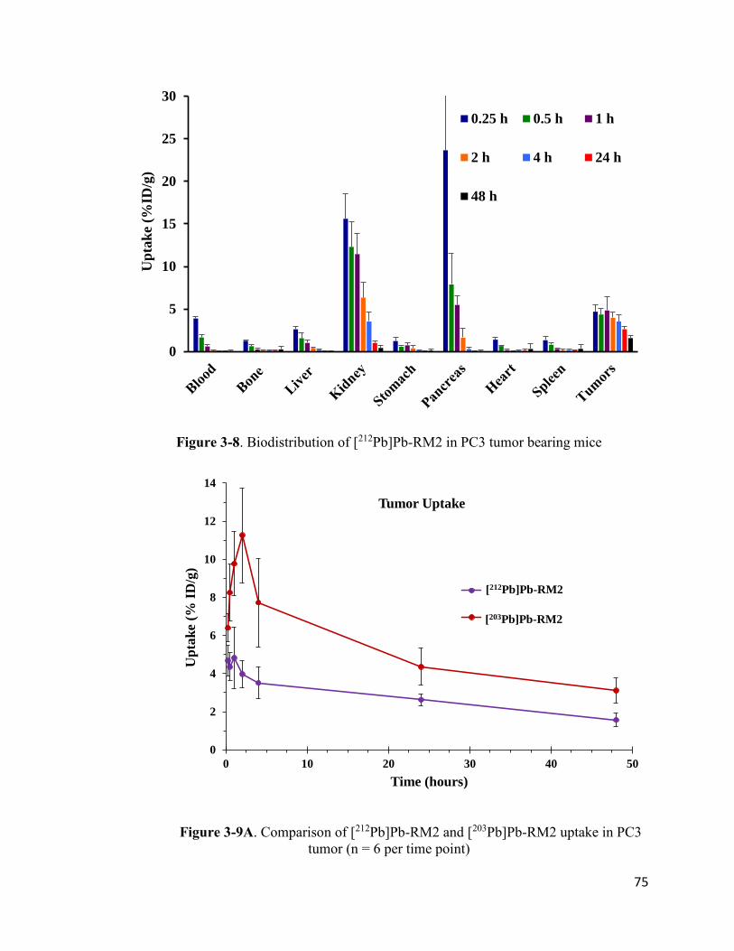
75
Figure 3-8. Biodistribution of [212Pb]Pb-RM2 in PC3 tumor bearing mice
Figure 3-9A. Comparison of [212Pb]Pb-RM2 and [203Pb]Pb-RM2 uptake in PC3
tumor (n = 6 per time point)
0
5
10
15
20
25
30
Up
tak
e (%
ID/g
)
0.25 h 0.5 h 1 h
2 h 4 h 24 h
48 h
0
2
4
6
8
10
12
14
0 10 20 30 40 50
Up
tak
e (%
ID
/g)
Time (hours)
Tumor Uptake
212Pb-RM2
203Pb-RM2
[212Pb]Pb-RM2
[203Pb]Pb-RM2

76
Figure 3-9B. Comparison of [212Pb]Pb-RM2 and [203Pb]Pb-RM2 uptake in
pancreas (n = 6 per time point)
One possible explanation for the discrepancies observed in the BB2 receptor
expressing organs/tissues between [203Pb]Pb-RM2 and [212Pb]Pb-RM2 is potential
differences in the specific activity of the [203Pb]PbCl2 and [212Pb]PbCl2 precursor. As
described in Chapter 2, the [203Pb]PbCl2 precursor contained substantial amounts of non-
radioactive Pb, which would lead to competition between [203Pb]Pb-RM2 and non-
radioactive Pb-RM2 for the same BB2 receptors, resulting in lower [203Pb]Pb-RM2 uptake
values in BB2 receptor-expressing organs/tissues. In theory, the [212Pb]PbCl2 precursor
should have high specific activity since it is obtained from a 224Ra/212Pb generator and
should contain very minimal Pb impurities. To rule out the presence of metallic impurities
in the [212Pb]PbCl2 generator eluent, 200 L aliquots were collected and analyzed by ICP-
MS after decay of 212Pb. Surprisingly, it was observed that the [212Pb]PbCl2 generator
0
4
8
12
16
20
24
28
0 1 2 3 4 5
Up
tak
e (%
ID
/g)
Time (hours)
Pancreas Uptake
212Pb-RM2
203Pb-RM2
[212Pb]Pb-RM2
[203Pb]Pb-RM2

77
eluent contained significant amounts of metallic contaminants, predominantly Pb, Fe and
Zn (Table S3-2).
The Fe and Zn contamination was significantly reduced after purification using Pb-
resin, as shown in Figures 3-10A to 3-10C. Based on the amount of non-radioactive Pb
observed in the [212Pb]PbCl2 generator eluent, it was calculated that the specific activity is
~28 mCi/g on the first day of eluting the 224Ra/212Pb generator. This indicates that 2% of
the total Pb atoms are present as 212Pb, which is comparable to what was determined in the
[203Pb]PbCl2 precursor as described in Chapter 2. Based on this information, it can be
concluded that the difference in the specific activities (with respect to total non-radioactive
Pb atoms) between the [203Pb]PbCl2 and [212Pb]PbCl2 precursor is not significant enough
to explain the discrepancies in the uptake values observed in the BB2 receptor expressing
organs/tissues between [203Pb]Pb-RM2 and [212Pb]Pb-RM2.
Figure 3-10A. Comparison of Pb contamination in [212Pb]PbCl2 generator eluent
before and after purification (n = 1, generator #4)
0
0.05
0.1
0.15
0.2
0.25
1 2 3 6
ng
/
L
Days after generator receipt
Pb
Crude
Purified
Blank 2M HCl

78
Figure 3-10B. Comparison of Fe contamination in [212Pb]PbCl2 generator eluent
before and after purification (n = 1, generator #4)
Figure 3-10C. Comparison of Zn contamination in [212Pb]PbCl2 generator eluent
before and after purification (n = 1, generator #4)
Variations in specific activity with respect to the total amount of peptide used for
radiolabeling and variations in the activity concentration administered to the animals can
influence the uptake values observed in the BB2 receptor-expressing organs/tissues (Table
0
0.2
0.4
0.6
0.8
1
1.2
1.4
1 2 3 6
ng
/
L
Days after generator receipt
Fe
Crude
Purified
Blank 2M HCl
0
0.04
0.08
0.12
0.16
0.2
1 2 3 6
ng
/
L
Days after generator receipt
Zn
Crude
Purified
Blank 2M HCl

79
3-4). As expected, the total [212Pb]PbCl2 activity eluted from the 224Ra/212Pb generator
decreases over time, thereby resulting in a decrease in the [212Pb]Pb-RM2 activity over
time. Therefore, the activity concentration and specific activity per mol RM2 was
different depending on the day the radiolabeling experiment was performed (Table 3-5).
Considering that ~ 20 Ci (0.7 MBq) of [212Pb]Pb-RM2 in a ~100 L bolus is typically
administered to each mouse, the [212Pb]Pb-RM2 activity required more dilution in the
earlier days than in the later during the biodistribution study. Thus, a greater amount of
unlabeled RM2 peptide conjugate was injected into each mouse in the later days during the
biodistribution study, which possibly contributed to the observed decrease in [212Pb]Pb-
RM2 uptake in the BB2 receptor-expressing organs/tissues. The influence of the total
amount of injected peptide on pancreas uptake was also reported by Dalm et al.22 for
[177Lu]Lu-NeoBOMB, which is another BB2 receptor antagonist peptide conjugate. The
authors observed a pancreas uptake value of 105 ± 13% ID/g with the injection of 1
MBq/10 pmol of [177Lu]Lu-NeoBOMB whereas, the pancreas uptake value was 19.8 ±
6.9% ID/g with the injection of 1 MBq/200 pmol of [177Lu]Lu-NeoBOMB. Similar peptide
related influence on the uptake values in BB2 receptor-expressing organs was reported for
[111In]In-AMBA, which is a BB2r agonist peptide conjugate.65 Effect of peptide mass on
pharmacokinetics have also been reported for radiolabeled peptide conjugates targeting the
somatostatin receptor.66

80
Table 3-4. Variations in [212Pb]Pb-RM2 pancreas and tumor uptake with the date of
synthesis
10/27/2017 10/31/2017 11/01/2017 11/06/2017
15 min
Pancreas 34.66 ± 6.25 * 12.63 ± 0.48 *
Tumor 5.20 ± 0.71 * 4.18 ± 0.48 *
30 min
Pancreas 12.41 ± 2.36 ǂ 5.63 ± 0.61 #
Tumor 4.71 ± 0.87 ǂ 4.18 ± 0.64 #
1 hour
Pancreas 8.60 ± 3.67 * 4.26 ± 0.86 *
Tumor 4.91 ± 2.26 * 4.72 ± 0.62 *
2 hours
Pancreas 3.09 ± 1.21 ǂ 1.22 ± 0.33 * 0.75 ± 0.01ǂ
Tumor 4.72 ± 0.18 ǂ 4.06 ± 0.44 * 3.12 ± 0.12 ǂ
ǂ n = 2, * n = 3, # n = 4
Table 3-5. Variations in [212Pb]Pb-RM2 specific activity and activity concentration
with the date of synthesis
Date
Activity after Sep-
Pack purification
(mCi) RM2 (mol)
Specific Activity
(mCi/ mol
RM2)
Activity
concentration
(mCi/mL)*
10/27/2017 5.74 0.037 149.5 2.73
10/31/2017 2.96 0.037 80.9 1.48
11/01/2017 2.26 0.037 61.7 1.13
11/06/2017 0.93 0.037 25.4 0.46
* assuming final volume of 2 mL

81
3.4. On-going Studies
3.4-1. In-vitro Evaluation of [212Pb]Pb-RM2 Therapeutic Efficacy
The in vitro evaluation of the therapeutic efficacy of [212Pb]Pb-RM2 is currently
underway in various prostate cancer cell lines including PC3, 22RV1, DU-145, LNCAP,
LNCAP C42B, LNCAP C42, and VCAP. The cells were exposed to increasing doses of
[212Pb]Pb-RM2 (0.5 Ci, 1 Ci, 2 Ci and 4 Ci). Subsequently, the therapeutic efficacy
was determined by assessing cell viability using the MTT assay, cell survival and
proliferation using the clonogenic assay, and DNA damage using immunofluorescence
staining of 53BPI and H2AX markers.
Initial data on cell survival (using clonogenic assay) obtained in two cell lines (PC3
and LNCAP) after exposure to [212Pb]Pb-RM2 and [177Lu]Lu-RM2 show significantly
improved therapeutic efficacy (as indicated by the lower survival fraction) with [212Pb]Pb-
RM2, as shown in Figure 3-11.
3.4-2. Evaluation of [212Pb]Pb-RM2 Maximum Tolerated Dose and In
Vivo Toxicity
Studies to evaluate the maximum tolerated dose of [212Pb]Pb-RM2 in CF-1 normal
mice are currently underway. Three treatment groups and one control group are being
investigated. The treatment groups received an intravenous injection of [212Pb]Pb-RM2 at
three dose levels (50 Ci, 100 Ci and 200 Ci), with 26 mice per dose level. A control
group of 21 mice received an intravenous injection of saline. Body weight (BW), complete
blood counts (CBC), and body condition score (BCS) were recorded prior to injection of
either [212Pb]Pb-RM2 or saline. After injection, BW, CBC, and BCS will be monitored
weekly for a duration of 30 weeks. A subset of the animals in each study group will be

82
sacrificed at day 30, 50, 70 and 90 (n = 4 per time point) for tissue histopathological
examination and toxicity evaluation by monitoring serum chemistry markers for liver
function [e.g., alanine aminotransferase (ALT), alkaline phosphatase (ALP), aspartate
aminotransferase (AST), and bilirubin], kidney function [blood urea nitrogen (BUN),
calcium, albumin, and creatinine], pancreas function [amylase and lipase] and general
metabolic function [glucose, inorganic phosphorus and total cholesterol].
Figure 3-11. Comparison of clonogenic response of PC3 and LNCAP cells lines to
[177Lu]Lu-RM2 treatment (left) and [212Pb]Pb-RM2 (right)
As at week 10 into the maximum tolerated dose studies, no deaths in any of the
treatment groups and no adverse effects on body weight have been observed. In fact, the
average body weight has steadily increased over time in all study groups (Figure 3-12).
Additionally, no apparent signs of hematological toxicity have been observed through

83
week 10 into the maximum tolerated dose studies, with no significant differences in the
average white blood cell counts (Figure 3-13) and average platelets counts (Figure 3-14)
between the control and treatment groups. Furthermore, the absence of a significant
decrease in white blood cell and platelet counts is an indication of normal bone marrow
activity and lack of myelosuppression.
3.5. Conclusions
The method described in this study for the synthesis of [212Pb]Pb-RM2 incorporates
a step for the purification of the [212Pb]PbCl2 generator eluent using the Eichrom Pb-resin,
which cuts down the synthesis time to ~1.5 hours compared to the ~3.5 hours that has been
traditionally reported for 212Pb labeling.58 We demonstrated that this purification step
significantly reduces the amount of radiometal impurities from 212Pb daughter
radionuclides (212Bi, 208Tl, and 212Po). ICP-MS analysis of the crude [212Pb]PbCl2 generator
eluent revealed the presence of significant amounts of Pb, Fe and Zn metal contamination;
however, the Pb-resin purification step reduced the Fe and Zn contamination to negligible
amounts.
A major concern with alpha-emitting radionuclides is the recoil energy imparted to
the daughter radionuclide upon alpha decay as a result of conservation of momentum. This
recoil energy is typically around 100 keV and is significantly greater that the bond energies
for most common bonds (e.g., C–O = 3.6 eV; C–C = 3.6 eV; C–N = 3.0 eV; C≡O = 11.2
eV). Therefore, there is a possibility that the chemical bonds in any chelator used for the
complexation of an alpha-emitting radionuclide would break as a result of the recoil
energy, resulting in possible redistribution of the daughter radionuclide. However, it is
worth mentioning the decay of 212Pb into 212Bi occurs by beta emission.

84
Figure 3-12. Effect of [212Pb]Pb-RM2 administration on body weight in CF-1 mice
Figure 3-13. Effect of [212Pb]Pb-RM2 administration on white blood cells in CF-1 mice
25
30
35
40
45
50
0 1 2 3 4 5 6 7 8 9 10
Bo
dy
Wei
gh
t (g
)
Week Post Administration
Average Body Weight
NTC 50uCi 100uCi 200uCi
0
2,500
5,000
7,500
10,000
12,500
15,000
17,500
20,000
0 1 2 3 4 5 6 7 8 9 10
Cel
ls /
L
Week Post Administration
Average White Blood Cell Count
NTC 50uCi 100uCi 200uCi
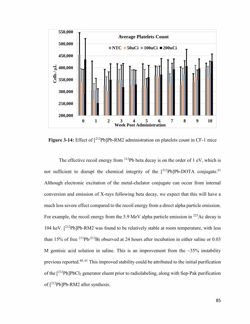
85
Figure 3-14: Effect of [212Pb]Pb-RM2 administration on platelets count in CF-1 mice
The effective recoil energy from 212Pb beta decay is on the order of 1 eV, which is
not sufficient to disrupt the chemical integrity of the [212Pb]Pb-DOTA conjugate.61
Although electronic excitation of the metal-chelator conjugate can occur from internal
conversion and emission of X-rays following beta decay, we expect that this will have a
much less severe effect compared to the recoil energy from a direct alpha particle emission.
For example, the recoil energy from the 5.9 MeV alpha particle emission in 225Ac decay is
104 keV. [212Pb]Pb-RM2 was found to be relatively stable at room temperature, with less
than 15% of free 212Pb/212Bi observed at 24 hours after incubation in either saline or 0.03
M gentisic acid solution in saline. This is an improvement from the ~35% instability
previous reported.48, 61 This improved stability could be attributed to the initial purification
of the [212Pb]PbCl2 generator eluent prior to radiolabeling, along with Sep-Pak purification
of [212Pb]Pb-RM2 after synthesis.
200,000
250,000
300,000
350,000
400,000
450,000
500,000
550,000
0 1 2 3 4 5 6 7 8 9 10
Cel
ls /
L
Week Post Administration
Average Platelets Count
NTC 50uCi 100uCi 200uCi

86
[212Pb]Pb-RM2 exhibited specific tumor targeting in PC3 tumor-bearing SCID
mice. Additionally, rapid clearance from the blood and non-target organs were observed
primarily through the urinary system. Most importantly, negligible [212Pb]Pb-RM2 activity
was observed in the bone and liver at every time point studied. The is an indication of the
high in vivo stability [212Pb]Pb-RM2 because redistribution of free 212Pb/212Bi should result
in accumulation of activity in the bone and liver. The [212Pb]Pb-RM2 activity concentration
and specific activity per mol of RM2 was found to be related to its uptake in BB2 receptor-
expressing organs/tissues (pancreas and PC3 tumor), with lower specific activity
(mCi/ mol RM2) resulting in lower uptake values. Preliminary data from ongoing in vitro
studies show that exposure of [212Pb]Pb-RM2 to PC3 and LNCAP cells caused significant
cell death. Additionally, no radiation-related toxicity has been observed as at week 10 in
ongoing [212Pb]Pb-RM2 maximum tolerated dose studies in CF-1 normal mice.
In conclusion, the favorable pharmacokinetic profile of [212Pb]Pb-RM2, together
with the preliminary demonstration of in vitro therapeutic efficacy and lack of toxicity in
ongoing MTD studies, highlight its potential as a therapeutic radiopharmaceutical for
prostate cancer. As with other radionuclides of interest in targeted alpha therapy, a major
factor that could hinder future routine clinical utility of 212Pb-based radiopharmaceuticals
is the availability of GMP grade 224Ra/212Pb generators. The completion of a 212Pb
generator production facility (Laboratoire Maurice Tubiana) in France was recently
announced by Orano Med, although no information on the production method was
provided. A potential method for increasing the availability of 224Ra/212Pb generators is to
develop new production routes for 228Th other than through the natural decay of 232U and
232Th. Thorium-228 is the parent isotope of 224Ra; hence, increasing the production of 228Th

87
would lead to increased availability of 224Ra for producing the 224Ra/212Pb generators. One
potential production route for 228Th is by neutron irradiation of 226Ra via the 226Ra (n,)
227Ra (-) 227Ac (n,) 228Ac (-) 228Th reaction.67-68 Interestingly, the neutron irradiation of
226Ra has been primarily investigated for the production of 229Th as a means of increasing
the availability of 225Ac (Figure 3-15). However, more 228Th is produced from the neutron
irradiation of 226Ra. For example, Hogle et al.67 reported that the production yield of for
228Th and 229Th was 1450 ± 110 kBq/g and 74.0 ± 7.4 Bq/g, respectively, after an ~26
days irradiation of 226Ra target. Additionally, the co-production of Ac-227 does not
constitute a major concern since there are established methods to separate Ac from Th.67-
68 Furthermore, the data presented in this study show that the incorporation of a final
purification step for the 224Ra/212Pb generator eluent using the Eichrom Pb-resin is
successful in removing residual radionuclidic contamination prior to radiolabeling.
3.6. Future Studies
The data obtained upon completion of the [212Pb]Pb-RM2 MTD studies will be used
to initiate therapy studies to evaluate the in vivo therapeutic efficacy of [212Pb]Pb-RM2 in
SCID mice bearing PC3 prostate tumor xenografts. The therapy doses will be administered
at levels that did not induce radiation related toxicity based on the MTD studies. Four study
groups will be investigated: (1) non-tumor bearing control group receiving an intravenous
administration of saline, (2) tumor bearing control group receiving an intravenous
administration of saline, (3) tumor bearing therapy group receiving an intravenous
administration of [212Pb]Pb-RM2, and (4) tumor bearing therapy group receiving an
intravenous administration of non-radioactive Pb-RM2. Each study group will consist of
15 mice. Similar to the MTD studies, BW, CBC, and BCS will be monitored weekly for a

88
duration of 30 weeks. Additionally, tumor volumes will be measured weekly for the same
duration using calipers.
Figure 3-15. Experimental and theoretical yields of 228Th, 227Ac, 228Ra and 229 Th
from 226Ra irradiation at ORNL HFIR. Reproduced with permission from Hogle at al.67

89
3.7. References
1. American Cancer Society. Cancer Facts & Figures 2019. American Cancer Society; 2019.
2. Mohler, J.; Antonarakis, E.; Armstrong, A., NCCN Guidelines, version 2.2017: Prostate Cancer Updates. 2017.
3. Reubi, J. C.; Macke, H. R.; Krenning, E. P., Candidates for peptide receptor radiotherapy today and in the future. Journal of Nuclear Medicine 2005, 46 (1 suppl), 67s-75s.
4. Dash, A.; Chakraborty, S.; Pillai, M. R. A.; Knapp Jr, F. F., Peptide receptor radionuclide therapy: an overview. Cancer Biotherapy and Radiopharmaceuticals 2015, 30 (2), 47-71.
5. Cutler, C. S.; Hennkens, H. M.; Sisay, N.; Huclier-Markai, S.; Jurisson, S. S., Radiometals for Combined Imaging and Therapy. Chemical Reviews 2013, 113 (2), 858-883.
6. Strosberg, J.; El-Haddad, G.; Wolin, E.; Hendifar, A.; Yao, J.; Chasen, B.; Mittra, E.; Kunz, P. L.; Kulke, M. H.; Jacene, H., Phase 3 trial of 177Lu-Dotatate for midgut neuroendocrine tumors. New England Journal of Medicine 2017, 376 (2), 125-135.
7. Silver, D. A.; Pellicer, I.; Fair, W. R.; Heston, W.; Cordon-Cardo, C., Prostate-specific membrane antigen expression in normal and malignant human tissues. Clinical Cancer Research 1997, 3 (1), 81-85.
8. Tsourlakis, M. C.; Klein, F.; Kluth, M.; Quaas, A.; Graefen, M.; Haese, A.; Simon, R.; Sauter, G.; Schlomm, T.; Minner, S., PSMA expression is highly homogenous in primary prostate cancer. Applied Immunohistochemistry & Molecular Morphology 2015, 23 (6), 449-455.
9. Bostwick, D. G.; Pacelli, A.; Blute, M.; Roche, P.; Murphy, G. P., Prostate specific membrane antigen expression in prostatic intraepithelial neoplasia and adenocarcinoma: a study of 184 cases. Cancer: Interdisciplinary International Journal of the American Cancer Society 1998, 82 (11), 2256-2261.
10. Markwalder, R.; Reubi, J. C., Gastrin-releasing peptide receptors in the human prostate relation to neoplastic transformation. Cancer Research 1999, 59 (5), 1152-1159.
11. Bartholdi, M. F.; Wu, J. M.; Pu, H.; Troncoso, P.; Eden, P. A.; Feldman, R. I., In situ
hybridization for gastrin‐releasing peptide receptor (GRP receptor) expression in prostatic carcinoma. International Journal of Cancer 1998, 79 (1), 82-90.
12. Sun, B.; Halmos, G.; Schally, A. V.; Wang, X.; Martinez, M., Presence of receptors
for bombesin/gastrin‐releasing peptide and mRNA for three receptor subtypes in human prostate cancers. The Prostate 2000, 42 (4), 295-303.
13. Körner, M.; Waser, B.; Rehmann, R.; Reubi, J. C., Early over‐expression of GRP receptors in prostatic carcinogenesis. The Prostate 2014, 74 (2), 217-224.

90
14. Ischia, J.; Patel, O.; Bolton, D.; Shulkes, A.; Baldwin, G. S., Expression and function
of gastrin‐releasing peptide (GRP) in normal and cancerous urological tissues. BJU International 2014, 113, 40-47.
15. Kratochwil, C.; Bruchertseifer, F.; Giesel, F. L.; Weis, M.; Verburg, F. A.; Mottaghy, F.; Kopka, K.; Apostolidis, C.; Haberkorn, U.; Morgenstern, A., 225Ac-PSMA-617 for PSMA-
targeted α-radiation therapy of metastatic castration-resistant prostate cancer. Journal of Nuclear Medicine 2016, 57 (12), 1941-1944.
16. Afshar-Oromieh, A.; Hetzheim, H.; Kratochwil, C.; Benesova, M.; Eder, M.; Neels, O. C.; Eisenhut, M.; Kübler, W.; Holland-Letz, T.; Giesel, F. L., The theranostic PSMA ligand PSMA-617 in the diagnosis of prostate cancer by PET/CT: Biodistribution in humans, radiation dosimetry, and first evaluation of tumor lesions. Journal of Nuclear Medicine 2015, 56 (11), 1697-1705.
17. Weineisen, M.; Schottelius, M.; Simecek, J.; Baum, R. P.; Yildiz, A.; Beykan, S.; Kulkarni, H. R.; Lassmann, M.; Klette, I.; Eiber, M., 68Ga-and 177Lu-labeled PSMA I&T: optimization of a PSMA-targeted theranostic concept and first proof-of-concept human studies. Journal of Nuclear Medicine 2015, 56 (8), 1169-1176.
18. Sterzing, F.; Kratochwil, C.; Fiedler, H.; Katayama, S.; Habl, G.; Kopka, K.; Afshar-Oromieh, A.; Debus, J.; Haberkorn, U.; Giesel, F. L., 68Ga-PSMA-11 PET/CT: a new technique with high potential for the radiotherapeutic management of prostate cancer patients. European Journal of Nuclear Medicine and Molecular Imaging 2016, 43 (1), 34-41.
19. Mansi, R.; Wang, X. J.; Forrer, F.; Waser, B.; Cescato, R.; Graham, K.; Borkowski, S.; Reubi, J. C.; Maecke, H. R., Development of a potent DOTA-conjugated bombesin antagonist for targeting GRPr-positive tumours. European Journal of Nuclear Medicine and Molecular Imaging 2011, 38 (1), 97-107.
20. Minamimoto, R.; Sonni, I.; Hancock, S.; Vasanawala, S.; Loening, A.; Gambhir, S. S.; Iagaru, A., Prospective Evaluation of 68Ga-RM2 PET/MRI in Patients with Biochemical Recurrence of Prostate Cancer and Negative Conventional Imaging. Journal of Nuclear Medicine 2018, 59 (5), 803-808.
21. Lantry, L. E.; Cappelletti, E.; Maddalena, M. E.; Fox, J. S.; Feng, W.; Chen, J.; Thomas, R.; Eaton, S. M.; Bogdan, N. J.; Arunachalam, T., 177Lu-AMBA: Synthesis and Characterization of a Selective 177Lu-Labeled GRP-R Agonist for Systemic Radiotherapy of Prostate Cancer. Journal of Nuclear Medicine 2006, 47 (7), 1144.
22. Dalm, S. U.; Bakker, I. L.; de Blois, E.; Doeswijk, G. N.; Konijnenberg, M. W.; Orlandi, F.; Barbato, D.; Tedesco, M.; Maina, T.; Nock, B. A., 68Ga/177Lu-NeoBOMB1, a novel radiolabeled GRPR antagonist for theranostic use in oncology. Journal of Nuclear Medicine 2017, 58 (2), 293-299.
23. Nock, B. A.; Kaloudi, A.; Lymperis, E.; Giarika, A.; Kulkarni, H. R.; Klette, I.; Singh, A.; Krenning, E. P.; De Jong, M.; Maina, T., Theranostic perspectives in prostate cancer

91
with the gastrin-releasing peptide receptor antagonist NeoBOMB1: preclinical and first clinical results. Journal of Nuclear Medicine 2017, 58 (1), 75-80.
24. Kranzbühler, B.; Salemi, S.; Umbricht, C. A.; Müller, C.; Burger, I. A.; Sulser, T.;
Eberli, D., Pharmacological upregulation of prostate‐specific membrane antigen (PSMA) expression in prostate cancer cells. The Prostate 2018, 78 (10), 758-765.
25. Qiao, J.; Grabowska, M. M.; Forestier, I. S.; Mirosevich, J.; Case, T. C.; Chung, D. H.; Cates, J. M.; Matusik, R. J.; Manning, H. C.; Jin, R., Activation of GRP/GRP-R signaling contributes to castration-resistant prostate cancer progression. Oncotarget 2016, 7 (38), 61955.
26. Heinrich, E.; Probst, K.; Michel, M. S.; Trojan, L., Gastrin‐releasing peptide:
Predictor of castration‐resistant prostate cancer? The Prostate 2011, 71 (6), 642-648.
27. Serda, R. E.; Bisoffi, M.; Thompson, T. A.; Ji, M.; Omdahl, J. L.; Sillerud, L. O., 1α,
25‐Dihydroxyvitamin D3 down‐regulates expression of prostate specific membrane antigen in prostate cancer cells. The Prostate 2008, 68 (7), 773-783.
28. Minamimoto, R.; Hancock, S.; Schneider, B.; Chin, F.; Jamali, M.; Loening, A. M.; Vasanawala, S.; Gambhir, S. S.; Iagaru, A., Pilot Comparison of 68Ga-RM2 PET and 68Ga-PSMA PET in Patients with Biochemically Recurrent Prostate Cancer. Journal of Nuclear Medicine 2016, 557-562.
29. Kähkönen, E.; Jambor, I.; Kemppainen, J.; Lehtiö, K.; Grönroos, T. J.; Kuisma, A.; Luoto, P.; Sipilä, H. J.; Tolvanen, T.; Alanen, K., In vivo imaging of prostate cancer using [68Ga]-labeled bombesin analog BAY86-7548. Clinical Cancer Research 2013, 19 (19), 5434-5443.
30. Wieser, G.; Popp, I.; Rischke, H. C.; Drendel, V.; Grosu, A.-L.; Bartholomä, M.; Weber, W. A.; Mansi, R.; Wetterauer, U.; Schultze-Seemann, W., Diagnosis of recurrent prostate cancer with PET/CT imaging using the gastrin-releasing peptide receptor antagonist 68Ga-RM2: Preliminary results in patients with negative or inconclusive [18F]Fluoroethylcholine-PET/CT. European Journal of Nuclear Medicine and Molecular Imaging 2017, 1-10.
31. Sgouros, G.; Roeske, J. C.; McDevitt, M. R.; Palm, S.; Allen, B. J.; Fisher, D. R.; Brill, A. B.; Song, H.; Howell, R. W.; Akabani, G., MIRD Pamphlet No. 22 (abridged): radiobiology
and dosimetry of α-particle emitters for targeted radionuclide therapy. Journal of Nuclear Medicine 2010, 51 (2), 311-328.
32. Baidoo, K. E.; Yong, K.; Brechbiel, M. W., Molecular pathways: targeted α-particle radiation therapy. Clinical Cancer Research 2013, 19 (3), 530-537.
33. Parker, C.; Lewington, V.; Shore, N.; Kratochwil, C.; Levy, M.; Lindén, O.; Noordzij, W.; Park, J.; Saad, F., Targeted Alpha Therapy, an Emerging Class of Cancer Agents: A Review. JAMA Oncology 2018, 4 (12), 1765-1772.
34. Kim, Y.-S.; Brechbiel, M. W., An overview of targeted alpha therapy. Tumor Biology 2012, 33 (3), 573-590.

92
35. Nayak, T.; Norenberg, J.; Anderson, T.; Atcher, R., A comparison of high-versus low-linear energy transfer somatostatin receptor targeted radionuclide therapy in vitro. Cancer Biotherapy & Radiopharmaceuticals 2005, 20 (1), 52-57.
36. Chan, H. S.; de Blois, E.; Morgenstern, A.; Bruchertseifer, F.; de Jong, M.; Breeman, W.; Konijnenberg, M., In Vitro comparison of 213Bi-and 177Lu-radiation for peptide receptor radionuclide therapy. PloS One 2017, 12 (7), e0181473.
37. Yong, K.; Brechbiel, M. W., Towards translation of 212Pb as a clinical therapeutic; getting the lead in! Dalton Transactions 2011, 40 (23), 6068-6076.
38. Yong, K. J.; Milenic, D. E.; Baidoo, K. E.; Brechbiel, M. W., 212Pb-radioimmunotherapy induces G2 cell-cycle arrest and delays DNA damage repair in tumor xenografts in a model for disseminated intraperitoneal disease. Molecular Cancer Therapeutics 2012, 11 (3), 639-648.
39. Milenic, D. E.; Garmestani, K.; Brady, E. D.; Albert, P. S.; Ma, D.; Abdulla, A.;
Brechbiel, M. W., α-Particle radioimmunotherapy of disseminated peritoneal disease using a 212Pb-labeled radioimmunoconjugate targeting HER2. Cancer Biotherapy & Radiopharmaceuticals 2005, 20 (5), 557-568.
40. Milenic, D. E.; Baidoo, K. E.; Kim, Y. S.; Barkley, R.; Brechbiel, M. W., Targeted alpha-Particle Radiation Therapy of HER1-Positive Disseminated Intraperitoneal Disease: An Investigation of the Human Anti-EGFR Monoclonal Antibody, Panitumumab. Translational Oncology 2017, 10 (4), 535-545.
41. Tan, Z.; Chen, P.; Schneider, N.; Glover, S.; Cui, L.; Torgue, J.; Rixe, O.; Spitz, H. B.; Dong, Z., Significant systemic therapeutic effects of high-LET immunoradiation by 212Pb-trastuzumab against prostatic tumors of androgen-independent human prostate cancer in mice. International Journal of Oncology 2012, 40 (6), 1881-1888.
42. Kasten, B. B.; Azure, M. T.; Schoeb, T. R.; Fisher, D. R.; Zinn, K. R., Imaging, biodistribution, and toxicology evaluation of 212Pb-TCMC-trastuzumab in nonhuman primates. Nuclear Medicine and Biology 2016, 43 (7), 391-396.
43. Schneider, N.; Lobaugh, M.; Tan, Z.; Sandwall, P.; Chen, P.; Glover, S.; Cui, L.; Murry, M.; Dong, Z.; Torgue, J., Biodistribution of 212Pb conjugated trastuzumab in mice. Journal of Radioanalytical and Nuclear Chemistry 2013, 296 (1), 75-81.
44. Kasten, B. B.; Arend, R. C.; Katre, A. A.; Kim, H.; Fan, J.; Ferrone, S.; Zinn, K. R.; Buchsbaum, D. J., B7-H3-targeted 212Pb radioimmunotherapy of ovarian cancer in preclinical models. Nuclear Medicine and Biology 2017, 47, 23-30.
45. Kasten, B. B.; Gangrade, A.; Kim, H.; Fan, J.; Ferrone, S.; Ferrone, C. R.; Zinn, K. R.; Buchsbaum, D. J., 212Pb-labeled B7-H3-targeting antibody for pancreatic cancer therapy in mouse models. Nuclear Medicine and Biology 2018, 58, 67-73.
46. Miao, Y.; Hylarides, M.; Fisher, D. R.; Shelton, T.; Moore, H.; Wester, D. W.; Fritzberg, A. R.; Winkelmann, C. T.; Hoffman, T.; Quinn, T. P., Melanoma therapy via
peptide-targeted α-radiation. Clinical Cancer Research 2005, 11 (15), 5616-5621.

93
47. Shah, M.; Zhang, X.; Rossin, R.; Robillard, M. S.; Fisher, D.; Bueltmann, T.; Hoeben, F. J.; Quinn, T., Metal-free Cycloaddition Chemistry Driven Pretargeted
Radioimmunotherapy Using α-Particle Radiation. Bioconjugate Chemistry 2017.
48. Su, F.-M.; Beaumier, P.; Axworthy, D.; Atcher, R.; Fritzberg, A., Pretargeted radioimmunotherapy in tumored mice using an in vivo 212Pb/212Bi generator. Nuclear Medicine and Biology 2005, 32 (7), 741-747.
49. Saidib, A.; Maalanda, A.; Torgueb, J.; Heyerdahla, H.; Dahlea, J., Targeted Alpha Therapy with 212Pb-NNV003 for the Treatment of CD37 Positive B-Cell Chronic Lymphocytic Leukemia (CLL) and Non-Hodgkin Lymphoma (NHL). American Society of Hematology: 2018.
50. Meredith, R. F.; Torgue, J. J.; Rozgaja, T. A.; Banaga, E. P.; Bunch, P. W.; Alvarez, R. D.; Straughn Jr, J. M.; Dobelbower, M. C.; Lowy, A. M., Safety and outcome measures of
first-in-human intraperitoneal α radioimmunotherapy with 212Pb-TCMC-trastuzumab. American Journal of Clinical Oncology 2018, 41 (7), 716.
51. Meredith, R.; Torgue, J.; Shen, S.; Fisher, D. R.; Banaga, E.; Bunch, P.; Morgan, D.; Fan, J.; Straughn, J. M., Jr., Dose escalation and dosimetry of first-in-human alpha radioimmunotherapy with 212Pb-TCMC-trastuzumab. Journal of Nuclear Medicine 2014, 55 (10), 1636-42.
52. Meredith, R. F.; Torgue, J.; Azure, M. T.; Shen, S.; Saddekni, S.; Banaga, E.; Carlise, R.; Bunch, P.; Yoder, D.; Alvarez, R., Pharmacokinetics and imaging of 212Pb-TCMC-trastuzumab after intraperitoneal administration in ovarian cancer patients. Cancer Biotherapy & Radiopharmaceuticals 2014, 29 (1), 12-7.
53. Atcher, R. W.; Friedman, A. M.; Hines, J., Isotopic generator for bismuth-212 and lead-212 from radium. U.S. Patent No. 4,663,129.: 1987.
54. Mirzadeh, S., Generator-produced alpha-emitters. Applied Radiation and Isotopes 1998, 49 (4), 345-349.
55. Li, M.; Zhang, X.; Quinn, T. P.; Lee, D.; Liu, D.; Kunkel, F.; Zimmerman, B. E.; McAlister, D.; Olewein, K.; Menda, Y., Automated cassette-based production of high specific activity [203/212Pb] peptide-based theranostic radiopharmaceuticals for image-guided radionuclide therapy for cancer. Applied Radiation and Isotopes 2017, 127, 52-60.
56. Horwitz, E. P.; Dietz, M. L.; Rhoads, S.; Felinto, C.; Gale, N. H.; Houghton, J., A lead-selective extraction chromatographic resin and its application to the isolation of lead from geological samples. Analytica Chimica Acta 1994, 292 (3), 263-273.
57. Milenic, D.; Baidoo, K.; Brechbiel, M., Bench to bedside: stability studies of GMP produced trastuzumab-TCMC in support of a clinical trial. Pharmaceuticals 2015, 8 (3), 435-454.
58. Baidoo, K. E.; Milenic, D. E.; Brechbiel, M. W., Methodology for labeling proteins and peptides with lead-212 (212 Pb). Nuclear Medicine and Biology 2013, 40 (5), 592-599.

94
59. Milenic, D. E.; Molinolo, A. A.; Solivella, M. S.; Banaga, E.; Torgue, J.; Besnainou, S.; Brechbiel, M. W.; Baidoo, K. E., Toxicological studies of 212Pb intravenously or intraperitoneally injected into mice for a phase 1 trial. Pharmaceuticals 2015, 8 (3), 416-434.
60. Torgue, J.; Maquaire, P.; Young, J.; Andreoletti, G.; Bourdet, P. Method and apparatus for the production of lead 212 for medical use. United States Patent. 2015.
61. Mirzadeh, S.; Kumar, K.; Gansow, O. A., The chemical fate of 212Bi-DOTA formed
by β- decay of 212Pb(DOTA)2-. Radiochimica Acta 1993, 60 (1), 1-10.
62. Mansi, R.; Wang, X.; Forrer, F.; Kneifel, S.; Tamma, M.-L.; Waser, B.; Cescato, R.; Reubi, J. C.; Maecke, H. R., Evaluation of a 1, 4, 7, 10-Tetraazacyclododecane-1, 4, 7, 10-Tetraacetic acid–conjugated bombesin-based radioantagonist for the labeling with single-photon emission computed tomography, positron emission tomography, and therapeutic radionuclides. Clinical Cancer Research 2009, 15 (16), 5240-5249.
63. Hoffman, T. J.; Gali, H.; Smith, C. J.; Sieckman, G. L.; Hayes, D. L.; Owen, N. K.; Volkert, W. A., Novel series of 111In-labeled bombesin analogs as potential radiopharmaceuticals for specific targeting of gastrin-releasing peptide receptors expressed on human prostate cancer cells. Journal of Nuclear Medicine 2003, 44 (5), 823-831.
64. Zhang, H.; Schuhmacher, J.; Waser, B.; Wild, D.; Eisenhut, M.; Reubi, J. C.; Maecke, H. R., DOTA-PESIN, a DOTA-conjugated bombesin derivative designed for the imaging and targeted radionuclide treatment of bombesin receptor-positive tumours. European Journal of Nuclear Medicine and Molecular Imaging 2007, 34 (8), 1198-1208.
65. De Blois, E.; Schroeder, R.; De Ridder, C.; Van Weerden, W.; Breeman, W.; De Jong, M., Improving radiopeptide pharmacokinetics by adjusting experimental conditions for bombesin receptor-mediated imaging of prostate cancer. Quarterly Journal of Nuclear Medicine and Molecular Imaging 2013, 57, 1-9.
66. Nicolas, G. P.; Mansi, R.; McDougall, L.; Kaufmann, J.; Bouterfa, H.; Wild, D.; Fani, M., Biodistribution, pharmacokinetics, and dosimetry of 177Lu-, 90Y-, and 111In-labeled somatostatin receptor antagonist OPS201 in comparison to the agonist 177Lu-DOTATATE: the mass effect. Journal of Nuclear Medicine 2017, 58 (9), 1435-1441.
67. Hogle, S.; Boll, R. A.; Murphy, K.; Denton, D.; Owens, A.; Haverlock, T. J.; Garland, M.; Mirzadeh, S., Reactor production of Thorium-229. Applied Radiation and Isotopes 2016, 114, 19-27.
68. Kuznetsov, R.; Butkalyuk, P.; Tarasov, V.; Baranov, A. Y.; Butkalyuk, I.; Romanov, E.; Kupriyanov, V.; Kazakova, E., Yields of activation products in 226Ra irradiation in the high-flux SM reactor. Radiochemistry 2012, 54 (4), 383-387.

95
CHAPTER 4: Microwave-assisted Synthesis of Rh(III)
Complexes and Radiochemical Evaluation of 105Rh Produced
from Recycled 104Ru Metal Target
4.1. Introduction
Grazman and Troutner1-2 first identified 105Rh as a potential therapeutic
radionuclide. Rhodium-105 (half-life = 35.4 hours) is a moderate energy beta emitting
radionuclide [
max = 567 keV (75%),
avg = 152 keV], with low energy gamma emissions
[319 keV (19%) and 306 keV (5%)]. The beta energy of 105Rh is comparable to the beta
energies of 177Lu (
avg = 134 keV) and 131I (
avg = 182 keV), which are currently used in
the production of FDA-approved therapeutic radiopharmaceuticals.
Rhodium-105 is produced at the University of Missouri Research Reactor Center
(MURR) by thermal neutron (~9.78 x 10¹³ n/cm2/s) irradiation of an enriched (>98%) 104Ru
metal target to give 105Ru (half-life = 4.4 hours), which subsequently decays by beta
particle emission to 105Rh, as previously reported by Grazman and Troutner (Equation 4-
1).1-2 A 24 hour wait period is allowed to ensure the decay of 105Ru into 105Rh. Rhodium-
105 is separated from the bulk 104Ru metal target by oxidation of 104Ru metal to
[104Ru]RuO4 using hypochlorite generated in situ by bubbling Cl2 into a 2 M NaOH
solution containing the 104Ru metal target (Figure 4-1). [104Ru]RuO4 is distilled off at
elevated temperature (~ 90 °C) and trapped in a 3 M HCl solution for future recovery,
leaving behind 105Rh in the original target dissolution vial. Rhodium-105 is acidified with
1 M HCl to yield a mixture of 105Rh chloride species such as [105Rh]RhCl3(OH2)3,
[105Rh]RhCl4(OH2)21-, [105Rh]RhCl5(OH2)2
2-, and [105Rh]RhCl63-, as previously determined
by electrophoresis analysis.1-3

96
Equation 4-1. Production of 105Rh
Figure 4-1. Schematic for 105Rh separation
Due to the high cost of the enriched 104Ru metal target, it is necessary that the target
material be recycled for reuse in order to make routine 105Rh production economically
feasible. In addition, less than 1% of the total 104Ru atoms are converted to 105Ru atoms
following neutron irradiation; hence, the 104Ru metal target can be reused multiple times if
successfully recovered. Recent research by Phelps and colleagues at the University of
Missouri have demonstrated the possibility of recycling and reusing 104Ru trapped in 3 M
HCl as ruthenate ([104Ru]RuO42-) during the 105Rh production process.4 The target
recycling process involves concentrating the 3 M HCl solutions to dryness to yield hydrated
ruthenium chloride ([104Ru]RuCl3∙xH2O), dehydrating [104Ru]RuCl3∙xH2O with Ar at 525
°C, and subsequently reducing to 104Ru metal with H2 at 800 °C. ICP-MS analysis revealed
that the isotopic enrichment of the recycled metal target was 98.9% 104Ru.4 Neutron
Air Cl2
HCl trap
NaOH trap
NaOH trap
104Ru in 2M NaOH

97
activation analysis on the recycled 104Ru metal target produced mainly 105Ru, with very
minimal amounts of 24Na (0.3% of total 105Ru) produced from neutron activation of minor
NaCl contaminants in the recycled 104Ru metal.4
A very attractive chemical property of Rh is the kinetic inertness of Rh(III)
complexes, which favors a low spin octahedral d6 arrangement.5 This suggests that
105Rh(III) complexes should exhibit high in vivo stability since they should be stable to
transchelation by serum proteins. Therefore, several researchers have investigated
105Rh(III) complexes as potential radiotherapeutic agents.6-13 Initial studies were done
using polyamine chelates, especially derivatives of cyclam and cyclen.6 Troutner and his
research group investigated several diethylenetriamine,7, 10 amine oxime,11 and porphyrin,9
ligands and their derivatives. Li et al.8 evaluated the in vivo pharmacokinetics of a series
of 105Rh-labeled tetradentate thiamacrocyclic ligands in rats. The authors observed very
minimal in vivo degradation of the radiolabeled complexes and clearance through both the
hepatobiliary and urinary systems was also observed depending on the ligand structure and
derivatization.8
More recently, efforts on the development of chelators for 105Rh complexation have
focused on tetrathioether frameworks.12-14 Goswami et al.12 investigated how the number
of carbon chain length [ethylene (222S4), propylene (333S4), or a combination of both
(232S4)] between the sulfur atoms in the tetrathioether ligand backbone affected the
conformation of the final Rh(III) complex. The authors found that the smaller backbone
chain length (222S4) favored the formation of the cis-dichloro isomer, while the larger
backbone chain length (333S4) favored the formation of the trans-dichloro isomer.12
Similar results were also reported by Dame15 and Crenshaw16 using tetrathioether ligand

98
frameworks containing pendant methyl acetate groups, shown in Figure 4-2A and 4-2B.
The intermediate backbone chain length (232S4) gave a mixture of both the cis-dichloro
and trans-dichloro isomers.12 Goswami et al.13 subsequently evaluated the in vivo
pharmacokinetics of 105Rh-labeled tetrathioether ligands (222S4, 232S4, and 333S4)
containing pendant carboxylic acid groups. The 105Rh complexes effectively cleared from
the blood and tissues primarily through the urinary system; however, the [105Rh]Rh-222S4
complex was shown to exhibit slightly superior stability and lesser hepatobiliary clearance
than the [105Rh]Rh-333S4 complex.13 Akgun et al.17 investigated Rh(III) complexes derived
from a series of diaminedithioether ligands and found that the ligand with 232 carbon
backbone chain length with gem-dimethyl groups was preferred.
Figure 4-2A. Crystal structure of cis-[RhCl2-222S4diAcOMe]PF6 as reported by Dame.15

99
Figure 4-2B. Crystal structure of trans-[RhCl2-333S4diAcOMe]PF6 as reported by
Crenshaw.16
Recently, Carroll18 performed 105Rh-labeling studies using 333-S4-8Aoc-BBN(7-
14)NH2, which is a bifunctional chelate containing the 333S4diAcOH tetrathioether ligand
conjugated to the bombesin(7-14) agonist peptide for targeting the gastrin-releasing
peptide receptor. Carroll reported very low radiolabeling yields (<10%) and observed the
formation of multiple radiolabeled species. The multiple radiolabeled species were
attributed to acid catalyzed esterification of the pendant carboxylic acid groups on the
tetrathioether ligand because the reaction was carried out under acidic conditions (pH 4 –
5) in the presence of refluxing ethanol. In addition, the pendant carboxylic acid groups
were found to directly coordinate with the Rh(III) core.14 To avoid the formation of acid
catalyzed esterification products, Crenshaw16 and Dame15 investigated the formation of
Rh(III) complexes of 333S4 and 222S4 tetrathioether ligands respectively, using SnCl2 as
reducing agent instead of refluxing ethanol. It has been widely reported that the catalytic
reduction of Rh+3 to Rh+1, with subsequent re-oxidation to Rh+3 by atmospheric oxygen, is

100
necessary for the formation of Rh(III) complexes.14, 19-20 Hence refluxing alcohols,
particularly ethanol, have been traditionally used as reducing agents for the formation of
Rh(III) complexes.3, 8, 12-13
This study reports the synthesis of Rh(III) complexes using two tetrathioether
ligands (222S4diAcOMe and 222S4diAcOH) and a tetradentate thiamacrocyclic ligand
(16S4-diol) without refluxing ethanol or SnCl2 as a reducing agent. A rapid microwave-
assisted synthesis procedure for Rh(III) complexes was developed and an improved HPLC
method for distinguishing the various product species is reported. Additionally, we report
the production of 105Rh from recycled 104Ru targets. Finally, 105Rh-labeling studies using
[105Rh]RhCl3 produced from both new and recycled 104Ru metal target materials are
reported.
4.2. Experimental
4.2-1. Materials and Methods
1,5,9,13-tetrathiacyclohexadecane-3,11-diol (16S4-diol; cis/trans mixture) was
purchased from Sigma-Aldrich and used without further purification. All other chemicals
and reagents were purchased from Sigma-Aldrich or Fisher Scientific and used without
further purification. Plastic-backed silica gel TLC plates were purchased from Sigma-
Aldrich or Fisher Scientific and were used for radio TLC. Only 18 MΩ water was used.
[105Rh]RhCl3 was produced at the University of Missouri Research Reactor Center
(MURR) by the bombardment of enriched 104Ru metal targets (2 – 5 mg) encapsulated in
a quartz vial with thermal neutron irradiation (~9.78 x 10¹³ n/cm2/s) for ~90 hours at the
reflector position. CAUTION! 105Rh is radioactive and must be handled in laboratories
outfitted and approved for work with radioactive materials. 1H and 13C NMR spectra were

101
obtained using a 500 MHz or a 600 MHz Bruker ARX spectrometer. Microwave synthesis
was carried out in dynamic mode using a CEM Discover SP microwave synthesizer
(Matthews, NC). Electrospray Ionization Mass Spectrometry (ESI-MS) was performed on
a Thermo Finnigan TSQ7000 triple-quadrupole instrument with an API2 source.
Inductively coupled plasma mass spectrometry (ICP-MS) was performed using a
PerkinElmer NexION 300X Model equipped with a glass cyclonic spray chamber and glass
concentric nebulizer and operated in KED mode. An Eckert & Ziegler Bioscan AR-2000
Imager using LabLogic Win-Scan imaging scanner software was used for scanning
radioTLC plates. High-performance liquid chromatography (HPLC) was performed using
a Shimadzu Prominence HPLC system equipped with a UV-Vis absorbance detector (set
at 254 and 280 nm), and a NaI(Tl) scintillation detector. All HPLC analyses were
performed using a mobile phase consisting of solvent A [99.9% H2O and 0.1%
trifluoroacetic acid (TFA)] and solvent B [99.9% CH3CN (ACN) and 0.1% TFA] run on a
Thermo Scientific Beta Basic Column (C18, 5, 150 × 4.6 mm). Three different linear
gradient systems were developed for HPLC analyses. Gradient 1: 10% solvent B:A
increased to 80% solvent B:A over 18 minutes, followed by an additional 1 minute at 80%
solvent B:A, then decreased to 10% solvent B:A over 1 minute. Gradient 2: 2% solvent
B:A increased to 5% solvent B:A over 10 minutes, followed by an additional 5 minute at
5% solvent B:A, then increased to 80% solvent B:A over 9 minutes, and finally decreased
to 2% solvent B:A over 1 minute. Gradient 3: 5% solvent B:A increased to 20% solvent
B:A over 15 minutes, followed by an additional 2 minutes at 20% solvent B:A, then
decreased to 5% solvent B:A over 3 minutes.
102
4.2-2. Synthesis of methyl 2-((2-chloroethyl)thio)acetate [C5H9ClO2S],
intermediate 1
Intermediate 1 was synthesized following a slightly modified literature
procedure.13, 15, 18 1,2-dichloroethane (88.6 mL, 1.12 mol) and triethylamine (17.2 mL, 0.12
mol) were added into a one-necked round bottom flask and heated to 90 °C. While stirring
at 90 °C, methyl thioglycolate (10 mL, 0.11 mol) was added drop wise. The reaction was
refluxed overnight (~ 18 hours) at 90 °C under N2. The reaction progress was monitored
using silica gel TLC, with 1:1 hexane / ethyl ether as the mobile phase. Upon reaction
completion, the reaction mixture was cooled to room temperature and ethyl ether (~ 70
mL) was added to precipitate the trimethylammonium salts. The mixture was filtered and
the solid residues were washed with ethyl ether. The filtrate was concentrated to minimal
volume using a rotary evaporator. The condensed filtrate was then transferred to a smaller
round bottom flask. Any remaining 1,2-dichloroethane was removed by vacuum
distillation (~13 mbar) beginning at 60 °C and gradually increased to 115 °C. The
distillation apparatus was replaced with clean glassware. The desired product was finally
obtained as a colorless oily liquid by vacuum distillation (~13 mbar) between 135 °C to
145 °C. Yield, 12.81 g, 69%. 1H NMR (500 MHz, CDCl3) δ ppm: 2.97 (t, CH2CH2S, 2H),
3.25 (s, SCH2COO, 2H), 3.65 (t, CH2CH2Cl, 2H), 3.72 (s, COOCH3, 3H). 13C NMR (500
MHz, CDCl3) δ ppm: 33.80 (CH2), 34.98 (SCH2), 42.88 (CH2), 52.84 (OCH3), 170.86
(COOMe).
4.2-3. Synthesis of dimethyl 3,6,9,12-tetrathiatetradecanedioate
[C12H22O4S4], 222S4diAcOMe
222S4diAcOMe was synthesized following a slightly modified literature
procedure.13, 15, 18 Intermediate 1 (5 g, 30 mmol), triethylamine (5 mL, 36 mmol), and 1,2-

103
ethanedithiol (1.13 mL, 13 mmol) was added into a round bottom flask and refluxed
overnight (> 18 hours) at 95 °C under N2. The reaction progress was monitored using silica
gel TLC, with dichloromethane as the mobile phase. Upon reaction completion, the
reaction mixture was cooled to room temperature and ethyl ether (~ 70 mL) was added to
precipitate the trimethylammonium salts. The solution was filtered and the filtrate was
concentrated to dryness using a rotary evaporator to afford white solids. The white solids
were dissolved in hot ethyl ether and 15 mL of hexane was added to the mixture. The
mixture was place in a dry ice bath to precipitate the desired product as white solids. Yield,
2.37 g, 51%. 1H NMR (600 MHz, CDCl3) δ ppm: 2.78 – 2.88 (m, SCH2CH2, 12H), 3.26
(s, SCH2COO, 4H), 3.74 (s, OCH3, 6H). 13C NMR (600 MHz, CDCl3) δ ppm: 31.64 (CH2),
32.22 (CH2), 32.63 (CH2), 33.36 (SCH2COOMe), 52.52 (CH3), 170.72 (COOMe).
4.2-4. Synthesis of 3,6,9,12-tetrathiatetradecanedioic acid [C10H18O4S4],
222S4diAcOH
222S4diAcOH was synthesized following a slightly modified literature
procedure.13, 15 222S4diAcOMe (0.37 g, 1.03 mmol) was dissolved in methanol (15 mL)
inside a 35 mL quartz microwave reaction vial. Potassium hydroxide (0.36 g, 6.4 mmol)
was dissolved in 1 mL of deionized H2O and added into the reaction vial. The reaction was
heated at 100 °C for 15 minutes using a CEM Discover SP microwave synthesizer in
dynamic mode. The mixture was allowed to cool to room temperature to afford a white
precipitate. The precipitate was filtered and washed with cold methanol. The precipitate
was dissolved in deionized H2O and acidified by dropwise addition of 3 M HCl to afford
the desired product as a white solid. Yield, 0.20 g, 59%. 1H NMR (500 MHz, CD3CN) δ
ppm: 2.78 – 2.87 (m, SCH2CH2, 12H), 3.30 (s, SCH2COO, 4H). 13C NMR (500 MHz,

104
CD3CN) δ ppm: 31.01 (CH2), 31.68 (CH2), 32.25 (CH2), 32.71 (SCH2COOH), 170.99
(COOH). ESI-MS (m/z): 329.02 (330.01 calculated for C10H18O4S4[M - H]-).
4.2-5. Synthesis of [Rh(III)Cl2-222S4diAcOMe]X (X = Cl- or PF6-)
The Rh(III) complex of 222S4diAcOMe was synthesized following a modified
procedure.12, 15 In summary, RhCl3∙xH2O (40 – 15 mg) was dissolved in 1 mL of deionized
H2O. 222S4diAcOMe (50 – 13 mg) was dissolved in acetonitrile. The molar ratio between
222S4diAcOMe and RhCl3 was 1:1.2. The RhCl3 solution and the 222S4diAcOMe solution
were combined into a 10 mL a quartz microwave reaction vial and the mixture was heated
at 100 °C for at least 15 minutes using a CEM Discover SP microwave synthesizer in
dynamic mode. Several reaction conditions, as indicated in Scheme 4-1, were investigated
to determine the different product species formed. Upon reaction completion, the original
color of the reaction solution changed from wine red to bright yellow. The solution was
filtered using a 0.2 m syringe filter. The desired product was obtained as the Cl- salt by
concentrating the filtrate to dryness in a rotary evaporator to afford a yellow solid. The PF6-
salt was obtained by adding 1 mL of a 0.1 g/mL aqueous solution of NH4PF6 and additional
2 mL deionized H2O to the filtrate to afford a yellow precipitate. The solution was
centrifuged and the supernatant was removed. The precipitate was washed twice with cold
ethanol and then dissolved in acetonitrile. The solution was concentrated to dryness in a
rotary evaporator to afford the desired product as a yellow solid. The identity and purity of
the product was analyzed using NMR, HPLC and ESI-MS.

105
Scheme 4-1. Reaction conditions for the synthesis of [RhCl2-222S4diAcOMe]Cl
4.2-6. Synthesis of [Rh(III)Cl2-222S4diAcOH]Cl
The Rh(III) complex of 222S4diAcOH was synthesized following a modified
procedure.12-13, 15 In summary, RhCl3∙xH2O (25 – 15 mg) was dissolved in 1 mL of
deionized H2O. 222S4diAcOH (35 – 13 mg) was dissolved in acetonitrile or deionized H2O.
The molar ratio between 222S4diAcOH and RhCl3 was 1:1.2. The RhCl3 solution and the
222S4diAcOH solution were combined into a 10 mL a quartz microwave reaction vial and
the mixture was heated at 90 °C for 10 minutes using a CEM Discover SP microwave
synthesizer in dynamic mode. Several reaction conditions, as indicated in Scheme 4-2,
were investigated to determine the different product species formed. Upon reaction
completion, the original color of the reaction solution changed from wine red to bright
yellow. The solution was filtered using a 0.2 m syringe filter and subsequently evaporated
RhCl3 + 222S4diAcOMeACN (0.5 mL), H2O (1.5 mL)
microwave, 100 °C, 10 min[RhCl2-222S4diAcOMe]Cl …. (1)
RhCl3 + 222S4diAcOMeACN (0.5 mL), H2O (1.5 mL)
microwave, 100 °C, 5 min[RhCl2-222S4diAcOMe]Cl …. (2)
RhCl3 + 222S4diAcOMeACN (0.5 mL), H2O (1.5 mL)
microwave, 80 °C, 10 min[RhCl2-222S4diAcOMe]Cl …. (3)
RhCl3 + 222S4diAcOMeACN (0.5 mL), H2O (1.5 mL)
microwave, 60 °C, 10 min[RhCl2-222S4diAcOMe]Cl …. (4)
RhCl3 + 222S4diAcOMeEtOH (1 mL), H2O (1 mL)
microwave, 100 °C, 10 min[RhCl2-222S4diAcOMe]Cl …. (5)
RhCl3 + 222S4diAcOMeACN (0.5 mL) , EtOH (0.5 mL), H2O (1 mL)
microwave, 100 °C, 15 min[RhCl2-222S4diAcOMe]Cl …. (6)
RhCl3 + 222S4diAcOMeACN (0.5 mL), H2O (1 mL), 0.5M HCl (1 mL)
microwave, 100 °C, 10 min[RhCl2-222S4diAcOMe]Cl …. (7)
RhCl3 + 222S4diAcOMeACN (0.5 mL), H2O (1 mL), sat. NaCl (0.5 mL)
microwave, 100 °C, 15 min[RhCl2-222S4diAcOMe]Cl …. (8)
RhCl3 + 222S4diAcOMeACN (0.5 mL), H2O (2 mL), SnCl2 (7.8 mg)
microwave, 100 °C, 10 min[RhCl2-222S4diAcOMe]Cl …. (9)

106
to dryness to obtain a yellow solid. The identity and purity of the product was analyzed
using HPLC and ESI-MS.
Scheme 4-2. Reaction conditions investigated for the synthesis of
[RhCl2-222S4diAcOH]Cl
4.2-7. Synthesis of [Rh(III)Cl2-16S4diol]Cl
The Rh(III) complex of 16S4diol was synthesized following a modified procedure
described by Venkatesh et al.3 In summary, RhCl3∙xH2O (30 – 5 mg) was dissolved in 1
mL of deionized H2O. 16S4diol (50 – 8 mg) was dissolved in either ethanol or acetonitrile.
The molar ratio between 16S4diol and RhCl3 was 1:1.2. The RhCl3 solution and the
16S4diol solution were combined into a 10 mL a quartz microwave reaction vial and the
mixture was heated using a CEM Discover SP microwave synthesizer in dynamic mode.
The color of the reaction solution changed from wine red to bright yellow. A
comprehensive detail of the various reaction conditions investigated is listed in Scheme 4-
3. Upon reaction completion, the reaction solution was filtered using a 0.2 m syringe
filter. The identity and purity of the product was analyzed using HPLC and ESI-MS.
RhCl3 + 222S4diAcOHH2O (2.0 mL)
microwave, 90 °C, 10 min[RhCl2-222S4diAcOH]Cl …. (10)
RhCl3 + 222S4diAcOHH2O (1.0 mL), ACN (1.0 mL)
microwave, 90 °C, 10 min[RhCl2-222S4diAcOH]Cl …. (11)
RhCl3 + 222S4diAcOHH2O (1.0 mL), ACN (1.0 mL), 0.5M HCl (1.0mL)
microwave, 90 °C, 10 min[RhCl2-222S4diAcOH]Cl …. (14)
RhCl3 + 222S4diAcOHmicrowave, 90 °C, 5 min
[RhCl2-222S4diAcOH]Cl …. (12)
RhCl3 + 222S4diAcOHmicrowave, 90 °C, 10 min
[RhCl2-222S4diAcOH]Cl …. (13)
RhCl3 + 222S4diAcOHmicrowave, 90 °C, 10 min
[RhCl2-222S4diAcOH]Cl …. (15)
H2O (1.0 mL), ACN (1.0 mL)
EtOH (1 mL), H2O (1 mL)
EtOH (1 mL), H2O (1 mL), 0.5M HCl (1 mL)

107
Scheme 4-3. Reaction conditions investigated for the synthesis of [RhCl2-16S4diol]Cl
4.2-8. Production of 105Rh from Recycled 104Ru Metal Target
Ruthenium-104 trapped in 3 M HCl as ruthenate ([104Ru]RuO42-) from previous
105Rh productions at MURR over a period of more than two decades was recovered and
recycled by Phelps and colleagues.4 The target recycling process involved concentrating
the 3 M HCl solutions to dryness to yield hydrated ruthenium chloride
([104Ru]RuCl3∙xH2O), dehydrating [104Ru]RuCl3∙xH2O with Ar at 525 °C, and subsequently
reducing to 104Ru metal with H2 at 800 °C.4
Three separate neutron irradiations of the recycled 104Ru metal and one irradiation
of new 104Ru metal were performed at MURR as summarized in Table 4-1. Briefly, 104Ru
metal target was encapsulated in a 4 x 6 mm quartz vial and irradiated for ~90 hours with
thermal neutrons (~9.78 x 10¹³ n/cm2/s) at the MURR neutron reflector position (G1 or
H1). A 24-hour wait period was allowed to ensure the decay of 105Ru into 105Rh. After the
wait period, the total activity of the irradiated target in the quartz vial was measured.
Subsequently, the quartz vial was scored and broken, and the irradiated target was
ACN (0.5 mL), H2O (1 mL), SnCl2 (5.6 mg)
microwave, 110 °C, 5 min
RhCl3 + 16S4diolACN (0.5 mL), H2O (1.5 mL)
microwave, 90 °C, 10 min[RhCl2-16S4diol]Cl …. (16)
EtOH (0.5 mL), H2O (1.5 mL)
microwave, 90 °C, 10 min
ACN (1 mL), H2O (1 mL)
microwave, 110 °C, 5 min
EtOH (1 mL), H2O (1 mL)
microwave, 110 °C, 5 min
RhCl3 + 16S4diol [RhCl2-16S4diol]Cl …. (17)
RhCl3 + 16S4diol
RhCl3 + 16S4diol
[RhCl2-16S4diol]Cl …. (18)
[RhCl2-16S4diol]Cl …. (19)
RhCl3 + 16S4diol [RhCl2-16S4diol]Cl …. (20)
ACN (1 mL), 0.5M HCl (1 mL)
microwave, 110 °C, 5 minRhCl3 + 16S4diol [RhCl2-16S4diol]Cl …. (21)

108
transferred into a 30 mL glass midget impinger containing 2 mL of 2 M NaOH. The
impinger was connected to one 3 M HCl (500 mL) and two 5 M NaOH (500 mL) traps
using teflon-lined tubing as shown in Figure 4-1.
Rhodium-105 was separated from the bulk 104Ru metal target by oxidation of 104Ru
metal to [104Ru]RuO4 using hypochlorite generated in situ, and subsequent distillation of
[104Ru]RuO4 at elevated temperatures. To accomplish this, Cl2 at ~ 30 mL/min (50 seconds
for production 1 and 2, 2 minutes for production 3, and 5 minutes for production 4) was
bubbled into the impinger containing the irradiated 104Ru metal target in 2 mL of 2 M
NaOH. The impinger was heated at 40 °C for 1 hour under constant airflow (~30 mL/min).
Additional Cl2 at ~ 30 mL/min (12 minutes for productions 1 and 2, and 25 minutes for
productions 3 and 4) was bubbled into the impinger followed by heating at 90 °C for 1 hour
under constant airflow (~30 mL/min). The target solution was filtered through a 0.8 µm
syringe filter into a clean midget impinger. The solution was heated for an additional 30
minutes at 90 °C under constant airflow (~30 mL/min). Finally, 0.25 mL of 1 M HCl was
added to the solution and heated for 30 minutes at 90 °C without airflow. The radionuclidic
purity of the final 105Rh stock solution was evaluated using an HPGe gamma spectrometer.
Additionally, a 200 L aliquot of the 105Rh stock solution was analyzed by inductively
coupled plasma mass spectrometry (ICP-MS) after decay of 105Rh.
4.2-9. 105Rh-labeling
[105Rh]RhCl3 (22 - 3 mCi / 814 - 111 MBq) in ~ 2 mL 1 M HCl (pH 0.5 – 1) was
obtained from MURR. 200 L of deionized H2O was added to a 400 L aliquot of the
105RhCl3 stock solution and the pH was carefully adjusted to ~3.5 using 1M NaOH (20 L)
and 0.5M NaOH (20 L).

109
The 105Rh complex of 222S4diAcOMe was synthesized by adding 100 L (550 –
100 Ci / 20.4 - 3.7 MBq) of the pH-adjusted [105Rh]RhCl3 solution to a solution containing
100 L (10 g) of a 0.1 mg/mL (0.3 mM) 222S4diAcOMe ligand solution in acetonitrile,
200 L of H2O and 100 L of 50% ACN : H2O. The resultant solution was heated for 1
hour at 90 °C in a water bath. Upon cooling to room temperature, the reaction solution was
analyzed using radio-HPLC and TLC.
The 105Rh complex of 222S4diAcOH was synthesized by adding 100 L (550 - 100
Ci / 20.4 - 3.7 MBq) of the pH-adjusted [105Rh]RhCl3 solution to a solution containing
100 L (10 g) of 0.1 mg/mL (0.3 mM) 222S4diAcOH ligand in 50% acetonitrile : H2O,
200 L of H2O and 100 L of 50% ACN : H2O. The resultant solution was heated for 1
hour at 90 °C in a water bath. Upon cooling to room temperature, the reaction solution was
analyzed using radio-HPLC and TLC.
The 105Rh complex of 16S4-diol was synthesized by adding 100 L (550 - 100 Ci
/ 20.4 - 3.7 MBq) of the pH-adjusted [105Rh]RhCl3 solution to a solution containing 100
L (10 g) of 0.1 mg/mL (0.3 mM) 16S4-diol ligand in 50% ethanol : H2O, 200 L of H2O
and 100 L of 50% ethanol : H2O. The resultant solution was heated for 1 hour at 90 °C in
a water bath. Upon cooling to room temperature, the reaction solution was analyzed using
radio-HPLC and TLC.
4.2-10. Radiochemical Analyses
The radiochemical purity of the 105Rh complexes was determined using radio-
HPLC and silica gel TLC. The TLC plates for all 105Rh complexes were developed in three
different mobile phases (0.9% saline, acetonitrile and 0.4M NaBPh4 in acetonitrile). Three

110
different linear gradient systems (Gradient 1, Gradient 2, and Gradient 3 described in
materials and methods) were used for radio-HPLC analyses.
4.3. Results and Discussion
The synthesis of 222S4diAcOMe and 222S4diAcOH was accomplished following
modified literature procedures, as shown in Figure 4-3.13, 15 NMR characterization of these
tetrathioether ligands is consistent with previously reported data, as shown in Figure 4-4A
through 4-6C.13, 15 Additionally, 222S4diAcOH was identified as the [M-H]- ion [m/z =
329.02 (found), 330.01 (calculated)] based on ESI-MS analysis, as shown in Figure 4-7.
A key detail in the purification of Intermediate 1 is to ensure that the vacuum distillation
glassware has no air leaks and that strong vacuum (~ 13 mbar) is constantly applied
throughout the distillation process. The vacuum distillation method described here
eliminates the need for silica gel column separation, as reported by Dame.15
A microwave synthesizer was utilized for the synthesis of all Rh(III) complexes
reported in this study. Previously, it was reported that at least a 1 hour reaction time was
required for the formation of Rh(III) complexes of both tetrathioether ligands and
tetradentate thiamacrocyclic ligands.3, 12-13 The microwave-assisted synthesis procedure
reported in this study significantly reduced the reaction time to 10 minutes or less, while
also reducing the formation of unwanted byproducts.

111
Figure 4-3. Synthesis of 222S4diAcOMe and 222S4diAcOH
S
OCH3Cl
O
Cl ClHS
OCH3
O
+TEA
90 °C18 hours
S
OCH3Cl
O
+ HS SH
TEA
90 °C18 hours
S S
SS
OCH3H3CO
OO
S S
SS
OHHO
OO
Intermediate 1
Intermediate 1 222S4diAcOMe
S S
SS
OCH3H3CO
OO
222S4diAcOMe
100 °C15 min, micorwave
KOH
222S4diAcOH

112
Figure 4-4A. 1H NMR spectrum of Intermediate 1

113
Figure 4-4B. 13C NMR spectrum of Intermediate 1

114
Figure 4-5A. 1H NMR spectrum of 222S4diAcOMe

115
Figure 4-5B. 13C NMR spectrum of 222S4diAcOMe
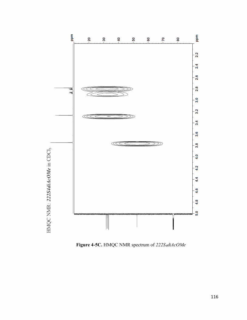
116
Figure 4-5C. HMQC NMR spectrum of 222S4diAcOMe

117
Figure 4-6A. 1H NMR spectrum of 222S4diAcOH

118
Figure 4-6B. 13C NMR spectrum of 222S4diAcOH

119
Figure 4-6C: HMQC NMR spectrum of 222S4diAcOH

120
Figure 4-7. ESI-MS of 222S4diAcOH
4.3-1. Rh(III) Complexes With 222S4diAcOMe Chelate System
[Rh(III)Cl2-222S4diAcOMe]Cl was synthesized by reacting RhCl3 with
222S4diAcOMe using a microwave synthesizer. Based on the crystal structures reported by
Dame15 (Figure 4-2A) and Goswami et al.12, we expected the formation of the cis-dichloro
isomer, where Rh(III) is coordinated to two chlorides and to the four sulfur atoms in the
222S4diAcOMe tetrathioether ligand.

121
Ten different reaction conditions were investigated as shown in Scheme 4-1.
Reactions 1 – 4 were performed in acetonitrile and water only. HPLC analysis of these
reactions revealed a predominant peak (peak area ~ 88%) at approximately 8.2 minutes
using Gradient 1 (10% solvent B:A increased to 80% solvent B:A over 18 minutes,
followed by an additional 1 minute at 80% solvent B:A, then decreased to 10% solvent
B:A over 1 minute), as shown in Figures S4-1A to S4-1I. ESI-MS analysis of this peak
indicates the presence of a strong signal for the M+ ion of cis-[Rh(III)Cl2-
222S4diAcOMe]+ [m/z = 530.85 (found), 530.88 (calculated for C12H22Cl2O4RhS4+)], as
shown in Figure 4-8. There was no significant difference in the HPLC profile between
reactions 1 – 4 (no ethanol or SnCl2 used) and reactions 5 - 6 (ethanol used) or reaction 9
(SnCl2 used). To the best of our knowledge, this is the first report of a Rh(III) tetrathioether
complex synthesized without using ethanol or SnCl2 for the catalytic reduction of Rh+3 to
Rh+1, as previously reported.3, 12-13, 15-16, 18-19
Two other minor product species were identified in reactions 1 – 4; one with a
retention time of approximately 6.5 minutes, and the other with a retention time of
approximately 4.4 minutes, as shown in Figures S4-1A to S4-1I. ESI-MS analysis revealed
that the peak at approximately 6.5 minutes is consistent with the species where one of the
pendant methyl acetate groups has been hydrolyzed to a carboxylic acid group [m/z =
516.90 (found), 516.87 (calculated for C11H20Cl2O4RhS4+)], as shown in Figure 4-9. The
m/z of the peak at approximately 4.4 minutes is consistent with the species where one of
the pendant methyl acetate groups has been hydrolyzed to a carboxylic acid and is directly
coordinated to the Rh(III) center along with one chloride atom [m/z = 480.93 (found),
480.89 (calculated for C11H19ClO4RhS4+)], as shown in Figure 4-10.

122
Figure 4-8. ESI-MS of cis-[Rh(III)Cl2-222S4diAcOMe]+

123
Figure 4-9. ESI-MS of C11H20Cl2O4RhS4+

124
Figure 4-10. ESI-MS of C11H19ClO4RhS4+
Reaction 7 was performed in acetonitrile and water, with the addition of 1 mL 0.5
M HCl. In addition to the peaks at 8.2 minutes, 6.5 minutes, and 4.4 minutes described
above, a new and intense peak (peak area ~ 46%) was observed at 2.2 minutes (Figure S4-
1G). ESI-MS analysis revealed that this new peak is consistent with the species where both
of the pendant methyl acetate groups have been hydrolyzed to carboxylic acid groups and
are both directly coordinated to the Rh(III) center [m/z = 430.94 (found), 430.90 (calculated

125
for C10H16O4RhS4+)], as shown in Figure 4-11. This reaction condition is more
representative of the reaction condition at the radiotracer scale because 105RhCl3 is obtained
in a 1 M HCl solution; hence, it is likely the acid hydrolysis of the pendant methyl acetate
groups in 222S4diAcOMe will occur at the radiotracer scale.
Figure 4-11. ESI-MS of C10H16O4RhS4+
13C NMR of [Rh(III)Cl2-222S4diAcOMe]Cl (Figure 4-12A) shows four CH2
signals (36.85 – 41. 52 ppm) and one CH3 signal (53.90 ppm), which is indicative of the

126
presence of a single isomer. If more than one isomer were present, additional CH2 signals
would have been observed, similar to data reported by Goswami et al.12 The 1H NMR of
[Rh(III)Cl2-222S4diAcOMe]Cl (Figure 4-12B) is not as straightforward to interpret as the
13C NMR. Each sulfur atom on the 222S4diAcOMe ligand becomes a chiral center upon
complexation with Rh(III); hence, different enantiomeric isomers are possible, which
explains the complexity of the 1H NMR. However, two-dimensional 13C - 1H heteronuclear
multiple-quantum correlation (HMQC) NMR allowed the assignment of the multiple 1H
peaks between 2.5 – 4.5 ppm to their corresponding 13C peak, as shown in Figure 4-12C.

127
Figure 4-12A. 13C NMR spectrum of [Rh(III)Cl2-222S4diAcOMe]Cl

128
Figure 4-12B. 1H NMR spectrum of [Rh(III)Cl2-222S4diAcOMe]Cl

129
Figure 4-12C. HMQC NMR spectrum of [Rh(III)Cl2-222S4diAcOMe]Cl
4.3-2. Rh(III) Complexes With 222S4diAcOH Chelate System
Six different reaction conditions were investigated for the synthesis of Rh(III)Cl2-
222S4diAcOH]Cl, as shown in Scheme 4-2. HPLC analysis of these reactions revealed a
predominant peak with a retention time of approximately 7.5 minutes (Figure S4-2A to
S4-2D) using Gradient 2 (2% solvent B increased to 5% solvent B over 10 minutes,
followed by an additional 5 minute at 5% solvent B, then increased to 80% solvent B over
9 minute, and finally decreased to 2% solvent B over 1 minute). ESI-MS analysis revealed
that this peak is consistent with the species where one of the pendant carboxylic acid group

130
and one chloride group is directly coordinated to the Rh(III) center [m/z = 466.96 (found),
466.88 (calculated for C10H17ClO4RhS4+)], as shown in Figure 4-13.
Figure 4-13. ESI-MS of C10H17ClO4RhS4+
Two other product species were observed with retention times of approximately 2.3
minutes and 11.8 minutes. The m/z of the peak at approximately 2.3 minutes is consistent
with the species where both of the pendant carboxylic acid groups are directly coordinated
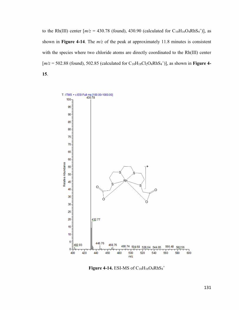
131
to the Rh(III) center [m/z = 430.78 (found), 430.90 (calculated for C10H16O4RhS4+)], as
shown in Figure 4-14. The m/z of the peak at approximately 11.8 minutes is consistent
with the species where two chloride atoms are directly coordinated to the Rh(III) center
[m/z = 502.88 (found), 502.85 (calculated for C10H18Cl2O4RhS4+)], as shown in Figure 4-
15.
Figure 4-14. ESI-MS of C10H16O4RhS4+

132
Figure 4-15. ESI-MS of for C10H18Cl2O4RhS4+
4.3-3. Rh(III) Complexes With 16S4diol Chelate System
16S4-diol is generally used as the chelate of choice for quick assessment of 105Rh-
labeling efficiency. Hence, non-radioactive Rh(III) complexes formed with 16S4-diol were
evaluated as a standard for comparing Rh(III) coordination chemistry with other chelators
at the tracer scale. [Rh(III)Cl2-16S4-diol]Cl was synthesized by reacting RhCl3 with 16S4-
diol using a microwave synthesizer. The crystal structure previously reported by Venkatesh

133
et al.3 indicates that Rh(III) was coordinated to four S atoms in a square planar geometry,
with two Cl atoms bound to the Rh(III) core in the trans axial positions. However, two
isomers were identified, which resulted from the diol oxygens being in either the cis or
trans position. This result was expected because the 16S4-diol ligand was purchased as the
cis/trans mixture.
Six different reaction conditions were investigated as shown in Scheme 4-3. HPLC
analysis of reactions 16 and 17 revealed 5 predominant peaks with retention times of
approximately 10.5 minutes, 9.7 minutes, 8.5 minutes, 8.0 minutes, and 7.3 minutes using
Gradient 3 (5% solvent B increased to 20% solvent B over 15 minutes, followed by an
additional 2 minutes at 20% solvent B, then decreased to 5% solvent B over 3 minutes), as
shown in (Figure S4-3A to S4-3F). ESI-MS analysis revealed that the peaks at
approximately 10.5 minutes and 9.7 minutes (combined peak area ~ 80%) are consistent
with the M+ ion of [Rh(III)Cl2-16S4diol]+ [m/z = 501.12 (found), 500.91 (calculated for
C12H24Cl2O2RhS4+)], as shown in Figure 4-16. We can conclude that these two isomers
result from the diol oxygens being in either the cis or trans position since the 16S4-diol
ligand was purchased as the cis/trans mixture. In addition, similar results were reported by
Venkatesh et al.3
The peaks at 8.5 minutes and 8.0 minutes correspond to the species where two
hydroxyl groups (OH) are directly coordinated to the Rh(III) metal center instead of two
chlorides [m/z = 465.08 (found), 464.98 (calculated for C12H26O4RhS4+)], as shown in
Figure 4-17. Similar product species were observed by Carroll18. No significant difference
was observed in the HPLC profile between reaction 16 (no ethanol used) and reaction 17

134
(ethanol used), which further suggests that Rh(III) complexes can be formed without the
addition of ethanol for the catalytic reduction of Rh+3 to Rh+1.
Interestingly, the product species with a retention time of approximately 10.5
minutes was preferentially formed when the reaction temperature was increased to 110 °C
(reactions 18 – 21). No significant difference was observed in the HPLC profile between
reaction 18 (no ethanol used), reaction 19 (ethanol used), and reaction 20 (SnCl2 used).
Figure 4-16. ESI-MS of for [Rh(III)Cl2-16S4diol]+

135
Figure 4-17. ESI-MS of for [Rh(III)OH2-16S4diol]+
4.3-4. Production of 105Rh from Recycled 104Ru Metal Target
Three separate neutron irradiations of the recycled 104Ru metal (production 1, 3 and
4) and one irradiation of new 104Ru metal (production 2) were performed at MURR as
summarized in Table 4-1. The 105Rh recovery in productions 3 and 4 (57.9% and 57.6%,
respectively) was significantly higher than the 105Rh recovery from productions 1 and 2
(22.9% and 15.1%, respectively). To the best of our knowledge, the ~ 57% 105Rh recovery

136
reported here for productions 3 and 4 is the highest that has been reported for 105Rh
production. The time for Cl2 bubbling during the target processing procedure in
productions 3 and 4 was more than double that used in productions 1 and 2. Bubbling more
Cl2 into NaOH solution would potentially generate more hypochlorite in situ, which would
facilitate the dissolution of the 104Ru target and the release of 105Rh into the solution. This
might explain why the 105Rh recovery was higher in productions 3 and 4, which had more
time for Cl2 bubbling.
Table 4-1: 105Rh production from recycled 104Ru metal target at MURR
Pull Date 104Ru Target*
(mg)
Irradiation
time (h)
Initial 105Rh
activity (mCi)
Recovered 105Rh
activity (mCi)
% 105Rh recovery
(decay corrected)
Production 1 4/17/2018 1.44 90.02 7.17 1.50 22.9
Production 2 5/17/2018 3.24 89.15 23.90 3.30 15.1
Production 3 6/11/2018 4.78 89.67 38.20 19.79 57.9
Production 4 8/6/2018 4.46 90.02 36.60 19.10 57.6
*Recycled 104Ru metal target was used except for production 2 where new 104Ru metal target was used
Although high 105Rh recovery was achieved in productions 3 and 4, visual
inspection of the midget impinger during the dissolution of the recycled 104Ru target
revealed that the target did not completely dissolve, given that some very fine black
particles were still observed after heating in in situ generated hypochlorite for 1 hour at 40
°C. It is possible that the high temperatures required during the target recycling process
might have affected the particle size of the metal powder, thereby making it more difficult
to dissolve. Additionally, there is a possibility that passivation of the 104Ru metal target

137
occurred during the recycling process that included a long storage period of [104Ru]RuO4
in 3 M HCl.21
HPGe gamma spectrometry analysis of the final 105Rh stock solutions revealed the
predominant gamma peaks associated with 105Rh (318 and 306 keV). However, very
minimal impurities from 24Na (< 0.1%) at 1368 keV were observed. The 24Na impurity is
most likely from the neutron activation of NaCl contaminants contained in the recycled
104Ru metal. The 24Na impurities reported here are consistent with previous results reported
by Phelps.4 No other radionuclide impurities were identified.
ICP-MS analysis was performed on a 200 L aliquot of decayed 105Rh stock
solution to determine the separation efficiency between 104Ru metal target and 105Rh during
the target processing procedure. The 105Rh stock solution is usually obtained in a total
volume of ~2 mL; hence, the amount of metallic impurities obtained from the 200 L ICP-
MS analysis aliquot was multiplied by 10 to get an estimate of the total amount of metallic
impurities in the stock solution. For all four 105Rh productions, the amount of 104Ru
remaining after separation was less than 0.05% of the original amount of irradiated 104Ru
metal target (Table 4-2). This is assuming that all signals at mass 104 are due to Ru with
an isotopic composition identical to the 98.9% enriched 104Ru metal target. Other metals
identified during the ICP-MS analysis include 105Pd, 106Pd and 103Rh. Pallidium-105 is
produced from the beta decay of 105Rh. Pallidium-106 is produced from the beta decay of
106Rh, which is produced from the neutron capture reaction on 105Rh as shown in Equation
4-2. Given the very high thermal neutron capture cross section for 105Rh (11,000 barns),
there is a high probability for neutron activation of 105Rh produced from the beta decay of
105Ru during the neutron irradiation of 104Ru metal target. Rhodium-103 is produced from

138
the beta decay of 103Ru, which is produced from the neutron activation of 102Ru impurity
in the 104Ru metal target as shown in Equation 4-3. Phelps previously reported that the
104Ru metal target contains about 0.5% 102Ru impurities, which would explain the
formation of 103Rh.4
Equation 4-2. Possible production route for 106Pd
Equation 4-3. Possible production route for 103Rh
Table 4-2: ICP-MS analysis of metals in decayed 105Rh stock solution*
104Ru Metal
Target (ng)
Total 105Pd (ng)
Total 106Pd (ng)
Total 103Rh (ng)
Total 104Ru
remaining (ng)
% 104Ru
remaining
Production 1 1.44 E+06 4.5 0.71 0.95 52.9 0.004
Production 2 3.24 E+06 23.5 4.05 0.10 35.4 0.001
Production 3 4.78 E+06 46.4 6.32 8.42 745 0.016
Production 4 4.46 E+06 40.7 6.03 7.48 2130 0.048
*Based on 2 mL of 105Rh stock solution
4.3-5. 105Rh-labeling
Prior to radiolabeling, the pH of the [105Rh]RhCl3 solution was carefully adjusted
to ~ 3.5 using 1 M NaOH (20 L) and 0.5 M NaOH (20 L). Previous studies have shown
that Rh+3 tends to hydrolyze under basic conditions to form Rh(OH)3 (Ksp = 4.8 × 10⁻²³),
which may form dimers or bridged species.22 Once Rh(OH)3 is formed, it is almost

139
impossible to convert back to Rh+3. Hence, it is critical that 105Rh-labeling experiments be
performed under acidic conditions (pH < 5). Care should be taken when adjusting the pH
of the [105Rh]RhCl3 solution to avoid going above pH 7. The Rf value of [105Rh]RhCl3
developed using 0.9% saline is 0.05 when the pH of the [105Rh]RhCl3 solution is above 7
(Figure S4-4) as opposed to 1.0 when the pH is ~4 (Figure S4-5). This indicates that
[105Rh]Rh(OH)3 (formed when pH > 7) remains at the origin, while [105Rh]RhCl3 moves to
the solvent front in 0.9% saline. Radiolabeling experiments that were performed in pH > 7
resulted in no radiolabeling yields based on TLC analysis, with all radioactivity remaining
in the origin.
105Rh-labeling was performed using 222S4diAcOMe, 222S4diAcOH, and 16S4-
diol. The pH-adjusted [105Rh]RhCl3 solution was incubated with the ligand solutions for
1 hour at 90 °C in a water bath. Silica gel TLC analysis was performed using three mobile
phases; 0.9% saline, acetonitrile, and 0.4 M NaBPh4 in acetonitrile. The Rf values of
[105Rh]RhCl3, [105Rh]RhCl2-222S4diAcOMe, [105Rh]RhCl2-222S4diAcOH, and
[105Rh]RhCl2-16S4diol in the three mobile phases are tabulated in Table 4-3 and the
images of the TLC scan are shown in Figures S4-4 to S4-16.
Table 4-3. Silica gel TLC analysis of 105Rh complexes
Compound Rf (0.9% Saline) Rf (ACN) Rf (0.4 M NaBPh4 in ACN)
[105Rh]RhCl3 1.0 0.05 0.05
[105Rh]RhCl2-222S4diAcOMe ~0.8 ~0.6 0.3-0.7
[105Rh]RhCl2-222S4diAcOH ~0.8 ~0.6 0.3-0.7
[105Rh]RhCl2-16S4diol 0.3 0.2-0.4 0.9

140
The radiolabeling yield for [105Rh]RhCl2-16S4diol was 80.9 ± 0.4% (n = 3), which
was determined using the TLC scans developed in saline as the mobile phase. The
radiolabeling yields for [105Rh]RhCl2-222S4diAcOMe and [105Rh]RhCl2-222S4diAcOH
were 92.5 ± 2.0% (n = 3) and 91.0 ± 1.5% (n = 3), respectively, which were determined
using the TLC scans developed in acetonitrile as the mobile phase. The TLC scans from
[105Rh]RhCl2-222S4diAcOMe and [105Rh]RhCl2-222S4diAcOH were almost identical in
all three mobile phases. It is likely that the pendant methyl acetate groups in the
222S4diAcOMe ligand would be hydrolyzed to carboxylic acid groups given the acidic
reaction conditions used for radiolabeling. This would result in identical product species
with the 222S4diAcOH ligand. Acid hydrolysis of the pendant methyl acetate groups in
222S4diAcOMe was also observed during the macro-scale synthesis of Rh(III)Cl2-
222S4diAcOMe with the addition of 1 mL of 0.5 M HCl.
4.4. Conclusions
Rh-105 remains an attractive radioisotope for the development of therapeutic
radiopharmaceuticals due to its favorable nuclear properties (half-life = 35.4 hours,
avg =
152 keV). Historically, it has been reported that the catalytic reduction of Rh+3 to Rh+1,
with subsequent re-oxidation to Rh+3 by atmospheric oxygen, is necessary for the ligand
exchange and formation of Rh(III) complexes.14, 19-20 Hence refluxing alcohols, particularly
ethanol, have been traditionally used as reducing agents for the formation of Rh(III)
complexes.3, 8, 12-13 However, the studies reported here demonstrates that Rh(III) complexes
can be synthesized without the addition of ethanol or SnCl2 as reducing agents.
Additionally, a microwave-assisted synthesis method for Rh(III) complexes was reported.
The use a microwave significantly reduced the reaction time to 10 minutes or less

141
compared to the over 1 hour reaction time reported using the conventional bench top
synthesis method.15, 18 An improved HPLC method was developed to identify the various
Rh(III) product species, which were subsequently characterized using ESI-MS.
We also report the production of 105Rh using recycled 104Ru metal target.
Subsequently, 105Rh-labeling experiments were performed using three ligands (16S4-diol,
222S4diAcOMe, and 222S4diAcOH). To the best of our knowledge, the is the first reported
synthesis of Rh(III) complexes using 105Rh produced from recycled 104Ru metal targets.
Additionally, 105Rh-labeling of 222S4diAcOMe, and 222S4diAcOH was achieved without
ethanol or SnCl2 as reducing agents.
4.5. Future Studies
Although the production of 105Rh using recycled 104Ru metal target was successful,
there is room to improve the target processing procedure. The use of Cl2 to generate
hypochlorite in situ for the oxidation of 104Ru metal creates room for possible
inconsistencies between different batches of target processing. This is because the amount
of Cl2 used may vary due to changes in tubing length or pressure in the Cl2 tank. The data
reported here shows that the amount of 105Rh recovered is influenced by the amount of Cl2
(i.e., increased Cl2 bubbling time) used during target processing with more 105Rh recovered
with longer Cl2 bubbling time. Additionally, Cl2 is a toxic gas and should be avoided if
possible. As an alternative, commercially available NaOCl can be used to oxidize 104Ru
metal to 104RuO4. Phelps has demonstrated the oxidation of natRu metal using 12-15%
NaOCl.4 Therefore, this method can be applied for processing irradiated 104Ru metal target
to separate 105Rh.

142
Potential preclinical and clinical translation of 105Rh-based radiopharmaceuticals
will require the development of bifunctional analogues of 222S4diAcOH that are
conjugated to suitable targeting molecules. The pendant carboxylic acid groups in
222S4diAcOH provide a suitable functional group for the formation of an amide bond with
terminal amines in peptide molecules. Rhodium-105 labeling of the resulting bifunctional
chelator can then be performed without the addition of ethanol or SnCl2 as reducing agents,
following the synthesis procedure that is reported in this work. Additionally, the
microwave synthesis method reported here for the synthesis of non-radioactive Rh(III)
complexes can also be applied for 105Rh labeling studies to decrease reaction time.

143
4.6. References
1. Grazman, B.; Troutner, D., Rh-105 as a potential radiotherapeutic agent. Journal of Labelled Compounds and Radiopharmaceuticals 1986, 23 (10-12), 1371-1373.
2. Grazman, B.; Troutner, D., 105Rh as a potential radiotherapeutic agent. International Journal of Radiation Applications and Instrumentation. Part A. Applied Radiation and Isotopes 1988, 39 (3), 257-260.
3. Venkatesh, M.; Goswami, N.; Volkert, W.; Schlemper, E.; Ketring, A.; Barnes, C.; Jurisson, S., An Rh-105 complex of tetrathiacyclohexadecane diol with potential for formulating bifunctional chelates. Nuclear Medicine and Biology 1996, 23 (1), 33-40.
4. Phelps, T. Radionuclide Production and Separation Techniques for Radiopharmaceutical Development and Nuclear Waste Processing. Doctoral Dissertation, University of Missouri-Columbia, 2018.
5. Greenwood, N. N.; Earnshaw, A., Chemistry of the Elements. Elsevier: 2012.
6. Kruper Jr, W. J.; Fordyce, W. A.; Pollock, D. K.; Fazio, M. J.; Inbaskaran, M. N.; Muthyala, R., Functionalized polyamine chelants and rhodium complexes thereof for conjugation to antibodies. U.S. Patent No. 5,284,644: 1994.
7. Pillai, M.; John, C.; Troutner, D. E., Labeling of human IgG with rhodium-105 using a new pentadentate bifunctional ligand. Bioconjugate Chemistry 1990, 1 (3), 191-197.
8. Li, N.; Struttman, M.; Higginbotham, C.; Grall, A. J.; Skerlj, J. F.; Vollano, J. F.; Bridger, S. A.; Ochrymowycz, L. A.; Ketring, A. R.; Abrams, M. J.; Volkert, W. A., Biodistribution of model 105Rh-labeled tetradentate thiamacrocycles in rats. Nuclear Medicine and Biology 1997, 24 (1), 85-92.
9. Pillai, M.; Lo, J.; Troutner, D., Labeling of hematoporphyrin with 105Rh and binding studies with human gamma globulin. International Journal of Radiation Applications and Instrumentation. Part A. Applied Radiation and Isotopes 1990, 41 (1), 69-73.
10. Pillai, M.; Lo, J.; John, C.; Troutner, D., Labeling of proteins using [105Rh]Rh-4-(p-aminobenzyl)-diethylenetriamine. International Journal of Radiation Applications and Instrumentation. Part B. Nuclear Medicine and Biology 1990, 17 (4), 419-426.
11. Efe, G. E.; Pillai, M.; Schlemper, E.; Troutner, D., Rhodium complexes of two bidentate secondary amine oxime ligands and application to the labelling of proteins. Polyhedron 1991, 10 (14), 1617-1624.
12. Goswami, N.; Alberto, R.; Barnes, C. L.; Jurisson, S., Rhodium(III) complexes with acyclic tetrathioether ligands. Effects of backbone chain length on the conformation of the Rh(III) complex. Inorganic Chemistry 1996, 35 (26), 7546-7555.
13. Goswami, N.; Higginbotham, C.; Volkert, W.; Alberto, R.; Nef, W.; Jurisson, S., Rhodium-105 tetrathioether complexes: radiochemistry and initial biological evaluation. Nuclear Medicine and Biology 1999, 26 (8), 951-7.

144
14. Feng, Y.; Phelps, T. E.; Carroll, V.; Gallazzi, F.; Sieckman, G.; Hoffman, T. J.; Barnes, C. L.; Ketring, A. R.; Hennkens, H. M.; Jurisson, S. S., Chemistry and radiochemistry of As, Re and Rh isotopes relevant to radiopharmaceutical applications: high specific activity radionuclides for imaging and treatment. Dalton Transactions 2017, 46 (42), 14677-14690.
15. Dame, A. Synthesis Design and Nuclear Medicine Applications for Radiometals, Beta and Gamma Emission, Chelated Complexes. Doctoral Dissertation, University of Missouri-Columbia, 2017.
16. Crenshaw, N. N. Characterization of Acyclic Rhodium Tetrathioether Ligand Systems for 105Rh (III). Masters Thesis, University of Missouri-Columbia, 2015.
17. Akgun, Z.; Engelbrecht, H.; Fan, K.-H.; Barnes, C. L.; Cutler, C. S.; Jurisson, S. S.; Lever, S. Z., The complexation of rhodium (III) with acyclic diaminedithioether (DADTE) ligands. Dalton Transactions 2010, 39 (42), 10169-10178.
18. Carroll, V. N., Development of a rhodium tetrathioether bombesin analogue and investigation of cyclic and acyclic ligand systems for 105Rh (III). University of Missouri-Columbia: 2013.
19. Rund, J. V.; Basolo, F.; Pearson, R. G., Catalysis of substitution reactions of rhodium (III) complexes. The reaction of aquopentachlororhodate (III) ion with pyridine. Inorganic Chemistry 1964, 3 (5), 658-661.
20. Jurisson, S. S.; Ketring, A. R.; Volkert, W. A., Rhodium-105 complexes as potential radiotherapeutic agents. Transition Metal Chemistry 1997, 22 (3), 315-317.
21. Llopis, J.; Vazquez, M., Passivation of ruthenium in hydrochloric acid solution. Electrochimica Acta 1966, 11 (6), 633-640.
22. Forrester, J.; Ayres, G., Rhodium (III) in aqueous solutions. The Journal of Physical Chemistry 1959, 63 (11), 1979-1981.

145
Supplementary Information
Supplementary Data - Chapter 2
Table S2-1. Efficiency of 59Fe stripping from Pb-resin
Column # 59FeCl3 (Ci) Fe (g)* Pb (g)** Fraction 1
(Ci)
Fraction 2
(Ci)
Total 59Fe
recovery
1 129 0 0 125.5 0 97%
2 129 4.4 0 126 0 98%
3 121.5 22.1 0 120.7 0 99%
4 117.6 11.0 41.0 114.4 0.8 98%
5 108.5 11.0 41.0 106.5 1.9 100%
6 108.5 11.0 41.0 106.3 1.9 100%
7 55.8 6.1 6.1 53.4 0.6 97%
8 55.8 6.1 6.1 54 0.6 98%
9 55.6 6.1 6.1 53.7 0.3 97%
* from 0.54 mg/ml FeCl3.6H2O solution
** from 0.64 mg/ml Pb(NO3)2 solution
Figure S2-1. HPLC profile of Pb-RM2 (gradient = 22% ACN:H2O increased to 25%
ACN:H2O over 19 minutes, followed by an additional 1 minute at 25% ACN:H2O, then
decreased to 22% ACN:H2O over 1 minute at a flow rate of 1.5 mL/min)

146
Table S2-2. [203Pb]PbCl2 biodistribution in CF1 mice Data shown as mean ± SD %ID (n = 5 mice per time point)
Tissue 15 min 1 h 4 h 24 h
Blood 7.64 ± 0.97 6.70 ± 1.06 5.27 ± 0.98 3.64 ± 0.45
Heart 0.12 ± 0.04 0.07 ± 0.02 0.06 ± 0.02 0.03 ± 0.01
Lung 0.61 ± 0.08 0.54 ± 0.12 0.42 ± 0.23 0.18 ± 0.04
Liver 22.17 ± 2.07 22.79 ± 1.49 23.89 ± 1.36 16.53 ± 3.50
Stomach 0.47 ± 0.07 0.45 ± 0.14 0.65 ± 0.28 0.42 ± 0.34
Sm. Intestines 4.17 ± 0.40 4.35 ± 0.42 4.19 ± 0.38 1.70 ± 0.20
Lg. Intestines 1.53 ± 0.55 1.17 ± 0.07 2.70 ± 0.43 1.71 ± 0.23
Kidney 26.26 ± 3.10 28.52 ± 2.84 25.46 ± 2.10 18.97 ± 1.51
Spleen 0.13 ± 0.03 0.16 ± 0.05 0.18 ± 0.03 0.10 ± 0.06
Brain 0.07 ± 0.01 0.07 ± 0.02 0.08 ± 0.03 0.05 ± 0.01
Pancreas 0.83 ± 0.14 0.78 ± 0.13 0.65 ± 0.14 0.34 ± 0.06
Muscle 0.05 ± 0.01 0.05 ± 0.02 0.03 ± 0.02 0.03 ± 0.03
Bone 0.75 ± 0.16 0.95 ± 0.12 0.95 ± 0.10 1.29 ± 0.14
Urine (%ID) 0.95 ± 0.54 2.05 ± 0.32 4.63 ± 0.84 10.22 ± 2.86
Excretion (%ID) 0.95 ± 0.19 2.05 ± 0.19 4.63 ± 0.86 19.82 ± 4.61
Note: Values for urine include radioactivity measured in cage paper

147
Ta
ble
S2
-3.
[20
3P
b]P
b-R
M2
b
iod
istr
ibu
tio
n i
n P
C3
tu
mo
r-b
eari
ng
mic
e.
Da
ta s
ho
wn
as
mea
n ±
SD
%ID
(n
= 5
mic
e p
er t
ime
po
int)
Tis
sue
15
min
3
0 m
in
1 h
2
h
4 h
2
4 h
4
8 h
Blo
od
5
.03
± 1
.55
2.5
7 ±
0.7
0
0.8
4 ±
0.2
8
0.1
3 ±
0.0
6
0.0
9 ±
0.0
2
0.0
5 ±
0.0
1
0.0
3 ±
0.0
3
Hea
rt
0.1
5 ±
0.0
4
0.0
7 ±
0.0
2
0.0
3 ±
0.0
1
0.0
1 ±
0.0
0
0.0
1 ±
0.0
0
0.0
0 ±
0.0
0
0.0
1 ±
0.0
1
Lu
ng
0
.44
± 0
.13
0.2
4 ±
0.0
4
0.1
0 ±
0.0
1
0.0
3 ±
0.0
1
0.0
2 ±
0.0
0
0.0
1 ±
0.0
1
0.0
1 ±
0.0
1
Liv
er
2.8
3 ±
0.8
1
1.3
9 ±
0.2
1
0.7
1 ±
0.1
2
0.3
3 ±
0.0
5
0.3
7 ±
0.0
7
0.2
6 ±
0.0
4
0.1
5 ±
0.0
2
Sto
ma
ch
0.5
5 ±
0.1
3
0.3
5 ±
0.0
8
0.2
1 ±
0.0
6
0.9
7 ±
1.8
6
0.0
7 ±
0.0
1
0.0
2 ±
0.0
1
0.0
0 ±
0.0
0
Sm
. In
test
ines
3
.25
± 0
.66
2.5
5 ±
0.4
5
1.5
3 ±
0.2
1
3.8
0 ±
6.5
6
0.4
8 ±
0.2
2
0.2
6 ±
0.2
8
0.0
4 ±
0.0
0
Lg
. In
test
ines
1.0
4 ±
0.3
3
0.7
6 ±
0.2
0
0.3
6 ±
0.0
9
0.7
6 ±
0.1
9
1.9
0 ±
2.0
0
0.1
3 ±
0.0
5
0.0
9 ±
0.0
4
Kid
ney
3
.33
± 0
.73
2.2
2 ±
0.3
9
1.8
9 ±
0.1
5
1.4
7 ±
0.2
3
1.5
1 ±
0.2
7
0.8
8 ±
0.2
6
0.4
2 ±
0.1
1
Sp
leen
0
.06
± 0
.02
0.0
3 ±
0.0
1
0.0
1 ±
0.0
0
0.0
1 ±
0.0
1
0.0
1 ±
0.0
0
0.0
1 ±
0.0
0
0.0
1 ±
0.0
1
Bra
in
0.0
6 ±
0.0
1
0.0
3 ±
0.0
1
0.0
2 ±
0.0
0
0.0
1 ±
0.0
1
0.0
1 ±
0.0
0
0.0
0 ±
0.0
0
0.0
1 ±
0.0
1
Pa
ncr
eas
4.2
4 ±
0.7
2
3.2
8 ±
0.6
8
1.4
4 ±
0.4
1
0.5
6 ±
0.2
5
0.1
2 ±
0.0
6
0.0
4 ±
0.0
0
0.0
2 ±
0.0
1
Mu
scle
0
.15
± 0
.05
0.0
9 ±
0.0
4
0.0
3 ±
0.0
1
0.0
1 ±
0.0
0
0.0
1 ±
0.0
1
0.0
1 ±
0.0
1
0.0
0 ±
0.0
1
Bo
ne
0.0
5 ±
0.0
1
0.0
3 ±
0.0
1
0.0
1 ±
0.0
0
0.0
1 ±
0.0
2
0.0
1 ±
0.0
0
0.0
1 ±
0.0
0
0.0
1 ±
0.0
1
Tu
mo
rs
1.4
4 ±
1.0
3
1.4
5 ±
0.4
9
2.2
0 ±
0.9
5
1.2
3 ±
0.3
9
1.5
7 ±
0.7
2
1.3
8 ±
0.7
9
0.6
7 ±
0.3
9
Uri
ne (
%ID
) 4
1.6
6 ±
0.2
7
57
.42 ±
4.4
0
79
.36 ±
1.2
5
86
.75 ±
8.0
1
90
.44 ±
2.9
2
92
.32 ±
2.0
6
91
.72 ±
4.8
6
Excr
etio
n (
%ID
) 4
1.6
6 ±
0.2
7
57
.42 ±
4.4
0
79
.36 ±
1.2
5
86
.75 ±
8.0
1
90
.44 ±
2.9
2
94
.59 ±
1.8
5
97
.36 ±
0.9
2
No
te:
Val
ues
fo
r uri
ne
incl
ud
e r
adio
acti
vit
y m
easu
red
in c
age
pap
er

148
Supplementary Data – Chapter 3

149
Figure S3-1. Gamma spectrum peak analysis report for Pb-212 generator eluent

150

151
Figure S3-2. Gamma spectrum peak analysis report for purified Pb-212

152
Ta
ble
S3
-1.
[21
2P
b]P
b-R
M2
b
iod
istr
ibu
tio
n i
n P
C3
tu
mo
r b
eari
ng
mic
e
Data
sh
ow
n a
s m
ean
± S
D %
ID (
n =
6 m
ice
per
tim
e p
oin
t)
Tis
sue
15
min
3
0 m
in
1 h
2
h *
4
h*
2
4 h
4
8 h
Blo
od
7
.39
± 2
.94
3.3
6 ±
1.3
9
1.1
2 ±
0.5
4
0.1
7 ±
0.1
1
0.0
6 ±
0.0
7
0.0
3 ±
0.0
4
0.1
2 ±
0.1
9
Hea
rt
0.1
7 ±
0.0
7
0.0
8 ±
0.0
3
0.0
2 ±
0.0
2
0.0
0 ±
0.0
1
0.0
1 ±
0.0
2
0.0
1 ±
0.0
2
0.0
3 ±
0.0
7
Lu
ng
0
.54
± 0
.24
0.2
9 ±
0.1
2
0.1
0 ±
0.0
5
0.0
3 ±
0.0
1
0.0
2 ±
0.0
2
0.0
2 ±
0.0
2
0.0
4 ±
0.0
8
Liv
er
4.4
4 ±
2.0
1
2.8
2 ±
1.4
4
1.5
6 ±
0.6
7
0.5
8 ±
0.0
9
0.3
2 ±
0.1
2
0.0
8 ±
0.0
4
0.0
8 ±
0.0
9
Sto
ma
ch
0.6
8 ±
0.2
9
0.2
9 ±
0.1
2
0.3
1 ±
0.1
7
0.1
8 ±
0.1
9
0.0
7 ±
0.0
3
0.0
1 ±
0.0
3
0.0
6 ±
0.0
8
Sm
. In
test
ines
4
.05
± 1
.99
1.9
0 ±
0.9
9
1.4
9 ±
0.7
2
0.7
2 ±
0.5
5
0.2
7 ±
0.1
3
0.1
0 ±
0.0
5
0.0
6 ±
0.0
7
Lg
. In
test
ines
1.3
4 ±
0.5
6
0.6
0 ±
0.2
5
0.4
5 ±
0.1
8
0.6
9 ±
0.1
8
0.6
8 ±
0.1
8
0.4
5 ±
0.3
4
0.1
3 ±
0.1
5
Kid
ney
6
.96
± 3
.03
5.5
7 ±
2.2
4
4.9
8 ±
1.9
4
2.3
0 ±
0.5
2
1.4
2 ±
0.3
9
0.4
1 ±
0.1
7
0.1
7 ±
0.1
4
Sp
leen
0
.10
± 0
.04
0.0
9 ±
0.0
4
0.0
3 ±
0.0
1
0.0
1 ±
0.0
1
0.0
1 ±
0.0
1
0.0
1 ±
0.0
1
0.0
3 ±
0.0
5
Bra
in
0.0
7 ±
0.0
3
0.0
4 ±
0.0
2
0.0
2 ±
0.0
2
0.0
0 ±
0.0
0
0.0
1 ±
0.0
2
0.0
0 ±
0.0
0
0.0
3 ±
0.0
6
Pa
ncr
ea
s 7
.03
± 3
.71
1.9
3 ±
1.0
0
1.4
2 ±
0.7
7
0.4
8 ±
0.4
4
0.0
7 ±
0.0
4
0.0
2 ±
0.0
2
0.0
1 ±
0.0
2
Mu
scle
0
.16
± 0
.07
0.0
8 ±
0.0
4
0.0
2 ±
0.0
1
0.0
1 ±
0.0
1
0.0
1 ±
0.0
1
0.0
1 ±
0.0
1
0.0
5 ±
0.0
6
Bo
ne
0.1
6 ±
0.0
7
0.0
8 ±
0.0
4
0.0
3 ±
0.0
2
0.0
1 ±
0.0
1
0.0
1 ±
0.0
1
0.0
1 ±
0.0
1
0.0
3 ±
0.0
4
Tu
mo
rs
1.2
6 ±
0.4
4
1.2
8 ±
0.4
3
1.0
8 ±
0.4
2
1.3
1 ±
0.6
2
1.1
5 ±
0.5
1
0.7
5 ±
0.2
2
0.4
7 ±
0.2
4
Uri
ne
(%ID
) 3
3.6
7 ±
11
.23
6
2.9
3 ±
8.6
7
76
.73 ±
2.1
6
89
.87 ±
2.0
4
93
.76 ±
1.4
0
92
.92 ±
3.1
7
88
.28±
10
.58
Excr
etio
n (
%ID
) 3
3.6
7 ±
11
.23
6
2.9
3 ±
8.6
7
76
.73 ±
2.1
6
89
.87 ±
2.0
4
93
.76 ±
1.4
0
96
.89 ±
0.3
8
98
.11±
1.1
8
* n
= 7
No
te:
Val
ues
fo
r uri
ne
incl
ud
e r
adio
acti
vit
y m
easu
red
in b
lad
der
and
cag
e p
aper

153
Table S3-2. ICP-MS analysis of crude and purified 212Pb samples **
Sample ID Date Description Fe (ng) Zn (ng) Pb (ng)
Generator 3
T 1 09/19/2018 2M HCl 6.00 0.50 3.46
T 2 09/19/2018 0.5M NaOAc <LOD 0.71 0.30
T 3 09/19/2018 Crude Pb-212 113 20.7 50.9
T 4 09/19/2018 Purified Pb-212 6.12 7.20 40.0
T 5 09/20/2018 Crude Pb-212 65.2 40.6 90.7
T 6 09/20/2018 Purified Pb-212 1.77 14.7 32.0
T 7 09/21/2018 Crude Pb-212 22.2 24.4 82.4
T 8 09/21/2018 Purified Pb-212 1.30 13.2 28.6
T 9 09/24/2018 Crude Pb-212 37.7 27.3 5.06
T 10 09/24/2018 Purified Pb-212 1.70 8.44 16.5
T 11 09/26/2018 Crude Pb-212 18.3 29.0 17.3
T 12 09/26/2018 Purified Pb-212 1.02 8.25 48.1
T 13 09/28/2018 Crude Pb-212 17.5 24.4 4.83
T 14 09/28/2018 Purified Pb-212 1.93 7.18 9.51
Generator 4
T 18 10/10/2018 Crude Pb-212 104 22.2 42.7
T 19 10/10/2018 Purified Pb-212 <LOD 2.43 30.1
T 20 10/11/2018 Crude Pb-212 121 23.1 14.5
T 26 10/11/2018 Purified Pb-212 <LOD 2.02 6.53
T 27 10/12/2018 Crude Pb-212 96.8 34.8 6.01
T 28 10/12/2018 Purified Pb-212 <LOD 3.28 4.80
T 29 10/15/2018 Crude Pb-212 243 28.0 2.58
T 30 10/15/2018 Purified Pb-212 <LOD 2.04 4.01
T 31 10/16/2018 Crude Pb-212 137 24.2 8.39
** Each sample contains 200 L

154
Supplementary Data – Chapter 4
Figure S4-1A. HPLC spectrum for reaction 1
Figure S4-1B. HPLC spectrum for reaction 2
Figure S4-1C. HPLC spectrum for reaction 3
0.0 2.5 5.0 7.5 10.0 12.5 15.0 17.5 min
0.00
0.25
0.50
0.75
1.00
1.25
1.50
1.75
mV(x1,000)Detector A Ch1:280nm
4.5
73
6.5
70
8.3
16
0.0 2.5 5.0 7.5 10.0 12.5 15.0 17.5 min
0.0
0.5
1.0
1.5
2.0
2.5
mV(x1,000)Detector A Ch1:280nm
4.4
02
6.4
54
8.1
70
0.0 2.5 5.0 7.5 10.0 12.5 15.0 17.5 min
0.00
0.25
0.50
0.75
1.00
1.25
mV(x1,000)Detector A Ch1:280nm
4.8
25
6.6
63
8.6
32
10
.84
7

155
Figure S4-1D. HPLC spectrum for reaction 4
Figure S4-1E. HPLC spectrum for reaction 5
Figure S4-1F. HPLC spectrum for reaction 6
0.0 2.5 5.0 7.5 10.0 12.5 15.0 17.5 min
0.00
0.25
0.50
0.75
1.00
1.25
1.50
mV(x1,000)Detector A Ch1:280nm
2.2
36
7.2
00
8.1
66
0.0 2.5 5.0 7.5 10.0 12.5 15.0 17.5 min
0.0
0.5
1.0
1.5
2.0
2.5
mV(x1,000)Detector A Ch1:280nm
2.2
37
4.3
85
8.1
28
9.3
54
10
.17
3
0.0 2.5 5.0 7.5 10.0 12.5 15.0 17.5 min
0.0
0.5
1.0
1.5
2.0
2.5mV(x1,000)
Detector A Ch1:280nm
4.3
79
6.4
60
8.1
71
9.8
80

156
Figure S4-1G. HPLC spectrum for reaction 7
Figure S4-1H. HPLC spectrum for reaction 8
Figure S4-1I. HPLC spectrum for reaction 9
0.0 2.5 5.0 7.5 10.0 12.5 15.0 17.5 min
0.00
0.25
0.50
0.75
1.00
1.25mV(x1,000)
Detector A Ch1:280nm
2.2
13
4.3
66 6
.40
2
8.1
54
0.0 2.5 5.0 7.5 10.0 12.5 15.0 17.5 min
0.00
0.25
0.50
0.75
1.00
1.25
1.50
1.75
2.00mV(x1,000)
Detector A Ch1:280nm
4.4
54
6.4
26
8.1
94
0.0 2.5 5.0 7.5 10.0 12.5 15.0 17.5 min
0.00
0.25
0.50
0.75
1.00
1.25
1.50
1.75
2.00
2.25
mV(x1,000)Detector A Ch1:280nm
4.5
44
6.5
29
8.2
70

157
Figure S4-2A. HPLC spectrum for reaction 10
Figure S4-2B. HPLC spectrum for reaction 11
Figure S4-2C. HPLC spectrum for reaction 12
Figure S4-2D. HPLC spectrum for reaction 13
Figure S4-2E. HPLC spectrum for reaction 14
0.0 2.5 5.0 7.5 10.0 12.5 15.0 17.5 min
0.0
2.5
5.0
7.5
mV(x100)Detector A Ch1:280nm
2.1
74
5.7
81
7.5
24
0.0 2.5 5.0 7.5 10.0 12.5 15.0 17.5 min
0.0
1.0
2.0
3.0
4.0
5.0
6.0
mV(x100)Detector A Ch1:280nm
2.4
08
5.5
86
7.2
45
11
.59
0
0.0 2.5 5.0 7.5 10.0 12.5 15.0 17.5 min
0.0
2.5
5.0
mV(x100)Detector A Ch1:280nm
2.2
11
6.0
13
7.7
94
12
.43
4
0.0 2.5 5.0 7.5 10.0 12.5 15.0 17.5 min
0.00
0.25
0.50
0.75
1.00
mV(x1,000)Detector A Ch1:280nm
2.1
82
5.8
40
7.5
63
12
.17
9
0.0 2.5 5.0 7.5 10.0 12.5 15.0 17.5 min
0.0
2.5
5.0
mV(x100)Detector A Ch1:280nm
7.5
15
11
.86
6

158
Figure S4-2F. HPLC spectrum for reaction 15
Figure S4-3A. HPLC spectrum for reaction 16
Figure S4-3B. HPLC spectrum for reaction 17
Figure S4-3C. HPLC spectrum for reaction 18
0.0 2.5 5.0 7.5 10.0 12.5 15.0 17.5 min
0.0
2.5
5.0
7.5mV(x100)
Detector A Ch1:280nm
2.1
77
7.4
00
11
.78
9
0.0 2.5 5.0 7.5 10.0 12.5 15.0 17.5 min
0.00
0.25
0.50
0.75
1.00
1.25
1.50
1.75
2.00
2.25mV(x1,000)
Detector A Ch1:280nm
6.9
01
7.3
56
8.0
36
8.5
25
9.8
84
10
.57
9
0.0 2.5 5.0 7.5 10.0 12.5 15.0 17.5 min
0.0
0.5
1.0
1.5
2.0
2.5
3.0
3.5
4.0
mV(x1,000)Detector A Ch1:280nm
6.7
79 7.2
26
7.9
18
8.4
06
9.7
17
10
.45
5
0.0 2.5 5.0 7.5 10.0 12.5 15.0 17.5 min
0.00
0.25
0.50
0.75
1.00
1.25
1.50
1.75
2.00
2.25
mV(x1,000)Detector A Ch1:280nm
7.6
09
8.4
81
10
.15
1
10
.72
1

159
Figure S4-3D. HPLC spectrum for reaction 19
Figure S4-3E. HPLC spectrum for reaction 20
Figure S4-3F. HPLC spectrum for reaction 21
0.0 2.5 5.0 7.5 10.0 12.5 15.0 17.5 min
0.0
0.5
1.0
1.5
2.0
2.5
3.0mV(x1,000)
Detector A Ch1:280nm
6.1
41
7.2
63
8.6
11
10
.30
3
10
.86
00.0 2.5 5.0 7.5 10.0 12.5 15.0 17.5 min
0.0
0.5
1.0
1.5
2.0
2.5
mV(x1,000)Detector A Ch1:280nm
6.8
78
7.3
51
8.2
06
8.4
87
9.9
02
10
.47
9
0.0 2.5 5.0 7.5 10.0 12.5 15.0 17.5 min
0.00
0.25
0.50
0.75
1.00
1.25
1.50
mV(x1,000)Detector A Ch1:280nm
7.0
13
7.4
68
8.3
65
10
.08
4
10
.69
1

160
Figure S4-4. TLC scan of [105Rh]RhCl3 solution (pH > 7) with 0.9% saline as mobile
phase
Figure S4-5. TLC scan of [105Rh]RhCl3 solution (pH ~ 4) using 0.9% saline as mobile
phase

161
Figure S4-6. TLC scan of [105Rh]RhCl2-222S4diAcOH using 0.9% saline as mobile
phase
Figure S4-7. TLC scan of [105Rh]RhCl2-222S4diAcOMe using 0.9% saline as mobile
phase

162
Figure S4-8. TLC scan of [105Rh]RhCl2-16S4diol using 0.9% saline as mobile phase
Figure S4-9. TLC scan of [105Rh]RhCl3 solution using ACN as mobile phase
Figure S4-10. TLC scan of [105Rh]RhCl2-222S4diAcOH using ACN as mobile phase

163
Figure S4-11. TLC scan of [105Rh]RhCl2-222S4diAcOMe using ACN as mobile phase
Figure S4-12. TLC scan of [105Rh]RhCl2-16S4diol using ACN as mobile phase
Figure S4-13. TLC scan of [105Rh]RhCl3 solution using 0.4M NaBPh4 in ACN as mobile
phase
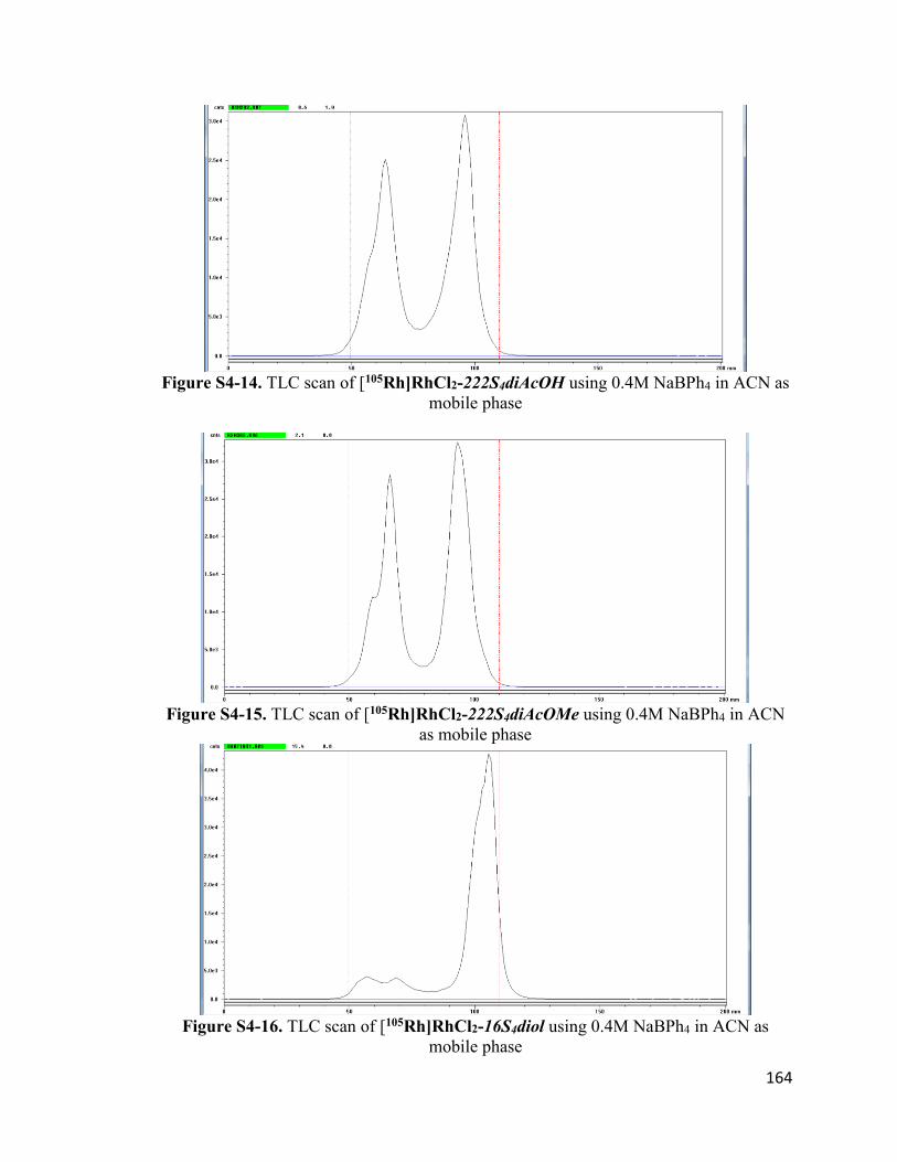
164
Figure S4-14. TLC scan of [105Rh]RhCl2-222S4diAcOH using 0.4M NaBPh4 in ACN as
mobile phase
Figure S4-15. TLC scan of [105Rh]RhCl2-222S4diAcOMe using 0.4M NaBPh4 in ACN
as mobile phase
Figure S4-16. TLC scan of [105Rh]RhCl2-16S4diol using 0.4M NaBPh4 in ACN as
mobile phase

165
VITA
Nkemakonam Chukwuebuka Okoye was born on February 26, 1992 in Aba, Abia
State, Nigeria to the family of Sir Josiah Okoye and Lady Charity Okoye. He attended
Dority International Secondary School where he completed the West African Senior
School Certificate Examination in 2009. Nkem moved to the United States of America in
August 2010 on a full academic scholarship to attend South Carolina State University
(SCSU) in Orangeburg South Carolina. Nkem was involved in undergraduate research in
the environmental radiochemistry laboratory at SCSU under the mentorship of Dr. Zheng
Chang and subsequently earned his Bachelor of Science degree in Chemistry (with honors)
in May, 2014.
Nkem joined the chemistry Ph.D program at the University of Missouri-Columbia in
August 2014. Under the supervision of Dr. Silvia S. Jurisson and Dr. Timothy J. Hoffman,
Nkem conducted his graduate research on the synthesis and preclinical evaluation of
targeted diagnostic and therapeutic radiopharmaceuticals using a range of radiometals
including 212Pb, 203Pb, 177Lu, 68Ga, and 105Rh. Nkem’s academic and research
accomplishments were recognized by his award of the 2018 Bradley-Alavi Student
Fellowship from the Society of Nuclear Medicine and Molecular Imaging, the 2018 David
E. Troutner Radiochemistry Fellowship and the 2019 Breckenridge/Lyons Award for
Outstanding Graduate Research both from the University of Missouri Department of
Chemistry. In May 2019, Nkem earned his Doctor of Philosophy degree in chemistry with
a specialization in radiopharmaceutical chemistry.
Nkem has accepted a postdoctoral fellowship in clinical chemistry at the University
of Utah Department of Pathology / ARUP Laboratories. He is excited to broaden his
knowledge in radiopharmaceutical chemistry to include other clinical diagnostic systems
that analyze changes in a patient’s biochemical and physiological function in order to
facilitate better medical decisions by clinicians.
Related Documents