Synthesis and in vitro Inhibition Properties of siRNA Conjugates Carrying Acridine and Quindoline Moieties by Anna Avin ˜o ´ a ), Sandra M. Ocampo a ), Jose ´ C. Perales b ), and Ramon Eritja* a ) a ) Institute for Research in Biomedicine (IRB Barcelona), IQAC-CSIC, CIBER-BBN Networking Centre on Bioengineering, Biomaterials and Nanomedicine, Baldiri Reixac 10, ES-08028 Barcelona (phone: þ 34-93-4039942; fax: þ 34-93-2045904; e-mail: [email protected]) b ) Biophysics Unit, Department of Physiological Sciences II, School of Medicine, University of Barcelona, Feixa Llarga s/n, ES-08907 L) Hospitalet de Llobregat, Barcelona The synthesis of RNA molecules carrying acridine or quindoline residues at their 3’- and 5’-termini is reported. These conjugates are fully characterized by MALDI-TOF mass spectrometry. Modified siRNA duplexes carrying acridine or quindoline moieties were evaluated for inhibition of the tumor necrosis factor. The conjugates showed inhibitory properties similar to those of unmodified RNA duplexes in HeLa cells transfected with oligofectamine. The fluorescent properties of acridine derivatives allow direct observation of the cytoplasmatic distribution of modified siRNA inside the cells. Introduction. – Chemically modified oligonucleotides have a tremendous potential as therapeutics. Antisense oligonucleotides were used against a wide range of targets, and some chemically modified oligonucleotides have entered in the clinic. However, RNA interference (RNAi) has energized the field, and the successful use of small interfering RNA (siRNA; also known as short interfering RNA or silencing RNA) for gene silencing has attracted most of the attention. The opportunities of siRNA in therapeutics are reduced due to poor cellular uptake [1] , degradation by nucleases [2] , and side effects such as stimulation of the immune response [3]. Modified siRNAs, have solved partially some of these obstacles [4] . Conjugation of oligonucleotides with molecules that provide novel properties to the conjugate has been extensively used [5] . Small drug molecules, aptamers, lipids, peptides, carbohydrates, or polymers are conjugated to siRNA to improve pharmacokinetic behavior, while maintaining sufficient gene silencing activity [6]. Targeted delivery of siRNAs, to the desired cells and tissues has been considered a big challenge in the development of therapeutic siRNA. Specific interaction between a specific ligand and its cellular membrane receptor generally enhances the cellular uptake by the aid of a mechanism of receptor- mediated endocytosis [7]. Small molecules considered as groove binders or intercalators have also received considerable interest as potential groups for conjugation to oligonucleotides [8] . These conjugates do not primarily exhibit enhanced cellular uptake or cell type-specific targeting, but they can stabilize nucleic acid structures by p-stacking interactions, as they are usually planar aromatic molecules. Conjugation at any terminus of oligoribonucleotides could also prevent degradation by exonucleases. In addition, these molecules are constituted of chromophores that could be used to study intracellular distribution of nucleic acids. CHEMISTRY & BIODIVERSITY – Vol. 9 (2012) 557 # 2012 Verlag Helvetica Chimica Acta AG, Zɒrich

Welcome message from author
This document is posted to help you gain knowledge. Please leave a comment to let me know what you think about it! Share it to your friends and learn new things together.
Transcript

Synthesis and in vitro Inhibition Properties of siRNA Conjugates CarryingAcridine and Quindoline Moieties
by Anna Avinoa), Sandra M. Ocampoa), Jose C. Peralesb), and Ramon Eritja*a)
a) Institute for Research in Biomedicine (IRB Barcelona), IQAC-CSIC, CIBER-BBN NetworkingCentre on Bioengineering, Biomaterials and Nanomedicine, Baldiri Reixac 10, ES-08028 Barcelona
(phone: þ34-93-4039942; fax: þ34-93-2045904; e-mail: [email protected])b) Biophysics Unit, Department of Physiological Sciences II, School of Medicine, University of
Barcelona, Feixa Llarga s/n, ES-08907 L� Hospitalet de Llobregat, Barcelona
The synthesis of RNA molecules carrying acridine or quindoline residues at their 3’- and 5’-termini isreported. These conjugates are fully characterized by MALDI-TOF mass spectrometry. Modified siRNAduplexes carrying acridine or quindoline moieties were evaluated for inhibition of the tumor necrosisfactor. The conjugates showed inhibitory properties similar to those of unmodified RNA duplexes inHeLa cells transfected with oligofectamine. The fluorescent properties of acridine derivatives allowdirect observation of the cytoplasmatic distribution of modified siRNA inside the cells.
Introduction. – Chemically modified oligonucleotides have a tremendous potentialas therapeutics. Antisense oligonucleotides were used against a wide range of targets,and some chemically modified oligonucleotides have entered in the clinic. However,RNA interference (RNAi) has energized the field, and the successful use of smallinterfering RNA (siRNA; also known as short interfering RNA or silencing RNA) forgene silencing has attracted most of the attention. The opportunities of siRNA intherapeutics are reduced due to poor cellular uptake [1], degradation by nucleases [2],and side effects such as stimulation of the immune response [3]. Modified siRNAs,have solved partially some of these obstacles [4]. Conjugation of oligonucleotides withmolecules that provide novel properties to the conjugate has been extensively used [5].Small drug molecules, aptamers, lipids, peptides, carbohydrates, or polymers areconjugated to siRNA to improve pharmacokinetic behavior, while maintainingsufficient gene silencing activity [6]. Targeted delivery of siRNAs, to the desired cellsand tissues has been considered a big challenge in the development of therapeuticsiRNA. Specific interaction between a specific ligand and its cellular membranereceptor generally enhances the cellular uptake by the aid of a mechanism of receptor-mediated endocytosis [7].
Small molecules considered as groove binders or intercalators have also receivedconsiderable interest as potential groups for conjugation to oligonucleotides [8]. Theseconjugates do not primarily exhibit enhanced cellular uptake or cell type-specifictargeting, but they can stabilize nucleic acid structures by p-stacking interactions, asthey are usually planar aromatic molecules. Conjugation at any terminus ofoligoribonucleotides could also prevent degradation by exonucleases. In addition,these molecules are constituted of chromophores that could be used to studyintracellular distribution of nucleic acids.
CHEMISTRY & BIODIVERSITY – Vol. 9 (2012) 557
� 2012 Verlag Helvetica Chimica Acta AG, Z�rich
Different strategies are used to introduce these derivatives to the oligonucleotides.One strategy is based on the introduction of the ligands during solid-phase synthesis ofthe oligonucleotides either as a phosphoramidite derivative, or as a modifiednucleoside or solid support [9]. The second strategy consists of the conjugation ofthe derivative and the oligonucleotide using a post-synthetic approach [10].
Several derivatives including acridine, porphyrin, quindoline, and other polycyclicmolecules have been reported to bind and stabilize different nucleic acid structures. Inparticular, oligonucleotide conjugates carrying moieties derivied from these com-pounds have been used in the antisense field showing improved uptake andbiodistribution, as well as increased triplex stability [11].
Small interfering RNA (siRNA) is formed of two complementary strands (guideand passenger), so there are four terminal ends for potential conjugation sites. Previousstudies have demonstrated that 3’- and 5’-ends of the sense strand and the 3’-end of theantisense are considered the best sites for conjugation without decreasing RNAiactivity [12].
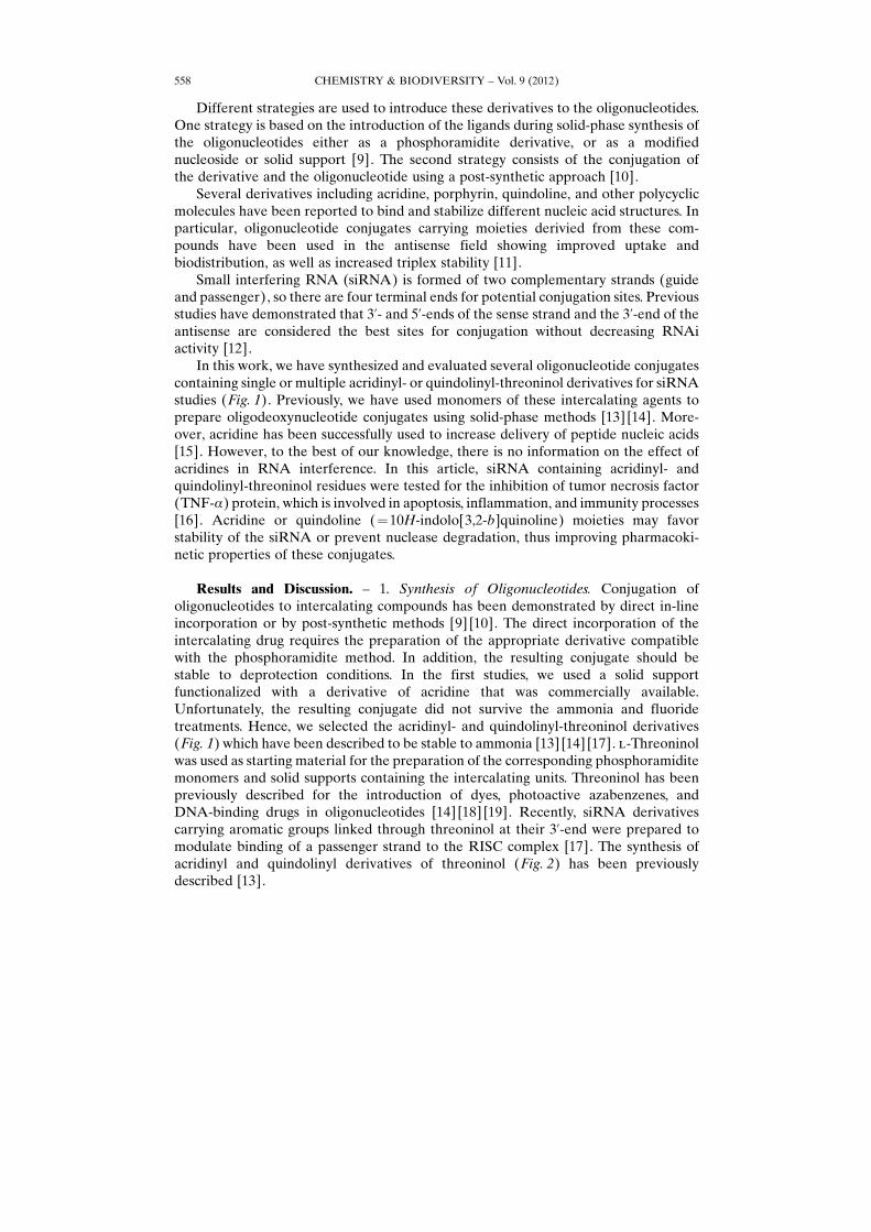
In this work, we have synthesized and evaluated several oligonucleotide conjugatescontaining single or multiple acridinyl- or quindolinyl-threoninol derivatives for siRNAstudies (Fig. 1). Previously, we have used monomers of these intercalating agents toprepare oligodeoxynucleotide conjugates using solid-phase methods [13] [14]. More-over, acridine has been successfully used to increase delivery of peptide nucleic acids[15]. However, to the best of our knowledge, there is no information on the effect ofacridines in RNA interference. In this article, siRNA containing acridinyl- andquindolinyl-threoninol residues were tested for the inhibition of tumor necrosis factor(TNF-a) protein, which is involved in apoptosis, inflammation, and immunity processes[16]. Acridine or quindoline (¼10H-indolo[3,2-b]quinoline) moieties may favorstability of the siRNA or prevent nuclease degradation, thus improving pharmacoki-netic properties of these conjugates.
Results and Discussion. – 1. Synthesis of Oligonucleotides. Conjugation ofoligonucleotides to intercalating compounds has been demonstrated by direct in-lineincorporation or by post-synthetic methods [9] [10]. The direct incorporation of theintercalating drug requires the preparation of the appropriate derivative compatiblewith the phosphoramidite method. In addition, the resulting conjugate should bestable to deprotection conditions. In the first studies, we used a solid supportfunctionalized with a derivative of acridine that was commercially available.Unfortunately, the resulting conjugate did not survive the ammonia and fluoridetreatments. Hence, we selected the acridinyl- and quindolinyl-threoninol derivatives(Fig. 1) which have been described to be stable to ammonia [13] [14] [17]. l-Threoninolwas used as starting material for the preparation of the corresponding phosphoramiditemonomers and solid supports containing the intercalating units. Threoninol has beenpreviously described for the introduction of dyes, photoactive azabenzenes, andDNA-binding drugs in oligonucleotides [14] [18] [19]. Recently, siRNA derivativescarrying aromatic groups linked through threoninol at their 3’-end were prepared tomodulate binding of a passenger strand to the RISC complex [17]. The synthesis ofacridinyl and quindolinyl derivatives of threoninol (Fig. 2) has been previouslydescribed [13].
CHEMISTRY & BIODIVERSITY – Vol. 9 (2012)558
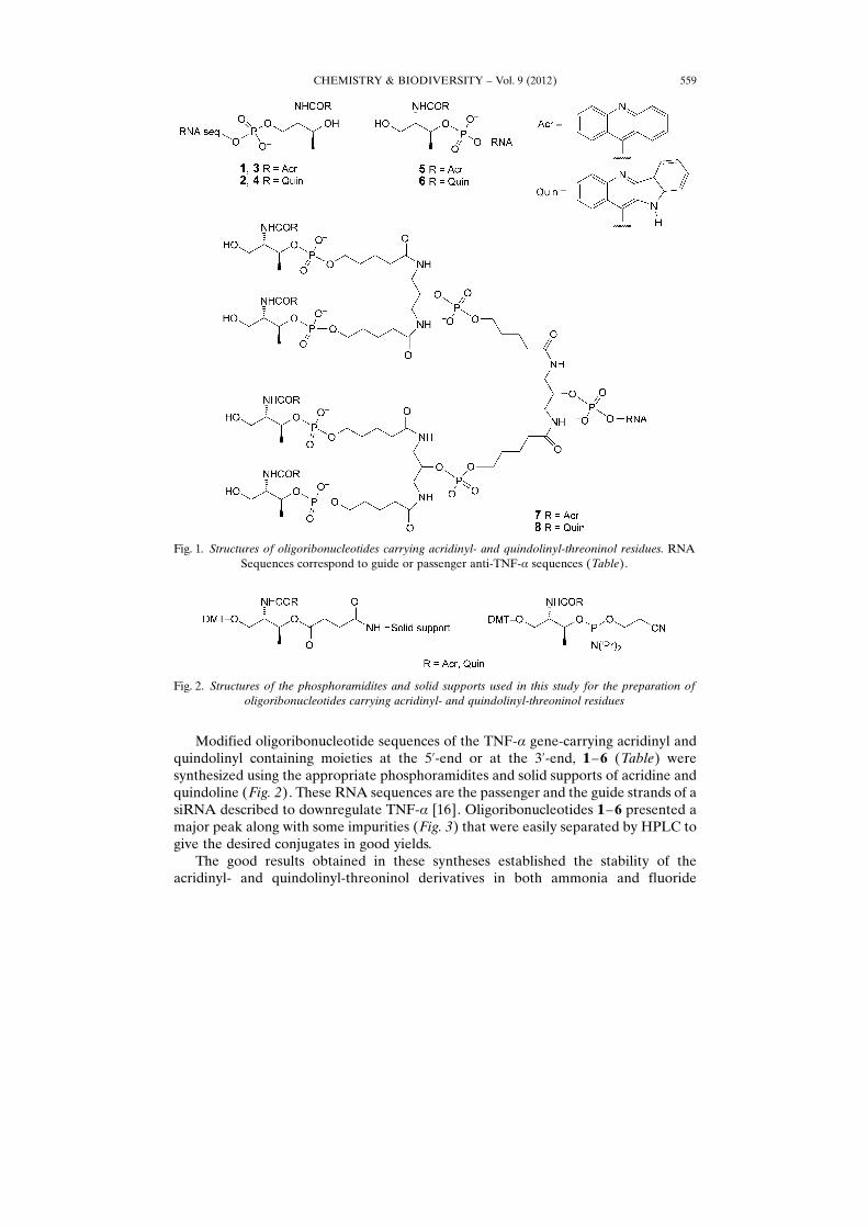
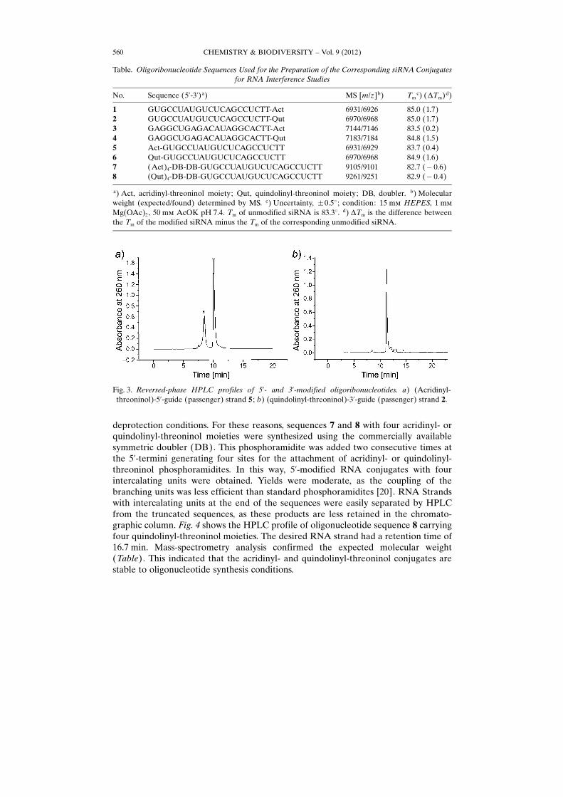
Modified oligoribonucleotide sequences of the TNF-a gene-carrying acridinyl andquindolinyl containing moieties at the 5’-end or at the 3’-end, 1 –6 (Table) weresynthesized using the appropriate phosphoramidites and solid supports of acridine andquindoline (Fig. 2). These RNA sequences are the passenger and the guide strands of asiRNA described to downregulate TNF-a [16]. Oligoribonucleotides 1– 6 presented amajor peak along with some impurities (Fig. 3) that were easily separated by HPLC togive the desired conjugates in good yields.
The good results obtained in these syntheses established the stability of theacridinyl- and quindolinyl-threoninol derivatives in both ammonia and fluoride
Fig. 2. Structures of the phosphoramidites and solid supports used in this study for the preparation ofoligoribonucleotides carrying acridinyl- and quindolinyl-threoninol residues
CHEMISTRY & BIODIVERSITY – Vol. 9 (2012) 559
Fig. 1. Structures of oligoribonucleotides carrying acridinyl- and quindolinyl-threoninol residues. RNASequences correspond to guide or passenger anti-TNF-a sequences (Table).
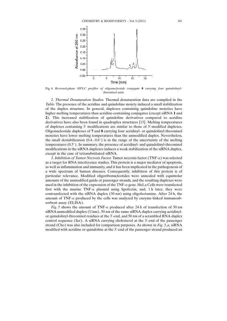
deprotection conditions. For these reasons, sequences 7 and 8 with four acridinyl- orquindolinyl-threoninol moieties were synthesized using the commercially availablesymmetric doubler (DB). This phosphoramidite was added two consecutive times atthe 5’-termini generating four sites for the attachment of acridinyl- or quindolinyl-threoninol phosphoramidites. In this way, 5’-modified RNA conjugates with fourintercalating units were obtained. Yields were moderate, as the coupling of thebranching units was less efficient than standard phosphoramidites [20]. RNA Strandswith intercalating units at the end of the sequences were easily separated by HPLCfrom the truncated sequences, as these products are less retained in the chromato-graphic column. Fig. 4 shows the HPLC profile of oligonucleotide sequence 8 carryingfour quindolinyl-threoninol moieties. The desired RNA strand had a retention time of16.7 min. Mass-spectrometry analysis confirmed the expected molecular weight(Table). This indicated that the acridinyl- and quindolinyl-threoninol conjugates arestable to oligonucleotide synthesis conditions.
CHEMISTRY & BIODIVERSITY – Vol. 9 (2012)560
Table. Oligoribonucleotide Sequences Used for the Preparation of the Corresponding siRNA Conjugatesfor RNA Interference Studies
No. Sequence (5’-3’)a) MS [m/z]b) Tmc) (DTm)d)
1 GUGCCUAUGUCUCAGCCUCTT-Act 6931/6926 85.0 (1.7)2 GUGCCUAUGUCUCAGCCUCTT-Qut 6970/6968 85.0 (1.7)3 GAGGCUGAGACAUAGGCACTT-Act 7144/7146 83.5 (0.2)4 GAGGCUGAGACAUAGGCACTT-Qut 7183/7184 84.8 (1.5)5 Act-GUGCCUAUGUCUCAGCCUCTT 6931/6929 83.7 (0.4)6 Qut-GUGCCUAUGUCUCAGCCUCTT 6970/6968 84.9 (1.6)7 (Act)4-DB-DB-GUGCCUAUGUCUCAGCCUCTT 9105/9101 82.7 (� 0.6)8 (Qut)4-DB-DB-GUGCCUAUGUCUCAGCCUCTT 9261/9251 82.9 (� 0.4)
a) Act, acridinyl-threoninol moiety; Qut, quindolinyl-threoninol moiety; DB, doubler. b) Molecularweight (expected/found) determined by MS. c) Uncertainty, �0.58 ; condition: 15 mm HEPES, 1 mm
Mg(OAc)2, 50 mm AcOK pH 7.4. Tm of unmodified siRNA is 83.38. d) DTm is the difference betweenthe Tm of the modified siRNA minus the Tm of the corresponding unmodified siRNA.
Fig. 3. Reversed-phase HPLC profiles of 5’- and 3’-modified oligoribonucleotides. a) (Acridinyl-threoninol)-5’-guide (passenger) strand 5 ; b) (quindolinyl-threoninol)-3’-guide (passenger) strand 2.
2. Thermal Denaturation Studies. Thermal denaturation data are compiled in theTable. The presence of the acridine and quindoline moiety induced a small stabilizationof the duplex structure. In general, duplexes containing quindoline moieties havehigher melting temperatures than acridine-containing conjugates (except siRNA 1 and2). This increased stabilization of quindoline derivatives compared to acridinederivatives have also been found in quadruplex structures [13]. Melting temperaturesof duplexes containing 3’ modifications are similar to those of 5’-modified duplexes.Oligonucleotide duplexes of 7 and 8 carrying four acridinyl- or quindolinyl-threoninolmoieties have lower melting temperatures than the unmodified duplex. Nevertheless,the small destabilization (0.4 – 0.68) is in the range of the uncertainty of the meltingtemperatures (0.58). In summary, the presence of acridinyl- and quindolinyl-threoninolmodifications in the siRNA duplexes induces a weak stabilization of the siRNA duplex,except in the case of tetrasubstituted siRNA.
3. Inhibition of Tumor Necrosis Factor. Tumor necrosis factor (TNF-a) was selectedas a target for RNA interference studies. This protein is a major mediator of apoptosis,as well as inflammation and immunity, and it has been implicated in the pathogenesis ofa wide spectrum of human diseases. Consequently, inhibition of this protein is ofparticular relevance. Modified oligoribonucleotides were annealed with equimolaramounts of the unmodified guide or passenger strands, and the resulting duplexes wereused in the inhibition of the expression of the TNF-a gene. HeLa Cells were transfectedfirst with the murine TNF-a plasmid using lipofectin, and, 1 h later, they werecontransfected with the siRNA duplex (50 nm) using oligofectamine. After 24 h, theamount of TNF-a produced by the cells was analyzed by enzyme-linked immunoab-sorbent assay (ELISA).
Fig. 5 shows the amount of TNF-a produced after 24 h of transfection of 50 nmsiRNA unmodified duplex (Unm), 50 nm of the same siRNA duplex carrying acridinyl-or quindolinyl-threoninol residues at the 3’-end, and 50 nm of a scrambled RNA duplexcontrol sequence (Scr). A siRNA carrying cholesterol at the 3’-end of the passengerstrand (Cho) was also included for comparison purposes. As shown in Fig. 5,a, siRNAmodified with acridine or quindoline at the 3’-end of the passenger strand produced an
Fig. 4. Reversed-phase HPLC profiles of oligonucleotide conjugate 8 carrying four quindolinyl-threoninol units
CHEMISTRY & BIODIVERSITY – Vol. 9 (2012) 561

inhibition of the production of TNF-a by ca. 90% compared to the Scr RNA duplexcontrol and slightly better than unmodified siRNA. The inhibition potency was similarto that of the siRNA carrying cholesterol at the 3’-end of the passenger strand. Incontrast, the situation changed with siRNAs containing acridine- or quindoline-containing moieties in the 3’-end of the guide strand (Fig. 5,b). In this case, a smalldecrease in the inhibitory activity was found compared to unmodified siRNAs,although the inhibitory activity was still good compared to Src (80– 85%). This result isin agreement with previous observations on other aromatic derivatives at the 3’-end ofthe guide strand in the inhibition of a luciferase gene [17]. In general, it can beconcluded that the introduction of acridine- or quindoline-containing moieties at the 3’-end of the passenger or guide strand of an RNA duplex is well tolerated by the RNAimachinery, and these derivatives do not interfere in the recognition of siRNA by RISCcomplex.
In addition, we have analyzed the inhibition of the TNF-a production by the siRNAduplexes carrying acridine- or quindoline-containing moieties in the sense or antisensestrand without using oligofectamine in HeLa cells (data not shown). In this case, therewas no inhibition of the TNF-a, indicating that intercalating modified siRNA duplexesare unable to enter HeLa cells.
Fig. 6 shows the amount of TNF-a produced when siRNA unmodified duplex(Unm), siRNA duplex carrying acridine- or quindoline-containing moieties at the 5’-end of sense strand, and a scrambled RNA duplex control sequence (Scr) weretransfected with oligofectamine. Moreover, siRNA modified in the sense strand withfour acridine- or quindoline-containing moieties in the 5’-end is also included in thiswork. Inhibition of TNF-a was ca. 75% in the latter case. These results indicate thatconjugates of intercalating agents at the 5’-end of the sense strand are less effective thanunmodified siRNA, but they do not interfere in the recognition of the RISC complex.Unfortunately, conjugation of these compounds does not enhance cell delivery, becausethe inhibition of TNF-a without using oligofectamine in HeLa cells turned out to besimilar to that of the unmodified siRNA (data not shown). In addition, a small loss ofinhibition was detected in siRNA containing intercalating agents at the 5’-end of the
Fig. 5. Plot of gene-specific silencing activities for 3’-acridinyl- and 3’-quindolinyl-threoninol-moietycarrying oligonucleotide conjugates with transfecting agent at 50 nm. a) Modifications at the 3’-end of thepassenger strand. b) Modifications at the 3’-end of the guide strand. Unm, unmodified siRNA duplex;Scr, scrambled siRNA duplex; Cho, siRNA carrying a cholesterol molecule at the 3’-end of the passenger
strand.
CHEMISTRY & BIODIVERSITY – Vol. 9 (2012)562

sense strand, compared to the 3’-end of the sense strand. In summary, acridine andquindoline derivatives are well tolerated in both ends of the sense strand, and at the 3’-end of the antisense strand. The addition of four derivatives carrying acridinyl- orquindoline-threoninol moieties at the 5’-end of the sense strand do not enhanceinhibitory activity compared with a single modification.
4. Cellular Uptake. When HeLa cells were incubated with acridine- and quindoline-containing siRNA duplexes, the cells became highly fluorescent, especially cells treatedwith acridine-containing siRNA duplexes. To distinguish between the fluorescence dueto membrane absorption or the fluorescence due to cellular uptake, confocal-microscopy studies were performed. Fluorescence was only observed when acridine-containing siRNA was transfected with lipofectamine. As seen in Fig. 7, a, transfectionof acridine-modified siRNA was highly efficient, and the fluorescence was localized inthe cytoplasm where the siRNA mechanism was active, indicating that the fluorescentproperties of acridine can be used for the direct observation of cellular uptake ofsiRNA.
Conclusions. – We have reported an efficient strategy for the introduction of one orseveral acridinyl- and quindolinyl-threoninol moieties at the 3’- or 5’-end of RNAmolecules that are stable to oligonucleotide synthesis conditions. We have analyzed theeffect of 3’- or 5’-insertions on the inhibitory properties of TNF-a in HeLa cells.Modified siRNAs containing intercalating compounds are as efficient as unmodifiedsiRNA using oligofectamine. Multiple intercalating agents have been introduced at the5’-end of the sense strand. This large structure does not interfere in the RNAi pathwayto silence gene expression but has similar efficiency compared to single modifications.No inhibition of siRNA conjugates was observed, when cells were transfected withoutoligofectamine. The fluorescent properties of acridine were used to study cell delivery.Transfection of siRNAs modified with acridine-containing moieties showed a clearcytoplasmatic localization of siRNAs in HeLa cells.
This study was supported by the Spanish Ministry of Education (CTQ2010-20541) and Generalitat deCatalunya (2009/SGR/208). CIBER-BBN is an initiative funded by the VI National R&D&i Plan 2008–
Fig. 6. Plot of gene-specific silencing activities of 5’-acridinyl- and 5’-quindolinyl-threoninol moieties-carrying oligonucleotide conjugates with transfecting agent at 50 nm. In all cases, modifications at the 5’-
end were in the passenger strand.
CHEMISTRY & BIODIVERSITY – Vol. 9 (2012) 563

2011, Iniciativa Ingenio 2010, Consolider Program, CIBER Actions, and financed by the Instituto de SaludCarlos III with assistance from the European Regional Development Fund.
Experimental Part
General. Oligonucleotide sequences were prepared using solid-phase methodology. The syntheseswere carried out on an Applied Biosystems Model 3400 DNA synthesizer using 1 mmol scale.Oligoribonucleotides were purified by �DMT-on�-based protocols and by reversed-phase HPLC.MALDI-TOF-MS: Fisons VG Tofspec spectrometer; ESI-MS: Fisons VG Plattform II spectrometer.
Oligonucleotides. The following RNA sequences were obtained from commercial sources (Sigma-Proligo, Dharmacon): sense or passenger scrambled 5’-CAG UCG CGU UUG CGA CUG GTT-3’,antisense or guide scrambled 5’-CCA GUC GCA AAC GCG ACU GTT-3’, antisense or guide anti-TNF-a : 5’-GAG GCU GAG ACA UAG GCA CTT-3’ and sense or passenger anti-TNF-a : 5’-GUG CCUAUG UCU CAG CCU CTT-3’. RNA Monomers in capital letters, T represents thymidine. The 3’-cholesterol passenger anti-TNF-a : strand 5’- GUG CCU AUG UCU CAG CCU CTT-3’-cholesterol wasprepared using the cholesterol-tetraethyleneglycol 3’-cholesteryl-TEG-CPG support from commercialsources (Glen Research).
Synthesis of Oligoribonucleotide Conjugates Carrying Acridine or Quindoline-Containing Moieties.Oligoribonucleotide carrying acridinyl-threoninol (¼N-[(1S,2S)-2-hydroxy-1-(hydroxymethyl)propyl]-acridine-9-carboxamide; Act) or quindolinyl-threoninol (¼N-[(1S,2S)-2-hydroxy-1-(hydroxymethyl)-propyl]-10H-quindoline-11-carboxamide; Qut) moieties either at the 3’- or the 5’-end were synthesizedby a DNA/RNA synthesizer (Applied Biosystems 3400) using 2-cyanoethyl phosphoramidites. Sequenceswith modifications at the 3’-end were synthesized with controlled pore glass (CPG) solid supports(0.2 mmol) functionalized with the threoninol derivatives of acridine or quindoline (Fig. 2) prepared asdescribed in [13] [14]. Sequences carrying one acridine- or one quindoline-containing moieties weresynthesized on polystyrene solid supports (LV200) using Act or Qut phosphoramidites (Fig. 2) prepared
CHEMISTRY & BIODIVERSITY – Vol. 9 (2012)564
Fig. 7. Cellular uptake of siRNA conjugate carrying acridinyl-threoninol moiety. a) HeLa Cells weretransfected with siRNA 1 carrying acridinyl-threoninol moiety using lipofectamine. b) HeLa Cells weretransfected with siRNA 1 without using lipofectamine. After 24 h, HeLa cells were fixed in 10%paraformaldehyde at room temperature for 10 min and then subjected to TO-PRO-3 staining.Localization of the duplex siRNA was monitored by Leica confocal microscopy. Overlay image of
siRNA (blue) and nucleus (red; TO-PRO-3) is shown here.
as described in [13]. Sequences carrying four acridine- or quindoline-containing moieties were preparedassembling two units of the symmetric doubler (DB) phosphoramidite (Glen Research) between theRNA sequence and the acridine- or quindoline-containing groups. The sequences synthesized are shownin the Table.
Guanosine was protected with the (dimethylamino)methylidene group, cytidine was protected withthe Ac group, and adenosine with the benzoyl group. The 2’-OH protecting group for the RNAmonomers was the (tert-butyl)(dimethyl)silyl (TBDMS) group. The phosphoramidites were dissolved indry MeCN (0.1m), and a modified cycle was used with an increased coupling time of 10 min. The solidsupports were treated with conc. aq. NH3/EtOH 3 :1 for 1 h at 558. The supports were filtered and washedwith EtOH, and the combined solns. were evaporated to dryness. Sequences were treated with 0.15 ml ofEt3N· 3 HF/Et3N/N-methylpyrrolidin-2-one (4 :3 : 6) for 2.5 h at 658 to remove the TBDMS groups. Thereactions were quenched by addition of 0.3 ml of Me3SiOiPr and 0.75 ml of Et2O. The resulting mixtureswere mixed with vortex and cooled to 48. A precipitate was formed that was centrifuged at 7000 rpm for5 min at 48. The precipitates were washed with Et2O and centrifuged again. The residues were dissolvedin H2O, and the conjugates were purified by HPLC. HPLC Conditions: column: Nucleosil 120–10 C18
(250�4 mm); 20 min linear gradient from 0 to 50% B (�DMT-off� conditions); flow rate, 3 ml/min; soln.A, 5% MeCN in 0.1m aq. Et3NHOAc (TEAA) buffer, soln. B, 70% MeCN in 0.1m aq. TEAA. Thepurified products were analyzed by MALDI-TOF-MS (Table). Yields (0.2-mmol-scale synthesis) werebetween 10–15 OD units at 260 nm except for sequences 7 and 8 that were ca. 5 OD units.
Thermal Denaturation Studies. The thermal denaturation curves were obtained by monitoring theabsorption change at 260 nm for duplexes formed by oligonucleotides 1–8 and their unmodifiedcomplementary strands. Samples were heated from 20 to 908, with a linear temp. ramp of 0.58/min in aJASCO V-650 spectrophotometer equipped with a Peltier temp. control. Concentration of the sampleswere ca. 2 mm. All the measurements were repeated three times, conducted in 15 mm HEPES, 1 mm
Mg(OAc)2, 50 mm AcOK, pH 7.4Cell Culture, Transfection and Cellular Assays. HeLa Cells were cultured under standard conditions
(378, 5% CO2, Dulbecco�s Modified Eagle Medium, 10% fetal bovine serum, 2 mm l-glutamine,supplemented with penicillin (100 U/ml), and streptomycin (100mg/ml)). All in vitro experiments wereconducted at 40 –60% confluence. HeLa Cells were transfected with 250 ng of murine-expressing TNF-aplasmid using lipofectin (Invitrogen) according to the manufacturer�s instructions. One h followingtransfection, murine-TNF-a expressing HeLa cells were transfected with 50 nm siRNA (5’-GUG CCUAUG UCU CAG CCU CTT-3’ / 5’-GAG GCU GAG ACA UAG GCA CTT-3’) against TNF-a, usingoligofectamine (Invitrogen). TNF-a Concentration was determined from cell culture supernatant byenzyme-linked immunoabsorbent assay kit (Bender MedSystems) according to the manufacturer�sinstructions.
Cellular-Uptake Studies. For analysis of cellular uptake and distribution, HeLa cells were cultured in24-well plates with glass cover slip bottoms. siRNA carrying acridine-containing moiety was transfectedinto HeLa cells using oligofectamine (Invitrogen) according to the manufacture�s instructions. Thenucleus was stained by TO-PRO-3 (red) 24 h after transfection. A series of images were obtained byusing a confocal laser scanning microscope (BioRad). Acridine fluorescence was detected by exciting at300 nm and emission spectrum was recorded from 350 to 560 nm. The spectrum peak at 358 nmrepresented the fluorescence intensity of acridine and was an indicator of siRNA uptake.
REFERENCES
[1] K. Tiemann, J. J. Rossi, EMBO Mol. Med. 2009, 1, 142.[2] D. A. Braasch, S. Jensen, Y. Liu, K. Kaur, K. Arar, M. A. White, D. R. Corey, Biochemistry 2003, 42,
7967.[3] F. Eberle, K. Giessler, C. Deck, K. Heeg, M. Peter, C. Richert, A. H. Dalphe, J. Immunol. 2008, 180, 3229.[4] G. F. Deleavey, J. K. Watts, M. J. Damha, in �Current Protocols in Nucleic Acid Chemistry�, Eds.
S. L. Beaucage, D. E. Bergstrom, G. D. Glick, R. A. Jones, John Wiley & Sons, New York, 2009,Chapt. 16, pp. 16.3.1.
CHEMISTRY & BIODIVERSITY – Vol. 9 (2012) 565
[5] Y. Singh, P. Murat, E. Defrancq, Chem. Soc. Rev. 2010, 39, 2054.[6] H. Lçnnberg, Bioconjugate Chem. 2009, 20, 1065.[7] X. Ming, Expert Opin. Drug Delivery 2011, 8, 435.[8] M. Manoharan, Antisense Nucleic Acid Drug Dev. 2002, 12, 103.[9] U. Asseline, N. T. Thuong, C. Helene, New J. Chem. 1997, 21, 5.
[10] R. Eritja, Chem. Biodiversity 2004 1, 289.[11] F. Birg, D. Praseuth, A. Zerial, N. T. Thuong, T. Le Doan, C. Helene, Nucleic Acids Res. 1990, 18,
2901.[12] T. M. Rana, Nat. Rev. Mol. Cell. Biol. 2007, 8, 23.[13] A. Avino, I. Navarro, J. Farrera-Sinfreu, M. Royo, J. Aymam�, A. Delgado, A. Llebaria, F. Albericio,
R. Eritja, Bioorg. Med. Chem. Lett. 2008, 18, 2306.[14] A. Avino, S. Mazzini, R. Ferreira, R. Eritja, Bioorg. Med. Chem. 2008, 18, 7348.[15] T. Shiraishi, P. E. Nielsen, Nucleic Acids Res. 2004, 32, 4893.[16] D. R. Sørensen, M. Leirdal, M. J. Sioud, J. Mol. Biol. 2003, 327, 761.[17] A. Somoza, M. Terrazas, R. Eritja, Chem. Commun. 2010, 46, 4270.[18] H. Asanuma, K. Shirasuka, I. Takarada, H. Kashida, M. Komiyama, J. Am. Chem. Soc. 2003, 125,
2217.[19] H. Kashida, X. Liang, H. Asanuma, Curr. Org. Chem. 2009, 13, 1065.[20] M. Grimau, D. Iacopino, A. Avino, B. G. de la Torre, A. Ongaro, D. Fitzmaurice, J. Wessels, R.
Eritja, Helv. Chim. Acta 2003, 86, 2814.
Received October 4, 2011
CHEMISTRY & BIODIVERSITY – Vol. 9 (2012)566
Related Documents