Synthesis and Evaluation of Novel α‑Fluorinated (E)‑3-((6- Methylpyridin-2-yl)ethynyl)cyclohex-2-enone‑O‑methyl Oxime (ABP688) Derivatives as Metabotropic Glutamate Receptor Subtype 5 PET Radiotracers Selena Milicevic Sephton, † Linjing Mu, † W. Bernd Schweizer, ‡ Roger Schibli, † Stefanie D. Kra ̈ mer, † and Simon M. Ametamey* ,† † Center for Radiopharmaceutical Sciences of ETH, PSI and USZ, Department of Chemistry and Applied Biosciences, Swiss Federal Institute of Technology (ETH) Zurich, Wolfgang-Pauli Strasse 10, 8093, Zurich, Switzerland ‡ Laboratory of Organic Chemistry, Department of Chemistry and Applied Biosciences, Swiss Federal Institute of Technology (ETH) Zurich, Wolfgang-Pauli Strasse 10, 8093 Zurich, Switzerland * S Supporting Information ABSTRACT: In the search for an optimal fluorine-18-labeled positron emission tomography (PET) radiotracer for imaging metabotropic glutamate receptor subtype 5 (mGluR5), we have prepared a series of five α-fluorinated derivatives based on the ABP688 structural manifold by application of a two-step enolization/ NFSI α-fluorination method. Their binding affinities were evaluated in vitro, and the most promising candidate (Z)-16 exhibited a K i of 5.7 nM and a clogP value of 2.3. The synthesis of the precursor tosylate (E)-22 revealed a preference for the (E)-configurational isomer (K i = 31.2 nM), and successful radiosynthesis afforded (E)- [ 18 F]-16 which was used as a model PET tracer to establish plasma and PBS stability. (E)-[ 18 F]-16 (K d = 70 nM) exhibited excellent specificity for mGluR5 in autoradiographic studies on horizontal rat brain slices in vitro. ■ INTRODUCTION Positron emission tomography (PET) is a noninvasive imaging technique in which 3D concentration images are obtained through computational analysis of pairs of γ rays emitted indirectly from compounds containing positron emitting nuclides such as [ 11 C] or [ 18 F]. 1−3 Metabotropic glutamate receptor subtype 5 (mGluR5) is a G-protein-coupled postsynaptic receptor, and it belongs to group I of metabotropic glutamate receptors, which together with ionotropic glutamate receptors regulate glutamate, a major excitatory neurotransmitter in mammalian brain. 4−8 In 2006, the Ametamey group reported on the synthesis, radiolabeling, and pharmacological evaluation of [ 11 C]-1 ([ 11 C]-ABP688, Figure 1) and subsequently illustrated its application as a PET radiotracer for imaging of mGluR5 in vivo in human subjects. 9−11 The success of this first mGluR5 PET tracer was immediate, and [ 11 C]-1 was employed in many clinical studies 12−19 particularly because mGluR5 emerged as an important drug target due to its demonstrated involvement in long-term potentiation processes as well as several CNS disorders 20 (e.g., schizophrenia, 21 depression, 22 neuropathic pain, 23,24 drug addiction, 25 Fragile X syndrome, 26 and Alzheimer’s 19,27 and Parkinson’s disease 28,29 ). Although clin- ically applied with success, [ 11 C]-1 has one significant limitation which is the short physical half-life (20 min) of the carbon-11 nuclide that limits its application to facilities with an on-site cyclotron. This opened the possibility for further advancement of mGluR5 PET tracers with the aim of designing a fluorine-18- labeled tracer. Received: May 9, 2012 Published: July 23, 2012 Figure 1. Structures of carbon-11 and fluorine-18 mGluR5 PET radiotracers from the Ametamey group and the synthetic plan to a new series of α-fluorinated analogues of 1. A crossed double bond is used to indicate double bond isomers (E and Z). Article pubs.acs.org/jmc © 2012 American Chemical Society 7154 dx.doi.org/10.1021/jm300648b | J. Med. Chem. 2012, 55, 7154−7162

Welcome message from author
This document is posted to help you gain knowledge. Please leave a comment to let me know what you think about it! Share it to your friends and learn new things together.
Transcript

Synthesis and Evaluation of Novel α‑Fluorinated (E)‑3-((6-Methylpyridin-2-yl)ethynyl)cyclohex-2-enone‑O‑methyl Oxime(ABP688) Derivatives as Metabotropic Glutamate Receptor Subtype5 PET RadiotracersSelena Milicevic Sephton,† Linjing Mu,† W. Bernd Schweizer,‡ Roger Schibli,† Stefanie D. Kramer,†
and Simon M. Ametamey*,†
†Center for Radiopharmaceutical Sciences of ETH, PSI and USZ, Department of Chemistry and Applied Biosciences, Swiss FederalInstitute of Technology (ETH) Zurich, Wolfgang-Pauli Strasse 10, 8093, Zurich, Switzerland‡Laboratory of Organic Chemistry, Department of Chemistry and Applied Biosciences, Swiss Federal Institute of Technology (ETH)Zurich, Wolfgang-Pauli Strasse 10, 8093 Zurich, Switzerland
*S Supporting Information
ABSTRACT: In the search for an optimal fluorine-18-labeledpositron emission tomography (PET) radiotracer for imagingmetabotropic glutamate receptor subtype 5 (mGluR5), we haveprepared a series of five α-fluorinated derivatives based on theABP688 structural manifold by application of a two-step enolization/NFSI α-fluorination method. Their binding affinities were evaluatedin vitro, and the most promising candidate (Z)-16 exhibited a Ki of5.7 nM and a clogP value of 2.3. The synthesis of the precursortosylate (E)-22 revealed a preference for the (E)-configurationalisomer (Ki = 31.2 nM), and successful radiosynthesis afforded (E)-[18F]-16 which was used as a model PET tracer to establish plasmaand PBS stability. (E)-[18F]-16 (Kd = 70 nM) exhibited excellentspecificity for mGluR5 in autoradiographic studies on horizontal rat brain slices in vitro.
■ INTRODUCTION
Positron emission tomography (PET) is a noninvasive imagingtechnique in which 3D concentration images are obtainedthrough computational analysis of pairs of γ rays emittedindirectly from compounds containing positron emittingnuclides such as [11C] or [18F].1−3 Metabotropic glutamatereceptor subtype 5 (mGluR5) is a G-protein-coupledpostsynaptic receptor, and it belongs to group I ofmetabotropic glutamate receptors, which together withionotropic glutamate receptors regulate glutamate, a majorexcitatory neurotransmitter in mammalian brain.4−8 In 2006,the Ametamey group reported on the synthesis, radiolabeling,and pharmacological evaluation of [11C]-1 ([11C]-ABP688,Figure 1) and subsequently illustrated its application as a PETradiotracer for imaging of mGluR5 in vivo in humansubjects.9−11 The success of this first mGluR5 PET tracerwas immediate, and [11C]-1 was employed in many clinicalstudies12−19 particularly because mGluR5 emerged as animportant drug target due to its demonstrated involvement inlong-term potentiation processes as well as several CNSdisorders20 (e.g., schizophrenia,21 depression,22 neuropathicpain,23,24 drug addiction,25 Fragile X syndrome,26 andAlzheimer’s19,27 and Parkinson’s disease28,29). Although clin-ically applied with success, [11C]-1 has one significant limitation
which is the short physical half-life (20 min) of the carbon-11nuclide that limits its application to facilities with an on-sitecyclotron. This opened the possibility for further advancementof mGluR5 PET tracers with the aim of designing a fluorine-18-labeled tracer.
Received: May 9, 2012Published: July 23, 2012
Figure 1. Structures of carbon-11 and fluorine-18 mGluR5 PETradiotracers from the Ametamey group and the synthetic plan to a newseries of α-fluorinated analogues of 1. A crossed double bond is usedto indicate double bond isomers (E and Z).
Article
pubs.acs.org/jmc
© 2012 American Chemical Society 7154 dx.doi.org/10.1021/jm300648b | J. Med. Chem. 2012, 55, 7154−7162

Fluorine-18 has a longer physical half-life (110 min) and, todate, two of the most successful mGluR5 18F-labeled PETtracers are 3-fluoro-5-(2-([18F](fluoromethyl)thiazol-4-yl)-ethynyl)benzonitrile ([18F]-SP203), developed by the Pikegroup,30−32 which showed limited defluorination in humansubjects, and 3-[18F]fluoro-5-(2-pyridinylethynyl)benzonitrile([18F]-FPEB), developed by the Hamill group33,34 which hasalso been applied in human subjects but is typically obtained inlow radiochemical yields. Neither Pike’s nor Hamill’s analogueswere based on the structural scaffold of 1, and the success of[11C]-1 in human clinical practice prompted us to explore closeanalogues of [11C]-1. Among other evaluated fluorinatedradiotracers35−39 to date, (E)-3-(pyridin-2-ylethynyl)cyclohex-2-enone O-(2-(2-[18F]fluoroethoxy)ethyl) oxime ([18F]-FDEGPECO, Figure 1)40,41 was identified as the mostpromising candidate, which was successful in visualizingmGluR5 in vivo in a rat brain without defluorination, albeitwith relatively high background activity.Taking advantage of the success of [11C]-1 while
incorporating fluorine-18 with the more desirable physicalhalf-life, we aimed to investigate structures most closely relatedto [11C]-1, in which a hydrogen atom is replaced by fluorine.Apart from increasing the physical half-life of the radiotracer,incorporation of a fluorine atom also alters chemical andphysical properties of the molecule particularly with respect tothe lipophilicity of the molecule, mainly due to the electro-negativity of the fluorine atom.42−44 Fluorine-containingmolecules typically show increased log D when compared totheir nonfluorinated analogues,45 and this is favorable for thedevelopment of CNS tracers, as it typically allows greaterpermeation of the blood−brain barrier. On the basis of the easeof their synthetic accessibility as well as their optimal clogPvalues such that they are in the range of that of [11C]-1 (2.4),we designed a series of α-fluoro-substituted compounds (Figure1) and herein we report on their chemical syntheses, structuralelucidation, and determination of binding affinities, as well asthe establishment of the model PET radiotracer and evaluationof its in vitro properties.
■ RESULTS AND DISCUSSIONThe synthesis of α-fluoro [11C]-1 derivatives was envisioned viathe Sonogashira cross-coupling of α-fluoro oxime ether 3 andcorresponding bromopyridines (2) as depicted in Figure 1.Ethylene enone 6 (Scheme 1) was prepared by reacting
commercially available ethoxy enone 5 with ethynylmagnesiumbromide. In order to obtain α-fluoroenone 7, enone 6 wastreated with SelectFluor following the procedure reported byZupan and co-workers.46,47 While in methanol 6 wascompletely unreactive, in acetonitrile a 19% conversion wasdetermined by NMR (entry 1 vs entry 2, Scheme 1). Withlonger reaction times, conversion to 7 was significantlyimproved (entries 2−4); however, purification by chromatog-raphy on a silica gel column afforded only 8% of α-fluoroenone7.Alternatively, α-fluorination was accomplished via a two-step
procedure. In the first step, enone 6 was enolized with LDAand quenched with chlorotrimethylsilane48 to afford TMS-enolether 8 with the concurrent protection of the free acetylene in6. Silyl enol ether 8 was then fluorinated using N-fluorobenzenesulfonimide (NFSI) in 24% yield, which couldnot be improved regardless of the duration or temperature ofthe reaction. Application of a two-step protocol enolization/NFSI α-fluorination has been accomplished with ketones,
giving rise to related products in low yields requiring longreaction times.49 To our knowledge, γ-fluorination50 was theonly application of NFSI with unsaturated ketones in whichcase enolization was achieved using triphenylborane.Recently, the MacMillan group and others described
advanced applications of NFSI for asymmetric α-fluorinationof aldehydes51−53 and ketones54 in excellent yields and highenantioselectivity. In our hands, the enantioselective α-fluorination of 4 under MacMillan’s reaction conditionsproduced ent-9 in 8% yield and 94% ee. Our initialpharmacological evaluation, however, was performed withracemic α-fluoro samples.In an attempt to optimize α-fluorination we explored several
other possibilities, but a two-step enolization/α-fluorinationwas the most successful. Interestingly, direct lithiation of ethoxyenone 5 with LDA48 followed by α-fluorination with NFSI ledto an inseparable mixture of 2- and 4-fluoro derivatives, and itwas not further explored. On the other hand, enone 10, whichwas accessed via the Sonogashira coupling reaction at 60 °C,was successfully enolized with LDA to give the TMS-enol etherin 60% conversion, but the reaction with NFSI failed to yield 11under conditions analogous to those for the α-fluorination of 8(Scheme 2). Similarly, under MacMillan’s reaction conditions,product was not formed.Next, α-fluoro enone 9 was reacted with methyl or ethyl
hydroxylamine to afford the corresponding oximes 12 and 13 ina ratio of geometrical isomers E:Z of 1:2 (Scheme 2). Thesestable geometrical isomers were separable via columnchromatography. The assignment of E and Z configuration ofthe oxime bond for isomers of 12 and 13 was initially madebased on the 1H NMR comparison to the known structure of 1using the following principle. The hydrogen atom geminal tothe fluorine atom (Hf, Figure 2a) is shifted downfield whencloser through space to the oxime ether oxygen atom, and ahydrogen on the cyclohexenyl double bond (HCy) is shiftedupfield when closer through space to the nitrogen atom lonepair and vice versa (Figure 2a). In a comprehensive study,Denmark and co-workers reported a similar geometricalassignment of oxime double bond based on strong anisotropicdeshielding by the oxime oxygen on the equatorial protons atC2 or C6 positions.
55 Preference for the Z-isomer in this casewas attributed to additional anomeric stabilization of the sp2
Scheme 1. Optimization of α-Fluorination of Enone 6
Journal of Medicinal Chemistry Article
dx.doi.org/10.1021/jm300648b | J. Med. Chem. 2012, 55, 7154−71627155
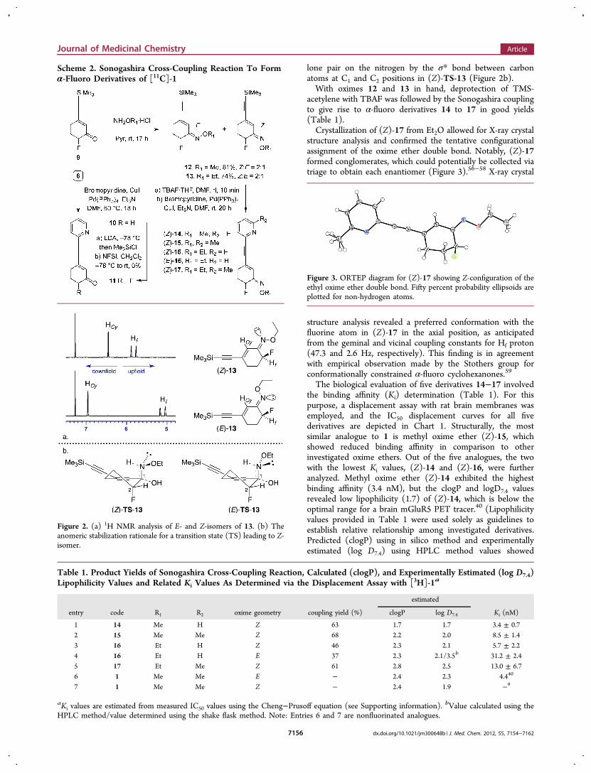
lone pair on the nitrogen by the σ* bond between carbonatoms at C1 and C2 positions in (Z)-TS-13 (Figure 2b).With oximes 12 and 13 in hand, deprotection of TMS-
acetylene with TBAF was followed by the Sonogashira couplingto give rise to α-fluoro derivatives 14 to 17 in good yields(Table 1).Crystallization of (Z)-17 from Et2O allowed for X-ray crystal
structure analysis and confirmed the tentative configurationalassignment of the oxime ether double bond. Notably, (Z)-17formed conglomerates, which could potentially be collected viatriage to obtain each enantiomer (Figure 3).56−58 X-ray crystal
structure analysis revealed a preferred conformation with thefluorine atom in (Z)-17 in the axial position, as anticipatedfrom the geminal and vicinal coupling constants for Hf proton(47.3 and 2.6 Hz, respectively). This finding is in agreementwith empirical observation made by the Stothers group forconformationally constrained α-fluoro cyclohexanones.59
The biological evaluation of five derivatives 14−17 involvedthe binding affinity (Ki) determination (Table 1). For thispurpose, a displacement assay with rat brain membranes wasemployed, and the IC50 displacement curves for all fivederivatives are depicted in Chart 1. Structurally, the mostsimilar analogue to 1 is methyl oxime ether (Z)-15, whichshowed reduced binding affinity in comparison to otherinvestigated oxime ethers. Out of the five analogues, the twowith the lowest Ki values, (Z)-14 and (Z)-16, were furtheranalyzed. Methyl oxime ether (Z)-14 exhibited the highestbinding affinity (3.4 nM), but the clogP and logD7.4 valuesrevealed low lipophilicity (1.7) of (Z)-14, which is below theoptimal range for a brain mGluR5 PET tracer.40 (Lipophilicityvalues provided in Table 1 were used solely as guidelines toestablish relative relationship among investigated derivatives.Predicted (clogP) using in silico method and experimentallyestimated (log D7.4) using HPLC method values showed
Scheme 2. Sonogashira Cross-Coupling Reaction To Formα-Fluoro Derivatives of [11C]-1
Figure 2. (a) 1H NMR analysis of E- and Z-isomers of 13. (b) Theanomeric stabilization rationale for a transition state (TS) leading to Z-isomer.
Table 1. Product Yields of Sonogashira Cross-Coupling Reaction, Calculated (clogP), and Experimentally Estimated (log D7.4)Lipophilicity Values and Related Ki Values As Determined via the Displacement Assay with [3H]-1a
estimated
entry code R1 R2 oxime geometry coupling yield (%) clogP log D7.4 Ki (nM)
1 14 Me H Z 63 1.7 1.7 3.4 ± 0.72 15 Me Me Z 68 2.2 2.0 8.5 ± 1.43 16 Et H Z 46 2.3 2.1 5.7 ± 2.24 16 Et H E 37 2.3 2.1/3.5b 31.2 ± 2.45 17 Et Me Z 61 2.8 2.5 13.0 ± 6.76 1 Me Me E − 2.4 2.3 4.440
7 1 Me Me Z − 2.4 1.9 −9
aKi values are estimated from measured IC50 values using the Cheng−Prusoff equation (see Supporting information). bValue calculated using theHPLC method/value determined using the shake flask method. Note: Entries 6 and 7 are nonfluorinated analogues.
Figure 3. ORTEP diagram for (Z)-17 showing Z-configuration of theethyl oxime ether double bond. Fifty percent probability ellipsoids areplotted for non-hydrogen atoms.
Journal of Medicinal Chemistry Article
dx.doi.org/10.1021/jm300648b | J. Med. Chem. 2012, 55, 7154−71627156

excellent agreement.) We therefore decided to evaluate ethyloxime ether (Z)-16 which not only had a desired Ki value of 5.7nM but also had a clogP of 2.3. Interestingly, this SAR studyrevealed that the most significant difference in Ki was inducedby the configuration of the oxime ether double bond (E- vs Z-16), similar to ABP688 oxime ether double bond isomers,9 andminimal change in Ki binding affinity is observed with variationof either the oxime ether (R1, Figure 1) or the pyridine ring(R2, Figure 1) functionalities.It was then desired to introduce fluorine-18 into the
molecule, but the application of an α-fluorination methodusing [18F]-NFSI60 required an electrophilic source of fluorine-18 (18F2), and this was not feasible for technical reasons. Thiswas due to the nucleophilic nature (18F−) of the fluorine-18source produced in the cyclotron in our facilities. An alternativeapproach was employed based on a nucleophilic substitution atthe α-position to the oxime ether, which required theintroduction of a suitable leaving group.The tosyl leaving group was selected and the synthesis of
radiolabeling precursor 22 (Scheme 3) was envisioned via aRubottom oxidation61−63 of the previously prepared TMS-enolether 8.
Trimethylsilyl enol ether 8 was treated with mCPBA64 toafford epoxy enone 18 (Scheme 3). Enone 18 was purified onlyto remove mCPBA residue on a silica plug, and as such it wasused for the next step in which the epoxide was opened underbasic conditions using a TBAF·THF complex to give rise to α-hydroxy enone 19 in modest yields. α-Hydroxy enone 19 wasfurther converted to ethyl oxime ether 20 by reaction with O-ethylhydroxyl amine in 64% yield. To our surprise, the Z:E ratioof the oxime ether double bond was 1:10. Tentative assignmentof oxime ether double bond geometry was made based on theγ-effect in the 13C NMR spectra65,66 of the two isomers (Figure4a). In the two alkene molecules with all other identicalcomponents, the sterically compressed carbon atom at theallylic position is shifted upfield in comparison to the samecarbon with a higher degree of steric freedom. We applied theγ-effect to ethyl oxime ethers (Z)-13 and (E)-13 to observe the
Chart 1. [3H]-1 Displacement Curves for Derivatives 14−17a
aFor each compound, three experiments were performed in triplicate,and the average values are depicted in the curves (solid lines) fitted forone binding site. Dotted lines represent estimated IC50 values.
Scheme 3. Synthesis of Radiolabelling Precursor Tosylate(E)-22 via the Rubottom Oxidation
Journal of Medicinal Chemistry Article
dx.doi.org/10.1021/jm300648b | J. Med. Chem. 2012, 55, 7154−71627157

chemical shift of 7.6 ppm upfield for sterically compressedcarbon atom Callylic in (E)-13. A similar chemical shift of 7.7ppm units upfield was found for α-hydroxy ethyl oxime ether(E)-20 for the Callylic carbon atom with a lower degree of stericfreedom (Figure 4a). Further confirmation of stereochemicalassignment was sought, and the NOESY experimentsperformed for both geometrical isomers failed to detect NOEbetween Callylic and the ethyl group from the oximefunctionality presumably due to significant distance betweenthe two. However, 1H NMR showed comparable shifts as thoseseen in the fluorine case (Figure 2a). One possible explanationfor the preference of the E-isomer could be postulated byinvoking dipole−dipole interactions in 20 (Figure 4b). The E-isomer configuration facilitates minimization of dipole−dipoleinteractions.At this juncture, we encountered another challenge of
separating the two isomers. While the E-isomer was easilyobtained in meaningful amounts, only trace amounts of Z-isomer were isolated after the purification, and we thereforeselected the E-isomer of 16 for completion of the synthesis.Ethyl oxime ether (E)-20 was coupled with 2-bromopyridineunder the Sonogashira reaction conditions to yield the alcohol(E)-21, which was tosylated to afford (E)-22 in 38% yield(Scheme 3). Both the Sonogashira cross-coupling andtosylation were performed under basic conditions, but noisomerization of the oxime ether double bond was observed.This finding was further compounded when tosylate (E)-22was heated in deuterated chloroform over 3 h at 60 °C to showthe full stability of the E-isomer.We next investigated the radiolabeling of the model tosylate
precursor (E)-22 in DMF using Kryptofix/18F− complex(Scheme 4). The radiolabeling was successful, albeit in low
radiochemical yield after purification via semipreparative HPLCto afford model radiotracer (E)-[18F]-16 with 99% radio-chemical purity. Initially performed at 90 °C for 10 min, adecay-corrected radiochemical yield of 2% was obtained. Aslight improvement to 3% was accomplished by increasing thereaction time to 13 min. The identity of the product wasconfirmed through coinjection with cold reference (E)-16;however, under the employed HPLC conditions for qualitycontrol, E- and Z-isomers were inseparable. Due to similarpolarity of the two isomers, only partial separation wasaccomplished using LiChrosorb column to show an E:Z ratioof ca. 9:1, suggesting partial isomerization of the oxime etherdouble bond during radiolabeling. A similar observation wasmade for [11C]-ABP688 where typical E:Z ratio was ca. 10:1.9
The stability of the model compound (E)-[18F]-16 wasdetermined, and over a period of 2 h, PET radiotracer (E)-[18F]-16 was stable when incubated at 37 °C in both rat plasmaand PBS. (Z)-Derivatives 14 to 17 are likely to exhibit similarstability in plasma and PBS.Lipophilicity of (E)-[18F]-16 was determined using a shake
flask method to show log D7.4 of 3.5 ± 0.1, significantly higherthan that predicted by the in silico method (2.3), orexperimentally estimated using HPLC method (2.1).The equilibrium dissociation constant Kd of (E)-[
18F]-16 wasfurther explored in a Scatchard assay (see SupportingInformation). A Kd value of 70 nM was estimated for (E)-[18F]-16 in a single experiment performed in triplicate whichwas in complete agreement with the previously determinedIC50 (68 nM).Radiolabeled (E)-[18F]-16 also served as a model compound
to establish if the binding was specific to mGluR5 in vitro. Theautoradiographic study was performed on rat brain slices using1 or 10 nM solutions of (E)-[18F]-16 (Figure 5). Importantly,the distribution of radioactivity was heterogeneous, and thehighest uptake was observed in regions where mGluR5 is highlyexpressed in the brain (i.e., hippocampus and cortex).Additionally, the brain slices were incubated with the
solution of (E)-[18F]-16 (1 or 10 nM) and cold 1 (100 nM).A complete blockade of radioactivity was observed, indicatingthat (E)-[18F]-16 specifically binds to mGluR5 in vitro (Figure5).
■ CONCLUSIONSIn the search for a fluorine-18-labeled PET radiotracer forimaging mGluR5, we have successfully synthesized andstructurally assigned five novel derivatives based on thestructural manifold of [11C]-1 by employing an enolization/NFSI α-fluorination method. Our in vitro data enabledidentification of (Z)-16 as a potential mGluR5 PET radiotracer.However, the synthesis of the tosylate precursor 22 revealed astereochemical preference for the E-isomer of α-hydroxyderivative (E)-20 which prompted us to prepare (E)-[18F]-16
Figure 4. (a) Application of γ-effect for assignment of oxime etherdouble bond. (b) Dipole−dipole minimization rationale for thepreference of E-isomer for α-hydroxy derivatives.
Scheme 4. Radiolabeling of Precursor Tosylate (E)-22 ToAfford (E)-[18F]-16 in 3% Decay-Corrected RadiochemicalYield and 99% Purity As Determined by HPLC
Journal of Medicinal Chemistry Article
dx.doi.org/10.1021/jm300648b | J. Med. Chem. 2012, 55, 7154−71627158

as a model compound for the establishment of a radiolabelingstrategy and in vitro testing. We successfully radiosynthesized(E)-[18F]-16 and demonstrated its stability in vitro in plasmaand PBS and specificity to mGluR5. Encouraged by the successof fluorine-18 radiolabeling in an α-position of the oxime etherdouble bond and the heterogeneous mGluR5 specific uptake of(E)-[18F]-16 in rat brain slices in vitro, exploration ofalternative routes to access the Z-isomer selectively arecurrently underway in our lab. The results of these studieswill be reported in due course.
■ MATERIALS AND METHODSGeneral Techniques. All reactions requiring anhydrous conditions
were conducted in flame-dried glass apparatus under an atmosphere ofinert gas. All chemicals and anhydrous solvents were purchased fromAldrich or ABCR and used as received unless otherwise noted.Reported density values are for ambient temperature. [3H]-1 (2.405GBq/μmol, 37 MBq/mL solution in EtOH) was obtained fromAstraZeneca. Purity of compounds was ≥95% as determined byanalytical HPLC method on an Agilent HPLC system. Preparativechromatographic separations were performed on Aldrich Science silicagel 60 (35−75 μm) and reactions followed by TLC analysis usingSigma-Aldrich silica gel 60 plates (2−25 μm) with fluorescentindicator (254 nm) and visualized with UV or potassiumpermanganate. Infrared spectra were recorded on a JASCO FT/IR6200 (OmniLab) spectrometer using a chloroform solution ofcompound. 1H and 13C NMR spectra were recorded in Fouriertransform mode at the field strength specified on Bruker Avance FT-NMR spectrometers. Spectra were obtained from the specifieddeuterated solvents in 5 mm diameter tubes. Chemical shift in ppmis quoted relative to residual solvent signals calibrated as follows:CDCl3 δH (CHCl3) = 7.26 ppm, δC = 77.2 ppm; (CD3)2SO δH(CD3SOCHD2) = 2.50 ppm, δC = 39.5 ppm. Multiplicities in the 1HNMR spectra are described as follows: s = singlet, d = doublet, t =triplet, q = quartet, quint. = quintet, m = multiplet, b = broad; couplingconstants are reported in hertz. Numbers in parentheses followingcarbon atom chemical shifts refer to the number of attached hydrogenatoms as revealed by the DEPT spectral editing technique.Electrospray (ES) mass spectra (LRMS) were obtained with aMicromass Quattro micro API LC electrospray ionization, andelectrospray (ES) mass spectra (HRMS) were obtained with a BrukerFTMS 4.7 T BioAPEXII spectrometer. Electron-impact (EI) andchemical ionization (CI) mass spectra (LRMS and HRMS) were
obtained with a Waters Micromass AutoSpec Ultima MassLynx 4.0spectrometer. Ion mass/charge (m/z) ratios are reported as values inatomic mass units. Semipreparative purification of radiolabeledmaterial was performed on a Merck-Hitachi L6200A system equippedwith Knauer variable wavelength detector and an Eberline radiationdetector using a reverse phase column (C18 Phenomenex Gemini, 5μm, 250 × 10 mm) and eluting with the following gradient: 0−5 min5% aq MeCN, 5−15 min 5−50% aq MeCN, 15−30 min 50% aqMeCN, 30−50 min, 50−90% aq MeCN, 50−65 min, 65% MeCN atflow rate 5 mL/min. Analytical HPLC samples were analyzed by anAgilent HPLC 1100 system equipped with a UV multiwavelengthdetector and a Raytest Gabi star radiation detector using a reversephase column (ACE 111-0546, C18, 3 μm, 50 × 4.6 mm) and elutingwith 45% aq MeCN at flow rate 1 mL/min. Samples for PBS andplasma stability were analyzed by HPLC using an Agilent 1100 systemwith Gina software, equipped with UV multiwavelength and RaytestGabi Star detectors. For HPLC analysis, a reversed phase column(Phenomenex, Gemini 10 μm C18 column, 300 × 3.9 mm,Phenomenex) was used at 1 mL/min flow of 70% aq MeCN.
Trimethyl((5-((trimethylsilyl)ethynyl)cyclohexa-1,5-dien-1-yl)oxy)silane (8). A one-neck round-bottom flame-dried flask wascharged at ambient temperature under N2 atmosphere with anhydroustetrahydrofuran (8 mL), diisopropylamine (0.77 mL, 554 mg, 5.49mmol, d = 0.722) was added, and the colorless solution was cooled to−20 °C (few pieces of dry ice in the acetone bath). The mixture wasthen treated with n-butyllithium (3.5 mL, 4.99 mmol, c = 1.43 Msolution in hexanes) dropwise via syringe over 2 min, and the resultingpale yellow solution was stirred for 36 min. After this time, the paleyellow clear LDA solution was further cooled to −78 °C (dry ice/acetone bath), and it was then treated with a solution of 3-ethynylcyclohex-2-enone (500 mg, 4.16 mmol) in anhydroustetrahydrofuran (4 mL) dropwise via syringe over 9 min during atwhich time the mixture turned brown (on the surface purplecoloration was observed) and a precipitate formed. The resultingheterogeneous mixture was stirred at −78 °C under N2 atmosphere for64 min. After this time, chlorotrimethylsilane (1.0 mL, 904 mg, 8.32mmol, d = 0.856) was added in one portion, and the resulting mixturewas stirred at −78 °C under N2 for 64 min, during which time themixture turned bright orange and clear. After this time, the coolingbath was removed, the orange clear mixture was allowed to warm toambient temperature over 30 min, and the crude mixture was thenpoured over ice-cold 5% wt aq NaHCO3 (40 mL) and was dilutedwith Et2O (50 mL). The two layers were shaken well and separated.The organic phase was washed with H2O (3 × 35 mL) and brine (1 ×35 mL), dried (Na2SO4), and concentrated in vacuo to give crudematerial as a bright red oily residue (1.0 g, 3.79 mmol, 91%). Thecrude mixture was used for the next step without further purification:1H NMR (400 MHz, CDCl3) δ 6.06 (bd, J = 1.8 Hz, 1H), 5.02−4.96(m, 1H), 2.24−2.20 (m, 4H), 0.20 (s, 9H), 0.19 (s, 9H) ppm.
6-Fluoro-3-((trimethylsilyl)ethynyl)cyclohex-2-enone (9). Atambient temperature under N2 atmosphere, a one-neck round-bottomflask was charged with the crude mixture of trimethyl((5-((trimethylsilyl)ethynyl)cyclohexa-1,5-dien-1-yl)oxy)silane (1.08 g,4.10 mmol), anhydrous dichloromethane (6 mL) was added, andthe resulting clear red mixture was cooled to −78 °C (dry ice/acetonebath). The mixture was then treated with N-fluorodibenzenesulfoni-mide (1.30 g, 4.10 mmol, 97% pure) portionwise (with each portion,the flask was quickly opened to air) over 4 min, and the resultingbrown heterogeneous mixture was stirred under N2 and slowlywarmed to ambient temperature over 26.5 h. After this time, the crudereaction mixture was quenched with 0.1 M aq HCl (7 mL), themixture was further diluted with H2O (7 mL) and CH2Cl2 (12 mL),and the two layers were shaken well and separated. The aqueous phasewas extracted with CH2Cl2 (2 × 12 mL). The combined organicextracts were dried (Na2SO4) and concentrated in vacuo to give crudematerial as a red oily residue (2.56 g) which was purified bychromatography on a silica gel column (eluting with 5% EtOAc/pentane) to give the title compound (205 mg, 0.97 mmol, 24%) as apale yellow oil: IR (neat) 2962, 2904, 1696, 1591, 1430, 1354, 1253,1203, 1154, 1096, 846, 765, 703 cm−1; 1H NMR (400 MHz, CDCl3) δ
Figure 5. Autoradiography on horizontal rat brain slices, indicatingheterogeneous distribution of activity with highest uptake in mGluR5-rich regions. The first and second rows represent application of 1 and10 nM [18F]-16, respectively, and the first and second columnrepresent baseline and blocking conditions, respectively.
Journal of Medicinal Chemistry Article
dx.doi.org/10.1021/jm300648b | J. Med. Chem. 2012, 55, 7154−71627159

6.24−6.22 (m, 1H), 4.91 (ddd, J = 48.0, 12.2, 5.1 Hz, 1H), 2.67−2.60(m, 2H), 2.47−2.16 (m, 2H), 0.23 (s, 9H) ppm; 13C NMR (100 MHz,CDCl3) δ 193.3 (d, J = 15.3 Hz, 0), 143.4 (0), 131.4 (1), 108.2 (0),102.6 (0), 89.7 (d, J = 186 Hz, 1), 29.4 (d, J = 12.6 Hz, 2), 29.3 (d, J =3.6 Hz, 2), −0.31 (3) ppm; 19F NMR (376 MHz, CDCl3) δ −192.4(dm, J = 49.3 Hz) ppm; MS (ES+) m/z 211 (M + H)+; HRMS (ESI)m/z 211.0938 (calcd for C11H16FOSi: 211.0949).6-Fluoro-3-((trimethylsilyl)ethynyl)cyclohex-2-enone O-
Ethyl Oxime (13). At ambient temperature under an Ar atmosphere,a pear-shaped flask was charged with 6-fluoro-3-((trimethylsilyl)-ethynyl)cyclohex-2-enone (150 mg, 0.71 mmol), pyridine (2.4 mL)was added, and the resulting pale clear mixture was further treated withO-ethylhydroxylamine hydrochloride (104 mg, 1.07 mmol) in oneportion. The resulting mixture was stirred at ambient temperatureunder Ar for 21 h. After this time, the mixture was diluted with H2O (6mL) and Et2O (10 mL), and the two layers were shaken well andseparated. The aqueous phase was extracted with Et2O (2 × 10 mL).The combined organic extracts were washed with saturated aq CuSO4(3 × 7 mL), H2O (1 × 10 mL), and brine (1 × 10 mL), dried(Na2SO4), and concentrated in vacuo to give crude material as a palebrown oil. The crude mixture was purified by chromatography on asilica gel column (eluting with 2% EtOAc/pentane) to give (Z)-6-fluoro-3-((trimethylsilyl)ethynyl)cyclohex-2-enone O-ethyl oxime(86.7 mg, 0.34 mmol, 48%): IR (neat) 2958, 2146, 1360, 1250,1051, 987, 843, 760 cm1; 1H NMR (400 MHz, CDCl3) δ 6.46 (dm, J= 2.8 Hz, 1H), 5.83 (dt, J = 47.5, 2.8 Hz, 1H), 4.22 (ddm, J = 7.1, 6.0Hz, 2H), 2.61−2.59 (m, 1H), 2.33−2.20 (m, 2H), 1.73 (dm, J = 46.4Hz, 1H), 1.30 (t, J = 7.1 Hz, 3H), 0.21 (s, 9H) ppm; 13C NMR (100MHz, CDCl3) δ 150.7 (d, J = 13.7 Hz, 0), 128.5 (d, J = 1.2 Hz, 1),127.6 (d, J = 2.3 Hz, 0), 105.4 (0), 99.1 (0), 77.8 (d, J = 167 Hz, 1),70.8 (2), 27.7 (d, J = 22.3 Hz, 2), 24.6 (d, J = 3.2 Hz, 2), 14.7 (3), 0.0(3) ppm; 19F NMR (376 MHz, CDCl3) δ − 188.5 (td, J = 47.0, 12.4Hz) ppm; MS (ES+) m/z 254 (M + H)+; HRMS (ESI) m/z 254.1373(calcd for C13H21FNOSi: 254.1371) and (E)-6-fluoro-3-((trimethylsilyl)ethynyl)cyclohex-2-enone O-ethyl oxime (46.8 mg,0.18 mmol, 26%): IR (neat) 2960, 2142, 1595, 1573, 1428, 1250,1049, 975, 842, 760, 638 cm−1; 1H NMR (400 MHz, CDCl3) δ 6.96(dm, J = 2.7 Hz, 1H), 5.11 (dm, J = 49.2 Hz, 1H), 4.21 (q, 7.0 Hz,2H), 2.66−2.53 (m, 1H), 2.39−2.28 (m, 2H), 1.85 (dm, J = 43.2 Hz,1H), 1.30 (t, J = 7.1 Hz, 3H), 0.22 (s, 9H) ppm; 13C NMR (100 MHz,CDCl3) δ 148.5 (d, J = 16.7 Hz, 0), 130.6 (d, J = 2.8 Hz, 0), 120.9 (1),105.2 (0), 100.7 (0), 86.6 (d, J = 169 Hz, 1), 70.6 (2), 28.3 (d, J = 22.8Hz, 2), 25.8 (d, J = 4.8 Hz, 2), 14.7 (3), 0.0 (3) ppm; 19F NMR (376MHz, CDCl3) δ − 178.5 (ddd, J = 50.4, 43.2, 7.9 Hz) ppm; MS (ES+)m/z 254 (M + H)+; HRMS (ESI) m/z 254.1375 (calcd forC13H21FNOSi: 254.1371).(Z)-6-Fluoro-3-(pyridin-2-ylethynyl)cyclohex-2-enone O-
Ethyl Oxime ((Z)-16). At ambient temperature under a N2atmosphere, a one-neck pear-shaped flask was charged with (Z)-6-fluoro-3-((trimethylsilyl)ethynyl)cyclohex-2-enone O-ethyl oxime (43mg, 0.17 mmol), anhydrous N,N-dimethylformamide (0.8 mL) wasadded, and the resulting pale yellow solution was further treated withtetrabutylammonium fluoride solution in tetrahydrofuran (0.34 mL,0.34 mmol, c = 1 M) dropwise over 1 min during which time themixture turned brown. Then the mixture was stirred for 13 min. TLCanalysis indicated that all starting material was consumed, and withoutworkup and/or further purification, the mixture was used for the nextstep. A two-neck round-bottom flask was evacuated at ambienttemperature, and it was then backfilled with N2, repeating this threetimes. This flask was then charged with the crude reaction mixture of(Z)-3-ethynyl-6-fluorocyclohex-2-enone O-ethyl oxime (31 mg, 0.17mmol, still containing TBAF·THF and DMF), and additionalanhydrous N,N-dimethylformamide (1.5 mL) was used for quantita-tive transfer. The resulting red mixture was further treated with 2-bromopyridine (16 μL, 27.0 mg, 0.17 mmol, d = 1.657) followed bytriethylamine (0.28 mL, 206 mg, 2.04 mmol, d = 0.726), and thencopper(I) iodide (3.2 mg, 17.0 μmol) was added in one portion (flaskwas quickly opened to the air). Finally tetrakis(triphenylphosphine)-palladium(0) (6.0 mg, 5.1 μmol) was added in one portion (the flaskwas once again opened to air quickly), and the resulting brown
mixture was stirred at ambient temperature for 22 h. After this time,the brown mixture was quenched with saturated aq NH4Cl (8 mL)and then diluted with H2O (3 mL) and EtOAc (11 mL), and the twolayers were shaken well and separated. The aqueous phase wasextracted with EtOAc (2 × 11 mL). The combined organic phase waswashed with H2O (3 × 8 mL) and brine (1 × 8 mL), dried (Na2SO4),and concentrated in vacuo to give crude material as a brown oilyresidue (338 mg) which was then purified by chromatography on asilica gel column (eluting with gradient 20−30% EtOAc/pentane) togive the title compound (20 mg, 0.08 mmol, 46%) as a pale yellow oil:IR (neat) 2978, 2933, 1580, 1562, 1462, 1428, 1048, 987, 959, 891,859, 779 cm−1; 1H NMR (400 MHz, CDCl3) δ 8.60 (ddd, J = 4.9, 1.7,1.0 Hz, 1H), 7.66 (td, J = 7.7, 1.8 Hz, 1H), 7.45 (dt, J = 7.8, 1.1 Hz,1H), 7.23 (ddd, J = 7.6, 4.9, 1.2 Hz, 1H), 6.59 (dm, J = 2.6 Hz, 1H),5.86 (dt, J = 47.4, 2.5 Hz, 1H), 4.24 (ddm, J = 7.1, 5.8 Hz, 2H), 2.71−2.59 (m, 1H), 2.43−2.26 (m, 2H), 1.79 (dm, J = 46.3 Hz, 1H), 1.31 (t,J = 7.0 Hz, 3H) ppm; 13C NMR (100 MHz, CDCl3) δ 150.4 (d, J =13.7 Hz, 0), 150.1 (1), 143.1 (0), 136.1 (1), 129.0 (d, J = 1.1 Hz, 1),127.3 (1), 126.6 (d, J = 2.2 Hz, 0), 123.1 (d, J = 20.0 Hz, 1), 92.4 (0),89.3 (0), 77.6 (d, J = 167 Hz, 1), 70.8 (3), 27.5 (d, J = 22.2 Hz, 1),24.3 (d, J = 3.2 Hz, 1), 14.5 (3) ppm; 19F NMR (376 MHz, CDCl3) δ− 188.4 (td, J = 47.0, 12.0 Hz) ppm; MS (ES+) m/z 259 (M + H)+;HRMS (ESI) m/z 259.1240 (calcd for C15H16FN2O: 259.1241).
(E)-6-Fluoro-3-(pyridin-2-ylethynyl)cyclohex-2-enone O-Ethyl Oxime ((E)-16). A method analogous to that described for(Z)-16 was employed, starting with (E)-6-fluoro-3-((trimethylsilyl)-ethynyl)cyclohex-2-enone O-ethyl oxime (48 mg, 0.19 mmol) to givecrude material as a brown oily residue which was then purified bychromatography on a silica gel column (eluting with 20% EtOAc/pentane) to give the title compound (18 mg, 0.07 mmol, 37%) as apale yellow oil: IR (neat) 2977, 2934, 1581, 1463, 1429, 1369, 1049,977, 912, 890, 781, 742 cm−1; 1H NMR (400 MHz, CDCl3) δ 8.62(ddd, J = 4.8, 1.7, 0.9 Hz, 1H), 7.68 (td, J = 7.7, 1.8 Hz, 1H), 7.47 (dt,J = 7.8, 1.1 Hz, 1H), 7.25 (ddd, J = 7.6, 4.9, 1.2 Hz, 1H), 7.14 (dm, J =2.7 Hz, 1H), 5.15 (dm, J = 50.1 Hz, 1H), 4.23 (qd, J = 7.1, 0.6 Hz,2H), 2.77−2.64 (m, 1H), 2.51−2.33 (m, 2H), 1.91 (dm, J = 43.3 Hz,1H), 1.31 (t, J = 7.1 Hz, 3H) ppm; 13C NMR (100 MHz, CDCl3) δ150.4 (1), 148.3 (d, J = 16.6 Hz, 0), 143.0 (0), 136.4 (1), 129.8 (0),127.6 (1), 123.4 (1), 121.8 (1), 93.6 (0), 89.3 (0), 86.5 (d, J = 170 Hz,1), 70.7 (2), 28.4 (d, J = 22.6 Hz, 2), 25.5 (d, J = 4.7 Hz, 2), 14.6 (3)ppm; 19F NMR (376 MHz, CDCl3) δ − 178.4 (ddd, J = 50.7, 43.2, 8.3Hz) ppm; MS (ES+) m/z 259 (M + H)+; HRMS (ESI) m/z 259.1236(calcd for C15H16FN2O: 259.1241).
X-ray Analysis: Crystal Data for (Z)-17. C16H17FN2O, MW =272.32, orthorhombic space group P212121, a = 6.2820(1) Å, b =11.4820(3) Å, c = 19/6867(5) Å; data measurement performed on aBruker APEX II Duo diffractometer at 100 K, radiation Mo Kα (λ =0.71073 Å), z = 4, R = 0.032 for 3090 I > 2σ(I), 0.033 for all 3248unique reflections, GOF = 1.16. Full crystallographic data have beendeposited with the Cambridge Crystallographic Data Centre underdeposition number CCDC 880418 and can be obtained free of chargevia www.ccdc.cam.ac.uk/.
(E)-6-Fluoro-3-(pyridin-2-ylethynyl)cyclohex-2-enone O-Ethyl Oxime ((E)-[18F]-16). No-carrier-added [18F]-fluoride wasproduced via nuclear 18O(p, n)18F reaction from enriched 18O-waterusing an IBA cyclone 18/9 cyclotron, and it was immediately trappedon a QMA cartridge (preconditioned with 0.5 M aq K2CO3 (1 × 5mL) and then H2O (1 × 5 mL) and dried in air). The trapped [18F]-fluoride was eluted from the cartridge with 0.25 wt % Kryptofix-222solution (1 mL) in basic (0.05 wt % K2CO3) aq MeCN (75% vv) intoa tightly closed reaction vial. The solvents were evaporated in vacuo(130 mbar) with a gentle stream of N2 gas at 110 °C over 5 min. Tothe resulting solid residue was then added anhydrous MeCN (1 mL),and the mixture was azeotropically dried in vacuo (130 mbar) with agentle stream of N2 at 110 °C. To the dried Kryptofix-222/[18F−]complex was added a solution of (E)-22 (2.19 mg, 5.33 μmol) inanhydrous N,N-dimethylformamide (0.3 mL), and the dark brownmixture was heated at 90 °C for 13 min. The crude mixture wasdiluted with 50% v/v aq MeCN (2 mL) and purified viasemipreparative HPLC. The desired product was collected (retention
Journal of Medicinal Chemistry Article
dx.doi.org/10.1021/jm300648b | J. Med. Chem. 2012, 55, 7154−71627160

time: 30.9 min) and immediately diluted with H2O (10 mL). Theaqueous solution was passed through a C18 cartridge (preconditionedwith EtOH (1 × 5 mL) and then H2O (1 × 5 mL) and dried in air),the cartridge was washed with H2O (2 × 1 mL), and the product waseluted from the C18 cartridge with EtOH (1 × 0.3 mL) into a sterilevial containing 50% aq PEG200 (5 mL) to afford the radiolabeled titlecompound in a 3% decay-corrected yield. Typically, starting from ca.35 GBq of activity, 740 MBq of product was obtained. Theradiochemical purity was >99%, and specific activity was 30 GBq/μmol.Competition Binding Assay. Brain membranes were prepared
from Sprague−Dawley rat brains as described previously.40 Frozenmembranes were thawed on ice and pelleted at 45000g at 4 °C for 5min. The membranes were washed twice with HEPES buffer (30 mMHEPES, 110 mM NaCl, 5 mM KCl, 2.5 mM CaCl2, 1.2 mM MgCl2,pH 8 at 4 °C) and resuspended in HEPES buffer at a proteinconcentration of 1.3 mg/mL. The binding assay was performed aspreviously described.40 In brief, brain membranes (0.1 mg of protein)were incubated in triplicate at ambient temperature with 2 nM [3H]-1and (E)-16 at concentrations between 10 pM and 100 μM in a totalvolume of 0.2 mL of HEPES. (E)-16 was diluted from a 1 mMethanolic (50%) solution. The corresponding EtOH concentrationsdid not affect [3H]-1 binding (data not shown). Nonpecific binding of[3H]-1 was estimated with 100 μM MMPEP. After 45 min, thesamples were filtered, and the filters containing the membranes withbound [3H]-1 were measured in a β-counter (Beckman LS6500).Bound [3H]-1 (B, pmol per mg protein) was fitted with Excel solver toeq 1 to estimate IC50.
= + − +B B B B C(( )/(1 ( /IC )))min max min 50 (1)
where C is the total (E)-16 concentration, Bmax is the maximal B, i.e.,the plateau in the B/C plot at low log C and Bmin is the minimal B, i.e.,the plateau at high log C. The inhibition constant Ki of (E)-16 wasestimated from the IC50 and Kd of [
11C]-1 (1.7 ± 0.2 nM)9 with theCheng−Prusoff equation.Stability in PBS and Plasma. (E)-[18F]-16 (21 MBq) was
incubated in phosphate buffer (4 mM KH2PO4/Na2HPO4, 155 mMNaCl, pH 7.4) or rat plasma at 37 °C for up to 2 h. At different timepoints, samples were61−63 diluted and reactions stopped with ice-coldMeCN (140 μL). Plasma samples were centrifuged at 12000g for 10min. The samples were filtered, and supernatants were analyzed byHPLC.In Vitro Autoradiography. Frozen horizontal brain slices (20
μm) from a male Wistar rat (221 g) adsorbed to SuperFrost Plus slideswere thawed at ambient temperature and preincubated on ice for 10min in HEPES buffer (see above) containing 0.1% bovine serumalbumin (BSA). Excess solution was carefully removed, and slides wereincubated with 1 or 10 nM (E)-[18F]-16 alone or together with 100nM 1 in HEPES buffer for 45 min at ambient temperature. Afterincubation, the solutions were decanted and the slides washed on icein HEPES buffer containing 0.1% BSA and twice in HEPES buffer (3min each) and finally dipped in H2O. Dried slides were exposed to aphosphor imager plate for 30 min, and the plate was scanned in aBAS5000 reader (Fuji).
■ ASSOCIATED CONTENT*S Supporting InformationFull experimental procedures and characterization of allcompounds, NMR data, HPLC and GC traces, IC50 bindingcurves, Scatchard plot for (E)-[18F]-16, and original X-raycrystallography data for (Z)-17. This material is available free ofcharge via the Internet at http://pubs.acs.org.
■ AUTHOR INFORMATIONCorresponding Author*Phone: +41 44 6337463. E-mail: [email protected].
Author ContributionsAll authors have given approval to the final version of themanuscript.
NotesThe authors declare no competing financial interest.
■ ACKNOWLEDGMENTS
The authors acknowledge Mrs. Petra Wirth and Dr. AdrienneMuller for technical support. Professor Paul R. Blakemore(Oregon State University) is acknowledged for helpfuldiscussions on NMR assignments. Drs. Bernhard Pfeiffer(Altman group) and Marc-Olivier Ebert (Laboratory forOrganic Chemistry, ETH) are acknowledged for technicalsupport with NMR analysis. Dr. Ebert is acknowledged forperforming additional NOE experiments for compounds (E)-20 and (Z)-20. Ms. Sara Wyss (Werner group) is acknowl-edged for performing chiral GC analysis of ent-9. Dr. Mark A.Sephton (ZHAW) is acknowledged for proofreading themanuscript. Ms. Cindy Fisher, Drs. Cindy A. Wanger-Baumann,and Thomas Betzel are acknowledged for useful discussions.
■ ABBREVIATIONS USED
mGluR5, metabotropic glutamate receptor subtype 5; PET,positron emission tomography; NFSI, N-fluorodibenzenesulfo-nimide; mCPBA, m-chloroperbenzoic acid; DMF, N,N′-dimethylformamide; THF, tetrahydrofuran; [11C]-ABP688,(E)-3-((6-methylpyridin-2-yl)ethynyl)cyclohex-2-enone O-[11C]methyl oxime; [3T]-ABP688, (E)-3-((6-methylpyridin-2-yl)ethynyl)cyclohex-2-enone O-[3T]methyl oxime; [18F]-SP203, 3-fluoro-5-(2-([18F](fluoromethyl)thiazol-4-yl)-ethynyl)benzonitrile; [18F]-FPEB, 3-[18F]fluoro-5-(2-pyridinylethynyl)benzonitrile; [18F]-FDEGPECO, (E)-3-(pyr-idin-2-ylethynyl)cyclohex-2-enone O-(2-(2-[18F]¯uoroethoxy)-ethyl) oxime; MMPEP, 2-[(3-methoxyphenyl)ethynyl]-6-meth-ylpyridine; PBS, phosphate buffer in saline; SAR, structure−activity relationship; TLC, thin layer chromatography; HPLC,high pressure liquid chromatography; GC, gas chromatography;IC50, half maximal inhibitory concentration; Ki, inhibitionconstant; Kd, dissociation constant
■ REFERENCES(1) Ametamey, S. M.; Honer, M.; Schubiger, P. A. Chem. Rev. 2008,108, 1501.(2) Jacobs, A. H.; Li, H.; Winkeler, A.; Hilker, R.; Knoess, C.; Ruger,A.; Galldiks, N.; Schaller, B.; Sobesky, J.; Kracht, L.; Monfared, P.;Klein, M.; Vollmar, S.; Bauer, B.; Wagner, R.; Graf, R.; Wienhard, K.;Herholz, K.; Heiss, W. D. Eur. J. Nucl. Med. Mol. Imaging 2003, 30,1051.(3) Mu, L.; Schubiger, P. A.; Ametamey, S. M. Curr. Top. Med. Chem.2010, 10, 1558.(4) Pin, J. P.; Duvoisin, R. Neuropharmacology 1995, 34, 1.(5) Shigemoto, R.; Kinoshita, A.; Wada, E.; Nomura, S.; Ohishi, H.;Takada, M.; Flor, P. J.; Neki, A.; Abe, T.; Nakanishi, S.; Mizuno, N. J.Neurosci. 1997, 17, 7503.(6) Shigemoto, R.; Mizuno, N. Handb. Chem. Neuroanat. 2000, 18,63.(7) Shigemoto, R.; Nomura, S.; Ohishi, H.; Sugihara, H.; Nakanishi,S.; Mizuno, N. Neurosci. Lett. 1993, 163, 53.(8) Tanabe, Y.; Masu, M.; Ishii, T.; Shigemoto, R.; Nakanishi, S.Neuron 1992, 8, 169.(9) Ametamey, S. M.; Kessler, L. J.; Honer, M.; Wyss, M. T.; Buck,A.; Hintermann, S.; Auberson, Y. P.; Gasparini, F.; Schubiger, P. A. J.Nucl. Med. 2006, 47, 698.
Journal of Medicinal Chemistry Article
dx.doi.org/10.1021/jm300648b | J. Med. Chem. 2012, 55, 7154−71627161

(10) Ametamey, S. M.; Treyer, V.; Streffer, J.; Wyss, M. T.; Schmidt,M.; Blagoev, M.; Hintermann, S.; Auberson, Y.; Gasparini, F.; Fischer,U. C.; Buck, A. J. Nucl. Med. 2007, 48, 247.(11) Hintermann, S.; Vranesic, I.; Allgeier, H.; Bruelisauer, A.; Hoyer,D.; Lemaire, M.; Moenius, T.; Urwyler, S.; Whitebread, S.; Gasparini,F.; Auberson, Y. P. Bioorg. Med. Chem. 2007, 15, 903.(12) Burger, C.; Deschwanden, A.; Ametamey, S.; Johayem, A.;Mancosu, B.; Wyss, M.; Hasler, G.; Buck, A. Nucl. Med. Biol. 2010, 37,845.(13) Calcinaghi, N.; Jolivet, R.; Wyss, M. T.; Ametamey, S. M.;Gasparini, F.; Buck, A.; Weber, B. J. Cereb. Blood Flow Metab. 2011, 31,e1.(14) DeLorenzo, C.; Kumar, J. S. D.; Mann, J. J.; Parsey, R. V. J.Cereb. Blood Flow Metab. 2011, 31, 2169.(15) Deschwanden, A.; Karolewicz, B.; Feyissa, A. M.; Treyer, V.;Ametamey, S. M.; Johayem, A.; Burger, C.; Auberson, Y. P.; Sovago, J.;Stockmeier, C.; Buck, A.; Hasler, G. Am. J. Psychiatry 2011, 168, 727.(16) Ouattara, B.; Gregoire, L.; Morissette, M.; Gasparini, F.;Vranesic, I.; Bilbe, G.; Johns, D. R.; Rajput, A.; Hornykiewicz, O.;Rajput, A. H.; Gomez-Mancilla, B.; Paolo, T. D. Neurobiol. Aging 2011,32, 1286.(17) Treyer, V.; Streffer, J.; Wyss, M. T.; Bettio, A.; Ametamey, S. M.;Fischer, U.; Schmidt, M.; Gasparini, F.; Hock, C.; Buck, A. J. Nucl.Med. 2007, 48, 1207.(18) Wyss, M. T.; Ametamey, S. M.; Valerie, T.; Andrea, B.; Blagoev,M.; Kessler, L. J.; Burger, C.; Weber, B.; Schmidt, M.; Gasparini, F.;Alfred, B. NeuroImage 2007, 35, 1086.(19) Bruno, V.; Ksiazek, I.; Battaglia, G.; Lukic, S.; Leonhardt, T.;Sauer, D.; Gasparini, F.; Kuhn, R.; Nicoletti, F.; Flor, P. J.Neuropharmacology 2000, 39, 2223.(20) Luscher, C.; Huber, K. M. Neuron 2010, 65, 445.(21) Ohnuma, T.; Augood, S. J.; arai, H.; McKenna, P. J.; Emson, P.C. Mol. Brain Res. 1998, 56, 207.(22) Pilc, A.; Klodzinska, A.; Branski, P.; Nowak, G.; Palucha, A.;Szewczyk, B.; Tatarczynska, E.; Chojnacka-Wojcik, E.; Wieronska, J.M. Neuropharmacology 2002, 43, 181.(23) Cosford, N. D. P.; Tehrani, L.; Roppe, J.; Schweiger, E.; Smith,N. D.; Anderson, J.; Bristow, L.; Brodkin, J.; Jiang, X. H.; McDonald,I.; Rao, S.; Washburn, M.; Varney, M. A. J. Med. Chem. 2003, 46, 204.(24) Gasparini, F.; Lingenhohl, K.; Stoehr, N.; Flor, P. J.; Heinrich,M.; Vranesic, I.; Biollaz, M.; Allgeier, H.; Heckendorn, R.; Urwyler, S.;Varney, M. A.; Johnson, E. C.; Hess, S. D.; Rao, S. P.; Sacaan, A. I.;Santori, E. M.; Velicelebi, G.; Kuhn, R. Neuropharmacology 1999, 38,1493.(25) Chiamulera, C.; Epping-Jordan, M. P.; Zocchi, A.; Marcon, C.;Cottiny, C. C.; Tacconi, S.; Corsi, M.; Orzi, F.; Conquet, F. O. Nat.Neurosci. 2001, 4, 873.(26) Todd, P. K.; Mack, K. J.; alter, J. S. M. Proc. Natl. Acad. Sci.U.S.A. 2003, 100, 14374.(27) Wang, Q.; Walsh, D. M.; Rowan, M. J.; Selkoe, D. J.; Anwyl, R. J.Neurosci. 2004, 24, 3370.(28) Ossowska, K.; Konieczny, J.; Wardas, J.; Pietraszek, M.; Kuter,K.; Wolfarth, S.; Pilc, A. Amino Acids 2007, 32, 179.(29) Rouse, S. T.; Marino, M. J.; Bradley, S. R.; Awad, H.; Wittmann,M.; Conn, P. J. Pharmacol. Ther. 2000, 88, 427.(30) Brown, A. K.; Kimura, Y.; Zoghbi, S. S.; Simeon, F. G.; Liow, J.S.; Kreisl, W. C.; Taku, A.; Fujita, M.; Pike, V. W.; Innis, R. B. J. Nucl.Med. 2008, 49, 2042.(31) Shetty, H. U.; Zoghbi, S. S.; Simeon, F. G.; Liow, J. S.; Brown, A.K.; Kannan, P.; Innis, R. B.; Pike, V. W. J. Pharmacol. Exp. Ther. 2008,327, 727.(32) Simeon, F. G.; Brown, A. K.; Zoghbi, S. S.; Patterson, V. M.;Innis, R. B.; Pike, V. W. J. Med. Chem. 2007, 50, 3256.(33) Barret, O.; Tamagnan, G.; Batis, J.; Jennings, D.; Zubal, G.;Russell, D.; Marek, K.; Seibyl, J. J. Nucl. Med. Meet. Abstr. 2010, 51,215.(34) Hamill, T. G.; Krause, S. R. C.; Bonnefous, C.; Govek, S.;Seiders, T. G.; Cosford, N. P. D.; Roppe, J.; Kamenecka, T.; Patel, S.;Gibson, R. E.; Sanabria, S.; Riffel, K.; Eng, W.; King, C.; Yang, X.;
Green, M. D.; O’Malley, S. S.; Hargreaves, R.; Burns, H. D. Synapse2005, 56, 205.(35) Baumann, C. A.; Mu, L.; Wertli, N.; Kramer, S. D.; Honer, M.;Schubiger, P. A.; Ametamey, S. M. Bioorg. Med. Chem. 2010, 18, 6044.(36) Honer, M.; Stoffel, A.; Kessler, L. J.; Schubiger, P. A.;Ametamey, S. M. Nucl. Med. Biol. 2007, 34, 973.(37) Lucatelli, C.; Honer, M.; Salazar, J. F.; Ross, T. L.; Schubiger, P.A.; Ametamey, S. M. Nucl. Med. Biol. 2009, 36, 613.(38) Sephton, S. M.; Dennler, P.; Leutwiler, D.; Mu, L.; Schibli, R.;Kramer, S. D.; Ametamey, S. M. Chimia 2012, 66, 201.(39) Sephton, S. M.; Dennler, P.; Leutwiler, D.; Mu, L.; Wanger-Baumann, C. A.; Schibli, R.; Kramer, S. D.; Ametamey, S. M. Am. J.Nucl. Med. Mol. Imaging 2012, 2, 14.(40) Baumann, C. A.; Mu, L.; Johannsen, S.; Honer, M.; Schubiger,P. A.; Ametamey, S. M. J. Med. Chem. 2010, 53, 4009.(41) Wanger-Baumann, C. A.; Mu, L.; Honer, M.; Belli, S.; Alf, M. F.;Schubiger, P. A.; Kramer, S. D.; Ametamey, S. M. NeuroImage 2011,56, 984.(42) Fowler, J. S.; Volkow, N. D.; Wang, G. J.; Ding, Y. S.; Dewey, S.L. J. Nucl. Med. 1999, 40, 1154.(43) Reichel, A. Chem. Biodivers. 2009, 6, 2030.(44) Wilson, A. A.; Jin, L.; Garcia, A.; DaSilva, J. N.; Houle, S. Appl.Radiat. Isot. 2001, 54, 203.(45) Kirsch, P. Modern fluoroorganic chemistry: synthesis, reactivity,applications; Wiley-VCH: Germany, 2004.(46) Stavber, S.; Jereb, M.; Zupan, M. Synthesis 2002, 17, 2609.(47) Stavber, S.; Zupan, M. Tetrahedron Lett. 1996, 37, 3591.(48) Perkins, J. R.; Carter, R. G. J. Am. Chem. Soc. 2008, 130, 3290.(49) Differding, E.; Ofner, H. Synlett 1991, 187.(50) Poss, A. J.; Shia, G. A. Tetrahedron Lett. 1995, 36, 4721.(51) Beeson, T. D.; MacMillan, D. W. C. J. Am. Chem. Soc. 2005, 127,8826.(52) Marigo, M.; Fielenbach, D.; Braunton, A.; Kjaersgaard, A.;Jorgensen, K. A. Angew. Chem., Int. Ed. 2005, 44, 3703.(53) Steiner, D. D.; Mase, N.; C., F. B., III. Angew. Chem., Int. Ed.2005, 44, 3706.(54) Kwiatkowski, P.; Beeson, T. D.; Conrad, J. C.; MacMillan, D. W.C. J. Am. Chem. Soc. 2011, 133, 1738.(55) Denmark, S. E.; Dappen, M. S. J. Org. Chem. 1984, 49, 798.(56) Collet, A.; Brienne, M. J.; Jacques, J. Chem. Rev. 1980, 80, 215.(57) Perez-Garcia, L.; Amabilino, D. B. Chem. Soc. Rev. 2007, 36, 941.(58) Sephton, M. A.; Emerson, C. R.; Zakharov, L. N.; Blakemore, P.R. Chem. Commun. 2010, 46, 2094.(59) Pan, Y. H.; Stothers, J. B. Can. J. Chem. 1967, 45, 2943.(60) Teare, H.; Robins, E. G.; Arstad, E.; Luthra, S. K.; Gouverneur,V. Chem. Commun. 2007, 2330.(61) Chavan, S. P.; Thakkar, M.; Jogdand, G. F.; Kalkote, U. R. J. Org.Chem. 2006, 71, 8986.(62) Lin, J.; Nikaido, M. M.; Clark, G. J. Org. Chem. 1987, 52, 3745.(63) Murray, L. M.; O’Brien, P.; Taylor, R. J. K. Org. Lett. 2003, 5,1943.(64) Thompson, C. F.; Jamison, T. F.; Jacobsen, E. N. J. Am. Chem.Soc. 2000, 122, 10482.(65) Pretsch, E.; Clerc, J. T.; Seibl, J.; Simon, W. Tabellen zurstrukturaufklaerung organischer verbindungen mit spektroskopischenmethoden; Springer: Berlin, 1976; Vol. 15.(66) Stothers, J. B. Carbon-13 NMR spectroscopy; Academic Press:New York, 1972; Vol. 24.
■ NOTE ADDED AFTER ASAP PUBLICATIONThis manuscript was published ASAP on August 9, 2012. Dueto a production error, additional corrections were made toTable 1 and the revised version was reposted on August 14,2012.
Journal of Medicinal Chemistry Article
dx.doi.org/10.1021/jm300648b | J. Med. Chem. 2012, 55, 7154−71627162
Related Documents


![Ultralong purely organic aqueous phosphorescence ...supramolecular phosphorescence material that is composed of cucurbit[6]uril (CB[6]) and 4-(4-bromophenyl)-1-methylpyridin-1-ium](https://static.cupdf.com/doc/110x72/60de7cf0abbbe251c3180d33/ultralong-purely-organic-aqueous-phosphorescence-supramolecular-phosphorescence.jpg)

![Am. Chem. SOC. 1981,103, Selenium-Stabilized Anions ... · of rearrangement (the silyl enone) and syn elimination product are observed. Competition between syn elimination and [2.3]](https://static.cupdf.com/doc/110x72/5b94c97409d3f272648b673e/am-chem-soc-1981103-selenium-stabilized-anions-of-rearrangement-the.jpg)

![2-[(Cyclohex-3-en-1-ylmethoxy)methyl]-6-phenyl-1,2,4-triazine-3,5(2 H ,4 H )-dione](https://static.cupdf.com/doc/110x72/6324d4c1545c645c7f0962a9/2-cyclohex-3-en-1-ylmethoxymethyl-6-phenyl-124-triazine-352-h-4-h-dione.jpg)

