Full length article Synthesis and characterization of citrate-based fluorescent small molecules and biodegradable polymers Zhiwei Xie a , Jimin P. Kim a , Qing Cai b , Yi Zhang c , Jinshan Guo a , Ranjodh S. Dhami a , Li Li d , Bin Kong e , Yixue Su a , Kevin A. Schug d , Jian Yang a,⇑ a Department of Biomedical Engineering, Materials Research Institutes, The Huck Institutes of Life Sciences, The Pennsylvania State University, University Park, PA 16801, United States b Beijing Laboratory of Biomedical Materials, Beijing University of Chemical Technology, Beijing 100029, China c Institute of Medical Engineering and Science, Massachusetts Institute of Technology, Cambridge, MA 02139, United States d Department of Chemistry and Biochemistry, University of Texas at Arlington, Arlington, TX 76010, United States e Institute of Chemistry, Chinese Academy of Sciences, Beijing 100190, China article info Article history: Received 7 September 2016 Received in revised form 16 December 2016 Accepted 5 January 2017 Available online 6 January 2017 Keywords: Fluorescence Fluorescence spectroscopy Dyes Polymers Degradable abstract Novel citric acid based photoluminescent dyes and biodegradable polymers are synthesized via a facile ‘‘one-pot” reaction. A comprehensive understanding of the fluorescence mechanisms of the resulting citric acid-based fluorophores is reported. Two distinct types of fluorophores are identified: a thiozolopy- ridine family with high quantum yield, long lifetime, and exceptional photostability, and a dioxopyridine family with relatively lower quantum yield, multiple lifetimes, and solvent-dependent band shifting behavior. Applications in molecular labeling and cell imaging were demonstrated. The above discoveries contribute to the field of fluorescence chemistry and have laid a solid foundation for further development of new fluorophores and materials that show promise in a diversity of fluorescence-based applications. Statement of Significance Photoluminescent materials are pivotal for fluorescence based imaging, labeling and sensing applications. Understanding their fluorescence mechanism is challenging and imperative. We develop a new class of citric acid-derived fluorescent materials in forms of polymers and small molecular dyes by a one-step sol- vent free reaction. We discovered two different classes of citric acid-derived fluorophores. A two-ring thiozolopyridine structure demonstrates strong fluorescence and exceptional resistance to photo- bleaching. A one-ring dioxopyridine exhibits relative weak fluorescence but with intriguing excitation and solvent-dependent emission wavelength shifting. Our methodology of synthesizing citric acid- derived fluorophores and the understanding on their luminescence are instrumental to the design and production of a large number of new photoluminescent materials for biological and biomedical applications. Ó 2017 Acta Materialia Inc. Published by Elsevier Ltd. All rights reserved. 1. Introduction Fluorescence imaging is a powerful and versatile tool for appli- cations ranging from molecular biology to disease diagnostics due to its high resolution and sensitivity. Advances in fluorescent imag- ing probes and technologies have empowered researchers to visu- alize and analyze biological systems in an unprecedented fashion [1,2]. Organic dyes are the most widely used and studied imaging agents, partly because their fluorescence mechanisms are well understood. Several non-traditional fluorescent probes, including quantum dots, green fluorescent proteins, graphene oxides, and carbon dots have recently been developed. However, challenges still remain for both organic dyes and non-traditional light emit- ting probes. For example, poor photostability and short lifetimes of fluorescence proteins and organic dyes hinder their applications in continuous cellular imaging and lifetime imaging [3]. The in vivo applications of carbon dots, graphene oxides, and quantum dots are limited due to their intrinsic toxicities. These challenges pre- sent an urgent need of developing biocompatible fluorophores that have long lifetimes, excellent photostability, and suitable physical http://dx.doi.org/10.1016/j.actbio.2017.01.019 1742-7061/Ó 2017 Acta Materialia Inc. Published by Elsevier Ltd. All rights reserved. ⇑ Corresponding author. E-mail address: [email protected] (J. Yang). Acta Biomaterialia 50 (2017) 361–369 Contents lists available at ScienceDirect Acta Biomaterialia journal homepage: www.elsevier.com/locate/actabiomat

Welcome message from author
This document is posted to help you gain knowledge. Please leave a comment to let me know what you think about it! Share it to your friends and learn new things together.
Transcript
Acta Biomaterialia 50 (2017) 361–369
Contents lists available at ScienceDirect
Acta Biomaterialia
journal homepage: www.elsevier .com/locate /actabiomat
Full length article
Synthesis and characterization of citrate-based fluorescent smallmolecules and biodegradable polymers
http://dx.doi.org/10.1016/j.actbio.2017.01.0191742-7061/� 2017 Acta Materialia Inc. Published by Elsevier Ltd. All rights reserved.
⇑ Corresponding author.E-mail address: [email protected] (J. Yang).
Zhiwei Xie a, Jimin P. Kim a, Qing Cai b, Yi Zhang c, Jinshan Guo a, Ranjodh S. Dhami a, Li Li d, Bin Kong e,Yixue Su a, Kevin A. Schug d, Jian Yang a,⇑aDepartment of Biomedical Engineering, Materials Research Institutes, The Huck Institutes of Life Sciences, The Pennsylvania State University, University Park, PA 16801, United StatesbBeijing Laboratory of Biomedical Materials, Beijing University of Chemical Technology, Beijing 100029, Chinac Institute of Medical Engineering and Science, Massachusetts Institute of Technology, Cambridge, MA 02139, United StatesdDepartment of Chemistry and Biochemistry, University of Texas at Arlington, Arlington, TX 76010, United Statese Institute of Chemistry, Chinese Academy of Sciences, Beijing 100190, China
a r t i c l e i n f o
Article history:Received 7 September 2016Received in revised form 16 December 2016Accepted 5 January 2017Available online 6 January 2017
Keywords:FluorescenceFluorescence spectroscopyDyesPolymersDegradable
a b s t r a c t
Novel citric acid based photoluminescent dyes and biodegradable polymers are synthesized via a facile‘‘one-pot” reaction. A comprehensive understanding of the fluorescence mechanisms of the resultingcitric acid-based fluorophores is reported. Two distinct types of fluorophores are identified: a thiozolopy-ridine family with high quantum yield, long lifetime, and exceptional photostability, and a dioxopyridinefamily with relatively lower quantum yield, multiple lifetimes, and solvent-dependent band shiftingbehavior. Applications in molecular labeling and cell imaging were demonstrated. The above discoveriescontribute to the field of fluorescence chemistry and have laid a solid foundation for further developmentof new fluorophores and materials that show promise in a diversity of fluorescence-based applications.
Statement of Significance
Photoluminescent materials are pivotal for fluorescence based imaging, labeling and sensing applications.Understanding their fluorescence mechanism is challenging and imperative. We develop a new class ofcitric acid-derived fluorescent materials in forms of polymers and small molecular dyes by a one-step sol-vent free reaction. We discovered two different classes of citric acid-derived fluorophores. A two-ringthiozolopyridine structure demonstrates strong fluorescence and exceptional resistance to photo-bleaching. A one-ring dioxopyridine exhibits relative weak fluorescence but with intriguing excitationand solvent-dependent emission wavelength shifting. Our methodology of synthesizing citric acid-derived fluorophores and the understanding on their luminescence are instrumental to the design andproduction of a large number of new photoluminescent materials for biological and biomedicalapplications.
� 2017 Acta Materialia Inc. Published by Elsevier Ltd. All rights reserved.
1. Introduction
Fluorescence imaging is a powerful and versatile tool for appli-cations ranging from molecular biology to disease diagnostics dueto its high resolution and sensitivity. Advances in fluorescent imag-ing probes and technologies have empowered researchers to visu-alize and analyze biological systems in an unprecedented fashion[1,2]. Organic dyes are the most widely used and studied imagingagents, partly because their fluorescence mechanisms are well
understood. Several non-traditional fluorescent probes, includingquantum dots, green fluorescent proteins, graphene oxides, andcarbon dots have recently been developed. However, challengesstill remain for both organic dyes and non-traditional light emit-ting probes. For example, poor photostability and short lifetimesof fluorescence proteins and organic dyes hinder their applicationsin continuous cellular imaging and lifetime imaging [3]. The in vivoapplications of carbon dots, graphene oxides, and quantum dotsare limited due to their intrinsic toxicities. These challenges pre-sent an urgent need of developing biocompatible fluorophores thathave long lifetimes, excellent photostability, and suitable physical

362 Z. Xie et al. / Acta Biomaterialia 50 (2017) 361–369
properties for a wide range of fluorescence imaging applications[4].
Recently, many efforts have been put into creating new organicfluorescent materials that meet these key challenges for biologicaland medical applications. For example, hyperbranching poly(amido amine) (PAMAM) dendrimers and citric acid-derived car-bon dots have been used as novel imaging agents [5,6]. Despitetheir practical utility, the fluorescence mechanism of these non-traditional fluorescent materials remains unclear. For new fluores-cence probes, it is important to understand their fluorescencemechanisms to further innovations. For example, the field of quan-tum dots was significantly boosted after the discovery that the flu-orescence of quantum dots (QDs) is attributed to the energy bandgap and size-dependent confinement [4].
Herein, we develop a novel family of water-soluble fluorescentdyes, referred to as citric acid-derived photoluminescent dyes(CPDs) through facile one-pot, organic solvent free reactionsbetween citric acid and various primary amines such as aminoacids. Two different classes of CPD are identified and their photo-luminescent (PL) mechanisms are studied systematically by usingtime resolved fluorescence spectroscopy and computational mod-eling. We also investigated the ‘‘band-shifting behaviors” of thesefluorophores for the first time. Parallels between the chemicalstructures and band-shifting behaviors of Biodegradable Photolu-minescent Polymers (BPLPs) with those of PAMAM dendrimersand carbon dots may provide insight into the fluorescence mecha-nisms of these novel classes of fluorophores.
2. Experimental section
2.1. Synthesis of polymers and dyes
All chemicals and solvents were purchased from Sigma Aldrich(St. Luis, MO). Citric acid-derived photoluminescent dyes (CPDs)were synthesized by dissolving 50 mM citric acid (or tricarballylicacid, succinic acid) and 50 mM of a primary amine or amino acidinto 20 mL of DI water in a flask. The reaction was conducted at140 �C, open cap, until water mostly evaporated, followed byapplying vacuum for 4 h. Afterwards, the reaction was terminatedby adding 25 mL cold DI water to dissolve the products. The thia-zolo pyridine carboxylic acid (TPA) products were purified threetimes by recrystallization in DI water, and the dioxo-pyridine ring(DPR) products were purified by preparative HPLC with a Shi-madzu HPLC system equipped with a C18 column and a fractioncollector. The average yields for CA-Cys and CA-Ala were 34% and28.9% respectively. Biodegradable photoluminescent polymers(BPLPs) were synthesized according to our previous method [7].Briefly, 100 mM citric acid (or tricarballylic acid, succinic acid),100 mM 1,8-octanediol, and 20 mM of a primary amine or aminoacid were reacted in a flask at 140 �C under nitrogen flow for 2 h.Next, 50 mL 1, 4-dioxane was added to terminate the reactionand dissolve the resulting polymer, followed by precipitation inDI water and lyophilization for purification. The average yieldsfor BPLP-Cys and BPLP-Ala were 89.4% and 59.8%. All chemicalswere purchased from Sigma-Aldrich and used without furtherpurification.
2.2. Fluorescence of polymers and dyes
Fluorescence spectra were recorded on a Horiba FluoroMax-4spectrofluorometer (Horiba Scientific, Edison NJ). All CPDs weredissolved in DI water at optical density <0.1, and fluorescence mea-sured with excitation and emission slit sizes of 1 nm by 1 nmunless otherwise specified. The fluorescence properties of poly-mers were measured in 2 wt% 1,4-dioxane solutions under same
settings as above. BPLPs were hydrolyzed in 1 M K2CO3 solutionat 37 �C for 24 h and then neutralized with 1 N HCl solution topH 7. The resulting solutions were then subjected to further PLcharacterization. Quantum yields were determined on the samespectrofluorometer by using a Quantum-u integrating sphere(Horiba Scientific, Edison NJ) at the same concentration and slitsize used with blank solvent as the reference. The photostabilitiesof small molecules and polymers were determined by monitoringthe emission intensity decay at their spectral maximum excitationand emission wavelengths over 3 h of continuous illumination at1 nm excitation and 1 nm emission bandpass in thespectrofluorometer.
2.3. Time-resolved fluorescence spectroscopy
Fluorescence lifetimes were determined by using the Time-Correlation Single Photon Counting (TCSPC) accessory to theFluroMax-4 (Horiba, NJ). NanoLED pulse light sources at wave-lengths of 352 nm and 390 nm were used for excitation. All exper-iments and data fitting were performed by followingmanufacturer’s manual. For each decay curve, 10,000 photons werecollected. Fluorescence lifetime decays were fitted with an expo-nential series according to Eq. (1) below:
FðtÞ ¼ Aþ B1expts1
� �þ B2exp
ts2
� �þ B3exp
ts3
� �þ . . . ð1Þ
Here, F(t) is the lifetime decay function with respect to time t, siis the lifetime value of the emitting species, A is the backgroundoffset, and Bi is the pre-exponential function of the emitting spe-cies. The method of least squares was used to quantify a v2 valuebased on the decay data and fitting function, where v2 valuessmaller than 1.2 indicate a good fit, and values above 1.2 indicatea need for multiple exponential fittings according to Eq. (1). Ifthe lifetime decay is dominated by a single emitting species, theequation can be simplified to include only the first two terms. Sin-gle exponential fitting was accurate for TPA based fluorophores.However, for DPR based fluorophores, only three exponential fit-ting gave a v2 value smaller than 1.2 and randomized the residuedistribution. In addition, Bi measures the relative percentage of thespecified species with the corresponding lifetime si.
2.4. Computational modeling
All calculations reported in this work were performed by meansof the Gaussian 09 program package [8]. Geometries of all com-pounds are allowed to fully relax during the B3LYP/6-311++G⁄⁄
optimization process [9]. NICS values were also computed withthe B3LYP/6-311++G⁄⁄ method through the gauge-includingatomic orbital method (GIAO) implemented in Gaussian 09 [10].NICS values at the geometrical center of the perpendicular planeof the ring were calculated [11].
To calculate the theoretical absorbance wavelengths, theground state geometry was optimized with density functional the-ory B3LYP, at the 6-311G+(d,p) level of theory with a IEFPCMwatersolvent model, and theoretical absorption spectra were calculatedwith ZINDO energy calculations by using Gaussian 09 [8].
2.5. Protein labeling and cell imaging
To conjugate CA-Cys onto proteins, 1 mg CA-Cys were first dis-solved in 10 mL PBS buffer (pH 7.5). Next, 10 mg 1-ethyl-3-(3-dimethylaminopropyl) carbodiimide (EDC) and 10 mg N-hydroxysuccinimide (NHS) were added sequentially to activatethe carboxyl groups CA-Cys under stirring for one hour each atroom temperature. 40 mg bovine serum albumin (BSA) were dis-

Z. Xie et al. / Acta Biomaterialia 50 (2017) 361–369 363
solved in 20 mL PBS solution separately and then added into theactivated CA-Cys solution. The mixtures were stirred for four hoursat room temperature. The resulting BSA-CA-Cys was purified bydialysis in a bag with molecular weight cut-off (MWCO) of 1000Dalton against DI water for 24 h at 4 �C and followed by lyophiliza-tion. Pristine BSA and CA-Cys labeled BSA were then dissolved inPBS in a concentration of 50 lg/mL and subjected for fluorescencespectrophotometer by using excitation of 365 nm.
For cell imaging, CA-Ala was activated by EDC/NHS in a similarfashion. Briefly, 1 mg CA-Ala was dissolved in 10 ml PBS, and then10 mg EDC and 10 mg NHS were added sequentially and reactedfor 1 h each at room temperature. Next, the mixture was purifiedby dialysis against DI water in a dialysis bag of 500 MWCO for24 h and lyophilized. For cell culture, NIH 3T3 mouse fibroblastwas selected as a model. 3T3 cells were cultured in Dulbecco’sModified Eagle’s Medium (DMEM) supplemented with 10% FBSand 1% penicillin-streptomycin (1–100 dilution of 100� stock solu-tion in DMEM) at 37 �C under 5% CO2. Then, 104 cells were seededonto a cover slip and washed by PBS after 24 h. NHS-CA-Ala wasadded with a final concentration of 100 lg/mL and allowed to bindto cells for 2 h. Afterwards, 3T3 fibroblasts were washed gently byPBS for three times and fixed by 4% paraformaldehyde for 30 min.40,6-Diamidino-2-Phenylindole (DAPI) was used to stain the nucleiof 3T3 cells. Finally, the cells were imaged by using an OlympusFluorview 100 confocal microscope and the excitation/emissionsettings for fluorescein isothiocyanate (FITC).
3. Results and discussion
In this work, synthesis routes and chemical structures of CPDsare summarized in Scheme 1. Unlike many traditional organicdyes, most CPDs are water soluble as made, due to the presenceof carboxyl groups from citric acid. As shown in Scheme 1, the firsttype of CPD was synthesized from citric acid and b-/c-aminothiolsand the second type was generated from citric acid and primaryamines without a thiol group. The former type is represented bydye synthesized from citric acid and L-cysteine, referred to as CA-Cys; the latter is represented by dye synthesized from citric acidand L-alanine, referred to as CA-Ala. Our facile synthesis strategiesproduce yields of 34% for CA-Cys and 28.9% for CA-Ala. The struc-tures and photophysical properties of the synthesized CPDs aresummarized in Table S1.
3.1. Synthesis and fluorescent properties of thiazolo pyridinecarboxylic acid based dyes
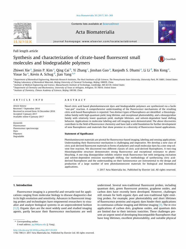
To synthesize the first class of CPDs, equimolar amounts of citricacid and b-/c-aminothiols were reacted, resulting in a thiazolo pyr-idine carboxylic acid (TPA) (Figs. 1a and S4). TPA structures werefirst reported by Kasprzyk and co-workers, however, the fluores-cence mechanism have not been studied in details [12]. As anexample, CA-Cys showed strong fluorescence with quantum yieldsas high as 81% and an extinction coefficient of 8640 M�1 cm�1,resulting in strong fluorescence that can even be observed underwhite light (Fig. 1c). The emission peak of CA-Cys remained fixedat 430 nm independent of the wavelength of excitation. In otherwords, band-shifting behavior, defined as fluorescence emissionpeak shifting with different excitation wavelengths (Fig. 1b), wasnot observed [7]. This behavior is marked by the approximate sym-metry of the 3D fluorescence spectra. In these cases, fluorescenceresults from the relaxation of the electronically excited singletstate in its lowest vibrational energy level to the ground state(Kasha’s Rule) [13]. By reacting citric acid with other b- orc-aminothiols including homocysteine, cysteamine, and penicil-
lamine, similarly strong fluorescence emissions lacking bandshifting behavior were observed (Figs. S1–S3). In addition, simplyby adding an aliphatic diol into the reaction, we can producebiodegradable photoluminescent polymers (BPLP), reported previ-ously [7]. For instance, BPLP-Cys showed fluorescence propertiessimilar to CA-Cys and in vivo degradability [14].
Since the fluorescence of most organic dyes stems from conju-gated aromatic rings [4,15], we next calculated the aromaticity ofTPA molecules based on the Nucleus Independent Chemical Shift(NICS) model [16,17]. The class of TPAs including CA-Cys, CA-Cysteamine, and CA-Homocysteine all demonstrated high aro-maticity with NICS < �3.0 (Table S2). Thus, the fluorescence mech-anism of TPAs resembles that of most organic dyes, whosefluorescence results from p-p⁄ electronic excitation that leads toemission from the lowest energy band. The time-dependent fluo-rescence of CA-Cys can be fitted to a single-exponential decay,resulting in a single lifetime of s = 9.86 ± 0.078 ns (Fig. S38). Thusthe fluorescence emission of TPA molecules obeys Kasha’s rule,as illustrated by a Jablonski diagram (Fig. S5).
3.2. Synthesis and fluorescent properties of dioxo-pyridine ring baseddyes
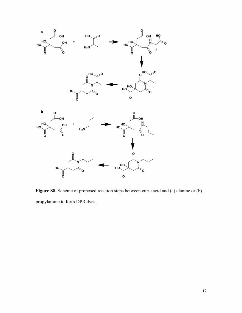
The second type of CPDs was made from citric acid and amineslacking a thiol group by the same simple solvent-free reaction(Scheme1). A dioxo-pyridine ring (DPR) structure was identifiedas the fluorophore, exemplified by CA-Ala (Figs. 2a and S8). SomeDPRs, for example CA-Gly, showed minor impurities in NMR, sug-gesting that the purification may need to be further improved.Compared to TPAs, DPRs exhibited relatively weak fluorescencewith quantum yields lower than 40%, as well as distinct band shift-ing of emission and varying Stokes Shifts that were dependent onthe excitation wavelengths (Figs. 2b, S9 and S10). Moreover, unlikeTPAs, DPRs do not have conjugated structures, as supported by lowaromaticity with NICS > 1.5 (Table S2). Thus, the fluorescencemechanism of conventional conjugated organic dyes is not applica-ble here, and DPR likely represents a distinct PL mechanism [18].We hypothesized that the aliphatic tertiary nitrogen is the sourceof fluorescence, as previously suggested for PAMAM dendrimers[19,20]. In an early study, monovalent tertiary amines in gas phaseshowed emissions limited to the range of 250–400 nm [21,22].Interestingly, DPRs, PAMAM dendrimers [19,20,23], and amine-containing carbon dots [6,24] all exhibit maximum excitation at350–380 nm, and maximum emission at 420–450 nm with signif-icantly stronger intensity in the liquid phase. This dichotomy posesan interesting question of how the mechanism of red shift andincreased fluorescence of DPRs in the liquid phase differs from thatof monovalent tertiary amines in the gas phase [21,22,25]. We pro-pose that both phenomena can be explained by the same mecha-nism: n-p⁄ and n-r⁄ transitions of the lone pair electrons of thetertiary amine undergo a red-shift due to the electron withdrawingeffects of the adjacent carbonyl groups, resulting in stronger visiblefluorescence. Indeed, both electron-withdrawing carbonyl groupsextend resonance from the tertiary amine, as depicted in the com-puted isosurfaces in Table 1, resulting in a red shift of absorbancefrom a smaller highest occupied and lowest unoccupied molecularorbital (HOMO-LUMO) gap. Calculations showed an absorptionpeak of 263 nm for the tetrahydropyridine lacking both carbonylgroups (molecule 4), which shifted to 303 nm/325 nm (2 and 3),and then to 333 nm (1) with each addition of a carbonyl group.These results provide insight into the fluorescence red shiftingbehavior of DPRs, and the same mechanism can be used to explainthe behavior of PAMAM dendrimers [26] that similarly have car-bonyl groups pulling electrons from tertiary amines.

Scheme 1. The synthetic routes and structures of CPDs. TPAs (blue) are synthesized by reacting citric acid with b or c-aminothiols such as cysteine, cysteamine, 2-aminothiolphenol, etc. DPRs (red) are synthesized from citric acid and other primary amines/anilines that do not contain a thiol group, e.g. alanine, c-Aminobutyric acid,propylamine, ethylenediamine, ethanolamine, phenylenediamine, and others. All chiral molecules used were L-isomers unless specifically stated otherwise. (Forinterpretation of the references to colour in this figure legend, the reader is referred to the web version of this article.)
364 Z. Xie et al. / Acta Biomaterialia 50 (2017) 361–369
3.3. Band shifting of DPRs
More interestingly, all DPRs showed dynamic Stokes shifts andexcitation-dependent emission wavelengths, referred to as ‘‘bandshifting behavior”. For instance, the 3D fluorescence spectra ofCA-Ala (Fig. 2b) is not symmetrical as expected for typical fluo-rophores, but marked by a clear ‘‘tail” that expands to longer wave-lengths. Similar band shifting behavior, which breaks Kasha’s Rule,has also been found in PAMAM dendrimers [27], carbon dots [6],and graphene oxide [28]. To unveil the mechanism of the bandshifting phenomenon of DPRs, time-resolved fluorescence spec-troscopy was performed. First, the fluorescence decay of CA-Alacannot be fitted to either a single- or double-exponential decayas the residue distribution is not random (Figs. S39 and S40), sug-gesting the presence of multiple excited state energy levels thatgive fluorescence. A triple exponential model adequately (meaningv2 < 1.2 and the residue distributions are random) fits the lifetime
decay (Fig. S41), resulting in s1 = 1.03 ± 0.029 ns,s2 = 4.33 ± 0.128 ns, and s3 = 10.07 ± 0.031 ns. Multiple lifetimesindicate that the fluorescence emission of CA-Ala is not from a sin-gle energy band. Thus, we hypothesis that the band shifting phe-nomenon is a result of the ‘‘red edge effect”, where the presenceof rotating auxochromic groups generates additional dipole inter-actions between the fluorophore and solvent during intersystemrelaxation, prolonging the solvation time to the approximate time-scale of fluorescence emission [29,30]. As illustrated in Fig. S12,longer solvation time further relaxes the excited state to variouslower energy levels, resulting in multiple lifetimes and red-shifting emissions. The absence of band shifting seen in TPA canbe explained by its non-rotating highly conjugated auxochromicring structure. In the cases of TPA and other conjugated organicdyes, the solvation times, ss, are normally around 10 ps, whichare considerably shorter than the fluorescence lifetimes (s), whichare in the range of 0.5–30 ns [31]. In contrast, non-aromatic DPR

Fig. 1. Synthesis and fluorescence properties of CA-Cys and BPLP-Cys. (a) Synthetic schemes for CA-Cys (from citric acid and cysteine), BPLP-Cys (from citric acid, 1,8-octanediol, and cysteine), and BPLP-TPA (from CA-Cys and 1,8-octanediol). The hydrolysis reaction of BPLP-Cys to form CA-Cys is also illustrated. (b) 3D excitation-emissionspectra of CA-Cys in water solution. (c) Images of CA-Cys solution under white light with white/black background and under UV light (from left to right). (d) Comparison ofmaximum excitation and emission spectra of BPLP-Cys and BPLP-TPA (which resulted from the reaction of CA-Cys and 1,8-octanediol).
Z. Xie et al. / Acta Biomaterialia 50 (2017) 361–369 365
possesses rotating auxochromic groups, and thus solvation may beallowed to ss � s in polar solvents. Furthermore, the solvation pro-cess of DPR explains our earlier observations, such as the relativelylow quantum yields of DPRs (Table S1), as well as the presence of atleast three distinct lifetimes for CA-Ala compared to a single life-time for CA-Cys.
To prove that the dynamic band shifting exhibited by DPRs isindeed generated by the red-edge effect, we demonstrate a corre-lation between the band shifting and solvent polarity. The relax-ation kinetics of DPRs were measured by the extent of band shift(Fig. 3a–c) and fluorescence lifetimes (Fig. 3d–f). The extent ofband shift (i.e. intensity of emission at longer wavelengths) isshown to increase with solvent polarity, as the band-shift effectof CA-Ala is strongest in water (dielectric constant e = 80.1), mod-erate in acetone (e = 20.7), and minimal in non-polar solvents suchas dichloromethane (e = 8.93) (Fig. 3a–c). Interestingly, when fluo-rescence lifetime decays of CA-Ala were collected at different emis-sion wavelengths, the decay plots varied significantly in water(Fig. 3d), changed slightly in acetone (Fig. 3e), but remained rela-tively constant in dichloromethane (Fig. 3f). As the fluorescenceemission wavelengths represent the band edge energy levels thatcorrespond to the allowed timescale of solvent relaxation, the life-times of CA-Ala in polar solvents responded dynamically to theextent of solvent relaxation [28,32]. As a result, the relaxationkinetics of DPRs is largely influenced by dipole alignments inresponse to polar solvents [28,29]. The effect of solvent also canbe observed in the 3D spectra of polymeric BPLP-Ala (Fig. 2c),which is dissolved in 1,4-dioxane. Furthermore, the band shiftingof BPLP-Ala is clearly not as strong as CA-Ala in water solution(Fig. 2b). Thus, the band shifting of DPR is caused by the red-edge effect, which is ultimately governed by fluorophore/solventinteractions.
3.4. Photostability of citric acid-derived photoluminescent dyes
In addition to the photoluminescent behavior discussed above,CPDs have the advantages of high photostability and long lifetimes.Just as high photostability was previously reported in BPLPs[14,33], CA-Cys was found to be extremely stable, with 95% ofthe fluorescence intensity remaining after continuous UV excita-tion for 3 h (Fig. 4a). In contrast, DPRs such as CA-Ala showed pho-tobleaching on par with Fluorescein, but were more resistant tophotobleaching than Rhodamine B. We propose that the high pho-tostability of TPA structures arises from the presence of efficientdeactivation pathways of photoexcited molecules along the 2-pyridone fused ring structure, much like the deactivation pathwaysstudied in adenine [34,35]. Such internal conversion pathwaysthrough out-of-plane modes of vibrations are reportedly efficientand lead to high photostability [35], whereas inefficient radiation-less deactivation of excited DPR molecules may arise from solventrelaxation processes due to rotation of the auxiliary group, result-ing in lower photostability. When excited by a pulsed laser, CPDs,including both CA-Cys and CA-Ala, exhibited longer emission decaylifetimes than Rhodamine B and Fluorescein (Fig. 4b). Long life-times are typically found in blue dyes; however, the band shiftingexhibited by DPRs (exemplified by CA-Ala) may have potential foruse in fluorescence lifetime imaging (FLIM) of biological tissuesand molecules [36].
3.5. Applications of CPDs
Fluorescent dyes have wide applications ranging from molecu-lar labeling to in vivo imaging. To demonstrate the potential appli-cations of our newly-developed CPDs, we first conjugated bovineserum albumin (BSA), as a representative biomolecule, with CA-

Fig. 2. Synthesis and fluorescence properties of CA-Ala and BPLP-Ala. (a) Synthesis schemes of CA-Ala (from citric acid and alanine), BPLP-Ala (from citric acid, 1,8-octanediol,and Alanine), and BPLP-DPR (from CA-Ala and 1,8-octanediol). The hydrolysis reaction of BPLP-Ala resulting in CA-Ala is also illustrated. (b) 3D excitation-emission spectra ofCA-Ala in water solution. (c) 3D excitation-emission spectra of BPLP-Ala in 1,4-dioxane solution. (d) Comparison of excitation and emission spectra of BPLP-Ala and BPLP-DPR-Ala. (e) Optical images of BPLP-Ala solutions at increasing excitation wavelengths from left to right.
366 Z. Xie et al. / Acta Biomaterialia 50 (2017) 361–369
Cys. The free carboxyl groups of our CPDs enable facile conjugationby carbodiimide chemistry to produce a wide range of molecularlabels. As shown in Fig. 5a, strong blue fluorescence can beobserved from purified CA-Cys conjugated BSA molecules. Wewere able to establish a calibration curve of CA-Cys labeled BSAto quantify the BSA concentration as well (data not shown). Inaddition, the carboxyl groups on CPDs are also available for modi-fication in cellular labeling. For example, CA-Ala with activatedcarboxyl groups were used to label fibroblasts, and imaged usingconfocal microscopy (Fig. 5b). Fibroblasts labeled by CA-Alashowed strong green fluorescence in the FITC channel, owed tothe band shifting behavior of CA-Ala. It is clear that CPDs havepotential in cellular imaging, tracing and visualization.
3.6. CPD and biodegradable photoluminescent polymer
The discovery of CPDs not only establishes a new family of flu-orophores, but it also provides insight into the fluorescence mech-anism of our previously developed biodegradablephotoluminescent polymers (BPLPs) that were synthesized bydirectly reacting citric acid, an amino acid such as L-cysteine, anda diol such as 1,8-octanediol [7]. Previously, our group has demon-strated the applications of BPLPs in tissue engineering, bioimaging,theranostic drug delivery, and more recently, selective halide(chloride, bromide, iodide) sensing for fluorescence based diagno-
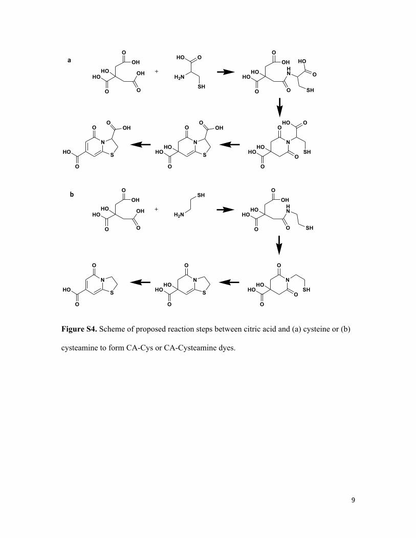
sis of cystic fibrosis [7,14,37,38]. To establish that the TPA structureis indeed the fluorescent moiety of BPLPs, we synthesized poly-mers by reacting purified CA-Cys with 1,8-octanediol. The PL prop-erties of the resultant BPLP-TPA were identical to that of BPLP-Cyssynthesized by reacting citric acid, 1,8-octanediol, and L-cysteinedirectly (Fig. 1d), including a lack of band-shifting (Fig. S6). Addi-tionally, CA-Ala could be directly reacted with 1,8-octanediol toform a BPLP-DPR whose PL properties were very similar to thatof CA-Ala (Fig. 2d). Moreover, CA-Cys and CA-Ala were found inthe alkaline hydrolysis products of BPLP-Cys and BPLP-Ala, respec-tively, which confirmed that the fluorophores of BPLPs are indeedCPDs. (Figs. S7 and S11).
4. Conclusions
In summary, we have synthesized a series of citric acid-derivedfluorophores and determined their mechanisms of fluorescence.The primary approach for generating citric acid derived photolumi-nescent dyes (CPDs) is by reacting citric acid with primary amine-containing molecules. If the primary amines used in the reactionare b or c-aminothiol, conjugated TPA structures will be synthe-sized, exhibiting strong fluorescence emission with high quantumyield, single-exponential lifetimes, and absence of band shiftingbehavior. If primary amines without thiol groups are used to reactwith citric acid, non-conjugated DPRs will be synthesized, emitting

Table 1Computed absorption peak wavelengths and isosurfaces of LUMO for CA-Ala and its analogs without one or two carbonyl groups.
Structure Absorption wavelength Isosurfaces of HOMO Isosurfaces of LUMO
333 nm
303 nm
325 nm
263 nm
Fig. 3. (a–c) Fluorescence emission spectra of CA-Ala at different excitation wavelengths in water, acetone and dichloromethane. (d–f) Fluorescence intensity-time traces ofCA-Ala at different emission wavelengths in water, acetone and dichloromethane.
Z. Xie et al. / Acta Biomaterialia 50 (2017) 361–369 367
fluorescence with relatively lower quantum yields, multiple life-times, band shifting behavior, and dynamic Stokes shifts. We alsodemonstrate that CPDs can be used as fluorescent molecules to
react with other monomers to form fluorescent polymers sup-ported by the syntheses of BPLP-TPAs and BPLP-DPRs. In conclu-sion, we have established a methodology to design and prepare

Fig. 4. (a) Fluorescence photostabilities of CA-Cys and CA-Ala with 3 h continuous excitation at their respective maximums. Fluorescein and Rhodamine B served as controls.(b) Fluorescence intensity-time traces of CA-Cys, CA-Ala, Fluorescein and Rhodamine B after pulsed excitation at 352 nm.
Fig. 5. (a) Emission spectra of BSA and CA-Cys labeled BSA solutions in PBS, excited at 365 nm. (b) Confocal microscope image of 3T3 fibroblasts, stained by NHS activate CA-Ala (imaged under FITC filter) and DAPI. Scale bar = 30 lm.
368 Z. Xie et al. / Acta Biomaterialia 50 (2017) 361–369
new fluorescent dyes and polymers derived from citric acid andamine-containing molecules in an extremely facile and low-costmanner, to meet the ever-growing needs in applications where flu-orescence is an enabling tool.
Notes
Dr. Yang and The Pennsylvania State University have a financialinterest in Aleo BME, Inc. These interests have been reviewed bythe University’s Institutional and Individual Conflict of InterestCommittees and are currently being managed by the University.
Acknowledgements
The authors acknowledge the financial support from NationalInstitutes of Health (NIH – United States) Awards (NIBIBEB012575, NCI CA182670, NHLBI HL118498), and National ScienceFoundation (NSF – United States) Awards (DMR1313553).
Appendix A. Supplementary data
Supplementary data associated with this article can be found, inthe online version, at http://dx.doi.org/10.1016/j.actbio.2017.01.019.
References
[1] M. Fernandez-Suarez, A.Y. Ting, Fluorescent probes for super-resolutionimaging in living cells, Nat. Rev. Mol. Cell Biol. 9 (12) (2008) 929–943.
[2] F. Wang, W.B. Tan, Y. Zhang, X.P. Fan, M.Q. Wang, Luminescent nanomaterialsfor biological labelling, Nanotechnology 17 (1) (2006) R1–R13.
[3] M.Y. Berezin, S. Achilefu, Fluorescence lifetime measurements and biologicalimaging, Chem. Rev. 110 (5) (2010) 2641–2684.
[4] U. Resch-Genger, M. Grabolle, S. Cavaliere-Jaricot, R. Nitschke, T. Nann,Quantum dots versus organic dyes as fluorescent labels, Nat. Methods 5 (9)(2008) 763–775.
[5] P. Ferruti, M.A. Marchisio, R. Duncan, Poly(amido-amine)s: biomedicalapplications, Macromol. Rapid Commun. 23 (5–6) (2002) 332–355.
[6] S. Zhu, Q. Meng, L. Wang, J. Zhang, Y. Song, H. Jin, K. Zhang, H. Sun, H. Wang, B.Yang, Highly photoluminescent carbon dots for multicolor patterning, sensors,and bioimaging, Angew. Chem. Int. Ed. 52 (14) (2013) 3953–3957.

Z. Xie et al. / Acta Biomaterialia 50 (2017) 361–369 369
[7] J. Yang, Y. Zhang, S. Gautam, L. Liu, J. Dey, W. Chen, R.P. Mason, C.A. Serrano, K.A. Schug, L. Tang, Development of aliphatic biodegradable photoluminescentpolymers, Proc. Natl. Acad. Sci. U.S.A. 106 (25) (2009) 10086–10091.
[8] M.J. Frisch, G.W. Trucks, H.B. Schlegel, G.E. Scuseria, M.A. Robb, J.R. Cheeseman,G. Scalmani, V. Barone, B. Mennucci, G.A. Petersson, H. Nakatsuji, M. Caricato,X. Li, H.P. Hratchian, A.F. Izmaylov, J. Bloino, G. Zheng, J.L. Sonnenberg, M.Hada, M. Ehara, K. Toyota, R. Fukuda, J. Hasegawa, M. Ishida, T. Nakajima, Y.Honda, O. Kitao, H. Nakai, T. Vreven, J.A. Montgomery Jr., J.E. Peralta, F. Ogliaro,M.J. Bearpark, J. Heyd, E.N. Brothers, K.N. Kudin, V.N. Staroverov, R. Kobayashi,J. Normand, K. Raghavachari, A.P. Rendell, J.C. Burant, S.S. Iyengar, J. Tomasi, M.Cossi, N. Rega, N.J. Millam, M. Klene, J.E. Knox, J.B. Cross, V. Bakken, C. Adamo, J.Jaramillo, R. Gomperts, R.E. Stratmann, O. Yazyev, A.J. Austin, R. Cammi, C.Pomelli, J.W. Ochterski, R.L. Martin, K. Morokuma, V.G. Zakrzewski, G.A. Voth,P. Salvador, J.J. Dannenberg, S. Dapprich, A.D. Daniels, Ö. Farkas, J.B. Foresman,J.V. Ortiz, J. Cioslowski, D.J. Fox, Gaussian 09, Gaussian Inc: Wallingford, CT,USA, 2009.
[9] R. Krishnan, J.S. Binkley, R. Seeger, J.A. Pople, Self-consistent molecular orbitalmethods. XX. A basis set for correlated wave functions, J. Chem. Phys. 72 (1)(1980) 650–654.
[10] R. Ditchfield, W.J. Hehre, J.A. Pople, Self-consistent molecular-orbital methods.IX. An extended Gaussian-type basis for molecular-orbital studies of organicmolecules, J. Chem. Phys. 54 (2) (1971) 724–728.
[11] J. Kruszewski, T.M. Krygowski, Definition of aromaticity basing on theharmonic oscillator model, Tetrahedron Lett. 13 (36) (1972) 3839–3842.
[12] W. Kasprzyk, S. Bednarz, D. Bogdał, Luminescence phenomena ofbiodegradable photoluminescent poly (diol citrates), Chem. Commun. 49(57) (2013) 6445–6447.
[13] A.N. Fletcher, Fluorescence emission band shift with wavelength of excitation,J. Phys. Chem. 72 (8) (1968) 2742–2749.
[14] Z. Xie, Y. Zhang, L. Liu, H. Weng, R.P. Mason, L. Tang, K.T. Nguyen, J.-T. Hsieh, J.Yang, Development of intrinsically photoluminescent and photostablepolylactones, Adv. Mater. 26 (26) (2014) 4491–4496.
[15] G. Ulrich, R. Ziessel, A. Harriman, The chemistry of fluorescent bodipy dyes:versatility unsurpassed, Angew. Chem. Int. Ed. 47 (7) (2008) 1184–1201.
[16] P.V.R. Schleyer, C. Maerker, A. Dransfeld, H. Jiao, N.J.R.v.E. Hommes, Nucleus-independent chemical shifts: a simple and efficient aromaticity probe, J. Am.Chem. Soc. 118 (26) (1996) 6317–6318.
[17] A. Stanger, Nucleus-independent chemical shifts (NICS): distance dependenceand revised criteria for aromaticity and antiaromaticity, J. Org. Chem. 71 (3)(2005) 883–893.
[18] R.L. Joseph, Principle of Fluorescence Microscopy, Springer Science + BusinessMedia, LLC, New York, NY, 2006.
[19] D. Wang, T. Imae, Fluorescence emission from dendrimers and its pHdependence, J. Am. Chem. Soc. 126 (41) (2004) 13204–13205.
[20] M. Sun, C.-Y. Hong, C.-Y. Pan, A unique aliphatic tertiary amine chromophore:fluorescence, polymer structure, and application in cell imaging, J. Am. Chem.Soc. 134 (51) (2012) 20581–20584.
[21] A.M. Halpern, T. Gartman, Structural effects on photophysical processes insaturated amines. II, J. Am. Chem. Soc. 96 (5) (1974) 1393–1398.
[22] R.A. Beecroft, R.S. Davidson, T.D. Whelan, Fluorescent excimer formation byalpha, omega-diaminoalkanes and related compounds, J. Chem. Soc. PerkinTrans. 2 (7) (1985) 1069–1072.
[23] G. Jiang, X. Sun, Y. Wang, M. Ding, Synthesis and fluorescence properties ofhyperbranched poly(amidoamine)s with high density tertiary nitrogen, Polym.Chem. 1 (10) (2010) 1644–1649.
[24] S. Sahu, B. Behera, T.K. Maiti, S. Mohapatra, Simple one-step synthesis of highlyluminescent carbon dots from orange juice: application as excellent bio-imaging agents, Chem. Commun. 48 (70) (2012) 8835–8837.
[25] C.G. Freeman, M.J. McEwan, R.F.C. Claridge, L.F. Phillips, Fluorescence ofaliphatic amines, Chem. Phys. Lett. 8 (1) (1971) 77–78.
[26] D. Wang, T. Imae, M. Miki, Fluorescence emission from PAMAM and PPIdendrimers, J. Colloid Interface Sci. 306 (2) (2007) 222–227.
[27] W. Yang, C.-Y. Pan, Synthesis and fluorescent properties of biodegradablehyperbranched poly(amido amine)s, Macromol. Rapid Commun. 30 (24)(2009) 2096–2101.
[28] S.K. Cushing, M. Li, F. Huang, N. Wu, Origin of strong excitation wavelengthdependent fluorescence of graphene oxide, ACS Nano 8 (1) (2013) 1002–1013.
[29] A.P. Demchenko, The red-edge effects: 30 years of exploration, Luminescence17 (1) (2002) 19–42.
[30] A. Samanta, Dynamic stokes shift and excitation wavelength dependentfluorescence of dipolar molecules in room temperature ionic liquids, J. Phys.Chem. B 110 (28) (2006) 13704–13716.
[31] L. Reynolds, J.A. Gardecki, S.J.V. Frankland, M.L. Horng, M. Maroncelli, Dipolesolvation in nondipolar solvents: experimental studies of reorganizationenergies and solvation dynamics, J. Phys. Chem. 100 (24) (1996) 10337–10354.
[32] S.K. Cushing, M. Li, F. Huang, N. Wu, Origin of strong excitation wavelengthdependent fluorescence of graphene oxide, ACS Nano 8 (1) (2014) 1002–1013.
[33] Y. Zhang, J. Yang, Design strategies for fluorescent biodegradable polymericbiomaterials, J. Mater. Chem. B 1 (2) (2013) 132–148.
[34] C. Canuel, M. Mons, F. Piuzi, B. Tardivel, I. Dimicoli, M. Elhanine, Excited statesdynamics of DNA and RNA bases: characterization of a stepwise deactivationpathway in the gas phase, J. Chem. Phys. 122 (7) (2005) 074316.
[35] Andrzej L. Sobolewski, Wolfgang Domcke, The chemical physics of thephotostability of life, Europhys. News 37 (4) (2006) 20–23.
[36] W. Becker, Fluorescence lifetime imaging – techniques and applications, J.Microsc. 247 (2) (2012) 119–136.
[37] J.P. Kim, Z. Xie, M. Creer, Z. Liu, J. Yang, Citrate-based fluorescent materials forlow-cost chloride sensing in the diagnosis of cystic fibrosis, Chem. Sci. 8 (2017)550–558.
[38] Y. Zhang, R.T. Tran, I.S. Qattan, Y.-T. Tsai, L. Tang, C. Liu, J. Yang, Fluorescenceimaging enabled urethane-doped citrate-based biodegradable elastomers,Biomaterials 34 (16) (2013) 4048–4056.

1
SUPPORTING INFORMATION
Synthesis and characterization of citrate-based
fluorescent small molecules and biodegradable
polymers
Zhiwei Xie,a Jimin P. Kim,a Qing Cai,b Yi Zhang,c Jinshan Guo,a Ranjodh S. Dhami,a Li Li,d Bin
Kong,e Yixue Su, a Kevin A. Schug,d Jian Yang*a
a Department of Biomedical Engineering, Materials Research Institutes, the Huck Institutes of Life Sciences, The Pennsylvania State University, University Park, PA 16801
b Beijing Laboratory of Biomedical Materials, Beijing University of Chemical Technology, Beijing China 100029
c Institute of Medical Engineering and Science, Massachusetts Institute of Technology, Cambridge, MA 02139
d Department of Chemistry and Biochemistry, University of Texas at Arlington, Arlington, TX 76010
e Institute of Chemistry, Chinese Academy of Sciences, Beijing, China 100190
* To whom correspondence should be addressed. Email: [email protected]

2
Identification of examples of CPDs:
CA-Cys: 1H NMR (DMSO-d6) δ 6.58 (d, 1H), 5.48 (d, 1H), 3.90 (t, 1H), 3.60 (d, 2H). 13C NMR
(DMSO-d6) δ 169.7, 166.1, 161.5, 150.4, 143.2, 115.5, 98.7, 63.0, 32.2. ESI-MS m/z=241:
(M+H)+; m/z=264: (M+Na)+.
CA-Cysteamine: 1H NMR (DMSO-d6) δ 6.54 (d, 1H), 4.37 (t, 1H), 3.53 (t, 2H), 2.51-3.12 (m,
2H). 13C NMR (DMSO-d6) δ 171.5, 160.6, 148.7, 101.3, 84.7, 57.5, 49.9, 34.0. ESI-MS m/z= 198:
(M+H)+.
CA-Propylamine: 1H NMR (DMSO-d6) δ 6.18-6.49 (s, 1H), 3.32-3.37 (t, 2H), 2.76-3.01 (m, 2H),
1.45-1.52 (q, 2H), 0.80-0.85 (t, 3H). 13C NMR (DMSO-d6) δ 179.5, 175.9, 172.3, 166.9, 141.9,
42.74, 40.0, 22.2, 11.9. ESI-MS m/z=216: (M+H2O+H)+; m/z=238: (M+ H2O+Na)+.
CA-Ala: 1H NMR (DMSO-d6) δ 4.58-4.78 (m, 1H), 2.50-3.07 (m, 4H), 1.32 (d, 3H). 13C NMR
(DMSO-d6) δ 178.6, 174.8, 172.1, 171.5, 72.3, 47.7, 39.7, 14.5. ESI-MS m/z=228: (M+H)+;
m/z=268: (M+ H2O+Na)+.
CA-Gly: 1H NMR (DMSO-d6) δ 6.58 (s, 1H), 4.08-4.21 (m, 2H), 2.50-2.89 (m, 2H). 13C NMR
(DMSO-d6) δ 175.3, 172.0, 169.0, 166.7, 140.9, 72.8, 39.8, 34.9. ESI-MS m/z=232: (M+
H2O+H)+; m/z=254: (M+ H2O+Na)+; m/z=311: (M+Gly+H2O+Na)+.

3
Table S1. Summary of synthesis and photophysical properties of CPDs. Compound 1 is
the aliphatic acid, compound 2 is one of the listed primary amines, amino acids, or
analogs of amino acids. λab is the maximum absorption wavelength. ε is the extinction
coefficiency. λex and λex are the maximum excitation and emission wavelengths. Φ is the
quantum yield.
Compound 1 Compound 2 Fluorophore Structure λex (nm)
λem (nm)
ε (M-
1cm-1)
Φ (%)
Stokes Shift (nm)
Band Shift
?
TCA Cysteine N
OOHO
SH
O
HOO
366 431 11.6 54.2 65 Y
TCA Alanine N
OOHO
O
HOO
355 423 6.1 0.2 68 Y
SucA Cysteine NA NA NA NA 0 NA NA
SucA Alanine NA NA NA NA 0 NA NA
CA Propionic acid NA NA NA NA 0 NA NA
CA 3-
mercaptopropionic acid
NA NA NA NA 0 NA NA
CA Alanine N
O
OHO
O
OHO
361 430 79 21.2 69 Y
CA Arginine N
O
OHO
O
OHO
HN
NH
NH2
382 451 11.6 15.5 69 Y
CA Asparagine N
O
OHO
O
OHO
NH2
O
378 440 35.5 23.8 62 Y
CA Aspartic Acid N
O
OHO
O
OHO
OH
O
356 427 6.8 19.7 71 Y

4
CA Glutamic Acid N
O
OHO
O
OHO
O
OH
358 423 5.1 9.1 65 Y
CA Glutamine N
O
OHO
O
OHO
O
NH2
383 448 102.4 7.3 65 Y
CA Glycine N
O
OHO
O
HO O
357 434 12 39.0 77 Y
CA Isoleucine N
O
OHO
O
OHO
357 430 5.6 17.2 73 Y
CA Leucine N
O
OHO
O
OHO
359 423 6.2 8.8 64 Y
CA Lysine N
O
OHO
O
OHO
NH2
359 431 11.4 8.6 72 Y
CA Methionine N
O
OHO
O
OHO
S
362 424 5.3 10.5 62 Y
CA Phenylalanine N
O
OHO
O
OHO
358 423 7.8 12.7 65 Y
CA Serine N
O
OHO
O
OHO
OH
369 440 437.9 4.2 71 Y
CA Tryptophan N
O
OHO
O
OHO N
386 445 14.6 5 59 Y

5
CA Valine N
O
OHO
O
OHO
361 428 5.3 17.5 67 Y
CA 3-
Aminobutanoic Acid
N
O
OHO
O
OH
O
356 430 6.3 8.8 74 Y
CA γ-Aminobutyric acid N
O
OHO
O
OHO
355 422 4.9 22.1 67 Y
CA Propylamine N
O
OHO
O
359 424 6.6 24.6 65 Y
CA Heptylamine N
O
OHO
O
356 424 4.7 13.5 68 Y
CA Phenylenediamine
N
O
OHO
O
NH2
360 433 12.1 4.5 73 Y
CA Hexamethylenediamine
N
O
OHO
O
NH2
357 426 9.4 11.5 69 Y
CA ethylenediamine N
O
OHO
O
NH2
377 440 828.3 9.2 63 Y
CA ethanolamine N
O
OHO
O
OH
368 419 34.6 42.1 51 Y
CA Cysteine N
OOHO
S
O
HO
364 445 8640 81.4 81 N

6
Table S2. Harmonic Oscillator Model of Electron Delocalization (HOMED) and Nucleus
Independent Chemical Shift (NICS) values of selected citric acid based dyes. CA-Cys,
CA-Homocysteine, CA-Cysteamine are examples for TPAs. CA-Ala, CA-Gly, and CA-
Propylamine are examples for DPRs.
CA-
Cys
CA-
HomoCys
CA-
Cysteamine CA-Ala CA-Gly
CA-
Propylamine
NICS -3.2671 -3.0824 -3.0674 1.5852 1.7880 1.8341
CA Cysteamine N
O
S
O
HO
365 445 3880 79.3 80 N
CA Homocysteine N
OOHO
O
HOS
375 445 1014 70.8 70 N
CA Threonine N
OOHO
O
O
HO
386 436 418 78.6 50 N
CA 2-
Aminothiolphenol
N
O
HO
O
S
396 439 877.8 98.5 43 N
CA Penicillamine N
OOHO
S
O
HO
369 432 3180 80.6 63 N

7
350 400 450 500 550 600 650 700
PL In
tens
ity (a
.u.)
Wavelength (nm)
Ex at 320nm Ex at 340nm Ex at 360nm Ex at 367nm Ex at 380nm Ex at 400nm
Figure S1. Emission spectra of CA-Cysteamine under different excitation wavelengths.
400 500 600 700
PL In
tens
ity (a
.u.)
Wavelength (nm)
Ex at 320nm Ex at 340nm Ex at 350nm Ex at 360nm Ex at 380nm
Figure S2. Emission spectra of CA-Homocysteine under different excitation wavelengths.

8
400 500 600 700
PL In
tens
ity (a
.u.)
Wavelength (nm)
Ex at 320nm Ex at 340nm Ex at 350nm Ex at 360nm Ex at 380nm
Figure S3. Emission spectra of CA-Penicillamine under different excitation wavelengths.
9
Figure S4. Scheme of proposed reaction steps between citric acid and (a) cysteine or (b)
cysteamine to form CA-Cys or CA-Cysteamine dyes.
O
HO
O
OH
O
OH
H2N
HO O
O
HO
O
HN
O
OH
O
HO
O
HO
O
HO
HO HO
N
O
OHO
+
SH SH
SH
N
S
O
HO
OO
OH
N
S
O
HO
OO
OH
HO
O
HO
O
OH
O
OH
H2N
O
HO
O
HN
O
OH
O
HO
O
HO
HO HO
N
O
+
SH
SH
N
S
O
HO
O
N
S
O
HO
O
HO
SHb
a

10
Figure S5. A Jablonski diagram of fluorescence excitation and emission processes of a
TPA fluorophore.
350 400 450 500 550 600 650 700
PL In
tens
ity (a
.u.)
Wavelength (nm)
Ex at 320nm Ex at 340nm Ex at 360nm Ex at 367nm Ex at 380nm Ex at 400nm
Figure S6. Emission spectra of BPLP-TPA under different excitation wavelengths.
11
Figure S7. Emission spectra of BPLP-Cys degradation products in 1M NaOH solution.
400 450 500 550 600 650 700
PL
Inte
nsity
(a.u
.)
Wavelength (nm)
0 Days 7 Days 14 Days
12
Figure S8. Scheme of proposed reaction steps between citric acid and (a) alanine or (b)
propylamine to form DPR dyes.
O
HO
O
OH
O
OH
H2N
HO O
O
HO
O
HN
O
OH
O
HO
HO HO+
O
HO
O
HON
O
OHO
O
HO
O
N
O
OHO
O
HO
O
OH
O
OH
H2N
O
HO
O
HN
O
OHHO HO+
O
HO
O
HON
O
O
HO
O
N
O
b
a

13
400 500 600 700
PL In
tens
ity (a
.u.)
Wavelength (nm)
Ex at 340nm Ex at 360nm Ex at 380nm Ex at 400nm Ex at 420nm Ex at 440nm Ex at 460nm
Figure S9. Emission spectra of CA-Gly at different excitation wavelengths.
400 500 600 700
PL In
tens
ity (a
.u.)
Wavelength (nm)
Ex at 340nm Ex at 360nm Ex at 380nm Ex at 400nm Ex at 420nm Ex at 440nm Ex at 460nm Ex at 480nm
Figure S10. Emission spectra of CA-Propylamine at different excitation wavelengths.

14
350 400 450 500 550 600 650 700
PL In
tens
ity (a
.u.)
Wavelength (nm)
Ex at 320nm Ex at 340nm Ex at 360nm Ex at 380nm Ex at 400nm Ex at 420nm
Figure S11. Emission spectra of BPLP-Ala degradation products in 1M NaOH solution
Figure S12. A Jablonski diagram of fluorescence excitation and emission processes
showing band shifting behaviors of a DPR fluorophore.

15
Figure S13. Spatial conformations of CA-Cys. Dihedral angles of any four atoms are
listed on the right side.
Figure S14. Spatial conformations of CA-Ala. Dihedral angles of any four atoms are
listed on the right side.

16
Figure S15. ESI-MS spectrum (m/z) of CA-Cys. m/z=241: (M+H)+; m/z=264: (M+Na)+.
Figure S16. 1H NMR (DMSO-d6) spectrum of CA-Cys. δ 6.58 (d, 1H), 5.48 (d, 1H), 3.90
(t, 1H), 3.60 (d, 2H).
14 12 10 8 6 4 2 0ppm
17
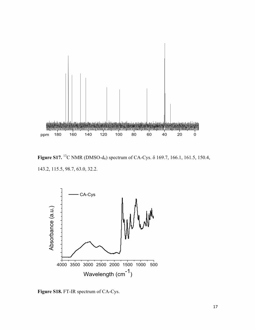
Figure S17. 13C NMR (DMSO-d6) spectrum of CA-Cys. δ 169.7, 166.1, 161.5, 150.4,
143.2, 115.5, 98.7, 63.0, 32.2.
Figure S18. FT-IR spectrum of CA-Cys.
180 160 140 120 100 80 60 40 20 0ppm
4000 3500 3000 2500 2000 1500 1000 500
Abso
rban
ce (a
.u.)
Wavelength (cm-1)
CA-Cys

18
Figure S19. ESI-MS spectrum (m/z) of BPLP-Cys degradation product. m/z=264:
(M+Na)+.
Figure S20. ESI-MS spectrum (m/z) of CA-Cysteamine. m/z= 198: (M+H)+.
Figure S21. 1H NMR (DMSO-d6) spectrum of CA-Cysteamine. δ 6.54 (d, 1H), 4.37 (t,
1H), 3.53 (t, 2H), 2.51-3.12 (m, 2H).
125.0 150.0 175.0 200.0 225.0 250.0 275.0 300.0 325.0 350.0 375.0 400.0 425.0 450.0 475.0 500.0 525.0 m/z0.0
1.0
2.0
3.0
4.0
5.0Inten. (x1,000,000)
198.0182
395.0323
430.0372402.0188219.9985 484.9795446.9927323.4915167.5033 256.0235 520.9787298.0431188.0094 346.0569 365.0290146.9988547.4718
116.5450
14 12 10 8 6 4 2 0ppm
19
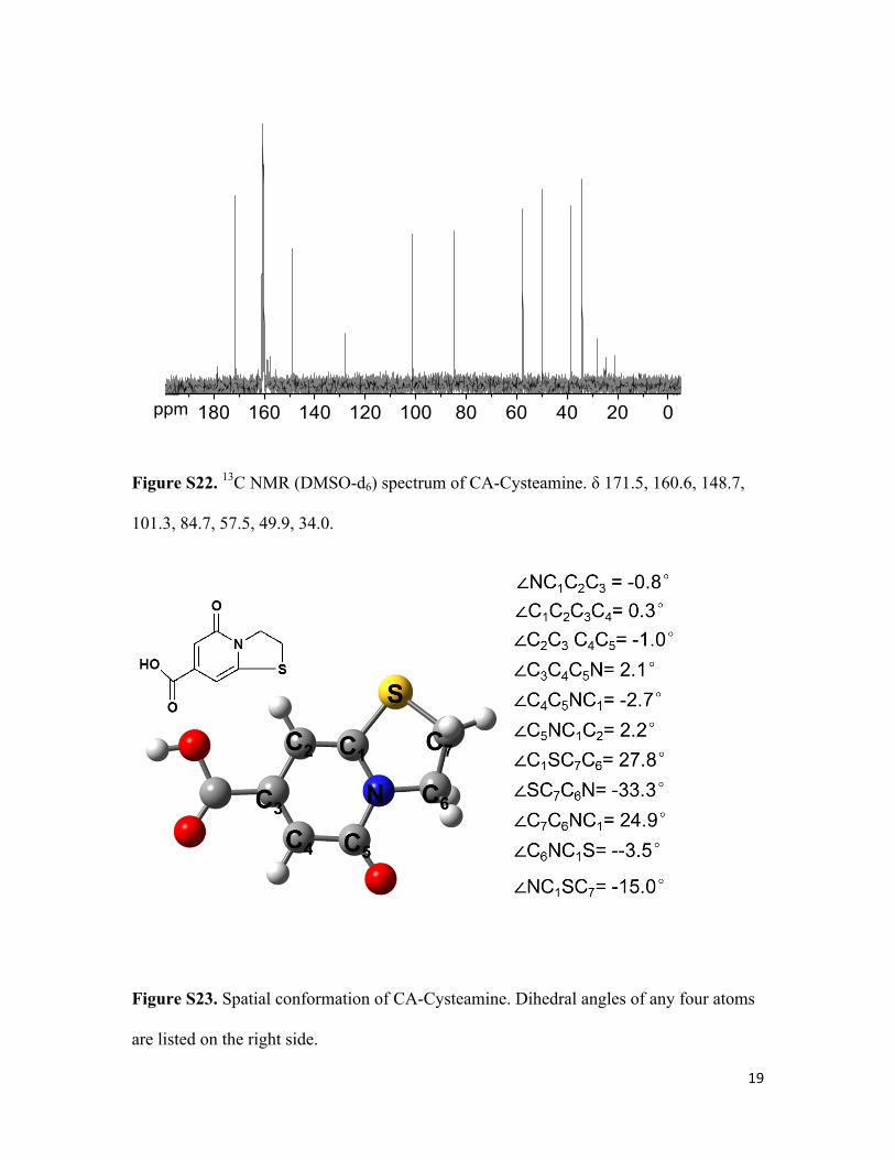
Figure S22. 13C NMR (DMSO-d6) spectrum of CA-Cysteamine. δ 171.5, 160.6, 148.7,
101.3, 84.7, 57.5, 49.9, 34.0.
Figure S23. Spatial conformation of CA-Cysteamine. Dihedral angles of any four atoms
are listed on the right side.
180 160 140 120 100 80 60 40 20 0ppm

20
Figure S24. ESI-MS spectrum (m/z) of BPLP-Cysteamine degradation product. m/z=198:
(M+H)+.
Figure S25. ESI-MS spectrum (m/z) of CA-Propylamine. m/z=216: (M+H2O+H)+;
m/z=238: (M+ H2O+Na)+.
Figure S26. 1H NMR (DMSO-d6) spectrum of CA-Propylamine. δ 6.18-6.49 (s, 1H),
3.32-3.37 (t, 2H), 2.76-3.01 (m, 2H), 1.45-1.52 (q, 2H), 0.80-0.85 (t, 3H).
150.0 175.0 200.0 225.0 250.0 275.0 300.0 325.0 350.0 375.0 400.0 425.0 450.0 475.0 m/z0.0
2.5
5.0
Inten. (x1,000,000)238.0609
216.0797
198.0700 485.0794180.0596 254.0351
412.0799 425.0573390.0964343.1801311.0252 469.0871156.0502 269.9938 372.0719 441.0844 492.0758

21
Figure S27. 13C NMR (DMSO-d6) spectrum of CA-Propylamine. δ 179.5, 175.9, 172.3,
166.9, 141.9, 42.74, 40.0, 22.2, 11.9.
Figure S28. Spatial conformation of CA-Propylamine. Dihedral angles of any four atoms
are listed on the right side.
22
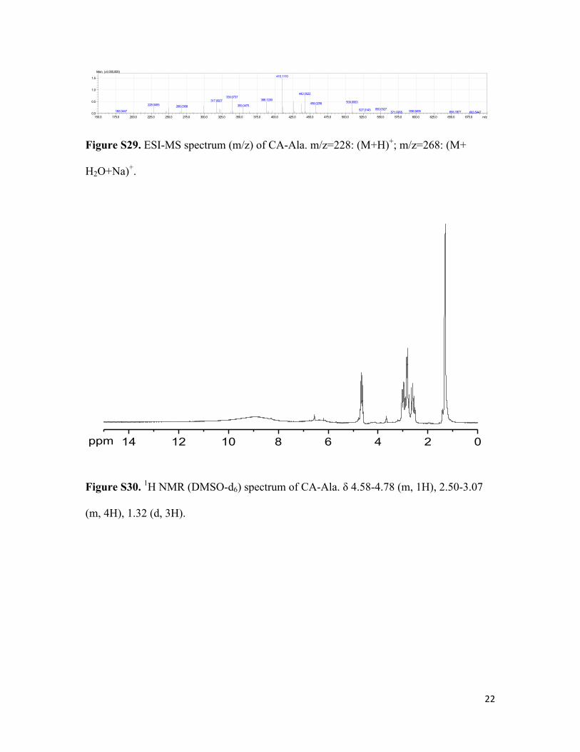
Figure S29. ESI-MS spectrum (m/z) of CA-Ala. m/z=228: (M+H)+; m/z=268: (M+
H2O+Na)+.
Figure S30. 1H NMR (DMSO-d6) spectrum of CA-Ala. δ 4.58-4.78 (m, 1H), 2.50-3.07
(m, 4H), 1.32 (d, 3H).
150.0 175.0 200.0 225.0 250.0 275.0 300.0 325.0 350.0 375.0 400.0 425.0 450.0 475.0 500.0 525.0 550.0 575.0 600.0 625.0 650.0 675.0 m/z0.0
0.5
1.0
1.5
Inten. (x1,000,000)410.1110
442.0522339.0737
388.1290317.0927 509.0063458.0258228.0455 355.0475268.0368550.0327527.0143182.0447 598.0495 655.1877571.9765 683.5447
14 12 10 8 6 4 2 0ppm
23
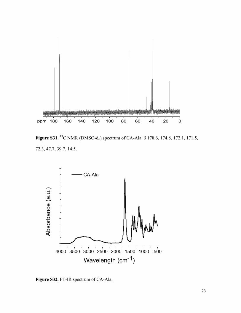
Figure S31. 13C NMR (DMSO-d6) spectrum of CA-Ala. δ 178.6, 174.8, 172.1, 171.5,
72.3, 47.7, 39.7, 14.5.
Figure S32. FT-IR spectrum of CA-Ala.
180 160 140 120 100 80 60 40 20 0ppm
4000 3500 3000 2500 2000 1500 1000 500
Abs
orba
nce
(a.u
.)
Wavelength (cm-1)
CA-Ala
24
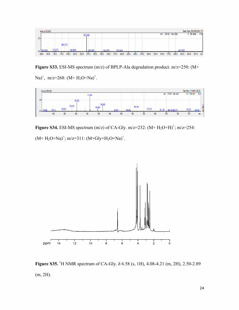
Figure S33. ESI-MS spectrum (m/z) of BPLP-Ala degradation product. m/z=250: (M+
Na)+, m/z=268: (M+ H2O+Na)+.
Figure S34. ESI-MS spectrum (m/z) of CA-Gly. m/z=232: (M+ H2O+H)+; m/z=254:
(M+ H2O+Na)+; m/z=311: (M+Gly+H2O+Na)+.
Figure S35. 1H NMR spectrum of CA-Gly. δ 6.58 (s, 1H), 4.08-4.21 (m, 2H), 2.50-2.89
(m, 2H).

25
Figure S36. 13C NMR spectrum of CA-Gly. δ 175.3, 172.0, 169.0, 166.7, 140.9, 72.8,
39.8, 34.9.
Figure S37. Spatial conformation of CA-Gly. Dihedral angles of any four atoms are listed on the right side.

26
Figure S38 Time-resolved fluorescence spectra of CA-Cys with a single exponential fitting (top), output lifetimes (inset), and the fitting residue distribution (bottom)
Figure S39. Time-resolved fluorescence spectra of CA-Ala with a single exponential fitting (top), output lifetimes (inset), and the fitting residue distribution (bottom)

27
Figure S40. Time-resolved fluorescence spectra of CA-Ala with a double exponential fitting (top), output lifetimes (inset), and the fitting residue distribution (bottom)
Figure S41. Time-resolved fluorescence spectra of CA-Ala with a triple exponential fitting (top), output lifetimes (inset), and the fitting residue distribution (bottom)

28
REFERENCES [1] J.Yang,Y.Zhang,S.Gautam,L.Liu,J.Dey,W.Chen,R.P.Mason,C.A.Serrano,K.A.
Schug,L.Tang,ProceedingsoftheNationalAcademyofSciencesoftheUnitedStatesofAmerica2009,106,10086-10091.
[2] M.J.Frisch,G.W.Trucks,H.B.Schlegel,G.E.Scuseria,M.A.Robb,J.R.Cheeseman,G.Scalmani,V.Barone,B.Mennucci,G.A.Petersson,H.Nakatsuji,M.Caricato,X.Li,H.P.Hratchian,A.F.Izmaylov,J.Bloino,G.Zheng,J.L.Sonnenberg,M.Hada,M.Ehara,K.Toyota,R.Fukuda,J.Hasegawa,M.Ishida,T.Nakajima,Y.Honda,O.Kitao,H.Nakai,T.Vreven,J.A.MontgomeryJr.,J.E.Peralta,F.Ogliaro,M.J.Bearpark,J.Heyd,E.N.Brothers,K.N.Kudin,V.N.Staroverov,R.Kobayashi,J.Normand,K.Raghavachari,A.P.Rendell,J.C.Burant,S.S.Iyengar,J.Tomasi,M.Cossi,N.Rega,N.J.Millam,M.Klene,J.E.Knox,J.B.Cross,V.Bakken,C.Adamo,J.Jaramillo,R.Gomperts,R.E.Stratmann,O.Yazyev,A.J.Austin,R.Cammi,C.Pomelli,J.W.Ochterski,R.L.Martin,K.Morokuma,V.G.Zakrzewski,G.A.Voth,P.Salvador,J.J.Dannenberg,S.Dapprich,A.D.Daniels,Ö.Farkas,J.B.Foresman,J.V.Ortiz,J.Cioslowski,D.J.Fox,Gaussian,Inc.,Wallingford,CT,USA,2009.
[3] R.Krishnan,J.S.Binkley,R.Seeger,J.A.Pople,TheJournalofChemicalPhysics1980,72,650-654.
[4] R.Ditchfield,W.J.Hehre,J.A.Pople,TheJournalofChemicalPhysics1971,54,724-728.[5] J.Kruszewski,T.M.Krygowski,TetrahedronLetters1972,13,3839-3842.[6] A.P.Demchenko,Luminescence:thejournalofbiologicalandchemicalluminescence
2002,17,19-42.
Related Documents


