iii Synthesis and Characterization of Ammonium Ionenes Containing Hydrogen Bonding Functionalities Mana Tamami Dissertation submitted to the faculty of the Virginia Polytechnic Institute and State University in partial fulfillment of the requirements for the degree of Doctor of Philosophy in Chemistry Timothy E. Long, Chair Garth L. Wilkes Robert B. Moore S. Richard Turner Susan E. Duncan December 6, 2012 Blacksburg, Virginia Keywords: Segmented ionenes, non-segmented ionenes, nucleobase, hydrogen bonding, Michael addition, self-healing, bio-adhesion, molecular recognition, step-growth polymerization, non- covalent interactions Copyright © 2012, Mana Tamami

Welcome message from author
This document is posted to help you gain knowledge. Please leave a comment to let me know what you think about it! Share it to your friends and learn new things together.
Transcript

iii
Synthesis and Characterization of Ammonium Ionenes Containing Hydrogen Bonding Functionalities
Mana Tamami
Dissertation submitted to the faculty of the Virginia Polytechnic Institute and State University in partial fulfillment of the requirements for the degree of
Doctor of Philosophy
in Chemistry
Timothy E. Long, Chair
Garth L. Wilkes
Robert B. Moore
S. Richard Turner
Susan E. Duncan
December 6, 2012
Blacksburg, Virginia
Keywords: Segmented ionenes, non-segmented ionenes, nucleobase, hydrogen bonding, Michael addition, self-healing, bio-adhesion, molecular recognition, step-growth polymerization, non-
covalent interactions
Copyright © 2012, Mana Tamami

i.
Synthesis and Characterization of Ammonium Ionenes Containing Hydrogen Bonding Functionalities
Mana Tamami
Abstract
Ammonium ionenes are polycations that have quaternary nitrogens in their macromolecular
backbone and are synthesized via step-growth polymerization technique. They offer interesting
coulombic properties, and the synthetic design provides control over charge density. Non-
covalent interactions including nucleobase hydrogen bonding and electrostatics were studied in
ammonium ionenes. The non-covalent interactions are expected to increase the effective
molecular weight of polymeric precursors and induce microphase separation due to
intermolecular associations. The influence of non-covalent interactions on structure-property
relationships of ammonium ionenes were studied regarding mechanical (tensile, DMA), thermal
(DSC, TGA), and morphological (AFM, SAXS) properties.
Hydrogen bonding interaction (10-40 kJ/mol) was introduced using DNA nucleobase pairs
such as adenine and thymine. Novel adenine and thymine functionalized segmented and non-
segmented ammonium ionenes were successfully synthesized using Michael addition chemistry.
In non-segmented systems, we investigated the influence of spacer length on homoassociation
and heteroassociation of complementary nucleobase-containing ionenes. Based on DSC
analyses, complementary non-segmented ionenes made miscible blends. The Tgs of ionene
blends with shorter spacer length (4 bonds between the nucleobase and secondary amine in the

iii
polymer backbone) followed the Fox equation, which indicated no intermolecular interactions.
The longer alkyl spacer (9 bonds between nucleobase and secondary amine in the polymer
backbone) provided efficient flexibility for the self-assembly process to occur. Thus, increasing
the spacer length from 4-bonds to 9-bonds, the Tgs of the blends deviated from both Fox and
Gordon-Taylor equations and demonstrated the presence of hydrogen bonding interactions.
In segmented systems, we investigated the association between nucleobase-containing
ionenes and their complementary guest molecules. Job’s method revealed a 1:1 stoichiometry for
the hydrogen-bonded complexes. These association constants for the 1:1 complexes, based on
the Benesi-Hildebrand model were 94 and 130 M-1 respectively, which were in agreement with
literature values for adenine and thymine nucleobase pairs (10-100 M-1). DSC thermograms
confirmed no macrophase separation for 1:1 [ionene-A/T]:[guest molecule] complexes based on
the disappearance of the melting peak of the guest molecule. Morphological studies including
atomic force microscopy (AFM) demonstrated a reduced degree of microphase separation for the
1:1 complexes due to the disruption of adenine-adenine or thymine-thymine interactions.
Poly(dimethyl siloxane)-based ammonium ionenes having various hard segment contents
were synthesized. The charge density or hard segment content was tuned for appropriate
application using low molecular weight monomer. The change in hard segment content had a
profound effect on thermal, mechanical, rheological, and gas permeability. Microphase
separation was confirmed using DSC and DMA in these systems. DMA showed that the rubbery
plateau modulus extended to higher temperatures with increasing hard segment content. Tensile
analysis demonstrated systematic increase in modulus of PDMS-ionenes with increasing hard
segment content. Oxygen transmission rates decreased linearly as the wt% hard segment
increased.

iv
Acknowledgements
I would like to thank my advisor, Prof. Timothy E. Long, for his guidance, encouragement,
and support over the course of my graduate career. I am grateful to work under his supervision;
he taught me polymer science, to be a good researcher, presenter, and a teacher. I would also like
to thank my committee members for their participation and support. I would like to express my
special thanks to Prof. Garth Wilkes for his helpful discussions and constant guidance. He has
been an insightful and helpful committee member and teacher during my graduate career. I
would never forget his kindness and support throughout my graduate career. I would also like to
thank Prof. S. Richard Turner for his helpful guidance and helping me with job opportunities in
industry. I would like to thank Prof. Robert B. Moore for his guidance and close research
collaboration. I would also like to thank Prof. Susan Duncan for her helpful guidance and
direction during the MILES program. I would like to thank Victoria Long for teaching me
technical writing skills and organizing summer group meeting events. I would also like to thank
Naya Sou, Valerie Owens, Laurie Good, and Tammy Jo Hiner. The staffs at Virginia Tech have
been a great resource. I would especially like to acknowledge Steve McCartney and John
McIntosh for their help with AFM and TEM imaging. Geno Iannacone, Hugo Azurmendi, and
John Burleson have also been very helpful with NMR spectroscopy.
I would like to thank my manager, Dr. Guiru Zhang during the three-month summer
internship program at Proctor & Gamble in Cincinnati, OH in 2010. He was a knowledgeable
scientist and a great mentor. I also would like to thank our collaborators in SHIELD project
including Dr. Vishnu Baba Sundaresan and Andrew Morgan from Virginia Commonwealth
University. I would like to thank collaborations with Prof. Karen Winey and David Salas-de la

v
Cruz from University of Pennsylvania. I acknowledge the funding source from Kimberly-Clark
and special thanks to Dr. Clay Bunyard from Kimberly-Clark and Prof. Michael Rubinstein from
the University of North Carolina as a consultant on the project.
I would like to thank all my group members, previous and present, for their advice,
encouragement, discussions, time, and support including Dr. Sharlene Williams, Dr. Rebecca
Huyck, Erika Borgerding, Dr. Gozde Ozturk, Dr. Tomonori Saito, Dr. Akshay Kokil, Dr. Sean
Ramirez, Dr. Matthew Hunley, Dr. Mathew Green, Dr. Mathew Cashion, Dr. Askim Senyurt, Dr.
Bill Heath, Dr. Emily Anderson, Dr. Andy Duncan, Dr. Steve June, Dr. Shijing Cheng, Tianyu
Wu, Nancy Zhang, Dr. Renlong Gao, Ali Nebipasagil, Dr. Philippe Bissel, Dr. Takeo Suga, Dr.
John Layman, Mike Allen, Sean Hemp, Dr. Adam Smith, Keren Zhang, Chanika Jangu, Ashley
Nelson, Alex Fersner, Dan Buckwalter, Dr. Daisuke Yamamato, Dr. Erin Murphy, Evan
Margaretta, David Inglefield, and Alison Schultz.
Finally, I am forever grateful to my family: Afsaneh, Bahman, and Mehrnaz for their
continued love and support in my life endeavors. I am deeply thankful to my father, Prof.
Bahman Tamami, he is my role model, a great mentor and a true scientist. Words cannot express
my love for him. With all my heart I would like to make him proud and thank him for all his
support throughout the years. I cannot ever forget my parents’ sacrifices to provide me
encouragement and motivation to pursue my goals. I would be nothing without them on the
journey of life. Finally, I would like to thank my very dear friend Dr. Anton Sizovs for his help
and support during the hardest times of my graduate career, he was my inspiration.

vi
TABLE OF CONTENTS
CHAPTER 1. INTRODUCTION............................................................................................. 1
1.1. DISSERTATION OVERVIEW .................................................................................................... 1
CHAPTER 2. ROLE OF INTERMOLECULAR INTERACTIONS IN ADHESIVE DESIGN………………. ................................................................................................................ 3
2.1. ABSTRACT ............................................................................................................................. 3
2.2. DEFINITION OF ADHESION ..................................................................................................... 3
2.3. PHYSICAL ADHESION ............................................................................................................. 4
2.3.1. Adhesion using van der Waals interactions .................................................................. 4
2.3.2. Adhesion using supramolecular interactions................................................................ 6
2.3.3. Adhesion using hydrogen bonding interactions ............................................................ 9
2.4. ADHESIVE POLYMERS IN BIO-RELATED FIELDS .................................................................... 12
2.4.1. Polymers used as substrates for cell adhesion ........................................................... 12
2.4.2. Polymers used for mucosal adhesion .......................................................................... 15
2.5. CONCLUSIONS ..................................................................................................................... 15
2.6. REFERENCES ....................................................................................................................... 16
CHAPTER 3. EFFECT OF SPACER LENGTH ON ASSOCIATION OF NUCLEOBASE-CONTAINING AMMONIUM IONENES................................................... 19
3.1. ABSTRACT ........................................................................................................................... 19
3.2. INTRODUCTION.................................................................................................................... 20
3.3. EXPERIMENTAL ................................................................................................................... 22
3.3.1. Materials ..................................................................................................................... 22
3.3.2. Instrumentation ........................................................................................................... 22
3.3.3. Synthesis of 4-(bis(3-(dimethylamino)propyl)amino)-4-oxobutyl acrylate ................ 23
3.3.4. Synthesis of N,N-bis(3-(dimethylamino)propyl)acrylamide ....................................... 24
3.3.5. Synthesis of non-segmented acrylate- and acrylamide-based ionene precursor ........ 25
3.3.6. Synthesis of non-segmented nucleobase-containing ionene (9-bond spacer)............. 25
3.3.7. Synthesis of non-segmented nucleobase-containing ionene (4-bond spacer)............. 26
3.3.8. Preparation of ionene blends ...................................................................................... 27
3.4. RESULTS AND DISCUSSION .................................................................................................. 27
3.4.1. Synthesis of Non-segmented Nucleobase-Functionalized Ionene Homopolymers ..... 27
3.4.2. Infrared Spectroscopy ................................................................................................. 31
3.4.3. Thermal Transitions .................................................................................................... 32
3.4.4. Morphology ................................................................................................................. 36
3.5. CONCLUSIONS ..................................................................................................................... 37
3.6. REFERENCES ....................................................................................................................... 38
CHAPTER 4. NUCLEOBASE SELF-ASSEMBLY IN SEGMENTED POLY(ETHYLENE GLYCOL)-BASED AMMONIUM IONENES ..................................... 45
4.1. ABSTRACT ........................................................................................................................... 45

vii
4.2. INTRODUCTION.................................................................................................................... 46
4.3. EXPERIMENTAL ................................................................................................................... 48
4.3.1. Materials ..................................................................................................................... 48
4.3.2. Instrumentation ........................................................................................................... 48
4.3.3. Synthesis of N,N-bis(3-(dimethylamino)propyl)-4-hydroxybutanamide ..................... 49
4.3.4. Synthesis of 4-(bis(3-(dimethylamino)propyl)amino)-4-oxobutyl acrylate ................ 50
4.3.5. Synthesis of Bromine End-Capped PEG (Br-PEG-Br) ............................................... 51
4.3.6. Synthesis of n-Butyl thymine (nBT) Guest Molecule................................................... 51
4.3.7. Synthesis of n-Butyl adenine (nBA) Guest Molecule .................................................. 52
4.3.8. Synthesis of Acrylate-Containing PEG-Based Ionene Precursor ............................... 52
4.3.9. Synthesis of Nucleobase-Containing PEG-Based Ionene using Post-Polymerization Functionalization .................................................................................................................. 53
4.3.10. Preparation of Ionene Blend with Guest Molecules ................................................ 53
4.4. RESULTS AND DISCUSSION .................................................................................................. 54
4.4.1. Synthesis of Nucleobase Functionalized PEG-Based Ionene Homopolymers ............ 54
4.4.2. 1H NMR Titrations ...................................................................................................... 56
4.4.3. Thermal Transitions .................................................................................................... 64
4.4.4. Morphology ................................................................................................................. 66
4.5. CONCLUSIONS ..................................................................................................................... 68
4.6. REFERENCES ....................................................................................................................... 68
CHAPTER 5. SYNTHESIS AND CHARACTERIZATION OF SILICONE-BASED AMMONIUM IONENES AS CANDIDATES FOR SELF-HEALING POLYMER S ......... 74
5.1. ABSTRACT ........................................................................................................................... 74
5.2. INTRODUCTION.................................................................................................................... 75
5.3. EXPERIMENTAL. .................................................................................................................. 77
5.3.1. Materials ..................................................................................................................... 77
5.3.2. Instrumentation ........................................................................................................... 77
5.3.3. Synthesis of bromine end-capped poly(dimethyl siloxane) (Br-PDMS-Br) ................ 78
5.3.4. Synthesis of poly(dimethyl siloxane)-based ionene homopolymer .............................. 79
5.3.5. Synthesis of poly(dimethyl siloxane)-based ionene random copolymer ..................... 80
5.4. RESULTS AND DISCUSSION .................................................................................................. 81
5.4.1. Synthesis of PDMS-based ionenes .............................................................................. 81
5.4.2. Thermal Transitions .................................................................................................... 82
5.4.3. Dynamic Mechanical Analysis (DMA) ....................................................................... 84
5.4.4. Tensile test .................................................................................................................. 85
5.4.5. Rheology ..................................................................................................................... 86
5.4.6. Oxygen Transmission Rate ......................................................................................... 88
5.5. CONCLUSIONS ..................................................................................................................... 89
5.6. REFERENCES ....................................................................................................................... 91
CHAPTER 6. OVERALL CONCLUSIONS ........................................................................ 92

viii
CHAPTER 7. SUGGESTED FUTURE WORK .................................................................. 94
7.1. SYNTHESIS AND CHARACTERIZATION OF PEG-BASED CYTOSINE AND GUANINE-CONTAINING
AMMONIUM IONENES.................................................................................................................. 94
7.2. SYNTHESIS AND CHARACTERIZATION OF IONENES WITH POTENTIAL APPLICATION IN
ADHESIVES ................................................................................................................................. 95
7.3. SYNTHESIS AND CHARACTERIZATION OF SELF-HEALING AMMONIUM IONENES .................... 96

i.
L IST OF FIGURES
FIGURE 2.1. FIBRILLAR STRUCTURE ON THE BOTTOM OF GECKO’S FOOT. A) VENTRAL VIEW OF
TOKAY GECKO WHILE CLIMBING ON THE GLASS B) VENTRAL VIEW OF GECKO’S FOOT WITH
ADHESIVE LAMELLAE C) SINGLE LAMELLAE WITH AN ARRAY OF INDIVIDUAL SETAES D)SINGLE
SETA WITH BRANCHED STRUCTURE AT THE END E) SPATULAR TIPS AT THE ENDS OF SETA.[9] .......... 5 FIGURE 2.2. A) STRUCTURE OF POLYISOBUTYLENE (PIBUT) CONTAINING BIS-UREA MOIETY. B) SUPRAMOLECULAR STRUCTURE OF PIBUT IN SOLUTION
[36] ............................................................ 7
FIGURE 2.3. SYNTHESIS OF ADENINE- AND THYMINE-CONTAINING POLY(N-BUTYL ACRYLATE ) COPOLYMERS
[38] ............................................................................................................................... 8
FIGURE 2.4. MOLECULAR RECOGNITION BETWEEN ADENINE-FUNCTIONALIZED SILICONE SURFACE
AND THYMINE -CONTAINING POLYSTYRENE[40] ................................................................................. 9
FIGURE 2.5. SYNTHESIS OF PHOTO-CURABLE ACRYLIC COPOLYMER CONTAINING HYDROGEN
BONDING FUNCTIONALITY[42] ......................................................................................................... 10
FIGURE 2.6.CHEMICAL STRUCTURE OF PVA-G-NIPAM [48] ............................................................ 11
FIGURE 2.7. REPRESENTATION OF CROSSLINKED EPOXY NETWORK. SOLID LINE REPRESENTS
COVALENT BONDS AND DOTTED LINES REPRESENT CONTINUATION OF COVALENT BONDS[47] ......... 11
FIGURE 2.8. REPRESENTATION OF COUPLING REACTION AND GRAFT POLYMERIZATION[58] ............ 14
FIGURE 3.1. VARIABLE TEMPERATURE FT-IR SPECTRA IN THE 1550-1750 CM-1
REGION FOR THE
LONGER SPACER IONENE BLEND (TOP) AND SHORTER SPACER IONENE BLEND (BOTTOM) ............... 32 FIGURE 3.2. TG VERSUS COMPOSITION CURVE OF EXPERIMENTAL DATA AND FOX FITTING EQUATION
FOR SHORTER SPACER IONENE BLENDS ........................................................................................... 33
FIGURE 3.3. TG VERSUS COMPOSITION CURVES FROM EXPERIMENTAL DATA AND DIFFERENT FITTING
EQUATIONS FOR LONGER SPACER IONENE BLENDS ......................................................................... 35
FIGURE 3.4. AFM PHASE IMAGES OF IONENE HOMOPOLYMERS AND BLENDS HAVING SHORTER
SPACER (BOTTOM IMAGE) AND LONGER SPACER (TOP IMAGE) ........................................................ 36
FIGURE 4.1. BENESI-HILDEBRAND PLOT OF IONENE-A AND UOP+ GUEST MOLECULE ASSOCIATION
IN CDCL3 ....................................................................................................................................... 58
FIGURE 4.2. JOB’S PLOT TO DETERMINE THE STOICHIOMETRY OF (A) [NBT]:[NBA], (B) [IONENE-T]:[NBA] AND [IONENE-A]:[ NBT] COMPLEXES IN CDCL3 ............................................................. 59
FIGURE 4.3. (A) NONLINEAR RELATIONSHIP BETWEEN INDUCE CHANGE FOR THYMINE NH
CHEMICAL SHIFT AND IONENE-A CONCENTRATION, (B) BENESI-HILDEBRAND PLOT OF IONENE-A
AND NBT GUEST MOLECULE ASSOCIATION IN CDCL3 .................................................................... 61
FIGURE 4.4. (A) NONLINEAR RELATIONSHIP BETWEEN INDUCE CHANGE FOR ADENINE NH2
CHEMICAL SHIFT AND IONENE-T CONCENTRATION, (B) BENESI-HILDEBRAND PLOT OF IONENE-T
AND NBA GUEST MOLECULE ASSOCIATION IN CDCL3 .................................................................... 62
FIGURE 4.5. (A) NONLINEAR RELATIONSHIP BETWEEN INDUCE CHANGE FOR ADENINE NH2
CHEMICAL SHIFT AND NBT CONCENTRATION, (B) BENESI-HILDEBRAND PLOT OF NBT AND NBA
GUEST MOLECULE ASSOCIATION IN CDCL3 .................................................................................... 63
FIGURE 4.6. DSC THERMOGRAMS OF IONENE-A HOMOPOLYMER AND 1:1 COMPLEX WITH NBT. SECOND HEATING CYCLE IS SHOWN. .............................................................................................. 65

x
FIGURE 4.7. AFM PHASE IMAGES OF NUCLEOBASE-CONTAINING IONENE HOMOPOLYMERS (1,3) AND
THEIR 1:1 COMPLEXES WITH NBT AND NBA (2,4) ........................................................................... 67
FIGURE 4.8. SAXS DATA FOR NUCLEOBASE-CONTAINING IONENE HOMOPOLYMERS AND BLENDS . 67
FIGURE 5.1. 1H NMR SPECTRUM OF BROMINE-TERMINATED PDMS .............................................. 79
FIGURE 5.2. 1H NMR SPECTRUM OF PDMS-BASED IONENE HOMOPOLYMER ................................. 80
FIGURE 5.3. DMA OF PDMS-BASED IONENE HAVING 5 WT% HS .................................................. 85
FIGURE 5.4. TENSILE ANALYSIS OF PDMS-BASED IONENE HAVING 5 WT% AND 15 WT% HS ........ 86 FIGURE 5.5. MELT VISCOSITY OF PDMS-BASED IONENES HAVING 5 WT% AND 15 WT% HS ......... 87 FIGURE 5.6. MASTER CURVE OF PDMS-BASED IONENE WITH 5WT% HS ....................................... 88
FIGURE 5.7. OTR VALUES OF PDMS-BASED IONENES WITH VARIOUS HS CONTENTS .................... 89 FIGURE 7.1. SYNTHESIS OF CYTOSINE AND GUANINE-CONTAINING POLY(ETHYLENE GLYCOL)-BASED
AMMONIUM IONENES...................................................................................................................... 95
FIGURE 7.2. SYNTHESIS OF OH-CONTAINING PEG-BASED AMMONIUM IONENE ............................. 96
xi
L IST OF SCHEMES SCHEME 3.1. SYNTHESIS OF ACRYLATE-CONTAINING DITERTIARY AMINE MONOMER (A) AND
ACRYLAMIDE -CONTAINING DITERTIARY AMINE MONOMER (B) ...................................................... 28
SCHEME 3.2. SYNTHESIS OF NUCLEOBASE-CONTAINING IONENES HAVING 4-BOND SPACER ........... 29 SCHEME 3.3. SYNTHESIS OF NUCLEOBASE-CONTAINING IONENES HAVING 9-BOND SPACER ........... 30 SCHEME 4.1. SYNTHESIS OF ACRYLIC DITERTIARY AMINE MONOMER (A), BROMINE END-CAPPED
1000 G/MOL PEG (B) ..................................................................................................................... 54
SCHEME 4.2. POST-POLYMERIZATION FUNCTIONALIZATION OF PEG-BASED IONENE ..................... 56 SCHEME 5.1. SYNTHESIS OF BROMINE TERMINATED PDMS ........................................................... 82
SCHEME 5.2. SYNTHESIS OF PDMS-BASED SEGMENTED IONENE COPOLYMERS ............................. 83

xii
L IST OF TABLES
TABLE 3.1. GLASS TRANSITION TEMPERATURES OF IONENES WITH 4-BOND SPACER AND 9-BOND
SPACER ........................................................................................................................................... 35
TABLE 4.1. THERMAL TRANSITIONS OF NUCLEOBASE-CONTAINING IONENES AND THEIR BLENDS .. 65
TABLE 5.1. SEGMENTED COPOLYMER COMPOSITIONS BASED ON MOLAR EQUIVALENTS OF
MONOMER AND HS/SS CONTENT ................................................................................................... 82
TABLE 5.2. TGA AND DSC RESULTS OF SEGMENTED PDMS-BASED IONENES ............................... 84
TABLE 5.3. TENSILE DATA FOR PDMS-IONENE FILMS AS A FUNCTION OF WT% HS ....................... 86

1
Chapter 1. Introduction
1.1. Dissertation Overview
The effect of non-covalent interactions, electrostatics and hydrogen bonding on structure-
property relationships of ammonium ionenes was studied in this dissertation. Following this
chapter, the role of hydrogen bonding interactions and supramolecular assembly on the design of
adhesives was reviewed. The third chapter describes the synthesis and characterization of non-
segmented adenine- and thymine-containing ionenes having various side-chain spacer lengths.
The spacer length is the distance between ionene backbone and nucleobase in the side chain. In
the beginning, the synthesis of novel ditertiary amine monomers containing vinyl side groups,
their subsequent polymerization with an alkyl dihalide, followed by the post-polymerization
functionalization with nucleobases is discussed. Following the functionalization, the effect of
spacer length on the complementary hydrogen bonding interaction in the blends was
investigated. This investigation was mainly on studying the trend in glass transition temperatures
of blends containing various molar ratios of complementary ionene components.
The fourth chapter illustrates the synthesis and characterization of segmented poly(ethylene
glycol)-based ammonium ionenes functionalized with adenine and thymine nucleobases. In the
beginning of this chapter the synthesis of PEG-based ionene followed by the post-polymerization
functionalization with nucleobases is explained. Due to enhanced solubility of the segmented
systems (compared to non-segmented) in nonpolar solvents such as chloroform, an extensive 1H
NMR titration studies were performed in CDCl3. Using 1H NMR titatrion results, the
stoichiometry of 1:1 complexes between nucleobase-containing ionenes and complementary
guest molecules was measured by Job’s method. In addition, association constants were

2
calculated for 1:1 complexes and compared with current literature values for adenine-thymine
association.
The fifth chapter focuses on the synthesis and structure-property relationships of poly(dimethyl
siloxane)-based ionene copolymers. The thermomechanical properties as well as gas
permeability were evaluated as a function of hard segment content. This project was based on
collaboration between Virginia CommonWealth University and Astro Terra Corp. One of the
project objectives was to synthesize PDMS-based ionenes for self-healing inflatable modules as
an alternative to Surlyn®. This project was funded through NASA STTR program administered
by Johnson Space Center under the Contract No. NNX11CI22P for the project titled "Self-
healing Inflatable Extraterrestrial shieLD (SHIELD)"
The sixth chapter summarizes the dissertation accomplishments and the seventh chapter
describes potential future work.

3
Chapter 2. Role of Intermolecular Interactions in Adhesive Design Mana Tamami and Timothy E. Long*
Macromolecules and Interfaces Institute, Department of Chemistry, Virginia Tech, Blacksburg,
VA 24061, USA
*E-mail address: [email protected]
2.1. Abstract
Adhesion involves molecular interactions at the interface between two surfaces. The focus of this
review article is on physical adhesion that involves the use of non-covalent interactions. These
interactions include van der Waals, electrostatics, hydrogen bonding, and supramolecular
associations. Herein, we briefly discuss the gecko feet inspired high adhesive superhydrophobic
surface properties. We then mainly summarize the recent work in the use of polymers having
supramolecular interactions and hydrogen bonding interactions for adhesion applications. In the
end, we cover adhesive polymers that are applicable to biomedical areas.
2.2. Definition of adhesion
Adhesion involves the tendency of dissimilar atoms or molecules to stick to each other and
cohesion relates to like materials sticking together. The area of adhesion focuses on formation of
adhesion or cohesion, characterization of the adhesive or cohesive interfaces, destruction of the
interfaces, and the failure analysis of interfaces.[1] Based on the type of bonding (physical,
chemical, mechanical) across the interface, the adhesion or cohesion is categorized. The physical
adhesion is the weakest interfacial force and is due to van der Waals forces, electrostatic
interactions, supramolecular interactions, and hydrogen bonding. Chemical adhesion involves
covalent, metallic, and chelation bonding. Mechanical adhesion is the strongest among all, is

4
very common, and involves the adhesive penetrating into the adherent and become mechanically
interlocked, for example, dental cements that fill in the coarseness of the castings and help to
retain them.[2, 3]
2.3. Physical adhesion
2.3.1. Adhesion using van der Waals interactions
Geckos are lizards that possess unique adhesive characteristic known in nature. Geckos along
with many other small insects use seta or fibrillar structures on their feet to adhere to different
surfaces (Figure 2.1).[4, 5] Setal adhesion has unique properties compared to other common
adhesives like pressure sensitive adhesives (PSAs). These properties include; adhesion surfaces
remaining clean and reusable, adhesion being directional, and adhesion having controlled “lift-
off mechanism”.[6, 7] Many experiments have been performed to investigate the possible
mechanism behind setal adhesion. The hypotheses were secretion of a glue, suction,
electrostatics, and intermolecular forces. However, enough evidence demonstrates that setal
adhesion mainly uses van der Waals interactions which are as a result of the size and shape of
tips and the adhesion is not governed by surface chemistry.[8] The van der Waals forces are
strong enough to allow the gecko to climb vertical walls. This type of adhesion has inspired
many researchers to develop synthetic materials that show unique properties similar to setal or
fibrillar adhesives.

5
Figure 2.1. Fibrillar structure on the bottom of gecko’s foot. A) Ventral view of tokay gecko while climbing on the glass B) Ventral view of gecko’s foot with adhesive lamellae C) Single lamellae with an array of individual setaes D)Single seta with branched structure at the end E)
Spatular tips at the ends of seta.[9]
Inspired by the high adhesive ability of gecko’s feet, Choi et al.[10] prepared hairy hard
poly(dimethyl siloxane) (PDMS) films containing nanopillars with controllable lengths using
nanoporous anodic aluminum oxide membranes as templates. They coated the glass surface with
nanostructured hairy PDMS and showed that the water droplets can adhere strongly to the glass
surface. The adhesive properties was due to the densely packed nanopillars by generating large
van der Waals forces between the large surface area and water molecules that are in close
contact.

6
2.3.2. Adhesion using supramolecular interactions
Hydrogen bonding in contrast to nondirectional interactions such as electrostatics, demonstrate
lower enthalpies (10-40 kj/mol) with greater specificity which induces molecular recognition.
The strength of hydrogen bonding interactions is highly dependent on the temperature, solvent,
humidity, and pH. Therefore, these interactions enable us to synthesize novel architectures that
are responsive to environmental parameters. Hydrogen bond containing polymers have many
advantages such as enhanced rheological properties due to decrease in melt-viscosity, increase in
modulus, tensile strength, polarity, and adhesion.
Recently hydrogen bonding interactions have been used to design supramolecular structures.[11,
12] Supramolecular chemistry in polymers involves the synthesis of macromolecules using
secondary interactions between small molecules (monomers) to develop polymer-like
structures.[13-15] These secondary interactions can be hydrogen bonding,[16] π-π interactions,[17]
metal coordination,[18] electrostatics, and van der Waals interactions. Many researchers have used
supramolecular hydrogen bonded polymers[19-30] for various applications; including formation of
large vesicles,[12] attach functional small molecules on polymers,[31] and reversibly adhere
polymers on to surfaces.[32]
In this section, we will mainly focus on supramolecular polymers that have application in
adhesion. Surprisingly, they are few reports on the use of supramolecular chemistry for
application in adhesion. One area of adhesives that supramolecuar chemistry can be applied is
pressure sensitive adhesives (PSAs). PSAs are usually made from lightly crosslinked polymer
networks with a low glass transition temperature (Tg),[7, 33, 34] and bond to many substrates upon
applying very low pressure.[35]

7
Courtois et al.[36] investigated the influence of supramolecular interactions on adhesive properties
of functionalized polyisobutene on steel and silicone surfaces. They prepared polyisobutene with
a bis-urea moiety in the middle of the chain (PIBUT) (Figure 2.2a). The hydrogen bonding
between bis-urea moieties induced supramolecular assembly leading to ordered pattern. They
showed that supramolecular polymers modified the rheological properties of low Tg
polyisobutene and have promising adhesive applications. PIBUT polymers can dissipate energy
upon adhesive debonding and make stronger interactions with substrates such as silicone
compared to acrylic-based PSAs.[37]
Figure 2.2. a) Structure of polyisobutylene (PIBUT) containing bis-urea moiety. b) Supramolecular structure of PIBUT in solution[36]
Supramolecular interactions are also present in systems containing complementary hydrogen
bonding moieties. One category of bio-inspired complementary units include DNA nucleobases
such as adenine, thymine, cytosine, and guanine. Long et al.[38] synthesized acrylic nucleobase-
containing copolymers using radical polymerization (Figure 2.3). They synthesized novel acrylic
adenine and thymine monomers using aza-Michael addition and then copolymerized with n-butyl
acrylate. Adenine-containing polyacrylates demonstrated unique morphologies due to adenine-
adenine π-π interactions. The adenine and thymine polymer blend showed the presence of

8
complementary hydrogen bonding leading to supramolecular structures. In order to measure peel
and shear strengths, a strip of PET film was coated with the hydrogen-bonded polymer (adenine
or thymine) and adhered to the same or complementary polymer coated on stainless steel
substrate. The hydrogen-bonded supramolecular polymers showed enhanced peel and shear
strengths (3-4 times) compared to acrylic acid- and 4-vinylpyridine- based polymer analogues.
Figure 2.3. Synthesis of adenine- and thymine-containing poly(n-butyl acrylate) copolymers[38]
In addition, a limited number of studies referred to supramolecular interactions on surfaces using
nucleobase pairs.[39] Long et al.[40] were first to report the modification of silicone surfaces with
adenine-containing triethoxysilane (ADPTES). They demonstrated the specific and reversible
adhesion of ADPTES silicon surface with complementary thymine-functionalized polystyrene
(PS-thymine). The reversibility of adhesion was examined using hydrogen bond disruptive
solvent (DMSO). The hydrogen bonding interactions were disrupted while rinsing the surface
with aprotic DMSO and were reformed following the removal of DMSO and addition of
chloroform (Figure 2.4). This behavior demonstrated the reversibility nature of the adenine and
thymine association. These polymers that show reversible interactions with solid surfaces have
potential application in releasable coatings and smart adhesives.

9
Figure 2.4. Molecular recognition between adenine-functionalized silicone surface and thymine-containing polystyrene[40]
2.3.3. Adhesion using hydrogen bonding interactions
Hydrogen bonding association provides strategies to increase the apparent molecular weight after
application. These interactions are used to design adhesives and prevent creep and cohesive
failure. Poly(acrylic acids) (PAAs) contain hydrogen bonding functionalities and are applied in
PSA formulations. However, one limitation in PAAs is that they can undergo thermal
crosslinking above 150 °C and form intermolecular anhydrides.[41] In hot melt pressure sensitive
adhesives (HMPSAs), crosslinking during processing is problematic, therefore PAAs have
limited utility. Long et al.[42] synthesized low Tg acrylic copolymers that were functionalized
with hydrogen bonding (urethane) groups and photo-reactive (cinnamate) functionalities for
HMPSAs application (Figure 2.5). The synergy of these groups resulted in higher peel values. In
addition the isothermal rheological studies showed that at 150 °C the copolymer was stable with
no crosslinking and therefore has potential in HMPSA application.

10
Figure 2.5. Synthesis of photo-curable acrylic copolymer containing hydrogen bonding functionality[42]
Another hot topic in adhesive research is the development of reversible adhesives. In some
applications we require debonding of adhesive from adherent when the adhesion is not required
at the time.[43] These applications can be removable labels, surface protection films, easily
placeable and removable notepaper. Researchers used many strategies to develop reversible
adhesives such as using fibrillar structure of a gecko foot,[44] and shape memory effect to induce
microscopic or macroscopic change for “self-peel”.[45-47] Another strategy is to use
theromosensitive polymer to achieve reversible adhesion properties. Hu et al.[48] synthesized
poly(vinyl alcohol)-g-N-isopropylacrylamide PVA-g-NIPAM as a novel thermosensitive
copolymer membrane with thermally induced adhesion around the lower critical solution
temperature (LCST) of 31 °C. At temperatures below LCST, the copolymer becomes more
hydrophilic and enhances the adhesion effect and at temperatures above LCST, the copolymer
became hydrophobic with decreased adhesion. The adhesive strength of PVA-g-NIPAM was
measured for the T-type peel adhesion toward the paper. The adhesive ability of the copolymer
was mainly due to the hydrogen bonding interaction between the PVA and cellulose of the paper.
O O
O OO
O
OH
a b c NCO
O O
O OO
O
OH
a b d
O
O
O
e
NH
O
O O
O OO
O
OH
a b d
O
O
O
e
NH
O
Cl
O
O O
O OO
O
O
a b d
O
O
O
e
NH
O O
(i)
(ii)

11
Figure 2.6.Chemical structure of PVA-g-NIPAM [48]
Another reversible adhesive system was designed by Xie et al.[49] where they prepared hydrogen
bonding-based epoxy thermosets (Figure 2.7). In order to have good adhesion at a solid interface,
interfacial contact and good molecular interactions are required. Xie et al. demonstrated that the
epoxy thermosets are ideal candidates for reversible adhesion. Firstly, the modulus of epoxy
thermosets would drop two orders of magnitude upon glass transition temperature, which would
lead to an effective interfacial contact with solid surface.[47] Secondly, the hydrogen bonding
moieties will provide the reversibility for adhesion. The adhesion between two identical polymer
surfaces was through interfacial hydrogen bonding interaction between the free hydroxyl groups
(H-bond doner) and oxygen atoms (H-bond acceptor) in the epoxy.
Figure 2.7. Representation of crosslinked epoxy network. Solid line represents covalent bonds and dotted lines represent continuation of covalent bonds[47]
A popular area of adhesion is based on bio-inspired hydrogen-bonded polymers. It has been
shown that Mussels can adhere to many organic and inorganic surfaces by producing 3,4-
n

12
dihydroxyphenyl-L-alanine which contains catechol groups.[50-53] Although the adhesion
mechanism is still not completely understood, but it is hypothesized that adhesion is due to
hydrogen bonding interactions between catechol groups and OH-containing substrates. Kaneko
et al.[54] synthesized Mussel-mimetic adhesive resin from copolymerization of 3,4-
dihydroxycinnamic acid (DHCA) and 4-hydroxycinnamic acid (4HCA) and confirmed it’s
adhesive properties. The chain-ends of the hyperbranched polymer resin contain catechol
moieties which are hydrogen bond donors and can strongly adhere to organic/inorganic surfaces.
Since this novel adhesive resin is made from biomass monomers, it is environmentally friendly
and non-toxic.
2.4. Adhesive polymers in bio-related fields
2.4.1. Polymers used as substrates for cell adhesion
One of the requirements in the design of many medical devices is to have patterned adhesion of
human or animal cells on artificial substrates. There are two routes to perform this process; one
way is to attach photoactive proteins or peptides to the substrate and the other way is to either
chemically modify the substrate or deposit thiols or silanes on the substrate to adhere
biomolecules. Both routes would lead to structured substrates that act as adhesion sites and cells
will attach to them via ligand/receptor interactions. Polymers have become unique substrates due
to the simplicity of cell adhesion process on to them and also have lower cost.
Welle and Gottwald[55] used commercially available polycarbonate, poly(methyl methacrylate)
(PMMA), and polystyrene as substrates for cell adhesion. They exposed the polymeric surfaces
to UV light and modified their physical behavior and chemical composition. This led to strong
adhesion of hepatocyte and fibroblast cells.

13
Most implant materials such as polymers, carbon fibers, and metals are nontoxic, biocompatible,
and do not degrade in the organism. However, their lifetime can be short due to the improper
mechanical contact between implant surface and the regenerating cells. Therefore it is necessary
to coat implant surfaces with cell-adhesive molecules or macromolecules to obtain strong
mechanical contact between cells and the surface. Kessler et al.[56] showed that functionalization
of PMMA surface coated with integrin-selected peptides effectively bind to osteoblast murine
and human cells compared to uncoated PMMAs. Ohashi and Dauskardt[57] studied the debonding
behavior of prosthetic-PMMA interface. They demonstrated that precoating the surface of the
implant with PMMA at higher temperatures would drastically enhance the adhesion and fatigue
resistance in both air and physiological conditions.
In order for the biomaterial to be used clinically, not only it needs to have excellent bulk
properties, but also surface properties play a major role as well. The initial response of body
organisms depends on biomaterial’s surface property. Poor adhesion between the biomaterial and
the tissue causes numerous complications including infection. However, most polymers need
surface modification to be used as biomaterials. One of the ways to do surface modification is by
grafting. Grafting can either be through coupling reaction between reactive polymers and
functionalized substrate polymer surface or it can be through graft polymerization of monomers.
Ikada[58] reviewed surface modification of polymers using different grafting techniques to obtain
lubricous, blood compatible or physiologically bioactive polymer surfaces (Figure 2.8).

14
Figure 2.8. Representation of coupling reaction and graft polymerization[58]
Surface modification is also applied to the area of gene delivery. DNA is usually condensed into
nanoparticle-sized complexes and is introduced to the culture media (liquid gene transfection or
LGT method). However, studies have shown that localized gene delivery to the targeted cells
using LGT method is not favorable.[59, 60] Another strategy is to use substrate-mediated delivery,
where DNA is attached to the surface of substrate and selective adhesion between the substrate-
supported DNA and adherent cells would occur.[61, 62] Liu et al.[63] designed high-strength
hydrogels with both hydrogen bonding and thermoresponsive characteristics. The hydrogels
were synthesized from copolymerization of N-isopropylacrylamide (NIPAM) and 2-vinyl-4,6-
diamino-1,3,5-triazine (VDT) and crosslinked with poly(ethylene glycol) diacrylate. The VDT
functionalities contributed to the formation of complementary hydrogen bonding interactions
between the substrate and nucleobase pairs.[64-66] The NIPAM components of the hydrogels
contributed to the thermoresponsiveness behavior and allowed the adhesion and detachment of
cells by temperature change.[67, 68]

15
2.4.2. Polymers used for mucosal adhesion
When polymeric materials adhere to mucosal tissues is called mucoadhesion. The mucoadhesive
polymers have been used to deliver drugs in a controlled-release dosage forms and enhance the
bioavailability of the drug for various mucosal tissues such as nasal, gastrointestinal, vaginal,
rectal, and ocular.[69-73] Mucoadhesive polymers need to have certain structural properties as
listed here; hydrogen bonding groups (hydroxyl, carboxyl, amino, sulphate), ionically charged,
high molecular weight, chain flexibility, higher surface energy to spread on mucos. From
mucoadhesive polymers, we can name poly(acrylic acid), poly(methacrylic acid), chitosan,
cellulose ethers, and sodium alginate. Park et al.[74] illustrated that poly(acrylic acid) hydrogel
interacts with mucin glycoproteins using hydrogen bonding interactions. At lower pHs the
carboxylic groups are protonated and show strong mucoadhesion, however, at higher pHs the
mucoadhesion decreases due to deprotonation of carboxylic groups.
2.5. Conclusions
In this review, we discussed the recent developments in the use of supramolecular chemisty and
hydrogen bonding interactions for application in adhesion. Although there were not many reports
on the use of supramolecular chemistry for application in adhesion, however, few examples were
reported on the use of complementary hydrogen bonding moieties to generate reversible and
smart adhesives. Several studies demonstrated the use of hydrogen bonding interactions for
adhesion on the surface. We also reviewed reports that studied polymers with unique properties
to adhere to cells. These adhesive polymers played a major role in the design of implant
materials.

16
2.6. References
(1) Marshall, S. J.; Bayne, S. C.; Baier, R.; Tomsia, A. P.; Marshall, G. W. Dent. Mater. 2010, 26, e11-6.
(2) Bayne, S.; Taylor, D.; Zardiackas, L., Biomaterials science. Brightstar Publishing: Chapel Hill, NC, 1992.
(3) Marshall, G.; Marshall, S., Biomaterials science for restorative dentistry. San Francisco: UCSF, 1999.
(4) Arzt, E.; Gorb, S.; Spolenak, R. Proc. Natl. Acad. Sci. U. S. A. 2003, 100, 10603-10606. (5) Scherge, M.; Gorb, S. N., Biological micro and nanotribology: Nature’s solutions.
Springer: Berlin, 2001. (6) Autumn, K.; Liang, Y. A.; Hsleh, S. T.; Zesch, W.; Chan, W. P.; Kenny, T. W.; Fearilng,
R.; Full, R. J. Nature (London) 2000, 405, 681-686. (7) Creton, C. MRS Bull. 2003, 28, 434-439. (8) Autumn, K.; Sitti, M.; Liang, Y. A.; Peattie, A. M.; Hansen, W. R.; Sponberg, S.; Kenny,
T. W.; Fearing, R.; Israelachvili, J. N.; Full, R. J. Proc. Natl. Acad. Sci. U. S. A. 2002, 99, 12252-12256.
(9) Liu, K.; Yao, X.; Jiang, L. Chem. Soc. Rev. 2010, 39, (8), 3240-3255. (10) Cho, W. K.; Choi, I. S. Adv. Funct. Mater. 2008, 18, 1089-1096. (11) Brunsveld, L.; Folmer, B. J. B.; Meijer, E. W.; Sijbesma, R. P. Chem. Rev 2001, 101,
4071-4097. (12) Ilhan, F.; Galow, T. H.; Gray, M.; Clavier, G.; Rotello, V. M. J. Am. Chem. Soc. 2000,
122, 5895-5896. (13) Bosman, A. W.; Brunsveld, L.; Folmer, B. J. B.; Sijbesma, R. P.; Meijer, E. W.
Macromol. Symp. 2003, 201, 143-154. (14) Shimizu, L. S. Polym. Int. 2007, 56, 444-452. (15) Fox, J. D.; Rowan, S. J. Macromolecules (Washington, DC, U. S.) 2009, 42, 6823-6835. (16) Sijbesma, R. P.; Beijer, F. H.; Brunsveld, L.; Folmer, B. J. B.; Hirschberg, J. H. K. K.;
Lange, R. F. M.; Lowe, J. K. L.; Meijer, E. W. Science 1997, 278, 1601-1604. (17) Adam, D.; Schuhmacher, P.; Simmerer, J.; Haeussling, L.; Siemensmeyer, K.; Etzbach,
K. H.; Ringsdorf, H.; Haarer, D. Nature (London) 1994, 371, 141-3. (18) Michelsen, U.; Hunter, C. A. Angew. Chem., Int. Ed. 2000, 39, 764-767. (19) Keeling, D. L.; Oxtoby, N. S.; Wilson, C.; Humphry, M. J.; Champness, N. R.; Beton, P.
H. Nano Lett. 2003, 3, 9-12. (20) De, F. S.; Miura, A.; Yao, S.; Chen, Z.; Wuerthner, F.; Jonkheijm, P.; Schenning, A. P.
H. J.; Meijer, E. W.; De, S. F. C. Nano Lett. 2005, 5, 77-81. (21) Kihara, H.; Kato, T.; Uryu, T.; Frechet, J. M. J. Chem. Mater. 1996, 8, 961-8. (22) Kato, T.; Frechet, J. M. J. Macromolecules 1989, 22, 3818-19. (23) MacDonald, J. C.; Whitesides, G. M. Chem. Rev. (Washington, D. C.) 1994, 94, 2383-
420. (24) Schwiebert, K. E.; Chin, D. N.; MacDonald, J. C.; Whitesides, G. M. J. Am. Chem. Soc.
1996, 118, 4018-29. (25) Fouquey, C.; Lehn, J. M.; Levelut, A. M. Adv. Mater. (Weinheim, Fed. Repub. Ger.)
1990, 2, 254-7. (26) Kotera, M.; Lehn, J. M.; Vigneron, J. P. J. Chem. Soc., Chem. Commun. 1994, 197-9.

17
(27) Sijbesma, R. P.; Beijer, F. H.; Brunsveld, L.; Folmer, B. J. B.; Hirschberg, J. H. K. K.; Lange, R. F. M.; Lowe, J. K. L.; Meijer, E. W. Science (Washington, D. C.) 1997, 278, 1601-1604.
(28) Ligthart, G. B. W. L.; Ohkawa, H.; Sijbesma, R. P.; Meijer, E. W. J. Am. Chem. Soc. 2005, 127, 810-811.
(29) Shenhar, R.; Sanyal, A.; Uzun, O.; Nakade, H.; Rotello, V. M. Macromolecules 2004, 37, 4931-4939.
(30) Park, T.; Zimmerman, S. C.; Nakashima, S. J. Am. Chem. Soc. 2005, 127, 6520-6521. (31) Ilhan, F.; Gray, M.; Rotello, V. M. Macromolecules 2001, 34, 2597-2601. (32) Norsten, T. B.; Jeoung, E.; Thibault, R. J.; Rotello, V. M. Langmuir 2003, 19, 7089-7093. (33) Zosel, A. Colloid Polym. Sci. 1985, 263, 541-53. (34) Zosel, A. In Fracture energy and tack of pressure-sensitive adhesives, 1992; Satas
Assoc.: 1992; pp 92-127. (35) Benedek, I., Pressure-sensitive adhesives and applications. Marcel Dekker: New York,
2004. (36) Courtois, J.; Baroudi, I.; Nouvel, N.; Degrandi, E.; Pensec, S.; Ducouret, G.; Chaneac, C.;
Bouteiller, L.; Creton, C. Adv. Funct. Mater. 2010, 20, 1803-1811. (37) Gower, M. D.; Shanks, R. A. J. Polym. Sci., Part B: Polym. Phys. 2006, 44, 1237-1252. (38) Cheng, S.; Zhang, M.; Dixit, N.; Moore, R. B.; Long, T. E. Macromolecules 2012, 45,
805-812. (39) Park, J. S.; Lee, G. S.; Lee, Y.-J.; Park, Y. S.; Yoon, K. B. J. Am. Chem. Soc. 2002, 124,
13366-13367. (40) Viswanathan, K.; Ozhalici, H.; Elkins, C. L.; Heisey, C.; Ward, T. C.; Long, T. E.
Langmuir 2006, 22, 1099-1105. (41) Maurer, J. J.; Eustace, D. J.; Ratcliffe, C. T. Macromolecules 1987, 20, 196-202. (42) Cashion, M. P.; Park, T.; Long, T. E. J. Adhes. 2009, 85, 1-17. (43) Creton, C.; Papon, E. MRS Bull. 2003, 28, 419-421. (44) del, C. A.; Arzt, E. Macromol. Biosci. 2007, 7, 118-127. (45) Kim, S.; Sitti, M.; Xie, T.; Xiao, X. Soft Matter 2009, 5, 3689-3693. (46) Reddy, S.; Arzt, E.; del, C. A. Adv. Mater. (Weinheim, Ger.) 2007, 19, 3833-3837. (47) Xie, T.; Xiao, X. Chem. Mater. 2008, 20, 2866-2868. (48) Yang, J.; Hu, D.-D.; Zhang, H. React. Funct. Polym. 2012, 72, 438-445. (49) Wang, R.; Xie, T. Langmuir 2010, 26, 2999-3002. (50) Waite, J. H.; Tanzer, M. L. Science (Washington, D. C., 1883-) 1981, 212, 1038-40. (51) Lin, Q.; Gourdon, D.; Sun, C.; Holten-Andersen, N.; Anderson, T. H.; Waite, J. H.;
Israelachvili, J. N. Proc. Natl. Acad. Sci. U. S. A. 2007, 104, 3782-3786. (52) Yu, M.; Deming, T. J. Macromolecules 1998, 31, 4739-4745. (53) Yu, M.; Hwang, J.; Deming, T. J. J. Am. Chem. Soc. 1999, 121, 5825-5826. (54) Kaneko, D.; Wang, S.; Matsumoto, K.; Kinugawa, S.; Yasaki, K.; Chi, D. H.; Kaneko, T.
Polym. J. (Tokyo, Jpn.) 2011, 43, 855-858. (55) Welle, A.; Gottwald, E. Biomed. Microdevices 2002, 4, (1), 33-41. (56) Kantlehner, M.; Schaffner, P.; Finsinger, D.; Meyer, J.; Jonczyk, A.; Diefenbach, B.;
Nies, B.; Hölzemann, G.; Goodman, S. L.; Kessler, H. ChemBioChem 2000, 1, (2), 107-114.
(57) Ohashi, K. L.; Dauskardt, R. H. J. Biomed. Mater. Res. 2000, 51, (2), 172-183. (58) Ikada, Y. Biomaterials 1994, 15, 725-36.

18
(59) Bonadio, J. Adv. Drug Delivery Rev. 2000, 44, 185-194. (60) De, L. L.; Lei, Y. A.; Shea, L. D. Biomaterials 2009, 30, 2361-2368. (61) Erfle, H.; Neumann, B.; Liebel, U.; Rogers, P.; Held, M.; Walter, T.; Ellenberg, J.;
Pepperkok, R. Nat. Protoc. 2007, 2, 392-399. (62) Rea, J. C.; Gibly, R. F.; Davis, N. E.; Barron, A. E.; Shea, L. D. Biomacromolecules
2009, 10, 2779-2786. (63) Tang, L.; Liu, W.; Liu, G. Adv. Mater. (Weinheim, Ger.) 2010, 22, 2652-2656. (64) Asanuma, H.; Ban, T.; Gotoh, S.; Hishiya, T.; Komiyama, M. Macromolecules 1998, 31,
371-377. (65) Fujimori, A.; Sato, N.; Kanai, K.; Ouchi, Y.; Seki, K. Langmuir 2009, 25, 1112-1121. (66) Janssen, P. G. A.; van, D. J. L. J.; Meijer, E. W.; Schenning, A. P. H. J. Chem.--Eur. J.
2009, 15, 352-360. (67) Hou, Y.; Matthews, A. R.; Smitherman, A. M.; Bulick, A. S.; Hahn, M. S.; Hou, H.; Han,
A.; Grunlan, M. A. Biomaterials 2008, 29, 3175-3184. (68) Alexander, C.; Shakesheff, K. M. Adv. Mater. (Weinheim, Ger.) 2006, 18, 3321-3328. (69) Edsman, K.; Hagerstrom, H. J. Pharm. Pharmacol. 2005, 57, 3-22. (70) Grabovac, V.; Guggi, D.; Bernkop-Schnuerch, A. Adv. Drug Delivery Rev. 2005, 57,
1713-1723. (71) Harding, S. E. Biochem. Soc. Trans. 2003, 31, 1036-1041. (72) Jung, Y. J.; Lee, J. S.; Kim, Y. M. J. Pharm. Sci. 2000, 89, 594-602. (73) Smart, J. D. Adv. Drug Delivery Rev. 2005, 57, 1556-1568. (74) Park, H.; Robinson, J. R. Pharm. Res. 1987, 4, 457-64.

19
Chapter 3. Effect of Spacer Length on Association of Nucleobase-Containing Ammonium Ionenes
Mana Tamami, Keren Zhang, Ninad Dixit, Amanda Hudson, Robert B. Moore, and Timothy E. Long*
Macromolecules and Interfaces Institute, Department of Chemistry, Virginia Tech, Blacksburg,
VA 24061, USA
*E-mail address: [email protected]
3.1. Abstract Adenine and thymine functionalized ammonium ionenes having shorter (4-bonds) and longer (9-
bonds) side chain spacer lengths were successfully synthesized using Michael addition
chemistry. Herein, we investigated the influence of spacer length on homoassociation and
heteroassociation of complementary nucleobase-containing ionenes. The films of ionene
homopolymers and blends having various side chain spacer lengths showed a single glass
transition temperature. Higher glass transition temperatures were observed for the shorter spacer
films of ionene homopolymers and the blends. The hydrogen bonding interaction in the blends of
adenine-containing ionene (ionene-A) and thymine-containing ionene (ionene-T) having shorter
spacer length as well as the blends of ionene-A with ionene-T having longer spacer length was
studied using differential scanning calorimetry (DSC), fourier-transform infrared (FT-IR)
spectroscopy and atomic force microscopy (AFM). DSC analyses showed no hydrogen bonding
interactions in the ionene blends with shorter spacer length. The longer spacer length ionene
blends showed the presence of hydrogen bonding interaction and demonstrated that the
homoassociation is stronger than heteroassociation. FT-IR confirmed hydrogen bonding
interactions for the longer spacer ionene blends.
Keywords: non-segmented ionenes, nucleobases, hydrogen bonding, miscibility, spacer length

20
3.2. Introduction
Ionenes are polyelectrolytes that have regularly placed quaternary nitrogen atoms in their
macromolecular backbone. Gibbs et al.[1] were first to synthesize this type of polycations from
dimethylamino-n-alkyl halides. Ionenes are usually synthesized from a reaction of a ditertiary
amine and a dihalide called a Menschutkin reaction.[2] The ionene is named from the number of
methylene spacers, which correspond to the diamine and dihalide monomers, respectively (i.e.
x,y-ionene).
The introduction of non-covalent interactions such as hydrogen bonding has recently received
increasing attention due to the construction and design of supramolecular polymers.[3, 4]
Hydrogen bonding provides thermoreversible characteristics for macromolecules through non-
covalent intermolecular interactions.[5] Herein, we introduced hydrogen bonding interactions into
ionenes through complementary hetereocyclic DNA nucleobases such as adenine and thymine.
The association strength for DNA base pairs is in the order of 102 M-1 magnitude.[6, 7] However,
structural design of hydrogen-bonded polymers influences the strength of complementary
hydrogen bonding interactions.
There are many reports on the effect of the placement of hydrogen bonding motifs on either the
chain ends of polymers[8-11] or polymer side chains.[12-14] Chang et al.[15] utilized the Michael
addition reaction to functionalize poly(ε-caprolactone) chain ends with adenine and uracil
heteronucleobases. They studied the self-assembly behavior of the functionalized pol(ε-
caprolactone) on solution and solid state properties. Long et al.[16] also synthesized four-arm,
star-shaped poly(D,L-lactide) (PDLLA) functionalized with complementary adenine (A) and
thymine (T) nucleobase pairs to obtain PDLLA-A and PDLLA-T. They demonstrated a 1:1

21
stoichiometry and an association constant (Ka) of 84 M-1 for the A-T complexes. Rowan et al.[17]
reported the supramolecular polymerization of adenine and cytosine-terminated
poly(tetrahydrofuran) macromonomers. These macromonomers self-assembeled in the solid-state
and yielded film and fiber-forming materials. Long et al.[12] synthesized and characterized
nucleobase-functionalized triblock copolymers having styrene outer blocks and a n-butyl acrylate
center block. These copolymers had nucleobase functionalities as side groups on the styrene
outer blocks.
Recently Meijer et al.[18] prepared poly(n-butyl acrylate) (PnBA) functionalized with ureido-
pyrimidinone (UPy) units using ATRP. They investigated the effect of side chain spacer length
on the homoassociation and heteroassociation of UPy-containing PnBA. The homoassociation
and hetereassociation of UPy-containing PnBA decreased for the shorter spacer (C2) due to
competitive non-covalent intramolecular interactions and no change in the association strength
was observed for the longer aliphatic spacer (C6).
Herein, we synthesized non-segmented ionenes and functionalized the side chains with adenine
and thymine units using Michael addition. We investigated the effect of spacer length (4-bond
spacer versus 9-bond spacer) on the association of complementary ionene chains. In these
systems, the spacer is the length of aliphatic unit between the ionene backbone and the
nucleobase. The influence of complementary hydrogen bond interactions for the ionene blends
having 9-bond spacer was clearly observed on thermal and morphological properties. Infrared
spectroscopy monitored the thermoreversibility of ionene blends using variable temperature (30
°C to 170 °C).

22
3.3. Experimental
3.3.1. Materials
Adenine (A, 99%), thymine (T, 99%), potassium tert-butoxide (99.99%), sodium bicarbonate
(99.7%), magnesium sulfate (99.5%), γ- butyrolactone (+99%) and diisobutylalumium hydride
1.0 M solution in toluene were purchased from Sigma-Aldrich and used without further
purification. 3,3-Iminobis(N,N-dimethylpropylamine) (97%), acryloyl chloride (97%),
triethylamine (TEA, 99%) were purchased from Aldrich and vacuum distilled. 1,12-
Dibromododecane (98%) was purchased from Sigma-Aldrich and sublimed before use.
Dichloromethane (DCM, HPLC grade), tetrahydrofuran (THF, HPLC grade), dimethyl
formamide (DMF, HPLC grade) were passed through columns packed with alumina and
molecular sieves before use. Chloroform (CHCl3, HPLC grade) and ethyl acetate (EtOAc, HPLC
grade) were purchased from Fischer Scientific and were used as received.
3.3.2. Instrumentation
1H NMR spectra was collected in CDCl3 or CD3OD using a Varian Advance 500 MHz
spectrometer to confirm the monomer and polymer composition at ambient temperature. Fast
Atom Bombardment Mass Spectrometry (FAB-MS) was conducted in positive ion mode on a
JEOL HX110 dual focusing mass spectrometer. FT-IR experiments were carried out using a
Varian 670-IR spectrometer (DTGS detector) equipped with Pike Technologies variable
temperature GladiATRTM attachment (Diamond crystal). The spectra were collected at 4 cm-1
resolution and as an average of 32 scans. The samples were subjected to a temperature ramp of 1
°C/min, starting from 30 °C to 170 °C and FT-IR spectra was collected at every 10 °C beginning
from 30 °C. Differential scanning calorimetry (DSC) was performed on a TA Instrument Q100
under nitrogen with a flow rate of 50 mL/min and a heating rate of 10 °C/min. The glass

23
transition temperatures were measured as the midpoint of the transition in the second heating
scan. Thermogravimetric analysis (TGA) was conducted on a TA Instruments Hi-Res TGA 2950
under nitrogen at a heating rate of 10 °C/min. Atomic force microscopy (AFM) was performed
using a Veeco MultiMode AFM equipped with nanosensor silicon tips having a spring constant
of 42 N/m. SAXS was performed using a Rigaku S-Max 3000 3 pinhole SAXS system, equipped
with a rotating anode emitting X-ray with a wavelength of 0.154 nm (Cu Kα). Scattering from a
Silver behenate standard was used to calibrate the sample-to-detector distance. For SAXS, the
sample-to-detector distance was 1603 mm. Two-dimensional SAXS patterns were obtained using
a fully integrated 2D multiwire, proportional counting, gas-filled detector, with an exposure time
of 1 hour. All SAXD data were analyzed using the SAXSGUI software package to obtain
radically integrated SAXS intensity versus the scattering vector q (SAXS), where q=(4�/
�)sin(θ), θ is one half of the scattering angle and � is the wavelength of X-ray profiles.
3.3.3. Synthesis of 4-(bis(3-(dimethylamino)propyl)amino)-4-oxobutyl acrylate
The acrylic monomer was synthesized via two steps. In the first step a flame-dried, 2000 mL,
three-neck, round-bottomed flask was attached to a 250 mL addition funnel and a condenser.
Round-bottomed flask was charged with 50 mL (1.00 eq) of 3,3-iminobis(N,N-
dimethylpropylamine) and 700 mL tetrahydrofuran (THF), and nitrogen was purging through the
glassware. 225 mL (1.00 eq) of diisobutylalumium hydride 1.0 M solution in toluene was added
to the addition funnel and subsequently added to the reaction flask in a drop-wise fashion. The
reaction was allowed 7 h at 0 °C. After 7 hours, the reaction flask was warmed to room
temperature and 17 mL (1.00 eq) of γ- butyrolactone was added to the reaction mixture and
refluxed for 12 h. Water (10 mL) was added slowly to a cooled reaction flask and THF was
evaporated under reduced pressure. 100 mL solution of 15% sodium hydroxide was added and

24
stirred for an hour. The reaction mixture was poured into a separatory funnel and organic layer
was separated from aqueous layer and extracted 3x (100 mL) with chloroform. The solvent was
evaporated and the product was purified using Kugel-rohr distillation. Evaporation of the total
eluent gave a yellow oil with an overall yield of 70%. 1H NMR (500 MHz, CDCl3): 1.71 (m, 4H,
Hc), 1.90 (m, 2H, Hf), 2.21 (d, 12H, Ha), 2.26 (t, 4H, Hb), 2.52 (t, 2H, He), 3.35 (m, 4H, Hd), 3.67
(t, 2H, Hg), 3.45 (s, 1H, Hi). HRMS (ES+): m/z calcd for [M+H+] 273.24 g/mol, found 274.17
g/mol. (Figure S 3.1). In the second step, a flame-dried, 100 mL, round-bottomed flask was
connected to a 50-mL addition funnel and was charged with dichloromethane (DCM) and 1.00
eq of hydroxyl-containing ditertiary amine monomer that was synthesized from the first step.
The flask was cooled to 0 °C and acryloyl chloride (1.20 eq) was added to the addition funnel
containing DCM and the solution was drop-wise added to the reaction flask. The reaction was
allowed to proceed for 12 h. Upon reaction completion, DCM was evaporated and the salt was
dissolved in a mixture of saturated NaHCO3 (aq) and saturated Na2CO3 (aq). The aqueous
solution was introduced to a separatory funnel and extracted 6x with DCM. Solvent was
evaporated and the product was purified using column chromatography. Evaporation of the total
eluent gave a yellow oil with an overall yield of 30%. 1H NMR (500 MHz, CDCl3): 1.68 (m, 4H,
Hc), 2.02 (m, 2H, Hf), 2.19 (d, 12H, Ha), 2.24 (t, 4H, Hb), 2.42 (t, 2H, He), 3.31 (m, 4H, Hd), 4.20
(t, 2H, Hg), 5.78-5.83 (dd, 1H, HJ1), 6.05-6.14 (m, 1H, Hk), 6.35-6.42 (dd, 1H, HJ2) (Figure S
3.2).
3.3.4. Synthesis of N,N-bis(3-(dimethylamino)propyl)acrylamide
A flame-dried, 100 mL, round-bottomed flask was charged with 20 mL 3,3-iminobis(N,N-
dimethylpropylamine), 14 mL of anhydrous triethylamine (TEA) (1.1 eq) and 150 mL of
dichloromethane (DCM). The system was then purged with nitrogen and cooled to 0 °C. 8 mL of

25
acryloyl chloride (1.1 eq) was diluted with 80 mL of DCM and cooled to 0 °C. Acryloyl chloride
solution was then added to the flask through a cannula in a drop-wise fashion over 2 h at 0 °C.
The reaction was then warmed up to room temperature and stirred for 3 h. A short alumina
column was used to remove the TEA salt and DCM in the filtrate was then evaporated under
reduced pressure. Diethyl ether was used to dissolve any remaining solid and evaporated after
filtered through alumina column and a yellow oil was obtained. Product was purified by Kugel-
rohr distillation with 30% yield. 1H NMR (500 MHz, CDCl3): 1.74 (m, 4H, Hb), 2.21 (d, 12H,
Ha), 2.26 (m, 4H, Hc), 3.41(m, 4H, Hd), 5.64-5.68 (dd, 1H, Hf2), 6.31-6.38 (dd, 1H, Hf1), 6.60-
6.69 (m, 1H, He) (Figure S 3.6).
3.3.5. Synthesis of non-segmented acrylate- and acrylamide-based ionene precursor
A 1:1 ratio of acrylate- or acrylamide- containing ditertiary amine and 1,12-dibromododecane
monomers were polymerized in DMF for 24 h at 80 °C in the presence of catalytic amount of
BHT. The polymer was stored in DMF solution until the post-polymerization functionalization.
1H NMR (400 MHz, CD3OD) for acrylate-ionene: 1.27-1.47 (m, 16H, Hb ), 1.71-1.86 (m, 4H,
Hc), 1.97-2.17 (m, 6H, Hd1-3), 2.57 (t, 2H, Hg), 3.08-3.20 (d, 12H, Ha), 3.32-3.56 (m+t, 12H, e1-6),
4.24 (t, 2H, Hf), 5.87-5.93 (dd, 1H, Hj2), 6.12-6.23 (m, 1H, Hh), 6.36-6.42 (dd, 1H, Hj1) (Figure S
3.3). 1H NMR (400 MHz, CD3OD) for acrylamide-ionene: 1.37 (m, 16H, Ha), 1.79 (m, 4H,
Hb+b’), 2.11 (m, 4H, Hc+c’), 3.12 (d, 12H, Hd1-4), 3.32-3.51 (m, 8H, He1-4), 3.52-3.68 (m, 4H,
Hf+f’ ), 5.78-5.84 (dd, 1H, Hh1), 6.26-6.35 (dd, 1H, Hh2), 6.80-6.93 (m, 1H, Hg) (Figure S 3.7).
3.3.6. Synthesis of non-segmented nucleobase-containing ionene (9-bond spacer)
Upon reaction completion acrylate-cotaining ionene solution in DMF was charged with 1.01 eq
of adenine or thymine and 0.15 eq of tBuOK for 24 h. The nucleobase-containing ionene product

26
was precipitated in ethyl acetate and dried in vacuo (0.1 mmHg) for 24 h. Ionene homopolymers
were synthesized in high yields (97%). 1H NMR (400 MHz, CD3OD) for adenine-containing
ionene: 1.17-1.47 (m, 16H, Hb ), 1.68-1.83 (m, 4H, Hc), 1.90 (t, 2H, Hd), 1.96-2.14 (m, 4H,
He+e’), 2.44 (t, 2H, Hf), 3.00 (t, 2H, Hg), 3.06-3.16 (d, 12H, Ha), 3.32-3.55 (m, 12H, HK1-6), 4.14
(t, 2H, Hi), 4.52 (t, 2H, Hh), 8.17 (s, 1H, Hj2), 8.21 (s, 1H, Hj1) (Figure S 3.4). 1H NMR (400
MHz, CD3OD) for thymine-containing ionene: 1.22-1.49 (m, 16H, Hb), 1.72-1.88 (m+s, 7H,
Hc+m), 1.95 (t, 2H, Hd), 2.00-2.19 (m, 4H, He), 2.54 (t, 2H, Hf), 2.78 (t, 2H, Hg), 3.05-3.24 (d,
12H, Ha), 3.32-3.58 (m, 12H, HK1-6), 4.00 (t, 2H, Hi), 4.17 (t, 2H, Hh), 7.55 (s, 1H, Hj) (Figure S
3.5).
3.3.7. Synthesis of non-segmented nucleobase-containing ionene (4-bond spacer)
Upon completion of polymerization, acrylamide-based ionene solution in DMF was charged with
1.5 eq of adenine and 0.5 eq of tBuOK for 10 d. Solution was decanted and precipitant was
washed with DMF and then dried in vacuo (0.1 mmHg) overnight. Product was then dialyzed
against water for 3 d and filtered to remove excess adenine. Product was dried by lyophilization
over 3 d with 50% yield. 1H NMR (500 MHz, CD3OD): 1.26 (m, 16H, Ha), 1.69 (m, 4H, Hb+b’),
1.99 (m, 4H, Hc+c’), 3.03 (d, 12H, Hd1-5), 3.23-3.45 (m, 12H, He1-6), 3.92 (t, 2H, Hf), 7.47 (d, 2H,
Hg+g’) (Figure S 3.8). For thymine-containing ionene, acrylic ionene solution in DMF was
charged with 1.5 eq of thymine and 0.2 eq of tBuOK for 5 d. DMF was evaporated under
reduced pressure followed by dialysis against water for 3 d. Precipitant was removed by filtration
and product was dried in lyophilizer for 3 d and the yield was 90%. 1H NMR (500 MHz,
CD3OD): 1.37 (m, 16H, Ha), 1.78 (m, 4H, Hb+b’), 1.84 (s, 3H, Hi), 2.04 (m, 4H, Hc+c’), 2.56 (t,
2H, Hh), 3.12 (d, 12H, Hd1-4), 3.34-3.53 (m, 12H, He1-6), 3.57 (t, 2H, Hf), 7.45 (s, 1H, Hg) (Figure
S 3.9).

27
3.3.8. Preparation of ionene blends
A 1:1 solution of adenine-containing ionene in methanol and thymine-containing ionene in
methanol were mixed and stirred for an hour. Films were cast into Teflon molds and the solvent
was slowly evaporated over 2 days. The films were then annealed at 110 °C-130 °C in vacuum
for 24 h and stored on drying agents (drierite) and kept inside the desiccator until prior to any
characterization.
3.4. Results and Discussion
3.4.1. Synthesis of Non-segmented Nucleobase-Functionalized Ionene Homopolymers
Scheme 1 represents the synthesis of acrylate- and acrylamide-containing ditertiary amine
monomers. The acrylate-containing monomer synthesis involved two steps (Scheme 3.1a). In the
first step, ring opening of γ-butyrolactone occurred in the presence of DIBAL to produce OH-
containing ditertiary amine monomer in high yields. In the second step, an acid-chloride reaction
between the OH-containing amine and acryloyl chloride yielded acrylate-containing ditertiary
amine monomer. The acrylamide-containing monomer synthesis involved a one-step acid-
chloride reaction (Scheme 3.1b).

28
Scheme 3.1. Synthesis of acrylate-containing ditertiary amine monomer (a) and acrylamide-containing ditertiary amine monomer (b)
Upon the synthesis of ditertiary amine monomers in high purity, we synthesized two kinds of
ionene homopolymers, one having longer spacer length (9-bond spacer) and one having shorter
spacer length (4-bond spacer). Scheme 3.2 and Scheme 3.3 illustrate the polymerization of
acrylamide-containing amine monomer and acrylate-containing amine monomer with 1,12-
dibormododecane to yield 4-bond spacer and 9-bond spacer ionenes respectively. Both
polymerizations occurred in polar DMF solvent and completed in 24 h. In our earlier report,[19]
we monitored the reaction progress for the synthesis of non-segmented 12/6,12-ammonium
ionenes, using the C-N+ stretch at ≈905 cm-1 and confirmed the reaction completion within 24 h.
Herein, we followed the reaction progress using 1H NMR spectroscopy with monitoring the
growth of methyl protons connected to quaternized nitrogens at ca. 3.10 ppm and subsequently
confirmed the structures of ionene homopolymers upon completion of the polymerization
(Figure S 3.3 & Figure S 3.7). Upon the synthesis of the ionene precursors, base-catalyzed

29
Michael addition occurred in the same reaction flask and yielded nucleobase-functionalized
ionenes. It should be mentioned that based on 1H NMR spectrum, there were no chemical
degradation observed for ionenes in the presence of the base catalyst (tBuO-) at 80 °C.
Regioselective base-catalyzed Michael addition promotes substitution of adenine and thymine
units at N9 and N1 respectively.[20]
Scheme 3.2. Synthesis of nucleobase-containing ionenes having 4-bond spacer
Scheme 3.2 and Scheme 3.3 respectively illustrate the post-polymerization functionalization of
nucleobase-containing ionenes having shorter spacer length (4-bond spacer) and longer spacer
length (9-bond spacer). For the longer spacer ionene synthesis, the nucleobase Michael addition
started heterogeneously and as the reaction preceded the reaction mixture became homogenous.
We precipitated the adenine- and thymine-containing ionenes in ethyl acetate to remove both

30
DMF and the slight excess of adenine or thymine. For the shorter spacer ionene synthesis, the
adenine-containing ionene precipitated upon production due to lower solubility in DMF.
However, the thymine-containing ionene remained soluble in DMF throughout the reaction time
due to better solubility of thymine unit compared to adenine in polar solvents. Both adenine- and
thymine-containing ionenes having 4-bond spacer were dialyzed against water to remove DMF
and excess adenine or thymine. Obtaining absolute molecular weights for the charged
ammonium ionenes was challenging due to polymer-polymer and polymer-stationary phase
interactions.
Scheme 3.3. Synthesis of nucleobase-containing ionenes having 9-bond spacer

31
3.4.2. Infrared Spectroscopy
One method to detect the intermolecular interaction between two polymers is to use FT-IR
spectrometry. FT-IR monitors the thermoreversibility of hydrogen bonding interactions.[21, 22] We
performed variable temperature FT-IR on the ionene blends having shorter and longer spacer
length. Figure 3.1 represents the FT-IR spectrum (1550 cm-1-1750 cm-1) of both ionenes having a
ratio of [A]:[T] = 1:1 at various temperatures (30 °C to 170 °C). The peak centered around 1590
cm-1 corresponded to the N–H bending/scissoring vibration of adenine. In the longer spacer
ionene blend the band at 1590 cm-1 shifted to lower wavenumbers upon heating from 30 °C to
170 °C which indicated the dissociation of hydrogen bonds. However, for the shorter spacer
ionene blend, we did not observe any significant shift for the N–H bending/scissoring vibration
of adenine indicating little or no hydrogen bonding association present between chains. Another
band at 1670 cm-1 attributed to the C=O stretching vibration of thymine unit. Upon heating, the
C=O stretching vibration of thymine in the longer spacer ionene blend shifted to higher
wavenumbers and did not change for the shorter spacer blend illustrating little or no
complementary hydrogen bonding interaction. Therefore variable temperature FT-IR showed
that the spacer length influences the complementary hydrogen bond association within the
nucleobase pairs. The longer spacer is necessary to increase the distance between nucleobase
units and the charged backbone to eliminate charge-charge repulsion between complementary
chains and removes the steric hindrance between the nucleobase unit and the ionene backbone
providing more flexibility for the complementary units to find each other and bind.

32
Figure 3.1. Variable temperature FT-IR spectra in the 1550-1750 cm-1 region for the longer spacer ionene blend (top) and shorter spacer ionene blend (bottom)
3.4.3. Thermal Transitions
In order to understand the effect of spacer length on the association of nucleobase-containing
ionenes, we studied the thermal properties of ionene homopolymers as well as their blends. DSC
analysis of all ionene homopolymers and blends with different spacer lengths showed a single
glass transition temperature. The fact that the blends showed only a single glass transition
temperature indicated that they are fully miscible and form a homogenous amorphous phase. The
4-bond spacer ionenes showed higher Tg’s compared to the 9-bond spacer ionenes due to the
sterically hindered ionene backbone and lower flexibility (Table 3.1). It is common to use Fox

33
equation to estimate the Tg for miscible polymer blends and copolymers.[23] The Fox equation
assumes the blend is completely miscible and no intermolecular interaction is present. Therefore
we prepared various [A]:[T] blends of ionenes having different spacer lengths and measured
their Tg’s. We observed a linear increase of Tg with an increase of adenine mole fraction for
blends with the shorter spacer. The Tg’s were well fitting within the Fox equation indicating that
there are no intermolecular on intramolecular hydrogen bonding interaction present in the ionene
blends having 4-bond spacer (Figure 3.2).
Figure 3.2. Tg versus composition curve of experimental data and Fox fitting equation for shorter spacer ionene blends
However, many polymer blends fail description by the Fox equation due to intermolecular
interactions such as hydrogen bonding.[24, 25] Therefore researchers have developed equations to
expand the Fox equation for Tg composition dependence of miscible polymer blends such as
Gordon-Taylor,[26] Couchman,[27, 28] Kwei,[29] and Karasz.[30] In our system, the Tg’s measured
Weight fraction of A-ionene
Glass Transition Temperature (°C)

34
for the 9-bond spacer ionene blends showed a significant negative deviation from the Fox
equation. This deviation is attributed to the hydrogen bonding interactions. The most suitable
equation for this system is the Kwei equation shown below. Previously Chang et al.[31-33]
showed various systems having hydrogen bonding interactions and the Tgs of the blends either
had positive or negative deviation from Fox equation and were well fit by the Kwei equation.
Where W1 and W2 are weight fractions of the compositions and Tg1 and Tg2 represent the
corresponding glass transition temperatures, and k and q are fitting constants. This equation is
applied to miscible polymer blends with specific interaction. The parameter q corresponds to the
strength of hydrogen bonding in the blend reflecting a balance between the breaking of self-
association and forming inter-association hydrogen bonding. In most systems, if the inter-
association equilibrium constant is greater than the self-association equilibrium constant, the q
value will be positive, whereas if the self-association equilibrium constant is greater than the
inter-association, the q is negative. Figure 3.3 demonstrates the plots of the Tg of the blend
versus its composition for cases where the experimental data did not fit well with either the
Gordon-Taylor or Fox equations. However, the Kwei equation correlated well with the
experimental data and based on the non-linear least squares “best-fit”, k = 1 and q = -316. A
negative q value of “-316” indicates that the intermolecular hydrogen bonding is weaker than
intramolecular ones.
W1 + kW2Tg =
W1Tg1 + kW2Tg2+ qW1W2

35
Table 3.1. Glass transition temperatures of ionenes with 4-bond spacer and 9-bond spacer
Sample Tg (°C) - shorter spacer Tg (°C) - longer spacer
Ionene-precursor 80 61
A-ionene 124 99
T-ionene 104 88
Figure 3.3. Tg versus composition curves from experimental data and different fitting equations for longer spacer ionene blends
Weight fraction of A-ionene
Glass Transition Temperature (°C)

36
3.4.4. Morphology
After annealing films of the ionene homopolymers and blends, AFM phase images of the free
surface allowed us to study the surface texture of ionene structures (Figure 3.4). According to
SAXS data, all of these systems are not microphase separated, however, what we are seeing is
the contrast between hard and soft domains of the polymer backbone. The nucleobases and ions
contributed to the brighter regions of the image and the methylene spacers in the backbone
contributed to the darker regions of the image. The top images in Figure 3.4 show the phase
images of ionenes with longer spacer length and the bottom images represent the ionenes with
shorter spacer length. The longer spacer length ionenes lose their surface morphological texture
upon 1:1 blending due to efficient formation of hydrogen bonds. Shorter spacer length ionenes
keep their morphological texture upon 1:1 blending, which indicates no hydrogen bonds.
Figure 3.4. AFM phase images of ionene homopolymers and blends having shorter spacer (bottom image) and longer spacer (top image)

37
3.5. Conclusions
Using post-polymerization functionalization, we synthesized novel nucleobase-containing ionene
homopolymers having two different spacer lengths and studied the effect of spacer length on the
hydrogen bonding interactions in the blends. The shorter spacer ionene homopolymers and
blends showed higher glass transition temperatures than longer spacer ionenes due to the
closeness of the bulky nucleobase units to the backbone which hindered the segmental motion of
the ionene backbone. We observed a single glass transition temperature for all ionenes having
various spacer lengths. Each ionene blend showing a single Tg confirmed the miscibility of both
blends. The glass transition temperatures of ionene blends with shorter spacer followed the Fox
equation indicating no intermolecular interactions and with increasing the spacer length from 4-
bonds to 9-bonds the Tgs of the blends deviated from both the Fox and Gordon-Taylor equations
demonstrating a presence of hydrogen bonding interactions. The Kwei equation accurately
predicted the Tgs from the experimental results. We did not calculate the self-association and
inter-association equilibrium constants for these ionenes, however based on the negative
deviation from Fox equation and a negative q value we were able to confirm that the average
strength of inter-hydrogen bonding was weaker than the intra-hydrogen bonding in the adenine
or thymine ionene homopolymers. The variable-temperature FT-IR confirmed the hydrogen
bonding interactions for the longer spacer ionenes with NH2 bending vibration shifting to lower
wave numbers and C=O stretching vibration shifting to higher wave numbers with increasing
temperature. AFM phase image of longer spacer ionene blend showed the disappearance of
surface texture compared to the shorter spacer ionene blend.

38
3.6. References
(1) Gibbs, C. F.; Marvel, C. S. J. Am. Chem. Soc. 1934, 56, 725-7. (2) Abboud, J. L. M.; Notario, R.; Bertran, J.; Sola, M. Progress in Physical Organic
Chemistry 1993, 19, 1-182. (3) Brunsveld, L.; Folmer, B. J. B.; Meijer, E. W.; Sijbesma, R. P. Chem. Rev 2001, 101,
4071-4097. (4) Ilhan, F.; Galow, T. H.; Gray, M.; Clavier, G.; Rotello, V. M. J. Am. Chem. Soc. 2000,
122, 5895-5896. (5) Yamauchi, K.; Lizotte, J. R.; Hercules, D. M.; Vergne, M. J.; Long, T. E. J. Am. Chem.
Soc. 2002, 124, 8599-8604. (6) Sivakova, S.; Rowan, S. J. Chem. Soc. Rev. 2005, 34, 9-21. (7) Kyogoku, Y.; Lord, R. C.; Rich, A. Proc. Natl. Acad. Sci. U. S. A. 1967, 57, 250-7. (8) Rieth, S.; Baddeley, C.; Badjic, J. D. Soft Matter 2007, 3, 137-154. (9) Yamauchi, K.; Lizotte, J. R.; Long, T. E. Macromolecules 2002, 35, (23), 8745-8750. (10) Snip, E.; Shinkai, S.; Reinhoudt, D. N. Tetrahedron Lett. 2001, 42, 2153-2156. (11) Dankers, P. Y. W.; Harmsen, M. C.; Brouwer, L. A.; Van, L. M. J. A.; Meijer, E. W. Nat.
Mater. 2005, 4, 568-574. (12) Mather, B. D.; Baker, M. B.; Beyer, F. L.; Berg, M. A. G.; Green, M. D.; Long, T. E.
Macromolecules 2007, 40, (19), 6834-6845. (13) Shenhar, R.; Xu, H.; Frankamp, B. L.; Mates, T. E.; Sanyal, A.; Uzun, O.; Rotello, V. M.
J. Am. Chem. Soc. 2005, 127, 16318-16324. (14) Binder, W. H.; Kluger, C.; Straif, C. J.; Friedbacher, G. Macromolecules 2005, 38, 9405-
9410. (15) Lin, I. H.; Cheng, C.-C.; Yen, Y.-C.; Chang, F.-C. Macromolecules 2010, 43, 1245-1252. (16) Karikari, A. S.; Mather, B. D.; Long, T. E. Biomacromolecules 2007, 8, 302-308. (17) Rowan, S. J.; Suwanmala, P.; Sivakova, S. J. Polym. Sci., Part A: Polym. Chem. 2003,
41, 3589-3596. (18) De, G. T. F. A.; Kade, M. J.; Feldman, K. E.; Kramer, E. J.; Hawker, C. J.; Meijer, E. W.
J. Polym. Sci., Part A: Polym. Chem. 2011, 49, 4253-4260. (19) Tamami, M.; Salas-de, l. C. D.; Winey, K. I.; Long, T. E. Macromol. Chem. Phys. 2012,
213, 965-972. (20) Lira, E. P.; Huffman, C. W. J. Org. Chem. 1966, 31, 2188-91. (21) Sivakova, S.; Bohnsack, D. A.; Mackay, M. E.; Suwanmala, P.; Rowan, S. J. J. Am.
Chem. Soc. 2005, 127, 18202-18211. (22) Sijbesma, R. P.; Beijer, F. H.; Brunsveld, L.; Folmer, B. J. B.; Hirschberg, J. H. K. K.;
Lange, R. F. M.; Lowe, J. K. L.; Meijer, E. W. Science 1997, 278, 1601-1604. (23) Fox, T. G. Bull. Am. Phys. Soc. 1956, 1, 123. (24) Coleman, M. M.; Xu, Y.; Painter, P. C. Macromolecules 1994, 27, 127-34. (25) Xu, H.; Hong, R.; Lu, T.; Uzun, O.; Rotello, V. M. J. Am. Chem. Soc. 2006, 128, 3162-
3163. (26) Gordon, M.; Taylor, J. S. J. Appl. Chem. 1952, 2, 493-500. (27) Couchman, P. R. Macromolecules 1991, 24, 5772-4. (28) Couchman, P. R. Polym. Eng. Sci. 1984, 24, 135-43. (29) Kwei, T. K. J. Polym. Sci., Polym. Lett. Ed. 1984, 22, 307-13.

39
(30) Couchman, P. R.; Karasz, F. E. Macromolecules 1978, 11, 117-19. (31) Kuo, S. W.; Chang, F. C. Macromolecules 2001, 34, 5224-5228. (32) Kuo, S. W.; Chang, F. C. Macromol. Chem. Phys. 2002, 203, 868-878. (33) Huang, M.-W.; Kuo, S.-W.; Wu, H.-D.; Chang, F.-C.; Fang, S.-Y. Polymer 2002, 43,
2479-2487.

40
Supplemental 1H NMR Figures:
Figure S 3.1 1H NMR of N,N-bis(3-(dimethylamino)propyl)-4-hydroxybutanamide monomer
a
a
a
abb
c c
dd
ef
gi
a
Chemical Shift (ppm)
ce
b
fd
i
g

41
Figure S 3.2. 1H NMR of 4-(bis(3-dimethylamino)propyl)amino)-4-oxobutyl acrylate monomer
Figure S 3.3. 1H NMR of acrylate-containing ionene (9-bond spacer)
Chemical Shift (ppm)
aa
aa
bbc c
d d
ef
a
bcd
ef
g
g
j1j2
j2 j1
k
kCHCl3
Chemical Shift (ppm)
a
be1
c cd1
d1-3
a a
a
a
b
c
d3
d2e3
e6
e2
e4 e5
e1-6
f
f
g
g
h
h
j1j2
j1 j2
DMF
DMFH2O
CH3OH

42
Figure S 3.4. 1H NMR of adenine-containing ionene (9-bond spacer)
Figure S 3.5. 1H NMR of thymine-containing ionene (9-bond spacer)
a
h
b
c c’
def
g
a a
ae’
i
j1 j2
k1
k2 k3
k4 k5 k6
Chemical Shift (ppm)
a
b
c+c’de+e’f
gh ij1
j2k1-6
H2O CH3OH
Chemical Shift (ppm)
k1-6
k1 a
a
fb
ce
da
a
ab
c'
c+c’d
e'
e+e’f
g
gh
h
i
i
k2 k3
k4 k5 k6
j
j
H2O
CH
3 OH
DMF
mm

43
Figure S 3.6. 1H NMR of N,N-bis(3-(dimethylamino)propyl)acrylamide monomer
Figure S 3.7. 1H NMR of acrylamide-containing ionene (4-bond spacer)
a
a
a
a
a
bb
b
c c
c
d d
d
e
e
f1
f2
f1 f2CHCl3
Chemical Shift (ppm)
Chemical Shift (ppm)
a
bc
a
b'
b+b'
c'
c+c'
d1
d2
d3
d4
d1-4
e1
e2e3
e4
e1-4
f f '
f+f 'g
h1
h2
g
h2h1
CH3OH
H2O

44
Figure S 3.8. 1H NMR of adenine-containing ionene (4-bond spacer)
Figure S 3.9. 1H NMR of adenine-containing ionene (4-bond spacer)
Chemical Shift (ppm)
a
b+b'c+c'
d1-5
e1-6f
g+g'
a
bc
b'c'
d1
d2
d3
d4
e1
e4e5
e6e2 e3
d5
fg'
gCH3OH
H2O
Chemical Shift (ppm)
a
bc
b'c'
d1
d2
d3
d4
e1
e4e5
e6e2 e3
hfg
a
b+b'c+c'
e1-6
fg
d1-4
h
CH3OHH2Oi
i

45
Chapter 4. Nucleobase Self-Assembly in Segmented Poly(ethylene glycol)-Based Ammonium Ionenes
Mana Tamami, Sean Hemp, Keren Zhang, Mingqiang Zhang, Robert B. Moore, and Timothy E.
Long*
Macromolecules and Interfaces Institute, Department of Chemistry, Virginia Tech, Blacksburg, VA 24061, USA
*E-mail address: [email protected]
4.1. Abstract
Novel adenine-functionalized ionene (ionene-A) and thymine-functionalized ionene (ionene-T)
were synthesized using aza-Michael addition chemistry. Complexes of these ionenes with
nucleobase-containing guest molecules (n-butyl thymine, nBT) and (n-butyl adenine, nBA) showed
well-defined complementary hydrogen bonding interactions. Job’s analysis revealed a 1:1
stoichiometry for the hydrogen-bonded complexes of [ionene-A]:[nBT], [ionene-T]:[nBA], and
[nBA]:[ nBT]. Fitting the chemical shifts to the Benesi-Hildebrand method, the calculated
association constants were 94 M-1, 130 M-1, and 137 M-1 for complexes of [ionene-A]:[nBT],
[ionene-T]:[nBA], and [nBA]:[ nBT], respectively. Furthermore, the DSC thermograms of 1:1
[ionene-A/T]:[guest molecule] complexes showed the disappearance of the guest molecule’s
melting peak confirming no macrophase separation of the nucleobase guest from the polymer
matrix. Morphological studies including atomic force microscopy (AFM) and small-angle X-ray
scattering (SAXS) revealed a microphase-seperated morphology for the nucleobase-containing
ionenes and demonstrated a reduced degree of microphase separation for the 1:1 complexes.
Keywords: segmented ionenes, nucleobases, 1H NMR titration, guest molecule, microphase separation

46
4.2. Introduction
Current advances in polymer science involve the use of non-covalent interactions, such as
hydrogen bonding, electrostatics, π-π interactions, metal coordination, and van der waals forces to
develop supramolecular polymers for various applications. DNA utilizes complementary hydrogen
bonding through nucleobases to generate complex structures and many polymer chemists have
drawn inspiration from DNA to synthesize nucleobase-containing polymers.[1] Environmental
parameters including temperature, solvent polarity, humidity, concentration, and pH readily control
the strength of these interactions, thus leading to stimuli-responsive polymers.[2]
Ionenes are ion-containing polymers that contain quaternized nitrogens in their molecular
backbone.[3] The step-growth polymerization of a ditertiary amine and a dihalide monomer
synthesizes ionenes. The synthetic design of this reaction provides control over charge density and
thus makes ionenes ideal models to investigate the structure-property relationships of well-defined
cationic polymers. Due to the positive charge of the ammonium ionenes, they demonstrate many
potential biomedical applications as gene transfection agents,[4, 5] components of cosmetics,[6]
antimicrobial agents,[7] and flocculants for water treatments.[8]
Two general types of ionenes are segmented and non-segmented. These ionenes can have
various topologies (linear, branched, etc). Segmented[9] ionenes have oligomeric, low Tg spacers
between their charged sites and demonstrate enhanced mechanical properties compared to non-
segmented[10] ionenes, which contain shorter distances between charges and therefore higher charge
density along the backbone. Previous literature describes a wide variety of synthetic polymers
functionalized with complementary adenine and thymine nucleobases.[11-16] However, few reports
detail the incorporation of complementary hydrogen bonding along with ionic interactions. Rotello
et al.[17] used orthogonal self-assembly of polymers and nanoparticles to micropattern surfaces.

47
However, the hydrogen bonding recognition units and charged species resided on separate polymer
backbones. Weck et al.[18] first synthesized ammonium and 2,6-diamonopyridine (DAP)-
functionalized norbornene diblock copolymers containing both electrostatic and hydrogen bonding
interactions. Based on 1H NMR titrations, they concluded that both non-covalent interactions were
orthogonal with electrostatics causing no disruption of hydrogen bonding. Recently, Long et al.[19]
copolymerized 9-vinylbenzyladenine (VBA) and 2-(dimethylamino)ethyl methacrylate
(DMAEMA) with subsequent protonation to provide adenine-containing polyelectrolyte.
Copolymers having 11, 22, and 35 mol% of VBA showed polyelectrolyte behavior in the dilute and
semidilute regimes. Due to the hydrogen bonding association between adenine units, they did not
observe polyelectrolyte behavior in the concentrated regime. In addition, the electrospinning
behavior showed a strong dependence on the VBA incorporation. Another example that
demonstrated the interplay between hydrogen bonding and electrostatic interactions in both solid
state and solution state was the synthesis of poly(9-vinylbenzyladenine-b-n-butylacrylate-b-9-
vinylbenzyladenine) triblock copolymer and the selective association of uracil-containing salt with
adenine units in the hard phase.[20]
In this article, we discuss the synthesis of segmented 1000 g/mol poly(ethylene glycol) (PEG)-
based ionenes that contain adenine or thymine units. The PEG segment enhanced the polymer
solubility in organic solvents and increased chain mobility compared to its non-segmented ionene
counterpart, leading to improved complementary hydrogen bonding interactions. For the first time,
we report the synthesis and characterization of water-soluble, adenine (A) and thymine (T)
functionalized (KAT ca. 100 M-1 in CDCl3) segmented PEG-based ammonium ionenes with
supramolecular characteristics similar to the DNA molecule. Supramolecular assembly studies
between the adenine and thymine nucleobases probed the influence of electrostatics on
complementary hydrogen bonding. In the solution state, we performed 1H NMR titration studies to

48
determine the stoichiometry as well as the association constants, Ka, between adenine- and thymine-
containing PEG-ionenes with complementary guest molecules. In the solid state, we investigated
the effect of self-complementary hydrogen bonds and complementary hydrogen bonds on thermal
and morphological properties of adenine- and thymine-ionene homopolymers as well as their
complexes with guest molecules.
4.3. Experimental
4.3.1. Materials
3,3-Iminobis(N,N-dimethylpropylamine) (DMPA, 97%), acryloyl chloride (97% ), 6-
bromohexanoyl chloride (97%), and triethylamine (TEA, 99%) were purchased from Aldrich and
vacuum distilled prior to use. Potassium tert-butoxide (99.99%), adenine (A, 99%), thymine (T,
99%), poly(ethylene glycol) (PEG) with Mn = 1000 g/mol, sodium bicarbonate (99.7%), magnesium
sulfate (99.5%), 1-bromobutane (99%), and potassium carbonate (>99%) were obtained from
Sigma-Aldrich and used without further purification. γ-butyrolactone (+99%) and
diisobutylalumium hydride 1.0 M solution in toluene were purchased from Aldrich and used as
received. Dichloromethane (DCM, HPLC grade), tetrahydrofuran (THF, HPLC grade), and
dimethyl formamide (DMF, HPLC grade) were passed through an alumina and molecular sieve
column before use. Chloroform (CHCl3, HPLC grade) and ethyl acetate (EtOAc, HPLC grade) were
purchased from Fischer Scientific and were used as received.
4.3.2. Instrumentation
1H NMR spectroscopic analyses were performed on a Varian Advance 500 MHz spectrometer to
confirm the monomer and polymer composition at ambient temperature in CDCl3 or CD3OD. Fast

49
Atom Bombardment Mass Spectrometry (FAB-MS) was conducted in a positive ion mode on a
JEOL HX110 dual focusing mass spectrometer. Differential scanning calorimetry (DSC) was
conducted on a TA Instruments Q100 under a nitrogen purge of 50 mL/min at a heating rate of 10
°C/min. The glass transition temperatures were measured as the midpoint of the transition in the
second heating scan. Thermogravimetric analysis (TGA) was conducted on a TA Instruments Hi-
Res TGA 2950 under nitrogen at a heating rate of 10 °C/min. Atomic force microscopy (AFM) was
performed using a Veeco MultiMode AFM equipped with (Veeco Instruments, Plainview, NYA).
Samples were imaged at a set-point ratio of 0.70 with a magnification of 1 µm×1 µm. Veeco’s
Nanosensor silicon tips having a spring constant of 42 N/m were utilized for imaging. SAXS was
performed using a Rigaku S-Max 3000 3 pinhole SAXS system, equipped with a rotating anode
emitting X-ray with a wavelength of 0.154 nm (Cu Kα). Scattering from a Silver behenate standard
was used to calibrate the sample-to-detector distance. For SAXS, the sample-to-detector distance
was 1603 mm. Two-dimensional SAXS patterns were obtained using a fully integrated 2D
multiwire, proportional counting, gas-filled detector, with an exposure time of 1 hour. All SAXD
data were analyzed using the SAXSGUI software package to obtain radically integrated SAXS
intensity versus the scattering vector q (SAXS), where q=(4�/�)sin(θ), θ is one half of the
scattering angle and � is the wavelength of X-ray profiles.
4.3.3. Synthesis of N,N-bis(3-(dimethylamino)propyl)-4-hydroxybutanamide
The acrylic monomer was synthesized using two steps. In the first step, a flame-dried, 2000-mL,
three-neck, round-bottomed flask was attached to a 250-mL addition funnel and a condenser. The
round-bottomed flask was charged with 50 mL (1.00 mol) of 3,3-iminobis(N,N-
dimethylpropylamine) and 700 mL tetrahydrofuran (THF). The round-bottomed flask was purged
with nitrogen. Diisobutylalumium hydride (1.0 M solution in toluene, 225 mL, 1.00 mol) was added

50
to the addition funnel and subsequently added to the reaction flask in a drop-wise fashion. The
solution was stirred for 7 h at 0 °C. Subsequently, the reaction flask was warmed to room
temperature and 17 mL (1.00 mol) of γ-butyrolactone was added to the reaction mixture and the
solution was refluxed for 12 h. Water (10 mL) was added slowly to the cooled reaction flask and
THF was evaporated under reduced pressure. A 15% sodium hydroxide solution (100 mL) was
added and stirred for an hour. The aqueous layer was extracted three times with 100 mL
dichloromethane and the organic fractions were combined. The solvent was concentrated in vacuo
and the product was purified using Kugel-rohr distillation and yellow oil was obtained. An overall
yield of 70% was obtained. 1H NMR (500 MHz, CDCl3): 1.71 (m, 4H, Hc), 1.90 (m, 2H, Hf), 2.21
(d, 12H, Ha), 2.26 (t, 4H, Hb), 2.52 (t, 2H, He), 3.35 (m, 4H, Hd), 3.67 (t, 2H, Hg), 3.45 (s, 1H, Hi).
HRMS (ES+): m/z calcd for [M+H+] 273.24 g/mol, found 274.17 g/mol. (Figure S 4.1).
4.3.4. Synthesis of 4-(bis(3-(dimethylamino)propyl)amino)-4-oxobutyl acrylate
In the second step, a flame-dried, 100-mL, round-bottomed flask was connected to a 50-mL
addition funnel and was charged with dichloromethane (DCM) and 1.00 eq of hydroxyl-containing
ditertiary amine monomer that was synthesized from the first step. The flask was cooled to 0 °C and
acryloyl chloride (1.20 eq) was added to the addition funnel containing DCM and the solution was
added drop-wise added to the reaction flask. The reaction was allowed to proceed for 12 h. Upon
reaction completion, DCM was evaporated and the salt was dissolved in a mixture of saturated
NaHCO3 (aq) and saturated Na2CO3 (aq). The aqueous solution was extracted six times with DCM.
The solution was concentrated in vacuo and the product was purified using Kugel-rohr distillation.
A yellow oil with an overall yield of 30% was obtained. 1H NMR (500 MHz, CDCl3): 1.68 (m, 4H,
Hc), 2.02 (m, 2H, Hf), 2.19 (d, 12H, Ha), 2.24 (t, 4H, Hb), 2.42 (t, 2H, He), 3.31 (m, 4H, Hd), 4.20 (t,
2H, Hg), 5.78-5.83 (m, 1H, HJ1), 6.05-6.14 (m, 1H, Hk), 6.35-6.42 (m, 1H, HJ2) (Figure S 4.2).
51
4.3.5. Synthesis of Bromine End-Capped PEG (Br-PEG-Br)
The commercially available 1000 g/mol poly(ethylene glycol) (PEG) (10 g, 1.00 mol) was
introduced to a 250-mL, two-neck, round-bottomed flask equipped with a magnetic stir bar,
addition funnel, and nitrogen inlet. Anhydrous dichloromethane (100 mL) was added to the round-
bottomed flask and 50 mL was added to the addition funnel. The flask was cooled to 0 °C and 6-
bromohexanoyl chloride (2.20 eq) was added to the addition funnel with a syringe and subsequently
added to the reaction flask in a drop wise fashion. The reaction was allowed to proceed for 24 h.
Upon reaction completion, the reaction mixture was washed twice with saturated NaHCO3 (aq) and
twice with distilled water. The DCM layer was separated and dried over magnesium sulfate. The
solution was concentrated in vacuo and dried under vacuum at 100 °C for 12 h (97% yield). The 1H
NMR number-average molecular weight was 1300 g/mol. The 1H NMR (400 MHz, CDCl3)
spectroscopy for the bromine end-capped 1K PEG is as follows: δ = 1.47 ppm (m, 4H, per chain,
Hc), 1.65 ppm (m, 4H, Hd), 1.87 ppm (m, 4H, Hb), 2.35 ppm (t, 4H, He), 3.40 ppm (t, 4H, Ha), 3.64
ppm (m, 76H, Hh), 3.69 ppm (m, 4H, Hg), 4.22 ppm (m, 4H, Hf) (Figure S 4.3).
4.3.6. Synthesis of n-Butyl thymine (nBT) Guest Molecule
Thymine (10.05 g, 80.0 mmol), potassium carbonate (11.06 g, 80.0 mmol), 1-bromobutane (3.67 g,
26.8 mmol), and DMSO (200 mL) were added to a 500-mL round-bottomed flask. The solution
was heated to 50 °C for 8 h and then cooled. The resulting precipitate was removed through
filtration and the DMSO solution was added to 600 mL water. The aqueous solution was extracted
four times with 100 mL DCM and then the organic layer was washed four times with 300 mL water.
The organic layer was dried over magnesium sulfate and then concentrated in vacuo to obtain a
solid. The solid product was recrystallized from chloroform:hexanes to obtain a white solid with a
yield of 51%. 1H NMR (400 MHz, CDCl3): 0.95 ppm (t, 3H, Ha), 1.36 ppm (m, 2H, Hb), 1.66 ppm

52
(p, 2H, Hc), 1.92 ppm (s, 3H, He), 3.69 ppm (t, 2H, Hd), 6.97 ppm (s, 1H, Hf), 8.65 ppm (s, 1H, Hg)
(Figure S 4.4).
4.3.7. Synthesis of n-Butyl adenine (nBA) Guest Molecule
Adenine (6.05 g, 44.8 mmol), 1-bromobutane (6.16 g, 45.0 mmol), potassium carbonate (8.33 g,
60.3 mmol), and DMSO (60 mL) were added to a 250-mL round-bottomed flask. The resulting
solution was stirred for 48 h at 23 °C and then poured into 600 mL water. The aqueous solution
was extracted three times with 100 mL DCM and then the organic layer was washed three times
with 100 mL water. The organic layer was dried over magnesium sulfate and concentrated in vacuo
to obtain a solid. The product was obtained as a white solid after recrystallization from
chloroform:hexanes. 1H NMR (400 MHz, CDCl3): 0.96 ppm (t, 3H, Ha), 1.37 ppm (m, 2H, Hb),
1.88 ppm (p, 2H, Hc), 4.20 ppm (t, 2H, Hd), 5.67 ppm (s, 2H, He), 7.79 ppm (s, 1H, Hf), 8.37 ppm
(s, 1H, Hg) (Figure S 4.5).
4.3.8. Synthesis of Acrylate-Containing PEG-Based Ionene Precursor
Upon the synthesis of acrylic ditertiary amine monomer and bromine end-capped PEG, a 1:1 ratio
of monomers were polymerized in DMF for 24 h at 80 °C in the presence of a catalytic amount of
BHT. The polymer was stored in the DMF solution until the next synthetic step. 1H NMR (400
MHz, CD3OD):1.42 (m, 4H, Hm), 1.72 (m, 4H, Hn), 1.81 (m, 4H, Hi), 2.05 (m, 6H, Hd), 2.40 (m,
4H, Hp), 2.55 (t, 2H, He), 3.11 (d, 12H, Ha), 3.33-3.47 (m, 8H, Hb+c), 3.50 (t, 4H, Hg), 3.63 (s, 75H,
Hk1+k2), 3.69 (t, 4H, Hl), 4.18-4.27 (m, 6H, Hf), 5.88-5.93 (dd, 1H, Hj2), 6.13-6.22 (m, 1H, Hh), 6.36-
6.42 (dd, 1H, Hj1) (Figure S 4.6).

53
4.3.9. Synthesis of Nucleobase-Containing PEG-Based Ionene using Post-Polymerization Functionalization
After polymerization, the acrylic ionene solution in DMF was charged with 1.20 mol of adenine or
thymine and 0.30 mol of tBuOK for 4 d. The nucleobase-containing ionene product was
precipitated in ethyl acetate and dried in vacuo (0.1 mmHg) for 24 h (90% yield). 1H NMR (400
MHz, CD3OD) for adenine-containing ionene:1.42 (m, 4H, Hm), 1.70 (m, 4H, Hn), 1.81 (m, 4H, Hi),
1.91 (p, 2H, Hd’), 2.06 (m, 4H, Hd), 2.30 (t, 2H, He), 2.40 (m, 4H, Hp), 3.01 (t, 2H, Hh), 3.12 (d,
12H, Ha), 3.33-3.59 (m, 8H, Hb+c+g), 3.63 (s, 70H, Hk1+k2), 3.69 (t, 4H, Hl), 4.15 (t, 2H, Hq), 4.20 (m,
4H, Hf), 4.52 (m, 2H, Hj), 8.16 (s, 1H, Hs), 8.22 (s, 1H, Hr).1H NMR (400 MHz, CD3OD) for
thymine-containing ionene: 1.44 (m, 4H, Hm), 1.71(m, 4H, Hn), 1.81 (m, 4H, Hi), 1.86 (s, 3H, Hs),
1.95 (p, 2H, Hd’), 2.08 (m, 4H, Hd), 2.32(t, 2H, He), 2.40 (t, 4H, Hp), 2.77 (t, 2H, Hh), 3.13 (d, 12H,
Ha), 3.34-3.59 (m, 8H, Hb+c+g), 3.63 (s, 70H, Hk1+k2), 3.69 (t, 4H, Hl), 4.00 (t, 2H, Hq), 4.17 (t, 2H,
Hj), 4.20 (t, 4H, Hf), 7.49 (s, 1H, Hr). (Figure S 4.7)
4.3.10. Preparation of Ionene Blend with Guest Molecules
The segmented adenine-containing ionene and thymine-containing ionene solutions in chloroform
were mixed with nBT and nBA chloroform solutions in a 1:1 molar ratio respectively. The blends
were stirred for an hour, and cast in Teflon® molds. The chloroform slowly evaporated at room
temperature for 48 h and the films were then annealed at 100 °C for 24 hours in vacuo. Upon
drying, the films were stored on drying agents (Drierite) and kept inside desiccator until further
characterizations.

54
4.4. Results and Discussion
4.4.1. Synthesis of Nucleobase Functionalized PEG-Based Ionene Homopolymers
Synthesis of nucleobase functional PEG-based ionenes involved the synthesis of an acrylic
ditertiary amine monomer and an oligomeric bromine-terminated PEG spacer. In the first synthetic
step of the acrylic monomer synthesis, the secondary amine ring-opened the γ-butyrolactone in the
presence of DIBAL, an efficient amidating agent for the conversion of lactones to amides.[21] Thus
the reaction produced an OH-containing ditertiary amine monomer in high yields. In the second
step, an acid chloride reaction between the primary alcohol and acryloyl chloride yielded an acrylic
ditertiary amine monomer (Scheme 4.1a). We also used an acid chloride reaction to synthesize the
difunctional bromine-terminated PEG (Scheme 4.1b). In our earlier work, we used similar reaction
conditions to synthesize bromine end-capped poly(propylene glycol) oligomers and confirmed their
difunctionalities with MALDI-TOF mass spectroscopy and titration analysis.[9]
Scheme 4.1. Synthesis of acrylic ditertiary amine monomer (a), bromine end-capped 1000 g/mol PEG (b)

55
As shown in Scheme 4.2, the pure, difunctional tertiary amine and bromide monomers reacted
under Menshutkin reaction conditions to provide acrylate-containing PEG-based ionene. In the
same reaction flask, the post-polymerization functionalization using base-catalyzed Michael
addition to the acrylate ionene precursor in DMF proceeded to yield the adenine-containing ionene
(ionene-A) and thymine-containing ionene (ionene-T). The Michael addition reaction solution
initially began heterogeneous and became homogeneous as it proceeded due to the enhanced
solubility of the final nucleobase ionene product. The thermodynamically controlled base-catalyzed
Michael addition promoted a regioselective substitution of adenine and thymine at the N9 and N1
positions, respectively.[22] Herein, we optimized the reaction conditions such as solvent,
temperature, and base to obtain the regioselective nucleobase-containing ionenes. 1H NMR
spectroscopy confirmed successful incorporation of the heterocyclic base pairs. The disappearance
of the olefinic protons at 5.8-6.6 ppm confirmed the quantitative Michael addition of the
nucleobases to the acrylate ionene.

56
Scheme 4.2. Post-polymerization functionalization of PEG-based ionene
4.4.2. 1H NMR Titrations
When adenine and thymine nucleobases form complementary hydrogen bonds, the chemical shift
for the NH (thymine) and NH2 (adenine) protons shift downfield compared to their original peak
position. We prepared a 1:1 molar ratio blend of [ionene-A]:[ionene-T] at a 4 mM nucleobase in
chloroform to investigate the formation of complementary multiple hydrogen bonds. However, 1H
NMR resonances of NH and NH2 protons for the [ionene-A]:[ionene-T] blend had no significant
change in their chemical shifts compared to the ionene homopolymers. In order for the side-group
nucleobases to interact, PEG-based ionene chains must be in close affinity. However, due to the
charged nature of the ionene backbone and steric hindrance between two bulky nucleobase-
containing PEG chains, the formation of hydrogen bond between the complementary nucleobases
57
was restricted. In order to examine the effect of charge and the bulkiness of complementary
nucleobase carriers, we introduced uracil octyl phosphonium salt (UOP+) as a complementary small
charged guest molecule to the [ionene-A]. For each repeat unit the charge ratio of [ionene-A] to
[UOP+] was two to one. Therefore not only we reduced the charge density per repeat unit but also
we reduced the bulkiness of the complementary nucleobase carrier by using a low molecular weight
guest molecule compared to the entangled PEG chains. Solutions of [ionene-A]:[UOP+] complexes
were prepared where the [ionene-A] concentration remained constant at 4 mM and the [UOP+]
concentration systematically increased. The position of the NH2 resonance of ionene-A in the
complex shifted down field (from 6.27 to 6.64 ppm) with the increase in [UOP+] concentration. The
association constant (Ka) based on Benesi-Hildebrand plot (Figure 4.1) was 19 M-1 which was in
acceptable range (10-100 M-1) but compared to the neutral guest molecules, studied in the
following, was quite low. While charge-charge repulsion still reduced the strength of association
between complementary bases, the less sterically hindered guest molecule promoted hydrogen
bonding interaction. Thus, we synthesized neutral adenine and thymine-containing small guest
molecules to examine the supramolecular assembly of the small molecule nucleobase guests and the
nucleobase ionenes. These guest molecules are small (Mw ≈ 190 g/mol ), have no charge, and are
highly soluble in chloroform, which makes them ideal candidates to interact with the
complementary ionene homopolymers.

58
Figure 4.1. Benesi-Hildebrand plot of ionene-A and UOP+ guest molecule association in CDCl3
We first determined the stoichiometry of the host-guest complex, which is necessary before
calculating their association constant (Ka).[23] Job’s method, a continuous variation method,
elucidates the host-guest stoichiometry using 1H NMR spectroscopy.[24, 25] Solutions containing host
nucleobase ionenes and small molecule nucleobase guest were prepared in chloroform. The total
solution concentration maintained constant, while the molar ratios of the two components varied.
We monitored the adenine NH2 chemical shift at different mole fractions of [ionene-T]:[nBA], and
[nBT]:[nBA] complexes and thymine NH chemical shift at different mole fractions of [ionene-
A]:[ nBT]. Figure 4.2 demonstrates Job’s plots for [ionene-A]:[nBT], [ionene-T]:[nBA], and
[nBT]:[nBA]. The x-axis value of the parabolic maximum of the Job’s plot represents the
stoichiometry of the complex. The fitting of the parabolic had R2 value of ca. 0.9998 confirming a
negligible error in Job’s plots. All plots are symmetric and have a maximum at a 0.5 mole fraction,
which means that the base pairing occurs in a 1:1 fashion. Therefore, the majority of the
concentration in these complexes with various fractions contains 1:1 molar ratios of host and
y = -9.7465x - 0.1896R² = 0.9968
-14
-12
-10
-8
-6
-4
-2
0
0.00 0.20 0.40 0.60 0.80 1.00 1.20 1.40
1/∆δ
(pp
m-1)
1/[UOP+] (mM-1)

59
guest.[26]
Figure 4.2. Job’s plot to determine the stoichiometry of (a) [nBT]:[nBA], (b) [ionene-T]:[nBA] and [ionene-A]:[nBT] complexes in CDCl3
0
0.03
0.06
0.09
0.12
0.15
0 0.2 0.4 0.6 0.8 1
[ionene-T]:[nBA]
[ionene-A]:[nBT]
(b)
X (ionene)
X (
gu
est m
ole
cule
) *
∆δ N
H (
ione
ne-g
uest
mol
ecu
le)(p
pm
)
0
0.03
0.06
0.09
0.12
0.15
0 0.2 0.4 0.6 0.8 1
[nBA]:[nBT](a)
X (nBT)
X (
nBA
) * ∆δ N
H (
nBA
/nB
T)(p
pm
)
60
1H NMR titration experiments in chloroform, which favored hydrogen bonding interactions due to
the relatively low dielectric constant, determined the association constants (Ka) between the adenine
and thymine nucleobases. Solutions of [ionene-A]:[nBT] complexes were prepared where the
[nBT] concentration remained constant at 4 mM and the [ionene-A] concentration systematically
increased from 4 mM to 16 mM. The position of the NH resonance of nBT in the complex shifted
down field (from 8.11 to 8.35 ppm) with the increase in [ionene-A] concentration. The curvature of
chemical shift data with increasing adenine concentration remained consistent with typical 1H NMR
titration curves.[27] The change in chemical shift with complexation results from a faster exchange
between the associated and dissociated A-T complex on the NMR time scale.[23, 28]
Figure 4.3 demonstrates a typical non-linear NMR titration curve of induced chemical shift versus
solution concentration. The Benesi-Hildebrande model is a mathematical method to determine the
association constant (Ka) from NMR titration experiments. This model fits the nonlinear chemical
shift data for a dimeric hydrogen bond association assuming that the complex is formed in a 1:1
stoichiometry.[23, 29] Fitting of this data to the Benesi-Hildebrande method produces a linear double
reciprocal plot based on the association of A-T complex, which further confirms the 1:1
stoichiometry (Figure 4.3). We calculated the association constant (Ka) from the Benesi-Hildebrand
analysis using the equation: 1/∆δ = 1/(Ka∆δmax[ionene-A])+ 1/∆δmax. The ∆δmax is the maximum
change of the chemical shift of the thymine NH proton. The slope of the double reciprocal plot is
1/Ka∆δmax and the intercept is 1/∆δmax. The Ka for the supramolecular assembly of nBT and ionene-
A was 94 M-1, which was consistent with earlier reports on adenine-thymine base pair recognition
(10-100 M-1 in CDCl3).[26, 30, 31]
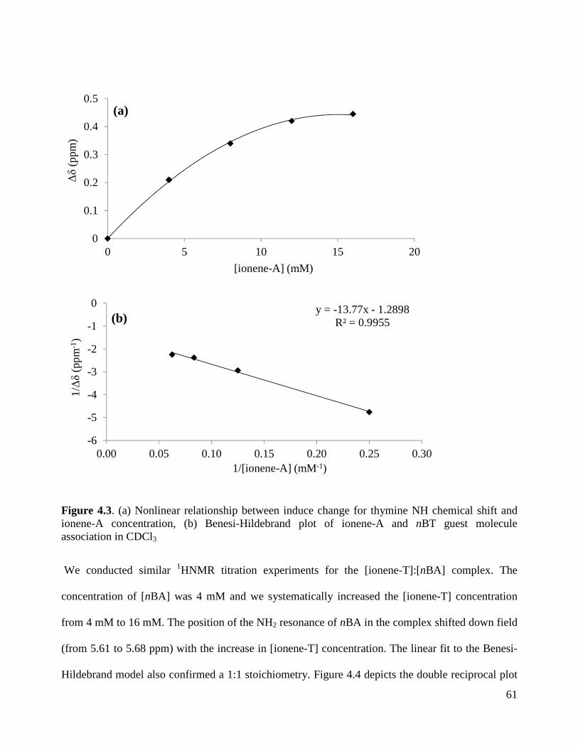
61
Figure 4.3. (a) Nonlinear relationship between induce change for thymine NH chemical shift and ionene-A concentration, (b) Benesi-Hildebrand plot of ionene-A and nBT guest molecule association in CDCl3
We conducted similar 1HNMR titration experiments for the [ionene-T]:[nBA] complex. The
concentration of [nBA] was 4 mM and we systematically increased the [ionene-T] concentration
from 4 mM to 16 mM. The position of the NH2 resonance of nBA in the complex shifted down field
(from 5.61 to 5.68 ppm) with the increase in [ionene-T] concentration. The linear fit to the Benesi-
Hildebrand model also confirmed a 1:1 stoichiometry. Figure 4.4 depicts the double reciprocal plot
y = -13.77x - 1.2898R² = 0.9955
-6
-5
-4
-3
-2
-1
0
0.00 0.05 0.10 0.15 0.20 0.25 0.30
0
0.1
0.2
0.3
0.4
0.5
0 5 10 15 20
1/∆δ
(pp
m-1
)
1/[ionene-A] (mM-1)
∆δ
(ppm
)
[ionene-A] (mM)
(a)
(b)

62
of Benesi-Hildebrand for the association of [ionene-T] and [nBA]. The Ka calculated from the slope
of this plot is 130 M-1 .
Figure 4.4. (a) Nonlinear relationship between induce change for adenine NH2 chemical shift and ionene-T concentration, (b) Benesi-Hildebrand plot of ionene-T and nBA guest molecule association in CDCl3
In order to have a control experiment, we also performed 1H NMR titrations with the nBA and nBT
guest molecules. The association constant Ka based on the slope of the plot represented in Figure
4.5 was 137 M-1. Although the Ka values calculated for the three complexes are quite comparable
and within acceptable range for adenine-thymine interaction, however the similarity of the Ka
y = -37.323x - 4.8535R² = 0.9889
-16
-12
-8
-4
0
0 0.05 0.1 0.15 0.2 0.25 0.3
0
0.04
0.08
0.12
0.16
0 5 10 15 20
1/∆δ
(ppm
-1)
1/[ionene-T] (mM-1)
∆δ
(ppm
)
[ionene-T] (mM)

63
values of 130 M-1 for [ionene-T]:[nBA] and [nBA]:[ nBT] with Ka of 137 M-1 can be due to better
solubility of ionene-T compared to ionene-A in CDCl3. This can lead to efficient accessibility of
nucleobases and stronger association between nucleobase pairs in solution.
Figure 4.5. (a) Nonlinear relationship between induce change for adenine NH2 chemical shift and nBT concentration, (b) Benesi-Hildebrand plot of nBT and nBA guest molecule association in CDCl3
y = -10.025x - 1.3713R² = 0.9926
-16
-12
-8
-4
0
0 0.2 0.4 0.6 0.8 1 1.2 1.4
0
0.1
0.2
0.3
0.4
0 2 4 6 8 10
1/∆δ
(ppm
-1)
1/[nBT] (mM-1)
[nBT] (mM)
∆δ
(ppm
)
(a)
(b)

64
4.4.3. Thermal Transitions
Upon discovering the complementary behavior of nucleobase-containing ionenes with guest
molecules in solution, we investigated the solid state properties of 1:1 molar ratios of ionene
complexes with small guest molecules. To understand the association of nucleobase pairs, we
studied the thermal properties of ionene-A, ionene-T and their complexes. All films were solution
cast from chloroform and annealed at 100 °C for 24 h in vacuum. DSC curves for the nucleobase-
functionalized ionene homopolymers and complexes showed a single glass transition temperature at
roughly -40 °C. This transition corresponded to the Tg of the PEG soft segment (SS) (1000 g/mol)
and confirmed a microphase separation of PEG SS from the ionic hard segment (HS). Since the
nucleobases were incorporated into the hard phase, the hydrogen-bonding interactions did not
significantly influence the Tg of the SS. In the solution-cast 1:1 blend of [ionene-A]:[nBT] and
[ionene-T]:[nBA] from chloroform, the crystallization and melting peak of nBT and nBA were
absent from the DSC thermograms (Figure 4.6) and the films were optically clear. Previously Long
et al.[20] also showed that the addition of uracil-containing phosphonium salt to the adenine-
containing triblock copolymers resulted in the disappearance of the phosphonium salt crystallization
peak in the DSC thermograms. Thus, the absence of melting and crystallization peaks of the guest
molecules indicates a well-defined hydrogen bonding interaction between the polymer and the guest
molecule. Table 1 illustrates the thermal transitions of ionene homopolymers and ionene complexes.

65
Figure 4.6. DSC thermograms of ionene-A homopolymer and 1:1 complex with nBT. Second heating cycle is shown.
Table 4.1. Thermal transitions of nucleobase-containing ionenes and their blends
Sample Tg (°C) DSC Td5% (°C) TGA
Ionene-A -36 246 Ionene-T -40 248
[Ionene-A]:[nBT] -31 243 [Ionene-T]:[nBA] -48 222
-5
0
5
10
15
20
0.00
0.05
0.10
0.15
0.20
0.25
0.30
0.35
0.40
-80 -30 20 70 120 170 220
[Ionene-A]:[nBT] = 1:1[Ionene-A] = 100 mol%[nBT]
Hea
t Flo
w (
W/g
)
Temperature (°C)

66
4.4.4. Morphology
We conducted atomic force microscopy (AFM) and X-ray scattering on the films of ionene
homopolymers and ionene complexes. Figure 4.7 represents the AFM phase images of ionene-A
and ionene-T as well as their complexes. AFM images of ionene homopolymers revealed
microphase-seperated morphology. The darker regions corresponded to the PEG SS (78 wt%) and
the brighter regions corresponded to the harder ionic domains and heterocyclic nucleobases (22
wt%). Comparing the ionene homopolymers with the blends demonstrated a decreased phase
contrast. This suggested the disruption of the adenine-adenine or thymine-thymine hard phase
through incorporation of complementary small molecules. Figure 4.8 illustrates the corresponding
SAXS data. Due to the difference in electron density of the HS relative to the SS, a single peak was
observed in the SAXS profile of ionene homopolymers. The Bragg spacing, the distance between
the ionic aggregates, of 5.75 nm for ionene-A was in agreement with our previous report on the 1k
PPG-based ammonium ionenes having a Bragg spacing of 6.6 nm.[9] The SAXS data revealed that
the addition of the nBT guest molecule resulted in a disruption of original morphology and led to a
broad peak at shorter Bragg spacing of 4.62 nm.

67
Figure 4.7. AFM phase images of nucleobase-containing ionene homopolymers (1,3) and their 1:1 complexes with nBt and nBA (2,4)
Figure 4.8. SAXS data for nucleobase-containing ionene homopolymers and blends
0.01
0.1
1
10
100
1000
0.1 1 10
[Ionene-A]
[Ionene-A]:[nBT]
q(nm-1)
I(cm
-1)
68
4.5. Conclusions
We synthesized and characterized nucleobase-containing ammonium ionenes using post-
polymerization functionalization. The blends of the ionene homopolymers with complementary
nucleobase-containing guest molecules resulted in efficient hydrogen bonding interactions. Job’s
plots and Benesi-Hildebrand analyses revealed 1:1 complexation between ionene homopolymers
and guest molecules. The Ka for [ionene-A]:[nBT], [ionene-T]:[nBA], and [nBT]:[nBA] complexes
were 94, 130, and 137 M-1 respectively. Ionene homopolymers and complexes showed a single Tg at
-40 °C that corresponded to the Tg of PEG soft segment due to the microphase separation. The AFM
and SAXS further confirmed a microphase-separated morphology for ionene homopolymers.
4.6. References
(1) Lutz, J.-F.; Thuenemann, A. F.; Rurack, K. Macromolecules 2005, 38, 8124-8126. (2) Mather, B. D.; Baker, M. B.; Beyer, F. L.; Berg, M. A. G.; Green, M. D.; Long, T. E.
Macromolecules 2007, 40, (19), 6834-6845. (3) Williams, S. R.; Long, T. E. Prog. Polym. Sci. 2009, 34, (8), 762-782. (4) Ramirez, S. M.; Layman, J. M.; Long, T. E. Macromol. Biosci. 2009, 9, 1127-1134. (5) Trukhanova, E. S.; Izumrudov, V. A.; Litmanovich, A. A.; Zelikin, A. N.
Biomacromolecules 2005, 6, (6), 3198-3201. (6) Jacquet, B.; Lang, G. Quaternized polymer for use as a cosmetic agent in cosmetic
compositions for the hair and skin. US4217914A, 1980. (7) Narita, T.; Ohtakeyama, R.; Nishino, M.; Gong, J. P.; Osada, Y. Colloid. Polym. Sci. 2000,
278, (9), 884-887. (8) Factor, A.; Heinsohn, G. E. J. Polym. Sci., Part C: Polym. Lett. 1971, 9, (4), 289-95. (9) Tamami, M.; Williams, S. R.; Park, J. K.; Moore, R. B.; Long, T. E. J. Polym. Sci., Part A:
Polym. Chem. 2010, 48, (19), 4159-4167. (10) Tamami, M.; Salas-de, l. C. D.; Winey, K. I.; Long, T. E. Macromol. Chem. Phys. 2012,
213, 965-972. (11) Cheng, S.; Zhang, M.; Dixit, N.; Moore, R. B.; Long, T. E. Macromolecules 2012, 45, 805-
812. (12) Lin, I. H.; Cheng, C.-C.; Yen, Y.-C.; Chang, F.-C. Macromolecules 43, (3), 1245-1252. (13) Lo, P. K.; Sleiman, H. F. J. Am. Chem. Soc. 2009, 131, 4182-4183. (14) Spijker, H. J.; van, D. F. L.; van, H. J. C. M. Macromolecules 2007, 40, 12-18. (15) Lutz, J.-F.; Pfeifer, S.; Chanana, M.; Thuenemann, A. F.; Bienert, R. Langmuir 2006, 22,
7411-7415. (16) Bazzi, H. S.; Sleiman, H. F. Macromolecules 2002, 35, 9617-9620.

69
(17) Xu, H.; Hong, R.; Lu, T.; Uzun, O.; Rotello, V. M. J. Am. Chem. Soc. 2006, 128, 3162-3163.
(18) Nair, K. P.; Weck, M. Macromolecules 2007, 40, 211-219. (19) Hemp, S. T.; Hunley, M. T.; Cheng, S.; DeMella, K. C.; Long, T. E. Polymer 2012, 53,
1437-1443. (20) Mather, B. D.; Baker, M. B.; Beyer, F. L.; Green, M. D.; Berg, M. A. G.; Long, T. E.
Macromolecules 2007, 40, 4396-4398. (21) Huang, P.-Q.; Zheng, X.; Deng, X.-M. Tetrahedron Lett. 2001, 42, 9039-9041. (22) Lira, E. P.; Huffman, C. W. J. Org. Chem. 1966, 31, 2188-91. (23) Fielding, L. Tetrahedron 2000, 56, 6151-6170. (24) Job, P. Ann. Chim. Appl. 1928, 9, 113-203. (25) Sahai, R.; Loper, G. L.; Lin, S. H.; Eyring, H. Proc. Nat. Acad. Sci. U. S. A. 1974, 71, 1499-
503. (26) Nowick, J. S.; Chen, J. S.; Noronha, G. J. Am. Chem. Soc. 1993, 115, 7636-44. (27) Karikari, A. S.; Mather, B. D.; Long, T. E. Biomacromolecules 2007, 8, 302-308. (28) Park, T.; Zimmerman, S. C. J. Am. Chem. Soc. 2006, 128, 11582-11590. (29) Mesplet, N.; Morin, P.; Ribet, J.-P. Eur. J. Pharm. Biopharm. 2005, 59, 523-526. (30) Sivakova, S.; Bohnsack, D. A.; Mackay, M. E.; Suwanmala, P.; Rowan, S. J. J. Am. Chem.
Soc. 2005, 127, 18202-18211. (31) Kyogoku, Y.; Lord, R. C.; Rich, A. Biochim. Biophys. Acta, Nucleic Acids Protein Synth.
1969, 179, 10-17.

70
Supplemental 1H NMR Figures:
Figure S 4.1. 1H NMR of N,N-bis(3-(dimethylamino)propyl)-4-hydroxybutanamide
Figure S 4.2. 1H NMR of 4-(bis(3-(dimethylamino)propyl)amino)-4-oxobutyl acrylate
a
a
a
abb
c c
d
d
ef
gi
a
Chemical Shift (ppm)
ce
b
fd
i
g
Chemical Shift (ppm)
aa
aa
bbc c
d d
ef
a
bcd
ef
g
g
j1j2
j2 j1
k
kCHCl3

71
Figure S 4.3. 1H NMR of bromine end-capped PEG (Br-PEG-Br)
Figure S 4.4. 1H NMR of n-butyl thymine guest molecule (nBT)
Chemical Shift (ppm)
f
a
a
b
c
b c
d
d
e
e
f
g
g
h
h
Chemical Shift (ppm)
aa
bc
cb
d
d
e
e
f
f
g
CHCl3
g

72
Figure S 4.5. 1H NMR of n-butyl adenine guest molecule (nBA)
Figure S 4.6. 1H NMR of acrylate-containing PEG-based ionene precursor
Chemical Shift (ppm)
a
cb
d
a
bcd
e
e
f g
g f
CHCl3
Chemical Shift (ppm)
a
a
bc b
c
b,c
dd
d
d
e
e
f
f
f
f
h
h
j1j2
j1 j2
g
g
g
k1
k2
k1,k2
l l
l
i
i
i
m
m
m
nn
n
p
p
p
H2O
CH
3OH

73
Figure S 4.7. 1H NMR of nucleobase-containing ionenes: adenine-ionene (top), thymine-ionene (bottom)
Chemical Shift (ppm)
g
i
m
n
p
f
k1
k2
la
b
fg
l i
mn
pc
b
cd d
mnid'
e
e
p
d'
h j
h
a
g,b,c
k1,k2
l
q
fqj
r
r
s
sd
Chemical Shift (ppm)
g
i
m
n
p
f
k1
k2
l fg
l i
mn
p
a
b
c cd dbe
d'
h j
q
mn
s
s
d'd
ep
h
ak1,k2
lf
qj
r
r i
g,b,c

74
Chapter 5. Synthesis and Characterization of Silicone-Based Ammonium Ionenes as Candidates for Self-Healing Polymers
Mana Tamami and Timothy E. Long*
Macromolecules and Interfaces Institute, Department of Chemistry, Virginia Tech, Blacksburg,
VA 24061, USA
*E-mail address: [email protected]
5.1. Abstract
We used Menshutkin reaction to synthesize PDMS-based ionenes having various hard segment
contents. The effect of hard segment content on thermal, mechanical, rheological, and gas
permeability was investigated. DSC showed one transition at -120 °C corresponding to the Tg of
PDMS and upon 30 wt% HS a melting transition appeared for PDMS-ionenes. Mechanical
properties of PDMS-ionenes were dependent on the amount of hard segment content. DMA
confirmed microphase separation and showed that the rubbery plateau modulus extended to
higher temperatures (from 87 °C to 164 °C) with increase in hard segment content (from 5 wt%
to 15 wt%). Tensile analysis demonstrated systematic increase in modulus for PDMS-ionenes
with increasing hard segment content. The melt viscosity of 5 wt% HS PDMS-based ionene was
more temperature dependent compared to 15 wt% HS ionene. The OTR values for virgin PDMS-
ionenes decreased linearly from 60000 to 15000 cc/m2/day as the wt% hard segment increased.
Keywords: poly(dimethyl siloxane)-ionene, oxygen transmission rate, melt viscosity, self-healing, microphase separation

75
5.2. Introduction
Self-healing polymers have the ability to recover their strength and properties after damage.
Since self-healing materials will not require replacement after damage or wear, this characteristic
is very attractive especially for conservation purposes. Over the years, many self-healing
materials have been invented.[1] Based on the self-healing process, the self-healing materials can
be divided into two categories: self-healing based on covalent interactions which require carbon-
carbon bond formation and self-healing driven by non-covalent interactions which require heat
or light to stimulate the responsive polymer. Non-covalent interactions such as electrostatics[2, 3]
and hydrogen bonding[4] not only improve thermal and mechanical properties of polymers, but
also impart stimuli responsiveness that leads to self-healing.
Ionomers or ion-containing polymers are macromolecules that contain less than 15 mol% of
ionic groups.[5, 6] The incompatibility of ionic groups and the organic polymer matrix causes
ionic aggregation. Eisenberg et al.[5, 7, 8] proposed a model to describe the structure of ionic
aggregates. They defined a multiplet as “an aggregate consisting of several ion pairs with a
diameter of 5-10 Ǻ”. Multiplets act as physical crosslinks and restrict the mobility of
surrounding polymer chains. Upon increasing the ionic content, more multiplets form close to
each other. Increase in charge density restricts the mobility surrounding each multiplet and
causes it to overlap into continuous phase known as clusters. It is reported in the literature that
ionic aggregates have drastic effect on mechanical[9, 10] and thermal properties.[11]
Recently the application of ionomers in self-healing materials has been explored. The
copolymers of ethylene and methacrylic acid (EMAA) neutralized to a certain extent to form
their salt form have shown self-healing behavior. Among these copolymers, Surlyn®
manufactured by DuPont is a semicrystalline copolymer of ethylene and methacrylic acid. It has

76
been shown that Surlyn® heals through a viscoelastic healing process.[12] It is proposed that upon
puncture, heat that is generated melts the ethylene crystallites in the damaged area. The clusters
disorder and reaggregate to provide molten polymer with sufficient energy to bounce back. The
polymer chains interdiffuse together and heal the hole. The neighboring polymer chains that
remained at room temperature acted as an anchor for the mobile chains to pull against.
Ionenes are ion-containing polymers that have quaternized nitrogen atoms along the polymer
backbone and are synthesized using step-growth polymerization of a ditertiary amine monomer
with an alkyl dihalide monomer.[13] This polymerization is straight forward and there are no
byproducts generated. The synthetic design of this reaction provides control over charge density
and we can explore novel topologies using various monomer architectures. Our group has
extensively synthesized ionenes having different chemical structures and investigated the effect
of charge density on thermal, mechanical, rheological, and morphological properties.[14-17] We
demonstrated that electrostatic interactions significantly influenced the ionene properties
compared to their non-ionic counterparts. However, ionenes compared to ionomers have not
been investigated for their self-healing properties. It is anticipated that the melt-elastic behavior
of ionenes above their ionic dissociation temperature will assist in self-healing. The morphology
of ionenes, unlike Surlyn®, can be more easily tailored for a targeted ionic dissociation
temperature and tuned to have a programmed heal temperature as demanded by an application.
Herein, we report the synthesis of poly(dimethyl siloxane)-based ammonium ionenes adapted
from our earlier research in collaboration with Wilkes et al.[18] we synthesized a series of PDMS-
based ionenes in order to investigate the effect of HS content on ionic dissociation temperature,
thermal, mechanical, and gas permeation properties.

77
5.3. Experimental.
5.3.1. Materials
α,ω-Amino propyl-polydimethylsiloxane (PDMS) oligomer with number-average molecular
weight of 2500 g/mol was purchased from Gelest and dried in vacuo (0.1 mm Hg) for 24 h prior
to use. 1,12-Dibromododecane (Aldrich, 98%) and 1,4-diazabicyclo[2.2.2]octane (DABCO,
98%) were sublimed before use. 6-bromohexanoyl chloride (97%) was purchased from Aldrich
and used as received. Triethylamine (TEA, Aldrich, 99%) was distilled prior to use. Sodium
bicarbonate (NaHCO3) (99.7%) and magnesium sulfate (99.5%) were obtained from Sigma-
Aldrich and used without further purification. Methanol (MeOH, Fisher, HPLC grade) was
passed through alumina and molecular sieves columns. Chloroform (CHCl3, Fisher, Optima
grade) was distilled from calcium hydride.
5.3.2. Instrumentation
1H NMR was utilized to determine monomer and polymer composition in CDCl3 or CD3OD at
23 °C with a 400 MHz Inova spectrometer. Thermogravimetric analysis (TGA) was conducted
on a TA Instruments Hi-Res TGA 2950 with a temperature ramp of 10 °C/min in a nitrogen
atmosphere. Differential scanning calorimetry (DSC) was performed using a TA Instruments
Q100 differential scanning calorimeter under a nitrogen flow of 50 mL/min.
Dynamic mechanical analysis (DMA) was conducted on a TA Instruments Q800 dynamic
mechanical analyzer in tension mode at a frequency of 1 Hz and temperature ramp of 3 °C/min.
Tensile tests were performed on a 5500R Instron universal testing instrument with a cross-head
speed of 50 mm/min using manual grips at ambient temperature. Melt rheology experiment was
conducted on a TA Instruments G2 Rheometer in parallel-plate geometry with a diameter of 8

78
mm and a gap distance of 1mm. Strain amplitude was 1%. Oxygen transmission rate was
measured using Illinois Instruments Model 8001 Oxygen Permeation Analyzer at 23 °C.
5.3.3. Synthesis of bromine end-capped poly(dimethyl siloxane) (Br-PDMS-Br)
A literature procedure was used to synthesize Br-PDMS-Br.[18] The commercially available
amine-terminated PDMS (20.00 g, 1 eq) was added to a two-neck, round-bottomed flask
equipped with a magnetic stir bar, addition funnel, nitrogen inlet, and 250 mL dichloromethane
(DCM). Distilled triethylamine (2.2 eq) was added to the flask with a syringe. The flask was
cooled to 0 °C. 6-Bromohexanoyl chloride (2.2 eq) was added to the addition funnel containing
50 mL DCM and subsequently added to the reaction flask in a drop wise fashion. The reaction
was allowed to proceed for 24 h. Triethylamine salt was filtered through a fritted funnel. The
product in DCM was introduced into a separatory funnel and washed twice with saturated
NaHCO3(aq) and twice with distilled water. The DCM layer was separated and dried over
magnesium sulfate, filtered, and evaporated. The product was further dried under vacuum and
the yield was 97%. The 1H NMR number average molecular weight for Br-PDMS-Br was 3052
g/mol. 1H NMR (400 MHz, CDCl3): δ = 0.00 (m, 230H, Ha), 0.46 (m, 4H, Hb), 1.43 (m, 8H, Hc),
1.60 (p, 4H, Hd), 1.81 (p, 4H, He), 2.10 (t, 4H,Hf), 3.17 (q, 4H, Hg), 3.33 (t, 4H, Hh), 5.37 (s, 2H,
Hi) (Figure 5.1).

79
Figure 5.1. 1H NMR spectrum of bromine-terminated PDMS
5.3.4. Synthesis of poly(dimethyl siloxane)-based ionene homopolymer
Upon the synthesis of difunctional Br-PDMS-Br, the commercially available cyclic ditertiary
amine monomer (DABCO) was sublimed for further purification and dryness. Then a 1:1 molar
ratio of Br-PDMS-Br (5.6 g, 2.6 mmol) with DABCO (0.29 g, 2.6 mmol) were introduced to a
flame-dried round-bottom flask equipped with a condenser. The dried chloroform (15 mL) was
added to the flask and the reaction was refluxed for 24 h at 65 °C. Upon reaction completion no
further purification was needed. The excess solvent was removed and the rest of reaction
solution was cast in Teflon mold. The films were slowly air-dried for 48 h and were then
annealed at 100 °C for 24 h. The ionene films were then stored on drying agents (drierite) and
inside the desiccator until analyzed. 1H NMR (400 MHz, CD3OD): δ = 0.00 (m, 304H, Ha), 0.48
(m, 4H, Hb), 1.24-1.87 (m, 16H, Hc), 2.16 (m, 4H, Hd), 3.07 (m, 4H, He), 3.52 (m, 4H, Hf), 3.93
(s, 12H, Hg) (Figure 5.2).
Chemical Shift (ppm)
a
bcdefgh
i
ab
ci
bc cc
dd ee
ff
gg
h hi

80
Figure 5.2. 1H NMR spectrum of PDMS-based ionene homopolymer
5.3.5. Synthesis of poly(dimethyl siloxane)-based ionene random copolymer
The copolymer containing 15 wt % hard segment is used as an example; however, all
copolymers were prepared in a similar fashion. 1,12-dibromododecane (1.08 g, 3.28 mmol), Br-
PDMS-Br (10.00 g, 3.28 mmol), and DABCO (0.74 g, 6.55 mmol) were added to a two-neck,
round-bottomed flask equipped with a stir bar, condenser, and nitrogen inlet. Dry chloroform (20
wt % solids) was added to the flask via syringe. The reaction was allowed to proceed for 24 h at
65 °C and upon completion, the polymer was cast into a film. The films were slowly air-dried for
48 h and were then annealed at 100 °C for 24 h. The ionene films were then stored on drying
agents (drierite) and inside the desiccator until analyzed. 1H NMR confirmed the structures of
PDMS-based ionene random copolymers.
Chemical Shift (ppm)
a
bcde
fg
a bbc
c c
dd
ee
fcf
g

81
5.4. Results and Discussion
5.4.1. Synthesis of PDMS-based ionenes
We synthesized soft segment precursor, bromine-terminated PDMS, using quantitative
derivatization of 2.5k g/mol aminopropyl-terminated PDMS with 6-bromohexanoyl chloride to
yield an oligomeric dibromide (Scheme 5.1). Previously we reported the bromine
functionalization of poly(propylene glycol) and poly(tetramethylene oxide) using the same
procedure.[15, 17] 1H NMR confirmed the structure of this oligomer (Figure 5.1). The number
average molecular weight based on 1H NMR was 3052 g/mol. Scheme 5.2 shows the synthesis of
PDMS-based ionenes. We varied the amount of soft segment (SS) and hard segment (HS), while
maintaining the 1:1 molar ratio of DABCO:dibromide. In order to obtain high molecular weight
step-growth polymer, a 1:1 molar ratio of monomers is critical.[19] As shown in Table 5.1, five
compositions were synthesized and 1H NMR spectroscopy confirmed their structures. The
resonance at 2.8 ppm in the 1H NMR spectrum due to protons from DABCO ring shifted to 3.9
ppm after polymerization, confirming the quaternization of tertiary amine nitrogens within the
ring. The total amount of 1,12-dibromododecane and DABCO corresponded to the HS content;
the HS content varied from 5-60 wt%. Ionenes containing 5-60 wt% HS formed uniform solvent-
cast films, however, due to the increase in HS content, the films of 50 and 60 wt% HS were very
tacky and brittle

82
Scheme 5.1. Synthesis of bromine terminated PDMS
Table 5.1. Segmented copolymer compositions based on molar equivalents of monomer and HS/SS content
Br-PDMS-Br (mole eq.)
1,12-dibromododecane (mole eq.)
DABCO (mole eq.)
HS
(wt%)
SS
(wt%)
1.00 0.00 1.00 5 95
0.50 0.50 1.00 15 85
0.24 0.76 1.00 30 70
0.12 0.88 1.00 50 50
0.08 0.92 1.00 60 40
5.4.2. Thermal Transitions
The effect of hard segment content on thermal transitions was investigated using TGA and DSC.
We used TGA to measure the thermal stability of all ionenes. TGA indicated that the ionic
content within the polymer backbone dictates the thermal degradation behavior. The thermal
degradation temperature at 5% weight loss (TD) decreased from 273 °C to 170 °C with

83
increasing ionic content. The actual mechanism of thermal degradation is complex. Chen et al.[20]
and Jerome et al.[21] proposed dequaternization of nitrogens according to the Hoffman
elimination pathway. The Hoffman elimination reaction depends on the basicity of the anion.[22]
At higher temperatures, the bromine counterion acts as a strong base and can dequaternize
nitrogens, leaving tertiary amine and alkene chain ends. Herein, as the ionic content increases,
the number of quaternized nitrogens increase along the backbone and therefore the degradation
occurs earlier at lower temperatures. All ionenes revealed glass transition temperatures (Tg) of -
123 °C, which corresponded to the Tg of the PDMS soft segment. Upon 30 wt% hard segment
content the melting temperature (Tm) appeared which demonstrated an ordered crystalline phase
within the soft PDMS amorphous phase. The data in Table 5.2 strongly suggest that these
PDMS-based ionenes were microphase separated.
Scheme 5.2. Synthesis of PDMS-based segmented ionene copolymers

84
Table 5.2. TGA and DSC results of segmented PDMS-based ionenes HS (wt%) SS (wt%) Td5% (°C) Tg (°C) Tm (°C)
5 95 273 -123 NA
15 85 242 -123 NA
30 70 209 -123 -48
50 50 190 -123 -50
60 40 170 -123 -46
5.4.3. Dynamic Mechanical Analysis (DMA)
PDMS-based ionenes having 5 and 15 wt% HS formed homogenous and elastomeric films that
were ideal candidates to measure their temperature-dependent moduli using DMA. The films
were stored in desiccator after annealing and were quickly removed from desiccator and loaded
on to the DMA clamps to prevent absorption of moisture. Figure 5.3 shows the DMA analysis
for 5 wt% HS-ionene as an example. DMA confirmed microphase separation in these systems.
Ionene showed high moduli (>103 MPa) in glassy state and a drastic drop in modulus occurred at
-120 °C due to the glass transition temperature of PDMS soft segment. The rubbery plateau
modulus for 15 wt% HS-ionene was higher than 5 wt% HS and it also extended to higher
temperatures compared to 5 wt% HS-ionene (from 87 °C and 164 °C). This shows the
dependency of rubbery plateau modulus on the charge density and hard segment content. As the
charge density increased along the ionene backbone, stronger physical crosslinks occurred which
led to an extension of rubbery plateau modulus. Therefore controlling the hard segment content

85
can tune the ionic dissociation temperature and moduli. Similarly, we reported the effect of
charge density and soft segment molecular weight on poly(propylene glycol)-based ionenes.[17]
As shown in Figure 5.3, both ionenes showed three transitions; first transition at -120 °C
corresponded to the Tg of 2.5k PDMS, the second transition at ca. 20 °C corresponded to the
melting of PDMS crystallites, and the third transition which occurred at 87 °C for 5 wt% HS-
ionene and at 164 °C for 15 wt% HS-ionene attributed to the ionic dissociation temperature
followed by the flow. The upward storage modulus for 15 wt% HS-ionene between 50 °C and
150 °C is not real and is due to an extreme softness of the sample at those high temperatures.
Figure 5.3. DMA of PDMS-based ionene having 5 wt% HS
5.4.4. Tensile test
We performed tensile analysis at ambient conditions for PDMS-ionenes having 5 and 15 wt%
HS content (Figure 5.4). The 15 wt% HS ionene showed an ultimate tensile strength of 0.52 MPa
and elongation at break of 151%. Due to increased wt% HS and stronger physical crosslinks, the
15 wt% HS-ionene showed higher modulus, tensile strength, and elongation compared to 5 wt%
0.1
1
10
100
1000
10000
-150 -100 -50 0 50 100 150
Temperature ( C)
Sto
rage
Mo
dulu
s (M
Pa)

86
HS-ionene (Table 5.3). It should be mentioned that the tensile moduli of both ionenes were
comparable with moduli observed at ambient condition using DMA.
Figure 5.4. Tensile analysis of PDMS-based ionene having 5 wt% and 15 wt% HS
Table 5.3. Tensile data for PDMS-ionene films as a function of wt% HS
PDMS-ionenes
Tensile Stress at Break (MPa)
Tensile Strain at Break (%)
Young’s Modulus (MPa)
5 wt% HS 0.13 ± 0.01 85.76 ± 7.17 0.70 ± 0.30
15 wt% HS 0.52 ± 0.04 150.66 ± 5.64 1.65 ± 0.11
5.4.5. Rheology
It is important to understand the effect of hard segment content on viscosity. In the healing event
the material must flow to reach intimate contact. In order for the healing to occur, the viscosity
of the polymer must be reduced by increasing the heat. This allows the polymer to reach a low
enough viscosity to flow and then heal. Therefore upon film preparation of PDMS-based ionenes
(5 and 15 wt% HS), we investigated the viscosity of the polymer melt as a function of
temperature. It is illustrated in Figure 5.5 that melt viscosity of 5 wt% HS PDMS-ionene is
0.00
0.10
0.20
0.30
0.40
0.50
0.60
0 20 40 60 80 100 120 140 160
15 wt% HS5 wt% HS
Tensile Strain (%)
Ten
sile
Str
ess
(M
Pa
)

87
decreasing with temperature and in comparison the 15 wt% HS PDMS-ionene is independent of
temperature. In addition, master curve of the storage and loss modulus versus frequency was
generated for 5 wt% HS PDMS-ionene to estimate the changes in modulus over decades in time.
Master curve of storage modulus versus frequency was referenced to 90 °C. As shown in Figure
5.6, time-temperature superposition was satisfied for the PDMS-ionene having 5 wt% HS.
Figure 5.5. Melt viscosity of PDMS-based ionenes having 5 wt% and 15 wt% HS
Temperature (°C)
Viscosity (pa.s
)

88
Figure 5.6. Master curve of PDMS-based ionene with 5wt% HS
5.4.6. Oxygen Transmission Rate
One technique to determine the efficiency of healing process is to measure and compare oxygen
transmission rate (OTR) of virgin and healed samples. If the sample has healed successfully, the
OTR values for the virgin sample and healed sample must be quite comparable. Therefore we
measured the OTR values for virgin PDMS-based ionenes as a starting point. Although PDMS is
well-known for its high oxygen permeation, however, the synthetic strategy and its modular
nature will allow facile extension to other soft segments, including high oxygen barrier
segments. Figure 5.7 demonstrates OTR values for virgin PDMS-ionenes with 5, 15, and 30 wt%
hard segment content. The OTR values decreased linearly from 60000 to 15000 cc/m2/day as the
wt% hard segment increased. This indicates that the composition of the continuous phase
influenced permeation, and higher ionic concentrations impeded efficient gas transport. The next
step is to damage the samples and perform healing above the ionic dissociation temperature of
-6 -5 -4 -3 -2 -1 0 1 2 3Log [ang. frequency (rad/s)]
2
3
4
5
6
Lo
g [
G' (
Pa
)]
2
3
4
5
6
Lo
g [G
'' (Pa
)]*90.0100.0110.0120.0130.0140.0150.0160.0170.0

89
each sample (The healing studies will be performed in collaboration with Virginia Common
Wealth University). Upon healing the OTR values will be determined and compared with virgin
samples to study the healing efficiency.
Figure 5.7. OTR values of PDMS-based ionenes with various HS contents
5.5. Conclusions
PDMS-based ionenes having various hard segment content were synthesized using Menshutkin
reaction. The thermal analysis of PDMS-ionenes using DSC showed that they have one transition
at -120 °C corresponding to the Tg of PDMS, further confirming microphase separation. Upon 30
wt% HS a melting transition appeared for PDMS-ionenes, corresponding to crystalline phase
within the soft PDMS amorphous phase. DMA confirmed microphase separation and showed
that the breadth of rubbery plateau moduli and their final softening temperatures change with the
nature and content of hard segment. As the hard segment content increased from 5 wt% to 15
wt%, the rubbery plateau modulus extended from 87 °C to 164 °C due to higher physical
OT
R (
cc/m
2 /da
y)
Time (min)
0
20,000
40,000
60,000
0 100 200 300 400
5 wt% HS
15 wt% HS
30 wt% HS

90
crosslinking. Tensile analysis demonstrated systematic increase in modulus of PDMS-ionenes
with increasing hard segment content. The viscosity of polymer melt as a function of temperature
showed that the viscosity of 5wt% HS PDMS-based ionene is more temperature dependent
compared to 15 wt% HS PDMS-based ionene. OTR analysis revealed that OTR values for virgin
PDMS-ionenes decreased linearly from 60000 to 15000 cc/m2/day as the wt% hard segment
increased, indicating the influence of hard segment continuous phase on gas permeation.
Acknowledgements
The authors would like to acknowledge the financial support for this work through NASA STTR
program administered by Johnson Space Center under the Contract No. NNX11CI22P for the
project titled "Self-healing Inflatable Extraterrestrial shieLD (SHIELD)"
91
5.6. References
(1) Bergman, S. D.; Wudl, F. J. Mater. Chem. 2008, 18, (1), 41-62. (2) Kalista, S. J., Jr.; Ward, T. C.; Oyetunji, Z. Mech. Adv. Mater. Struct. 2007, 14, 391-397. (3) Kalista, S. J., Jr.; Ward, T. C. J. R. Soc. Interface 2007, 4, 405-411. (4) Sijbesma, R. P.; Beijer, F. H.; Brunsveld, L.; Folmer, B. J. B.; Hirschberg, J. H. K. K.;
Lange, R. F. M.; Lowe, J. K. L.; Meijer, E. W. Science 1997, 278, 1601-1604. (5) Eisenberg, A.; Hird, B.; Moore, R. B. Macromolecules 1990, 23, 4098-107. (6) Tant, M. R.; Wilkes, G. L. In Structure and properties of hydrocarbon-based ionomers,
1997; Blackie: 1997; pp 261-289. (7) Eisenberg, A. Macromolecules 1970, 3, 147-54. (8) Marx, C. L.; Caulfield, D. F.; Cooper, S. L. Macromolecules 1973, 6, 344-53. (9) Bellinger, M.; Sauer, J. A.; Hara, M. Macromolecules 1994, 27, 1407-12. (10) Hara, M.; Sauer, J. A. J. Macromol. Sci., Rev. Macromol. Chem. Phys. 1994, C34, 325-
73. (11) Tadano, K.; Hirasawa, E.; Yamamoto, H.; Yano, S. Macromolecules 1989, 22, 226-33. (12) Varley, R. J.; Shen, S.; van, d. Z. S. Polymer 2010, 51, 679-686. (13) Williams, S. R.; Long, T. E. Prog. Polym. Sci. 2009, 34, (8), 762-782. (14) Williams, S. R.; Borgerding, E. M.; Layman, J. M.; Wang, W.; Winey, K. I.; Long, T. E.
Macromolecules 2008, 41, (14), 5216-5222. (15) Williams, S. R.; Salas-de la Cruz, D.; Winey, K. I.; Long, T. E. Polymer 2010, 51, (6),
1252-1257. (16) Tamami, M.; Salas-de, l. C. D.; Winey, K. I.; Long, T. E. Macromol. Chem. Phys. 2012,
213, 965-972. (17) Tamami, M.; Williams, S. R.; Park, J. K.; Moore, R. B.; Long, T. E. J. Polym. Sci., Part
A: Polym. Chem. 2010, 48, (19), 4159-4167. (18) Das, S.; Goff, J. D.; Williams, S.; Salas-De, L. C. D.; Riffle, J. S.; Long, T. E.; Winey, K.
I.; Wilkes, G. L. J. Macromol. Sci., Part A: Pure Appl. Chem. 2010, 47, 215-224. (19) Odian, G., Principles of polymerization. Fourth ed.; John Wiley & Sons, Inc., Hoboken:
New Jersey, 2004. (20) Ruckenstein, E.; Chen, X. Macromolecules 2000, 33, (24), 8992-9001. (21) Yano, S.; Tadano, K.; Jerome, R. Macromolecules 1991, 24, (24), 6439-42. (22) March, J., Advanced organic chemistry: Reactions, mechanims, and structure. 3rd ed.;
Wiley: New York: 1985.

92
Chapter 6. Overall Conclusions
The synthesis and characterization of novel architectures of ion-containing polymers named
ionenes were described. The synergistic effect of non-covalent interactions including nucleobase
hydrogen bonding and electrostatics were investigated on the properties of segmented and non-
segmented ammonium ionenes.
With regard to non-segmented ionene systems, novel adenine and thymine functionalized
ionene homopolymers having two different side chain spacer lengths were synthesized using
Menshutkin reaction and subsequent post-polymerization functionalization was conducted via
Michael addition chemistry. The effect of spacer length on hydrogen bonding interactions in 1:1
blends was studied. The shorter spacer ionene homopolymers and blends showed higher glass
transition temperatures than longer spacer ionenes due to closeness of the bulky nucleobase units
to the backbone and hindering the segmental motion in the ionene backbone. A single glass
transition temperature confirmed the miscibility of ionene blends. The glass transition
temperatures of ionene blends with shorter spacer followed the Fox equation indicating no
hydrogen bonding interactions. The glass transition temperatures of the blends with longer
spacer lengths deviated from both Fox and Gordon-Taylor equations, demonstrating a presence
of hydrogen bonding interactions. The variable-temperature FT-IR (30 °C to 170 °C) confirmed
the hydrogen bonding interactions for the longer spacer ionenes with NH2 bending vibration
shifting to lower wave numbers and C=O stretching vibration shifting to higher wave numbers
with increasing temperature.
Regarding segmented nucleobase-containing ionene systems, similar post-polymerization
functionalization was applied to synthesize poly(ethylene glycol)-based adenine and thymine

93
containing ionenes. The blends of the ionene homopolymers with complementary nucleobase-
containing guest molecules resulted in efficient hydrogen bonding interactions. Job’s plots and
Benesi-Hildebrand analyses revealed 1:1 complexation between ionene homopolymers and guest
molecules. The association constants for nucleobase-containing ionenes with complementary
guest molecules were in the order of 100 M-1 which was in agreement with literature values.
Ionene homopolymers and complexes showed a single glass transition temperature at -40 °C
which corresponded to the Tg of PEG soft segment and confirmed the microphase separation.
The synthesis of silicone containing ionene copolymers was achieved through the use of
PDMS dibromide oligomers, 1,12-dibromododecane, and DABCO. Hard segment contents
ranged from 5-60 wt%, and structure-property relationships were established as a function of
hard segment content. Thermal properties were measured via TGA and DSC. Film formation
characteristics varied as a function of hard segment content, and free-standing elastic films were
obtained for 5 and 15 wt% hard segment content. DMA confirmed microphase separation and
showed that the breadth of rubbery plateau moduli and their final softening temperatures change
with the nature and content of hard segment. As the hard segment content increased from 5 wt%
to 15 wt%, the rubbery plateau modulus extended from 87 °C to 164 °C due to higher physical
crosslinking. The viscosity of polymer melt as a function of temperature showed that the
viscosity of 5 wt% HS PDMS-based ionene is more temperature dependent compared to 15 wt%
HS ionene. Oxygen transmission rates for PDMS-ionenes decreased linearly from 60000 to
15000 cc/m2/day as the wt% hard segment increased, indicating the influence of hard segment
continuous phase on gas permeation.

94
Chapter 7. Suggested Future Work
7.1. Synthesis and characterization of PEG-based cytosine and guanine-containing ammonium ionenes
Deoxyribose nucleic acid (DNA) structure includes four nucleobases, pyrimidines
(thymine and cytosine) and purines (adenine and guanine). DNA with its supramolecular
characteristics has inspired us along with many other researchers to utilize these features
and develop polymers that can exhibit similar supramolecular properties. Chapter three
and four of this thesis were focused on the synthesis and characterization of non-
segmented and segmented adenine and thymine-containing ammonium ionenes. We
studied the synergistic effect of complementary hydrogen bonding and electrostatics on
various properties of ammonium ionenes. Future efforts should focus on the synthesis
and characterization of cytosine and guanine-containing ammonium ionenes. Cytosine
and guanine base pair interact via three hydrogen bonds with stronger degree of
interaction (KCG ca. 104-105 M-1in CDCl3) compared to adenine and thymine base pair
with two hydrogen bonds (KAT ca. 102 M-1 in CDCl3). Therefore synthesizing polymers
that contain cytosine-guanine hydrogen bonding motifs are attractive for their enhanced
supramolecular characteristics. However, the synthesis of these polymers can be more
challenging due to reduced solubility of these nucleobases compared to adenine and
thymine. Figure 7.1 represents the proposed synthesis of cytosine and guanine-containing
PEG-based ionene.

95
Figure 7.1. Synthesis of cytosine and guanine-containing poly(ethylene glycol)-based ammonium ionenes
7.2. Synthesis and characterization of ionenes with potential application in adhesives
As mentioned in chapter two, non-covalent interactions such as van der Waals, hydrogen
bonding and electrostatics can provide physical adhesion. Hydrogen bonded polymers are
specially used to prepare pressure sensitive adhesives (PSAs). Although, there have been
extensive studies on the application of hydrogen bonded polymers in adhesive design, but
not enough literature is present on the application of polymers that contain both hydrogen
bonding and electrostatics in adhesives. Therefore, one of the future directions in the field
of ionenes is to design novel architectures that have hydrogen bonding groups or moieties
and measure their adhesive properties. These architectures can be linear, star-shaped,
hyper-branched, and comb ionenes. For example, the synthesis of linear PEG-based
ionene containing hydroxyl side groups is demonstrated in Figure 7.2. This polymer has
shown potential in adhesive application.

96
Figure 7.2. Synthesis of OH-containing PEG-based ammonium ionene
7.3. Synthesis and characterization of self-healing ammonium ionenes
Self-healing is a behavior in which a material detects its damage and heals itself either
spontaneously (autonomic) or with the aid of a stimulus (non-autonomic). One series of
non-autonomic materials are ionomers. Recently the application of ionomers in self-
healing materials has been explored. The copolymers of ethylene and methacrylic acid
(EMAA) such as Surlyn® manufactured by DuPont have shown promising self-healing
behavior. In chapter five, the synthesis and structure-property relationships of silicone-
based ammonium ionenes as an alternative candidate to Surlyn® were discussed. The
future direction in this field is to design ionene systems that contain multiple hydrogen
bonding motifs throughout the ionene backbone and investigate the effect of
electrostatics and supramolecular association on self-healing behavior.
Related Documents