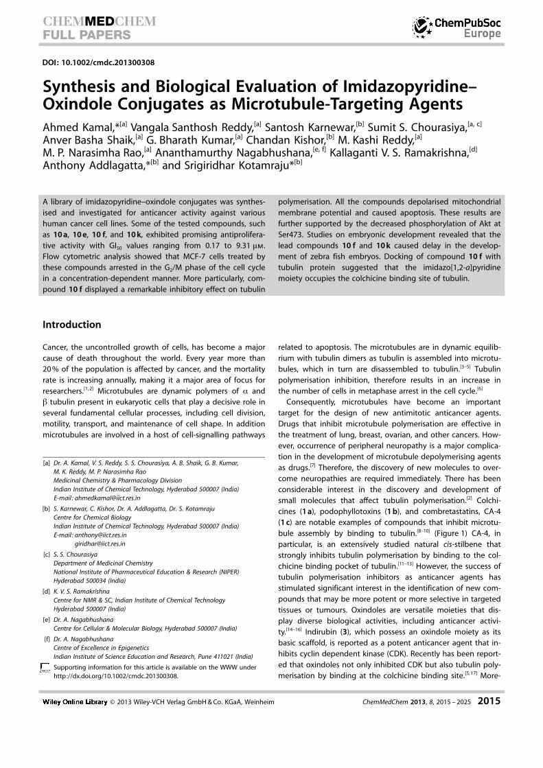
DOI: 10.1002/cmdc.201300308 Synthesis and Biological Evaluation of Imidazopyridine– Oxindole Conjugates as Microtubule-Targeting Agents Ahmed Kamal,* [a] Vangala Santhosh Reddy, [a] Santosh Karnewar, [b] Sumit S. Chourasiya, [a, c] Anver Basha Shaik, [a] G. Bharath Kumar, [a] Chandan Kishor, [b] M. Kashi Reddy, [a] M. P. Narasimha Rao, [a] Ananthamurthy Nagabhushana, [e, f] Kallaganti V. S. Ramakrishna, [d] Anthony Addlagatta,* [b] and Srigiridhar Kotamraju* [b] Introduction Cancer, the uncontrolled growth of cells, has become a major cause of death throughout the world. Every year more than 20% of the population is affected by cancer, and the mortality rate is increasing annually, making it a major area of focus for researchers. [1, 2] Microtubules are dynamic polymers of a and b tubulin present in eukaryotic cells that play a decisive role in several fundamental cellular processes, including cell division, motility, transport, and maintenance of cell shape. In addition microtubules are involved in a host of cell-signalling pathways related to apoptosis. The microtubules are in dynamic equilib- rium with tubulin dimers as tubulin is assembled into microtu- bules, which in turn are disassembled to tubulin. [3–5] Tubulin polymerisation inhibition, therefore results in an increase in the number of cells in metaphase arrest in the cell cycle. [6] Consequently, microtubules have become an important target for the design of new antimitotic anticancer agents. Drugs that inhibit microtubule polymerisation are effective in the treatment of lung, breast, ovarian, and other cancers. How- ever, occurrence of peripheral neuropathy is a major complica- tion in the development of microtubule depolymerising agents as drugs. [7] Therefore, the discovery of new molecules to over- come neuropathies are required immediately. There has been considerable interest in the discovery and development of small molecules that affect tubulin polymerisation. [2] Colchi- cines (1a), podophyllotoxins (1b), and combretastatins, CA-4 (1c) are notable examples of compounds that inhibit microtu- bule assembly by binding to tubulin. [8–10] (Figure 1) CA-4, in particular, is an extensively studied natural cis-stilbene that strongly inhibits tubulin polymerisation by binding to the col- chicine binding pocket of tubulin. [11–13] However, the success of tubulin polymerisation inhibitors as anticancer agents has stimulated significant interest in the identification of new com- pounds that may be more potent or more selective in targeted tissues or tumours. Oxindoles are versatile moieties that dis- play diverse biological activities, including anticancer activi- ty. [14–16] Indirubin (3), which possess an oxindole moiety as its basic scaffold, is reported as a potent anticancer agent that in- hibits cyclin dependent kinase (CDK). Recently has been report- ed that oxindoles not only inhibited CDK but also tubulin poly- merisation by binding at the colchicine binding site. [5, 17] More- A library of imidazopyridine–oxindole conjugates was synthes- ised and investigated for anticancer activity against various human cancer cell lines. Some of the tested compounds, such as 10 a, 10 e, 10 f, and 10 k, exhibited promising antiprolifera- tive activity with GI 50 values ranging from 0.17 to 9.31 mm. Flow cytometric analysis showed that MCF-7 cells treated by these compounds arrested in the G 2 /M phase of the cell cycle in a concentration-dependent manner. More particularly, com- pound 10 f displayed a remarkable inhibitory effect on tubulin polymerisation. All the compounds depolarised mitochondrial membrane potential and caused apoptosis. These results are further supported by the decreased phosphorylation of Akt at Ser473. Studies on embryonic development revealed that the lead compounds 10 f and 10 k caused delay in the develop- ment of zebra fish embryos. Docking of compound 10 f with tubulin protein suggested that the imidazo[1,2-a]pyridine moiety occupies the colchicine binding site of tubulin. [a] Dr. A. Kamal, V. S. Reddy, S. S. Chourasiya, A. B. Shaik, G. B. Kumar, M. K. Reddy, M. P. Narasimha Rao Medicinal Chemistry & Pharmacology Division Indian Institute of Chemical Technology, Hyderabad 500007 (India) E-mail : [email protected] [b] S. Karnewar, C. Kishor, Dr. A. Addlagatta, Dr. S. Kotamraju Centre for Chemical Biology Indian Institute of Chemical Technology, Hyderabad 500007 (India) E-mail : [email protected] [email protected] [c] S. S. Chourasiya Department of Medicinal Chemistry National Institute of Pharmaceutical Education & Research (NIPER) Hyderabad 500034 (India) [d] K. V. S. Ramakrishna Centre for NMR & SC, Indian Institute of Chemical Technology Hyderabad 500007 (India) [e] Dr. A. Nagabhushana Centre for Cellular & Molecular Biology, Hyderabad 500007 (India) [f] Dr. A. Nagabhushana Centre of Excellence in Epigenetics Indian Institute of Science Education and Research, Pune 411021 (India) Supporting information for this article is available on the WWW under http://dx.doi.org/10.1002/cmdc.201300308. # 2013 Wiley-VCH Verlag GmbH & Co. KGaA, Weinheim ChemMedChem 2013, 8, 2015 – 2025 2015 CHEMMEDCHEM FULL PAPERS

Welcome message from author
This document is posted to help you gain knowledge. Please leave a comment to let me know what you think about it! Share it to your friends and learn new things together.
Transcript
DOI: 10.1002/cmdc.201300308
Synthesis and Biological Evaluation of Imidazopyridine–Oxindole Conjugates as Microtubule-Targeting AgentsAhmed Kamal,*[a] Vangala Santhosh Reddy,[a] Santosh Karnewar,[b] Sumit S. Chourasiya,[a, c]
Anver Basha Shaik,[a] G. Bharath Kumar,[a] Chandan Kishor,[b] M. Kashi Reddy,[a]
M. P. Narasimha Rao,[a] Ananthamurthy Nagabhushana,[e, f] Kallaganti V. S. Ramakrishna,[d]
Anthony Addlagatta,*[b] and Srigiridhar Kotamraju*[b]
Introduction
Cancer, the uncontrolled growth of cells, has become a majorcause of death throughout the world. Every year more than20 % of the population is affected by cancer, and the mortalityrate is increasing annually, making it a major area of focus forresearchers.[1, 2] Microtubules are dynamic polymers of a andb tubulin present in eukaryotic cells that play a decisive role inseveral fundamental cellular processes, including cell division,motility, transport, and maintenance of cell shape. In additionmicrotubules are involved in a host of cell-signalling pathways
related to apoptosis. The microtubules are in dynamic equilib-rium with tubulin dimers as tubulin is assembled into microtu-bules, which in turn are disassembled to tubulin.[3–5] Tubulinpolymerisation inhibition, therefore results in an increase inthe number of cells in metaphase arrest in the cell cycle.[6]
Consequently, microtubules have become an importanttarget for the design of new antimitotic anticancer agents.Drugs that inhibit microtubule polymerisation are effective inthe treatment of lung, breast, ovarian, and other cancers. How-ever, occurrence of peripheral neuropathy is a major complica-tion in the development of microtubule depolymerising agentsas drugs.[7] Therefore, the discovery of new molecules to over-come neuropathies are required immediately. There has beenconsiderable interest in the discovery and development ofsmall molecules that affect tubulin polymerisation.[2] Colchi-cines (1 a), podophyllotoxins (1 b), and combretastatins, CA-4(1 c) are notable examples of compounds that inhibit microtu-bule assembly by binding to tubulin.[8–10] (Figure 1) CA-4, inparticular, is an extensively studied natural cis-stilbene thatstrongly inhibits tubulin polymerisation by binding to the col-chicine binding pocket of tubulin.[11–13] However, the success oftubulin polymerisation inhibitors as anticancer agents hasstimulated significant interest in the identification of new com-pounds that may be more potent or more selective in targetedtissues or tumours. Oxindoles are versatile moieties that dis-play diverse biological activities, including anticancer activi-ty.[14–16] Indirubin (3), which possess an oxindole moiety as itsbasic scaffold, is reported as a potent anticancer agent that in-hibits cyclin dependent kinase (CDK). Recently has been report-ed that oxindoles not only inhibited CDK but also tubulin poly-merisation by binding at the colchicine binding site.[5, 17] More-
A library of imidazopyridine–oxindole conjugates was synthes-ised and investigated for anticancer activity against varioushuman cancer cell lines. Some of the tested compounds, suchas 10 a, 10 e, 10 f, and 10 k, exhibited promising antiprolifera-tive activity with GI50 values ranging from 0.17 to 9.31 mm.Flow cytometric analysis showed that MCF-7 cells treated bythese compounds arrested in the G2/M phase of the cell cyclein a concentration-dependent manner. More particularly, com-pound 10 f displayed a remarkable inhibitory effect on tubulin
polymerisation. All the compounds depolarised mitochondrialmembrane potential and caused apoptosis. These results arefurther supported by the decreased phosphorylation of Akt atSer473. Studies on embryonic development revealed that thelead compounds 10 f and 10 k caused delay in the develop-ment of zebra fish embryos. Docking of compound 10 f withtubulin protein suggested that the imidazo[1,2-a]pyridinemoiety occupies the colchicine binding site of tubulin.
[a] Dr. A. Kamal, V. S. Reddy, S. S. Chourasiya, A. B. Shaik, G. B. Kumar,M. K. Reddy, M. P. Narasimha RaoMedicinal Chemistry & Pharmacology DivisionIndian Institute of Chemical Technology, Hyderabad 500007 (India)E-mail : [email protected]
[b] S. Karnewar, C. Kishor, Dr. A. Addlagatta, Dr. S. KotamrajuCentre for Chemical BiologyIndian Institute of Chemical Technology, Hyderabad 500007 (India)E-mail : [email protected]
[c] S. S. ChourasiyaDepartment of Medicinal ChemistryNational Institute of Pharmaceutical Education & Research (NIPER)Hyderabad 500034 (India)
[d] K. V. S. RamakrishnaCentre for NMR & SC, Indian Institute of Chemical TechnologyHyderabad 500007 (India)
[e] Dr. A. NagabhushanaCentre for Cellular & Molecular Biology, Hyderabad 500007 (India)
[f] Dr. A. NagabhushanaCentre of Excellence in EpigeneticsIndian Institute of Science Education and Research, Pune 411021 (India)
Supporting information for this article is available on the WWW underhttp://dx.doi.org/10.1002/cmdc.201300308.
� 2013 Wiley-VCH Verlag GmbH & Co. KGaA, Weinheim ChemMedChem 2013, 8, 2015 – 2025 2015
CHEMMEDCHEMFULL PAPERS
over another class of com-pounds, namely imidazopyridine(2) (Figure 1), possess many bio-logical activities such as antipy-retic, anti-inflammatory, antipla-telet aggregation,[18] and anti-cancer activities.[19] Some of thepotent hybrid molecules thathave been recently developedas new anticancer agents areobtained by the combination ofdifferent pharmacophores.[20, 21]
The promising biological activityexhibited by these conjugatesprompted us to explore somenewer conjugates by linkingtwo pharmacophores such asoxindole and imidazopyridinescaffolds to enhance the anti-cancer activity. In this contextwe have designed and synthes-ised imidazopyrimidine–oxin-dole conjugates (10 a–t) thatwere evaluated for their anti-cancer potential. Interestingly,some of the compounds such as10 a, 10 e, 10 f, and 10 k, couldbe considered as potential leadconjugates with impressive anti-proliferative activities (GI50 0.17–9.31 mm) against differenthuman cancer cell lines(Table 1). Furthermore, flow cy-tometric analysis was performedto determine the effect of activecongeners in altering the cell-cycle phase distribution in MCF-
7 cells. The results of cell cycle and mitotic arrestshowed that these compounds affect the G2/M phasealong with the inhibition of tubulin assembly.
Results and Discussion
Chemistry
The imidazopyridine–oxindole conjugates 10 a–twere synthesised by employing the Knoevenagel re-action between equimolar mixtures of substitutedimidazo[1,2-a]pyridine aldehydes (8 a–e) and oxin-doles (9 a–d). The compound structures were con-firmed by means of 1H NMR, 13C NMR, HRMS, and IRspectra. All the compounds were obtained in pure Eisomeric forms and conformed with those previouslyreported.[22, 23] The key intermediates, imidazo[1,2-
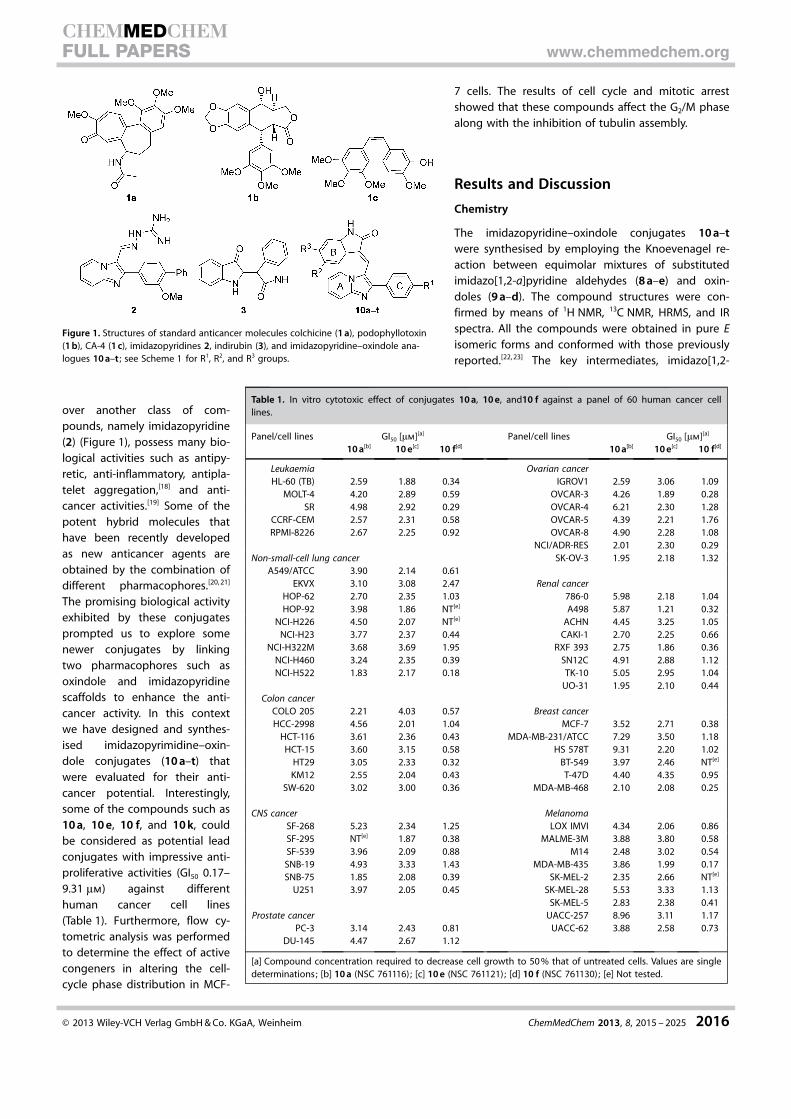
Figure 1. Structures of standard anticancer molecules colchicine (1 a), podophyllotoxin(1 b), CA-4 (1 c), imidazopyridines 2, indirubin (3), and imidazopyridine–oxindole ana-logues 10 a–t ; see Scheme 1 for R1, R2, and R3 groups.
Table 1. In vitro cytotoxic effect of conjugates 10 a, 10 e, and10 f against a panel of 60 human cancer celllines.
Panel/cell lines GI50 [mm][a] Panel/cell lines GI50 [mm][a]
10 a[b] 10 e[c] 10 f[d] 10 a[b] 10 e[c] 10 f[d]
Leukaemia Ovarian cancerHL-60 (TB) 2.59 1.88 0.34 IGROV1 2.59 3.06 1.09
MOLT-4 4.20 2.89 0.59 OVCAR-3 4.26 1.89 0.28SR 4.98 2.92 0.29 OVCAR-4 6.21 2.30 1.28
CCRF-CEM 2.57 2.31 0.58 OVCAR-5 4.39 2.21 1.76RPMI-8226 2.67 2.25 0.92 OVCAR-8 4.90 2.28 1.08
NCI/ADR-RES 2.01 2.30 0.29Non-small-cell lung cancer SK-OV-3 1.95 2.18 1.32
A549/ATCC 3.90 2.14 0.61EKVX 3.10 3.08 2.47 Renal cancer
HOP-62 2.70 2.35 1.03 786-0 5.98 2.18 1.04HOP-92 3.98 1.86 NT[e] A498 5.87 1.21 0.32
NCI-H226 4.50 2.07 NT[e] ACHN 4.45 3.25 1.05NCI-H23 3.77 2.37 0.44 CAKI-1 2.70 2.25 0.66
NCI-H322M 3.68 3.69 1.95 RXF 393 2.75 1.86 0.36NCI-H460 3.24 2.35 0.39 SN12C 4.91 2.88 1.12NCI-H522 1.83 2.17 0.18 TK-10 5.05 2.95 1.04
UO-31 1.95 2.10 0.44Colon cancer
COLO 205 2.21 4.03 0.57 Breast cancerHCC-2998 4.56 2.01 1.04 MCF-7 3.52 2.71 0.38
HCT-116 3.61 2.36 0.43 MDA-MB-231/ATCC 7.29 3.50 1.18HCT-15 3.60 3.15 0.58 HS 578T 9.31 2.20 1.02
HT29 3.05 2.33 0.32 BT-549 3.97 2.46 NT[e]
KM12 2.55 2.04 0.43 T-47D 4.40 4.35 0.95SW-620 3.02 3.00 0.36 MDA-MB-468 2.10 2.08 0.25
CNS cancer MelanomaSF-268 5.23 2.34 1.25 LOX IMVI 4.34 2.06 0.86SF-295 NT[e] 1.87 0.38 MALME-3M 3.88 3.80 0.58SF-539 3.96 2.09 0.88 M14 2.48 3.02 0.54SNB-19 4.93 3.33 1.43 MDA-MB-435 3.86 1.99 0.17SNB-75 1.85 2.08 0.39 SK-MEL-2 2.35 2.66 NT[e]
U251 3.97 2.05 0.45 SK-MEL-28 5.53 3.33 1.13SK-MEL-5 2.83 2.38 0.41
Prostate cancer UACC-257 8.96 3.11 1.17PC-3 3.14 2.43 0.81 UACC-62 3.88 2.58 0.73
DU-145 4.47 2.67 1.12
[a] Compound concentration required to decrease cell growth to 50 % that of untreated cells. Values are singledeterminations; [b] 10 a (NSC 761116); [c] 10 e (NSC 761121); [d] 10 f (NSC 761130); [e] Not tested.
� 2013 Wiley-VCH Verlag GmbH & Co. KGaA, Weinheim ChemMedChem 2013, 8, 2015 – 2025 2016
CHEMMEDCHEMFULL PAPERS www.chemmedchem.org
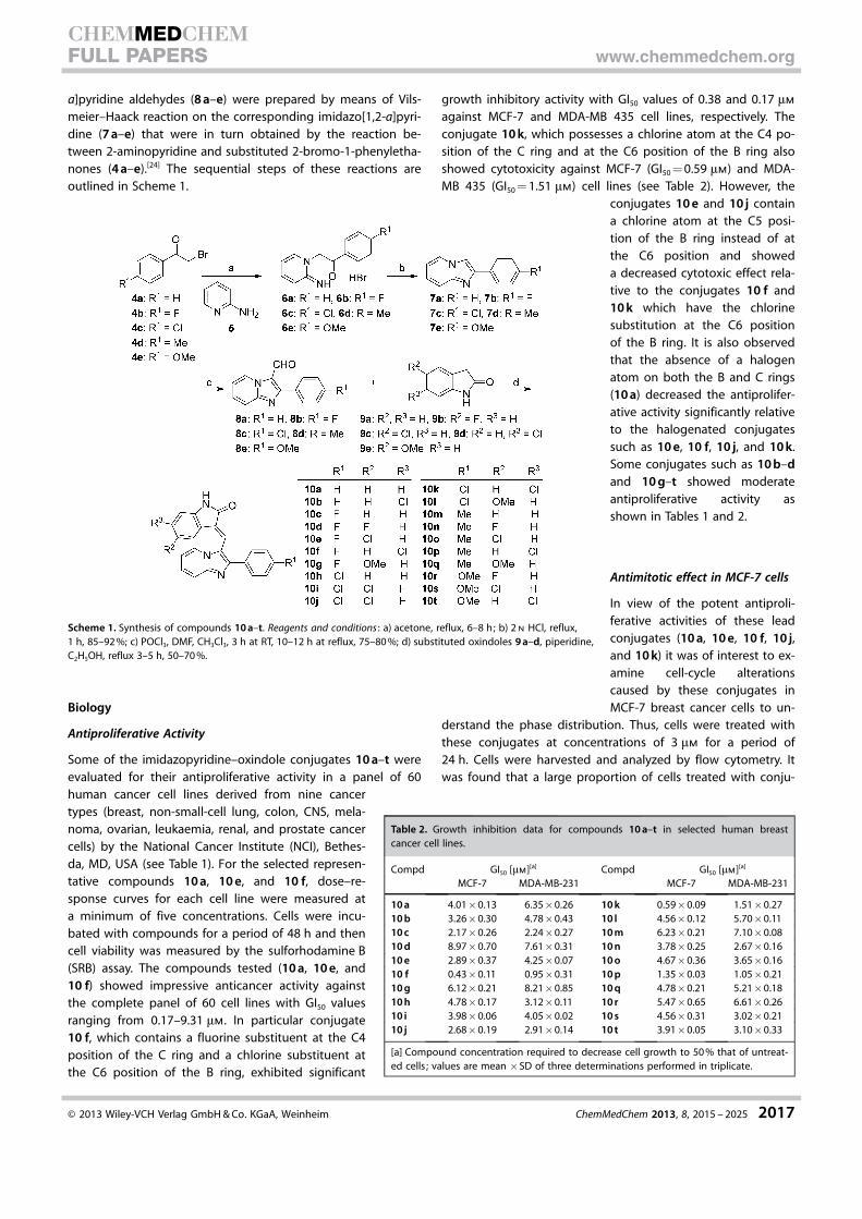
a]pyridine aldehydes (8 a–e) were prepared by means of Vils-meier–Haack reaction on the corresponding imidazo[1,2-a]pyri-dine (7 a–e) that were in turn obtained by the reaction be-tween 2-aminopyridine and substituted 2-bromo-1-phenyletha-nones (4 a–e).[24] The sequential steps of these reactions areoutlined in Scheme 1.
Biology
Antiproliferative Activity
Some of the imidazopyridine–oxindole conjugates 10 a–t wereevaluated for their antiproliferative activity in a panel of 60human cancer cell lines derived from nine cancertypes (breast, non-small-cell lung, colon, CNS, mela-noma, ovarian, leukaemia, renal, and prostate cancercells) by the National Cancer Institute (NCI), Bethes-da, MD, USA (see Table 1). For the selected represen-tative compounds 10 a, 10 e, and 10 f, dose–re-sponse curves for each cell line were measured ata minimum of five concentrations. Cells were incu-bated with compounds for a period of 48 h and thencell viability was measured by the sulforhodamine B(SRB) assay. The compounds tested (10 a, 10 e, and10 f) showed impressive anticancer activity againstthe complete panel of 60 cell lines with GI50 valuesranging from 0.17–9.31 mm. In particular conjugate10 f, which contains a fluorine substituent at the C4position of the C ring and a chlorine substituent atthe C6 position of the B ring, exhibited significant
growth inhibitory activity with GI50 values of 0.38 and 0.17 mm
against MCF-7 and MDA-MB 435 cell lines, respectively. Theconjugate 10 k, which possesses a chlorine atom at the C4 po-sition of the C ring and at the C6 position of the B ring alsoshowed cytotoxicity against MCF-7 (GI50 = 0.59 mm) and MDA-MB 435 (GI50 = 1.51 mm) cell lines (see Table 2). However, the
conjugates 10 e and 10 j containa chlorine atom at the C5 posi-tion of the B ring instead of atthe C6 position and showeda decreased cytotoxic effect rela-tive to the conjugates 10 f and10 k which have the chlorinesubstitution at the C6 positionof the B ring. It is also observedthat the absence of a halogenatom on both the B and C rings(10 a) decreased the antiprolifer-ative activity significantly relativeto the halogenated conjugatessuch as 10 e, 10 f, 10 j, and 10 k.Some conjugates such as 10 b–dand 10 g–t showed moderateantiproliferative activity asshown in Tables 1 and 2.
Antimitotic effect in MCF-7 cells
In view of the potent antiproli-ferative activities of these leadconjugates (10 a, 10 e, 10 f, 10 j,and 10 k) it was of interest to ex-amine cell-cycle alterationscaused by these conjugates inMCF-7 breast cancer cells to un-
derstand the phase distribution. Thus, cells were treated withthese conjugates at concentrations of 3 mm for a period of24 h. Cells were harvested and analyzed by flow cytometry. Itwas found that a large proportion of cells treated with conju-
Scheme 1. Synthesis of compounds 10 a–t. Reagents and conditions : a) acetone, reflux, 6–8 h; b) 2 n HCl, reflux,1 h, 85–92 %; c) POCl3, DMF, CH3Cl3, 3 h at RT, 10–12 h at reflux, 75–80 %; d) substituted oxindoles 9 a–d, piperidine,C2H5OH, reflux 3–5 h, 50–70 %.
Table 2. Growth inhibition data for compounds 10 a–t in selected human breastcancer cell lines.
Compd GI50 [mm][a] Compd GI50 [mm][a]
MCF-7 MDA-MB-231 MCF-7 MDA-MB-231
10 a 4.01�0.13 6.35�0.26 10 k 0.59�0.09 1.51�0.2710 b 3.26�0.30 4.78�0.43 10 l 4.56�0.12 5.70�0.1110 c 2.17�0.26 2.24�0.27 10 m 6.23�0.21 7.10�0.0810 d 8.97�0.70 7.61�0.31 10 n 3.78�0.25 2.67�0.1610 e 2.89�0.37 4.25�0.07 10 o 4.67�0.36 3.65�0.1610 f 0.43�0.11 0.95�0.31 10 p 1.35�0.03 1.05�0.2110 g 6.12�0.21 8.21�0.85 10 q 4.78�0.21 5.21�0.1810 h 4.78�0.17 3.12�0.11 10 r 5.47�0.65 6.61�0.2610 i 3.98�0.06 4.05�0.02 10 s 4.56�0.31 3.02�0.2110 j 2.68�0.19 2.91�0.14 10 t 3.91�0.05 3.10�0.33
[a] Compound concentration required to decrease cell growth to 50 % that of untreat-ed cells ; values are mean �SD of three determinations performed in triplicate.
� 2013 Wiley-VCH Verlag GmbH & Co. KGaA, Weinheim ChemMedChem 2013, 8, 2015 – 2025 2017
CHEMMEDCHEMFULL PAPERS www.chemmedchem.org
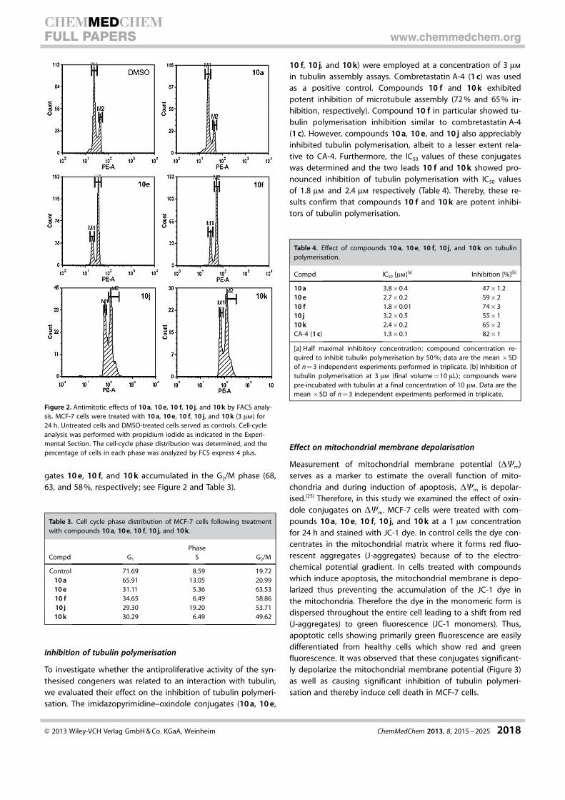
gates 10 e, 10 f, and 10 k accumulated in the G2/M phase (68,63, and 58 %, respectively; see Figure 2 and Table 3).
Inhibition of tubulin polymerisation
To investigate whether the antiproliferative activity of the syn-thesised congeners was related to an interaction with tubulin,we evaluated their effect on the inhibition of tubulin polymeri-sation. The imidazopyrimidine–oxindole conjugates (10 a, 10 e,
10 f, 10 j, and 10 k) were employed at a concentration of 3 mm
in tubulin assembly assays. Combretastatin A-4 (1 c) was usedas a positive control. Compounds 10 f and 10 k exhibitedpotent inhibition of microtubule assembly (72 % and 65 % in-hibition, respectively). Compound 10 f in particular showed tu-bulin polymerisation inhibition similar to combretastatin A-4(1 c). However, compounds 10 a, 10 e, and 10 j also appreciablyinhibited tubulin polymerisation, albeit to a lesser extent rela-tive to CA-4. Furthermore, the IC50 values of these conjugateswas determined and the two leads 10 f and 10 k showed pro-nounced inhibition of tubulin polymerisation with IC50 valuesof 1.8 mm and 2.4 mm respectively (Table 4). Thereby, these re-sults confirm that compounds 10 f and 10 k are potent inhibi-tors of tubulin polymerisation.
Effect on mitochondrial membrane depolarisation
Measurement of mitochondrial membrane potential (DYm)serves as a marker to estimate the overall function of mito-chondria and during induction of apoptosis, DYm is depolar-ised.[25] Therefore, in this study we examined the effect of oxin-dole conjugates on DYm. MCF-7 cells were treated with com-pounds 10 a, 10 e, 10 f, 10 j, and 10 k at a 1 mm concentrationfor 24 h and stained with JC-1 dye. In control cells the dye con-centrates in the mitochondrial matrix where it forms red fluo-rescent aggregates (J-aggregates) because of to the electro-chemical potential gradient. In cells treated with compoundswhich induce apoptosis, the mitochondrial membrane is depo-larized thus preventing the accumulation of the JC-1 dye inthe mitochondria. Therefore the dye in the monomeric form isdispersed throughout the entire cell leading to a shift from red(J-aggregates) to green fluorescence (JC-1 monomers). Thus,apoptotic cells showing primarily green fluorescence are easilydifferentiated from healthy cells which show red and greenfluorescence. It was observed that these conjugates significant-ly depolarize the mitochondrial membrane potential (Figure 3)as well as causing significant inhibition of tubulin polymeri-sation and thereby induce cell death in MCF-7 cells.
Figure 2. Antimitotic effects of 10 a, 10 e, 10 f, 10 j, and 10 k by FACS analy-sis. MCF-7 cells were treated with 10 a, 10 e, 10 f, 10 j, and 10 k (3 mm) for24 h. Untreated cells and DMSO-treated cells served as controls. Cell-cycleanalysis was performed with propidium iodide as indicated in the Experi-mental Section. The cell-cycle phase distribution was determined, and thepercentage of cells in each phase was analyzed by FCS express 4 plus.
Table 3. Cell cycle phase distribution of MCF-7 cells following treatmentwith compounds 10 a, 10 e, 10 f, 10 j, and 10 k.
PhaseCompd G1 S G2/M
Control 71.69 8.59 19.7210 a 65.91 13.05 20.9910 e 31.11 5.36 63.5310 f 34.65 6.49 58.8610 j 29.30 19.20 53.7110 k 30.29 6.49 49.62
Table 4. Effect of compounds 10 a, 10 e, 10 f, 10 j, and 10 k on tubulinpolymerisation.
Compd IC50 [mm][a] Inhibition [%][b]
10 a 3.8�0.4 47�1.210 e 2.7�0.2 59�210 f 1.8�0.01 74�310 j 3.2�0.5 55�110 k 2.4�0.2 65�2CA-4 (1 c) 1.3�0.1 82�1
[a] Half maximal inhibitory concentration: compound concentration re-quired to inhibit tubulin polymerisation by 50 %; data are the mean �SDof n = 3 independent experiments performed in triplicate. [b] Inhibition oftubulin polymerisation at 3 mm (final volume = 10 mL); compounds werepre-incubated with tubulin at a final concentration of 10 mm. Data are themean �SD of n = 3 independent experiments performed in triplicate.
� 2013 Wiley-VCH Verlag GmbH & Co. KGaA, Weinheim ChemMedChem 2013, 8, 2015 – 2025 2018
CHEMMEDCHEMFULL PAPERS www.chemmedchem.org
Effect on chromatin condensation
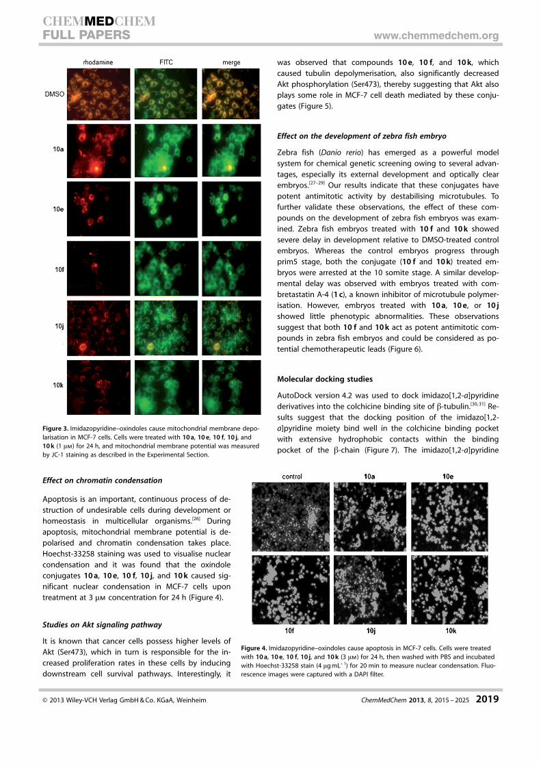
Apoptosis is an important, continuous process of de-struction of undesirable cells during development orhomeostasis in multicellular organisms.[26] Duringapoptosis, mitochondrial membrane potential is de-polarised and chromatin condensation takes place.Hoechst-33258 staining was used to visualise nuclearcondensation and it was found that the oxindoleconjugates 10 a, 10 e, 10 f, 10 j, and 10 k caused sig-nificant nuclear condensation in MCF-7 cells upontreatment at 3 mm concentration for 24 h (Figure 4).
Studies on Akt signaling pathway
It is known that cancer cells possess higher levels ofAkt (Ser473), which in turn is responsible for the in-creased proliferation rates in these cells by inducingdownstream cell survival pathways. Interestingly, it
was observed that compounds 10 e, 10 f, and 10 k, whichcaused tubulin depolymerisation, also significantly decreasedAkt phosphorylation (Ser473), thereby suggesting that Akt alsoplays some role in MCF-7 cell death mediated by these conju-gates (Figure 5).
Effect on the development of zebra fish embryo
Zebra fish (Danio rerio) has emerged as a powerful modelsystem for chemical genetic screening owing to several advan-tages, especially its external development and optically clearembryos.[27–29] Our results indicate that these conjugates havepotent antimitotic activity by destabilising microtubules. Tofurther validate these observations, the effect of these com-pounds on the development of zebra fish embryos was exam-ined. Zebra fish embryos treated with 10 f and 10 k showedsevere delay in development relative to DMSO-treated controlembryos. Whereas the control embryos progress throughprim5 stage, both the conjugate (10 f and 10 k) treated em-bryos were arrested at the 10 somite stage. A similar develop-mental delay was observed with embryos treated with com-bretastatin A-4 (1 c), a known inhibitor of microtubule polymer-isation. However, embryos treated with 10 a, 10 e, or 10 jshowed little phenotypic abnormalities. These observationssuggest that both 10 f and 10 k act as potent antimitotic com-pounds in zebra fish embryos and could be considered as po-tential chemotherapeutic leads (Figure 6).
Molecular docking studies
AutoDock version 4.2 was used to dock imidazo[1,2-a]pyridinederivatives into the colchicine binding site of b-tubulin.[30, 31] Re-sults suggest that the docking position of the imidazo[1,2-a]pyridine moiety bind well in the colchicine binding pocketwith extensive hydrophobic contacts within the bindingpocket of the b-chain (Figure 7). The imidazo[1,2-a]pyridine
Figure 3. Imidazopyridine–oxindoles cause mitochondrial membrane depo-larisation in MCF-7 cells. Cells were treated with 10 a, 10 e, 10 f, 10 j, and10 k (1 mm) for 24 h, and mitochondrial membrane potential was measuredby JC-1 staining as described in the Experimental Section.
Figure 4. Imidazopyridine–oxindoles cause apoptosis in MCF-7 cells. Cells were treatedwith 10 a, 10 e, 10 f, 10 j, and 10 k (3 mm) for 24 h, then washed with PBS and incubatedwith Hoechst-33258 stain (4 mg mL�1) for 20 min to measure nuclear condensation. Fluo-rescence images were captured with a DAPI filter.
� 2013 Wiley-VCH Verlag GmbH & Co. KGaA, Weinheim ChemMedChem 2013, 8, 2015 – 2025 2019
CHEMMEDCHEMFULL PAPERS www.chemmedchem.org
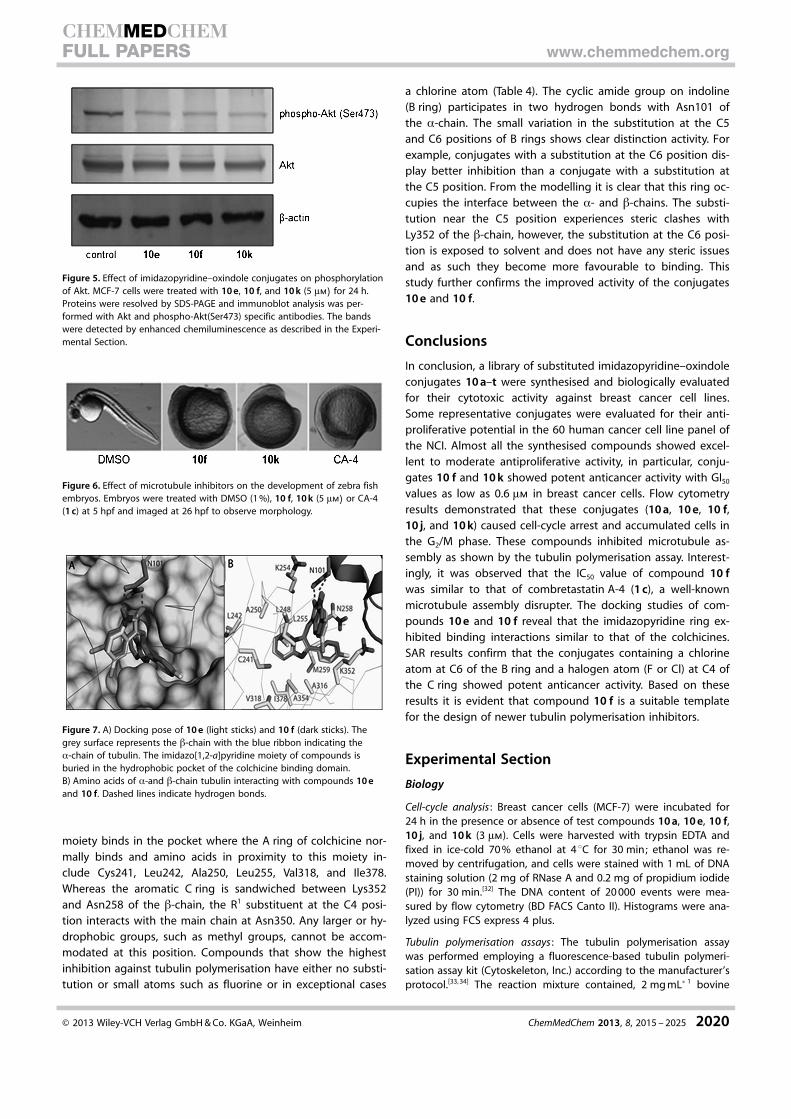
moiety binds in the pocket where the A ring of colchicine nor-mally binds and amino acids in proximity to this moiety in-clude Cys241, Leu242, Ala250, Leu255, Val318, and Ile378.Whereas the aromatic C ring is sandwiched between Lys352and Asn258 of the b-chain, the R1 substituent at the C4 posi-tion interacts with the main chain at Asn350. Any larger or hy-drophobic groups, such as methyl groups, cannot be accom-modated at this position. Compounds that show the highestinhibition against tubulin polymerisation have either no substi-tution or small atoms such as fluorine or in exceptional cases
a chlorine atom (Table 4). The cyclic amide group on indoline(B ring) participates in two hydrogen bonds with Asn101 ofthe a-chain. The small variation in the substitution at the C5and C6 positions of B rings shows clear distinction activity. Forexample, conjugates with a substitution at the C6 position dis-play better inhibition than a conjugate with a substitution atthe C5 position. From the modelling it is clear that this ring oc-cupies the interface between the a- and b-chains. The substi-tution near the C5 position experiences steric clashes withLy352 of the b-chain, however, the substitution at the C6 posi-tion is exposed to solvent and does not have any steric issuesand as such they become more favourable to binding. Thisstudy further confirms the improved activity of the conjugates10 e and 10 f.
Conclusions
In conclusion, a library of substituted imidazopyridine–oxindoleconjugates 10 a–t were synthesised and biologically evaluatedfor their cytotoxic activity against breast cancer cell lines.Some representative conjugates were evaluated for their anti-proliferative potential in the 60 human cancer cell line panel ofthe NCI. Almost all the synthesised compounds showed excel-lent to moderate antiproliferative activity, in particular, conju-gates 10 f and 10 k showed potent anticancer activity with GI50
values as low as 0.6 mm in breast cancer cells. Flow cytometryresults demonstrated that these conjugates (10 a, 10 e, 10 f,10 j, and 10 k) caused cell-cycle arrest and accumulated cells inthe G2/M phase. These compounds inhibited microtubule as-sembly as shown by the tubulin polymerisation assay. Interest-ingly, it was observed that the IC50 value of compound 10 fwas similar to that of combretastatin A-4 (1 c), a well-knownmicrotubule assembly disrupter. The docking studies of com-pounds 10 e and 10 f reveal that the imidazopyridine ring ex-hibited binding interactions similar to that of the colchicines.SAR results confirm that the conjugates containing a chlorineatom at C6 of the B ring and a halogen atom (F or Cl) at C4 ofthe C ring showed potent anticancer activity. Based on theseresults it is evident that compound 10 f is a suitable templatefor the design of newer tubulin polymerisation inhibitors.
Experimental Section
Biology
Cell-cycle analysis : Breast cancer cells (MCF-7) were incubated for24 h in the presence or absence of test compounds 10 a, 10 e, 10 f,10 j, and 10 k (3 mm). Cells were harvested with trypsin EDTA andfixed in ice-cold 70 % ethanol at 4 8C for 30 min; ethanol was re-moved by centrifugation, and cells were stained with 1 mL of DNAstaining solution (2 mg of RNase A and 0.2 mg of propidium iodide(PI)) for 30 min.[32] The DNA content of 20 000 events were mea-sured by flow cytometry (BD FACS Canto II). Histograms were ana-lyzed using FCS express 4 plus.
Tubulin polymerisation assays : The tubulin polymerisation assaywas performed employing a fluorescence-based tubulin polymeri-sation assay kit (Cytoskeleton, Inc.) according to the manufacturer’sprotocol.[33, 34] The reaction mixture contained, 2 mg mL�1 bovine
Figure 5. Effect of imidazopyridine–oxindole conjugates on phosphorylationof Akt. MCF-7 cells were treated with 10 e, 10 f, and 10 k (5 mm) for 24 h.Proteins were resolved by SDS-PAGE and immunoblot analysis was per-formed with Akt and phospho-Akt(Ser473) specific antibodies. The bandswere detected by enhanced chemiluminescence as described in the Experi-mental Section.
Figure 6. Effect of microtubule inhibitors on the development of zebra fishembryos. Embryos were treated with DMSO (1 %), 10 f, 10 k (5 mm) or CA-4(1 c) at 5 hpf and imaged at 26 hpf to observe morphology.
Figure 7. A) Docking pose of 10 e (light sticks) and 10 f (dark sticks). Thegrey surface represents the b-chain with the blue ribbon indicating thea-chain of tubulin. The imidazo[1,2-a]pyridine moiety of compounds isburied in the hydrophobic pocket of the colchicine binding domain.B) Amino acids of a-and b-chain tubulin interacting with compounds 10 eand 10 f. Dashed lines indicate hydrogen bonds.
� 2013 Wiley-VCH Verlag GmbH & Co. KGaA, Weinheim ChemMedChem 2013, 8, 2015 – 2025 2020
CHEMMEDCHEMFULL PAPERS www.chemmedchem.org
brain tubulin, 10 mm fluorescent reporter, and 1 mm GTP in thepresence or absence of test compounds in PEM buffer (80 mm
PIPES, 0.5 mm EGTA, 2 mm MgCl2, pH 6.9) at 37 8C in a finalvolume of 10 mL. Fluorescence readings were recorded on a Tecanmultimode reader at l 360/420 nm (excitation/emission) every2 min for up to 3 h. Combretastatin A-4 (1 c) was used as positivecontrol under similar conditions. To evaluate the IC50 values of thecompounds against tubulin assembly, the compounds 10 a, 10 e,10 f, 10 j, and 10 k were incubated at various concentrations (1–5 mm) with tubulin.
Analysis of mitochondrial membrane potential : MCF-7 cells were in-cubated for a period of 24 h in the presence or absence of testcompounds 10 a, 10 e, 10 f, 10 j, and 10 k (1 mm). At the end oftreatment, the medium was aspirated, cells were washed withmedium without FBS, and JC-1 stain (Sigma cat. no. CS0390) wasadded to the cells for 20 min at 37 8C under humidified atmos-phere. Cells were washed again with medium. The cells were cov-ered with medium and observed under a fluorescence microscopeequipped with rhodamine and FITC filters.[25]
Hoechst-33258 staining : MCF-7 cells were incubated for a period of24 h in the presence or absence of test compounds 10 a, 10 e, 10 f,10 j and 10 k (3 mm). At the end of treatment, the medium was as-pirated, cells were washed with medium without FBS, andHoechst-33258 stain (Invitrogen cat. no. H3570) was added to thecells for 20 min at 37 8C under humidified atmosphere. Cells werewashed again with medium. The cells were covered with mediumand observed under a fluorescence microscope equipped withDAPI filter.[26]
Western blot analysis : After the treatment of MCF-7 cells with com-pounds 10 e, 10 f and 10 k at 3 mm, cells were washed with ice-cold phosphate-buffered saline (PBS) and homogenised in 100 mLof radioimmunoprecipitation assay (RIPA) buffer (20 mm Tris·HCl,pH 7.4, 2.5 mm EDTA, 1 % Triton X-100, 1 % sodium deoxycholate,1 % SDS, 100 mm NaCl, 100 mm NaF) containing 1 mm sodium or-thovanadate and a mixture of protease inhibitors.[35] The homoge-nate was centrifuged at 750 g for 10 min at 4 8C to pellet out thenuclei. The remaining supernatant was centrifuged for 30 min at12 000 g. Proteins were resolved on SDS-PAGE and blotted onto ni-trocellulose membranes. Membranes were probed with AKT andphospho-AKT (Ser473) procured from Millipore. The detection wascarried out with HRP-labelled rabbit anti-mouse IgG (Amersham)using an enhanced chemiluminescence detection system (ECL Ad-vanced Kit, GE Health care).
Zebra fish maintenance and screening : Wild-type zebra fish (Daniorerio) were raised and maintained at 28.5 8C with 14/10 h light–dark cycle. Two or three pairs of zebra fish were synchronouslymated and embryos were pooled. The embryos were dechorionat-ed and 3–4 embryos were dispensed in 200 mL of E3 embryomedium (5 mm NaCl, 0.17 mm KCl, 0.44 mm CaCl2, 0.68 mm MgCl2)into each well of a 96-well plate. The embryos were treated withcompounds 10 a, 10 e, 10 f, 10 j, 10 k, and combretastatin A-4 (1 c)(5, 10, and 25 mm) and DMSO (1 %) at 5 h post-fertilisation (hpf)and incubated at 28.5 8C. Approximately 50 embryos werescreened for each compound in biological replicates. They wereobserved after 28 hpf with a Leica stereomicroscope. Only thosecompounds that resulted in similar phenotypic defects in most ofthe embryos were considered active.
Docking studies
Molecular docking simulations were performed using AutoDock 4.2and the atomic coordinates of b-tubulin as the target (PDB code3E22). After removing the ligand and solvent molecules, hydrogenatoms and Kollman charges were added to each protein atom. Co-ordinates for each compound were obtained from Chemdraw11followed by MM2 energy minimisation. Docking was carried out byAutoDock 4.2 in the colchicine binding pocket.[30, 31] Grid map inAutoDock was used to define the interaction of protein and ligandin the binding pocket. The grid map was used with 60 points ineach x, y, and z direction, equally spaced at 0.375 �. Docking wasperformed using the Lamarckian genetic algorithm.[36] Each dock-ing experiment was performed 100 times, yielding 100 dockedconformations. Parameters used for the docking were as follows:population size of 150; random starting position and conforma-tion; maximal mutation of 2 � translation and 50 degrees rotation;elitism of 1; mutation rate of 0.02 and crossover rate of 0.8; andlocal search rate of 0.06. Simulations were performed with a maxi-mum of 1.5 million energy evaluations and a maximum of 50 000generations. Final docked conformations were clustered using a tol-erance of 1 � root mean square deviation (RMSD). The best modelwas chosen based on the most favourable stabilisation energy.
Chemistry
General : All chemicals and reagents were obtained from Lancaster(Alfa Aesar, Johnson Matthey Company, Ward Hill, MA, USA),Sigma–Aldrich (St. Louis, MO, USA), and Spectrochem Pvt. Ltd.(Mumbai, India) and were used without further purification. Reac-tions were monitored by TLC performed on silica gel coated glassplates containing 60 GF254 with visualisation achieved by UV lightor iodine indicator. Column chromatography was performed withMerck 60–120 mesh silica gel. 1H and 13C NMR spectra were record-ed on Bruker UXNMR/XWIN-NMR (300 MHz) or Inova Varian VXRUnity (500 MHz) instruments. Chemical shifts (d) are reported inppm downfield from an internal TMS standard. ESI mass spectrawere recorded on a Micro mass Quattro LC using ESI + softwarewith a capillary voltage of 3.98 kV and an ESI mode positive iontrap detector. High-resolution mass spectra were recorded ona QSTAR XL Hybrid MS–MS mass spectrometer. Melting pointswere determined with an Electro thermal melting point apparatus,and are uncorrected. 1H NMR, 13C NMR, and HRMS spectra of finalcompounds 10 a–t are provided in the Supporting Information.
2-Phenylimidazo[1,2-a]pyridine (7 a): 2-Aminopyridine 5 (1 g,10 mmol) was dissolved in acetone (100 mL) and treated with 4 a(2.1 g, 10 mmol). The reaction mixture was heated at reflux for 4–5 h, and the resulting salt (6 a) was collected by filtration, washedwith acetone, and then dissolved in 3 n HCl (200 mL) and heatedat reflux for 1 h. Before complete cooling, the solution was cau-tiously basified by dropwise addition of 15 % aq NH4OH to pH 8.The resulting base was collected by filtration and crystallised fromEtOH to afford compound 7 a as a white solid (1.75 g, 85 % yield);mp: 144–146 8C; 1H NMR (300 MHz, CDCl3): d= 6.70 (t, J = 6.7 Hz,1 H), 7.10 (dt, J = 6.7, 1.5 Hz, 1 H), 7.25 (dt, J = 6.7, 1.5 Hz, 1 H), 7.37(t, J = 7.5 Hz, 2 H), 7.57 (d, J = 9.0 Hz, 1 H), 7.79 (s, 1 H), 7.89 (d, J =7.5 Hz, 2 H), 8.05 ppm (d, J = 6.7 Hz, 1 H); MS (ESI): m/z 195 [M + H]+
.
2-(4-Chlorophenyl)imidazo[1,2-a]pyridine (7 c): Compound 7 cwas prepared according to the method described for compound7 a, employing 2-Bromo-1-(4-chlorophenyl)ethanone 4 c (2.46 g,10 mmol) and 2-aminopyridine 5 (1 g, 10 mmol) to obtain the pure
� 2013 Wiley-VCH Verlag GmbH & Co. KGaA, Weinheim ChemMedChem 2013, 8, 2015 – 2025 2021
CHEMMEDCHEMFULL PAPERS www.chemmedchem.org
product 7 c as a white solid (2.6 g, 89 % yield); mp: 171–173 8C;1H NMR (300 MHz, CDCl3): d= 6.77 (t, J = 6.6 Hz, 1 H), 7.07–7.21 (m,3 H), 7.62 (d, J = 9.1 Hz, 1 H), 7.79 (s, 1 H), 7.87–7.96 (m, 2 H),8.10 ppm (d, J = 6.8 Hz, 1 H); MS (ESI): m/z 229 [M + H]+ .
2-para-Tolylimidazo[1,2-a]pyridine (7 d): Compound 7d was pre-pared according to the method described for compound 7 a, em-ploying 2-bromo-1-p-tolylethanone 4 d (2.6 g, 10 mmol) and 2-ami-nopyridine 5 (1 g, 10 mmol) to obtain the pure product 7 d asa white solid (2 g, 91 % yield); mp: 165–168 8C; 1H NMR (300 MHz,CDCl3): d= 2.38 (s, 3 H), 6.78 (dt, J = 6.7 Hz, J = 1.1 Hz, 1 H), 7.18 (dt,J = 6.7, 1.2 Hz, 1 H), 7.24 (s, 1 H), 7.66 (d, J = 9.1 Hz, 1 H), 7.82–7.87(m, 4 H), 8.11 ppm (td, J = 6.7 Hz, J = 1.1 Hz, 1 H); MS (ESI): m/z 209[M + H]+ .
2-Phenylimidazo[1,2-a]pyridine-3-carbaldehyde (8 a): The Vilsmei-er reagent was prepared at 0–5 8C by dropping POCl3 (2.5 mL,10.5 mmol) into a stirred solution of DMF (2 mL, 10 mmol) in CHCl3
(10 mL). The 2-phenylimidazo[1,2-a]pyridine 7 a (500 mg, 2.6 mmol)in chloroform (20 mL) was added to Vilsmeier reagent whilst main-taining stirring and cooling. The reaction mixture was kept at RTfor 3 h and at reflux for 10–12 h (according to a TLC test). Chloro-form was removed under reduced pressure and the resulting oilyliquid was poured onto ice. The aldehyde 8 a was collected by fil-tration and crystallised from EtOH (5 mL) to obtain the pure prod-uct 8 a as a white solid (418 mg, 73 % yield); mp: 155–158 8C;1H NMR (300 MHz, CDCl3): d= 7.14 (t, J = 6.8 Hz, 1 H), 7.50–7.62 (m,4 H), 7.81–7.86 (m, 3 H), 9.68 (d, J = 6.7 Hz, 1 H), 10.08 ppm (s, 1 H);MS (ESI): m/z 223 [M + H]+ .
2-(4-Chlorophenyl)imidazo[1,2-a]pyridine-3-carbaldehyde (8 c):Compound 8 c was prepared according to the method describedfor compound 8 a, employing 2-(4-chlorophenyl)imidazo[1,2-a]pyri-dine 7 c (500 mg, 2.19 mmol) to obtain the pure product 8 c asa white solid (393 mg, 70 % yield); mp: 151–153 8C; 1H NMR(300 MHz, CDCl3): d= 7.16 (t, J = 6.9 Hz, 1 H), 7.52 (d, J = 8.5 Hz, 2 H),7.61 (t, J = 6.9 Hz, 1 H), 7.75–7.85 (m, 3 H), 9.66 (d, J = 6.9 Hz, 1 H),10.05 ppm (s, 1 H); MS (ESI): m/z 257 [M + H]+ .
2-para-Tolylimidazo[1,2-a]pyridine-3-carbaldehyde (8 d): Com-pound 8 c was prepared according to the method described forcompound 8 a, employing 2-(4-chlorophenyl)imidazo[1,2-a]pyridine7 d (500 mg, 2.4 mmol) to obtain the pure product 8 d as a whitesolid, yield (442 mg, 78 % yield); mp: 169–171 8C; 1H NMR(300 MHz, CDCl3): d= 2.45 (s, 3 H), 7.12 (dt, J = 6.9, 1.2 Hz, 1 H), 7.35(d, J = 7.9 Hz, 2 H), 7.58 (dt, J = 6.8 Hz, J = 1.2 Hz, 1 H), 7.74 (d, J =8.1 Hz, 2 H), 7.80 (td, J = 9.0 Hz, J = 1.1 Hz, 1 H), 9.66 (td, J = 6.9,1.1 Hz, 1 H), 10.06 ppm (s, 1 H); MS (ESI): m/z 237 [M + H]+ .
(E)-3-((2-Phenylimidazo[1,2-a]pyridin-3-yl)methylene)indolin-2-one (10 a): An appropriate mixture of 2-phenylimidazo[1,2-a]pyri-dine-3-carbaldehyde 8 a (200 mg 0.9 mmol) and indolin-2-one 9 a(120 mg, 0.9 mmol) was dissolved in EtOH (10 mL) and piperidine(2–3 drops) was added. The reaction mixture was held at reflux for3–5 h (according to a TLC test) and the precipitate formed on cool-ing was collected by filtration and crystallisation with EtOH toobtain the pure product 10 a as a yellow solid (200 mg, 66 %); mp:288–299 8C; IR (KBr) n= 3405, 3075, 1712, 745 cm�1; 1H NMR(300 MHz, CDCl3): d= 6.37 (d, J = 7.9 Hz, 1 H), 6.83 (t, J = 7.9 Hz, 1 H),7.02 (d, J = 7.9 Hz, 1 H), 7.32 (t, J = 7.9 Hz, 1 H), 7.48 (t, J = 8.9 Hz,1 H), 7.52–7.60 (m, 3 H), 7.73 (d, J = 6.9 Hz, 2 H), 7.79 (s, 1 H), 8.03 (t,J = 8.9 Hz, 1 H), 8.10 (d, J = 8.9 Hz, 1 H), 8.30 ppm (d, J = 8.9 Hz, 1 H),9.26 (s, 1 H); 13C NMR (300 MHz, CDCl3 + 1 drop [D]TFA): d= 169.6,141.8, 140.5, 137.5, 134.2, 132.4, 131.8, 129.8, 128.9, 128.3, 126.7,125.4, 124.9, 123.4, 121.4, 119.4, 118.1, 115.8, 114.0, 111.4 ppm; MS
(ESI): 338 [M + H]+ ; HRMS (ESI) calcd for C22H16N3O [M + H]+
338.1287; found: 338.1286.
(E)-6-Chloro-3-((2-phenylimidazo[1,2-a]pyridin-3-yl)methylene)in-dolin-2-one (10 b): Compound 10 b was prepared according tothe method described for compound 10 a, employing2-phenylimidazo[1,2-a]pyridine-3-carbaldehyde 8 a (200 mg,0.9 mmol) and 6-chloroindolin-2-one 9 d (150 mg, 0.9 mmol) toobtain the pure product 10 b as a yellow solid (197 mg, 59 %); mp:309–310 8C; IR (KBr) n= 3408, 3004, 1712, 754 cm�1; 1H NMR(300 MHz, CDCl3): d= 6.32 (d, J = 7.6 Hz, 1 H), 6.78 (dd, J = 2.0 Hz,J = 7.6 Hz, 1 H), 7.10 (s, 1 H), 7.44–7.48 (m, 2 H), 7.55–7.63 (m, 2 H),7.68 (d, J = 7.1 Hz, 2 H), 7.80 (s, 1 H), 8.00 (t, J = 8.8 Hz, 1 H), 8.04 (d,J = 8.8 Hz, 1 H), 8.19 (d, J = 8.8 Hz, 1 H), 9.24 ppm (s,1 H); 13C NMR(300 MHz, CDCl3 + 1 drop [D]TFA): d= 166.7, 143.0, 141.9, 140.7,137.8, 133.7, 131.7, 129.6, 129.0, 128.4, 126.3, 125.6, 123.0, 122.1,120.5, 117.9, 115.3, 114.3, 113.1, 111.6 ppm; MS(ESI): 372 [M + H]+ ;HRMS (ESI) calcd for C22H15N3OCl [M + H]+ 372.09; found: 372.09.
(E)-3-((2-(4-Fluorophenyl)imidazo[1,2-a]pyridin-3-yl)methylene)indolin-2-one (10 c): Compound 10 c was prepared according tothe method described for compound 10 a, employing 2-(4-fluorophenyl)imidazo[1,2-a]pyridine-3-carbaldehyde 8 b (200 mg,0.83 mmol) and indolin-2-one 9 a (111 mg, 0.83 mmol) to obtainthe pure product 10 c as a yellow solid (204 mg, 69 %); mp: 247–248 8C; IR (KBr) n= 3430, 3188, 1691, 757 cm�1; 1H NMR (300 MHz,CDCl3): d= 6.35 (d, J = 7.8 Hz, 1 H), 6.82 (t, J = 7.8 Hz, 1 H), 6.98 (d,J = 7.8 Hz, 1 H), 7.22 (t, J = 7.8 Hz, 1 H), 7.26–7.38 (m, 2 H), 7.45 (t,J = 8.9 Hz, 1 H), 7.73–7.80 (m, 3 H), 8.00 (t, J = 8.9 Hz, 1 H), 8.06 (d,J = 8.9 Hz, 1 H), 8.33 (d, J = 8.9 Hz, 1 H), 8.98 ppm (s, 1 H): 13C NMR(300 MHz, CDCl3 + 1 drop [D]TFA): d= 165.5, 163.4, 142.1, 140.7,136.7, 133.8, 132.33, 130.7, 128.2, 126.5, 124.8, 123.1, 122.1, 121.4,119.4, 117.8, 117.2, 115.0, 114.2, 111.1 ppm; MS (ESI): 355 [M + H]+ ;HRMS (ESI) calcd for C22H15N3OF [M + H]+ 356.1194; found:356.1201.
(E)-5-Fluoro-3-((2-(4-fluorophenyl)imidazo[1,2-a]pyridin-3-yl)me-thylene)indolin-2-one (10 d): Compound 10 d was prepared ac-cording to the method described for compound 10 a, employing2-(4-fluorophenyl)imidazo[1,2-a]pyridine-3-carbaldehyde 8 b(200 mg, 0.83 mmol) and 5-fluoroindolin-2-one 9 b (126 mg,0.83 mmol) to obtain the pure product 10 d as a yellow solid(159 mg, 51 %); mp: 291–292 8C; IR (KBr) n= 3431, 3004, 1633,772 cm�1; 1H NMR (300 MHz, CDCl3): d= 6.16 (dd, J = 2.0 Hz, J =7.5 Hz, 1 H), 7.02 (dd, J = 2.9, 7.5 Hz, 1 H), 7.17 (dt, J = 2.0, 7.5 Hz,1 H), 7.26–7.31 (m, 2 H), 7.45–7.48 (m, 1 H), 7.69–7.75 (m, 2 H), 7.82(s, 1 H), 8.00 (t, J = 8.4 Hz, 1 H), 8.05 (d, J = 8.4 Hz, 1 H), 8.18 (d, J =8.4 Hz, 1 H), 9.21 ppm (s, 1 H); 13C NMR (300 MHz, CDCl3 + 1 drop[D]TFA): d= 165.7, 163.7, 140.7, 140.1, 137.4, 137.1, 136.9, 134.6,131.4, 130.5, 128.2, 126.3, 121.6, 118.4, 117.4, 117.3, 114.2, 113.1,112.2 ppm; MS (ESI): 374 [M + H]+ ; HRMS (ESI) calcd forC22H14N3OF2 [M + H]+ 374.1105; found: 374.1112.
(E)-5-Chloro-3-((2-(4-fluorophenyl)imidazo[1,2-a]pyridin-3-yl)me-thylene)indolin-2-one (10 e): Compound 10 e was prepared ac-cording to the method described for compound 10 a, employing2-(4-fluorophenyl)imidazo[1,2-a]pyridine-3-carbaldehyde 8 b(200 mg, 0.83 mmol) and 5-chloroindolin-2-one 9 c (139 mg,0.83 mmol) to obtain the pure product 10 e as a yellow solid(188 mg, 58 %); mp: 299–300 8C; IR (KBr) n= 3430, 3226, 1702,751 cm�1; 1H NMR (300 MHz, CDCl3): d= 6.39 (s, 1 H), 6.89 (d, J =7.8 Hz, 1 H), 7.18–7.23 (m, 1 H), 7.53 (t, J = 8.2 Hz, 1 H), 7.71–7.78 (m,2 H), 7.82 (s, 1 H), 7.97–8.11 (m, 3 H), 8.21 (d, J = 8.2 Hz, 1 H), 8.33 (d,J = 8.2 Hz, 1 H), 9.06 ppm (s, 1 H); 13C NMR (300 MHz, CDCl3 +1 drop [D]TFA): d= 169.6, 165.7, 140.6, 139.8, 137.0, 134.8, 132.0,
� 2013 Wiley-VCH Verlag GmbH & Co. KGaA, Weinheim ChemMedChem 2013, 8, 2015 – 2025 2022
CHEMMEDCHEMFULL PAPERS www.chemmedchem.org

131.3, 130.4, 128.9, 128.2, 126.4, 125.0, 121.6, 120.5, 118.6, 117.5,117.3, 113.7, 112.2, 111.4 ppm; MS (ESI): 390 [M + H]+ ; HRMS (ESI)calcd for C22H14N3OClF [M + H]+ 390.08; found: 390.08.
(E)-6-Chloro-3-((2-(4-fluorophenyl)imidazo[1,2-a]pyridin-3-yl)me-thylene)indolin-2-one (10 f): Compound 10 f was prepared ac-cording to the method described for compound 10 a, employing2-(4-fluorophenyl)imidazo[1,2-a]pyridine-3-carbaldehyde 8 b(200 mg, 0.83 mmol) and 6-chloroindolin-2-one 9 d (139 mg,0.83 mmol) to obtain the pure product 10 f as a yellow solid(204 mg, 63 %); mp: 315–316 8C; IR (KBr) n= 3428, 3079, 1712,758 cm�1; 1H NMR (300 MHz, CDCl3): d= 6.24 (d, J = 7.1 Hz, 1 H),6.71 (d, J = 7.1 Hz, 1 H), 7.09–7.16 (m, 1 H), 7.41 (t, J = 6.9 Hz, 1 H),7.64–7.70 (m, 3 H), 7.89 (t, J = 8.7 Hz, 1 H), 7.94 (d, J = 7.7 Hz, 2 H),8.12 (d, J = 6.9 Hz, 1 H), 8.22 (d, J = 6.9 Hz, 1 H), 8.92 ppm (s, 1 H);13C NMR (300 MHz, CDCl3 + 1 drop [D]TFA): d= 169.4, 163.6, 142.8,140.6, 138.5, 136.7, 134.4, 132.3, 130.5, 128.1, 126.4, 125.6, 123.4,122.3, 121.6, 118.3, 117.2, 115.5, 114.0, 112.0 ppm; MS (ESI): 390[M + H]+ ; HRMS (ESI) calcd for C22H14N3OClF [M + H]+ 390.08;found: 390.08.
(E)-3-((2-(4-Fluorophenyl)imidazo[1,2-a]pyridin-3-yl)methylene)-5-methoxyindolin-2-one (10 g): Compound 10 g was prepared ac-cording to the method described for compound 10 a, employing2-(4-fluorophenyl)imidazo[1,2-a]pyridine-3-carbaldehyde 8 b(200 mg, 0.83 mmol) and 5-methoxyindolin-2-one 9 e (136 mg,0.83 mmol) to obtain the pure product 10 g as a yellow solid(154 mg, 48 %); mp: 261–262 8C; IR (KBr) n= 3457, 3053, 1709,746 cm�1; 1H NMR (300 MHz, CDCl3): d= 3.48 (s, 3 H, OCH3), 5.94 (s,1 H), 6.86 (dd, J = 2.2, 7.2 Hz, 1 H), 6.91 (d, J = 7.2 Hz, 1 H), 7.52 (t,J = 7.6 Hz, 1 H), 7.76–7.78 (m, 3 H), 8.04 (t, J = 7.6 Hz, 1 H), 8.14 (d,J = 7.6 Hz, 1 H), 8.26–8.29 (m, 3 H), 9.12 ppm (s, 1 H); 13C NMR(300 MHz, CDCl3 + 1 drop [D]TFA): d= 169.6, 163.6, 156.0, 140.5,136.5, 135.4, 134.4, 133.8, 130.5, 126.7, 121.6, 120.1, 118.2, 117.4,116.2, 115.7, 113.9, 111.5, 107.6, 55.6 ppm; MS (ESI): 386 [M + H]+ ;HRMS (ESI) calcd for C23H17N3O2F [M + H]+ 386.13; found: 386.13.
(E)-3-((2-(4-Chlorophenyl)imidazo[1,2-a]pyridin-3-yl)methylene)indolin-2-one (10 h): Compound 10 h was prepared according tothe method described for compound 10 a, employing 2-(4-chlorophenyl)imidazo[1,2-a]pyridine-3-carbaldehyde 8 c (200 mg,0.78 mmol) and indolin-2-one 9 a (104 mg, 0.78 mmol) to obtainthe pure product 10 h as a yellow solid (188 mg, 65 %); mp: 280–281 8C; IR (KBr) n= 3430, 3133, 1701, 754 cm�1; 1H NMR (300 MHz,CDCl3): d= 6.37 (d, J = 7.9 Hz, 1 H), 6.84 (t, J = 7.9 Hz, 1 H), 7.01 (d,J = 7.9 Hz, 1 H), 7.33 (t, J = 7.9 Hz, 1 H), 7.46–7.51 (m, 3 H), 7.70–7.77(m, 3 H), 8.03 (t, J = 8.9 Hz, 1 H), 8.08 (d, J = 8.9 Hz, 1 H), 8.32 (d, J =8.9 Hz, 1 H), 9.60 ppm (s, 1 H); 13C NMR (300 MHz, CDCl3 + 1 drop[D]TFA): d= 179.2, 147.6, 141.0, 139.8, 138.4, 136.9, 134.4, 133.7,132.6, 131.2, 130.4, 129.5, 123.6, 122.4, 119.8, 118.2, 116.0, 113.9,112.1, 111.5 ppm; MS (ESI): 372 [M + H]+ ; HRMS (ESI) calcd forC22H15N3OCl [M + H]+ 372.0898; found: 372.0914.
(E)-3-((2-(4-Chlorophenyl)imidazo[1,2-a]pyridin-3-yl)methylene)-5-fluoroindolin-2-one (10 i): Compound 10 i was prepared accord-ing to the method described for compound 10 a, employing 2-(4-chlorophenyl)imidazo[1,2-a]pyridine-3-carbaldehyde 8 c (200 mg,0.78 mmol) and 5-fluoroindolin-2-one 9 b (118 mg, 0.78 mmol) toobtain the pure product 10 i as a yellow solid (212 mg, 70 %); mp:298–299 8C; IR (KBr) n= 3432, 3159, 1705, 757 cm�1; 1H NMR(300 MHz, CDCl3): d= 6.13 (dd, J = 1.9, 7.8 Hz, 1 H), 7.01 (dd, J = 3.2,7.8 Hz, 1 H), 7.16 (dt, J = 1.9, 7.8 Hz, 1 H), 7.46 (t, J = 8.7 Hz, 1 H), 7.55(d, J = 6.8 Hz, 2 H), 7.63 (d, J = 6.8 Hz, 2 H), 7.80 (s, 1 H), 7.99 (t, J =8.7 Hz, 1 H), 8.05 (d, J = 8.7 Hz, 1 H), 8.18 (d, J = 8.7 Hz, 1 H),9.12 ppm (s, 1 H); 13C NMR (300 MHz, CDCl3 + 1 drop [D]TFA): d=
179.2, 169.6, 158.0, 140.6, 138.7, 136.5, 135.0, 133.1, 131.1, 130.3,130.1, 129.2, 126.6, 123.7, 118.9, 118.6, 117.2, 116.0, 113.8, 112.2,109.1 ppm; MS (ESI): 390 [M + H]+ ; HRMS (ESI) calcd forC22H14N3OClF [M + H]+ 390.08; found: 390.08.
(E)-5-Chloro-3-((2-(4-chlorophenyl)imidazo[1,2-a]pyridin-3-yl)me-thylene)indolin-2-one (10 j): Compound 10 j was prepared accord-ing to the method described for compound 10 a, employing 2-(4-chlorophenyl)imidazo[1,2-a]pyridine-3-carbaldehyde 8 c (200 mg,0.78 mmol) and 5-chloroindolin-2-one 9 c (130 mg, 0.78 mmol) toobtain the pure product 10 j as a yellow solid (215 mg, 68 %); mp:305–306 8C; IR (KBr) n= 3434, 3040, 1685, 756 cm�1; 1H NMR(300 MHz, CDCl3): d= 6.35 (s, 1 H), 6.88 (d, J = 7.6 Hz, 1 H), 6.96 (d,J = 7.6 Hz, 1 H), 7.39–7.44 (m, 1 H), 7.54 (d, J = 6.8 Hz, 2 H), 7.79 (s,1 H), 7.91 (t, J = 8.8 Hz, 1 H), 8.01 (d, J = 6.8 Hz, 2 H), 8.18 (d, J =8.8 Hz, 1 H), 8.34 (d, J = 8.8 Hz, 1 H), 8.97 ppm (s, 1 H); 13C NMR(300 MHz, CDCl3 + 1 drop [D]TFA): d= 168.8, 140.8, 140.1, 138.3,136.7, 134.5, 132.8, 131.8, 130.1, 129.3, 128.3, 126.5, 124.8, 124.1,121.5, 118.3, 116.9, 114.0, 112.9, 112.1 ppm; MS (ESI): 406 [M + H]+ ;HRMS (ESI) calcd for C22H14N3OCl2 [M + H]+ 406.05; found: 406.05.
(E)-6-Chloro-3-((2-(4-chlorophenyl)imidazo[1,2-a]pyridin-3-yl)me-thylene)indolin-2-one (10 k): Compound 10 k was prepared ac-cording to the method described for compound 10 a, employing2-(4-chlorophenyl)imidazo[1,2-a]pyridine-3-carbaldehyde 8 c(200 mg, 0.78 mmol) and 6-chloroindolin-2-one 9 d (130 mg,0.78 mmol) to obtain the pure product 10 k as a yellow solid(190 mg, 60 %); mp: 278–279 8C; IR (KBr) n= 3413, 3050, 1705,749 cm�1; 1H NMR (300 MHz, CDCl3): d= 6.25 (d, J = 7.8 Hz, 1 H),6.77 (d, J = 7.8 Hz, 1 H), 6.97 (s, 1 H),7.39 (t, J = 8.7 Hz, 1 H), 7.49 (d,J = 7.8 Hz, 2 H), 7.69–7.79 (m, 3 H), 7.92 (t, J = 8.7 Hz, 1 H), 7.98 (d,J = 8.7 Hz, 1 H), 8.38 (d, J = 8.7 Hz, 1 H), 9.76 ppm (s, 1 H); 13C NMR(300 MHz, CDCl3 + 1 drop [D]TFA): d= 169.4, 142.7, 140.6, 138.5,136.7, 134.4, 132.3, 131.2, 130.2, 129.4, 128.0, 126.4, 125.7, 123.8,123.5, 122.3, 118.3, 117.0, 114.0, 112.1 ppm; MS (ESI): 406 [M + H]+ ;HRMS (ESI) calcd for C22H14N3OCl2 [M + H]+ 406.05; found: 406.05.
(E)-3-((2-(4-Chlorophenyl)imidazo[1,2-a]pyridin-3-yl)methylene)-5-methoxyindolin-2-one (10 l): Compound 10 l was prepared ac-cording to the method described for compound 10 a, employing2-(4-chlorophenyl)imidazo[1,2-a]pyridine-3-carbaldehyde 8 c(200 mg, 0.78 mmol) and 5-methoxyindolin-2-one 9 e (127 mg,0.78 mmol) to obtain the pure product 10 l as a yellow solid(147 mg, yield 47 %); mp: 268–269 8C; IR (KBr) n= 3214, 1710,758 cm�1; 1H NMR (300 MHz, CDCl3): d= 3.46 (s, 3 H), 5.87 (s, 1 H),6.83–6.86 (m, 2 H), 7.43 (t, J = 8.9 Hz, 1 H), 7.50 (d, J = 6.9 Hz, 2 H),7.61 (d, J = 6.9 Hz, 2 H), 7.72 (s, 1 H), 7.92–7.97 (m, 1 H), 8.05 (d, J =8.9 Hz, 1 H), 8.27 (d, J = 8.9 Hz, 1 H), 8.55 ppm (s, 1 H); 13C NMR(300 MHz, CDCl3 + 1 drop [D]TFA): d= 179.2, 156.1, 147.6, 140.5,139.8, 136.8, 134.5, 131.2, 130.4, 130.2, 129.4, 126.7, 122.4, 119.8,118.3, 117.6, 114.0, 113.8, 111.5, 55.6 ppm; MS (ESI): 402 [M + H]+ ;HRMS (ESI) calcd for C23H17N3O2Cl [M + H]+ 402.10; found: 402.10.
(E)-3-((2-para-Tolylimidazo[1,2-a]pyridin-3-yl)methylene)indolin-2-one (10 m): Compound 10 m was prepared according to themethod described for compound 10 a, employing 2-p-tolylimidazo-[1,2-a]pyridine-3-carbaldehyde 8 d (200 mg, 0.85 mmol) and indo-lin-2-one 9 a (113 mg, 0.85 mmol) to obtain the pure product 10 mas a yellow solid (205 mg, 69 %); mp: 263–264 8C; IR (KBr) n= 3428,3061, 1686, 746 cm�1; 1H NMR (300 MHz, CDCl3): d= 2.40 (s, 3 H),6.33 (d, J = 7.0 Hz, 1 H), 6.83 (t, J = 8.9 Hz, 1 H), 6.99 (d, J = 7.0 Hz,1 H), 7.29–7.32 (m, 3 H), 7.42 (t, J = 8.0 Hz, 1 H), 7.67 (d, J = 8.0 Hz,2 H), 7.75 (s, 1 H), 7.97 (t, J = 8.0 Hz, 1 H), 8.01 (d, J = 8.0 Hz, 1 H),8.36 (d, J = 8.0 Hz, 1 H), 8.93 ppm (s, 1 H); 13C NMR (300 MHz, CDCl3
+ 1 drop [D]TFA): d= 168.6, 142.3, 141.9, 140.8, 138.6, 133.1, 132.7,
� 2013 Wiley-VCH Verlag GmbH & Co. KGaA, Weinheim ChemMedChem 2013, 8, 2015 – 2025 2023
CHEMMEDCHEMFULL PAPERS www.chemmedchem.org
132.0, 130.4, 129.2, 128.4, 126.3, 125.1, 123.0, 121.1, 119.7, 117.4,114.6, 113.4, 110.9, 21.5 ppm; MS (ESI): 352 [M + H]+ ; HRMS (ESI)calcd for C23H18N3O [M + H]+ 352.14; found: 352.14.
(E)-5-Fluoro-3-((2-para-tolylimidazo[1,2-a]pyridin-3-yl)methyl-ene)indolin-2-one (10 n): Compound 10 n was prepared accordingto the method described for compound 10 a, employing 2-p-tolylimidazo[1,2-a]pyridine-3-carbaldehyde 8 d (200 mg, 0.85 mmol)and 5-fluoroindolin-2-one 9 b (128 mg, 0.85 mmol) to obtain thepure product 10 n as a yellow solid (221 mg, 71 %); mp: 305–306 8C; IR (KBr) n= 3389, 2934, 1627, 803 cm�1; 1H NMR (300 MHz,CDCl3): d= 2.45 (s, 3 H), 6.12 (dd, J = 2.9 Hz, J = 7.8 Hz, 1 H), 6.99 (d,J = 7.8 Hz, 1 H), 7.14 (dt, J = 2.9, 7.8 Hz, 1 H), 7.38 (d, J = 7.4 Hz, 2 H),7.49 (t, J = 8.7 Hz, 1 H), 7.57 (d, J = 7.4 Hz, 2 H), 7.83 (s, 1 H), 7.95 (t,J = 8.7 Hz, 1 H), 8.00 (d, J = 8.7 Hz, 1 H), 8.19 (d, J = 8.7 Hz, 1 H),9.11 ppm (s, 1 H); 13C NMR (300 MHz, CDCl3 + 1 drop [D]TFA): d=169.6, 157.8, 143.0, 140.6, 138.2, 136.7, 134.3, 132.2, 130.6, 129.0,128.1, 126.4, 122.5, 118.2, 117.2, 115.4, 114.0, 112.5, 111.9, 108.7,21.4 ppm; MS (ESI): 370 [M + H]+ ; HRMS (ESI) calcd for C23H17N3OF[M + H]+ 370.14; found: 370.13.
(E)-5-Chloro-3-((2-para-tolylimidazo[1,2-a]pyridin-3-yl)methyl-ene)indolin-2-one (10 o): Compound 10 o was prepared accordingto the method described for compound 10 a, employing 2-p-tolylimidazo[1,2-a]pyridine-3-carbaldehyde 8 d (200 mg, 0.85 mmol)and 5-chloroindolin-2-one 9 c (141 mg, 0.85 mmol) to obtain thepure product 10 o as a yellow solid (209 mg, 64 %); mp: 264–265 8C; IR (KBr) n= 3441, 3052, 1707, 751 cm�1; 1H NMR (300 MHz,CDCl3): d= 2.44 (s, 3 H), 6.32 (s, 1 H), 6.98 (d, J = 6.0 Hz, 1 H), 7.30 (d,J = 6.0 Hz, 1 H), 7.36–7.43 (m, 3 H), 7.56 (d, J = 8.0 Hz, 2 H), 7.82 (s,1 H), 7.93 (t, J = 9.1 Hz, 1 H), 7.98 (d, J = 9.1 Hz, 1 H), 8.18 (d, J =9.1 Hz, 1 H), 9.12 ppm (s, 1 H); 13C NMR (300 MHz, CDCl3 + 1 drop[D]TFA): d= 166.4, 142.6, 140.7, 140.1, 138.4, 133.8, 131.4, 130.5,128.1, 128.1, 126.4, 125.0, 122.8, 121.2, 117.9, 115.6, 114.1, 112.9,111.8, 21.4 ppm; MS (ESI): 386 [M + H]+ ; HRMS (ESI) calcd forC23H17N3OCl [M + H]+ 386.13; found: 386.13.
(E)-6-Chloro-3-((2-para-tolylimidazo[1,2-a]pyridin-3-yl)methyl-ene)indolin-2-one (10 p): Compound 10 p was prepared accordingto the method described for compound 10 a, employing 2-p-tolylimidazo[1,2-a]pyridine-3-carbaldehyde 8 d (200 mg, 0.85 mmol)and 6-chloroindolin-2-one 9 d (141 mg, 0.85 mmol) to obtain thepure product 10 p as a yellow solid (189 mg, 58 %); mp: 283–284 8C; IR (KBr) n= 3429, 3042, 1687, 755 cm�1; 1H NMR (300 MHz,CDCl3): d= 2.41 (s, 3 H), 6.31 (d, J = 8.1 Hz, 1 H), 6.82 (dd, J = 1.8,8.1 Hz, 1 H), 7.06 (s, 1 H), 7.34 (d, J = 7.9 Hz, 2 H), 7.50–7.59 (m, 3 H),7.80 (s, 1 H), 8.02–8.11 (m, 2 H), 8.24 (d, J = 8.8 Hz, 1 H), 9.41 ppm (s,1 H); 13C NMR (300 MHz, CDCl3 + 1 drop [D]TFA): d= 143.1, 140.3,138.2, 137.8, 134.4, 130.6, 130.5, 128.8, 128.0, 126.5, 125.7, 123.4,122.2, 118.3, 115.6, 113.6, 112.0, 21.4 ppm: MS (ESI): 386 [M + H]+ ;HRMS (ESI) calcd for C23H17N3OCl [M + H]+ 386.11; found: 386.11.
(E)-5-Methoxy-3-((2-para-tolylimidazo[1,2-a]pyridin-3-yl)methyl-ene)indolin-2-one (10 q): Compound 10 q was prepared accordingto the method described for compound 10 a, employing 2-p-tolylimidazo[1,2-a]pyridine-3-carbaldehyde 8 d (200 mg, 0.85 mmol)and 5-methoxyindolin-2-one 9 e (138 mg, 0.85 mmol) to obtain thepure product 10 q as a yellow solid (139 mg, 43 %); mp: 250–251 8C; IR (KBr) n= 3395, 2927, 1627, 808 cm�1; 1H NMR (300 MHz,CDCl3): d= 2.41 (s, 3 H), 3.47 (s, 3 H), 5.93 (s, 1 H), 6.85 (dd, J = 2.2,8.4 Hz, 1 H), 6.94 (d, J = 8.4 Hz, 1 H), 7.35 (d, J = 7.9 Hz, 2 H), 7.52 (t,J = 6.9 Hz, 1 H), 7.62 (d, J = 7.9 Hz, 2 H), 7.79 (s, 1 H), 8.04 (t, J =6.9 Hz, 1 H), 8.11 (d, J = 6.9 Hz, 1 H), 8.28 (d, J = 6.9 Hz, 1 H),9.32 ppm (s, 1 H); 13C NMR (300 MHz, CDCl3 + 1 drop [D]TFA): d=169.8, 156.1, 143.0, 140.3, 137.6, 135.2, 134.3, 133.4, 130.6, 128.1,
126.8, 122.3, 120.2, 118.2, 117.5, 115.7, 113.6, 113.2, 111.4, 109.4,55.6, 21.4 ppm; MS (ESI): 381 [M + H]+ .
(E)-5-Fluoro-3-((2-(4-methoxyphenyl)imidazo[1,2-a]pyridin-3-yl)methylene)indolin-2-one (10 r): Compound 10 r was prepared ac-cording to the method described for compound 10 a, employing2-(4-methoxyphenyl)imidazo[1,2-a]pyridine-3-carbaldehyde 8 e(200 mg, 0.79 mmol) and 5-fluoroindolin-2-one 9 b (120 mg,0.79 mmol) to obtain the pure product 10 r as a yellow solid(171 mg, 56 %); mp: 251–252 8C; IR (KBr) n= 3080, 1707, 744 cm�1;1H NMR (300 MHz, CDCl3): d= 3.86 (s, 3 H), 6.14 (dd, J = 2.2, 7.1 Hz,1 H), 6.96–7.05 (m, 2 H), 7.46–7.55 (m, 3 H), 7.67 (d, J = 8.4 Hz, 2 H),7.85 (s, 1 H), 8.04 (t, J = 8.1 Hz, 1 H), 8.14 (d, J = 8.1 Hz, 1 H), 8.24 (d,J = 8.12 Hz, 1 H), 9.36 ppm (s, 1 H); 13C NMR (300 MHz, CDCl3 +1 drop [D]TFA): d= 170.2, 162.7, 140.4, 138.2, 134.4, 131.7, 130.8,129.9, 128.4, 126.4, 120.4, 118.3, 117.6, 117.2, 115.5, 114.8, 113.8,112.7, 112.0, 108.8, 55.5 ppm; MS (ESI): 386 [M + H]+ ; HRMS (ESI)calcd for C23H17N3O2F [M + H]+ 386.13; found: 386.13.
(E)-5-Chloro-3-((2-(4-methoxyphenyl)imidazo[1,2-a]pyridin-3-yl)methylene)indolin-2-one (10 s): Compound 10 s was prepared ac-cording to the method described for compound 10 a, employing2-(4-methoxyphenyl)imidazo[1,2-a]pyridine-3-carbaldehyde 8 e(200 mg, 0.79 mmol) and 5-chloroindolin-2-one 9 c (132 mg,0.79 mmol) to obtain the pure product 10 d as a yellow solid(159 mg, 50 %); mp: 257–258 8C; IR (KBr) n= 3376, 3065, 1757,840 cm�1; 1H NMR (300 MHz, CDCl3): d= 3.86 (s, 3 H), 6.40 (s, 1 H),6.94–7.03 (m, 2 H), 7.24–7.28 (m, 2 H), 7.53–7.64 (m, 3 H), 7.88 (s,1 H), 8.08 (t, J = 8.3 Hz, 1 H), 8.18–8.25 (m, 2 H), 9.39 ppm (s, 1 H);13C NMR (300 MHz, CDCl3 + 1 drop [D]TFA): d= 162.7, 140.3, 139.9,138.7, 138.0, 134.8, 131.7, 130.7, 129.7, 128.5, 126.5, 125.3, 121.5,118.5, 117.8, 115.6, 113.5, 113.0, 112.6, 109.2, 55.5 ppm; MS (ESI):402 [M + H]+ ; HRMS (ESI) calcd for C23H17N3O2Cl [M + H]+ 402.10;found: 402.10.
(E)-6-Chloro-3-((2-(4-methoxyphenyl)imidazo[1,2-a]pyridin-3-yl)methylene)indolin-2-one (10 t): Compound 10 t was prepared ac-cording to the method described for compound 10 a, employing2-(4-methoxyphenyl)imidazo[1,2-a]pyridine-3-carbaldehyde 8 e(200 mg, 0.79 mmol) and 6-chloroindolin-2-one 9 d (132 mg,0.79 mmol) to obtain the pure product 10 t as a yellow solid(187 mg, 59 %); mp: 221–222 8C; IR (KBr) n= 3463, 3079, 1713,760 cm�1; 1H NMR (300 MHz, CDCl3): d= 3.89 (s, 3 H), 6.33 (d, J =8.3 Hz, 1 H), 6.82 (dd, J = 1.3, 8.3 Hz, 1 H), 7.10 (s, 1 H), 7.19 (d, J =8.1, 2 H), 7.55–7.67 (m, 3 H), 7.78 (s, 1 H), 7.94–8.05 (m, 2 H), 8.12 (d,J = 7.9 Hz, 1 H), 9.3 ppm (s, 1 H); 13C NMR (300 MHz, CDCl3 + 1 drop[D]TFA): d= 162.6, 140.9, 139.8, 138.4, 137.2, 134.4, 130.7, 129.8,128.3, 125.8, 123.9, 122.2, 120.5, 118.3, 117.4, 115.4, 113.6, 112.6,112.0, 109.1, 55.5 ppm; MS (ESI): 402 [M + H]+ ; HRMS (ESI) calcd forC23H17N3O2Cl [M + H]+ 402.10; found: 402.10.
Acknowledgements
V.S.R, A.B.S, G.B.K. , M.P.N.R. , and C.K. acknowledge the Council ofScientific & Industrial Research (CSIR)–University Grant Commis-sion (UGC), New Delhi (India) for the award of senior research fel-lowships. The authors also acknowledge the CSIR for financialsupport under the 12th Five-Year Plan projects “AffordableCancer Therapeutics (ACT)” (CSC0301) and “Small Molecules inLead Exploration (SMiLE)” (CSC0111). Finally, the authors thankDr. K. Ravinder and Dr. Rakesh K. Mishra (Centre for Cellular &Molecular Biology, Hyderabad, India) for assistance with zebrafish assays.
� 2013 Wiley-VCH Verlag GmbH & Co. KGaA, Weinheim ChemMedChem 2013, 8, 2015 – 2025 2024
CHEMMEDCHEMFULL PAPERS www.chemmedchem.org
Keywords: apoptosis · cytotoxicity · imidazopyridine–oxindoles · molecular docking · tubulin polymerisation
[1] M. Garcia, A. Jemal, E. M. Ward, M. M. Center, Y. Hao, R. L. Siegel, M. J.Thun, Global Cancer Facts & Figures, 2nd ed., American Cancer Society,Atlanta, USA, 2007: http://www.cancer.org/acs/groups/content/@nho/documents/document/globalfactsandfigures2007rev2p.pdf (accessedAugust 28, 2013).
[2] a) F. Pellegrini, D. R. Budman, Cancer Invest. 2005, 23, 264 – 273; b) S.Honore, E. Pasquier, D. Braguer, Cell. Mol. Life Sci. 2005, 62, 3039 – 3065.
[3] M. A. Jordan, L. Wilson, Nat. Rev. Cancer 2004, 4, 253 – 265.[4] a) J. Lçwe, H. Li, K. Downing, E. Nogales, J. Mol. Biol. 2001, 313, 1045 –
1057; b) B. Gigant, C. Wang, R. B. G. Ravelli, F. Roussi, M. O. Steinmetz,P. A. Curmi, A. Sobel, M. Knossow, Nature 2005, 435, 519 – 522.
[5] Z. Chen, P. J. Merta, N. H. Lin, S. K. Tahir, P. Kovar, H. L. Sham, H. Zhang,Mol. Cancer Ther. 2005, 4, 562 – 568.
[6] A. Jordan, J. A. Hadfield, N. J. Lawrence, A. T. McGown, Med. Res. Rev.1998, 18, 259 – 296.
[7] H. Zhuang, W. Jiang, W. Cheng, K. Qian, W. Dong, L. Cao, Q. Huang, S.Li, J. F. Chiu, X. X. Fang, M. Lu, Z. C. Hua, Lung Cancer 2009.
[8] F. D. Boyer, J. Dubois, S. Thoret, M. E. T. H. Dau, I. Hanna, Bioorg. Chem.2010, 38, 149 – 158.
[9] M. N. Semenova, A. S. Kiselyov, D. V. Tsyganov, L. D. Konyushkin, S. I. Fir-gang, R. V. Semenov, O. R. Malyshev, M. M. Raihstat, F. Fuchs, A. Stielow,M. Lantow, A. A. Philchenkov, M. P. Zavelevich, N. S. Zefirov, S. A. Kuznet-sov, V. V. Semenov, J. Med. Chem. 2011, 54, 7138 – 7149.
[10] B. L. Flynn, G. S. Gill, D. W. Grobelny, J. H. Chaplin, D. Paul, A. F. Leske,T. C. Lavranos, D. K. Chalmers, S. A. Charman, E. Kostewicz, D. M. Shack-leford, J. Morizzi, E. Hamel, M. K. Jung, G. Kremmidiotis, J. Med. Chem.2011, 54, 6014 – 6027.
[11] G. R. Pettit, G. M. Cragg, D. L. Herald, J. M. Schmidt, P. Lohavanijaya, Can.J. Chem. 1982, 60, 1374 – 1376.
[12] H. Hsieh, J. Liou, N. Mahindroo, Curr. Pharm. Des. 2005, 11, 1655 – 1677.[13] N. H. Nam, Curr. Med. Chem. 2003, 10, 1697 – 1722.[14] J. J. Cui, M. McTigue, M. Nambu, M. Tran-Dub�, M. Pairish, H. Shen, L.
Jia, H. Cheng, J. Hoffman, P. Le, M. Jalaie, G. H. Goetz, K. Ryan, N. Grod-sky, Y. L. Deng, M. Parker, S. Timofeevski, B. W. S. M. Yamazaki, S. Aguirre,Q. Li, H. Zou, J. Christensen, J. Med. Chem. 2012, 55, 8091 – 8109.
[15] P. Singh, M. Kaur, S. Sachdeva, J. Med. Chem. 2012, 55, 6381 – 6390.[16] A. Kamal, G. Ramakrishna, P. Raju, A. V. S. Rao, A. Viswanath, V. L. Nayak,
S. Ramakrishna, Eur. J. Med. Chem. 2011, 46, 2427 – 2435; b) A. Kamal,M. K. Reddy, T. B. Shaik, Y. V. V. Srikanth, V. S. Reddy, G. B. Kumar, S. V. Ka-livendi, Eur. J. Med. Chem. 2012, 50, 9 – 17; c) A. Kamal, Y. V. V. Srikanth,T. B. Shaik, M. N. A. Khan, M. Ashraf, M. K. Reddy, K. A. Kumar, S. V. Kali-vendi, Med. Chem. Commun. 2011, 2, 819 – 823.
[17] H. N. Bramson, J. Corona, S. T. Davis, S. H. Dickerson, M. Edelstein, S. V.Frye, R. T. Gampe, Jr. , P. A. Harris, A. Hassell, W. D. Holmes, R. N. Hunter,K. E. Lackey, B. Lovejoy, M. J. Luzzio, V. Montana, W. J. Rocque, D.Rusnak, L. Shewchuk, J. M. Veal, D. H. Walker, L. F. Kuyper, J. Med. Chem.2001, 44, 4339 – 4358.
[18] H. Mikashima, K. Goto, Yakugaku Zasshi 1982, 102, 99 – 103.[19] J. I. Raats, G. Falkson, H. C. Falkson, J. Clin. Oncol. 1992, 10, 111 – 116.[20] P. Singh, M. Kaur, W. Holzer, Eur. J. Med. Chem. 2010, 45, 4968 – 4982.[21] A. Kamal, Y. V. Srikanth, M. N. Khan, T. B. Shaik, M. Ashraf, Bioorg. Med.
Chem. Lett. 2010, 20, 5229 – 5231.[22] a) A. Andreani, A. Locatelli, A. Leoni, M. Rambaldi, R. Morigi, R. Bossa, M.
Chiericozzi, A. Fraccari, I. Galatulas, Eur. J. Med. Chem. 1997, 32, 919 –924; b) A. Andreani, S. Burnelli, M. Granaiola, A. Leoni, A. Locatelli, R.Morigi, M. Rambaldi, L. Varoli, N. Calonghi, C. Cappadone, M. Voltattorni,M. Zini, C. Stefanelli, L. Masotti, R. H. Shoemaker, J. Med. Chem. 2008,51, 7508 – 7513; c) A. Andreani, S. Burnelli, M. Granaiola, A. Leoni, A. Lo-catelli, R. Morigi, M. Rambaldi, L. Varoli, N. Calonghi, C. Cappadone, G.Farruggia, M. Zini, C. Stefanelli, L. Masotti, J. Med. Chem. 2007, 50,3167 – 3172.
[23] A. Andreani, M. Granaiola, A. Leoni, A. Locatelli, R. Morigi, M. Rambaldi,G. Giorgi, L. Salvini, V. Garaliene, Anti-Cancer Drug Des. 2001, 16, 167 –174.
[24] A. Kamal, J. S. Reddy, M. J. Ramaiah, D. Dastagiri, E. V. Bharathi, M. V. P.Sagar, S. N. C. V. L. Pushpavalli, P. Ray, M. P. Bhadra, Med. Chem.Commun. 2010, 1, 355 – 360.
[25] A. Dhanasekaran, S. Kotamraju, S. V. Kalivendi, T. Matsunaga, T. Shang,A. Keszler, J. Joseph, B. Kalyanaraman, J. Biol. Chem. 2004, 279, 37575 –37587.
[26] Y. Zhang, X. Wang, W. Fang, X. Cai, F. Chu, X. Liao, J. Lu, Bioinorg. Chem.Appl. 2013, 437134.
[27] L. I. Zon, R. T. Peterson, Nat. Rev. Drug Discovery 2005, 4, 35 – 44.[28] H. S. Moon, E. M. Jacobson, S. M. Khersonsky, M. R. Luzung, D. P. Walsh,
W. Xiong, J. W. Lee, P. B. Parikh, J. C. Lam, T. W. Kang, G. R. Rosania, A. F.Schier, Y. T. Chang, J. Am. Chem. Soc. 2002, 124, 11608 – 11609.
[29] C. B. Kimmel, W. W. Ballard, S. R. Kimmel, B. Ullmann, T. F. Schilling, Dev.Dyn. 1995, 203, 253 – 310.
[30] AutoDock version 4.2: http://autodock.scripps.edu/ (accessed August28, 2013).
[31] R. B. Ravelli, B. Gigant, P. A. Curmi, I. Jourdain, S. Lachkar, A. Sobel, M.Knossow, Nature 2004, 428, 198 – 202.
[32] S. Kotamraju, C. L. Williams, B. Kalyanaraman, Cancer Res. 2007, 67,7386 – 7394.
[33] D. Bonne, C. Heus�le, C. Simon, D. Pantaloni, J. Biol. Chem. 1985, 260,2819 – 2825.
[34] K. Huber, P. Patel, I. Zhang, H. Evans, A. D. Westwell, P. M. Fischer, S.Chan, S. Martin, Mol. Cancer Ther. 2008, 7, 143 – 151.
[35] S. Kotamraju, Y. Tampo, A. Keszler, C. R. Chitambar, J. Joseph, A. L. Haas,B. Kalyanaraman, Proc. Natl. Acad. Sci. USA 2003, 100, 10653 – 10658.
[36] G. M. Morris, D. S. Goodsell, R. S. Halliday, R. Huey, W. E. Hart, R. K.Belew, A. J. Olson, J. Comput. Chem. 1998, 19, 1639 – 1662.
Received: July 12, 2013
Published online on September 23, 2013
� 2013 Wiley-VCH Verlag GmbH & Co. KGaA, Weinheim ChemMedChem 2013, 8, 2015 – 2025 2025
CHEMMEDCHEMFULL PAPERS www.chemmedchem.org
Related Documents