PAPER www.rsc.org/obc | Organic & Biomolecular Chemistry Syntheses, p-stacking interactions and base-pairings of uracil pyridinium salts and uracilyl betaines with nucleobases† Andreas Schmidt,* a Anika Lindner, a Martin Nieger, b Maria del Carmen Ruiz-Delgado c and Francisco Javier Ramirez c Received 3rd May 2006, Accepted 19th June 2006 First published as an Advance Article on the web 10th July 2006 DOI: 10.1039/b606249k Reaction of 6-chlorouracil with 4-(dimethylamino)pyridine, 4-methylpyridine, and pyridin-4-yl-morpholine yielded pyridinium-substituted uracils as chlorides which were converted into pyridinium uracilates by deprotonation. These heterocyclic mesomeric betaines are cross-conjugated and thus possess separate cationic (pyridinium) and anionic (uracilate) moieties. Calculations and X-ray single crystal analyses were performed in order to characterize these systems and to compare the salts with the betaines. 1 H NMR experiments in D 2 O proved p-interactions between the uracilyl betaines and adenine, adenosine, as well as adeninium. No p-stacking interactions were detected between the betaines and guanosine. The acidic N8-H group of the uracil pyridinium salts caused acid–base reactions which were observed in parallel to p-stacking interactions. Self-complementarity of the modified uracils was detected by 1 H NMR experiments in DMSO-d 6 and electrospray ionisation mass spectrometry (ESIMS). Ab initio calculations predicted base-pairings of the modified uracils with adeninium, cytosine, and guanine. Several geometries of hydrogen-bonded associates were calculated. Hoogsteen pairings between the uracil-4-(dimethylamino)pyridinium salt and adeninium, as well as associates between the corresponding betaine plus cytosine, and the betaine plus guanine were calculated, and the most stable conformations were determined. In the ESI mass spectra, prominent peaks of associates between the modified uracils and adeninium, cytosine, cytidine, guanosine and d(CpGp) were detected. Introduction Nature produces a surprisingly large variety of conjugated molecules which can exclusively be represented by dipolar canon- ical formulae. These molecules, the so-called mesomeric betaines (MB), delocalize an even number of positive and negative charges within a common p-electron system. Numerous mesomeric be- taines were identified as alkaloids, and it was recognized that a smaller number of these systems serve as modified nucleobases. 1 The degree of charge-separation in mesomeric betaines is mod- ulated by the type of conjugation, and, as a consequence, four distinct types of this class of compounds were defined: (i) conjugated (CMB), (ii) cross-conjugated (CCMB), (iii) pseudo- cross-conjugated heterocyclic mesomeric betaines (PCCMB) in addition to (iv) ylides such as N-oxides and N-ylides as an addi- tional subclass of CMBs. 2 The type of conjugation significantly influences the biological, chemical, and physical properties of these molecules. 3 Examples of naturally occurring mesomeric betaines are the alkaloids Fumonisin (CMB), 4 Pyridinoline (CMB), 5 Neooxygambirtannine (CMB), 6 Trigonellin (CCMB), 7 a Clausthal University of Technology, Institute of Organic Chemistry, Leib- nizstrasse 6, D-38678, Clausthal-Zellerfeld, Germany. E-mail: schmidt@ ioc.tu-clausthal.de; Fax: +49-(0)5323-722858; Tel: +49-(0)5323-723861 b University of Bonn, Institute of Inorganic Chemistry, Gerhard-Domagk- Strasse 1, D-53121, Bonn, Germany c University of M´ alaga, Faculty of Sciences, Department of Physical Chem- istry, 29071, M´ alaga, Spain †Electronic supplementary information (ESI) available: Molecular draw- ings of uracilium salt 10 (X-ray). See DOI: 10.1039/b606249k Pyridinebetaine A and B (CCMB), 8 Nigellicine (PCCMB) 9 and Homarine (PCCMB). 10 Among the exceptional number of post- transscriptionally modified nucleosides, 7-methylguanosine (m 7 G) 1, 2,7-dimethylguanosine (m 2,7 G) 2, and 2,2,7-trimethylguanosine (m 2,2,7 G) 3 are members of the class of conjugated mesomeric be- taines (Scheme 1). They were isolated from distinct types of RNA (ribosomal RNA, 11 archaeal, bacterial , and eukaryotic transfer- RNA, 12 sn, 13 viral 14 and messenger RNA 15 ). These compounds undergo non-standard base-pairings such as m 7 G=G≡C, 16 and unusual p-stacking interactions such as the intercalation of ade- nine into m 7 G and G to stabilize the tertiary structures of RNA. 17 7-Methylguanosine, 1, which was also isolated as an alkaloid from the marine sponge Geodia gigas, 18 forms furthermore the 5 -capping structure of eukaryotic messenger RNA and is joined to the RNA through an unique triphosphate bridge Gp(5 – 5)ppN. The m 7 G(5 )ppp(5 )N mRNA cap is recognized in the splicing of the first intron in nascent transcripts, transport of mRNA through the nuclear envelope, 19 and translation of the message by ribosomes. 20 Thus, formation of a mesomeric betaine from guanosine must present a ligand that is distinct from the large pool of unmethylated guanine nucleotides in cells. 21 The atomic structures of two specific m 7 G-protein complexes have been determined. In one complex the mesomeric betaine is stacked between two tryptophan residues and a glutamate side chain that forms a hydrogen-bond to the purine ring. 22 Studies on model compounds suggest a complex between the positively charged p-ring of m 7 G and the electron rich indole moiety of trytophan. 23 In the other complex, m 7 G performs p-stacking interactions to tyrosine and phenylalanine. 24 Results of studies 3056 | Org. Biomol. Chem., 2006, 4, 3056–3066 This journal is © The Royal Society of Chemistry 2006

Welcome message from author
This document is posted to help you gain knowledge. Please leave a comment to let me know what you think about it! Share it to your friends and learn new things together.
Transcript

PAPER www.rsc.org/obc | Organic & Biomolecular Chemistry
Syntheses, p-stacking interactions and base-pairings of uracil pyridiniumsalts and uracilyl betaines with nucleobases†
Andreas Schmidt,*a Anika Lindner,a Martin Nieger,b Maria del Carmen Ruiz-Delgadoc andFrancisco Javier Ramirezc
Received 3rd May 2006, Accepted 19th June 2006First published as an Advance Article on the web 10th July 2006DOI: 10.1039/b606249k
Reaction of 6-chlorouracil with 4-(dimethylamino)pyridine, 4-methylpyridine, andpyridin-4-yl-morpholine yielded pyridinium-substituted uracils as chlorides which were converted intopyridinium uracilates by deprotonation. These heterocyclic mesomeric betaines are cross-conjugatedand thus possess separate cationic (pyridinium) and anionic (uracilate) moieties. Calculations and X-raysingle crystal analyses were performed in order to characterize these systems and to compare the saltswith the betaines. 1H NMR experiments in D2O proved p-interactions between the uracilyl betaines andadenine, adenosine, as well as adeninium. No p-stacking interactions were detected between the betainesand guanosine. The acidic N8-H group of the uracil pyridinium salts caused acid–base reactions whichwere observed in parallel to p-stacking interactions. Self-complementarity of the modified uracils wasdetected by 1H NMR experiments in DMSO-d6 and electrospray ionisation mass spectrometry(ESIMS). Ab initio calculations predicted base-pairings of the modified uracils with adeninium,cytosine, and guanine. Several geometries of hydrogen-bonded associates were calculated. Hoogsteenpairings between the uracil-4-(dimethylamino)pyridinium salt and adeninium, as well as associatesbetween the corresponding betaine plus cytosine, and the betaine plus guanine were calculated, and themost stable conformations were determined. In the ESI mass spectra, prominent peaks of associatesbetween the modified uracils and adeninium, cytosine, cytidine, guanosine and d(CpGp) were detected.
Introduction
Nature produces a surprisingly large variety of conjugatedmolecules which can exclusively be represented by dipolar canon-ical formulae. These molecules, the so-called mesomeric betaines(MB), delocalize an even number of positive and negative chargeswithin a common p-electron system. Numerous mesomeric be-taines were identified as alkaloids, and it was recognized that asmaller number of these systems serve as modified nucleobases.1
The degree of charge-separation in mesomeric betaines is mod-ulated by the type of conjugation, and, as a consequence,four distinct types of this class of compounds were defined:(i) conjugated (CMB), (ii) cross-conjugated (CCMB), (iii) pseudo-cross-conjugated heterocyclic mesomeric betaines (PCCMB) inaddition to (iv) ylides such as N-oxides and N-ylides as an addi-tional subclass of CMBs.2 The type of conjugation significantlyinfluences the biological, chemical, and physical properties ofthese molecules.3 Examples of naturally occurring mesomericbetaines are the alkaloids Fumonisin (CMB),4 Pyridinoline(CMB),5 Neooxygambirtannine (CMB),6 Trigonellin (CCMB),7
aClausthal University of Technology, Institute of Organic Chemistry, Leib-nizstrasse 6, D-38678, Clausthal-Zellerfeld, Germany. E-mail: [email protected]; Fax: +49-(0)5323-722858; Tel: +49-(0)5323-723861bUniversity of Bonn, Institute of Inorganic Chemistry, Gerhard-Domagk-Strasse 1, D-53121, Bonn, GermanycUniversity of Malaga, Faculty of Sciences, Department of Physical Chem-istry, 29071, Malaga, Spain† Electronic supplementary information (ESI) available: Molecular draw-ings of uracilium salt 10 (X-ray). See DOI: 10.1039/b606249k
Pyridinebetaine A and B (CCMB),8 Nigellicine (PCCMB)9 andHomarine (PCCMB).10 Among the exceptional number of post-transscriptionally modified nucleosides, 7-methylguanosine (m7G)1, 2,7-dimethylguanosine (m2,7G) 2, and 2,2,7-trimethylguanosine(m2,2,7G) 3 are members of the class of conjugated mesomeric be-taines (Scheme 1). They were isolated from distinct types of RNA(ribosomal RNA,11 archaeal, bacterial , and eukaryotic transfer-RNA,12 sn,13 viral14 and messenger RNA15). These compoundsundergo non-standard base-pairings such as m7G=G≡C,16 andunusual p-stacking interactions such as the intercalation of ade-nine into m7G and G to stabilize the tertiary structures of RNA.17
7-Methylguanosine, 1, which was also isolated as an alkaloidfrom the marine sponge Geodia gigas,18 forms furthermore the5′-capping structure of eukaryotic messenger RNA and is joinedto the RNA through an unique triphosphate bridge Gp(5′–5)ppN. The m7G(5′)ppp(5′)N mRNA cap is recognized in thesplicing of the first intron in nascent transcripts, transport ofmRNA through the nuclear envelope,19 and translation of themessage by ribosomes.20 Thus, formation of a mesomeric betainefrom guanosine must present a ligand that is distinct from thelarge pool of unmethylated guanine nucleotides in cells.21
The atomic structures of two specific m7G-protein complexeshave been determined. In one complex the mesomeric betaine isstacked between two tryptophan residues and a glutamate sidechain that forms a hydrogen-bond to the purine ring.22 Studieson model compounds suggest a complex between the positivelycharged p-ring of m7G and the electron rich indole moiety oftrytophan.23 In the other complex, m7G performs p-stackinginteractions to tyrosine and phenylalanine.24 Results of studies
3056 | Org. Biomol. Chem., 2006, 4, 3056–3066 This journal is © The Royal Society of Chemistry 2006

Scheme 1
on structural requirements for the specific recognition of m7GDPsuggest that a complicated pattern of both orientation and identityof stacking residues are necessary for the selective binding.21
As part of an ongoing project we are interested in modifiednucleobases that are members of the class of heterocyclic me-someric betaines.25–28 We present here the syntheses of uraciliumsalts and their corresponding heterocyclic mesomeric betaines,pyridinium uracilates. We performed calculations to characterizethe charge-separated ground state of these substances and studiedintermolecular interactions. In our compounds, the uracil repre-sents the anionic partial structure of a mesomeric betaine whichis stabilized by a pyridinium substituent in cross-conjugation. Wereport our results of NMR measurements, electrospray ionizationmass spectrometry (ESIMS), X-ray single crystal analyses, andcalculations on base-pairing properties of the modified uracils toadenine, guanine, and cytosine, as well as to the DNA modelcompound d(CpGp).
Results and discussion
Syntheses and classifications
The syntheses of the cross-conjugated mesomeric betaines 7, 9,and 11 are depicted in Scheme 2. The synthesis started fromtrichloropyrimidine 4 which was converted into 6-chlorouracil5 according to known procedures.29 Substitution of the chlorosubstituent by 4-(dimethylamino)pyridine, 4-methylpyridine, and4-pyridin-4-yl-morpholine yielded the water-soluble uraciliumsalts 6, 8, and 10, respectively.
Deprotonation with Amberlite IRA-400 in its hydroxy formgave the mesomeric betaines 7, 9, and 11. Whereas 7 and 11 wereformed in almost quantitative yields, the betaine 9 was obtainedin only 26% yield. This presumably is due to deprotonation ofthe acidic methyl group and side-reactions on the anion exchangeresin.
On betaine formation, nearly all resonance frequencies shiftconsiderably upfield. For example, the singlet of 12-H of 8 shiftsfrom 6.25 to 5.71 ppm on formation of 9, so that this resonancefrequency can serve as a reliable indicator to observe the acid–baseproperties of the modified uracils. Furthermore, the resonancefrequency of 12-H proved to be a very reliable tool for the detectionof p-stacking interactions and base-pairing properties, as it formsa sharp singlet which is not overlapped by the signals of addednucleobases. As presented in Fig. 1, the Watson–Crick bindingsites are involved in the delocalization of the negative chargein the pyrimidine rings. Deprotonation of the salts 6, 8, and10 converts the acceptor–donor motifs (AD) C2=O/N1–H into
Scheme 2
Fig. 1 Classification of the betaines.
acceptor–acceptor motifs (AA) in 7, 9, and 11. Furthermore, inview of the characteristics of heterocyclic mesomeric betaines, p-donor (pyrimidine) and p-acceptor (heteroarenium) moieties ofthe pyridinium uracilates can be expected. A precondition forthis is cross-conjugation between the positive and the negativepartial structures. Thus, the positive fragment is joined to theanionic partial structure (the uracil) through an unstarred atom,i.e. a nodal position of the isoconjugated equivalent, the 1,3,5-heptatrienyl anion. This position serves as an isolator between thecharges, which are therefore strictly delocalized in the separatedparts of the molecule. These features are characteristic for cross-conjugated heterocyclic mesomeric betaines.1–3
Profound differences between the salts and the betaines be-came obvious by calculations of two model compounds, the 4-(dimethylamino)pyridine-substituted uracils 6 and 7. Calculations
This journal is © The Royal Society of Chemistry 2006 Org. Biomol. Chem., 2006, 4, 3056–3066 | 3057
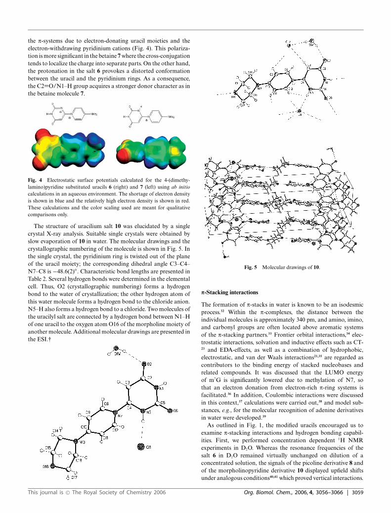
on the uracil pyridinium salt 6 in an aqueous environment led toa twisted molecule with a torsion angle of 53.55◦ between thepyrimidine and the pyridinium ring. Natural bond orders (NBO)indicate that the pyridinium ring adopts a quinoid structure. TheNBO values for C2–C3, C5–C6 and C4–N are 1.87, 1.86 and1.96, respectively. The calculated bond lengths reinforce this fact.Thus, the optimized values for the same bonds are, respectively,136.2, 136.2 and 133.6 pm, while those of the C3–C4 and C4–C5 are both 142.8 pm. The highest occupied molecular orbital(HOMO: −0.25496 eV) as well as the lowest unoccupied molecularorbital (LUMO: −0.08484 eV) are located in the pyrimidine aswell as in the pyridinium ring (Fig. 2). TD calculations on theB3PW91/6-31G**/PCM optimized structure predicted the lowestenergy transitions at 294.4 nm (4.212 eV) and 279.2 nm (4.441eV), with oscillator strengths of 0.439 and 0.025, respectively. Thelowest transition energy is usually compared with the HOMO–LUMO energy gap since this electronic transition is describedas the promotion of a single electron from HOMO to LUMO.30
However, this comparison clearly fails for this molecule, as thecomputed energy for the first electronic transition is significantlyhigher than the calculated HOMO–LUMO gap. This fact isattributed to the reduced interelectronic interaction between thesingle one-electron excitation.31 It also indicates that this transitionhas to be described as a linear combination of single one-electronpromotions between a set of frontier orbitals. For this molecule,the TD/PCM calculation assigned the transition at 4.441 eV tothe HOMO to LUMO (62%) and the HOMO-1 to LUMO (21%)excitations, together with other minor contributions.
Fig. 2 HOMO (above) and LUMO (below) of 6.
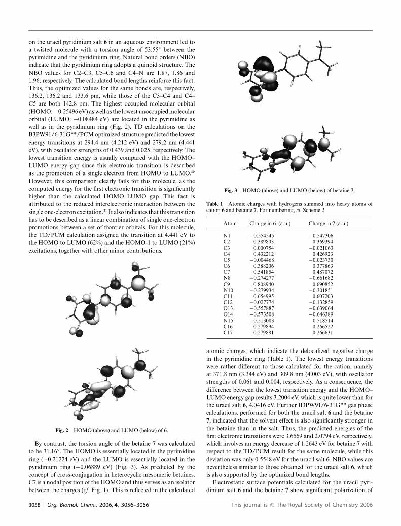
By contrast, the torsion angle of the betaine 7 was calculatedto be 31.16◦. The HOMO is essentially located in the pyrimidinering (−0.21224 eV) and the LUMO is essentially located in thepyridinium ring (−0.06889 eV) (Fig. 3). As predicted by theconcept of cross-conjugation in heterocyclic mesomeric betaines,C7 is a nodal position of the HOMO and thus serves as an isolatorbetween the charges (cf. Fig. 1). This is reflected in the calculated
Fig. 3 HOMO (above) and LUMO (below) of betaine 7.
Table 1 Atomic charges with hydrogens summed into heavy atoms ofcation 6 and betaine 7. For numbering, cf. Scheme 2
Atom Charge in 6 (a.u.) Charge in 7 (a.u.)
N1 −0.554545 −0.547306C2 0.389803 0.369394C3 0.000754 −0.021063C4 0.432212 0.426923C5 −0.004468 −0.023730C6 0.388206 0.377863C7 0.541854 0.487072N8 −0.274277 −0.661682C9 0.808940 0.690852N10 −0.279934 −0.301851C11 0.654995 0.607203C12 −0.027774 −0.132859O13 −0.557887 −0.639064O14 −0.573508 −0.646389N15 −0.513083 −0.518514C16 0.279894 0.266522C17 0.279881 0.266631
atomic charges, which indicate the delocalized negative chargein the pyrimidine ring (Table 1). The lowest energy transitionswere rather different to those calculated for the cation, namelyat 371.8 nm (3.344 eV) and 309.8 nm (4.003 eV), with oscillatorstrengths of 0.061 and 0.004, respectively. As a consequence, thedifference between the lowest transition energy and the HOMO–LUMO energy gap results 3.2004 eV, which is quite lower than forthe uracil salt 6, 4.0416 eV. Further B3PW91/6-31G** gas phasecalculations, performed for both the uracil salt 6 and the betaine7, indicated that the solvent effect is also significantly stronger inthe betaine than in the salt. Thus, the predicted energies of thefirst electronic transitions were 3.6569 and 2.0794 eV, respectively,which involves an energy decrease of 1.2643 eV for betaine 7 withrespect to the TD/PCM result for the same molecule, while thisdeviation was only 0.5548 eV for the uracil salt 6. NBO values arenevertheless similar to those obtained for the uracil salt 6, whichis also supported by the optimized bond lengths.
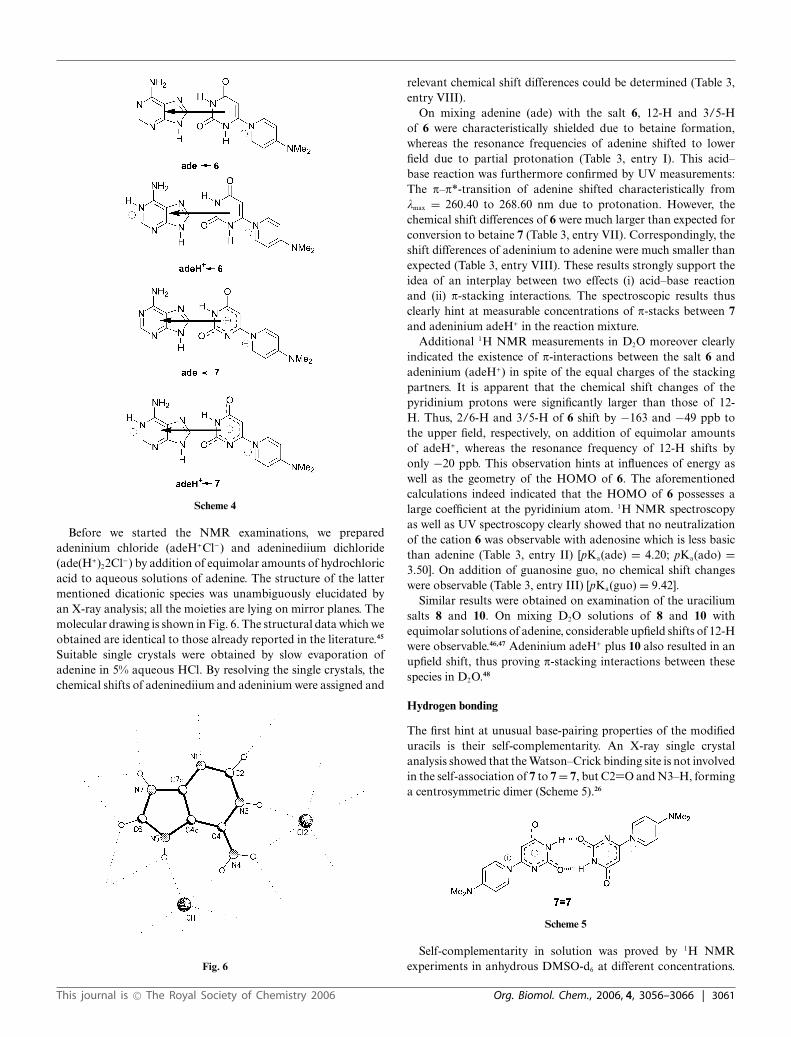
Electrostatic surface potentials calculated for the uracil pyri-dinium salt 6 and the betaine 7 show significant polarization of
3058 | Org. Biomol. Chem., 2006, 4, 3056–3066 This journal is © The Royal Society of Chemistry 2006
the p-systems due to electron-donating uracil moieties and theelectron-withdrawing pyridinium cations (Fig. 4). This polariza-tion is more significant in the betaine 7 where the cross-conjugationtends to localize the charge into separate parts. On the other hand,the protonation in the salt 6 provokes a distorted conformationbetween the uracil and the pyridinium rings. As a consequence,the C2=O/N1–H group acquires a stronger donor character as inthe betaine molecule 7.
Fig. 4 Electrostatic surface potentials calculated for the 4-(dimethy-lamino)pyridine substituted uracils 6 (right) and 7 (left) using ab initiocalculations in an aqueous environment. The shortage of electron densityis shown in blue and the relatively high electron density is shown in red.These calculations and the color scaling used are meant for qualitativecomparisons only.
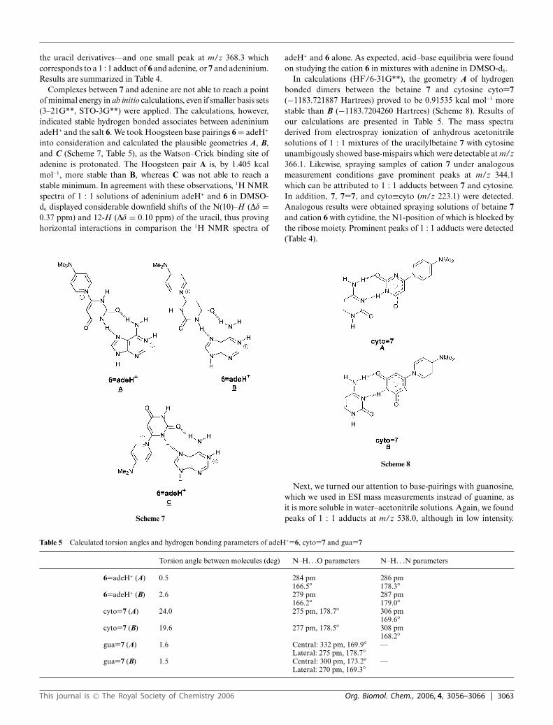
The structure of uracilium salt 10 was elucidated by a singlecrystal X-ray analysis. Suitable single crystals were obtained byslow evaporation of 10 in water. The molecular drawings and thecrystallographic numbering of the molecule is shown in Fig. 5. Inthe single crystal, the pyridinium ring is twisted out of the planeof the uracil moiety; the corresponding dihedral angle C3–C4–N7–C8 is −48.6(2)◦. Characteristic bond lengths are presented inTable 2. Several hydrogen bonds were determined in the elementalcell. Thus, O2 (crystallographic numbering) forms a hydrogenbond to the water of crystallization; the other hydrogen atom ofthis water molecule forms a hydrogen bond to the chloride anion.N5–H also forms a hydrogen bond to a chloride. Two molecules ofthe uracilyl salt are connected by a hydrogen bond between N1–Hof one uracil to the oxygen atom O16 of the morpholine moiety ofanother molecule. Additional molecular drawings are presented inthe ESI.†
Fig. 5 Molecular drawings of 10.
p-Stacking interactions
The formation of p-stacks in water is known to be an isodesmicprocess.32 Within the p-complexes, the distance between theindividual molecules is approximately 340 pm, and amino, imino,and carbonyl groups are often located above aromatic systemsof the p-stacking partners.33 Frontier orbital interactions,34 elec-trostatic interactions, solvation and inductive effects such as CT-23 and EDA-effects, as well as a combination of hydrophobic,electrostatic, and van der Waals interactions21,35 are regarded ascontributors to the binding energy of stacked nucleobases andrelated compounds. It was discussed that the LUMO energyof m7G is significantly lowered due to methylation of N7, sothat an electron donation from electron-rich p-ring systems isfacilitated.36 In addition, Coulombic interactions were discussedin this context,37 calculations were carried out,38 and model sub-stances, e.g., for the molecular recognition of adenine derivativesin water were developed.39
As outlined in Fig. 1, the modified uracils encouraged us toexamine p-stacking interactions and hydrogen bonding capabil-ities. First, we performed concentration dependent 1H NMRexperiments in D2O. Whereas the resonance frequencies of thesalt 6 in D2O remained virtually unchanged on dilution of aconcentrated solution, the signals of the picoline derivative 8 andof the morpholinopyridine derivative 10 displayed upfield shiftsunder analogous conditions40,41 which proved vertical interactions.
This journal is © The Royal Society of Chemistry 2006 Org. Biomol. Chem., 2006, 4, 3056–3066 | 3059

Table 2 Calculated values and selected results of the single crystal X-ray analysis of 10 (crystallographic numbering)
Bond lengths (calculated/found)/pm Bond angles (calculated/found) (deg) Dihedral angles (calculated/found) (deg)
N1–C2 140/1.385(2) N1–C2–C3 114/115.0(1) N1–C2–C3–C4 −1.7/3.3(2)C2–C3 145/1.445(2) N1–C2–O2 121/119.5(1) C6–N1–C2–O2 1.2/175.5(1)C3–C4 135/1.339(2) C2–C3–C4 119/117.9(1) C2–C3–C4–N7 0.1/−177.6(1)C4–N5 137/1.359(2) C3–C4–N5 123/123.9(1) C3–C4–N7–C8 52.5/−48.6(2)N5–C6 139/1.375(2) C3–C4–N7 122/122.1(1) N1–C6–N5–C4 0.8/4.0(2)C6–N1 138/1.376(2) C4–N5–C6 123/121.6(1) N7–C8–C9–C10 −0.3/0.3(2)C2–O2 123/1.232(2) N5–C6–O6 122/122.8(1) C9–C10–N13–C18 −8.3/−174.6(1)C6–O6 122/1.219(2) C4–N7–C8 120/120.4(1) C10–N13–C14–C15 27.2/−132.0(1)C4–N7 142/1.434(2) N7–C8–C9 121/121.4(1)N7–C8 137/1.366(2) C9–C10–N13 122/122.0(1)C10–N13 134/1.344(2) C10–N13–C14 122/123.6(1)
Upfield shifts were also observed on dilution of concentrated D2Osolutions of the betaines 7 and 11. As already mentioned, it provedto be advantageous to observe the resonance frequency of 12-H, as it forms a sharp, not H/D-exchangeable singlet which isnot overlapped with other NMR signals. As an example, dilutionof a 25.71 mmol L−1 solution of 11 in D2O to 2.57 mmol L−1
caused an upfield shift of the resonance frequency of 12-H from5.589 to 5.723 ppm. The solubility of the picoline derivative 9,however, proved to be insufficient for measurements in D2O, asthe maximum concentration is only 6.86 mmol L−1.
With these informations in hand, we examined chemical shiftchanges of the modified uracils at given concentrations on additionof equimolar amounts of the nucleobases adenine (ade), adenosine(ado) and guanosine (guo). Unfortunately, guanine is essentiallyinsoluble in water at pH 7, so that we were prevented fromexaminations of this nucleobase. Literature-known chemical shiftdifferences of the thymine–adenine p-stack are 3 to 216 ppb to theupper field of 6-H of the pyrimidine nucleobase,42 and 0.10 ppb tothe lower field of the resonance frequencies of the purine system.In accordance to this, the signal of 12-H of the uracilyl betaine7 shifted significantly to the upper field on mixing an aqueoussolution with adenine (ade). The chemical shift differences were−140 ppb in comparison to the concentrated solution in D2O(Table 3, entry IV) and −250 ppb in comparison to the molecule ina highly diluted solution. By dilution to less than 1 mM solutions,the chemical shifts of “monomeric” species can be measured.43
Thus, the differences were more than twice as large as observedfor the natural nucleobase uracil, the resonance frequencies ofwhich shift 60 ppb to the upper field on addition of equimolaramounts of adenine. In parallel, the signals of adenine shifted by
the factor 4 to lower field. As displayed in Table 3, similar resultswere obtained on studying 1 : 1 mixtures of 7 with adenosine(ado) (Table 3, entry V), although the effect is much smaller.Guanosine (guo), however, caused only very small shift differencesso that no p-stacks were proved under these conditions (Table 3,entry VI).
The spectroscopic properties of the salt 6 in the presence ofadenine, adenosine, guanine and guanosine are also influenced bythe acidic N8–H proton. Thus, equilibria between protonated anddeprotonated species according to Scheme 3 were formed, so thatin general p-stacking interactions between all species depicted inScheme 4 had to be taken into consideration. Due to a considerablelower LUMO energy,44 adeninium (adeH+) is known to form morestable p-complexes with p-donor molecules than adenine itself.
Scheme 3
Table 3 Chemical shift changes Dd to the lower (+) and upper field (−) in relation to the pure compounds in the same concentration on addition ofequimolar amounts of adenine (ade), adenosine (ado), and guanosine (guo). Values are give in ppb (parts per billion: 1000 ppb = 1 ppm. Concentration:10 mM L−1 in D2O, temperature: 25 ◦C, d(D2O) = 2.500 ppm
Dd of the uracils Dd of the purines
Entry Base 12-H 2/6-H 3/5-H 2-H 8-H
6 I ade −190 120 −110 170 190II ado −110 80 −65 160 130III guo −20 3 −20 — 10
7 IV ade −140 −50 −140 40 45V ado −90 −25 −65 50 40VI guo −25 −10 −20 — −90
6 → 7 VII blind −135 125 −62 — —ade → adeH+ VIII probes — — — 340 310
3060 | Org. Biomol. Chem., 2006, 4, 3056–3066 This journal is © The Royal Society of Chemistry 2006
Scheme 4
Before we started the NMR examinations, we preparedadeninium chloride (adeH+Cl−) and adeninediium dichloride(ade(H+)22Cl−) by addition of equimolar amounts of hydrochloricacid to aqueous solutions of adenine. The structure of the lattermentioned dicationic species was unambiguously elucidated byan X-ray analysis; all the moieties are lying on mirror planes. Themolecular drawing is shown in Fig. 6. The structural data which weobtained are identical to those already reported in the literature.45
Suitable single crystals were obtained by slow evaporation ofadenine in 5% aqueous HCl. By resolving the single crystals, thechemical shifts of adeninediium and adeninium were assigned and
Fig. 6
relevant chemical shift differences could be determined (Table 3,entry VIII).
On mixing adenine (ade) with the salt 6, 12-H and 3/5-Hof 6 were characteristically shielded due to betaine formation,whereas the resonance frequencies of adenine shifted to lowerfield due to partial protonation (Table 3, entry I). This acid–base reaction was furthermore confirmed by UV measurements:The p–p*-transition of adenine shifted characteristically fromkmax = 260.40 to 268.60 nm due to protonation. However, thechemical shift differences of 6 were much larger than expected forconversion to betaine 7 (Table 3, entry VII). Correspondingly, theshift differences of adeninium to adenine were much smaller thanexpected (Table 3, entry VIII). These results strongly support theidea of an interplay between two effects (i) acid–base reactionand (ii) p-stacking interactions. The spectroscopic results thusclearly hint at measurable concentrations of p-stacks between 7and adeninium adeH+ in the reaction mixture.
Additional 1H NMR measurements in D2O moreover clearlyindicated the existence of p-interactions between the salt 6 andadeninium (adeH+) in spite of the equal charges of the stackingpartners. It is apparent that the chemical shift changes of thepyridinium protons were significantly larger than those of 12-H. Thus, 2/6-H and 3/5-H of 6 shift by −163 and −49 ppb tothe upper field, respectively, on addition of equimolar amountsof adeH+, whereas the resonance frequency of 12-H shifts byonly −20 ppb. This observation hints at influences of energy aswell as the geometry of the HOMO of 6. The aforementionedcalculations indeed indicated that the HOMO of 6 possesses alarge coefficient at the pyridinium atom. 1H NMR spectroscopyas well as UV spectroscopy clearly showed that no neutralizationof the cation 6 was observable with adenosine which is less basicthan adenine (Table 3, entry II) [pKa(ade) = 4.20; pKa(ado) =3.50]. On addition of guanosine guo, no chemical shift changeswere observable (Table 3, entry III) [pKa(guo) = 9.42].
Similar results were obtained on examination of the uraciliumsalts 8 and 10. On mixing D2O solutions of 8 and 10 withequimolar solutions of adenine, considerable upfield shifts of 12-Hwere observable.46,47 Adeninium adeH+ plus 10 also resulted in anupfield shift, thus proving p-stacking interactions between thesespecies in D2O.48
Hydrogen bonding
The first hint at unusual base-pairing properties of the modifieduracils is their self-complementarity. An X-ray single crystalanalysis showed that the Watson–Crick binding site is not involvedin the self-association of 7 to 7 = 7, but C2=O and N3–H, forminga centrosymmetric dimer (Scheme 5).26
Scheme 5
Self-complementarity in solution was proved by 1H NMRexperiments in anhydrous DMSO-d6 at different concentrations.
This journal is © The Royal Society of Chemistry 2006 Org. Biomol. Chem., 2006, 4, 3056–3066 | 3061

On dilution of concentrated solutions of the cations and betaines 6,7, 8, 9, and 10 all resonance frequencies shift upfield in accordancewith horizontal interactions in this solvent. As example, the curveof the picoline derivative 8 is presented in Fig. 7. The solubilityof betaine 11, however, proved to be insufficient for satisfyingmeasurements. All compounds are insufficiently soluble in lesspolar solvents.
Fig. 7
In the electrospray ionisation mass spectra (ESIMS), the dimersform prominent peaks at 0 V fragmentor voltage. Mixtures ofall compounds give rise to combinations of homo- and hetero-intermolecular base pairings. Thus, in the ESI mass spectrum ofa 1 : 1 mixture of 6 and 10 peaks of the individual molecules atm/z 233.1 [6]+ and 275.1 [10]+, as well as homo-intermolecularpairs of [6 + 7]+ at m/z 465.1 and [10 + 11]+ at m/z 549.2 andhetero-intermolecular adducts of [6 + 11]+ or [7 + 10]+ at m/z507.1 (Scheme 6, Table 4) are detectable.
Scheme 6
We next turned our attention to the base-pairing propertiesof the modified uracils. NMR titrations clearly show, that nohydrogen bonds between uracilylbetaine 7 and adenine, adenosine,cytosine, guanine, and guanosine can be observed in DMSO-d6 atroom temperature. One reason could be the very limited solubilityof 7 which does not exceed 4 mM per L solvent. This behaviour isanalogous to the natural pyrimidine nucleobases under analogousreaction conditions.42
Even the extremely mild electrospray ionization (ESI) tech-nique, which proved to be highly valuable for the detectionof oligonucleotides, proteins, enzyme–substrate and enzyme–product complexes,49 was not able to detect base pairs betweenthe uracil derivatives 8 and 10, respectively, and adenine.
However, the measurements performed starting from equimolarsolutions of 6 or 7 and adenine in acetonitrile–water (9 : 1) at0 V fragmentor voltage displayed peaks of the monomeric speciesof 7 at m/z 233.1 (100%) and of 7 = 7 at m/z 465 (20%)—in accordance with the aforementioned self-complementarity of T
able
4R
esul
tsof
ESI
MS
mea
sure
men
ts.S
pect
raw
ere
take
nfr
omso
luti
ons
ofth
esa
lts
6,8,
and
10in
wat
er–a
ceto
nitr
ile(9
:1).
The
sam
ples
wer
esp
raye
d(3
5ps
ig,s
olve
ntflo
w0.
6m
Lm
in−1
)fr
omw
ater
–ace
toni
trile
(9:1
)at
0V
frag
men
tor
volt
age,
4000
Vca
pilla
ryvo
ltag
e,an
d30
0◦ C
dryi
ngga
ste
mpe
ratu
re(N
2).
ade
=ad
enin
e;cy
to=
cyto
sine
;cyt
i=cy
tidi
ne,g
uo=
guan
osin
e
Pur
esa
mpl
esA
deC
yto
Cyt
iG
uod(
CpG
p)
623
3.1
[6]+
368.
3[7
+ad
eH]+
343.
1[6
+cy
to]+
476.
1[6
+cy
ti]+
538.
0[7
+gu
o+
Na]
+16
20.2
[d(C
pGp)
+6]
+
255.
1[7
+N
a]+
365.
1[7
+cy
to+
Na]
+49
8.0
[7+
cyti
+N
a]+
1642
.2[d
(CpG
p)+
7+
Na]
+
465.
2[6
+7]
+18
74.1
[d(C
pGp)
+7
+7
+N
a]+
487.
0[7
+7
+N
a]+
1642
.2[d
(CpG
p)+
7+
Na]
+
820
4.2
[8]+
N.d
.a31
4.9
[8+
cyto
]+44
7.0
[8+
cyti
]+51
0.0
[8+
guo]
+16
11.9
[d(C
pGp)
+9
+N
a]+
226.
1[9
+N
a]+
336.
2[9
+cy
to+
Na]
+46
9.1
[9+
cyti
+N
a]+
407.
1[8
+9]
+
429.
0[9
+9
+N
a]+
1027
5.1
[10]
+N
.d.a
407.
1[1
0+
cyto
+N
a]+
540.
3[1
0+
cyti
+N
a]+
581.
2[1
0+
guo
+N
a]+
1684
.3[d
(CpG
p)+
10+
Na]
+
297.
9[1
1+
Na]
+
549.
3[1
0+
11]+
571.
3[1
1+
11+
Na]
+
aN
otde
tect
ed.
3062 | Org. Biomol. Chem., 2006, 4, 3056–3066 This journal is © The Royal Society of Chemistry 2006
the uracil derivatives—and one small peak at m/z 368.3 whichcorresponds to a 1 : 1 adduct of 6 and adenine, or 7 and adeninium.Results are summarized in Table 4.
Complexes between 7 and adenine are not able to reach a pointof minimal energy in ab initio calculations, even if smaller basis sets(3–21G**, STO-3G**) were applied. The calculations, however,indicated stable hydrogen bonded associates between adeniniumadeH+ and the salt 6. We took Hoogsteen base pairings 6 = adeH+
into consideration and calculated the plausible geometries A, B,and C (Scheme 7, Table 5), as the Watson–Crick binding site ofadenine is protonated. The Hoogsteen pair A is, by 1.405 kcalmol−1, more stable than B, whereas C was not able to reach astable minimum. In agreement with these observations, 1H NMRspectra of 1 : 1 solutions of adeninium adeH+ and 6 in DMSO-d6 displayed considerable downfield shifts of the N(10)–H (Dd =0.37 ppm) and 12-H (Dd = 0.10 ppm) of the uracil, thus provinghorizontal interactions in comparison the 1H NMR spectra of
Scheme 7
adeH+ and 6 alone. As expected, acid–base equilibria were foundon studying the cation 6 in mixtures with adenine in DMSO-d6.
In calculations (HF/6-31G**), the geometry A of hydrogenbonded dimers between the betaine 7 and cytosine cyto=7(−1183.721887 Hartrees) proved to be 0.91535 kcal mol−1 morestable than B (−1183.7204260 Hartrees) (Scheme 8). Results ofour calculations are presented in Table 5. The mass spectraderived from electrospray ionization of anhydrous acetonitrilesolutions of 1 : 1 mixtures of the uracilylbetaine 7 with cytosineunambigously showed base-mispairs which were detectable at m/z366.1. Likewise, spraying samples of cation 7 under analogousmeasurement conditions gave prominent peaks at m/z 344.1which can be attributed to 1 : 1 adducts between 7 and cytosine.In addition, 7, 7=7, and cyto≡cyto (m/z 223.1) were detected.Analogous results were obtained spraying solutions of betaine 7and cation 6 with cytidine, the N1-position of which is blocked bythe ribose moiety. Prominent peaks of 1 : 1 adducts were detected(Table 4).
Scheme 8
Next, we turned our attention to base-pairings with guanosine,which we used in ESI mass measurements instead of guanine, asit is more soluble in water–acetonitrile solutions. Again, we foundpeaks of 1 : 1 adducts at m/z 538.0, although in low intensity.
Table 5 Calculated torsion angles and hydrogen bonding parameters of adeH+=6, cyto=7 and gua=7
Torsion angle between molecules (deg) N–H. . .O parameters N–H. . .N parameters
6=adeH+ (A) 0.5 284 pm 286 pm166.5◦ 178.3◦
6=adeH+ (B) 2.6 279 pm 287 pm166.2◦ 179.0◦
cyto=7 (A) 24.0 275 pm, 178.7◦ 306 pm169.6◦
cyto=7 (B) 19.6 277 pm, 178.5◦ 308 pm168.2◦
gua=7 (A) 1.6 Central: 332 pm, 169.9◦ —Lateral: 275 pm, 178.7◦
gua=7 (B) 1.5 Central: 300 pm, 173.2◦ —Lateral: 270 pm, 169.3◦
This journal is © The Royal Society of Chemistry 2006 Org. Biomol. Chem., 2006, 4, 3056–3066 | 3063
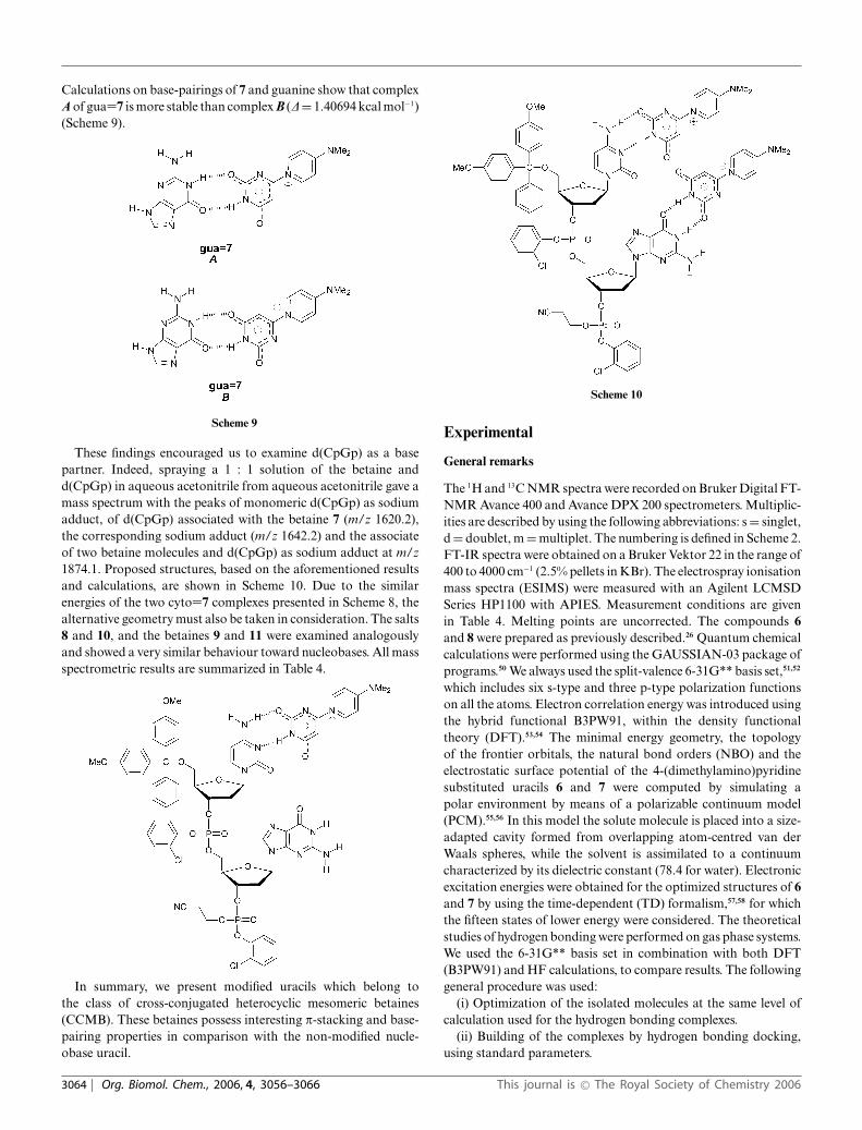
Calculations on base-pairings of 7 and guanine show that complexA of gua=7 is more stable than complex B (D = 1.40694 kcal mol−1)(Scheme 9).
Scheme 9
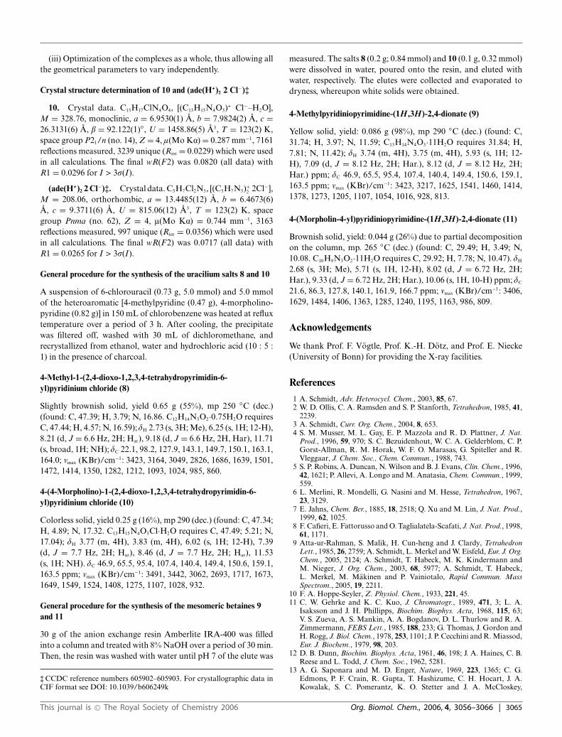
These findings encouraged us to examine d(CpGp) as a basepartner. Indeed, spraying a 1 : 1 solution of the betaine andd(CpGp) in aqueous acetonitrile from aqueous acetonitrile gave amass spectrum with the peaks of monomeric d(CpGp) as sodiumadduct, of d(CpGp) associated with the betaine 7 (m/z 1620.2),the corresponding sodium adduct (m/z 1642.2) and the associateof two betaine molecules and d(CpGp) as sodium adduct at m/z1874.1. Proposed structures, based on the aforementioned resultsand calculations, are shown in Scheme 10. Due to the similarenergies of the two cyto=7 complexes presented in Scheme 8, thealternative geometry must also be taken in consideration. The salts8 and 10, and the betaines 9 and 11 were examined analogouslyand showed a very similar behaviour toward nucleobases. All massspectrometric results are summarized in Table 4.
In summary, we present modified uracils which belong tothe class of cross-conjugated heterocyclic mesomeric betaines(CCMB). These betaines possess interesting p-stacking and base-pairing properties in comparison with the non-modified nucle-obase uracil.
Scheme 10
Experimental
General remarks
The 1H and 13C NMR spectra were recorded on Bruker Digital FT-NMR Avance 400 and Avance DPX 200 spectrometers. Multiplic-ities are described by using the following abbreviations: s = singlet,d = doublet, m = multiplet. The numbering is defined in Scheme 2.FT-IR spectra were obtained on a Bruker Vektor 22 in the range of400 to 4000 cm−1 (2.5% pellets in KBr). The electrospray ionisationmass spectra (ESIMS) were measured with an Agilent LCMSDSeries HP1100 with APIES. Measurement conditions are givenin Table 4. Melting points are uncorrected. The compounds 6and 8 were prepared as previously described.26 Quantum chemicalcalculations were performed using the GAUSSIAN-03 package ofprograms.50 We always used the split-valence 6-31G** basis set,51,52
which includes six s-type and three p-type polarization functionson all the atoms. Electron correlation energy was introduced usingthe hybrid functional B3PW91, within the density functionaltheory (DFT).53,54 The minimal energy geometry, the topologyof the frontier orbitals, the natural bond orders (NBO) and theelectrostatic surface potential of the 4-(dimethylamino)pyridinesubstituted uracils 6 and 7 were computed by simulating apolar environment by means of a polarizable continuum model(PCM).55,56 In this model the solute molecule is placed into a size-adapted cavity formed from overlapping atom-centred van derWaals spheres, while the solvent is assimilated to a continuumcharacterized by its dielectric constant (78.4 for water). Electronicexcitation energies were obtained for the optimized structures of 6and 7 by using the time-dependent (TD) formalism,57,58 for whichthe fifteen states of lower energy were considered. The theoreticalstudies of hydrogen bonding were performed on gas phase systems.We used the 6-31G** basis set in combination with both DFT(B3PW91) and HF calculations, to compare results. The followinggeneral procedure was used:
(i) Optimization of the isolated molecules at the same level ofcalculation used for the hydrogen bonding complexes.
(ii) Building of the complexes by hydrogen bonding docking,using standard parameters.
3064 | Org. Biomol. Chem., 2006, 4, 3056–3066 This journal is © The Royal Society of Chemistry 2006
(iii) Optimization of the complexes as a whole, thus allowing allthe geometrical parameters to vary independently.
Crystal structure determination of 10 and (ade(H+)2 2 Cl−)‡
10. Crystal data. C13H17ClN4O4, [(C13H15N4O3)+ Cl−–H2O],M = 328.76, monoclinic, a = 6.9530(1) A, b = 7.9824(2) A, c =26.3131(6) A, b = 92.122(1)◦, U = 1458.86(5) A3, T = 123(2) K,space group P21/n (no. 14), Z = 4, l(Mo Ka) = 0.287 mm−1, 7161reflections measured, 3239 unique (Rint = 0.0229) which were usedin all calculations. The final wR(F2) was 0.0820 (all data) withR1 = 0.0296 for I > 3r(I).
(ade(H+)2 2 Cl−)‡. Crystal data. C5H7Cl2N5, [(C5H7N5)+2 2Cl−],
M = 208.06, orthorhombic, a = 13.4485(12) A, b = 6.4673(6)A, c = 9.3711(6) A, U = 815.06(12) A3, T = 123(2) K, spacegroup Pnma (no. 62), Z = 4, l(Mo Ka) = 0.744 mm−1, 3163reflections measured, 997 unique (Rint = 0.0356) which were usedin all calculations. The final wR(F2) was 0.0717 (all data) withR1 = 0.0265 for I > 3r(I).
General procedure for the synthesis of the uracilium salts 8 and 10
A suspension of 6-chlorouracil (0.73 g, 5.0 mmol) and 5.0 mmolof the heteroaromatic [4-methylpyridine (0.47 g), 4-morpholino-pyridine (0.82 g)] in 150 mL of chlorobenzene was heated at refluxtemperature over a period of 3 h. After cooling, the precipitatewas filtered off, washed with 30 mL of dichloromethane, andrecrystallized from ethanol, water and hydrochloric acid (10 : 5 :1) in the presence of charcoal.
4-Methyl-1-(2,4-dioxo-1,2,3,4-tetrahydropyrimidin-6-yl)pyridinium chloride (8)
Slightly brownish solid, yield 0.65 g (55%), mp 250 ◦C (dec.)(found: C, 47.39; H, 3.79; N, 16.86. C12H14N3O2·0.75H2O requiresC, 47.44; H, 4.57; N, 16.59); dH 2.73 (s, 3H; Me), 6.25 (s, 1H; 12-H),8.21 (d, J = 6.6 Hz, 2H; Har), 9.18 (d, J = 6.6 Hz, 2H, Har), 11.71(s, broad, 1H; NH); dC 22.1, 98.2, 127.9, 143.1, 149.7, 150.1, 163.1,164.0; mmax (KBr)/cm−1: 3423, 3164, 3049, 2826, 1686, 1639, 1501,1472, 1414, 1350, 1282, 1212, 1093, 1024, 985, 860.
4-(4-Morpholino)-1-(2,4-dioxo-1,2,3,4-tetrahydropyrimidin-6-yl)pyridinium chloride (10)
Colorless solid, yield 0.25 g (16%), mp 290 (dec.) (found: C, 47.34;H, 4.89; N, 17.32. C13H15N4O3Cl·H2O requires C, 47.49; 5.21; N,17.04); dH 3.77 (m, 4H), 3.83 (m, 4H), 6.02 (s, 1H; 12-H), 7.39(d, J = 7.7 Hz, 2H; Har), 8.46 (d, J = 7.7 Hz, 2H; Har), 11.53(s, 1H; NH). dC 46.9, 65.5, 95.4, 107.4, 140.4, 149.4, 150.6, 159.1,163.5 ppm; mmax (KBr)/cm−1: 3491, 3442, 3062, 2693, 1717, 1673,1649, 1549, 1524, 1408, 1275, 1107, 1028, 932.
General procedure for the synthesis of the mesomeric betaines 9and 11
30 g of the anion exchange resin Amberlite IRA-400 was filledinto a column and treated with 8% NaOH over a period of 30 min.Then, the resin was washed with water until pH 7 of the elute was
‡ CCDC reference numbers 605902–605903. For crystallographic data inCIF format see DOI: 10.1039/b606249k
measured. The salts 8 (0.2 g; 0.84 mmol) and 10 (0.1 g, 0.32 mmol)were dissolved in water, poured onto the resin, and eluted withwater, respectively. The elutes were collected and evaporated todryness, whereupon white solids were obtained.
4-Methylpyridiniopyrimidine-(1H ,3H)-2,4-dionate (9)
Yellow solid, yield: 0.086 g (98%), mp 290 ◦C (dec.) (found: C,31.74; H, 3.97; N, 11.59; C13H14N4O3·11H2O requires 31.84; H,7.81; N, 11.42); dH 3.74 (m, 4H), 3.75 (m, 4H), 5.93 (s, 1H; 12-H), 7.09 (d, J = 8.12 Hz, 2H; Har.), 8.12 (d, J = 8.12 Hz, 2H;Har.) ppm; dC 46.9, 65.5, 95.4, 107.4, 140.4, 149.4, 150.6, 159.1,163.5 ppm; mmax (KBr)/cm−1: 3423, 3217, 1625, 1541, 1460, 1414,1378, 1273, 1205, 1107, 1054, 1016, 928, 813.
4-(Morpholin-4-yl)pyridiniopyrimidine-(1H ,3H)-2,4-dionate (11)
Brownish solid, yield: 0.044 g (26%) due to partial decompositionon the column, mp. 265 ◦C (dec.) (found: C, 29.49; H, 3.49; N,10.08. C10H9N3O2·11H2O requires C, 29.92; H, 7.78; N, 10.47). dH
2.68 (s, 3H; Me), 5.71 (s, 1H, 12-H), 8.02 (d, J = 6.72 Hz, 2H;Har.), 9.33 (d, J = 6.72 Hz, 2H; Har.), 10.06 (s, 1H, 10-H) ppm; dC
21.6, 86.3, 127.8, 140.1, 161.9, 166.7 ppm; vmax (KBr)/cm−1: 3406,1629, 1484, 1406, 1363, 1285, 1240, 1195, 1163, 986, 809.
Acknowledgements
We thank Prof. F. Vogtle, Prof. K.-H. Dotz, and Prof. E. Niecke(University of Bonn) for providing the X-ray facilities.
References
1 A. Schmidt, Adv. Heterocycl. Chem., 2003, 85, 67.2 W. D. Ollis, C. A. Ramsden and S. P. Stanforth, Tetrahedron, 1985, 41,
2239.3 A. Schmidt, Curr. Org. Chem., 2004, 8, 653.4 S. M. Musser, M. L. Gay, E. P. Mazzola and R. D. Plattner, J. Nat.
Prod., 1996, 59, 970; S. C. Bezuidenhout, W. C. A. Gelderblom, C. P.Gorst-Allman, R. M. Horak, W. F. O. Marasas, G. Spiteller and R.Vleggaar, J. Chem. Soc., Chem. Commun., 1988, 743.
5 S. P. Robins, A. Duncan, N. Wilson and B. J. Evans, Clin. Chem., 1996,42, 1621; P. Allevi, A. Longo and M. Anatasia, Chem. Commun., 1999,559.
6 L. Merlini, R. Mondelli, G. Nasini and M. Hesse, Tetrahedron, 1967,23, 3129.
7 E. Jahns, Chem. Ber., 1885, 18, 2518; Q. Xu and M. Lin, J. Nat. Prod.,1999, 62, 1025.
8 F. Cafieri, E. Fattorusso and O. Taglialatela-Scafati, J. Nat. Prod., 1998,61, 1171.
9 Atta-ur-Rahman, S. Malik, H. Cun-heng and J. Clardy, TetrahedronLett., 1985, 26, 2759; A. Schmidt, L. Merkel and W. Eisfeld, Eur. J. Org.Chem., 2005, 2124; A. Schmidt, T. Habeck, M. K. Kindermann andM. Nieger, J. Org. Chem., 2003, 68, 5977; A. Schmidt, T. Habeck,L. Merkel, M. Makinen and P. Vainiotalo, Rapid Commun. MassSpectrom., 2005, 19, 2211.
10 F. A. Hoppe-Seyler, Z. Physiol. Chem., 1933, 221, 45.11 C. W. Gehrke and K. C. Kuo, J. Chromatogr., 1989, 471, 3; L. A.
Isaksson and J. H. Phillipps, Biochim. Biophys. Acta, 1968, 115, 63;V. S. Zueva, A. S. Mankin, A. A. Bogdanov, D. L. Thurlow and R. A.Zimmermann, FEBS Lett., 1985, 188, 233; G. Thomas, J. Gordon andH. Rogg, J. Biol. Chem., 1978, 253, 1101; J. P. Cecchini and R. Miassod,Eur. J. Biochem., 1979, 98, 203.
12 D. B. Dunn, Biochim. Biophys. Acta, 1961, 46, 198; J. A. Haines, C. B.Reese and L. Todd, J. Chem. Soc., 1962, 5281.
13 A. G. Saponara and M. D. Enger, Nature, 1969, 223, 1365; C. G.Edmons, P. F. Crain, R. Gupta, T. Hashizume, C. H. Hocart, J. A.Kowalak, S. C. Pomerantz, K. O. Stetter and J. A. McCloskey,
This journal is © The Royal Society of Chemistry 2006 Org. Biomol. Chem., 2006, 4, 3056–3066 | 3065
J. Bacteriol., 1991, 173, 3138; Y. Kuchino, M. Ihara, Y. Yabusaki andS. Nishimura, Nature, 1982, 298, 684.
14 C. C. Hsu-Chen and D. T. Dubin, Nature, 1976, 264, 190; L. Osorio-Almeida, P. Guillemaut, G. Keith, J. Canady and J. H. Weil, Biochem.Biophys. Res. Commun., 1980, 92, 102.
15 R. Liou and T. Blumenthal, Mol. Cell. Biol., 1990, 10, 1764; G.Dirheimer, in Modified Nucleosides and Cancer, ed. G. Glass, Springer-Verlag, Berlin Heidelberg, 1983, pp. 15–46.
16 S.-H. Kim, Prog. Nucleic Acid. Res. Mol. Biol., 1976, 17, 181; J. L.Sussman, S. R. Holbrook, R. Wade Warrant, G. M. Church and S.-H.Kim, J. Mol. Biol., 1978, 123, 607.
17 A. Klug, J. Ladner and J. D. Robertus, J. Mol. Biol., 1974, 89, 511.18 D. Ackermann, Angew. Chem., 1958, 70, 80.19 J. D. Lewis and E. Itaurralde, Eur. J. Biochem., 1997, 247, 461.20 G. Varani, Structure, 1997, 5, 855; E. Izzauralde and I. W. Mattaj, Cell,
1995, 81, 153.21 P.-C. Hsu, M. R. Hodel, J. W. Thomas, L. J. Taylor, C. H. Hagedorn
and A. E. Hodel, Biochemistry, 2000, 39, 13730.22 J. Marcotrigiano, A. C. Gingras, N. Sonenberg and S. K. Burley, Cell,
1997, 89, 951; H. Matsuo, H. Li, A. M. McGuire, C. M. Fletcher, A. C.Gingras, N. Sonenberg and G. Wagner, Nat. Struct. Biol., 1997, 4,717.
23 T. Ishida, M. Doi, H. Uead, M. Inoue and G. M. Scheldrick, J. Am.Chem. Soc., 1988, 110, 2286; R. Stolarski, A. Sitek, J. Stepinski, M.Jankowska, P. Oksman, A. Temeriusz, E. Darzynkiewicz, H. Lonnbergand D. Shugar, Biochim. Biophys. Acta, 1996, 97, 1293.
24 A. E. Hodel, P. D. Gershon, X. Shi, S. M. Wang and F. A. Quiocho,Nat. Struct. Biol., 1997, 4, 350; A. E. Hodel, P. D. Gershon and F. A.Quiocho, Mol. Cell, 1998, 1, 443.
25 A. Schmidt, N. Kobakhidze and M. K. Kindermann, J. Chem. Soc.,Perkin Trans. 1, 2002, 7, 982.
26 A. Schmidt, M. K. Kindermann, P. Vainiotalo and M. Nieger, J. Org.Chem., 1999, 64, 9499.
27 A. Schmidt and M. K. Kindermann, J. Org. Chem., 1997, 62, 3910.28 A. Schmidt and N. Kobakhidze, Heterocycles, 2002, 57, 2231.29 R. M. Cresswell and H. C. S. Wood, J. Chem. Soc., 1960, 4768.30 J. P. Hay, J. Phys. Chem. A, 2000, 106, 1634.31 J. F. Briere and M. J. Cote, J. Phys. Chem. B, 2004, 108, 3123.32 N. I. Nakano and S. J. Irigashi, Biochemistry, 1970, 9, 577.33 W. Saenger, Principles of Nucleic Acid Research, Springer Verlag, New
York, 1983.34 L. D. Wright and D. B. McCormick, Experientia, 1964, 20, 501.35 K. M. Guckian, B. A. Schweitzer, R. X.-F. Ren, C. J. Sheils, D. C.
Tahmassebi and E. T. Kool, J. Am. Chem. Soc., 2000, 122, 2213; F.Prat, K. N. Houk and C. S. Foote, J. Am. Chem. Soc., 1998, 10, 845; J.Sponer, J. Leszcynski and P. Hobza, J. Phys. Chem., 1996, 100, 5590; C.Alhambra, F. J. Luque, F. Gago and M. Orozco, J. Phys. Chem., 1997,101, 3846; M. Aida, J. Theor. Biol., 1988, 130, 327; D. B. McCormick,Photochem. Photobiol., 1977, 26, 169; C. Helene and G. Lancelot, Prog.Biophys. Mol. Biol., 1982, 39, 1.
36 Y. Nishimura, S. Takahashi, T. Yamamoto, M. Tsuboi, M. Hattori, K.Miura, K. Yamaguchi, S. Ohtani and T. Hata, Nucleic Acids Res., 1980,8, 1107.
37 D. A. Dougherty, Science, 1996, 271, 163.38 C. A. Hunter and J. K. M. Sanders, J. Am. Chem. Soc., 1990, 112, 5525;
C. A. Hunter, J. Mol. Biol., 1993, 230, 1025; C. A. Hunter, Chem. Soc.Rev., 1994, 101; P. Leighton, J. A. Cowan, R. J. Abraham and J. K. M.Sanders, J. Org. Chem., 1988, 53, 733.
39 V. M. Rotello, E. A. Viani, G. Deslongchamps, B. A. Murray and J.Rebek, Jr., J. Am. Chem. Soc., 1993, 115, 797.
40 Dilution of a 68.57 mmol L−1 solution of 8 to 6.86 mmol L−1 causedan upfield shift of the resonace frequency of 12-H from 6.161 to6.1124 ppm.
41 Dilution of a 51.43 mmol L−1 solution of 10 to 5.14 mmol L−1 causedan upfield shift of the resonance frequency of 12-H from 5.930 to5.895 ppm.
42 M. P. Schweizer, Nucleic Acids: Base Stacking & Base Pairing Inter-actions, in Encyclopedia of Nuclear Magnetic Resonance, vol. 5, ed.M. D. Grant, R. K. Harris, John Wiley & Sons, Chichester, 1996.
43 A. B. Broom, M. P. Schweizer and P. O. P. Ts’O, J. Am. Chem. Soc.,1967, 89, 3612.
44 T. Ishida, M. Shibata, K. Fujii and M. Inoue, Biochemistry, 1983, 22,3571.
45 T. J. Kistenmacher and T. Shigematsu, Acta Crystallogr., Sect. B, 1974,30, 1528.
46 Treatment of a 10.6 mmol L−1 solution of 8 in D2O with an equimolarsolution of adenine resulted in an upfield of 12-H of 0.4631 ppm.
47 Treatment of a 10.6 mmol L−1 solution of 10 in D2O with an equimolarsolution of adenine resulted in an upfield of 12-H of 0.2633 ppm.
48 Treatment of a 37.0 mmol L−1 solution of 10 in D2O with an equimolarsolution of adeninium chloride resulted in an upfield of 12-H of0.2115 ppm.
49 Electrospray Ionization Mass Spectrometry, ed. R. B. Cole, John Wiley& Sons, Inc., New York, 1997; M. J. Greig, H. Gaus, L. L. Cummins, H.Sasmor and R. H. Griffey, J. Am. Chem. Soc., 1995, 117, 10765; M. C.Griffith, L. M. Risen, M. J. Greig, E. A. Lesnik, K. G. Sprankle, R. H.Griffey, J. Kiely and S. M. Freier, J. Am. Chem. Soc., 1995, 117, 831; B.Ganem, Y.-T. Li and J. D. Henion, Tetrahedron Lett., 1993, 34, 1445;D. L. Little, R. A. Chorush, J. P. Speir, M. W. Senko, N. L. Kelleher andF. W. McLafferty, J. Am. Chem. Soc., 1994, 116, 4893; M. J. Doktycz,S. Habibi-Goudarzi and S. A. McLuckey, Anal. Chem., 1994, 66, 3416;E. Bayer, T. Bauer, K. Schmeer, K. Bleicher, M. Maier and H.-J. Gaus,H.-J., Anal. Chem., 1994, 66, 3858.
50 M. J. Frisch, G. W. Trucks, H. B. Schlegel, G. E. Scuseria, M. A.Robb, J. R. Cheeseman, J. A. Montgomery, Jr., T. Vreven, K. N.Kudin, J. C. Burant, J. M. Millam, S. S. Iyengar, J. Tomasi, V. Barone,B. Mennucci, M. Cossi, G. Scalmani, N. Rega, G. A. Petersson, H.Nakatsuji, M. Hada, M. Ehara, K. Toyota, R. Fukuda, J. Hasegawa,M. Ishida, T. Nakajima, Y. Honda, O. Kitao, H. Nakai, M. Klene, X.Li, J. E. Knox, H. P. Hratchian, J. B. Cross, C. Adamo, J. Jaramillo,R. Gomperts, R. E. Stratmann, O. Yazyev, A. J. Austin, R. Cammi,C. Pomelli, J. W. Ochterski, P. Y. Ayala, K. Morokuma, G. A. Voth,P. Salvador, J. J. Dannenberg, V. G. Zakrzewski, S. Dapprich, A. D.Daniels, M. C. Strain, O. Farkas, D. K. Malick, A. D. Rabuck, K.Raghavachari, J. B. Foresman, J. V. Ortiz, Q. Cui, A. G. Baboul, S.Clifford, J. Cioslowski, B. B. Stefanov, G. Liu, A. Liashenko, P. Piskorz,I. Komaromi, R. L. Martin, D. J. Fox, T. Keith, M. A. Al-Laham, C. Y.Peng, A. Nanayakkara, M. Challacombe, P. M. W. Gill, B. Johnson, W.Chen, M. W. Wong, C. Gonzalez, J. A. Pople, GAUSSIAN 03 (RevisionB.0), Gaussian, Inc., Pittsburgh, PA, 2003.
51 T. Clark, J. Chandrasekhar, G. W. Spitznagel and P. Schleyer, J. Comput.Chem., 1983, 4, 294.
52 P. C. Hariharan and J. A. Pople, Theor. Chim. Acta, 1973, 28, 213.53 A. D. Becke, Phys. Rev. A, 1998, 38, 3098.54 J. P. Perdew and Y. Wang, Phys. Rev. B, 1992, 45, 13244.55 S. Miertus, E. Scrocco and J. Tomasi, Chem. Phys., 1981, 55, 117.56 S. Miertus and J. Tomasi, Chem. Phys., 1982, 65, 239.57 E. K. U. Gross and W. Kohn, Adv. Quantum Chem., 1990, 21, 255.58 M. E. Casida, in Recent Advances in Density Functional Methods, Part
I, ed. D. P. Chong, World Scientific, Singapore, 1995.
3066 | Org. Biomol. Chem., 2006, 4, 3056–3066 This journal is © The Royal Society of Chemistry 2006
Related Documents


