MOLECULAR AND CELLULAR BIOLOGY, Oct. 1996, p. 5726–5736 Vol. 16, No. 10 0270-7306/96/$04.0010 Copyright q 1996, American Society for Microbiology Synergistic Interaction of the Neu Proto-Oncogene Product and Transforming Growth Factor a in the Mammary Epithelium of Transgenic Mice WILLIAM J. MULLER, 1,2 * CARLOS L. ARTEAGA, 3,4 SENTHIL K. MUTHUSWAMY, 1,5 PETER M. SIEGEL, 1,5 MARC A. WEBSTER, 1,5 ROBERT D. CARDIFF, 6 KATHERINE S. MEISE, 3 FENG LI, 3 SUSAN A. HALTER, 4 AND ROBERT J. COFFEY 3 Institute for Molecular Biology and Biotechnology 1 and Departments of Pathology 2 and Biology, 5 McMaster University, Hamilton, Ontario, Canada; Department of Pathology, School of Medicine, University of California at Davis, Davis, California 95616 6 ; and Departments of Medicine and Cell Biology 3 and Pathology, 4 Veterans Affairs Medical Center and Vanderbilt University, Nashville, Tennessee 37232 Received 8 May 1996/Returned for modification 24 June 1996/Accepted 15 July 1996 Transgenic mice expressing either the neu proto-oncogene or transforming growth factor (TGF-a) in the mammary epithelium develop spontaneous focal mammary tumors that occur after a long latency. Since the epidermal growth factor receptor (EGFR) and Neu are capable of forming heterodimers that are responsive to EGFR ligands such as TGF-a, we examined whether coexpression of TGF-a and Neu in mammary epithelium could cooperate to accelerate the onset of mammary tumors. To test this hypothesis, we interbred separate transgenic strains harboring either a mouse mammary tumor virus/TGF-a or a mouse mammary tumor virus/neu transgene to generate bitransgenic mice that coexpress TGF-a and neu in the mammary epithelium. Female mice coexpressing TGF-a and neu developed multifocal mammary tumors which arose after a signif- icantly shorter latency period than either parental strain alone. The development of these mammary tumors was correlated with the tyrosine phosphorylation of Neu and the recruitment of c-Src to the Neu complex. Immunoprecipitation and immunoblot analyses with EGFR- and Neu-specific antisera, however, failed to detect physical complexes of these two receptors. Taken together, these observations suggest that Neu and TGF-a cooperate in mammary tumorigenesis through a mechanism involving Neu and EGFR transactivation. The epidermal growth factor receptor (EGFR) family com- prises four closely related type 1 receptor tyrosine kinases (RTKs) (EGFR, Neu [erbB-2, HER2], erbB-3 [HER3], and erbB-4 [HER4]) that are receptors for a variety of mitogenic growth factors (36). Enhanced expression of the EGFR family has been implicated in the genesis of human breast cancers. For example, amplification and consequent overexpression of neu have been observed in a significant proportion of human breast cancers and appear to be inversely correlated with pa- tient survival (9, 12, 25, 32, 33, 37). More recently, overexpres- sion of the other members of the EGFR family, including EGFR, erbB-3, and erbB-4, has also been implicated in the pathogenesis of human breast cancer (15, 16, 26, 27). The activity of these EGFR family members can also be affected by expression of a variety of specific ligands for these RTKs. For example, Neu is a substrate of the activated EGFR following stimulation of cells with EGF or transforming growth factor a (TGF-a) (1, 8, 13, 35). Although Neu shares homology with the EGFR, Neu does not bind these EGF ligands. Rather, the observed tyrosine phosphorylation of Neu by the EGFR is thought to be mediated by heterodimerization and/or transac- tivation between Neu and EGFR, resulting in a high-affinity receptor for these EGFR ligands (8, 39). Consistent with these observations, coexpression of Neu and EGFR results in effi- cient transformation of fibroblasts in vitro (14). Moreover, elevated expression of both Neu and EGFR can be detected in primary human breast cancers (16). Taken together, these ob- servations suggest that these two closely related RTKs may collaborate in mammary tumorigenesis. Direct evidence of the involvement of EGFR family mem- bers in the induction of mammary tumors derives from obser- vations made with transgenic mice expressing neu in the mam- mary epithelium (4, 10, 21). High-level expression of a constitutively active form of neu bearing a point mutation in the transmembrane domain (3) resulted in the development of nonstochastic, multifocal mammary tumors that affected every female carrier (21). In contrast, expression of the wild-type neu proto-oncogene in the mammary epithelium of transgenic mice resulted in the focal development of mammary tumors that arose after long latency (10). Interestingly, induction of mam- mary tumors in wild-type neu transgenic mice correlated with the frequent occurrence of activating mutations in the neu transgene (31). Thus, activation of the Neu RTK appears to be a pivotal step in the induction of mammary tumors in these mice. Additional evidence implicating the EGFR family in mam- mary tumorigenesis derives from observations made with transgenic strains expressing an EGFR-specific ligand, TGF-a, in the mammary epithelium. Mammary gland-targeted expres- sion of TGF-a in various transgenic strains results in the de- velopment of mammary epithelial hyperplasias that progress to focal mammary tumors after a long latency, as in wild-type neu transgenic mice (11, 18, 29). In mouse mammary tumor virus (MMTV)/TGF-a transgenic mice, increased expression of EGFR was observed in mammary tumors compared with ad- jacent, histologically normal tissue (18). Given the potential of Neu and TGF-a (along with en- hanced EGFR expression) to cooperate through a mechanism * Corresponding author. 5726 on January 8, 2015 by guest http://mcb.asm.org/ Downloaded from

Welcome message from author
This document is posted to help you gain knowledge. Please leave a comment to let me know what you think about it! Share it to your friends and learn new things together.
Transcript
MOLECULAR AND CELLULAR BIOLOGY, Oct. 1996, p. 5726–5736 Vol. 16, No. 100270-7306/96/$04.0010Copyright q 1996, American Society for Microbiology
Synergistic Interaction of the Neu Proto-Oncogene Productand Transforming Growth Factor a in the Mammary
Epithelium of Transgenic MiceWILLIAM J. MULLER,1,2* CARLOS L. ARTEAGA,3,4 SENTHIL K. MUTHUSWAMY,1,5
PETER M. SIEGEL,1,5 MARC A. WEBSTER,1,5 ROBERT D. CARDIFF,6 KATHERINE S. MEISE,3
FENG LI,3 SUSAN A. HALTER,4 AND ROBERT J. COFFEY3
Institute for Molecular Biology and Biotechnology1 and Departments of Pathology2 and Biology,5
McMaster University, Hamilton, Ontario, Canada; Department of Pathology, School of Medicine,University of California at Davis, Davis, California 956166; and Departments of Medicine
and Cell Biology3 and Pathology,4 Veterans Affairs Medical Center andVanderbilt University, Nashville, Tennessee 37232
Received 8 May 1996/Returned for modification 24 June 1996/Accepted 15 July 1996
Transgenic mice expressing either the neu proto-oncogene or transforming growth factor (TGF-a) in themammary epithelium develop spontaneous focal mammary tumors that occur after a long latency. Since theepidermal growth factor receptor (EGFR) and Neu are capable of forming heterodimers that are responsive toEGFR ligands such as TGF-a, we examined whether coexpression of TGF-a and Neu in mammary epitheliumcould cooperate to accelerate the onset of mammary tumors. To test this hypothesis, we interbred separatetransgenic strains harboring either a mouse mammary tumor virus/TGF-a or a mouse mammary tumorvirus/neu transgene to generate bitransgenic mice that coexpress TGF-a and neu in the mammary epithelium.Female mice coexpressing TGF-a and neu developed multifocal mammary tumors which arose after a signif-icantly shorter latency period than either parental strain alone. The development of these mammary tumorswas correlated with the tyrosine phosphorylation of Neu and the recruitment of c-Src to the Neu complex.Immunoprecipitation and immunoblot analyses with EGFR- and Neu-specific antisera, however, failed todetect physical complexes of these two receptors. Taken together, these observations suggest that Neu andTGF-a cooperate in mammary tumorigenesis through a mechanism involving Neu and EGFR transactivation.
The epidermal growth factor receptor (EGFR) family com-prises four closely related type 1 receptor tyrosine kinases(RTKs) (EGFR, Neu [erbB-2, HER2], erbB-3 [HER3], anderbB-4 [HER4]) that are receptors for a variety of mitogenicgrowth factors (36). Enhanced expression of the EGFR familyhas been implicated in the genesis of human breast cancers.For example, amplification and consequent overexpression ofneu have been observed in a significant proportion of humanbreast cancers and appear to be inversely correlated with pa-tient survival (9, 12, 25, 32, 33, 37). More recently, overexpres-sion of the other members of the EGFR family, includingEGFR, erbB-3, and erbB-4, has also been implicated in thepathogenesis of human breast cancer (15, 16, 26, 27).The activity of these EGFR family members can also be
affected by expression of a variety of specific ligands for theseRTKs. For example, Neu is a substrate of the activated EGFRfollowing stimulation of cells with EGF or transforming growthfactor a (TGF-a) (1, 8, 13, 35). Although Neu shares homologywith the EGFR, Neu does not bind these EGF ligands. Rather,the observed tyrosine phosphorylation of Neu by the EGFR isthought to be mediated by heterodimerization and/or transac-tivation between Neu and EGFR, resulting in a high-affinityreceptor for these EGFR ligands (8, 39). Consistent with theseobservations, coexpression of Neu and EGFR results in effi-cient transformation of fibroblasts in vitro (14). Moreover,elevated expression of both Neu and EGFR can be detected inprimary human breast cancers (16). Taken together, these ob-
servations suggest that these two closely related RTKs maycollaborate in mammary tumorigenesis.Direct evidence of the involvement of EGFR family mem-
bers in the induction of mammary tumors derives from obser-vations made with transgenic mice expressing neu in the mam-mary epithelium (4, 10, 21). High-level expression of aconstitutively active form of neu bearing a point mutation inthe transmembrane domain (3) resulted in the development ofnonstochastic, multifocal mammary tumors that affected everyfemale carrier (21). In contrast, expression of the wild-type neuproto-oncogene in the mammary epithelium of transgenic miceresulted in the focal development of mammary tumors thatarose after long latency (10). Interestingly, induction of mam-mary tumors in wild-type neu transgenic mice correlated withthe frequent occurrence of activating mutations in the neutransgene (31). Thus, activation of the Neu RTK appears to bea pivotal step in the induction of mammary tumors in thesemice.Additional evidence implicating the EGFR family in mam-
mary tumorigenesis derives from observations made withtransgenic strains expressing an EGFR-specific ligand, TGF-a,in the mammary epithelium. Mammary gland-targeted expres-sion of TGF-a in various transgenic strains results in the de-velopment of mammary epithelial hyperplasias that progress tofocal mammary tumors after a long latency, as in wild-type neutransgenic mice (11, 18, 29). In mouse mammary tumor virus(MMTV)/TGF-a transgenic mice, increased expression ofEGFR was observed in mammary tumors compared with ad-jacent, histologically normal tissue (18).Given the potential of Neu and TGF-a (along with en-
hanced EGFR expression) to cooperate through a mechanism* Corresponding author.
5726
on January 8, 2015 by guesthttp://m
cb.asm.org/
Dow
nloaded from
of receptor transactivation, we were interested in determiningwhether coexpression of neu and TGF-a could accelerate theinduction of mammary tumors in vivo. To accomplish this,separate strains of transgenic mice carrying either an MMTV/neu or an MMTV/TGF-a transgene were interbred to generatedual carriers that coexpressed both neu and TGF-a in themammary epithelium. The bigenic animals developed multifo-cal mammary tumors within a significantly shorter latency pe-riod than either parental strain alone. The induction of mam-mary tumors in these strains was further correlated to tyrosinephosphorylation of Neu and the recruitment of the c-Src ty-rosine kinase to this complex, even in the absence of neumutations. Cross-linking studies and reciprocal EGFR andNeu immunoprecipitations, however, did not demonstrate aphysical association between EGFR and Neu in bigenic mice.Although these negative data do not disprove a transient phys-ical association between these two receptors, the evidence fa-vors receptor transactivation as the mechanism responsible forthe observed cooperativity between EGFR and Neu in mam-mary tumorigenesis. Taken together, these data argue thatcoexpression of TGF-a and neu can act synergistically to trans-form the mammary epithelium.
MATERIALS AND METHODS
DNA constructs and generation of transgenic mice. The plasmid used togenerate the antisense neu riboprobe was constructed by inserting an SmaI-XbaIfragment (nucleotides 1684 to 2332) into pSL301 (Invitrogen) (31). The phos-phoglycerate kinase 1 (PGK-1) internal control plasmid was obtained from M.Rudnicki and was generated by inserting an AccI-PstI fragment (nucleotides 939to 1633 of the PGK-1 cDNA) (20) into the PstI site of pSP64 (Promega). TheTGF-a riboprobe was constructed by inserting the 632-bp NcoI fragment into thecorresponding site in PSL301. The generation and characterization of bothMMTV/wild-type neu and MMTV/TGF-a mice have been described previously(10, 18).RNase protection assays. Total RNA was isolated from tissues by guanidinium
thiocynate extraction, followed by CsCl gradient fractionation (5). The RNAyield was determined, after resuspension in sterile H2O, by measuring the UVA260. To generate the antisense neu probe, the template plasmids describedabove were linearized with SmaI and then subjected to an in vitro transcriptionreaction with T7 RNA polymerase (19). The PGK-1 internal control probe wasproduced by digesting the template plasmid with EcoNI and transcribing theproduct with SP6 RNA polymerase. The TGF-a riboprobe was generated bycleavage of the template plasmid with XhoI, followed by in vitro transcriptionwith the T3 RNA polymerase. The RNase protection assays were performed byhybridizing the above-mentioned probes to 20 mg of total RNA as previouslydescribed (19). The protected fragments were separated on a sodium dodecylsulfate (SDS)–6% polyacrylamide gel and subjected to autoradiography.Immunoprecipitation and immunoblotting. Tissue samples were ground to
powder under liquid nitrogen and lysed for 20 min on ice in TNE lysis buffer (50mM Tris-HCl [pH 7.6], 150 mM NaCl, 1% Nonidet P-40, 2 mM EDTA, 1 mMsodium orthovanadate, 2 mM dithiothreitol, 10 mg of leupeptin per ml, 10 mg ofaprotinin per ml). The lysates were cleared by centrifugation at 12,0003 g for 10min at 48C. Immunoprecipitations were performed by incubating 2.0 mg of theprotein lysate with either 300 ng of anti-Neu monoclonal antibody 7.16.4 (6) or300 ng of an anti-EGFR antibody (Transduction Laboratories catalog no.E12020) for 30 min at 48C. Following incubation with protein G-Sepharose beads(Pharmacia) on a rotating platform at 48C for 30 min, the precipitates werewashed four times with TNE. The Neu and EGFR immunoprecipitates wereresuspended in SDS-gel loading buffer, and the proteins were resolved on anSDS–9% polyacrylamide gel. The proteins were transferred onto a polyvinyli-dene difluoride membrane (Millipore) with an immunoblot transfer apparatus(Bio-Rad). Following overnight incubation in 3% bovine serum albumin (Sigma)in Tris-buffered saline (20 mM Tris-HCl [pH 7.5], 150 mMNaCl, 5 mMKCl), themembrane was probed for 2 h with antiphosphotyrosine antibodies (1:500; Up-state Biotechnology, Inc.) in bovine serum albumin in Tris-buffered saline. Afterbeing washed in Tris-buffered saline–0.05% Tween 20, the blots were incubatedin 3% milk in Tris-buffered saline for 1 h. The membrane was incubated withgoat anti-mouse immunoglobulin G, and the proteins were visualized by theenhanced-chemiluminescence detection system (Amersham).For studies demonstrating the in vivo association of Neu with c-Src, tumor
lysates were prepared and cleared as described previously (24). Proteins wereimmunoprecipitated by incubating 1.0 to 2.0 mg of total cell lysate with 2 mg ofanti-c-Src antibody 7D10 (Quality Biotech) for 3 h at 48C and subsequentlywashed five times with lysis buffer. The samples were resolved on an SDS–8%polyacrylamide gel and blotted onto a polyvinylidene difluoride membrane. The
membrane was probed with anti-Neu antibody AB.3 (1:1,000; Oncogene Sci-ence) as previously described (24).For studies examining receptor association, tumor membranes were prepared
from 0.3- to 0.6-g tissue aliquots by homogenization in a detergent-free hypo-tonic buffer as described previously (2). Following 45 min of ultracentrifugationat 100,000 3 g and 48C, pellets were solubilized for 45 min in 3-[(3-cholamido-propyl)-dimethyl-amonio]-1-propanesulfonate (CHAPS) buffer (50 mM Tris[pH 8.0], 0.7% CHAPS, 50 mM NaCl, 1 mM sodium orthovandate, 10 mg ofaprotinin per ml, 10 mg of leupeptin per ml). Nonidet P-40-insoluble materialwas removed by centrifugation at 14,000 3 g for 15 min. Equivalent amounts ofmembrane protein were immunoprecipitated with either Neu polyclonal anti-serum 21N (30) or anti-EGFR polyclonal antiserum 986 (30) and Staph A cells(Calbiochem) for 1 to 2 h. After washes, both Neu and EGFR immunoprecipi-tates were subjected to immunoblot analyses with the 21N polyclonal antiserumor an anti-EGFR monoclonal antibody (Transduction Laboratories). For detec-tion, horseradish peroxidase-linked sheep anti-rabbit or anti-mouse antibodieswere utilized. Direct binding assays were performed primarily as described pre-viously (23).Histological evaluation. Complete autopsies were performed. Tissues were
fixed in 4% paraformaldehyde, sectioned at 4 mm, routinely stained with hema-toxylin and eosin, and examined as indicated in the legend to Fig. 4. Whole-mount analyses were performed as previously described (11).
RESULTS
Detection of TGF-a and neu transcripts in mammary epi-thelium of transgenic mice carrying both MMTV/neu andMMTV/TGF-a transgenes. To determine if coexpression ofTGF-a and neu could cooperate in mammary tumorigenesis,separate strains of MMTV/neu and MMTV/TGF-a were in-terbred to generate offspring carrying both transgenes. TheMMTV/TGF-a strain is derived from line 29 and originatesfrom a C57BL 3 DBA genetic background (18), whereas theMMTV/neu strain is derived from the N#202 founder animaland is derived from an inbred FVB genetic background (10).Because TGF-a-expressing females are unable to nurse theiryoung, the F1 progeny from this cross were generated by cross-ing MMTV/TGF-a males with MMTV/neu females. Femalesderived from this cross were segregated into breeding andvirgin female groups. Analyses of the multiparous animals de-rived from this cross revealed that dual carriers possessing boththe TGF-a and neu transgenes were incapable of nursing theiryoung, like TGF-a-expressing females. In addition to the ap-parent lactation defect, multiparous female transgenic micebearing the TGF-a and neu transgenes exhibited uniform hy-pertrophy of the mammary glands (22).To assess whether the mammary epithelium derived from
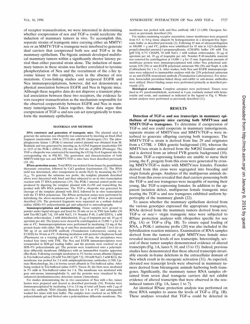
the various genotypes expressed the appropriate transgenes,RNAs derived from the mammary tumors derived from neu/TGF-a or neu/1 virgin transgenic mice were subjected toRNase protection analyses with riboprobes specific for neu(Fig. 1A) or TGF-a (Fig. 1B). To ensure equal loading ofRNA, a PGK-1 antisense probe (20) was also included in thehybridization reaction mixtures. Examination of RNA samplesderived from the tumors of eight MMTV/neu female micerevealed increased levels of neu transcripts. Interestingly, sev-eral of these tumor samples demonstrated evidence of alteredtranscripts (Fig. 1A, lanes 9, 10, and 13 to 15). Indeed, previousstudies have demonstrated that these altered transcripts invari-ably encode in-frame deletions in the extracellular domain ofNeu which result in its oncogenic activation (31). As expected,elevated neu transcript levels were observed in mammary tu-mors derived from bitransgenic animals harboring both trans-genes. Significantly, the mammary tumor RNA samples ob-tained from seven dual transgene carriers did not exhibitevidence of altered transcripts that were observed in the neu-induced tumors (Fig. 1A, lanes 1 to 7).An identical RNase protection analysis was performed on
these RNA samples to assess the levels of TGF-a (Fig. 1B).These analyses revealed that TGF-a could be detected in
VOL. 16, 1996 SYNERGISTIC INTERACTION OF Neu AND TGF-a 5727
on January 8, 2015 by guesthttp://m
cb.asm.org/
Dow
nloaded from
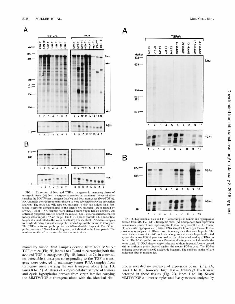
mammary tumor RNA samples derived from both MMTV/TGF-amice (Fig. 2B, lanes 1 to 10) and mice carrying both theneu and TGF-a transgenes (Fig. 1B, lanes 1 to 7). In contrast,no detectable transcripts corresponding to the TGF-a trans-gene were detected in mammary tumor RNA samples fromtransgenic mice carrying the neu transgene alone (Fig. 1B,lanes 8 to 15). Analyses of a representative sample of tumorsand cystic hyperplasias derived from virgin females carryingthe MMTV/TGF-a transgene alone with the identical ribo-
probes revealed no evidence of expression of neu (Fig. 2A,lanes 1 to 10); however, high TGF-a transcript levels weredetected in these tissues (Fig. 2B, lanes 1 to 10). SevenMMTV/TGF-a tumor samples and five cysts were analyzed by
FIG. 1. Expression of Neu and TGF-a transgenes in mammary tissue oftransgenic mice. (A) Neu transgene expression in mammary tissues of micecarrying the MMTV/neu transgene (neu/1) and both transgenes (Neu/TGF-a).RNA samples derived from tumor tissue (T) were subjected to RNase protectionanalyses. The protected wild-type neu transcript is 640 nucleotides long. Pro-tected fragments corresponding to the altered neu transcript are indicated byarrows. Tumor RNA samples were derived from virgin female animals. Anantisense riboprobe directed against the mouse PGK-1 gene was used to controlfor equal loading of RNA on the gel. The PGK-1 probe protects a 124-nucleotidefragment, as indicated in the lower panels. (B) The identical RNA tissue sampleswere hybridized with an antisense probe directed against the mouse TGF-a gene.The TGF-a antisense probe protects a 632-nucleotide fragment. The PGK-1probe protects a 124-nucleotide fragment, as indicated in the lower panels. Thenumbers on the left are molecular sizes in nucleotides.
FIG. 2. Expression of Neu and TGF-a transcripts in tumors and hyperplasiasderived from MMTV/TGF-a transgenic mice. (A) Endogenous Neu expressionin mammary tissues of mice expressing the TGF-a transgene (TGF-a/1). Tumor(T) and cystic hyperplastic (C) tissue RNA samples from virgin female TGF-acarriers were subjected to RNase protection analyses with a neu riboprobe. Theprotected neu transcript is 640 nucleotides long. An antisense riboprobe directedagainst the mouse PGK-1 gene was used to control for equal loading of RNA onthe gel. The PGK-1 probe protects a 124-nucleotide fragment, as indicated in thelower panel. (B) RNA tissue samples identical to those in panel A were probedwith an antisense probe directed against the mouse TGF-a gene. The TGF-aantisense probe protects a 632-nucleotide fragment. The numbers on the left aremolecular sizes in nucleotides.
5728 MULLER ET AL. MOL. CELL. BIOL.
on January 8, 2015 by guesthttp://m
cb.asm.org/
Dow
nloaded from
RNase protection. Taken together, these observations indicatethat the dual carriers coexpress elevated levels of TGF-a andneu in the mammary epithelium.Coexpression of TGF-a and neu in mammary epithelium
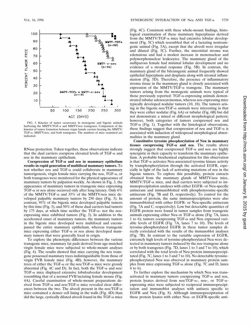
results in rapid generation of multifocal mammary tumors. Totest whether neu and TGF-a could collaborate in mammarytumorigenesis, virgin female mice carrying the neu, TGF-a, orboth transgenes were monitored for the physical appearance ofmammary tumors by palpation weekly. As shown in Fig. 3, theappearance of mammary tumors in transgenic mice expressingTGF-a or neu alone occurred only after long latency. Only 6%of the MMTV/TGF-a and 35% of the MMTV/neu mice de-veloped palpable mammary tumors by 250 days (Fig. 3). Incontrast, 95% of the bigenic mice developed palpable tumorsby this time (Fig. 3), and 50% of these dual carriers had tumorsby 175 days, at which time neither the TGF-a- nor the neu-expressing mice exhibited tumors (Fig. 3). In addition to theaccelerated onset of mammary tumors, the mammary tumorsin the bigenic mice developed were multifocal and encom-passed the entire mammary epithelium, whereas transgenicmice expressing either TGF-a or neu alone developed mam-mary tumors that were generally focal in origin.To explore the phenotypic differences between the various
transgenic mice, mammary fat pads derived from age-matchedvirgin female mice were subjected to whole-mount analyses(Fig. 4). The results showed that mice carrying the neu trans-gene possessed mammary trees indistinguishable from those ofvirgin FVB female mice (Fig. 4B); however, the mammarytrees of either the TGF-a or the neu/TGF-a mice were grosslyabnormal (Fig. 4C and D). In fact, both the TGF-a and neu/TGF-a mice displayed extensive lobuloalveolar developmentresembling that of a normal FVB lactating female mouse (Fig.4A). Careful examination of whole-mount preparations de-rived from TGF-a and neu/TGF-a mice revealed clear differ-ences between the two. The alveoli present in the neu/TGF-amice contained a denser cell lining in the walls (Fig. 4D) thandid the large, cystically dilated alveoli found in the TGF-amice
(Fig. 4C). Consistent with these whole-mount findings, histo-logical examination of these mammary hyperplasias derivedfrom the MMTV/TGF-a mice had extensive lobular develop-ment (Fig. 5C) which resembled that of a lactating nontrans-genic animal (Fig. 5A), except that the alveoli were irregularand dilated (Fig. 5C). Further, the interstitial stroma wasedematous and had a modest increase in mononuclear andpolymorphonuclear leukocytes. The mammary gland of thenulliparous female had minimal lobular development and noevidence of a stromal response (Fig. 5B). In contrast, themammary gland of the bitransgenic animal frequently showedepithelial hyperplasia and dysplasia along with stromal inflam-mation (Fig. 5D). Therefore, the presence of inflammatorystroma tissue in the mammary gland is closely associated withexpression of the MMTV/TGF-a transgene. The mammarytumors arising from the monogenic animals were typical ofthose previously reported: TGF-a-expressing animals demon-strated tubular adenocarcinomas, whereas neu-expressing micetypically developed nodular tumors (10, 18). The tumors aris-ing in the bigenic neu/TGF-a animals were interesting in thatthey were either nodular (Fig. 6A) or tubular (Fig. 6B) but didnot demonstrate a mixed or different morphological pattern;however, both categories of tumors coexpressed neu andTGF-a (Fig. 1). Together with the histological observations,these findings suggest that coexpression of neu and TGF-a isassociated with induction of widespread morphological abnor-malities in the mammary gland.Constitutive tyrosine phosphorylation of Neu in mammary
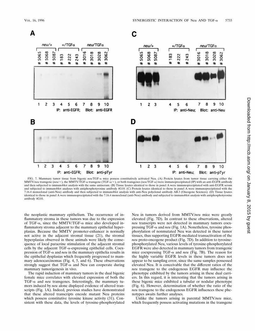
tissues coexpressing TGF-a and neu. The results abovestrongly suggest that coexpressed TGF-a and neu are highlysynergistic in their capacity to transform the mammary epithe-lium. A probable biochemical explanation for this observationis that TGF-a activates Neu-associated tyrosine kinase activityby transphosphorylation through the activated EGFR, espe-cially in light of the lack of activating mutations of neu in thebigenic tumors. To explore this possibility, protein extractsobtained from the mammary glands of MMTV/neu mice,MMTV/TGF-a mice, and dual carriers were subjected to im-munoprecipitation analyses with either EGFR- or Neu-specificantiserum and immunoblotted with phosphotyrosine-specificantiserum (Fig. 7B and D, respectively). To control for theamount of protein, the same immunoprecipitates were alsoimmunoblotted with either EGFR- or Neu-specific antiserum(Fig. 7A and C, respectively). Low but detectable quantities ofEGFR protein were found in tumor samples from transgenicanimals expressing either Neu or TGF-a alone (Fig. 7A, lanes1 to 6); tumors coexpressing TGF-a and Neu expressed vari-able levels of EGFR (Fig. 7A, lanes 7 to 10). The levels oftyrosine-phosphorylated EGFR in these tumor samples di-rectly correlated with the results of the immunoblot analyses(Fig. 7B). In contrast to the variable expression of EGFR,extremely high levels of tyrosine-phosphorylated Neu were de-tected in mammary tumors induced by the neu transgene aloneor by both transgenes (Fig. 7D, lanes 1 to 3 and 7 to 10), whichcorrelated with the total levels of Neu protein immunoprecipi-tated (Fig. 7C, lanes 1 to 3 and 7 to 10). No detectable tyrosine-phosphorylated Neu was observed in mammary protein sam-ples from mice expressing TGF-a alone (Fig. 7C and D, lanes4 to 6).To further explore the mechanism by which Neu was trans-
activated in mammary tumors coexpressing TGF-a and neu,protein lysates derived from neu/TGF-a-, neu-, or TGF-a-expressing mice were subjected to reciprocal immunoprecipi-tation and immunoblot analyses with antisera specific toEGFR and Neu (Fig. 8). Although immunoprecipitation ofthese protein lysates with either Neu- or EGFR-specific anti-
FIG. 3. Kinetics of tumor occurrence in monogenic and bigenic animalsharboring the MMTV/TGF-a and MMTV/neu transgenes. Comparison of thekinetics of tumor formation between virgin female carriers bearing the MMTV/TGF-a, MMTV/neu, and both transgenes. The numbers of mice examined areindicated.
VOL. 16, 1996 SYNERGISTIC INTERACTION OF Neu AND TGF-a 5729
on January 8, 2015 by guesthttp://m
cb.asm.org/
Dow
nloaded from
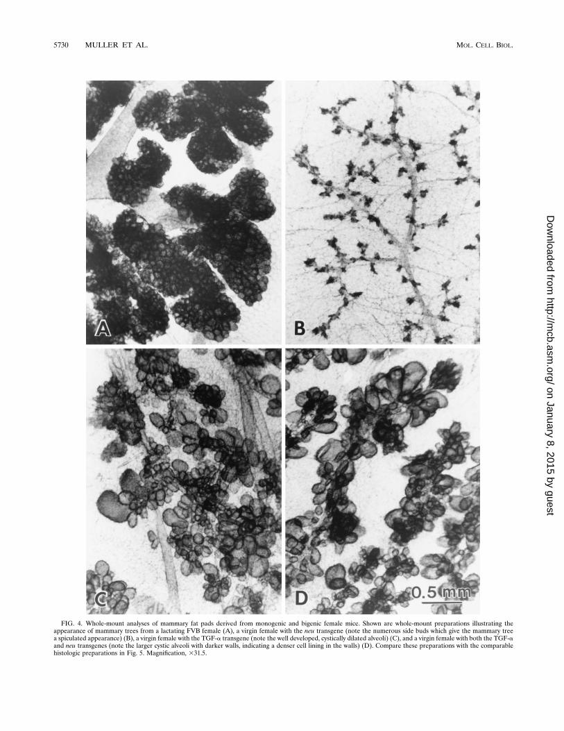
FIG. 4. Whole-mount analyses of mammary fat pads derived from monogenic and bigenic female mice. Shown are whole-mount preparations illustrating theappearance of mammary trees from a lactating FVB female (A), a virgin female with the neu transgene (note the numerous side buds which give the mammary treea spiculated appearance) (B), a virgin female with the TGF-a transgene (note the well developed, cystically dilated alveoli) (C), and a virgin female with both the TGF-aand neu transgenes (note the larger cystic alveoli with darker walls, indicating a denser cell lining in the walls) (D). Compare these preparations with the comparablehistologic preparations in Fig. 5. Magnification, 331.5.
5730 MULLER ET AL. MOL. CELL. BIOL.
on January 8, 2015 by guesthttp://m
cb.asm.org/
Dow
nloaded from
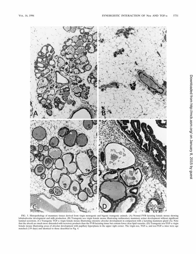
FIG. 5. Histopathology of mammary tissues derived from virgin monogenic and bigenic transgenic animals. (A) Normal FVB lactating female mouse showinglobuloalveolar development and milk production. (B) Transgenic neu virgin female mouse illustrating rudimentary mammary acinar development without significantluminal secretions. (C) Transgenic TGF-a virgin female mouse illustrating extensive alveolar development in comparison with a lactating mammary gland (A). Notethat the alveoli are much more distended with secretory products than the FVB lactating tissue but contain fewer clear lipid vacuoles. (D) Transgenic neu/TGF-a virginfemale mouse illustrating areas of alveolar development with papillary hyperplasia in the upper right corner. The virgin neu, TGF-a, and neu/TGF-a mice were agematched (139 days) and identical to those described in Fig. 4.
VOL. 16, 1996 SYNERGISTIC INTERACTION OF Neu AND TGF-a 5731
on January 8, 2015 by guesthttp://m
cb.asm.org/
Dow
nloaded from

bodies, followed by immunoblot analyses with Neu-specificantiserum, revealed abundant Neu protein in the immunopre-cipitates from the neu/TGF-a- and neu-expressing tumors, nodetectable Neu was found in the EGFR immunoprecipitates(Fig. 8A). Conversely, immunoprecipitation with EGFR-spe-cific antibodies, followed by immunoblot analyses with Neu-specific antiserum, failed to demonstrate the presence ofEGFR in these complexes (Fig. 8B). Thus, under these exper-imental conditions, we could not detect a physical interactionbetween the EGFR and Neu in bigenic mice, and these find-ings indicate that transactivation of Neu by TGF-a does notinvolve the formation of stable Neu-EGFR heterodimers.Induction of mammary tumors by neu and neu–TGF-a
transgenes correlates with the capacity of Neu to associatewith the c-Src tyrosine kinase in vitro and in vivo. One possi-ble explanation for the observed synergy between Neu andTGF-a/EGFR is that these closely related type 1 receptorsrecruit distinct but complementary pathways. Indeed, we havepreviously demonstrated that activation of c-Src by the acti-vated EGFR in fibroblasts is mediated by direct and specificassociation of c-Src with Neu (23). To test whether the c-Srcsignaling pathway was also recruited to Neu in neu/TGF-a-expressing tumors, Neu immunoprecipitates derived from neu,TGF-a, and neu/TGF-a tumor samples were resolved on anSDS-polyacrylamide gel, transferred onto a polyvinylidene di-fluoride membrane, and probed with a radiolabeled glutathi-one S-transferase (GST) fusion protein containing the SH2domain of c-Src (GSTag–c-Src–SH2) (Fig. 9A). Consistentwith previous observations (23), the radiolabeled GSTag–c-Src–SH2 probe bound to the Neu immunoprecipitates derivedfrom mammary tumors expressing Neu alone (Fig. 9A, lanes 1to 3). By contrast, the radiolabeled fusion protein failed to bindthe Neu immunoprecipitates derived from the TGF-a-inducedtumors (Fig. 9A, lanes 4 to 6). An identical analysis of the Neuimmunoprecipitates derived from tumors coexpressing bothNeu and TGF-a revealed that they also bound strongly to theradiolabeled GSTag–c-Src–SH2 probe (Fig. 9A, lanes 7 to 10).The observed binding of the GSTag–c-Src–SH2 probe is likelyspecific to tyrosine-phosphorylated Neu, since previous studies
have demonstrated that the radiolabeled GSTag–c-Src–SH2probe cannot bind comparable levels of tyrosine-phosphory-lated EGFR (23). Thus, Neu derived from neu/TGF-a tumorsis capable of interacting with c-Src in a direct manner in vitro.To confirm that c-Src could interact with Neu in vivo, the
same protein lysates were immunoprecipitated with c-Src-spe-cific antiserum and subjected to immunoblot analyses withNeu-specific antiserum (Fig. 9B). Consistent with the in vitrobinding data, Neu protein was found in c-Src immunoprecipi-tates derived from tumors expressing neu alone or coexpress-ing both neu and TGF-a (Fig. 9B, lanes 1, 2, and 5 and 7) butwas absent from tumors expressing TGF-a alone since thelatter fail to express detectable levels of Neu (Fig. 9B, lanes 3and 4). Taken together, these observations suggest that trans-activation of nonmutated Neu by the activated EGFR resultsin recruitment of the c-Src signaling pathway.
DISCUSSION
Our results show that coexpression of neu and TGF-a in themammary epithelium of transgenic mice results in the induc-tion of multiple growth disturbances in the mammary epithe-lium, leading to tumor formation. We also present evidencethat the occurrence of these growth disturbances correlateswith the constitutive activation of the tyrosine kinase activity ofNeu. These observations suggest that TGF-a and neu cooper-ate in mammary tumorigenesis, possibly through transactiva-tion of Neu by the EGFR.The phenotype exhibited by transgenic mice coexpressing
TGF-a and neu provides important insight into the interactionof EGFR family members in mammary tumorigenesis. Virginfemale mice coexpressing TGF-a and neu demonstrated dra-matic and distinct mammary morphological differences in com-parison with either parental strain (Fig. 4 and 5). In addition,dual transgene carriers developed mammary tumors withgreater penetrance and shorter latency than either neu orTGF-a animals alone (Fig. 3). One of the most striking fea-tures of the mammary tumor tissue derived from bigenic ani-mals is the hyperproliferation of the stromal tissue adjacent to
FIG. 6. Histology of the two types of mammary tumors observed in neu/TGF-a bigenic mice. (A) Nodular tumor typical of the Neu phenotype. (B) Tubular typetypical of the TGF-a phenotype. Magnification, about 3147.
5732 MULLER ET AL. MOL. CELL. BIOL.
on January 8, 2015 by guesthttp://m
cb.asm.org/
Dow
nloaded from
the neoplastic mammary epithelium. The occurrence of in-flammatory stroma in these tumors was due to the expressionof TGF-a, since the MMTV/TGF-a mice also developed in-flammatory stroma adjacent to the mammary epithelial hyper-plasias. Because the MMTV promoter-enhancer is normallynot active in the adjacent stromal tissue (21), the stromalhyperplasias observed in these animals were likely the conse-quence of local paracrine stimulation of the adjacent stromalcells by the adjacent TGF-a-expressing epithelial cells. Coex-pression of TGF-a and neu in the mammary epithelia results inthe epithelial dysplasias which frequently progressed to mam-mary adenocarcinomas (Fig. 4, 5, and 6). These observationsstrongly suggest that TGF-a and Neu can cooperate duringmammary tumorigenesis in vivo.The rapid induction of mammary tumors in the dual bigenic
female mice correlates with elevated expression of both theTGF-a and neu transgenes. Interestingly, the mammary tu-mors induced by neu alone displayed evidence of altered tran-scripts (Fig. 1A). Indeed, previous studies have demonstratedthat these altered transcripts encode mutant Neu proteinswhich possess constitutive tyrosine kinase activity (31). Con-sistent with these data, the levels of tyrosine-phosphorylated
Neu in tumors derived from MMTV/neu mice were greatlyelevated (Fig. 7D). In contrast to these observations, alteredneu transcripts were not detected in mammary tumors coex-pressing TGF-a and neu (Fig. 1A). Nonetheless, tyrosine phos-phorylation of nonmutated Neu was detected in these tumortissues, thus supporting EGFR-mediated transactivation of theneu proto-oncogene product (Fig. 7D). In addition to tyrosine-phosphorylated Neu, various levels of tyrosine-phosphorylatedEGFR were also detected in mammary tumors from transgenicmice coexpressing TGF-a and neu (Fig. 7B). The reason forthe highly variable EGFR levels in these tumors does notappear to be sampling error, since the same samples possessedelevated Neu. It is conceivable that the different ratios of theneu transgene to the endogenous EGFR may influence thephenotype exhibited by the tumors arising in these dual carri-ers. In this regard, it is interesting that the tumors arising inthese bigenic mice exhibited a tubular or nodular phenotype(Fig. 6). However, determination of whether the ratio of theneu transgene to the endogenous EGFR influences these phe-notypes awaits further analyses.Unlike the tumors arising in parental MMTV/neu mice,
which frequently possess activating mutations in the transgene
FIG. 7. Mammary tumor tissue from bigenic neu/TGF-a mice possess constitutively activated Neu. (A) Protein lysates from tumor tissue carrying either theMMTV/neu transgene (neu/1), the MMTV/TGF-a transgene (TGF-a/1), or both transgenes (neu/TGF-a) were immunoprecipitated (IP) with an anti-EGFR antibodyand then subjected to immunoblot analysis with the same antiserum. (B) Tissue lysates identical to those in panel A were immunoprecipitated with anti-EGFR serumand subjected to immunoblot analyses with antiphosphotyrosine antibody 4G10. (C) Protein lysates identical to those in panel A were immunoprecipitated with the7.16.4 monoclonal (anti-Neu) antibody and then subjected to immunoblot analysis with anti-Neu polyclonal antibody AB.3 (Oncogene Sciences). (D) Tissue lysatesidentical to those in panel A were immunoprecipitated with the 7.16.4 monoclonal (anti-Neu) antibody and subjected to immunoblot analysis with antiphosphotyrosineantibody 4G10.
VOL. 16, 1996 SYNERGISTIC INTERACTION OF Neu AND TGF-a 5733
on January 8, 2015 by guesthttp://m
cb.asm.org/
Dow
nloaded from

(31), comparable neu mutations were not required for mam-mary tumorigenesis in bigenic neu/TGF-a mice (Fig. 1A, lanes1 to 7). One possible explanation for the lack of activating neumutations in these tumors is that Neu is activated throughassociation with the EGFR. Consistent with this hypothesis,several groups have demonstrated in both fibroblasts andmammary epithelial cell lines that Neu can be transphospho-rylated by the activated EGFR following EGF stimulation (1,8, 13, 35). In fact, transphosphorylation of Neu can be medi-ated through the formation of a Neu-EGFR heterodimer (14).Moreover, EGFR-Neu heterodimers exhibit a 10-fold greateraffinity for EGFR ligands (39). However, in tumors derivedfrom neu/TGF-a mice, stable Neu-EGFR heterodimers werenot detected (Fig. 8). Therefore, if heterodimerization be-tween EGFR and Neu is involved in the synergistic inductionof mammary tumors, the formation of these complexes is likelytransient. This possibility cannot be ruled out by our experi-mental methods. On the other hand, these heterodimers havebeen reported only in cells with .105 EGF-binding sites percell and after the addition of .10 nM exogenous EGF (8, 39).A lower level of EGFR in neu/TGF-a breast tumors may notallow adequate stoichiometric interactions between both RTKsand thus explain our inability to detect receptor heterodimer-ization. The ability of Neu to cooperate with the activatedEGFR is consistent with a number of previous studies. Forexample, it has been demonstrated that EGFR and Neu cancooperate to transform cell lines in vitro (14). Conversely, ithas been shown that administration of antibodies directedagainst either the EGFR or Neu reverses the transformedphenotype of cells coexpressing both Neu and the EGFR (38).
FIG. 8. Lack of detectable EGFR-Neu association in transgenic mammarytumors. Tumor membranes were prepared as described in Materials and Meth-ods. Four hundred micrograms of membrane protein from each tumor wasimmunoprecipitated (ip) for 2 h with anti-Neu polyclonal antibody 21N or 986anti-EGFR serum and Staph A cells. Precipitates were then subjected to immu-noblot analysis with Neu (top panel)- or EGFR (bottom panel)-specific anti-serum. Despite a detectable level of precipitable EGFRs in all of the tumors,EGFR was undetectable in all of the Neu immunoprecipitates.
FIG. 9. c-Src is complexed with tyrosine-phosphorylated Neu in neu/TGF-a tumors in vitro (A) and in vivo (B). (A) Anti-Neu immunoprecipitates (Anti-Neu) fromneu/1-,1/TGF-a-, and neu/TGF-a-expressing tumors were resolved in an SDS-polyacrylamide gel, blotted onto a polyvinylidene difluoride membrane, and probed witha radiolabeled GSTag–c-Src–SH2 fusion protein. (B) c-Src was immunoprecipitated (IP) (anti-c-Src) from the identical set of lysates and probed with anti-Neu serum.
5734 MULLER ET AL. MOL. CELL. BIOL.
on January 8, 2015 by guesthttp://m
cb.asm.org/
Dow
nloaded from
Although it is clear from these results, as well as otherobservations, that activation of the Neu RTK by TGF-a resultsin synergistic transformation of mammary epithelial cells, themolecular basis for this cooperation is unclear. It is conceivablethat activation of these closely related RTKs results in therecruitment of distinct but complementary signaling pathwaysto each of these receptors that then cooperate to transform themammary epithelial cells. This hypothesis implies that each ofthese type 1 RTKs is coupled to distinct signaling pathways. Infact, several studies have suggested that coupling of the EGFRto the phosphatidylinositol 39-kinase requires participation ofthe c-erbB-3 RTK (28, 34). We have demonstrated that thedirect and specific interaction of c-Src with Neu is involved insignaling by the activated EGFR (23). Consistent with theseearlier observations, we have shown that in mammary tumorsinduced by coexpression of neu and TGF-a, c-Src is complexedboth in vitro and in vivo with tyrosine-phosphorylated Neu(Fig. 9). Although preliminary analyses suggested that c-Srcactivity was elevated in these neu- and TGF-a-coexpressingtumors, precise quantitation of the specific activity of c-Src inthese tumors was problematic because of the extensive inflam-matory stroma present in these tumors (Fig. 6).Although these studies strongly suggest that TGF-a cooper-
ates with Neu through the activated EGFR, it is unclearwhether activation of EGFR is necessary for the induction ofmammary tumors by Neu. However, several recent studiessuggest that the activity of the EGFR is required for normalmammary epithelial proliferation. For example, a naturallyoccurring mouse mutant known as waved-2, which possesses amutation in the EGFR catalytic domain that renders theEGFR functionally inactive (17), exhibits a severe lactationdefect (7). Crosses between MMTV/neu transgenic mice andwaved-2 mice should allow this question to be addressed.
ACKNOWLEDGMENTS
We appreciate the excellent photographic support of Robert Munnand the technical assistance of Monica Graham.This work was supported by research grants awarded to W.J.M. by
the Medical Research Council of Canada, the U.S. Army (DAMD17-94-J-4300), and the Canadian Breast Cancer Initiative. This work wasalso supported by operating grants to R.J.C. and R.D.C. from theNational Cancer Institute (NCI CA 46413 and RO1-CA S4285).W.J.M. is a recipient of a Medical Research Council of Canada Sci-entist award, M.A.W. was supported by a studentship provided by theCancer Research Society, P.M.S. was supported by a studentship fromthe Medical Research Council of Canada, and S.K.M. was a CanadianInternational Development scholar. R.J.C. and C.L.A. are supportedby a Clinical Investigator Career Development award and Merit Re-view grants from the Department of Veterans Affairs. R.J.C. acknowl-edges the generous support of the Joseph and Mary Keller Founda-tion.
REFERENCES
1. Akiyama, T., T. Saito, H. Ogawara, K. Toyoshima, and T. Yamamoto. 1988.Tumor promoter and epidermal growth factor stimulate phosphorylation ofthe c-erbB-2 gene product in MKN-7 human adenocarcinoma cells. Mol.Cell. Biol. 8:1019–1026.
2. Arteaga, C. L., M. D. Johnson, G. Todderud, R. J. Coffey, G. Carpenter, andD. L. Page. 1991. Elevated content of the tyrosine kinase substrate phospho-lipase C-g1 in primary human breast carcinomas. Proc. Natl. Acad. Sci. USA88:10435–10439.
3. Bargmann, C. I., M.-C. Hung, and R. A. Weinberg. 1986. Multiple indepen-dent activations of the neu oncogene by a point mutation altering the trans-membrane domain of p185. Cell 45:649–657.
4. Bouchard, L., L. Lamarre, P. J. Tremblay, and P. Jolicoeur. 1989. Stochasticappearance of mammary tumors in transgenic mice carrying the activatedc-neu oncogene. Cell 57:931–936.
5. Chirgwin, J. M., A. E. Przybyla, R. J. MacDonald, and W. J. Rutter. 1979.Isolation of biologically active ribonucleic acid from sources enriched inribonuclease. Biochemistry 18:5294–5299.
6. Drebin, J. A., D. F. Stern, V. C. Link, R. A. Weinberg, and M. I. Greene. 1984.Monoclonal antibodies identify a cell-surface antigen associated with anactivated cellular oncogene. Nature (London) 312:545–548.
7. Fowler, K. J., F. Walker, W. Alexander, M. L. Hibbs, E. C. Nice, R. M.Bohmer, G. B. Mann, C. Thumwood, R. Maglitto, J. Danks, R. Chetty, A. W.Burgess, and A. R. Dunn. 1995. A mutation in the epidermal growth factorreceptor in waved-2 mice has a profound effect on receptor biochemistry thatresults in impaired lactation. Proc. Natl. Acad. Sci. USA 92:1465–1469.
8. Goldman, R., R. Ben-Levy, E. Peles, and Y. Yarden. 1990. Heterodimeriza-tion of the erbB-1 and erbB-2 receptors in human breast carcinoma cells: amechanism for receptor transregulation. Biochemistry 29:11024–11028.
9. Gullick, W. J., S. B. Love, C. Wright, D. M. Barnes, B. Gutterson, A. L.Harris, and D. G. Altman. 1991. c-erbB-2 protein overexpression in breastcancer is a risk factor in patients with involved and uninvolved lymph nodes.Br. J. Cancer 63:434–438.
10. Guy, C. T., M. A. Webster, M. Schaller, T. J. Parson, R. D. Cardiff, and W. J.Muller. 1992. Expression of the neu proto-oncogene in the mammary epi-thelium of transgenic mice induces metastatic disease. Proc. Natl. Acad. Sci.USA 89:10578–10582.
11. Jhappan, C., C. Stahle, R. Harkins, N. Fausto, G. Smith, and G. Merlino.1990. TGFa overexpression in transgenic mice induces liver neoplasia andabnormal development of the mammary gland and pancreas. Cell 61:1137–1146.
12. King, C. R., M. H. Kraus, and S. A. Aaronson. 1985. Amplification of a novelv-erbB related gene in human mammary carcinoma. Science 229:974–976.
13. Kokai, Y., K. Dobashi, D. B. Weiner, J. N. Myers, P. C. Nowell, and M. I.Greene. 1988. Phosphorylation process induced by epidermal growth factoralters the oncogenic and cellular neu (NGL) gene products. Proc. Natl.Acad. Sci. USA 85:5389–5393.
14. Kokai, Y., J. N. Meyers, T. Wada, V. I. Brown, C. M. LeVea, J. G. Davis, K.Dobashi, and M. I. Greene. 1989. Synergistic interaction of p185 neu and theEGF receptor leads to transformation of rodent fibroblasts. Cell 58:287–292.
15. Kraus, M. H., I. Issing, T. Miki, N. C. Popescu, and S. A. Aaronson. 1989.Isolation and characterization of ERBB-3, a third member of the ERBB,epidermal growth factor receptor family. Evidence for overexpression in asubset of human mammary tumors. Proc. Natl. Acad. Sci. USA 86:9193–9197.
16. Lacroix, H., J. D. Iglehart, M. A. Skinner, and M. Kraus. 1988. Overexpres-sion of erbB-2 or EGF receptor proteins in early stage mammary carcinomais detected simultaneously in matched primary tumors and regional metas-tases. Oncogene 3:145–152.
17. Lutteke, N. C., H. K. Phillips, T. H. Qiu, N. G. Copeland, H. S. Earp, N. A.Jenkins, and D. C. Lee. 1994. The mouse waved-2 phenotype results from apoint mutation in the EGF receptor tyrosine kinase. Genes Dev. 8:399–413.
18. Matsui, Y., S. Halter, J. Holt, B. Hogan, and R. Coffey. 1990. Developmentof mammary hyperplasia and neoplasia in MMTV-TGFa transgenic mice.Cell 61:1147–1155.
19. Melton, D. A., P. A. Krieg, M. R. Rebagliati, T. Maniatis, K. Zinn, and M. R.Green. 1984. Efficient in vitro synthesis of biologically active RNA and RNAhybridization probes from plasmids containing a bacteriophage SP6 pro-moter. Nucleic Acids Res. 12:7035–7056.
20. Mori, N., J. Singer-Sam, C.-Y. Lee, and A. D. Riggs. 1986. The nucleotidesequence of a cDNA clone containing the entire coding region for the mouseX-chromosome-linked phosphoglycerate kinase. Gene 45:275–280.
21. Muller, W. J., E. Sinn, R. Wallace, P. K. Pattengale, and P. Leder. 1988.Single-step induction of mammary adenocarcinoma in transgenic mice bear-ing the activated c-neu oncogene. Cell 54:105–115.
22. Muthuswamy, S. K., and W. J. Muller. 1994. Unpublished observations.23. Muthuswamy, S. K., and W. J. Muller. 1995. Direct and specific interaction
of c-Src with Neu is involved in signaling by the epidermal growth factorreceptor. Oncogene 11:271–279.
24. Muthuswamy, S. K., P. M. Siegel, D. L. Dankort, M. A. Webster, and W. J.Muller. 1994. Mammary tumors expressing the neu proto-oncogene possesselevated c-Src tyrosine kinase activity. Mol. Cell. Biol. 14:735–743.
25. Paterson, M. C., K. D. Dietrich, J. Danyluk, A. H. Paterson, A. W. Lees, N.Jamil, J. Hanson, H. Jenkins, B. E. Krause, W. A. McBlain, D. J. Slamon,and R. M. Fourney. 1991. Correlation between c-erbB-2 amplification andrisk of recurrent disease in node-negative breast cancer. Cancer Res. 51:556–567.
26. Plowman, G., J. M. Colouscou, G. Whitney, J. Green, G. Carlton, L. Foy, M.Neubauer, and M. Shoyab. 1993. Ligand-specific activation of HER4/p180/erbB-4, a fourth member of the epidermal growth factor receptor family.Proc. Natl. Acad. Sci. USA 90:1746–1750.
27. Plowman, G., G. Whitney, M. Neubaue, J. Green, V. McDonald, G. Todaro,and M. Shoyab. 1990. Molecular cloning and expression of an additionalepidermal growth factor receptor-related gene. Proc. Natl. Acad. Sci. USA87:4905–4909.
28. Pringent, S. A., and W. J. Gullick. 1994. Identification of c-erbB-3 bindingsites for phosphatidylinositol 39-kinase and SHC using an EGF receptor/c-erbB-3 chimera. EMBO J. 13:2831–2841.
29. Sandgren, E., N. C. Lutteke, R. D. Palmiter, R. Brinster, and D. Lee. 1990.Overexpression of TGFa in transgenic mice: induction of epithelial hyper-
VOL. 16, 1996 SYNERGISTIC INTERACTION OF Neu AND TGF-a 5735
on January 8, 2015 by guesthttp://m
cb.asm.org/
Dow
nloaded from
plasia, pancreatic metaplasia, and carcinoma of the breast. Cell 61:1121–1135.
30. Shawver, L. K., E. Mann, S. S. Elliger, T. C. Dugger, and C. L. Arteaga. 1994.Ligand-like effects induced by anti-c-erbB-2 antibodies do not correlate andare required for growth inhibition of human carcinoma cells. Cancer Res.54:1367–1373.
31. Siegel, P. M., D. L. Dankort, W. R. Hardy, and W. J. Muller. 1994. Novelactivating mutations in the neu proto-oncogene involved in induction ofmammary tumors. Mol. Cell. Biol. 14:7068–7077.
32. Slamon, D. J., G. M. Clark, S. G. Wong, W. J. Levin, A. Ullrich, and W. L.McGuire. 1987. Human breast cancer: correlation of relapse and survivalwith amplification of Her-2/neu oncogene. Science 235:177–182.
33. Slamon, D. J., W. Godolphin, L. A. Jones, J. A. Holt, S. G. Wong, D. E. Keith,W. J. Levin, S. G. Styart, J. Udove, A. Ullrich, and M. F. Press. 1989. Studiesof the HER-2/neu protooncogene in human breast and ovarian cancer.Science 244:707–712.
34. Soltofff, S. P., K. L. Carraway III, S. A. Prigent, W. G. Gullick, and L. C.Cantley. 1994. ErbB3 is involved in activation of phosphatidylinositol 3-ki-
nase by epidermal growth factor. Mol. Cell. Biol. 14:3550–3558.35. Stern, D. F., and M. P. Kamps. 1988. EGF-stimulated tyrosine phosphory-
lation of p185 neu: a potential model for receptor interactions. EMBO J.7:995–1001.
36. Ullrich, A., and J. Schlessinger. 1990. Signal transduction by receptors oftyrosine kinase activity. Cell 61:203–212.
37. van de Vijver, M., R. van de Bersselaar, P. Devilee, C. Cornelisse, J. Peterse,and R. Nusse. 1987. Amplification of the neu (c-erbB-2) oncogene in humanmammary tumors is relatively frequent and is often accompanied by ampli-fication of the linked c-erbA oncogene. Mol. Cell. Biol. 7:2019–2023.
38. Wada, T., J. Myers, Y. Kokai, V. Brown, J. Hamuro, C. LaVea, and M. I.Greene. 1990. Anti-receptor antibodies reverse the phenotype of cells trans-formed by two interacting proto-oncogene encoded proteins. Oncogene5:489–495.
39. Wada, T., X. Quain, and M. I. Greene. 1990. Intermolecular association ofp185 neu proteins and the EGF receptor modulates EGF receptor function.Cell 61:1339–1347.
5736 MULLER ET AL. MOL. CELL. BIOL.
on January 8, 2015 by guesthttp://m
cb.asm.org/
Dow
nloaded from
Related Documents