Synergistic Effects of the Membrane Actions of Cecropin-Melittin Antimicrobial Hybrid Peptide BP100 Rafael Ferre, † Manuel N. Melo, ‡ Ana D. Correia, ‡ Lidia Feliu, † Eduard Bardajı ´, † Marta Planas, † and Miguel Castanho ‡ * † Laboratori d’Innovacio ´ en Processos i Productes de Sı ´ntesi Orga ` nica, Departament de Quı ´mica, Universitat de Girona, Girona, Spain; and ‡ Instituto de Medicina Molecular, Faculdade de Medicina, Universidade de Lisboa, Lisbon, Portugal ABSTRACT BP100 (KKLFKKILKYL-NH 2 ) is a short cecropin A-melittin hybrid peptide, obtained through a combinatorial chem- istry approach, which is highly effective in inhibiting both the in vitro and in vivo growth of economically important plant pathogenic Gram-negatives. The intrinsic Tyr fluorescence of BP100 was taken advantage of to study the peptide’s binding affinity and damaging effect on phospholipid bilayers modeling the bacterial and mammalian cytoplasmic membranes. In vitro cytotoxic effects of this peptide were also studied on mammalian fibroblast cells. Results show a stronger selectivity of BP100 toward anionic bacterial membrane models as indicated by the high obtained partition constants, one order of magnitude greater than for the neutral mammalian membrane models. For the anionic systems, membrane saturation was observed at high peptide/lipid ratios and found to be related with BP100-induced vesicle permeabilization, membrane electroneutrality, and vesicle aggregation. Occurrence of BP100 translocation was unequivocally detected at both high and low peptide/lipid ratios using a novel and extremely simple method. Moreover, cytotoxicity against mammalian models was reached at a concentration consid- erably higher than the minimum inhibitory concentration. Our findings unravel the relationships among the closely coupled processes of charge neutralization, permeabilization, and translocation in the mechanism of action of antimicrobial peptides. INTRODUCTION Antimicrobial peptides (AMPs) form an essential part of the innate immune system of virtually all forms of life (1–7). During the last decades, AMPs have been widely studied, as they may become an alternative to conventional antibi- otics, especially for the treatment of drug-resistant infections (8, 9). Hundreds of AMPs have been isolated (see a compre- hensive list at http://www.bbcm.univ.trieste.it/~tossi/pag1. htm) and several thousands have been de novo designed and synthetically produced. They display a wide range of biological activities against bacteria, fungi, protozoa, envel- oped viruses, and even tumor cells (9–14). Interestingly, they retain activity against antibiotic-resistant strains and do not readily elicit resistance (15–17). Despite displaying extensive sequence heterogeneity, most AMPs share two functionally important features: a net positive charge and the ability to assume an amphi- pathic structure. These structural characteristics are essential for the mode of action of most AMPs, which target the microbial membrane. The net positive charge promotes their binding to the anionic microbial surface, while the amphi- pathic structure favors peptide insertion into the membrane (10–12,15,16,18–20). Despite extensive studies, the precise mechanism of peptide-membrane interaction and cell killing has not been firmly established for many AMPs. Several models have been proposed to account for the morphological changes involved in AMPs-mediated membrane disruption, such as pore formation (21), cell lysis (22), or peptide trans- location into the cytoplasm (23). Recently, some studies have shown that, apart from membrane damage, other mech- anisms may be involved including intracellular targets (9,15,16). However, in such mechanisms, peptides still must traverse the cell membrane to reach their site of action, which stresses the relevance of peptide-membrane interac- tions for AMP activity. Cecropins, first isolated from the hemolymph of the giant silk moth Hyalophora cecropia, are some of the best studied AMPs (24–26). They represent a family of peptides composed of 31–39 amino acids with antibacterial activity against both Gram-negative and Gram-positive bacteria. Ce- cropins do not exhibit cytotoxic effects against human eryth- rocytes and other eukaryotic cells, but are susceptible to protease degradation (24,27,28). In an effort to overcome the high production costs of such long peptides and to improve their biological properties, short peptide analogs have been designed and synthesized. These studies have led to the identification of nontoxic and more stable peptide sequences displaying a broader and higher activity than their natural counterparts (29–36). In particular, the undecapep- tide WKLFKKILKVL-NH 2 (Pep3), derived from the well- known cecropin A(1–7)-melittin(2–9) hybrid (30,33,34), has been found to be sufficient for antifungal and antibacte- rial activities, while displaying low cytotoxicity (32,37–40). Recently, we have identified cecropin A-melittin hybrid undecapeptides derived from Pep3 which inhibit in vitro growth of economically important plant pathogenic bacteria such as Erwinia amylovora, Pseudomonas syringae Submitted August 22, 2008, and accepted for publication November 17, 2008. *Correspondence: [email protected] Rafael Ferre and Manuel N. Melo contributed equally to this work. Editor: Huey W. Huang. Ó 2009 by the Biophysical Society 0006-3495/09/03/1815/13 $2.00 doi: 10.1016/j.bpj.2008.11.053 Biophysical Journal Volume 96 March 2009 1815–1827 1815

Welcome message from author
This document is posted to help you gain knowledge. Please leave a comment to let me know what you think about it! Share it to your friends and learn new things together.
Transcript
Biophysical Journal Volume 96 March 2009 1815–1827 1815
Synergistic Effects of the Membrane Actions of Cecropin-MelittinAntimicrobial Hybrid Peptide BP100
Rafael Ferre,† Manuel N. Melo,‡ Ana D. Correia,‡ Lidia Feliu,† Eduard Bardajı,† Marta Planas,†
and Miguel Castanho‡*†Laboratori d’Innovacio en Processos i Productes de Sıntesi Organica, Departament de Quımica, Universitat de Girona, Girona, Spain;and ‡Instituto de Medicina Molecular, Faculdade de Medicina, Universidade de Lisboa, Lisbon, Portugal
ABSTRACT BP100 (KKLFKKILKYL-NH2) is a short cecropin A-melittin hybrid peptide, obtained through a combinatorial chem-istry approach, which is highly effective in inhibiting both the in vitro and in vivo growth of economically important plant pathogenicGram-negatives. The intrinsic Tyr fluorescence of BP100 was taken advantage of to study the peptide’s binding affinity anddamaging effect on phospholipid bilayers modeling the bacterial and mammalian cytoplasmic membranes. In vitro cytotoxiceffects of this peptide were also studied on mammalian fibroblast cells. Results show a stronger selectivity of BP100 towardanionic bacterial membrane models as indicated by the high obtained partition constants, one order of magnitude greaterthan for the neutral mammalian membrane models. For the anionic systems, membrane saturation was observed at highpeptide/lipid ratios and found to be related with BP100-induced vesicle permeabilization, membrane electroneutrality, and vesicleaggregation. Occurrence of BP100 translocation was unequivocally detected at both high and low peptide/lipid ratios usinga novel and extremely simple method. Moreover, cytotoxicity against mammalian models was reached at a concentration consid-erably higher than the minimum inhibitory concentration. Our findings unravel the relationships among the closely coupledprocesses of charge neutralization, permeabilization, and translocation in the mechanism of action of antimicrobial peptides.
INTRODUCTION
Antimicrobial peptides (AMPs) form an essential part of the
innate immune system of virtually all forms of life (1–7).
During the last decades, AMPs have been widely studied,
as they may become an alternative to conventional antibi-
otics, especially for the treatment of drug-resistant infections
(8, 9). Hundreds of AMPs have been isolated (see a compre-
hensive list at http://www.bbcm.univ.trieste.it/~tossi/pag1.
htm) and several thousands have been de novo designed
and synthetically produced. They display a wide range of
biological activities against bacteria, fungi, protozoa, envel-
oped viruses, and even tumor cells (9–14). Interestingly, they
retain activity against antibiotic-resistant strains and do not
readily elicit resistance (15–17).
Despite displaying extensive sequence heterogeneity,
most AMPs share two functionally important features:
a net positive charge and the ability to assume an amphi-
pathic structure. These structural characteristics are essential
for the mode of action of most AMPs, which target the
microbial membrane. The net positive charge promotes their
binding to the anionic microbial surface, while the amphi-
pathic structure favors peptide insertion into the membrane
(10–12,15,16,18–20). Despite extensive studies, the precise
mechanism of peptide-membrane interaction and cell killing
has not been firmly established for many AMPs. Several
models have been proposed to account for the morphological
Submitted August 22, 2008, and accepted for publication November 17,
2008.
*Correspondence: [email protected]
Rafael Ferre and Manuel N. Melo contributed equally to this work.
Editor: Huey W. Huang.
� 2009 by the Biophysical Society
0006-3495/09/03/1815/13 $2.00
changes involved in AMPs-mediated membrane disruption,
such as pore formation (21), cell lysis (22), or peptide trans-
location into the cytoplasm (23). Recently, some studies
have shown that, apart from membrane damage, other mech-
anisms may be involved including intracellular targets
(9,15,16). However, in such mechanisms, peptides still
must traverse the cell membrane to reach their site of action,
which stresses the relevance of peptide-membrane interac-
tions for AMP activity.
Cecropins, first isolated from the hemolymph of the giant
silk moth Hyalophora cecropia, are some of the best studied
AMPs (24–26). They represent a family of peptides
composed of 31–39 amino acids with antibacterial activity
against both Gram-negative and Gram-positive bacteria. Ce-
cropins do not exhibit cytotoxic effects against human eryth-
rocytes and other eukaryotic cells, but are susceptible to
protease degradation (24,27,28). In an effort to overcome
the high production costs of such long peptides and to
improve their biological properties, short peptide analogs
have been designed and synthesized. These studies have
led to the identification of nontoxic and more stable peptide
sequences displaying a broader and higher activity than their
natural counterparts (29–36). In particular, the undecapep-
tide WKLFKKILKVL-NH2 (Pep3), derived from the well-
known cecropin A(1–7)-melittin(2–9) hybrid (30,33,34),
has been found to be sufficient for antifungal and antibacte-
rial activities, while displaying low cytotoxicity (32,37–40).
Recently, we have identified cecropin A-melittin hybrid
undecapeptides derived from Pep3 which inhibit in vitro
growth of economically important plant pathogenic bacteria
such as Erwinia amylovora, Pseudomonas syringae
doi: 10.1016/j.bpj.2008.11.053
1816 Ferre et al.
pv. syringae, and Xanthomonas axonopodis pv. vesicatoria(38–40). In particular, KKLFKKILKYL-NH2 (BP100), ob-
tained through a combinatorial chemistry approach, displays
a bactericidal effect against these bacteria as well as mini-
mized cytotoxicity and low susceptibility to proteinase K
degradation (38). Moreover, BP100 is highly effective to
prevent infections of E. amylovora in pear and apple flowers,
being only slightly less potent than streptomycin, which is
the most active compound currently used in fire blight
control (38).
Although it has been proposed that the mode of action of
cecropins and melittin depends on the peptide concentration
and membrane composition (41–45), the mechanisms
involved in the action of cecropin-melittin hybrid peptides,
and especially that of short undecapeptides, are very far
from being completely understood. Insights into the mode
of action of BP100 are essential for the full rationalization
of the biological properties of this peptide as well as for their
further improvement. In this study, we investigated the inter-
action of BP100 with different model membranes using
spectroscopic methodologies, which can afford valuable
information about peptide-membrane interaction. A compre-
hensive study was carried out to ascertain the conditions
under which BP100 disrupts membranes or, alternatively,
translocates across them to reach the lumen of vesicles.
Moreover, the in vitro cytotoxic effects of this peptide
were also studied on mammalian fibroblast cells.
MATERIALS AND METHODS
Reagents and apparatus
The ultraviolet-visible absorption and steady-state fluorescence emission
assays were performed at room temperature in a model No. V-560 UV-
Vis spectrophotometer (JASCO, Hachioji, Japan) and in a model No. IBH
FL3-22-time-correlated single photon-counting (TCSPC) spectrofluorom-
eter (Horiba Jobin Yvon, Longjumeau, France), equipped with a 450 W
Xe lamp and double monochromators, or in a Cary Eclipse Thermo Spec-
tronic spectrofluorometer (Varian, Palo Alto, CA), equipped with a 75 kW
pulsed Xe lamp. Multiwell absorption measurements were performed in
a Multiskan RC plate reader (Labsystems, Helsinki, Finland). Time-resolved
fluorescence decays were collected in the FL3-22-TCSPC spectrofluorom-
eter using a time-correlated single photon counting (TCSPC) technique
with a 279-nm nanoLED source (IBH, Glasgow, UK); reduction of scattered
light contribution to the decays was achieved by horizontally polarizing the
excitation light with a Glan-Thompson polarizer; lifetimes were calculated
from time-resolved fluorescence intensity decays using at least 10 K counts
in the peak channel; fluorescence intensity decay curves were deconvoluted
with the software package DAS 6.1 from IBH.
Dynamic light scattering and z-potential measurements were taken in a Ze-
tasizer Nano-ZS (Malvern Instruments, Worcestershire, UK), equipped with
a 633-nm HeNe laser.
We used 2-(4-(2-hydroxyethyl)-1-piperazinyl)-ethanesulfonic acid
(HEPES), sodium chloride, chloroform, ethanol (spectroscopic grade),
acrylamide, dimethyl sulfoxide, and trypan blue (Merck, Darmstadt,
Germany). Phospholipids 1-palmitoyl-2-oleoyl-sn-glycero-3-phosphocho-
line (POPC) and 1-palmitoyl-2-oleoyl-sn-glycero-3-(phosphor-rac-(1-glyc-
erol)) (POPG) were from Avanti Polar Lipids (Alabaster, AL). Cholesterol,
cell culture media, serum, antimicrobials, trypsin/versine, 3-(4,5-dimethylth-
iazol-2-yl)-2,5-diphenyl-2H-tetrazolium bromide (MTT), and crystal violet
Biophysical Journal 96(5) 1815–1827
stain were from Sigma (St. Louis, MO). Lipophilic quenchers 5- and
16-NS (5- and 16-doxylstearic acids, respectively) were from Aldrich Chem-
ical (Milwaukee, WI).
All the 9-fluorenylmethoxycarbonyl (Fmoc)-amino acid derivatives,
reagents, and solvents used in the peptide synthesis were obtained from
Senn Chemicals International (Gentilly, France). Fmoc-Rink-4-methylbenz-
hydrylamine resin (0.64 mmol/g) was purchased from Novabiochem (Darm-
stadt, Germany). Trifluoroacetic acid, N-methyl-2-pyrrolidinone, and
triisopropylsilane were from Sigma-Aldrich (Madrid, Spain). Piperidine
and N,N-diisopropylethylamine were purchased from Fluka (Buchs,
Switzerland). Solvents for high-performance liquid chromatography
(HPLC) were obtained from J.T. Baker (Deventer, Holland).
Solutions were prepared in a 10 mM HEPES buffer at pH 7.4, containing
150 mM NaCl (the so-called physiologic ionic strength). All BP100 fluores-
cence measurements were recorded at an excitation wavelength of 275 nm,
except for the experiments involving acrylamide in which the peptide was
excited at 285 nm to minimize the relative quencher/fluorophore light
absorption ratio.
Peptide synthesis
BP100 was synthesized as a C-terminal carboxamide on a Rink p-methyl-
benzhydrylamine resin by the solid-phase synthesis method using standard
9-fluorenylmethoxycarbonyl (Fmoc) chemistry (38). The peptide was puri-
fied by reverse-phase semipreparative HPLC on a 5 mm, 1.0 � 25 cm
C18 Tracer column (Teknokroma, Barcelona, Spain) using a linear gradient
from 10 to 60% acetonitrile in water with 0.1% trifluoroacetic acid over
50 min. The peptide was obtained with >95% HPLC purity. Electrospray
ionization mass spectrometry was used to confirm peptide identity.
Preparation of model membrane vesicles
Large unilamellar vesicles (LUVs) of 100-nm diameter and multilamellar
vesicles (MLVs) of different phospholipid composition were used as biolog-
ical membrane models. MLVs were obtained by hydration of dried phospho-
lipid films under vortex agitation; when required, multilamellarity was
enhanced by first hydrating the films in a fraction of the final volume.
LUVs were prepared by freezing-thawing and extruding MLVs, as described
elsewhere (46). Sonication of vesicles, when needed, was carried out in an
Ultrasound Technology UP200S power sonicator (Hielscher Ultrasonics,
Teltow, Germany). Mammalian model systems included 100% POPC
LUVs and 2:1 POPC/cholesterol LUVs. Bacterial model systems included
2:1 and 4:1 POPG/POPC LUVs.
Photophysical characterization of BP100in aqueous solution
The linear dependence of the absorbance and fluorescence intensity of
BP100 on its concentration was tested over the 0–140 mM range. To check
whether peptide aggregation occurs in the aqueous phase, the Tyr fluores-
cence was quenched by sequentially adding aliquots of a 4 M acrylamide
solution to a 15 mM peptide sample, while recording both the absorbance
and fluorescence intensity. Quenching assays data were analyzed according
to the Stern-Volmer formalism (47) and were corrected for simultaneous
light absorption of fluorophore and quencher (48).
Peptide-membrane incorporation studies
The extent of the partition of BP100 to each model membrane was evaluated
by titrating a 15 mM peptide solution with the corresponding LUVs suspen-
sion and recording the fluorescence emission. Samples were incubated for
10 min after each addition of lipid suspension. The molar ratio partition
constants, Kp, were calculated by fitting the experimental data with Eq. 1,
as described elsewhere (49). The quantities IW and IL are the fluorescence
intensities the mixture would display if all the peptide is in the aqueous or
Membrane Actions of Hybrid Peptide BP100 1817
the lipidic phase, respectively; gL is the phospholipid molar volume, which
is considered to be 0.763 M�1, corresponding to the typical value for liquid
crystalline lipid bilayers (50); and [L] is the phospholipid concentration.
Fluorescence data were corrected both for dilution and scattered light (51),
I
IW
¼1 þ KpgL
IL
IW
½L�
1 þ KpgL½L�; (1)
Kp ¼½BP100�L½BP100�W
; (2)
where [BP100]L and [BP100]W are the peptide concentrations in the lipid
volume or in the aqueous phase, respectively. It should be noted that because
Kp implicitly includes electrostatic contributions that may be dependent
upon the global peptide concentration, it should be taken as an apparent
partition constant (52).
Membrane saturation studies were carried out with 2:1 POPG/POPC
LUVs. Saturation points were determined by adding small aliquots of
a 750 mM stock peptide solution to an LUV suspension (phospholipid
concentrations of 0, 40, 75, 125, 175, and 250 mM) containing 100 mM
acrylamide. The fluorescence emission was recorded after 10 min of each
peptide addition. To prevent dilution of acrylamide, the stock peptide solu-
tion also contained 100 mM of this aqueous phase quencher. The peptide/
lipid (P/L) ratio at saturation (s) and Kp were calculated by fitting the ob-
tained saturation points with Eq. 3, as described elsewhere (53):
½P� ¼ s
KpgL
þ s½L�: (3)
In-depth membrane localization studies
Differential quenching studies were carried out by sequentially adding
aliquots of a lipophilic quencher—either 5-NS or 16-NS—to a LUV
suspension previously equilibrated with 10 mM BP100; two different LUV
concentrations—125 and 250 mM—were used so as to set either saturation
or nonsaturation states and quencher concentration was increased or reduced
accordingly; time-resolved fluorescence measurements of the Tyr in BP100
were taken. To prevent bilayer alterations while adding the 5- and 16-NS
quencher aliquots, prepared in ethanol, care was taken to keep final ethanol
concentrations below 2% (v/v) (54). Results were analyzed with a method-
ology based on the knowledge of the quenchers’ in-depth distributions in
the membrane (55), modified to implement a least-squares fitting to the data.
Vesicle permeabilization studies
The kinetics of BP100-induced vesicle leakage was monitored by Co2þ
quenching of the fluorescence of 1% N-NBD-PE (56) incorporated into 125
mM 2:1 POPG/POPC vesicles, at BP100 concentrations ranging from 0 to
25 mM. Briefly, experiments were carried out by adding aliquots of BP100
to a suspension of vesicles in the presence of 20 mM CoCl2. The CoCl2, which
is unable to permeate phospholipid membranes, was added to the vesicles
shortly before the measurement, quenching the outer leaflet N-NBD-PE fluo-
rescence. The kinetics were started with the addition of BP100. Permeabiliza-
tion of the membrane to the Co2þ ions results in further quenching of the inner
leaflet N-NBD-PE population. The decrease of NBD fluorescence emission
intensity at 515 nm was monitored with excitation at 460 nm. The percentage
of leakage at time t after peptide addition was determined from Eqs. 4–6,
% leakage ðtÞ ¼�Co2þ �
inðtÞ=
�Co2þ �
outðtÞ
z½Co2þ�inðtÞ=½Co2þ�TOTAL; ð4Þ
where [Co2þ]TOTAL, [Co2þ]in, and [Co2þ]out correspond to the global,
luminal, and external quencher concentrations, respectively. The approxima-
tion of [Co2þ]out(t) as [Co2þ]TOTAL can be made because no significant
decrease of external quencher concentration is expected upon leakage: at
these lipid concentrations, the total internal vesicle volume can be calculated
to be <0.005% of the sample volume (57). From the collisional quenching
Stern-Volmer formulation (58), quencher concentrations can be related with
fluorescence intensities,
% leakage ðtÞ ¼ �DIinðtÞIinðtÞ � KSV
� �DImax
Ic � KSV
¼ Ic � DIinðtÞIinðtÞ � DImax
(5)
where KSV is the Stern-Volmer quenching constant, Iin is the contribution of
inner leaflet fluorophores to the global fluorescence intensity, DIin is the
change in Iin after peptide addition, DImax is the maximum change in global
fluorescence from before quencher addition to complete leakage, and Ic
corresponds to the minimum fluorescence intensity at 100% leakage, ob-
tained by vesicle sonication. There are two populations contributing to the
global fluorescence: the inner and outer leaflet fluorophores; because the
external quencher concentration remains virtually constant, the fluorescence
intensity of the outer fluorophore fraction will also be approximately
constant and DI ¼ DIin, where DI is the change in global intensity upon
peptide addition. In addition, from the moment of quencher addition, the
external fraction decreases to its minimum possible fluorescence intensity
which, given the large size of a vesicle, is roughly one-half of Ic. This allows
the substitution of Iin(t) as I(t) – 0.5 � Ic,
% leakage ðtÞ ¼ Ic � DIðtÞ�IðtÞ � 0:5Ic
���Ic � Ipre
�; (6)
where Ipre is the global fluorescence intensity before quencher addition.
Leakage kinetics were tentatively fitted with the system of ordinary differ-
ential equations described in Gregory et al. (59).
Membrane translocation studies
To determine the occurrence and extent of peptide translocation across the
membrane a novel method was devised: the increase of BP100 fluorescence
upon membrane interaction was followed after MLV addition. A control
experiment was performed in which the interaction kinetic was instead initi-
ated with LUVs produced from the same suspension of MLVs. In the occur-
rence of translocation, the interaction kinetic with the MLVs would be
slowed down with respect to LUV interaction due to the multiple membrane
crossing steps. In the absence of translocation, although the lipid concentra-
tion is the same in MLV and LUV suspensions, the peptide would sense an
apparently lower concentration of lipid in an MLV suspension, as only the
outer lipid shell is accessible; as such, fluorescence would never increase as
much with MLVs as with LUVs (see Fig. 1 for details). A quantity of 40 mM
2:1 POPG/POPC LUVs and MLVs were used. The sensitivity in the detec-
tion of peptide-lipid interaction was improved by adding aliquots of 200 mM
of acrylamide to increase the fluorescence change upon binding. Potential
artifactual increases of BP100 emission due to scattered light contribution
were controlled by monitoring the ratio of fluorescence intensities at 303
and 330 nm.
Vesicle aggregation and charge studies
Turbidity studies were carried out by monitoring the changes induced by
BP100 in the optical density (OD) of a vesicle suspension. Briefly, aliquots
of a 1 mM BP100 stock solution were added to 125 mM 2:1 POPG/POPC
vesicle suspensions. Peptide concentrations tested ranged from 0 to
21 mM. The OD was recorded at 450 nm every 2 s for 30 min after peptide
addition.
Dynamic light scattering measurements were carried out in similar condi-
tions as the OD measurements. BP100 concentrations ranged from 0 to
16.5 mM. The z-potential measurements were performed at 250 mM lipid
and at BP100 concentrations up to membrane saturation.
Biophysical Journal 96(5) 1815–1827
1818 Ferre et al.
Cytotoxicity assays
Cell culture
V79 Chinese hamster lung fibroblast cells (MZ subline (60)) were used for
the cytotoxicity assays (61). They were routinely cultured in 175 cm2 tissue
flasks in Ham’s F-10 medium, supplemented with 10% newborn calf serum
and 1% penicillin/streptomycin, in a humidified incubator at 37�C with 5%
CO2. Cells were routinely subcultured in the semiconfluent state over
a maximum of eight passages and regularly tested negative for Mycoplasma.
The V79 cells used in this study were kindly obtained from Prof. H. Glatt
(German Institute of Human Nutrition, Germany) and were routinely main-
tained and kindly provided by Dr. Nuno Oliveira (University of Lisbon,
Portugal).
Exposure conditions in the assays
V79 cells were suspended in 180 mL culture medium in 96-well plates, at
a density of 5 � 103 cells/well, optimized to keep the cultures in optimal
growth during the whole experiment. After seeding, the plates were incu-
bated for 24 h before the experiment. BP100 was dissolved in HEPES and
added to 20 mL of the medium to obtain final concentrations of 90, 60,
40, 30, and 10 mM. Two replicates for each peptide concentration were
used. Each plate contained a negative control (culture medium þ 10%
HEPES) and a positive control (culture medium þ 10 mM H2O2). Cell
viability was measured after incubation for 24 h at 37�C with 5% CO2.
Cell viability assays
Three assays were performed to test the cell viability. The MTT assay was
used to investigate the effect of BP100 on the mitochondrial dehydrogenase
activity, measured as the ability of viable cells to produce formazan crystals
(62). The cells were rinsed once with phosphate-buffered saline and then
200 mL MTT solution (5 mg/ml) were added to each well. After 2.5 h incuba-
tion at 37�C, 200 mL dimethyl sulfoxide were added to each well to dissolve
the purple formazan crystals (63). The absorbance of the resulting dispersion
was determined at 595 nm in the multiwell scanning spectrophotometer.
A
B
C
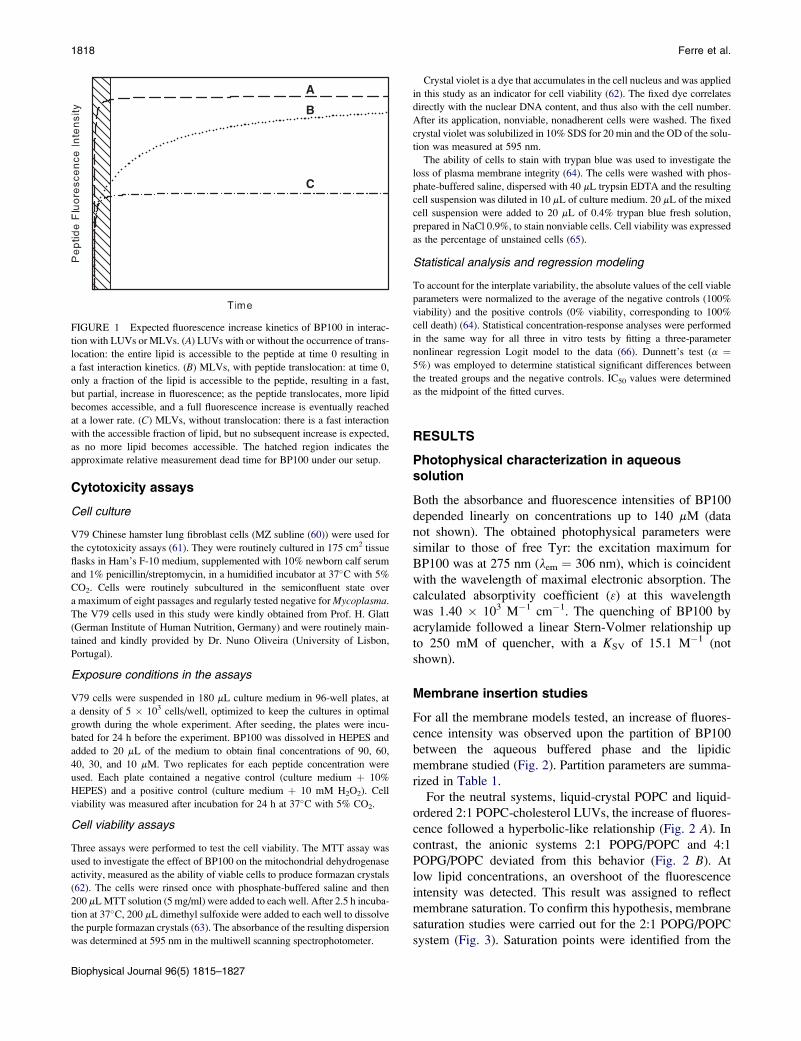
FIGURE 1 Expected fluorescence increase kinetics of BP100 in interac-
tion with LUVs or MLVs. (A) LUVs with or without the occurrence of trans-
location: the entire lipid is accessible to the peptide at time 0 resulting in
a fast interaction kinetics. (B) MLVs, with peptide translocation: at time 0,
only a fraction of the lipid is accessible to the peptide, resulting in a fast,
but partial, increase in fluorescence; as the peptide translocates, more lipid
becomes accessible, and a full fluorescence increase is eventually reached
at a lower rate. (C) MLVs, without translocation: there is a fast interaction
with the accessible fraction of lipid, but no subsequent increase is expected,
as no more lipid becomes accessible. The hatched region indicates the
approximate relative measurement dead time for BP100 under our setup.
Biophysical Journal 96(5) 1815–1827
Crystal violet is a dye that accumulates in the cell nucleus and was applied
in this study as an indicator for cell viability (62). The fixed dye correlates
directly with the nuclear DNA content, and thus also with the cell number.
After its application, nonviable, nonadherent cells were washed. The fixed
crystal violet was solubilized in 10% SDS for 20 min and the OD of the solu-
tion was measured at 595 nm.
The ability of cells to stain with trypan blue was used to investigate the
loss of plasma membrane integrity (64). The cells were washed with phos-
phate-buffered saline, dispersed with 40 mL trypsin EDTA and the resulting
cell suspension was diluted in 10 mL of culture medium. 20 mL of the mixed
cell suspension were added to 20 mL of 0.4% trypan blue fresh solution,
prepared in NaCl 0.9%, to stain nonviable cells. Cell viability was expressed
as the percentage of unstained cells (65).
Statistical analysis and regression modeling
To account for the interplate variability, the absolute values of the cell viable
parameters were normalized to the average of the negative controls (100%
viability) and the positive controls (0% viability, corresponding to 100%
cell death) (64). Statistical concentration-response analyses were performed
in the same way for all three in vitro tests by fitting a three-parameter
nonlinear regression Logit model to the data (66). Dunnett’s test (a ¼5%) was employed to determine statistical significant differences between
the treated groups and the negative controls. IC50 values were determined
as the midpoint of the fitted curves.
RESULTS
Photophysical characterization in aqueoussolution
Both the absorbance and fluorescence intensities of BP100
depended linearly on concentrations up to 140 mM (data
not shown). The obtained photophysical parameters were
similar to those of free Tyr: the excitation maximum for
BP100 was at 275 nm (lem ¼ 306 nm), which is coincident
with the wavelength of maximal electronic absorption. The
calculated absorptivity coefficient (3) at this wavelength
was 1.40 � 103 M�1 cm�1. The quenching of BP100 by
acrylamide followed a linear Stern-Volmer relationship up
to 250 mM of quencher, with a KSV of 15.1 M�1 (not
shown).
Membrane insertion studies
For all the membrane models tested, an increase of fluores-
cence intensity was observed upon the partition of BP100
between the aqueous buffered phase and the lipidic
membrane studied (Fig. 2). Partition parameters are summa-
rized in Table 1.
For the neutral systems, liquid-crystal POPC and liquid-
ordered 2:1 POPC-cholesterol LUVs, the increase of fluores-
cence followed a hyperbolic-like relationship (Fig. 2 A). In
contrast, the anionic systems 2:1 POPG/POPC and 4:1
POPG/POPC deviated from this behavior (Fig. 2 B). At
low lipid concentrations, an overshoot of the fluorescence
intensity was detected. This result was assigned to reflect
membrane saturation. To confirm this hypothesis, membrane
saturation studies were carried out for the 2:1 POPG/POPC
system (Fig. 3). Saturation points were identified from the
Membrane Actions of Hybrid Peptide BP100 1819
breaks observed in the curves obtained from titration of vesi-
cles with BP100 and were fitted with Eq. 3 (Fig. 3, inset). A
Kp value of 8.41 � 104 was obtained and a s of 0.118 was
determined, which corresponds to 8.4 phospholipids per
peptide at the saturation of the vesicles.
The depth of membrane internalization was probed using
stearic acid molecules labeled at different carbon positions
(carbons 5 and 16 for 5- and 16-NS, respectively) with tyro-
sine quenching nitroxyl groups. With previous knowledge of
the in-depth distribution of each quencher, an integrated
approach was used to infer the distribution of the tyrosine
residue of the peptide from each quencher’s quenching
profile (based on (55)). These profiles did not change signif-
icantly upon saturation (not shown), indicating little change
in the in-depth localization of the peptide. This was
A
B
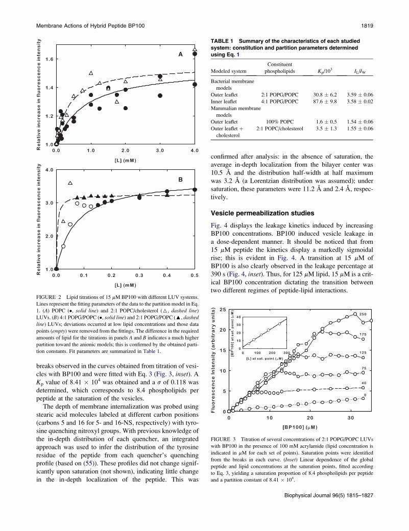
FIGURE 2 Lipid titrations of 15 mM BP100 with different LUV systems.
Lines represent the fitting parameters of the data to the partition model in Eq.
1. (A) POPC (�, solid line) and 2:1 POPC/cholesterol (6, dashed line)
LUVs. (B) 4:1 POPG/POPC (�, solid line) and 2:1 POPG/POPC (:, dashed
line) LUVs; deviations occurred at low lipid concentrations and those data
points (empty) were removed from the fittings. The difference in the required
amounts of lipid for the titrations in panels A and B indicates a much higher
partition toward the anionic models; this is confirmed by the obtained parti-
tion constants. Fit parameters are summarized in Table 1.
confirmed after analysis: in the absence of saturation, the
average in-depth localization from the bilayer center was
10.5 A and the distribution half-width at half maximum
was 3.2 A (a Lorentzian distribution was assumed); under
saturation, these parameters were 11.2 A and 2.4 A, respec-
tively.
Vesicle permeabilization studies
Fig. 4 displays the leakage kinetics induced by increasing
BP100 concentrations. BP100 induced vesicle leakage in
a dose-dependent manner. It should be noticed that from
15 mM peptide the kinetics display a markedly sigmoidal
rise; this is evident in Fig. 4. A transition at 15 mM of
BP100 is also clearly observed in the leakage percentage at
390 s (Fig. 4, inset). Thus, for 125 mM lipid, 15 mM is a crit-
ical BP100 concentration dictating the transition between
two different regimes of peptide-lipid interactions.
TABLE 1 Summary of the characteristics of each studied
system: constitution and partition parameters determined
using Eq. 1
Modeled system
Constituent
phospholipids Kp/103 IL/IW
Bacterial membrane
models
Outer leaflet 2:1 POPG/POPC 30.8 � 6.2 3.59 � 0.06
Inner leaflet 4:1 POPG/POPC 87.6 � 9.8 3.58 � 0.02
Mammalian membrane
models
Outer leaflet 100% POPC 1.6 � 0.5 1.54 � 0.06
Outer leaflet þcholesterol
2:1 POPC/cholesterol 3.5 � 1.3 1.55 � 0.06
FIGURE 3 Titration of several concentrations of 2:1 POPG/POPC LUVs
with BP100 in the presence of 100 mM acrylamide (lipid concentration is
indicated in mM for each set of points). Saturation points were identified
from the breaks in each curve. (Inset) Linear dependence of the global
peptide and lipid concentrations at the saturation points, fitted according
to Eq. 3, yielding a saturation proportion of 8.4 phospholipids per peptide
and a partition constant of 8.41 � 104.
Biophysical Journal 96(5) 1815–1827
1820 Ferre et al.
The sigmoidal curves could be fitted with the model
described by Gregory et al. (59) (not shown), even though
in their work only hyperbolic-like kinetics were fit. Both
rate constants of pore formation/dissipation needed to be
two-to-three orders-of-magnitude lower than those observed
by Gregory et al. (59) to generate a sigmoidal behavior
comparable to the one observed in Fig. 4. Further quantita-
tive analysis of leakage parameters is, however, not reliable,
due to multiple minima in the solution space and some
degree of correlation.
Membrane translocation studies
There were marked differences between the peptide-MLV
and peptide-LUV interaction kinetics (Fig. 5). Whereas the
increase in peptide fluorescence intensity upon LUV addi-
tion was almost instantaneous, the MLV-induced increase
spanned several minutes. Fluorescence intensity for the
MLV additions started out lower than that induced by
LUV additions of the same lipid concentration, but rose to
approximately the same relative level, as expected for the
occurrence of translocation (Fig. 1). To better compare the
fluorescence change at both peptide concentrations, Fig. 5
depicts the relative increase in fluorescence upon lipid addi-
tion. As a consequence there are small differences in the
endpoint of the kinetics at low BP100 concentrations
(Fig. 5—MLV1 and LUV1), attributable to error introduced
by the low initial Tyr fluorescence signal and further aggra-
vated by the use of acrylamide.
Vesicle aggregation and surface charge studies
Apart from a transient initial increase in turbidity, no signif-
icant changes in vesicle OD were observed at peptide
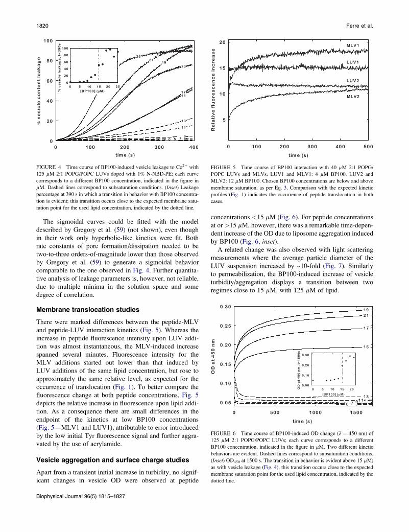
FIGURE 4 Time course of BP100-induced vesicle leakage to Co2þ with
125 mM 2:1 POPG/POPC LUVs doped with 1% N-NBD-PE; each curve
corresponds to a different BP100 concentration, indicated in the figure in
mM. Dashed lines correspond to subsaturation conditions. (Inset) Leakage
percentage at 390 s in which a transition in behavior with BP100 concentra-
tion is evident; this transition occurs close to the expected membrane satu-
ration point for the used lipid concentration, indicated by the dotted line.
Biophysical Journal 96(5) 1815–1827
concentrations <15 mM (Fig. 6). For peptide concentrations
at or >15 mM, however, there was a remarkable time-depen-
dent increase of the OD due to liposome aggregation induced
by BP100 (Fig. 6, inset).A related change was also observed with light scattering
measurements where the average particle diameter of the
LUV suspension increased by ~10-fold (Fig. 7). Similarly
to permeabilization, the BP100-induced increase of vesicle
turbidity/aggregation displays a transition between two
regimes close to 15 mM, with 125 mM of lipid.
FIGURE 5 Time course of BP100 interaction with 40 mM 2:1 POPG/
POPC LUVs and MLVs. LUV1 and MLV1: 4 mM BP100. LUV2 and
MLV2: 12 mM BP100. Chosen BP100 concentrations are below and above
membrane saturation, as per Eq. 3. Comparison with the expected kinetic
profiles (Fig. 1) indicates the occurrence of peptide translocation in both
cases.
FIGURE 6 Time course of BP100-induced OD change (l ¼ 450 nm) of
125 mM 2:1 POPG/POPC LUVs; each curve corresponds to a different
BP100 concentration, indicated in the figure in mM. Two different kinetic
behaviors are evident. Dashed lines correspond to subsaturation conditions.
(Inset) OD450 at 1500 s. The transition in behavior is evident above 15 mM;
as with vesicle leakage (Fig. 4), this transition occurs close to the expected
membrane saturation point for the used lipid concentration, indicated by the
dotted line.
Membrane Actions of Hybrid Peptide BP100 1821
The particles’ z-potential was�38.3 mV in the absence of
peptide but was brought close to zero (�0.1 mV) at satura-
tion (Fig. 8); aggregation and increase in turbidity prevented
z-potential measurement at higher peptide concentrations.
Effects of BP100 exposure on cell viability
Fig. 9 depicts the effects of BP100 on the mitochondrial
activity, cell monolayer adherence, and membrane integrity
of cultured hamster fibroblasts. All cell viability parameters
responded to the peptide (10–90 mM) in a clear dose-depen-
dent way. At the highest tested concentration (90 mM), low
cell viability (<10%) was observed. The peptide concentra-
tions, at which 50% inhibition was expected (IC50), were
interpolated from the regressions for each viability assay,
and ranged from 51.1 mM, for the crystal violet stain, to
64.3 mM for the trypan blue assay.
DISCUSSION
The biological activity of small, cationic antimicrobial
peptides has been largely associated with their interaction
with membranes. It is widely believed that for many of these
peptides, membrane disruption is the primary mechanism of
cell killing (10–12,15,16,18–20). However, their exact mode
of action is still poorly understood. Elucidating their mecha-
nism of action and their specific membrane damaging prop-
erties is crucial for the rational design of novel antibiotic
peptides with high antibacterial activity and low cytotox-
icity. With these observations in mind, and considering
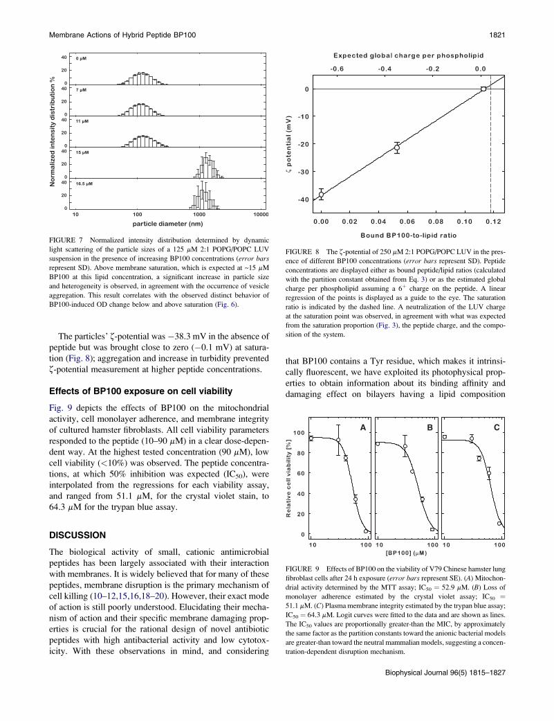
FIGURE 7 Normalized intensity distribution determined by dynamic
light scattering of the particle sizes of a 125 mM 2:1 POPG/POPC LUV
suspension in the presence of increasing BP100 concentrations (error bars
represent SD). Above membrane saturation, which is expected at ~15 mM
BP100 at this lipid concentration, a significant increase in particle size
and heterogeneity is observed, in agreement with the occurrence of vesicle
aggregation. This result correlates with the observed distinct behavior of
BP100-induced OD change below and above saturation (Fig. 6).
that BP100 contains a Tyr residue, which makes it intrinsi-
cally fluorescent, we have exploited its photophysical prop-
erties to obtain information about its binding affinity and
damaging effect on bilayers having a lipid composition
FIGURE 8 The z-potential of 250 mM 2:1 POPG/POPC LUV in the pres-
ence of different BP100 concentrations (error bars represent SD). Peptide
concentrations are displayed either as bound peptide/lipid ratios (calculated
with the partition constant obtained from Eq. 3) or as the estimated global
charge per phospholipid assuming a 6þ charge on the peptide. A linear
regression of the points is displayed as a guide to the eye. The saturation
ratio is indicated by the dashed line. A neutralization of the LUV charge
at the saturation point was observed, in agreement with what was expected
from the saturation proportion (Fig. 3), the peptide charge, and the compo-
sition of the system.
A B C
FIGURE 9 Effects of BP100 on the viability of V79 Chinese hamster lung
fibroblast cells after 24 h exposure (error bars represent SE). (A) Mitochon-
drial activity determined by the MTT assay; IC50 ¼ 52.9 mM. (B) Loss of
monolayer adherence estimated by the crystal violet assay; IC50 ¼51.1 mM. (C) Plasma membrane integrity estimated by the trypan blue assay;
IC50 ¼ 64.3 mM. Logit curves were fitted to the data and are shown as lines.
The IC50 values are proportionally greater-than the MIC, by approximately
the same factor as the partition constants toward the anionic bacterial models
are greater-than toward the neutral mammalian models, suggesting a concen-
tration-dependent disruption mechanism.
Biophysical Journal 96(5) 1815–1827
1822 Ferre et al.
similar to that of the bacterial and mammalian cytoplasmic
membranes.
Photophysical characterization of BP100in aqueous solution
The photophysical characterization of peptides in aqueous
solution is a prerequisite to understand their interaction
with phospholipid model membranes. The observed
behavior of BP100 in aqueous solution reflects that peptide
aggregation does not occur at the studied peptide concentra-
tion range. This is supported by the linear dependencies of
fluorescence emission intensity and electronic absorption
on concentration and by the obtained lexc, lem, and 3-values
as they are similar to those of free Tyr (275, 303, and 1400,
respectively (47)), indicating that the Tyr in BP100 is
exposed to an aqueous environment (47). Moreover, a linear
Stern-Volmer plot for the fluorescence quenching of BP100
with acrylamide is observed up to 250 mM (not shown). In
addition, there are no significant differences between the ob-
tained KSV and the one for acrylamide quenching of free Tyr,
evidencing that Tyr is totally accessible to the aqueous
phase. The absence of aggregation observed for BP100,
together with its overall positive charge (þ6), could account
for its high solubility in aqueous solution and facilitates the
interpretation of the peptide-membrane interaction results.
Membrane insertion studies
The extent of the partition of BP100 into model membranes
was studied using a partition model described by Santos et al.
that allows the calculation of the Nernst partition constant
(Kp) from fluorescence intensity (I) versus phospholipid
concentration ([L]) plots at a constant peptide concentration
([P]) (49). The Kp, defined as the ratio between the equilib-
rium membrane-bound and aqueous phase peptide concen-
trations, provides an easy assessment of the extent of
peptide-membrane interaction (Eq. 2). For both neutral
systems POPC and 2:1 POPC/cholesterol LUVs, used as
models of the outer leaflet of mammalian membranes, the
fluorescence intensity increased following an hyperbolic-
like relationship (Fig. 2 A). The moderate Kp values obtained
for vesicles composed of 100% POPC and POPC/cholesterol
mixtures (Table 1) could be attributed to the hydrophobic
effect and the van der Waals forces that are likely to domi-
nate the interactions between the neutral lipids and the
hydrophobic residues of BP100. In this case, no specific
interaction with cholesterol was observed, which is an indi-
cator of low toxicity toward mammalian cells. Furthermore,
cholesterol seems to play an important role in preventing the
intercalation of AMPs into eukaryotic cell membranes (67);
its presence and the absence of acidic phospholipids in the
eukaryotic membranes could account for the low cytotox-
icity displayed by BP100 against erythrocytes (38).
For the anionic liquid-crystalline 2:1 and 4:1 POPG/POPC
vesicles, which served as models for bacterial cell
Biophysical Journal 96(5) 1815–1827
membranes, the partition curves deviated from the hyper-
bolic-like progression at low lipid concentrations (Fig. 2
B). A similar behavior has been recently reported for the anti-
microbial peptide omiganan and has been attributed to
a membrane saturation process: at low phospholipid concen-
trations, membrane saturation may occur when the bound
peptide concentration, hypothetically dictated by Kp, is
higher than what the membrane can accommodate (53);
under these conditions, interaction changes may occur, as
has also been described for other AMPs upon the crossing
of threshold P/L ratios (68,69). Since the model of Santos
et al. (49) is not well suited to study these saturated systems,
the Kp values were obtained by fitting only the nonsaturated
points to the partition model (Fig. 2 B and Table 1). This
approach is obviously subject to error because the initial
points of the curve, which are important for the accurate
calculation of Kp, cannot be used. However, even with great
associated errors, the obtained partition constants were one
or more orders-of-magnitude higher than those of the neutral
systems (Table 1). These results are consistent with the ex-
pected preference of cationic peptides for negatively charged
membranes as a consequence of the strong electrostatic inter-
action.
To ensure that the deviation observed in the partition
curves of the anionic vesicles was due to a saturation of
the system, membrane saturation studies were carried out
using 2:1 POPG/POPC LUVs. LUV suspensions were
titrated with peptide in the presence of acrylamide while
monitoring BP100’s fluorescence intensity. Acrylamide is
an aqueous quencher that facilitates the identification of
alterations in the phase localization of peptides. In a nonsatu-
ration regime, a linear increase of the fluorescence intensity
is expected: as per the formalism behind Eq. 1, the fractions
of the peptide in each phase are constant with constant [L];
therefore, any variation in peptide concentration will result
in a proportional increase in each of these fractions and,
also therefore, in a global proportional increase of the fluo-
rescence intensity (53). Conversely, if saturation occurs, the
membrane will not be able to accommodate any more
peptide, which will then remain in the aqueous phase.
Because acrylamide quenches preferentially the fluores-
cence of the aqueous phase peptide population, a weaker
progression of the fluorescence intensity, relatively to a non-
saturation state, should then be detected (53). This behavior
was indeed observed in the BP100 titrations (Fig. 3),
showing the occurrence of saturation: two different slopes
were obtained for each I versus [P] curve. The first slope
corresponds to a nonsaturated state while the second one,
which is similar to that of the curve in the absence of lipid,
can be ascribed to a saturation of the system. The saturation
points could be easily obtained from the breaks of the initial
slopes of each titration curve. It was observed that the Iversus [P] curve with [L] ¼ 125 mM had its saturation point
close to [P] ¼ 17 mM (Fig. 3), which is slightly higher than
the peptide concentration that yielded an I versus [L] curve
Membrane Actions of Hybrid Peptide BP100 1823
with a deviation maximum close to [L] ¼ 125 mM
(Fig. 2 B). This result supports the hypothesis that the devi-
ations observed in the partition curves correspond to a satu-
ration of the membrane.
Further information from the saturation phenomenon was
obtained by representing the saturation point ([P],[L]) pairs
for the 2:1 POPG/POPC LUVs (Fig. 3, inset). This system
followed Eq. 3, which defines the total amount of peptide
at which a saturation point occurs as a linear function of
the amount of lipid in the system, and allows the calculation
of s—the P/L ratio at saturation—and the Nernst partition
constant Kp. However, it should be noticed that the values
for Kp have large associated errors because they are calcu-
lated from the reciprocal of a small intercept. Despite that,
the obtained Kp (8.41 � 104) had the same order of magni-
tude as that determined from the partition curve using the
model of Santos et al. (49) (3.08� 104). In addition, the satu-
ration P/L ratio was 0.118, which corresponds to 8.4 phos-
pholipids per peptide directly in contact with the membrane
at the saturation. Because there are 2/3 anionic phospholipids
in the used system, there will be 5.6 negatively charged
phospholipids per peptide at saturation. Interestingly, this
number is very close to the expected charge of the peptide
(þ6) at pH 7.4, which suggests that electroneutrality is
reached at the saturation of the system.
There was no significant alteration in the tyrosine in-depth
location upon saturation, indicating that most of BP100
molecules maintain their positioning within the membrane.
The location of the tyrosine residue, approximately halfway
across the membrane leaflet, is coherent with a relatively
deep burying of the peptide if it adopts, as expected (40),
a horizontally oriented a-helical structure. The lysines have
the ability to snorkel and keep their charged amino groups
near the headgroup region (70,71) while the hydrophobic
side chains could go as far as the bilayer center. This local-
ization within the bilayer is likely responsible, at least in part,
for the membrane destabilizing capabilities of BP100.
Vesicle permeabilization studies
Investigations on the mode of action of AMPs, such as cecro-
pins and melittin, have shown that they exert their activity by
inducing the formation of transmembrane pores or by
causing cell lysis, depending on both the peptide concentra-
tion and the membrane composition (41–45). Moreover, it
has been reported that cecropin-melittin hybrids are also
able to cause membrane permeabilization (72,73). These
findings prompted us to test BP100-induced permeabiliza-
tion of model lipidic membranes.
Results showed that BP100 has an important permeabiliz-
ing effect dependant on peptide concentration. The increase
in the permeabilization rate with BP100 concentration is,
however, not linear (Fig. 4). The clear change of behavior
at ~15 mM peptide, toward faster, sigmoidal, and more
intense leakage kinetics—visible both in the permeabiliza-
tion kinetics (Fig. 4) and in the leakage percentage profile
at 390 s (Fig. 4, inset)—occurs very close to the peptide
concentration expected to cause membrane saturation for
the 125 mM lipid concentration (Fig. 3). These results
show that membrane saturation affects more than just the
amount of bound peptide: high P/L ratios at, or close to,
membrane saturation are able to induce a change in a func-
tional property of the peptide. The sigmoidal leakage kinetic
induced by BP100 is uncommon, as such profiles are usually
hyperbolic-like (74). Nevertheless, a similar kinetic was
recently observed for an unrelated AMP (74). In both these
cases, because the interaction with LUVs is not a limiting
step (Fig. 5, LUV traces), the lag involved in the sigmoidal
behavior may be related to postbinding events in the
membrane (74).
Further information was extracted by fitting the data with
the model used by Gregory et al. (59) to describe cecropin
A-induced leakage. This model was only used to fit hyper-
bolic-like kinetics, but, even in those cases (59), close
inspection of the model in the first seconds of each kinetics
does reveal a brief sigmoidal behavior. Upon fitting, the
magnitude of this behavior could only be manipulated to
match the timescale of BP100-induced leakage kinetics by
lowering the k1 and k2 constants of the model two-to-three
orders of magnitude, relative to the values obtained in Greg-
ory et al. (59). As these parameters are the rate constants of
pore forming/dissipation, this result suggests that, after
binding, BP100 becomes disruptive at a slower rate than ce-
cropin A.
The high degree of peptide-induced leakage after satura-
tion may reflect severe membrane damage or lysis, whereas
the lower permeabilization before saturation could reflect
a lesser destabilization of the membrane upon peptide
binding. High P/L ratios close to saturation would then act
as the trigger between these two states, and could be the
biophysical parallel to the in vivo onset of antibacterial
activity. This is supported by the fact that the threshold
dependence on peptide concentration (Fig. 4, inset) could
not be accounted for with data fitting without assuming
some kind of parameter change with increasing BP100
concentration—such as an increase in the mentioned k1
and k2 disruption rates. Although hypothetical, this scenario
is plausible, and stresses the importance of high local peptide
concentrations in the membrane.
Membrane translocation studies
The determination of the occurrence of membrane transloca-
tion is an important functional characterization: a nontranslo-
cating peptide can only exert its activity at the extracellular/
membrane level, whereas one crossing a membrane may also
have cytoplasmic targets. However, detection of transloca-
tion can be troublesome, and, although there are several
methods available, many require peptide derivatization or
have limited applicability (75).
Biophysical Journal 96(5) 1815–1827
1824 Ferre et al.
Despite there being other published methods where MLVs
are used to enhance an internalization effect (75,76), the
method developed in our work is extremely simple and
requires only that the peptide has intrinsic fluorescence and
that its interaction kinetics with LUVs are significantly faster
than its translocation kinetics; quencher enhancement is not
an absolute requirement. The results clearly showed a trans-
location behavior at both high and low P/L ratios (Fig. 5). As
predicted in Fig. 1 for a translocating peptide, the interaction
with MLVs was slower than with LUVs, but eventually
reached the same fluorescence increase. Occurrence of trans-
location is unequivocal and, together with the permeabiliza-
tion assays, constitutes a further proof of the membrane
activity of the peptide.
Vesicle aggregation and surface charge studies
Turbidity measurements have been described as an useful
tool to investigate the affinity of cationic peptides toward
charged vesicles (77). The stability of a dispersion of
charged vesicles is mainly governed by three types of forces:
electrostatic repulsion, van der Waals attraction, and hydra-
tion (77). Cationic peptides can alter the charge density of
the vesicle surface inducing vesicle aggregation, which can
be followed as an increase of the OD. Turbidity results
showed two different kinetic patterns depending on BP100
concentration (Fig. 6). For 125 mM lipid (2:1 POPG/POPC
LUV) and peptide concentrations <15 mM, which corre-
spond to a nonsaturated state, no significant changes in
turbidity were observed. However, when membrane satura-
tion occurs (R15 mM peptide), the optical density of the
solution increased until a plateau was reached, ~30 min after
the addition of BP100. This increase is likely due to vesicle
aggregation induced under membrane saturation conditions.
These results confirm the affinity of BP100 for acidic phos-
pholipids and reinforce the hypothesis that electroneutrality
is reached at the membrane saturation point.
These conclusions were confirmed using light scattering
methodologies: the change in the LUV suspension OD is
related to an increase in the average particle size from
100 nm—in the absence of peptide and up to saturation—
to >1 mm upon saturation (Fig. 7). In addition, z-potential
measurements in this range showed that BP100 brings the
LUV charge to approximate electroneutrality at saturation,
confirming the prediction based on the saturation proportion
(Fig. 8). This effect is certainly favoring vesicle aggregation
by canceling the electrostatic repulsion between them.
Partition, saturation, and prediction of MIC
During our recent investigations, we have found that minimum
inhibitory concentration, MIC, and saturation can be corre-
lated for peptides, such as omiganan (53). For this peptide,
MICs were found to be similar to the peptide concentration
needed to reach the saturation state, reflecting the existence
of possible saturation-triggered antimicrobial mechanisms.
Biophysical Journal 96(5) 1815–1827
Since findings from BP100-membrane interaction studies
also suggest that membrane saturation is important for the
activity of this peptide, we examined whether the results ob-
tained are in agreement with the experimental MIC values.
As previously reported (53), under typical bacterial titers
and using the MIC as the total peptide concentration, the
membrane-bound peptide concentration ([P]L) is given by
Kp � MIC. On the other hand, s can be determined as
[P]L � gL. Combining both expressions, the MIC can be
readily calculated as MIC¼ s/(Kp� gL). Using the obtained
s (0.118) and Kp (3.08 � 104 or 8.41 � 104, from the parti-
tion and saturation studies, respectively) values, and consid-
ering gL as 0.763 M�1 (50), this equation leads to MIC
values of 2 or 5 mM, depending on the selected Kp. These
values are consistent with the antibacterial activity displayed
by BP100, which inhibited in vitro growth of the bacteria
E. amylovora, X. vesicatoria, and P. syringae at 2.5–7.5 mM
(38). In addition to validating the obtained values for Kp and
s, these results strongly support the correlation between these
constants and the MIC, evidencing the importance of the satu-
ration point in the mode of action of this peptide.
Physiological significance of saturation-inducedactivity
The obtained results clearly point toward the occurrence of
different membrane-disrupting events as saturation is
reached. Given the plausible correlation between saturation
and the onset of antibacterial activity of BP100, an extrapo-
lation of these events to an in vivo setting was sought.
Surface charge neutralization at saturation was found to
be an important occurrence, triggering the observed vesicle
aggregation, and probably being responsible for the destabili-
zation that led to an increase in membrane permeabilization,
as leakage enhancement correlates with vesicle aggregation.
The bacterial metabolism will certainly be sensitive to the
neutralization-induced loss of the membrane surface poten-
tial, as this will disturb the charge environment of the outer
leaflet proteins. The observed coupled permeabilization (if
not lysis) entails even further damage to the cell, namely the
dissipation of the transmembrane potential which, among
other effects, will halt ATP synthesis. Vesicle aggregation
may not have a parallel in vivo, as bacterial membranes
have additional layers of protection (LPS, peptidoglycan,
capsule) preventing direct membrane contact between
bacteria; its occurrence in vitro does, however, stress the
importance of the surface potential for membrane stability.
The observed translocation could be a consequence of the per-
meabilization or can be an independent event; either way,
direct interaction with cytoplasmic targets is yet another
possible cause of bacterial death.
Effects of BP100 exposure on cell viability
The experimental results from our studies show cytotoxic
effects in the cultured mammalian fibroblast cells at
Membrane Actions of Hybrid Peptide BP100 1825
concentrations of BP100 above 50–60 mM (Fig. 9). This is in
good agreement with similar findings in human erythrocytes
(38), where an increased release of hemoglobin was
observed above 150 mM. Although the membrane integrity
in our V79 cells was affected at lower concentrations (IC50
¼ 51.1 mM), it probably just reflects the different cell lines:
different sensibilities to antibacterial peptides were also
found between human erythrocytes and mammalian COS-7
kidney cells (65), and might indicate a better resistance of
the human erythrocytes to this class of peptides (78). Results
from the MTT assay (Fig. 9) demonstrated changes in the
metabolic activity of mitochondria V79 cells, as the dehydro-
genase enzymes started to be less active to convert the
yellow water-soluble salt into insoluble formazan crystals
at increasing peptide concentrations. Whether this means
that there is a direct action on the mitochondria, or indirect
loss of mitochondrial activity, cannot be ascertained without
further investigation.
A successful application of this peptide as a bactericide
demands a high therapeutic index, i.e., a high antimicrobial
activity but low cytotoxicity. The high antimicrobial potency
(MIC ¼ 2.5–7.5 mM) and relatively low cytotoxicity in
human erythrocytes (38) reveals promising values for
BP100. Although cytotoxic effects were observed in V79
cells at peptide concentrations above 50–60 mM, this range
is still far above the anticipated antimicrobial application
levels.
Cytotoxicity against mammalian models is reached at
a concentration higher than the MIC by roughly the same
proportion that Kp values toward mammalian model bilayers
are lower than toward bacterial ones. This observation
suggests that cell killing may be dependent on a constant
local membrane-bound concentration, independently of the
considered lipid system.
CONCLUSION
This work clearly points out a correlation between high
membrane concentrations (possibly even saturation) of
BP100 and bacterial death. Three different potential causes
of activity of AMP, i.e., charge neutralization, permeabiliza-
tion, and translocation, were identified. In addition, a concen-
tration dependence of the killing phenomena, in bacteria and
in mammalian cells, was suggested. While the exact mecha-
nism of action of the peptide may remain elusive in vivo, and
depend on the peptide and bacteria species, our findings
unravel the bases of the closely coupled occurrence of those
causes, as experimentally observed by Friedrich et al. (79).
Fundacao para a Ciencia e a Tecnologia (Portugal) is acknowledged for
a grant to M.N.M. (No. SFRH/BD/24778/2005). R.F. is the recipient of
a predoctoral fellowship from the Ministry of Education and Science of
Spain. This work was supported by grants from the Ministry of Education
and Science of Spain (No. AGL2006-13564/AGR), and from the Catalan
Government (No. 2005SGR00275).
REFERENCES
1. Brogden, K. A., M. Ackermann, P. B. McCray, Jr., and B. F. Tack.2003. Antimicrobial peptides in animals and their role in host defenses.Int. J. Antimicrob. Agents. 22:465–478.
2. Bulet, P., R. Stocklin, and L. Menin. 2004. Antimicrobial peptides:from invertebrates to vertebrates. Immunol. Rev. 198:169–184.
3. Ganz, T., and R. I. Lehrer. 1998. Antimicrobial peptides of vertebrates.Curr. Opin. Immunol. 10:41–44.
4. Garcia-Olmedo, F., A. Molina, J. M. Alamillo, and P. Rodriguez-Palen-zuela. 1998. Plant defense peptides. Biopolymers. 47:479–491.
5. Otvos, L., Jr. 2000. Antibacterial peptides isolated from insects. J. Pept.Sci. 6:497–511.
6. Zasloff, M. 2002. Antimicrobial peptides of multicellular organisms.Nature. 415:389–395.
7. Broekaert, W. F., B. P. A. Cammue, M. F. C. DeBolle, K. Thevissen,G. W. DeSamblanx, et al. 1997. Antimicrobial peptides from plants.Crit. Rev. Plant Sci. 16:297–323.
8. Hancock, R. E., and A. Patrzykat. 2002. Clinical development ofcationic antimicrobial peptides: from natural to novel antibiotics.Curr. Drug Targets Infect. Disord. 2:79–83.
9. Hancock, R. E., and H. G. Sahl. 2006. Antimicrobial and host-defensepeptides as new anti-infective therapeutic strategies. Nat. Biotechnol.24:1551–1557.
10. Boman, H. G. 2003. Antibacterial peptides: basic facts and emergingconcepts. J. Intern. Med. 254:197–215.
11. Hancock, R. E. 2001. Cationic peptides: effectors in innate immunityand novel antimicrobials. Lancet Infect. Dis. 1:156–164.
12. Jenssen, H., P. Hamill, and R. E. Hancock. 2006. Peptide antimicrobialagents. Clin. Microbiol. Rev. 19:491–511.
13. Montesinos, E. 2007. Antimicrobial peptides and plant disease control.FEMS Microbiol. Lett. 270:1–11.
14. Zhang, L., and T. J. Falla. 2006. Antimicrobial peptides: therapeuticpotential. Expert Opin. Pharmacother. 7:653–663.
15. Brogden, K. A. 2005. Antimicrobial peptides: pore formers or metabolicinhibitors in bacteria? Nat. Rev. Microbiol. 3:238–250.
16. Yeaman, M. R., and N. Y. Yount. 2003. Mechanisms of antimicrobialpeptide action and resistance. Pharmacol. Rev. 55:27–55.
17. Perron, G. G., M. Zasloff, and G. Bell. 2006. Experimental evolution ofresistance to an antimicrobial peptide. Proc. Biol. Sci. 273:251–256.
18. Shai, Y. 2002. Mode of action of membrane active antimicrobialpeptides. Biopolymers. 66:236–248.
19. Tossi, A., L. Sandri, and A. Giangaspero. 2000. Amphipathic, a-helicalantimicrobial peptides. Biopolymers. 55:4–30.
20. Bechinger, B. 2004. Structure and function of membrane-lytic peptides.Crit. Rev. Plant Sci. 23:271–292.
21. Yang, L., T. A. Harroun, T. M. Weiss, L. Ding, and H. W. Huang. 2001.Barrel-stave model or toroidal model? A case study on melittin pores.Biophys. J. 81:1475–1485.
22. Shai, Y. 1999. Mechanism of the binding, insertion and destabilizationof phospholipid bilayer membranes by a-helical antimicrobial and cellnon-selective membrane-lytic peptides. Biochim. Biophys. Acta.1462:55–70.
23. Kobayashi, S., A. Chikushi, S. Tougu, Y. Imura, M. Nishida, et al.2004. Membrane translocation mechanism of the antimicrobial peptidebuforin 2. Biochemistry. 43:15610–15616.
24. Hultmark, D., A. Engstrom, H. Bennich, R. Kapur, and H. G. Boman.1982. Insect immunity: isolation and structure of cecropin D and fourminor antibacterial components from Cecropia pupae. Eur. J. Biochem.127:207–217.
25. Hultmark, D., H. Steiner, T. Rasmuson, and H. G. Boman. 1980. Insectimmunity. Purification and properties of three inducible bactericidalproteins from hemolymph of immunized pupae of Hyalophora cecro-pia. Eur. J. Biochem. 106:7–16.
Biophysical Journal 96(5) 1815–1827
1826 Ferre et al.
26. Sato, H., and J. B. Feix. 2006. Peptide-membrane interactions andmechanisms of membrane destruction by amphipathic a-helical antimi-crobial peptides. Biochim. Biophys. Acta. 1758:1245–1256.
27. Andreu, D., R. B. Merrifield, H. Steiner, and H. G. Boman. 1983. Solid-phase synthesis of cecropin A and related peptides. Proc. Natl. Acad.Sci. USA. 80:6475–6479.
28. Steiner, H., D. Hultmark, A. Engstrom, H. Bennich, and H. G. Boman.1981. Sequence and specificity of two antibacterial proteins involved ininsect immunity. Nature. 292:246–248.
29. Alberola, J., A. Rodriguez, O. Francino, X. Roura, L. Rivas, et al. 2004.Safety and efficacy of antimicrobial peptides against naturally acquiredLeishmaniasis. Antimicrob. Agents Chemother. 48:641–643.
30. Andreu, D., J. Ubach, A. Boman, B. Wahlin, D. Wade, et al. 1992.Shortened cecropin A-melittin hybrids. Significant size reductionretains potent antibiotic activity. FEBS Lett. 296:190–194.
31. Boman, H. G., D. Wade, I. A. Boman, B. Wahlin, and R. B. Merrifield.1989. Antibacterial and antimalarial properties of peptides that are ce-cropin-melittin hybrids. FEBS Lett. 259:103–106.
32. Cavallarin, L., D. Andreu, and B. San Segundo. 1998. CecropinA-derived peptides are potent inhibitors of fungal plant pathogens.Mol. Plant Microbe Interact. 11:218–227.
33. Chicharro, C., C. Granata, R. Lozano, D. Andreu, and L. Rivas. 2001.N-terminal fatty acid substitution increases the leishmanicidal activityof CA(1–7)M(2–9), a cecropin-melittin hybrid peptide. Antimicrob.Agents Chemother. 45:2441–2449.
34. Giacometti, A., O. Cirioni, W. Kamysz, G. D’Amato, C. Silvestri,et al. 2004. In vitro activity and killing effect of the synthetic hybridcecropin A-melittin peptide CA(1–7)M(2–9)NH2 on methicillin-resis-tant nosocomial isolates of Staphylococcus aureus and interactionswith clinically used antibiotics. Diagn. Microbiol. Infect. Dis.49:197–200.
35. Lee, D. G., Y. Park, I. Jin, K. S. Hahm, H. H. Lee, et al. 2004. Structure-antiviral activity relationships of cecropin A-magainin 2 hybrid peptideand its analogues. J. Pept. Sci. 10:298–303.
36. Wade, D., D. Andreu, S. A. Mitchell, A. M. Silveira, A. Boman, et al.1992. Antibacterial peptides designed as analogs or hybrids of cecropinsand melittin. Int. J. Pept. Protein Res. 40:429–436.
37. Ali, G. S., and A. S. Reddy. 2000. Inhibition of fungal and bacterialplant pathogens by synthetic peptides: in vitro growth inhibition, inter-action between peptides and inhibition of disease progression. Mol.Plant Microbe Interact. 13:847–859.
38. Badosa, E., R. Ferre, M. Planas, L. Feliu, E. Besalu, et al. 2007. Alibrary of linear undecapeptides with bactericidal activity against phyto-pathogenic bacteria. Peptides. 28:2276–2285.
39. Bardajı, E., E. Montesinos, E. Badosa, L. Feliu, M. Planas, et al. 2006.Antimicrobial linear peptides. P200601098; priority date: April 28th,2006; Oficina Espanola de Patentes y Marcas, Spain.
40. Ferre, R., E. Badosa, L. Feliu, M. Planas, E. Montesinos, et al. 2006.Inhibition of plant-pathogenic bacteria by short synthetic cecropinA-melittin hybrid peptides. Appl. Environ. Microbiol. 72:3302–3308.
41. Christensen, B., J. Fink, R. B. Merrifield, and D. Mauzerall. 1988.Channel-forming properties of cecropins and related model compoundsincorporated into planar lipid membranes. Proc. Natl. Acad. Sci. USA.85:5072–5076.
42. Ladokhin, A. S., M. E. Selsted, and S. H. White. 1997. Sizingmembrane pores in lipid vesicles by leakage of co-encapsulatedmarkers: pore formation by melittin. Biophys. J. 72:1762–1766.
43. Ladokhin, A. S., and S. H. White. 2001. ‘‘Detergent-like’’ permeabili-zation of anionic lipid vesicles by melittin. Biochim. Biophys. Acta.1514:253–260.
44. Silvestro, L., K. Gupta, J. N. Weiser, and P. H. Axelsen. 1997. Theconcentration-dependent membrane activity of cecropin A. Biochem-istry. 36:11452–11460.
45. Steiner, H., D. Andreu, and R. B. Merrifield. 1988. Binding and actionof cecropin and cecropin analogues: antibacterial peptides from insects.Biochim. Biophys. Acta. 939:260–266.
Biophysical Journal 96(5) 1815–1827
46. Mayer, L. D., M. J. Hope, and P. R. Cullis. 1986. Vesicles of variablesizes produced by a rapid extrusion procedure. Biochim. Biophys. Acta.858:161–168.
47. Santos, N. C., and M. A. Castanho. 2002. Fluorescence spectroscopymethodologies on the study of proteins and peptides. On the 150th anni-versary of protein fluorescence. Trends Appl. Spectrosc. 4:113–125.
48. Coutinho, A., and M. Prieto. 1993. Ribonuclease-T1 and alcohol-dehy-drogenase fluorescence quenching by acrylamide—a laboratory exper-iment for undergraduate students. J. Chem. Educ. 70:425–428.
49. Santos, N. C., M. Prieto, and M. A. Castanho. 2003. Quantifying molec-ular partition into model systems of biomembranes: an emphasis onoptical spectroscopic methods. Biochim. Biophys. Acta. 1612:123–135.
50. Nagle, J. F., and M. C. Wiener. 1988. Structure of fully hydrated bilayerdispersions. Biochim. Biophys. Acta. 942:1–10.
51. Ladokhin, A. S., S. Jayasinghe, and S. H. White. 2000. How to measureand analyze tryptophan fluorescence in membranes properly, and whybother? Anal. Biochem. 285:235–245.
52. Wenk, M. R., and J. Seelig. 1998. Magainin 2 amide interaction withlipid membranes: calorimetric detection of peptide binding and poreformation. Biochemistry. 37:3909–3916.
53. Melo, M. N., and M. A. Castanho. 2007. Omiganan interaction withbacterial membranes and cell wall models. Assigning a biological roleto saturation. Biochim. Biophys. Acta. 1768:1277–1290.
54. Chalpin, D. B., and A. M. Kleinfeld. 1983. Interaction of fluorescencequenchers with the n-(9-anthroyloxy) fatty acid membrane probes. Bio-chim. Biophys. Acta. 731:465–474.
55. Fernandes, M. X., J. Garcia de la Torre, and M. A. Castanho. 2002. Jointdetermination by Brownian dynamics and fluorescence quenching ofthe in-depth location profile of biomolecules in membranes. Anal. Bio-chem. 307:1–12.
56. Chattopadhyay, A., and E. London. 1988. Spectroscopic and ionizationproperties of N-(7-nitrobenz-2-oxa-1,3-diazol-4-yl)-labeled lipids inmodel membranes. Biochim. Biophys. Acta. 938:24–34.
57. Pokorny, A., and P. F. Almeida. 2004. Kinetics of dye efflux and lipidflip-flop induced by d-lysin in phosphatidylcholine vesicles and themechanism of graded release by amphipathic, a-helical peptides.Biochemistry. 43:8846–8857.
58. Lakowicz, J. R. 1999. Quenching of fluorescence. Principles of Fluores-cence Spectroscopy, 2nd Ed. Kluwer Academic/Plenum, New York;London.
59. Gregory, S. M., A. Cavenaugh, V. Journigan, A. Pokorny, and P. F. Al-meida. 2008. A quantitative model for the all-or-none permeabilizationof phospholipid vesicles by the antimicrobial peptide cecropin A. Bio-phys. J. 94:1667–1680.
60. Rueff, J., C. Chiapella, J. K. Chipman, F. Darroudi, I. D. Silva, et al.1996. Development and validation of alternative metabolic systemsfor mutagenicity testing in short-term assays. Mutat. Res. 353:151–176.
61. Zucco, F., I. De Angelis, and A. Stammati. 1998. Cellular models forin vitro toxicity testing. In Animal Cell Culture Techniques.M. Clynes, editor. Springer, Berlin; London.
62. Mickuviene, I., V. Kirveliene, and B. Juodka. 2004. Experimentalsurvey of non-clonogenic viability assays for adherent cells in vitro.Toxicol. In Vitro. 18:639–648.
63. Mitchell, J. B. 1988. Potential applicability of nonclonogenic measure-ments to clinical oncology. Radiat. Res. 114:401–414.
64. Brink, C. B., A. Pretorius, B. P. van Niekerk, D. W. Oliver, andD. P. Venter. 2008. Studies on cellular resilience and adaptationfollowing acute and repetitive exposure to ozone in cultured humanepithelial (HeLa) cells. Redox Rep. 13:87–100.
65. Cudic, M., C. V. Lockatell, D. E. Johnson, and L. Otvos, Jr. 2003. Invitro and in vivo activity of an antibacterial peptide analog against uro-pathogens. Peptides. 24:807–820.
66. Scholze, M., W. Boedeker, M. Faust, T. Backhaus, R. Altenburger, et al.2001. A general best-fit method for concentration-response curves andthe estimation of low-effect concentrations. Environ. Toxicol. Chem.20:448–457.
Membrane Actions of Hybrid Peptide BP100 1827
67. Zhao, H., R. Sood, A. Jutila, S. Bose, G. Fimland, et al. 2006. Interac-tion of the antimicrobial peptide pheromone Plantaricin A with modelmembranes: implications for a novel mechanism of action. Biochim.Biophys. Acta. 1758:1461–1474.
68. Huang, H. W. 2000. Action of antimicrobial peptides: two-state model.Biochemistry. 39:8347–8352.
69. Huang, H. W. 2006. Molecular mechanism of antimicrobial peptides:the origin of cooperativity. Biochim. Biophys. Acta. 1758:1292–1302.
70. Segrest, J. P., H. De Loof, J. G. Dohlman, C. G. Brouillette, and G. M.Anantharamaiah. 1990. Amphipathic helix motif: classes and proper-ties. Proteins. 8:103–117.
71. Kandasamy, S. K., and R. G. Larson. 2006. Molecular dynamics simu-lations of model trans-membrane peptides in lipid bilayers: a systematicinvestigation of hydrophobic mismatch. Biophys. J. 90:2326–2343.
72. Abrunhosa, F., S. Faria, P. Gomes, I. Tomaz, J. C. Pessoa, et al. 2005.Interaction and lipid-induced conformation of two cecropin-melittinhybrid peptides depend on peptide and membrane composition.J. Phys. Chem. B. 109:17311–17319.
73. Juvvadi, P., S. Vunnam, E. L. Merrifield, H. G. Boman, and R. B.Merrifield. 1996. Hydrophobic effects on antibacterial and channel-forming properties of cecropin A-melittin hybrids. J. Pept. Sci.2:223–232.
74. Rathinakumar, R., and W. C. Wimley. 2008. Biomolecular engineering
by combinatorial design and high-throughput screening: small, soluble
peptides that permeabilize membranes. J. Am. Chem. Soc. 130:9849–
9858.
75. Henriques, S. T., M. N. Melo, and M. A. Castanho. 2007. How to
address CPP and AMP translocation? Methods to detect and quantify
peptide internalization in vitro and in vivo. (Review). Mol. Membr.Biol. 24:173–184.
76. Matsuzaki, K., S. Yoneyama, O. Murase, and K. Miyajima. 1996.
Transbilayer transport of ions and lipids coupled with mastoparan X
translocation. Biochemistry. 35:8450–8456.
77. Persson, D., P. E. Thoren, and B. Norden. 2001. Penetratin-induced
aggregation and subsequent dissociation of negatively charged phos-
pholipid vesicles. FEBS Lett. 505:307–312.
78. Otvos, L., Jr., K. Bokonyi, I. Varga, B. I. Otvos, R. Hoffmann, et al.
2000. Insect peptides with improved protease-resistance protect mice
against bacterial infection. Protein Sci. 9:742–749.
79. Friedrich, C. L., D. Moyles, T. J. Beveridge, and R. E. Hancock.
2000. Antibacterial action of structurally diverse cationic peptides
on Gram-positive bacteria. Antimicrob. Agents Chemother. 44:2086–
2092.
Biophysical Journal 96(5) 1815–1827
Related Documents