Synergistic anti-inflammatory effects of low doses of curcumin in combination with polyunsaturated fatty acids: Docosahexaenoic acid or eicosapentaenoic acid Constance Lay Lay Saw a,b , Ying Huang b,c , Ah-Ng Kong a,b,c, * a Center for Cancer Prevention Research, Ernest Mario School of Pharmacy, Rutgers, The State University of New Jersey, Piscataway, NJ 08854, United States b Department of Pharmaceutics, Ernest Mario School of Pharmacy, Rutgers, The State University of New Jersey, Piscataway, NJ 08854, United States c Graduate Program in Pharmaceutical Science, Ernest Mario School of Pharmacy, Rutgers, The State University of New Jersey, Piscataway, NJ 08854, United States 1. Introduction Inflammatory response plays a very important role not only in the normal physiology but also in the pathology such as cancers [1]. As chronic inflammations are associated with malignancies, it is important to prevent the inflammation before neoplasm formation, promotion and/or progression. One way of doing so is by the use of chemopreventive agents such as curcumin (CUR), a constituent of turmeric of ginger family, which is a potent anti- inflammatory [2] via inhibiting nuclear factor-kappa-B (NF-kB) pathway [3–5], induction of heat shock response [6] as well as activation of nuclear factor-erythroid 2-related factor 2 (Nrf2)/ antioxidant response element (ARE) pathway [7]. It is well- recognized that CUR activates Nrf2/ARE signaling pathway leading to induction of phase II genes such as glutathione S-transferase (GST) and hemeoxygenase-1 (HO-1) [8]. Recently, the effects of CUR in cancers management have been tested in clinical trials and the benefits have been reported [9–12]. Polyunsaturated fatty acids (PUFA) such as docosahexaenoic acid (DHA) or eicosapen- taenoic acid (EPA) are potentially potent anti-inflammatory agents by decreasing the production of inflammatory eicosanoids, cytokines, and reactive oxygen species (ROS) [13,14]. The benefits of the PUFA have also been demonstrated in clinical trials in cancer patients [15,16] and patients with ulcerative colitis [17]. Thus, it is of interest and clinical significance to examine the protective anti- inflammatory effects of CUR alone and in combination with PUFA, DHA or EPA. The potential benefits of using combination of different chemopreventive compounds could be quite obvious. It is hopeful that a greater chemopreventive effect could be achieved by compounds targeting different signaling mechanisms [18,19]. In this context, with our present study, we hypothesized that when Biochemical Pharmacology 79 (2010) 421–430 ARTICLE INFO Article history: Received 23 June 2009 Accepted 28 August 2009 Keywords: Curcumin Docosahexaenoic acid Eicosapentaenoic acid Nuclear factor-erythroid 2-related factor 2 Synergism Inflammation ABSTRACT Inflammatory response plays an important role not only in the normal physiology but also in the pathology such as cancers. As chronic inflammations are associated with malignancies, it is important to prevent inflammation-mediated neoplastic formation, promotion and/or progression. One possible intervention will be using cancer chemopreventive agents such as curcumin (CUR), a potent anti- inflammatory and anti-oxidative stress compound. Polyunsaturated fatty acids (PUFA) such as docosahexaenoic acid (DHA) or eicosapentaenoic acid (EPA) are potent anti-inflammatory agents by decreasing the production of inflammatory eicosanoids, cytokines, and reactive oxygen species (ROS). The present study aims at examining whether CUR with DHA or EPA would have synergistic anti- inflammatory effects in RAW 264.7 cells. Non-toxic concentrations of single and combination of the compounds were investigated at 6, 12 and 24 h. The nitric oxide (NO) suppression effects were most prominent at 24 h. All the combinations of CUR and DHA or EPA with lower concentrations of CUR 5 mM and 25 mM of DHA or EPA were found to have synergistic effects in suppressing LPS-stimulated NO and endogenous NO levels. Importantly, very low doses of CUR 2.5 mM and DHA or EPA of 0.78 mM could synergistically suppress the LPS-induced prostaglandin E 2 (PGE 2 ). The combinations were also found to suppress iNOS, COX-2, 5-lipoxygenase (5-LOX) and cPLA 2 but induce HO-1. Taken together, the present study clearly shows the synergistic anti-inflammatory as well as anti-oxidative stress effects of CUR and PUFA. ß 2009 Elsevier Inc. All rights reserved. Abbreviations: CUR, curcumin; DHA, docosahexaenoic acid; EPA, eicosapentaenoic acid; PUFA, polyunsaturated fatty acids; Nrf2, nuclear factor-erythroid 2-related factor 2; ARE, antioxidant response element; NF-kB, nuclear factor-kappa-B; LPS, lipopolysaccharide; COX, cyclooxygenase; LOX, lipoxygenase; iNOS, inducible nitric oxide synthase; HO-1, hemeoxygenase-1; ROS, reactive oxygen species; cPLA 2 , cytosolic phospholipase A 2 ; PGE 2 , prostaglandin E 2 ; qRT-PCR, quantitative reverse- transcriptase-polymerase chain reaction; CI, combination index. * Corresponding author at: Department of Pharmaceutics, Ernest Mario School of Pharmacy, Room 228, 160 Frelinghuysen Road, Rutgers, The State University of New Jersey, Piscataway, NJ 08854, United States. Tel.: +1 732 455 3831x228/6; fax: +1 732 455 3134. E-mail address: [email protected] (A.-N. Kong). Contents lists available at ScienceDirect Biochemical Pharmacology journal homepage: www.elsevier.com/locate/biochempharm 0006-2952/$ – see front matter ß 2009 Elsevier Inc. All rights reserved. doi:10.1016/j.bcp.2009.08.030

Welcome message from author
This document is posted to help you gain knowledge. Please leave a comment to let me know what you think about it! Share it to your friends and learn new things together.
Transcript
Biochemical Pharmacology 79 (2010) 421–430
Synergistic anti-inflammatory effects of low doses of curcumin in combinationwith polyunsaturated fatty acids: Docosahexaenoic acid or eicosapentaenoic acid
Constance Lay Lay Saw a,b, Ying Huang b,c, Ah-Ng Kong a,b,c,*a Center for Cancer Prevention Research, Ernest Mario School of Pharmacy, Rutgers, The State University of New Jersey, Piscataway, NJ 08854, United Statesb Department of Pharmaceutics, Ernest Mario School of Pharmacy, Rutgers, The State University of New Jersey, Piscataway, NJ 08854, United Statesc Graduate Program in Pharmaceutical Science, Ernest Mario School of Pharmacy, Rutgers, The State University of New Jersey, Piscataway, NJ 08854, United States
A R T I C L E I N F O
Article history:
Received 23 June 2009
Accepted 28 August 2009
Keywords:
Curcumin
Docosahexaenoic acid
Eicosapentaenoic acid
Nuclear factor-erythroid 2-related factor 2
Synergism
Inflammation
A B S T R A C T
Inflammatory response plays an important role not only in the normal physiology but also in the
pathology such as cancers. As chronic inflammations are associated with malignancies, it is important to
prevent inflammation-mediated neoplastic formation, promotion and/or progression. One possible
intervention will be using cancer chemopreventive agents such as curcumin (CUR), a potent anti-
inflammatory and anti-oxidative stress compound. Polyunsaturated fatty acids (PUFA) such as
docosahexaenoic acid (DHA) or eicosapentaenoic acid (EPA) are potent anti-inflammatory agents by
decreasing the production of inflammatory eicosanoids, cytokines, and reactive oxygen species (ROS).
The present study aims at examining whether CUR with DHA or EPA would have synergistic anti-
inflammatory effects in RAW 264.7 cells. Non-toxic concentrations of single and combination of the
compounds were investigated at 6, 12 and 24 h. The nitric oxide (NO) suppression effects were most
prominent at 24 h. All the combinations of CUR and DHA or EPA with lower concentrations of CUR 5 mM
and 25 mM of DHA or EPA were found to have synergistic effects in suppressing LPS-stimulated NO and
endogenous NO levels. Importantly, very low doses of CUR 2.5 mM and DHA or EPA of 0.78 mM could
synergistically suppress the LPS-induced prostaglandin E2 (PGE2). The combinations were also found to
suppress iNOS, COX-2, 5-lipoxygenase (5-LOX) and cPLA2 but induce HO-1. Taken together, the present
study clearly shows the synergistic anti-inflammatory as well as anti-oxidative stress effects of CUR and
PUFA.
� 2009 Elsevier Inc. All rights reserved.
Contents lists available at ScienceDirect
Biochemical Pharmacology
journal homepage: www.e lsev ier .com/ locate /b iochempharm
1. Introduction
Inflammatory response plays a very important role not only inthe normal physiology but also in the pathology such as cancers[1]. As chronic inflammations are associated with malignancies, itis important to prevent the inflammation before neoplasmformation, promotion and/or progression. One way of doing sois by the use of chemopreventive agents such as curcumin (CUR), aconstituent of turmeric of ginger family, which is a potent anti-inflammatory [2] via inhibiting nuclear factor-kappa-B (NF-kB)
Abbreviations: CUR, curcumin; DHA, docosahexaenoic acid; EPA, eicosapentaenoic
acid; PUFA, polyunsaturated fatty acids; Nrf2, nuclear factor-erythroid 2-related
factor 2; ARE, antioxidant response element; NF-kB, nuclear factor-kappa-B; LPS,
lipopolysaccharide; COX, cyclooxygenase; LOX, lipoxygenase; iNOS, inducible nitric
oxide synthase; HO-1, hemeoxygenase-1; ROS, reactive oxygen species; cPLA2,
cytosolic phospholipase A2; PGE2, prostaglandin E2; qRT-PCR, quantitative reverse-
transcriptase-polymerase chain reaction; CI, combination index.
* Corresponding author at: Department of Pharmaceutics, Ernest Mario School of
Pharmacy, Room 228, 160 Frelinghuysen Road, Rutgers, The State University of New
Jersey, Piscataway, NJ 08854, United States. Tel.: +1 732 455 3831x228/6;
fax: +1 732 455 3134.
E-mail address: [email protected] (A.-N. Kong).
0006-2952/$ – see front matter � 2009 Elsevier Inc. All rights reserved.
doi:10.1016/j.bcp.2009.08.030
pathway [3–5], induction of heat shock response [6] as well asactivation of nuclear factor-erythroid 2-related factor 2 (Nrf2)/antioxidant response element (ARE) pathway [7]. It is well-recognized that CUR activates Nrf2/ARE signaling pathway leadingto induction of phase II genes such as glutathione S-transferase(GST) and hemeoxygenase-1 (HO-1) [8]. Recently, the effects ofCUR in cancers management have been tested in clinical trials andthe benefits have been reported [9–12]. Polyunsaturated fattyacids (PUFA) such as docosahexaenoic acid (DHA) or eicosapen-taenoic acid (EPA) are potentially potent anti-inflammatory agentsby decreasing the production of inflammatory eicosanoids,cytokines, and reactive oxygen species (ROS) [13,14]. The benefitsof the PUFA have also been demonstrated in clinical trials in cancerpatients [15,16] and patients with ulcerative colitis [17]. Thus, it isof interest and clinical significance to examine the protective anti-inflammatory effects of CUR alone and in combination with PUFA,DHA or EPA.
The potential benefits of using combination of differentchemopreventive compounds could be quite obvious. It is hopefulthat a greater chemopreventive effect could be achieved bycompounds targeting different signaling mechanisms [18,19]. Inthis context, with our present study, we hypothesized that when
C.L.L. Saw et al. / Biochemical Pharmacology 79 (2010) 421–430422
CUR and DHA or EPA are used together, synergistic chemopre-ventive effects such as anti-inflammatory response could beachieved. Our study clearly demonstrates the synergistic anti-inflammatory as well as anti-oxidative stress effects of CUR andPUFA.
2. Materials and methods
2.1. Materials and cell cultures
All materials were purchased from Sigma–Aldrich (St. Louis,MO, USA) unless otherwise specified including Curcumin fromCurcuma longa (Turmeric), (CUR, catalog no. C1386, �70%), cis-4,7,10,13,16,19-docosahexaenoic acid (DHA, catalog no. D2534,�98%), cis-5,8,11,14,17-eicosapentaenoic acid (EPA, catalog no.E2011, �99%), 2, 3-diaminonapthalene (DAN), sodium nitrate(NaNO3), and lipopolysaccharide (LPS) derived from Escherichia coli
055:B5.Murine leukemic monocytic macrophage cell line, RAW 264.7
cells were maintained and cultured at 37 8C under a humidified, 5%CO2 atmosphere in Dulbecco’s Modified Eagle Medium (DMEM,GIBCO Invitrogen Corporation, Grand Island, NY, USA) supple-mented with 10% fetal bovine serum (FBS), 100 units/mL penicillin,100 mg/mL streptomycin and 1.176 g/L sodium bicarbonate.
2.2. Measurement of nitrite (NO) concentration by fluorimetric assay
Biologically produced nitric oxide (NO) is rather unstable and isvery rapidly oxidized to nitrite and nitrate in aqueous solution. Theassay of NO can be determined by measuring the stable oxidizedproduct of NO, nitrite. A commonly used well-known Griess assay,however lacks the sensitivity, since the detection of limit is about1 mM, which is relatively insensitive to detect the nitrite amount.Therefore, a fluorimetric assay was modified from Misko et al. [20]and used to detect the nitrite concentrations in the supernatant ofcell culture.
Nitrite standards made from nitrate sodium were dilutedfreshly in deionized water. 10 mL of freshly prepared DAN(0.05 mg/mL in 0.62 M HCl) was used to react with nitrite(50 mL of supernatant) to form 1-(H)-naphthotriazole, a fluor-escent product. After a 10 min of incubation at room temperaturein the dark, the reaction was terminated with 5 mL of 2.8 M NaOH.Formation of the 2,3-diaminonaphthotriazole in dark opaque 96-well plates was measured using a microplate fluorescence reader,FLx-800 from Bio-Tek Instruments Inc. (Winooski, VT, USA) withexcitation at 360 and 460 nm emission with a gain setting at 75%.
2.3. Testing the ability of CUR, DHA or EPA alone or combinations of
CUR + DHA and CUR + EPA for suppressing NO levels
All single compound and combinations of CUR and DHA or EPAat various concentrations were tested. 105 cells per wells in 96-well microplate were seeded overnight to allow the cells to adhereto the plate. On the day of experiment, the tested compounds wereprepared in DMEM without FBS. The cells were first treated with50 mL of various chemopreventive agents. One hour later, another50 mL of the chemopreventive agents with the stimulant ofinducible NO synthase (iNOS), LPS were added into the well toachieve final tested concentrations of single compound CUR 0.63,1.25, 2.5, 5, 10, 20, 40 and 80 mM, DHA 0.78, 1.56, 3.13, 6.25, 12.5,25, 50 and 100 mM, EPA 0.78, 1.56, 3.13, 6.25, 12.5, 25, 50 and100 mM. In combination study, total of different 24 combinationsof drugs were tested; 0.63 mM CUR + 0.78 mM DHA, 0.63 mMCUR + 0.78 mM EPA, 0.63 mM CUR + 1.56 mM DHA, 0.63 mMCUR + 1.56 mM EPA, 0.63 mM CUR + 3.13 mM DHA, 0.63 mMCUR + 3.13 mM EPA, 1.25 mM CUR + 0.78 mM DHA, 1.25 mM
CUR + 0.78 mM EPA, 1.25 mM CUR + 1.56 mM DHA, 1.25 mMCUR + 1.56 mM EPA, 2.5 mM CUR + 0.78 mM DHA, 2.5 mMCUR + 0.78 mM EPA, 2.5 mM CUR + 1.56 mM DHA, 2.5 mMCUR + 1.56 mM EPA, 2.5 mM CUR + 3.13 mM DHA, 2.5 mMCUR + 3.13 mM EPA, 2.5 mM CUR + 6.25 mM DHA, 2.5 mMCUR + 6.25 mM EPA, 5 mM CUR + 6.25 mM DHA, 5 mMCUR + 6.25 mM EPA, 5 mM CUR + 12.5 mM DHA, 5 mMCUR + 12.5 mM EPA, 5 mM CUR + 25 mM DHA and 5 mMCUR + 25 mM EPA.
Induction of iNOS activity was accomplished by treatment ofLPS (final concentration 10 mg/mL) in RAW 264.7 cells. Positivecontrol cells were treated only with LPS and negative control cellswere treated with 0.1% DMSO. The NO suppressing ability of singlecompound or combination treatments is calculated from thefollowing equation:
% NO inhibition
¼ðcorrected nitrite content in positive control�corrected nitrite content in treated sampleÞcorrected nitrite content in positive control
� 100%
In the cells, the nitrite content of negative control weresubtracted from the positive and treated samples (i.e., correctednitrite content in positive control and corrected nitrite content intreated sample) before the calculation of % NO inhibition wascarried out to give an indication of the absolute % NO inhibition bythe drugs, either in singly or in combination.
2.4. Cell viability test by MTS assay
The viability of the cells towards all tested compounds invarious concentrations were tested using the cell viability testbased on the ability of cells to convert a tetrazolium compound [3-(4,5-dimethylthiazol-2-yl)-5-(3-carboxymethoxyphenyl)-2-(4-sulfophenyl)-2H-tetrazolium, inner salt; MTS] into its reducedcolored formazen product. This bio-conversion is presumablyaccomplished by NADPH or NADH produced by dehydrogenaseenzymes in metabolically active cells. All single compound andcombinations of CUR and DHA or EPA tested for NO suppressingactivity were also tested in this MTS assay. Similar to NOsuppressing tests, 105 cells per wells in 96-well microplate wereseeded overnight to allow the cells to adhere to the plate. 10 mLMTS CellTiter 961 Aqueous one solution cell proliferation assay(Promega, Madison, WI, USA) was added to cells 24 h after the cellswere treated similar to the NO suppressing activity tests describedabove. The microplate was incubated for 1 h at 37 8C under ahumidified, 5% CO2 atmosphere. The microplate was measured at490 nm by a spectrophotometer (mQuant Biomolecular Spectro-photometer, Bio-Tek Instruments Inc., Winooski, VT, USA). Controlcells were treated with 0.1% DMSO and the viability of cells arecalculated from this equation: absorbance in treated sample/absorbance in control � 100%.
2.5. Measurement of prostaglandin E2 (PGE2) concentration
by ELISA assay
RAW 264.7 cells were treated in a similar fashion to the NOsuppressing test, using selected concentrations of drugs andcombination of compounds that were identified to be synergistic insuppressing the NO production stimulated by LPS. Followingtreatment, the PGE2 content in the supernatant of cell culture wasdetermined using a commercially available enzyme immunoassaykit (Prostaglandin EIA kit, catalog no. 514010, Cayman ChemicalCompany, Ann, Arbor, MI, USA) according to the manufacturer’sinstructions. The % suppression of PGE2 was calculated in a similarmanner as the calculation for % NO suppression above.

C.L.L. Saw et al. / Biochemical Pharmacology 79 (2010) 421–430 423
2.6. Investigation of synergistic effects of CUR and DHA or EPA by
combination index (CI)
The investigation of synergistic effects of CUR and DHA or EPAinvolves plotting the dose–response effects for CUR, DHA and EPAalone and for multiple combinations of agents. The combinationdrug effect analysis based on Loewe additivity model was used toexamine the drug interactions as reviewed by Lee et al. [21]. Theinteraction of drugs can be characterized by the combination index(CI) which is defined as:
CI ¼ d1
Dx;1þ d2
Dx;2
where d1 and d2 are doses of drug 1 (CUR) and drug 2 (DHA or EPA)in combination, which produces an effect x, while Dx,1 and Dx,2 arethe doses of drug 1 (CUR) and drug 2 (DHA or EPA) that produce thesame effect x when given alone. d1/Dx,1 + d2/Dx,2, is also called theinteraction index at the combination doses (d1 and d2). When the CIis equal to, less than or greater than 1, the combination doses willbe additive, synergistic or antagonistic, respectively. Although theexact mechanism of interaction may often be unknown, this testwill attempt to figure out whether the suppression of inflamma-tion is involved by reducing expression of iNOS and cyclooxygen-ase-2 (COX-2) as demonstrated by suppression of NO and PGE2,respectively. It was also concluded that the Loweve additivitymodel is one of the best general reference models for evaluatingpotential drug interactions [21].
2.7. RNA isolation and quantitative reverse-transcriptase
polymerase chain reaction (qRT-PCR) assay
RAW 264.7 cells were treated using selected concentrationsthat examined the compound singly or in combinations. The cellswere treated similarly as described above. After 6 h of treatment,total RNA was extracted using RNeasy Micro Kit (Qiagen, Valencia,CA, USA). RNA concentrations were determined by Quant-iTTM
RiboGreen1 RNA Reagent and Kit (Invitrogen, Grand Island, NY,USA). From each sample, 0.1 mg of total RNA was then reversetranscribed to single-stranded cDNA by TaqMan1 ReverseTranscription Reagents (catalog no. N808-0234, Applied Biosys-tems Inc., Foster City, CA, USA). Then qPCR analyses wereperformed on the aliquots of the cDNA preparations with SYBRGreen PCR Master Mix (catalog no. 4309155, Applied BiosystemsInc., Foster City, CA, USA) to detect quantitatively the geneexpression of COX-2, iNOS, hemeoxygenase-1 (HO-1), 5-lipox-ygenase (5-LOX), cytosolic phospholipase A2 (cPLA2) and glycer-aldehyde 3-phosphate dehydrogenase (GAPDH) (as an internalstandard) using Applied Biosystems 7900HT Fast Real-Time PCRSystem (Applied Biosystems Inc., Foster City, CA, USA). The primerpairs were designed using Primer Quest Oligo Design and AnalysisTool by Integrated DNA Technologies Inc. (Coralville, IA, USA) andthe sequences are listed in Table 1. All assays were confirmed usingmelting curves to confirm the presence of single PCR products.Minimum of two independent experiments were carried out, eachexperiment had at least triplicate samples for each treatment.
Table 1Oligonucleotide primers used for qualitative real-time RT-PCR (qRT-PCR).
Gene Accession no. Forward (50)
Cyclooxygenase-2 (COX-2) NM_011198.3 50-TGC CTG G
Cytosolic phospholipase A2 (cPLA2) NM_008869.3 50-ACG TGT C
Glyceraldehyde 3-phosphate
dehydrogenase (GAPDH)
XM_001473623.1 50-TGA AGC A
Hemeoxygenase-1 (HO-1) NM_010442.1 50-CCT CAC T
Inducible nitric oxide synthase 2 (iNOS) NM_010927.2 50-CCT GGT A
5-Lipoxygenase (5-LOX) activating protein NM_009663.1 50-CCA CAA G
2.8. Protein extraction and Western blotting
20 mg of protein samples were subjected to 4–15% gradientpolyacrylamide gel (Criterion Tris–HCl gel, Bio-Rad Lab, Hercules,CA, USA) electrophoresis and the resolved proteins were thentransferred to polyvinylidene difluoride (PVDF) membranes(Immobilon-P, Millipore, Bedford, MA, USA) using a semi-drytransfer system (Fisher Scientific, Pittsburgh, PA, USA). The non-specific binding of antibodies were blocked with 5% bovine serumalbumin (BSA) in PBST buffer (0.1% Tween 20 in PBS). Antibodieswere purchased from Santa Cruz Biotechnology, Inc. (Santa Cruz,CA, USA). Immunodetection of COX-2 (catalog no. sc-1745), iNOS(catalog no. sc-650), HO-1 (catalog no. sc-10789) and b-actin(catalog no. sc-1616) proteins was carried out using respectiveprimary antibodies (1:1000 in 3% BSA in PBST buffer) andhorseradish peroxidase (HRP) conjugated secondary antibodies(1:1000 in 3% BSA in PBST buffer). The immunocomplexes weredetermined by using the enhanced chemiluminescent system fordetecting HRP on immunoblots (Thermo Scientific, Rockford, IL,USA) and the bands were visualized and captured by BioRadChemiDoc XRS system (Hercules, CA, USA). The protein bands werequantified by densitometry using Image J software (version 1.37 g,National Institute of Health, USA) and represented as the protein/b-actin ratio. Minimum of three independent experiments werecarried out, each experiment had at least duplicated blots for eachtreatment.
2.9. Data presentation and statistical analysis
The results were presented as means � standard error of themean (SEM) unless otherwise indicated. Data were analyzed by aStudent’s t-test or one-way ANOVA to determine statistical differ-ences between groups using SPSS statistical software (version 16.0 forWindows, SPSS Inc., Chicago, IL, USA), the statistical significance ofmean differences was based on a p value of <0.05. Mann–Whitney U
tests were used to determine statistical differences between groupsfor the protein levels obtained from Western blots, with a p value of<0.05 is considered to be significant. Box-plots were used to presentthe normalized density data for the protein expression levels of iNOS,COX-2 and HO-1 as previously described [22].
3. Results
3.1. Non-toxic effects of CUR, DHA and EPA on RAW 264.7 cells treated
with or without LPS
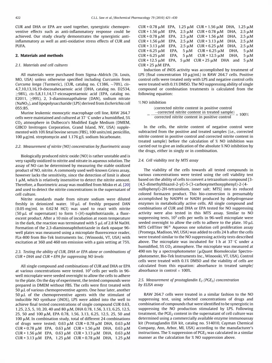
To determine the effects of single compound, CUR, DHA, EPAand various combinations of CUR with DHA and CUR with EPA oncells viability, RAW 264.7 cells were initially seeded in microplatesfollowed by different treatments. Fig. 1 shows the results of theMTS assay after 24 h treatment. Without LPS stimulation, with theexception of the highest concentrations of the compounds, i.e., CUR80 mM, DHA 100 mM and EPA 100 mM, all the treatments werenon-toxic as compared to the non-treated cells (Fig. 1A). In thepresence of LPS, the viability profiles of the treated cells are similar
primer Reverse (30) primer
TC TGA TGA TGT ATG CCA-30 50-AGT AGT CGC ACA CTC TGT TGT GCT-30
TC CAC GTC AAA CCT GAT-30 50-TGC TTC CAA ATA GGT CAG GAG CCA-30
GG CAT CTG AGG G-30 50-CGA AGG TGG AAG AGT GGG AG-30
GG CAG GAA ATC ATC-30 50-CCT CGT GGA GAC GCT TTA CAT A-30
CG GGC ATT GCT-30 50-GCT CAT GCG GCC TCC TTT-30
GT GGA GCA TGA AAG CAA-30 50-TAC GCA GTT CTG GTT GGC AGT GTA-30
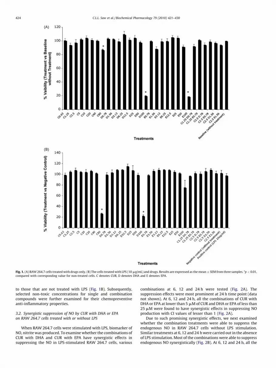
Fig. 1. (A) RAW 264.7 cells treated with drugs only. (B) The cells treated with LPS (10 mg/mL) and drugs. Results are expressed as the mean � SEM from three samples. *p < 0.01,
compared with corresponding value for non-treated cells. C denotes CUR, D denotes DHA and E denotes EPA.
C.L.L. Saw et al. / Biochemical Pharmacology 79 (2010) 421–430424
to those that are not treated with LPS (Fig. 1B). Subsequently,selected non-toxic concentrations for single and combinationcompounds were further examined for their chemopreventiveanti-inflammatory properties.
3.2. Synergistic suppression of NO by CUR with DHA or EPA
on RAW 264.7 cells treated with or without LPS
When RAW 264.7 cells were stimulated with LPS, biomarker ofNO, nitrite was produced. To examine whether the combinations ofCUR with DHA and CUR with EPA have synergistic effects insuppressing the NO in LPS-stimulated RAW 264.7 cells, various
combinations at 6, 12 and 24 h were tested (Fig. 2A). Thesuppression effects were most prominent at 24 h time point (datanot shown). At 6, 12 and 24 h, all the combinations of CUR withDHA or EPA at lower than 5 mM of CUR and DHA or EPA of less than25 mM were found to have synergistic effects in suppressing NOproduction with CI values of lesser than 1 (Fig. 2A).
Due to such promising synergistic effects, we next examinedwhether the combination treatments were able to suppress theendogenous NO in RAW 264.7 cells without LPS stimulation.Similar treatments at 6, 12 and 24 h were carried out in the absenceof LPS stimulation. Most of the combinations were able to suppressendogenous NO synergistically (Fig. 2B). At 6, 12 and 24 h, all the
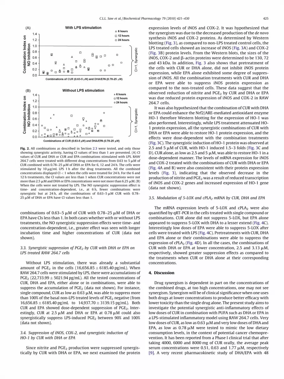
Fig. 2. All combinations as described in Section 2.3 were tested, and only those
showing synergistic activity, having CI values of less than 1 are presented. (A) CI
values of CUR and DHA or CUR and EPA combinations stimulated with LPS. RAW
264.7 cells were treated with different drug concentrations from 0.63 to 5 mM of
CUR combined with 0.78–25 mM of DHA or EPA for 6, 12 and 24 h. The cells were
stimulated by 10 mg/mL LPS 1 h after the drug treatments. All the combined
concentrations displayed CI < 1 when the cells were treated for 24 h. For the 6 and
12 h treatments, the CI values are less than 1 when CUR concentrations were not
more than 2.5 mM and DHA or EPA concentrations were not more than 6.25 mM. (B)
When the cells were not treated by LPS. The NO synergistic suppression effect is
time- and concentration-dependent, i.e., at 6 h, fewer combinations were
synergistic but at 24 h, all the combinations of 0.63–5 mM CUR with 0.78–
25 mM of DHA or EPA have CI values less than 1.
C.L.L. Saw et al. / Biochemical Pharmacology 79 (2010) 421–430 425
combinations of 0.63–5 mM of CUR with 0.78–25 mM of DHA orEPA have CIs less than 1. In both cases whether with or without LPStreatments, the NO synergistic suppression effects were time- andconcentration-dependent, i.e., greater effect was seen with longerincubation time and higher concentrations of CUR (data notshown).
3.3. Synergistic suppression of PGE2 by CUR with DHA or EPA on
LPS treated RAW 264.7 cells
Without LPS stimulation, there was already a substantialamount of PGE2 in the cells (16,656.85 � 6185.40 pg/mL). WhenRAW 264.7 cells were stimulated by LPS, there were accumulations ofPGE2 (22,733.99 � 5021.94 pg/mL). All the tested concentrations ofCUR, DHA and EPA, either alone or in combinations, were able tosuppress the accumulation of PGE2 (data not shown). For instance,single compound, CUR as low as 0.63 mM, was able to suppress morethan 100% of the basal non-LPS treated levels of PGE2 negative (from16,656.85 � 6185.40 pg/mL to 14,937.70 � 3139.15 pg/mL). BothCUR and EPA showed dose-dependent suppression of PGE2. Inter-estingly, CUR at 2.5 mM and DHA or EPA at 0.78 mM could alsosynergistically suppress LPS-induced PGE2 between 90% and 100%(data not shown).
3.4. Suppression of iNOS, COX-2, and synergistic induction of
HO-1 by CUR with DHA or EPA
Since nitrite and PGE2 production were suppressed synergis-tically by CUR with DHA or EPA, we next examined the protein
expression levels of iNOS and COX-2. It was hypothesized thatthe synergism was due to the decreased production of the de novo
synthesis iNOS and COX-2 proteins. As determined by Westernblotting (Fig. 3), as compared to non-LPS treated control cells, theLPS treated cells showed an increase of iNOS (Fig. 3A) and COX-2(Fig. 3B) protein levels. From the Western blots, the sizes of theiNOS, COX-2 and b-actin proteins were determined to be 130, 72and 43 kDa. In addition, Fig. 3 also shows that pretreatment ofthe cells with CUR or DHA alone, did not inhibit iNOS proteinexpression, while EPA alone exhibited some degree of suppres-sion of iNOS. All the combination treatments with CUR and DHAor EPA were able to suppress iNOS protein expression ascompared to the non-treated cells. These data suggest that theobserved reduction of nitrite and PGE2 by CUR and DHA or EPAwas due reduced protein expression of iNOS and COX-2 in RAW264.7 cells.
It was also hypothesized that the combination of CUR with DHAor EPA could enhance the Nrf2/ARE-mediated antioxidant enzymeHO-1 therefore Western blotting for the expression of HO-1 wasalso performed. Interestingly, while LPS treatment attenuated HO-1 protein expression, all the synergistic combinations of CUR withDHA or EPA were able to restore HO-1 protein expression, and theeffects were dose-dependent with the combination treatments(Fig. 3C). The synergistic induction of HO-1 protein was observed at2.5 and 5 mM of CUR, with HO-1 induced 1.5–3 folds (Fig. 3C andD). CUR alone, as low as 2.5 and 5 mM, was able to restore HO-1 in adose-dependent manner. The levels of mRNA expression for iNOSand COX-2 treated with the combinations of CUR with DHA or EPA(Fig. 4A and B) were also consistent with their protein expressionlevels (Fig. 3), indicating that the observed decrease in theproduction of nitrite and PGE2 was a result of reduced transcriptionof iNOS and COX-2 genes and increased expression of HO-1 gene(data not shown).
3.5. Modulation of 5-LOX and cPLA2 mRNA by CUR, DHA and EPA
The mRNA expression levels of 5-LOX and cPLA2 were alsoquantified by qRT-PCR in the cells treated with single compound orcombinations. CUR alone did not suppress 5-LOX, but EPA aloneappeared to suppress 5-LOX with DHA to a lesser extend (Fig. 4C).Interestingly low doses of EPA were able to suppress 5-LOX aftercells were treated with LPS (Fig. 4C). Pretreatments with CUR, DHAand EPA alone or their combinations were able to suppress theexpression of cPLA2 (Fig. 4D). In all the cases, the combinations ofCUR with DHA or EPA at lower concentration, 2.5 and 3.13 mM,respectively, showed greater suppression effects as compared tothe treatments when CUR or DHA alone at their correspondingconcentrations.
4. Discussion
Drug synergism is dependent in part on the concentrations ofthe combined drugs, at too high concentrations, one may not seesynergism. Synergism will be of clinical significance when one usesboth drugs at lower concentrations to produce better efficacy withlower toxicity than the single drug alone. The present study aims toinvestigate the potential synergistic anti-inflammatory effects oflow doses of CUR in combination with PUFA such as DHA or EPA ina LPS-stimulated inflammatory model using RAW 264.7 cells. Verylow doses of CUR, as low as 0.63 mM and very low doses of DHA andEPA, as low as 0.78 mM were tested to mimic the low dietaryconsumption levels, in the context of potential cancer chemopre-vention. It has been reported from a Phase I clinical trial that aftertaking 4000, 6000 and 8000 mg of CUR orally, the average peakserum concentrations were 0.51, 0.63 and 1.77 mM, respectively[9]. A very recent pharmacokinetic study of DHA/EPA with 48
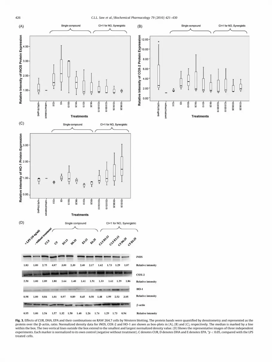
Fig. 3. Effects of CUR, DHA, EPA and their combinations on RAW 264.7 cells by Western blotting. The protein bands were quantified by densitometry and represented as the
protein over the b-actin, ratio. Normalized density data for iNOS, COX-2 and HO-1 are shown as box-plots in (A), (B) and (C), respectively. The median is marked by a line
within the box. The two vertical lines outside the box extend to the smallest and largest normalized density value. (D) Shows the representative images of three independent
experiments. Each marker is normalized to its own control (negative without treatment). C denotes CUR, D denotes DHA and E denotes EPA. *p < 0.05, compared with the LPS
treated cells.
C.L.L. Saw et al. / Biochemical Pharmacology 79 (2010) 421–430426
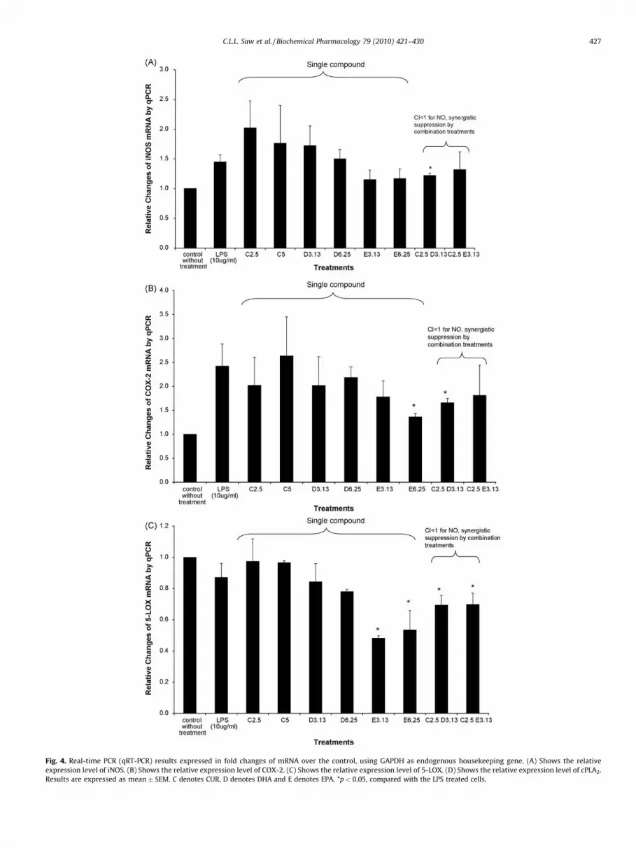
Fig. 4. Real-time PCR (qRT-PCR) results expressed in fold changes of mRNA over the control, using GAPDH as endogenous housekeeping gene. (A) Shows the relative
expression level of iNOS. (B) Shows the relative expression level of COX-2. (C) Shows the relative expression level of 5-LOX. (D) Shows the relative expression level of cPLA2.
Results are expressed as mean � SEM. C denotes CUR, D denotes DHA and E denotes EPA. *p < 0.05, compared with the LPS treated cells.
C.L.L. Saw et al. / Biochemical Pharmacology 79 (2010) 421–430 427

Fig. 4. (Continued ).
C.L.L. Saw et al. / Biochemical Pharmacology 79 (2010) 421–430428
subjects consuming fish 1–2 times a month had a detectableplasma DHA level of about 60 mg/mL and EPA of about 10 mg/mL,which is equivalent to 182 and 33 mM, respectively [23]. Theseconcentrations are much higher than the concentrations tested inthe present study. Therefore, the low doses of CUR in combinationwith DHA or EPA used in the present study provides strongevidence for translating the observed synergistic anti-inflamma-tory effects into human trials. The present study also shows thatsignificant synergism exists between CUR and DHA or EPA,particularly in this anti-inflammatory model. To examine whethersuch synergism is specific to RAW 264.7 cells model, we had alsoperformed similar combination treatments in a human coloncancer HT-29 cell line, significant synergistic suppression of NOproduction were also observed with CUR in combinations withDHA or EPA (unpublished observations). Ongoing experiments arebeing conducted to further study on the synergistic anti-inflammatory effects.
The synergistic anti-inflammatory effects of low doses of CUR incombination with DHA or EPA not only inhibited NO production inthe LPS-stimulated RAW 264.7 cells, but also clearly inhibited inthe non-LPS treated cells (Fig. 2B). Over the past 1–2 decadesresearch on NO has shown its importance in maintaining normalphysiological functions in the in vivo settings. The suppression ofthe endogenous NO may be unfavorable in certain normalphysiological activities such as smooth muscle relaxation, how-ever, in the context of the present study which would be underpathological conditions as with RAW 264.7 cell line, since it is aleukemic macrophage cell line that has been associated with tumorprogression [24]. Macrophage generally is an important compo-nent in the human immune defense mechanism and often timeenhanced immunity has been considered to be beneficial in theprognosis and survival of cancer patients [25,26] Recent reportsuggests that tumor-associated macrophages are implicated in thepathophysiology of cancer formation and progression, and theycould play a role in the microenvironments of numerousmalignancies [27].
In the present study, low doses of CUR, DHA and EPA alonecaused an increase of iNOS and COX-2 proteins and in the presenceof inflammatory agent such as LPS, low doses of CUR, DHA and EPAalone were unable to suppress the protein expression of iNOS andCOX-2 (Fig. 3). However, with these similar low concentrations, theanti-inflammatory synergistic effects for CUR with DHA or EPA
were evident. The reasons for these observations are unclear, butcould be related to differential signaling at these low doses.Certainly further study into the potential mechanism would beneeded to explain such observations.
Previously, our laboratory had identified more than 1000 Nrf2-dependent genes regulated by CUR [8] and the oxidative stressresponse gene, HO-1 was one of them. In the current study, theinduction of HO-1 by CUR (Fig. 3) is also observed which isconsistent with previous studies [7,8]. The present study alsoidentifies the synergistic induction of HO-1 by CUR with DHA orEPA (Fig. 3), which possibly mediated by the Nrf2/ARE signalingpathway. In addition, the suppression of inflammatory mediatorssuch as NO, PGE2, 5-LOX and cPLA2 also shows synergism with CURand DHA or EPA.
The development of inflammatory diseases, including cancer, iscommonly accompanied by increased production of leukotrienesand prostaglandins via the arachidonic acid (AA) pathway [28].Typically the cytosolic phospholipase A2 (cPLA2) cuts the AA out ofthe cellular phospholipids and when free AA are exposed to theenzymes such as 5-LOX and COX-1/2, leukotrienes and PGE2 areproduced, respectively. Clinically, inhibition of COX by nonster-oidal anti-inflammatory drugs (NSAIDs) and selective COX-2inhibitors reduces the levels of prostaglandins, resulting in areduction of pain and inflammation. However, this inhibition mayalso cause alternative processing of AA via the 5-LOX pathway,resulting in an increase of production of pro-inflammatory andgastrotoxic leukotrienes. Theoretically, the dual inhibitors of COXand 5-LOX could decrease the production of both leukotrienes andprostaglandins, and they could display enhanced anti-inflamma-tory effects [29]. A recent study proposes a potential role of cPLA2
in LPS-induced p38 MAPK pathways, and that the p38 MAPK/cPLA2/COX-2 pathway was implicated in the production of LPS-induced interleukin-1-beta and interleukin-6 in differentiatedU937 cells [30]. Previously Hong et al. [31] showed that CUR at20 mM potently inhibited 5-LOX, however, this 5-LOX inhibitoryactivity was not observed in our current study, although we usedmuch lower concentrations of 2.5 and 5 mM CUR. Our presentstudy shows that the cPLA2 was decreased by all the low doses ofCUR, DHA and EPA when they were used alone (Fig. 4D). Thesynergistic suppression of 5-LOX and cPLA2 were clearly demon-strated with 2.5 mM CUR when combined with 3.13 mM of DHA orEPA.
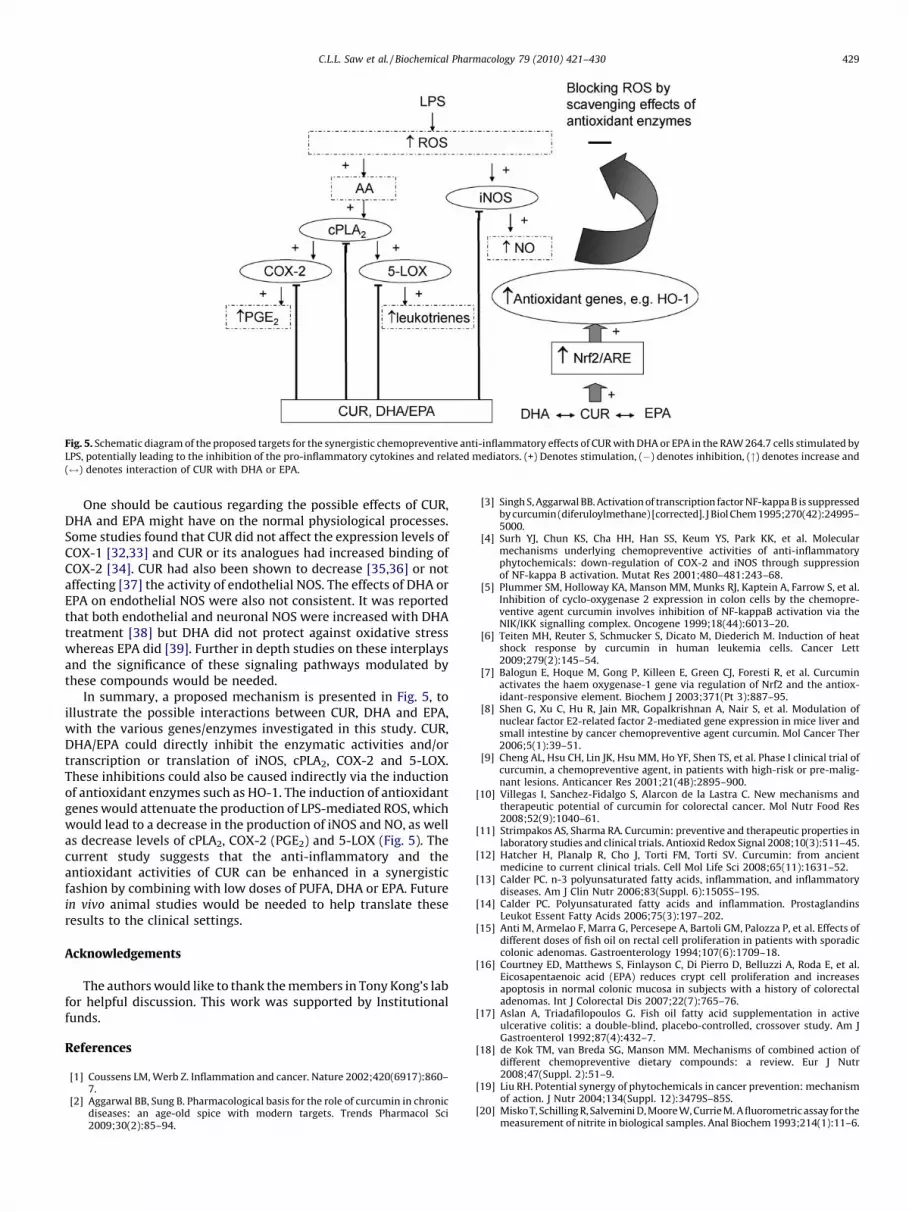
Fig. 5. Schematic diagram of the proposed targets for the synergistic chemopreventive anti-inflammatory effects of CUR with DHA or EPA in the RAW 264.7 cells stimulated by
LPS, potentially leading to the inhibition of the pro-inflammatory cytokines and related mediators. (+) Denotes stimulation, (�) denotes inhibition, (") denotes increase and
($) denotes interaction of CUR with DHA or EPA.
C.L.L. Saw et al. / Biochemical Pharmacology 79 (2010) 421–430 429
One should be cautious regarding the possible effects of CUR,DHA and EPA might have on the normal physiological processes.Some studies found that CUR did not affect the expression levels ofCOX-1 [32,33] and CUR or its analogues had increased binding ofCOX-2 [34]. CUR had also been shown to decrease [35,36] or notaffecting [37] the activity of endothelial NOS. The effects of DHA orEPA on endothelial NOS were also not consistent. It was reportedthat both endothelial and neuronal NOS were increased with DHAtreatment [38] but DHA did not protect against oxidative stresswhereas EPA did [39]. Further in depth studies on these interplaysand the significance of these signaling pathways modulated bythese compounds would be needed.
In summary, a proposed mechanism is presented in Fig. 5, toillustrate the possible interactions between CUR, DHA and EPA,with the various genes/enzymes investigated in this study. CUR,DHA/EPA could directly inhibit the enzymatic activities and/ortranscription or translation of iNOS, cPLA2, COX-2 and 5-LOX.These inhibitions could also be caused indirectly via the inductionof antioxidant enzymes such as HO-1. The induction of antioxidantgenes would attenuate the production of LPS-mediated ROS, whichwould lead to a decrease in the production of iNOS and NO, as wellas decrease levels of cPLA2, COX-2 (PGE2) and 5-LOX (Fig. 5). Thecurrent study suggests that the anti-inflammatory and theantioxidant activities of CUR can be enhanced in a synergisticfashion by combining with low doses of PUFA, DHA or EPA. Futurein vivo animal studies would be needed to help translate theseresults to the clinical settings.
Acknowledgements
The authors would like to thank the members in Tony Kong’s labfor helpful discussion. This work was supported by Institutionalfunds.
References
[1] Coussens LM, Werb Z. Inflammation and cancer. Nature 2002;420(6917):860–7.
[2] Aggarwal BB, Sung B. Pharmacological basis for the role of curcumin in chronicdiseases: an age-old spice with modern targets. Trends Pharmacol Sci2009;30(2):85–94.
[3] Singh S, Aggarwal BB. Activation of transcription factor NF-kappa B is suppressedby curcumin (diferuloylmethane) [corrected]. J Biol Chem 1995;270(42):24995–5000.
[4] Surh YJ, Chun KS, Cha HH, Han SS, Keum YS, Park KK, et al. Molecularmechanisms underlying chemopreventive activities of anti-inflammatoryphytochemicals: down-regulation of COX-2 and iNOS through suppressionof NF-kappa B activation. Mutat Res 2001;480–481:243–68.
[5] Plummer SM, Holloway KA, Manson MM, Munks RJ, Kaptein A, Farrow S, et al.Inhibition of cyclo-oxygenase 2 expression in colon cells by the chemopre-ventive agent curcumin involves inhibition of NF-kappaB activation via theNIK/IKK signalling complex. Oncogene 1999;18(44):6013–20.
[6] Teiten MH, Reuter S, Schmucker S, Dicato M, Diederich M. Induction of heatshock response by curcumin in human leukemia cells. Cancer Lett2009;279(2):145–54.
[7] Balogun E, Hoque M, Gong P, Killeen E, Green CJ, Foresti R, et al. Curcuminactivates the haem oxygenase-1 gene via regulation of Nrf2 and the antiox-idant-responsive element. Biochem J 2003;371(Pt 3):887–95.
[8] Shen G, Xu C, Hu R, Jain MR, Gopalkrishnan A, Nair S, et al. Modulation ofnuclear factor E2-related factor 2-mediated gene expression in mice liver andsmall intestine by cancer chemopreventive agent curcumin. Mol Cancer Ther2006;5(1):39–51.
[9] Cheng AL, Hsu CH, Lin JK, Hsu MM, Ho YF, Shen TS, et al. Phase I clinical trial ofcurcumin, a chemopreventive agent, in patients with high-risk or pre-malig-nant lesions. Anticancer Res 2001;21(4B):2895–900.
[10] Villegas I, Sanchez-Fidalgo S, Alarcon de la Lastra C. New mechanisms andtherapeutic potential of curcumin for colorectal cancer. Mol Nutr Food Res2008;52(9):1040–61.
[11] Strimpakos AS, Sharma RA. Curcumin: preventive and therapeutic properties inlaboratory studies and clinical trials. Antioxid Redox Signal 2008;10(3):511–45.
[12] Hatcher H, Planalp R, Cho J, Torti FM, Torti SV. Curcumin: from ancientmedicine to current clinical trials. Cell Mol Life Sci 2008;65(11):1631–52.
[13] Calder PC. n-3 polyunsaturated fatty acids, inflammation, and inflammatorydiseases. Am J Clin Nutr 2006;83(Suppl. 6):1505S–19S.
[14] Calder PC. Polyunsaturated fatty acids and inflammation. ProstaglandinsLeukot Essent Fatty Acids 2006;75(3):197–202.
[15] Anti M, Armelao F, Marra G, Percesepe A, Bartoli GM, Palozza P, et al. Effects ofdifferent doses of fish oil on rectal cell proliferation in patients with sporadiccolonic adenomas. Gastroenterology 1994;107(6):1709–18.
[16] Courtney ED, Matthews S, Finlayson C, Di Pierro D, Belluzzi A, Roda E, et al.Eicosapentaenoic acid (EPA) reduces crypt cell proliferation and increasesapoptosis in normal colonic mucosa in subjects with a history of colorectaladenomas. Int J Colorectal Dis 2007;22(7):765–76.
[17] Aslan A, Triadafilopoulos G. Fish oil fatty acid supplementation in activeulcerative colitis: a double-blind, placebo-controlled, crossover study. Am JGastroenterol 1992;87(4):432–7.
[18] de Kok TM, van Breda SG, Manson MM. Mechanisms of combined action ofdifferent chemopreventive dietary compounds: a review. Eur J Nutr2008;47(Suppl. 2):51–9.
[19] Liu RH. Potential synergy of phytochemicals in cancer prevention: mechanismof action. J Nutr 2004;134(Suppl. 12):3479S–85S.
[20] Misko T, Schilling R, Salvemini D, Moore W, Currie M. A fluorometric assay for themeasurement of nitrite in biological samples. Anal Biochem 1993;214(1):11–6.
C.L.L. Saw et al. / Biochemical Pharmacology 79 (2010) 421–430430
[21] Lee J, Kong M, Ayers G, Lotan R. Interaction index and different methods fordetermining drug interaction in combination therapy. J Biopharm Stat2007;17(3):461–80.
[22] Saw CL, Olivo M, Chin WW, Soo KC, Heng PW. Transport of hypericin acrosschick chorioallantoic membrane and photodynamic therapy vasculatureassessment. Biol Pharm Bull 2005;28(6):1054–60.
[23] Rusca A, Di Stefano AF, Doig MV, Scarsi C, Perucca E. Relative bioavailabilityand pharmacokinetics of two oral formulations of docosahexaenoic acid/eicosapentaenoic acid after multiple-dose administration in healthy volun-teers. Eur J Clin Pharmacol 2009;65(5):503–10.
[24] Siveen KS, Kuttan G. Role of macrophages in tumour progression. ImmunolLett 2009;123(2):97–102.
[25] Yuan A, Chen JJ, Yang PC. Pathophysiology of tumor-associated macrophages.Adv Clin Chem 2008;45:199–223.
[26] Dirkx AE, Oude Egbrink MG, Wagstaff J, Griffioen AW. Monocyte/macrophageinfiltration in tumors: modulators of angiogenesis. J Leukoc Biol 2006;80(6):1183–96.
[27] Lewis CE, Pollard JW. Distinct role of macrophages in different tumor micro-environments. Cancer Res 2006;66(2):605–12.
[28] Coruzzi G, Venturi N, Spaggiari S. Gastrointestinal safety of novel nonsteroidalantiinflammatory drugs: selective COX-2 inhibitors and beyond. Acta Biomed2007;78(2):96–110.
[29] Xu GL, Liu F, Ao GZ, He SY, Ju M, Zhao Y, et al. Anti-inflammatory effects andgastrointestinal safety of NNU-hdpa, a novel dual COX/5-LOX inhibitor. Eur JPharmacol 2009;611(1–3):100–6.
[30] Wang X, Xue H, Xu Q, Zhang K, Hao X, Wang L, et al. p38 kinase/cytosolicphospholipase A2/cyclooxygenase-2 pathway: a new signaling cascade forlipopolysaccharide-induced interleukin-1beta and interleukin-6 release in dif-ferentiated U937 cells. Prostaglandins Other Lipid Mediat 2008;86(1–4):61–7.
[31] Hong J, Bose M, Ju J, Ryu JH, Chen X, Sang S, et al. Modulation of arachidonicacid metabolism by curcumin and related beta-diketone derivatives: effectson cytosolic phospholipase A(2), cyclooxygenases and 5-lipoxygenase. Carci-nogenesis 2004;25(9):1671–9.
[32] Park C, Kim GY, Kim GD, Choi BT, Park YM, Choi YH. Induction of G2/M arrestand inhibition of cyclooxygenase-2 activity by curcumin in human bladdercancer T24 cells. Oncol Rep 2006;15(5):1225–31.
[33] Park C, Moon DO, Choi IW, Choi BT, Nam TJ, Rhu CH, et al. Curcumin inducesapoptosis and inhibits prostaglandin E(2) production in synovial fibroblasts ofpatients with rheumatoid arthritis. Int J Mol Med 2007;20(3):365–72.
[34] Handler N, Jaeger W, Puschacher H, Leisser K, Erker T. Synthesis of novelcurcumin analogues and their evaluation as selective cyclooxygenase-1 (COX-1) inhibitors. Chem Pharm Bull (Tokyo) 2007;55(1):64–71.
[35] Basaran UN, Dokmeci D, Yalcin O, Inan M, Kanter M, Aydogdu N, et al. Effect ofcurcumin on ipsilateral and contralateral testes after unilateral testiculartorsion in a rat model. Urol Int 2008;80(2):201–7.
[36] Shin HJ, Lee JY, Son E, Lee DH, Kim HJ, Kang SS, et al. Curcumin attenuates thekainic acid-induced hippocampal cell death in the mice. Neurosci Lett2007;416(1):49–54.
[37] Shen SQ, Zhang Y, Xiang JJ, Xiong CL. Protective effect of curcumin against liverwarm ischemia/reperfusion injury in rat model is associated with regulation ofheat shock protein and antioxidant enzymes. World J Gastroenterol2007;13(13):1953–61.
[38] Li P, Kim SW, Li X, Datta S, Pond WG, Wu G. Dietary supplementation withcholesterol and docosahexaenoic acid increases the activity of the arginine-nitric oxide pathway in tissues of young pigs. Nitric Oxide 2008;19(3):259–65.
[39] Tardivel S, Gousset-Dupont A, Robert V, Pourci ML, Grynberg A, Lacour B.Protective effects of EPA and deleterious effects of DHA on eNOS activity in Eahy 926 cultured with lysophosphatidylcholine. Lipids 2009;44(3):225–35.
Related Documents