Marmara Medical Journal VolumeU No:1 January 1998 Case Report SYMPTOMATIC PINEAL CYST: CASE REPORT (Received, 10 March 1996) M. Z. Berkman, M.D.* / S. Uluer, M.D.* / S. Bek, M.D.* T. Derinkok, M.D.* / D. Ozcan, M.D.** * Specialist, Department of neurosurgery. Social Security Okmeydanı Hospital, Istanbul, Turkey. ** Specialist, Department of Pathology, Social Seci my Okmeydanı Hospital, Istanbul, Turkey. ABSTRACT Although pineal cysts are incidental findings in as many as 1,4 - 4,3% of magnetic resonance imaging studies, symptomatic pineal cysts are quite rare because pineal cysts cannot be detected until they reach to a size that can cause compression symptoms. We present a case of symptomatic pineal cyst with complaints of mental dullness and headache for the last three years. His neurological examination revealed only bilateral upgaze palsy. CT and MRI scanning showed a regionally calcified cystic tumour at the pineal region with 1,5 cm in diameter which was thought to be the reason of Parinaud's syndrome. UGA, sitting position, by supratentorial suboccipital approach the cystic lesion was totally extirpated. Histological examination confirmed the diagnosis of a pineal cyst. Asymptomatic incidental cases do not require treatment but if there are neurological findings usually those that are more than 1,5-2 cm diameter then the cyst must be totally extirpated surgically. Key Words: Computerized tomography, Magnetic resonance imaging, Pineal cyst. INTRODUCTION Pineal cysts, which are usually encountered incidentally at autopsy studies are pathologies rarely reaching large dimensions and presenting with clinical findings of the pineal tumours. Recently, by the routine use of computerized tomography (CT) and magnetic resonance imaging (MRI) techniques, discovery rate of the pineal cysts has increased. The incidence is reported to be between 1,4 - 4,3 % among patients examined by MR imaging for neurological symptoms (1-4). In this report, we describe a case of symptomatic pineal cyst causing superior collicular compression with Parinaud's syndrome. CASE REPORT A 24 year old male patient was admitted with the complaints of mental dullness and headache which began three years ago and gradually became more disagreeable. There were no remarkable findings in personal and family history. Except for bilateral upgaze palsy no other pathological findings have been detected at physical and neurological examinations. Skull X-rays were normal. CT scan showed a regionally calcified cystic tumour at the pineal region 1,5 cm in diameter, centrally hypodense with surrounding hyperdensity (Fig. 1). MR findings revealed a cystic tumour of 1,5 cm in diameter at the pineal region in T1 and T2 weightings central hypointensity with peripheral hyperdensity which was more remarkable in T2 weighting (Fig.2). At sitting position, right occipital craniotomy has been done. After opening the dura and incising tentorium by retracting occipital lobe medially, gray pinkish colored, 1,5 cm in diameter, cystic tumor filling the pineal region has been observed. Clear cystic fluid has been aspirated by needle. Then by touring around the tumoral lesion it has totally been extracted. After confirming absolute haemostasis, dura and scalp has been closed. In histopathological observations, a cytic structure formed by outer surface glial tissue and inner surface solid medium comprising uniform cells in structure with hyperchromatic nuclei translucent and narrow in shape was noted. In stroma in which these cells were organized in alveolar fashion divided by thin vascular septates, broad calcification spots were localized. Smears prepared by cystic aspiration materials gave no remarkable cellular elements to take into consideration. Diagnosis was confirmed as pineal cyst (Fig.3). 27

Welcome message from author
This document is posted to help you gain knowledge. Please leave a comment to let me know what you think about it! Share it to your friends and learn new things together.
Transcript
Marmara Medical Journal VolumeU No:1 January 1998
Case Report
SYMPTOMATIC PINEAL CYST: CASE REPORT
(Received, 10 March 1996)
M . Z . Berkm an, M .D .* / S. U luer, M .D .* / S. Bek, M .D .* T. D erinkok, M .D .* / D. Ozcan, M .D .**
* Specialist, Departm ent o f neurosurgery. Social Security Okmeydanı Hospital, Istanbul, Turkey. ** Specialist, Departm ent o f Pathology, Social Seci m y Okm eydanı Hospital, Istanbul, Turkey.
ABSTRACT
Although pineal cysts are incidental findings in as many as 1,4 - 4,3% of magnetic resonance imaging studies, symptomatic pineal cysts are quite rare because pineal cysts cannot be detected until they reach to a size that can cause compression symptoms. We present a case of symptomatic pineal cyst with complaints of mental dullness and headache for the last three years. His neurological examination revealed only bilateral upgaze palsy. CT and MRI scanning showed a regionally calcified cystic tumour at the pineal region with 1,5 cm in diameter which was thought to be the reason of Parinaud's syndrome. UGA, sitting position, by supratentorial suboccipital approach the cystic lesion was totally extirpated. Histological examination confirmed the diagnosis of a pineal cyst. Asymptomatic incidental cases do not require treatment but if there are neurological findings usually those that are more than 1,5-2 cm diameter then the cyst must be totally extirpated surgically.
K ey W ords: Computerized tomography, Magnetic resonance imaging, Pineal cyst.
INTRODUCTION
Pineal cysts, which are usually encountered incidentally at autopsy studies are pathologies rarely reaching large dimensions and presenting with clinical findings of the pineal tumours. Recently, by the routine use of computerized tomography (CT) and magnetic resonance imaging (MRI) techniques, discovery rate of the pineal cysts has increased. The incidence is reported to be between 1,4 - 4,3 % among patients examined by MR imaging for neurological symptoms (1-4).
In this report, we describe a case of symptomatic pineal cyst causing superior collicular compression with Parinaud's syndrome.
CASE REPORT
A 24 year old male patient was admitted with the complaints of mental dullness and headache which began three years ago and gradually became more disagreeable.
There were no remarkable findings in personal and family history.
Except for bilateral upgaze palsy no other pathological findings have been detected at physical and neurological examinations.
Skull X-rays were normal. CT scan showed a regionally calcified cystic tumour at the pineal region1,5 cm in diameter, centrally hypodense with surrounding hyperdensity (Fig. 1). MR findings revealed a cystic tumour of 1,5 cm in diameter at the pineal region in T1 and T2 weightings central hypointensity with peripheral hyperdensity which was more remarkable in T2 weighting (Fig.2).
At sitting position, right occipital craniotomy has been done. After opening the dura and incising tentorium by retracting occipital lobe medially, gray pinkish colored, 1,5 cm in diameter, cystic tumor filling the pineal region has been observed. Clear cystic fluid has been aspirated by needle. Then by touring around the tumoral lesion it has totally been extracted. After confirming absolute haemostasis, dura and scalp has been closed.
In histopathological observations, a cytic structure formed by outer surface glial tissue and inner surface solid medium comprising uniform cells in structure with hyperchromatic nuclei translucent and narrow in shape was noted. In stroma in which these cells were organized in alveolar fashion divided by thin vascular septates, broad calcification spots were localized. Smears prepared by cystic aspiration materials gave no remarkable cellular elements to take into consideration. Diagnosis was confirmed as pineal cyst (Fig.3).
27

Marmara Medical Journal VolumeU No:1 January 1998
In the postoperative period, patient immediately recovered from upgaze palsy and in the 7th postoperative day the patient was discharged.
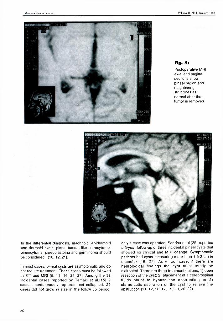
In the follow up visit 1 year after the operation, there was no neurological deficit detected in the examination. Pineal region and the neighbouring structures were reported as normal in the cranial MRI which has been taken at the follow up (Fig.4).
DISCUSSION
Pineal cysts are benign lesions which are encountered at autopy studies at a rate of 25-40% (5-7). Hasegawa et al. have detected 34 (21.8%) macroscopic, 28 (17,9%) microscopic pineal cysts among 168 autopsy cases (6).
Pineal cysts are seen rarely, because pineal cysts cannot be detected until they reach to a size that cause compression symptoms (1-3). Recently by the routine use of CT and MRI techniques, discovery rate of pineal cysts has increased. The incidence is reported to be between 1,4 - 4,3% among patients with neurological symptoms (1-4).
Tumors arising from the pineal region commonly cause symptoms by directly compressing neighboring structure. In the symptomatic pineal cysts, we come across headache, nausea, vomiting, mental changes, upgaze palsy (Parinaud's syndrome), diplopia, visual field defects, endocrinopathies like puberte precox, Dl, hypogonadism (8-17), Headache, mental
changes, upgaze palsy were the findings detected in our case.
There are different approaches to the pathogenesis of the cyst (5, 7-13, 17-19): 1- The cyst takes its origin from the remnants of cavum pineale which is formed by the diverticulation and proliferation of the cells on the roof of third ventricle during early fetal development, 2- After ependymal layer degeneration and cystic development, 3- By cavitation in the gliotic region formed by ischemic degeneration.
Histologically, a pineal cyst is a nonneoplastic lesion. The cyst containts clear, slightly xanthochromic or frankly hemorrhagic fluid (1, 4, 5. 8, 11-14, 17, 18, 20). Histopathological characteristic markings: 1- the cyst wall typically has 3 layers: a) collagenous fibers, b) glial-like cells, c) normal pineal cells, 2- the cyst wall is thin, 100-300 pm in thickness, 3- almost always there is calcification in the collagenous fibers. Benign pineal cysts are considered as the glial cysts of the pineal gland because of the previously reported cases which were almost always with thick glial tissue formed by fibrillar astrocytoma in the periphery of the cyst (1,3, 9, 13, 17, 18, 20-22).
On CT, a pineal cyst is seen as a low density region. It is not contrast enhanced and there may be calcifications around the cyst (2-4, 9, 11, 12, 16, 17, 20, 21, 23). On MRI in T1 weighting it is seen as hypointense in T2 weighting it is seen as hyperintense (1-4,9,10,12, 15-17, 19-21, 24, 25). CT and MRI findings of our case are typical for a pineal cyst.
Fig. 1:CT displays a regionally calcified cystic tumour at the pineal region centrally hypodense with surrounding hyperdensity.
28

Marmara Medical Journal VolumeU No:1 January 1998
' -v 1... j
1314 *
Fig. 2:Preoperative MRI axial and sagittal sections show a
cystic tumor at the pineal region with
marked central hypointensity and
peripheral hyperdensity in
T1.
Fig. 3sHistopathological preparation demonstrating a cystic structure formed by outer surface glial tissue and inner surface solid medium comprising uniform cells with hyperchromatic translucent narrow nuclei. (H&E x 100)
29
Marmara Medical Journal VolumeU No:1 January 1998
Fig. 4:Postoperative MRI axial and sagittal sections show pineal region and neighboring structures as normal after the tumor is removed.
In the differential diagnosis, arachnoid, epidermoid and dermoid cysts, pineal tumors like astrocytoma, pineocytoma, pineoblastoma and germinoma should be considered. (10,12,21).
In most cases, pineal cysts are asymptomatic and do not require treatment. These cases must be followed by CT and MRI (9, 11, 16, 26. 27). Among the 32 Incidental cases reported by Tamaki et al.(15) 2 cases spontaneously ruptured and collapsed, 29 cases did not grow in size in the follow up period,
only 1 case was operated. Sandhu et al (25) reported a 3-year follow-up of three incidental pineal cysts that showed no clinical and MRI change. Symptomatic patients had cysts measuring more than 1,5-2 cm in diameter (16, 27). As in our case, if there are neurological findings the cyst must totally be extirpated. There are three treatment options: 1) open resection of the cyst; 2) placement of a cerebrospinal fluids shunt to bypass the obstruction; or 3) stereotactic aspiration of the cyst to relieve the obstruction (11, 12, 16, 17, 19, 20, 26, 27).
30
Marmara Medical Journal VolumeU No:1 January 1998
REFERENCES
1. Lee DH, Norm an D, Newton TH. MR im aging o f p in e a l cys ts . J C o m p u t A s s is t T o m o g r 1 9 8 7 ;11:586-590.
2. Lum OB, W illiam JF, M achen BC, A kka ra ju V. B en ign c y s tic p in e a l le s io n s by m ag n e tic resonance im aging. J C o m pu t A ss is t Tom ogr 1987;11:228-235.
3. M am ourian AC, T ow figh i J. P inea l cysts: MR imaging. AJNR 1986;7 :1081-1086.
4. O sborne RE, Deen HG, B erbe r CW, Class RF. Acase o f h e m o rrh a g ic p in e a l cys t: M R/CTcorre lation. Neuroradiology 1989 ;31:187-189.
5. C arr JL. Cystic hydrops o f the p in e a l g land. J Nerv Dis 1994;99:552-572.
6. Hasegawa A, O htsubo K, M ori W. Pineal g land in o ld age , q u a n t i ta t iv e a n d q u a l i ta t iv e m o rp h o lo g ic a l s tu d y o f 168 hum an au to p sy cases. Brian Res 1987;409-343-349.
7. Tapp E, Huxley M. The h is to log ica l appearance o f the hum an p ine a l g land from pu be rty to o ld age. J Pathol 1972 ;108:137-144.
8. Cooper ERA. The hum an p ine a l g land and p inea l cyst. J A n a t 1932;67:28-46.
9. Fain J, Tom linson FH, S che ithaue r BW, e t al. S ym ptom atic g lia l cysts o f the p in e a l g land. J Neurosurg 1994;80:454-460.
10. Hadju SI, Porro RS. L ieberm ann PH, Foote FW. Degeneration o f the p ine a l g land o f pa tien ts with cancer. Cancer 1972;29:706-709.
11. K le in P, R ubinste in LJ. Benign sym ptom atic g lia l cysts o f the p inea l g land: A rep o rt o f seven cases and rev iew o f the lite ra ture . J N euro l Neurosurg Psychiatry 1989 ;52:991-995.
!2 . M au re r PK, E c lu nd J, P aris i JE, O ndra S. S y m p to m a tic p in e a l c y s t: Case re p o r t.Neurosurgery 1 9 9 0 ;2 7 :4 5 1 -454.
13. Russell DS, R ubenste in LJ. Cysts o f the the p in e a l g land. In : P atho logy o f tu m o rs o f the nervous system , 5 th ed. B a ltim ore : W illiam s & Wilkins, 1989:726-727.
14. Sevitt S, Schorstein J. A case o f p ine a l cyst. BMJ 1947;2 :490-491.
15. Tam aki N, S h ira ta k i K, Lin T, M asum ura M, Katayama S, M atsum oto S. Cysts o f the p inea l gland. Childs Nerv Syst I98 9 ;5 :1 72-1 76.
16. Todo T, Hondo T, Shinoura N, Yamada R. Large cysts o f the p in e a l g land: Report o f two cases. Neurosurgery 1 9 9 1 ;2 9 :101 -106.
17. Welton PL, Re icher MA, Kelle rhouse LE, O tt KH. MR o f benign p inea l cyst AJNR 19 8 8 :9 :6 12.
18. Apuzzo MLJ, Davey LM, M anue lid is EE. Pineal apop lexy associa ted w ith an ticoagu lan t therapy. J Neurosurg 1976;45:223-226.
19. F e te ll MR, B ru ce JN , B u rke AM, e t al. N o n n e o p la s t ic p in e a l cys ts . N e u ro lo g y 1991;41:1034-1040.
20. Vaquero J, M artinez R, Escandon J, Bravo O. S ym ptom atic g lia l cysts o f the p inea l gland. Surg Neurol 1988;30:468-470.
21. M iyatake S, K iku ch i H, Yamasaki T, e t al. G lia l c y s t o f the p in e a l g la n d w ith c o m p u te d tom ography, m agnetic resonance im aging, and pa tho log ica l find ings: Report o f two cases. Surg N euro l 1992;37:293-299.
22. Reyes PF. Age re la ted h is to log ic changes in the hu m an p in e a l g la n d . P rog C lin B io l Res 1982;92:253-261.
23. Abreo K, Steele TH. P ineal cyst associa ted with p o lycys tic k id n e y d isease: Case report. Am J Kidney Dis 1981;1 :106-109.
24. M am ourian AC, Yarnel T. Enhancem ent o f p ineal cysts on MR images. AJNR 19 9 1; 12:773-774.
25. S an dh u JS, M c L a u g h lin LR, G om ez CR. C h ara c te ris tics o f in c id e n ta l p in e a l cysts on m a g n e tic reso na nce im ag ing . N eurosu rge ry 1989;25:636-640.
26. M uso lino A, C am bria S, Rizzo G, Cam bria M. S y m p to m a tic cys ts o f th e p in e a l g la n d : S te re o tac tic d iagnos is an d tre a tm e n t o f two cases and rev iew o f the lite rature . Neurosurgery 1993;32:315-321.
27. Stern JD, Ross DA: S tereotactic m anagem ent o f benign p in e a l reg ion cysts: Report o f two cases. Neurosurgery 1993 ;32 :310 -3 14.
31
Related Documents